Whole Cell and Cell Organelle Immobilization on Siliceous Surfaces
Upload
khangminh22Category
view
0download
0
�����������������
Citation: Arnold, J.; Chapman, J.;
Arnold, M.; Dinu, C.Z. Hyaluronic
Acid Allows Enzyme Immobilization
for Applications in Biomedicine.
Biosensors 2022, 12, 28. https://
doi.org/10.3390/bios12010028
Received: 1 November 2021
Accepted: 31 December 2021
Published: 7 January 2022
Publisher’s Note: MDPI stays neutral
with regard to jurisdictional claims in
published maps and institutional affil-
iations.
Copyright: © 2022 by the authors.
Licensee MDPI, Basel, Switzerland.
This article is an open access article
distributed under the terms and
conditions of the Creative Commons
Attribution (CC BY) license (https://
creativecommons.org/licenses/by/
4.0/).
biosensors
Review
Hyaluronic Acid Allows Enzyme Immobilization forApplications in BiomedicineJackie Arnold 1, Jordan Chapman 1, Myra Arnold 2,3 and Cerasela Zoica Dinu 1,*
1 Department of Chemical and Biomedical Engineering, Benjamin M. Statler College of Engineering andMineral Resources, West Virginia University, Morgantown, WV 26505, USA; [email protected] (J.A.);[email protected] (J.C.)
2 Department of Sociology and Anthropology, Eberly College of Arts and Sciences, West Virginia University,Morgantown, WV 26505, USA; [email protected]
3 Department of Business Incubator, John Chambers College of Business and Economics, West VirginiaUniversity, Morgantown, WV 26505, USA
* Correspondence: [email protected]
Abstract: Enzymes are proteins that control the efficiency and effectiveness of biological reactions andsystems, as well as of engineered biomimetic processes. This review highlights current applications ofa diverse range of enzymes for biofuel production, plastics, and chemical waste management, as wellas for detergent, textile, and food production and preservation industries respectively. Challengesregarding the transposition of enzymes from their natural purpose and environment into syntheticpractice are discussed. For example, temperature and pH-induced enzyme fragilities, short shelflife, low-cost efficiency, poor user-controllability, and subsequently insufficient catalytic activitywere shown to decrease pertinence and profitability in large-scale production considerations. En-zyme immobilization was shown to improve and expand upon enzyme usage within a profit andimpact-oriented commercial world and through enzyme-material and interfaces integration. Withparticular focus on the growing biomedical market, examples of enzyme immobilization withinor onto hyaluronic acid (HA)-based complexes are discussed as a definable way to improve uponand/or make possible the next generation of medical undertakings. As a polysaccharide formed inevery living organism, HA has proven beneficial in biomedicine for its high biocompatibility andcontrollable biodegradability, viscoelasticity, and hydrophilicity. Complexes developed with thismolecule have been utilized to selectively deliver drugs to a desired location and at a desired rate,improve the efficiency of tissue regeneration, and serve as a viable platform for biologically acceptedsensors. In similar realms of enzyme immobilization, HA’s ease in crosslinking allows the moleculeto user-controllably enhance the design of a given platform in terms of both chemical and physicalcharacteristics to thus best support successful and sustained enzyme usage. Such examples do notonly demonstrate the potential of enzyme-based applications but further, emphasize future markettrends and accountability.
Keywords: enzyme; biomedicine; hyaluronic acid; immobilization; bioengineering; catalysis; nanomaterials
1. Introduction
Working from the cellular to organismic level, each of the physiological systemsthat govern the biological world requires and/or benefits from the inherited abilities ofenzymes, proteins that catalyze a wide variety of unique and specific processes. Suchbiocatalysts decrease the necessary energy input to facilitate biological reactions withincredible selectivity sparking interest for synthetic implementation as active candidates ina variety of engineered applications. There are, however, limitations that accompany thebenefits that enzymes bring to the scientific community, most associated with their pH andtemperature-induced fragility, as well as unprofitable user-controllability, shelf-life, andscale-up systematic integration, just to name a few.
Biosensors 2022, 12, 28. https://doi.org/10.3390/bios12010028 https://www.mdpi.com/journal/biosensors

Biosensors 2022, 12, 28 2 of 21
To mitigate such limitations, enzyme immobilization has become a prevalent approach.This review provides an overview of current implementation of enzymes in synthetic appli-cations with focus on biofuel [1–3], textile [4–8], detergent [4], waste management [8,9], andfood and drink industries [10,11] respectively. Subsequently, particular attention is directedtowards enzyme immobilization using hyaluronic acid (HA) scaffolds, a polysaccharideconsisting of alternating β-1,4-D-glucuronic acid and β-1,3-N-acetyl-D-glucosamine ringsknown for its high biocompatibility, biodegradability, viscoelasticity, and hydrophilic-ity [12–14], to thus help emphasize enzymes’ implementation in biomedical applications.Discussions are in support of specific applications in drug delivery [15–17], tissue engi-neering including wound healing [18–20] and biosensors [21–24], respectively. Challengesassociated with enzymes exploitations in synthetic environment are analyzed. Lastly, poten-tial for marketability and perspectives on consumer usage are provided to thus capitalizeon the extended implementation that enzyme-based products could have.
2. Brief Description of Applications of Enzymes in Consumer Industries2.1. Enzymes in Biofuels
Selective depletion of nonrenewable resources of fossil fuels as well as the high climateimpact that processing of such resources leads to are providing means for alternativeintegration of enzymes for and in biofuels production respectively. Such integrationis foreseen to reduce the environmental factor (E-factor; a known measure of the massof undesired non-water waste produced by a chemical process per mass of its desiredproduct [25]) as well as the dependence on imported fuel [26,27].
Advanced biochemical processes using enzymes are seen to improve biofuel produc-tion efficiency under less energy intensive requirements and possibly with low costs. Suchbiochemical processes frequently either rely on or employ focused genetic engineeringapproaches, based on the use of recombinant DNA technology especially known to pro-duce large quantities of recombinant enzymes, or enzymes that are efficient when usedin the synthetic production of biofuels. Moreover, such approaches have proved to leadto recombinants with high resistance capability toward contaminations and/or to highlythermo-stable forms. For instance, the fatty acid biosynthesis pathway as well as thetriacylglycerol (TAG) assembly of algal enzymes have been manipulated to demonstrateincreases in the oil content of biofuel-producing algae. Over-expression of key enzymesincluding but not limited to acyl-CoA synthetases (ACSs) and diacylglycerol acyltrans-ferase (DGAT), respectively, enabled a more cost-efficient biofuel with operative productionprocesses being realized by increasing the ability of the microalgae to produce lipids andsubsequently fuels [1,2]. The method led to a reduced impact environmental impact and areduction in cost relative to the traditional and more expensive non-modified microalgaebiofuel production systems [2].
Complementary, Council of Scientific and Industrial Research (CSIR)—Institute ofMinerals and Materials Technology in India has developed a database highlighting the en-zymes involved with the lipid biosynthetic pathway (i.e., the process by which microalgaeproduces bioactive lipids such as triacylglycerols [28]), with microalgal enzymes being clas-sified by their length, hydrophobicity, amino acids composition, subcellular location, geneontology, Kyoto Encyclopedia of Genes and Genomes (KEGG) pathway, orthologous group,Pfam domain, intron-exon organization, transmembrane topology, and secondary/tertiarystructural data, respectively [1]. Genetic and physical characteristics of such enzymes weresubsequently compiled for a concise source integration and optimized enzyme utiliza-tion [1]. Analysis by Misra et al., for instance, showed higher photosynthetic efficiency andbiomass production than starch-based and lignocellulosic plant species currently used forbiofuel-purposed lipid production.
Production of hydrogen (H2) for a comparatively clean and renewable option wasalso attempted through the usage of enzyme-facilitated metabolism algal functions [3,29].Among current methods for H2 production are pyrolysis [30–32] and gasification [33,34] ofcarbon-based fossil fuels, sewage sludge, biomass as well as steam reforming of natural

Biosensors 2022, 12, 28 3 of 21
gas [35,36], and electrolysis of water [34,37,38], respectively. However, such methods arelimited by the profitability in the H2 yield, its storage and distribution capabilities, severereaction conditions required at its implementation, as well as related equipment fragilityand energy input necessities [34,35,39] respectively. It was thus predicted that withinthe next 30 years, the H2 production would greatly help shift the global energy system(GES) integration towards more sustainable energy system (SES) [37], whereby greenhousegas emissions will approach net zero [39]. Though enzymes do come with a significantproduction cost, their potential for genetic modification and long-term economic profithave been considered to overcome the challenges associated with traditional methods forH2 production [40]. Meanwhile, the production costs of current commercial ways can onlybe pacified with the adoption of new theory and methodology altogether [33].
In this regard, research by Ben-Zvi et al., utilized algal biocatalysts within the C.reinhardtii for the natural photoproduction of H2 [3,29]. Authors have focused on hydroge-nase, an enzyme capable of catalyzing the production of H2 with incredible activity andefficiency [41,42], and especially [Fe-Fe]-hydrogenase [3], the well-known enzyme usedfor catalytic H2 turnover [41]. [Fe-Fe]-hydrogenase (HydA) and superoxide dismutase(SOD) were fused through genetic code sequencing to form a complex that was shown tolead to increased ability for catalyzed H2 production, with the Hyd-SOD fusion proteinbeing subsequently cloned. Cloned complexes possessed inherent ability to continuouslyand easily form H2; Hyd-SOD clones also saw greater prolonged H2 photoproductionrelative to the D66 C. reinhardtii wild type and a strain exhibiting the HydA1 gene, all whenevaluated following the natural production burst initially experienced (Figure 1a) [3]. Thepresence of the hydrogenase enzymes was imperative for the sustainable H2 generationsince the catalysts reduced the surplus electrons to form a useful energy carrier; moreover,they helped outcompete electron loss to the Calvin–Benson–Bassham (CBB) cycle [3]. Fur-thermore, the metalloenzyme SOD played an important role in increasing continuous andsteady H2 production as well as likely addressing the limitation of HydA in its sensitivityto oxygen (O2), a product of simultaneous water splitting [3].
To protect the hydrogenase from O2 byproducts, Li et al., proposed the use of car-boxysome shells, “virus-like” bacterial organelles that were shown to provide favorablemicroenvironments. Analysis showed increased ability for H2 catalytic performance espe-cially when Escherichia coli were used as genetically modified hosts to allow encapsulationof [Fe-Fe]-hydrogenases and ferredoxin isolated from C. reinhardtii algae [41]. The poten-tial of such complexes was proven in the increased enzymatic resistance to O2 as well asincreased amount of H2 produced with respect to free HydA, especially under aerobic con-ditions in vivo (Figure 1b). Moreover, difference in H2 production between the shell-HydAcomposite and free HydA increased over 100 min when tested in vitro, thus proving asustained benefit to the enzyme technology (Figure 1c). Additionally, purified shell-HydAcomplex maintained 13.33 ± 2.06% of the activity following O2 exposure when comparedto only 0.97 ± 0.15% activity observed for the free enzyme (Figure 1d). The encasementallowed for the hydrogenase complex to be more selectively permeable to substrates thusleading to improved enzymatic activity [41].
In addition to their O2 susceptibilities, Dolui et al., considered their water solubilityand acid stability limitations in applications involving H2-producing hydrogenase enzymes.Utilizing cobaloxime complexes consisting of six derivatives with varying side chains, anenzyme-inspired, multicomponent outer coordination sphere was studied as a means todecrease the effect of O2 on hydrogenase activity as well as to increase enzyme’s rate ofreaction, solubility in water, and resistance to acidic environments respectively. [42]. Au-thors demonstrated that such enzyme-based complexes led to fast, energy efficient, and/orstable catalytic functionality for more profitable integration in enzyme-based renewableenergy sector [42].

Biosensors 2022, 12, 28 4 of 21Biosensors 2022, 11, x FOR PEER REVIEW 4 of 22
(a) (b)
(c) (d)
Figure 1. (a) HydA-SOD and clones of the complex rates of reaction over time. Reprinted with per-
mission from ref. [3]. 2019 Springer Nature. (b) In vivo testing of anaerobic and aerobic pathways of
H2 production for Shell-HydA relative to free HydA enzyme (* signifies p=0.0352 and ** signifies
p=0.0018). Reprinted with permission from ref. [41]. 2020 Springer Nature. (c) In vitro enzyme-me-
diated H2 production over time of Shell-HydA composite relative to free HydA. Reprinted with
permission from ref. [41]. 2020 Springer Nature. (d) Anaerobic relative H2 production for Shell-
HydA and lone HydA found through in vitro testing of activity following 24 h of exposure to O2 at
4 °C (*** signifies p=0.0009). Reprinted with permission from ref. [41]. 2020 Springer Nature.
In addition to their O2 susceptibilities, Dolui et al., considered their water solubility
and acid stability limitations in applications involving H2-producing hydrogenase en-
zymes. Utilizing cobaloxime complexes consisting of six derivatives with varying side
chains, an enzyme-inspired, multicomponent outer coordination sphere was studied as a
means to decrease the effect of O2 on hydrogenase activity as well as to increase enzyme’s
rate of reaction, solubility in water, and resistance to acidic environments respectively.
[42]. Authors demonstrated that such enzyme-based complexes led to fast, energy effi-
cient, and/or stable catalytic functionality for more profitable integration in enzyme-based
renewable energy sector [42].
Lastly, in a study by Zhang et al., a 13-enzyme pathway converted starch and water
into H2 with an improved production yield over previously explored natural, anaerobic
fermentation methods, among others. Such improvement increased from the theoretical
value of 4 H2/glucose to 12 H2/glucose with biocatalyst cascade application. The use of
enzymes benefited from the mild reaction conditions (30 °C and atmospheric pressure),
as well as low production costs (~USD 2/kg H2), and a high energy-density carrier starch
(14.8 H2-based mass %)” [39].
2.2. Enzymes for Waste (Plastic and Chemical) Management
Aside from making an impact on the environment and on the growing demands as-
sociated with renewable energy and sustainable resources creation, enzymes have also
Figure 1. (a) HydA-SOD and clones of the complex rates of reaction over time. Reprinted withpermission from ref. [3]. 2019 Springer Nature. (b) In vivo testing of anaerobic and aerobic pathwaysof H2 production for Shell-HydA relative to free HydA enzyme (* signifies p = 0.0352 and ** signifiesp = 0.0018). Reprinted with permission from ref. [41]. 2020 Springer Nature. (c) In vitro enzyme-mediated H2 production over time of Shell-HydA composite relative to free HydA. Reprinted withpermission from ref. [41]. 2020 Springer Nature. (d) Anaerobic relative H2 production for Shell-HydAand lone HydA found through in vitro testing of activity following 24 h of exposure to O2 at 4 ◦C(*** signifies p = 0.0009). Reprinted with permission from ref. [41]. 2020 Springer Nature.
Lastly, in a study by Zhang et al., a 13-enzyme pathway converted starch and waterinto H2 with an improved production yield over previously explored natural, anaerobicfermentation methods, among others. Such improvement increased from the theoreticalvalue of 4 H2/glucose to 12 H2/glucose with biocatalyst cascade application. The use ofenzymes benefited from the mild reaction conditions (30 ◦C and atmospheric pressure),as well as low production costs (~USD 2/kg H2), and a high energy-density carrier starch(14.8 H2-based mass %)” [39].
2.2. Enzymes for Waste (Plastic and Chemical) Management
Aside from making an impact on the environment and on the growing demands asso-ciated with renewable energy and sustainable resources creation, enzymes have also beeninvestigated for their ability to breakdown plastic [9,43–46] and chemical waste [47–49].
Briefly, current techniques for plastic management consist of chemical or physicalrecycling [43,45], reusage of existing plastic materials, or plastic removal to dedicated land-fills [9,43,45] for incineration [50]. However, such techniques are simply inadequate in theircapacity to remove the environmental, health, and planetary life risk [43,45,50], namely wa-ter pollution, harm to flora and fauna, toxicity, and greenhouse gas emissions, just to namea few of such effects. Further, in the chemical depolymerization of poly(ethylene terephtha-late) (PET) main constituent synthetic polymer commonly used in the packaging [51] and

Biosensors 2022, 12, 28 5 of 21
textile [52] industries [9] or the mechanical recycling of such materials for instance, there isa significant loss to processing costs and resultant byproduct properties, respectively [9].As such, plastics’ inability to degrade quickly and safely requires a complete shift in themethods used for their disposal [43,50].
In a study by Austin et al., for instance, PET-ase enzymes were employed to decomposethe non-biodegradable PET. Biomimetic analysis identified that Ideonella sakaiensis secretesPET-ase to harvest the carbon available within environmentally harmful PET sources. Suchsources include single-use beverage bottles, clothing, packaging, and carpeting. With theuse of PETase, PET was primarily converted to mono(2-hydroxyethyl) terephthalic acid(MHET), though terephthalic acid and bis(2-hydrocyethyl)-TPA are also produced duringcatalysis [9,43]. Studies also showed that by mutating the PETase enzyme, the percentcrystallinity change as well as MHET and TPA production were increased in comparison tothose of the natural PETase enzyme [9].
Among the most significant locations increasingly ailed by plastic waste are theocean ecosystems [43,46,50,53,54]. To help dampen this astounding issue, Moog et al.,utilized Phaeodactylum tricornutum microalga to produce and secrete the PETase enzymefor the degradation of both PET and polyethylene terephthalate glycol (PETG) in saltwateranalysis showed that such PETase possessed degradation activity against both PET andPETG, including under moderate conditions and in saltwater [43] (Figure 2a,b respectively).While Ideonella sakaiensis bacteria was considered for its ability to produce the PETase, thisspecie showed limited performance in ocean-like environments presumably due to itsinstability and inability to survive in such aqueous habitats [43,55].
Biosensors 2022, 11, x FOR PEER REVIEW 5 of 23
2.2. Enzymes for Waste (Plastic and Chemical) Management Aside from making an impact on the environment and on the growing demands as-
sociated with renewable energy and sustainable resources creation, enzymes have also been investigated for their ability to breakdown plastic [9,43–46] and chemical waste [47–49].
Briefly, current techniques for plastic management consist of chemical or physical recycling [43,45], reusage of existing plastic materials, or plastic removal to dedicated landfills [9,43,45] for incineration [50]. However, such techniques are simply inadequate in their capacity to remove the environmental, health, and planetary life risk [43,45,50], namely water pollution, harm to flora and fauna, toxicity, and greenhouse gas emissions, just to name a few of such effects. Further, in the chemical depolymerization of poly(eth-ylene terephthalate) (PET) main constituent synthetic polymer commonly used in the packaging [51] and textile [52] industries [9] or the mechanical recycling of such materials for instance, there is a significant loss to processing costs and resultant byproduct proper-ties, respectively [9]. As such, plastics’ inability to degrade quickly and safely requires a complete shift in the methods used for their disposal [43,50].
In a study by Austin et al., for instance, PET-ase enzymes were employed to decom-pose the non-biodegradable PET. Biomimetic analysis identified that Ideonella sakaiensis secretes PET-ase to harvest the carbon available within environmentally harmful PET sources. Such sources include single-use beverage bottles, clothing, packaging, and car-peting. With the use of PETase, PET was primarily converted to mono(2-hydroxyethyl) terephthalic acid (MHET), though terephthalic acid and bis(2-hydrocyethyl)-TPA are also produced during catalysis [9,43]. Studies also showed that by mutating the PETase en-zyme, the percent crystallinity change as well as MHET and TPA production were in-creased in comparison to those of the natural PETase enzyme [9].
Among the most significant locations increasingly ailed by plastic waste are the ocean ecosystems [43,46,50,53,54]. To help dampen this astounding issue, Moog et al., utilized Phaeodactylum tricornutum microalga to produce and secrete the PETase enzyme for the degradation of both PET and polyethylene terephthalate glycol (PETG) in saltwater anal-ysis showed that such PETase possessed degradation activity against both PET and PETG, including under moderate conditions and in saltwater [43] (Figure 2a,b respectively). While Ideonella sakaiensis bacteria was considered for its ability to produce the PETase, this specie showed limited performance in ocean-like environments presumably due to its in-stability and inability to survive in such aqueous habitats [43,55].
(a) (b)
Biosensors 2022, 11, x FOR PEER REVIEW 6 of 23
(c)
Figure 2. (a) Untreated PET film, as seen via scanning electron microscopy, and representative of the standard, smooth surface of a PET-based bottle film. Reprinted with permission from ref. [43]. 2019 Springer Nature. (b) Scanning electron microscopy analysis of PET bottle film following five weeks of exposure to and subsequent degradation by cells expressing AP _PETase FLAG; degradation is shown as a sign of wear. Reprinted with permission from ref. [43]. 2019 Springer Nature. (c) Plots corresponding the total sum of the concentration of PET degradation products with time following the respective nanoplastics’ and microplastics’ incubation with IsPETase and Du-raPETase. Reprinted with permission from ref. [45]. 2021 American Chemical Society.
Further, Cui et al., applied the greedy accumulated strategy for protein engineering (GRAPE) to improve the robustness of a PETase isolated from Ideonella sakaiensis (IsPETase) [45]. Both DuraPETase and IsPETase caused total nanoplastic degradation fol-lowing treatment for 1 h at 37 °C, according to collected high-performance liquid chroma-tography data (Figure 2c (1)). As for microplastics, DuraPETase was able to breakdown the larger particles to close to completion within two weeks, displaying water treatment activity much greater than those of IsPETase (Figure 2c (2)).
PETase was also active towards low and high-crystallinity plastics such as bottle-grade PET; further, monohydroxythyl terephthalate hydrolase (MHETase), exo-PETase and bis-(2-hydroxyethyl)terephthalate (BHET)ase were also shown to catalyze PET film degradation [46]. Moreover, MHETase completed the biodegradation of PET into its basic components, with hydrolysis activities of MHETase toward PET films showing increased potential to impact PET degradation as a multifaceted approach for efficient waste man-agement [46].
Coppella et al., highlighted the use of parathion hydrolase against organophosphate wastes for insecticide-containing waste degradation [47]. Kinetics observation revealed the rapid enzymatic reaction followed saturation kinetics with authors concluding that parathion hydrolase enzyme was able to hydrolyze coumaphos and potasan under con-ditions that closely simulated those expected in the field [47].
In accordance with the waste generated via US nuclear weapons downsizing around 1999, Vanderberg et al., further demonstrated enzymes’ capability to treat hazardous ra-diologically contaminated heterogeneous paint-stripping waste. Authors showed that cel-lulase degraded cellulose-based organic materials with analysis revealing that cellulose bulk volume was decreased by 80% following cellulase digestion, with the resultant radi-oactive sugar products and additional hazardous metals being easily removed by polymer filtration methods [48].
2.3. Enzymes in Detergent and Textile Industries Biocatalysts are also commonly used in the detergent and textile industries [4–6,8].
Kumari et al., for instance, showed how leaf enzymes could serve as highly stable catalysts for stain removal, biowashing, and biopolishing tasks with mannanase, leaf lipase and
Figure 2. (a) Untreated PET film, as seen via scanning electron microscopy, and representative ofthe standard, smooth surface of a PET-based bottle film. Reprinted with permission from ref. [43].2019 Springer Nature. (b) Scanning electron microscopy analysis of PET bottle film following fiveweeks of exposure to and subsequent degradation by cells expressing APSP_PETaseR280A − FLAG;degradation is shown as a sign of wear. Reprinted with permission from ref. [43]. 2019 SpringerNature. (c) Plots corresponding the total sum of the concentration of PET degradation productswith time following the respective nanoplastics’ and microplastics’ incubation with IsPETase andDuraPETase. Reprinted with permission from ref. [45]. 2021 American Chemical Society.
Biosensors 2022, 12, 28 6 of 21
Further, Cui et al., applied the greedy accumulated strategy for protein engineer-ing (GRAPE) to improve the robustness of a PETase isolated from Ideonella sakaiensis(IsPETase) [45]. Both DuraPETase and IsPETase caused total nanoplastic degradationfollowing treatment for 1 h at 37 ◦C, according to collected high-performance liquid chro-matography data (Figure 2c (1)). As for microplastics, DuraPETase was able to breakdownthe larger particles to close to completion within two weeks, displaying water treatmentactivity much greater than those of IsPETase (Figure 2c (2)).
PETase was also active towards low and high-crystallinity plastics such as bottle-grade PET; further, monohydroxythyl terephthalate hydrolase (MHETase), exo-PETaseand bis-(2-hydroxyethyl)terephthalate (BHET)ase were also shown to catalyze PET filmdegradation [46]. Moreover, MHETase completed the biodegradation of PET into itsbasic components, with hydrolysis activities of MHETase toward PET films showingincreased potential to impact PET degradation as a multifaceted approach for efficientwaste management [46].
Coppella et al., highlighted the use of parathion hydrolase against organophosphatewastes for insecticide-containing waste degradation [47]. Kinetics observation revealedthe rapid enzymatic reaction followed saturation kinetics with authors concluding thatparathion hydrolase enzyme was able to hydrolyze coumaphos and potasan under condi-tions that closely simulated those expected in the field [47].
In accordance with the waste generated via US nuclear weapons downsizing around1999, Vanderberg et al., further demonstrated enzymes’ capability to treat hazardous ra-diologically contaminated heterogeneous paint-stripping waste. Authors showed thatcellulase degraded cellulose-based organic materials with analysis revealing that cellulosebulk volume was decreased by 80% following cellulase digestion, with the resultant radioac-tive sugar products and additional hazardous metals being easily removed by polymerfiltration methods [48].
2.3. Enzymes in Detergent and Textile Industries
Biocatalysts are also commonly used in the detergent and textile industries [4–6,8].Kumari et al., for instance, showed how leaf enzymes could serve as highly stable catalystsfor stain removal, biowashing, and biopolishing tasks with mannanase, leaf lipase andcellobiohydrolase all being tested against common enzymes used in the detergent industry(i.e., nineteen products in liquid and powder forms, including twelve endoglucanases, fivelipases, and two mannanases) [4].
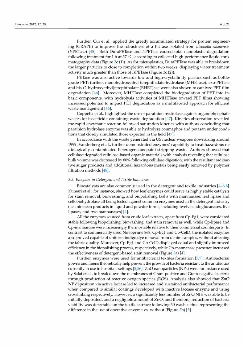
All the enzymes sourced from crude leaf extracts, apart from Cp-Eg1, were consideredstable following biopolishing, biowashing, and stain removal as well, while Cp-lipase andCp-mannanase were increasingly thermostable relative to their commercial counterparts. Incontrast to commercially used Novoprime 868, Cp-Eg1 and Cp-CelD, the isolated enzymesalso proved capable of uniform indigo dye removal from denim samples, without affectingthe fabric quality. Moreover, Cp-Eg1 and Cp-CelD displayed equal and slightly improvedefficiency in the biopolishing process, respectively, while Cp-mannanase presence increasedthe effectiveness of detergent-based stain removal (Figure 3a) [4].
Further, enzymes were used for antibacterial textiles formation [5,7]. Antibacterialgowns and linens theoretically help prevent the growth of bacteria resistant to the antibioticscurrently in use in hospitals settings [5,56]. ZnO nanoparticles (NPs) were for instance usedby Salat et al., to break down the membranes of Gram-positive and Gram-negative bacteriathrough production of reactive oxygen species (ROS). Analysis also showed that ZnONP deposition via active laccase led to increased and sustained antibacterial performancewhen compared to similar coatings developed with inactive laccase enzyme and usingcrosslinking respectively. However, a significantly less number of ZnO NPs was able to beinitially deposited, and a negligible amount of ZnO, and therefore, reduction of bacteriaviability was detectable on the textile surface following 30 washes thus representing thedifference in the use of operative enzyme vs. without (Figure 3b) [5].

Biosensors 2022, 12, 28 7 of 21
Biosensors 2022, 11, x FOR PEER REVIEW 7 of 23
cellobiohydrolase all being tested against common enzymes used in the detergent indus-try (i.e., nineteen products in liquid and powder forms, including twelve endoglucanases, five lipases, and two mannanases) [4].
All the enzymes sourced from crude leaf extracts, apart from Cp-Eg1, were consid-ered stable following biopolishing, biowashing, and stain removal as well, while Cp-li-pase and Cp-mannanase were increasingly thermostable relative to their commercial counterparts. In contrast to commercially used Novoprime 868, Cp-Eg1 and Cp-CelD, the isolated enzymes also proved capable of uniform indigo dye removal from denim sam-ples, without affecting the fabric quality. Moreover, Cp-Eg1 and Cp-CelD displayed equal and slightly improved efficiency in the biopolishing process, respectively, while Cp-man-nanase presence increased the effectiveness of detergent-based stain removal (Figure 3a) [4].
(a) (b)
(c) (d)
Figure 3. (a) Observation of chocolate stain removal as a result of enzyme and non-enzyme contain-ing detergent washes. Reprinted with permission from ref. [4]. 2019 The Authors. (b) Comparison of active and denatured laccase enzymes in their antibacterial capacity, as represented by percent reduction in bacterial viability. Reprinted with permission from ref. [5]. 2018 Elsevier. (c) Visual representation of wool and cotton textile samples before and after self-healing. Reprinted with per-mission from ref. [6]. 2016 American Chemical Society. (d) Efficiency of enzymatic activity, as rep-resented by fluorescence intensity, of multilayer systems both before and after the self-healing pro-cess. Reprinted with permission from ref. [6]. 2016 American Chemical Society.
Further, enzymes were used for antibacterial textiles formation [5,7]. Antibacterial gowns and linens theoretically help prevent the growth of bacteria resistant to the antibi-otics currently in use in hospitals settings [5,56]. ZnO nanoparticles (NPs) were for in-stance used by Salat et al., to break down the membranes of Gram-positive and Gram-
Figure 3. (a) Observation of chocolate stain removal as a result of enzyme and non-enzyme containingdetergent washes. Reprinted with permission from ref. [4]. 2019 The Authors. (b) Comparison ofactive and denatured laccase enzymes in their antibacterial capacity, as represented by percentreduction in bacterial viability. Reprinted with permission from ref. [5]. 2018 Elsevier. (c) Visualrepresentation of wool and cotton textile samples before and after self-healing. Reprinted withpermission from ref. [6]. 2016 American Chemical Society. (d) Efficiency of enzymatic activity, asrepresented by fluorescence intensity, of multilayer systems both before and after the self-healingprocess. Reprinted with permission from ref. [6]. 2016 American Chemical Society.
Moreover, Polak et al., used fungal laccases for synthesizing a phenazine-based dye withantibacterial properties. Resulting dye contained 10-((2-carboxy-6-methoxyphenyl)amino)-11-methoxybenzo[a]phenazine-8-carboxylic acid decreased Staphylococcus aureus growth,a human pathogen known to develop new clones with a multitude of antibacterial re-sistances [57] leading to bacterial infections in the skin, soft tissue, bone, bloodstream,and respiratory tract of human patients [58] respectively. The dye production via Cerrenaunicolor protein catalysis was not only bioactive but was also more sustainable, less toxic,less mutagenic, and easier to replicate when compared to chemical approaches that requireadditional and harmful coupling agents and additives including but not limited to benzene,boron tribromide (BBr3), and dimethylformamide (DMF) [7].
Further, Gaddes et al., applied urease as a proof-of-activity enzymatic component toself-healing textiles formed using versatile polyelectrolyte layer-by-layer (LBL) films alsonotably containing squid ring teeth (SRT) proteins that lend the films the ability to self-adhere in an environmentally friendly, cost-efficient, feasible, and sustainable, large-scaleway [6]. The coating design was envisioned to lead to extension of lifespan for both wovenand nonwoven materials through the ability of the textiles to self-repair damage such as

Biosensors 2022, 12, 28 8 of 21
scratches, holes, or rips (Figure 3c). The number of multilayers applied was a significantcontributor to the enzymes’ activity with activity increasing between 1, 3, and 5 layers,respectively (Figure 3d). Regardless of the number of layers deposited onto the cloth, therewas also only slight loss of this catalytic action following sample repair (Figure 3d) [6].
Lastly, Blánquez et al., showed that bacterial laccases, particularly SilA laccase, decolorand detoxify Acid Black 48, Acid Orange 63, Reactive Black 5, Orange II, Tartrazine, AzureB, Indigo carmine, and Cresol red. Such dyes are known to remain stable and reactivein sewage plants and eventually rivers following chemical, light exposure, and microbialdegradation treatments [8] and can induce toxic, carcinogenic, mutagenic, and teratogeniceffects [8,59]. Biologically based treatments offered eco-friendly, cost-competitive, andwaste-lacking options to approach this issue [8,59].
2.4. Enzymes in the Food Production and Preservation Industry
Applications of enzymes in the preservation industry consider the prominent economicand health issues in a world of exponentially expanding population with simultaneouspopulation aging [10,60]. Studies showed that humans are so much as transitioning into anera of malnutrition due to the global issue of unsustainable food sourcing, as affected byfood supply, waste, and negative environmental impact [61].
Proteolytic and chitinolytic enzymes demonstrated the ability to inhibit microorganismgrowth and survival that would otherwise cause fruit and vegetable spoilage [10].
In beer production, Lei et al., showed that enzyme addition to high gravity wortsat the beginning of the fermentation process led to increased brewing capacity, ethanolrecovery, and product stability, as well as a decrease in the cost of energy and labor [62].
Moreover, Xiao et al., considered a chemoenzymatic approach to synthesizing humanmilk oligosaccharides (HMOs) in the production of a library of 31 characterized HMOs [63].Further, El-Salam et al., focused on the ability of protease from the bacterial strain Lac-tobacillus plantarum, both in the form of crude enzyme extract and purified enzyme, toimprove the properties of the soft, white Domiati cheese throughout the ripening andstorage periods [64].
The above reports highlight that exploration of enzymes in food and beverages in-dustries, or related branches, offers the possibility for increased production [63] whileenhancing attributes such as flavor [64]. Benefits of formulating and understanding HMOs,for example, include expansion of their implementation in formulas, which thus enables im-proved prebiotic, gastrointestinal, inflammatory and immune response and promote brainand cognition development effects for infants, especially when considering the studies ofconcentrations of HMOs available through human lactation [63,65]. Lastly, as shown byEl-Salam et al., protease enzymes could make up 60% of the sales of enzymes worldwide,thus representing an impactful sector of industry [64].
3. Challenges and Perspectives for Enzymes Applications: A Case Study of EnzymesIntegration in Biomedical Engineering
Studies listed previously reveal a brief summary of specific applications of enzymes infew different industrial sectors and emphasize the advantages of such systems as associatedwith lack of hazardous waste, diverse and growing functionality, assistance toward highreaction and increased product formation rates, as well as the ability to quickly and easily bealtered through chemical, and/or physical methods. Analyses included above also identifythat enzyme-based biodegradation for instance, eliminates any contaminated or uneasilymanaged byproducts or volatile metals at fairly cost-effective strategies [48] while furtherincreasing our ability to address the global detriment different types of waste causes [45].
Many of these enzyme technologies applied enzymes in their native form, whereina solution of free enzyme was added into a given process to form a product. Whilethis form of enzyme applications has advantages including easy access of substrates andeasy release of products in the catalysis process respectively, there are limitations to thetransposition of enzymes from their natural state and environment respectively when

Biosensors 2022, 12, 28 9 of 21
various large-scale innovations are considered. Namely, enzyme implementations canhave a short shelf-life, can lack user controllability, and their assisted process can bedifficult to scale-up. Enzymes themselves can also be incredibly fragile when placed innon-natural conditions, with small changes in temperature and pH for instance knownto reduce their applicability, shelf life, and cost efficiency. Moreover, the benefits listedabove are also challenged by reduced profitability, continuous challenges associated withseparation of enzymes’ from resulting products, sustained enzyme activity over extendedperiods of time or cycles of production, inhibition by reactants and/or products presentin a given reaction mixture, as well as reduced enzyme shelf life or ability to control theirfunctionality in extended operational conditions otherwise meant to accommodate anevolving environmental challenge or profitability.
One developing approach to reduce such limitations is through enzyme immobiliza-tion. In particular, enzyme immobilization is seen as a viable means to provide greater usercontrollability through specific localization strategies and control of enzymes’ catalytic activ-ity using physical [66] or chemical [67] bounding to or within synthetic platforms includingbut not limited to hydrogels [17–20], particles [15,16,22], nanotubes [23], or frameworks [66].Meanwhile, the recyclability of enzymes can also increase through such immobilizationapproaches, thus resulting in their repeatable use and reduction in application costs [66].
The remainder of this review explores various enzyme immobilization strategiesand provides specific examples of how user-designed enzyme conjugates could offerincreased integration in biomedical field for namely drug delivery, tissue engineering,and biosensing applications, just to name a few. Approaches will be focused on the useof hyaluronic acid (HA), a naturally occurring polysaccharide consisting of alternatingβ-1,4-D-glucuronic acid and β-1,3-N-acetyl-D-glucosamine rings. HA is known for itshigh biocompatibility, biodegradability, viscoelasticity, and hydrophilicity [12–14]. Theseattributes lend HA relevance in biomedicine, particularly for in vivo enterprises. Thiscompound is easily linked to the development of amphiphilic complexes with strongimmobilization functionality in addition to its ability to breakdown in governable fashionsand periods of time, as beneficial in drug delivery, tissue engineering, and wound healingprocesses [15–18,20]. Particularly in the biosensing sector, HA’s biocompatibility andmoisture-retaining properties offer anti-fouling advantages to sensing composites; HA hasalso shown to improve electrical conductivity for biosensors in which it is applied [21–23].HA has high market potential with current analysis projecting roughly doubling incomes(i.e., an increase in the size of the HA market from USD 9.6 billion in 2020 to USD 16.6 billionin 2027) representative of an expected compound annual growth rate (CAGR) of 8.1% [68].
3.1. Drug Delivery
Enzyme immobilization has the potential to serve as targeted, effective, and efficientsystem that aids in drug delivery while eliminating possible unwanted biological entities,reactions, or processes associated with drug adsorption and metabolization at a tumorsite [16]. For instance, enzymes glucose oxidase (GOx) and catalase (CAT) were used ascomponents to a microneedle (MN) patch for a minimally invasive transdermal melanomatreatment (Figure 4a). The nanoparticles placed at the tips of the microneedles werespherical in shape and had an average hydrodynamic size of 250 nm. The patch wasformed from HA crosslinked with N,N-methylenebis (acrylamide) (MBA) under photoinitiator and UV-initiated polymerization respectively, and had a 9 × 9 mm2 area containing225 cone-shaped MNs. Encapsulated GOx converted glucose to gluconic acid, producingan acidic environment that promoted the gradual release of anti-programmed death-1(aPD1) previously encapsulated within dextran nanoparticles (NPs) to be used for skincancer immunotherapy. Functionally, aPD1 was shown to help negate the negative effectsto immunity caused by programmed death-1 receptors of T-cells [15].

Biosensors 2022, 12, 28 10 of 21
Biosensors 2022, 11, x FOR PEER REVIEW 10 of 23
pound is easily linked to the development of amphiphilic complexes with strong immo-bilization functionality in addition to its ability to breakdown in governable fashions and periods of time, as beneficial in drug delivery, tissue engineering, and wound healing processes [15–18,20]. Particularly in the biosensing sector, HA’s biocompatibility and moisture-retaining properties offer anti-fouling advantages to sensing composites; HA has also shown to improve electrical conductivity for biosensors in which it is applied [21–23]. HA has high market potential with current analysis projecting roughly doubling in-comes (i.e., an increase in the size of the HA market from USD 9.6 billion in 2020 to USD 16.6 billion in 2027) representative of an expected compound annual growth rate (CAGR) of 8.1% [68].
3.1. Drug Delivery Enzyme immobilization has the potential to serve as targeted, effective, and efficient
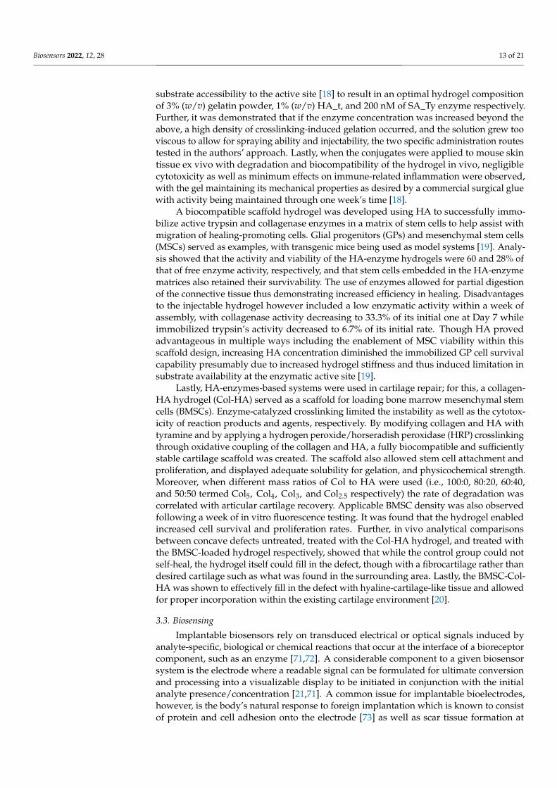
system that aids in drug delivery while eliminating possible unwanted biological entities, reactions, or processes associated with drug adsorption and metabolization at a tumor site [16]. For instance, enzymes glucose oxidase (GOx) and catalase (CAT) were used as com-ponents to a microneedle (MN) patch for a minimally invasive transdermal melanoma treatment (Figure 4a). The nanoparticles placed at the tips of the microneedles were spher-ical in shape and had an average hydrodynamic size of 250 nm. The patch was formed from HA crosslinked with N,N-methylenebis (acrylamide) (MBA) under photo initiator and UV-initiated polymerization respectively, and had a 9 × 9 mm2 area containing 225 cone-shaped MNs. Encapsulated GOx converted glucose to gluconic acid, producing an acidic environment that promoted the gradual release of anti-programmed death-1 (aPD1) previously encapsulated within dextran nanoparticles (NPs) to be used for skin cancer immunotherapy. Functionally, aPD1 was shown to help negate the negative effects to immunity caused by programmed death-1 receptors of T-cells [15].
(a) (b)
Biosensors 2022, 11, x FOR PEER REVIEW 11 of 23
Figure 4. (a) Design schematic of degradable HA microneedle patch for the site-specific delivery of anti-cancer drugs to melanoma cells. Reprinted with permission from ref. [15]. 2016 American Chemical Society. (b) Tumor observation via bioluminescence imaging on day zero, day three, and day six of the experimental treatment trial performed by Wang et al., with treatments 1-5 discerning melanoma untreated and treated with MN-GOx, free aPD1, MN-aPD1, and MN-GOx-aPD1, respec-tively. Reprinted with permission from ref. [15]. 2016 American Chemical Society. (c) Schematic representation of the movement of designed nanomotors through the ECM of a tumor before intra-cellular dissolution for the release of cytotoxic CPT as a mean of cancer therapy. Reprinted with permission from ref. [16]. 2019 Elsevier. (d) HepG2 and H22 cellular uptake percentages for JPs, HA-JPs, urease-JPs, JNMs, and free CPT, respectively, in media without urea, urea alone, urea with HA, or urea with Ficoll (* signifies p < 0.05). Reprinted with permission from ref. [16]. 2019 Elsevier.
Following a month of storage, the bioactivity of the MN patch system was maintained at 90%. The gradual release of aPD1 occurred over the course of three days. Increased T-cell infiltration was also observed in patch-treated tumors when compared to the control group, and most importantly, mice treated with the fully developed patch saw an increase in anti-tumor activity relative to mice treated with free aPD1, while tumor growth was significantly inhibited in mice treated with MN-GOx, free aPD1, MN-aPD1, and MN-GOx-aPD1, re-spectively(Figure 4b). Furthermore, there was no significant inflammation observed; mice saw increased lifespans, with 40% survival after 40 days following aPD1-GOx-MN treat-ment and about 70% of T-cell promoting anti-CTLA4 antibody and aPDI treated mice be-ing cancer-free beyond 60 days when the MN patch was used. The slow and sustained release over three days for the HA-based MN complex, in contrast to the diffusion away from the tumor sight observed following three days for free aPD1, leant a low cost of ad-ministration for such therapy with only one administration being necessary to produce the desired immune responses within the cancerous mice tested [15].
(c)
(d)
Figure 4. (a) Design schematic of degradable HA microneedle patch for the site-specific deliveryof anti-cancer drugs to melanoma cells. Reprinted with permission from ref. [15]. 2016 AmericanChemical Society. (b) Tumor observation via bioluminescence imaging on day zero, day three,and day six of the experimental treatment trial performed by Wang et al., with treatments 1-5discerning melanoma untreated and treated with MN-GOx, free aPD1, MN-aPD1, and MN-GOx-aPD1, respectively. Reprinted with permission from ref. [15]. 2016 American Chemical Society.(c) Schematic representation of the movement of designed nanomotors through the ECM of a tumorbefore intracellular dissolution for the release of cytotoxic CPT as a mean of cancer therapy. Reprintedwith permission from ref. [16]. 2019 Elsevier. (d) HepG2 and H22 cellular uptake percentages for JPs,HA-JPs, urease-JPs, JNMs, and free CPT, respectively, in media without urea, urea alone, urea withHA, or urea with Ficoll (* signifies p < 0.05). Reprinted with permission from ref. [16]. 2019 Elsevier.

Biosensors 2022, 12, 28 11 of 21
Following a month of storage, the bioactivity of the MN patch system was maintainedat 90%. The gradual release of aPD1 occurred over the course of three days. IncreasedT-cell infiltration was also observed in patch-treated tumors when compared to the controlgroup, and most importantly, mice treated with the fully developed patch saw an increasein antitumor activity relative to mice treated with free aPD1, while tumor growth wassignificantly inhibited in mice treated with MN-GOx, free aPD1, MN-aPD1, and MN-GOx-aPD1, respectively(Figure 4b). Furthermore, there was no significant inflammationobserved; mice saw increased lifespans, with 40% survival after 40 days following aPD1-GOx-MN treatment and about 70% of T-cell promoting anti-CTLA4 antibody and aPDItreated mice being cancer-free beyond 60 days when the MN patch was used. The slowand sustained release over three days for the HA-based MN complex, in contrast to thediffusion away from the tumor sight observed following three days for free aPD1, leant alow cost of administration for such therapy with only one administration being necessaryto produce the desired immune responses within the cancerous mice tested [15].
Complementary, Chen et al., described a self-propelling cancer drug delivery systemin which HA and urease were placed on opposing hemispheres of a Janus nanoparticle(JP) consisting of a mesoporous silica nanoparticle (MSN) half-coated with Au with anembedded hydroxyapatite (Hap) core and loaded with chemotherapeutic camptothecin(CPT) (Figure 4c). Such nanomotors were immobilized onto electrospun fiber fragments(JNM@EF) and administered directly to a local tumor area in cancer-bearing mice wherethe slightly acidic nature of the tumor matrix was enough to cause a sustained releaseof the JNMs from the EF platforms. Analysis showed that HA allowed targeted deliveryand uptake of the released JNMs into tumor cells, while the enzyme served as a powersource that projected the drug carrier forward through the ECMs of the tumor. Further, theacidic intracellular environment of cancerous tumors (pH 5.0) provided a controlled releasemechanism for CPT that remained unfazed by changes in the pHs of physiological or eventumor matrix conditions, valued at 7.4 and 6.5, respectively [16].
The enzymatic propulsion mechanism was considered an improvement over those ap-proaches based on the conversion of mechanical energy under externally applied magneticand electric fields, namely due to an increase in user-controllability. Undesired release ofCPT into the ECM was quantified at only about 8%, conferring robustness to the approachas well as to the nanocarriers prior to their uptake by the tumor cell. The fully developedJNM@EF, EF/JNM, and phosphate buffered solution (PBS) control treatments resulted in~14.7, ~53, and ~88% of ki-67-positive cells detected in tumors at 36-, 27-, and 16-day lifes-pans reached by over 50% of mice tested, respectively. As shown in Figure 4d, the additionof an HA coating to the JP surface also increased the cellular uptake substantially in HepG2and H22 cells, especially in media not containing urea, containing urea, or containing ureaand Ficoll respectively. The increased uptake was relative to JPs without HA or ureasecoatings, urease-JPs, and free CPT respectively, with the only other drug carrying complexwith greater percentages than HA-JPs being the complete JNM complex characterized withone hemisphere of HA and the other coated with urease enzyme, in all tested media andfor both tested cell types respectively [16].
Moreover, in a study by Montanari et al., bovine serum amine oxidase (BSAO) enzymewas immobilized within an injectable HA-based nanohydrogel (NH) to be used for treat-ment of melanoma cancer. Such self-assembled amphiphilic hydrogels were formulatedupon the covalent interaction between HA and cholesterol, under sonication and in water,with enzymes being conjugated via carbodiimide crosslinking. As the gel-loaded sourceof melanoma treatment, BSAO led to the formation of cytotoxic compounds includinghydrogen peroxide and a series of aldehydes resulted from the catalysis of polyamines. Byconverting polyamines such as spermine, spermidine, and putrescine, the tumor growthwas stifled. Moreover, the choice of the weaker ester bond between HA and bromo-butyricacid modified cholesterol (CH_Br) allowed a rapid release of BSAO [17]. This approachshowed that the integrity of a given enzyme immobilization platform, as characteristic ofthe designable bonding types at play in a biopolymer system, can control the rate of enzyme

Biosensors 2022, 12, 28 12 of 21
and/or drug expression and release respectively. Freeze-thawing and freeze-drying storageprocesses further allowed for the NH-BSAO samples to maintain their specific activitiesto 100 and 80% of their initial values. Supportive in vitro analysis of human melanomacancer cells revealed that BSAO post-immobilization successfully caused a cytotoxic effecton the cells equivalent to that of free BSAO, with such an effect also being accompanied bya negative correlation between cell viability and polyamine concentration [17].
3.2. Tissue Engineering and Wound Healing
The porous nature and structure of a HA-based hydrogel formed through crosslinkinghas proven imperative when scaffolds formation for cells and tissue growth as well aswhen regenerative processes were considered [69]. Further, the ability of the HA scaffold toswell was also shown to be critical for maintaining tissue viability [69], while its crosslink-ing capability conferred changes in gel stiffness [70], all to aid in HA’s implementationin tissue engineering and wound healing. Complementary, enzymatic integration wasshown to aid in the healing itself or provide a bio-fructuous environment that promotedfunctionality and viability. For instance, Kim et al., produced a HA hydrogel with adhesiveproperties to be used as a surgical glue capable of promoting cell delivery and recruiting(Figure 5). HA’s biocompatibility, biodegradability, hydrophilicity, and ability to crosslinkwith proteins, formed a plausible network for solidly binding to wet environments beforebeing replaced with repaired tissues. The first step of the gel forming procedure relied onthe extraction and amplification of the tyrosinase gene from Bacillus megaterium (BM_Ty),Agaricus bisporus (AB_Ty), and Streptomyces avermitilis (SA_Ty) respectively, followed bythe addition of crosslinker tyramine to the carboxyl group of HA, with the use of 1-ethyl-3-(3-dimethylaminopropyl) carbodiimide (EDC)/N-hydroxysuccinimide (NHS) couplingto result in a HA-tyramine conjugate (HA_t). Resulting tyrosinase was added to crosslinkHA_t and gelatin. It was found that the addition of gelatin encouraged phenol-phenol,amine-tyrosine, and thiol-tyrosine coupling, while addition of NaCl decreased the viscosityof the hydrogel to that of a sprayable liquid [18]. Moreover, it was found that HA facilitateddirect “molding” to the tissue and increased reactivity benefits as resulted from HA-enzymecomplexes integration, respectively [18].
Biosensors 2022, 11, x FOR PEER REVIEW 13 of 23
in tissue engineering and wound healing. Complementary, enzymatic integration was shown to aid in the healing itself or provide a bio-fructuous environment that promoted functionality and viability. For instance, Kim et al., produced a HA hydrogel with adhe-sive properties to be used as a surgical glue capable of promoting cell delivery and re-cruiting (Figure 5). HA’s biocompatibility, biodegradability, hydrophilicity, and ability to crosslink with proteins, formed a plausible network for solidly binding to wet environ-ments before being replaced with repaired tissues. The first step of the gel forming proce-dure relied on the extraction and amplification of the tyrosinase gene from Bacillus mega-terium (BM_Ty), Agaricus bisporus (AB_Ty), and Streptomyces avermitilis (SA_Ty) respec-tively, followed by the addition of crosslinker tyramine to the carboxyl group of HA, with the use of 1-ethyl-3-(3-dimethylaminopropyl) carbodiimide (EDC)/N-hydroxysuccin-imide (NHS) coupling to result in a HA-tyramine conjugate (HA_t). Resulting tyrosinase was added to crosslink HA_t and gelatin. It was found that the addition of gelatin encour-aged phenol-phenol, amine-tyrosine, and thiol-tyrosine coupling, while addition of NaCl decreased the viscosity of the hydrogel to that of a sprayable liquid [18]. Moreover, it was found that HA facilitated direct “molding” to the tissue and increased reactivity benefits as resulted from HA-enzyme complexes integration, respectively [18].
Following activity analysis of and comparisons among BM_Ty, AB_Ty, and SA_Ty, SA_Ty was found to be the most usable form of tyrosinase for crosslinking HA_t and gel-atin, with resulting complex allowing for high catalytic activity, speed of reaction, and increased substrate specificity respectively. The improvement was due to increased sub-strate accessibility to the active site [18] to result in an optimal hydrogel composition of 3% (w/v) gelatin powder, 1% (w/v) HA_t, and 200 nM of SA_Ty enzyme respectively. Fur-ther, it was demonstrated that if the enzyme concentration was increased beyond the above, a high density of crosslinking-induced gelation occurred, and the solution grew too viscous to allow for spraying ability and injectability, the two specific administration routes tested in the authors’ approach. Lastly, when the conjugates were applied to mouse skin tissue ex vivo with degradation and biocompatibility of the hydrogel in vivo, negligi-ble cytotoxicity as well as minimum effects on immune-related inflammation were ob-served, with the gel maintaining its mechanical properties as desired by a commercial surgical glue with activity being maintained through one week’s time [18].
Figure 5. Visualization of the development of an HA-based, injectable and sprayable formula to be applied in tissue engineering as a post-surgical tissue adhesive and encouragement for cellular re-cruitment to damaged tissue. Reprinted with permission from ref. [18]. 2018 Elsevier.
A biocompatible scaffold hydrogel was developed using HA to successfully immo-bilize active trypsin and collagenase enzymes in a matrix of stem cells to help assist with
Figure 5. Visualization of the development of an HA-based, injectable and sprayable formula tobe applied in tissue engineering as a post-surgical tissue adhesive and encouragement for cellularrecruitment to damaged tissue. Reprinted with permission from ref. [18]. 2018 Elsevier.
Following activity analysis of and comparisons among BM_Ty, AB_Ty, and SA_Ty,SA_Ty was found to be the most usable form of tyrosinase for crosslinking HA_t andgelatin, with resulting complex allowing for high catalytic activity, speed of reaction,and increased substrate specificity respectively. The improvement was due to increased
Biosensors 2022, 12, 28 13 of 21
substrate accessibility to the active site [18] to result in an optimal hydrogel compositionof 3% (w/v) gelatin powder, 1% (w/v) HA_t, and 200 nM of SA_Ty enzyme respectively.Further, it was demonstrated that if the enzyme concentration was increased beyond theabove, a high density of crosslinking-induced gelation occurred, and the solution grew tooviscous to allow for spraying ability and injectability, the two specific administration routestested in the authors’ approach. Lastly, when the conjugates were applied to mouse skintissue ex vivo with degradation and biocompatibility of the hydrogel in vivo, negligiblecytotoxicity as well as minimum effects on immune-related inflammation were observed,with the gel maintaining its mechanical properties as desired by a commercial surgical gluewith activity being maintained through one week’s time [18].
A biocompatible scaffold hydrogel was developed using HA to successfully immo-bilize active trypsin and collagenase enzymes in a matrix of stem cells to help assist withmigration of healing-promoting cells. Glial progenitors (GPs) and mesenchymal stem cells(MSCs) served as examples, with transgenic mice being used as model systems [19]. Analy-sis showed that the activity and viability of the HA-enzyme hydrogels were 60 and 28% ofthat of free enzyme activity, respectively, and that stem cells embedded in the HA-enzymematrices also retained their survivability. The use of enzymes allowed for partial digestionof the connective tissue thus demonstrating increased efficiency in healing. Disadvantagesto the injectable hydrogel however included a low enzymatic activity within a week ofassembly, with collagenase activity decreasing to 33.3% of its initial one at Day 7 whileimmobilized trypsin’s activity decreased to 6.7% of its initial rate. Though HA provedadvantageous in multiple ways including the enablement of MSC viability within thisscaffold design, increasing HA concentration diminished the immobilized GP cell survivalcapability presumably due to increased hydrogel stiffness and thus induced limitation insubstrate availability at the enzymatic active site [19].
Lastly, HA-enzymes-based systems were used in cartilage repair; for this, a collagen-HA hydrogel (Col-HA) served as a scaffold for loading bone marrow mesenchymal stemcells (BMSCs). Enzyme-catalyzed crosslinking limited the instability as well as the cytotox-icity of reaction products and agents, respectively. By modifying collagen and HA withtyramine and by applying a hydrogen peroxide/horseradish peroxidase (HRP) crosslinkingthrough oxidative coupling of the collagen and HA, a fully biocompatible and sufficientlystable cartilage scaffold was created. The scaffold also allowed stem cell attachment andproliferation, and displayed adequate solubility for gelation, and physicochemical strength.Moreover, when different mass ratios of Col to HA were used (i.e., 100:0, 80:20, 60:40,and 50:50 termed Col5, Col4, Col3, and Col2.5 respectively) the rate of degradation wascorrelated with articular cartilage recovery. Applicable BMSC density was also observedfollowing a week of in vitro fluorescence testing. It was found that the hydrogel enabledincreased cell survival and proliferation rates. Further, in vivo analytical comparisonsbetween concave defects untreated, treated with the Col-HA hydrogel, and treated withthe BMSC-loaded hydrogel respectively, showed that while the control group could notself-heal, the hydrogel itself could fill in the defect, though with a fibrocartilage rather thandesired cartilage such as what was found in the surrounding area. Lastly, the BMSC-Col-HA was shown to effectively fill in the defect with hyaline-cartilage-like tissue and allowedfor proper incorporation within the existing cartilage environment [20].
3.3. Biosensing
Implantable biosensors rely on transduced electrical or optical signals induced byanalyte-specific, biological or chemical reactions that occur at the interface of a bioreceptorcomponent, such as an enzyme [71,72]. A considerable component to a given biosensorsystem is the electrode where a readable signal can be formulated for ultimate conversionand processing into a visualizable display to be initiated in conjunction with the initialanalyte presence/concentration [21,71]. A common issue for implantable bioelectrodes,however, is the body’s natural response to foreign implantation which is known to consistof protein and cell adhesion onto the electrode [73] as well as scar tissue formation at

Biosensors 2022, 12, 28 14 of 21
the sensor side [74], all shown to inhibit biosensor performance and decrease its sensingfunctionality [21,73,74]. An electrochemical bioelectrode was assembled using HA andpolydopamine (PDA) deposited onto an indium tin oxide electrode, with HA to attributeanti-fouling qualities to the electrode interface while also permitting for maintenance ofelectrical properties of the sensing complex respectively. The anti-fouling quality was likelydue to the hydrophilic nature of HA which caused it to absorb and maintain considerablemoisture around the electrode, while PDA allowed for increased immobilization throughentrapping of HA molecules. Among the benefits resulting from such sensor electrodemodification, authors notes a decrease of undesired protein adsorption, fibroblast adhesion,and scar tissue formation [21].
Complementary, Cabral et al., showed that HA hybridized with carbon nanotubes(CNTs) could be used for sensing of hepatitis B core protein antibody (Anti-HBc) marker toaid in the determination of hepatitis B virus (HBV) infection. The HA-CNT film consistedof HA chains wrapped around the circumference of the nanotubes; Anti-HBc served as aproof-of-concept representation of how a similar HA and biocatalyst-based sensor couldbe used. As a globally leading health issue and cause of death, spreading of HBV viablood transfusion is a major concern of health professionals [23,75]. The marker testedwas envisioned to increase screening at blood banks, as anti-HBc can be present for theentirety of an individual’s life. CNTs met the demands of HA gelation required for thematrix’s stability as a viable platform for HBc immobilization as shown by scanning electronmicroscopy (SEM) [23], which further led to stable HA-induced dispersion of CNTs andeasy HA-CNT complex-induced arrested phase separation [76]. The resulting gels werebiocompatible, electrically conductive, and applicable for protein delivery via electricalstimulation. The CNT modification allowed for a more stable immobilization of HBc dueto an enhancement of electron transfer; following the addition of the HA complex onto theelectrode, there was about a 75.7% increase in the electroactive surface area. Furthermore,the electron transfer-based sensors were stable upon electrode immobilization [23].
Considering the above, as well as the ability to integrate enzymatic transformation ofspecific substrates, products, and signals respectively, applications of enzymes and HA asbiosensors have gained considerable interest for the detection of various analytes withina solution or a body sample. Placing enzymes within synthetic microenvironments, ascharacteristic of immobilization processes, was for instance shown to oftentimes increasetheir stability and reusability, qualities greatly desired for a biosensor [11,77–80]. Kim et al.,developed a glucose sensor using hyaluronate (the salt form of HA), AuNPs, GOx, andan integrated circuit chip; the sensor was intended to process sweat glucose data collectedat its interface [22]. HA’s integration inhibited the interference of otherwise detrimentalmolecules including lactic acid and L-ascorbic acid. As a result of these benefits, accurateand specific measurement, non-invasiveness, stability, sensitivity, and feasibility wereenabled when monitoring continuous diabetes detection in mice. As shown in Figure 6a,the complex consisted of enzyme and HA-coated AuNPs attached to the surface of afunctional electrode via binding of each element with 1,2, ethanedithiol [22].
The designed HA-AuNP/GOx complex, maintained 78% activity for about 4 daysunder repeated use in comparison to a complete loss of GOx coating and correspondingactivity following only 2 days of operation. Further, stability testing showed that theaddition of HA-AuNP to the enzyme system increased the percentage of initial current thatwas maintained from 29.6 to 98.1%, before and after HA-AuNP integration, respectively. Incomparison to the sensors coated with GOx and AuNP/GOx, the HA-AuNP/GOx sensorspossessed the lowest detection limit at 0.5 mg dL−1. Moreover, the HA-AuNP/GOxelectrode coating saw improvement in reusability when compared to sensors coated withGOx alone. Specifically, it was shown that the conjugation of HA and AuNPs allowed astable platform for GOx immobilization that inhibited enzyme degradation and improvedglucose sensing when compared to GOx and AuNP/GOx sensor coatings alone. Withthe known blood glucose levels of 179, 288, and 374 mg/dL, the output codes of the HA-AuNP/GOx glucose sensor were 289.7, 494.0, and 602.9 respectively, suggesting a direct

Biosensors 2022, 12, 28 15 of 21
and accurate positive correlation between glucose level and biosensor reading. Because ofthe fast response time of only 5 s, the sensor was nearly real-time, another major benefit fordiagnostics or glucose monitoring [22].
Biosensors 2022, 11, x FOR PEER REVIEW 16 of 23
tissues. The specific Hyal-1-induced disassembly of CHA and the implied application for fluorescence imaging of cancer sites is shown in Figure 6b, as most CHA particles were below 10 nm in diameter as compared to distributions entirely greater than this 10 nm mark seen in CHA alone and CHA with Hyal-2. Additionally, RBF was able to bind to cytoplasmic RNA to further amplify the recorded fluorescence signal thus aiding in the ease of detection. In comparison to other intracellular sensing systems, the use of fluores-cence detection was less invasive, displayed high resolution, and had the ability to show results in real-time [24].
Figure 6. (a) Fabrication and application of a wireless glucose sensor used to test sweat for glucose concentration and consisting of an HA-AuNP/GOx complex casted on an electrode surface. Re-printed with permission from ref. [22]. 2019 American Chemical Society. (b–d) TEM imaging and size distribution corresponding to a 10 μg/mL solution of CHA without the addition of any hyalu-ronidase, CHA with 20 μg/mL Hyal-1, and CHA with 20 μg/mLHyal-2, respectively. Reprinted with permission from ref. [24]. 2019 American Chemical Society.
Authors also showed that in the development of an innovative hyaluronidase iden-tification system, self-assembled, amphiphilic CHA shells encompassing and stifling the fluorescence of RBFs were more likely broken apart by Hyal-1 than by Hyal-2. Small levels of Hyal-1 were discernable by the RBF@CHA systems, as characterized by a detection limit of 1 × 10 μg mL and a linear detectable concentration range between 0 and 0.01 μg mL . Meanwhile, presence of Hyal-2 did not present a clear fluorescence amplifica-tion of the RBF@CHA assemblies, as expectedly characteristic of an enzyme-degraded
(b) (c)
(a)
(d)
Figure 6. (a) Fabrication and application of a wireless glucose sensor used to test sweat for glu-cose concentration and consisting of an HA-AuNP/GOx complex casted on an electrode surface.Reprinted with permission from ref. [22]. 2019 American Chemical Society. (b–d) TEM imagingand size distribution corresponding to a 10 µg/mL solution of CHA without the addition of anyhyaluronidase, CHA with 20 µg/mL Hyal-1, and CHA with 20 µg/mLHyal-2, respectively. Reprintedwith permission from ref. [24]. 2019 American Chemical Society.
Due to their ability to break down HA, Hyal-1 and Hyal-2 (hyaluronidase enzymes)were investigated by Li et al., as physiological analytes to a HA-based fluorescent sensingplatform. Specificity was both desired and achieved for the detection of Hyal-1 overHyal-2 because of its prevalence in tumor sites, more precisely found in bladder, prostate,and breast cancer patients. The biosensor design integrated HA functionalized withcholesterylamine (CHA) to form and entrap RNA-binding fluorophores (RBFs); whenHyal-1 was present, the CHA nano-assembly was degraded into small fragments to unveilthe fluorophores and thus offer a fluorescence-attributed detection of Hyal-1 containingcancerous tissues. The specific Hyal-1-induced disassembly of CHA and the impliedapplication for fluorescence imaging of cancer sites is shown in Figure 6b, as most CHAparticles were below 10 nm in diameter as compared to distributions entirely greater thanthis 10 nm mark seen in CHA alone and CHA with Hyal-2. Additionally, RBF was able to

Biosensors 2022, 12, 28 16 of 21
bind to cytoplasmic RNA to further amplify the recorded fluorescence signal thus aidingin the ease of detection. In comparison to other intracellular sensing systems, the use offluorescence detection was less invasive, displayed high resolution, and had the ability toshow results in real-time [24].
Authors also showed that in the development of an innovative hyaluronidase iden-tification system, self-assembled, amphiphilic CHA shells encompassing and stifling thefluorescence of RBFs were more likely broken apart by Hyal-1 than by Hyal-2. Small levelsof Hyal-1 were discernable by the RBF@CHA systems, as characterized by a detectionlimit of 1 × 10⁻⁵ µg mL−1 and a linear detectable concentration range between 0 and0.01 µg mL−1. Meanwhile, presence of Hyal-2 did not present a clear fluorescence amplifi-cation of the RBF@CHA assemblies, as expectedly characteristic of an enzyme-degradedcomposite. In addition to Hyal-2, Hyal-1 specificity analyses revealed that RBF@CHA wasfound not to be affected by biomolecules such as H2O2, biothiol glutathione, cathepsin,trypsin, thrombin, lysozyme, ribonuclease, and galactosidase respectively. Moreover, theRBF@CHA showed feasible biocompatibility for both cellular uptake and lack of cytotoxiceffects when studied in living HeLa cells [24].
4. Challenges and Perspectives for HA-Enzyme Applications: A ComplementaryMarket Analysis
Above examples demonstrate targeted strategies based on HA-enzyme conjugatesfor applications as drug nanocarriers, scaffolds to control compatibility and adherencewithin the body, to have sufficient stability, entrapment capabilities, and to allow for user-controllability with ease of implementation, or as stable, real-time, and biocompatiblebiosensors. From facilitating the accumulation at the tumor site, to helping receive in-creased penetration through cancerous tissue, and ultimately by allowing effective drugdelivery design strategies, HA-enzyme conjugates have not only demonstrated increasedease of implementation but further, increased preservation of biocompatibility as well assuperior degree of biodegradability [20], all while being reproducible at manufacturingand cost efficient.
It is envisioned that the benefits of the enzymatic technologies integration with HAare only to increase. Specifically, global enzyme market potential anticipates a 7.1% CAGRbetween 2020 and 2027 [81]. Moreover, if in 2019, the global enzyme market was worthUSD 9.9 billion it is now estimated that such market will reach USD 17.17 billion by theend of 2027 [81]. Furthermore, in addition to the overarching industry of biocatalysiswhere most of the market increases were initially noted, recently there has been an increasein the demand for immobilized enzyme technologies to be applied in areas such as thepharmaceutical, environmental, food, diagnostics, and biotranformation industries, al suchdesirable applications resulting from the advantages that such systems implementationprovide mainly their prolonged shelf-life and improved recyclability capacity especiallyrelative to free enzymes [82]. Such increase in enzymes’ and immobilized enzymes’ marketsare complemented by the rapid growth of their applicable industries namely the marketof biomaterials for drug-delivery grew from roughly USD 178.8 billion in 2015 to USD227.3 billion in 2020 [83].
Moreover, the drug delivery and biomaterial markets are expected to continue aparallel growth, along with that of other biomedical fields such as tissue engineering, theglobal medical enzyme market, and the hyaluronic acid market. For instance, in 2019, thetissue engineering market reached USD 25.4 billion, and over the span of 2017–2022, themarket is on the track to increase 17.22% in CAGR [84]. Complementarily, the biomaterialsmarket is particularly successful with a growing demand for products such as medicalimplants to be utilized as the primary scaffold of tissue-engineered heart valves [85], aswell as nano-, micro-, and macroscale drug systems developed for cancer immunotherapyand antitumor T-cell immunity [86]. To add in, the biomaterials sector is poised to increasefrom a worth of USD 105 billion in 2019 to USD 207 billion in 2024, which reaches a 14.4%CAGR [87]. Meanwhile, the global medical enzyme market is estimated to grow at a

Biosensors 2022, 12, 28 17 of 21
CAGR of 6.7% in the years spanning from 2021–2028, also projected to increase from USD4.0 billion in 2020 to USD 6.7 million in 2028 [88]. As a key component to several of therecent biomedical implementations of enzyme immobilization discussed, the HA markethas also seen a sizeable growth rate of 15% per annum since the market was established in1997. It is noted that HA has not established a particularly strong market in Europe butis seemingly successful in the US [89], likely explainable by the country’s comparativelyhigh interest in cosmetic enhancement. As such, the HA market is estimated to reach USD15.4 billion by the year 2025, which is a considerable increase from its previous value ofUSD 7.2 billion in 2016 [90].
These listed markets analysis have seen many contributing social and economic ad-vantages in terms of renewable sourcing, high reaction efficiency, low production costs,long shelf-life, and reusability respectively [82]. For example, biotechnologies are centeredaround materials such as enzymes and HA, both ethically sourced from growable biologicalmatter and able to be viably transposed to other biological entities in a non-species-specificmanner. When compared to free enzymes, immobilized enzymes have properties that makethem more catalytically active, substrate-specific, and easily separated and recovered fol-lowing catalysis [91]. This allows a wide breadth of efficiency and increased sustainabilityfor the immobilized catalysts and even consideration for new technologies implementation,thus making them more desirable as points of investment and lucrativeness in the businessworld as well as in science. When comparing the cost effectiveness of free and immobilizedenzymes for instance, a mathematical model was created and concluded that the immobi-lized enzymes see a 60% reduction in costs of production for instance [92]. This decrease inoverhead costs is predicted to allow markets to sustainably produce biomedical materialsand devices that utilize immobilized enzymes, as the high sourcing costs of enzymes canbe offset by the ability to reuse those catalytic feedstocks more than once.
However, HA-enzyme conjugates integration, while promising in terms of both bio-related benefits and market potential, continues to still be limited by elements such as thetype of administration route for implantation [15], loss of enzyme activity following storageprocesses such as freeze-thawing and freeze-drying [17], and/or the decrease in enzymestability especially at certain pH values [93]. Additionally, lack of knowledge of the spanof effects HA can have in vivo [19], especially considering that an HA-based platformsare often fragile, with changes in its structural integrity not only to decrease enzymaticactivity and impact the stability characteristics of a HA-based gel used as scaffold [18]but also to possibly lead to the production of by-products and free radicals that couldinduce inflammation and possible signs of cytotoxicity [70] especially when consideringthe shelf-life of such conjugates, since prolonged enzymatic activity is required to makedescribed gels and scaffolds be viable for storage, transportation, and use in the medicalindustry [19] respectively.
The main objectives of the markets that were discussed are profitability, efficiency,and most importantly, sustainability. We envision that the next generation of enzyme-HAconjugates need to be controlled, integrated, and optimized for synthetic applications.In doing so, the potential market growth in each of the mentioned sectors as well ascommercial development can be fulfilled. Continued development of enzyme and HA-based technologies is vital for the evolution of our health systems as well as energy andconsumption needs on a global scale. Therefore, with numeric proof of economic aptitudein addition to potential for positive societal impact, funding for industry-scale projects ishighly feasible in the near-future.
5. Conclusions
The application of enzymes for catalytic processes has become an impactful sector inbiofuels, plastic and chemical waste management, detergents, textiles, and food productionand preservation. Within these industries, biocatalysis provides the enablement of specificand desirable reactions at the core technological advancements, as related to the rate andyield of product formation, all in a green, self-sustainable and specific manner.

Biosensors 2022, 12, 28 18 of 21
There are limitations, however, as their systematic benefits are accompanied by theirsensitivity to synthetic environments, with such caveats leading to their denaturation andtheir loss of catalytic activity in temperatures and acidic conditions. Because so manydevelopments in biotechnology, particularly within the biomedical field, require extremeconditions upon execution and/or storage, there is frequently a necessity for a methodin which an enzyme’s stability can be enhanced relative to that of its free form. Exploredhere was the technique of enzyme immobilization, wherein enzymes are chemically orphysically linked or encapsulated within a structure that encourages enzyme stability andincreases its shelf-life while maintaining its functionality and product yield.
Focus was placed on enzyme integration with HA. HA-enzyme conjugates applica-tions in drug delivery, as scaffolds and as biosensors were thus discussed. With largeconsideration to biomedicine, a given immobilization platform must encompass the valuesof a stable scaffold that can be successfully conjugated with enzymes, yet it must alsohave intrinsic qualities pertaining to biocompatibility and in some cases, instrumentalmechanisms such as biodegradation and targeted delivery. HA advantages as a highlymodifiable polysaccharide were further analyzed from the perspective of the global marketimplementation in biomedicine. Thus, evaluating enzyme market potential and implemen-tation capacity are a matter of users and their ability to control or envision how productsand byproducts are to be integrated or separated in an efficient and controlled fashion.
Author Contributions: Conceptualization: J.A. and C.Z.D.; original draft preparation: J.A. andC.Z.D.; final draft preparation: J.A., J.C., M.A. and C.Z.D. All authors have read and agreed to thepublished version of the manuscript.
Funding: This research was funded by the National Science Foundation (NSF) 1454230 as well assupported by the Arnold and Mabel Beckman Foundation through the Beckman Scholars Program atWest Virginia University.
Conflicts of Interest: The authors declare no conflict of interest.
Ethical Statement: Consent for all copyrights has been obtained for inclusion in the study.
References1. Misra, N.; Panda, P.K.; Parida, B.K.; Mishra, B. dEMBF: A Comprehensive Database of Enzymes of Microalgal Biofuel Feedstock.
PLoS ONE 2016, 11, e0146158. [CrossRef] [PubMed]2. Jia, B.; Song, Y.; Wu, M.; Lin, B.; Xiao, K.; Hu, Z.; Huang, Y. Characterization of long-chain acyl-CoA syntheses which stimulate
secretion of fatty acids in green algae Chlamydomonas reinhardtii. Biotechnol. Biofuels 2016, 9, 1–11. [CrossRef] [PubMed]3. Ben-Zvi, O.; Dafni, E.; Feldman, Y.; Yacoby, I. Re-routing photosynthetic energy for continuous hydrogen production in vivo.
Biotechnol. Biofuel 2019, 12, 12. [CrossRef]4. Kumari, U.; Singh, R.; Ray, T.; Rana, S.; Saha, P.; Malhotra, K.; Daniell, H. Validation of leaf enzymes in the detergent and textile
industries: Launching of a new platform technology. Plant Biotechnol. J. 2019, 17, 1167–1182. [CrossRef]5. Salat, M.; Petkova, P.; Hoyo, J.; Perelshtein, I.; Gedanken, A.; Tzanov, T. Durable antimicrobial cotton textiles coated sono-
chemically with ZnO nanoparticles embedded in an in-situ enzymatically generated bioadhesive. Carbohydr. Polym. 2018, 189,198–203. [CrossRef] [PubMed]
6. Gaddes, D.; Jung, H.; Pena-Francesch, A.; Dion, G.; Tadigadapa, S.; Dressick, W.J.; Demirel, M.C. Self-Healing Textile: EnzymeEncapsulated Layer-by-Layer Structural Proteins. ACS Appl. Mater. Interfaces 2016, 8, 20371–20378. [CrossRef]
7. Polak, J.; Wlizło, K.; Pogni, R.; Petricci, E.; Graz, M.; Szałapata, K.; Osinska-Jaroszuk, M.; Kapral-Piotrowska, J.; Pawlikowska-Pawlega, B.; Jarosz-Wilkołazka, A. Structure and Bioactive Properties of Novel Textile Dyes Synthesised by Fungal Laccase. Int. J.Mol. Sci. 2020, 21, 2052. [CrossRef] [PubMed]
8. Blánquez, A.; Rodríguez, J.; Brissos, V.; Mendes, S.; Martins, L.O.; Ball, A.S.; Arias, M.E.; Hernández, M. Decolorization anddetoxification of textile dyes using a versatile Streptomyces lactase-natural mediator system. Saudi J. Biol. Sci. 2019, 26, 913–920.[CrossRef]
9. Austin, H.P.; Allen, M.D.; Donohoe, B.S.; Rorrer, N.A.; Kearns, F.L.; Silveira, R.L.; Pollard, B.C.; Dominick, G.; Duman, R.; ElOmari, K.; et al. Characterization and engineering of a plastic-degrading aromatic polyesterase. In Proceedings of the NationalAcademy of Sciences of the United States of America. Proc. Natl. Acad. Sci. USA 2018, 115, E4350–E4357. [CrossRef]
10. Rodrigues da Silva, R. Enzyme technology in food preservation: A promising and sustainable strategy for biocontrol of post-harvest fungal pathogens. Food Chem. 2018, 277, 531–532. [CrossRef]
11. Zhang, L.; Wang, P.; Wang, C.; Wu, Y.; Feng, X.; Huang, H.; Ren, L.; Liu, B.-F.; Gao, S.; Liu, X. Bacteriophage T4 capsid as ananocarrier for Peptide-N-Glycosidase F immobilization through self-assembly. Sci. Rep. 2019, 9, 4865. [CrossRef]

Biosensors 2022, 12, 28 19 of 21
12. Xie, J.; Ji, Y.; Xue, W.; Ma, D.; Hu, Y. Hyaluronic acid-containing ethosomes as a potential carrier for transdermal drug delivery.Colloids Surf. B Biointerfaces 2018, 172, 323–329. [CrossRef] [PubMed]
13. Luo, Y.; Kirker, K.R.; Prestwich, G.D. Cross-linked hyaluronic acid hydrogel films: New biomaterials for drug delivery. J. Control.Release 2000, 69, 169–184. [CrossRef]
14. Sahiner, N.; Suner, S.S.; Ayyala, R.S. Mesoporous, degradable hyaluronic acid Microparticles for sustainable drug deliveryapplication. Colloids Surf. B Biointerfaces 2019, 177, 284–293. [CrossRef] [PubMed]
15. Wang, C.; Ye, Y.; Hochu, G.M.; Sadeghifar, H.; Gu, Z. Enhanced Cancer Immunotherapy by Microneedle Patch-assisted de-liveryof Anti-PD1 Antibody. Nano Lett. 2016, 16, 2334–2340. [CrossRef] [PubMed]
16. Chen, Z.; Xia, T.; Zhang, Z.; Xie, S.; Wang, T.; Li, X. Enzyme-powered Janus nanomotors launched from intratumoral depots toaddress drug delivery barriers. Chem. Eng. J. 2019, 375, 122109. [CrossRef]
17. Montanari, E.; Capece, S.; Di Meo, C.; Meringolo, M.; Coviello, T.; Agostinelli, E.; Matricardi, P. Hyaluronic Acid Nanohy-drogelsas a Useful Tool for BSAO Immobilization in the Treatment of Melanoma Cancer Cells. Macromol. Biosci. 2013, 13, 1185–1194.[CrossRef] [PubMed]
18. Kim, S.-H.; Lee, S.-H.; Lee, J.-E.; Park, S.J.; Kim, K.; Kim, I.S.; Lee, Y.-S.; Hwang, N.S.; Kim, B.-G. Tissue adhesive, rapid forming,and sprayable ECM hydrogel via recombinant tyrosinase crosslinking. Biomaterials 2018, 178, 401–412. [CrossRef] [PubMed]
19. Piejko, M.; Jablonska, A.; Walczak, P.; Janowski, M. Proteolytic Rafts for Improving Intraparenchymal Migration of MinimallyInvasively Administered Hydrogel-Embedded Stem Cells. Int. J. Mol. Sci. 2019, 20, 3083. [CrossRef]
20. Zhang, Y.; Cao, Y.; Zhao, H.; Zhang, L.; Ni, T.; Liu, Y.; An, Z.; Liu, M.; Pei, R. An injectable BMSC-laden enzyme-catalyzed crosslinking collagen-hyaluronic acid hydrogel for cartilage repair and regeneration. J. Mater. Chem. 2020, 8, 4237–4244. [CrossRef]
21. Kim, S.; Lee, S.; Park, J.; Lee, J.Y. Electrochemical Co-deposition of Polydopamine/Hyaluronic Acid for Anti-biofouling Bioe-lectrodes. Front. Chem. 2019, 7, 262. [CrossRef]
22. Kim, S.-K.; Jeon, C.; Lee, G.-H.; Koo, J.; Cho, S.H.; Han, S.; Shin, M.-H.; Sim, J.-Y.; Hahn, S.K. Hyaluronate-Gold Nanoparti-cle/Glucose Oxidase Complex for Highly Sensitive Wireless Noninvasive Glucose Sensors. ACS Appl. Mater. Interfaces 2019, 11,37347–37356. [CrossRef] [PubMed]
23. Cabral, D.G.A.; Lima, E.C.S.; Moura, P.; Dutra, R.F. A label-free electrochemical immunosensor for hepatitis B based on hya-luronicacid-carbon nanotube hybrid film. Talanta 2015, 148, 209–215. [CrossRef] [PubMed]
24. Li, Y.; Yang, S.; Guo, L.; Xiao, Y.; Luo, J.; Li, Y.; Wong, M.S.; Yang, R. Differentiation of Intracellular Hyaluronidase Isoform bydegradable Nanoassembly Coupled with RNA-Binding Fluorescence Amplification. Anal. Chem. 2019, 91, 6887–6893. [CrossRef][PubMed]
25. Martinez-Guerra, E.; Gude, V.G. Assessment of Sustainability Indicators for Biodiesel Production. Appl. Sci. 2017, 7, 869.[CrossRef]
26. Campbell, A.S.; Dong, C.; Meng, F.; Hardinger, J.; Perhinschi, G.; Wu, N.; Dinu, C.Z. Enzyme Catalytic Efficiency: A Function ofBio-Nano Interface Reactions. ACS Appl. Mater. Interfaces 2014, 6, 5393–5403. [CrossRef]
27. Chapman, J.; Ismail, A.E.; Dinu, C.Z. Industrial Applications of Enzymes: Recent Advances, Techniques, and Outlooks. Catalysts2018, 8, 238. [CrossRef]
28. Sato, N.; Moriyama, T.; Mori, N.; Toyoshima, M. Lipid metabolism and potentials of biofuel ad high added-value oil productionin red algae. World J. Microbiol. Biotechnol. 2017, 33, 94. [CrossRef]
29. Kim, D.-H.; Kim, M.-S. Hydrogenases for biological hydrogen production. Bioresour. Technol. 2011, 102, 8423–8431. [CrossRef]30. Cao, Y.; Pawlowski, A. Sewage sludge-to-energy approaches based on anaerobic digestion and pyrolysis: Brief overview and
energy efficiency assessment. Renew. Sustain. Energy Rev. 2012, 16, 1657–1665. [CrossRef]31. Chukwuneke, J.L.; Ewulonu, M.C.; Chukwujike, I.C.; Okolie, P.C. Physico-chemical analysis of pyrolyzed bio-oil from swietenia
macrophylla (mahogany) wood. Heliyon 2019, 5, e01790. [CrossRef]32. Li, L.; Liu, G.; Li, Y.; Zhu, Z.; Xu, H.; Chen, J.; Ren, X. Release of Sulfur and nitrogen during Co-pyrolysis of Coal and Biomass
under Inert Atmosphere. ACS Omega 2020, 5, 30001–30010. [CrossRef]33. Meramo-Hurtado, S.I.; Puello, P.; Cabarcas, A. Process Analysis of Hydrogen Production via Biomass Gasification under
Computer-Aided Safety and Environmental Assessments. ACS Omega 2020, 5, 19667–19681. [CrossRef] [PubMed]34. Liu, Y.; Lin, R.; Man, Y.; Ren, J. Recent developments of hydrogen production from sewage sludge by biological and thermo-
chemical process. Int. J. Hydrogen Energy 2019, 44, 19676–19697. [CrossRef]35. Han, B.; Wei, W.; Li, M.; Sun, K.; Hu, Y.H. A thermo-photo hybrid process for steam reforming of methane: Highly efficient visible
light photocatalysis. Chem. Commun. 2019, 55, 7816–7819. [CrossRef] [PubMed]36. Yoo, J.; Park, S.; Song, J.H.; Song, I.K. Hydrogen Production by Steam Reforming of Natural Gas Over Vanadi-um-Nickel-Alumina
Catalysts. J. Nanosci. Nanotechnol. 2018, 18, 6541–6546. [CrossRef]37. Kannah, R.Y.; Kavitha, S.; Preethi; Karthikeyan, O.P.; Kumar, G.; Dai-Wiet, N.V.; Banu, R. Techno-economic assessment of various
hydrogen production methods—A review. Bioresour. Technol. 2020, 319, 124175. [CrossRef]38. Garlyyev, B.; Xue, S.; Fichtner, J.; Bandarenka, A.S.; Andronescu, C. Prospects of Value-Added Chemicals and Hydrogen via
Electrolysis. ChemSusChem 2020, 13, 2513–2521. [CrossRef]39. Zhang, Y.H.P.; Evans, B.R.; Mielenz, J.R.; Hopkins, R.C.; Adams, M.W.W. High-Yield Hydrogen Production from Starch and Water
by a Synthetic Enzymatic Pathway. PLoS ONE 2007, 2, e456. [CrossRef] [PubMed]40. Hauer, B. Embracing Nature’s Catalysts: A Viewpoint on the future of Biocatalysis. ACS Catal. 2020, 10, 8418–8427. [CrossRef]

Biosensors 2022, 12, 28 20 of 21
41. Li, T.; Jiang, Q.; Huang, J.; Aitchison, C.M.; Huang, F.; Yang, M.; Dykes, G.F.; He, H.-L.; Wang, Q.; Sprick, R.S.; et al. Repro-gramming bacterial protein organelles as a nanoreactor for hydrogen production. Nat. Commun. 2020, 11, 1–10. [CrossRef]
42. Dolui, D.; Khandelwal, S.; Shaik, A.; Gaat, D.; Thiruvenkatam, V.; Dutta, A. Enzyme-Inspired Synthetic Proton Relays Gen-erateFast and Acid-Stable Cobalt-Based H2 Production Electrocatalysts. ACS Catal. 2019, 9, 10115–10125. [CrossRef]
43. Moog, D.; Schmitt, J.; Senger, J.; Zarzycki, J.; Rexer, K.-H.; Linne, U.; Erb, T.; Maier, U.G. Using a marine microalga as a chassis forpolyethylene terephthalate (PET) degradation. Microb. Cell Factories 2019, 18, 1–15. [CrossRef]
44. Joo, S.; Cho, I.J.; Seo, H.; Son, H.F.; Sagong, H.-Y.; Shin, T.J.; Choi, S.Y.; Lee, S.Y.; Kim, K.-J. Structural insight into molecularmechanism of poly(ethylene terephthalate) degradation. Nat. Commun. 2017, 284, 309. [CrossRef]
45. Cui, Y.; Chenn, Y.; Liu, X.; Dong, S.; Tian, Y.; Qiao, Y.; Mitra, R.; Han, J.; Li, C.; Han, X.; et al. Computational Redesign of a PETasefor Plastic Biodegradation under Ambient Condition by the GRAPE Strategy. ACS Catal. 2021, 11, 1340–1350. [CrossRef]
46. Sagong, H.-Y.; Seo, H.; Kim, T.; Son, H.F.; Joo, S.; Lee, S.H.; Kim, S.; Woo, J.-S.; Hwang, S.Y.; Kim, K.-J. Decomposition of the PETFilm by MHETase Using Exo-PETase Function. ACS Catal. 2020, 10, 4805–4812. [CrossRef]
47. Coppella, S.J.; DelaCruz, N.; Payen, G.F.; Pogell, B.M.; Speedie, M.K.; Karns, J.S.; Sybert, E.M.; Connor, M.A. Genetic engi-neeringapproach to toxic waste management: Case study for organophosphate waste treatment. Biotechnol. Prog. 1990, 6, 76–81.[CrossRef]
48. Vanderberg, L.A.; Foreman, T.M.; Attrep, M.J.; Brainard, J.R.; Sauer, N.N. Treatment of Heterogeneous Mixed Wastes: EnzymeDegradation of Cellulosic Materials Contaminated with Hazardous Organics and Toxic and Radioactive Metals. Environ. Sci.Technol. 1999, 33, 1256–1262. [CrossRef]
49. Chen, J.; Liu, S.; Wang, Y.; Huang, W.; Zhou, J. Effect of different hydrolytic enzymes pretreatment for improving the hydrolysisand biodegradability of waste activated sludge. Water Sci. Technol. 2018, 2017, 592–602. [CrossRef]
50. Worm, B.; Lotze, H.K.; Jubinville, I.; Wilcox, C.; Jambeck, J. Plastic as a Persistent Marine Pollutant. Annu. Rev. Environ. Resour.2017, 42, 1–26. [CrossRef]
51. Limbo, S.; Pellegrino, L.; D’Incecco, P.; Gobbi, S.; Rosi, V.; Fracassetti, D. Storage of pasteurized milk in clear PET bottles com-binedwith light exposure on a retail display case: A possible strategy to define the shelf life and support a recyclable packaging. FoodChem. 2020, 329, 127116. [CrossRef]
52. Quartinello, F.; Tallian, C.; Auer, J.; Schön, H.; Vielnascher, R.; Weinberger, S.; Wieland, K.; Weihs, A.M.; Herrero-Rollett, A.; Lendl,B.; et al. Smart textiles in wound care: Functionalization of cotton/PET blends with antimicrobial nanocapsules. J. Mater. Chem.2019, 7, 6592–6603. [CrossRef] [PubMed]
53. Brandon, J.A.; Jones, W.; Ohman, M.D. Multidecadal increase in plastic particles in coastal ocean sediments. Sci. Adv. 2019, 5,eaax0587. [CrossRef] [PubMed]
54. Lebreton, L.C.M.; van der Zwet, J.; Damsteeg, J.-W.; Slat, B.; Andrady, A.; Reisser, J. River Plastic Emissions to the World’s Oceans.Nat. Commun. 2017, 8, 15611. [CrossRef]
55. Widyastuti, G. Genetic engineered ideonella Sakaiensis Bacteria: A solution of the legendary plastic waste problem. In Proceedingsof the 3rd International Conference of Integrated Intellectual Community, Hanover, Germany, 27–29 May 2018.
56. Matos, J.C.; Oliveira, C.; Gonçalves, M.C. Daylight Bactericidal Titania Textiles: A Contribution to Nosocomial Infections Control.Molecules 2019, 24, 1891. [CrossRef] [PubMed]
57. Lakhundi, S.; Zhang, K. Methicillin-Resistant Staphylococcus aureus: Molecular Characterization, Evolution, and Epidemi-ology.Clin. Microbiol. Rev. 2018, 31, e00020-18. [CrossRef] [PubMed]
58. David, M.Z.; Daum, R.S. Treatment of Staphylococcus aureus Infections. Curr. Top. Microbiol. Immunol. 2017, 409, 325–383.[CrossRef] [PubMed]
59. Berkessa, Y.W.; Yan, B.; Li, T.; Jegatheesan, V.; Zhang, Y. Treatment of anthraquinone dye textile wastewater using anaerobicdynamic membrane bioreactor: Performance and microbial dynamics. Chemosphere 2020, 238, 124539. [CrossRef]
60. Saucier, L. Microbial spoilage, quality and safety within the context of meat sustainability. Meat Sci. 2016, 120, 78–84. [CrossRef][PubMed]
61. Lindgren, E.; Harris, F.; Danger, A.D.; Gasparatos, A.; Hiramatsu, M.; Javadi, F.; Loken, B.; Murakami, T.; Scheelbeek, P.; Haines,A. Sustainable food systems-a health perspective. Sustain. Sci. 2018, 13, 1505–1517. [CrossRef]
62. Lei, H.; Zhao, H.; Zhao, M. Proteases supplementation to high gravity worts enhances fermentation performance of brewer’syeast. Biochem. Eng. J. 2013, 77, 1–6. [CrossRef]
63. Xiao, Z.; Guo, Y.; Liu, Y.; Li, L.; Zhang, Q.; Wen, L.; Wang, X.; Kondengaden, S.M.; Wu, Z.; Zhou, J.; et al. ChemoenzymaticSynthesis of a Library of Human Milk Oligosaccharides. J. Org. Chem. 2016, 81, 5851–5865. [CrossRef] [PubMed]
64. El-Salam, B.A.-Y.A.; Ahmed, M.S.; Yasser, M.M.; Taha, S.S.E.-D. Soft white cheese ripening using bacterial protease enzyme. ActaSci. Pol. Technol. Aliment. 2019, 18, 385–397. [CrossRef]
65. Han, N.S.; Kim, T.-J.; Park, Y.-C.; Kim, J.; Seo, J.-H. Biotechnological production of human milk oligosaccharides. Biotechnol. Adv.2012, 30, 1268–1278. [CrossRef]
66. Ye, N.; Kou, X.; Shen, J.; Huang, S.; Chen, G.; Ouyang, G. Metal-Organic Frameworks: A New Platform for Enzyme Immobi-lization. ChemBioChem 2020, 21, 2585–2590. [CrossRef]
67. Weltz, J.S.; Kienle, D.F.; Schwartz, D.K.; Kaar, J.L. Reduced Enzyme Dynamics upon Multipoint Covalent Immobilization Leadsto Stability-Activity Trade-off. J. Am. Chem. Soc. 2020, 142, 3463–3471. [CrossRef] [PubMed]

Biosensors 2022, 12, 28 21 of 21
68. Grand View Research Inc. Hyaluronic Acid Market Size, Share & Trends Analysis Report By Application (Dermal Fillers, Osteoarthritis(Single Injection, Three Injection, Five Injection), Ophthalmic, Vesicoureteral Reflux), By Region, And Segment Forecasts, 2020–2027;978-1-68038-333-1; Grand View Research Inc.: San Francisco, CA, USA, 2020.
69. Miranda, D.G.; Malmonge, S.M.; Campos, D.M.; Attik, N.G.; Grosgogeat, B.; Gritsch, K. A chitosan-hyaluronic acid hydrogelscaffold for periodontal tissue engineering. J. Biomed. Mater. Res. 2016, 104, 1691–1702. [CrossRef]
70. Fujita, S.; Hara, S.; Hosono, A.; Sugihara, S.; Uematsu, H.; Suye, S.-I. Hyaluronic Acid Hydrogel Crosslinked with Comple-mentaryDNAs. Adv. Polym. Technol. 2020, 2020, 1470819. [CrossRef]
71. Bhalla, N.; Jolly, P.; Formisano, N.; Estrela, P. Introduction to biosensors. Essays Biochem. 2016, 60, 1–8. [CrossRef]72. Chalklen, T.; Jing, Q.; Kar-Narayan, S. Biosensors Based on Mechanical and Electrical Detection Techniques. Sensors 2020, 20, 5605.
[CrossRef]73. Campbell, A.; Wu, C. Chronically Implanted Intracranial Electrodes: Tissue Reaction and Electrical Changes. Micromachines 2018,
9, 430. [CrossRef]74. Wellman, S.M.; Kozai, T.D.Y. Understanding the Inflammatory Tissue Reaction to Brain Implants to Improve Neurochemical
Sensing Performance. ACS Chem. Neurosci. 2017, 8, 2578–2582. [CrossRef]75. Seo, D.H.; Whang, D.H.; Song, E.Y.; Han, K.S. Occult hepatitis B virus infection and blood transfusion. World J. Hepatol. 2015, 7,
600–606. [CrossRef]76. Zamora-Ledezma, C.; Buisson, L.; Moulton, S.E.; Wallace, G.; Zakri, C.; Blanc, C.; Anglaret, E.; Poulin, P. Carbon Nanotubes
Induced Gelation of Unmodified Hyaluronic Acid. Langmuir 2013, 29, 10247–10253. [CrossRef] [PubMed]77. Erfkamp, J.; Guenther, M.; Gerlach, G. Enzyme-Functionalized Piezoresistive Hydrogel Biosensors for the Detection of Urea.
Sensors 2019, 19, 2858. [CrossRef] [PubMed]78. Rocchitta, G.; Spanu, A.; Babudieri, S.; Latte, G.; Madeddu, G.; Galleri, G.; Nuvoli, S.; Bagel, P.; Demartis, M.I.; Fiore, V.; et al.
Enzyme Biosensors for Biomedical Applications: Strategies for Safeguarding Analytical Performances in Biological Fluids. Sensors2016, 16, 780. [CrossRef] [PubMed]
79. Bandodkar, A.J.; Imani, S.; Nuñez-Flores, R.; Kumar, R.; Wang, C.; Mohan, A.M.V.; Wang, J.; Mercier, P.P. Re-usable elec-trochemical glucose sensors integrated into a smartphone platform. Biosens. Bioelectron. 2018, 101, 181–187. [CrossRef]
80. Keller, D.; Beloqui, A.; Martínez-Martínez, M.; Ferrer, M.; Delaittre, G. Nitrilotriacetic Amine-Functionalized Polymeric Core-Shellnanoparticles as Enzyme Immobilization Supports. Biomacromolecules 2017, 18, 2777–2788. [CrossRef]
81. Market Study Report, L. Global Enzymes Market Size Valuation to Reach USD 17.17 Billion Over 2020–2027. Available on-line: https://www.globenewswire.com/en/news-release/2021/04/29/2219472/0/en/Global-enzymes-market-size-valuation-to-reach-USD-17-17-billion-over-2020-2027.html (accessed on 12 September 2021).
82. Datta, S.; Christena, L.R.; Rajaram, Y.R.S. Enzyme immobilization: An overview on techniques and support materials. 3 Biotech2013, 3, 1–9. [CrossRef]
83. Fenton, O.S.; Olafson, K.N.; Pillal, P.S.; Mitchel, M.J.; Langer, R. Advances in Biomaterials for Drug Delivery. Adv. Mater. 2018, 30,e1705328. [CrossRef]
84. Izzo, G.M. Market Analysis on Biomaterials & Tissue Engineering. Der Pharm. Sin. 2019, 10.85. Liu, C.; Qiao, W.; Cao, H.; Dai, J.; Li, F.; Shi, J.; Dong, N. A riboflavin-ultraviolet light A-crosslinked decellularized heart valve for
improved biomechanical properties, stability, and biocompatibility. Biomater. Sci. 2020, 8, 2549–2563. [CrossRef]86. Huang, P.; Wang, X.; Liang, X.; Yang, J.; Zhang, C.; Kong, D.; Wang, W. Nano-, micro-, and macroscale drug delivery systems for
cancer immunotherapy. Acta Biomater. 2019, 85, 1–26. [CrossRef]87. Montoya, C.; Du, Y.; Gianforcaro, A.L.; Orrego, S.; Yang, M.; Lelkes, P.I. On the road to smart biomaterials for bone research:
Definitions, concepts, advances, and outlook. Bone Res. 2021, 9, 12. [CrossRef] [PubMed]88. PRNewswire. Global Medical Enzyme Technology Market (2021 to 2028)—COVID-19 Impact and Analysis. Available online:
https://www.prnewswire.com/news-releases/global-medical-enzyme-technology-market-2021-to-2028---covid-19-impact-and-analysis-301295039.html (accessed on 12 September 2021).
89. Chong, B.F.; Blank, L.M.; Mclaughlin, R.; Nielsen, L.K. Microbial hyaluronic acid production. Appl. Microbiol. Biotechnol. 2005, 66,341–351. [CrossRef] [PubMed]
90. Fallacara, A.; Baldini, E.; Manfredini, S.; Vertuani, S. Hyaluronic Acid in the Third Millennium. Polymers 2018, 10, 701. [CrossRef][PubMed]
91. Sánchez-deAlcázar, D.; Liutkus, M.; Cortajarena, A.L. Immobilization of Enzymes in Protein Films. Immobilization of Enzymes andCells; Guisan, J., Bolivar, J., López-Gallego, F., Rocha-Martín, J., Eds.; Springer: New York, NY, USA, 2020; Volume 2100, pp.211–226. [CrossRef]
92. Sóti, V.; Lanaerts, S.; Cornet, I. Of enzyme use in cost-effective high solid simultaneous saccharification and fermentation processes.J. Biotechnol. 2018, 270, 70–76. [CrossRef]
93. Bonzom, C.; Schild, L.; Gustafsson, H.; Olsson, L. Feruloyl esterase immobilization in mesoporous silica particles and charac-terization in hydrolysis and transesterification. BMC Biochem. 2018, 19, 1–12. [CrossRef]
Copyright © 2022 FDOKUMEN