Immobilization of biomolecules on nanostructured films for biosensing
11
This article appeared in a journal published by Elsevier. The attached copy is furnished to the author for internal non-commercial research and education use, including for instruction at the authors institution and sharing with colleagues. Other uses, including reproduction and distribution, or selling or licensing copies, or posting to personal, institutional or third party websites are prohibited. In most cases authors are permitted to post their version of the article (e.g. in Word or Tex form) to their personal website or institutional repository. Authors requiring further information regarding Elsevier’s archiving and manuscript policies are encouraged to visit: http://www.elsevier.com/copyright
Transcript of Immobilization of biomolecules on nanostructured films for biosensing

This article appeared in a journal published by Elsevier. The attachedcopy is furnished to the author for internal non-commercial researchand education use, including for instruction at the authors institution
and sharing with colleagues.
Other uses, including reproduction and distribution, or selling orlicensing copies, or posting to personal, institutional or third party
websites are prohibited.
In most cases authors are permitted to post their version of thearticle (e.g. in Word or Tex form) to their personal website orinstitutional repository. Authors requiring further information
regarding Elsevier’s archiving and manuscript policies areencouraged to visit:
http://www.elsevier.com/copyright

Author's personal copy
Biosensors and Bioelectronics 25 (2010) 1254–1263
Contents lists available at ScienceDirect
Biosensors and Bioelectronics
journa l homepage: www.e lsev ier .com/ locate /b ios
Review
Immobilization of biomolecules on nanostructured films for biosensing
José R. Siqueira Jr. a, Luciano Caselib, Frank N. Crespilhoc, Valtencir Zucolottoa,Osvaldo N. Oliveira Jr. a,∗
a Instituto de Física de São Carlos, Universidade de São Paulo, 13560-970 São Carlos, SP, Brazilb Departamento de Ciências Exatas e da Terra, Universidade Federal de São Paulo, 09972-270 Diadema, SP, Brazilc Centro de Ciências Naturais e Humanas, Universidade Federal do ABC, 09210-170 Santo André, SP, Brazil
a r t i c l e i n f o
Article history:Received 31 July 2009Received in revised form14 September 2009Accepted 30 September 2009Available online 9 October 2009
Keywords:BiosensorsLangmuir–BlodgettLayer-by-layer filmsProteinsDetection principles
a b s t r a c t
This paper brings an overview of the use of nanostructured films in several types of biosensors, withemphasis on the advantageous control of molecular architecture which is typical of the layer-by-layer(LbL) and Langmuir–Blodgett films. Following introductory sections on film fabrication and detectionmethods, we concentrate on the immobilization of biomolecules on these nanostructured films used inunits for biosensing. Important contributions in the literature in biosensors based on electrochemicaland optical measurements are highlighted. Furthermore, a discussion is presented on how the concept ofelectronic tongues has been extended to biosensing, which resulted in increased sensitivity and selectiv-ity. The integration of sensing units with micro-electronics is commented upon, especially in the contextof using field-effect transistors (FETs) for biosensing. Examples of LbL and LB films containing proteins,lipids, metallic nanoparticles and carbon nanotubes, which are used for detecting a variety of analytes,will be provided. The prospects for clinical diagnosis with such biosensors are also assessed. Through-out the review, emphasis is placed on the importance of control of molecular architecture, particularlywith synergistic combination of organic and inorganic materials. For example, nanostructured films con-taining capped gold nanoparticles or carbon nanotubes exhibited enhanced performance in biosensing.It is hoped that this survey may assist researchers in choosing materials, molecular architectures, anddetection principles, which may be tailored for specific applications.
© 2009 Elsevier B.V. All rights reserved.
Contents
1. Introduction . . . . . . . . . . . . . . . . . . . . . . . . . . . . . . . . . . . . . . . . . . . . . . . . . . . . . . . . . . . . . . . . . . . . . . . . . . . . . . . . . . . . . . . . . . . . . . . . . . . . . . . . . . . . . . . . . . . . . . . . . . . . . . . . . . . . . . . . . . 12542. Methodologies . . . . . . . . . . . . . . . . . . . . . . . . . . . . . . . . . . . . . . . . . . . . . . . . . . . . . . . . . . . . . . . . . . . . . . . . . . . . . . . . . . . . . . . . . . . . . . . . . . . . . . . . . . . . . . . . . . . . . . . . . . . . . . . . . . . . . . . 1255
2.1. Film fabrication . . . . . . . . . . . . . . . . . . . . . . . . . . . . . . . . . . . . . . . . . . . . . . . . . . . . . . . . . . . . . . . . . . . . . . . . . . . . . . . . . . . . . . . . . . . . . . . . . . . . . . . . . . . . . . . . . . . . . . . . . . . . . . . 12552.2. Methods of detection . . . . . . . . . . . . . . . . . . . . . . . . . . . . . . . . . . . . . . . . . . . . . . . . . . . . . . . . . . . . . . . . . . . . . . . . . . . . . . . . . . . . . . . . . . . . . . . . . . . . . . . . . . . . . . . . . . . . . . . . . 1256
3. Recent trends in electrochemical layer-by-layer biosensors . . . . . . . . . . . . . . . . . . . . . . . . . . . . . . . . . . . . . . . . . . . . . . . . . . . . . . . . . . . . . . . . . . . . . . . . . . . . . . . . . . . . . . . . 12574. Extending the concept of electronic tongues . . . . . . . . . . . . . . . . . . . . . . . . . . . . . . . . . . . . . . . . . . . . . . . . . . . . . . . . . . . . . . . . . . . . . . . . . . . . . . . . . . . . . . . . . . . . . . . . . . . . . . . . 12585. Biosensing based on FETs . . . . . . . . . . . . . . . . . . . . . . . . . . . . . . . . . . . . . . . . . . . . . . . . . . . . . . . . . . . . . . . . . . . . . . . . . . . . . . . . . . . . . . . . . . . . . . . . . . . . . . . . . . . . . . . . . . . . . . . . . . . . 12586. Optical biosensing. . . . . . . . . . . . . . . . . . . . . . . . . . . . . . . . . . . . . . . . . . . . . . . . . . . . . . . . . . . . . . . . . . . . . . . . . . . . . . . . . . . . . . . . . . . . . . . . . . . . . . . . . . . . . . . . . . . . . . . . . . . . . . . . . . . . 12597. Ultimate control of molecular architectures . . . . . . . . . . . . . . . . . . . . . . . . . . . . . . . . . . . . . . . . . . . . . . . . . . . . . . . . . . . . . . . . . . . . . . . . . . . . . . . . . . . . . . . . . . . . . . . . . . . . . . . . 12608. Final remarks . . . . . . . . . . . . . . . . . . . . . . . . . . . . . . . . . . . . . . . . . . . . . . . . . . . . . . . . . . . . . . . . . . . . . . . . . . . . . . . . . . . . . . . . . . . . . . . . . . . . . . . . . . . . . . . . . . . . . . . . . . . . . . . . . . . . . . . . . 1261
Acknowledgments . . . . . . . . . . . . . . . . . . . . . . . . . . . . . . . . . . . . . . . . . . . . . . . . . . . . . . . . . . . . . . . . . . . . . . . . . . . . . . . . . . . . . . . . . . . . . . . . . . . . . . . . . . . . . . . . . . . . . . . . . . . . . . . . . . . 1261References . . . . . . . . . . . . . . . . . . . . . . . . . . . . . . . . . . . . . . . . . . . . . . . . . . . . . . . . . . . . . . . . . . . . . . . . . . . . . . . . . . . . . . . . . . . . . . . . . . . . . . . . . . . . . . . . . . . . . . . . . . . . . . . . . . . . . . . . . . . 1261
∗ Corresponding author.E-mail address: [email protected] (O.N. Oliveira Jr.).
1. Introduction
Sensing and biosensing have become ubiquitous in modern life,not only because of the need to monitor products and processes viadetecting analytes with high accuracy but also to fulfill the strin-gent requirements of control systems that perform many activitiesautomatically. This applies to security (Vaseashta et al., 2007) andespecially in health-related systems (Kubik et al., 2005). Even more
0956-5663/$ – see front matter © 2009 Elsevier B.V. All rights reserved.doi:10.1016/j.bios.2009.09.043

Author's personal copy
J.R. Siqueira Jr. et al. / Biosensors and Bioelectronics 25 (2010) 1254–1263 1255
Fig. 1. Schematic procedure for the fabrication of LbL films, where the substrate is alternately dipped into solutions containing polycations and polyanions to producebilayers. Also depicted are the steps for washing and drying the film + substrate system.
sophisticated sensing devices are envisaged for the near futurewith developments in nanotechnology, which allows for enhancedperformance arising from the use of nanomaterials. Such a levelof sophistication is indeed required for advanced nanotech appli-cations as in drug delivery systems where the release is boundto be triggered by physiological events that need to be identi-fied precisely. Exploiting nanotechnology concepts in sensing andbiosensing has encompassed many research areas, from food mon-itoring to medicine.
There has been extensive work where biosensors are obtainedwith nanotech methods, normally implying immobilization ofbiomolecules in miniaturized structures, which may also containhybrid materials for enhancing sensitivity and selectivity (Turner,2000). Indeed, many biosensors integrate enzymes (Campas et al.,2009; Bankar et al., 2009), oligonucleotide and DNA (Mertens et al.,2008), cellular organelles (Vanengelenburg and Palmer, 2008), tis-sues (Hone and Kam, 2007) or whole microorganisms with metalnanoparticles (Debabov, 2004), carbon nanotubes, solid matrices(Kim et al., 2007) and polymers (Lutkenhaus and Hammond, 2007).Proteins can be immobilized in a variety of structures (Benson etal., 2001), as in engineered transmembrane pores (Gu et al., 1999;Bayley and Cremer, 2001), and phospholipid Langmuir–Blodgettfilms (Girard-Egrot et al., 2005). For electrical detection, in partic-ular, mediators or conducting matrices may be used to enhanceconduction (Xiao et al., 2003; Bader et al., 2000). In addition,because lock-and-key mechanisms between ligands and receptorscan be useful in sensing, computational design has been used toenhance performance with the control of short-range interactions(Looger et al., 2003).
Of particular relevance for nanotechnology applied in biosens-ing have been the methods to immobilize biomolecules, whoseactivity may be preserved for long periods of time. Two of the mostused methods for this purpose are the electrostatic layer-by-layer(LbL) (Decher et al., 1992) and the Langmuir–Blodgett techniques(Blodgett, 1934), which are complementary to each other in termsof the types of material that can be employed. With the LbL tech-nique (see details below), one usually utilizes alternating layers ofpositive and negatively charged materials soluble in water, whichis suitable for proteins. The LB films, on the other hand, are obtainedvia transfer of insoluble films from the air/water interface onto solidsupports. Traditional materials forming stable monolayers are fattyacids, phospholipids, sterols, and substances with a long alkyl chainand a hydrophilic moiety. Soluble substances with affinity to theair–water interface, such as proteins and nucleic acids, can also beincorporated into the monolayers by adsorption from the aqueoussubphase. Therefore, a variety of materials may be immobilized onsolid matrices through the LB method, opening the way to fabricatehybrid systems.
As far as biosensing is concerned, the advantages of the LbLand LB methods lie on the easy control of thickness and possibletuning of molecular architectures to yield tailored sensing units.Here we shall concentrate on the immobilization of proteins using
experimental methods leading to nanostructured films, where thethickness and molecular architecture can be controlled accurately.The paper is organized as follows. Section 2 describes the method-ologies for film fabrication and principles of detection. The majorresults from the literature are then grouped into large areas, cov-ering electrochemical biosensors in Section 3, sensors based onimpedance spectroscopy in Section 4, sensing exploring field-effecttransistors in Section 5, and optical biosensing in Section 6. Section7 brings a discussion on the ultimate control of molecular architec-tures for enhanced sensing, while the review is closed with FinalRemarks in Section 8.
2. Methodologies
2.1. Film fabrication
The production of sensing units in the form of thin films has tocomply with three important requirements for biosensing: molec-ular recognition, signal transduction and signal detection by theabiotic component. This is a challenge for researchers in the field,who have striven to find adequate materials capable of molecu-lar recognition while keeping signal transduction and detectionmechanisms in miniaturized devices. Nanostructured films pro-duced with the LbL and LB methods may satisfy these stringentrequirements because they offer easy control of molecular archi-tectures.
In the LbL method, solid supports are immersed into aqueoussolutions of the materials to immobilize. Alternating layers of pos-itive and negatively charged materials are adsorbed in a sequence,which may lead to multilayers with as many bilayers as desired.After deposition of each given layer, the substrate with the filmdeposited may be washed to remove weakly adsorbed molecules,and dried, in a procedure that is illustrated in Fig. 1. The amountof material adsorbed and the film morphology depend on variousparameters, including pH, ionic strength and the concentration ofthe material to be adsorbed. Though the method was primarilyconceived for electrostatic interactions between adjacent layers,LbL films may also rely on H-bonding and other interactions (Lvet al., 2009). The resulting films are normally thermally stable andresist to washing in aqueous solutions. As far as biosensors are con-cerned, LbL films have been prepared from proteins, nucleic acids,and polysaccharides (Tang et al., 2006; Liu et al., 2008).
In contrast to the LbL method, in the LB technique the film-forming materials are necessarily insoluble in water and thismakes the two methods complementary. LB films are obtainedby transferring a Langmuir film from the air/water interface ontosolid supports via vertical dipping into the aqueous subphase.Multilayers may be deposited by repeating the support immer-sion/withdrawal steps. The requirement of an insoluble film mayseem rather restrictive, but ways were found to circumvent thislimitation by adsorbing soluble materials – e.g. some proteins –from the subphase onto an already-formed Langmuir film (Badia
Author's personal copy
1256 J.R. Siqueira Jr. et al. / Biosensors and Bioelectronics 25 (2010) 1254–1263
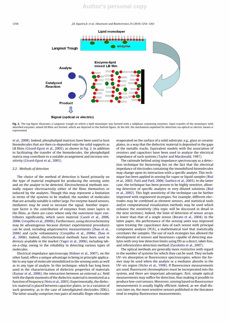
Fig. 2. The top figure illustrates a Langmuir trough on which a lipid monolayer was formed with a subphase containing enzymes. Upon transfer of the monolayer withadsorbed enzymes, mixed LB films are formed, which are depicted in the bottom figure. At the left, the mechanism exploited for detection via optical or electric means isrepresented.
et al., 2008). Indeed, phospholipid matrices have been used to hostbiomolecules that are then co-deposited onto the solid supports asLB films (Girard-Egrot et al., 2005), as shown in Fig. 2. In additionto facilitating the transfer of the biomolecules, the phospholipidmatrix may contribute to a suitable arrangement and increase sen-sitivity (Girard-Egrot et al., 2005).
2.2. Methods of detection
The choice of the method of detection is based primarily onthe type of material employed for producing the sensing unitsand on the analyte to be detected. Electrochemical methods nor-mally require electroactivity either of the films themselves orinduced by the analytes. Though this may represent a limitationin terms of the systems to be studied, the number of moleculesthat are actually suitable is rather large. For enzyme-based sensors,mediators may be used to increase the signal. Another impor-tant factor is the contribution of enzymes from inner layers inthe films, as there are cases where only the outermost layer con-tributes significantly, which saves material (Caseli et al., 2006,2008; Crespilho et al., 2006b). Furthermore, using electrochemistrymay be advantageous because distinct experimental procedurescan be used, including amperometric measurements (Zhao et al.,2006) and cyclic voltammetry (Crespilho et al., 2006e; Zhao etal., 2006). Indeed, electrochemical methods have been used indevices available in the market (Yager et al., 2006), including lab-on-a-chip, owing to the reliability in detecting various types ofmolecules.
Electrical impedance spectroscopy (Kidner et al., 2007), on theother hand, offers a unique advantage in being in principle applica-ble to any type of molecule immobilized in the sensing units as wellas to any type of analyte. In this technique, which has long beenused in the characterization of dielectric properties of materials(Kumar et al., 2006), the interaction between an external a.c. fieldwith the dipole moments of the dielectric material is monitored as afunction of frequency (Neto et al., 2003). Experimentally, the dielec-tric material is placed between capacitor plates, or in a variation ofsuch geometry, as in the case of interdigitated electrodes (IDEs).The latter usually comprises two pairs of metallic finger electrodes
evaporated on the surface of a solid substrate, e.g., glass or ceramicplates, in a way that the dielectric material is deposited in the gapsof the metallic tracks. Equivalent models with the association ofresistors and capacitors have been used to analyze the electricalimpedance of such systems (Taylor and Macdonald, 1987).
The rationale behind using impedance spectroscopy as a detec-tion technique for biosensing lies on the fact that the electricalimpedance of electrodes containing the immobilized biomoleculesmay change upon its interaction with a specific analyte. This tech-nique has been applied in sensing for vapor or liquid samples (Riulet al., 2002; Patil and Patil, 2006; Suehiro et al., 2003). In the lattercase, the technique has been proven to be highly sensitive, allow-ing detection of specific analytes in very diluted solutions (Riulet al., 2002). This high sensitivity of the technique can be furtherimproved with engineered strategies. For example, different elec-trodes may be combined as element sensors, and statistical toolsand/or computational visualization methods may be used whichenhance the sensitivity (this topic will be discussed in detail inthe next sections). Indeed, the limit of detection of sensor arraysis lower than that of a single sensor (Borato et al., 2004). In thelatter paper, the performance of the sensing units was improvedupon treating the capacitance data of each sensor with principalcomponent analysis (PCA), a mathematical tool that statisticallycorrelates the samples. The use of such strategies has allowed thedevelopment of sensors and biosensors capable of detecting ana-lytes with very low detection limits using EIS as a direct, label-free,and referenceless detection method (Zucolotto et al., 2007).
The optical methods are generally more restrictive with regardto the number of systems for which they can be used. They includeUV–vis absorption or fluorescence spectroscopies, where the for-mer may be used when the analyte or a mediator absorbs in theUV–vis region (Dicko et al., 1998). If fluorescence measurementsare used, fluorescent chromophores must be incorporated into thesystem, and there are important advantages: first, simple opticalmeasurements may suffice for detection, thus making it possible toproduce low-cost sensors. Moreover, sensing based on fluorescencemeasurements is usually highly efficient. Indeed, as we shall dis-cuss later on, the most sensitive sensors published in the literaturetend to employ fluorescence measurements.

Author's personal copy
J.R. Siqueira Jr. et al. / Biosensors and Bioelectronics 25 (2010) 1254–1263 1257
Fig. 3. Diagram illustrating the insulator poly(allylaminehydrochloride) layer and CoTsPc redox activity layer in the multilayer structure. The redox active material in themultilayer is electrochemically addressable via electron hopping between neighboring sites after cysteine oxidation (from ref. (Santos et al., in press), with kind permissionof Brazilian Chemical Society Publications).
3. Recent trends in electrochemical layer-by-layerbiosensors
A variety of electrochemical sensors are now used in detect-ing biomolecules and/or with electrodes modified by biomolecules.In electrochemical analyses, either a current or a potential ismeasured during an electrochemical reaction (Mcnaught andWilkinson, 1997). Electrochemical sensors are interesting due tothe versatility in size, geometry, selection of components, methodof detection and surface chosen according to the analyte properties.Several electrochemical techniques are used in signal transducing,including cyclic voltammetry (Mohadesi et al., 2008), pulse voltam-metry (conventional and differential) (Bergamini et al., 2009; DosReis et al., 2008), square-wave voltammetry (Sartori et al., 2009;Bergamini et al., 2009), stripping voltammetry (Mohadesi et al.,2008) and potentiometry (Santini et al., 2009), and amperometry(Santos et al., 2009; Karimi-Maleh et al., 2009; Dos Reis et al., 2008;Cervini and Cavalheiro, 2008). With electrochemical cells, measure-ments can be made of the electrode potential, cell current and cellresistance as a function of the analyte at the electrode surface or atthe electrochemical interfaces.
Nanostructured films modify electrodes and may allow tuningelectrochemical properties upon synergistic interactions betweenthe film components (Crespilho et al., 2006e). For example, chargetransfer mechanisms in LbL films containing biomolecules andnanomaterials have been investigated at the molecular level(Crespilho et al., 2006e). Constitutional dynamic chemistry (CDC)(Lehn, 2007) concepts with electrodes modified with covalent andsupramolecular non-covalent interactions have also been used tostudy the recomposition, recombination and reorganization pro-cesses (Alencar et al., 2007, 2009), which may lead to enhancedsensing. Indeed, Silva and co-authors (Alencar et al., 2007, 2009),have exploited CDC in sensing with LbL films comprising nanoparti-cles, polyelectrolytes and metallotetrasulfonated phthalocyanines(MTsPc).
The charge transport within LbL films may be due to non-conventional mechanisms (Crespilho et al., 2006e). Schlenoff etal. (1998) showed that electron transport was facilitated in LbLfilms with polyelectrolyte multilayers containing electrochemi-cally active units because the material within the redox active layercould be distributed over a distance of several layers. This studywas made possible by interposing non-electrochemically activelayer pairs between active layers (Laurent and Schlenoff, 1997;Schlenoff et al., 1998). The enhanced transport occurred since the
alternating polyelectrolyte layers were interpenetrating, and atleast one pair of layers were required to fully insulate one redoxactive layer from another. In a multilayered system, the redox activematerial was electrochemically addressable throughout the multi-layer via electron hopping between neighboring sites. This typeof electron hopping was observed by Santos et al. (in press) inLbL films of cobalt–tetrasulfonated phthalocyanines (CoTsPc) andpoly(allylaminehydrochloride) (PAH), used to detect the cysteineamino acid catalytic oxidation. The electrode configuration con-sisting of three bilayers of PAH/CoTsPc served as electron acceptorfrom cysteine molecules, as depicted schematically in Fig. 3.
The electrochemical response from hybrid electrodes has alsobeen improved by using the concept of electroactive nanostruc-tured membranes (ENMs), in which enzymes were immobilized inLbL films containing dendrimer/gold nanoparticles or metalloph-thalocyanines (Crespilho et al., 2006b,d). Such strategy alloweddetection of biological molecules at very low concentrations andwith a high signal-to-noise ratio (Crespilho et al., 2008), owingto an enhanced charge transport in addition to the maximiza-tion of the biological activity (Crespilho et al., 2006b,d, 2008).Yet another strategy for producing enzyme-based biosensors is todeposit enzymes on aligned magnetic nanowires (Crespilho et al.,2009a). With the electrodeposition of nanowires perpendicularlyto the gold electrode, enzymes could be immobilized in LbL filmsthat also contained a block layer to minimize non-specific reactionswhen in the presence of interfering agents (Crespilho et al., 2009a;Hammond, 2004; Okahata et al., 1988, 1989).
The way through which biomolecules are immobilized affectssignificantly the electrode response (Merkoci, 2007; Ariga et al.,2007; Vidotti and De Torresi, 2009; Goncales et al., 2008; Vidottiet al., 2009). Goncales et al. (2008), Vidotti and De Torresi (2009),Vidotti et al. (2009) obtained an optimized response in terms ofsensitivity towards urea, lifetime and short response times for anamperometric biosensor with urease covalently attached to thesubstrate. In the latter study, urease was immobilized onto a sub-strate consisting of poly(pyrrole) and poly(5-amino-1-naphthol)using different approaches, namely direct adsorption, entrap-ment in cellulose acetate layer, cross-linking with glutaraldehyde,and covalent attachment to the polymeric matrix. Urease hasalso been immobilized on polyaniline (PAni) modified electrodes,whose electroactive nanostructured membranes (ENMs) madewith silver nanoparticles (AgNP) stabilized in polyvinyl alcohol(PAni/PVA–AgNP) were applied at the enzyme/electrode interface(Crespilho et al., 2009b). The performance of the modified elec-

Author's personal copy
1258 J.R. Siqueira Jr. et al. / Biosensors and Bioelectronics 25 (2010) 1254–1263
Fig. 4. Immobilization of 1,2 Cl-cathecol dioxygenase (CCD) with polyamidoaminePAMAM dendrimer in LbL film on an interdigitated electrode.
trodes toward urea hydrolysis was evaluated with amperometricmeasurements, revealing a fast increase in cathodic current with awell-defined peak upon addition of urea to the electrolytic solution.Also, the friendly environment provided by the PAni/PVA–AgNPelectrode to the immobilized enzyme promoted efficient catalyticconversion of urea into ammonium and bicarbonate ions.
Because electrodes prepared with LbL films may exhibit highelectrochemical stability, biosensors have been proposed for clin-ical applications (Crespilho et al., 2006e). In fact, using the LbLtechnique may allow one to enhance both the stability and theenzymatic activity (Onda et al., 1999; Dong and Chen, 2002;Crespilho et al., 2006e). Also, the LbL method has been used to pro-duce hybrid materials (Merkoci, 2007; Ariga et al., 2007; Vidottiand De Torresi, 2009; Goncales et al., 2008; Vidotti et al., 2009;Crespilho et al., 2005, 2006a, 2007, 2009d) and modified inter-faces (Crespilho et al., 2005, 2006e, 2007; Merkoci, 2007), withenhanced charge transport and preservation of the biological activ-ity (Crespilho et al., 2005, 2007). The main challenge now is toimprove the reproducibility and sensitivity, in order to apply thesedevices for diagnosis of diseases (Kerman et al., 2008).
4. Extending the concept of electronic tongues
The seminal paper by Riul et al. (2002) has demonstrated thatimpedance spectroscopy allied to nanostructured films can pro-vide high sensitivity for sensing units, which are used in a sensorarray for analyzing tastes. Subsequent studies indeed confirmedthat trace amounts of compounds representing the basic tastes(Ferreira et al., 2003) or impurities in the water (Oliveira et al.,2004) may be detected. Furthermore, the high sensitivity has beenexplored in distinguishing very similar complex liquid samples, asin the distinction of wines (Riul et al., 2003a), coffee (Riul et al.,2003b) and orange juice (Ciosek et al., 2007).
In contrast to the general concept of global selectivity employedin the electronic tongue systems, a concept based on the specificinteraction between biological systems was proposed (Perinottoet al., 2008; Zucolotto et al., 2006), which is illustrated in Fig. 4.In the latter, interdigitated electrodes containing immobilized 1,2Cl-cathecol dioxygenase (CCD) enzymes were employed in detect-ing cathecol molecules, which specifically interact with CCD. Thesame strategy was extended for immunosensors with immobilizedantigenic proteins capable of detecting specific anti-P. multocida(causative agents of Pasteurellosis) antibodies at concentrations aslow as 10−9 g/mL (Zucolotto et al., 2007).
If combined with artificial intelligence methods, electronictongues may provide correlation between electrical measurementsand the human taste (Ferreira et al., 2007). The latter has beenshown for coffee samples that had been assessed by professional
tasters who assigned scores – ranging from ca. 3–8 – correspond-ing to the quality of the coffee. Regression methods combined tomachine learning were then used to treat the data obtained withan electronic tongue whose sensing units were made of nanostruc-tured films from semiconducting polymers. Significantly, a highdegree of correlation could be established between the scoresassigned by the human experts and those inferred from the regres-sion methods applied to the electronic tongue data (Ferreira et al.,2007).
5. Biosensing based on FETs
The integration of a chemical or biological recognition elementinto a semiconductor platform has become attractive for foster-ing new sensor concepts. In this context, silicon-based technologyis being merged with materials and biomaterials science to pro-duce sensors and biosensors that can be used in medicine andbiotechnology, e.g. as in food control and monitoring of the envi-ronment (Cui et al., 2001; Lu and Lieber, 2007; Schöning andPoghossian, 2006; Schöning, 2005). The main advantage is thecompatibility with semiconductor processing, since sensors canbe manufactured as small, reliable and rugged devices (Cui etal., 2001; Lu and Lieber, 2007; Schöning and Poghossian, 2006;Schöning, 2005). Furthermore, with semiconductor technology onemay strive for miniaturization and integration of sensors, actuatorsand mechanical or fluidic elements in a single chip. Thus, minia-turized multifunctional analytical systems such as “lab-on-a-chip”,electronic tongues, micro total analysis systems (�TAS) and micro-electro-mechanical system (MEMS) become possible (Schöning,2005; Schöning and Poghossian, 2006).
Silicon-based chemical sensors and biosensors developedare the field-effect-devices (FEDs), which are derived frommetal-insulator-semiconductor capacitances or insulated-gatefield-effect transistors, with the gate electrode being replaced byan electrolyte solution (test sample) and a reference electrode.Capacitive EIS (electrolyte-insulator-semiconductor) sensors, LAPS(light-addressable potentiometric sensors) and ISFETs (ion-sensitive field-effect transistors), represent typical examples ofsensors based on FEDs (Schöning, 2005; Schöning and Poghossian,2006). They offer advantages such as small size and weight, a fastresponse time, robustness, integration of sensor arrays on a chip,and possible low-cost fabrication.
With the introduction of an additional ion- and/or charge-sensitive gate layer, (bio-)chemical FEDs are sensitive to anyelectrical interaction at or nearby the interface to the electrolyte.For example, changes in the chemical composition of the analytewill induce changes in the electrical surface charge of the FED, thusmodulating the capacitance of the EIS sensor, the photocurrentof the LAPS or the current of the ISFET channel. The mecha-nisms involved for the signal generation of these (bio-)chemicalsensors encompass a pH or ion-concentration change, the ion-concentration change due to an enzymatic reaction, the adsorptionof charged macromolecules (e.g., polyelectrolytes, proteins, DNA),and the binding affinity of molecules (e.g., antigen-antibody affin-ity reaction, or DNA hybridization) (Schöning, 2005; Schöning andPoghossian, 2006).
The successful development of the sensor systems mentionedabove requires infrastructure and knowledge on both silicon- andthin-film processing technologies. A key feature in this regard is toproduce materials with control at the molecular level. In particular,the Layer-by-Layer (LbL) technique is promising for silicon-basedsensors as this method allows a control of film architecture andthickness, in addition to the synergy between properties of distinctmaterials, including carbon nanotubes (Lee et al., 2009), proteins(Lvov et al., 1995), antigen-antibody pairs (Zucolotto et al., 2007),

Author's personal copy
J.R. Siqueira Jr. et al. / Biosensors and Bioelectronics 25 (2010) 1254–1263 1259
Fig. 5. Schematic representation and operation principle of a penicillin biosensor based on a capacitive EIS sensor containing SWNT/PAMAM LbL film and penicillinase.
DNA (Elbakry et al., 2009), nanoparticles (Crespilho et al., 2006c),and other charged macromolecules (Siqueira et al., 2006).
Recent efforts to obtain field-effect sensors containing LbL filmsinclude the work of Cui et al. (2004), in which a FET was fabricatedwith the LbL technique containing poly(dimethyldiallylammoniumchloride) (PDDA) alternated with SnO2 and SiO2 nanoparticles. Inanother study Cui et al. (2005) proposed a FET with a LbL structureof PDDA and poly(sodium 4-styrene sulfonate) (PSS) in combina-tion with alternated deposition of In2O3 and SiO2 nanoparticles aschannel and gate dielectrics. Both of these researches were basedon the development of low-cost micro/nanoelectronic prototypedevices. Poghossian et al. (2006, 2007a,b) reported the possibil-ities and limitations of a direct electrical detection of chargedmacromolecules using a field-effect-based sensor platform, focus-ing on the study of possible mechanisms of signal generation ofthe LbL adsorption of charged macromolecules of poly(allylaminehydrochloride) (PAH) and PSS on a capacitive EIS structure. Xu etal. (2005) produced a biosensor for lactate with LbL films con-taining MnO2 nanoparticles and lactate oxidase immobilized inpoly(dimethyldiallylammonium chloride) (PDDA) on the gate ofan ISFET, achieving better sensitivity and performance. Siqueira etal. (2009a,b) proposed an EIS capacitive sensor and a LAPS sensor(Siqueira et al., 2009c) functionalized with a LbL structure con-taining polyamidoamine (PAMAM) dendrimer and single-walledcarbon nanotubes (SWNT) with the enzyme penicillinase immobi-lized atop the film surface for detecting penicillin G. Fig. 5 depictsthe operation principle and film architecture, which was essentialfor the improvement of response time, drift and stability, in addi-tion to a higher sensitivity of the EIS-penicillin biosensors. An LbLassembly of nanowires (NW) building blocks was reported by Javeyet al. (2007) for the fabrication of NW FETs using Ge/Si core-shellNWs as an approach for three-dimensional (3D) multifunctionalelectronics.
6. Optical biosensing
Optical sensors have been used in detecting various compounds,including alcohols, drugs and pesticides. They are generally pro-duced with immobilization of enzymes in hybrid materials, e.g.using the LbL and LB methods. The choice of suitable scaffoldsis crucial to provide film architectures for which the optical sig-nal depends on the analyte concentration, but is independent ofother factors such as the interaction with the inert matrix. Reportedhybrid films include phospholipid–nucleic acid LB films for detect-ing antitumor drugs, such as daunomycin (Montrel et al., 1997).
The affinity of the drug enhanced the adsorption of a dye (acridineorange) used as mediator onto the polynucleotide. Another exam-ple of optical detection via mediator is the urease-based biosensorto detect urea (Dicko et al., 1998), where the hydrolysis of ureaproduced ammonia, causing an increase in the pH-sensitive dyebromocresol purple. Urea was also detected with mixed LB filmsof urease–phospholipid as sensing unit (Caseli et al., 2008). Alka-line phosphatases can be employed to detect phenols in wine,as their activity toward phenols has been demonstrated in Lang-muir (Caseli et al., 2005) and Langmuir–Blodgett films (Caseli etal., 2002, 2004, 2007), whose sensing ability was determined opti-cally by analyzing the evolution of an absorption band at 410 nmfor p-nitrophenolphosphate hydrolysis.
In spite of its simplicity, colorimetry is useful for biosensing,as demonstrated with horseradish peroxidase (HRP) immobilizedin phospholipid LB (Schmidt et al., 2008) and chitosan LbL films(Schmidt et al., 2009). In these works, the enzyme activity wasdetermined optically with pyrogallol oxidized to purpurogallin inthe presence of hydrogen peroxide.
Fluorescence spectroscopy is particularly sensitive to smallquantities of analyte. It has been used for sensing paraoxon inter-acting with LbL films of chitosan and OPH (organophosphoroushydrolase) (Constantine et al., 2003), and for antibodies interactingwith LB films of viologen and protein-A (Lee et al., 2004). Sensorsfor integrin were based on its interaction with hybrid LbL-LB filmsmade with lipid dye labeled receptors and streptavidin (Worsfold etal., 2004). Not only fluorescence enhancement but also fluorescencequenching is used to detect analytes. For instance, OPH bondedwith a fluorescein probe was exploited in paraoxon sensors, withfluorescence being quenched due to the hydrolysis of the pesticide.
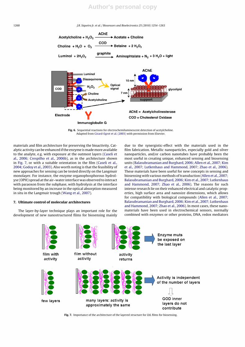
The control of molecular architectures of LB films was essentialfor building a biosensor in which a highly organized proteo-lipidstructure was obtained with liposome fusion (Godoy et al., 2003) byinserting a non-inhibitor monoclonal antibody in a function posi-tion. This allowed the assembly to sequester acetylcholinesterase(Elbakry et al., 2009) in a suitable orientation and enhance theperformance of the optical screen-printed choline sensor basedon luminal electrochemiluminescence. In another paper from thesame group (Girard-Egrot et al., 2003), immunoglobulin was shownto have preferential orientation in the glycolipid LB monolayer,then enhancing the luminescence signal. In this case, the enzymewas sequestered from an external aqueous solution by the mixedimmunoglobulin–lipid LB film, as shown in the scheme of Fig. 6.
The extensive work on optical biosensors made with immo-bilized enzymes pointed to the importance of the scaffolding
Author's personal copy
1260 J.R. Siqueira Jr. et al. / Biosensors and Bioelectronics 25 (2010) 1254–1263
Fig. 6. Sequential reactions for electrochemiluminescent detection of acetylcholine.Adapted from Girard-Egrot et al. (2003) with permission from Elsevier.
materials and film architecture for preserving the bioactivity. Cat-alytic activity can be enhanced if the enzyme is made more availableto the analyte, e.g. with exposure at the outmost layers (Caseli etal., 2006; Crespilho et al., 2006b), as in the architecture shownin Fig. 7, or with a suitable orientation in the film (Caseli et al.,2004; Godoy et al., 2003). Also worth noting is that the feasibility ofnew approaches for sensing can be tested directly on the Langmuirmonolayer. For instance, the enzyme organophosphorous hydrol-yse (OPH) spread at the air–water interface was observed to interactwith paraoxon from the subphase, with hydrolysis at the interfacebeing monitored by an increase in the optical absorption measuredin situ in the Langmuir trough (Wang et al., 2007).
7. Ultimate control of molecular architectures
The layer-by-layer technique plays an important role for thedevelopment of new nanostructured films for biosensing mainly
due to the synergistic-effect with the materials used in thefilm fabrication. Metallic nanoparticles, especially gold and silvernanoparticles, and/or carbon nanotubes have probably been themost useful in creating unique, enhanced sensing and biosensingunits (Balasubramanian and Burghard, 2006; Allen et al., 2007; Kimet al., 2007; Lutkenhaus and Hammond, 2007; Zhao et al., 2006).These materials have been useful for new concepts in sensing andbiosensing with various methods of transduction (Allen et al., 2007;Balasubramanian and Burghard, 2006; Kim et al., 2007; Lutkenhausand Hammond, 2007; Zhao et al., 2006). The reasons for suchintense research lie on their enhanced electrical and catalytic prop-erties, high surface area and nanosize dimensions, which allowsfor compatibility with biological compounds (Allen et al., 2007;Balasubramanian and Burghard, 2006; Kim et al., 2007; Lutkenhausand Hammond, 2007; Zhao et al., 2006). In most cases, these nano-materials have been used in electrochemical sensors, normallycombined with enzymes or other proteins, DNA, redox mediators
Fig. 7. Importance of the architecture of the layered structure for LbL films for biosensing.

Author's personal copy
J.R. Siqueira Jr. et al. / Biosensors and Bioelectronics 25 (2010) 1254–1263 1261
Table 1Summary of modified electrodes containing nanoparticles and carbon nanotubes for electrochemical sensors.
Analyte LbL biosensor Biological compound References
Glucose (PDDA/MWNT)n-(PDDA/GOx)n Yan et al. (2007)(GOx/PtNp-DENs)n/CNTs/Pt Glucose oxidase (GOx) Xu et al. (2007)(GOx/AuNp/MWCNTs) Wu et al. (2007b)(Chi/MWNTs/GOx) Zou et al. (2008)(GOx/GNp/CS/GOx/GNp/PAA) Wu et al. (2007a)
H2O2 (TB/AuNp)n-HRP Horseradish peroxidase (HRP) Chen et al. (2006)Cholesterol (PDDA/ChOx)n-MWNT Cholesterol oxidade (ChOx) Guo et al. (2004)DNA (MWCNTs/GNPs)n-DNA DNA Zhang et al. (2009)
ssDNA-(cys/MWCNTs/cys/GNPs)n Ma et al. (2008)
Ethanol (PDDA/AOx)5a alcohol oxidase (AOx) Li et al. (2008)
a Although this system does not contain nanoparticles or nanotubes, it is an important example from the point of view of application of LbL nanostructures in biosensing.
and other electroactive species (Allen et al., 2007; Balasubramanianand Burghard, 2006; Kim et al., 2007; Lutkenhaus and Hammond,2007; Zhao et al., 2006). An example of the latter are the metal-lophthalocyanines in modified electrodes that were suitable forreaching high sensitivity, selectivity, and adequate response time,drift and charge transfer process (Siqueira et al., 2009a, 2007, 2008).
The advantages of using nanoparticles and 1D nanostructures,such as nanotubes, nanowires, etc., in LbL films stem from the pos-sible immobilization of biomolecules with their biological activitypreserved, in addition to the enhanced electronic conductivity thatpermits direct electron transfer between redox proteins and elec-trode surfaces (Crespilho et al., 2006b). Furthermore, the formationof a porous LbL nanostructure may contribute to a better electrode-electrolyte interface and large surface area, which are important forsensing (Siqueira et al., 2008, 2009b,c). Modified electrodes withthese nanomaterials are able to catalyze the redox reactions of ana-lytes, such as H2O2 and NADH (Balasubramanian and Burghard,2006; Zhao et al., 2006). Also worth mentioning are the efforts tobuild electrochemical devices with the geometry controlled at thenanometer level (Tans et al., 1998; Cui et al., 2001; Besteman etal., 2003; Crespilho et al., 2009c). The use of individual 1D struc-tures, e.g. single-walled carbon nanotubes (SWNTs) (Besteman etal., 2003; Tans et al., 1998), boron-doped silicon nanowires (SiNWs)(Cui et al., 2001), Sn-doped In2O3 nanowires (ITO–NWs) (Crespilhoet al., 2009c), may allow electrochemical nanosensors to selectivelydetect proteins at different concentrations on real time. It is likelythat detecting single molecules with bioelectrochemical methodsmay become a reality in the near future, with important implica-tions to the field of biofuel cells and biosensors (Hoeben et al., 2008;Crespilho et al., 2009c).
Crespilho et al. (2006d), for example, proposed a new core-shellsystem with the electrodeposition of Prussian Blue as electro-chemical mediator onto Au nanoparticles immobilized in PAMAMdendrimers in LbL films of ITO–PVS/PAMAM–Au. The aim was toenhance charge transport via electron hopping, which was alsoexploited in a similar system with cobalt hexacyanoferrate (CoHCF)as redox mediator (Crespilho et al., 2006b). The latter nanostructureacted as an electroactive membrane for the immobilization of glu-cose oxidase using gluteraldehyde as cross-linker. This core shellPAMAM-Au@ CoHCF system made it possible to detect hydrogenperoxide in the enzymatic reaction at 0.0 V (vs. SCE).
Siqueira et al. (2007) studied the influence of detectingdopamine with ITO-modified electrodes with LbL films of nickelphthalocyanines (NiTsPc) combined with Pt-nanoparticles andMWNTs (Siqueira et al., 2008) in PAMAM dendrimers. Both sys-tems displayed a high sensitivity and electrocatalytic activitytoward dopamine. In particular, the first system possessed a bi-functionality with the capability of detecting two compounds withdissimilar chemical properties, namely dopamine and hydrogenperoxide, with a single electrode. This bifunctionality was madepossible because the surfaces of Pt-nanoparticles were available
for H2O2 oxidation even after the electrode had been used indopamine experiments. The incorporation of nanotubes in theNiTsPc/PAMAM–MWNT system led to sensors with a peak current3-fold higher than for the same structure without nanotubes. More-over, this sensor was able to distinguish dopamine in the presenceof a natural interferent, the ascorbic acid.
Other examples of modified electrodes containing nanoparticlesand/or carbon nanotubes in LbL films are summarized in Table 1.One should also note that the use of LbL nanostructures in field-effect devices is promising for developing (bio-)chemical sensorsbased on the silicon platform, as already discussed.
8. Final remarks
In this survey on the use of nanostructured films produced withthe Langmuir–Blodgett (LB) and the layer-by-layer (LbL) methodsfor biosensing, the major emphasis was placed on the importance oftemplate or scaffolding materials to warrant the activity preserva-tion of biomolecules employed as transducing materials. Particularattention was also given to the control of molecular architecturesafforded by both of these film-forming methods, especially in caseswhere layers containing biomolecules could be combined withlayers of metallic nanoparticles, carbon nanotubes, polymers andphthalocyanines. The success of the LB and LbL films for biosensingcan largely be attributed to the fact that entrained water remainsin the film structure, even after drying, and this helps preserve theactivity of the biomolecules. Various are the methods which maybe used in biosensors built with LB and LbL films. When extremelyhigh sensitivity is required, the optical methods based on lumi-nescence are among the best, if the system under investigationallows for such measurements. Electrochemical biosensors are use-ful for many applications, in spite of requiring electroactivity of themolecules in the sensing unit or in the analytes. With impedancespectroscopy, one may extend the concept of electronic tongues toinclude specific interactions between the film-forming moleculesand the analyte, thus leading to selectivity in addition to a high sen-sitivity. Also important is the possibility of integrating sensing unitswith silicon-based technology, an avenue to pursue for achievinglow-cost biosensors with scalable production.
Acknowledgments
This work was supported by FAPESP, CNPq, INEO and CAPES(Rede Bionano – Brazil).
References
Alencar, W.S., Crespilho, F.N., Martins, M.V.A., Zucolotto, V., Oliveira Jr., O.N., Silva,W.C., 2009. Phys. Chem. Chem. Phys. 11, 5086–5091.
Alencar, W.S., Crespilho, F.N., Santos, M., Zucolotto, V., Oliveira Jr., O.N., Silva, W.C.,2007. J. Phys. Chem. C 111, 12817–12821.
Allen, B.L., Kichambare, P.D., Star, A., 2007. Adv. Mater. 19, 1439–1451.
Author's personal copy
1262 J.R. Siqueira Jr. et al. / Biosensors and Bioelectronics 25 (2010) 1254–1263
Ariga, K., Hill, J.P., Ji, Q.M., 2007. Phys. Chem. Chem. Phys. 9, 2319–2340.Bader, B., Kuhn, K., Owen, D.J., Waldmann, H., Wittinghofer, A., Kuhlmann, J., 2000.
Nature 403, 223–226.Badia, A., Moraille, P., Tang, N.Y.W., Randlett, M.E., 2008. Int. J. Nanotech. 5,
1371–1395.Balasubramanian, K., Burghard, M., 2006. Anal. Bional. Chem. 385, 452–468.Bankar, S.B., Bule, M.V., Singhal, R.S., Ananthanarayan, L., 2009. Biotech. Adv. 27,
489–501.Bayley, H., Cremer, P.S., 2001. Nature 413, 226–230.Benson, D.E., Conrad, D.W., De Lorimer, R.M., Trammell, S.A., Hellinga, H.W., 2001.
Science 293, 1641–1644.Bergamini, M.F., Santos, D.P., Zanoni, M.V.B., 2009. J. Braz. Chem. Soc. 20, 100–106.Besteman, K., Lee, J.O., Wiertz, F.G.M., Heering, H.A., Dekker, C., 2003. Nano Lett. 3,
727–730.Blodgett, K.B., 1934. J. Am. Chem. Soc. 56, 495–1495.Borato, C.E., Riul Jr., A., Ferreira, M., Oliveira Jr., O.N., Mattoso, L.H.C., 2004. Instru-
ment. Sci. Tech. 32, 21–30.Campas, M., Prieto-Simon, B., Marty, J.L., 2009. Semin. Cell Dev. Biol. 20, 3–9.Caseli, L., Crespilho, F.N., Nobre, T.M., Zaniquelli, M.E.D., Zucolotto, V., Oliveira Jr.,
O.N., 2008. J. Colloid Interf. Sci. 319, 100–108.Caseli, L., Dos Santos, D.S., Foschini, M., Goncalves, D., Oliveira Jr., O.N., 2006. J. Colloid
Interf. Sci. 303, 326–331.Caseli, L., Furriel, R.P.M., De Andrade, J.F., Leone, F.A., Zaniquelli, M.E.D., 2004. J.
Colloid Interf. Sci. 275, 123–130.Caseli, L., Masui, D.C., Prazeres, R., Furriel, M., Leone, F.A., Zaniquelli, M.E.D., 2007.
Thin Solid Films 515, 4801–4807.Caseli, L., Oliveira, R.G., Masui, D.C., Furriel, R.P.M., Leone, F.A., Maggio, B., Zaniquelli,
M.E.D., 2005. Langmuir 21, 4090–4095.Caseli, L., Zaniquelli, M.E.D., Furriel, R.P.M., Leone, F.A., 2002. Colloid Surf. B 25,
119–128.Cervini, P., Cavalheiro, E.T.G., 2008. J. Braz. Chem. Soc. 19, 836–841.Chen, S.H., Yuan, R., Chai, Y.Q., Xu, L., Wang, N., Li, X.N., Zhang, L.Y., 2006. Electro-
analysis 18, 471–477.Ciosek, P., Maminska, R., Dybko, A., Wroblewski, W., 2007. Sens. Actuators B-Chem.
127, 8–14.Constantine, C.A., Mello, S.V., Dupont, A., Cao, X.H., Santos, D., Oliveira Jr., O.N., Strix-
ino, F.T., Pereira, E.C., Cheng, T.C., Defrank, J.J., Leblanc, R.M., 2003. J. Am. Chem.Soc. 125, 1805–1809.
Crespilho, F.N., Borges, T., Zucolotto, V., Leite, E.R., Nart, F.C., Oliveira Jr., O.N., 2006a.J. Nanosci. Nanotech. 6, 2588–2590.
Crespilho, F.N., Esteves, M.C., Sumodjo, P.T.A., Podlaha, E.J., Zucolotto, V., 2009a. J.Phys. Chem. C 113, 6037–6041.
Crespilho, F.N., Ghica, M.E., Florescu, M., Nart, F.C., Oliveira Jr., O.N., Brett, C.M.A.,2006b. Electrochem. Commun. 8, 1665–1670.
Crespilho, F.N., Ghica, M.E., Gouveia-Caridade, C., Oliveira Jr., O.N., Brett, C.M.A., 2008.Talanta 76, 922–928.
Crespilho, F.N., Huguenin, F., Zucolotto, V., Olivi, P., Nart, F.C., Oliveira Jr., O.N., 2006c.Electrochem. Commun. 8, 348–352.
Crespilho, F.N., Iost, R.M., Travain, S.A., Oliveira Jr., O.N., Zucolotto, V., 2009b. Biosens.Bioelectron. 24, 3073–3077.
Crespilho, F.N., Lanfredi, A.J.C., Leite, E.R., Chiquito, A.J., 2009c. Electrochem. Com-mun. 11, 1744–1747.
Crespilho, F.N., Lima, F.C.A., Da Silva, A.B.F., Oliveira Jr., O.N., Zucolotto, V., 2009d.Chem. Phys. Lett. 469, 186–190.
Crespilho, F.N., Nart, F.C., Oliveira Jr., O.N., Brett, C.M.A., 2007. Electrochim. Acta 52,4649–4653.
Crespilho, F.N., Zucolotto, V., Brett, C.M.A., Oliveira Jr., O.N., Nart, F.C., 2006d. J. Phys.Chem. B 110, 17478–17483.
Crespilho, F.N., Zucolotto, V., Oliveira Jr., O.N., Nart, F., 2006e. Int. J. Electrochem. Sci.1, 194–214.
Crespilho, F.N., Zucolotto, V., Siqueira Jr., J.R., Constantino, C.J.L., Nart, F.C., OliveiraJr., O.N., 2005. Env. Sci. Tech. 39, 5385–5389.
Cui, T.H., Hua, F., Lvov, Y., 2004. IEEE Trans. Elec. Dev. 51, 503–506.Cui, T.H., Liu, Y., Zhu, M., 2005. Appl. Phys. Lett., 87.Cui, Y., Wei, Q.Q., Park, H.K., Lieber, C.M., 2001. Science 293, 1289–1292.Debabov, V.G., 2004. Mol. Biol. 38, 482–493.Decher, G., Hong, J.D., Schmitt, J., 1992. Thin Solid Films 210, 831–835.Dicko, A., Bourque, H., Pezolet, M., 1998. Chem. Phys. Lipids 96, 125–139.Dong, S., Chen, X., 2002. Rev. Mol. Biotech. 82, 302–323.Dos Reis, A.P., Tarley, C.R.T., Kubota, L.T., 2008. J. Braz. Chem. Soc. 19, 1567–1573.Elbakry, A., Zaky, A., Liebkl, R., Rachel, R., Goepferich, A., Breunig, M., 2009. Nano
Lett. 9, 2059–2064.Ferreira, E.J., Pereira, R.C.T., Delbem, A.C.B., Oliveira Jr., O.N., Mattoso, L.H.C., 2007.
Electron. Lett. 43, 1138–1140.Ferreira, M., Constantino, C.J.L., Riul Jr., A., Wohnrath, K., Aroca, R.F., Giacometti, J.A.,
Oliveira Jr., O.N., Mattoso, L.H.C., 2003. Polymer 44, 4205–4211.Girard-Egrot, A.P., Godoy, S., Blum, L.J., 2005. Adv. Colloid Interf. Sci. 116, 205–225.Girard-Egrot, A.P., Godoy, S., Chauvet, J.P., Boullanger, P., Coulet, P.R., 2003. Biochim.
Biophys. Acta: Biomembr. 1617, 39–51.Godoy, S., Chauvet, J.P., Boullanger, P., Blum, L.J., Girard-Egrot, A.P., 2003. Langmuir
19, 5448–5456.Goncales, V.R., Salvador, R.P., Alcantara, M.R., De Torresi, S.I.C., 2008. J. Electrochem.
Soc. 155, K140–K145.Gu, L.Q., Braha, O., Conlan, S., Cheley, S., Bayley, H., 1999. Nature 398, 686–690.Guo, M.L., Chen, J.H., Li, J., Nie, L.H., Yao, S.Z., 2004. Electroanalysis 16, 1992–1998.Hammond, P.T., 2004. Adv. Mater. 16, 1271–1293.
Hoeben, F.J.M., Meijer, F.S., Dekker, C., Albracht, S.P.J., Heering, H.A., Lemay, S.G.,2008. Acs Nano 2, 2497–2504.
Hone, J., Kam, L., 2007. Nat. Nanotech. 2, 140–141.Javey, A., Nam, S., Friedman, R.S., Yan, H., Lieber, C.M., 2007. Nano Lett. 7, 773–777.Karimi-Maleh, H., Ensafi, A.A., Ensafi, H.R., 2009. J. Braz. Chem. Soc. 20, 880–887.Kerman, K., Saito, M., Yamamura, S., Takamura, Y., Tamiya, E., 2008. Trac-Trends
Anal. Chem. 27, 585–592.Kidner, N.J., Meier, A., Homrighaus, Z.J., Wessels, B.W., Mason, T.O., Garboczi, E.J.,
2007. Thin Solid Films 515, 4588–4595.Kim, S.N., Rusling, J.F., Papadimitrakopoulos, F., 2007. Adv. Mater. 19, 3214–
3228.Kubik, T., Bogunia-Kubik, K., Sugisaka, M., 2005. Curr. Pharm. Biotech. 6, 17–33.Kumar, A., Singh, B.P., Choudhary, R.N.P., Thakur, A.K., 2006. Mater. Chem. Phys. 99,
150–159.Laurent, D., Schlenoff, J.B., 1997. Langmuir 13, 1552–1557.Lee, S.W., Kim, B.S., Chen, S., Shao-Horn, Y., Hammond, P.T., 2009. J. Am. Chem. Soc.
131, 671–679.Lee, W.C., Chun, B.S., Oh, B.K., Lee, W.H., Choi, J.W., 2004. BBA—Biomembranes 9,
241–244.Lehn, J.M., 2007. Chem. Soc. Rev. 36, 151–160.Li, S., Wang, Y.Y., Zhao, Z.X., Qin, X., Wang, X.S., Wang, H.C., Yu, M., Miao, Z.Y., Wu,
B.Y., Chen, Q., 2008. Sens. Lett. 6, 347–351.Liu, X.D., Yu, W.T., Wang, W., Xiong, Y., Ma, X.J., Yuan, Q.A., 2008. Prog. Chem. 20,
126–139.Looger, L.L., Dwyer, M.A., Smith, J.J., Hellinga, H.W., 2003. Nature 423, 185–190.Lu, W., Lieber, C.M., 2007. Nat. Mater. 6, 841–850.Lutkenhaus, J.L., Hammond, P.T., 2007. Soft Matter 3, 804–816.Lv, F., Peng, Z.H., Zhang, L.L., Yao, L.S., Liu, Y., Xuan, L., 2009. Liq. Cryst. 36, 43–51.Lvov, Y., Ariga, K., Ichinose, I., Kunitake, T., 1995. J. Am. Chem. Soc. 117, 6117–6123.Ma, H.Y., Zhang, L.P., Pan, Y., Zhang, K.Y., Zhang, Y.Z., 2008. Electroanalysis 20,
1220–1226.Mcnaught, A.D., Wilkinson, A., 1997. IUPAC Compendium of Chemical Terminology,
second ed. Blackwell Science.Merkoci, A., 2007. Electroanalysis 19, 739–741.Mertens, J., Rogero, C., Calleja, M., Ramos, D., Martin-Gago, J.A., Briones, C., Tamayo,
J., 2008. Nat. Nanotech. 3, 301–307.Mohadesi, A., Salmanipour, A., Mohammadi, S.Z., Pourhatami, A., Taher, M.A., 2008.
J. Braz. Chem. Soc. 19, 956–962.Montrel, M.M., Sukhorukov, G.B., Petrov, A.I., Shabarchina, L.I., Sukhorukov, B.I.,
1997. Sens. Actuators B-Chem. 42, 225–231.Neto, J.M.G., Ferreira, G.F.L., Santos, J.R., Da Cunha, H.N., Dantas, I.F., Bianchi, R.F.,
2003. Braz. J. Phys. 33, 371–375.Okahata, Y., Tsuruta, T., Ijiro, K., Ariga, K., 1988. Langmuir 4, 1373–1375.Okahata, Y., Tsuruta, T., Ijiro, K., Ariga, K., 1989. Thin Solid Films 180, 65–72.Oliveira Jr., O.N., Riul Jr., A., Leite, V.B.P., 2004. Braz. J. Phys. 34, 73–83.Onda, M., Ariga, K., Kunitake, T., 1999. J. Biosci. Bioeng. 87, 69–75.Patil, L.A., Patil, D.R., 2006. Sens. Actuators B-Chem. 120, 316–323.Perinotto, A.C., Caseli, L., Hayasaka, C.O., Riul Jr., A., Oliveira Jr., O.N., Zucolotto, V.,
2008. Thin Solid Films 516, 9002–9005.Poghossian, A., Abouzar, M.H., Amberger, F., Mayer, D., Han, Y., Ingebrandt, S., Offen-
hausser, A., Schöning, M.J., 2007a. Biosens. Bioelectron. 22, 2100–2107.Poghossian, A., Abouzar, M.H., Sakkari, M., Kassab, T., Han, Y., Ingebrandt, S., Offen-
hausser, A., Schöning, M.J., 2006. Sens. Actuators B-Chem. 118, 163–170.Poghossian, A., Ingebrandt, S., Abouzar, M.H., Schöning, M.J., 2007b. Appl. Phys. A:
Mater. 87, 517–524.Riul Jr., A., Dos Santos Jr., D.S., Wohnrath, K., Di Tommazo, R., Carvalho, A.A.C.P.L.F.,
Fonseca, F.J., Oliveira Jr., O.N., Taylor, D.M., Mattoso, L.H.C., 2002. Langmuir 18,239–245.
Riul Jr., A., Malmegrim, R.R., Fonseca, F.J., Mattoso, L.H.C., 2003a. Biosens. Bioelectron.18, 1365–1369.
Riul Jr., A., Soto, A.M.G., Mello, S.V., Bone, S., Taylor, D.M., Mattoso, L.H.C., 2003b.Synth. Metals 132, 109–116.
Santini, A.O., Pezza, H.R., Sequinel, R., Rufino, J.L., Pezza, L., 2009. J. Braz. Chem. Soc.20, 64–73.
Santos, A.C., Crespilho, F.N., Luz, R.A.S., Ferreira, L.G.F., Dos Santos, J.R., Silva, W.C.,Supramolecular organization of cobalt (ii) phthalocyanine on the pathway ofcysteine oxidation. Quim. Nova, in press.
Santos, W.D.R., Sousa, A.L., Sotomayor, M.D.T., Damos, F.S., Tanaka, S., Kubota, L.T.,Tanaka, A.A., 2009. J. Braz. Chem. Soc. 20, 1180–1187.
Sartori, E.R., Medeiros, R.A., Rocha, R.C., Fatibello, O., 2009. J. Braz. Chem. Soc. 20,360–366.
Schlenoff, J.B., Laurent, D., Ly, H., Stepp, J., 1998. Adv. Mater. 10, 347–349.Schmidt, T.F., Caseli, L., Dos Santos, D.S., Oliveira Jr., O.N., 2009. Mater. Sci. Eng. C 29,
1889–1892.Schmidt, T.F., Caseli, L., Viitala, T., Oliveira Jr., O.N., 2008. Biochim. Biophys. Acta:
Biomembr. 1778, 2291–2297.Schöning, M.J., 2005. Sensors 5, 126–138.Schöning, M.J., Poghossian, A., 2006. Electroanalysis 18, 1893–1900.Siqueira Jr., J.R., Abouzar, M.H., Bäcker, M., Zucolotto, V., Poghossian, A., Oliveira Jr.,
O.N., Schöning, M.J., 2009a. Phys. Stat. Sol. A 206, 462–467.Siqueira Jr., J.R., Abouzar, M.H., Poghossian, A., Zucolotto, V., Oliveira Jr., O.N., Schön-
ing, M.J., 2009b. Biosens. Bioelectron. 25, 497–501.Siqueira Jr., J.R., Crespilho, F.N., Zucolotto, V., Oliveira Jr., O.N., 2007. Electrochem.
Comm. 9, 2676–2680.Siqueira Jr., J.R., Gasparotto, L.H.S., Crespilho, F.N., Carvalho, A.J.F., Zucolotto, V.,
Oliveira Jr., O.N., 2006. J. Phys. Chem. B 110, 22690–22694.

Author's personal copy
J.R. Siqueira Jr. et al. / Biosensors and Bioelectronics 25 (2010) 1254–1263 1263
Siqueira Jr., J.R., Gasparotto, L.H.S., Oliveira Jr., O.N., Zucolotto, V., 2008. J. Phys. Chem.C 112, 9050–9055.
Siqueira Jr., J.R., Werner, C.F., Bäcker, M., Poghossian, A., Zucolotto, V., Oliveira Jr.,O.N., Schöning, M.J., 2009c. J. Phys. Chem. C 113, 14765–14770.
Suehiro, J., Zhou, G.B., Hara, M., 2003. J. Phys. D: Appl. Phys. 36, L109–L114.Tang, Z.Y., Wang, Y., Podsiadlo, P., Kotov, N.A., 2006. Adv. Mater. 18, 3203–3224.Tans, S.J., Verschueren, A.R.M., Dekker, C., 1998. Nature 393, 49–52.Taylor, D.M., Macdonald, A.G., 1987. J. Phys. D: Appl. Phys. 20, 1277–1283.Turner, A.P.F., 2000. Science 290, 1315–1317.Vanengelenburg, S.B., Palmer, A.E., 2008. Curr. Opp. Chem. Biol. 12, 60–65.Vaseashta, A., Vaclavikova, M., Vaseashta, S., Gallios, G., Roy, P., Pummakarnchana,
O., 2007. Sci. Tech. Adv. Mater. 8, 47–59.Vidotti, M., De Torresi, S.I.C., 2009. Electrochim. Acta 54, 2800–2804.Vidotti, M., Salvador, R.P., De Torresi, S.I.C., 2009. Ultrason. Sonochem. 16, 35–40.Wang, C.S., Zheng, J.Y., Oliveira Jr., O.N., Leblanc, R.M., 2007. J. Phys. Chem. C 111,
7826–7833.Worsfold, O., Toma, C., Nishiya, T., 2004. Biosens. Bioelectron. 19, 1505–1511.Wu, B.Y., Hou, S.H., Yin, F., Li, J., Zhao, Z.X., Huang, J.D., Chen, Q., 2007a. Biosens.
Bioelectron. 22, 838–844.
Wu, B.Y., Hou, S.H., Yin, F., Zhao, Z.X., Wang, Y.Y., Wang, X.S., Chen, Q., 2007b. Biosens.Bioelectron. 22, 2854–2860.
Xiao, Y., Patolsky, F., Katz, E., Hainfeld, J.F., Willner, I., 2003. Science 299, 1877–1881.Xu, J.J., Zhao, W., Luo, X.L., Chen, H.Y., 2005. Chem. Commun., 792–794.Xu, L.H., Zhu, Y.H., Tang, L.H., Yang, X.L., Li, C.Z., 2007. Electroanalysis 19, 717–
722.Yager, P., Edwards, T., Fu, E., Helton, K., Nelson, K., Tam, M.R., Weigl, B.H., 2006.
Nature 442, 412–418.Yan, X.B., Chen, X.J., Tay, B.K., Khor, K.A., 2007. Electrochem. Commun. 9, 1269–1275.Zhang, Y.Z., Ma, H.Y., Zhang, K.Y., Zhang, S.J., Wang, J., 2009. Electrochim. Acta 54,
2385–2391.Zhao, W., Xu, J.J., Chen, H.Y., 2006. Electroanalysis 18, 1737–1748.Zou, Y.J., Xian, C.L., Sun, L.X., Xu, F., 2008. Electrochim. Acta 53, 4089–4095.Zucolotto, V., Daghastanli, K.R.P., Hayasaka, C.O., Riul Jr., A., Ciancaglini, P., Oliveira
Jr., O.N., 2007. Anal. Chem. 79, 2163–2167.Zucolotto, V., Pinto, A.P.A., Tumolo, T., Moraes, M.L., Baptista, M.S., Riul Jr., A., Araujo,
A.P.U., Oliveira Jr., O.N., 2006. Biosens. Bioelectron. 21, 1320–1326.