biomolecules - Semantic Scholar
26
biomolecules Review The Architectural Dynamics of the Bacterial Flagellar Motor Switch Shahid Khan Molecular Biology Consortium, Lawrence Berkeley National Laboratory, Berkeley, CA 94720, USA; [email protected] Received: 31 March 2020; Accepted: 25 May 2020; Published: 29 May 2020 Abstract: The rotary bacterial flagellar motor is remarkable in biochemistry for its highly synchronized operation and amplification during switching of rotation sense. The motor is part of the flagellar basal body, a complex multi-protein assembly. Sensory and energy transduction depends on a core of six proteins that are adapted in different species to adjust torque and produce diverse switches. Motor response to chemotactic and environmental stimuli is driven by interactions of the core with small signal proteins. The initial protein interactions are propagated across a multi-subunit cytoplasmic ring to switch torque. Torque reversal triggers structural transitions in the flagellar filament to change motile behavior. Subtle variations in the core components invert or block switch operation. The mechanics of the flagellar switch have been studied with multiple approaches, from protein dynamics to single molecule and cell biophysics. The architecture, driven by recent advances in electron cryo-microscopy, is available for several species. Computational methods have correlated structure with genetic and biochemical databases. The design principles underlying the basis of switch ultra-sensitivity and its dependence on motor torque remain elusive, but tantalizing clues have emerged. This review aims to consolidate recent knowledge into a unified platform that can inspire new research strategies. Keywords: rotary molecular motor; protein allostery; chemotactic signaling 1. The Problem Framed—Historical Background (1973–2003) Bacterial motility has been a long-standing example of motion on a microscopic scale [1]. The modern era began with the realization that bacterial flagella rotate, as opposed to eukaryotic flagella that beat [2]. The fundamental issues that drive current research on the bacterial flagellar switch were framed in the first thirty years (1973–2003). The first stage, the “classical period”, established that the energy source for motility was the chemiosmotic ion potential rather than ATP. Tethered cell assays demonstrated cell rotation driven by a single flagellum immobilized on glass coverslips. These assays showed that eubacterial motors rotate both counterclockwise (CCW) and clockwise (CW) and switch rotation sense without a detectible change in rotation speed. The CW and CCW rotation intervals were Poisson distributed. Chemo-effectors changed motor rotation bias with sub-second excitation followed by adaptation over seconds back to the pre-stimulus level. Motor rotation bias (CW/(CW + CCW)) measured in tethered cell assays coupled to flagellar filament polymorphic transitions could be correlated with the swim-tumble motility of free-swimming bacteria. This literature has been reviewed [3]. It established there was a fundamental difference in switch design and operation between bacterial and eukaryotic flagella (see [4] for a minireview). Advances in bacterial flagellar switch function and structure in the second half of these thirty years were based on the development of high-throughput genetic screens, sophisticated motor rotation assays, isolation and biochemical characterization of the intact switch and sub-complexes together with atomic structures as Biomolecules 2020, 10, 833; doi:10.3390/biom10060833 www.mdpi.com/journal/biomolecules
-
Upload
khangminh22 -
Category
Documents
-
view
2 -
download
0
Transcript of biomolecules - Semantic Scholar

biomolecules
Review
The Architectural Dynamics of the Bacterial FlagellarMotor Switch
Shahid Khan
Molecular Biology Consortium, Lawrence Berkeley National Laboratory, Berkeley, CA 94720, USA;[email protected]
Received: 31 March 2020; Accepted: 25 May 2020; Published: 29 May 2020�����������������
Abstract: The rotary bacterial flagellar motor is remarkable in biochemistry for its highly synchronizedoperation and amplification during switching of rotation sense. The motor is part of the flagellarbasal body, a complex multi-protein assembly. Sensory and energy transduction depends on a core ofsix proteins that are adapted in different species to adjust torque and produce diverse switches. Motorresponse to chemotactic and environmental stimuli is driven by interactions of the core with smallsignal proteins. The initial protein interactions are propagated across a multi-subunit cytoplasmicring to switch torque. Torque reversal triggers structural transitions in the flagellar filament tochange motile behavior. Subtle variations in the core components invert or block switch operation.The mechanics of the flagellar switch have been studied with multiple approaches, from proteindynamics to single molecule and cell biophysics. The architecture, driven by recent advances inelectron cryo-microscopy, is available for several species. Computational methods have correlatedstructure with genetic and biochemical databases. The design principles underlying the basis ofswitch ultra-sensitivity and its dependence on motor torque remain elusive, but tantalizing clueshave emerged. This review aims to consolidate recent knowledge into a unified platform that caninspire new research strategies.
Keywords: rotary molecular motor; protein allostery; chemotactic signaling
1. The Problem Framed—Historical Background (1973–2003)
Bacterial motility has been a long-standing example of motion on a microscopic scale [1].The modern era began with the realization that bacterial flagella rotate, as opposed to eukaryoticflagella that beat [2]. The fundamental issues that drive current research on the bacterial flagellarswitch were framed in the first thirty years (1973–2003). The first stage, the “classical period”,established that the energy source for motility was the chemiosmotic ion potential rather than ATP.Tethered cell assays demonstrated cell rotation driven by a single flagellum immobilized on glasscoverslips. These assays showed that eubacterial motors rotate both counterclockwise (CCW) andclockwise (CW) and switch rotation sense without a detectible change in rotation speed. The CWand CCW rotation intervals were Poisson distributed. Chemo-effectors changed motor rotationbias with sub-second excitation followed by adaptation over seconds back to the pre-stimulus level.Motor rotation bias (CW/(CW + CCW)) measured in tethered cell assays coupled to flagellar filamentpolymorphic transitions could be correlated with the swim-tumble motility of free-swimming bacteria.This literature has been reviewed [3]. It established there was a fundamental difference in switchdesign and operation between bacterial and eukaryotic flagella (see [4] for a minireview). Advances inbacterial flagellar switch function and structure in the second half of these thirty years were based onthe development of high-throughput genetic screens, sophisticated motor rotation assays, isolation andbiochemical characterization of the intact switch and sub-complexes together with atomic structures as
Biomolecules 2020, 10, 833; doi:10.3390/biom10060833 www.mdpi.com/journal/biomolecules
Biomolecules 2020, 10, 833 2 of 26
summarized in this section (Figure 1). Subsequent sections in this review consider progress in switchdynamics and architecture in the light of these advances.
The structural complexity of the flagellar switch: Swarm plate assays [5] provided high-throughputisolation of motility mutants that could be grouped into three categories (non-flagellate (fla),non-motile (mot) and non-chemotactic (che)). In 1986, a switch complex of three interacting proteins wasproposed based on swarm plate assays of suppressor mutations [6]. Five years later, the gene sequencesencoding the proteins were obtained by polymerase chain reaction (PCR), an early example of the use ofPCR in bacterial motility research. The sequences indicated that all three proteins (renamed FliG, FliMand FliN) were cytoplasmic [7], and revealed mutation hotspots. The che mutations mostly localizedto fliM; the mot mutations largely localized to fliG [8,9]. The clustering of the fliM che mutations todistinct regions that accentuated CW or CCW rotation suggested that the FliM structural determinantsassigned the rotation state [8]. Genetic evidence for electrostatic residue interactions between theFliG C-terminal and MotA [10,11] implicated FliG in motor function. The motA and motB genes onlyhad mot alleles in contrast to genes for the switch complex. Tethered cell motility was resurrected insimilar, stepwise increments by motA or motB induction in the corresponding deletion strains, implyingmultiple, independently acting MotA and MotB stator complexes [12]. None of the protonatable E. coliFliG, FliM or FliN residues that were sites for mot substitutions was essential for motility [13]. Thesedata indicated that the energizing proton flux did not traverse the switch complex.
The development of gentler protocols in 1992 led to the isolation and morphological identificationof the switch complex, based on its impaired mutant structures, as an extended cytoplasmic componentof the basal body [14], subsequently termed the C ring [15]. Further purification enabled its biochemicalcharacterization [16]. FliN copy numbers (n) were 3–4 times the estimates for FliM (n = 34 ± 3).Analysis of the sub-complexes concurrently established that the switch proteins self-associate andinteract with each other [17]. Finally, structures that were morphologically identical to the C ringformed upon overproduction of the switch complex proteins together with the FliF MS ring [18].3D reconstructions in ice of the S. enterica basal body [15] combined developments in cryo-electronmicroscopy with single-particle image analysis (reviewed in [19,20]) to resolve C ring periodicity [21]and position individual domains, with FliG an early example [22]. The overproduced C rings hadvariable symmetry (n = 32–38) [23], but the dominant 34-fold symmetry was consistent with thebiochemical estimates of FliM copies in the native C ring. This advance exploited the fact that fliF, fliG,fliM and fliN were “early” genes in the flagellar regulon [24], and built upon MS ring assembly by FliFoverproduction [25].
Torque generation and switch activation: The torque, T, on a rotating spherical tethered cell ofradius, a, is balanced by the hydrodynamic drag. 8Πηa3W, where W is the angular velocity, and η themedium viscosity.
The torque velocity relation was examined over a limited load (8Πηa3) range by changing η [26].The relation had a biphasic form for CCW rotation. Visualization of the rotation of the “tethered”beads attached to flagellar stubs extended the range of the torque velocity relation to high rotationspeeds. The torque was constant at low speed, and then decreased linearly above a threshold speed.Temperature and isotope effects in the linearly decreasing, but not the constant, torque regime impliedthe energizing proton transfer reactions limited the decreasing velocity [27]. Comparable results wereobtained for the sodium Vibrio alginolyticus motor [28]. Application of an external force with opticaltraps [29] or electrorotation [30] allowed the study of the relation at negative as well as positive torque.The integration of optical trap and bead rotation assays revealed the load-dependent modulationof CW rotation interval [31], showing that the switching mechanism was not isolated from motormechanics. Rapid switching events, damped out in tethered cells due to compliance of the hookstructure connecting the basal body with the flagellar filament, were resolved earlier by laser dark-fieldmicroscopy measurements of filament rotation under conditions similar to free-swimming bacteria [32].This study recorded slowed rotation and pausing events in addition to rapid reversals and importantly
Biomolecules 2020, 10, 833 3 of 26
showed that these events increased in strains carrying switch complex mutations providing a directwindow into switch mechanics not accessed by swarm plate assays.
The atomic structure of the FliG carboxy-terminal domain (FliGC) [33] heralded the molecular erain structural analysis of the switch complex. Armadillo (ARM) folds, a ubiquitous architecture foundin signal proteins, characterized both middle (FliGM) and C-terminal FliG domains [34]. The structuresrevealed that charged FliG residues essential for torque generation [11] localized to a surface-exposedface of an α-helix. Suppressor residue substitutions for the MotB stator protein [35] clustered to theFliGM–FliGC inter-domain loop that included a conserved glycine pair. The CW and CCW biasingsubstitutions mainly localized to FliGM and the inter-domain linker, but importantly also to the FliGC
conserved MXVF loop and adjacent α-helices.Ultra-sensitivity of the chemotactic motor response: The 1989 atomic structure of the chemotaxis signal
protein CheY [36] identified a cluster of three aspartate residues as the probable phosphorylationsite. This study was an early application of site-specific mutagenesis for structure determination inbacterial motility and chemotaxis. Subsequent studies established that CheY aspartyl phosphorylationby the receptor associated CheA kinase coupled receptor occupancy to the motor response. CheYis one of a large superfamily of response regulators with diverse functional roles (reviewed in [37]).Aspartyl phosphate is labile in contrast to the corresponding serine/threonine phosphates exploited indevelopmental circuits, but beryllium fluoride (BeF3), an acyl-phosphate analogue, binds stably [38].Biochemical studies determined the FliM N-terminus (FliMN) to be the CheY binding target at theflagellar switch [39,40]. Phospho-CheY and BeF3-CheY had a comparable affinity, with the activatingstructural transitions visualized in the BeF3-CheY.FliMN crystal structure [41]. 2D-NMR furthershowed the bound FliMN influenced phosphorylation site dynamics [42]. CheY did not associate withincomplete switch complexes formed by FliF MS rings with FliG [43]. A library of cheY mutant alleleswas generated guided by the atomic structures (reviewed in [37]). The phospho-mimetic mutations13DK and 13DK106YW have figured prominently in the study of switch physiology (e.g., [44]).
The motor rotation bias, reported by tethered beads, was a function of intracellular GFP-taggedCheY concentration, estimated by correlation intensity analysis in single S. typhimurium cells.The bacteria carried mutations that ensured CheY was phosphorylated. The bias changed sharplywith CheY concentration (Hill coefficient, H = 10.3) [45]. Binding assays of CheY with overproducedcomplexes reported a similar difference in affinity for the phosphorylated and non-phosphorylatedforms to that for FliMN, but the binding was not cooperative (H ~ 1) [46]. Early models had formalizedthe switch as an equilibrium thermal isomerization machine [44,47]. An important advance overthese models was the conformational spread model. that explicitly considered the multiple subunitstoichiometry, N. The individual subunits fluctuated between the CW and CCW states with adjacentsubunits linked by a coupling energy term influenced by ligand (CheY) occupancy. The mean size ofthe contiguous CW or CCW domains increased with the coupling energy. Above a critical threshold,the entire ring flipped as a 1D Ising type switch to simulate the ultra-sensitive response with n = 34 [48].
In conclusion, a cluster of key publications between 1986 and 2003 (A) established a conceptualframework for the bacterial flagellar motor (BFM) switch and (B) introduced new methodologiespivotal for future advances in structure and dynamics. (A) The idea of the switch had developed froma process, rotation reversal, to a material entity, the switch complex, to a physical object, the C ring,The C ring was composed of a small set of core proteins that self-assembled into a large multi-subunitassembly attached to the MS-ring. The C ring did not conduct protons but interacted as the rotormodule with the proton-conducting Mot complexes to generate torque. Mutant phenotypes linkedcomponent lesions to switch phenotypes, influenced by motor operation, that included, but were notrestricted to, rotation reversal. The switch set-point was shown to be an ultrasensitive function ofthe activity of the CheY signal protein, in contrast to non-cooperative CheY binding to the C ring.The linkage between the highly cooperative output (motor rotation) and the non-cooperative inputrequired long-range allosteric communication across subunits between the FliMN binding sites forCheY and the FliGC interface with stator complexes. A formal model was developed [48] while atomic
Biomolecules 2020, 10, 833 4 of 26
protein structures [33,34,36,41] provided important clues into possible interactions. (B) The periodwitnessed the timely application of new NMR methodologies for structure determination of largemacromolecules [49] to CheY complexes [42], as well as cryo-EM allied single-particle image processingof multi-subunit assemblies [19,20] to isolated basal bodies [15,23,50]. The Thermatoga maritamaFliG structures [33,34] set an important precedent for X-ray crystallography of thermophile switchproteins. The first applications of live cell imaging with GFP biotechnology [51] to determine CheYbias modulation [45] or localization [43]—and together with single-molecule, force microscopy tocharacterize the load-dependent switching [31]—were to prove equally influential.
Biomolecules 2020, 10, x FOR PEER REVIEW 4 of 26
macromolecules [49] to CheY complexes [42], as well as cryo-EM allied single-particle image processing of multi-subunit assemblies [19,20] to isolated basal bodies [15,23,50]. The Thermatoga. maritama FliG structures [33,34] set an important precedent for X-ray crystallography of thermophile switch proteins. The first applications of live cell imaging with GFP biotechnology [51] to determine CheY bias modulation [45] or localization [43]—and together with single-molecule, force microscopy to characterize the load-dependent switching [31]—were to prove equally influential.
Four fundamental issues could now be addressed. First, what was the nature of the coupling between the FliGC motor domain and the FliMN CheY binding target? Secondly, how were the dynamics of the C ring, a large multi-subunit assembly, synchronized for smooth rotation and rapid reversal? Thirdly, how did CheY activation trigger an ultrasensitive switch response? Fourthly, how was the switch regulated by the motor operation as dictated by the torque–velocity relations?
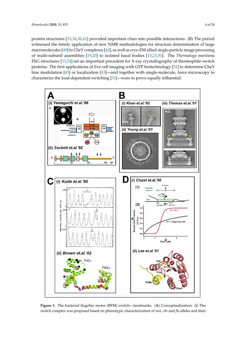
Figure 1. The bacterial flagellar motor (BFM) switch—landmarks. (A) Conceptualization: (i) Theswitch complex was proposed based on phenotypic characterization of mot, che and fla alleles and their
Biomolecules 2020, 10, 833 5 of 26
suppressor mutations in swarm plate assays. Its interactions with chemotaxis components and Motproteins were also identified. {a} Schematic of a swarm plate—the native (WT) strain forms a swarmwith chemotactic rings. Strains carrying mot mutations (Mot-) do not swarm while those with chemutations (Che-) have reduced swarms. Suppressor mutations yield pseudo-revertant strain (PR)with partially restored swarming. {b} Color codes are followed in subsequent Figures for the switchcomplex components (FliG (green), FliM (gold), FliN (cyan)), the CheY protein (salmon) and theMS-ring scaffold (orange) (adapted from [6]). (ii) Gene sequencing identified the mutations. The fliMgene (N–C terminal residue numbers) predominantly contained the che lesions, clustered into distinctCW (green) and CCW (magenta) regions. Arrows mark mot lesions (adapted from [8]). (B) Structuralidentification: (i) An extended cytoplasmic structure contiguous with the basal body MS-ring (yellowarrow) was isolated using gentler protocols and subsequently established as the switch complex byimmuno-EM and biochemistry (from [14]). (ii) Assembly of switch complex by overproduction ofplasmid-encoded components allowed biochemical characterization culminating in the determinationof the C ring subunit stoichiometry (n = 33–34) (from [23]). (iii) Single-particle analysis resolved FliGdomain substructure (yellow arrows) from differences in central sections from wild-type (WT) and∆FliFFliG (∆) 3D basal-body reconstructions (from [22] with permission). (C) Motor function andmechanism: (i) Temporally resolved measurement of filament rotation, as a sinusoidal variation oflaser dark-field spot intensity, characterized aberrant phenotypes in switch complex mutant strains.Panels (top to bottom) show slow rotation (S), pausing (P) and reversal (R) episodes (reproducedfrom [32] with permission). (ii) The first atomic structure of a switch component (FliGc [33]) followedby the FliGMC structure localized much of the mutant library then available ((mot lesions (black);CW lesions (red); CCW lesions (yellow); CW or CCW, depending on the residue substitution, orange;and motB suppressors (purple)) to generate chemically explicit ideas for motor reversal (PDB: 1lkv(modified from [34])). (D) Switch chemotactic signal transduction: (i) {1}—Determination of switch“ultra-sensitivity” (Hill coefficient, H = 10.3) by simultaneous measurement of the CW bias of beads onflagellar stubs (red) and concentration of a fluorescent GFP-CheY fusion (green) locked in the activestate (*) in engineered strains (reproduced from [45] with permission). {2}—Plots show non-cooperativebinding of acetate-activated CheY to overproduced C rings [46] compared to the in-vivo change in CWbias. (ii) The atomic structure of beryllium-fluoride (BeF3 (black))-activated CheY (salmon) boundto the FliM N-terminal peptide (yellow) initiated structure guided mutagenesis to explain the switchultra-sensitivity. Aromatic residue (W58, Y106 (orange)) motions were early diagnostics for activation.Magnesium ion (red) (PDB: 1f4v (modified from [41])).
Four fundamental issues could now be addressed. First, what was the nature of the couplingbetween the FliGC motor domain and the FliMN CheY binding target? Secondly, how were thedynamics of the C ring, a large multi-subunit assembly, synchronized for smooth rotation and rapidreversal? Thirdly, how did CheY activation trigger an ultrasensitive switch response? Fourthly, howwas the switch regulated by the motor operation as dictated by the torque–velocity relations?
2. Switch Physiology and Mathematical Models
Motor dynamics are the direct outcome of the architectural dynamics of the molecular machinery.Knowledge of motor dynamics, and the associated development of mathematical models, has advancedconcurrently with knowledge of the molecular architecture and dynamics. The spatiotemporalresolution and mechanical range of the rotation assays have continued to increase for characterizationof speed fluctuations, the load dependence and stochastic properties of the switch machinery inunprecedented detail. These advances provide additional constraints that must be addressed by thestudy of the molecular mechanism. Motor dynamics have been reviewed recently [52]. An overview isgiven in this short section, summarized in Figure 2, to provide additional context for the main body ofthe review.
Focal back-plane interferometry with high spatiotemporal resolution (1◦, 10−3 s) recordedincomplete, intermediate switching events to extend the temporally resolved measurement of switchtransitions [53]. More recently, gold nanospheres (d = 60 nm) conjugated to genetically engineered, rigid
Biomolecules 2020, 10, 833 6 of 26
Salmonella flagellar hooks, imaged by dark-field and rapid (5000 Hz) CMOS cameras, have been exploitedto measure motor rotation [54]. This study interpreted the large speed fluctuations recorded nearthe zero-torque speed (~400 Hz) that persisted over many revolutions as the association–dissociationof individual stator units. The highlight of such studies was that the resolution of the angular stepperiodicity per revolution was resolved in motors resurrected by single or a few stator units; first fora sodium powered chimeric motor [55] and then for the S. enterica proton motor [56]. In each case,26 steps per revolution were reported for both CW and CCW rotation. However, while the step size wassymmetrical for the Salmonella motor, the CW steps were smaller than the CCW steps in the chimericmotor. A study of the unidirectional R. sphaeroides motor reported a similar estimate of 27–28 discretestopping angles [57]. The steps may be due to modulation of the elastic potential well by periodiccontacts between the static and mobile motor modules rather than individual energy coupling events.A formal model of this idea successfully predicted the difference in CW versus CW step size [58].
The long-standing tethered cell measurements of the Poisson-distributed CCW and CW intervalshad been the bedrock for theoretical models. Filament-associated bead rotation measurements inthe phospho-mimetic E. coli CheY13DK mutant strain first revealed gamma distributions for bothCW and CCW intervals [59] to challenge the view of switch transitions as a single Poisson processbased on tethered cell data. Subsequent measurements of the rotation of beads attached to flagellarstubs [53] and nanoparticles attached to flagellar hooks [60] obtained exponential distributions inapparent contrast to [59], fueling speculation that filament polymorphic transitions [61] may havecomplicated the rotation of filament-associated latex beads (0.5 µm). The use of different-sized latexspheres extended the dependence of switching kinetics on load [62]. The study reported that at nearzero-load speed both CCW<->CW and CW<->CCW transitions increased with the load. Furthermore,the torque–velocity curve for CW rotation was linear in contrast to CCW rotation [63]. The loaddependence raised the possibility that it may underlie the apparent discrepancy between the gammaand Poisson interval distributions. The comprehensive analysis of the rotation of different-sized beadsin the double mutant phospho-mimetic strain CheY13DK106YW showed this was indeed the case.Both exponential and peaked distributions were obtained, depending on the different load, protonpotential and torque [64], to rule out speculation that the gamma distribution was an artefact.
The knowledge about the regulation of (CW/CCW) rotation bias by CheY has also advanced.Temporally resolved recordings of bead rotation over minutes re-evaluated motor individuality in termsof its rotation bias. Long-term recordings of variation in single bead rotation compared to populationvariation of multiple beads concluded that individuality was due to intracellular chemistry. The bias ofindividual motors had a bimodal distribution with cells with high switching frequencies binned intoa central trough. The CW and CCW fractions were dominant, consistent with the ultrasensitive switchin rotation bias by CheY [65]. Single bead CW and CCW rotational intervals had earlier been reportedto be asymmetrically distributed around the bias midpoint ((CW/(CW + CCW)) = 0.5), with the CCWbut not CW intervals varying with rotation bias [66]. These different relations argued against thedetermination of transition rates by CheY or any other single parameter. The ultra-sensitivity has alsobeen re-evaluated. The local GFP-CheY concentration around single flagellar motors was measuredand correlated with (CW/CCW) rotation bias in a ∆cheY E. coli strain [67] for an estimate of the meanCheY rotor occupancy during CW rotation. GFP-tagged fusion proteins also provided evidence thatthe basal C ring components FliM and FliN undergo dynamic exchange [68,69]. An experiment wasdesigned to correct for the FliM copy variation that would be obtained based on the exchange beingadaptive. It estimated the Hill coefficient, H, for the ultra-sensitivity from bias changes due to attractantremoval and addition in strains lacking receptor adaptation was as high as 21 [70].
The experiments motivated the development of the conformational spread model [48] and theformulation of a fundamentally distinct non-equilibrium model [71]. The initial interpretation forthe peaked gamma distribution was based on multiple Poisson events [59] to refine, but not radicallyalter the conformational spread model. The asymmetry in the experimental torque–velocity curvesand the load dependence of the switching kinetics could also be accounted for by an integrated

Biomolecules 2020, 10, 833 7 of 26
model that combined torque generation models with the switch ultra-sensitivity as formalized byconformational spread [72]. Theoretical models, in general, seek to explain the interdependencebetween rotation bias, switching frequency and interval distributions. The experimental relationsbetween these motor parameters were set as constraints to model the coupling between synchronousswitching and signal amplification as a function of subunit number [73]. More recent simulationsexplained the correlation between local CheY concentration and single motor rotation bias as well asthe utility of dynamic subunit exchange in the maintenance of switch ultra-sensitivity [74]. Detailedbalance within the equilibrium conformational spread framework was followed in both studies [73,74].Tu’s non-equilibrium model, where the detailed balance is broken based on energy input, also explainsthe gamma distribution data [71,75]. The model is consistent with the expanded set of intervaldistributions obtained under different load and energization conditions as detailed in [64]. The energyinput for switching need only be a small fraction of the total input that is dominantly utilized topower rotation.
Biomolecules 2020, 10, x FOR PEER REVIEW 7 of 26
switching and signal amplification as a function of subunit number [73]. More recent simulations explained the correlation between local CheY concentration and single motor rotation bias as well as the utility of dynamic subunit exchange in the maintenance of switch ultra-sensitivity [74]. Detailed balance within the equilibrium conformational spread framework was followed in both studies [73,74]. Tu’s non-equilibrium model, where the detailed balance is broken based on energy input, also explains the gamma distribution data [71,75]. The model is consistent with the expanded set of interval distributions obtained under different load and energization conditions as detailed in [64]. The energy input for switching need only be a small fraction of the total input that is dominantly utilized to power rotation.
Figure 2. Advances in switch physiology. (A) Time-resolved motor rotation: Schematic of a motor rotation assay with a nanosphere conjugated to the hook connector contiguous with the rod and basal body (reproduced from [54] with permission). (B) CW and CCW interval distributions: (i) Idealized time series of a motor alternating between CW and CCW rotation. (ii) Interval (τCW, τCCW) distributions measured under low and high torque (reproduced from [75] with permission). (C) Coupled energization and switching of rotation: (i) Torque velocity curves for CW and CCW rotation [63]. (ii)
Figure 2. Advances in switch physiology. (A) Time-resolved motor rotation: Schematic of a motorrotation assay with a nanosphere conjugated to the hook connector contiguous with the rod and basal
Biomolecules 2020, 10, 833 8 of 26
body (reproduced from [54] with permission). (B) CW and CCW interval distributions: (i) Idealizedtime series of a motor alternating between CW and CCW rotation. (ii) Interval (τCW, τCCW) distributionsmeasured under low and high torque (reproduced from [75] with permission). (C) Coupled energizationand switching of rotation: (i) Torque velocity curves for CW and CCW rotation [63]. (ii) Free energydiagram of CW <-> CCW transitions and the mechanical work (blue arrow) contribution (reproducedfrom [72] with permission). (D) Models for non-Poisson interval distributions: (i) Conformational spreadseeded from multiple CheY binding events (reproduced from [59] with permission). (ii) Breakdown ofdetailed balance from motor energy dissipation (reproduced from [64] with permission).
3. Architecture and Molecular Mechanism
Structural work on the issues outlined in the previous two sections can be grouped into four majorareas based on the protein–protein interactions: the central processing unit (FliMM.FliGN), the triggermachinery (CheY/C ring), the torque reversal mechanism (FliGC/FliGM) and the torque transmissionplatform (FliGN/FliFC).
3.1. The Central Processing Unit—The FliMM.FliGM Complex
X-ray crystallography led the effort guided by mutagenesis, in situ crosslinking and EMreconstruction to characterize the linkage between the domains responsible for torque generation andCheY signal reception. The atomic structure of the T. maritima FliG middle (FliGM) and C-terminal(FliGC) domains had an established ARM architecture for both domains and localized hotspots forE. coli che mutations to the sequence encoding the linker region between FliGM and FliGC [34]. The atomicstructure of the T. maritima FliM middle domain (FliMM), the central element in switch function, showedthat the CCW and CW substitutions localized to residue positions at the intradomain contact interface,as deduced by subunit crosslinking in situ [76]. The CW substitutions aligned differently to the CCWsubstitutions, suggesting distinct FliMM orientations for CW versus CCW rotation (Figure 3A). In situcrosslinking had been introduced in earlier work on FliGM domain organization [77]. The in-situcrosslinking experiments probed the subunit organization of the switch proteins as appropriatecontrols established that cross-linking was negligible in non-flagellate strains [76,77]. The structuresof FliMM.FliGM complexes from T. maritima [78,79] and H. pylori [80] determined the functionalimportance of the FliM GXXG and FliG EHPQR loop motifs at the FliMM.FliGM interface. Comparisonof the T. maritima and H. pylori interfaces showed conservation of essential GXXG and EHPQR loopcontacts and their stabilization by complex formation. Additional interfacial residues important forcoupling were identified in H. pylori, supported by mutagenesis.
Protein dynamic simulations showed that the core architecture and dynamics of FliMM donot change upon complex formation and are conserved between T. maritima and H. pylori. FliMM
conformational ensembles generated from the T. maritima structure had a bimodal distribution, while theFliGM domain movements also alternated between the bi-stable states strongly coupled to FliMM [81].Targeted tryptophan substitutions had identified FliGM residue positions important for associationwith FliMM [82]. In addition to the EHPQR loop, these residues were in a FliGc ARM-C hydrophobicpatch adjacent to the FliGMC GG linker as well as non-interfacial FliGM residue positions. RecentNMR measurements of the FliGM dynamics in the sodium V. alginolyticus motor have providedimportant validation, supported by mutagenesis, of the conformational plasticity of the FliGM domainbetween the bi-stable conformational states [83]. The EHPQR E144D residue substitution increased theswitching frequency, a similar response to one obtained upon phenol repellent addition. It did notalter FliGM conformation. In contrast, residue substitutions in the GG hinge both locked the bias andreduced the FliGM dynamics, as assessed by NMR. The E144D and CCW-biased G214S motors wereboth CCW-locked in the ∆che strains, implying a close linkage between CheY occupancy and FliGM
conformational fluctuations (Figure 3B).

Biomolecules 2020, 10, 833 9 of 26
In H. pylori, co-crystallization of putative spermidine synthase (SpeE) with FliMM, combinedwith mutagenesis and motility assays, has shown how this protein affects motile speed and switchingbehavior. The SpeE.FliMN contact interface partially overlaps with the FliGM.FliMM interface [84].While several proteins important for cell metabolism have been identified in diverse bacteria toinfluence motile behavior via direct interaction with FliG (cited in [84]), SpeE is the first knownto act at the FliGM.FliMM interface. In conclusion, the crystal structure, allelic mutations, in situcrosslinking, and protein dynamic simulations all argue for the alternation of FliMM between bi-stableconformational states, inter-species conservation of its fold and interfacial FliGM coupling.
Biomolecules 2020, 10, x FOR PEER REVIEW 9 of 26
and protein dynamic simulations all argue for the alternation of FliMM between bi-stable conformational states, inter-species conservation of its fold and interfacial FliGM coupling.
Figure 3. The central signal processing unit. (A) FliMM dimer model. The model is based on the T. maritima FliMM structure (gold)) guided by in situ cross-link data (grey). Localized CW (green) and CCW (magenta) biasing residue substitutions from bacterial homologs are mapped onto the colored structural elements. FliG GXXG interaction loop (blue). C-terminus (orange) (PDB: 2hp7 (modified from ([76])). (B) The central processing interface—FliGM-FliMM. (i) T. maritima structure (PDB: 4fhr) highlighted with homologous FliGM residues to those implicated by E. coli tryptophan mutagenesis (red. [82]) and V. alginolyticus switch mutants G214S, G215A and E144D (orange [83]). (ii) 2D-NMR 1H–15N correlation spectra reveal that the native (WT) V. alginolyticus FliGM architecture is substantially altered by substitutions in the conserved glycine pair that alter rotation bias, but is unaffected by the EPQR E144D residue substitution that increases the switching frequency. Axes indicate the shift in the magnetic field, in parts per million (ppm), relative to a reference compound for resonance (reproduced from [83] with permission).
3.2. The Trigger Machinery—CheY and the Basal C Ring
Motor response to chemotactic signals can be parsed into CheY activation and binding to the C ring to bias FliMM conformational fluctuations. There is a 20-fold increase upon phosphorylation in CheY affinity for E. coli FliMN [41]. The molecular analysis of CheY activation by the phospho-mimetic CheY 13DK106YW double residue substitutions used to study motor response was initiated by co-crystallization of the E. coli protein alone and in complex with the FliM N-terminal peptide (FliMN) [85]. Strikingly, while the CheY13DK106YW in the complex was in a conformation akin to BeF3
Figure 3. The central signal processing unit. (A) FliMM dimer model. The model is based on theT. maritima FliMM structure (gold)) guided by in situ cross-link data (grey). Localized CW (green) andCCW (magenta) biasing residue substitutions from bacterial homologs are mapped onto the coloredstructural elements. FliG GXXG interaction loop (blue). C-terminus (orange) (PDB: 2hp7 (modifiedfrom ([76])). (B) The central processing interface—FliGM-FliMM. (i) T. maritima structure (PDB: 4fhr)highlighted with homologous FliGM residues to those implicated by E. coli tryptophan mutagenesis(red [82]) and V. alginolyticus switch mutants G214S, G215A and E144D (orange [83]). (ii) 2D-NMR1H–15N correlation spectra reveal that the native (WT) V. alginolyticus FliGM architecture is substantiallyaltered by substitutions in the conserved glycine pair that alter rotation bias, but is unaffected by theEPQR E144D residue substitution that increases the switching frequency. Axes indicate the shift in themagnetic field, in parts per million (ppm), relative to a reference compound for resonance (reproducedfrom [83] with permission).
Biomolecules 2020, 10, 833 10 of 26
3.2. The Trigger Machinery—CheY and the Basal C Ring
Motor response to chemotactic signals can be parsed into CheY activation and binding to theC ring to bias FliMM conformational fluctuations. There is a 20-fold increase upon phosphorylation inCheY affinity for E. coli FliMN [41]. The molecular analysis of CheY activation by the phospho-mimeticCheY 13DK106YW double residue substitutions used to study motor response was initiated byco-crystallization of the E. coli protein alone and in complex with the FliM N-terminal peptide(FliMN) [85]. Strikingly, while the CheY13DK106YW in the complex was in a conformation akin toBeF3 activated CheY, the free CheY13DK106YW adopted the inactive conformation. Subsequently,the atomic structure of the native CheY.FliMN complex revealed that the electron density of acritical hinge, the α4–β4 loop, in this complex partitioned between its active and inactive states [86].Quantitative comparison of MD conformational ensembles from crystal structures of the CheYsuperfamily representatives has extracted common signatures, including the α4–β4 hinge, for allostericcommunication between the phosphorylation and target binding sites [87]; the importance of theα4–β4 loop further emphasized by MD simulations of acetate-activated CheY [88]. MD simulationssupported by oxygen-radical foot-printing solution measurements, a sensitive probe for sidechainsolvent accessibility [89], have now shown that closure of this loop hinge buried the allosteric relayaromatic sidechains to both stabilize the global CheY fold and increase the local FliMN affinity.
The motor response is likely to depend on the intervening disordered linker between FliMN andFliMM that may, in principle, serve as a flexible tether to deliver FliMN-bound CheY to second bindingsites. Occupancy of these sites could control FliMM conformational fluctuations and inter-subunitcoupling. Candidates have been identified in two species. There is NMR evidence for an interactionbetween T. maritima FliMN-tethered CheY and FliMM [90]. Alternatively, in E. coli, pull-down assayshave reported evidence for an association of the CheY-FliMN fusion with FliN [91]. This evidence issummarized in Figure 4A.
There is a close homology between FliN and FliMC. The atomic structure of the T. maritima FliNhomodimer [92] motivated mutagenesis and in situ crosslinking experiments to infer FliN tetramericquaternary organization in the E. coli flagellar motor [93]. These experiments reported cross-linkchanges upon addition of chemotactic stimuli, consistent with domain motions, but the connectivitybetween FliMN and the FliMC
1.FliN3 module is not well-understood as the FliM inter-domain linkershave not been structurally characterized. The atomic structure of the FliMC.FliN heterodimer is similarto the FliN homodimer [94], consistent with a FliMC
1.FliN3 tetramer identified by mass spectroscopyand structural homology with the Type III injectosome components [95]. The C-terminal tail of theFliH component of the flagellar export ATPase assembly has been co-crystallized and bound to theFliMC.FliN heterodimer (Figure 4B). The co-crystal validated early predictions of the interactions ofthe C ring with the flagellar export apparatus [96], as well as the proposal based on cryo-tomographic(cryo-ET) reconstruction and FliH sequences from diverse species that FliH acts as a spacer to set Cring diameter [97].
The switch complex contains FliY instead of, or in addition to, FliM and FliN in numerousspecies [98]. The structure of T. maritima FliY reveals a middle domain with strong structural similarityto FliMM, while the FliY C-terminal domain is similar to FliN [98]. This study also reported solutionassays to show that T. maritima FliY homodimerizes via its N-terminal domain and does not have anincreased affinity for activated versus inactive CheY, and that its middle domain does not bind FliG.FliY was first reported in the Gram-positive B. subtilis [99] where CheY phosphorylation enhancesCCW, not CW rotation [100], in contrast to the Gram-negative γ-proteobacteria E. coli and S. enterica.The characterization of the B. subtilis rotor module by in situ crosslinking found that the FliMMFliGM
interface is conserved as in other bacteria with FliY proposed to form an external ring adjacent to theFliMM ring [101].
Thus, FliMC interacts with FliN to form the C ring base and can, in principle, transmit structuralperturbations triggered by CheY-FliN association to FliMM. Other bacteria, such as T. maritima, mayutilize a different signal strategy and a distinct basal ring architecture.
Biomolecules 2020, 10, 833 11 of 26
Biomolecules 2020, 10, x FOR PEER REVIEW 11 of 26
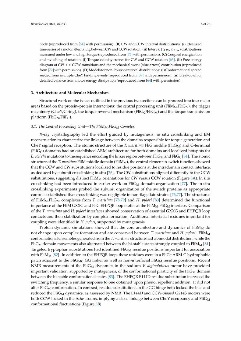
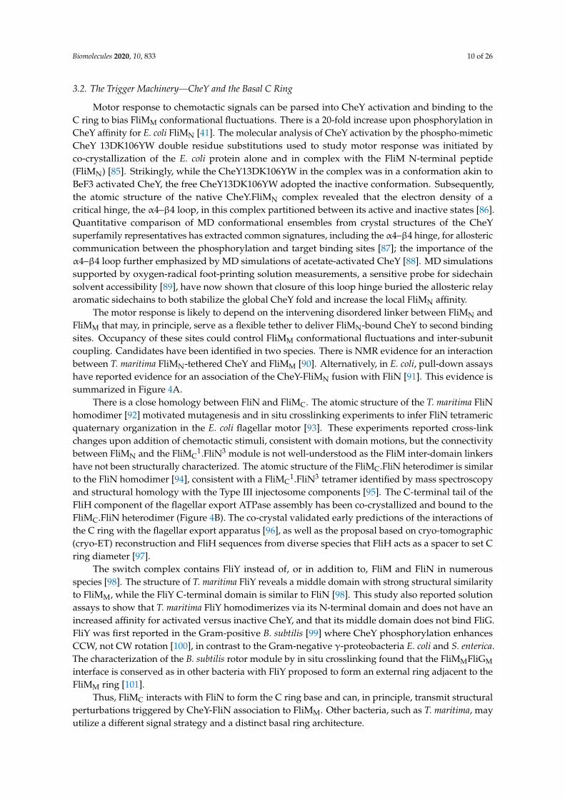
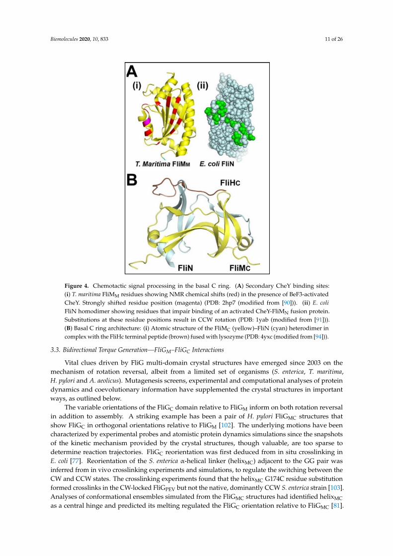
Figure 4. Chemotactic signal processing in the basal C ring. (A) Secondary CheY binding sites: (i) T. maritima FliMM residues showing NMR chemical shifts (red) in the presence of BeF3-activated CheY. Strongly shifted residue position (magenta) (PDB: 2hp7 (modified from [90])). (ii) E. coli FliN homodimer showing residues that impair binding of an activated CheY-FliMN fusion protein. Substitutions at these residue positions result in CCW rotation (PDB: 1yab (modified from [91])). (B) Basal C ring architecture: (i) Atomic structure of the FliMC (yellow)–FliN (cyan) heterodimer in complex with the FliHc terminal peptide (brown) fused with lysozyme (PDB: 4yxc (modified from [94])).
The variable orientations of the FliGC domain relative to FliGM inform on both rotation reversal in addition to assembly. A striking example has been a pair of H. pylori FliGMC structures that show FliGC in orthogonal orientations relative to FliGM [102]. The underlying motions have been characterized by experimental probes and atomistic protein dynamics simulations since the snapshots of the kinetic mechanism provided by the crystal structures, though valuable, are too sparse to determine reaction trajectories. FliGC reorientation was first deduced from in situ crosslinking in E. coli [77]. Reorientation of the S. enterica α-helical linker (helixMC) adjacent to the GG pair was inferred from in vivo crosslinking experiments and simulations, to regulate the switching between the CW and CCW states. The crosslinking experiments found that the helixMC G174C residue substitution formed crosslinks in the CW-locked FliG PEV but not the native, dominantly CCW S. enterica strain [103]. Analyses of conformational ensembles simulated from the FliGMC structures had identified helixMC as a central hinge and predicted its melting regulated the FliGC orientation relative to FliGMC [81]. These studies, taken together, supported the large reorientations deduced from the superimposition of different crystal structures.
The simulations also reported that the T. maritima MFXF linker between FliGC ARM-C and Cα1-
6 amplifies thermal fluctuations of the coupled FliMM–FliGM central processing unit [81]. This idea was supported and extended in important ways by NMR and MD simulations of V. alginolyticus FliGC. The conformational dynamics of FliGC-Cα1-6 helix α1 in FliG A282T were reduced relative to the native protein to give detectible peaks in the NMR spectra. The A282T strain has a CW bias in
Figure 4. Chemotactic signal processing in the basal C ring. (A) Secondary CheY binding sites:(i) T. maritima FliMM residues showing NMR chemical shifts (red) in the presence of BeF3-activatedCheY. Strongly shifted residue position (magenta) (PDB: 2hp7 (modified from [90])). (ii) E. coliFliN homodimer showing residues that impair binding of an activated CheY-FliMN fusion protein.Substitutions at these residue positions result in CCW rotation (PDB: 1yab (modified from [91])).(B) Basal C ring architecture: (i) Atomic structure of the FliMC (yellow)–FliN (cyan) heterodimer incomplex with the FliHc terminal peptide (brown) fused with lysozyme (PDB: 4yxc (modified from [94])).
3.3. Bidirectional Torque Generation—FliGM–FliGC Interactions
Vital clues driven by FliG multi-domain crystal structures have emerged since 2003 on themechanism of rotation reversal, albeit from a limited set of organisms (S. enterica, T. maritima,H. pylori and A. aeolicus). Mutagenesis screens, experimental and computational analyses of proteindynamics and coevolutionary information have supplemented the crystal structures in importantways, as outlined below.
The variable orientations of the FliGC domain relative to FliGM inform on both rotation reversalin addition to assembly. A striking example has been a pair of H. pylori FliGMC structures thatshow FliGC in orthogonal orientations relative to FliGM [102]. The underlying motions have beencharacterized by experimental probes and atomistic protein dynamics simulations since the snapshotsof the kinetic mechanism provided by the crystal structures, though valuable, are too sparse todetermine reaction trajectories. FliGC reorientation was first deduced from in situ crosslinking inE. coli [77]. Reorientation of the S. enterica α-helical linker (helixMC) adjacent to the GG pair wasinferred from in vivo crosslinking experiments and simulations, to regulate the switching between theCW and CCW states. The crosslinking experiments found that the helixMC G174C residue substitutionformed crosslinks in the CW-locked FliGPEV but not the native, dominantly CCW S. enterica strain [103].Analyses of conformational ensembles simulated from the FliGMC structures had identified helixMC
as a central hinge and predicted its melting regulated the FliGC orientation relative to FliGMC [81].

Biomolecules 2020, 10, 833 12 of 26
These studies, taken together, supported the large reorientations deduced from the superimposition ofdifferent crystal structures.
The simulations also reported that the T. maritima MFXF linker between FliGC ARM-C and Cα1-6
amplifies thermal fluctuations of the coupled FliMM–FliGM central processing unit [81]. This ideawas supported and extended in important ways by NMR and MD simulations of V. alginolyticusFliGC. The conformational dynamics of FliGC-Cα1-6 helix α1 in FliG A282T were reduced relative tothe native protein to give detectible peaks in the NMR spectra. The A282T strain has a CW bias incontrast to the CCW bias of the native strain. MD simulations further showed the reduction was dueto additional hydrogen bonding contacts between the ARM-C and FliGC α1-6 that constrained theflexibility of the intervening linker. MFXF254 hinge orientation, monitored by residue F254, partitionedinto conformational clusters that overlapped the subsets of the crystal structures representing eitherthe CW or CCW rotation states based on other criteria (Figure 5A,B). A prescient early analysis ofE. coli FliG residue substitutions in the helixMC linker, sensitized by a serendipitous E232G substitutiona few positions upstream of the MFXF hinge, had reported these gave rise to diverse phenotypes withaltered switching frequencies, pausing or altered bias [104].
The complete Aquifex aeolicus FliG structure revealed an ARM fold for the N-terminal domain(FliGN) in addition to FliGM and FliGC, separated by α-helical linkers [105]). Sequence similaritysuggests these domains arose from gene duplication [106]. The structure reported inter-molecularstacking between the FliGM and FliG ARM_C domains. In contrast, the S. enterica FliGPEV [107]and T. maritima [78] crystal structures of the FliGMC complexes showed intramolecular stacking ofthese domains. The consensus view now is that the assembly of the FliG middle and C-terminaldomain is mediated by intermolecular stacking. Solution pulsed dipolar ESR spectroscopy combinedwith residue substitutions first indicated that, in T. maritima, these domains self-assemble via theFliGM–FliG ARM_C intermolecular stacking contact; then, direct assembly of FliM [108]. The centraland membrane-proximal sections of the S. enterica C ring map was fit well by the ESR-derived model(Figure 5C). A domain-swap mechanism for FliG ring assembly in the E. coli motor was subsequentlydetermined with SAXS analysis supported by in situ cross-linking [109]. Binding energy from FliGassociation with the FliF MS ring was speculated to alter the conformational equilibrium between thecompact and extended conformation of the malleable helixMC to favor polymerization of a chainedFliGMC ring in the flagellar motor but not in solution.
Coevolutionary information has emerged as an important high throughput tool to assess thedesign principles of bacterial multi-protein complexes at the single residue level. Residue coevolutionsupported the design of the switch machinery framed by experiments and simulations. First, the role ofFliMM as the central relay was consistent with its strongly coevolved inter-subunit and FliGM interfacialcontacts [110]. Two distinct T. maritima FliMM dimer configurations were obtained when dimerformation was simulated based on coevolved subunit couplings [111], although one orientation did notmatch either the CW and CCW dimers deduced from S. enterica residue substitutions [76], either becauseof limited sampling and/or because the bi-directional switch is not universal across species. Secondly,coevolution provided strong support for conservation of the FliGC–FliGM stacking contact [109,112]relative to other contacts identified by cross-link data [77]. Thirdly, the coevolved coupling was mapped,in part, onto the identified dynamic couplings and modules. Notably, the coevolved FliMMc–FliGM
contacts was mapped onto the dynamic couplings across the T. maritima FliMM.FliGM interface [81]and FliGMC, as a coevolved network with distinct nodes as allosteric sectors [110]. The nodes includedthe well-characterized EHPQR and PEV (in S. enterica) at the FliMM.FliGM interface as well as α-helices(helixMC, Cα1-6 helix α1). The melting of these α-helices has been noted above. The coevolution signalfrom FliGC ARM-C is sparse. This sub-domain may be the converter element that encodes differentspecies-specific outputs from a conserved input signal (Figure 5D).

Biomolecules 2020, 10, 833 13 of 26
Figure 5. Mechanics of rotation reversal. (A) Mechanical amplification: (i) The T. maritima FliMMFliGMC
crystal structure (PDB: 4fhr. FliMM (gold), FliGMC (green)), modelled as a segmented rod withflexible hinges. Two-stage amplification of FliMM motions triggers large FliGC domain reorientations.(ii) Bimodal hinge planar angle distributions from the dominant principal collective motions 1–3extracted from the generated conformational ensemble. Lines plot the 3D angular displacementdistribution (solid) and unimodal fit (dotted). The distribution of the FlIMMFliGM interfacial hinge(1 (gold)) has a narrow spread relative to the MFXF hinge distribution (2 (green)), indicating most of theamplification occurs at the latter hinge (from [81]). (B) MFXF hinge dynamics: (i) The representativestructure of the V. alginolyticus FliGc A282T homology model from cluster analysis of the MD ensemble.The predicted dynamic helix α1 (pale green), the MFXF254 motif, and residues G214, G215 (orange)and T282 (magenta with an asterisk) are marked. 2D-NMR reports the CW-biasing A282T residuesubstitution melts helix α1 and reorients MFXF254. (ii) The 2D plot of F254 dihedral angle versus helixα6 orientation relative to the FliGM.FliGC interface obtained from the MD trajectories. The wild-type(WT) and mutant (A282T) conformational clusters map onto different subsets of the FliGc crystalstructures. Labeled circles (magenta) mark structures from T. maritima (CW-locked. PDB: 3ajc; wild type.PDB: 1lkv; complexed with FliMM (PDB: 4fhr)), Helicobacter pylori (PDB: 3usy and 3usw) and A. aeolicus(full-length. PDB: 3hjl) (from [113] with permission). (C) The FliGM-ARMc stacking interaction: Thebest-fit of the pulsed dipolar ESR spectroscopy model of the T. maritima FliGM-ARMc inter-domainstack to the 3D S. enterica electron density map (from [108] with permission). (D) Residue coevolutionmodel for rotation reversal: Strong, coevolved FliMM (yellow) contacts mediate conformational spread(arrows) in the FliMM ring. The FliG αC3-6 “motor domain” (dark green) is organized around the αC5“torque helix” with charged residues (red) important for rotor–stator interactions. The primary nodesof the coevolved network form a relay of allosteric sectors (numbered grey patches) across ARM-M

Biomolecules 2020, 10, 833 14 of 26
and ARM-C (light green). ARM-C has sparse coevolved contacts implying a variable fold to generatedifferent motor responses from a conserved FliMM switch transition Box. Coevolved interfacial pairs(residues (white spheres) and contacts (red)) link the FliMM GGXG motif with the FliGM EHPQ motif.The contacts and nodes are mapped onto PDB: 4fhr (from [110]).
In conclusion, the library of crystal structures now available, together with experimental andcomputational measurements of their dynamics, position FliM, FliG and FliN in the S. enterica C ringmap and charts out their connectivity and conformational plasticity. FliMM has a central location inthe C ring with subunit contacts designed for propagation of both the CW and CCW conformationalstates in the species studied thus far. FliMM forms a tightly coupled complex with FliGM, with acentral location in the C ring, connected via FliGM to FliGC adjacent to the membrane stator complexes,and via FliMM to FliMC in the basal C ring.
3.4. The Association of the Switch with the Mot Stators and the FliFC Scaffold
The interactions of the FliGc domain with the MotA.MotB stator complexes determine torque.The biochemical studies of rotor–stator interactions in E. coli [11] were soon extended to other bacterialmotors, notably the sodium-driven V. alginolyticus motor [114]. The FliGC torque helix and adjacentsegments in the V. alginolyticus motor contain more charged residues compared to E. coli [115].Furthermore, substitutions at two residue positions selectively impair only CCW rotation [116].Interestingly, the V. alginolyticus FliGC also determines the correct polar localization and assembly ofthe stator complexes [117]. The mutagenesis of the residues involved in rotor–stator interactions inthe E. coli motor was extended in the related S. enterica [118]. Notably, these investigators coupledfluorescent localization of the GFP-MotB fusion proteins with motility assays to parse out chargedresidues at the FliGc–MotA interface that influence stator assembly from those dedicated to torquegeneration. The first cryo-ET images documenting the alteration of C ring morphology by statorcomplexes have now been obtained in the spirochete Borrelia burgdorferi. Comparative study of motBmutant strains with defective versus restored proton conduction suggests that functional stators arerequired for full expression of this effect [119] (Figure 6A). Thus, torque affects switch architecture,in addition to CW/CCW rotation bias (Section 2), while the FliG rotor ting, in turn, affects assembly ofthe force-generating stator complexes.
The CCW and CW torque generated by stator interactions with the FliG ring must be transmittedvia the intervening FliF MS-ring to the external components of the filament whose rotation is thephysiologically relevant parameter. The fliF and fliG genes are adjacent in one operon for middle geneexpression in the S. enterica flagellar regulon [120]. Strains with these fusions are motile and form Crings but have aberrant CW/CCW bias [121]. Their analysis has provided valuable clues on the effectsof the FliF–FliG association on switch operation and C ring morphology.
The bias of the full-frame fusion was restored by suppressor mutations in FliMM and FliGN,as well as by insertion of a flexible, nine-residue glycine-rich linker at the fusion site [121], emphasizingthe influence of FliGN and FliGM conformational plasticity on the sign of the transmitted torque.The deletion the FliF–FliG fusion (∆FliF.FliG) strain lacked a major part of FliGN,and formed smallerC rings with lower (n = 31) subunit stoichiometry [122]. In contrast, the FliGM∆PAA deletionstrain formed smaller C rings with altered packing as their subunit stoichiometry was unchanged.The bias of the ∆FliF.FliG strain could be also be compensated by residue substitutions localizedto the FliMM.FliGM interface (FliGD124Y) and a likely FliMM dimerization interface (F188Y, V186A).Cryo-electron microscopy of isolated FliGD124Y basal bodies showed that D124Y substitution partlyrestored the C ring diameter towards wild-type values. These results demonstrate that either subunitnumber or spacing variation can change C ring size. While FliF.FliGN association primarily transmitstorque, it also has downstream effects on the switch machinery for torque reversal. There seems to be asynergistic relationship between the supramolecular organization of the FliGN and FliMM rings withdefects in one restored by compensating alterations in the other (Figure 6B).

Biomolecules 2020, 10, 833 15 of 26
Biomolecules 2020, 10, x FOR PEER REVIEW 15 of 26
FliGM fold [125], and possibly also conformational plasticity compatible with the compensatory interactions between the two domains in the regulation of C ring size. The crystal structure of the H. pylori co-folded domain corroborated the essential features reported for T. maritima [126].
Figure 6. C ring modulation by interfacial membrane protein interactions. (A) C ring modulation by stator operation: (i) Central tomogram section of the wild-type B. burgdorferi flagellar basal structure. The C ring wall (box) is almost perpendicular (1.8°) to the Mot stator complexes (yellow arrows). The C ring has different orientations in (ii) ∆MotB (7.8°) and (iii) MotB-24DE (3.2°) mutants. The ∆motB strain does not assemble stators; the motB-24DE strain assembles stators and is motile (reproduced from [119] with permission). (B) FliF-FliG deletions alter C ring size. Size differences between S. enterica (i) CCW and (ii) CW-locked (FliG∆PAA) C rings. (iii.a) The CW-biased S. enterica ∆FliF–FliG fusion assembles smaller C rings. (iii.b) 2D class averages from the ∆FliF–FliG fusion basal body; without and with the FliGM D124Y residue substitution that restores normal bias (reproduced from [122] with permission).
The cytoplasmic FliF segment contiguous with the co-folded FliGN.FliFC-tail domain is predicted to form a predominantly α-helical connector to the periplasmic C-terminal FliF (FliFCper) [112]. The recent 3D-cryoelectron microscopy reconstruction of overproduced S. enterica FliF rings has shown that FliFCper forms a periplasmic scaffold with a split ring-building motif (RBM) that staples together an anti-parallel β-barrel to form the external periplasmic modules of the MS ring while the N-terminal FliF forms inner RBM modules structurally homologous to RBMs characterized for Type-III injectosomes [127]. Both the split RBM and the β-barrel were predicted by residue coevolution in the course of ongoing work on the full-frame FliF.FliG fusion ring [112]. Thus, the FliF flagellar motor scaffold has evolved to add a particularly stable, periplasmic C-terminal domain to the injectosome
Figure 6. C ring modulation by interfacial membrane protein interactions. (A) C ring modulation bystator operation: (i) Central tomogram section of the wild-type B. burgdorferi flagellar basal structure.The C ring wall (box) is almost perpendicular (1.8◦) to the Mot stator complexes (yellow arrows).The C ring has different orientations in (ii) ∆MotB (7.8◦) and (iii) MotB-24DE (3.2◦) mutants. The ∆motBstrain does not assemble stators; the motB-24DE strain assembles stators and is motile (reproducedfrom [119] with permission). (B) FliF-FliG deletions alter C ring size. Size differences between S. enterica(i) CCW and (ii) CW-locked (FliG∆PAA) C rings. (iii.a) The CW-biased S. enterica ∆FliF–FliG fusionassembles smaller C rings. (iii.b) 2D class averages from the ∆FliF–FliG fusion basal body; withoutand with the FliGM D124Y residue substitution that restores normal bias (reproduced from [122]with permission).
How does FliGN transmit torque generated at the FliG ring periphery to the axial rod andfilament via FliF? The architecture of the S. enterica hook basal body revealed by the work ofDeRosier and colleagues (see [50] and references therein) notably shows different symmetries for theinternal and external modules; the cytoplasmic C ring (n = 33–36) and the external hook (n = 11),

Biomolecules 2020, 10, 833 16 of 26
for example. The architectural design by which torque transmission is achieved and symmetry mismatchaccommodated is starting to be understood. The structural adaptation for torque transmission wasdetermined in T. maritima [123] and, subsequently, the sodium-powered V. alginolyticus motor [124].NMR reported extensive conformational changes in the T. maritima FliGN ARM fold upon interactionwith 46 FliF C-terminal residues. These changes were due to formation of the co-folded FliGN andFiF C-tail domain. FliGN in the crystallized co-folded domain alters conformation to closely matchthe FliGM fold [125], and possibly also conformational plasticity compatible with the compensatoryinteractions between the two domains in the regulation of C ring size. The crystal structure of theH. pylori co-folded domain corroborated the essential features reported for T. maritima [126].
The cytoplasmic FliF segment contiguous with the co-folded FliGN.FliFC-tail domain is predicted toform a predominantly α-helical connector to the periplasmic C-terminal FliF (FliFC
per) [112]. The recent3D-cryoelectron microscopy reconstruction of overproduced S. enterica FliF rings has shown that FliFC
per
forms a periplasmic scaffold with a split ring-building motif (RBM) that staples together an anti-parallelβ-barrel to form the external periplasmic modules of the MS ring while the N-terminal FliF formsinner RBM modules structurally homologous to RBMs characterized for Type-III injectosomes [127].Both the split RBM and the β-barrel were predicted by residue coevolution in the course of ongoingwork on the full-frame FliF.FliG fusion ring [112]. Thus, the FliF flagellar motor scaffold has evolved toadd a particularly stable, periplasmic C-terminal domain to the injectosome RBMs. The map furtherreveals that FliFC
per symmetry (n = 33–34), within the design tolerance reported for other biomolecularassemblies, matches the C ring symmetry to dispel the MS and C ring symmetry mismatch conundrumraised by initial estimates of a lower FliF subunit stoichiometry. Figure 7 summarizes these advances.
Biomolecules 2020, 10, x FOR PEER REVIEW 16 of 26
RBMs. The map further reveals that FliFCper symmetry (n = 33–34), within the design tolerance reported for other biomolecular assemblies, matches the C ring symmetry to dispel the MS and C ring symmetry mismatch conundrum raised by initial estimates of a lower FliF subunit stoichiometry. Figure 7 summarizes these advances.
Figure 7. Transmembrane torque transmission. (A) (i) Conserved FliGN/FliFC-tail coevolved contacts mapped onto the T. maritima crystal structure (PDB: 5tdy [125]). (ii) Predicted FliFCper architecture obtained by residue coevolution. Red lines denote coevolved residue pairs as in Figure 5D (modified from [112]). (B) The 3D model of the S. enterica hook–basal body complex. The basal body part of the structure was from a 3D reconstruction as published [50]. The hook with attached FliD cap structure was done by single-particle methods using the entire hook with the cap as the single-particle (Dennis Thomas, unpublished results (with permission)). (C) The 3D map of the S. enterica FliFC torque transmitter module (EMD-10143, 3.1-angstrom resolution (modified from [127])). The en-face view resolves 33-subunits.
In summary, rotor–stator interactions employ a core set of charged residues and control large scale changes in stator assembly and C ring morphology either side of the interaction interface. The FliFCper module presumably templates the assembly of the FliG ring via FliFC-tail that is part of the co-folded domain. FliGM then dictates FliM assembly via contacts with FliMM. The FliMM ring drives reorientation of FliGC for bidirectional torque generation and remodels in response to lesions in the co-folded FliGN.FliFC-tail domain, Thus, there is conformational coupling of FliMM to these distant domains via FliGM. The correlation between C ring size and rotation state, driven by defined lesions, is comparable to the rotation–fluorescence intensity correlation reported for GFP-tagged motors. It provides the first structural clue for the difference reported for the CW versus CCW torque–velocity relations.
Figure 7. Transmembrane torque transmission. (A) (i) Conserved FliGN/FliFC-tail coevolved contactsmapped onto the T. maritima crystal structure (PDB: 5tdy [125]). (ii) Predicted FliFC
per architectureobtained by residue coevolution. Red lines denote coevolved residue pairs as in Figure 5D (modifiedfrom [112]). (B) The 3D model of the S. enterica hook–basal body complex. The basal body partof the structure was from a 3D reconstruction as published [50]. The hook with attached FliD capstructure was done by single-particle methods using the entire hook with the cap as the single-particle(Dennis Thomas, unpublished results (with permission)). (C) The 3D map of the S. enterica FliFC torquetransmitter module (EMD-10143, 3.1-angstrom resolution (modified from [127])). The en-face viewresolves 33-subunits.
Biomolecules 2020, 10, 833 17 of 26
In summary, rotor–stator interactions employ a core set of charged residues and control largescale changes in stator assembly and C ring morphology either side of the interaction interface.The FliFC
per module presumably templates the assembly of the FliG ring via FliFC-tail that is part ofthe co-folded domain. FliGM then dictates FliM assembly via contacts with FliMM. The FliMM ringdrives reorientation of FliGC for bidirectional torque generation and remodels in response to lesionsin the co-folded FliGN.FliFC-tail domain, Thus, there is conformational coupling of FliMM to thesedistant domains via FliGM. The correlation between C ring size and rotation state, driven by definedlesions, is comparable to the rotation–fluorescence intensity correlation reported for GFP-taggedmotors. It provides the first structural clue for the difference reported for the CW versus CCWtorque–velocity relations.
4. Current Challenges
This review draws overwhelmingly from the E. coli, S. enterica and sodium V. alginolyticus motorsfor the elucidation of their molecular mechanisms, with a notable contribution from thermophile andH. pylori crystal structures. The fundamental issues regarding switch synchrony and ultra-sensitivityhad been posed by 2003. The present knowledge of the structural basis for torque generation andtransmission does not answer these issues but takes their study to a new level. Other species withdiverse phylogeny have revealed the diversity of the switch operation. The bacillus B. subtilis hasinverted motile responses to CheY. V. alginolyticus is one of several bacteria that alter switch frequencyrather than rotation bias in response to CheY activation. The flagellum of the α-proteobacteriumR. sphaeroides stops and starts. The thermophile A. aeolicus has been reported to lack FliM, consistentwith its mostly smooth-swim motile behavior [128]. A major new challenge as illustrated by thephylogenetic tree of FliGC, the “motor” domain (Figure 8A), is to explain how such diversity can arisefrom a small core of protein components.
Cryo-electron tomography (cryo-ET) has been an important role for the appreciation of the diversemorphology of flagellar motors (reviewed in [129,130]), even though it is presently limited to thinbacteria or mini-cells. Crucially, the technique provides 3D-reconstructions of complete motors thatcapture rotor–stator interactions [131], the effect of the cell membrane upon rotor flexure [132]and novel cell wall motor components [133]. Mutagenesis guided Cryo-ET has, interestingly,established diversification of C ring architecture for various cellular functions within a single species(Campylobacter jejuni) [133], and has led to the realization that high-torque motors have larger diameterstator and C rings [134].
The emergence of informatics due to developments in high-throughput sequencing, massspectroscopy and high-performance computing is the highlight of this era, as appreciated by theincrease in protein sequences (135,850 (2003) -> 177,754,527 (2020) (www.ebi.ac.uk/uniprot)) and atomicstructures (<20,000 (2003) -> >140,000 (2018) [135]). Computational strategies for protein dynamicsimulations [87,136] have benefited from the expanded databases. Most core switch componentsbelong to a larger superfamily. The remarkable diversity of the CheY response regulator superfamilyhas been reviewed recently [137]. Several response regulators are expressed concurrently within onespecies. Many species have multiple CheYs of which typically one interacts with the flagellar motor:a study on V. cholerae being an early example [138]. FliF, FliM, FliN and FliY are members of largerfamilies that include Type III injectosome components (reviewed in [139]). FliG has distant homologyto the MgtE transporter family [140]. The diversity of the basal C ring and the CheY atomic structuressuggests that bacteria utilize multiple strategies for signal reception.
The E. coli and S. enterica bacteria remain the primary source for motor rotation assays in conjunctionwith native and chimeric V. alginolyticus sodium motors. Structural knowledge, while anchored inthese bacteria, now has important contributions from other species driven by the developments incryo-ET and informatics. The current understanding of the flagellar motor switch is based on a generic,integrated assembly with contributions from multiple species, as schematized in Figure 8B.
Biomolecules 2020, 10, 833 18 of 26
Biomolecules 2020, 10, x FOR PEER REVIEW 18 of 26
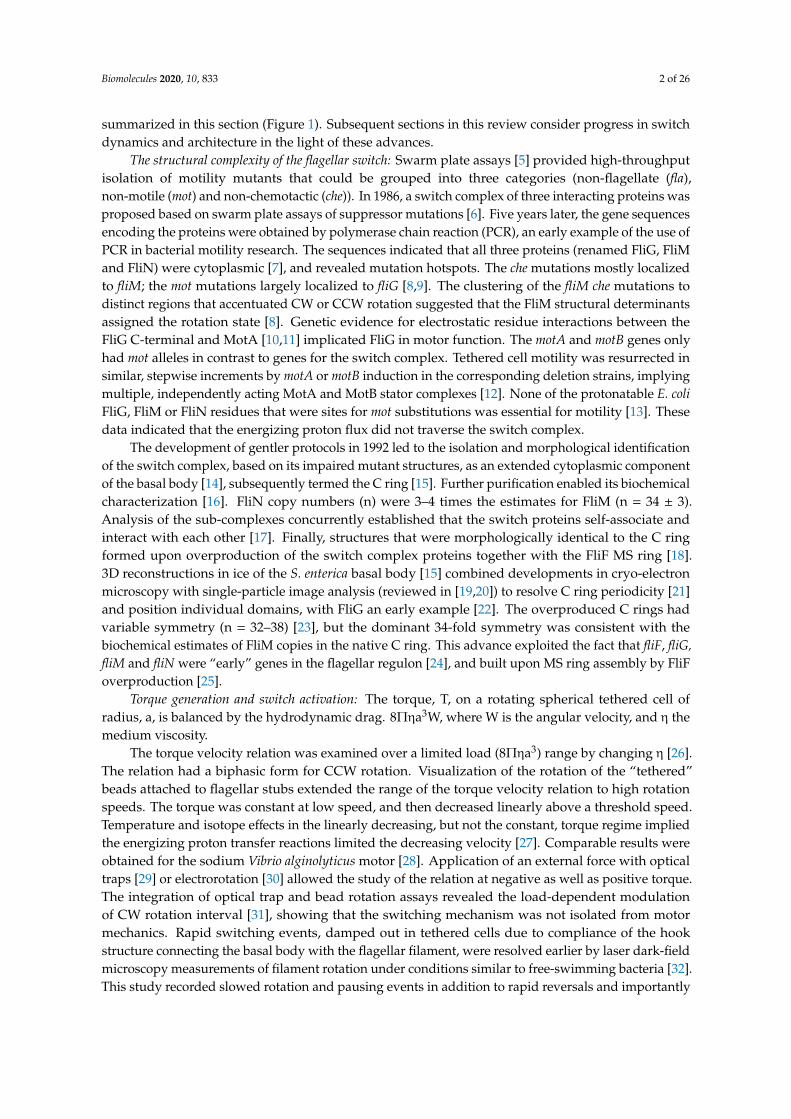
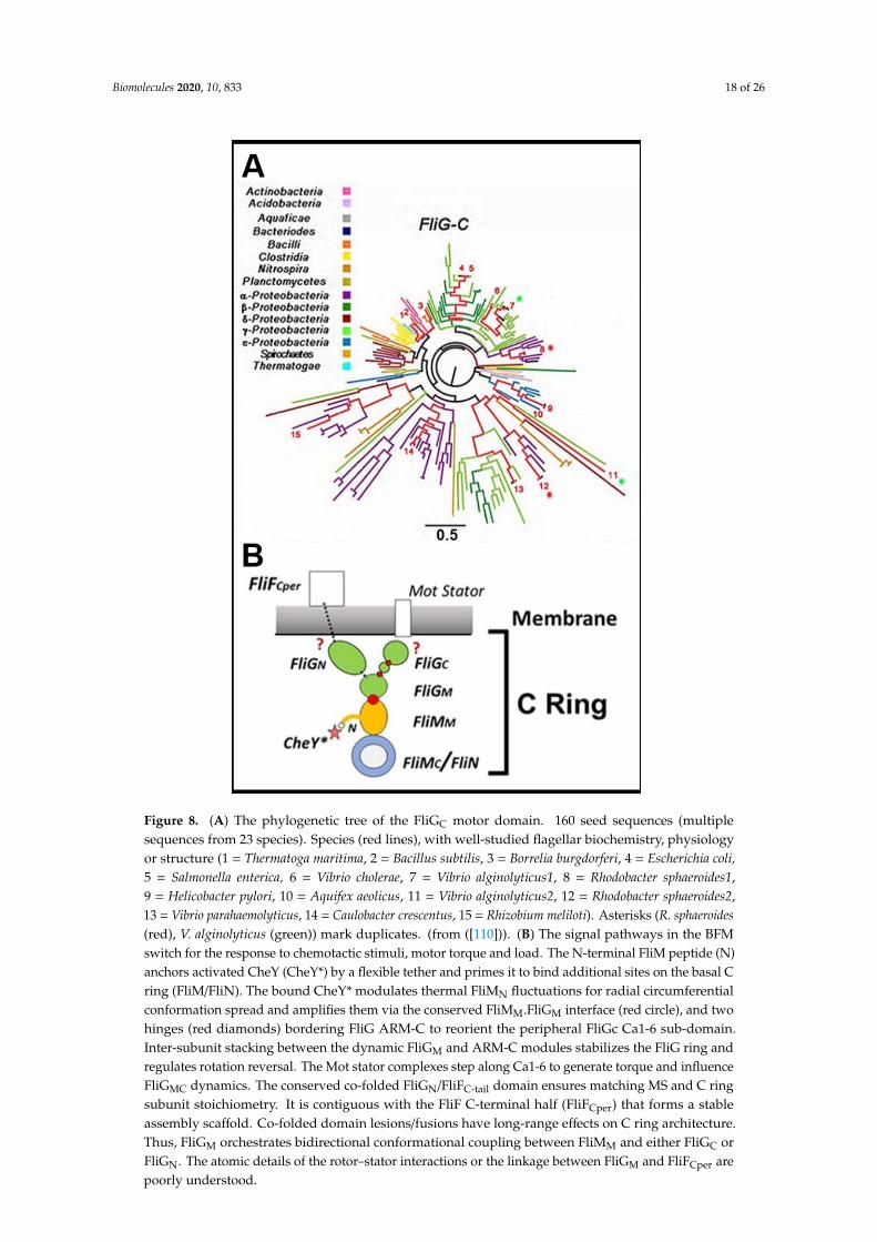
Figure 8. (A) The phylogenetic tree of the FliGC motor domain. 160 seed sequences (multiple sequences from 23 species). Species (red lines), with well-studied flagellar biochemistry, physiology or structure (1 = Thermatoga maritima, 2 = Bacillus subtilis, 3 = Borrelia burgdorferi, 4 = Escherichia coli, 5 = Salmonella enterica, 6 = Vibrio cholerae, 7 = Vibrio alginolyticus1, 8 = Rhodobacter sphaeroides1, 9 = Helicobacter pylori, 10 = Aquifex aeolicus, 11 = Vibrio alginolyticus2, 12 = Rhodobacter sphaeroides2, 13 = Vibrio parahaemolyticus, 14 = Caulobacter crescentus, 15 = Rhizobium meliloti). Asterisks (R. sphaeroides (red), V. alginolyticus (green)) mark duplicates. (from ([110])). (B) The signal pathways in the BFM switch for the response to chemotactic stimuli, motor torque and load. The N-terminal FliM peptide (N) anchors activated CheY (CheY*) by a flexible tether and primes it to bind additional sites on the basal C ring (FliM/FliN). The bound CheY* modulates thermal FliMN fluctuations for radial circumferential conformation spread and amplifies them via the conserved FliMM.FliGM interface (red circle), and two hinges (red diamonds) bordering FliG ARM-C to reorient the peripheral FliGc Ca1-6 sub-domain. Inter-subunit stacking between the dynamic FliGM and ARM-C modules stabilizes the FliG ring and regulates rotation reversal. The Mot stator complexes step along Ca1-6 to generate torque and influence FliGMC dynamics. The conserved co-folded FliGN/FliFC-tail domain ensures matching MS and C ring subunit stoichiometry. It is contiguous with the FliF C-terminal half (FliFCper) that forms a stable assembly scaffold. Co-folded domain lesions/fusions have long-range effects on C ring architecture. Thus, FliGM orchestrates bidirectional conformational coupling between FliMM and either FliGC or FliGN. The atomic details of the rotor–stator interactions or the linkage between FliGM and FliFCper are poorly understood.
The E. coli and S. enterica bacteria remain the primary source for motor rotation assays in conjunction with native and chimeric V. alginolyticus sodium motors. Structural knowledge, while anchored in these bacteria, now has important contributions from other species driven by the developments in cryo-ET and informatics. The current understanding of the flagellar motor switch is
Figure 8. (A) The phylogenetic tree of the FliGC motor domain. 160 seed sequences (multiplesequences from 23 species). Species (red lines), with well-studied flagellar biochemistry, physiologyor structure (1 = Thermatoga maritima, 2 = Bacillus subtilis, 3 = Borrelia burgdorferi, 4 = Escherichia coli,5 = Salmonella enterica, 6 = Vibrio cholerae, 7 = Vibrio alginolyticus1, 8 = Rhodobacter sphaeroides1,9 = Helicobacter pylori, 10 = Aquifex aeolicus, 11 = Vibrio alginolyticus2, 12 = Rhodobacter sphaeroides2,13 = Vibrio parahaemolyticus, 14 = Caulobacter crescentus, 15 = Rhizobium meliloti). Asterisks (R. sphaeroides(red), V. alginolyticus (green)) mark duplicates. (from ([110])). (B) The signal pathways in the BFMswitch for the response to chemotactic stimuli, motor torque and load. The N-terminal FliM peptide (N)anchors activated CheY (CheY*) by a flexible tether and primes it to bind additional sites on the basal Cring (FliM/FliN). The bound CheY* modulates thermal FliMN fluctuations for radial circumferentialconformation spread and amplifies them via the conserved FliMM.FliGM interface (red circle), and twohinges (red diamonds) bordering FliG ARM-C to reorient the peripheral FliGc Ca1-6 sub-domain.Inter-subunit stacking between the dynamic FliGM and ARM-C modules stabilizes the FliG ring andregulates rotation reversal. The Mot stator complexes step along Ca1-6 to generate torque and influenceFliGMC dynamics. The conserved co-folded FliGN/FliFC-tail domain ensures matching MS and C ringsubunit stoichiometry. It is contiguous with the FliF C-terminal half (FliFCper) that forms a stableassembly scaffold. Co-folded domain lesions/fusions have long-range effects on C ring architecture.Thus, FliGM orchestrates bidirectional conformational coupling between FliMM and either FliGC orFliGN. The atomic details of the rotor–stator interactions or the linkage between FliGM and FliFCper arepoorly understood.

Biomolecules 2020, 10, 833 19 of 26
Charged residues on a single FliGC α-helix (torque helix) are the primary determinant ofrotor–stator interactions. FliGN co-folds with a FliF C-terminal fragment (FliGN.FliFC-tail) to form adomain with a similar architecture to FliGM; FliGN full-frame or deletion fusions can be compensated byengineered modifications in the FliGM domain. The co-folded domain connects to FliFC
per, the assemblyscaffold for the C ring. The matching 33–34 symmetries of FliFC
per and the C ring support the 1:1stoichiometry for FliGN,FliFC-tail seen in the crystal structures. A chained FliGC–FliGM stack is theworking model for rotor ring organization, but its assembly is probably modulated by other C ringcomponents whose genes encode mot alleles [8,9]. There is not a straightforward match of the FliGsubunit symmetry with the 26 steps per revolution resolved in rotation assays. Other stator–rotorcontacts may determine step periodicity. The in-situ conformation and dynamics of the linkage betweenthe chained FliGC–FliGM, the co-folded FliGN.FliFC-tail domain and FliFC
per remain to be determined.The connectivity of the molecular linkage between the FliMN CheY binding site and the FliGC
torque helix is now understood in broad outlines. FliMN tethered the CheY associates with secondarybinding sites on the basal C ring that may differ with species. FliMM monomers fluctuate betweenbi-stable conformations that likely reflect the CCW and CW operational states. These states propagateacross the C ring via inter-domain contacts and across a conserved interface to FliGM. FliGM and itsstacking contact with FliGC ARM-C are dynamic. HelixMC melting within the flexible inter-domainGG linker in conjunction with adjacent α-helices orchestrates these dynamics. The FliGc intra-domainMFXF hinge amplifies FliGM motions to cause a large re-orientation of the FliGC αC3-6 torque helixsubdomain. These reorientations may reflect CCW <-> CW transitions. CW and CCW-locked motorshave different torque velocity relations. Substitutions that alter the fold of FliGC αC3-6 affect MFXFhinge dynamics and selectively impair CCW rotor-stator interactions. Residue substitutions thatenhance CW or CCW bias have different FliGM dynamics. Rotation state is influenced by changes inC ring size due to subunit number or packing variations. C ring morphology is also altered by thepresence and activity of the stator complexes. The molecular basis of the coupling between FliGM
dynamics and CCW <–> CW transitions is not defined, let alone the coupling between the C ringdynamics and the torque–velocity relation. Adaptive subunit exchange of GFP-tagged basal-ringcomponents may influence switch ultra-sensitivity to CheY activity. The C-ring is disrupted bysingle residue perturbations [141] and might also be impaired by GFP, which has a similar size tothe proteins tagged. However, the CCW adaptive increase in basal body fluorescence is difficult toexplain by GFP perturbation of C ring assembly. Nevertheless, the correlation between adaptation andsubunit turnover is qualitative and determination of whether it is determinative or incidental mustawait structural elucidation of the turnover mechanism. Finally, the integrated picture is based onstudies of a few species. The complete diversity of the species under current study is substantiallymore, as glimpsed in motor output, CheY function, basal C ring composition and C ring architecture.The determination of the conformational transitions of the stator complexes during the work cycle is alikely prerequisite for the explanation of their long-range effects on C ring morphology. More generally,elucidation, as opposed to description, of switch diversity is a severe challenge that may be intractableis the absence of the fundamental signal and energy transduction mechanism.
Mathematical models have closely tracked the progress in motor physiology. The conformationalspread model has been modified and extended, while a fundamentally different non-equilibrium motormodel has been developed. These advances have led to the appreciation that switch operation cannotbe understood in isolation from motor mechanics. However, the details of the chemical machinerythat these models seek to explain has remained at the 2003 level. There is an urgent need for atop–down development of these models to discriminate between possible molecular mechanisms.MD simulations based on X-ray structures, NMR, ESR spectra and foot-printing techniques offer abottoms-up approach to supply the kinetics required to correlate the molecular level descriptors knownthus far with motor physiology. An atomic-level model of a rotor module from even one species willbe a game-changer for a kinetic model with the necessary predictive power, although the scale-up ofthe simulations will be a logistic challenge with present-day resources. Nevertheless, research on this

Biomolecules 2020, 10, 833 20 of 26
remarkable biomolecular machine has consistently advanced in unforeseen ways with fundamentalimplications for protein energetics and allostery. The innovation of its research community will ensurethat it continues to do so.
Funding: This article received no external funding.
Acknowledgments: The author is grateful for the following colleagues for comments, discussion and permissionto use material—Fan Bai, David Blair, Phillippe Cluzel, Brian Crane, Michio Homma, Lawrence Lee, Jun Liu,Chien Jung-Lo, Tohru Minamino, David DeRosier, Jonathon Scholey, Dennis Thomas, Yuhai Tu, and Junhua Yuan.
Conflicts of Interest: The author declares no conflict of interest.
References
1. Lane, N. The unseen world: Reflections on Leeuwenhoek (1677) ‘Concerning little animals’. Philos. Trans. R.Soc. Lond. Ser. B Biol. Sci. 2015, 370. [CrossRef] [PubMed]
2. Berg, H.C.; Anderson, R.A. Bacteria swim by rotating their flagellar filaments. Nature 1973, 245, 380–382.[CrossRef] [PubMed]
3. Berg, H.C. The rotary motor of bacterial flagella. Annu. Rev. Biochem. 2003, 72, 19–54. [CrossRef] [PubMed]4. Khan, S.; Scholey, J.M. Assembly, Functions and Evolution of Archaella, Flagella and Cilia. Curr. Biol. 2018,
28, R278–R292. [CrossRef] [PubMed]5. Parkinson, J.S. cheA, cheB, and cheC genes of Escherichia coli and their role in chemotaxis. J. Bacteriol. 1976,
126, 758–770. [CrossRef] [PubMed]6. Yamaguchi, S.; Aizawa, S.; Kihara, M.; Isomura, M.; Jones, C.J.; Macnab, R.M. Genetic evidence for a
switching and energy-transducing complex in the flagellar motor of Salmonella typhimurium. J. Bacteriol.1986, 168, 1172–1179.
7. Kihara, M.; Homma, M.; Kutsukake, K.; Macnab, R.M. Flagellar switch of Salmonella typhimurium: Genesequences and deduced protein sequences. J. Bacteriol. 1989, 171, 3247–3257.
8. Sockett, H.; Yamaguchi, S.; Kihara, M.; Irikura, V.M.; Macnab, R.M. Molecular analysis of the flagellar switchprotein FliM of Salmonella typhimurium. J. Bacteriol. 1992, 174, 793–806. [CrossRef]
9. Irikura, V.M.; Kihara, M.; Yamaguchi, S.; Sockett, H.; Macnab, R.M. Salmonella typhimurium fliG and fliNmutations causing defects in assembly, rotation, and switching of the flagellar motor. J. Bacteriol. 1993, 175,802–810. [CrossRef]
10. Zhou, J.; Lloyd, S.A.; Blair, D.F. Electrostatic interactions between rotor and stator in the bacterial flagellarmotor. Proc. Natl. Acad. Sci. USA 1998, 95, 6436–6441.
11. Lloyd, S.A.; Blair, D.F. Charged residues of the rotor protein FliG essential for torque generation in theflagellar motor of Escherichia coli. J. Mol. Biol. 1997, 266, 733–744. [CrossRef] [PubMed]
12. Blair, D.F.; Berg, H.C. Restoration of torque in defective flagellar motors. Science 1988, 242, 1678–1681.[CrossRef] [PubMed]
13. Zhou, J.; Sharp, L.L.; Tang, H.L.; Lloyd, S.A.; Billings, S.; Braun, T.F.; Blair, D.F. Function of protonatableresidues in the flagellar motor of Escherichia coli: A critical role for Asp 32 of MotB. J. Bacteriol. 1998, 180,2729–2735. [CrossRef]
14. Khan, I.H.; Reese, T.S.; Khan, S. The cytoplasmic component of the bacterial flagellar motor. Proc. Natl. Acad.Sci. USA 1992, 89, 5956–5960. [PubMed]
15. Francis, N.R.; Sosinsky, G.E.; Thomas, D.; DeRosier, D.J. Isolation, characterization and structure of bacterialflagellar motors containing the switch complex. J. Mol. Biol. 1994, 235, 1261–1270. [CrossRef]
16. Zhao, R.; Pathak, N.; Jaffe, H.; Reese, T.S.; Khan, S. FliN is a major structural protein of the C-ring in theSalmonella typhimurium flagellar basal body. J. Mol. Biol. 1996, 261, 195–208. [CrossRef]
17. Tang, H.; Braun, T.F.; Blair, D.F. Motility protein complexes in the bacterial flagellar motor. J. Mol. Biol. 1996,261, 209–221. [CrossRef]
18. Lux, R.; Kar, N.; Khan, S. Overproduced Salmonella typhimurium flagellar motor switch complexes.J. Mol. Biol. 2000, 298, 577–583. [PubMed]
19. Taylor, K.A.; Glaeser, R.M. Retrospective on the early development of cryoelectron microscopy ofmacromolecules and a prospective on opportunities for the future. J. Struct. Biol. 2008, 163, 214–223.[CrossRef]
Biomolecules 2020, 10, 833 21 of 26
20. Shaikh, T.R.; Gao, H.; Baxter, W.T.; Asturias, F.J.; Boisset, N.; Leith, A.; Frank, J. SPIDER image processing forsingle-particle reconstruction of biological macromolecules from electron micrographs. Nat. Protoc. 2008, 3,1941–1974. [CrossRef]
21. Thomas, D.R.; Morgan, D.G.; DeRosier, D.J. Rotational symmetry of the C ring and a mechanism for theflagellar rotary motor. Proc. Natl. Acad. Sci. USA 1999, 96, 10134–10139. [CrossRef] [PubMed]
22. Thomas, D.; Morgan, D.G.; DeRosier, D.J. Structures of bacterial flagellar motors from two FliF-FliG genefusion mutants. J. Bacteriol. 2001, 183, 6404–6412. [PubMed]
23. Young, H.S.; Dang, H.; Lai, Y.; DeRosier, D.J.; Khan, S. Variable symmetry in Salmonella typhimuriumflagellar motors. Biophys. J. 2003, 84, 571–577. [PubMed]
24. Kubori, T.; Shimamoto, N.; Yamaguchi, S.; Namba, K.; Aizawa, S. Morphological pathway of flagellarassembly in Salmonella typhimurium. J. Mol. Biol. 1992, 226, 433–446. [PubMed]
25. Ueno, T.; Oosawa, K.; Aizawa, S. M ring, S ring and proximal rod of the flagellar basal body of Salmonellatyphimurium are composed of subunits of a single protein, FliF. J. Mol. Biol. 1992, 227, 672–677. [PubMed]
26. Meister, M.; Berg, H.C. The stall torque of the bacterial flagellar motor. Biophys. J. 1987, 52, 413–419.[CrossRef]
27. Chen, X.; Berg, H.C. Torque-speed relationship of the flagellar rotary motor of Escherichia coli. Biophys. J.2000, 78, 1036–1041. [CrossRef]
28. Sowa, Y.; Hotta, H.; Homma, M.; Ishijima, A. Torque-speed relationship of the Na+-driven flagellar motor ofVibrio alginolyticus. J. Mol. Biol. 2003, 327, 1043–1051. [CrossRef]
29. Berry, R.M.; Berg, H.C. Absence of a barrier to backwards rotation of the bacterial flagellar motor demonstratedwith optical tweezers. Proc. Natl. Acad. Sci. USA 1997, 94, 14433–14437. [CrossRef]
30. Berry, R.M.; Berg, H.C. Torque generated by the flagellar motor of Escherichia coli while driven backward.Biophys. J. 1999, 76, 580–587. [CrossRef]
31. Fahrner, K.A.; Ryu, W.S.; Berg, H.C. Biomechanics: Bacterial flagellar switching under load. Nature 2003, 423,938. [CrossRef] [PubMed]
32. Kudo, S.; Magariyama, Y.; Aizawa, S. Abrupt changes in flagellar rotation observed by laser dark-fieldmicroscopy. Nature 1990, 346, 677–680. [CrossRef] [PubMed]
33. Lloyd, S.A.; Whitby, F.G.; Blair, D.F.; Hill, C.P. Structure of the C-terminal domain of FliG, a component of therotor in the bacterial flagellar motor. Nature 1999, 400, 472–475. [CrossRef] [PubMed]
34. Brown, P.N.; Hill, C.P.; Blair, D.F. Crystal structure of the middle and C-terminal domains of the flagellarrotor protein FliG. EMBO J. 2002, 21, 3225–3234. [CrossRef] [PubMed]
35. Garza, A.G.; Biran, R.; Wohlschlegel, J.A.; Manson, M.D. Mutations in motB suppressible by changes in statoror rotor components of the bacterial flagellar motor. J. Mol. Biol. 1996, 258, 270–285. [CrossRef]
36. Stock, A.M.; Mottonen, J.M.; Stock, J.B.; Schutt, C.E. Three-dimensional structure of CheY, the responseregulator of bacterial chemotaxis. Nature 1989, 337, 745–749. [CrossRef]
37. Silversmith, R.E.; Bourret, R.B. Throwing the switch in bacterial chemotaxis. Trends Microbiol. 1999, 7, 16–22.[CrossRef]
38. Yan, D.; Cho, H.S.; Hastings, C.A.; Igo, M.M.; Lee, S.Y.; Pelton, J.G.; Stewart, V.; Wemmer, D.E.; Kustu, S.Beryllofluoride mimics phosphorylation of NtrC and other bacterial response regulators. Proc. Natl. Acad.Sci. USA 1999, 96, 14789–14794. [CrossRef]
39. Welch, M.; Oosawa, K.; Aizawa, S.; Eisenbach, M. Phosphorylation-dependent binding of a signal moleculeto the flagellar switch of bacteria. Proc. Natl. Acad. Sci. USA 1993, 90, 8787–8791. [CrossRef]
40. Eisenbach, M.; Caplan, S.R. Bacterial chemotaxis: Unsolved mystery of the flagellar switch. Curr. Biol. 1998,8, R444–R446. [CrossRef]
41. Lee, S.Y.; Cho, H.S.; Pelton, J.G.; Yan, D.; Henderson, R.K.; King, D.S.; Huang, L.; Kustu, S.; Berry, E.A.;Wemmer, D.E. Crystal structure of an activated response regulator bound to its target. Nat. Struct. Biol. 2001,8, 52–56. [CrossRef] [PubMed]
42. McEvoy, M.M.; Bren, A.; Eisenbach, M.; Dahlquist, F.W. Identification of the binding interfaces on CheYfor two of its targets, the phosphatase CheZ and the flagellar switch protein fliM. J. Mol. Biol. 1999, 289,1423–1433. [CrossRef] [PubMed]
43. Khan, S.; Pierce, D.; Vale, R.D. Interactions of the chemotaxis signal protein CheY with bacterial flagellarmotors visualized by evanescent wave microscopy. Curr. Biol. 2000, 10, 927–930. [CrossRef]

Biomolecules 2020, 10, 833 22 of 26
44. Scharf, B.E.; Fahrner, K.A.; Turner, L.; Berg, H.C. Control of direction of flagellar rotation in bacterialchemotaxis. Proc. Natl. Acad. Sci. USA 1998, 95, 201–206. [CrossRef] [PubMed]
45. Cluzel, P.; Surette, M.; Leibler, S. An ultrasensitive bacterial motor revealed by monitoring signaling proteinsin single cells. Science 2000, 287, 1652–1655. [PubMed]
46. Sagi, Y.; Khan, S.; Eisenbach, M. Binding of the chemotaxis response regulator CheY to the isolated, intactswitch complex of the bacterial flagellar motor: Lack of cooperativity. J. Biol. Chem. 2003, 278, 25867–25871.
47. Khan, S.; Macnab, R.M. The steady-state counterclockwise/clockwise ratio of bacterial flagellar motors isregulated by protonmotive force. J. Mol. Biol. 1980, 138, 563–597. [CrossRef]
48. Duke, T.A.; Le Novere, N.; Bray, D. Conformational spread in a ring of proteins: A stochastic approach toallostery. J. Mol. Biol. 2001, 308, 541–553. [CrossRef]
49. Pervushin, K.; Riek, R.; Wider, G.; Wuthrich, K. Attenuated T2 relaxation by mutual cancellation ofdipole-dipole coupling and chemical shift anisotropy indicates an avenue to NMR structures of very largebiological macromolecules in solution. Proc. Natl. Acad. Sci. USA 1997, 94, 12366–12371. [CrossRef]
50. Thomas, D.R.; Francis, N.R.; Xu, C.; DeRosier, D.J. The three-dimensional structure of the flagellar rotor froma clockwise-locked mutant of Salmonella enterica serovar Typhimurium. J. Bacteriol. 2006, 188, 7039–7048.[CrossRef]
51. Chalfie, M.; Tu, Y.; Euskirchen, G.; Ward, W.W.; Prasher, D.C. Green fluorescent protein as a marker for geneexpression. Science 1994, 263, 802–805. [CrossRef] [PubMed]
52. Nirody, J.A.; Sun, Y.-R.; Lo, C.-J. The biophysicist’s guide to the bacterial flagellar motor. Adv. Phys. 2017, 2,324–343. [CrossRef]
53. Bai, F.; Branch, R.W.; Nicolau, D.V., Jr.; Pilizota, T.; Steel, B.C.; Maini, P.K.; Berry, R.M. Conformational spreadas a mechanism for cooperativity in the bacterial flagellar switch. Science 2010, 327, 685–689. [CrossRef][PubMed]
54. Nakamura, S.; Hanaizumi, Y.; Morimoto, Y.V.; Inoue, Y.; Erhardt, M.; Minamino, T.; Namba, K. Directobservation of speed fluctuations of flagellar motor rotation at extremely low load close to zero. Mol. Microbiol.2020, 113, 755–765. [CrossRef]
55. Sowa, Y.; Rowe, A.D.; Leake, M.C.; Yakushi, T.; Homma, M.; Ishijima, A.; Berry, R.M. Direct observation ofsteps in rotation of the bacterial flagellar motor. Nature 2005, 437, 916–919. [CrossRef]
56. Nakamura, S.; Kami-ike, N.; Yokota, J.P.; Minamino, T.; Namba, K. Evidence for symmetry in the elementaryprocess of bidirectional torque generation by the bacterial flagellar motor. Proc. Natl. Acad. Sci. USA 2010,107, 17616–17620. [CrossRef]
57. Pilizota, T.; Brown, M.T.; Leake, M.C.; Branch, R.W.; Berry, R.M.; Armitage, J.P. A molecular brake, not aclutch, stops the Rhodobacter sphaeroides flagellar motor. Proc. Natl. Acad. Sci. USA 2009, 106, 11582–11587.[CrossRef]
58. Mora, T.; Yu, H.; Sowa, Y.; Wingreen, N.S. Steps in the bacterial flagellar motor. PLoS Comput. Biol. 2009, 5,e1000540. [CrossRef]
59. Korobkova, E.A.; Emonet, T.; Park, H.; Cluzel, P. Hidden stochastic nature of a single bacterial motor.Phys. Rev. Lett. 2006, 96, 058105. [CrossRef]
60. Wang, F.; Yuan, J.; Berg, H.C. Switching dynamics of the bacterial flagellar motor near zero load. Proc. Natl.Acad. Sci. USA 2014, 111, 15752–15755. [CrossRef]
61. Turner, L.; Ryu, W.S.; Berg, H.C. Real-time imaging of fluorescent flagellar filaments. J. Bacteriol. 2000, 182,2793–2801. [CrossRef] [PubMed]
62. Yuan, J.; Fahrner, K.A.; Berg, H.C. Switching of the bacterial flagellar motor near zero load. J. Mol. Biol. 2009,390, 394–400. [CrossRef] [PubMed]
63. Yuan, J.; Fahrner, K.A.; Turner, L.; Berg, H.C. Asymmetry in the clockwise and counterclockwise rotation ofthe bacterial flagellar motor. Proc. Natl. Acad. Sci. USA 2010, 107, 12846–12849. [CrossRef] [PubMed]
64. Wang, F.; Shi, H.; He, R.; Wang, R.; Zhang, R.; Yuan, J. Non-equilibrium effects in the allosteric regulation ofthe bacterial flagellar switch. Nat. Phys. 2017, 13, 710–714. [CrossRef]
65. Bai, F.; Che, Y.S.; Kami-ike, N.; Ma, Q.; Minamino, T.; Sowa, Y.; Namba, K. Populational heterogeneity vs.temporal fluctuation in Escherichia coli flagellar motor switching. Biophys. J. 2013, 105, 2123–2129. [CrossRef]
66. Mora, T.; Bai, F.; Che, Y.S.; Minamino, T.; Namba, K.; Wingreen, N.S. Non-genetic individuality in Escherichiacoli motor switching. Phys. Biol. 2011, 8, 024001. [CrossRef]
Biomolecules 2020, 10, 833 23 of 26
67. Fukuoka, H.; Sagawa, T.; Inoue, Y.; Takahashi, H.; Ishijima, A. Direct imaging of intracellular signalingcomponents that regulate bacterial chemotaxis. Sci. Signal. 2014, 7, ra32. [CrossRef]
68. Fukuoka, H.; Inoue, Y.; Terasawa, S.; Takahashi, H.; Ishijima, A. Exchange of rotor components in functioningbacterial flagellar motor. Biochem. Biophys. Res. Commun. 2010, 394, 130–135. [CrossRef]
69. Delalez, N.J.; Berry, R.M.; Armitage, J.P. Stoichiometry and turnover of the bacterial flagellar switch proteinFliN. mBio 2014, 5, e01216-14. [CrossRef]
70. Yuan, J.; Berg, H.C. Ultrasensitivity of an adaptive bacterial motor. J. Mol. Biol. 2013, 425, 1760–1764.[CrossRef]
71. Tu, Y. The nonequilibrium mechanism for ultrasensitivity in a biological switch: Sensing by Maxwell’sdemons. Proc. Natl. Acad. Sci. USA 2008, 105, 11737–11741. [CrossRef] [PubMed]
72. Bai, F.; Minamino, T.; Wu, Z.; Namba, K.; Xing, J. Coupling between switching regulation and torquegeneration in bacterial flagellar motor. Phys. Rev. Lett. 2012, 108, 178105. [CrossRef] [PubMed]
73. Ma, Q.; Nicolau, D.V., Jr.; Maini, P.K.; Berry, R.M.; Bai, F. Conformational spread in the flagellar motor switch:A model study. PLoS Comput. Biol. 2012, 8, e1002523. [CrossRef]
74. Ma, Q.; Sowa, Y.; Baker, M.A.; Bai, F. Bacterial Flagellar Motor Switch in Response to CheY-P Regulation andMotor Structural Alterations. Biophys. J. 2016, 110, 1411–1420. [CrossRef] [PubMed]
75. Tu, Y. Driven to peak. Nat. Phys. 2017, 13, 1–2. [CrossRef]76. Park, S.Y.; Lowder, B.; Bilwes, A.M.; Blair, D.F.; Crane, B.R. Structure of FliM provides insight into assembly
of the switch complex in the bacterial flagella motor. Proc. Natl. Acad. Sci. USA 2006, 103, 11886–11891.[CrossRef]
77. Lowder, B.J.; Duyvesteyn, M.D.; Blair, D.F. FliG subunit arrangement in the flagellar rotor probed by targetedcross-linking. J. Bacteriol. 2005, 187, 5640–5647. [CrossRef]
78. Vartanian, A.S.; Paz, A.; Fortgang, E.A.; Abramson, J.; Dahlquist, F.W. Structure of flagellar motor proteinsin complex allows for insights into motor structure and switching. J. Biol. Chem. 2012, 287, 35779–35783.[CrossRef]
79. Paul, K.; Gonzalez-Bonet, G.; Bilwes, A.M.; Crane, B.R.; Blair, D. Architecture of the flagellar rotor. EMBO J.2011, 30, 2962–2971. [CrossRef]
80. Lam, K.H.; Lam, W.W.; Wong, J.Y.; Chan, L.C.; Kotaka, M.; Ling, T.K.; Jin, D.Y.; Ottemann, K.M.; Au, S.W.Structural basis of FliG-FliM interaction in Helicobacter pylori. Mol. Microbiol. 2013, 88, 798–812. [CrossRef]
81. Pandini, A.; Morcos, F.; Khan, S. The Gearbox of the Bacterial Flagellar Motor Switch. Structure 2016, 24,1209–1220. [CrossRef] [PubMed]
82. Brown, P.N.; Terrazas, M.; Paul, K.; Blair, D.F. Mutational analysis of the flagellar protein FliG: Sites ofinteraction with FliM and implications for organization of the switch complex. J. Bacteriol. 2007, 189, 305–312.[CrossRef] [PubMed]
83. Nishikino, T.; Hijikata, A.; Miyanoiri, Y.; Onoue, Y.; Kojima, S.; Shirai, T.; Homma, M. Rotational directionof flagellar motor from the conformation of FliG middle domain in marine Vibrio. Sci. Rep. 2018, 8, 17793.[CrossRef] [PubMed]
84. Zhang, H.; Lam, K.H.; Lam, W.W.L.; Wong, S.Y.Y.; Chan, V.S.F.; Au, S.W.N. A putative spermidine synthaseinteracts with flagellar switch protein FliM and regulates motility in Helicobacter pylori. Mol. Microbiol.2017, 106, 690–703. [CrossRef] [PubMed]
85. Dyer, C.M.; Quillin, M.L.; Campos, A.; Lu, J.; McEvoy, M.M.; Hausrath, A.C.; Westbrook, E.M.; Matsumura, P.;Matthews, B.W.; Dahlquist, F.W. Structure of the constitutively active double mutant CheYD13K Y106Walone and in complex with a FliM peptide. J. Mol. Biol. 2004, 342, 1325–1335. [CrossRef] [PubMed]
86. Dyer, C.M.; Dahlquist, F.W. Switched or not?: The structure of unphosphorylated CheY bound to the Nterminus of FliM. J. Bacteriol. 2006, 188, 7354–7363. [CrossRef]
87. Pandini, A.; Fornili, A.; Fraternali, F.; Kleinjung, J. Detection of allosteric signal transmission byinformation-theoretic analysis of protein dynamics. FASEB J. Off. Publ. Fed. Am. Soc. Exp. Biol.2012, 26, 868–881. [CrossRef]
88. Fraiberg, M.; Afanzar, O.; Cassidy, C.K.; Gabashvili, A.; Schulten, K.; Levin, Y.; Eisenbach, M. CheY’sacetylation sites responsible for generating clockwise flagellar rotation in Escherichia coli. Mol. Microbiol.2015, 95, 231–244. [CrossRef]
89. Wheatley, P.; Gupta, S.; Chen, Y.; Petzold, C.J.; Ralston, C.R.; Blair, D.F.; Khan, S. Allosteric priming of E. coliCheY by the flagellar motor protein FliM. BioRxiv 2020. [CrossRef]

Biomolecules 2020, 10, 833 24 of 26
90. Dyer, C.M.; Vartanian, A.S.; Zhou, H.; Dahlquist, F.W. A molecular mechanism of bacterial flagellar motorswitching. J. Mol. Biol. 2009, 388, 71–84. [CrossRef]
91. Sarkar, M.K.; Paul, K.; Blair, D. Chemotaxis signaling protein CheY binds to the rotor protein FliN to controlthe direction of flagellar rotation in Escherichia coli. Proc. Natl. Acad. Sci. USA 2010, 107, 9370–9375.[CrossRef] [PubMed]
92. Brown, P.N.; Mathews, M.A.; Joss, L.A.; Hill, C.P.; Blair, D.F. Crystal structure of the flagellar rotor proteinFliN from Thermotoga maritima. J. Bacteriol. 2005, 187, 2890–2902. [CrossRef]
93. Sarkar, M.K.; Paul, K.; Blair, D.F. Subunit organization and reversal-associated movements in the flagellarswitch of Escherichia coli. J. Biol. Chem. 2010, 285, 675–684. [CrossRef] [PubMed]
94. Notti, R.Q.; Bhattacharya, S.; Lilic, M.; Stebbins, C.E. A common assembly module in injectisome and flagellartype III secretion sorting platforms. Nat. Commun. 2015, 6, 7125. [CrossRef]
95. McDowell, M.A.; Marcoux, J.; McVicker, G.; Johnson, S.; Fong, Y.H.; Stevens, R.; Bowman, L.A.;Degiacomi, M.T.; Yan, J.; Wise, A.; et al. Characterisation of Shigella Spa33 and Thermotoga FliM/Nreveals a new model for C-ring assembly in T3SS. Mol. Microbiol. 2016, 99, 749–766. [CrossRef]
96. Gonzalez-Pedrajo, B.; Minamino, T.; Kihara, M.; Namba, K. Interactions between C ring proteins and exportapparatus components: A possible mechanism for facilitating type III protein export. Mol. Microbiol. 2006,60, 984–998. [CrossRef]
97. Abrusci, P.; Vergara-Irigaray, M.; Johnson, S.; Beeby, M.D.; Hendrixson, D.R.; Roversi, P.; Friede, M.E.;Deane, J.E.; Jensen, G.J.; Tang, C.M.; et al. Architecture of the major component of the type III secretionsystem export apparatus. Nat. Struct. Mol. Biol. 2013, 20, 99–104. [CrossRef]
98. Sircar, R.; Greenswag, A.R.; Bilwes, A.M.; Gonzalez-Bonet, G.; Crane, B.R. Structure and activity of theflagellar rotor protein FliY: A member of the CheC phosphatase family. J. Biol. Chem. 2013, 288, 13493–13502.[CrossRef]
99. Bischoff, D.S.; Ordal, G.W. Identification and characterization of FliY, a novel component of the Bacillussubtilis flagellar switch complex. Mol. Microbiol. 1992, 6, 2715–2723. [CrossRef]
100. Szurmant, H.; Ordal, G.W. Diversity in chemotaxis mechanisms among the bacteria and archaea.Microbiol. Mol. Biol. Rev. MMBR 2004, 68, 301–319. [CrossRef]
101. Ward, E.; Kim, E.A.; Panushka, J.; Botelho, T.; Meyer, T.; Kearns, D.B.; Ordal, G.; Blair, D.F. Organization ofthe Flagellar Switch Complex of Bacillus subtilis. J. Bacteriol. 2019, 201. [CrossRef] [PubMed]
102. Lam, K.H.; Ip, W.S.; Lam, Y.W.; Chan, S.O.; Ling, T.K.; Au, S.W. Multiple conformations of the FliG C-terminaldomain provide insight into flagellar motor switching. Structure 2012, 20, 315–325. [CrossRef] [PubMed]
103. Kinoshita, M.; Namba, K.; Minamino, T. Effect of a clockwise-locked deletion in FliG on the FliG ring structureof the bacterial flagellar motor. Genes Cells 2018, 23, 241–247. [CrossRef]
104. Van Way, S.M.; Millas, S.G.; Lee, A.H.; Manson, M.D. Rusty, jammed, and well-oiled hinges: Mutationsaffecting the interdomain region of FliG, a rotor element of the Escherichia coli flagellar motor. J. Bacteriol.2004, 186, 3173–3181.
105. Lee, L.K.; Ginsburg, M.A.; Crovace, C.; Donohoe, M.; Stock, D. Structure of the torque ring of the flagellarmotor and the molecular basis for rotational switching. Nature 2010, 466, 996–1000. [CrossRef]
106. Zhulin, I.B. By Staying Together, Two Genes Keep the Motor Running. Structure 2017, 25, 214–215. [CrossRef]107. Minamino, T.; Imada, K.; Kinoshita, M.; Nakamura, S.; Morimoto, Y.V.; Namba, K. Structural insight into the
rotational switching mechanism of the bacterial flagellar motor. PLoS Biol. 2011, 9, e1000616.108. Sircar, R.; Borbat, P.P.; Lynch, M.J.; Bhatnagar, J.; Beyersdorf, M.S.; Halkides, C.J.; Freed, J.H.; Crane, B.R.
Assembly states of FliM and FliG within the flagellar switch complex. J. Mol. Biol. 2015, 427, 867–886.[CrossRef]
109. Baker, M.A.; Hynson, R.M.; Ganuelas, L.A.; Mohammadi, N.S.; Liew, C.W.; Rey, A.A.; Duff, A.P.; Whitten, A.E.;Jeffries, C.M.; Delalez, N.J.; et al. Domain-swap polymerization drives the self-assembly of the bacterialflagellar motor. Nat. Struct. Mol. Biol. 2016, 23, 197–203. [CrossRef]
110. Pandini, A.; Kleinjung, J.; Rasool, S.; Khan, S. Coevolved Mutations Reveal Distinct Architectures for TwoCore Proteins in the Bacterial Flagellar Motor. PLoS ONE 2015, 10, e0142407. [CrossRef]
111. Dos Santos, R.N.; Khan, S.; Morcos, F. Characterization of C-ring component assembly in flagellar motorsfrom amino acid coevolution. R. Soc. Open Sci. 2018, 5, 171854. [CrossRef] [PubMed]
112. Khan, S.; Guo, T.W.; Misra, S. A coevolution-guided model for the rotor of the bacterial flagellar motor.Sci. Rep. 2018, 8, 11754. [CrossRef] [PubMed]
Biomolecules 2020, 10, 833 25 of 26
113. Miyanoiri, Y.; Hijikata, A.; Nishino, Y.; Gohara, M.; Onoue, Y.; Kojima, S.; Kojima, C.; Shirai, T.; Kainosho, M.;Homma, M. Structural and Functional Analysis of the C-Terminal Region of FliG, an Essential MotorComponent of Vibrio Na(+)-Driven Flagella. Structure 2017, 25, 1540–1548.e1543. [CrossRef] [PubMed]
114. Yakushi, T.; Yang, J.; Fukuoka, H.; Homma, M.; Blair, D.F. Roles of charged residues of rotor and stator inflagellar rotation: Comparative study using H+-driven and Na+-driven motors in Escherichia coli. J. Bacteriol.2006, 188, 1466–1472. [CrossRef] [PubMed]
115. Onoue, Y.; Takekawa, N.; Nishikino, T.; Kojima, S.; Homma, M. The role of conserved charged residues in thebidirectional rotation of the bacterial flagellar motor. Microbiologyopen 2018, 7, e00587. [CrossRef] [PubMed]
116. Takekawa, N.; Kojima, S.; Homma, M. Contribution of many charged residues at the stator-rotor interface ofthe Na+-driven flagellar motor to torque generation in Vibrio alginolyticus. J. Bacteriol. 2014, 196, 1377–1385.[CrossRef]
117. Kojima, S.; Nonoyama, N.; Takekawa, N.; Fukuoka, H.; Homma, M. Mutations targeting the C-terminaldomain of FliG can disrupt motor assembly in the Na(+)-driven flagella of Vibrio alginolyticus. J. Mol. Biol.2011, 414, 62–74. [CrossRef]
118. Morimoto, Y.V.; Nakamura, S.; Hiraoka, K.D.; Namba, K.; Minamino, T. Distinct roles of highly conservedcharged residues at the MotA-FliG interface in bacterial flagellar motor rotation. J. Bacteriol. 2013, 195,474–481. [CrossRef]
119. Chang, Y.; Moon, K.H.; Zhao, X.; Norris, S.J.; Motaleb, M.A.; Liu, J. Structural insights into flagellar stator-rotorinteractions. eLife 2019, 8, e48979. [CrossRef]
120. Chilcott, G.S.; Hughes, K.T. Coupling of flagellar gene expression to flagellar assembly in Salmonella entericaserovar typhimurium and Escherichia coli. Microbiol. Mol. Biol. Rev. MMBR 2000, 64, 694–708. [CrossRef]
121. Kim, E.A.; Panushka, J.; Meyer, T.; Carlisle, R.; Baker, S.; Ide, N.; Lynch, M.; Crane, B.R.; Blair, D.F. Architectureof the Flagellar Switch Complex of Escherichia coli: Conformational Plasticity of FliG and Implications forAdaptive Remodeling. J. Mol. Biol. 2017, 429, 1305–1320. [CrossRef] [PubMed]
122. Sakai, T.; Miyata, T.; Terahara, N.; Mori, K.; Inoue, Y.; Morimoto, Y.V.; Kato, T.; Namba, K.; Minamino, T.Novel Insights into Conformational Rearrangements of the Bacterial Flagellar Switch Complex. mBio 2019,10, e00079-19. [CrossRef] [PubMed]
123. Levenson, R.; Zhou, H.; Dahlquist, F.W. Structural insights into the interaction between the bacterial flagellarmotor proteins FliF and FliG. Biochemistry 2012, 51, 5052–5060. [CrossRef] [PubMed]
124. Ogawa, R.; Abe-Yoshizumi, R.; Kishi, T.; Homma, M.; Kojima, S. Interaction of the C-Terminal Tail of FliF withFliG from the Na+-Driven Flagellar Motor of Vibrio alginolyticus. J. Bacteriol. 2015, 197, 63–72. [CrossRef][PubMed]
125. Lynch, M.J.; Levenson, R.; Kim, E.A.; Sircar, R.; Blair, D.F.; Dahlquist, F.W.; Crane, B.R. Co-Folding of aFliF-FliG Split Domain Forms the Basis of the MS:C Ring Interface within the Bacterial Flagellar Motor.Structure 2017, 25, 317–328. [CrossRef]
126. Xue, C.; Lam, K.H.; Zhang, H.; Sun, K.; Lee, S.H.; Chen, X.; Au, S.W.N. Crystal structure of the FliF-FliGcomplex from Helicobacter pylori yields insight into the assembly of the motor MS-C ring in the bacterialflagellum. J. Biol. Chem. 2018, 293, 2066–2078. [CrossRef]
127. Johnson, S.; Fong, Y.H.; Deme, J.C.; Furlong, E.J.; Kuhlen, L.; Lea, S.M. Symmetry mismatch in the MS-ring ofthe bacterial flagellar rotor explains the structural coordination of secretion and rotation. Nat. Microbiol.2020. [CrossRef]
128. Takekawa, N.; Nishiyama, M.; Kaneseki, T.; Kanai, T.; Atomi, H.; Kojima, S.; Homma, M. Sodium-drivenenergy conversion for flagellar rotation of the earliest divergent hyperthermophilic bacterium. Sci. Rep. 2015,5, 12711. [CrossRef]
129. Zhao, X.; Norris, S.J.; Liu, J. Molecular architecture of the bacterial flagellar motor in cells. Biochemistry 2014,53, 4323–4333. [CrossRef]
130. Terashima, H.; Kawamoto, A.; Morimoto, Y.V.; Imada, K.; Minamino, T. Structural differences in the bacterialflagellar motor among bacterial species. Biophys. Phys. 2017, 14, 191–198. [CrossRef]
131. Murphy, G.E.; Leadbetter, J.R.; Jensen, G.J. In situ structure of the complete Treponema primitia flagellarmotor. Nature 2006, 442, 1062–1064. [CrossRef] [PubMed]
132. Liu, J.; Lin, T.; Botkin, D.J.; McCrum, E.; Winkler, H.; Norris, S.J. Intact flagellar motor of Borrelia burgdorferirevealed by cryo-electron tomography: Evidence for stator ring curvature and rotor/C-ring assembly flexion.J. Bacteriol. 2009, 191, 5026–5036. [CrossRef] [PubMed]

Biomolecules 2020, 10, 833 26 of 26
133. Henderson, L.D.; Matthews-Palmer, T.R.S.; Gulbronson, C.J.; Ribardo, D.A.; Beeby, M.; Hendrixson, D.R.Diversification of Campylobacter jejuni Flagellar C-Ring Composition Impacts Its Structure and Function inMotility, Flagellar Assembly, and Cellular Processes. mBio 2020, 11, e02286-19. [CrossRef] [PubMed]
134. Beeby, M.; Ribardo, D.A.; Brennan, C.A.; Ruby, E.G.; Jensen, G.J.; Hendrixson, D.R. Diverse high-torquebacterial flagellar motors assemble wider stator rings using a conserved protein scaffold. Proc. Natl. Acad.Sci. USA 2016, 113, E1917–E1926. [CrossRef]
135. wwPDB Consortium. Protein Data Bank: The single global archive for 3D macromolecular structure data.Nucleic Acids Res. 2019, 47, D520–D528. [CrossRef]
136. Seeliger, D.; Haas, J.; de Groot, B.L. Geometry-based sampling of conformational transitions in proteins.Structure 2007, 15, 1482–1492. [CrossRef]
137. Gao, R.; Bouillet, S.; Stock, A.M. Structural Basis of Response Regulator Function. Annu. Rev. Microbiol. 2019,73, 175–197. [CrossRef]
138. Hyakutake, A.; Homma, M.; Austin, M.J.; Boin, M.A.; Hase, C.C.; Kawagishi, I. Only one of the five CheYhomologs in Vibrio cholerae directly switches flagellar rotation. J. Bacteriol. 2005, 187, 8403–8410. [CrossRef]
139. Diepold, A.; Armitage, J.P. Type III secretion systems: The bacterial flagellum and the injectisome. Philos. Trans.R. Soc. Lond. Ser. B Biol. Sci. 2015, 370. [CrossRef]
140. Pallen, M.J.; Gophna, U. Bacterial flagella and Type III secretion: Case studies in the evolution of complexity.Genome Dyn. 2007, 3, 30–47.
141. Zhao, R.; Schuster, S.C.; Khan, S. Structural effects of mutations in Salmonella typhimurium flagellar switchcomplex. J. Mol. Biol. 1995, 251, 400–412. [CrossRef] [PubMed]
© 2020 by the author. Licensee MDPI, Basel, Switzerland. This article is an open accessarticle distributed under the terms and conditions of the Creative Commons Attribution(CC BY) license (http://creativecommons.org/licenses/by/4.0/).