A Case of Upper Gastrointestinal Bleeding Due to ... - Cureus
Upload
khangminh22Category
view
0download
0
Received 11/11/2019 Review began 11/20/2019 Review ended 12/16/2019 Published 12/21/2019
© Copyright 2019Reddy et al. This is an open access articledistributed under the terms of theCreative Commons Attribution LicenseCC-BY 3.0., which permits unrestricteduse, distribution, and reproduction in anymedium, provided the original author andsource are credited.
Efficacy of Low-level Laser Therapy, HyaluronicAcid Gel, and Herbal Gel as Adjunctive Tools inGingivectomy Wound Healing: A RandomizedComparative Clinical and Histological StudySana Priyanka Reddy , Rekha R. Koduganti , Veerendranath Reddy Panthula , Jammula Surya Prasanna , Himabindu Gireddy , Rajashree Dasari , Manasa Ambati , Bharath Chandra G
1. Periodontics, Panineeya Mahavidyalaya Institute of Dental Sciences and Research Centre, Hyderabad, IND
Corresponding author: Rekha R. Koduganti, [email protected]
AbstractIntroductionGingival overgrowth is usually an inflammatory response to plaque present on tooth surfaces. The othercauses could be drugs and other systemic conditions. When the local factors are responsible and subgingivalscaling does not help, gingivectomy is performed. The gingivectomy wound is raw and heals slowly. Low-level laser therapy (LLLT), hyaluronic acid, and herbal gels aid in healing after a gingivectomy. This studycompared the efficacy of LLLT, hyaluronic acid, and herbal gel when used topically after a gingivectomy.This was a single-arm, interventional trial wherein 30 patients aged between 18 and 40 years with moderategingival overgrowth participated. The study was approved by the institutional ethical committee. (DN/0109-16). The participants signed the consent form and the study was also registered (NCT03569683).
Materials and methodsThe samples were equally divided into three groups. Group A received LLLT immediately postop, and on thefirst, third, and seventh days after surgery. Group B received hyaluronic acid (Gengigel) while Group Creceived an herbal gel (Hiora SG) for the same time periods after surgery, respectively. Analysis of variance(ANOVA) followed by a post-hoc Bonferroni test was used to evaluate differences within groups. Intergroupcomparison was performed using the independent t-test. A p-value of <0.05 was considered statisticallysignificant.
ResultsThe plaque index (PI), gingival index (GI), and gingival enlargement index (GEI) showed good improvementpostoperatively within the groups, which was statistically significant. However, on an intergroupcomparison, the GEI pertaining only to the laser group showed significant changes. Also, the pain perceptionanalyzed by the visual analog score (VAS) was the least, and histologically, the amount of mature collagenfibers laid down were more in the laser group.
ConclusionPatients irradiated with laser after gingivectomy (Group A) showed superior results in the clinical,histological variables as compared to Groups B and C.
Categories: Medical Education, Pathology, DentistryKeywords: gingivectomy, lllt, gengigel, hiora sg gel, polarized microscopy
IntroductionGingival enlargement (hyperplasia) is a condition often occurring due to plaque being present on the toothsurface. Systemic intake of drugs and some systemic conditions can exacerbate the same. If the overgrowthis due to local factors, the removal of the same by subgingival scaling will cause the regression of theovergrowth. But sometimes, the enlargement remains even after repeated scaling and root planing. In suchinstances, the gingiva has to be excised surgically. The wound produced by gingivectomy heals by secondaryintention. It takes six weeks for the epithelization to be completed. During this period, patients experience alot of pain and discomfort. To alleviate this discomfort, topical agents like zinc have been employed in thepast. This study was done to assess the efficacy of low-level laser therapy (LLLT), hyaluronic acid gel, andherbal gel when used topically after a gingivectomy.
LLLT augments the healing of soft tissues by increasing the metabolic activity of cells. There is an increasedproliferation of epithelial cells and fibroblasts and an increase in the synthesis of the extracellular matrix. Ithas indicated a good response in the repair of open wounds like burns and ulcerative lesions [1-
1 1 1 1
1 1 1 1
Open Access OriginalArticle DOI: 10.7759/cureus.6438
How to cite this articleReddy S, Koduganti R R, Panthula V, et al. (December 21, 2019) Efficacy of Low-level Laser Therapy, Hyaluronic Acid Gel, and Herbal Gel asAdjunctive Tools in Gingivectomy Wound Healing: A Randomized Comparative Clinical and Histological Study. Cureus 11(12): e6438. DOI10.7759/cureus.6438

2]. The hyaluronic acid gel has anti-inflammatory properties and has been observed to induce moreepithelial tissue formation, as well as an increase in vascular supply to the connective tissue [3-4]. Theherbal gel has astringent and anti-inflammatory properties [5].
Materials And MethodsThe gingival enlargement was graded according to the index by Miller and Damn [6]. Patients withsuprabony pockets and with gingival enlargement index (GEI) ≥2 mm participated in the study. Smokers,diabetic patients and those having infrabony pockets were excluded. All the patients underwent thepreliminary phase wherein scaling and root planing, occlusal adjustments, and dietary evaluation were done.
A total of 30 patients of both sexes with moderate (>2 mm) gingival overgrowth were selected from theoutpatient ward of a hospital in Hyderabad and equally distributed into three sections. To achieve a meandifference of 1.94 between the groups with the level of significance being 0.05 and power 95%, it wasassessed that 10 patients per group would be sufficient.
The samples were screened and randomly assigned in sealed envelopes by investigator KRR into LLLT (GroupA), hyaluronic acid gel (Group B), and herbal gel (Group C). The treatment was performed by investigator SP.
The site earmarked for surgery was anesthetized with 2% lignocaine (1 in 80,000). A Williams probe was usedto verify the pocket depth, which was marked using a pocket marker. External bevel gingivectomy using BPblade no. 15 was initiated. The incision was made 1 mm apical to the pockets marked. Then, the tissue wasexcised using periodontal knives. After the removal of the excised pocket wall, the area was cleared ofgranulation tissue using curettes and shaped to prevent the formation of ledges, which favor plaqueaccumulation. Saline was used to irrigate the operated site. After the gingivectomy, the patients were treatedeither with LLLT (Group A), hyaluronic acid gel (Group B), or herbal gel (Group C).
In Group A, diode laser biostimulation (using a biostimulation probe), 980 nm, an output power of 50 mW,and an energy density of 4J/cm on the surgical site was carried out in the contact defocussed mode for threeminutes and repeated on the first, third, and seventh days postsurgery (Figure 1).
FIGURE 1: Laser biostimulation
In Group B, hyaluronic acid gel (Ricerfarma, Milano, Italy) was applied topically on the surgical site. Then, atin foil was placed over which a periodontal dressing was given and this was repeated on the earliermentioned days after surgery (Gengigel formulation: 0.2% hyaluronic acid, and 7.5% xylitol). Hyaluronicacid has good anti-inflammatory and wound-healing properties. As there are no studies wherein hyaluronicacid (Gengigel) had been applied topically after gingivectomy, this study included Gengigel to assess itswound-healing properties (Figure 2).
2019 Reddy et al. Cureus 11(12): e6438. DOI 10.7759/cureus.6438 2 of 10
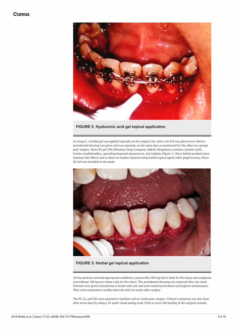
FIGURE 2: Hyaluronic acid gel topical application
In Group C, a herbal gel was applied topically on the surgical site, then a tin foil was placed over which aperiodontal dressing was given and was repeated, on the same days as mentioned for the other two groupspost-surgery. Hiora SG gel (The Himalaya Drug Company, Makali, Bengaluru) contains: jasmine (jati),licorice (yashtimadhu), spreading hogweed (punarnava), and triphala (Figure 3). Since herbal products haveminimal side effects and as there no studies reported using herbal topical agents after gingivectomy, HioraSG Gel was included in the study.
FIGURE 3: Herbal gel topical application
All the patients received appropriate antibiotics (amoxicillin 500 mg thrice daily for five days) and analgesics(aceclofenac 100 mg two times a day for five days). The periodontal dressing was removed after one week.Patients were given instructions to brush with care and were reinstructed about oral hygiene maintenance.They were examined at weekly intervals until six weeks after surgery.
The PI, GI, and GEI were assessed at baseline and six weeks post-surgery. Clinical evaluation was also doneafter seven days by using a 10-point visual analog scale (VAS) to score the healing of the surgical wounds.
2019 Reddy et al. Cureus 11(12): e6438. DOI 10.7759/cureus.6438 3 of 10

VAS consists of a horizontal line of 10 cm with two end-points representing 'no pain' and 'worstpain.’ Patients were asked to mark a point on the line to show their magnitude of pain (0: No pain 1-3: Slightpain 3.1-6: Moderate pain 6.1-10: Severe pain).
The histological evaluation was done using a polarized microscope, using Picrosirus staining [7] six weekspost-gingivectomy to assess the quality and quantity of collagen fibers laid down. The Picrosirius red stain(also called "Sirius red" stain) is one of the best-understood histochemical techniques able to selectivelyhighlight collagen networks. The technique relies on the birefringent properties of collagen molecules.While the picrosirius red stain alone does not selectively bind the collagen network, it becomes more specificthan the other common collagen stains when combined with polarized light detection. The stained sectionsshowing stromal collagen fibers in the three experimental groups were captured with a camera with a 40xobjective. Fiber thickness was determined on the image captured, using image analysis software (ImageJ) [8].All the measurements were assessed in micrometers.
Care was taken in procuring 0.5 mm of tissue from the interdental papilla between the canine and firstpremolar, which was processed for examination using Picrosirius staining. This was performed six weekspostop to record the new collagen laid down. The reagents used were Sirius red dye (Sigma-Aldrich, St.Louis, Missouri, United States), picric acid, xylene, DPX mounting media. "The procedure was done byinitially dewaxing and hydrating the paraffin sections. Then, the nuclei were stained with Mayer’shematoxylin for 8 minutes and then the slides were washed for 10 minutes in running tap water. The slideswere then stained with Picrosirius red for one hour. They were then washed in two changes of acidified waterand the water from the slides was removed by vigorous shaking. Later, the slides were dehydrated in threechanges of 100% ethanol. They were then cleared in xylene and mounted in a resinous medium. 5μmsections were made and stained with Picrosirius red stain and observed under a polarized microscope."
ResultsThe red-stained sections depicting stromal collagen fibers in the three groups were captured with a camerawith a 40x objective. Fiber thickness was determined on the image captured using image analysis software(ImageJ). All the measurements were in micrometers. In each case, three high-power fields (40x) wereselected and from each field, the thickness of fibers was measured. The thickness of the fibers was rangingfrom 0.1-2.4 micrometers. Based on thickness, the collagen fibers were divided into thin and thick fibers andthe number of fibers was calculated. Thin fibers ranged from 0.1-0.80 micrometers. Thick fibers 0.81-2.4micrometers. The data recorded was marked on an Excel sheet for further interpretations and statisticalanalysis. (Figures 5-7).
FIGURE 4: Laser group (Group A) showing OR birefringence of thickcollagen fibers40X Magnification
2019 Reddy et al. Cureus 11(12): e6438. DOI 10.7759/cureus.6438 4 of 10

FIGURE 5: Image of hyaluronic acid gel group (Group B) showingmoderately well-formed collagen fibers40X Magnification
FIGURE 6: Image of the herbal gel group (Group C) showing thincollagen fibers40X Magnification
The reduction in PI, GI, and GEI six weeks postoperatively was statistically significant when compared withbaseline in all three groups (Table 1).
2019 Reddy et al. Cureus 11(12): e6438. DOI 10.7759/cureus.6438 5 of 10

Group Variables Baseline Post (6 Weeks) Difference P-Value Significant
LLLT (Group A)
PI 1.64 0.32042 0.99 0.3755 0.65 0.1118 0 SIG
GI 2.06 0.189 1.03 0.539 1.03 0.43729 0 SIG
GEI 2.3 0.483 1 0 1.3 0.483 0* SIG
Gengigel (Group B)
PI 1.585 0.334 1 0.456 0.585 0.41836 0.002* SIG
GI 2.38 0.456 1.4 0.581 0.98 0.18738 0* SIG
GEI 2.3 0.483 1.1 0.316 1.2 0.421 0* SIG
Hiora SG gel (Group C)
PI 1.7 0.21602 1.05 0.57591 0.65 0.55025 0* SIG
GI 2.39 0.4508 1.46 0.769 0.93 0.67831 0.002* SIG
GEI 2.3 0.483 1.4 0.516 0.9 0.316 0* SIG
TABLE 1: Intragroup comparison among groups using the paired t-testLLLT: low-level laser therapy
Hiora SG: The Himalaya Drug Company, Makali, Bengaluru
When the values were compared between the three groups, there was an improvement in the values of GEIonly (P<0.05). The GEI at baseline was 2.3±0.483 and six weeks postop was 1±0 in the LLLT group. Thebaseline values of GEI for the Gengigel group were 2.3±0.483, which reduced to 1.1±0.316, six weeks after theprocedure. The Hiora SG group recorded a GEI of 2.3±0.483 before surgery, which improved to 1.4±0.516, 6weeks later (P<0.05) (Table 2).
Clinical ParametersLaser (GROUP A) Gengigel (GROUP B) Hiora SG Gel (GROUP C)
P-Value SigMean SD Mean SD Mean SD
PI 1.64 0.32042 1.585 0.334 1.7 0.21602 0.687 NS
PI_POST 0.99 0.3755 1 0.456 1.05 0.57591 0.956 NS
GI 2.06 0.189 2.38 0.456 2.39 0.4508 0.113 NS
GI_POST 1.03 0.539 1.4 0.581 1.46 0.769 0.28 NS
GEI 2.3 0.483 2.3 0.483 2.3 0.483 0.165 NS
GEI_POST 1 0 1.1 0.316 1.4 0.516 0.043* SIG
TABLE 2: Intergroup comparison among groups using ANOVAANOVA: analysis of variance; GEI: gingival enlargement index; GI: gingival index
Hiora SG: The Himalaya Drug Company, Makali, Bengaluru
Related to the VAS between the groups at one week postsurgery, it was observed that the score was the leastin the LLLT group, followed by the Gengigel group and the Hiora SG gel group (Figure 8).
2019 Reddy et al. Cureus 11(12): e6438. DOI 10.7759/cureus.6438 6 of 10

FIGURE 7: Comparison of postoperative visual analog scores betweenthe groups
Pertaining to the histological evaluation, the mean values of thin green-yellow (GY) fibers among groupswere not statistically significant. The mean values of thin orange-red (OR) fibers were 34.7±8.49 for the LLLTgroup, 22.36±5.20 for the Gengigel group, 18.33±3.62 for the Hiora SG gel group and were statisticallysignificant. The mean value of thick GY fibers was 16.06±2.15 in the LLLT group, 14.56±1.98 in the Gengigelgroup, and 17.7±1.95 in the Hiora SG gel group and was statistically significant. Also, the mean values ofthick OR fibers were 31.1±6.27 for the LLLT group, 22.8±5.06 for the Gengigel group, and 20.5±4.11 for theHiora SG group, which was statistically significant. On an intergroup comparison, a difference in thick GY,thick OR, and thin OR fibers was noted in Group A. Hence, it can be inferred that the LLLT group has showna predominance of collagen fibers with orange-red birefringence, indicating the presence of mature collagenas compared to the Gengigel and Hiora SG Gel groups (Table 3).
Collagen Fibers
Group
P-value SIGLaser (GROUP A) Gengigel (GROUP B) Hiora SG Gel (GROUP C)
Mean SD Mean SD Mean SD
THIN GY 17.233 1.678 16.567 2.554 16.567 2.63 0.763 NS
THIN OR 34.732 8.497 22.366 5.2048 18.332 3.627 0* SIG
THICK GY 16.068 2.157 14.567 1.982 17.7 1.951 0.007* SIG
THICK OR 31.1 6.278 22.831 5.069 20.534 4.116 0* SIG
TABLE 3: Intergroup comparison between thick and thin fibers using the ANOVA testANOVA: analysis of variance
DiscussionGingival enlargement is often encountered by clinicians and its apt treatment depends on detecting theetiopathogenesis [9]. The microbe-laden plaque plays a pivotal role in inflammatory gingival enlargement.There are different therapeutic modalities to eliminate periodontal pockets, depending on the type andrelation to the alveolar crest. When the pockets are supra-alveolar, they are called pseudo-pockets, as thereis no loss of attachment but a coronal enlargement of the gingival margin. Poor oral hygiene and trauma tothe gingiva by improper restorations and orthodontic appliances are the main reasons [10].
The selection of an appropriate therapeutic technique depends on the state, as well as the size of the gingivalovergrowth. When there is no regression in the size of the gingiva, even after repeated scaling and root
2019 Reddy et al. Cureus 11(12): e6438. DOI 10.7759/cureus.6438 7 of 10

planing, gingivectomy is done. There are different techniques for performing gingivectomy: conventional,laser, electrocautery, and cryosurgery. Among these techniques, conventional gingivectomy is consideredthe gold standard [11].
In a study comparing conventional gingivectomy with the laser protocol, researchers observed that both theplaque index and the gingival index reduced postoperatively in both the groups when the pockets were <3mm; however, in deeper pockets, conventional gingivectomy yielded better results [12].
Gingivectomy wounds heal by secondary intention. It takes approximately four weeks for the healing tooccur clinically and six weeks for it to be completed histologically. To hasten the healing process, variousagents have been experimented upon and in this study, we have compared the healing between LLLT,hyaluronic acid gel, and herbal gel.
LLLT applied to soft tissues excites specific metabolic processes in healing wounds. The major changesobserved include increased granulation tissue, early epithelialization, increased fibroblast proliferation, andmatrix synthesis. Laser use requires minimal anesthesia and less operative time and results in goodpostoperative healing.
Researchers in a study assessing gingival healing after gingivectomy, with the use of adjunctive LLLT,observed that a 685-nm wavelength laser with an output power of 50 mW and an energy density of 4 J/cmwas effective as an adjunct in promoting healing [13].
In another study, the authors evaluated the effects of LLLT after gingivectomy and gingivoplasty. Theresearchers observed that LLLT using 588 nm wavelength, accelerated epithelization, and woundhealing [14].
In this study also it was observed that there was a reduction in PI (1.64±0.99), GI (2.06±1.03), and GEI(2.3±1.0) from preop to postop in the laser group using a wavelength of 980 nm, output power of 50mW, andenergy of 4J/cm (Group A) (Table 1).
Hyaluronan present in the extracellular matrix of all vertebrates is a glycosaminoglycan playing a pivotalrole in scarless wound healing. It aids in the early deposition of healthy granulation tissue by promotingepithelial cell turnover, good vascularity to the connective tissue, and inhibiting inflammation. Whentopically applied hyaluronic acid has been observed to hasten healing in patients with gingivitis andperiodontitis [3].
A study showed that when 0.2% HA was applied topically, twice daily for a three-week period, it had abeneficial effect in patients with gingivitis. There was an improvement in the plaque index, papillarybleeding index (PBI), and gingival crevicular fluid (GCF) variables [4].
In another study, 0.2% HA gel, when applied on inflamed gingiva, twice daily for a four-week period, as anadjunct to SRP, showed a marked reduction in the gingival index (GI) and PBI when compared with both thecontrol group (scaling plus placebo gel) and the negative control group (scaling only) [15].
In the present study, there was an improvement in PI (1.585±1.0), GI (2.06±1.03), and GEI (2.3±1.1) six weekspostoperatively in the Gengigel group (Group B) (Table 1).
The Hiora SG gel is an herbal gel with analgesic, anti-inflammatory, and antibacterial properties. It repairsthe damaged oral mucosa and has often been used to treat aphthous ulcers. In a study evaluating the efficacyof Hiora SG gel in patients with stomatitis, it was observed that the gel, significantly reduced the swelling,pain, and size of the ulcer at the end of three weeks of treatment. It was thus concluded that Hiora SG gelwas clinically effective and safe in the management of stomatitis [5].
As per our knowledge, Hiora SG gel has not been used as a topical medicament after gingivectomy, hence, itwas decided to evaluate the effects of this herbal gel. In the present study, there was a marked improvementin PI (1.7±1.05), GI (2.39±1.46), and GEI (2.3±1.4) in the Hiora SG (Group C), six weeks after thegingivectomy (Group C) (Table 1).
Though all the three test groups showed an improvement in the clinical variables when an intergroupcomparison was made, it was observed that the LLLT (Group A) showed better results followed by Gengigel(Group B) (Table 2).
Another study compared the use of the 940 nm diode laser with scalpel surgery for the gingivectomyprocedure, in terms of patient satisfaction. The postsurgical discomfort level was recorded using VAS. Theresearchers observed that the bleeding rate and pain level in the diode laser group were found to be less thanin the scalpel group [16].
2019 Reddy et al. Cureus 11(12): e6438. DOI 10.7759/cureus.6438 8 of 10

In this study, there was a marked difference in pain perception between the patients who received threedifferent types of treatments, i.e., P<0.05. The LLLT group showed the least VAS scores followed by theGengigel group, and, finally, the Hiora SG group.
Most of the studies assessing healing after gingivectomy have been done using the Haematoxylin and Eosinstain to observe the epithelization and the amount of connective tissue laid down, however, the quality ofcollagen laid down has not been assessed. So in this study, the Picrosirius red staining method was used toassess the quality of collagen laid down. As per our knowledge, Picrosirius red staining has not been used toassess healing after a gingivectomy.
The connective tissue is made up of collagen fiber bundles, which can be differentiated as thin and thickfibers. Type I and III collagens are predominant types in which type I collagen consists of thick and maturefibers and type III consists of thin and immature fibers [17-18].
Depending on the type, collagen exhibits a differential birefringence pattern, ranging from green-greenishyellow to orange-red, to red. Type I collagen fibers expressed an orange-red to red color, as they are made upof thick, mature, and closely packed fibrils presenting as an intense birefringence whereas type III collagenhaving a weak birefringence expressed a greenish-yellow to yellow color of the fibrils, as they were observedto be thin and immature.
The limitations of this study were the absence of a control group, which would have validated the studybetter. Moreover, the sample size was small and there were no studies available to compare the results ofhistological evaluation after six weeks post-gingivectomy with Picrosirius red.
ConclusionsThis study compared the efficacy of LLLT, Gengigel, and Hiora SG gel post-gingivectomy. Statisticallysignificant results were observed in the LLLT group (Group A) pertaining to the GEI and VAS. Also, thehistological evaluation showed more mature collagen fibers in the laser group. Gengigel performed betterthan Hiora SG gel both clinically and histologically, ranking next to the laser group. However, many morestudies with a larger sample size have to be done to assert the benefits of LLLT and Gengigel, as was noticedin this study.
Additional InformationDisclosuresHuman subjects: Consent was obtained by all participants in this study. Institutional Ethical Committeeissued approval PMVIDS&RC/IEC/PERI/DN/0109-16. Animal subjects: All authors have confirmed that thisstudy did not involve animal subjects or tissue. Conflicts of interest: In compliance with the ICMJE uniformdisclosure form, all authors declare the following: Payment/services info: All authors have declared that nofinancial support was received from any organization for the submitted work. Financial relationships: Allauthors have declared that they have no financial relationships at present or within the previous three yearswith any organizations that might have an interest in the submitted work. Other relationships: All authorshave declared that there are no other relationships or activities that could appear to have influenced thesubmitted work.
AcknowledgementsWe would like to acknowledge the help and support rendered by Dr. P Bhagirath, Professor and HOD,Department of Oral Pathology, PMVIDS, Hyderabad. We would also like to thank Dr. Chinta Ankitha forhelping us with the statistical work.
References1. Amorim JC, de Sousa GR, de Barros Silveira L, Prates RA, Pinotti M, Ribeiro MS: Clinical study of the gingiva
healing after gingivectomy and low-level laser therapy. Photomed Laser Surg. 2006, 24:588-594.10.1089/pho.2006.24.588
2. Ozcelik O, CenkHaytac M, Kunin A, Seydaoglu G: Improved wound healing by low‐level laser irradiationafter gingivectomy operations: a controlled clinical pilot study. J Clin Periodontol. 2008, 35:250-254.10.1111/j.1600-051X.2007.01194.x
3. Batavia PD, Bhushan KS, Vandana K L, Desai R: Use of hyaluronan (Gengigel) in the treatment of gingivitisin orthodontic patients: a clinical, biochemical, and microbiological study. J Int Clin Dent Res Organ. 2016,8:44-50. 10.4103/2231-0754.176255
4. Sahayata VN, Bhavsar NV, Brahmbhatt NA: An evaluation of 0.2% hyaluronic acid gel (Gengigel®) in thetreatment of gingivitis: a clinical & microbiological study. Oral Health Dent Manag. 2014, 13:779-785.
5. Sukumaran VG, Amrutha, Vivekananda P, Palaniyamma D: A randomized placebo controlled comparativestudy to evaluate the efficacy of Hiora SG gel in stomatitis. Probe. 2011, 1:1-5.http://himalayainfoline.com/journalinfoline/pdfs/2011/JM11/Probe_JM11.pdf.
6. Miller CS, Damm DD: Incidence of verapamil-induced gingival hyperplasia in a dental population . JPeriodontol. 1992, 63:453-456. 10.1902/jop.1992.63.5.453
2019 Reddy et al. Cureus 11(12): e6438. DOI 10.7759/cureus.6438 9 of 10

7. Coleman R: Picrosirius red staining revisited. Acta Histochem. 2011, 3:231-233.10.1016/j.acthis.2010.02.002
8. Kamath VV, Satelur K, Komali Y, Krishnamurthy SS: Image analyses of collagen types and thickness in oralsub mucous fibrosis stained with picrosirius red under polarizing microscope. J Orofac Sci. 2013, 5:123-127.10.4103/0975-8844.124258
9. Agrawal AA: Gingival enlargements: differential diagnosis and review of literature . World J Clin Cases. 2015,3:779-788.
10. Seymour RA: Effects of medications on the periodontal tissues in health and disease . Periodontol 2000.2006, 40:120-129. 10.1111/j.1600-0757.2005.00137.x
11. Funde S, Baburaj MD, Pimpale SK: Comparison between laser, electrocautery and scalpel in the treatment ofdrug-induced gingival overgrowth-a case report. IJSS Case Reports Rev. 2015, 1:27-30.https://pdfs.semanticscholar.org/ddf9/a2c8f00775641488e839a24b624f1f8f35ee.pdf. 10.17354/cr/2015/43.
12. Waite IM: A comparison between conventional gingivectomy and a non‐surgical regime in the treatment ofperiodontitis. J Clin Periodontol. 1976, 3:173-185. 10.1111/j.1600-051x.1976.tb01865.x
13. Chawla K, Lamba AK, Tandon S, Faraz F, Gaba V: Effect of low-level laser therapy on wound healing afterdepigmentation procedure: a clinical study. J Indian Soc Periodontol. 2016, 20:184-188. 10.4103/0972-124X.176393
14. Evaluation of the effect of low level laser therapy after gingivectomy on wound healing . (2017).https://www.longdom.org/conference-abstracts-files/2247-2452-C1-048-010.pdf .
15. Jentsch H, Pomowski R, Kundt G, Göcke R: Treatment of gingivitis with hyaluronan . J Clin Periodontol.2003, 30:159-164. 10.1034/j.1600-051x.2003.300203.x
16. Öncü E, Erbeyoğlu AA, Alan R: Comparison of gingivectomy procedures for patient satisfaction:conventional and diode laser surgery. Selcuk Dent J. 2017, 4:6-9. 10.15311/1441.309572
17. Rich L, Whittaker P: Collagen and picrosirius red staining: a polarized light assessment of fibrillar hue andspatial distribution. Braz J Morphol Sci. 2005, 22:97-104. https://www.semanticscholar.org/paper/Collagen-and-picrosirius-red-staining%3A-a-polarized-Rich-Whittaker/81282ace6a9....
18. Lattouf R, Younes R, Lutomski D, Naaman N, Godeau G, Senni K, Changotade S: Picrosirius red staining: auseful tool to appraise collagen networks in normal and pathological tissues. J Histochem Cytochem. 2014,62:751-758. 10.1369/0022155414545787
2019 Reddy et al. Cureus 11(12): e6438. DOI 10.7759/cureus.6438 10 of 10
Copyright © 2022 FDOKUMEN