Strategies for surface immobilization of whole bacteriophages
95
HAL Id: cea-03245827 https://hal-cea.archives-ouvertes.fr/cea-03245827 Submitted on 2 Jun 2021 HAL is a multi-disciplinary open access archive for the deposit and dissemination of sci- entific research documents, whether they are pub- lished or not. The documents may come from teaching and research institutions in France or abroad, or from public or private research centers. L’archive ouverte pluridisciplinaire HAL, est destinée au dépôt et à la diffusion de documents scientifiques de niveau recherche, publiés ou non, émanant des établissements d’enseignement et de recherche français ou étrangers, des laboratoires publics ou privés. Strategies for surface immobilization of whole bacteriophages: A review Larry O’connell, Pierre Marcoux, Yoann Roupioz To cite this version: Larry O’connell, Pierre Marcoux, Yoann Roupioz. Strategies for surface immobilization of whole bacteriophages: A review. ACS Biomaterials Science and Engineering, ACS, 2021, 10.1021/acsbio- materials.1c00013. cea-03245827
-
Upload
khangminh22 -
Category
Documents
-
view
3 -
download
0
Transcript of Strategies for surface immobilization of whole bacteriophages

HAL Id: cea-03245827https://hal-cea.archives-ouvertes.fr/cea-03245827
Submitted on 2 Jun 2021
HAL is a multi-disciplinary open accessarchive for the deposit and dissemination of sci-entific research documents, whether they are pub-lished or not. The documents may come fromteaching and research institutions in France orabroad, or from public or private research centers.
L’archive ouverte pluridisciplinaire HAL, estdestinée au dépôt et à la diffusion de documentsscientifiques de niveau recherche, publiés ou non,émanant des établissements d’enseignement et derecherche français ou étrangers, des laboratoirespublics ou privés.
Strategies for surface immobilization of wholebacteriophages: A review
Larry O’connell, Pierre Marcoux, Yoann Roupioz
To cite this version:Larry O’connell, Pierre Marcoux, Yoann Roupioz. Strategies for surface immobilization of wholebacteriophages: A review. ACS Biomaterials Science and Engineering, ACS, 2021, �10.1021/acsbio-materials.1c00013�. �cea-03245827�

1
Strategies for Surface Immobilization of Whole 1
Bacteriophages: A Review 2
Larry O’Connell1,2, Pierre R. Marcoux1, Yoann Roupioz2,* 3
1 Univ. Grenoble Alpes, CEA, LETI, F38054 Grenoble, France 4
2 Univ. Grenoble Alpes, CNRS, CEA, IRIG, SyMMES, 38000 Grenoble, France 5
*Correspondence: [email protected]; Tel.:+33-4-38-78-98-79 6
Keywords: Biomaterials, Biosensing, Functional surfaces, Surface functionalization, 7
Bacteriophage 8
9
Abstract 10
Bacteriophage immobilization is a key unit operation in emerging biotechnologies, enabling 11
many new possibilities for biodetection of pathogenic microbes at low concentration, 12
production of materials with novel antimicrobial properties, and fundamental research on 13
bacteriophages themselves. 14
Wild type bacteriophages exhibit extreme binding specificity for a single species – and often 15
for a particular subspecies – of bacteria. Since their specificity originates in epitope recognition 16
by capsid proteins, which can be altered by chemical or genetic modification, their binding 17
specificity may also be redirected towards arbitrary substrates or a variety of analytes in 18

2
addition to bacteria. The immobilization of bacteriophages on planar and particulate substrates 1
is thus an area of active and increasing scientific interest. 2
This review assembles the knowledge gained so far in the immobilization of whole phage 3
particles, summarizing the main chemistries and presenting the current state-of-the-art both for 4
an audience well-versed in bioconjugation methods as well as for those who are new to the 5
field. 6
7
Introduction 8
Bacteriophages, obligate intracellular parasitic viruses that replicate only in their host 9
bacterium, are the most numerous replicating biological entity on Earth, with an estimated 1031 10
phage particles contained within the biosphere, compared to an estimated 1030 bacterial cells 11
1,2. Indeed, the estimated daily turnover of 15% of all bacterial cells due to phage lysis is a 12
testament to their crucial role in microbiological ecology 3. 13

3
1
2
Figure 1. Illustrations of the bacteriophage morphologies that appear most often in the phage-3
functionalization literature. The Inoviridae family comprises filamentous phages and includes 4
fd and M13. The Myoviridae family features long contractile-tailed phages and includes the 5
well-known T4 coliphage. The Siphoviridae family features long non-contractile-tailed phages 6
and includes D29. Meanwhile, the Podoviridae family are short-tailed and include P22. 7
Relative scales are approximate. 8
9
Phage structure exhibits large variation which falls within a few stereotyped forms (Erreur ! 10
Source du renvoi introuvable.). A small number of phage morphologies are over-represented 11

4
in the phage immobilization literature and include: long contractile-tailed phages (Myoviridae 1
e.g. T4), long non-contractile-tailed phages (Siphoviridae), short-tailed phages (Podoviridae 2
e.g. T7 or P68), filamentous phages (Inoviridae e.g. M13 and fd) 4. Less frequently seen in the 3
phage immobilization literature are tailless phages families Tectiviridae (comprising non-tailed 4
icosahedonal phages, such as PRD1) and Cystoviridae (featuring an outer lipid membrane and 5
no tail, such as phage phi6). Phages are also described in terms of their replication cycle. Lytic 6
phages terminate their replicative cycle with the biochemical lysis of their host, rupturing the 7
cell membrane and releasing up to several hundred progeny virions in one burst. In contrast, 8
upon infection of a suitable host, temperate phages incorporate their genetic material into that 9
of the bacterial cell and may lay dormant before shifting to a lytic cycle, or may instead 10
continually produce a smaller number of phages which are shed from the host on a continuous 11
basis 5. 12
History 13
Bacteriophages were independently discovered by microbiologists Frederick Twort in 14
London in 1915 and by Felix d’Herelle in Paris in 1917 6.It should be noted that extensive 15
research on phage phenomena was also carried out in Poland during the interwar period7, as 16
well as in Brazil. Indeed, the oft-cited pioneering 1919 work by d’Hérelle in the use of phage 17
to treat dysentary in French soldiers was followed relatively soon afterwards by similar trials 18
in 1923 in both Poland7 and Brazil8. 19
Such research eventually led to the widespread use of phages in the Soviet Union for the 20
treatment of routine bacterial infections, with phage therapy being extensively mobilized to 21
meet the needs of the Soviet military beginning in 1939 with the Winter War with Finland, 22
and later in World War II 9. 23
5
Meanwhile in the West, the discovery by Alexander Fleming in 1928 of penicillin, a broad 1
spectrum antibiotic, led to a paradigm shift in medicine that relied heavily on the widespread 2
administration of what was seen at the time as a “magic bullet”. 3
The geopolitical paranoia of the Cold War and a lack of scientific rigor in reporting of early 4
soviet phage therapy studies resulted in a progressive dismissal of phage therapy in Western 5
medicine 10. In the 1970s, up to 70 patients per year underwent phage therapy to treat bone and 6
joint infections (BJI) in Croix Rousse Hospital, Lyon 11. However, non soviet-aligned states 7
instead pursued a policy of unfettered use of antibiotics 12, the profligate administration of 8
which has now led to widespread antimicrobial resistance (AMR), threatening to return 9
medicine to the “dark ages” before widespread availability of microbial control 13. With the 10
World Health Organization (WHO) announcement in 2014 that AMR was no longer a looming 11
threat but a contemporary crisis 14, the problem of antibiotic resistance is proving increasingly 12
salient 15. 13
In a global context where common pathogenic bacterial strains are rapidly gaining new 14
resistance mechanisms, the pharmaceutical sector is largely withdrawing from the antibiotic 15
discovery field, which has led to a failure to discover any new classes of antimicrobial agents 16
in over three decades 16,17. 17
A growing appreciation for the importance of antibiotic stewardship and the urgency of 18
identifying novel therapies has led to renewed interest in phage therapy as a plausible 19
replacement for antibiotics 18. In the field of biodetection, too, researchers have leveraged the 20
high specificity of bacteriophages for their hosts to create biosensors with single strain 21
specificity for a variety of common human pathogens and with extremely low detection limits. 22
New use cases are also being explored, including the use of phages for antimicrobial, bioactive 23
packaging 19,20 or as nanostructural scaffolds for various supramolecular structures 21. 24
6
Within this context, biomedical, agricultural, and environmental monitoring applications of 1
bacteriophages would be aided by well-characterized, repeatable, versatile, and (ideally) 2
morphology-agnostic immobilization methods. 3
State of the art 4
The literature concerning phage-functionalization of substrates falls into three main 5
categories, with some overlap: 6
Coupling to a transducer for the specific detection of bacteria or other analytes. 7
Starting in earnest in 2005 22, the last 15 years have seen bacteriophages 8 immobilized on biosensors using a variety of transduction mechanisms, towards the 9 detection of a large variety of target analytes including bacteria and their spores 23–10 28, antibodies 29–32, prostate specific antigen 32–34, enzymes 35,36, cancer biomarkers 11 37, and glucose 38. 12
As a biosorbent layer for species-targeted biocontrol and anti-fouling 13
The incorporation of bacteriophages into materials (e.g. food packaging and wound 14 dressing) can confer them targeted anti-bacterial properties which can help reduce 15 the proliferation of bacteria in foodstuffs or surrounding wounds. Such bioactive 16 materials have been demonstrated in food packaging that targets Listeria 20,39, 17 Escherichia coli 20,39,40, and Salmonella 19. Bioactive fabrics have been 18 demonstrated with specific antimicrobial action against Pseudomonas aeruginosa 19 41 and E. coli 42. 20
As a structural scaffold for supramolecular nanostructure fabrication 21
The tessellated and highly redundant structure of bacteriophage capsids, together 22 with their chemical uniformity and physical monodispersity, also opens up the 23 possibility of bottom-up fabrication of highly ordered supramolecular structures 24 21,43. Liquid crystalline bacteriophage films 21,43 have been proposed for use as tissue 25 regenerating scaffolds 44, piezoelectric energy harvesting 45, and colorimetric 26 sensors 46. 27
Across all applications, the vast majority of papers reviewed demonstrate immobilization of 28
classic phages such as the lytic T4 coliphage, or the temperate M13 and fd phages. However, 29
examples are also to be found of successful immobilization of various other phages of the 30
Autographiviridae 29,47–49, Tectiviridae 50–52, Herelleviridae 53, Leviviridae 50,53, Myoviridae 31
39,50–64, Siphoviridae 39,41,53,60,65,66 and Podoviridae 50,53,57,58,67–72 families. 32
Viral properties involved in immobilization 33

7
When considering the immobilization of bacteriophages on surfaces or particles, we should 1
first consider what properties of the phage, which moieties and/or surface charges, can be 2
modified or leveraged for chemical and/or physical interaction with the surface or intermediate 3
linker molecules. The vast majority of bacteriophages exhibit an outermost protein layer which 4
encapsulates genetic material on the interior (with the exception of Cystoviridae, which 5
encapsulated the capsid in a lipid envelope). Bacteriophage capsids are composed of proteins 6
which are in turn composed of long chains of amino acid subunits, which display primary 7
amine (−NH2) groups on their N-terminus, and carboxyl groups (-COOH) on their C-terminus 8
(Erreur ! Source du renvoi introuvable.). Both the amine and carboxyl groups of polypeptide 9
amino-termini are frequent targets for immobilization chemistry, but in the specific case of 10
bacteriophages, the termini available for conjugation will be greatly outnumbered by amino 11
acid side chains. 12
Amino acids vary in the composition of their side chains, with some side chains endowing a 13
polarity, charge, or hydrophobic/hydrophilic character, each of which may be leveraged in 14
isolation or in combination for the purposes of conjugation. For example, two amino acids in 15
particular, cysteine and methionine, contain sulfhydryl side chains which interact strongly with 16
gold, a common substrate for bio-functionalization. The amino acids lysine, arginine, 17
asparagine, and glutamine feature a second primary amine group on the side chain in addition 18
to their N-terminus. 19
Each amino acid also has a characteristic isoelectric point which determines the pH below 20
which the side chain is protonated in aqueous solutions. The local hydrophobic character and 21
charge of a phage is the result of an aggregate sum of the local constituent amino acids. The 22
isoelectric point of a bacteriophage dictates the charge it presents to the solution and thus its 23
stability and resilience against aggregation73–75. There is significant evidence that tailed 24
bacteriophages – which make up over 95% of described phages 76 – have a net dipole moment, 25

8
with a negatively charged head and positively charged tail and tail fibers at physiological 1
pH39,77,78. 2
Genes coding amino acids on the outer bacteriophage surface can also be targeted for site-3
specific genetic engineering and mutagenesis, altering them in order to introduce new 4
functional groups in order to optimize their chemical and/or physical characteristics for the 5
purposes of immobilization 38,48,79–89. 6
To summarize, when developing immobilization strategies, the researcher has at their disposal: 7
Primary amine groups (−NH2), present at the N amino-termini of proteins and in the side chains 8 of lysine, arginine, asparagine, and glutamine 9
Carboxylic groups (−COOH), present at the C amino-termini of proteins and in the side chains 10 of aspartic and glutamic acid 11
Sulfhydryl side chains (−SH) of the amino acids cysteine and methionine 12 The local and overall charge of the phage 13 The dipole moment of the phage 14 Site-directed genetic engineering of the phage coat 15
It is starting from this basis that the full suite of immobilization strategies can be derived. 16
Immobilizing biological entities on surface or nanoparticle substrates can lead to 17
conformational and other changes that impede their original function. When choosing an 18
immobilization strategy, it is important to consider what purpose the phage immobilization is 19
serving. Is the goal to maximize analyte capture itself? Or is it rather to maximize the limit of 20
detection (LOD) of a biosensor? Is the surface to be used for the purpose of biocontrol (e.g. 21
wound dressing or food packaging)? What kind of environment will the phage-functionalized 22
surface be used in? Is it important that the phage maintains the same infectivity as its non-23
immobilized form? Is it important that the phage be oriented in a given direction relative to the 24
substrate? 25
The answers to each of these questions places different requirements on the surface chemistry 26
and optimal phage density. For example, Naidoo et al. have found that bacterial capture 27
efficiency only correlates with increasing phage surface density up to a certain threshold, 28
beyond which the capture efficiency actually decreases 58. However, in a biocontrol context 29
9
where shedding of phage particles from a substrate may be desirable, this effect may not be 1
problematic and we may indeed aim to achieve a maximal surface density limited only by 2
geometric constraints. 3
Consideration of the end result is needed in order to optimize the immobilization strategy. 4
Phage density may represent only a proximal measure for a different performance metric of the 5
phage-functionalized substrate. The researcher may indeed aim for lower phage surface 6
densities depending on the application, and one should not necessarily aim to maximize surface 7
density for its own sake. 8
Scope and organization of the review 9
This review groups the literature based on immobilization technique, rather than grouping 10
by substrate, phage, or detection scheme (in the case of biosensors). This arrangement makes 11
the particularities and commonalities between immobilization strategies more apparent. 12
This review does not treat the immobilization of phage-derived proteins (e.g. endolysins or 13
recombinant coat proteins), since this has been treated elsewhere and overlaps with general 14
strategies for protein immobilization, a topic too large for a single review. For an excellent 15
treatment of protein immobilization, see Hermanson 90. Likewise, this review does not treat 16
phage immobilization on chromatographic columns or microtiter plates, since this falls in the 17
category of phage display literature, thoroughly reviewed in Hust and Lim 91. 18
The most common substrates are briefly described, as well as the scientific interest in phage-19
functionalization in each case. Thereafter, the most common and successful immobilization 20
strategies are outlined with reference to their application in the literature, grouped into covalent 21
chemistries, non-covalent and physical methods (e.g. physisorption, electrostatic adsorption, 22
avidin-biotin linkage etc.), and finally genetic modification techniques, which are treated 23
separately since they exist as a modification of the bacteriophage itself but facilitate the 24
preceding chemisorptive and physisorptive immobilization strategies. 25

10
Oriented immobilization 1
A phage’s host range and ability to replicate is largely based on its ability to specifically 2
recognize cells of its host bacterial strain via the phage’s receptor-binding domains (RBDs) – 3
epitope-recognizing regions on the phage capsid. This initiating event is a sina qua non and 4
relies on serendipitous encounters between the RBDs and a bacterial cell surface. 5
Over 95% of described phages are tailed 76. and thus have an inherent asymmetric, bilateral 6
geometry; having a preferred orientation with regards to attachment to their host cell. For a 7
conjugated phage to retain its binding and replicative capacity, it must be oriented on the 8
surface such that its RBDs are exposed to the environment, and hence any host cells that may 9
be present 92. Such a tail-upward orientation will hereafter be referred to simply as “oriented” 10
immobilization. Most authors simply accept the lower bacterial capture associated with random 11
phage orientation, relying on immobilization of a sufficiently large number of phages that 12
includes a small subpopulation of serendipitously tail-upward oriented phages. Nevertheless, 13
Nogueira et al. recognize that maximizing tail exposure is a key criterion for maintaining 14
immobilized phage infectivity 41. and Hosseinidoust et al. conclude in an influential review 15
paper that the main hurdle to designing efficient phage-based biosensors appears to be 16
controlling the orientation of the immobilized phages 92. 17
The bacteriophage head presents a net negative charge, while the tail fibers present a net 18
positive charge 77. Thus, the dipole moment of a phage particle causes the head to be oriented 19
towards a positively charged surface 78. In addition to local charge distribution, many phages 20
such as T7, present an overall net negative charge 93,94. 21
In most circumstances there is a strong entropic preference towards horizontal alignment of 22
tailed and filamentous phages. However, as we shall see, a significant minority of the studies 23
reviewed have taken advantage of the local charge or other properties of bacteriophage capsids 24
to claim oriented immobilization as a result of their protocols (Figure 2). Frequently, however, 25

11
insufficient evidence and/or no clear mechanisms are presented to prove oriented 1
immobilization has been achieved. This is partly due to the difficulty in determining the 2
orientation of nanoscale objects, but post-functionalization characterization techniques do exist 3
that can help justify such claims. 4
5
Figure 2. Scanning electron micrograph of in vivo biotinylated T4 phage, immobilized on a 6
streptavidin-coated gold surface. Arrows indicate phage particles with tail-up orientation. 7
Reproduced from Tolba et al. 83. 8
A note on surface density 9
A high phage surface density is not the sole criterion for high phage infectivity and/or 10
bacterial capture. For example, Tawil et al. found that phages were immobilized on gold with 11
double the density when cross-linked via L-cysteine compared to glutaraldehyde, but that no 12
significant difference was found in the bound phages’ lytic behavior 72. Similarly, Leppänen et 13
al. rigorously compared infectivity and phage surface density after surface treatments with 14
different combinations of 11-MUA and (3-aminopropyl)triethoxysilane (APTES), concluding 15

12
that a higher phage density did not always result in a higher infectivity 95. Indeed, Naidoo et al. 1
achieved an impressive surface density of 199±2 phages/µm2 but found that – above a value 2
of a 18.9±0.8 phages/µm2 – higher phage density led to a reduction in total bacterial capture 3
58. 4
These results suggest that careful consideration and design of the surface functionalization 5
can lead to dramatically improved activity and/or bacterial capture of phage-functionalized 6
surfaces. 7
Common substrates 8
Gold 9
Gold surfaces are by far the most popular surface for phage functionalization, serving as the 10
substrate in just over half of all papers reviewed on this topic. Gold exhibits ideal properties 11
for functionalization with many biological entities 90, and for bacteriophages the case is no 12
different. Gold is biocompatible, non-oxidizable, readily available, easily cleaned, and its 13
deposition on a variety of substrates is a mature technology 96–98. Gold also exhibits a very high 14
binding affinity for sulfhydryl (R-SH, aka thiol) groups of around 200 kJ/mol, which also 15
permits easy formation of self-assembled monolayers 98. Thiol groups are frequently found – 16
or easily introduced – in most target ligands for the purpose of immobilization 98. 17
Facilitating bioconjugation is the principal motivation behind the deposition of a gold layer 18
on transducers that do not involve the intrinsic properties of gold itself (e.g. magnetoelastic or 19
quartz crystal microbalance (QCM) biosensors, which instead rely on the resonance of an 20
underlying amorphous ferromagnetic material 99 or quartz crystal 100, respectively). However, 21
gold also exhibits ideal properties where its biocompatibility and ease of conjugation is a happy 22
coincidence. Gold features a high density of easily polarizable free electrons – a prerequisite 23
for strong interaction with electromagnetic fields 101 – making it an ideal material for use in 24

13
surface plasmon resonance (SPR). For SPR, there are effectively only two metals with 1
appropriate properties: gold and silver 102. While silver actually yields a superior SPR effect, it 2
is less biocompatible and less chemically stable – particularly towards oxidation – compared 3
to gold, and so the latter has become the de facto default for SPR biosensors 101,102. 4
Magnetic beads 5
Magnetic bead surfaces are a frequent substrate for phage conjugation since, once 6
functionalized, they can be used for immunomagnetic separation (also sometimes referred to 7
as phagomagnetic separation) and concentration of low-titer analytes in order to boost the 8
capture efficiency of biosensor surfaces (Figure 4) 47,48,56,68,69,89,103,104. Briefly, phages specific 9
to a given bacterial strain are immobilized on the beads, which are then mixed with a sample 10
and bind to the target analyte (Figure 5). Application of a magnetic field can then be used to 11
concentrate and confine the captured analyte to a given region, for example on the surface of a 12
biosensor. Phage immobilization has been shown for magnetic beads with a variety of surface 13
chemistries including tosyl 68,69, carboxyl 47,49,69,103, streptavidin 48,83,89,105, azide 81, and 14
isothiocyanate-terminated coatings 104. 15
Carbon allotropes 16
Carbon allotropes (e.g. graphene, nanotubes) and glassy/vitreous carbon are common 17
components of inks used to fabricate screen-printed electrodes (SPEs). Electrode screen-18
printing enables cheap, versatile electrodes for use in clinical assays106, food processing107, and 19
environmental monitoring 108. Such electrodes can also be composed of gold, silver, or 20
platinum; but carbon is more typical due to its relatively low cost 109. The versatility and easy 21
manufacture of SPEs has led to carbon allotropes becoming a target for phage immobilization 22
for the fabrication of biosensors specific to various bacterial strains 54,55,65,110,111, and in one 23
paper a biosensor for West Nile virus-specific IgG 29. Carbon nanotubes can also be used as an 24

14
intermediate structure for the immobilization of phages on different substrates, as was 1
demonstrated by Farooq et al. for bacterial cellulose matrices for ultra-sensitive and selective 2
electrochemical detection of Staphylococcus aureus 112. Carbon allotrope-specific binding 3
peptides have been discovered that may enable recombinant phage immobilization on such 4
substrates 113–116 (see Polymer and other binding domains below). 5
Polymers 6
Phage-functionalization of polymers is of interest since such materials can be formed into 7
antimicrobial wound dressing 41,117, food packaging 20, and biocontrol surfaces resistant to 8
biofilm formation 118. They also form the coating of many commercially available magnetic 9
beads 47,49,68,69,103. Polymers that have been successfully conjugated or co-polymerized with 10
phages include polycaprolactone 41, polymethyl methacrylate (PMMA) 86, polyethersulfone 118, 11
polystyrene 119, polyhydroxyalkanoates 40, polyethylene 120, and poly(3,4-12
ethylenedioxythiophene) (PEDOT) 33,121,122. 13
It is interesting to note that a large number of peptides have been discovered that have high 14
binding affinities for specific polymers 123–129, and that such proteins have been expressed on 15
the capsids of recombinant phages to facilitate immobilization 83,86,89. 16
Cellulose-based materials 17
A particularly important subcategory of polymers is cellulose and cellulose-derived 18
materials. As the most abundant natural polymer on Earth 130, cellulose features in the phage-19
functionalization literature because it forms the principal component of paper. With its high 20
porosity, hydrophilicity, chemically inert character, and slight negative charge at neutral pH 21
42,53, paper lends itself to the fabrication of cheap, mass-produced devices for healthcare 131 and 22
environmental monitoring 132. 23

15
Phage-functionalization of paper and cellulose-derived polymers is motivated by the 1
development of antimicrobial food packaging materials 19,20,39, and low cost biosensors 2
53,133,134. Phage-containing bio-inks can be printed onto paper substrates, and it has been shown 3
that T4 phages are capable of resisting the shear stress and drying processes involved in 4
industrial printing 135. It has also been demonstrated that careful consideration of the bio-ink 5
constituents can enable production of bioactive paper that is stable for several days after 6
printing 136. Anany et al. have demonstrated a dipstick assay based on inkjet-printed phages 7
that efficiently captured and infected E. coli and Salmonella Newport in broth and food 8
matrices 53. Farooq et al. demonstrated phage-functionalization of bacteria-derived cellulose 9
fibers, but via intermediate multi-walled carbon nanotubes 112. 10
There is ample precedent for lateral flow devices that rely on capillary action along capillary 11
beds to channel fluid for the purposes of biochemical analysis, the most well-known of which 12
is probably modern pregnancy tests 137 and covid-19 antigen tests. Such microfluidic paper-13
based devices may also be combined with electrode screen printing for more elaborate 14
microfluidic electrochemical sensors 137,138. Phage-derived proteins 139 have been incorporated 15
into lateral flow assays, but to our knowledge in all cases have been immobilized onto 16
intermediate supports (e.g. nanoparticles) rather than the polymer substrate of the device itself. 17
The ability to reliably immobilize phages directly on paper substrates would open new avenues 18
for the development of low-cost, mass-produced bioassays 134. 19
Some evidence suggests immobilization is more successful if paper has been precoated with 20
polydiallyldimethylammonium chloride (polyDADMAC), a cationic polyelectrolyte which 21
imparts a positive charge to the cellulose fibers and thus electrostatically binding the phage 22
heads 134. Phage resistance to dry environments can be improved by incorporating gelatin into 23
the deposition ink 136 24

16
When considering cellulose-based substrates, of note is the existence of cellulose binding 1
modules (CBMs) – polypeptides that bind strongly to cellulose 140 and which can be expressed 2
by genetically-engineered phages to facilitate their immobilization on such substrates 83,89. 3
Silicon-based materials 4
In this review, phage-functionalization of several materials including silicon nitride 37, 5
optical fibers 141–143, atomic force microscopy probes 144, silica nanoparticles 60,62, and glass 6
50,51,67,79,80 are all grouped into the category of silicon-based substrates. Such surfaces are 7
typically rendered reactive for immobilization chemistry through amino-silanization 8
37,46,50,51,60,62,67,79,80,95,141,142,144–148. 9
Optical fiber biosensing is of particular interest since it can exploit the properties of optical 10
biosensing techniques (e.g. SPR), while avoiding drawbacks associated with bulky equipment 11
141. Using a smaller sensing element and sample volume can be advantageous in the analysis 12
of small amounts of precious analytes, and can accommodate multiple parallel measurements 13
on adjacent bundled optical fibers 149. 14
Since the fiber-optic material itself is cheap, this may offer the possibility of disposable, 15
single-use sensors 150,151. Finally, since the nature of optical fiber geometry means that signal 16
transduction can take place at a physically remote location relative to the optical setup, this 17
form of biosensing enables remote detecting and monitoring capabilities in potentially harsh 18
environments 149, allowing one to bring the sensor to the sample rather than bring the sample 19
to the sensor. Phage-functionalized optical fibers have been demonstrated for the specific 20
detection of E. coli 141–143. 21
Covalent methods 22

17
1

18
Figure 3. Schematic representation of a selection of the most popular bacteriophage 1
immobilization strategies, broadly grouped into covalent bonding, physisorption, and genetic 2
modification. At left, the substrates that have been demonstrated in the literature with the 3
adjacent immobilization method. Note: bacteriophages are represented by a generic 4
Podoviridae but in most cases represent other phage morphologies 5
EDC/NHS Chemistry 6
For substrates presenting carboxyl (−COOH) groups – be they endogenous or introduced – 7
a common covalent conjugation method involves the use of the carbodiimide EDC (1-Ethyl-3-8
(3-dimethylaminopropyl)carbodiimide) to activate the carboxyl groups, creating an unstable 9
ester intermediate 152. Typically, a separate molecule containing a succinimidal group in the 10
form of NHS (N-hydroxysuccinimide) is then introduced to the surface and supplants the 11
carbodiimide, which is released into the solution. This stabilizes the activated carboxyl group 12
and forms a second NHS-ester intermediate which is more vulnerable to nucleophilic attack 13
from amino groups, in turn priming the NHS to be supplanted by the primary amine of an 14
amino acid 67,152. 15
When a suspension of amine-containing ligands – such as phages or proteins – is introduced 16
to the sensor surface, a primary amine of the ligand reacts with the NHS (the leaving group) 17
and is then covalently bonded to the surface while the succinimidal group is released into the 18
solution. After ligand binding is complete, the surface may be washed with ethanolamine 19
whose amine groups will react with the remaining activated carboxyl “sites”, which blocks 20
further binding.152. In this way, the EDC/NHS solution facilitates amide bonding between 21
carboxyl groups of the surface and amine groups of the ligand 67. 22
Such EDC/NHS coupling is extremely popular for surface functionalization of gold 23
following introduction of exogenous carboxyl groups (for example by formation of an 24

19
alkanethiol self-assembled monolayer ) 34,57,153–158, but has also been demonstrated for a variety 1
of carboxylated substrates including glassy carbon electrodes 55, glass 50, magnetic beads 103, 2
and polymers 40,159. 3
The creation of the second NHS-ester intermediate is not obligatory and in cases where a 4
high yield of conjugated ligands is not crucial, the use of NHS may be omitted entirely. Such 5
EDC-mediated binding has been used to covalently immobilize phages on glassy carbon 6
electrodes 110,111, magnetic microbeads 103, and carboxyl-polystyrene latex beads 119. However, 7
the addition of even small amount of NHS to an EDC coupling reaction can boost the yield of 8
conjugated ligand by a factor of 20 160. NHS activation decreases the water-solubility of the 9
activated carboxylate molecule and for this reason is often instead sold as sulfo-NHS, wherein 10
the charged sulfonate group preserves or increases water-solubility. 11
One paper made a rigorous comparison of the surface density of immobilized phage BP14 12
on gold that resulted from different combinations of EDC/NHS with L-cysteine, 11-MUA, and 13
glutaraldehyde cross-linking; finding that cysteine – when combined with 11-MUA and 14
ECD/NHS – gives an incredible 103 improvement in phage activity (measured by bacterial 15
lysis) compared to simple physisorption 72. 16
EDC/NHS can also be used in the other direction: activating the surface carboxyl groups of 17
the phages themselves in order to facilitate grafting to substrates featuring primary amines – 18
be they endogenous or introduced (e.g. via aminosilanization) 67,144,146,161,162. However, this 19
approach is believed by some researchers to lead to increased blocking of the phage receptors 20
responsible for bacterial capture 50, which seems to have been confirmed in experiment 95,146. 21
One feature of amide binding is the non-uniform nature of the ligand orientation. Since a 22
ligand typically displays several primary amines, any one of which may react with the activated 23
substrate carboxyl group, this allows a variety of ligand orientations during immobilization. 24
This issue is compounded for very large molecules. On the scale of a bacteriophage, the large 25

20
number of available primary amines means orientation may be effectively random unless 1
effective mitigation strategies are employed, as discussed below. 2
Some substrates will already feature carboxyl groups, without the need to introduce a 3
carboxyl intermediate. Such is the case for polyhydroxyalkanoate (PHA), a bacterially 4
produced biopolymer 163 that is currently under investigation as a biodegradable food-5
packaging. Wang et al. have demonstrated that plasma treatment of PHA films results in 6
surface carboxyl groups which can be activated and bound to phages using EDC/NHS, for the 7
purposes of selective bioburden reduction in foodstuffs 40. 8
Carboxyl-activated magnetic beads are commercially available, for example Dynabeads 9
MyOne™ Carboxylic Acid magnetic beads from Invitrogen. Although of proprietary 10
formulation, the manufacturer describes these beads as having a coating of glycidyl ether and 11
a core of highly cross-linked polystyrene with ferromagnetic inclusions. These beads have been 12
employed for the detection of E. coli with an LOD as low as 103 colony-forming units/milliliter 13
(CFU/mL) by impedimetric 49,103 and linear sweep voltammetry assays 49, as well as a 14
colorimetric scheme based on release of endemic β-galactosidase from lysed analyte cells 47. 15

21
1

22
Figure 4. Schematic representation of a typical magnetic bead and its use for phagomagnetic 1
separation. (A) A carboxyl-terminated magnetic bead is activated with EDC and NHS, priming 2
it for conjugation. (B) A ligand bearing primary amines, in this case a phage, is mixed with the 3
beads and is conjugated to the bead surface through amide bonding. (C) Mixing the 4
functionalized beads with bacteria causes the beads to bind to the surface of any bacteria 5
present. (D) and (E) The bacteria can now be retained in a standard tube while the supernatant 6
is removed, or concentrated near a biosensor transducer surface, by the application of a 7
magnetic field. 8
9

23
1
Figure 5. Scanning electron micrograph of P22 phages immobilized on carboxyl-activated 2
magnetic beads following magnetic capture of a Salmonella bacterium. Reproduced from 3
Laube et al. (2014) 69. 4
A similar product is that of Ademtech SA, in the form of Carboxyl-Adembeads which feature 5
a superparamagnetic core of magnetite, covered with a proprietary styrene-based copolymer 6
which presents carboxyl groups to the solution.† Regardless of the magnetic bead product used, 7
† Personal communication

24
in all cases in the literature reviewed, the particles’ carboxyl groups are activated by EDC/NHS, 1
exposed to the phage for immobilization, and used for the magneto-separation and subsequent 2
detection of bacteria (e.g. Salmonella 69 or E. coli 47,49,103) by varying detection mechanisms 3
(Scheme 1). 4
Alternatively, carboxyl groups may be grafted to the substrate. This has been shown for 5
graphene which can be electrochemically oxidized to produce carboxyl sites, allowing phage-6
functionalization of glassy 110 and screen-printed carbon electrodes 111. A similar process has 7
been demonstrated for carboxylated multi-walled carbon nanotubes for the phage-mediated 8
detection of S. aureus 112.There are many advantages of EDC/NHS chemistry that have led to 9
it becoming a staple in the bioconjugation literature. Both EDC and the resultant isourea formed 10
after binding are water soluble, meaning one can avoid organic solvents which could otherwise 11
harm the ligand. The carbodiimide reaction occurs with high yield up to pH 7.5, allowing 12
conjugation at physiological pH which is well-tolerated by most biological ligands. Since 13
carbodiimides such as EDC are what is known as zero-length crosslinkers, no additional 14
chemical structure is introduced between the conjugated molecules after cross-linking 90. 15
16
17
Self-assembled monolayers of thiolated molecules 18
No discussion of surface immobilization techniques would be complete without mention of 19
thiol-gold bonding, a workhorse of surface chemistry. Also known as sulfhydryl groups, thiol 20
moieties are widely exploited for the formation of self-assembled monolayers (SAMs) on soft 21
metal substrates 98, and are regularly employed in the functionalization of planar and 22
nanoparticulate metals – gold in particular – with proteins, antibodies, and DNA. The most 23
popular thiol-containing molecules are the alkanethiols which feature a sulfhydryl headgroup, 24
an alkane chain of specified length, and a terminal functional group which can be used to 25

25
introduce carboxyl sites to the substrate, for example (Figure 6). This stereotypical structure 1
serves as a good model for a brief discussion of SAM formation. 2
In a classical SAM formation model, a soft metal surface is incubated with a solution of 3
alkanethiol molecules. The strong affinity of the sulfur headgroup for the metal surface leads 4
to an initial “lying-down” configuration of the alkanethiol molecules, chemisorbed to the 5
surface strongly by the metal-sulfur bond of the headgroup but also lightly physisorbed via van 6
der Waals interactions between the surface and the alkane chain 164. 7
8
Figure 6. Structure of a typical alkanethiol, in this case 11-mercaptoundecanoic acid (11-9
MUA). Hydrogens have been omitted for simplicity. 10
Since the sulfur headgroups of arriving conjugate molecules have a higher affinity for the 11
metal surface compared to the hydrocarbon chains, over time the latter are displaced from the 12
surface as more alkanethiol molecules diffuse to and bind with the substrate (Figure 7). This 13
gives rise to a phase transition to a “standing up” configuration with a strong entropic 14
preference for upright orientation of the conjugated molecules relative to the surface. Mutual 15
intramolecular van der Waals interactions between the alkane chains stabilize the SAM, 16
resulting in the formation of a closely packed (surface coverage ≈1/3), crystalline monolayer 17
of alkanethiolate molecules 164. 18

26
1
Figure 7. Progressive formation of an alkanethiol self-assembled monolayer (SAM) on a gold 2
substrate. Diffusion of alkanethiol from the solution to the surface is followed by binding of 3
the molecule to the gold surface through a strong gold-sulfur bond and also by interactions with 4
the alkane chain, leading to a “lying down” configuration. With increasing incubation duration, 5
more of the molecule diffuses to the surface. As surface density increases, arriving sulfur 6
headgroups compete with and displace the alkane chains on the surface, leading to a standing-7
up configuration. Finally, above a threshold density, intra-molecular forces between the alkane 8
chains stabilize the SAM with the functional groups presented to the solution above 9
10
While thiols exhibit high binding affinity for a variety of transition metals (Au, Ag, Cu, Pt, 11
Pd, Ni)98 and metal alloys, gold’s other advantageous properties make it the de facto standard 12
substrate for SAM formation and bioconjugation, for reasons discussed above 98. 13
An advantage of thiol-based immobilization is that the thiol groups readily displace 14
biological contaminants and interferents during adsorption 98. Thiol-based SAMs are known to 15
be stable for periods from days to weeks even in the presence of complex liquid media, as is 16
often the case in biological experiments 98. 17
Cross-linking molecules containing thiol moieties have been used to immobilize phages of 18
various morphologies on gold surfaces 72,95,153, nanoparticles 158, and plasmonic quasicrystals 19
70. 20
Carbon
Oxygen
Sulfur
Carboxyl group

27
Both cysteamine and the amino acid L-cysteine bind to gold through a strong thiol linkage 1
while presenting an amine group which can be cross-linked to primary amines via 2
glutaraldehyde (Erreur ! Source du renvoi introuvable.) 66,72,165. In the case of L-cysteine, a 3
carboxyl group is also present and is available for activation, for example by EDC/NHS 57,66. 4
11-mercapto-undecanoic acid (11-MUA) is an example of an alkanethiol featuring a long 5
alkane chain terminated at one end by a thiol moiety and on the other end by a carboxyl group 6
(Erreur ! Source du renvoi introuvable.). Mutual van der Waals interactions between the 7
alkane chains and the high affinity of the sulfur headgroup for the gold surface leads 11-MUA 8
to readily form SAMs on gold surfaces, presenting abundant carboxyl groups to the solution 9
which may be activated by EDC/NHS 164. 10
11-MUA has been used as a tether molecule to functionalize gold substrates with phages 11
such as M13 to detect peptides by SPR 153; T4 and BP14 to detect E. coli and S. aureus by SPR 12
and impedance measurements 57; T4 to detect E. coli by differential pulse voltammetry (DPV) 13
157; and various phages for the detection of S. aureus by SPR, and Bacillus cereus by 14
ferromagnetoelasticity 155. 15
As mentioned above, by combining 11-MUA with L-cysteine, Tawil et al. were able to 16
achieve a 103-fold increase in phage activity relative to simple physisorption 72. 17
The authors attributed this improvement to the creation of uniform, regularly interspaced 18
depressions in the L-cysteine / 11-MUA monolayer, with a length scale similar to that of the 19
phage head of 36 ± 2 nm, permitting uniform and oriented immobilization of the phages. 20
21
Another example of a thiol-based linker is 3-mercaptopropionic acid (3-MPA) (Erreur ! 22
Source du renvoi introuvable.). 3-MPA has a similar structure to 11-MUA, featuring terminal 23
carboxyl and thiol moieties, but with a much shorter carbon chain linking them. Sedki et al. 24
functionalized glassy carbon electrodes by coating them with 3-MPA-functionalized gold 25

28
nanoparticles which were then activated with EDC/NHS and conjugated to M13 phages, 1
forming an electrochemical impedance-based bacterial biosensor which achieved an 2
impressive LOD of just 14 CFU/mL 158. Han et al. used a SAM of 3-MPA to immobilize fd 3
phage for DPV-based detection of prostate specific antigen at concentrations as low as 3 pg/mL 4
34. 5
While 11-MUA and 3-MPA are both examples of monothiols, there also exist dithiol linkers 6
such as dithiobis(succinimidyl propionate), also known as Lomant’s reagent, DTSP, or 7
sometimes DSP 90. DTSP is a homobifunctional NHS ester crosslinking agent, in the form of 8
a homodimer exhibiting both thiol and succinimide moieties, linked by a disulfide bridge 9
allowing SAM-formation on clean gold substrates (Erreur ! Source du renvoi introuvable.). 10
Upon introduction to a gold surface, DTSP cleaves at the disulfide bridge, resulting in two 11
thiolate moieties bonded to the surface, each bound via a carboxyl group to a terminal 12
succinimidyl group that is exposed to the solution. This succinimidyl group is thus available as 13
a binding site for primary amines of a target ligand, as with the EDC/NHS method 166. 14
Following conjugation, rinsing with ethanolamine blocks further binding of primary amines. 15

29
1

30
Figure 8. Schematic of a generalized protocol for DTSP functionalization and phage-1
conjugation, followed by surface blocking with bovine serum albumin (BSA). (A) A bare gold 2
substrate is cleaned by plasma or otherwise. (B) After incubation on the gold surface, DTSP 3
cleaves at the disulfide bridge and forms a monolayer on the substrate. (C) Sequential 4
introduction of the ligand (phage) followed by ethanolamine results in conjugation of the phage 5
to the surface and blocking of the remaining activated carboxyl sites by ethanolamine. (D) The 6
surface is then incubated with BSA in order to reduce non-specific binding. Not to scale. Note 7
that immobilization will not always result in a tail-upward orientation as shown. 8
9
Arya et al. first proposed DTSP for the immobilization of phages in 2011 166. By 2012, 10
Naidoo et al. were able to leverage DTSP to reach an incredible surface density of 199±2 11
phages/µm2. However, they found a threshold surface coverage of 18.9±0.8 phages/µm2, 12
beyond which higher phage density led to a reduction in total bacterial capture 58. This result 13
was influential and is widely cited in the phage immobilization literature, with many 14
subsequent papers quoting their results with reference to this “jamming effect”. Notable 15
subsequent research by Richter et al. combined DTSP with application of an alternating electric 16
field during immobilization to “bake in” a tail-upward orientation of T4 phage as a surface 17
density of 13.64 phages/µm2 167. DTSP has also been used to immobilize T4 166 and P22 58 18
bacteriophages on gold surfaces for detection of E. coli 58,166,167 and Salmonella Typhimurium 19
58. 20
4-amino-thiophenol (4-ATP) is an aromatic thiol featuring a primary amine moiety (Erreur ! 21
Source du renvoi introuvable.). 4-ATP has been used to immobilize T4 phages on nano-22
sculptured thin films of silver for detection of E. coli by surface-enhanced Raman scattering 23
(SERS) with an LOD of 1.5×10² CFU/mL 168. In this method, glutaraldehyde is used to cross-24
link primary amines present both on 4-ATP and on the phage capsid. 25
31
In a different approach, Rippa et al. deposited and diazotized an SAM of 4-ATP on plasmonic 1
quasicrystals of gold 70. The diazonium moieties were then available for covalent linkage to 2
the histidyl groups of bacteriophage Tbilisi. This SERS-based sensor was capable of detecting 3
femtomolar concentration of immobilized phages, but this work did not extend the method for 4
the detection of an analyte. The choice of plasmonic quasicrystals is motivated, among other 5
considerations, by the higher spatial density and larger field strengths of electromagnetic hot-6
spots, enhancing SPR and SERS effect and thus the sensitivity of the sensor. 7
Horikawa et al. made a comparison between carboxy-terminated, aldehyde-terminated, and 8
methyl-terminated SAMs for immobilization of filamentous fd phages on the gold surface of 9
magnetoelastic sensors 169. The resultant phage surface coverage was found to be 46.8%, 10
49.4%, 4.2%, and 5.2% for bare gold, carboxy-, aldehyde-, and methyl-functionalized 11
resonators, respectively. This paper is notable in its findings that physisorption can be as 12
performant as activated carboxyl-mediated immobilization. The authors propose that the 13
aldehyde and methyl surface treatments were so effective in reducing phage adsorption that 14
they may be employed as “anti-phage surfaces”, which may provide the possibility of negative 15
surface patterning using these surface treatments as a form of negative resist. 16
Glutaraldehyde 17
Glutaraldehyde – a symmetrical, bireactive compound with an aldehyde (−CHO) at each end 18
– is frequently used as a cross-linker (Erreur ! Source du renvoi introuvable.). The aldehydes 19
can react with an amine to form an imine – a group with a carbon-nitrogen double bond. In this 20
way, glutaraldehyde cross-links amine groups. 21
Singh et al. immobilized L-cysteine and cysteamine on a gold surface via their thiol side-22
chains, and then cross-linked the amine groups of these immobilized species to those exposed 23
on the capsids of wild type T4 phages. The surfaces were then used for the specific capture of 24
E. coli EC12 bacteria, as confirmed by scanning electron microscopy (SEM) and X-ray 25

32
photoelectron spectroscopy (XPS) 165. Compared to simple physisorption, they found a 37-fold 1
improvement of phage immobilization at 18±0.15 phages/µm2, resulting in a 9-fold higher 2
bacterial capture density of 11.9±0.2 bacteria/100µm2. 3
Similarly, He et al. coated interdigitated gold electrodes with L-cysteine before crosslinking 4
via glutaraldehyde to phage D29, to form an impedimetric sensor of Mycobacterium smegmatis 5
and Mycobacterium tuberculosis 66. 6
Richter et al. further developed this technique to achieve some of the most convincing 7
evidence for oriented immobilization in the literature. By combining an alternating electric 8
field with glutaraldehyde cross-linking to a gold substrate, the authors leveraged the permanent 9
dipole moment of T4 phages to yield a tail-outward orientation that is “baked in” at the time of 10
immobilization 167. This allowed the authors to achieve immobilized phage densities on gold 11
of 13.64 phage/µm2 and 17.32 phage/µm2 for oriented and non-oriented layers, respectively. 12
The same paper demonstrated a very low LOD of 102 CFU/mL E. coli without any pre-13
enrichment step. 14
Immobilization chemistries featuring glutaraldehyde have been demonstrated for phage-15
functionalization of silica-based materials such as optical fibers 141, silicon nitride 37, and silica 16
nanoparticles 62 for the purposes of biodetection and biocontrol, respectively. A widely-cited 17
paper demonstrated glutaraldehyde cross-linking of T4 phages onto long-period gratings 18
etched into optical fiber for the specific detection of E. coli with an LOD of 103 CFU/mL 141. 19
Glutaraldehyde has also been used for the creation of bacteriophage-functionalized metal-20
organic frameworks (MOFs). MOFs – coordination networks with organic ligands 170 – are 21
characterized by an easily tunable size (nano- to micron scale depending on synthesis 22
parameters) 171 and potentially large surface area 63. An area of active research is the use of 23
MOFs as fluorescent probes for molecular sensing. Bharwaj et al. have used glutaraldehyde to 24
create bacteriophage-functionalized MOFs for the detection of Staphylococcus arlettae 63 by 25

33
means of fluorescence and S. aureus 64 by photoluminescence-based biodetection. Very low 1
LODs were demonstrated of 102 CFU/mL for S. arlettae and 31 CFU/mL for S. aureus. 2
In a different approach, Yoo et al. demonstrated a variation of glutaraldehyde cross-linking 3
whereby a solution of M13 phages was simply drop cast and allowed to dry on a cysteamine 4
monolayer on gold, then exposed to a glutaraldehyde vapor for three days in order to cross-link 5
the phages 147. This allowed the study of mouse fibroblast proliferation under the influence of 6
growth factors immobilized to the phages themselves, taking advantage of the phages’ self-7
assembled nanofibrous matrix structure. 8
In all cases, cross-linking with glutaraldehyde relies on the presence of amine groups on the 9
substrate, which is typically achieved through amino-silanization of the surface, deposition of 10
an intermediate molecule presenting both thiol and amine moieties (e.g. L-cysteine and 11
cysteamine), or through electropolymerisation, for example of polytyramine 172. 12
Silane-based self-assembled monolayers 13
For immobilization on silicate-based materials, a preferred method is silanization of the 14
substrate using alkoxysilanes which act as hetero-bifunctional crosslinkers between inorganic 15
mineral surfaces and organic ligands 37,50,51,60,62,67,79,80,95,141,142,144–146,173,174. 16
Typically, the inorganic surface is treated with a strong oxidizer (e.g. piranha solution or 17
aqua regia) or oxygen plasma to form surface silanol groups which feature dangling hydroxyl 18
(-OH) groups. These hydroxyl groups will then condensate with the three alkoxy groups on the 19
alkoxysilane*, leaving a primary amine bonded to the surface. One can then perform standard 20
amine-immobilization chemistry with these amines, as above (Figure 3). 21
* In reality the hydroxy groups will replace between zero and three alkoxy groups in each alkoxysilane. One can even have the terminal amino (−NH2) group bond with the surface hydroxyl group 257. Recent research supports a model wherein the silane monolayer is in fact

34
1
Figure 9. Transmission electron micrograph of T4 myovirus covalently immobilized on 2
APTES-functionalized 1 µm diameter silica particles 146. Reproduced with the permission. 3
The most popular alkoxysilanes for phage immobilization are (3-4
aminopropyl)triethoxysilane (APTES or APTS) and (3-aminopropyl)trimethoxysilane 5
(APTMS) 90. Alkoxysilanes can be used either for electrostatic or covalent immobilization of 6
proteinaceous ligands (Figure 3). In covalent bonding, the amine groups of the alkoxysilane 7
react with aldehyde either present on a cross-linker molecule such as glutaraldehyde 37,141,142, 8
or on the phage itself after enzymatic modification to yield a reactive aldehyde as demonstrated 9
by Kwak et al. 79. Alternatively, the phage's endogenous carboxyl groups can be activated by 10
EDC/NHS to facilitate amide bonding with the substrate aminosilane layer 67,144,146 (Figure 3), 11
although – as explained above – activation of the phage by EDC/NHS can be detrimental to 12
phage infectivity 50,95,146. 13
highly cross-linked via these remaining alkoxy groups, with only occasional bonds to the surface 258.

35
In contrast, electrostatic binding with alkoxysilanes relies on the differing isoelectric points 1
of the ligand and the silanized surface, as discussed later in this work in the ‘Electrostatic 2
binding’ subsection. 3
4
Hosseinidoust et al. demonstrated covalent immobilization of a variety of phage families 5
through silanization of glass with APTES, achieving a consistent surface density of 4.5 +/- 0.7 6
phages/μm2, as calculated from SEM imagery of the surface 50. 7
A modification of this strategy is to instead use EDC/NHS to activate the carboxyl groups of 8
the phages themselves, which are then bound to the primary amines of an amino-silanized 9
surface. Handa et al. used this technique to functionalize atomic force microscopy probes 144, 10
and in a separate paper achieved 67% surface coverage of phage P22 on glass substrates 67. 11
However, EDC/NHS activation of the phage carboxyl groups is believed by some researchers 12
to lead to increased blocking of the phage receptors 50,146. 13
An interesting application of organosilane grafting has been demonstrated for the phage-14
functionalization of indium tin oxide (ITO) 173,174. ITO is the most well-known of all 15
transparent conductors – exhibiting exceptional optical transmissivity combined with low 16
electrical resistance 175 – and is seen as a promising material in biosensing technology 176. In a 17
pair of papers, Liana et al. investigate phage adsorption onto bare, amine, methyl, and carboxyl-18
functionalized planar and particulate ITO 173,174. These comparative studies found divergent 19
results between planar and particulate ITO, but a consistent drop in performance was observed 20
with the introduction of amine groups on the substrate compared to carboxylic and hydroxyl 21
groups, suggesting the latter as more promising routes for phage immobilization on ITO. 22
Miscellaneous covalent techniques 23
Isothiocyanate 24

36
Isothiocyanate compounds feature a terminal sulfur atom and a central electrophilic carbon 1
which is susceptible to nucleophilic attack by the primary amines of amino acids, yielding a 2
thiourea linkage with the latter, with no leaving group in the reaction 90. Isothiocyanates react 3
best at alkaline pH, and it has been demonstrated that by carefully controlling pH during 4
conjugation, it is possible to modify only the N-terminal α-amines while leaving side-chain 5
amines unmodified 177. Zhang et al. functionalized isothiocyanate-terminated magnetic beads 6
for the purposes of phagomagnetic separation in an enzymatic assay for the presence of E. coli 7
O157:H7, with a comparatively poor LOD of 4.9×10⁴ CFU/mL 104. 8
Electro-deposited polytyramine 9
Niyomdecha et al. demonstrated immobilization of M13 phages using glutaraldehyde cross-10
linking between primary amines on the phage capsid and the activated primary amine groups 11
of an electrodeposited polytyramine layer on a gold surface, for use as a capacitive biosensor 12
for Salmonella 172. Rinsing with ethanolamine blocks any unoccupied aldehyde groups and 13
reduces non-specific binding following functionalization. The use of electrodeposited 14
polytyramine for immobilization of phages has some precedent, having been demonstrated for 15
enzymes 178, myoglobin 179, and oligonucleotides 180. 16
However, a potential complication in the use of electropolymerisation for phage 17
functionalization may lie in the necessity of an electric field during immobilization. Richter et 18
al. have already demonstrated that application of an electric field during immobilization can 19
influence the final orientation of the phages 78,167. It is the opinion of the author that this effect 20
may prove detrimental to phage infectivity since an anodic substrate may attract the positively 21
charged phage tail fibers and result in head-outward immobilization. 22
Tosyl 23
37
A toluenesulfonyl (tosyl) is a molecule or group with the formula CH3C6H4SO2 (Erreur ! 1
Source du renvoi introuvable.). Tosyl chloride is used to activate hydroxyl groups on a 2
substrate in nonaqueous conditions, creating a tosyl ester. When subsequently placed in 3
aqueous conditions, this ester can then react with sulfhydryls, amines, and hydroxyls to form 4
thioether, secondary amine, and ether linkages, respectively. This allows the immobilization of 5
proteins either through the primary amino group or sulfhydryl side-groups 90,181. The tosyl 6
group is an effective “leaving group”, since it is easily released during the conjugation reaction. 7
Dynabeads M-280 from Invitrogen are tosyl-activated, 2.8 µm diameter polystyrene beads 8
which feature superparamagnetic inclusions and an outer polyurethane coating. They have been 9
demonstrated to be capable of immobilizing P22 Podoviridae for the purposes of 10
phagomagnetic separation and detection of Salmonella in two papers 68,69. Such methods were 11
capable of reaching an impressive LOD of only 3 CFU/mL 68 and as low as 0.06 CFU/mL if 12
combined with a pre-enrichment step 69. 13
Non-covalent & physical methods 14
Physisorption 15
Many papers make use of simple “physisorption” which is generally not considered to 16
involve covalent bonding between the phage and the substrate. The simplicity of these methods 17
is appealing since the functionalization operation can be as trivial as cleaning the substrate and 18
then incubating it with a purified phage suspension 35,143,182–204. However, physisorption yields 19
a functionalization that is more variable and less robust since the physisorbed phage can detach 20
following changes in ionic strength, temperature, pH, or even high fluid velocities at the 21
substrate surface 92. 22
There appears to be no clear consensus on the physical mechanism behind physisorption, 23
with many papers proposing several factors which could play a role such as van der Waals 24

38
forces, hydrophobic bonding, H+ bonding, or weak covalent bonding between cysteine residues 1
and gold surfaces 185,205. 2
Several papers present physisorption and electrostatic attachment as synonymous, attributing 3
the binding to charge differences: positively charged surfaces attracting negatively charged 4
phages 146. Following this convention, electrostatic binding will be presented here as a 5
subcategory of physisorption. 6
Electrostatic binding 7
The basis of electrostatic immobilization is to take advantage of the relative charges of the 8
surface and of the ligand to be immobilized. Most phages have a net negative charge and 9
permanent dipole moment at neutral pH 39,77,78. In the case of T4 and T7 phages, the head 10
acquires a negative charge above a pH of 4 and is thought to be responsible for the overall 11
negative charge of the virion despite the positive charge on the tail fibers 39,94. 12
Electrostatic immobilization can be acheived by varying the pH of the aqueous environment 13
to control the surface charge of the substrate and/or phage in order to produce favorable 14
conditions for attraction between the two. If there exists a pH at which the surface species 15
present a positive charge while the phage – or even just the phage head – presents a negative 16
charge (i.e. if the isoelectric point of the surface is higher than that of the phage), then the pH 17
of the solution can be tuned to facilitate electrostatic attraction of phage particles 93. 18
Alternatively, chemical modification of the substrate can also result in a positive surface 19
charge, facilitating phage immobilization without recourse to tuning the pH. 20
Alkoxysilanes such as APTES and APTMS will present NH3+ groups – and thus a positive 21
charge on the substrate – from a neutral pH 62 to as high as 9 206,207. This surface charge can 22
then be used to electrostatically attract phages that display a negative charge at the same pH 23
(Figure 13) 145, and indeed achieve oriented immobilization. Such oriented electrostatic 24
39
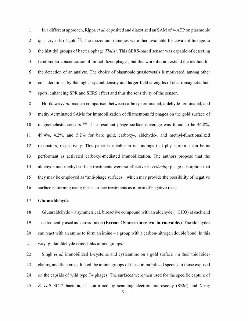
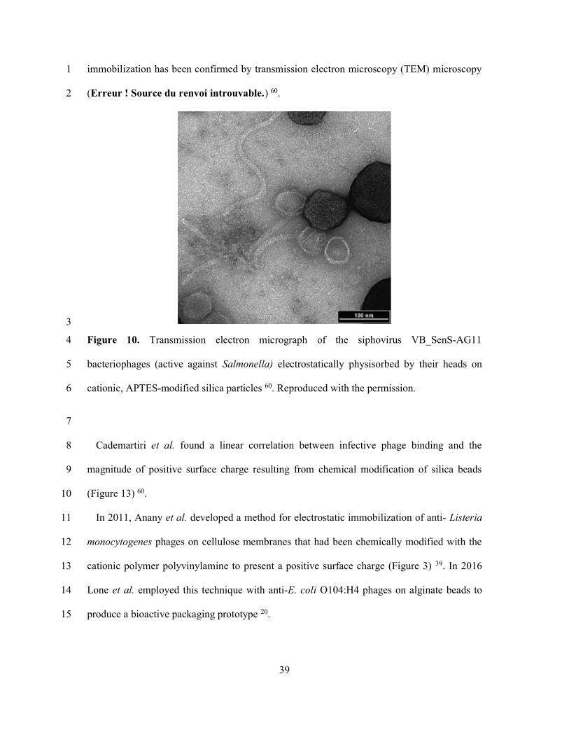
immobilization has been confirmed by transmission electron microscopy (TEM) microscopy 1
(Erreur ! Source du renvoi introuvable.) 60. 2
3
Figure 10. Transmission electron micrograph of the siphovirus VB_SenS-AG11 4
bacteriophages (active against Salmonella) electrostatically physisorbed by their heads on 5
cationic, APTES-modified silica particles 60. Reproduced with the permission. 6
7
Cademartiri et al. found a linear correlation between infective phage binding and the 8
magnitude of positive surface charge resulting from chemical modification of silica beads 9
(Figure 13) 60. 10
In 2011, Anany et al. developed a method for electrostatic immobilization of anti- Listeria 11
monocytogenes phages on cellulose membranes that had been chemically modified with the 12
cationic polymer polyvinylamine to present a positive surface charge (Figure 3) 39. In 2016 13
Lone et al. employed this technique with anti-E. coli O104:H4 phages on alginate beads to 14
produce a bioactive packaging prototype 20. 15

40
Similarly, Zhou et al. used polyethylenimine (PEI) to impart a positive surface charge to a 1
carbon nanotube (CNT)-modified glassy carbon electrode, for the specific detection of E. Coli 2
B 54. In this case T2 phages were electrostatically oriented by the positive surface charge, while 3
immobilization was achieved through PBSE-mediated conjugation (see π - π stacking). 4
Vonasek et al. similarly used PEI to electrostatically immobilize T7 phages on electrospun 5
cellulose microfibers for biocontrol in the context of food packaging and biomedical 6
applications 42. More recently, Farooq et al. used PEI to phage functionalize CNT-modified 7
bacterial cellulose for the electrochemical detection of S. aureus 112. 8
Poly(diallyl dimethylammonium chloride) or polyDADMAC is a cationic polyelectrolyte 9
that has been used to electrostatically bind T4 phages on paper 134, M13 phages on glass 208, 10
and S13’ phages on gold 71; in the latter case for the specific detection of S. aureus. 11
An alternative strategy to impart a positive charge to the substrate was demonstrated by 12
Richter et al. wherein a positive potential was applied to a gold substrate, yielding an oriented 13
physisorbed layer of T4 phages with a density of 14.3 PFU/mL (Figure 11) 78. 14
15 Figure 11. Due to the inherent dipole moment of T4 phage particles, an externally-applied 16
electric field orients the phages such that their tail fibers point outward from the substrate. Such 17

41
an orientation is considered essential for increasing the immobilized phages’ infectivity and 1
host capture efficiency. Adapted from Richter et al. 78. 2
3
In the same paper, Richter et al. proposed a model of orientation of bacteriophages based on 4
electrical screening of the substrate surface due to the formation of an electric double layer 78. 5
Briefly, the Debye length (denoted κ-1) refers to the physical distance in an electrolyte solution 6
over which an electrical potential will decrease by a factor of 1/e. This length is proportional 7
to the inverse square root of the ionic strength of the solution, since it is the mobile charge 8
carriers of the solution which form an electric double layer, screening the electric field that 9
results from the substrate surface potential. Richter et al. proposed that when κ-1 is larger than 10
the phage, they will align along electric field lines; whereas when κ-1 is smaller, the phage 11
orientation occurs instead due to electrostatic interactions (Figure 12). 12
13
14
Figure 12. The electrostatic binding model proposed by Richter et al. 78. At low ionic strength, 15
fewer charge carriers are available to screen the surface charge. This leads to a larger Debye 16
length of the same scale as a phage particle and thus oriented immobilization. Conversely, at 17
high ionic strength the surface charge is more effectively screened, leading to a Debye length 18

42
smaller than the phage. In the latter case, the phages are still attracted to the substrate due to 1
their overall negative charge, but the immobilization is not appreciably oriented. 2
3
The ionic strength also influences the stability of the phage solution and aggregation of the 4
phages73–75. Archer and Liu reported that physisorbed T4 phages are susceptible to aggregation 5
in high ionic strength solutions (>100mM) or low pH 93. However, the same study observed 6
phage immobilization even on negatively charged surfaces, indicating that interactions other 7
than purely electrostatic attraction might be at play. Confusingly, a different paper found that 8
a relatively high ionic strength of 420 mM NaCl gave the best surface coverage for 9
physisorption of filamentous phage fd 26. These seemingly contradictory results suggest that 10
phage morphology may play a role in immobilization and aggregative effects of different ionic 11
strengths. 12
Whatever the mechanism, while simple physisorption of phages has been demonstrated to 13
enable bacterial capture 198, it is not sufficient to prevent phages from migrating on the substrate 14
while in storage or during binding assays 166. Covalent attachment of phages offers a much 15
stronger bond than simple physisorption, yielding surface functionalization more resistant to 16
phage detachment and resulting in a higher phage surface density 92. 17

43
1
Figure 13. Left: A neutral surface charge leads to non-oriented adsorption of phages on silica 2
particles. Right: A polymer layer (e.g. X or X) is protonated as neutral pH, electrostatically 3
binding the phage head, leading to oriented immobilization. Adapted from Immobilization of 4
bacteriophages on modified silica particles 60. 5
6
π - π stacking 7
Zhou et al. demonstrated a novel strategy for immobilization of T2 phages on multiwall 8
carbon nanotubes (CNT) on glassy carbon electrodes 54. First, the CNT are functionalized with 9
polyethylenimine (PEI), which introduces a positive charge to the CNT surface, facilitating 10
tail-outward phage orientation. A hetero-bifunctional molecular tethering agent, 1-11
pyrenebutanoic acid succinimidyl ester (PBSE) (Erreur ! Source du renvoi introuvable.), is 12
then used to link the phages to the CNT. PBSE features four aromatic rings which interact with 13
CNT sidewalls through π-π stacking 209, and a succinimidyl group which facilitates amide 14
44
bonding 54. Application of a positive potential of +0.5 V vs. Ag/AgCl further facilitates oriented 1
phage immobilization. 2
Kim et al. leveraged π- π stacking between the imidazole rings of recombinantly expressed 3
histidine tags on M13 phages (see Genetic modification below) to achieve a nematically 4
aligned unidirectional bundle structure on the surface of an SPR biosensor 210. The resultant 5
colorimetric sensor was capable of detecting streptavidin – a surrogate analyte in this work – 6
down to femtomolar concentrations. 7
Biotinylation 8
The biotin-avidin bond is one of the strongest non-covalent bonds in nature, with a 9
dissociation constant as low as 10-15 M 90. The binding is also extremely stable, exhibiting high 10
resistance to breakdown by extremes of pH, temperature, or the presence of denaturants or 11
detergents 211. Avidin is a tetrameric protein, binding up to four biotin molecules 12
simultaneously. These qualities have made the avidin-biotin interaction particularly useful and 13
widespread in bioconjugation chemistry, and it is frequently used to cross-link biotinylated 14
ligands to one another as well as to substrates. 15
While avidin is a component of egg-whites, streptavidin is a similar biotin-binding protein 16
of bacterial origin which exhibits superior properties to avidin. The main disadvantage of 17
avidin is its high isoelectric point of 10, presenting a positive charge at neutral pH making it 18
susceptible to non-specific binding to negatively charged components other than biotin, such 19
as cell surfaces for example 90. This problem is circumvented by instead using streptavidin 20
which has a pI of 5-6, which greatly reduces nonspecific binding due to ionic interactions with 21
non-analyte molecules. For this reason, streptavidin has largely replaced avidin in most (but 22
not all) conjugation protocols. 23
The use of streptavidin-biotin heterobifunctional cross-linkers has been demonstrated for 24
phage immobilization using sulfo-NHS-ss-biotin and sulfo-NHS-biotin (Erreur ! Source du 25

45
renvoi introuvable.). These molecules can participate in an amide bond with the phage capsid 1
– facilitated by a terminal succinimidyl group on one end – tagging the phage with a terminal 2
biotin group which can bind to a streptavidin-functionalized surface. Such covalent 3
biotinylation of the phage was used by Sun et al. to immobilize of SJ2 phages on streptavidin-4
capped magnetic beads to create a biosorbent for the specific capture of Salmonella Enteritidis 5
105. 6
Sulfo-NHS-ss-biotin differs from sulfo-NHS-biotin in that the former features a spacer to 7
mitigate steric hindrance effects, and a cleavable disulfide bon. This latter molecule has been 8
used by Fernandes et al. to biotinylate a gold substrate, which was then crosslinked via 9
streptavidin to genetically biotinylated T4 phages 61. 10
Despite the above successes seen with covalent biotinylation, all immobilization mediated 11
by streptavidin-biotin since 2001 has instead made use of genetic biotinylation (see Genetic 12
biotinylation) 13
Genetic modification 14
Genetic engineering can be used either as an alternative immobilization strategy in itself or 15
to enhance the above chemi- and physisorption methods. A popular technique in the literature 16
is site-directed mutagenesis, wherein a foreign coding sequence is spliced in-frame into 17
bacteriophage capsid protein genes. This allows the targeted expression of “guest” peptides 18
which are fused to the coat protein and which exhibit an affinity for a given substrate, or onto 19
which a moiety can be conjugated 212. Since filamentous phages in the Ff class (e.g. fd and 20
M13) typically have several thousand identical, helically-tessellated copies of the pVIII coat 21
protein 213, they are the most frequent examples of genetic modification for the purpose of 22
immobilization 38,79,80,84–87,214, although examples of such modification of T4 81,83,88,89 and T7 23
215 phages are also found. 24

46
The popularity of this technique and of the filamentous phages M13 and fd, stems from the 1
success of phage display-based selection and the closely related technique of biopanning 212. 2
While the 50-residue pVIII protein of filamentous phages are typically modified 212–214 (but 3
also pIII 79), in the case of phages T4 and T7 it is rather Soc and Hoc proteins of the phage head 4
that are targeted for modification 81,216,217. 5
A drawback of genetic modification is that the expression of peptides, especially of large 6
size, can interfere with phage assembly and hence its physical and infective properties 93. 7
Genetic modification can also prove laborious, time-consuming and expensive 88, and for these 8
reasons genetic modification techniques are somewhat marginal compared to physisorption 9
and covalent techniques. 10
Genetic biotinylation 11
Several papers demonstrate phage immobilization facilitated by streptavidin/biotin, wherein 12
the biotinylation of the target phages is a result of recombinant expression of a biotin tag rather 13
than the result of chemical modification. 14
In 2006, Edgar et al. genetically modified T7 bacteriophage to exhibit a small peptide on the 15
major capsid protein, which is then post-translationally biotinylated at a specific lysine residue 16
by the host bacterium’s biotin-ligase protein during phage assembly 82. This allows conjugation 17
to streptavidin-coated quantum dots, resulting in a biosensor with a LOD of as low as 10 18
bacterial cells/mL. 19
Gervais et. al demonstrated an impedance-based biosensor by immobilizing T4 phages on 20
streptavidin-coated gold at a density of 4.4 phage/µm2, with tail-outward orientation enabled 21
by the strategic genetic biotinylation of only the head capsid protein 88. Phages were found to 22
retain their infectivity, burst size and latent period compared to the wild-type. Attractive 23
features of this method are the retention of infective activity after biotinylation, and the 24
47
localization of the modified tagged peptide exclusively on the phage head, leading to a 15-fold 1
increase in phage attachment to the gold surface. 2
Wang et al. immobilized a genetically biotinylated T4 phage to streptavidin-terminated 3
magnetic beads, calculating that each bead was on average conjugated to 248 ± 15 phages per 4
2.8 µm-diameter bead after 12 hours of incubation 48. 5
6 7
Formylglycine 8
Kwak et al. used genetic engineering to introduce a cysteine-containing peptide motif to the 9
pIII coat protein of fd phage, which was converted to formylglycine by formylglycine-10
generating enzyme (FGE) (Figure 14). This yields a reactive aldehyde (-CHO) group that is 11
then available for immobilization chemistry, for example to form a Schiff base with primary 12
amines on a silanized substrate surface 79. As with genetic biotinylation, an advantage of this 13
technique is that the formylglycine modification is site-specific and can be targeted to 14
exclusively modify the pIII coat protein which is displayed at one extremity of filamentous 15
phage fd. This allows the pVIII coat protein to be preferentially presented to the solution, since 16
the phage is immobilized in an upright orientation 79. This method has been further developed 17
to enable surface patterning of filamentous phages with 200µm spot size as a template for 18
organic and inorganic materials 80. 19
20
21
22
23
24

48
1
Figure 14. Recombinant expression of a specific peptide motif exclusively on the pIII coat 2
protein of an M13 phage enables enzymatic modification of cysteine residues to yield 3
formylglycine and its associated carboxyl side-chains. Formylglycine can then undergo amide 4
bonding with an amino-silanized surface. This oriented immobilization yields a high density 5
of pVIII coat proteins displayed at the surface. Adapted from Kwak et al. 79. Not to scale 6
Carbohydrate-binding modules 7
Carbohydrate-binding modules, also known as cellulose-binding modules (CBM), are short 8
protein domains normally found within carbohydrate-active enzymes, that exhibit high affinity 9
for a given substrate. Fusion of phage capsid and CBM genes results in expression of 10
recombinant CBM on the phage capsid. This allows oriented immobilization of the phages on 11
cellulosic substrates such as paper or cellulose beads. Tolba et al. demonstrated such 12
modification of T4 phages which, when combined with polymerase chain reaction (PCR), 13
resulted in an assay with a LOD of 800 cells/mL 83. A similar method allowed immobilization 14
of T4 phages on microcrystalline cellulose beads 89. 15
Polymer and other binding domains 16

49
Short peptides have also been identified that exhibit highly specific affinities against a range 1
of materials. Sawada et al. have demonstrated the immobilization of M13 phages on isotactic 2
poly(methyl methacrylate) (it-PMMA) via an (it-PMMA)-binding terminal peptide expressed 3
on the pIII coat protein 86. This result is intriguing since it demonstrates a potentially fruitful 4
avenue for further research. As the same authors point out in another paper 128, similar peptides 5
have been identified that exhibit binding specificity towards targets as varied as metals (silver 6
218, titanium 219, platinum 220), metal oxides (iron oxide 221, magnetite 222), silica 223, semi-7
conductors 224,225, carbon allotropes (nanotubes 113,114, nanohorns 115, and fullerenes 116), and 8
various polymers (polystyrene and polyvinyl chloride 123, nano-imprinted methacrylate 9
polymer 124, chlorine-doped polypyrrole 125, PMMA 126, syndiotactic PMMA 127, syndiotactic 10
polystyrene 129). Thus, the successful demonstration of one of these peptides as a strategy for 11
M13 phage immobilization, presents a compelling case for the use of substrate-specific 12
peptides as a general phage grafting strategy. 13
Click chemistry 14
An alterative class of reactions, thusfar seldom exploited for phage immobilization, is click 15
chemistry. Click reactions proceeds quickly at room temperature, producing strongly stabalized 16
products and either no byproducts or only water as a byproduct 226. Furthermore, click reactions 17
are highly tolerant to water and in some cases even accelrated in acqueous solutions. 18
Click chemistry is therefore a popular choice for making carbon−heteroatom−carbon bonds 19
in aqueous solutions in a wide variety of chemical and biological applications in drug 20
discovery, chemical biology, and proteomic applications227. 21
An elegant method of phage capsid modification was recently demonstrated by Zurier et al. 22
wherein a genetically engineered variant of T4 known as NRGp17 was decorated with a self-23
assembled cage of 870 small outer capsid (soc) proteins engineered with unnatural alkynylated 24
amino acids 81. These alkynes enable oriented immobilization of the phage heads on azide-25
50
terminated magnetic beads228,229 (TurboBeads, Zurich, SUI) via click chemistry, creating a 1
biosorbent that can concentrate target bacteria from a very low initial concentration. NRGp17 2
causes expression of a nanoluciferase (Nluc) reporter enzyme fused to a cellulose-binding 3
module (CBM) 230. The bioluminescent activity of Nluc serves to transduce the presence of 4
their host E. coli into a luminescence signal, while the fused CBM enables immobilization and 5
concentration of the reporter enzyme on a nitrocellulose membrane to improve sensitivity. This 6
assay presents an impressive LOD of <10 CFU E. coli in 100 mL of tap water 7
8
Optimization of the immobilized bacteriophage layer 9
In discussing the targeted immobilization of bacteriophage particles, we must also confront 10
the issue of immobilization of undesirabe interferents which will have a significant impact on 11
the quality of the immobilized layer both during preparation as well as in its final application, 12
be it an immoblized interaction layer on a biosensor transducer or a biosorbent for bio-active 13
food packaging. The purification protocol immediately preceding conjugation can have a large 14
effect on the success, homogeneity, and overall quality of the immobilized phage layer 231, and 15
is thus of critical importance and an integral part of any phage immobilizaiton protocol. In 16
addition, following immobilization, an additional blocking step 17
18
Phage propagation and purification methods 19
The process of phage propogation (also referred to as phage amplification) necessarily 20
generates large amounts of bacterial debris (e.g. DNA, peptidoglycan, lipopolsaccharide232 21
etc.) which must be separated from the phage particles, since many immobilization techniques 22
will equally – if not preferentially – bind these contaminants to the surface along with the 23
phages of interest 231. Furthermore, storage of phages in the presence of certain cellular debris 24
can trigger premature ejection of phage genetic material233–235, potentially rendering the phage 25

51
inactive. For these reasons, immobilization of phages requires first the isolation of a high-1
purity, high-titer phage suspension. Typical phage concentrations used for immobilizations 2
range between 1010 and 1012 plaque-forming units per milliliter (PFU/mL), although some 3
success has been seen with lower infective titers. The lower phage concentrations in this range 4
tend to be biased towards myoviruses such as T4 and podoviruses such as P22, while the higher 5
concentrations are almost exclusively seen in papers using the filamentous phages M13 and fd. 6
The topic of phage purification necessitates a review in its own right, but the main methods 7
will be outlined briefly here. 8
Dead-end filtration through 0.45µm and 0.2µm pore-size membranes is typically employed† 9
– along with differential centrifugation236 – as an initial first purification step to rid phage 10
suspensions of the largest bacterial debris and leftover culture medium constituents. 11
Differential centrifugation can be used to pellet contaminants brought out of solution using 12
chemical precipitation (e.g. using PEG and/or NaCl)236,237. It is carried out at relatively low 13
centrifugal force of ~5000g, in larger volumes (between 1 and 1000 mL), and in simple 14
centrifugation media (typically simply the phage buffer) compared to ultracentrifugation. 15
In contrast, in density gradient ultracentrifugation (DGU) the components of a sample are 16
fractionated based on their buoyant density. DGU necessitates centrifugation at very high 17
acceleration (>70-80,000g for 4-6 hours is typical 238), smaller volumes (~6 mL), and the 18
careful selection and preparation of centrifugation media such as CsCl or sucrose239. DGU is 19
performant enough to permit the separation of terminated viruses from empty ghost particles 20
240. Potential drawbacks of DGU include the high cost of ultracentrifuge equipment, the 21
potential for phage aggregation due to high ionic strength of centrifugation media 93, and the 22
difficulty in harvesting the final product. DGU purification has also been found to lose un-23
† Unfortunately, such filtration can immediately reduce the infectious titer by a factor of 10.

52
tailed phages 241 and can yield solutions contaminated by – for example – large quantities of 1
host bacterium flagella fragments 58,67. Despite these disadvantages, the use of DGU has 2
become an established technique for the isolation and purification of virus particles 78,240,242,243. 3
PEG precipitation – either employed on its own or in conjunction with DGU 239 – is another 4
popular method for concentration and purification of viruses 237,244–247. PEG, a polymerized 5
form of ethylene glycol (used at various molecular weights 238 but most often at 6 kD 237,248–250 6
for phage purification), is employed as a fractional precipitating agent which separates proteins 7
by virtue of their solubility. PEG acts as an inert solvent sponge, reducing water availability. 8
With increasing concentration of PEG the effective protein concentration is increased until 9
solubility is exceeded and precipitation occurs. Thus, larger proteins will precipitate at lower 10
concentrations of PEG and can be separated with differential centrifugation 251,252. PEG 11
precipitation is considered by some experts to be “a crude and non-specific technique” 252. 12
An increasingly popular suite of purification methods involve various forms of 13
chromatography 253. Boratynski et al. used size-exclusion chromatography (SEC) to produce 14
phage suspensions with endotoxin levels low enough to permit intravenous administration 232. 15
SEC can also be used as part of a short-turnaround purification protocol, used by Naidoo et al. 16
to avoid CsCl DGU and PEG precipitation, giving a total process time of only 3 hours 58. 17
Recombinant affinity tags expressed on T4 capsids have been used by Ceglarek et al. to 18
facilitate affinity chromatography 253. While elegant, this method relies on genetic modification 19
of each host bacterial strain which may be infeasible for some researchers and applications. 20
Adriaenssens et al. demonstrated anion-exchange chromatography (AEC) for the purification 21
of eleven morphologically distinct phages. The authors concluded that although this method 22
presents easier scalability for industrial-scale throughput compared to CsCl DGU, it requires 23
more laborious optimization of purification at the outset 254. 24
Surface blocking 25

53
Due to their inherent charge – and often their hydrophobicity – proteins have a tendency to 1
spontaneously adhere to surfaces in an aqueous environment. Extremely pure reagents and 2
clean surfaces are a sine qua non for well-controlled and reproducible fabrication of biosensors 3
and bioactive surfaces. However, a clean surface is also susceptible to non-specific binding of 4
proteins in aqueous solutions. Non-specific binding of interferents can hinder specific 5
biofunctionalization, obscure the transduction of specific interaction events and decrease the 6
signal-to-noise ratio (SNR) in the case of biosensors, and generally compromise 7
reproducibility, even when phage infectivity is not impacted 51. 8
For this reason, a common unit operation in phage-functionalization is to follow conjugation 9
with a blocking step, whereby the surface is flooded with an amphiphilic biological species 10
(e.g. casein, salmon sperm DNA, bovine serum albumin (BSA)) or synthetic polymer (e.g. 11
PEG) that will occupy non-functionalized sites on the surface without impeding the sensitivity 12
of the functionalized sites. In this way, the non-specific binding of proteins – and other 13
components of the sample – is reduced, while specific reactions with the target analyte are 14
(ideally) unaffected. If a researcher finds that they have trouble with non-specific binding to 15
their phage-functionalized substrate, surface blocking may be an easy first step to boost 16
performance. 17
PEG and casein have also been investigated for surface blocking and have in some cases 18
been found to be more performant than BSA 192,255. However, incubating with BSA remains – 19
by a wide margin – the most popular strategy for passivation of biofunctionalized surfaces to 20
reduce or even eliminate non-specific binding, due to its low cost and its ease of storage, 21
preparation, and use. 22
23
Conclusion and Remarks 24

54
Since the first papers on phage immobilization began to appear at the turn of the century 1
105,256, a variety of strategies have been developed. By far the most popular single method relies 2
on simple physisorption of phages to the substrate (usually a gold layer) without any chemical 3
modification of the surface, with over one third of papers making use of this method. Although 4
such easily implemented methods may be seductive, physisorbed phages have been shown to 5
easily detach following changes in ionic strength, temperature, and pH at the substrate surface 6
92, when compared to covalent immobilization. 7
Covalent conjugation of phages has been demonstrated with a variety of techniques, but three 8
surface modifications in particular dominate: carbodiimide activation of surface carboxyl 9
moieties (i.e. by EDC and NHS); self-assembled monolayer formation of thiol linkers (e.g. 11-10
MUA, L-cysteine, and DTSP); and amino-silanization (e.g. APTES, APTMS). Together, these 11
chemistries cover half of all the reviewed literature on phage conjugation, and each relies on 12
heterofunctional linkers with at least one terminal functional group (carboxyl (-COOH), or 13
primary amine (-NH2)) that engages in amide bonding with the phage capsid. 14
Genetic modification-based methods compose a significant minority of the immobilization 15
techniques demonstrated in the literature, being used in about 10% of all papers reviewed. 16
Genetic engineering of phages may prove time-consuming and laborious compared to 17
physisorption or covalent conjugation 88, but may prove practical in some instances. 18
Nevertheless, new avenues for immobilization via display of recombinant phage coat proteins 19
may be revealed as a result of the discovery of a plethora of peptides exhibiting binding 20
specificity for a variety of substrates including metals218–220 and their oxides221,222, silica 223, 21
semi-conductors 224,225, carbon allotropes 113–116, and several polymers 123–129. 22
The protocols presented in many papers omit a surface-blocking step to prevent non-specific 23
binding, despite ample evidence in the literature that such a step increases sensitivity and 24
overall performance in the case of biosensors. Surface blocking can be as simple as soaking a 25
55
substrate for 30 minutes in a 1% BSA solution, which is unlikely to prove too onerous to be 1
incorporated into any workflow. 2
Regarding the purity of phage suspensions, since the purification protocol immediately 3
preceding immobilization can have a large effect on the success, homogeneity, and overall 4
quality of the immobilized phage layer, researchers may have difficulty replicating and 5
comparing results in the literature where the purification protocol has not been stated explicitly. 6
Many papers do not clearly describe the purification procedure for the bacteriophages used, 7
particularly when these phages have been obtained from collaborators. Historically, a large 8
impediment to the widespread adoption of phage therapy in the west was the lack of 9
standardization of phage preparation procedures or the criteria for purity and potency 10. In 10
order to facilitate greater reproducibility of results and comparison between studies, the authors 11
of this review would recommend inclusion of any purification procedures in future papers. 12
In conclusion, phage-functionalization of substrates presents many promising avenues for 13
the development of both novel bioactive surfaces and specific interaction layers within 14
biosensors. The performance of these phage-functionalized substrates depends on the 15
production of high-purity phage suspensions and careful consideration of the immobilization 16
technique employed. As the pernicious effects of antimicrobial resistance become more 17
apparent in the 21st century, bacteriophages and adjacent research are well-placed to play an 18
increasing role in biomedical, agricultural, and environmental monitoring applications for 19
many years to come. 20
21
Schemes 22

56
1
Scheme 1. The chemical reactions involved in EDC/NHS-mediated ligand conjugation. The 2
carboxyl group (−COOH) is activated with EDC/NHS (A. and B.), and then covalently bonds 3
to the primary amine of the target ligand (C.). The remaining NHS esters are blocked using 4
ethanolamine (D.). Here, R represents any arbitrary radical, in this case a bacteriophage. The 5
side group R1 is –CH2CH3 and R2 is –(CH2)3N+H(CH3)2Cl–. Adapted from Chapter 3 - Surface 6
plasmon resonance, Phillips and Cheng 152. 7
8
9
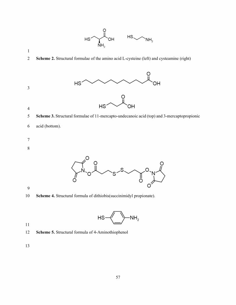
57
1
Scheme 2. Structural formulae of the amino acid L-cysteine (left) and cysteamine (right) 2
3
4
Scheme 3. Structural formulae of 11-mercapto-undecanoic acid (top) and 3-mercaptopropionic 5
acid (bottom). 6
7
8
9
Scheme 4. Structural formula of dithiobis(succinimidyl propionate). 10
11
Scheme 5. Structural formula of 4-Aminothiophenol 12
13
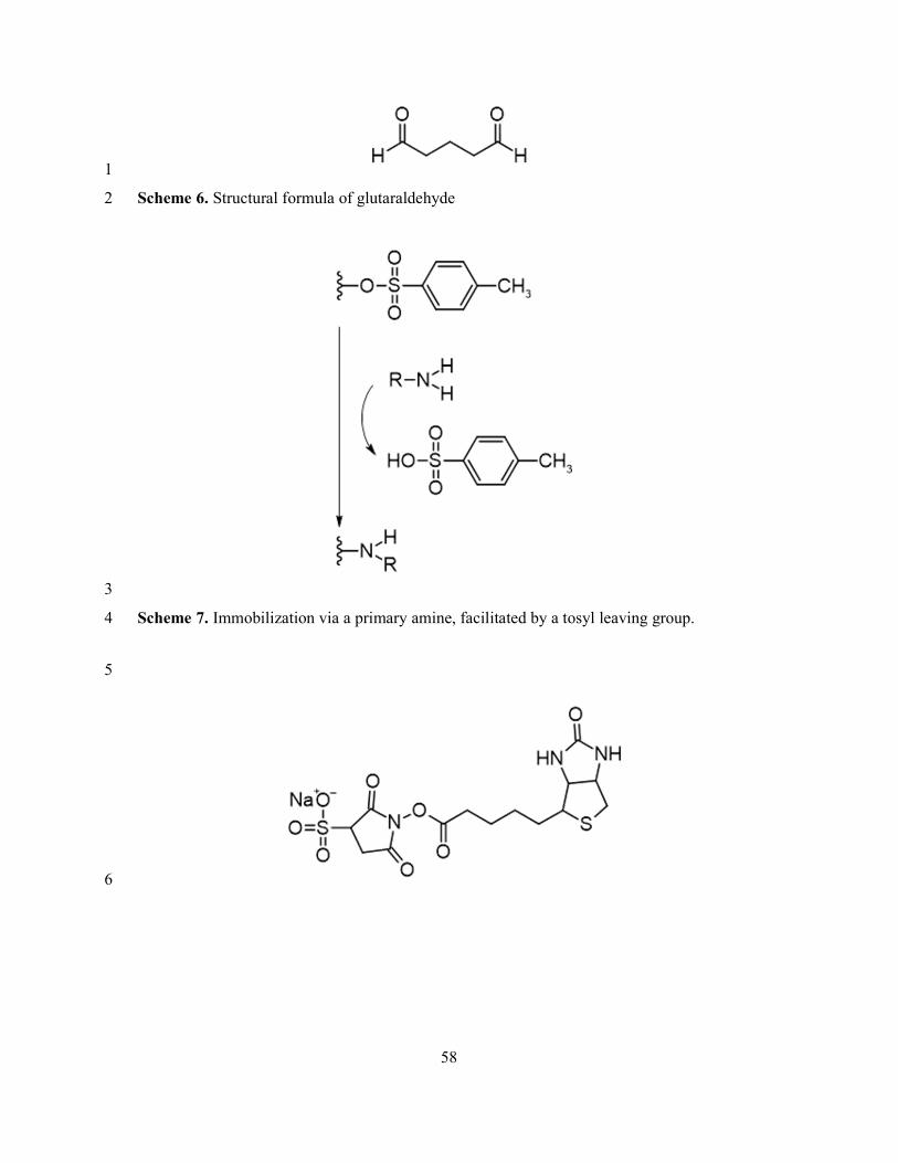
58
1
Scheme 6. Structural formula of glutaraldehyde 2
3
Scheme 7. Immobilization via a primary amine, facilitated by a tosyl leaving group. 4
5
6

59
1
Scheme 8. Structural formulae of and sulfo-NHS-biotin (top) and sulfo-NHS-ss-biotin 2
(bottom). 3
4
5
6
Scheme 9. Structural formula of 1-pyrenebutanoic acid succinimidyl ester (PBSE), illustrating 7
pi-stacking interactions between the pyrene moiety and a carbon nanotube sidewall 8
9
Tables 10
Table 1 11
Covalent phage immobilization techniques 12
13

60
Table 2 1
Physisorptive phage immobilization techniques 2
3
Table 3 4
Phage immobilization techniques based on genetic engineering 5
6
7
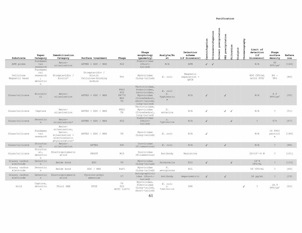
61
Purification
Substrate Paper
Category Immobilization
Category Surface treatment Phage
Phage morphology (geometry)
Analyte/Host
Detection scheme
(if biosensor) Centrifugation
Ultracentrifugation
Solvent precipitation
PEG precipitation
Filtration
Dialysis
Chromatography
Limit of detection
(if biosensor)
Phage surface density
Reference
AFM probe Fundamen
tal research
Amino-silanization
APTMS + EDC / NHS P22 Podoviridae
(Short-tailed)
N/A AFM ✓ ✓ N/A 40
PFU/μm2 [144]
Cellulose Magnetic bead
Fundamental
research,
detection
Streptavidin / biotin*
Streptavidin / biotin
Cellulose-binding module
T4† Myoviridae
(Long-tailed) E. coli
Magnetic separation +
qPCR
800 CFU/mL (with PCR)
84 - 98%
[80]
Glass/silicate Biocontr
ol Amino-
silanization APTES + EDC / NHS
PRD1 P22
PR772 MS2 T4
Tectiviridae, Podoviridae, Leviviridae, Myoviridae
(Icosahedral, short-tailed, long-tailed)
E. coli S.
Typhimurium
N/A ✓ ✓ N/A 4.5
PFU/μm2 [50]
Glass/silicate Capture Amino-
silanization APTES + EDC / NHS
PRD1 T4
Tectiviridae, Myoviridae
(Icosahedral, long-tailed)
S. enterica
N/A ✓ ✓ ✓ N/A ? [51]
Glass/silicate Detectio
n Amino-
silanization* APTMS + EDC / NHS P22
Podoviridae (Short-tailed)
S. Typhimurium N/A ✓ ✓ ? 67% [67]
Glass/silicate Fundamen
tal research
Amino-silanization,
Amino-silanization + electrostatic physisorption*
APTES + EDC / NHS T4 Myoviridae
(Long-tailed) E. coli N/A ✓ N/A
16 PFU/ particl
e [146]
Glass/silicate Structur
al Amino-
silanization APTES fd†
Inoviridae (Filamentous)
E. coli N/A ✓ ✓ N/A ? [88]
Glass/silicate
Structural,
detection
Electropolymerization
PEDOT M13 Inoviridae
(Filamentous) Antibody Resistive 20×10^-9 M ? [121]
Glassy carbon electrode
Detection
Amide bond EDC T4 Myoviridae
(Long-tailed) Salmonella EIS ✓ ✓ 10^4
CFU/mL ? [110]
Glassy carbon electrode
Detection
Amide bond EDC / NHS PaP1 Myoviridae
(Long-tailed) P.
aeruginosa ECL 56 CFU/mL ? [60]
Glassy carbon electrode
Detection
Electropolymerization
Pyrrole-alkyl ammonium
T7 Autographiviridae (Short-
tailed) Antibody Amperometric ✓ ✓ 36 pg/mL ? [29]
Gold Capture, detectio
n Thiol SAM DTSP
T4 P22
NCTC 12673
Myoviridae, Podoviridae
(Long-tailed, short-tailed)
E. coli S.
Typhimurium SPR ✓ ?
18.9 PFU/µm2
[63]
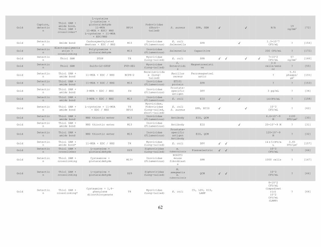
62
Gold Capture, detectio
n
Thiol SAM + amide bond, Thiol SAM + crosslinker*
L-cysteine L-cysteine +
gluteraldehyde 11-MUA
11-MUA + EDC/ NHS L-cysteine + 11-MUA
+ EDC/NHS
BP14 Podoviridae
(Short-tailed)
S. aureus SPR, SEM ✓ ✓ N/A 19
ng/mm2 [72]
Gold Detectio
n Amide bond
Carboxymethylated dextran + EDC / NHS
M13 Inoviridae
(Filamentous) E. coli
Salmonella SPR ✓ 1.3×10^7
CFU/mL ? [154]
Gold Detectio
n
Electropolymerisation +
crosslinking
Polytyramine + glutaraldehyde
M13 Inoviridae
(Filamentous) Salmonella Capacitive 200 CFU/mL ? [172]
Gold Detectio
n Thiol SAM DTSP T4
Myoviridae (Long-tailed)
E. coli SPR ✓ ✓ ✓ ✓ 7×10^2 CFU/mL
57 ng/mm2
[166]
Gold Detectio
n Thiol SAM Sulfo-LC-SPDP PVP-SE1
Myoviridae (Long-tailed)
S. Enteritidi
s
Magnetoresistive
3–4 cells/sens
or ? [55]
Gold Detectio
n Thiol SAM + amide bond
11-MUA + EDC / NHS BCP8-2 Herellevirida
e (Long-tailed)
Bacillus cereus
Ferromagnetoelastic
? 5.51
phages/µm2
[155]
Gold Detectio
n Thiol SAM + amide bond
11-MUA + EDC / NHS M13 Inoviridae
(Filamentous) ET101
protein SPR ? ? [153]
Gold Detectio
n Thiol SAM + amide bond
3-MPA + EDC / NHS fd Inoviridae
(Filamentous)
Prostate-specific antigen
DPV 3 pg/mL ? [34]
Gold Detectio
n Thiol SAM + amide bond
3-MPA + EDC / NHS M13 Inoviridae
(Filamentous) E. coli EIS ✓ ✓ 14 CFU/mL ? [158]
Gold Detectio
n Thiol SAM + amide bond
L-cysteine + 11-MUA + EDC / NHS
T4 BP14
Myoviridae, Podoviridae
(Long-tailed, short-tailed)
E. coli S. aureus
SPR, ECIS ✓ ✓ 10^3 CFU/mL
? [62]
Gold Detectio
n Thiol SAM + amide bond
NHS thioctic ester M13 Inoviridae
(Filamentous) Antibody EIS, QCM 6.6×10^-9
M 1100
PFU/µm2 [30]
Gold Detectio
n Thiol SAM + amide bond
NHS thioctic ester M13 Inoviridae
(Filamentous) Antibody EIS ✓ 20×10^-9 M ? [31]
Gold Detectio
n Thiol SAM + amide bond
NHS thioctic ester M13 Inoviridae
(Filamentous)
Prostate-specific antigen Antibody
EIS, QCM 120×10^-9 M
? [32]
Gold Detectio
n Thiol SAM + amide bond*
11-MUA + EDC / NHS T4 Myoviridae
(Long-tailed) E. coli DPV ✓ ✓ 14 ± 5 CFU/m
L 3.65
PFU/µm2 [157]
Gold Detectio
n Thiol SAM + crosslinker
L-cysteine + glutaraldehyde
D29 Siphoviridae (Long-tailed)
M. tuberculosis Piezoelectric ✓ ✓
10^3 CFU/mL
? [66]
Gold Detectio
n Thiol SAM + crosslinking
Cysteamine + glutaraldehyde
M13† Inoviridae
(Filamentous)
NIH3T3 mouse
fibroblasts
SPR 1000 cells ? [147]
Gold Detectio
n Thiol SAM + crosslinking
L-cysteine + glutaraldehyde
D29 Siphoviridae (Long-tailed)
M. smegmatis
M. tuberculosis
QCM ✓ ✓ 10^3
CFU/mL ? [66]
Gold Detectio
n Thiol SAM +
crosslinking*
Cysteamine + 1,4-phenylene
diisothiocyanate T4
Myoviridae (Long-tailed)
E. coli CV, LSV, EIS,
LAMP
8×10^2 CFU/mL
(impedimetric) 10^2
CFU/mL (LAMP)
? [64]

63
Gold Detectio
n Thiol SAM +
crosslinking*
DTSP Cysteamine +
glutaraldehyde T4
Myoviridae (Long-tailed)
E. coli Confocal
microscopy ✓ ✓ ✓ ✓ 10^2 CFU/mL
13.64 - 17.32 PFU/µm2
[167]
Gold Detectio
n
Thiol SAM + streptavidin /
biotin*
Sulfo-NHS-SS-biotin + streptavidin /
biotin T4†
Myoviridae (Long-tailed)
E. coli ECIS ✓ ✓ ? 4.4
PFU/µm2 [85]
Gold
Detection,
fundamental
research
Thiol SAM
carboxy-terminated SAM
aldehyde-terminated SAM
methyl-terminated SAM
fd-tet Inoviridae
(Filamentous) Microbeads Magnetoelastic 3.6×10^5
beads/mL 5.2 - 49.4%
[169]
Gold
Fundamental
research,
capture
Thiol SAM + crosslinking
Histidine + glutaraldehyde L-cysteine +
glutaraldehyde Cysteamine +
glutaraldehyde
T4 Myoviridae
(Long-tailed) E. coli SPR ✓ ✓ ✓ ✓ N/A
18 PFU/µm2
[41]
Gold Structur
al Electropolymeriz
ation PEDOT M13
Inoviridae (Filamentous)
Prostate-specific antigen
N/A N/A 7.2
μg/cm2 [33]
Gold (plasmonic quasicyrstal)
Fundamental
research Thiol SAM 4-ATP Tbilisi
Podoviridae (Short-tailed)
Brucella abortus
SERS ✓ ✓ ✓ ✓ ? ? [70]
Gold Glass/silicate
Fundamental
research,
biocontrol
Amino-silanization Thiol SAM + amide bond
11-MUA + EDC / NHS (on gold)
APTES (on glass) FL-1
Myoviridae (Long-tailed)
Flavobacterium
columnare N/A ✓ ✓ ✓ N/A
0.5 PFU/μm2
[95]
Indium tin oxide
Biocontrol
Amino-silanization
APTES APTES + succinic
anhydride Octadecyltrimethoxy
silane
T4 Myoviridae
(Long-tailed) E. coli N/A N/A ? [174]
Indium tin oxide
Fundamental
research
Amino-silanization
APTES APTES + succinic
anhydride Octadecyltrimethoxy
silane
T4 Myoviridae
(Long-tailed) E. coli N/A N/A
~25 - 200
PFU/µm2 [173]
Magnetic bead Capture Crosslinking* Bissulfosuccinimidy
l suberate P100
(Listex) Myoviridae
(Long-tailed)
L. monocytoge
nes N/A N/A ? [61]
Magnetic bead Capture, detectio
n Amide bond
Carboxyl-activation + EDC / NHS
Tosyl-activation P22
Podoviridae (Short-tailed)
Salmonella
Magnetic separation +
optical reporter
✓ ✓ ✓ ✓
19 CFU/mL (without
preenrichment) 0.06
CFU/mL (with pre-enrichment
)
1650 PFU/bea
d [69]
Magnetic bead Capture, detectio
n Amide bond EDC / NHS T4
Myoviridae (Long-tailed)
E. coli Magnetic
separation + impedimetric
✓ ✓ ✓ ✓ 10^3 CFU/mL
? [103]
Magnetic bead Capture, detectio
n Amide bond EDC / NHS T7
Autographiviridae (Short-
tailed) E. coli
Magnetic separation +
LSV
10^5 CFU/mL
? [49]

64
Magnetic bead Capture, detectio
n Amide bond EDC / NHS T7
Autographiviridae (Short-
tailed) E. coli Colorimetric ✓
1×10^4 CFU/mL
(without preenrichm
ent) 10 CFU/mL (with pre-enrichment
)
1871 PFU/bea
d [47]
Magnetic bead Capture, detectio
n Misc.* Tosyl-activation P22
Podoviridae (short-tailed)
Salmonella Magnetic
separation + PCR
✓ ✓ ✓ ✓ 3 CFU/mL
(with PCR)
2000 PFU/bea
d [68]
Magnetic bead Capture, detectio
n Thiol SAM Isothiocyanate O157-IOV-4 ?
E. coli O157:H7
Magnetic separation + colorimetric
✓ ✓ ✓ ✓ 4.9×10^4 CFU/mL
? [104]
Metal-organic framework
Detection
Crosslinking Glutaraldehyde ? ? S.
arlettae Fluorescence ✓ ✓ ✓ 10^2
CFU/mL ? [57]
Metal-organic framework
Detection
Crosslinking* Glutaraldehyde ? Siphoviridae (Long-tailed)
S. aureus Photoluminescence quenching ✓ ✓ ✓ ✓ 31 CFU/mL ? [58]
Optical fiber Detectio
n
Amino-silanization + crosslinking
APTES + glutaraldehyde
T4 Myoviridae
(Long-tailed) E. coli SPR ✓ ✓
10^3 CFU/mL
? [141]
Optical fiber Detectio
n
Amino-silanization + crosslinking
APTES + glutaraldehyde
T4 Myoviridae
(Long-tailed) E. coli
Optical mode interference ✓ ✓ 10^3 CFU/mL ? [142]
Polycaprolactone
Biocontrol
Amide bond* vB_Pae_Kakheti25
Siphoviridae (Long-tailed)
P. aeruginosa
N/A N/A 1 phage per 118
nm [41]
Polyethersulfone
Biocontrol
Amide bond* EDC / NHS T4 Myoviridae
(Long-tailed) E. coli N/A ✓ ✓ N/A
2 PFU/µm2
[86]
Polyhydroxyalkanoate
Fundamental
research,
capture
Amide bond EDC / NHS T4 Myoviridae
(Long-tailed) E. coli N/A ✓ ✓ N/A
4.24 PFU/μm2
[40]
Polystyrene latex beads
Detection
Amide bond EDC / NHS M13 Inoviridae
(Filamentous) P.
aeruginosa Micro-Raman spectroscopy
10^3 CFU/mL
? [119]
Screen-printed carbon
electrode
Detection
Amide bond EDC ? Siphoviridae (Long-tailed)
S. arlettae
EIS ✓ ✓ 2 CFU ? [111]
Screen-printed carbon
electrode
Detection
Crosslinking Glutaraldehyde Gamma phage Siphoviridae (Long-tailed)
B. anthracis spores
Impedimetric 10^3 CFU/mL
? [65]
Silica nanoparticle
Fundamental
research,
biocontrol
Amino-silanization + crosslinking
Amino-silanization + electrostatic Physisorption*
APTMS AEAPTMS +
glutaraldehyde ?
Siphoviridae (Long-tailed)
S. aureus N/A ✓ ✓ ✓ N/A ? [56]
Silicon Nitride (Si3N4)
Detection
Amino-silanization + crosslinking
APTES + glutaraldehyde
? ? Cancer
biomarkers Potentiometric ? ? [37]
Silver Detectio
n Thiol SAM + crosslinking
4-ATP + glutaraldehyde
T4 Myoviridae
(Long-tailed) E. coli SERS ✓ ✓ 1.5×10^2
CFU/mL ? [168]
N/A Not applicable
% Surface coverage
† Phage was genetically engineered

65
* Oriented immobilization
Purification
Substrate Paper
Category Immobilization
Category Surface
treatment Phage Phage morphology
(geometry) Analyte/Host
Detection scheme
(if biosensor) Centrifugation
Ultracentrifugation
Solvent precipitation
PEG precipitation
Filtration
Dialysis
Chromatography
Limit of detection
(for biosensors)
Phage surface density
Carbon nanotubes Detection Electrostatic Physisorption*
Polyethylenimine ? ? S. aureus CV, DPV ✓ ✓ 3 CFU/mL 11.7
PFU/μm
Carboxymethylcellulose (paper)
Detection Amide bond*
StyM-AG6 SenS-AG11 LmoM-AG20 EcoM-AGl0
T4 MS2 rV5 AG2A
vB_SnwM CGG4-1
Myoviridae, Herelleviridae, Siphoviridae, Leviviridae, Podoviridae
(Long-tailed, icosahedral, short-tailed)
E. coli O157:H7 E. coli O45:H2
Salmonella L.
monocytogenes
qPCR ✓ ✓ 10–50 CFU/mL
0.01 PFU/µm
Cellulose Biocontrol Electrostatic Physisorption*
Cibacron Blue F3GA
Polyethylenimine Chitan
T7 Autographiviridae (Short-tailed)
E. coli N/A N/A ?
Cellulose Biocontrol Electrostatic Physisorption*
Polyvinylamine
LinM-AG8 LmoM-AG13 LmoM-AG20 EcoM-HG2 EcoM-HG7 EcoM-HG8
(LISTEX P100 commercial cocktail)
Myoviridae (Long-tailed)
L. monocytogenes
E. coli O104:H4
N/A N/A ?
Cellulose Biocontrol Electrostatic Physisorption*
Polyvinylamine
T4 EcoM-AG2 SboM-AG3 LinM-AG8
Myoviridae, Siphoviridae (Long-tailed)
L. monocytogenes
E. coli O157:H7
N/A ✓ N/A ?
Cellulose Biocontrol Physisorption
BFSE16 BFSE18 PaDTA1 PaDTA9 PaDTA10 PaDTA11
? S. Typhimurium N/A N/A ?
Cellulose (paper) Fundamental research
Physisorption T4 Myoviridae (Long-
tailed) E. coli N/A N/A ?
Cellulose (paper) Fundamental research
Physisorption T4 Myoviridae (Long-
tailed) E. coli Colorimetric N/A ?

66
Glass/silicate Detection Electrostatic Physisorption
Poly-L-lysine PRD1 E79
Tectiviridae, Myoviridae
(Icosahedral, long-tailed)
P. aeruginosa S. Typhimurium
QCM w/ dissipation monitoring
✓ ✓ ✓ ? 1.6 -2.2
μg/cm2
Glass/silicate Fundamental research
Amino-silanization + electrostatic physisorption
APTES fd-tet Inoviridae
(Filamentous) E. coli N/A ✓ ✓ ✓ N/A ?
Glass/silicate Fundamental research
Amino-silanization + electrostatic physisorption
ED3A PEG APTS
VB_EcoM-AG2 VB_LinM-AG8 VB_SenS-AG11 VB_SboM-AG3
Myoviridae, Myoviridae,
Siphoviridae, Myoviridae (Long-
tailed)
E. coli O126:H8
L. innocua S.
Enteritidis S. boydii
N/A ✓ ✓ N/A ?
Glass/silicate Structural Physisorption PolyDADMAC M13 Inoviridae
(Filamentous) N/A SERS N/A N/A
Glassy carbon electrode
Detection Electrostatic Physisorption*
Polyethylenimine + PBSE
T2 Myoviridae (Long-
tailed) E. coli EIS ✓ ✓ 10^3 CFU/mL ?
Gold Detection Physisorption 1G40 fd
Inoviridae (Filamentous)
Beta-galactosidase SPR 10^-12 M
3nm thick
adlayer
Gold Detection Physisorption bacteriophage 12600
? S. aureus SPR ✓ ✓ 10^4 CFU/mL 3.1
ng/mm2
Gold Detection Physisorption E2 Inoviridae
(Filamentous) S. aureus Acoustic wave 10^2 CFU/mL
300 PFU/µm
Gold Detection Physisorption E2 Inoviridae
(Filamentous) S. Typhimurium Magnetoelastic 2.03 log
CFU/mL ?
Gold Detection Physisorption E2 Inoviridae
(Filamentous) S. Typhimurium Magnetoelastic 5×10^3
CFU/mL ?
Gold Detection Physisorption E2 Inoviridae
(Filamentous) S. Typhimurium Magnetoelastic 10^3 CFU/mL ?
Gold Detection Physisorption E2 Inoviridae
(Filamentous) S. Typhimurium Magnetostrictive ? ?
Gold Detection Physisorption E2 Inoviridae
(Filamentous) S. Typhimurium Magnetoelastic 5×10^2
CFU/mL ?
Gold Detection Physisorption E2 Inoviridae
(Filamentous) S. Typhimurium Magnetoelastic ✓ ? ?
Gold Detection Physisorption E2 Inoviridae
(Filamentous) S. Typhimurium Magnetoelastic 1.6 x 10^2
CFU/cm2 ~50%
Gold Detection Physisorption E2 Inoviridae
(Filamentous) S. Typhimurium Magnetoelastic ✓ ? ?
Gold Detection Physisorption E2 Inoviridae
(Filamentous) S. Typhimurium Magnetoelastic ? ?
Gold Detection Physisorption E2 Inoviridae
(Filamentous) S. Typhimurium Magnetoelastic ✓ 1.94 log
CFU/spinach ?
Gold Detection Physisorption E2 Inoviridae
(Filamentous) S. Typhimurium Magnetoelastic ? ?
Gold Detection Physisorption E2 Inoviridae
(Filamentous) S. Typhimurium Magnetoelastic ? 12.5%
Gold Detection Physisorption E2 Inoviridae
(Filamentous) S. Typhimurium Magnetoelastic 5×10^2
CFU/mL ?
Gold Detection Physisorption E2 Inoviridae
(Filamentous)
S. Typhimurium B. anthracis
spores Magnetoelastic 5×10^3
CFU/mL ?
Gold Detection Physisorption fd Inoviridae
(Filamentous) B. anthracis
spores Magnetoelastic 10^3
spores/mL ?
Gold Detection Physisorption fd Inoviridae
(Filamentous) B. anthracis
spores Magnetoelastic 5*10^3
CFU/mL ?
Gold Detection Physisorption fd Inoviridae
(Filamentous) Beta-
galactosidase QCM 10^-9 M ?

67
Gold Detection Physisorption fd-tet Inoviridae
(Filamentous) B. anthracis
spores Magnetoelastic ? ?
Gold Detection Physisorption JRB7 Inoviridae
(Filamentous) B. anthracis
spores Magnetoelastic 10^2 CFU/mL ?
Gold Detection Physisorption JRB7 Inoviridae
(Filamentous) B. anthracis
spores Magnetostrictive 10^4
spores/mL ?
Gold Detection Physisorption JRB7 Inoviridae
(Filamentous)
B. anthracis Sterne spores
Bacillus cereus spores
Bacillus megaterium
spores
Magnetoelastic 10^3 CFU/mL ?
Gold Detection Physisorption JRB7 Inoviridae
(Filamentous) B. anthracis
spores Magnetoelastic 10^3
spores/mL ?
Gold Detection Physisorption Lm P4:A8 M13K07
Inoviridae (Filamentous)
L. monocytogenes SPR 2×10^6
CFU/mL ?
Gold Detection Physisorption M13 Inoviridae
(Filamentous) S. Typhimurium Magnetoelastic ✓ ✓ 7.85×10^3
CFU/mm^2 ?
Gold Detection Physisorption bacteriophage 12600
? S. aureus Magnetoelastic 1.76 log CFU/25 mm^2
26 PFU/μm
Gold Detection Physisorption T4 Myoviridae (Long-
tailed) E. coli EIS ✓ ✓ 10^4 CFU/mL ?
Gold Detection Physisorption* PolyDADMAC S13′ Podoviridae
(Short-tailed) S. aureus
Dark-field microscopy
8×10^4 CFU/mL
?
Gold Detection Physisorption* bacteriophage 12600
? S. aureus Magnetoelastic 10^3 CFU/mL 0.455
µm/phage
Gold Detection Physisorption* T4 Myoviridae (Long-
tailed) E. coli
Confocal microscopy ✓ ✓ ✓ 10^2 CFU/mL
14.3 PFU/µm
Gold Detection Physisorption* T4 Myoviridae (Long-
tailed) E. coli EIS ✓ ✓ ✓ 10^3 CFU/mL ?
Gold Fundamental research, detection
Physisorption fd Inoviridae
(Filamentous) S. Typhimurium Flow cytometry ✓ ? ?
Gold Structural Genetic
engineering methionine fd†
Inoviridae (Filamentous)
E. coli N/A ✓ ✓ ✓ N/A ?
Gold Structural, detection
Physisorption* M13 Inoviridae
(Filamentous) Streptavidin SPR 10^-15 M ?
Magnetic bead Capture, biocontrol
Streptavidin / biotin
Sulfo-NHS-biotin + Streptavidin /
biotin SJ2 ?
S. Enteritidis
N/A ? ?
Magnetic bead Alumina nanofibers
Cellulose
Capture, detection
Streptavidin / biotin*
Streptavidin / biotin
Cellulose-binding module
T4† Myoviridae (Long-
tailed) E. coli
S. Typhimurium
Magnetic separation + ATP bioluminescence
6×10^3 CFU/mL
?
Metallic glass (Fe80B20 alloy)
Detection Physisorption E2 Inoviridae
(Filamentous) S. Typhimurium Magnetoelastic 50 CFU/mL ?
Optical fiber Detection Physisorption T4 Myoviridae (Long-
tailed) E. coli
Resonance wavelength shift
? ?
Organic-inorganic nanoflowers
Detection Physisorption T4 Myoviridae (Long-
tailed) E. coli EIS ✓ ✓ 1 CFU/mL ?
Paper Fundamental research
Physisorption PolyDADMAC T4 Myoviridae (Long-
tailed) E. coli N/A N/A ?
Polyethylene (optical fiber)
Detection Physisorption T4 Myoviridae (Long-
tailed) E. coli
Optical fiber transmission ✓ ✓ ✓ 10^4 CFU/mL 10%

68
Silver Detection Physisorption M13 Inoviridae
(Filamentous) Paraquat
(herbicide) SERS N/A ?
N/A Not applicable
% Surface coverage
† Phage was genetically engineered
* Oriented immobilization
Purification
Substrate Paper
Category Immobilization Category
Surface treatment Phage
Phage morphology (geometry)
Analyte/Host
Detection scheme (for
biosensors)
Centrifugation
Ultracentrifugation
Solvent precipitation
PEG precipitation
Filtration
Dialysis
Chromatography
Limit of detection (for
biosensors)
Phage surfac
e densit
y Referenc
e
Cellulose Magnetic
bead
Fundamental
research, detection
Streptavidin / biotin*
Streptavidin /
biotin Cellulose-binding module
T4† Myoviridae (Long-
tailed) E. coli
Magnetic separation +
qPCR
800 CFU/mL (with PCR)
84 - 98%
[80]
Glass/silicate
Structural
Amino-silanization
APTES fd† Inoviridae
(Filamentous) E. coli N/A ✓ ✓ N/A ? [88]
Glass/silicate
Magnetic bead
Fundamental
research
Amino-silanization*
APTES + formylglyc
ine fd-tet†
Inoviridae (Filamentous)
E. coli N/A ✓ ✓ N/A ? [87]
Gold Detection Genetic
engineering Peptide linkers
M13† Inoviridae
(Filamentous) E. coli SPR ✓ N/A ? [81]
Gold Detection Thiol SAM +
streptavidin / biotin*
Sulfo-NHS-SS-biotin
+ streptavid
in / biotin
T4† Myoviridae (Long-
tailed) E. coli ECIS ✓ ✓ ?
4.4 PFU/µm2
[85]
Gold Structura
l Genetic
engineering methionine fd†
Inoviridae (Filamentous)
E. coli N/A ✓ ✓ ✓ N/A ? [82]
Magnetic bead
Capture, detection
Streptavidin / biotin
Streptavidin /
biotin T7†
Autographiviridae (Short-tailed)
E. coli Magnetic
separation + PCR
✓ ✓ ✓ 10^2
CFU/mL (with PCR)
248 PFU/bea
d [48]

69
Magnetic bead
Alumina nanofibers Cellulose
Capture, detection
Streptavidin / biotin*
Streptavidin /
biotin Cellulose-binding module
T4† Myoviridae (Long-
tailed)
E. coli S.
Typhimurium
Magnetic separation +
ATP bioluminescence
6×10^3 CFU/mL
? [86]
Platinum Fundament
al research
Genetic engineering*
fd† Inoviridae
(Filamentous) E. coli CV, EIS N/A ? [44]
PMMA Fundament
al research
Genetic engineering*
PMMA-binding peptide
M13† Inoviridae
(Filamentous) E. coli N/A N/A ? [83]
Quantum dot Detection Streptavidin /
biotin*
Streptavidin /
biotin T4†
Myoviridae (Long-tailed)
E. coli Fluorescence 10 CFU/mL ? [79]
N/A Not applicable
% Surface coverage
† Phage was genetically engineered
* Oriented immobilization

70
AUTHOR INFORMATION
Corresponding Author
*Correspondence: [email protected]; Tel.:+33-4-38-78-98-79
Author Contributions
The manuscript was written through contributions of all authors. All authors have given approval
to the final version of the manuscript.
Funding Sources
This work has been partially supported by Labex Arcane and CBH-EUR-GS (ANR-17-EURE-0003).
ACKNOWLEDGMENT
All image credits are to the author unless otherwise stated. The authors would like to acknowledge the contribution of the Blender project (www.blender.org), wtihout which the illustrations in this review would not have been possible.
References for protein structures used in illustrations:
Structural analysis of PRD1
PDB ID: 1W8X
Abrescia, N.G.A. et al. (2004) Insights Into Assembly from Structural Analysis of Bacteriophage Prd1. Nature 432: 68
PRD1 vertex protein P5
PDB ID: 1YQ8
Merckel, M.C. et al. (2005) The structure of the bacteriophage PRD1 spike sheds light on the evolution of viral capsid architecture. Mol Cell 18: 161-170

71
Atomic models of P1, P4 C-terminal fragment and P8 fitted in the bacteriophage phi6 nucleocapsid reconstructed with icosahedral symmetry
PDB ID: 6HY0
Ilca, S.L. et al. (2019) Multiple liquid crystalline geometries of highly compacted nucleic acid in a dsRNA virus. Nature 570: 252-256
Cryo-electron microscopy structure of the hexagonal pre-attachment T4 baseplate-tail tube complex
PDB ID: 5IV5
Taylor, N.M. et al. (2016) Structure of the T4 baseplate and its function in triggering sheath contraction. Nature 533: 346-352
CryoEM single particle reconstruction of prolate head of bacteriophage T4
EMD ID: 6323
Sun, L. et al. (2015) Cryo-EM structure of the bacteriophage T4 portal protein assembly at near-atomic resolution in Nat Commun 6, 7548
Fitting of gp18M crystal structure into 3D cryo-EM reconstruction of bacteriophage T4 extended tail PDB ID: 3FOH
Aksyuk et al. (2009) The tail sheath structure of bacteriophage T4: a molecular machine for infecting bacteria in EMBO J 28(7):821-9
Structure of native bacteriophage P68
PDB ID: 6Q3G
Hrebík et al. (2019) Structure and genome ejection mechanism of Staphylococcus aureus phage P68 in Sci Adv. 16;5(10):eaaw7414
Structure of the bacteriophage C1 tail knob protein, gp12
PDB ID: 4EO2

72
Aksyuk et al. (2012) Structural investigations of a Podoviridae streptococcus phage C1, implications for the mechanism of viral entry in Proc.Natl.Acad.Sci.USA 109: 14001-14006
Localized reconstruction of tail-spike of bacteriophage P68
PDBJ ID: EMD-4450
Hrebík et al. (2019) Structure and genome ejection mechanism of Staphylococcus aureus phage P68 in Sci Adv. 16;5(10):eaaw7414
Capsid model of M13 bacteriophage virus from Magic-angle spinning NMR and Rosetta modeling
PDB ID: 2mjz
Morag, O. et al. (2015) The NMR-Rosetta capsid model of M13 bacteriophage reveals a quadrupled hydrophobic packing epitope in Proc Natl Acad Sci USA 112: 971-976
Crystal structure of Bovine Serum Albumin
PDB ID: 3v03
Majorek et al. (2012) Structural and immunologic characterization of bovine, horse, and rabbit serum albumins in Mol. Immunol. 52: 174-182
REFERENCES
(1) Wommack, K. E.; Colwell, R. R. Virioplankton: Viruses in Aquatic Ecosystems. Microbiol. Mol. Biol. Rev. 2000, 64 (1), 69–114. https://doi.org/10.1128/mmbr.64.1.69-114.2000.
(2) Chibani-Chennoufi, S.; Bruttin, A.; Brüssow, H.; Dillmann, M.; Bru, H. Phage-Host Interaction : An Ecological Perspective. J. Bacteriol. 2004, 186 (12), 3677–3686. https://doi.org/10.1128/JB.186.12.3677.
(3) Suttle, C. A. The Significance of Viruses to Mortality in Aquatic Microbial Communities. Microb. Ecol. 1994, 28 (2), 237–243. https://doi.org/10.1007/BF00166813.
(4) Prescott, J. M. W.; Sherwood, L.; Woolverton, C. J.; M, L. Chapter 17 - The Viruses: Viruses of Bacteria and Archaea. In Microbiology; McGraw-Hill, Ed.; 2008; pp 427–446.
(5) Richard Calendar. The Bacteriophages, 2nd ed.; Calendar, R., Ed.; Oxford University Press, 2006.
(6) Duckworth, D. H. Who Discovered Bacteriophage? Bacteriol. Rev. 1976, 40 (4), 793–802.

73
(7) Żaczek, M.; Weber-Dąbrowska, B.; Międzybrodzki, R.; Łusiak-Szelachowska, M.; Górski, A. Phage Therapy in Poland – a Centennial Journey to the First Ethically Approved Treatment Facility in Europe. Front. Microbiol. 2020, 11 (June). https://doi.org/10.3389/fmicb.2020.01056.
(8) Almeida, G. M. de F.; Sundberg, L. R. The Forgotten Tale of Brazilian Phage Therapy. Lancet Infect. Dis. 2020, 20 (5), e90–e101. https://doi.org/10.1016/S1473-3099(20)30060-8.
(9) Myelnikov, D. An Alternative Cure: The Adoption and Survival of Bacteriophage Therapy in the USSR, 1922-1955. J. Hist. Med. Allied Sci. 2018, 73 (4), 385–411. https://doi.org/10.1093/jhmas/jry024.
(10) Summers, W. C. Bacteriophage Therapy. Annu. Rev. Microbiol. 2001, No. 55, 437–451.
(11) Ferry, T.; Kolenda, C.; Gustave, C.-A.; Lustig, S.; Josse, J.; Batailler, C.; Pirot, F.; Leboucher, G.; Laurent, F. Phage Therapy in Bone and Joint Infection: History, Scientific Basis, Feasibility and Perspectives in France. Virologie 2020, 24 (1), 49–56. https://doi.org/10.1684/vir.2020.0808.
(12) Levy, S. B.; Bonnie, M. Antibacterial Resistance Worldwide: Causes, Challenges and Responses. Nat. Med. 2004, 10 (12S), S122–S129. https://doi.org/10.1038/nm1145.
(13) Elhani, D. L’émergence de la résistance aux antibiotiques annonce-t-elle le retour des âges sombres ? Ann. Biol. Clin. (Paris). 2011, 69 (6), 637–646. https://doi.org/10.1684/abc.2011.0632.
(14) Antimicrobial Resistance Global Report on Surveillance; 2014.
(15) Jim O’Neill. Antimicrobial Resistance : Tackling a Crisis for the Health and Wealth of Nations. Rev. Antimicrob. Resist. 2014, No. December, 1–16. https://doi.org/10.1038/510015a.
(16) Projan, S. J. Why Is Big Pharma Getting out of Antibacterial Drug Discovery? Curr. Opin. Microbiol. 2003, 6 (5), 427–430. https://doi.org/10.1016/j.mib.2003.08.003.
(17) Broughton, C. E.; Van Den Berg, H. A.; Wemyss, A. M.; Roper, D. I.; Rodger, A. Beyond the Discovery Void: New Targets for Antibacterial Compounds. Sci. Prog. 2016, 99 (2), 153–182. https://doi.org/10.3184/003685016X14616130512308.
(18) Kakasis, A.; Panitsa, G. Bacteriophage Therapy as an Alternative Treatment for Human Infections. A Comprehensive Review. Int. J. Antimicrob. Agents 2019, 53 (1), 16–21. https://doi.org/10.1016/j.ijantimicag.2018.09.004.
(19) Gouvêa, D. M.; Mendonça, R. C. S.; Soto, M. L.; Cruz, R. S. Acetate Cellulose Film with Bacteriophages for Potential Antimicrobial Use in Food Packaging. LWT - Food Sci. Technol. 2015, 1–7. https://doi.org/10.1016/j.lwt.2015.03.014.
(20) Lone, A.; Anany, H.; Hakeem, M.; Aguis, L.; Avdjian, A. C.; Bouget, M.; Atashi, A.; Brovko, L.; Rochefort, D.; Griffiths, M. W. Development of Prototypes of Bioactive

74
Packaging Materials Based on Immobilized Bacteriophages for Control of Growth of Bacterial Pathogens in Foods. Int. J. Food Microbiol. 2016, 217, 49–58. https://doi.org/10.1016/j.ijfoodmicro.2015.10.011.
(21) Lee, J. H.; Warner, C. M.; Jin, H. E.; Barnes, E.; Poda, A. R.; Perkins, E. J.; Lee, S. W. Production of Tunable Nanomaterials Using Hierarchically Assembled Bacteriophages. Nat. Protoc. 2017, 12 (9), 1999–2013. https://doi.org/10.1038/nprot.2017.085.
(22) Sorokulova, I. B.; Olsen, E. V.; Chen, I. H.; Fiebor, B.; Barbaree, J. M.; Vodyanoy, V. J.; Chin, B. A.; Petrenko, V. A. Landscape Phage Probes for Salmonella Typhimurium. J. Microbiol. Methods 2005, 63 (1), 55–72. https://doi.org/10.1016/j.mimet.2005.02.019.
(23) Wan, J.; Johnson, M. L.; Guntupalli, R.; Petrenko, V. A.; Chin, B. A. Detection of Bacillus Anthracis Spores in Liquid Using Phage-Based Magnetoelastic Micro-Resonators. Sensors Actuators, B Chem. 2007, 127 (2), 559–566. https://doi.org/10.1016/j.snb.2007.05.017.
(24) Chen, I. H.; Petrenkc, V. A.; Huang, S.; Chin, B. A. Phage-Based Magnetoelastic Wireless Biosensors for Detecting Bacillus Anthracis Spores. IEEE Sens. J. 2007, 7 (3), 470–477. https://doi.org/10.1109/JSEN.2006.890135.
(25) Wan, J.; Johnson, M. L.; Horikawa, S.; Chin, B. A.; Petrenko, V. A. Characterization of Phage-Coupled Magnetoelastic Micro-Particles for the Detection of Bacillus Anthracis Sterne Spores. In Proceedings of IEEE Sensors; 2007; pp 1085–1088. https://doi.org/10.1109/ICSENS.2007.4388594.
(26) Huang, S.; Yang, H.; Lakshmanan, R. S.; Johnson, M. L.; Chen, I.; Wan, J.; Wikle, H. C.; Petrenko, V. A.; Barbaree, J. M.; Cheng, Z. Y.; Chin, B. A. The Effect of Salt and Phage Concentrations on the Binding Sensitivity of Magnetoelastic Biosensors for Bacillus Anthracis Detection. Biotechnol. Bioeng. 2008, 101 (5), 1014–1021. https://doi.org/10.1002/bit.21995.
(27) Fu, L.; Li, S.; Zhang, K.; Chen, I. H.; Barbaree, J. M.; Zhang, A.; Cheng, Z. Detection of Bacillus Anthracis Spores Using Phage-Immobilized Magnetostrictive Milli/Micro Cantilevers. IEEE Sens. J. 2011, 11 (8), 1684–1691. https://doi.org/10.1109/JSEN.2010.2095002.
(28) Johnson, M. L.; Wan, J.; Huang, S.; Cheng, Z.; Petrenko, V. A.; Kim, D. J.; Chen, I. H.; Barbaree, J. M.; Hong, J. W.; Chin, B. A. A Wireless Biosensor Using Microfabricated Phage-Interfaced Magnetoelastic Particles. Sensors Actuators, A Phys. 2008, 144 (1), 38–47. https://doi.org/10.1016/j.sna.2007.12.028.
(29) Ionescu, R. E.; Cosnier, S.; Herrmann, S.; Marks, R. S. Amperometric Immunosensor for the Detection of Anti-West Nile Virus IgG. Anal. Chem. 2007, 79 (22), 8662–8668. https://doi.org/10.1021/ac0707129.
(30) Yang, L. M. C.; Diaz, J. E.; McIntire, T. M.; Weiss, G. A.; Penner, R. M. Covalent Virus Layer for Mass-Based Biosensing. Anal. Chem. 2008, 80 (4), 933–943. https://doi.org/10.1021/ac071470f.
(31) Yang, L. M. C.; Diaz, J. E.; McIntire, T. M.; Weiss, G. A.; Penner, R. M. Direct Electrical

75
Transduction of Antibody Binding to a Covalent Virus Layer Using Electrochemical Impedance. Anal. Chem. 2008, 80 (15), 5695–5705. https://doi.org/10.1021/ac8008109.
(32) Yang, L. M. C.; Tam, P. Y.; Murray, B. J.; McIntire, T. M.; Overstreet, C. M.; Weiss, G. A.; Penner, R. M. Virus Electrodes for Universal Biodetection. Anal. Chem. 2006, 78 (10), 3265–3270. https://doi.org/10.1021/ac052287u.
(33) Donavan, K. C.; Arter, J. A.; Weiss, G. A.; Penner, R. M. Virus-PEDOT Biocomposite Films. Langmuir 2012, 28 (34), 12581–12587. https://doi.org/doi:10.1021/la302473j.
(34) Han, L.; Xia, H.; Yin, L.; Petrenko, V. A.; Liu, A. Selected Landscape Phage Probe as Selective Recognition Interface for Sensitive Total Prostate-Specific Antigen Immunosensor. Biosens. Bioelectron. 2018, 106 (October 2017), 1–6. https://doi.org/10.1016/j.bios.2018.01.046.
(35) Nanduri, V.; Sorokulova, I. B.; Samoylov, A. M.; Simonian, A. L.; Petrenko, V. A.; Vodyanoy, V. Phage as a Molecular Recognition Element in Biosensors Immobilized by Physical Adsorption. Biosens. Bioelectron. 2007, 22 (6), 986–992. https://doi.org/10.1016/j.bios.2006.03.025.
(36) Nanduri, V.; Balasubramanian, S.; Sista, S.; Vodyanoy, V. J.; Simonian, A. L. Highly Sensitive Phage-Based Biosensor for the Detection of β-Galactosidase. Anal. Chim. Acta 2007, 589 (2), 166–172. https://doi.org/10.1016/j.aca.2007.02.071.
(37) Jia, Y.; Qin, M.; Zhang, H.; Niu, W.; Li, X.; Wang, L.; Li, X.; Bai, Y.; Cao, Y.; Feng, X. Label-Free Biosensor: A Novel Phage-Modified Light Addressable Potentiometric Sensor System for Cancer Cell Monitoring. Biosens. Bioelectron. 2007, 22 (12), 3261–3266. https://doi.org/10.1016/j.bios.2007.01.018.
(38) Kang, Y. R.; Park, E. J.; Kim, J. H.; Min, N. K.; Kim, S. W. Development of Bio-Nanowire Networks Using Phage-Enabled Assembly for Biological Sensor Application. Talanta 2010, 81 (4–5), 1425–1430. https://doi.org/10.1016/j.talanta.2010.02.047.
(39) Anany, H.; Chen, W.; Pelton, R.; Griffiths, M. W. Biocontrol of Listeria Monocytogenes and Escherichia Coli O157:H7 in Meat by Using Phages Immobilized on Modified Cellulose Membranes. Appl. Environ. Microbiol. 2011, 77 (18), 6379–6387. https://doi.org/10.1128/AEM.05493-11.
(40) Wang, C.; Sauvageau, D.; Elias, A. Immobilization of Active Bacteriophages on Polyhydroxyalkanoate Surfaces. ACS Appl. Mater. Interfaces 2016, 8 (2), 1128–1138. https://doi.org/10.1021/acsami.5b08664.
(41) Nogueira, F.; Karumidze, N.; Kusradze, I.; Goderdzishvili, M.; Teixeira, P.; Gouveia, I. C. Immobilization of Bacteriophage in Wound-Dressing Nanostructure. Nanomedicine Nanotechnology, Biol. Med. 2017, 13 (8), 2475–2484. https://doi.org/10.1016/j.nano.2017.08.008.
(42) Vonasek, E.; Lu, P.; Hsieh, Y. Lo; Nitin, N. Bacteriophages Immobilized on Electrospun Cellulose Microfibers by Non-Specific Adsorption, Protein–Ligand Binding, and Electrostatic Interactions. Cellulose 2017, 24 (10), 4581–4589.

76
https://doi.org/10.1007/s10570-017-1442-3.
(43) Chung, W. J.; Oh, J. W.; Kwak, K.; Lee, B. Y.; Meyer, J.; Wang, E.; Hexemer, A.; Lee, S. W. Biomimetic Self-Templating Supramolecular Structures. Nature 2011, 478 (7369), 364–368. https://doi.org/10.1038/nature10513.
(44) Merzlyak, A.; Indrakanti, S.; Lee, S. W. Genetically Engineered Nanofiber-like Viruses for Tissue Regenerating Materials. Nano Lett. 2009, 9 (2), 846–852. https://doi.org/10.1021/n18036728.
(45) Lee, B. Y.; Zhang, J.; Zueger, C.; Chung, W. J.; Yoo, S. Y.; Wang, E.; Meyer, J.; Ramesh, R.; Lee, S. W. Virus-Based Piezoelectric Energy Generation. Nat. Nanotechnol. 2012, 7 (6), 351–356. https://doi.org/10.1038/nnano.2012.69.
(46) Oh, J. W.; Chung, W. J.; Heo, K.; Jin, H. E.; Lee, B. Y.; Wang, E.; Zueger, C.; Wong, W.; Meyer, J.; Kim, C.; Lee, S. Y.; Kim, W. G.; Zemla, M.; Auer, M.; Hexemer, A.; Lee, S. W. Biomimetic Virus-Based Colourimetric Sensors. Nat. Commun. 2014, 5, 1–8. https://doi.org/10.1038/ncomms4043.
(47) Chen, J.; Alcaine, S. D.; Jiang, Z.; Rotello, V. M.; Nugen, S. R. Detection of Escherichia Coli in Drinking Water Using T7 Bacteriophage-Conjugated Magnetic Probe. Anal. Chem. 2015, 87 (17), 8977–8984. https://doi.org/10.1021/acs.analchem.5b02175.
(48) Wang, Z.; Wang, D.; Chen, J.; Sela, D. A.; Nugen, S. R. Development of a Novel Bacteriophage Based Biomagnetic Separation Method as an Aid for Sensitive Detection of Viable Escherichia Coli. Analyst 2016, 141 (3), 1009–1016. https://doi.org/10.1039/c5an01769f.
(49) Wang, D.; Hinkley, T.; Chen, J.; Talbert, J. N.; Nugen, S. R. Phage Based Electrochemical Detection of: Escherichia Coli in Drinking Water Using Affinity Reporter Probes. Analyst 2019, 144 (4), 1345–1352. https://doi.org/10.1039/c8an01850b.
(50) Hosseinidoust, Z.; Van De Ven, T. G. M.; Tufenkji, N. Bacterial Capture Efficiency and Antimicrobial Activity of Phage-Functionalized Model Surfaces. Langmuir 2011, 27 (9), 5472–5480. https://doi.org/10.1021/la200102z.
(51) Dixon, D. V.; Hosseinidoust, Z.; Tufenkji, N. Effects of Environmental and Clinical Interferents on the Host Capture Efficiency of Immobilized Bacteriophages. Langmuir 2014, 30 (11), 3184–3190. https://doi.org/10.1021/la500059u.
(52) Olsson, A. L. J.; Wargenau, A.; Tufenkji, N. Optimizing Bacteriophage Surface Densities for Bacterial Capture and Sensing in Quartz Crystal Microbalance with Dissipation Monitoring. ACS Appl. Mater. Interfaces 2016, 8 (22), 13698–13706. https://doi.org/10.1021/acsami.6b02227.
(53) Anany, H.; Brovko, L.; El Dougdoug, N. K.; Sohar, J.; Fenn, H.; Alasiri, N.; Jabrane, T.; Mangin, P.; Monsur Ali, M.; Kannan, B.; Filipe, C. D. M.; Griffiths, M. W. Print to Detect: A Rapid and Ultrasensitive Phage-Based Dipstick Assay for Foodborne Pathogens. Anal. Bioanal. Chem. 2018, 410 (4), 1217–1230. https://doi.org/10.1007/s00216-017-0597-y.

77
(54) Zhou, Y.; Marar, A.; Kner, P.; Ramasamy, R. P. Charge-Directed Immobilization of Bacteriophage on Nanostructured Electrode for Whole-Cell Electrochemical Biosensors. Anal. Chem. 2017, 89 (11), 5734–5741. https://doi.org/10.1021/acs.analchem.6b03751.
(55) Yue, H.; He, Y.; Fan, E.; Wang, L.; Lu, S.; Fu, Z. Label-Free Electrochemiluminescent Biosensor for Rapid and Sensitive Detection of Pseudomonas Aeruginosa Using Phage as Highly Specific Recognition Agent. Biosens. Bioelectron. 2017, 94 (November 2016), 429–432. https://doi.org/10.1016/j.bios.2017.03.033.
(56) Zhou, Y.; Ramasamy, R. P. Isolation and Separation of Listeria Monocytogenes Using Bacteriophage P100-Modified Magnetic Particles. Colloids Surfaces B Biointerfaces 2019, 175, 421–427. https://doi.org/10.1016/j.colsurfb.2018.12.007.
(57) Tawil, N.; Sacher, E.; Mandeville, R.; Meunier, M. Surface Plasmon Resonance Detection of E. Coli and Methicillin-Resistant S. Aureus Using Bacteriophages. Biosens. Bioelectron. 2012, 37 (1), 24–29. https://doi.org/10.1016/j.bios.2012.04.048.
(58) Naidoo, R.; Singh, A.; Arya, S. K.; Beadle, B.; Glass, N.; Tanha, J.; Szymanski, C. M.; Evoy, S. Surface-Immobilization of Chromatographically Purified Bacteriophages for the Optimized Capture of Bacteria. Bacteriophage 2012, 2 (1), 15–24. https://doi.org/10.4161/bact.19079.
(59) Tlili, C.; Sokullu, E.; Safavieh, M.; Tolba, M.; Ahmed, M. U.; Zourob, M. Bacteria Screening, Viability, And Confirmation Assays Using Bacteriophage-Impedimetric/Loop-Mediated Isothermal Amplification Dual-Response Biosensors. Anal. Chem. 2013.
(60) Cademartiri, R.; Anany, H.; Gross, I.; Bhayani, R.; Griffiths, M.; Brook, M. A. Immobilization of Bacteriophages on Modified Silica Particles. Biomaterials 2010, 31 (7), 1904–1910. https://doi.org/10.1016/j.biomaterials.2009.11.029.
(61) Fernandes, E.; Martins, V. C.; Nóbrega, C.; Carvalho, C. M.; Cardoso, F. A.; Cardoso, S.; Dias, J.; Deng, D.; Kluskens, L. D.; Freitas, P. P.; Azeredo, J. A Bacteriophage Detection Tool for Viability Assessment of Salmonella Cells. Biosens. Bioelectron. 2014, 52, 239–246. https://doi.org/10.1016/j.bios.2013.08.053.
(62) Bhardwaj, N.; Bhardwaj, S.; Devi, P.; Dahiya, S.; SingLa, M. L.; Ghanshyam, C.; Prasad, M. Phage Immobilized Antibacterial Silica Nanoplatform: Application against Bacterial Infections. Adv. Anim. Vet. Sci. 2015, 3 (1s), 1–9. https://doi.org/10.14737/journal.aavs/2015/3.1s.1.9.
(63) Bhardwaj, N.; Bhardwaj, S. K.; Mehta, J.; Nayak, M. K.; Deep, A. Bacteriophage Conjugated IRMOF-3 as a Novel Opto-Sensor for S. Arlettae. New J. Chem. 2016, 40 (9), 8068–8073. https://doi.org/10.1039/c6nj00899b.
(64) Bhardwaj, N.; Bhardwaj, S. K.; Mehta, J.; Kim, K. H.; Deep, A. MOF-Bacteriophage Biosensor for Highly Sensitive and Specific Detection of Staphylococcus Aureus. ACS Appl. Mater. Interfaces 2017, 9 (39), 33589–33598. https://doi.org/10.1021/acsami.7b07818.
(65) Shabani, A.; Marquette, C. A.; Mandeville, R.; Lawrence, M. F. Carbon Microarrays for

78
the Direct Impedimetric Detection of Bacillus Anthracis Using Gamma Phages as Probes. Analyst 2013, 138 (5), 1434–1440. https://doi.org/10.1039/c3an36830k.
(66) He, F.; Xiang, M.; Mi, X. A New Bacteriophage-Modified Piezoelectric Sensor for Rapid and Specific Detection of Mycobacterium. Anal. Lett. 2012, 45 (10), 1242–1253. https://doi.org/10.1080/00032719.2012.673106.
(67) Handa, H.; Gurczynski, S.; Jackson, M. P.; Auner, G.; Walker, J.; Mao, G. Recognition of Salmonella Typhimurium by Immobilized Phage P22 Monolayers. Surf. Sci. 2008, 602 (7), 1392–1400. https://doi.org/10.1016/j.susc.2008.01.036.
(68) Liébana, S.; Spricigo, D. A.; Cortés, M. P.; Barbé, J.; Llagostera, M.; Alegret, S.; Pividori, M. I. Phagomagnetic Separation and Electrochemical Magneto-Genosensing of Pathogenic Bacteria. Anal. Chem. 2013, 85 (6), 3079–3086. https://doi.org/10.1021/ac3024944.
(69) Laube, T.; Cortés, P.; Llagostera, M.; Alegret, S.; Pividori, M. I. Phagomagnetic Immunoassay for the Rapid Detection of Salmonella. Appl. Microbiol. Biotechnol. 2014, 98 (4), 1795–1805. https://doi.org/10.1007/s00253-013-5434-4.
(70) Rippa, M.; Castagna, R.; Zhou, J.; Paradiso, R.; Borriello, G.; Bobeico, E.; Petti, L. Dodecagonal Plasmonic Quasicrystals for Phage-Based Biosensing. Nanotechnology 2018, 29 (40). https://doi.org/10.1088/1361-6528/aad2f5.
(71) Imai, M.; Mine, K.; Tomonari, H.; Uchiyama, J.; Matuzaki, S.; Niko, Y.; Hadano, S.; Watanabe, S. Dark-Field Microscopic Detection of Bacteria Using Bacteriophage-Immobilized SiO2@AuNP Core-Shell Nanoparticles. Anal. Chem. 2019, 91 (19), 12352–12357. https://doi.org/10.1021/acs.analchem.9b02715.
(72) Tawil, N.; Sacher, E.; Mandeville, R.; Meunier, M. Strategies for the Immobilization of Bacteriophages on Gold Surfaces Monitored by Surface Plasmon Resonance and Surface Morphology. J. Phys. Chem. C 2013, 117 (13), 6686–6691. https://doi.org/10.1021/jp400565m.
(73) Szermer-Olearnik, B.; Drab, M.; Makosa, M.; Zembala, M.; Barbasz, J.; Dabrowska, K.; Boratyński, J. Aggregation/Dispersion Transitions of T4 Phage Triggered by Environmental Ion Availability. J. Nanobiotechnology 2017, 15 (1), 1–15. https://doi.org/10.1186/s12951-017-0266-5.
(74) Langlet, J.; Gaboriaud, F.; Duval, J. F. L.; Gantzer, C. Aggregation and Surface Properties of F-Specific RNA Phages: Implication for Membrane Filtration Processes. Water Res. 2008, 42 (10–11), 2769–2777. https://doi.org/10.1016/j.watres.2008.02.007.
(75) Langlet, J.; Gaboriaud, F.; Gantzer, C. Effects of PH on Plaque Forming Unit Counts and Aggregation of MS2 Bacteriophage. J. Appl. Microbiol. 2007, 103 (5), 1632–1638. https://doi.org/10.1111/j.1365-2672.2007.03396.x.
(76) Ackermann, H. W. Bacteriophage Observations and Evolution. Res. Microbiol. 2003, 154 (4), 245–251. https://doi.org/10.1016/S0923-2508(03)00067-6.
(77) Baran, G. J.; Bloomfield, V. A. Tail-Fiber Attachement in Bacteriophage T4D Studied by

79
Quasielastic Light Scattering–Band Electrophoresis. Biopolymers 1978, 17 (8), 2015–2028. https://doi.org/10.1002/bip.1978.360170815.
(78) Richter, Ł.; Matuła, K.; Leśniewski, A.; Kwaśnicka, K.; Łoś, J.; Łoś, M.; Paczesny, J.; Hołyst, R. Ordering of Bacteriophages in the Electric Field: Application for Bacteria Detection. Sensors Actuators B Chem. 2016, 224, 233–240. https://doi.org/10.1016/j.snb.2015.09.042.
(79) Kwak, E. A.; Jaworski, J. Controlled Surface Immobilization of Viruses via Site-Specific Enzymatic Modification. J. Mater. Chem. B 2013, 1 (28), 3486–3493. https://doi.org/10.1039/c3tb20526f.
(80) Ahn, S.; Jeon, S.; Kwak, E. A.; Kim, J. M.; Jaworski, J. Virus-Based Surface Patterning of Biological Molecules, Probes, and Inorganic Materials. Colloids Surfaces B Biointerfaces 2014, 122, 851–856. https://doi.org/10.1016/j.colsurfb.2014.08.019.
(81) Zurier, H. S.; Duong, M. M.; Goddard, J. M.; Nugen, S. R. Engineering Biorthogonal Phage-Based Nanobots for Ultrasensitive, in Situ Bacteria Detection. ACS Appl. Bio Mater. 2020, 3 (9), 5824–5831. https://doi.org/10.1021/acsabm.0c00546.
(82) Edgar, R.; McKinstry, M.; Hwang, J.; Oppenheim, A. B.; Fekete, R. A.; Giulian, G.; Merril, C.; Nagashima, K.; Adhya, S. High-Sensitivity Bacterial Detection Using Biotin-Tagged Phage and Quantum-Dot Nanocomplexes. Proc. Natl. Acad. Sci. U. S. A. 2006, 103 (13), 4841–4845. https://doi.org/10.1073/pnas.0601211103.
(83) Tolba, M.; Minikh, O.; Brovko, L. Y.; Evoy, S.; Griffiths, M. W.; Tolba, M.; Brovko, L. Y.; Minikh, O. Oriented Immobilization of Bacteriophages for Biosensor Applications. Appl. Environ. Microbiol. 2010, 76 (2), 528–535. https://doi.org/10.1128/AEM.02294-09.
(84) White, S. J.; Johnson, S.; Szymonik, M.; Wardingley, R. A.; Pye, D.; Giles Davies, A.; Wälti, C.; Stockley, P. G. Directed Surface Attachment of Nanomaterials via Coiled-Coil-Driven Self-Assembly. Nanotechnology 2012, 23 (49). https://doi.org/10.1088/0957-4484/23/49/495304.
(85) Korkmaz, N. Recombinant Bacteriophages as Gold Binding Bio-Templates. Colloids Surfaces B Biointerfaces 2013, 112, 219–228. https://doi.org/10.1016/j.colsurfb.2013.07.063.
(86) Sawada, T.; Serizawa, T. Immobilization of Highly Oriented Filamentous Viruses onto Polymer Substrates. J. Mater. Chem. B 2013, 1 (2), 149–152. https://doi.org/10.1039/c2tb00066k.
(87) Kim, Y. J.; Jin, Y. H.; Salieb-Beugelaar, G. B.; Nam, C. H.; Stieglitz, T. Genetically Engineered Bacteriophage Delivers a Tumor Necrosis Factor Alpha Antagonist Coating on Neural Electrodes. Biomed. Mater. 2014, 9 (1). https://doi.org/10.1088/1748-6041/9/1/015009.
(88) Gervais, L.; Gel, M.; Allain, B.; Tolba, M.; Brovko, L.; Zourob, M.; Mandeville, R.; Griffiths, M.; Evoy, S. Immobilization of Biotinylated Bacteriophages on Biosensor Surfaces. Sensors Actuators, B Chem. 2007, 125 (2), 615–621.

80
https://doi.org/10.1016/j.snb.2007.03.007.
(89) Minikh, O.; Tolba, M.; Brovko, L. Y.; Griffiths, M. W. Bacteriophage-Based Biosorbents Coupled with Bioluminescent ATP Assay for Rapid Concentration and Detection of Escherichia Coli. J. Microbiol. Methods 2010, 82 (2), 177–183. https://doi.org/10.1016/j.mimet.2010.05.013.
(90) Hermanson, G. T. Bioconjugate Techniques, 3rd ed.; Elsevier, 2013.
(91) Phage Display: Methods and Protocols, 1st ed.; Hust, M., Lim, T. S., Eds.; Humana Press, 2018.
(92) Hosseinidoust, Z.; Olsson, A. L. J.; Tufenkji, N. Going Viral: Designing Bioactive Surfaces with Bacteriophage. Colloids Surfaces B Biointerfaces 2014, 124, 2–16. https://doi.org/10.1016/j.colsurfb.2014.05.036.
(93) Archer, M. J.; Liu, J. L. Bacteriophage T4 Nanoparticles as Materials in Sensor Applications: Variables That Influence Their Organization and Assembly on Surfaces. Sensors 2009, 9 (8), 6298–6311. https://doi.org/10.3390/s90806298.
(94) Serwer, P.; Hayes, S. J. Agarose Gel Electrophoresis of Bacteriophages and Related Particles. I. Avoidance of Binding to the Gel and Recognizing of Particles with Packaged DNA. Electrophoresis 1982, 3 (2), 76–80. https://doi.org/10.1002/elps.1150030203.
(95) Leppänen, M.; Maasilta, I. J.; Sundberg, L. R. Antibacterial Efficiency of Surface-Immobilized Flavobacterium-Infecting Bacteriophage. ACS Appl. Bio Mater. 2019, 2 (11), 4720–4727. https://doi.org/10.1021/acsabm.9b00242.
(96) Busch, R. T.; Karim, F.; Weis, J.; Sun, Y.; Zhao, C.; Vasquez, E. S. Optimization and Structural Stability of Gold Nanoparticle-Antibody Bioconjugates. ACS omega 2019, 4 (12), 15269–15279. https://doi.org/10.1021/acsomega.9b02276.
(97) Couto, C.; Vitorino, R.; Daniel-da-silva, A. L. Gold Nanoparticles and Bioconjugation : A Pathway for Proteomic Applications Applications. Crit. Rev. Biotechnol. 2016, 8551 (February). https://doi.org/10.3109/07388551.2016.1141392.
(98) Love, J. C.; Estroff, L. A.; Kriebel, J. K.; Nuzzo, R. G.; Whitesides, G. M. Self-Assembled Monolayers of Thiolates on Metals as a Form of Nanotechnology; 2005; Vol. 105. https://doi.org/10.1021/cr0300789.
(99) Huang, S.; Hu, J.; Wan, J.; Johnson, M. L.; Shu, H.; Chin, B. A. The Effect of Annealing and Gold Deposition on the Performance of Magnetoelastic Biosensors. Mater. Sci. Eng. C 2008, 28, 380–386. https://doi.org/10.1016/j.msec.2007.04.006.
(100) Janshoff, A.; Galla, H.; Steinem, C. Piezoelectric Mass-Sensing Devices as Biosensors: An Alternative to Optical Biosensors? Angew Chem Int Ed Engl. 2000, 39 (22), 4004–4032. https://doi.org/10.1002/1521-3773(20001117)39:22<4004::aid-anie4004>3.0.co;2-2.
(101) Amendola, V.; Pilot, R.; Frasconi, M. Surface Plasmon Resonance in Gold Nanoparticles : A Review. J. Phys. Condens. Matter 2017, 29 (20). https://doi.org/10.1088/1361-

81
648X/aa60f3.
(102) Schasfoort, R. B. M. Handbook of Surface Plasmon Resonance, 2nd ed.; Schasfoort, R. B. M., Ed.; Royal Society of Chemistry, 2017. https://doi.org/10.1039/9781788010283.
(103) Shabani, A.; Marquette, C. A.; Mandeville, R.; Lawrence, M. F. Magnetically-Assisted Impedimetric Detection of Bacteria Using Phage-Modified Carbon Microarrays. Talanta 2013, 116, 1047–1053. https://doi.org/10.1016/j.talanta.2013.07.078.
(104) Zhang, Y.; Yan, C.; Yang, H.; Yu, J.; Wei, H. Rapid and Selective Detection of E. Coli O157:H7 Combining Phagomagnetic Separation with Enzymatic Colorimetry. Food Chem. 2017, 234, 332–338. https://doi.org/10.1016/j.foodchem.2017.05.013.
(105) Sun, W.; Brovko, L.; Griffiths, M. Use of Bioluminescent Salmonella for Assessing the Efficiency of Constructed Phage-Based Biosorbent. J. Ind. Microbiol. Biotechnol. 2001, 27 (2), 126–128. https://doi.org/10.1038/sj.jim.7000198.
(106) Lee, P. T.; Goncalves, L. M.; Compton, R. G. Electrochemical Determination of Free and Total Glutathione in Human Saliva Samples. Sensors Actuators B Chem. 2015, 221, 962–968. https://doi.org/https://doi.org/10.1016/j.snb.2015.07.050.
(107) Filik, H.; Aslıhan Avan, A. Conducting Polymer Modified Screen-Printed Carbon Electrode Coupled with Magnetic Solid Phase Microextraction for Determination of Caffeine. Food Chem. 2018, 242, 301–307. https://doi.org/https://doi.org/10.1016/j.foodchem.2017.09.068.
(108) Hayat, A.; Marty, J. Disposable Screen Printed Electrochemical Sensors: Tools for Environmental Monitoring. Sensors 2014, 14 (6), 10432–10453. https://doi.org/10.3390/s140610432.
(109) González-Sánchez, M. I.; Gómez-Monedero, B.; Agrisuelas, J.; Iniesta, J.; Valero, E. Highly Activated Screen-Printed Carbon Electrodes by Electrochemical Treatment with Hydrogen Peroxide. Electrochem. commun. 2018, 91 (February), 36–40. https://doi.org/10.1016/j.elecom.2018.05.002.
(110) Shabani, A.; Zourob, M.; Allain, B.; Marquette, C. A.; Lawrence, M. F.; Mandeville, R. Bacteriophage-Modified Microarrays for the Direct Impedimetric Detection of Bacteria. Anal. Chem. 2008, 80 (24), 9475–9482.
(111) Bhardwaj, N.; Bhardwaj, S. K.; Mehta, J.; Mohanta, G. C.; Deep, A. Bacteriophage Immobilized Graphene Electrodes for Impedimetric Sensing of Bacteria (Staphylococcus Arlettae). Anal. Biochem. 2016, 505, 18–25. https://doi.org/10.1016/j.ab.2016.04.008.
(112) Farooq, U.; Ullah, M. W.; Yang, Q.; Aziz, A.; Xu, J.; Zhou, L.; Wang, S. High-Density Phage Particles Immobilization in Surface-Modified Bacterial Cellulose for Ultra-Sensitive and Selective Electrochemical Detection of Staphylococcus Aureus. Biosens. Bioelectron. 2020, 157 (March), 112163. https://doi.org/10.1016/j.bios.2020.112163.
(113) Wang, S.; Humphreys, E. S.; Chung, S. Y.; Delduco, D. F.; Lustig, S. R.; Wang, H.; Parker, K. N.; Rizzo, N. W.; Subramoney, S.; Chiang, Y. M.; Jagota, A. Peptides with Selective

82
Affinity for Carbon Nanotubes. Nat. Mater. 2003, 2 (3), 196–200. https://doi.org/10.1038/nmat833.
(114) Li, X.; Chen, W.; Zhan, Q.; Dai, L.; Sowards, L.; Pender, M.; Naik, R. R. Direct Measurements of Interactions between Polypeptides and Carbon Nanotubes. J. Phys. Chem. B 2006, 110 (25), 12621–12625. https://doi.org/10.1021/jp061518d.
(115) Kase, D.; Kulp, J. L.; Yudasaka, M.; Evans, J. S.; Iijima, S.; Shiba, K. Affinity Selection of Peptide Phage Libraries against Single-Wall Carbon Nanohorns Identifies a Peptide Aptamer with Conformational Variability. Langmuir 2004, 20 (20), 8939–8941. https://doi.org/10.1021/la048968m.
(116) Morita, Y.; Ohsugi, T.; Iwasa, Y.; Tamiya, E. A Screening of Phage Displayed Peptides for the Recognition of Fullerene (C60). J. Mol. Catal. B Enzym. 2004, 28 (4–6), 185–190. https://doi.org/10.1016/j.molcatb.2004.01.018.
(117) Sulakvelidze, A.; Markoishvili, K.; Tsitlanadze, G.; Katsarava, R.; Morris Jr., J. G. A Novel Sustained-Release Matrix Based on Biodegradable Poly(Ester Amide)s and Impregnated with Bacteriophages and an Antibiotic Shows Promise in Management of Infected Venous Stasis Ulcers and Other Poorly Healing Wounds. Int. J. Dermatol. 2002, 41 (7), 453–458. https://doi.org/10.1046/j.1365-4362.2002.01451.x.
(118) Ma, W.; Panecka, M.; Tufenkji, N.; Rahaman, M. S. Bacteriophage-Based Strategies for Biofouling Control in Ultrafiltration: In Situ Biofouling Mitigation, Biocidal Additives and Biofilm Cleanser. J. Colloid Interface Sci. 2018, 523, 254–265. https://doi.org/10.1016/j.jcis.2018.03.105.
(119) Lentini, G.; Franco, D.; Fazio, E.; De Plano, L. M.; Trusso, S.; Carnazza, S.; Neri, F.; Guglielmino, S. P. P. Rapid Detection of Pseudomonas Aeruginosa by Phage-Capture System Coupled with Micro-Raman Spectroscopy. Vib. Spectrosc. 2016, 86, 1–7. https://doi.org/10.1016/j.vibspec.2016.05.003.
(120) Mazhorova, A.; Markov, A.; Ung, B.; Ng, A.; Chinnappan, R.; Zourob, M.; Skorobogatiy, M. Label-Free Bacteria Detection Using Evanescent Mode of a Suspended Core Terahertz Fiber. 2012 Conf. Lasers Electro-Optics, CLEO 2012 2012, 20 (5), 20675–20683. https://doi.org/10.1364/oe.20.005344.
(121) Arter, J. A.; Taggart, D. K.; McIntire, T. M.; Penner, R. M.; Weiss, G. A. Virus-PEDOT Nanowires for Biosensing. Nano Lett. 2010, 10 (12), 4858–4862. https://doi.org/10.1021/nl1025826.
(122) Donavan, K. C.; Arter, J. A.; Weiss, G. A.; Penner, R. M. Virus-Poly(3,4-Ethylenedioxythiophene) Biocomposite Films. Langmuir 2012, 28 (34), 12581–12587. https://doi.org/10.1021/la302473j.
(123) Adey, N. B.; Mataragnon, A. H.; Rider, J. E.; Carter, J. M.; Kay, B. K. Characterization of Phage That Bind Plastic from Phage-Displayed Random Peptide Libraries. Gene 1995, 156 (1), 27–31. https://doi.org/10.1016/0378-1119(95)00058-E.
(124) Berglund, J.; Lindbladh, C.; Nicholls, I. A.; Mosbach, K. Selection of Phage Display

83
Combinatorial Library Peptides with Affinity for a Yohimbine Imprinted Methacrylate Polymer. Anal. Commun. 1998, 35 (January), 3–7. https://doi.org/10.1039/A708046H.
(125) Sanghvi, A. B.; Miller, K. P.; Belcher, A. M.; Schmidt, C. E. Biomaterials Functionalization Using a Novel Peptide That Selectively Binds to a Conducting Polymer. Nat. Mater. 2005, 4 (June). https://doi.org/10.1038/nmat1397.
(126) Serizawa, T.; Sawada, T.; Matsuno, H.; Matsubara, T.; Sato, T. A Peptide Motif Recognizing a Polymer Stereoregularity. J. Am. Chem. Soc. 2005, 127 (40), 13780–13781. https://doi.org/10.1021/ja054402o.
(127) Serizawa, T.; Sawada, T.; Kitayama, T. Peptide Motifs That Recognize Differences in Polymer-Film Surfaces. Angew. Chemie 2007, 46 (5), 723–726. https://doi.org/10.1002/anie.200603212.
(128) Serizawa, T.; Sawada, T.; Matsuno, H. Highly Specific Affinities of Short Peptides against Synthetic Polymers. Langmuir 2007, 23 (11), 11127–11133. https://doi.org/10.1021/la701822n.
(129) Serizawa, T.; Techawanitchai, P. Isolation of Peptides That Can Recognize Syndiotactic Polystyrene. ChemBioChem 2007, 8 (9), 989–993. https://doi.org/10.1002/cbic.200700133.
(130) Kamide, K. Cellulose and Cellulose Derivatives; Kamide, K. B. T.-C. and C. D., Ed.; Elsevier: Amsterdam, 2005. https://doi.org/https://doi.org/10.1016/B978-044482254-3/50003-5.
(131) Kamel, S.; Ali, N.; Jahangir, K.; Shah, S. M. Pharmaceutical Significance of Cellulose : A Review. 2008, 2 (11), 758–778. https://doi.org/10.3144/expresspolymlett.2008.90.
(132) Dincer, C.; Bruch, R.; Costa-rama, E.; Fernández-abedul, M. T.; Merkoçi, A.; Manz, A.; Urban, G. A.; Güder, F. Disposable Sensors in Diagnostics , Food , and Environmental Monitoring. 2019, 1806739. https://doi.org/10.1002/adma.201806739.
(133) Links, D. A. Portable Self-Contained Cultures for Phage and Bacteria Made of Paper and Tape. Lab Chip 2012. https://doi.org/10.1039/c2lc40391a.
(134) Jabrane, T.; Dubé, M.; Mangin, P. J. Bacteriophage Immobilization on Paper Surface: Effect of Cationic Pre-Coat Layer. 2009.
(135) Jabrane, T.; Jeaidi, J.; Dubé, M.; Mangin, P. J. Gravure Printing of Enzymes and Phages. Adv Print Media Technol 2008, 35 (January), 279–288.
(136) Jabrane, T.; Laloi, M.; Dubé, M.; Mangin, P. Printing and Coating of T4 Phage Based Bioactive Paper. Adv. Print. Media Technol. 2010, 37 (November 2014), 351–358.
(137) Yetisen, A. K.; Akram, M. S.; Lowe, C. R. Paper-Based Microfluidic Point-of-Care Diagnostic Devices. Lab Chip 2013, 8–15. https://doi.org/10.1039/c3lc50169h.
(138) Tortorich, R. P.; Shamkhalichenar, H.; Choi, J. Inkjet-Printed and Paper-Based Electrochemical Sensors. 2018. https://doi.org/10.3390/app8020288.

84
(139) Kong, M.; Shin, J. H.; Heu, S.; Park, J.-K.; Ryu, S. Lateral Flow Assay-Based Bacterial Detection Using Engineered Cell Wall Binding Domains of a Phage Endolysin. Biosens. Bioelectron. 2017, 96, 173–177. https://doi.org/10.1016/j.bios.2017.05.010.
(140) Shoseyov, O.; Shani, Z.; Levy, I. Carbohydrate Binding Modules : Biochemical Properties and Novel Applications. 2006, 70 (2), 283–295. https://doi.org/10.1128/MMBR.00028-05.
(141) Tripathi, S. M.; Bock, W. J.; Mikulic, P.; Chinnappan, R.; Ng, A.; Tolba, M.; Zourob, M. Long Period Grating Based Biosensor for the Detection of Escherichia Coli Bacteria. Biosens. Bioelectron. 2012, 35 (1), 308–312. https://doi.org/10.1016/j.bios.2012.03.006.
(142) Li, Y.; HuiMa; Gan, L.; Gong, A.; Zhang, H.; Liu, D.; Sun, Q. Immobilized Optical Fiber Microprobe for Selective and Sensitive Escherichia Coli Detection. J. Biophotonics 2017. https://doi.org/10.1002/jbio.201700162.
(143) Smietana, M.; Bock, W. J.; Mikulic, P.; Ng, A.; Chinnappan, R.; Zourob, M. Detection of Bacteria Using Bacteriophages as Recognition Elements Immobilized on Long-Period Fiber Gratings. Opt. Express 2011, 19 (9), 7971. https://doi.org/10.1364/oe.19.007971.
(144) Handa, H.; Gurczynski, S.; Jackson, M. P.; Mao, G. Immobilization and Molecular Interactions between Bacteriophage and Lipopolysaccharide Bilayers. Langmuir 2010, 26 (14), 12095–12103. https://doi.org/10.1021/la1013413.
(145) Jeon, D. Y.; Hwang, K. H.; Park, S. J.; Kim, Y. J.; Joo, M. K.; Ahn, S. E.; Kim, G. T.; Nam, C. H. Controlled Surface Adsorption of Fd Filamentous Phage by Tuning of the PH and the Functionalization of the Surface. J. Appl. Phys. 2011, 109 (6). https://doi.org/10.1063/1.3549113.
(146) Bone, S.; Alum, A.; Markovski, J.; Hristovski, K.; Bar-Zeev, E.; Kaufman, Y.; Abbaszadegan, M.; Perreault, F. Physisorption and Chemisorption of T4 Bacteriophages on Amino Functionalized Silica Particles. J. Colloid Interface Sci. 2018, 532, 68–76. https://doi.org/10.1016/j.jcis.2018.07.107.
(147) Yoo, S. Y.; Oh, J. W.; Lee, S. W. Phage-Chips for Novel Optically Readable Tissue Engineering Assays. Langmuir 2012, 28 (4), 2166–2172. https://doi.org/10.1021/la203840n.
(148) Koh, E. H.; Mun, C.; Kim, C.; Park, S. G.; Choi, E. J.; Kim, S. H.; Dang, J.; Choo, J.; Oh, J. W.; Kim, D. H.; Jung, H. S. M13 Bacteriophage/Silver Nanowire Surface-Enhanced Raman Scattering Sensor for Sensitive and Selective Pesticide Detection. ACS Appl. Mater. Interfaces 2018, 10 (12), 10388–10397. https://doi.org/10.1021/acsami.8b01470.
(149) Chen, C.; Wang, J. Optical Biosensors: An Exhaustive and Comprehensive Review. Analyst 2020, 145 (5), 1605–1628. https://doi.org/10.1039/c9an01998g.
(150) Pinet, E.; Pham, A.; Rioux, S. Miniature Fiber Optic Pressure Sensor for Medical Applications: An Opportunity for Intra-Aortic Balloon Pumping (IABP) Therapy. 17th Int. Conf. Opt. Fibre Sensors 2005, 5855 (418), 234. https://doi.org/10.1117/12.623806.
(151) Hamel, C.; Pinet, É. Temperature and Pressure Fiber-Optic Sensors Applied to Minimally

85
Invasive Diagnostics and Therapies. Opt. Fibers Sensors Med. Diagnostics Treat. Appl. VI 2006, 6083, 608306. https://doi.org/10.1117/12.659935.
(152) Phillips, K. S.; Cheng, Q. J. Surface Plasmon Resonance; Mol, N. J. de, Fischer, M. J. E., Eds.; Humana Press, 2008. https://doi.org/10.1007/978-1-60327-375-6_46.
(153) Liu, F.; Luo, Z.; Ding, X.; Zhu, S.; Yu, X. Phage-Displayed Protein Chip Based on SPR Sensing. Sensors Actuators, B Chem. 2009, 136 (1), 133–137. https://doi.org/10.1016/j.snb.2008.11.031.
(154) Karoonuthaisiri, N.; Charlermroj, R.; Morton, M. J.; Oplatowska-Stachowiak, M.; Grant, I. R.; Elliott, C. T. Development of a M13 Bacteriophage-Based SPR Detection Using Salmonella as a Case Study. Sensors Actuators, B Chem. 2014, 190, 214–220. https://doi.org/10.1016/j.snb.2013.08.068.
(155) Choi, I. Y.; Park, J. H.; Gwak, K. M.; Kim, K. P.; Oh, J. H.; Park, M. K. Studies on Lytic, Tailed Bacillus Cereus-Specific Phage for Use in a Ferromagnetoelastic Biosensor as a Novel Recognition Element. J. Microbiol. Biotechnol. 2018, 28 (1), 87–94. https://doi.org/10.4014/jmb.1710.10033.
(156) Xu, J. Bacteriophage Based Micro Electrochemical Sensors and Extended Gate FET Sensors for Bacteria Detection. 2019.
(157) Xu, J.; Zhao, C.; Chau, Y.; Lee, Y. K. The Synergy of Chemical Immobilization and Electrical Orientation of T4 Bacteriophage on a Micro Electrochemical Sensor for Low-Level Viable Bacteria Detection via Differential Pulse Voltammetry. Biosens. Bioelectron. 2020, 151 (June 2019), 111914. https://doi.org/10.1016/j.bios.2019.111914.
(158) Sedki, M.; Chen, X.; Chen, C.; Ge, X.; Mulchandani, A. Non-Lytic M13 Phage-Based Highly Sensitive Impedimetric Cytosensor for Detection of Coliforms. Biosens. Bioelectron. 2020, 148 (September 2019), 111794. https://doi.org/10.1016/j.bios.2019.111794.
(159) Ma, L.; Green, S. I.; Trautner, B. W.; Ramig, R. F.; Maresso, A. W. Metals Enhance the Killing of Bacteria by Bacteriophage in Human Blood. Sci. Rep. 2018, 8 (1), 1–11. https://doi.org/10.1038/s41598-018-20698-2.
(160) Grabarek, Z.; Gergely, J. Zero-Length Crosslinking Procedure with the Use of Active Esters. Anal. Biochem. 1990, 185 (1), 131–135. https://doi.org/https://doi.org/10.1016/0003-2697(90)90267-D.
(161) Singh, A.; Arya, S. K.; Glass, N.; Hanifi-Moghaddam, P.; Naidoo, R.; Szymanski, C. M.; Tanha, J.; Evoy, S. Bacteriophage Tailspike Proteins as Molecular Probes for Sensitive and Selective Bacterial Detection. Biosens. Bioelectron. 2010, 26 (1), 131–138. https://doi.org/10.1016/j.bios.2010.05.024.
(162) Peng, H.; Chen, I. A. Rapid Colorimetric Detection of Bacterial Species through the Capture of Gold Nanoparticles by Chimeric Phages. ACS Nano 2019, 13 (2), 1244–1252. https://doi.org/10.1021/acsnano.8b06395.

86
(163) Lenz, R. W.; Marchessault, R. H. Bacterial Polyesters: Biosynthesis, Biodegradable Plastics and Biotechnology. Biomacromolecules 2005, 6 (1), 1–8. https://doi.org/10.1021/bm049700c.
(164) Vericat, C.; Vela, M. E.; Benitez, G.; Carro, P.; Salvarezza, R. C. Self-Assembled Monolayers of Thiols and Dithiols on Gold: New Challenges for a Well-Known System. Chem. Soc. Rev. 2010, 39 (5), 1805–1834. https://doi.org/10.1039/b907301a.
(165) Singh, A.; Glass, N.; Tolba, M.; Brovko, L.; Griffiths, M.; Evoy, S. Immobilization of Bacteriophages on Gold Surfaces for the Specific Capture of Pathogens. Biosens. Bioelectron. 2009, 24 (12), 3645–3651. https://doi.org/10.1016/j.bios.2009.05.028.
(166) Arya, S. K.; Singh, A.; Naidoo, R.; Wu, P.; McDermott, M. T.; Evoy, S. Chemically Immobilized T4-Bacteriophage for Specific Escherichia Coli Detection Using Surface Plasmon Resonance. Analyst 2011, 136 (3), 486–492. https://doi.org/10.1039/c0an00697a.
(167) Richter, Ł.; Bielec, K.; Leśniewski, A.; Łoś, M.; Paczesny, J.; Hołyst, R. Dense Layer of Bacteriophages Ordered in Alternating Electric Field and Immobilized by Surface Chemical Modification as Sensing Element for Bacteria Detection. ACS Appl. Mater. Interfaces 2017, 9 (23), 19622–19629. https://doi.org/10.1021/acsami.7b03497.
(168) Srivastava, S. K.; Hamo, H. Ben; Kushmaro, A.; Marks, R. S.; Grüner, C.; Rauschenbach, B.; Abdulhalim, I. Highly Sensitive and Specific Detection of E. Coli by a SERS Nanobiosensor Chip Utilizing Metallic Nanosculptured Thin Films. Analyst 2015, 140 (9), 3201–3209. https://doi.org/10.1039/c5an00209e.
(169) Horikawa, S.; Bedi, D.; Li, S.; Shen, W.; Huang, S.; Chen, I. H.; Chai, Y.; Auad, M. L.; Bozack, M. J.; Barbaree, J. M.; Petrenko, V. A.; Chin, B. A. Effects of Surface Functionalization on the Surface Phage Coverage and the Subsequent Performance of Phage-Immobilized Magnetoelastic Biosensors. Biosens. Bioelectron. 2011, 26 (5), 2361–2367. https://doi.org/10.1016/j.bios.2010.10.012.
(170) Batten, S. R.; Champness, N. R.; Chen, X.; Garcia-martinez, J.; Kitagawa, S.; Öhrström, L.; Keeffe, M. O.; Suh, M. P.; Reedijk, J. Terminology of Metal–Organic Frameworks and Coordination Polymers (IUPAC Recommendations 2013). Pure Appl. Chem. 2013, 85 (8), 1715–1724. https://doi.org/10.1351/PAC-REC-12-11-20.
(171) Li, P.; Klet, R. C.; Moon, S. Y.; Wang, T. C.; Deria, P.; Peters, A. W.; Klahr, B. M.; Park, H. J.; Al-Juaid, S. S.; Hupp, J. T.; Farha, O. K. Synthesis of Nanocrystals of Zr-Based Metal-Organic Frameworks with Csq-Net: Significant Enhancement in the Degradation of a Nerve Agent Simulant. Chem. Commun. 2015, 51 (54), 10925–10928. https://doi.org/10.1039/c5cc03398e.
(172) Niyomdecha, S.; Limbut, W.; Numnuam, A.; Kanatharana, P.; Charlermroj, R.; Karoonuthaisiri, N.; Thavarungkul, P. Phage-Based Capacitive Biosensor for Salmonella Detection. Talanta 2018, 188 (April), 658–664. https://doi.org/10.1016/j.talanta.2018.06.033.
(173) Liana, A. E.; Chia, E. W.; Marquis, C. P.; Gunawan, C.; Gooding, J. J.; Amal, R. Adsorption

87
of T4 Bacteriophages on Planar Indium Tin Oxide Surface via Controlled Surface Tailoring. J. Colloid Interface Sci. 2016, 468, 192–199. https://doi.org/10.1016/j.jcis.2016.01.052.
(174) Liana, A. E.; Marquis, C. P.; Gunawan, C.; Justin Gooding, J.; Amal, R. Antimicrobial Activity of T4 Bacteriophage Conjugated Indium Tin Oxide Surfaces. J. Colloid Interface Sci. 2018, 514, 227–233. https://doi.org/10.1016/j.jcis.2017.12.029.
(175) Ginley, D.; Hosono, H.; Paine, D. C. Handbook of Transparent Conductors; Springer, 2010.
(176) Aydın, E. B.; Sezgintürk, M. K. Indium Tin Oxide (ITO): A Promising Material in Biosensing Technology. Trends Anal. Chem. 2017, 97, 309–315. https://doi.org/https://doi.org/10.1016/j.trac.2017.09.021.
(177) Rana, T. M.; Meares, C. F. N-Terminal Modification of Immunoglobulin Polypeptide Chains Tagged with Isothiocyanato Chelates. Bioconjug. Chem. 1990, 1 (5), 357–362. https://doi.org/10.1021/bc00005a010.
(178) Situmorang, M.; Gooding, J. J.; Hibbert, D. B.; Barnett, D. Electrodeposited Polytyramine as an Immobilisation Matrix for Enzyme Biosensors. Biosens. Bioelectron. 1998, 13 (9), 953–962. https://doi.org/10.1016/S0956-5663(98)00033-5.
(179) Vilian, A. T. E.; Veeramani, V.; Chen, S.-M.; Madhu, R.; Kwak, C. H.; Huh, Y. S.; Han, Y.-K. Immobilization of Myoglobin on Au Nanoparticle-Decorated Carbon Nanotube/Polytyramine Composite as a Mediator-Free H2O2 and Nitrite Biosensor. Sci. Rep. 2015, 5 (1), 18390. https://doi.org/10.1038/srep18390.
(180) Tran, L. D.; Piro, B.; Pham, M. C.; Ledoan, T.; Angiari, C.; Dao, L. H.; Teston, F. A Polytyramine Film for Covalent Immobilization of Oligonucleotides and Hybridization. Synth. Met. 2003, 139 (2), 251–262. https://doi.org/https://doi.org/10.1016/S0379-6779(03)00131-0.
(181) Surface-Activated Dynabeads: Product Portfolio. Life Technologies 2010.
(182) Huang, S.; Yang, H.; Lakshmanan, R. S.; Johnson, M. L.; Wan, J.; Chen, I. H.; Wikle, H. C.; Petrenko, V. A.; Barbaree, J. M.; Chin, B. A. Sequential Detection of Salmonella Typhimurium and Bacillus Anthracis Spores Using Magnetoelastic Biosensors. Biosens. Bioelectron. 2009, 24 (6), 1730–1736. https://doi.org/10.1016/j.bios.2008.09.006.
(183) Shen, W.; Lakshmanan, R. S.; Mathison, L. C.; Petrenko, V. A.; Chin, B. A. Phage Coated Magnetoelastic Micro-Biosensors for Real-Time Detection of Bacillus Anthracis Spores. Sensors Actuators, B Chem. 2009, 137 (2), 501–506. https://doi.org/10.1016/j.snb.2009.01.027.
(184) Li, S.; Li, Y.; Chen, H.; Horikawa, S.; Shen, W.; Simonian, A.; Chin, B. A. Direct Detection of Salmonella Typhimurium on Fresh Produce Using Phage-Based Magnetoelastic Biosensors. Biosens. Bioelectron. 2010, 26 (4), 1313–1319. https://doi.org/10.1016/j.bios.2010.07.029.
(185) Park, M. K.; Oh, J. H.; Chin, B. A. The Effect of Incubation Temperature on the Binding of Salmonella Typhimurium to Phage-Based Magnetoelastic Biosensors. Sensors Actuators,

88
B Chem. 2011, 160 (1), 1427–1433. https://doi.org/10.1016/j.snb.2011.10.003.
(186) Chai, Y.; Li, S.; Horikawa, S.; Park, M.-K.; Vodyanoy, V.; Chin, B. A. Rapid and Sensitive Detection of Salmonella Typhimurium on Eggshells by Using Wireless Biosensors. J. Food Prot. 201AD, 75 (4), 631–636.
(187) Park, M. K.; Wikle, H. C.; Chai, Y.; Horikawa, S.; Shen, W.; Chin, B. A. The Effect of Incubation Time for Salmonella Typhimurium Binding to Phage-Based Magnetoelastic Biosensors. Food Control 2012, 26 (2), 539–545. https://doi.org/10.1016/j.foodcont.2012.01.061.
(188) Chai, Y.; Wikle, H. C.; Wang, Z.; Horikawa, S.; Best, S.; Cheng, Z.; Dyer, D. F.; Chin, B. A. Design of a Surface-Scanning Coil Detector for Direct Bacteria Detection on Food Surfaces Using a Magnetoelastic Biosensor. J. Appl. Phys. 2013, 114 (10). https://doi.org/10.1063/1.4821025.
(189) Park, M. K.; Park, J. W.; Wikle, H. C.; Chin, B. A. Evaluation of Phage-Based Magnetoelastic Biosensors for Direct Detection of Salmonella Typhimurium on Spinach Leaves. Sensors Actuators, B Chem. 2013, 176, 1134–1140. https://doi.org/10.1016/j.snb.2012.10.084.
(190) Park, M. K.; Li, S.; Chin, B. A. Detection of Salmonella Typhimurium Grown Directly on Tomato Surface Using Phage-Based Magnetoelastic Biosensors. Food Bioprocess Technol. 2013, 6 (3), 682–689. https://doi.org/10.1007/s11947-011-0708-2.
(191) Chen, I. H.; Horikawa, S.; Bryant, K.; Riggs, R.; Chin, B. A.; Barbaree, J. M. Bacterial Assessment of Phage Magnetoelastic Sensors for Salmonella Enterica Typhimurium Detection in Chicken Meat. Food Control 2017, 71, 273–278. https://doi.org/10.1016/j.foodcont.2016.07.003.
(192) Park, M.-K.; Chin, B. A. Novel Approach of a Phage-Based Magnetoelastic Biosensor for the Detection of Salmonella Enterica Serovar Typhimurium in Soil. J. Microbiol. Biotechnol. 2016, 26 (12), 2051–2059. https://doi.org/10.4014/jmb.1609.09062.
(193) Wang, F.; Horikawa, S.; Hu, J.; Wikle, H. C.; Chen, I. H.; Du, S.; Liu, Y.; Chin, B. A. Detection of Salmonella Typhimurium on Spinach Using Phage-Based Magnetoelastic Biosensors. Sensors 2017, 17 (2), 1–9. https://doi.org/10.3390/s17020386.
(194) Mejri, M. B.; Baccar, H.; Baldrich, E.; Del Campo, F. J.; Helali, S.; Ktari, T.; Simonian, A.; Aouni, M.; Abdelghani, A. Impedance Biosensing Using Phages for Bacteria Detection: Generation of Dual Signals as the Clue for in-Chip Assay Confirmation. Biosens. Bioelectron. 2010, 26 (4), 1261–1267. https://doi.org/10.1016/j.bios.2010.06.054.
(195) Moghtader, F.; Congur, G.; Zareie, H. M.; Erdem, A.; Piskin, E. Impedimetric Detection of Pathogenic Bacteria with Bacteriophages Using Gold Nanorod Deposited Graphite Electrodes. RSC Adv. 2016, 6 (100), 97832–97839. https://doi.org/10.1039/c6ra18884b.
(196) Li, S.; Horikawa, S.; Park, M. K.; Chai, Y.; Vodyanoy, V. J.; Chin, B. A. Amorphous Metallic Glass Biosensors. Intermetallics 2012, 30, 80–85. https://doi.org/10.1016/j.intermet.2012.03.030.

89
(197) Li, Y.; Xie, G.; Qiu, J.; Zhou, D.; Gou, D.; Tao, Y.; Li, Y.; Chen, H. A New Biosensor Based on the Recognition of Phages and the Signal Amplification of Organic-Inorganic Hybrid Nanoflowers for Discriminating and Quantitating Live Pathogenic Bacteria in Urine. Sensors Actuators, B Chem. 2018, 258, 803–812. https://doi.org/10.1016/j.snb.2017.11.155.
(198) Balasubramanian, S.; Sorokulova, I. B.; Vodyanoy, V. J.; Simonian, A. L. Lytic Phage as a Specific and Selective Probe for Detection of Staphylococcus Aureus — A Surface Plasmon Resonance Spectroscopic Study. Biosens. Bioelectron. 2007, 22 (6), 948–955. https://doi.org/10.1016/j.bios.2006.04.003.
(199) Hiremath, N.; Guntupalli, R.; Vodyanoy, V.; Chin, B. A.; Park, M. K. Detection of Methicillin-Resistant Staphylococcus Aureus Using Novel Lytic Phage-Based Magnetoelastic Biosensors. Sensors Actuators, B Chem. 2015, 210, 129–136. https://doi.org/10.1016/j.snb.2014.12.083.
(200) Byeon, H. M.; Vodyanoy, V. J.; Oh, J.-H.; Kwon, J.-H.; Park, M.-K. Lytic Phage-Based Magnetoelastic Biosensors for On-Site Detection of Methicillin-Resistant Staphylococcus Aureus on Spinach Leaves. J. Electrochem. Soc. 2015, 162 (8), B230–B235. https://doi.org/10.1149/2.0681508jes.
(201) Nanduri, V.; Bhunia, A. K.; Tu, S. I.; Paoli, G. C.; Brewster, J. D. SPR Biosensor for the Detection of L. Monocytogenes Using Phage-Displayed Antibody. Biosens. Bioelectron. 2007, 23 (2), 248–252. https://doi.org/10.1016/j.bios.2007.04.007.
(202) Lakshmanan, R. S.; Guntupalli, R.; Hu, J.; Petrenko, V. A.; Barbaree, J. M.; Chin, B. A. Detection of Salmonella Typhimurium in Fat Free Milk Using a Phage Immobilized Magnetoelastic Sensor. Sensors Actuators, B Chem. 2007, 126 (2), 544–550. https://doi.org/10.1016/j.snb.2007.04.003.
(203) Lakshmanan, R. S.; Guntupalli, R.; Hu, J.; Kim, D. J.; Petrenko, V. A.; Barbaree, J. M.; Chin, B. A. Phage Immobilized Magnetoelastic Sensor for the Detection of Salmonella Typhimurium. J. Microbiol. Methods 2007, 71 (1), 55–60. https://doi.org/10.1016/j.mimet.2007.07.012.
(204) Fu, L.; Li, S.; Zhang, K.; Chen, I. H.; Petrenko, V. A.; Cheng, Z. Magnetostrictive Microcantilever as an Advanced Transducer for Biosensors. Sensors 2007, 7 (11), 2929–2941. https://doi.org/10.3390/s7112929.
(205) Olsen, E. V.; Sorokulova, I. B.; Petrenko, V. A.; Chen, I. H.; Barbaree, J. M.; Vodyanoy, V. J. Affinity-Selected Filamentous Bacteriophage as a Probe for Acoustic Wave Biodetectors of Salmonella Typhimurium. Biosens. Bioelectron. 2006, 21 (8), 1434–1442. https://doi.org/10.1016/j.bios.2005.06.004.
(206) Kim, Y.-K.; Park, S. J.; Koo, J. P.; Kim, G. T.; Hong, S.; Ha, J. S. Control of Adsorption and Alignment of V2O5nanowires via Chemically Functionalized Patterns. Nanotechnology 2006, 18 (1), 15304. https://doi.org/10.1088/0957-4484/18/1/015304.
(207) Gu, G.; Schmid, M.; Chiu, P.-W.; Minett, A.; Fraysse, J.; Kim, G.-T.; Roth, S.; Kozlov, M.;

90
Muñoz, E.; Baughman, R. H. V2O5 Nanofibre Sheet Actuators. Nat. Mater. 2003, 2 (5), 316–319. https://doi.org/10.1038/nmat880.
(208) Devaraj, V.; Han, J.; Kim, C.; Kang, Y. C.; Oh, J. W. Self-Assembled Nanoporous Biofilms from Functionalized Nanofibrous M13 Bacteriophage. Viruses 2018, 10 (6), 1–11. https://doi.org/10.3390/v10060322.
(209) Chen, R. J.; Zhang, Y.; Wang, D.; Dai, H. Noncovalent Sidewall Functionalization of Single-Walled Carbon Nanotubes for Protein Immobilization. J. Am. Chem. Soc. 2001, 123 (16), 3838–3839. https://doi.org/10.1021/ja010172b.
(210) Kim, W.; Song, H.; Kim, C.; Moon, J.; Kim, K.; Lee, S.; Oh, J. Biomimetic Self-Templating Optical Structures Fabricated by Genetically Engineered M13 Bacteriophage. Biosens. Bioelectron. 2016, 85, 853–859. https://doi.org/10.1016/j.bios.2016.05.099.
(211) Sherman E. Ross, Steven D. Carson, L. M. F. Effects of Detergents on Avidin–Biotin Interaction. Biotechniques 1986, 4 (4), 350–353.
(212) Petrenko, V. A.; Sorokulova, I. B. Detection of Biological Threats. A Challenge for Directed Molecular Evolution. J. Microbiol. Methods 2004, 58 (2), 147–168. https://doi.org/10.1016/j.mimet.2004.04.004.
(213) Mao, C.; Liu, A.; Cao, B. Virus-Based Chemical and Biological Sensing. Angew Chem Int Ed Engl 2009, 48 (37), 6790–6810. https://doi.org/10.1002/anie.200900231.
(214) Kim, I.; Moon, J.-S.; Oh, J.-W. Recent Advances in M13 Bacteriophage-Based Optical Sensing Applications. Nano Converg. 2016, 3 (1). https://doi.org/10.1186/s40580-016-0087-5.
(215) Wu, L.; Song, Y.; Luan, T.; Ma, L.; Su, L.; Wang, S.; Yan, X. Specific Detection of Live Escherichia Coli O157: H7 Using Tetracysteine-Tagged PP01 Bacteriophage. Biosens. Bioelectron. 2016, 86, 102–108. https://doi.org/10.1016/j.bios.2016.06.041.
(216) Li, Q.; Shivachandra, S. B.; Zhang, Z.; Rao, V. B.; Ave, M. Assembly of the Small Outer Capsid Protein , Soc , on Bacteriophage T4 : A Novel System for High Density Display of Multiple Large Anthrax Toxins and Foreign Proteins on Phage Capsid. 2007, 1006–1019. https://doi.org/10.1016/j.jmb.2007.05.008.
(217) Ren, Z.; Black, L. W. Phage T4 SOC and HOC Display of Biologically Active , Full-Length Proteins on the Viral Capsid. Gene 1998, 215 (2), 439–444. https://doi.org/10.1016/s0378-1119(98)00298-4.
(218) Naik, R. R.; Stringer, S. J.; Agarwal, G.; Jones, S. E.; Stone, M. O. Biomimetic Synthesis and Patterning of Silver Nanoparticles. Nat. Mater. 2002, 1 (3), 169–172. https://doi.org/10.1038/nmat758.
(219) Sano, K. I.; Shiba, K. A Hexapeptide Motif That Electrostatically Binds to the Surface of Titanium. J. Am. Chem. Soc. 2003, 125 (47), 14234–14235. https://doi.org/10.1021/ja038414q.
91
(220) Seker, U. O. S.; Wilson, B.; Dincer, S.; Kim, I. W.; Oren, E. E.; Evans, J. S.; Tamerler, C.; Sarikaya, M. Adsorption Behavior of Linear and Cyclic Genetically Engineered Platinum Binding Peptides. Langmuir 2007, 23 (15), 7895–7900. https://doi.org/10.1021/la700446g.
(221) Brown, S. Engineered Iron Oxide-Adhesion Mutants of the Escherichia Coli Phage λ Receptor. Proc. Natl. Acad. Sci. U. S. A. 1992, 89 (18), 8651–8655. https://doi.org/10.1073/pnas.89.18.8651.
(222) Barbas, C. F.; Rosenblum, J. S.; Lerner, R. A. Direct Selection of Antibodies That Coordinate Metals from Semisynthetic Combinatorial Libraries. Proc. Natl. Acad. Sci. U. S. A. 1993, 90 (14), 6385–6389. https://doi.org/10.1073/pnas.90.14.6385.
(223) Naik, R. R.; Brott, L. L.; Clarson, S. J.; Stone, M. O. Silica-Precipitating Peptides Isolated from a Combinatorial Phage Display Peptide Library. J. Nanosci. Nanotechnol. 2002, 2 (1), 95–100. https://doi.org/10.1166/jnn.2002.074.
(224) Whaley, S. R.; Belcher, A. M. Borrowing Ideas from Nature: Peptide Specific Binding to Gallium Arsenide. In Materials Research Society Symposium - Proceedings; 2000; Vol. 599, pp 189–199. https://doi.org/10.1557/proc-599-189.
(225) Whaley, S. R.; English, D. S.; Hu, E. L.; Barbara, P. F.; Belcher, A. M. Selection of Peptides with Semiconductor Binding Specificity for Directed Nanocrystal Assembly. Nature 2000, 405 (6787), 665–668. https://doi.org/10.1038/35015043.
(226) Thirumurugan, P.; Matosiuk, D.; Jozwiak, K. Click Chemistry for Drug Development and Diverse Chemical-Biology Applications. Chem. Rev 2013, 113 (7), 4905–4979. https://doi.org/10.1021/cr200409f.
(227) Kolb, H. C.; Sharpless, K. B. The Growing Impact of Click Chemistry on Drug Discovery. Drug Discovery Today. Elsevier Current Trends December 15, 2003, pp 1128–1137. https://doi.org/10.1016/S1359-6446(03)02933-7.
(228) Grass, R. N.; Athanassiou, E. K.; Stark, W. J. Covalently Functionalized Cobalt Nanoparticles as a Platform for Magnetic Separations in Organic Synthesis. Angew. Chemie - Int. Ed. 2007, 46 (26), 4909–4912. https://doi.org/10.1002/anie.200700613.
(229) Schätz, A.; Grass, R. N.; Stark, W. J.; Reiser, O. TEMPO Supported on Magnetic C/Co-Nanoparticles: A Highly Active and Recyclable Organocatalyst. Chemistry (Easton). 2008, 14 (27), 8262–8266. https://doi.org/10.1002/chem.200801001.
(230) Duong, M. M.; Carmody, C. M.; Ma, Q.; Peters, J. E.; Nugen, S. R. Optimization of T4 Phage Engineering via CRISPR/Cas9. Sci. Rep. 2020, 10 (1), 18229. https://doi.org/10.1038/s41598-020-75426-6.
(231) Singh, A.; Arutyunov, D.; Szymanski, C. M.; Evoy, S. Bacteriophage Based Probes for Pathogen Detection. Analyst 2012, 137 (15), 3405–3421. https://doi.org/10.1039/c2an35371g.
(232) Boratýnski, J.; Syper, D.; Weber-Dąbrowska, B.; Łusiak-Szelachowska, M.; Poźniak, G.; Górski, A. Preparation of Endotoxin-Free Bacteriophages. Cell. Mol. Biol. Lett. 2004, 9

92
(November 2003), 253–259.
(233) Broeker, N. K.; Kiele, F.; Casjens, S. R.; Gilcrease, E. B.; Thalhammer, A.; Koetz, J.; Barbirz, S. In Vitro Studies of Lipopolysaccharide-Mediated DNA Release of Podovirus HK620. Viruses 2018, 10 (6). https://doi.org/10.3390/v10060289.
(234) Andres, D.; Roske, Y.; Doering, C.; Heinemann, U.; Seckler, R.; Barbirz, S. Tail Morphology Controls DNA Release in Two Salmonella Phages with One Lipopolysaccharide Receptor Recognition System. Mol. Microbiol. 2012, 83 (6), 1244–1253. https://doi.org/10.1111/j.1365-2958.2012.08006.x.
(235) Andres, D.; Hanke, C.; Baxa, U.; Seul, A.; Barbirz, S.; Seckler, R. Tailspike Interactions with Lipopolysaccharide Effect DNA Ejection from Phage P22 Particles in Vitro. J. Biol. Chem. 2010, 285 (47), 36768–36775. https://doi.org/10.1074/jbc.M110.169003.
(236) Lawrence, J. E.; Steward, G. F. Purification of Viruses by Centrifugation. Man. Aquat. Viral Ecol. 2010, 2 (2), 166–181. https://doi.org/10.4319/mave.2010.978-0-9845591-0-7.166.
(237) Yamamoto, R.; Alberts, M. Rapid Bacteriophage Glycol Sedimentation Its Application in the to Virus of Polyethylene Purification. Virology 1970, 40 (3), 734–744. https://doi.org/10.1016/0042-6822(70)90218-7.
(238) Clokie, M. R. J.; Kropinski, A. M. Bacteriophages - Methods and Protocols, Volume 1: Isolation, Characterization, and Interactions; Clokie, M. R. J., Kropinski, A., Eds.; Humana Press, 2009. https://doi.org/10.1007/978-1-60327-164-6.
(239) Thurber, R. V.; Haynes, M.; Breitbart, M.; Wegley, L.; Rohwer, F. Laboratory Procedures to Generate Viral Metagenomes. Nat. Protoc. 2009, 4 (4), 470–483. https://doi.org/10.1038/nprot.2009.10.
(240) Bachrach, U.; Friedmann, A. Practical Procedures for the Purification of Bacterial Viruses. Appl. Microbiol. 1971, 22 (4), 706–715.
(241) Holmfeldt, K.; Solonenko, N.; Shah, M.; Corrier, K.; Riemann, L.; VerBerkmoes, N. C.; Sullivan, M. B. Twelve Previously Unknown Phage Genera Are Ubiquitous in Global Oceans. Proc. Natl. Acad. Sci. 2013, 110 (31), 12798–12803. https://doi.org/10.1073/pnas.1305956110.
(242) Akhwale, J. K.; Rohde, M.; Rohde, C.; Bunk, B.; Spröer, C.; Boga, H. I.; Klenk, H. P.; Wittmann, J. Isolation, Characterization and Analysis of Bacteriophages from the Haloalkaline Lake Elmenteita, Kenya. PLoS One 2019, 14 (4), 1–19. https://doi.org/10.1371/journal.pone.0215734.
(243) Clokie, M. R. J.; Kropinski, A. M. Bacteriophages - Methods and Protocols, Volume 2: Molecular and Applied Aspects; Clokie, M. R. J., Kropinski, A., Eds.; 2009. https://doi.org/10.1007/978-1-60327-565-1.
(244) Kohno, T.; Mohan, S.; Goto, T.; Morita, C.; Nakano, T.; Hong, W.; Sangco, J. C. E.; Morimatsu, S.; Sano, K. A New Improved Method for the Concentration of HIV-1 Infective Particles. J. Virol. Methods 2002, 106 (2), 167–173. https://doi.org/10.1016/S0166-

93
0934(02)00162-3.
(245) Vajda, B. P. Concentration and Purification of Viruses and Bacteriophages with Polyethylene Glycol. Folia Microbiol. (Praha). 1978, 23 (1), 88–96. https://doi.org/10.1007/BF02876605.
(246) Kanarek, A. D.; Tribe, G. W. Concentration of Certain Myxoviruses with Polyethylene Glycol. Nature 1967, 214 (5091), 927–928. https://doi.org/10.1038/214927a0.
(247) Warner, C. M.; Barker, N.; Lee, S. W.; Perkins, E. J. M13 Bacteriophage Production for Large-Scale Applications. Bioprocess Biosyst. Eng. 2014, 37 (10), 2067–2072. https://doi.org/10.1007/s00449-014-1184-7.
(248) Lane, J. R. Quantitation of Cell-Free HIV by Reverse Transcriptase Activity. In HIV Protocols; Michael, N. L., Kim, J. H., Eds.; Humana Press, 1999; pp 17–22.
(249) Greene, R. A.; Japour, A. J.; Brewster, F.; Joseph, R. A.; Chung, P. H.; Kasila, P. A.; Chatis, P. A. Determination of HIV-1 Susceptibility to Reverse Transcriptase (RT) Inhibitors by a Quantitative Cell-Free RT Assay. Clin. Diagn. Virol. 1996, 7 (2), 111–119.
(250) Sarmati, L.; Ercoli, L.; Parisi, S. G.; Rocchi, G.; Giannini, G.; Galluzzo, C.; Vella, S.; Andreoni, M. High Rate of HIV Isolation from Plasma of Asymptomatic Patients through Polyethylene Glycol (PEG) Treatment. J. Acquir. Immune Defic. Syndr. 1994, 7 (1), 10–14.
(251) Atha, D. H.; Inghamg, K. C. Mechanism of Precipitation of Proteins by Polyethylene Glycols. J. Biol. Chem. 1981, 256 (23), 12108–12117.
(252) Fahie-Wilson, M.; Halsall, D. Polyethylene Glycol Precipitation: Proceed with Care. Ann. Clin. Biochem. 2008, 45 (3), 233–235. https://doi.org/10.1258/acb.2008.007262.
(253) Ceglarek, I.; Piotrowicz, A.; Lecion, D.; Miernikiewicz, P.; Owczarek, B.; Da, K. A Novel Approach for Separating Bacteriophages from Other Bacteriophages Using Affinity Chromatography and Phage Display. Sci. Rep. 2013, 1–6. https://doi.org/10.1038/srep03220.
(254) Adriaenssens, E. M.; Lehman, S. M.; Vandersteegen, K. CIM Monolithic Anion-Exchange Chromatography as a Useful Alternative to CsCl Gradient Purification of Bacteriophage Particles. Virology 2015, 434 (2), 265–270. https://doi.org/10.1016/j.virol.2012.09.018.CIM.
(255) Mack, J. D.; Yehualaeshet, T.; Park, M. K.; Tameru, B.; Samuel, T.; Chin, B. A. Phage-Based Biosensor and Optimization of Surface Blocking Agents to Detect Salmonella Typhimurium on Romaine Lettuce. J. Food Saf. 2017, 37 (2). https://doi.org/10.1111/jfs.12299.
(256) Bennett, A. R.; Davids, F. G. C.; Vlahodimou, S.; Banks, J. G.; Betts, R. P. The Use of Bacteriophage-Based Systems for the Separation and Concentration of Salmonella. J. Appl. Microbiol. 1997, 83 (2), 259–265. https://doi.org/10.1046/j.1365-2672.1997.00257.x.
(257) Kyaw, H. H.; Al-Harthi, S. H.; Sellai, A.; Dutta, J. Self-Organization of Gold Nanoparticles

94
on Silanated Surfaces. Beilstein J. Nanotechnol. 2015, 6 (1), 2345–2353. https://doi.org/10.3762/bjnano.6.242.
(258) Naik, V. V.; Crobu, M.; Venkataraman, N. V.; Spencer, N. D. Multiple Transmission-Reflection Ir Spectroscopy Shows That Surface Hydroxyls Play Only a Minor Role in Alkylsilane Monolayer Formation on Silica. J. Phys. Chem. Lett. 2013, 4 (16), 2745–2751. https://doi.org/10.1021/jz401440d.