Removal of adenovirus, calicivirus, and bacteriophages by conventional drinking water treatment
Upload
khangminh22Category
view
0download
0
viruses
Article
T4-like Bacteriophages Isolated from Pig Stools Infect Yersiniapseudotuberculosis and Yersinia pestis Using LPS and OmpFas Receptors
Mabruka Salem 1,2, Maria I. Pajunen 1 , Jin Woo Jun 3 and Mikael Skurnik 1,4,*
�����������������
Citation: Salem, M.; Pajunen, M.I.;
Jun, J.W.; Skurnik, M. T4-like
Bacteriophages Isolated from Pig
Stools Infect Yersinia pseudotuberculosis
and Yersinia pestis Using LPS and
OmpF as Receptors. Viruses 2021, 13,
296. https://doi.org/10.3390/
v13020296
Academic Editor: Dann Turner
Received: 31 December 2020
Accepted: 8 February 2021
Published: 13 February 2021
Publisher’s Note: MDPI stays neutral
with regard to jurisdictional claims in
published maps and institutional affil-
iations.
Copyright: © 2021 by the authors.
Licensee MDPI, Basel, Switzerland.
This article is an open access article
distributed under the terms and
conditions of the Creative Commons
Attribution (CC BY) license (https://
creativecommons.org/licenses/by/
4.0/).
1 Department of Bacteriology and Immunology, Medicum, Human Microbiome Research Program,Faculty of Medicine, University of Helsinki, 00290 Helsinki, Finland; [email protected] (M.S.);[email protected] (M.I.P.)
2 Department of Microbiology, Faculty of Medicine, University of Benghazi, Benghazi 16063, Libya3 Department of Aquaculture, Korea National College of Agriculture and Fisheries, Jeonju 54874, Korea;
[email protected] Division of Clinical Microbiology, Helsinki University Hospital, HUSLAB, 00290 Helsinki, Finland* Correspondence: [email protected]; Tel.: +358-50-336-0981
Abstract: The Yersinia bacteriophages fPS-2, fPS-65, and fPS-90, isolated from pig stools, havelong contractile tails and elongated heads, and they belong to genus Tequatroviruses in the orderCaudovirales. The phages exhibited relatively wide host ranges among Yersinia pseudotuberculosis andrelated species. One-step growth curve experiments revealed that the phages have latent periods of50–80 min with burst sizes of 44–65 virions per infected cell. The phage genomes consist of circularlypermuted dsDNA of 169,060, 167,058, and 167,132 bp in size, respectively, with a G + C content 35.3%.The number of predicted genes range from 267 to 271. The phage genomes are 84–92% identical toeach other and ca 85% identical to phage T4. The phage receptors were identified by whole genomesequencing of spontaneous phage-resistant mutants. The phage-resistant strains had mutations inthe ompF, galU, hldD, or hldE genes. OmpF is a porin, and the other genes encode lipopolysaccharide(LPS) biosynthetic enzymes. The ompF, galU, and hldE mutants were successfully complemented intrans with respective wild-type genes. The host recognition was assigned to long tail fiber tip proteinGp38, analogous to that of T-even phages such as Salmonella phage S16, specifically to the distalβ-helices connecting loops.
Keywords: Y. pseudotuberculosis; bacteriophage; receptor; Myoviridae; lipopolysaccharide; tail fiber; Gp38
1. Introduction
Yersinia pseudotuberculosis is a Gram-negative, zoonotic pathogen that occurs primarilyin the northern hemisphere [1], and it causes yersiniosis which is manifested by fever, acuteabdominal pain due to mesenteric lymphadenitis, and can be complicated with reactivearthritis and erythema nodosum [1–3]. Although infection with Y. pseudotuberculosis isusually self-limited, it can be accompanied with bacteremia, gastrointestinal bleeding oracute kidney injury [4], and in some cases can be misdiagnosed with acute appendicitisleading to unnecessary appendectomy [1,2]. Humans can get the infection by direct contactto the infected animals or via consumption of contaminated water or vegetables such asiceberg lettuce and carrots. Periodic outbreaks of Y. pseudotuberculosis are not uncommon,and they are more frequent at schools and day-care centers, which is considered as a majorhealth problem as huge number of students and children can be infected [1]. In Finland,Y. pseudotuberculosis infection is a notifiable disease [3], and most of the outbreaks are causedby fresh produce [1]. As a psychrophilic organism, Y. pseudotuberculosis can grow at 4 ◦C,which is regarded as a potential food safety risk [5]. Y. pseudotuberculosis is composed of15 serotypes, O:1 to O:15, of which serotypes O:1 and O:2 are further divided into subtypesa, b, and c, while O:4 and O:5 into a and b, respectively [6].
Viruses 2021, 13, 296. https://doi.org/10.3390/v13020296 https://www.mdpi.com/journal/viruses

Viruses 2021, 13, 296 2 of 15
Bacteriophages (phages) are viruses that infect bacteria, and can be used insteadof antibiotics to control bacterial infections. Most phages carry a double-stranded DNAgenome, that is packaged into a protein capsid, the head, to which is attached a tail thatmediates the interaction between the host bacteria and the phage. Phages start the hostinfection through interaction between the receptor-binding protein of the phage usuallylocated at the tip of the tail or the tail fibers. The receptor-binding protein binds specificallyto a typical bacterial surface structure such as polysaccharide or protein. In Gram-negativebacteria these are typically lipopolysaccharides (LPS) or porins, and, for example, theseboth are used as receptors of two closely related myoviridae, phages φR1-RT and TG1, thatinfect Yersinia enterocolitica [7].
Very few bacteriophages that infect Y. pseudotuberculosis have been described to date.In this work, we studied three phages infecting Y. pseudotuberculosis serotype O:1a whichwere isolated from pig stools collected from three different pig farms in Finland [8]. Theaim of this study was to characterize these phages so that they could be used in future fordetection and control of yersiniosis. To our current knowledge, this is the first comprehen-sive and detailed study on phages infecting Y. pseudotuberculosis O:1a bacteria. Generally,the studied phages, named fPS-2, fPS-65, and fPS-90, were characterized, sequenced,and annotated.
2. Materials and Methods2.1. Bacteria, Plasmids, Phages, and Media
The phages were isolated from pig stool samples collected from different Finnishpig farms as described previously [8]. The phages were propagated on Lysogeny broth(LB), Lysogeny agar (LA) plates (LB with 1.5% agar), and soft agar (LB with 0.4% agar).In certain situations, Yersinia-selective agar (=CIN agar, Cefsulodin-Irgasan-Novobiocinagar) and Tryptic Soy Broth (TSB) were used. The bacterial strains and plasmids usedin this work are listed in Tables S1 and S2, respectively. When appropriate, antibioticswere added to the growth media at the following concentrations: chloramphenicol (Clm),20 µg/mL; tetracyclin (Tet), 12.5 µg/mL, and kanamycin (Km), 100 µg/mL in agar platesand 20 µg/mL in broth. When necessary, diaminopimelic acid (DAP) was supplementedto a final concentration of 0.3 mM.
2.2. Electron Microscopy
The phage particles were concentrated by centrifugation at 16,000× g for 90 min at4 ◦C using an Eppendorf centrifuge (5415R, rotor model 3328, Enfield, NJ, USA). A carbon-coated copper grid was incubated with a drop of the phage preparation for one minuteand then stained with 1% uranyl acetate (pH about 4.2) for 30 s. The micrographs werethen captured using a transmission electron microscope (JEOL JEM-1400, Tokyo, Japan,80 kV) with an Olympus Morada CCD-camera (Olympus, Tokyo, Japan), operating atiTEM software (EMSIS GmbH, Muenster, Germany).
2.3. Phage DNA Isolation
The phage genomic DNAs were isolated as described in a previous study [8]. Briefly,to a 500 µL of phage suspension, 1 µL DNase I (10 mg/mL) and 3 µL RNase A (10 mg/mL)were added and incubated at 37 ◦C for 1 hr to degrade the bacterial nucleic acids. Then12 µL of 0.5 M EDTA, 1 µL of proteinase K (20 mg/mL), and 20 µL of 10% SDS were addedto the mixture and incubated at 56 ◦C for 1 hr with shaking. The suspension was cooleddown to room temperature (RT, 22 ◦C) before adding 1 volume of phenol. The mixturewas vortexed and then centrifuged at RT for 5 min. The water phase was extracted with1 volume phenol:chloroform:isoamyl alcohol (25:24:1) and then re-extracted with 1 volumeof chloroform: isoamyl alcohol (24:1). The phage DNA was precipitated with 60 µL of3 M sodium acetate pH 7.0 and 1.2 mL of cold absolute ethanol (EtOH). The DNA pelletwas washed with 1 mL of 70% EtOH, then the dried pellet was dissolved in 50 µL of

Viruses 2021, 13, 296 3 of 15
distilled water and stored at 4 ◦C. DNA concentration and quality were determined byspectrophotometer (NanoDrop spectrophotometer ND-1000, Wilmington, NC, USA).
2.4. Phage Genomes Sequencing and Bioinformatics
The phage genomes were sequenced using the Illumina Miseq platform at FIMM(https://www.fimm.fi/en/services/technology-centre/sequencing) (accessed on 15 Octo-ber 2015), resulting in >200-fold coverage. The raw sequence data were assembled using thede novo assembly A5-miseq pipeline [9]. The putative genes of each genome were identifiedby RAST [10] and Glimmer [11], and confirmed by manual inspection using the Artemistool [12]. BLASTP [13] was used to compare the gene products of fPS-65, fPS-90 and phageT4 to the corresponding gene products of fPS-2. The fPS-phage genome sequences alongwith that of T4 were aligned using clustal format alignment by MAFFT [14]. The search fortRNAs was performed using tRNAScan-SE [15,16]. Phylogenetic trees of the three phageswere constructed using VICTOR [17]. Putative phage promoter sequences were identifiedby searching for homologous sequences to the conserved promoter sequences of T4 phageswithin the 100 upstream region of each predicted gene. The putative phage terminatorsequences were identified using Arnold’s tool (http://rssf.i2bc.paris-saclay.fr/) (accessedon 11 April 2019), with further manual verification.
2.5. Host Range and Efficiency of Plating (EOP)
The ability of the three phages to infect representative strains of order Enterobacterialesand selected LPS mutant strains of Y. pseudotuberculosis and Yersinia pestis (Table S3) wastested using the spot method [18]. Briefly, 5 µL aliquots of serially 1:10-diluted phagestock were spotted onto bacterial lawns prepared with soft agar overlay. After overnightincubation at RT, the plates were inspected for the presence of lysis zones. To determinethe EOP values of the phages for different bacterial strains, 40 µL of the last phage dilutionthat showed discrete plaques was mixed with 90 µL of the bacterial culture (OD600 ~1.0)and 3 mL of soft agar, and poured on LA plate to calculate the exact number of plaques.To calculate the EOP value, the phage titer on the test bacteria was divided by the phagetiter on the original host bacteria [19].
2.6. One-Step Growth Curves
One-step growth curve experiments were performed to determine the latent time andthe burst sizes of the phages according to the previously published method [18]. Briefly,the phages were added to a mid-exponential phase culture of Y. pseudotuberculosis strainPB1 host bacteria at multiplicity of infection (MOI) of approximately 10−5 and allowed toadsorb for 10 min at RT. The mixture was then centrifuged to remove the non-adsorbedphages, and the pellet was resuspended in fresh TSB; from which subsequent 1:10, 1:100,and 1:1000 dilutions were prepared immediately. From these dilutions, samples werecollected every 10 min (up to 2 h), then immediately diluted and plated for phage titerquantifying using the double-layer agar method and Y. pseudotuberculosis strain PB1 asindicator bacteria. The experiments were performed at least twice with two replicates.
2.7. Phage Growth Curves Using the Bioscreen Incubator
The experiment was performed as previously described [20] with some modifications.Briefly, overnight culture of Y. pseudotuberculosis serotype O:1a strain PB1 was diluted1:10 in fresh LB medium to an initial OD600 value of 0.2; from which 180 µL aliquotswere distributed into honeycomb plate wells (Growth Curves Ab Ltd., Helsinki, Finland)to which the phages fPS-2, fPS-65, and fPS-90 were added in 20 µL aliquots resulting inapproximate MOIs of 0.001 for each phage. Positive controls contained 180 µL of thebacterial cultures and 20 µL of medium, while the negative controls were prepared bymixing 180 µL of medium with 20 µL of phage stock. The experiment was carried out at RTfor about 65 h using the Bioscreen C incubator (Growth Curves Ab Ltd., Helsinki, Finland)

Viruses 2021, 13, 296 4 of 15
with continuous shaking. The optical density OD600 values were measured at selected timeintervals. For each condition, the experiment was carried out in five parallel wells.
2.8. Heat-Stability of Phage Receptor
Overnight culture of Y. pseudotuberculosis O:1a strain PB1 was diluted 1:5 in fresh LBmedium, and 1 mL of this bacterial suspension was boiled at 100 ◦C for 10 min, immediatelycooled on ice, and then serially diluted (1:2, 1:4, 1:8, and 1:16) in LB medium. Similarlytreated non-boiled live bacteria were used as positive controls. A negative control wasprepared by replacing the bacterial culture with 100 µL growth medium alone. To eachdilution (100 µL) and the control, the same amount of phage fPS-65 was added (about140 PFUs). After incubation for 15 min with gentle shaking, the mixtures were centrifugedfor 3 min to remove the adsorbed phage particles. The numbers of free phages in thesupernatants were determined to reveal whether heat-treatment had destroyed the phagereceptor. The supernatant samples were mixed with the Y. pseudotuberculosis PB1 bacteriaand then added to soft agar tube and spread onto LA plates. The plates were incubated atRT and the number of plaques was counted the next day. The PFU value of the negativecontrol was set to 100% and the other values were calculated related to that.
2.9. Isolation of Spontaneous Phage-Resistant Mutants and Analysis of Their Genomes
Twenty µL aliquots of the phages fPS-2 and fPS-90 (about 107 PFU) were pipetted onfresh Y. pseudotuberculosis PB1 lawns on LA plates. Spontaneous phage-resistant mutantsgrowing as colonies within the lysis zones were isolated after incubation of the plates atRT for at least three days. The selected colonies were re-streaked individually on freshLA plates at least twice to guarantee pure cultures. The phage-sensitivity of the recoveredstrains was re-tested using the spot droplet method to ensure their resistance phenotypes.The confirmed phage-resistant mutants were stored for further experiments. With phagefPS-65, however, this plate isolation approach was not successful; all the isolated coloniesturned out to be still sensitive to the phage, indicating that the spontaneous mutants arerare. To enrich the mutants, we co-cultured the bacteria and the phage in liquid culturesimulating the procedure of the growth-curve experiments carried out with the Bioscreen(see Section 2.7). Thus, 200 µL of fPS-65 (~107 PFU) was mixed with 1.8 mL of the 1:10diluted overnight culture of the Y. pseudotuberculosis O:1a strain PB1 bacteria (~109 CFU)and incubated with aeration at RT for 48 h. The bacteria were pelleted by centrifugation,washed, and resuspended into 2 mL of fresh LB from which 50 µL drops were spread onLA plates. After incubation for six days at RT, twelve colonies were picked up and culturedon fresh Yersinia-selective CIN agar plates to prevent the growth of bacterial contaminants.The re-streaking was repeated at least twice to ensure pure cultures, and their resistantphenotype was tested as above.
The genomic DNA of the phage-resistant bacterial mutants was isolated and purifiedusing JetFlex Genomic DNA Purification Kit (Thermo Fisher Scientific,Waltham, MA, USA)following the manufacturer’s instructions. The obtained DNA was subjected to sequencingat Novogene Europe (Cambridge, UK) using Illumina HiSeq with 150-bp paired-end reads.The obtained sequence reads of the phage-resistant strains were aligned to the genomesequence of the parental Y. pseudotuberculosis O:1a strain PB1 (Acc no NC_010634) usingthe “Map to reference” -tool of the Geneious 10.2.6 (www.geneious.com) (accessed on 20September 2020). The resulting alignments of the reads were then manually inspected toidentify the differences.
2.10. Construction of the ompF Complementation Plasmid pTM100-ompF
The wild type ompF gene of Y. pseudotuberculosis O:1a strain PB1 including its upstreampromoter region was amplified with Phusion DNA polymerase using primers PB1ompF-F(5’-cgcggatccagctctgctggcttttat-3’) and PB1ompF-R (5’-cgcggatccagaggcttccatggcttag-3’).The obtained 1592 bp fragment was digested with BamHI and ligated into BamHI digested,alkaline phosphatase-treated pTM100 plasmid. The ligation mixture was electroporated

Viruses 2021, 13, 296 5 of 15
into E. coli DH10B cells. The proper insertion of the ompF gene in the resulting plasmidpTM100-ompF was verified by restriction digestions and by sequencing with pTM100specific primer ptmcla-f (5′-caaatgtagcacctgaagtc-3′).
2.11. Conjugation and Complementation
Triparental conjugation was performed as previously described [21]. Briefly, theplasmid-carrying E. coli donor strains were grown without shaking overnight at 37 ◦C in10 mL of LB broth supplemented with appropriate antibiotic, while the helper strain E. coliHB101/pRK2103 was grown in 10 mL of LB broth supplemented with Km overnight at37 ◦C with shaking. The recipient Y. pseudotuberculosis phage-resistant mutant strains weregrown with shaking overnight at RT in 10 mL LB broth. The overnight bacterial cultureswere centrifuged (1500× g, 10 min), the pellets washed twice with PBS (phosphate-bufferedsaline) to remove the remaining antibiotics, and then resuspended in 1 mL of PBS to OD600~1.0. The donor, recipient, and helper bacteria were mixed 1:1:1 and 50-µL drops of theobtained mating mixtures were spread on LA plates. After 24 h of incubation at RT, themating mixtures were collected and suspended in 1 mL PBS, from which serial dilutionswere plated on CIN agar plates supplemented with appropriate antibiotic to select forthe successful Y. pseudotuberculosis transconjugants. Colonies growing on the CIN agarplates were picked up, and pure cultures were prepared on LA plates supplemented withappropriate antibiotic. To complement the fPS-65-resistant mutants with the hldE genecarrying plasmid pTM100-hldE, the same protocol was followed but without the use ofthe helper strain. The growth media for the donor strain E. coliω7249/pTM100-hldE weresupplemented with DAP in addition to the selective antibiotic. The obtained ompF-, galU-and hldE- complemented strains were stored for further use.
2.12. Accession Numbers
The whole genome sequences of fPS-2, fPS-65, and fPS-90 were deposited in EuropeanNucleotide Archive under the accession numbers: LR215722, LR215724, and LR215723,respectively.
3. Results and Discussion3.1. Electron Microscopy
When visualized under the transmission electron microscope (Figure 1), the phageswere having long contractile tails with elongated heads suggesting that they belong tothe genus Tequatroviruses, Myoviridae family under the Caudovirales order. Overall thedimensions of the phage particles are close to that of phage T4 with head diameter of 85 nm,head length of 115 nm, and tail length of ca 100 nm [22]. The latter reflects perfectly theidentical sizes of 590 amino acid residues for the tape measure proteins of the fPS-phagesand that of T4.
Viruses 2021, 13, x FOR PEER REVIEW 6 of 17
Figure 1. Transmission electron micrographs of negatively stained bacteriophages: (a) fPS-2; (b) fPS-90; (c) fPS-65; (d) fPS-65 with a contracted tail. The scale bar is 100 nm.
3.2. Annotation and Analysis of the Phage Genomes The complete genomes of the three phages were obtained after performing the de
novo assembly and manual refinement of the raw sequenced data. The sizes of the linear double stranded genomes were 169,060, 167,058, and 167,132 bp for fPS-2, fPS-65, and fPS-90, respectively; with an average G + C content of 35.3%, which is close to the G + C content of T4 phages (34.5%) [23]. The phage genomes are 84–92% identical to each other and ca 85% identical to phage T4 (Figure 2). Although we did not experimentally address it here, the fPS-phages very likely contain linear, circularly permuted DNA, similar to phage T4 [23]. For phage fPS-2, the coding regions of the whole genome was 93.7% with 267 pre-dicted genes; 223 of which lie on the reverse strand and 44 on the forward strand. For fPS-65, 268 of predicted genes and 93.7% coding percentage were predicted with 227 genes on the reverse strand and 41 on the forward strand. The total number of predicted genes for phage fPS-90 was 271 of which 230 on the reverse strand and 41 on the forward strand. A cluster of eight tRNA genes was detected for fPS-2 and fPS-90, and 11 tRNA genes was predicted for fPS-65. It is common to find clustered tRNA-related genes in phages with large genomes, as the Myoviridae phages, and it is believed that they facilitate the transla-tion process [24]. The comparative analysis of the gene products of the phages to each other and to T4 phage (accession no. AF158101) revealed high similarity at the level of amino acid sequences and their arrangement (Figure 2 and Table S4). Phages usually have a sequential arrangement of different modules, and genes in each module are arranged very closely to form a gene cluster of specific function [25]. Accordingly, we could identify genes encoding products belonging to DNA replication and nucleotide biosynthesis, vi-rion morphogenesis, DNA packaging, and host lysis organized very close to that of the T4 genome (Figure 2). The absence of lysogenic genes in the phage genomes supports the conclusion of the lytic nature of these phages.
Figure 1. Transmission electron micrographs of negatively stained bacteriophages: (a) fPS-2; (b) fPS-90; (c) fPS-65; (d) fPS-65 with a contracted tail. The scale bar is 100 nm.

Viruses 2021, 13, 296 6 of 15
3.2. Annotation and Analysis of the Phage Genomes
The complete genomes of the three phages were obtained after performing the de novoassembly and manual refinement of the raw sequenced data. The sizes of the linear doublestranded genomes were 169,060, 167,058, and 167,132 bp for fPS-2, fPS-65, and fPS-90,respectively; with an average G + C content of 35.3%, which is close to the G + C content ofT4 phages (34.5%) [23]. The phage genomes are 84–92% identical to each other and ca 85%identical to phage T4 (Figure 2). Although we did not experimentally address it here, thefPS-phages very likely contain linear, circularly permuted DNA, similar to phage T4 [23].For phage fPS-2, the coding regions of the whole genome was 93.7% with 267 predictedgenes; 223 of which lie on the reverse strand and 44 on the forward strand. For fPS-65,268 of predicted genes and 93.7% coding percentage were predicted with 227 genes onthe reverse strand and 41 on the forward strand. The total number of predicted genes forphage fPS-90 was 271 of which 230 on the reverse strand and 41 on the forward strand.A cluster of eight tRNA genes was detected for fPS-2 and fPS-90, and 11 tRNA geneswas predicted for fPS-65. It is common to find clustered tRNA-related genes in phageswith large genomes, as the Myoviridae phages, and it is believed that they facilitate thetranslation process [24]. The comparative analysis of the gene products of the phages toeach other and to T4 phage (accession no. AF158101) revealed high similarity at the level ofamino acid sequences and their arrangement (Figure 2 and Table S4). Phages usually havea sequential arrangement of different modules, and genes in each module are arrangedvery closely to form a gene cluster of specific function [25]. Accordingly, we could identifygenes encoding products belonging to DNA replication and nucleotide biosynthesis, virionmorphogenesis, DNA packaging, and host lysis organized very close to that of the T4genome (Figure 2). The absence of lysogenic genes in the phage genomes supports theconclusion of the lytic nature of these phages.
Viruses 2021, 13, x FOR PEER REVIEW 7 of 17
Figure 2. Gene organization in the fPS-phage genomes. Panel (a) Detailed genomic map of phage fPS-2. The gene colors indicate predicted functions of their products: Red, RNA and DNA maintenance proteins; Blue, dNTP/NTP metabolism associated proteins; Light blue, toxin-antitoxin system and phage immunity; Brown, phage particle-associated proteins (PPAPs); Yellow, membrane/peptidoglycan active proteins; Pink, endonucleases, homing endonucleases and exonucle-ases; Dark green, regulatory proteins; Light green, anti-restriction proteins; Grey, hypothetical proteins or phage proteins. The figure was generated using Geneious 10.2.6. Panel (b). BLAST alignment of the fPS-genomes with that of phage T4 drawn using the EasyFig tool [26]. The annotated genes and their orientations are indicated by filled arrows and the se-quence BLAST identities of the corresponding genes between the phages by the red-shades quadrangles.
3.3. Phylogenetic Analysis A phylogenetic tree (Figure 3) for the whole phage genomes of 15 phages was con-
structed using the VICTOR Virus Classification and Tree Building Online Resource [17] using the Genome-BLAST Distance Phylogeny (GBDP F0) method [27] under settings rec-ommended for prokaryotic viruses [17]. Eight of the selected phage genomes (Escherichia virus T4 “AF158101”, Escherichia virus ECML-134 “NC_025449”, Escherichia virus slur04 “NC_042130”, Enterobacteria phage RB3 “NC_025419”, Shigella virus Shfl2 “NC_015457”, Shigella virus pss1 “NC_025829”, Yersinia virus D1 “NC_027353”, Yersinia virus PST “NC_027404”) represent the Tequatrovirus genus. The other seven phage genomes, Esche-richia virus BP7 (NC_019500), Enterobacter phage PG7 (NC_023561), Klebsiella virus JD18 (NC_028686), Salmonella virus S16 (NC_020416), Shigella virus SP18 (NC_014595), Citrobac-ter virus CF1 (NC_042067) and Vibrio phage ValKK3 (NC_028829) represent different gen-era of the Myoviridae family.
Figure 2. Gene organization in the fPS-phage genomes. Panel (a) Detailed genomic map of phage fPS-2. The gene colorsindicate predicted functions of their products: Red, RNA and DNA maintenance proteins; Blue, dNTP/NTP metabolismassociated proteins; Light blue, toxin-antitoxin system and phage immunity; Brown, phage particle-associated proteins(PPAPs); Yellow, membrane/peptidoglycan active proteins; Pink, endonucleases, homing endonucleases and exonucleases;Dark green, regulatory proteins; Light green, anti-restriction proteins; Grey, hypothetical proteins or phage proteins. Thefigure was generated using Geneious 10.2.6. Panel (b). BLAST alignment of the fPS-genomes with that of phage T4 drawnusing the EasyFig tool [26]. The annotated genes and their orientations are indicated by filled arrows and the sequenceBLAST identities of the corresponding genes between the phages by the red-shades quadrangles.
Viruses 2021, 13, 296 7 of 15
3.3. Phylogenetic Analysis
A phylogenetic tree (Figure 3) for the whole phage genomes of 15 phages was con-structed using the VICTOR Virus Classification and Tree Building Online Resource [17]using the Genome-BLAST Distance Phylogeny (GBDP F0) method [27] under settings rec-ommended for prokaryotic viruses [17]. Eight of the selected phage genomes (Escherichiavirus T4 “AF158101”, Escherichia virus ECML-134 “NC_025449”, Escherichia virus slur04“NC_042130”, Enterobacteria phage RB3 “NC_025419”, Shigella virus Shfl2 “NC_015457”,Shigella virus pss1 “NC_025829”, Yersinia virus D1 “NC_027353”, Yersinia virus PST“NC_027404”) represent the Tequatrovirus genus. The other seven phage genomes, Es-cherichia virus BP7 (NC_019500), Enterobacter phage PG7 (NC_023561), Klebsiella virus JD18(NC_028686), Salmonella virus S16 (NC_020416), Shigella virus SP18 (NC_014595), Citrobactervirus CF1 (NC_042067) and Vibrio phage ValKK3 (NC_028829) represent different generaof the Myoviridae family.
Viruses 2021, 13, x FOR PEER REVIEW 8 of 17
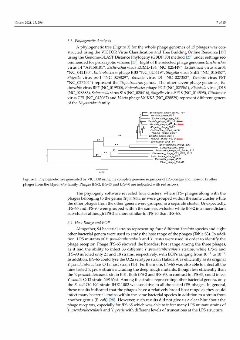
Figure 3. Phylogenetic tree generated by VICTOR using the complete genome sequences of fPS-phages and those of 15 other phages from the Myoviridae family. Phages fPS-2, fPS-65 and fPS-90 are indicated with red arrows.
The phylogeny software revealed four clusters, where fPS- phages along with the phages belonging to the genus Tequatrovirus were grouped within the same cluster while the other phages from the other genera were grouped in a separate cluster. Unexpectedly, fPS-65 and fPS-90 were grouped within the same sub-cluster while fPS-2 in a more distant sub-cluster although fPS-2 is more similar to fPS-90 than fPS-65.
3.4. Host Range and EOP Altogether, 94 bacterial strains representing four different Yersinia species and eight
other bacterial genera were used to study the host range of the phages (Table S3). In ad-dition, LPS mutants of Y. pseudotuberculosis and Y. pestis were used in order to identify the phage receptor. Phage fPS-65 showed the broadest host range among the three phages, as it had the ability to infect 33 different Y. pseudotuberculosis strains, while fPS-2 and fPS-90 infected only 21 and 18 strains, respectively, with EOPs ranging from 10−1 to 10−7. In addi-tion, fPS-65 could lyse the O:2a serotype strain Hatada A as efficiently as its original Y. pseudotuberculosis O:1a host strain PB1. Furthermore, fPS-65 was also able to infect all the nine tested Y. pestis strains including the deep rough mutants, though less efficiently than the Y. pseudotuberculosis strain PB1. Both fPS-2 and fPS-90, in contrast to fPS-65, could in-fect Y. similis O:12 strain N916Ysi. Among the strains representing other bacterial genera, only the E. coli O:1 K:1 strain IHE11002 was sensitive to all the tested fPS-phages. In gen-eral, these results indicated that the phages have a relatively broad host range as they could infect many bacterial strains within the same bacterial species in addition to a strain from another genus (E. coli) [28]. However, such results did not give us a clear hint about the phage receptors, especially for fPS-65 which was able to infect many LPS mutant strains of Y. pseudotuberculosis and Y. pestis with different levels of truncations at the LPS structure.
In general, host range determination has been studied for many T4-like phages; for example, Vibriophage KVP40 has a broad host range, infecting some pathogenic and non-pathogenic Vibrio species [29]. Additionally, the coliphages AR1 and LG1 have a wide host range as they are able to lyse many serogroups of E. coli and other members of enterobac-teria [30]. On the other hand, some studies have reported T4 phages with narrow host ranges, as vB_Kpn_F48, the K. pneumoniae phage, which has a lytic activity specific to K. pneumoniae strains of ST101 [31].
3.5. Growth Characteristics The one-step growth study revealed that fPS-2 and fPS-65 had similar latent period
of 50–55 min, with different burst sizes of about 65 and 44 virions per infected bacterium, respectively (Figure 4). Phage fPS-90 showed a bit different growth kinetics of 75–80 min latent period and a burst size of about 60 virions per infected cell (Figure 4).
Figure 3. Phylogenetic tree generated by VICTOR using the complete genome sequences of fPS-phages and those of 15 otherphages from the Myoviridae family. Phages fPS-2, fPS-65 and fPS-90 are indicated with red arrows.
The phylogeny software revealed four clusters, where fPS- phages along with thephages belonging to the genus Tequatrovirus were grouped within the same cluster whilethe other phages from the other genera were grouped in a separate cluster. Unexpectedly,fPS-65 and fPS-90 were grouped within the same sub-cluster while fPS-2 in a more distantsub-cluster although fPS-2 is more similar to fPS-90 than fPS-65.
3.4. Host Range and EOP
Altogether, 94 bacterial strains representing four different Yersinia species and eightother bacterial genera were used to study the host range of the phages (Table S3). In addi-tion, LPS mutants of Y. pseudotuberculosis and Y. pestis were used in order to identify thephage receptor. Phage fPS-65 showed the broadest host range among the three phages,as it had the ability to infect 33 different Y. pseudotuberculosis strains, while fPS-2 andfPS-90 infected only 21 and 18 strains, respectively, with EOPs ranging from 10−1 to 10−7.In addition, fPS-65 could lyse the O:2a serotype strain Hatada A as efficiently as its originalY. pseudotuberculosis O:1a host strain PB1. Furthermore, fPS-65 was also able to infect all thenine tested Y. pestis strains including the deep rough mutants, though less efficiently thanthe Y. pseudotuberculosis strain PB1. Both fPS-2 and fPS-90, in contrast to fPS-65, could infectY. similis O:12 strain N916Ysi. Among the strains representing other bacterial genera, onlythe E. coli O:1 K:1 strain IHE11002 was sensitive to all the tested fPS-phages. In general,these results indicated that the phages have a relatively broad host range as they couldinfect many bacterial strains within the same bacterial species in addition to a strain fromanother genus (E. coli) [28]. However, such results did not give us a clear hint about thephage receptors, especially for fPS-65 which was able to infect many LPS mutant strains ofY. pseudotuberculosis and Y. pestis with different levels of truncations at the LPS structure.

Viruses 2021, 13, 296 8 of 15
In general, host range determination has been studied for many T4-like phages; forexample, Vibriophage KVP40 has a broad host range, infecting some pathogenic andnon-pathogenic Vibrio species [29]. Additionally, the coliphages AR1 and LG1 have awide host range as they are able to lyse many serogroups of E. coli and other members ofenterobacteria [30]. On the other hand, some studies have reported T4 phages with narrowhost ranges, as vB_Kpn_F48, the K. pneumoniae phage, which has a lytic activity specific toK. pneumoniae strains of ST101 [31].
3.5. Growth Characteristics
The one-step growth study revealed that fPS-2 and fPS-65 had similar latent periodof 50–55 min, with different burst sizes of about 65 and 44 virions per infected bacterium,respectively (Figure 4). Phage fPS-90 showed a bit different growth kinetics of 75–80 minlatent period and a burst size of about 60 virions per infected cell (Figure 4).
Viruses 2021, 13, x FOR PEER REVIEW 9 of 17
Figure 4. One-step growth curves of fPS-phages in Y. pseudotuberculosis O.1a strain PB1: (a) fPS-2; (b) fPS-65; (c) fPS-90. Error bars represent standard deviations (SD).
Of note, there is a remarkable diversity of growth kinetics among T4-like phages; i.e., the latency periods and burst sizes vary from phage to other. For example, phage vB_Kpn_F48 has a short latent period of less than 10 min with a burst size of 72 phage particle per infected bacterium [31]. A similar short latent period was computed for the Enterobacter aerogenes phage vB_EaeM_φEap-3, with a higher burst size of 109 PFU per infected cell [32]. On the other hand the coliphages, LG1 and AR1, had different growth kinetics as the latent periods were 52 and 40 min, and the burst sizes 177 and 38 PFU per infected cell, respectively [30].
3.6. Identification of the Phage Receptors In order to identify the receptors on bacterial surface to which the phages adsorb, we
isolated 15 spontaneous phage-resistant mutants of Y. pseudotuberculosis O:1a strain PB1. Three mutants, resistant to fPS-2 were named M1-fps2-wt, M2-fps2-wt, and M3-fps2-wt. Nine mutants resistant to fPS-90 were named M1-fps90-wt, M3-fps90-wt, M4-fps90-wt, M5-fps90-wt, M6-fps90-wt, M7-fps90-wt, M8-fps90-wt, M9-fps90-wt, and M10-fps90-wt. The three mutants resistant to fPS-65 were named M1-fps65-wt, M2-fps65-wt, and M3-fps65-wt (Table 1). To confirm the resistance phenotype of the mutant strains, we tested their sensitivity also to the other fPS-phages. Generally, all the fPS-90-resistant strains were resistant to both fPS-90 and fPS-2 with the exception of M4-fps90-wt and M9-fps90-wt strains, which were sensitive to fPS-2 but not fPS-90, although with 104-fold reduced EOP (Figure S1). The mutants M1-fps2-wt, M2-fps2-wt, and M3-fps2-wt were resistant to both fPS-2 and fPS-90. However, all the fPS-2- and fPS-90-resistant mutants were sensitive to fPS-65 (Table 1). The comparison of the genomes of the mutants to that of the wild type strain PB1 revealed five different types of deletions that affected the galU gene. While an identical 12-bp deletion was present in M1-fps2-wt, M2-fps2-wt, M3-fps2-wt, M1-fps90-wt and M8-fps90-wt, a 6-bp deletion in M5-fps90-wt and M6-fps90-wt, and a 14-bp dele-tion in M3-fps90-wt, the whole galU gene was deleted in M7-fps90-wt and M10-fps90-wt, that had 7.9-kb and 4.3-kb deletions over the galU locus, respectively (Table 1). While in M4-fps90-wt, an insertion of a T in the middle of the ompF gene caused a frame shift mu-tation, in M9-fps90-wt, a single nucleotide substitution (T to C) was within the ompF pro-moter region (Table 1). To exclude the possibility that a second-site mutation had caused the resistance phenotypes, five of the galU-mutants (M1-fps90-wt, M3-fps90-wt, M5-fps90-wt, M7-fps90-wt, and M10-fps90-wt) and both ompF-mutants (M4-fps90-wt and M9-fps90-wt) were chosen for in trans complementation experiments. The sensitivity of all the galU-mutant strains to fPS-2 and fPS-90 was restored when they were complemented with plasmid pTM100-galU [33] containing the intact galU gene of Y. enterocolitica serotype O:3 (Figure S2). Additionally, M4-fps90-wt and M9-fps90-wt retained the sensitivity to fPS-90 when complemented with the plasmid pTM100-ompF carrying the intact ompF gene of Y. pseudotuberculosis strain PB1 (Figure S3). These results indicated that the GalU activity is required to maintain the receptor structure of both fPS-2 and fPS-90 phages. GalU is UDP-glucose pyrophosphorylase responsible for the synthesis of UDP-glucose that is one of the precursors required for LPS core oligosaccharide biosynthesis [34]. Thus, the galU mutants
Figure 4. One-step growth curves of fPS-phages in Y. pseudotuberculosis O.1a strain PB1: (a) fPS-2; (b) fPS-65; (c) fPS-90.Error bars represent standard deviations (SD).
Of note, there is a remarkable diversity of growth kinetics among T4-like phages;i.e., the latency periods and burst sizes vary from phage to other. For example, phagevB_Kpn_F48 has a short latent period of less than 10 min with a burst size of 72 phageparticle per infected bacterium [31]. A similar short latent period was computed for theEnterobacter aerogenes phage vB_EaeM_ϕEap-3, with a higher burst size of 109 PFU perinfected cell [32]. On the other hand the coliphages, LG1 and AR1, had different growthkinetics as the latent periods were 52 and 40 min, and the burst sizes 177 and 38 PFU perinfected cell, respectively [30].
3.6. Identification of the Phage Receptors
In order to identify the receptors on bacterial surface to which the phages adsorb, weisolated 15 spontaneous phage-resistant mutants of Y. pseudotuberculosis O:1a strain PB1.Three mutants, resistant to fPS-2 were named M1-fps2-wt, M2-fps2-wt, and M3-fps2-wt.Nine mutants resistant to fPS-90 were named M1-fps90-wt, M3-fps90-wt, M4-fps90-wt,M5-fps90-wt, M6-fps90-wt, M7-fps90-wt, M8-fps90-wt, M9-fps90-wt, and M10-fps90-wt.The three mutants resistant to fPS-65 were named M1-fps65-wt, M2-fps65-wt, and M3-fps65-wt (Table 1). To confirm the resistance phenotype of the mutant strains, we testedtheir sensitivity also to the other fPS-phages. Generally, all the fPS-90-resistant strains wereresistant to both fPS-90 and fPS-2 with the exception of M4-fps90-wt and M9-fps90-wtstrains, which were sensitive to fPS-2 but not fPS-90, although with 104-fold reduced EOP(Figure S1). The mutants M1-fps2-wt, M2-fps2-wt, and M3-fps2-wt were resistant to bothfPS-2 and fPS-90. However, all the fPS-2- and fPS-90-resistant mutants were sensitive tofPS-65 (Table 1). The comparison of the genomes of the mutants to that of the wild typestrain PB1 revealed five different types of deletions that affected the galU gene. While anidentical 12-bp deletion was present in M1-fps2-wt, M2-fps2-wt, M3-fps2-wt, M1-fps90-wtand M8-fps90-wt, a 6-bp deletion in M5-fps90-wt and M6-fps90-wt, and a 14-bp deletionin M3-fps90-wt, the whole galU gene was deleted in M7-fps90-wt and M10-fps90-wt, thathad 7.9-kb and 4.3-kb deletions over the galU locus, respectively (Table 1). While in M4-

Viruses 2021, 13, 296 9 of 15
fps90-wt, an insertion of a T in the middle of the ompF gene caused a frame shift mutation,in M9-fps90-wt, a single nucleotide substitution (T to C) was within the ompF promoterregion (Table 1). To exclude the possibility that a second-site mutation had caused theresistance phenotypes, five of the galU-mutants (M1-fps90-wt, M3-fps90-wt, M5-fps90-wt,M7-fps90-wt, and M10-fps90-wt) and both ompF-mutants (M4-fps90-wt and M9-fps90-wt)were chosen for in trans complementation experiments. The sensitivity of all the galU-mutant strains to fPS-2 and fPS-90 was restored when they were complemented withplasmid pTM100-galU [33] containing the intact galU gene of Y. enterocolitica serotype O:3(Figure S2). Additionally, M4-fps90-wt and M9-fps90-wt retained the sensitivity to fPS-90when complemented with the plasmid pTM100-ompF carrying the intact ompF gene ofY. pseudotuberculosis strain PB1 (Figure S3). These results indicated that the GalU activityis required to maintain the receptor structure of both fPS-2 and fPS-90 phages. GalU isUDP-glucose pyrophosphorylase responsible for the synthesis of UDP-glucose that is oneof the precursors required for LPS core oligosaccharide biosynthesis [34]. Thus, the galUmutants have a deep rough LPS phenotype, devoid of O-antigen and with a truncated LPSinner core oligosaccharide composed of lipid A, KDO/KO-residues (octulosonic acids),and two heptoses [34]. The results demonstrated that also the OmpF protein serves as areceptor for both fPS-90 and fPS-2. OmpF is a major outer membrane protein that formsa trimer with a central channel through which the bacteria import nutrients, and exportwaste products and some antibiotics [35].
Table 1. Overview of phage-resistant mutants and their sensitivity to the fPS-phages.
Mutant Strain Storage# Gene Mutation, Reference to Genome of PB1(acc no NC_010634)
Sensitivity to
fPS-2 fPS-65 fPS-90
M1-fps2-wt 6582 galU del-TAAAAGAGATCA, 1,869,879 Res * Sens ** ResM2-fps2-wt 6583 galU del-TAAAAGAGATCA, 1,869,879 Res Sens ResM3-fps2-wt 6584 galU del-TAAAAGAGATCA, 1,869,879 Res Sens ResM1-fps90-wt 6585 galU del-TAAAAGAGATCA, 1,869,879 Res Sens ResM3-fps90-wt 6586 galU del-AAGAGATCAAAAAT, 1,869,882 Res Sens ResM4-fps90-wt 6587 ompF C→ CT, 1,721,035 EOP *** Sens ResM5-fps90-wt 6650 galU del-AAAGAG, 1,869,881 Res Sens ResM6-fps90-wt 6651 galU del-AAAGAG, 1,869,881 Res Sens ResM7-fps90-wt 6652 galU ca. 7.9 kb del (1,870,248..1,878,146) Res Sens ResM8-fps90-wt 6653 galU del-TAAAAGAGATCA, 1,869,879 Res Sens ResM9-fps90-wt 6654 ompF promoter region, T→ C, 1,721,588 EOP Sens Res
M10-fps90-wt 6655 galU ca. 4.3 kb del (1,865,646..1,869,969) Res Sens ResM1-fps65-wt 6941 hldE 147 bp del (3,957,759..3,957,905) Res Res ResM2-fps65-wt 6942 hldD 517 bp del (61,956..62,473) Res Res ResM3-fps65-wt 6943 hldE 147 bp del (3,957,759..3,957,905) Res Res Res
* Res: resistant; ** Sens: sensitive; *** EOP: 104-fold reduced EOP.
In general, many studies have reported that most T-even phages walk on the bacterialsurface and the tips of the long tail fibres (LTFs) bind reversibly to a suitable outer mem-brane protein (OMP). This step, which brings the phage closer to its host cell surface, isfollowed by an irreversible attachment of the short tail fibres (STFs) to LPS [36]. AmongOMPs, OmpC, along with LPS, are well-known as the receptors of T4-like phages [36–38],and OmpF is found to be recognized by T2-like phages as a receptor [7,39].
As isolation of spontaneous phage-resistant mutants against fPS-65 with the ordinaryplating method was not successful, we determined the growth curves of Y. pseudotuberculosisPB1 bacteria in the presence of fPS-65 using the Bioscreen system. We reasoned that afterthe initial bacterial lysis, at later time points spontaneous phage-resistant mutant couldstart growing in the cultures. When visualizing the growth curves (Figure 5), from theinitial OD600 of 0.2, the control bacteria reached stationary phase after 20–25 h of incubation.We noticed that after the initial lysis phase of the bacteria that took place after 10–15 hincubation, the growth curves of the phage-infected cultures reached a minimum after20–25 h, followed by new growth phase until 40 h, an indication of the appearance ofspontaneous phage-resistant mutants. As described in Section 2.9, a 2 mL co-culture ofY. pseudotuberculosis PB1 and fPS-65 with an approximate MOI of 0.001 was set up andafter 48 h samples were streaked on LA plates to recover surviving bacteria. After 6 days,

Viruses 2021, 13, 296 10 of 15
altogether 12 colonies growing on LA-plates were streaked on CIN agar plates, five ofwhich failed to re-grow. Among the remaining clones only three strains (M1-fps65-wt, M2-fps65-wt, and M3-fps65-wt) were resistant to fPS-65. Whole genome sequencing analysisrevealed that both M1-fps65-wt and M3-fps65-wt had an identical out-of-frame deletionin the hldE gene. HldE is the bifunctional D,D-heptose 7-phosphate kinase/D,D-heptose1-phosphate adenylyltransferase [40]. In M2-fps65-wt there was a deletion in the hldDgene that encodes for the ADP-glycero-manno-heptose 6-epimerase [40]. Thus, both thehldE and hldD gene products are involved in the biosynthesis of ADP-L-β-D-heptose, thesubstrate for the proximal sugar residue of the LPS inner core. Thus, mutations in either ofthese genes would yield the same deep rough heptoseless LPS phenotype, i.e., with lipid Asubstituted just with the octulosonic acid residues [40–42]. These results suggested thatthe heptose region of the LPS inner core functions as receptor for fPS-65. The hldE mutantswere fully complemented in trans by a plasmid pTM100-hldE that carries a fully functionalhldE gene of Y. enterocolitica serotype O:3 strain 6471/76 [33]. Of note, LPS functions as avirulence factor in Y. pseudotuberculosis [43], suggesting that any phage-resistant mutantwould have lost virulence that might be important to keep in mind in the case of usingthese phages for phage therapy.
Viruses 2021, 13, x FOR PEER REVIEW 11 of 17
gene that encodes for the ADP-glycero-manno-heptose 6-epimerase [40]. Thus, both the hldE and hldD gene products are involved in the biosynthesis of ADP-L-β-D-heptose, the substrate for the proximal sugar residue of the LPS inner core. Thus, mutations in either of these genes would yield the same deep rough heptoseless LPS phenotype, i.e., with lipid A substituted just with the octulosonic acid residues [40–42]. These results suggested that the heptose region of the LPS inner core functions as receptor for fPS-65. The hldE mutants were fully complemented in trans by a plasmid pTM100-hldE that carries a fully functional hldE gene of Y. enterocolitica serotype O:3 strain 6471/76 [33]. Of note, LPS func-tions as a virulence factor in Y. pseudotuberculosis [43], suggesting that any phage-resistant mutant would have lost virulence that might be important to keep in mind in the case of using these phages for phage therapy.
Figure 5. Growth curves of Y. pseudotuberculosis O:1a strain PB1 infected with phages fPS-2, fPS-65 and fPS-90 at a MOI of 0.001. Each curve represents the average results for five replicates. Error bars represent standard deviation (SD).
3.7. Heat-Stability of the fPS-65 Receptor Heat treatment of bacteria can be used to differentiate whether the phage receptor is
a heat-sensitive protein or heat-resistant carbohydrate structure such as LPS. To verify that the LPS inner core is the receptor of fPS-65 we compared the ability of boiled and non-boiled bacteria to adsorb the phages (Figure 6). The results demonstrated that heat-treatment had no influence on the adsorption of the phages on bacteria, indicating as ex-pected that the phage receptor is heat-resistant. This supported the sequencing results.
Figure 5. Growth curves of Y. pseudotuberculosis O:1a strain PB1 infected with phages fPS-2, fPS-65 and fPS-90 at a MOI of0.001. Each curve represents the average results for five replicates. Error bars represent standard deviation (SD).
3.7. Heat-Stability of the fPS-65 Receptor
Heat treatment of bacteria can be used to differentiate whether the phage receptor is aheat-sensitive protein or heat-resistant carbohydrate structure such as LPS. To verify thatthe LPS inner core is the receptor of fPS-65 we compared the ability of boiled and non-boiledbacteria to adsorb the phages (Figure 6). The results demonstrated that heat-treatment hadno influence on the adsorption of the phages on bacteria, indicating as expected that thephage receptor is heat-resistant. This supported the sequencing results.

Viruses 2021, 13, 296 11 of 15Viruses 2021, 13, x FOR PEER REVIEW 12 of 17
Figure 6. The fPS-65 receptor is heat-stable. Phages were incubated 15 min with different dilutions of heat-treated and non-treated bacteria. Shown are percentages of free phages in the supernatants after centrifugation when compared to no-bacteria control set to 100%. The boiled bacteria adsorbed the phages as well as or better than viable bacteria indicating that the phage receptor is intact. Error bars represent the standard deviations for the average plaque numbers in two parallels of two independent experiments.
3.8. The Receptor-Binding Proteins of the Phages Generally, T-even phages encode Gp37 and Gp38 as the main components of the
LTFs that are critical for host attachment [44–46]. In phage T4, Gp38 acts as a chaperon that facilitates the trimerization of Gp37, while in T2-like phages, Gp38, after its function as a chaperone, will remain attached as the most distal part of the fibre structure. Thus in these phages, Gp38 is the adhesin that mediates the phage-host interaction [47,48]. While fPS-2 and fPS-90 both use OmpF and LPS core as receptors but had differences in their host range sensitivity data, the fPS-65 receptor is the heptose region of LPS inner core. To find out whether the receptor specificities of fPS-2, fPS-90, and fPS-65 could be explained by the predicted amino acid sequences of their respective LTF components, the identified Gp37 and Gp38 homologs of the fPS-phages were aligned for comparison. The alignment of the Gp37 homologs (Gp245, Gp246, and GP248 of fPS-2, fPS-65, and fPS-90, respec-tively) showed that the Gp245 amino acid sequence of fPS-2 is quite different from the Gp246 of fPS-65 and Gp248 of fPS-90, while the two latter are more similar to each other. Thus, this did not reflect at all the receptor specificities of the phages. Overall, the 60 most N-terminal and the 120 most C-terminal amino acid residues of the Gp37 homologs of the fPS phages were highly identical, and the sequences in between were more different (Figure 7a).
Figure 6. The fPS-65 receptor is heat-stable. Phages were incubated 15 min with different dilutions of heat-treated andnon-treated bacteria. Shown are percentages of free phages in the supernatants after centrifugation when compared tono-bacteria control set to 100%. The boiled bacteria adsorbed the phages as well as or better than viable bacteria indicatingthat the phage receptor is intact. Error bars represent the standard deviations for the average plaque numbers in twoparallels of two independent experiments.
3.8. The Receptor-Binding Proteins of the Phages
Generally, T-even phages encode Gp37 and Gp38 as the main components of the LTFsthat are critical for host attachment [44–46]. In phage T4, Gp38 acts as a chaperon thatfacilitates the trimerization of Gp37, while in T2-like phages, Gp38, after its function asa chaperone, will remain attached as the most distal part of the fibre structure. Thus inthese phages, Gp38 is the adhesin that mediates the phage-host interaction [47,48]. WhilefPS-2 and fPS-90 both use OmpF and LPS core as receptors but had differences in their hostrange sensitivity data, the fPS-65 receptor is the heptose region of LPS inner core. To findout whether the receptor specificities of fPS-2, fPS-90, and fPS-65 could be explained by thepredicted amino acid sequences of their respective LTF components, the identified Gp37and Gp38 homologs of the fPS-phages were aligned for comparison. The alignment ofthe Gp37 homologs (Gp245, Gp246, and GP248 of fPS-2, fPS-65, and fPS-90, respectively)showed that the Gp245 amino acid sequence of fPS-2 is quite different from the Gp246 offPS-65 and Gp248 of fPS-90, while the two latter are more similar to each other. Thus, thisdid not reflect at all the receptor specificities of the phages. Overall, the 60 most N-terminaland the 120 most C-terminal amino acid residues of the Gp37 homologs of the fPS phageswere highly identical, and the sequences in between were more different (Figure 7a).
In contrast, the host receptor specificity was better reflected in the multiple sequencealignment of the Gp38 homologs. When carrying out HHpred [49] search with Gp246of fPS-2, the best hit was the Gp38 homolog (DOI: 10.2210/pdb6F45/pdb) of Salmonellaphage vB_SenM_S16 (S16 for short) [48]. Multiple sequence alignment of the Gp38 fPS-homologs with that of S16 is shown in Figure 7b. The Gp38 homologs of the T2-like phages,including S16, have a modular organization: a conserved N-terminal domain forms ahighly hydrophobic surface that binds to the similarly highly hydrophobic Gp37 trimerictip of the tail fibre. The C-terminal domain in Gp38 homologs that determines the hostspecificity has a segmented structure [48]. The C-terminal domain contains a series often conserved polyglycine motifs that flank the five distal hypervariable loops that arecentrally involved in the receptor-binding specificity of the T2-like phages [45,48].
Viruses 2021, 13, 296 12 of 15Viruses 2021, 13, x FOR PEER REVIEW 13 of 17
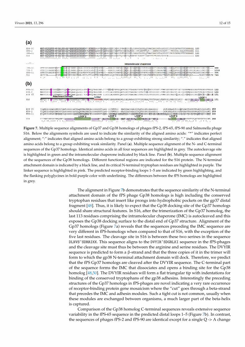
Figure 7. Multiple sequence alignments of Gp37 and Gp38 homologs of phages fPS-2, fPS-65, fPS-90 and Salmonella phage S16. Below the alignments symbols are used to indicate the similarity of the aligned amino acids: “*” indicates perfect alignment; “:” indicates that aligned amino acids belong to a group exhibiting strong similarity; “.” indicates that aligned amino acids belong to a group exhibiting weak similarity. Panel (a). Multiple sequence alignment of the N- and C-terminal sequences of the Gp37 homologs. Identical amino acids in all four sequences are highlighted in grey. The autocleavage site is highlighted in green and the intramolecular chaperone indicated by black line. Panel (b). Multiple sequence align-ment of the sequences of the Gp38 homologs. Different functional regions are indicated for the S16 protein. The N-terminal attachment domain is indicated by a black line, and its critical N-terminal tryptophan residues are highlighted in purple. The linker sequence is highlighted in pink. The predicted receptor-binding loops 1–5 are indicated by green highlighting, and the flanking polyglycines in bold purple color with underlining. The differences between the fPS homologs are high-lighted in grey.
In contrast, the host receptor specificity was better reflected in the multiple sequence alignment of the Gp38 homologs. When carrying out HHpred [49] search with Gp246 of fPS-2, the best hit was the Gp38 homolog (DOI: 10.2210/pdb6F45/pdb) of Salmonella phage vB_SenM_S16 (S16 for short) [48]. Multiple sequence alignment of the Gp38 fPS-homologs with that of S16 is shown in Figure 7b. The Gp38 homologs of the T2-like phages, includ-ing S16, have a modular organization: a conserved N-terminal domain forms a highly hy-drophobic surface that binds to the similarly highly hydrophobic Gp37 trimeric tip of the tail fibre. The C-terminal domain in Gp38 homologs that determines the host specificity has a segmented structure [48]. The C-terminal domain contains a series of ten conserved polyglycine motifs that flank the five distal hypervariable loops that are centrally involved in the receptor-binding specificity of the T2-like phages [45,48].
The alignment in Figure 7b demonstrates that the sequence similarity of the N-termi-nal attachment domain of the fPS phage Gp38 homologs is high including the conserved tryptophan residues that insert like prongs into hydrophobic pockets on the gp37 distal fragment [48]. Thus, it is likely to expect that the Gp38 docking site of the Gp37 homologs should share structural features. In S16, after the trimerization of the Gp37 homolog, the last 113 residues comprising the intramolecular chaperone (IMC) is autocleaved and this exposes the Gp38 docking surface to the distal end of Gp37 structure. Alignment of the Gp37 homologs (Figure 7a) reveals that the sequences preceding the IMC sequence are very different in fPS-homologs when compared to that of S16, with the exception of the five last residues. The cleavage site in S16 is between these two serines in the sequence DLNVS^SDRRIKK. This sequence aligns to the DVYIR^SDGRLKI sequence in the fPS-phages and the cleavage site must thus be between the arginine and serine residues. The DVYIR sequence is predicted to form a β-strand and that the three copies of it in the trimer will form to which the gp38 N-terminal attachment domain will dock. Therefore, we pre-dict that the fPS Gp37 homologs are cleaved after the DVYIR sequence. The C-terminal
Figure 7. Multiple sequence alignments of Gp37 and Gp38 homologs of phages fPS-2, fPS-65, fPS-90 and Salmonella phageS16. Below the alignments symbols are used to indicate the similarity of the aligned amino acids: “*” indicates perfectalignment; “:” indicates that aligned amino acids belong to a group exhibiting strong similarity; “.” indicates that alignedamino acids belong to a group exhibiting weak similarity. Panel (a). Multiple sequence alignment of the N- and C-terminalsequences of the Gp37 homologs. Identical amino acids in all four sequences are highlighted in grey. The autocleavage siteis highlighted in green and the intramolecular chaperone indicated by black line. Panel (b). Multiple sequence alignmentof the sequences of the Gp38 homologs. Different functional regions are indicated for the S16 protein. The N-terminalattachment domain is indicated by a black line, and its critical N-terminal tryptophan residues are highlighted in purple. Thelinker sequence is highlighted in pink. The predicted receptor-binding loops 1–5 are indicated by green highlighting, andthe flanking polyglycines in bold purple color with underlining. The differences between the fPS homologs are highlightedin grey.
The alignment in Figure 7b demonstrates that the sequence similarity of the N-terminalattachment domain of the fPS phage Gp38 homologs is high including the conservedtryptophan residues that insert like prongs into hydrophobic pockets on the gp37 distalfragment [48]. Thus, it is likely to expect that the Gp38 docking site of the Gp37 homologsshould share structural features. In S16, after the trimerization of the Gp37 homolog, thelast 113 residues comprising the intramolecular chaperone (IMC) is autocleaved and thisexposes the Gp38 docking surface to the distal end of Gp37 structure. Alignment of theGp37 homologs (Figure 7a) reveals that the sequences preceding the IMC sequence arevery different in fPS-homologs when compared to that of S16, with the exception of thefive last residues. The cleavage site in S16 is between these two serines in the sequenceDLNVSˆSDRRIKK. This sequence aligns to the DVYIRˆSDGRLKI sequence in the fPS-phagesand the cleavage site must thus be between the arginine and serine residues. The DVYIRsequence is predicted to form a β-strand and that the three copies of it in the trimer willform to which the gp38 N-terminal attachment domain will dock. Therefore, we predictthat the fPS Gp37 homologs are cleaved after the DVYIR sequence. The C-terminal partof the sequence forms the IMC that dissociates and opens a binding site for the Gp38homolog [48,50]. The DVYIR residues will form a flat triangular tip with indentations forbinding of the conserved tryptophans of the gp38 adhesins. Interestingly the precedingstructures of the Gp37 homologs in fPS-phages are novel indicating a very rare occurrenceof receptor-binding protein gene mosaicism where the “cut” goes through a beta-strandthat precedes the IMC and adhesin modules. Such a tight cut is not common, usually whenthese modules are exchanged between organisms, a much larger part of the beta-helixis captured.
Comparison of the Gp38 homolog C-terminal sequences reveals extensive sequencevariability in the fPS-65 sequence in the predicted distal loops 1–5 (Figure 7b). In contrast,the sequences of phages fPS-2 and fPS-90 are identical except for a single Q -> A change

Viruses 2021, 13, 296 13 of 15
after the loop 5. This single amino acid difference could explain the slightly different hostranges of fPS-2 and fPS-90. The highly variable loops in fPS-65 explain perfectly its uniquereceptor and host range.
4. Conclusions
In this work, we characterized three Y. pseudotuberculosis-infecting phages, fPS-2,fPS-65, and fPS-90, originally isolated from pig stool samples collected from Finnish pigfarms [8]. The genomic sequences of the phages were 85–92% identical to each other.Based on the electron microscopy and the phylogeny analysis, the phages can be classifiedas new members in the genus Tequatroviruses within the Myoviridae family under theCaudovirales order. The phages fPS-2, fPS-65, and fPS-90 lysed 21, 33, and 18 of the 56Y. pseudotuberculosis strains tested, respectively, and, in addition, one E. coli strain indicatingthat the phages have a relatively broad host range. On the genomic level, the genomes ofthe phages showed a high level of similarity between them and to T4 phage. While OmpFand LPS outer core oligosaccharide were identified as receptors on the bacterial surface forboth fPS-2 and fPS-90, the fPS-65 receptor structure required the presence of the heptosesof the LPS inner core. These receptor specificities were reflected to the amino acid sequencedifferences in the Gp38 homologs of the fPS-phages.
In general, being lytic and void of any genes encoding for toxicity, antibiotic resistance,or lysogeny, these phages can be used safely in phage therapy. Furthermore, as fPS-65 isalso able to infect Y. pestis, it could be considered for diagnostic and therapeutic purposesagainst the notorious pathogen, the etiologic agent of the Black Death.
Supplementary Materials: The following are available online at https://www.mdpi.com/1999-4915/13/2/296/s1. Table S1: Bacterial strains used in this work. Table S2: Plasmids used in thiswork, Table S3: Host range and EOP of the studied phages with different bacterial strains. Table S4:Comparison of the gene products of the fPS-phages with the corresponding T4 gene products.Figure S1: The EOP test of fPS-2 with M4-fps90-wt and M9-fps90-wt strains. Figure S2: Spot assay totest the sensitivity of the galU-complemented spontaneous phage-resistant mutant strains to fPS-2and fPS-90. Figure S3: The complementation of the phage-resistant mutant strains M4-fps90-wt andM9-fps90-wt with the plasmid pTM100-ompF carrying the intact ompF gene of Y. pseudotuberculosisstrain PB1.
Author Contributions: M.S. (Mabruka Salem) and M.S. (Mikael Skurnik) conceived and designedthe experiments; M.S. (Mabruka Salem), M.I.P., and J.W.J. performed the experiments; M.S. (MabrukaSalem) and M.S. (Mikael Skurnik) analyzed the data and wrote the paper. All authors have read andagreed to the published version of the manuscript.
Funding: Mabruka Salem was supported by the Ministry of Higher Education and Scientific Research,Tripoli, Libya, and by the Doctoral Programme in Microbiology and Biotechnology (MBDP) ofUniversity of Helsinki. We also acknowledge the research funding of the Academy of Finland (project1288701).
Institutional Review Board Statement: Not applicable.
Informed Consent Statement: Not applicable.
Data Availability Statement: Not applicable.
Acknowledgments: We thank Anu Wicklund for helping in taking the TEM pictures. We wish tothank specifically Mark van Raaij, Matthew Dunne and Petr Leiman for insightful discussions on theroles of Gp37 and Gp38 homologs of the fPS phages.
Conflicts of Interest: The authors declare no conflict of interest. The founding sponsors had no rolein the design of the study; in the collection, analyses, or interpretation of data; in the writing of themanuscript, and in the decision to publish the results.

Viruses 2021, 13, 296 14 of 15
References1. Jalava, K.; Hakkinen, M.; Valkonen, M.; Nakari, U.M.; Palo, T.; Hallanvuo, S.; Ollgren, J.; Siitonen, A.; Nuorti, J.P. An outbreak of
gastrointestinal illness and erythema nodosum from grated carrots contaminated with Yersinia pseudotuberculosis. J. Infect. Dis.2006, 194, 1209–1216. [CrossRef] [PubMed]
2. Savin, C.; Martin, L.; Bouchier, C.; Filali, S.; Chenau, J.; Zhou, Z.; Becher, F.; Fukushima, H.; Thomson, N.R.; Scholz, H.C.; et al.The Yersinia pseudotuberculosis complex: Characterization and delineation of a new species, Yersinia wautersii. Int. J. MedMicrobiol. 2014, 304, 452–463. [CrossRef]
3. Pärn, T.; Hallanvuo, S.; Salmenlinna, S.; Pihlajasaari, A.; Heikkinen, S.; Telkki-Nykänen, H.; Hakkinen, M.; Ollgren, J.; Huusko, S.;Rimhanen-Finne, R. Outbreak of Yersinia pseudotuberculosis O:1 infection associated with raw milk consumption, Finland,spring 2014. Eurosurveillance 2015, 20. [CrossRef] [PubMed]
4. Kim, Y.K.; Cho, M.H.; Hyun, H.S.; Park, E.; Ha, I.S.; Cheong, H.I.; Kang, H.G. Acute kidney injury associated with Yersiniapseudotuberculosis infection: Forgotten but not gone. Kidney Res. Clin. Pract. 2019, 38, 347–355. [CrossRef] [PubMed]
5. Fukushima, H.; Shimizu, S.; Inatsu, Y. Yersinia enterocolitica and Yersinia pseudotuberculosis detection in foods. J. Pathog. 2011,2011, 735308. [CrossRef] [PubMed]
6. De Castro, C.; Skurnik, M.; Molinaro, A.; Holst, O. Characterization of the specific O-polysaccharide structure and bio-syntheticgene cluster of Yersinia pseudotuberculosis serotype O:15. Innate Immun. 2009, 15, 351–359. [CrossRef]
7. Leon-Velarde, C.G.; Happonen, L.; Pajunen, M.; Leskinen, K.; Kropinski, A.M.; Mattinen, L.; Rajtor, M.; Zur, J.; Smith, D.;Chen, S.; et al. Yersinia enterocolitica-specific infection by bacteriophages TG1 and fR1-RT is dependent on temperature-regulatedexpression of the phage host receptor OmpF. Appl. Environ. Microbiol. 2016, 82, 5340–5353. [CrossRef] [PubMed]
8. Salem, M.; Virtanen, S.; Korkeala, H.; Skurnik, M. Isolation and characterization of Yersinia-specific bacteriophages from pigstools in Finland. J. Appl. Microbiol. 2015, 118, 599–608. [CrossRef] [PubMed]
9. Coil, D.; Jospin, G.; Darling, A.E. A5-miseq: An updated pipeline to assemble microbial genomes from Illumina MiSeq data.Bioinformatics 2015, 31, 587–589. [CrossRef] [PubMed]
10. Aziz, R.K.; Bartels, D.; Best, A.A.; DeJongh, M.; Disz, T.; Edwards, R.A.; Formsma, K.; Gerdes, S.; Glass, E.M.; Kubal, M.; et al. TheRAST Server: Rapid annotations using subsystems technology. BMC Genom. 2008, 9, 75. [CrossRef] [PubMed]
11. Delcher, A.L.; Harmon, D.; Kasif, S.; White, O.; Salzberg, S.L. Improved microbial gene identification with GLIMMER. Nucl. AcidsRes. 1999, 27, 4636–4641. [CrossRef] [PubMed]
12. Rutherford, K.; Parkhill, J.; Crook, J.; Horsnell, T.; Rice, P.; Rajandream, M.-A.; Barrell, B. Artemis: Sequence visualization andannotation. Bioinformatics 2000, 16, 944–945. [CrossRef] [PubMed]
13. Altschul, S.F.; Madden, T.L.; Schäffer, A.A.; Zhang, J.; Zhang, Z.; Miller, W.; Lipman, D.J. Gapped BLAST and PSI-BLAST: A newgeneration of protein database search programs. Nucleic Acids Res. 1997, 25, 3389–3402. [CrossRef] [PubMed]
14. Katoh, K.; Standley, D.M. MAFFT multiple sequence alignment software version 7: Improvements in performance and usability.Mol. Biol. Evol. 2013, 30, 772–780. [CrossRef] [PubMed]
15. Chan, P.P.; Lowe, T.M. tRNAscan-SE: Searching for tRNA genes in genomic sequences. Methods Mol. Biol. 2019, 1962, 1–14.[CrossRef] [PubMed]
16. Lowe, T.M.; Chan, P.P. tRNAscan-SE Online: Integrating search and context for analysis of transfer RNA genes. Nucl. Acids Res.2016, 44, W54–W57. [CrossRef] [PubMed]
17. Meier-Kolthoff, J.P.; Göker, M. VICTOR: Genome-based phylogeny and classification of prokaryotic viruses. Bioinformatics 2017,33, 3396–3404. [CrossRef] [PubMed]
18. Salem, M.; Skurnik, M. Genomic characterization of sixteen Yersinia enterocolitica-infecting podoviruses of pig origin. Viruses2018, 10, 174. [CrossRef] [PubMed]
19. Yu, Y.P.; Gong, T.; Jost, G.; Liu, W.H.; Ye, D.Z.; Luo, Z.H. Isolation and characterization of five lytic bacteriophages infecting aVibrio strain closely related to Vibrio owensii. FEMS Microbiol. Lett. 2013, 348, 112–119. [CrossRef]
20. Leskinen, K.; Blasdel, B.G.; Lavigne, R.; Skurnik, M. RNA-Sequencing reveals the progression of phage-host interactions betweenfR1-37 and Yersinia enterocolitica. Viruses 2016, 8, 111. [CrossRef] [PubMed]
21. Biedzka-Sarek, M.; Venho, R.; Skurnik, M. Role of YadA, Ail, and lipopolysaccharide in serum resistance of Yersinia enterocoliticaserotype O:3. Infect. Immun. 2005, 73, 2232–2244. [CrossRef] [PubMed]
22. Eiserling, F.A. Structure of the T4 virion. In Bacteriophage T4; Mathews, C.K., Kutter, E.M., Modsig, G., Berget, P.B., Eds.; AmericanSociety for Microbiology: Washington, DC, USA, 1983; pp. 11–24.
23. Miller, E.S.; Kutter, E.; Mosig, G.; Arisaka, F.; Kunisawa, T.; Ruüger, W. Bacteriophage T4 genome. Microbiol. Mol. Biol. Rev. 2003,67, 86–156. [CrossRef] [PubMed]
24. Bao, H.; Shahin, K.; Zhang, Q.; Zhang, H.; Wang, Z.; Zhou, Y.; Zhang, X.; Zhu, S.; Stefan, S.; Wang, R. Morphologic and genomiccharacterization of a broad host range Salmonella enterica serovar Pullorum lytic phage vB_SPuM_SP116. Microb. Pathog. 2019,136, 103659. [CrossRef]
25. Jiang, Y.-H.; Liu, J.-Q.; Zhao, C.-Y.; Yu, S.; Sun, Y.-B.; Shi, H.-Y.; Huang, H. Isolation and genome sequencing of a novelPseudomonas aeruginosa Phage PA-YS35. Curr. Microbiol. 2020, 77, 123–128. [CrossRef] [PubMed]
26. Sullivan, M.J.; Petty, N.K.; Beatson, S.A. Easyfig: A genome comparison visualizer. Bioinformatics 2011, 27, 1009–1010. [CrossRef]27. Meier-Kolthoff, J.P.; Auch, A.F.; Klenk, H.P.; Goker, M. Genome sequence-based species delimitation with confidence intervals
and improved distance functions. BMC Bioinform. 2013, 14, 60. [CrossRef] [PubMed]

Viruses 2021, 13, 296 15 of 15
28. Hyman, P. Phages for phage therapy: Isolation, characterization, and host range breadth. Pharmaceuticals 2019, 12, 35. [CrossRef] [PubMed]29. Miller, J.F. Bacteriophage and the evolution of epidemic Cholera. Infect. Immun. 2003, 71, 2981–2982. [CrossRef]30. Goodridge, L.; Gallaccio, A.; Griffiths, M.W. Morphological, host range, and genetic characterization of two coliphages. Appl.
Environ. Microbiol. 2003, 69, 5364–5371. [CrossRef] [PubMed]31. Ciacci, N.; D’Andrea, M.M.; Marmo, P.; Demattè, E.; Amisano, F.; Di Pilato, V.; Fraziano, M.; Lupetti, P.; Rossolini, G.M.;
Thaller, M.C. Characterization of vB_Kpn_F48, a Newly discovered lytic bacteriophage for Klebsiella pneumoniae of sequencetype 101. Viruses 2018, 10, 482. [CrossRef]
32. Zhao, J.; Zhang, Z.; Tian, C.; Chen, X.; Hu, L.; Wei, X.; Li, H.; Lin, W.; Jiang, A.; Feng, R.; et al. Characterizing the biology oflytic bacteriophage vB_EaeM_phiEap-3 infecting multidrug-resistant Enterobacter aerogenes. Front. Microbiol. 2019, 10, 420.[CrossRef]
33. Noszczynska, M.; Kasperkiewicz, K.; Duda, K.A.; Podhorodecka, J.; Rabsztyn, K.; Gwizdala, K.; Swierzko, A.S.; Radziejewska-Lebrecht, J.; Holst, O.; Skurnik, M. Serological characterization of the enterobacterial common antigen substitution of thelipopolysaccharide of Yersinia enterocolitica O:3. Microbiology 2015, 161, 219–227. [CrossRef] [PubMed]
34. Priebe, G.P.; Dean, C.R.; Zaidi, T.; Meluleni, G.J.; Coleman, F.T.; Coutinho, Y.S.; Noto, M.J.; Urban, T.A.; Pier, G.B.; Goldberg, J.B.The galU gene of Pseudomonas aeruginosa is required for corneal infection and efficient systemic spread following Pneumoniabut not for infection confined to the lung. Infect. Immun. 2004, 72, 4224–4232. [CrossRef] [PubMed]
35. Rokitskaya, T.I.; Kotova, E.A.; Naberezhnykh, G.A.; Khomenko, V.A.; Gorbach, V.I.; Firsov, A.M.; Zelepuga, E.A.; Antonenko, Y.N.;Novikova, O.D. Single channel activity of OmpF-like porin from Yersinia pseudotuberculosis. Biochim. Biophys. Acta 2016, 1858,883–891. [CrossRef]
36. Chen, P.; Sun, H.; Ren, H.; Liu, W.; Li, G.; Zhang, C. LamB, OmpC, and the core Lipopolysaccharide of Escherichia coli K-12function as receptors of bacteriophage Bp7. J. Virol. 2020, 94. [CrossRef] [PubMed]
37. Islam, M.Z.; Fokine, A.; Mahalingam, M.; Zhang, Z.; Garcia-Doval, C.; van Raaij, M.J.; Rossmann, M.G.; Rao, V.B. Molecularanatomy of the receptor binding module of a bacteriophage long tail fiber. PLoS Pathog. 2019, 15, e1008193. [CrossRef] [PubMed]
38. Yu, F.; Mizushima, S. Roles of lipopolysaccharide and outer membrane protein OmpC of Escherichia coli K-12 in the receptorfunction for bacteriophage T4. J. Bacteriol. 1982, 151, 718–722. [CrossRef] [PubMed]
39. Rakhuba, D.; Kolomiets, E.; Dey, E.S.; Novik, G. Bacteriophage receptors, mechanisms of phage adsorption and penetration intohost cell. Pol. J. Microbiol. 2010, 59, 145–155. [CrossRef] [PubMed]
40. Kneidinger, B.; Marolda, C.; Graninger, M.; Zamyatina, A.; McArthur, F.; Kosma, P.; Valvano, M.A.; Messner, P. Biosynthesispathway of ADP-L-glycero-beta-D-manno-heptose in Escherichia coli. J. Bacteriol. 2002, 184, 363–369. [CrossRef]
41. Kiljunen, S.; Datta, N.; Dentovskaya, S.V.; Anisimov, A.P.; Knirel, Y.A.; Bengoechea, J.A.; Holst, O.; Skurnik, M. Identificationof the lipopolysaccharide core of Yersinia pestis and Yersinia pseudotuberculosis as the receptor for bacteriophage fA1122.J. Bacteriol. 2011, 193, 4963–4972. [CrossRef] [PubMed]
42. Knirel, Y.A.; Kondakova, A.N.; Bystrova, O.V.; Lindner, B.; Shaikhutdinova, R.Z.; Dentovskaya, S.V.; Anisimov, A.P. New featuresof Yersinia Lipopolysaccharide structures as revealed by high-resolution electrospray ionization mass spectrometry. Adv. Sci. Lett.2008, 1, 192–198. [CrossRef]
43. Karlyshev, A.V.; Oyston, P.C.F.; Williams, K.; Clark, G.C.; Titball, R.W.; Winzeler, E.A.; Wren, B.W. Application of high-densityarray-based signature-tagged mutagenesis to discover novel Yersinia virulence-associated genes. Infect. Immun. 2001, 69,7810–7819. [CrossRef] [PubMed]
44. Yu, S.-L.; Ko, K.-L.; Chen, C.-S.; Chang, Y.-C.; Syu, W.-J. Characterization of the distal tail fiber locus and determination of thereceptor for phage AR1, which specifically infects Escherichia coli O157:H7. J. Bacteriol. 2000, 182, 5962–5968. [CrossRef] [PubMed]
45. Trojet, S.N.; Caumont-Sarcos, A.; Perrody, E.; Comeau, A.M.; Krisch, H.M. The gp38 Adhesins of the T4 superfamily: A complexmodular determinant of the phage’s host specificity. Genome Biol. Evol. 2011, 3, 674–686. [CrossRef] [PubMed]
46. Yoichi, M.; Abe, M.; Miyanaga, K.; Unno, H.; Tanji, Y. Alteration of tail fiber protein gp38 enables T2 phage to infect Escherichiacoli O157:H7. J. Biotechnol. 2005, 115, 101–107. [CrossRef]
47. Marti, R.; Zurfluh, K.; Hagens, S.; Pianezzi, J.; Klumpp, J.; Loessner, M.J. Long tail fibres of the novel broad-host-range T-evenbacteriophage S16 specifically recognize Salmonella OmpC. Mol. Microbiol. 2013, 87, 818–834. [CrossRef] [PubMed]
48. Dunne, M.; Denyes, J.M.; Arndt, H.; Loessner, M.J.; Leiman, P.G.; Klumpp, J. Salmonella phage S16 tail fiber adhesin features arare polyglycine rich domain for host recognition. Structure 2018, 26, 1573–1582. [CrossRef]
49. Alva, V.; Nam, S.-Z.; Söding, J.; Lupas, A.N. The MPI bioinformatics Toolkit as an integrative platform for advanced proteinsequence and structure analysis. Nucleic Acids Res. 2016, 44, W410–W415. [CrossRef]
50. Schulz, E.C.; Dickmanns, A.; Urlaub, H.; Schmitt, A.; Muhlenhoff, M.; Stummeyer, K.; Schwarzer, D.; Gerardy-Schahn, R.;Ficner, R. Crystal structure of an intramolecular chaperone mediating triple-beta-helix folding. Nat. Struct. Mol. Biol. 2010, 17,210–215. [CrossRef] [PubMed]
Copyright © 2022 FDOKUMEN