Genomic characterization of two Staphylococcus epidermidis bacteriophages with anti-biofilm...
10
RESEARCH ARTICLE Open Access Genomic characterization of two Staphylococcus epidermidis bacteriophages with anti-biofilm potential Diana Gutiérrez, Beatriz Martínez, Ana Rodríguez and Pilar García * Abstract Background: Staphylococcus epidermidis is a commensal bacterium but can colonize the hospital environment due to its ability to form biofilms favouring adhesion to host tissues, medical devices and increasing resistance to antibiotics. In this context, the use of phages to destroy biofilms is an interesting alternative. Results: The complete genomes of two Staphylococcus epidermidis bacteriophages, vB_SepiS-phiIPLA5 and vB_SepiS-phiIPLA7, have been analyzed. Their genomes are 43,581 bp and 42,123 bp, and contain 67 and 59 orfs. Bioinformatic analyses enabled the assignment of putative functions to 36 and 29 gene products, respectively, including DNA packaging and morphogenetic proteins, lysis components, and proteins necessary for DNA recombination, regulation, modification and replication. A point mutation in vB_SepiS-phiIPLA5 lysogeny control-associated genes explained its strictly lytic behaviour. Comparative analysis of phi-IPLA5 and phi-IPLA7 genome structure resembled those of S. epidermidis φPH15 and φCNPH82 phages. A mosaic structure of S. epidermidis prophage genomes was revealed by PCR analysis of three marker genes (integrase, major head protein and holin). Using these genes, high prevalence (73%) of phage DNA in a representative S. epidermidis strain collection consisting of 60 isolates from women with mastitis and healthy women was determined. Putative pectin lyase-like domains detected in virion-associated proteins of both phages could be involved in exopolysaccharide (EPS) depolymerization, as evidenced by both the presence of a clear halo surrounding the phage lysis zone and the phage-mediated biofilm degradation. Conclusions: Staphylococcus epidermidis bacteriophages, vB_SepiS-phiIPLA5 and vB_SepiS-phiIPLA7, have a mosaic structure similar to other widespread S. epidermidis prophages. Virions of these phages are provided of pectin lyase-like domains, which may be regarded as promising anti-biofilm tools. Background Staphylococcus epidermidis is a common skin and mu- cous commensal of healthy humans, and can easily be transmitted to medical devices being a serious clinical problem and one of the major causes of nosocomial infections [1] as well as mastitis in lactating women [2]. In the animal health context S. epidermidis has also been recognized as one of the main etiological agents of ovine and bovine mastitis [3]. S. epidermidis is a key factor in the transmission of viru- lence factors and it is involved in balancing epithelial microbiota. In contrast to S. aureus, S. epidermidis does not encode many virulence factors, but it can colonize the hospital environment due to its ability to form biofilms favouring adhesion to host tissues, medical devices and in- creasing resistance to antibiotics [4]. In addition, the enor- mous flexibility of this bacterium continuously generates continuously novel phenotypic and genotypic variants. Hospital isolates are often characterised by the carriage of several staphylococcal chromosome cassettes (SCCmec), conferring methicillin resistance [5]. Moreover, nosoco- mial S. epidermidis strains typically harbour multiple co- pies of the insertion sequence element IS256 in their genomes, which contribute to genetic adaptation during in- fection [6]. Recently, the first S. epidermidis pathogenicity * Correspondence: [email protected] DairySafe Group, Department of Technology and Biotechnology of Dairy Products, Instituto de Productos Lácteos de Asturias (IPLA-CSIC), Paseo Río Linares, s/n, 33300, Villaviciosa, Asturias, Spain © 2012 Gutiérrez et al.; licensee BioMed Central Ltd. This is an Open Access article distributed under the terms of the Creative Commons Attribution License (http://creativecommons.org/licenses/by/2.0), which permits unrestricted use, distribution, and reproduction in any medium, provided the original work is properly cited. Gutiérrez et al. BMC Genomics 2012, 13:228 http://www.biomedcentral.com/1471-2164/13/228
-
Upload
unicartagena -
Category
Documents
-
view
1 -
download
0
Transcript of Genomic characterization of two Staphylococcus epidermidis bacteriophages with anti-biofilm...

Gutiérrez et al. BMC Genomics 2012, 13:228http://www.biomedcentral.com/1471-2164/13/228
RESEARCH ARTICLE Open Access
Genomic characterization of two Staphylococcusepidermidis bacteriophages with anti-biofilmpotentialDiana Gutiérrez, Beatriz Martínez, Ana Rodríguez and Pilar García*
Abstract
Background: Staphylococcus epidermidis is a commensal bacterium but can colonize the hospital environment dueto its ability to form biofilms favouring adhesion to host tissues, medical devices and increasing resistance toantibiotics. In this context, the use of phages to destroy biofilms is an interesting alternative.
Results: The complete genomes of two Staphylococcus epidermidis bacteriophages, vB_SepiS-phiIPLA5 andvB_SepiS-phiIPLA7, have been analyzed. Their genomes are 43,581 bp and 42,123 bp, and contain 67 and 59 orfs.Bioinformatic analyses enabled the assignment of putative functions to 36 and 29 gene products, respectively,including DNA packaging and morphogenetic proteins, lysis components, and proteins necessary for DNArecombination, regulation, modification and replication. A point mutation in vB_SepiS-phiIPLA5 lysogenycontrol-associated genes explained its strictly lytic behaviour. Comparative analysis of phi-IPLA5 and phi-IPLA7genome structure resembled those of S. epidermidis φPH15 and φCNPH82 phages. A mosaic structure of S.epidermidis prophage genomes was revealed by PCR analysis of three marker genes (integrase, major head proteinand holin). Using these genes, high prevalence (73%) of phage DNA in a representative S. epidermidis straincollection consisting of 60 isolates from women with mastitis and healthy women was determined. Putative pectinlyase-like domains detected in virion-associated proteins of both phages could be involved in exopolysaccharide(EPS) depolymerization, as evidenced by both the presence of a clear halo surrounding the phage lysis zone andthe phage-mediated biofilm degradation.
Conclusions: Staphylococcus epidermidis bacteriophages, vB_SepiS-phiIPLA5 and vB_SepiS-phiIPLA7, have a mosaicstructure similar to other widespread S. epidermidis prophages. Virions of these phages are provided of pectinlyase-like domains, which may be regarded as promising anti-biofilm tools.
BackgroundStaphylococcus epidermidis is a common skin and mu-cous commensal of healthy humans, and can easily betransmitted to medical devices being a serious clinicalproblem and one of the major causes of nosocomialinfections [1] as well as mastitis in lactating women [2].In the animal health context S. epidermidis has also beenrecognized as one of the main etiological agents of ovineand bovine mastitis [3].S. epidermidis is a key factor in the transmission of viru-
lence factors and it is involved in balancing epithelial
* Correspondence: [email protected] Group, Department of Technology and Biotechnology of DairyProducts, Instituto de Productos Lácteos de Asturias (IPLA-CSIC), Paseo RíoLinares, s/n, 33300, Villaviciosa, Asturias, Spain
© 2012 Gutiérrez et al.; licensee BioMed CentrCommons Attribution License (http://creativecreproduction in any medium, provided the or
microbiota. In contrast to S. aureus, S. epidermidis doesnot encode many virulence factors, but it can colonize thehospital environment due to its ability to form biofilmsfavouring adhesion to host tissues, medical devices and in-creasing resistance to antibiotics [4]. In addition, the enor-mous flexibility of this bacterium continuously generatescontinuously novel phenotypic and genotypic variants.Hospital isolates are often characterised by the carriage ofseveral staphylococcal chromosome cassettes (SCCmec),conferring methicillin resistance [5]. Moreover, nosoco-mial S. epidermidis strains typically harbour multiple co-pies of the insertion sequence element IS256 in theirgenomes, which contribute to genetic adaptation during in-fection [6]. Recently, the first S. epidermidis pathogenicity
al Ltd. This is an Open Access article distributed under the terms of the Creativeommons.org/licenses/by/2.0), which permits unrestricted use, distribution, andiginal work is properly cited.

Gutiérrez et al. BMC Genomics 2012, 13:228 Page 2 of 10http://www.biomedcentral.com/1471-2164/13/228
island (SePI), which encodes the staphylococcal enterotoxinSEC3 and SElL, has been described [7].The widespread use of antibiotics in both humans and
animals has led to the emergence of infectious bacteriaresistant to a wide range of antimicrobials that greatlyhinders their treatment. As a result of the search forcomplementary agents to antibiotics, phage therapy hasresurfaced as means to prevent and treat infectious dis-eases. Phages have already been tested as anti-infectivesin humans and animals [8], and phage-encoded lyticproteins may also be used to inhibit pathogenic bacteria[9]. In addition, the use of phages to destroy biofilms hasgained much interest over the past years [10]. However,scarce information exists regarding the role of phages ineliminating S. epidermidis biofilms [11,12]. This is prob-ably due to the limited number of phages infecting thisspecies that have been characterized so far [13-15].We have previously isolated and characterized three
phages infecting S. epidermidis strains which belong tothe Siphoviridae family (vB_SepiS-phiIPLA5, vB_SepiS-phiIPLA6, and vB_SepiS-phiIPLA7) [15]. Phage vB_SepiS-phiIPLA5 (hereafter phi-IPLA5) behaved as a virulentphage, probably derived from vB_SepiS-phiIPLA6, whilevB_SepiS-phiIPLA7 (phi-IPLA7) was temperate. Bothphages exhibited plaques surrounded by an increasinghalo zone indicative of a polysaccharide depolymerase ac-tivity [16]. Moreover, in challenge assays phi-IPLA5 hadlytic capability against S. epidermidis [15].In the present work, the complete genome of phages
phi-IPLA5 and phi-IPLA7 has been sequenced, anno-tated and compared with those previously described forstaphylococcal phages. Genes encoding putative depoly-merase activities were identified in these genomes. Inaddition, a representative S. epidermidis strain collectionhas been analyzed by using a multiplex PCR and the fre-quency of certain prophage groups determined. Thisstudy thus provides the basis for the evaluation ofphages to control S. epidermidis strains.
Results and discussionDue to the renewed interest in phage therapy and theability of phages to successfully combat infections inboth animals and humans, the aim of this work was thegenetic characterization of two new S. epidermidisphages (phi-IPLA5 and phi-IPLA7) to investigate theirpotential as antimicrobials and, more specifically, asanti-biofilm agents based on our previous observationsof the presence of an increasing halo surrounding thelysis plaques, indicating a depolymerase activity [15].
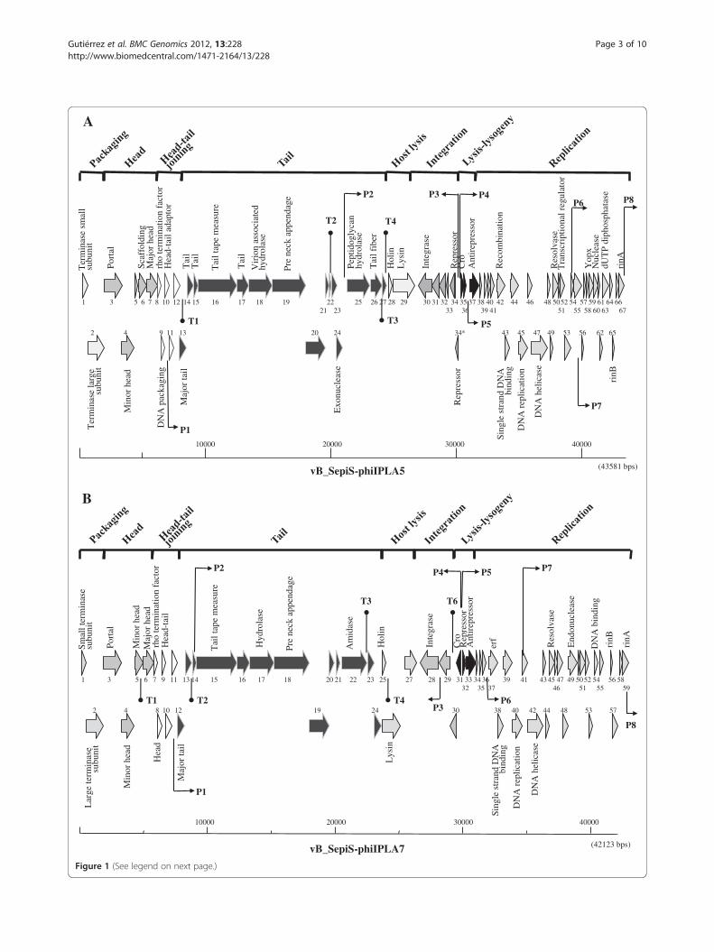
Genome overview of phi-IPLA5 and phi-IPLA7 phagesBoth phages have a linear, double-stranded DNA gen-ome consisting of 43,581 bp encoding 67 putative orfs inphi-IPLA5, while the phi-IPLA7 genome was 42,123 bp
and 59 putative orfs were identified (Additional file 1:Table S1 and Additional file 2: Table S2; Figure 1A and B).The G+C content of phi-IPLA5 and phi-IPLA7 was34.7%, which is slightly higher than that of S. epidermidisstrains (32%) [17]. A BLASTN search revealed that nu-cleotide sequence of phi-IPLA5 and phi-IPLA7 shared ahigh degree of similarity with the other two S. epidermidisphages phiPH15 and phiCNPH82 (64% and 65%, for phi-IPLA5, and 81% and 67% for phi-IPLA7, respectively)[14]. Bioinformatic analysis revealed a similar organizationof the two phages in five functional modules (packaging,structure/morphogenesis, host lysis, lysogeny and replica-tion/regulation) that perfectly fits the general structure ofmost double-stranded DNA bacteriophages [18]. Severalputative promoters and terminators in phi-IPLA5 and inphi-IPLA7 were found by searching for the S. aureus σ70-dependent promoter consensus motif (Figure 1A and B,Additional file 3: Table S3). The deduced promoter posi-tions are consistent with a modular arrangement andimply that there is modular control of gene expression.The putative transcription pattern of both phages revealstwo main groups of orfs running divergently and one ofthem includes the lysogenic cassette.The amino acid (aa) sequences of the predicted orfs were
searched for similarities to sequences from the availabledatabases (Additional file 1: Table S1 and Additional file 2:Table S2). Significant matches were obtained for 36 orfsfrom phi-IPLA5 and 29 orfs from phi-IPLA7 and biologicalfunctions were assigned. No tRNA genes were found. Novirulence genes were clearly identified.In the DNA packaging module, the putative large ter-
minase subunit of phi-IPLA5 and phi-IPLA7 showedhomology (59%) with those belonging to the pac-typephages such as Bacillus subtilis phage SPP1. It has beensuggested that different functional classes of phage-encoded terminases can be predicted from their aminoacid sequence [19]. Confirmation of this result wasobtained by restriction analysis of phi-IPLA5 and phi-IPLA7 DNAs with the endonuclease XbaI. The phagegenomes both have two cut sites but produced two sin-gle bands on agarose gels (data not shown), which sug-gests that both phi-IPLA5 and phi-IPLA7 genomes werecircularly permuted.In the structural module, the predicted major head
and major tail proteins had a molecular mass consistingwith previous protein analysis of virion particles, whichshowed a major polypeptide (34 kDa) in phage phi-IPLA5and two main proteins (27.5 and 34 kDa) in phi-IPLA7, re-spectively [15]. Putative virion-associate hydrolases (phi-IPLA5 gp18 and phi-IPLA7 gp17) with an aminoterminalendopeptidase tail domain and a SGNH hydrolase domainrelated with lipases and esterases, were identified. Finally,pre neck appendage proteins (phi-IPLA5 gp19 and phi-IPLA7 gp18) with pectin-lyase like domains weer identified
Ter
min
ase
smal
l su
buni
t
Port
al
Scaf
fold
ing
Maj
or h
ead
rho
term
inat
ion
fact
orH
ead-
tail
adap
tor
Tai
lT
ail
Tai
l tap
e m
easu
re
Tai
l
Vir
ion
asso
ciat
ed
hydr
olas
e
Pre
neck
app
enda
ge
Pept
idog
lyca
n hy
drol
ase
Tai
l fib
er
Hol
inL
ysin
Inte
gras
e
Rep
ress
orC
roA
ntir
epre
ssor
Rec
ombi
natio
n
Res
olva
seT
rans
crip
tiona
l reg
ulat
or
Yop
xN
ucle
ase
rinA
dUT
P di
phos
phat
ase
Ter
min
ase
larg
e su
buni
t
Min
or h
ead
DN
A p
acka
ging
Maj
or ta
il
Exo
nucl
ease
Rep
ress
or
Sing
le s
tran
d D
NA
bi
ndin
gD
NA
rep
licat
ion
DN
A h
elic
ase
rinB
(43581 bps)
10000 20000 30000 40000
1 3 5 6 7 8 10 12 14 15 16 17 18 19 22 25 26 27 28 29 30 31 32 34 35 37 38 40 42 44 46 48 5052 54 57 59 61 64 6621 23 33 36 39 41 51 55 58 60 63 67
2 4 9 11 13 20 24 34* 43 45 47 49 53 56 62 65
vB_SepiS-phiIPLA5
P2P6 P8
T1
P1
P7
T2 T4
T3
P3 P4
P5
A
(42123 bps)
10000 20000 30000 40000
Lar
ge te
rmin
ase
subu
nit
Min
or h
ead
Hea
d
Maj
or ta
il
Lys
in
Sing
le s
tran
d D
NA
bi
ndin
g
DN
A r
eplic
atio
n
DN
A h
elic
ase
Smal
l ter
min
ase
subu
nit
Port
al
Min
or h
ead
Maj
or h
ead
rho
term
inat
ion
fact
orH
ead-
tail
Tai
l tap
e m
easu
re
Hyd
rola
se
Pre
neck
app
enda
ge
Am
idas
e
Hol
in
Inte
gras
e
Rep
ress
orC
roA
ntir
epre
ssor
erf
Res
olva
se
End
onuc
leas
e
DN
A b
indi
ng
rinB
rinA
1 3 5 6 7 9 11 13 14 15 16 17 18 20 21 22 23 25 27 28 29 31 33 34 36 39 41 43 45 47 49 50 52 54 56 5832 35 37 46 51 55 59
2 4 8 10 12 19 24 30 38 40 42 44 48 53 57
vB_SepiS-phiIPLA7
P1
P2
P8
T1
T3
T2 T4 P6
P5 P7
P3
P4
T6
B
Figure 1 (See legend on next page.)
Gutiérrez et al. BMC Genomics 2012, 13:228 Page 3 of 10http://www.biomedcentral.com/1471-2164/13/228

(See figure on previous page.)Figure 1 Physical and genetic map of phages phi-IPLA5 (A) and phi-IPLA7 (B). The ORFs are sequentially numbered, indicated by arrowsproportional to their lengths and pointing toward their direction of transcription. Some ORFs have been placed below for clarity. The functionalmodules are indicated on top of the scheme, and the names of several putatively or experimentally identified genes are shown. Putativepromoter (P) and terminator (T) sequences are also indicated.
Gutiérrez et al. BMC Genomics 2012, 13:228 Page 4 of 10http://www.biomedcentral.com/1471-2164/13/228
that could also be involved in the extracellular materialdegradation.Phage phi-IPLA5, although strictly lytic, encoded a defi-
cient lysis-lysogeny module. The phi-IPLA5 gp34 and phi-IPLA5 gp34* proteins shared extended similarity withrepressors of the CI type. A one-base replacement thatshifted a TAC codon to the stop codon TAA was mappedat 29941 nt resulting in a second orf (orf34*). No RBS up-stream of orf34* could be detected. The presence of a trun-cated CI repressor in phi-IPLA5 would explain why thephage was unable to lysogenize [15]. In the cI-cro inter-genic regions of both phages, two adjacent and outward-facing putative promoters for cro and repressor genes wereidentified (Figure 1A and B, Additional file 3: Table S3).Additionally, two 7-bp direct repeats overlapping the twoputative promoters in phi-IPLA7 were recognized (datanot shown). These sequences might be putative operatorsfor the binding of CI repressor which have been reportedas regulators in the lysogeny module gene expression [20].In the replication module, both phages contained
DNA replication proteins (phi-IPLA5 gp45 and phi-IPLA7 gp40) with DnaB domains which are essential inreplication initiation, as well as DnaD domains whichare a component of the primosome. phi-IPLA5 gp50and phi-IPLA7 gp45 displayed homology to a Hollidayjunction resolvase (RusA) [21]. phi-IPLA5 gp59 hadhomology with Yopx proteins, an uncharacterized, well-conserved family of proteins found in bacteriophage andprophage regions of Gram-positive bacteria. A putativedUTPase gene gp63 was predicted in phi-IPLA5 genomewhich is highly conserved in several staphylococcal andlactococcal phages [22,23]. Gp65 and gp67 from phi-IPLA5 and gp56 and gp59 from phi-IPLA7 displayedsimilarity to the RinA and RinB family of transcriptionalregulators. Recently, the RinA family proteins have beenshowed as activators required for transcription of thelate operon in temperate staphylococcal phages [24].
Comparative genomics of phi-IPLA5 and phi-IPLA7phagesBLASTN database searches with the complete genome se-quence of phi-IPLA5 and phi-IPLA7 revealed similarity atthe nucleotide level with the two previously described S.epidermidis phages PH15 and CNPH82 and with S. aureusphages such as phiEW, phi29, phi37, phi52A and phi55.Comparison of these phages using Mauve software revealedgene synteny among the four S. epidermidis phages. Fourhomology blocks mostly matched the morphogenesis and
integration, the lysis-lysogeny, replication, and regulationmodules, (Figure 2). Extensive similarity in the head andtail morphogenesis modules was observed among the S.epidermidis phages genomes and other Staphylococcusphages (Figure 2; Additional file 1: Table S1 and Additionalfile 2: Table S2). In fact, S. epidermidis phages PH15 andCNPH82 have been shown to be highly similar to class IIclade C of S. aureus phages [14]. An exception wasobserved in the putative exonuclease encoding gene (orf24)from phi-IPLA5, which was not found in phi-IPLA7 or inother staphylococcal phages. The phi-IPLA5 lysis regionseemed to be rather unique, specifically in the holin encod-ing gene (orf28), to which no similarity was detected withinother S. epidermidis phages. The holin phi-IPLA5 gp28protein showed similarity (50%) to the holin from S. aureusphage 29 while the holin phi-IPLA7 gp25 protein was iden-tical to those in phages PH15 and CNPH82. Similarly, thephi-IPLA5 lysin encoding gene (orf29) did not have anymatching gene in other phages. Moreover, the lysin phi-IPLA5 gp29 protein belongs to the amidase_2 family whilethose of phi.IPLA7 (gp26) and the lysins from S. epidermi-dis phages PH15 and CNPH82 were highly similar (99%)and belong to the amidase_3 family. A similar pattern wasobserved in the region surrounding the integrase gene.The putative integrase gene (orf30) of phi-IPLA5 differedfrom that in phi-IPLA7, and the corresponding protein(gp30) was 70% similar to that of S. aureus phi96, while theintegrase phi-IPLA7 protein (gp28) resembled that of S.epidermidis PH15 and CNPH82. The phage integrasesfrom the newly isolated phages belonged to different fam-ilies (Additional file 1: Table S1 and Additional file 2: TableS2).Finally, no similarity with other phages was detected for
the antirepressor encoding gene (orf33) found in the phi-IPLA7 lysis-lysogeny region, Based on these data, S. epi-dermidis PH15 and CNPH82 and S. aureus phagesphiEW, phi29, phi37, phi52A and phi55 are the closestrelatives to phi-IPLA5 and phi-IPLA7. The extensive simi-larity between these phages is mainly in the morpho-genetic region as previously reported on otherSiphoviridae phages [25]. The observed homology in otherregions or modules supports the modular theory of phageevolution [26].
Incidence and typing of prophages in S. epidermidis strainsPrevious studies have shown that prophages integrated inthe bacterial chromosomes are the most widespread mo-bile genetic elements in S. aureus strains, which tipically

vB_SepiS-phiIPLA5
vB_SepiS-phiIPLA7
Staphylococcus phage PH15
Staphylococcus phage CNPH82
Staphylococcus phage EW
Staphylococcus phage 29
Staphylococcus phage 52A
Staphylococcus phage 37
Staphylococcus phage 55
Exonuclease Holin/lysin Integrase Antirepressor
AntirepressorHolin/lysin Integrase Replication
Figure 2 Alignment of the genome of the two S. epidermidis phages phi-IPLA5 and phi-IPLA7 with those of other Staphylococcus phagesusing the Mauve software. Each block represents a region of the genome sequence that aligned and is homologous to part of another genome.Regions outside blocks lack detectable homology among the input genomes. Inside each block nucleotide sequence similarity is indicated by theheight of the colored bars, while regions that are dissimilar are in white. Lines connecting blocks are indicative of homologous regions.
Gutiérrez et al. BMC Genomics 2012, 13:228 Page 5 of 10http://www.biomedcentral.com/1471-2164/13/228
carry between one and four prophages [27]. However, theprophage content in S. epidermidis has not been deter-mined to date. To approach this, the integrase (int), holin(hol) and major head protein (mhp) genes were selected asmarker modules as previously described in by [28] whostudied genome mosaicism in prophages of S. aureus.Based on the genome wide comparison among phages andfurther in silico analysis of the integrase genes from thefour S. epidermidis phages (phi-IPLA5, phi-IPLA7, PH15and CNPH82), two groups were identified: int1 comprisedby the highly similar int genes from phi-IPLA7, PH15(95%) and CNPH82 (99%) and int2 composed by int phi-IPLA5 to which no similarities were found among them.Likewise, S. epidermidis phage holin genes defined twogroups: hol1 group comprised by hol phi-IPLA7, PH15(91%) and CNPH82 (100%) and hol2 group that includedhol phi-IPLA5. Finally, the phi-IPLA7 mhp gene was verysimilar to phi-IPLA5 (97%) and PH15 (95%) but no coun-terpart was identified in CNPH82. Based on amino acidsequence homology this protein belongs to the phage-
capsid superfamily. The presence or absence of this genegenerated the mhp1 and mhp2 groups, respectively.The presence of prophages in our S. epidermidis col-
lection containing 60 isolates from women breast milkwas investigated by a multiplex PCR to amplify theabove mentioned genes (int, hol and mhp). Prophages ofthe group mhp1 were the most frequent (30%) but noneof the other targeted markers were amplified in thesestrains (Additional file 4: Table S4). About 10% of theisolates were included in the mhp2 group and weremostly associated to the int2 marker. We also observedthat some phage groups were completely absent andothers were less frequent. There were also some strainsin which no amplification was observed pointing to theabsence of prophages or the presence of other genes notdetected by our PCR approach. It is remarkable that, be-sides the 10 S. aureus bacteriophage integrase geneclasses analysed previously [28], many other types ofmodules containing lysogenic functions, DNA replica-tion, packaging, tail appendices and host lysis were

Figure 3 Aminoacid sequence analysis of the virion-associated protein gp18 from phi-IPLA7. A) ClustalW alignment of the predictedbinding site. Positions with a single, fully conserved residue are marked with an asterisk; the colon marks the conserved residues between groupsof strongly similar properties, and the period marks the residues weakly conserved between groups based on the Gonnet PAM 250 matrix, score<0,5. Highlighted are those residues involved in ligand association. B) Predicted 3D structure of phi-IPLA7 gp18. C) Predicted 3D structure of thedomain pectin lyase like (D1) (amino acids 316- 374).
Gutiérrez et al. BMC Genomics 2012, 13:228 Page 6 of 10http://www.biomedcentral.com/1471-2164/13/228
described in S. aureus phages, revealing the high diversityand the mosaic structure of prophages in this species [22].Previous studies suggested that the integrase type
group in S. aureus strains is closely linked to the viru-lence gene content of prophages and might thereforeconvey information about their pathogenic potential[28]. We found that the largest group of S. epidermidisprophages belongs to int2 group, which was mostlyfound in mastitic strains. On the other hand, prevalenceof prophages, as defined by the positive amplification ofat least one marker gene, was higher (82% vs 59%) instrains producing mastitis. This result would support thehypothesis that prophages are directly involved in viru-lence [29]. However, no virulence factors have beendescribed to date in S. epidermidis phages.In our previous work, the yield of mitomycin C indu-
cible prophages in S. epidermidis was rather low (3%) and
we hypothesized that it could be due to the lack of appro-priately sensitive host strains to detect them [15]. In viewof the new PCR results, phage DNA sequences werepresent in 73% of the analyzed S. epidermidis strains. Al-though amplification of at least one of the markers doesnot necessarily mean that the bacterial strains are lysogens(i.e. marker genes in defective prophages), our results sup-port the notion that lysogeny in S. epidermidis may behigher than anticipated and likely more frequent amongclinical strains, as noted previously in S. aureus strains iso-lated from diverse clinical samples [28].
Virion proteins with a putative hydrolytic activity ofextracellular componentsA pectin lyase-like domain (aminoacids 117 to 539) wasidentified in the pre-neck appendage protein of both phages(Additional file 1: Table S1 and Additional file 2: Table S2).

Gutiérrez et al. BMC Genomics 2012, 13:228 Page 7 of 10http://www.biomedcentral.com/1471-2164/13/228
Recently, it has been demonstrated that some tail spike pro-teins similar to pectin lyases have exopolysaccharide (EPS)degrading activity [30]. Similar proteins to phi-IPLA5 gp19and phi-IPLA7 gp18 were identified by BLASTp search inPH15 and CNPH82 phages, S. aureus phage 37, Bacillusphage phi29 and prophages in several Bacillus species(Figure 3A). Surprisingly, most of the phi-IPLA5 gp19 andphi-IPLA7 gp18 related proteins belonged to Bacillus spand not to other staphylococcal species. It is remarkablethat all the proteins analyzed were highly conserved at thepredicted binding site (amino acids 316-374) and the resi-dues responsible for the interaction with the ligand wereshared by the different phages (Figure 3A). To support thepredicted catalytic domains, the software Phyre2 was usedto predict the tridimensional structure (3day) of the proteinphi-IPLA7 gp18 to which, the primary structure of thecounterpart protein from phi-IPLA5 was 98% identical. Tomodel phi-IPLA7 gp18 (Figure 3B), the crystal structure ofthe bacteriophage phi29 gp12 neck protein from Bacillussubtilis [31] was used. 82% of phi-IPLA7 gp18 amino acidswere predicted at a confidence level of 90%. The pectinlyase-like domain (D1) (Figure 3C) displayed a right-handed continuous twelve helix repetition, where each coilof the helix featured three beta-strands and three turnregions. Proteins containing these repeats most often areenzymes with polysaccharide substrates, and it wasdemonstrated that this topology is shared by several pro-teins, with the variation in the number of coils, includingbacterial pectate lyases, fungal and bacterial galacturonasesand phage tail spikes [32]. On the other hand, no amino
A
I
IIS DS R
0
1
2
3
4
5
6
7
8
9
F12 LC016 CJBP3
Log
(CF
U/m
L)
Log
(PF
U/m
L)
Figure 4 Effect of phages A) phi-IPLA5 and B) phi-IPLA7 on a lawn ofdifferent S. epidermidis strains. (S) Sensitive strain; (R) resistant strain; (DS) drbacteria lawn (black) and phage titre (grey) of phages. Each value corresporepresented by bars.
acid sequence homology was detected between phi-IPLA7gp18 and Pseudomonas putida φ15 tail spike protein, al-though this also showed right handed beta helical foldsidentified in carbohydrate depolymerizing enzymes [30].
Biofilm degradation by phi-IPLA5 and phi-IPLA7 phagesThree lines of evidence were pointing to a putative anti-biofilm activity by the newly isolated S. epidermidisphages phi-IPLA5 and phi-IPLA7. First, as previouslyreported [15], a halo surrounding the clear lysis plaquescould be observed which increased with time. Thesehalos are regarded as an indicator for the presence ofphage-associated EPS depolymerases [16,30]. Secondly,a clear zone was previously observed when phages phi-IPLA5 and phi-IPLA7 were dropped on some bacterialstrains (drop-sensitive strains). These strains were resist-ant to infection meaning that no plaques were observedwhen the phage stock was diluted and plated on thesestrains [15]. Third, based on sequence comparison and3D predictions, two candidate proteins were identified asputative EPS degrading enzymes. To investigate if thesephages were involved in biofilm degradation, cell countsand phage titre of the clear zone where the phage sus-pension is dropped, and into the host cell lawn were car-ried out. As expected, lower bacterial counts were foundwhen the phages were dropped onto the sensitive strainS. epidermidis F12 while viable counts of the resistant S.epidermidis CJBP3 remained unaltered (Figure 4). Inter-estingly, the clear zone generated on the drop-sensitivestrains revealed similar bacterial counts to the bacterial
B
I
IIS DS R
0
1
2
3
4
5
6
7
8
9
F12 CJBP1 CJBP3
Log
(CF
U/m
L)
Log
(PF
U/m
L)
S. epidermidis. I) Morphology of phage lytic zone when dropped onop-sensitive strain. II) Bacteria viable number in drop-zone (white),nd with the mean of five different experiments, the standard error is

Gutiérrez et al. BMC Genomics 2012, 13:228 Page 8 of 10http://www.biomedcentral.com/1471-2164/13/228
lawn despite its distinct visual appearance. Moreover, S.epidermidis F12 clearly supported phage propagation,while no phages were detected when sampling the dropzone of the resistant and drop sensitive strains (Figure 4).Overall, these results supports the presence of phage de-polymerase activity, although we can not totally discardour previous hypothesis that ascribed the clear zone ondrop-sensitive strains to “lysis from without” [15].In order to confirm the ability of the phages to medi-
ate biofilm degradation, the three types of strains weregrown in microtiter plates for 7 days for biofilm forma-tion and subsequently challenged with phage phi-IPLA7.Viability of the sensitive S. epidermidis F12 in bothplanktonic and biofilm cells was readily reduced evenafter 1 h in the presence of the phage, and only 5% ofthe total counts survived (ANOVA; p< 0.05) after 3 h(Figure 5). In the case of the drop-sensitive S. epider-midis CJBP1, total cell numbers remained unchanged(ANOVA; p> 0.05) at both 1 h and 3 h. However, a dra-matic shift towards the planktonic state was observed inthe presence of the phage and, after 3 h, and just 2% ofthe cells remained attached (Figure 5B). These data sup-port the ability of phage phi-IPLA7 to degrade the extra-cellular material of the biofilm, releasing the attachedcells. On the contrary, neither the viability nor the bio-film formed by the resistant strain S. epidermidis CJBP3was affected by phage phi-IPLA7 (ANOVA; p> 0.05). Adifferent composition in the extracellular biofilm materialcould explain this result. Studies on staphylococcal biofilmdevelopment suggest that the extracellular matrix consistsof proteins, DNA, and/or polysaccharide (PIA) [1]. Re-cently, it has become evident that some strains are not re-liant on PIA for biofilm formation [33].
ConclusionsIn this work, we have presented a detailed genomic andmolecular characterization of two new S. epidermidisphages, phi-IPLA5 and phi-IPLA7. Based on this, a
A
C PT C PT C PT
F12 CJBP1 CJBP3
S RDS
100
80
60
40
20Via
ble
cells
(%
)
0
Figure 5 Killing of S. epidermidis cells forming a biofilm on microtiterResults are depicted as the percentage of attached cells (dark gray square)treated with SM buffer (C) and in biofilms treated with phage phi-IPLA7 (Pand the standard error is represented by bars.
multiplex PCR was designed that revealed a high preva-lence of prophage and/or defective prophages within S.epidermidis strains. Genome mining detected the pres-ence of virion-associated proteins with a putative EPSdepolymerase activity. To our knowledge, this is the firsttime that degradation of extracellular material has beenascribed to S. epidermidis phages. Further studies toconfirm the role of these proteins in phage-induced bio-fim destructuring are in progress with the hope to setthe foundation of new anti-biofilm strategies.
MethodsBacterial growth conditions and phages propagationS. epidermidis F12 was used as the host strain for phagesphi-IPLA5 and phi-IPLA7 [15]. A total of sixty S. epider-midis strains isolated from women’s breast milk [2] wereused in lysogeny analysis (Additional file 5: Figure S1).Staphylococcal cells were routinely cultured in TSB broth(Triptona Soy Broth, Scharlau, Barcelona, Spain) at 37°Cwith shaking or in TSB plates containing 2% (w/v) bac-teriological agar (TSA). Bacteriophages phi-IPLA5 andphi-IPLA7 were propagated as described previously [15].
Phage genome sequencing and analysisTo prepare bacterial DNA-free samples for sequenceanalysis, the purified phages were treated with DNaseIbovine pancreas (Sigma, Madrid, Spain) and the phageDNA was extracted as previously described [34]. Thegenome sequence of phages phi-IPLA5 and phi-IPLA7was generated by ultra-high throughput GS FLX sequen-cing with 20-fold redundancy on average. Orfs were pre-dicted with Clone Manager 7 version 7.10 software in allreading frames with a threshold of 40 codons. BLASTXand BLASTP (http://www.ncbi.nlm.nih.gov/blast/Blast.cgi) were used to search for homologous proteins. Struc-tural predictions and motif searches were performedwith InterProScan (http://www.ebi.ac.uk/InterProScan/).The search for putative tRNA encoding genes was
B
C PT C PT C PT
F12 CJBP1 CJBP3
RS DS
100
80
60
40
20
0
Via
ble
cells
(%
)
wells by phage phi-IPLA7 after incubation for 1 h (A) and 3 h (B).and planktonic cells (light gray square) detected in control biofilmsT). Each value corresponds with the mean of five different experiments

Gutiérrez et al. BMC Genomics 2012, 13:228 Page 9 of 10http://www.biomedcentral.com/1471-2164/13/228
performed with tRNAscan-SE 1.21 (http://selab.janelia.org/tRNAscan-SE/). σ70 promoter sequences were identi-fied using PPP (Prokaryotic Promoter Prediction at http://bioinformatics.biol.rug.nl/websoftware/ppp/ppp_start.php).The rho-independent terminators were identified usingthe Trans Term program (http://nbc3.biologie.uni-kl.de)and energy was calculated by the mfold web server (http://mfold.rna.albany.edu). Genomic comparisons at the nu-cleotide level were made with Mauve software, using aprogressive alignment with default settings (http://gel.ahabs.wisc.edu/mauve/). 3 day structure prediction of pro-teins was made by using the bioinformatic software Phyre2
(http://gel.ahabs.wisc.edu/mauve/download.php). To pre-dict the binding site of the ligands, the software 3Dligand-site was used (http://www.sbg.bio.ic.ac.uk/3dligandsite/).
Accession numbersThe sequences of phi-IPLA5 and phi-IPLA7 have beendeposited in the GenBank under accession numbersJN192400 and JN192401, respectively.
Lysogeny and prophage typingThe presence of resident prophages was screened by multi-plex PCR using the primers specified in Additional file 5:Figure S1. Total DNA extraction was carried out from S.epidermidis strains by using “GenElute™ Bacterial Gen-omic DNA Kit” (Sigma-Aldrich, Madrid, Spain). PCR reac-tions were performed using the kit ‘PureTaq Ready-To-Go™ PCR Beads’ (GE Healthcare, Munich, Germany). Aspositive control, pure phage DNA from phi-IPLA5 andphi-IPLA7 was used. Gel images were processed using thesoftware Quantity One software (BioRad Laboratories,Hercules, CA). The similarity matrix was calculated on thebasis of the simple matching coefficient, and its corre-sponding dendrogram was deduced using the unweightedpair group method with arithmetic averages.
Microbiological analysis of phage lysis zones and biofilmsTo visualize the lysis area formation, 5 μl of a 108 PFU/ml phage suspension was dropped on a S. epidermidisbacterial lawn and incubated at 37°C. For comparisonbetween the lysis zone and the bacterium lawn zone, anarea with the same volume was removed from eachzone, suspended in 200 μl of SM buffer (20 mM Tris–HCl, 10 mM MgSO4, 10 mM CaCl2, 100 mM NaCl, pH7.5) and vigorously vortexed. Phage titre was determinedusing the double layer agar method, while bacterial countwas determined by plating serial dilutions on TSB agar.To determine the potential of phages to degrade bio-films, S. epidermidis o/n cultures were diluted in freshTSB to 106 CFU/ml, poured into a 96 microwell plate(Thermo Scientific, Madrid, Spain) and incubated during7 day at 37°C. Wells were washed twice with SM bufferand either 220 μl of a phage stock (108 PFU/ml) or 220 μl
of SM buffer were added for test and control purposes,respectively. Plates were incubated for 1 h and 3 h andthen supernatants and adhered cells were collected andplated for bacteria counting. The results were representedas the viable cells percentage respect the total cell numberin the control wells without phage treatment (cells in thesupernatant + cells adhered to the well).Statistical analysis was performed by one-way analysis
of variance (ANOVA) followed by the Bonferroni multi-comparison test. Statistical significance was consideredat p< 0.05.
Additional files
Additional file 1: Table S1. Features of bacteriophage phi-IPLA5 orfs,gene products (gp) and functional assignments.
Additional file 2: Table S2. Features of bacteriophage phi-IPLA7 orfs,gene products (gp) and functional assignments.
Additional file 3: Table S3. Putative promoters and terminatorssequences of phi-IPLA5 and phi-IPLA7. -10 and -35 boxes are underlined.Nucleotide positions and presence of the TG dinucleotide were alsoindicated. At terminator sequences nucleotides in the stem-loopstructure are underlined.
Additional file 4: Table S4. Primers used for multiplex PCR reactions.
Additional file 5: Figure S1. Multiplex PCR detecting the integrase,holin and major head protein genes in S. epidermidis strains. (H) Strainsisolated from healthy woman, (M) strains isolated from mastitic women.Absence and presence of a specific gene is represented by white andgrey boxes, respectively.
Competing interestsThe authors declare that they have no competing interests.
Authors’ contributionsDG and PG performed bioinformatics analyses of nucleotide and proteinsequences. PG, BM and AR designed the study, obtained funding and wrotethe manuscript. All authors read and approved the final manuscript.
AcknowledgmentsThis research study was supported by grants AGL2009-13144-C02-01(Ministry of Science and Innovation, Spain) and PIE200970I090 (CSIC, Spain).We acknowledge support of the publication fee by the CSIC Open AccessPublication Support Initiative through its Unit of Information Resources forResearch (URICI). D. G. is a fellow of the Ministry of Science and Innovation,Spain. We would like to thank R. Calvo (IPLA-CSIC) for her technicalassistance. We also thank C. Billington (ESR, New Zealand) for critical readingof the manuscript and helpful discussions.
Received: 21 November 2011 Accepted: 17 May 2012Published: 8 June 2012
References1. Otto M: Staphylococcus epidermidis the “accidental” pathogen. Nat Rev
2009, 7:555–567.2. Delgado S, Arroyo R, Jiménez E, Marín ML, Del Campo R, Fernández L,
Rodríguez JM: Staphylococcus epidermidis strains isolated from breastmilk of women suffering infectious mastitis: potential virulence traitsand resistance to antibiotics. BMC Microbiol 2009, 9:82.
3. Oliveira M, Nunes SF, Carneiro C, Bexiga R, Bernardo F, Vilela CL: Timecourse of biofilm formation by Staphylococcus aureus and Staphylococcusepidermidis mastitis isolates. Vet Microbiol 2007, 124:187–191.
4. Jabbouri S, Sadovskaya I: Characteristics of the biofilm matrix and its roleas a possible target for the detection and eradication of Staphylococcusepidermidis associated with medical implant infections. FEMS ImmunolMed Microbiol 2010, 59:280–291.

Gutiérrez et al. BMC Genomics 2012, 13:228 Page 10 of 10http://www.biomedcentral.com/1471-2164/13/228
5. Schoenfelder SM, Lange C, Eckart M, Hennig S, Kozytska S, Ziebuhr W:Success through diversity - how Staphylococcus epidermidis establishesas a nosocomial pathogen. Int J Med Microbiol 2010, 300:380–386.
6. Kozitskaya S, Cho SH, Dietrich K, Marre R, Naber K, Ziebuhr W: The bacterialinsertion sequence element IS256 occurs preferentially in nosocomialStaphylococcus epidermidis isolates: association with biofilm formationand resistance to aminoglycosides. Infect Immun 2004, 72:1210–1215.
7. Madhusoodanan J, Seo KS, Remortel B, Park JY, Hwang SY, Fox LK, Park YH,Deobald CF, Wang D, Liu S, Daugherty SC, Gill AL, Bohach GA, Gill SR: Anenterotoxin-bearing pathogenicity island in Staphylococcus epidermidis.J Bacteriol 2011, 193:1854–1862.
8. Kutateladze M, Adamia R: Bacteriophages as potential new therapeuticsto replace or supplement antibiotics. Trends Biotechnol 2010, 28:591–595.
9. Fischetti VA: Bacteriophage endolysins: a novel anti-infective to controlGram-positive pathogens. Int J Med Microbiol 2010, 300:357–362.
10. Donlan RM: Preventing biofilms of clinically relevant organisms usingbacteriophage. Trends Microbiol 2009, 17:66–72.
11. Cerca N, Oliveira R, Azeredo J: Susceptibility of Staphylococcus epidermidisplanktonic cells and biofilms to the lytic action of Staphylococcusbacteriophage K. Lett Appl Microbiol 2007, 45:313–317.
12. Curtin JJ, Donlan RM: Using bacteriophages to reduce formation ofcatheter-associated biofilms by Staphylococcus epidermidis. AntimicrobAgents Chemother 2006, 50:1268–1275.
13. Aswani V, Tremblay DM, Moineau S, Shukla SK: Staphylococcus epidermidisbacteriophages from the anterior nares of humans. Appl Environ Microbiol2011, 77:7853–7855.
14. Daniel A, Bonnen PE, Fischetti VA: First complete genome sequence oftwo Staphylococcus epidermidis bacteriophages. J Bacteriol 2007,189:2086–2100.
15. Gutiérrez D, Martínez B, Rodríguez A, García P: Isolation andcharacterization of bacteriophages infecting Staphylococcus epidermidis.Curr Microbiol 2010, 61:601–608.
16. Hughes KA, Sutherland IW, Jones MV: Biofilm susceptibility tobacteriophage attack: the role of phage-borne polysaccharidedepolymerase. Microbiology 1998, 144:3039–3047.
17. Cates S: NCBI: National Center for Biotechnology Information. 2006.http://cnx.org/content/m11789/1.3/
18. Brüssow H, Desiere F: Comparative phage genomics and the evolution ofSiphoviridae: insights from dairy phages. Mol Microbiol 2001, 39:213–222.
19. Casjens SR, Gilcrease EB, Winn-Stapley DA, Schicklmaier P, Schmieger H,Pedulla ML, Ford ME, Houtz JM, Hatfull GF, Hendrix RW: The generalizedtransducing Salmonella bacteriophage ES18: complete genomesequence and DNA packaging strategy. J Bacteriol 2005, 187:1091–1104.
20. García P, Ladero V, Alonso JC, Suárez JE: Cooperative interaction of CIprotein regulates lysogeny of Lactobacillus casei by bacteriophage A2.J Virol 1999, 73:3920–3929.
21. Mahdi AA, Sharples GJ, Mandal TN, Lloyd RG: Holliday junction resolvasesencoded by homologous rusA genes in Escherichia coli K-12 and phage82. J Mol Biol 1996, 257:561–573.
22. Kahánková J, Pantůček R, Goerke C, Růžičková V, Holochová P, Doškař J:Multilocus PCR typing strategy for differentiation of Staphylococcusaureus siphoviruses reflecting their modular genome structure. EnvironMicrobiol 2010, 12:2527–2538.
23. Labrie S, Moineau S: Complete genomic sequence of bacteriophage ul36:demonstration of phage heterogeneity within the P335 quasi-species oflactococcal phages. Virol 2002, 296:308–320.
24. Ferrer MD, Quiles-Puchalt N, Harwich MD, Tormo-Más MA, Campoy S, Barbé J,Lasa I, Novick RP, Christie GE, Penadés JR: RinA controls phage-mediatedpackaging and transfer of virulence genes in Gram-positive bacteria.Nucleic Acids Res 2011, 39:5866–5878.
25. Proux C, van Sinderen D, Suarez J, García P, Ladero V, Fitzgerald GF, Desiere F,Brüssow H: The dilemma of phage taxonomy illustrated by comparativegenomics of Sfi21-like Siphoviridae in lactic acid bacteria. J Bacteriol 2002,184:6026–6036.
26. Brüssow H, Canchaya C, Hardt WD: Phages and the evolution of bacterialpathogens: from genomic rearrangements to lysogenic conversion.Microbiol Mol Biol Rev 2004, 68:560–602.
27. Pantůček R, Doskar J, Růzicková V, Kaspárek P, Orácová E, Kvardová V,Rosypal S: Identification of bacteriophage types and their carriage inStaphylococcus aureus. Arch Virol 2004, 149:1689–1703.
28. Goerke C, Pantůček R, Holtfreter S, Schulte B, Zink M, Grumann D, Bröker BM,Doskar J, Wolz C: Diversity of prophages in dominant Staphylococcus aureusclonal lineages. J Bacteriol 2009, 191:3462–3468.
29. Boyd EF, Brüssow H: Common themes among bacteriophage-encodedvirulence factors and diversity among the bacteriophages involved.Trends Microbiol 2002, 10:521–529.
30. Cornelissen A, Ceyssens PJ, T’Syen J, Van Praet H, Noben JP, Shaburova OV,Krylov VN, Volckaert G, Lavigne R: The T7-related Pseudomonas putidaphage φ15 displays virion-associated biofilm degradation properties.PLoS One 2011, 6:e18597.
31. Xiang Y, Leiman PG, Li L, Grimes S, Anderson DL, Rossmann MG:Crystallographic insights into the autocatalytic assembly mechanism of abacteriophage tail spike. Mol Cell 2009, 34:375–386.
32. Jenkins J, Mayans O, Pickersgill R: Structure and evolution of parallelbeta-helix proteins. J Struct Biol 1998, 122:236–246.
33. Tormo MA, Knecht E, Götz F, Lasa I, Penadés JR: Bap-dependent biofilmformation by pathogenic species of Staphylococcus: evidence ofhorizontal gene transfer? Microbiology 2005, 151:2465–2475.
34. Sambrook J, Fritsch EF, Maniatis T: Molecular cloning: a laboratory manual.2nd edition. Cold Spring Harbor, NY, Cold Spring Harbor Laboratory; 1989.
doi:10.1186/1471-2164-13-228Cite this article as: Gutiérrez et al.: Genomic characterization of twoStaphylococcus epidermidis bacteriophages with anti-biofilm potential.BMC Genomics 2012 13:228.
Submit your next manuscript to BioMed Centraland take full advantage of:
• Convenient online submission
• Thorough peer review
• No space constraints or color figure charges
• Immediate publication on acceptance
• Inclusion in PubMed, CAS, Scopus and Google Scholar
• Research which is freely available for redistribution
Submit your manuscript at www.biomedcentral.com/submit