Removal of adenovirus, calicivirus, and bacteriophages by conventional drinking water treatment
8
This article was downloaded by:[University of Arizona] On: 24 January 2008 Access Details: [subscription number 770844067] Publisher: Taylor & Francis Informa Ltd Registered in England and Wales Registered Number: 1072954 Registered office: Mortimer House, 37-41 Mortimer Street, London W1T 3JH, UK Journal of Environmental Science and Health, Part A Toxic/Hazardous Substances and Environmental Engineering Publication details, including instructions for authors and subscription information: http://www.informaworld.com/smpp/title~content=t713597268 Removal of adenovirus, calicivirus, and bacteriophages by conventional drinking water treatment Morteza Abbaszadegan a ; Patricia Monteiro b ; Nena Nwachuku c ; Absar Alum a ; Hodon Ryu a a National Science Foundation Water Quality Center, Department of Civil and Environmental Engineering, Arizona State University, Tempe, Arizona, USA b Department of Civil and Environmental Engineering, University of Brasilia, Brasilia, Brazil c Office of Water, Office of Science and Technology, Environmental Protection Agency (EPA), Washington, DC, USA Online Publication Date: 01 January 2008 To cite this Article: Abbaszadegan, Morteza, Monteiro, Patricia, Nwachuku, Nena, Alum, Absar and Ryu, Hodon (2008) 'Removal of adenovirus, calicivirus, and bacteriophages by conventional drinking water treatment', Journal of Environmental Science and Health, Part A, 43:2, 171 - 177 To link to this article: DOI: 10.1080/10934520701781541 URL: http://dx.doi.org/10.1080/10934520701781541 PLEASE SCROLL DOWN FOR ARTICLE Full terms and conditions of use: http://www.informaworld.com/terms-and-conditions-of-access.pdf This article maybe used for research, teaching and private study purposes. Any substantial or systematic reproduction, re-distribution, re-selling, loan or sub-licensing, systematic supply or distribution in any form to anyone is expressly forbidden. The publisher does not give any warranty express or implied or make any representation that the contents will be complete or accurate or up to date. The accuracy of any instructions, formulae and drug doses should be independently verified with primary sources. The publisher shall not be liable for any loss, actions, claims, proceedings, demand or costs or damages whatsoever or howsoever caused arising directly or indirectly in connection with or arising out of the use of this material.
-
Upload
independent -
Category
Documents
-
view
3 -
download
0
Transcript of Removal of adenovirus, calicivirus, and bacteriophages by conventional drinking water treatment

This article was downloaded by:[University of Arizona]On: 24 January 2008Access Details: [subscription number 770844067]Publisher: Taylor & FrancisInforma Ltd Registered in England and Wales Registered Number: 1072954Registered office: Mortimer House, 37-41 Mortimer Street, London W1T 3JH, UK
Journal of Environmental Scienceand Health, Part AToxic/Hazardous Substances andEnvironmental EngineeringPublication details, including instructions for authors and subscription information:http://www.informaworld.com/smpp/title~content=t713597268
Removal of adenovirus, calicivirus, and bacteriophagesby conventional drinking water treatmentMorteza Abbaszadegan a; Patricia Monteiro b; Nena Nwachuku c; Absar Alum a;Hodon Ryu aa National Science Foundation Water Quality Center, Department of Civil andEnvironmental Engineering, Arizona State University, Tempe, Arizona, USAb Department of Civil and Environmental Engineering, University of Brasilia, Brasilia,Brazil
c Office of Water, Office of Science and Technology, Environmental Protection Agency (EPA), Washington, DC, USA
Online Publication Date: 01 January 2008To cite this Article: Abbaszadegan, Morteza, Monteiro, Patricia, Nwachuku, Nena, Alum, Absar and Ryu, Hodon (2008)'Removal of adenovirus, calicivirus, and bacteriophages by conventional drinking water treatment', Journal ofEnvironmental Science and Health, Part A, 43:2, 171 - 177To link to this article: DOI: 10.1080/10934520701781541URL: http://dx.doi.org/10.1080/10934520701781541
PLEASE SCROLL DOWN FOR ARTICLE
Full terms and conditions of use: http://www.informaworld.com/terms-and-conditions-of-access.pdf
This article maybe used for research, teaching and private study purposes. Any substantial or systematic reproduction,re-distribution, re-selling, loan or sub-licensing, systematic supply or distribution in any form to anyone is expresslyforbidden.
The publisher does not give any warranty express or implied or make any representation that the contents will becomplete or accurate or up to date. The accuracy of any instructions, formulae and drug doses should beindependently verified with primary sources. The publisher shall not be liable for any loss, actions, claims, proceedings,demand or costs or damages whatsoever or howsoever caused arising directly or indirectly in connection with orarising out of the use of this material.

Dow
nloa
ded
By:
[Uni
vers
ity o
f Ariz
ona]
At:
23:0
2 24
Jan
uary
200
8 Journal of Environmental Science and Health Part A (2008) 43, 171–177Copyright C© Taylor & Francis Group, LLCISSN: 1093-4529 (Print); 1532-4117 (Online)DOI: 10.1080/10934520701781541
Removal of adenovirus, calicivirus, and bacteriophages byconventional drinking water treatment
MORTEZA ABBASZADEGAN1, PATRICIA MONTEIRO2, NENA NWACHUKU3, ABSAR ALUM1
and HODON RYU1
1National Science Foundation Water Quality Center, Department of Civil and Environmental Engineering, Arizona State University,Tempe, Arizona, USA2Department of Civil and Environmental Engineering, University of Brasilia, Brasilia, Brazil3Office of Water, Office of Science and Technology, Environmental Protection Agency (EPA), Washington, DC, USA
This study was conducted to evaluate the removal of adenovirus, feline calicivirus (FCV), and bacteriophages MS-2, fr, PRD-1, and�X-174 during conventional drinking water treatment using ferric chloride as a coagulant. Adenovirus and FCV were removed toa greater extent than PRD-1 and �X-174, indicating that these bacteriophages may be appropriate surrogates for both adenovirusand FCV. Of the four bacteriophages studied in the pilot plant, MS-2 was removed to the greatest extent (5.1 log), followed by fr (4.9log), PRD-1 (3.5 log), and �X-174 (1.3 log). The virus removal trend in the pilot-scale testing was similar to the bench-scale testing;however, the bench-scale testing seemed to provide a conservative estimate of the pilot plant performance. In the pilot-scale testing,MS-2 and fr were removed with the greatest efficiency during filtration, whereas PRD-1 and �X-174 showed the greatest removalduring sedimentation.
Keywords: Adenovirus, calicivirus, MS-2, fr, PRD-1, �X-174, conventional drinking water treatment, pilot plant study.
Introduction
Conventional drinking water treatment processes suchas coagulation, sedimentation, filtration and disinfectionhave demonstrated efficiency in the removal of a vari-ety of microorganisms, including viruses, bacteria, andprotozoa.[1−3] In the United States, alum and ferric chlorideare the most commonly used coagulants in water treatmentapplications.[4] Ferric chloride is known to remove natu-ral organic matter more efficiently than alum;[4,5] however,there is no apparent consensus regarding coagulant selec-tion for the removal of microorganisms during drinkingwater treatment processes.
During a survey conducted in 2003 by American Wa-ter Works Association Research Foundation, 86% of wa-ter utilities in the United States and Canada are switchingfrom the use of alum to some other coagulant, such as ferricsulfate, ferric chloride or polyaluminum chloride.[6] Conse-quently, there is a need to evaluate various coagulants forthe removal of microorganisms. In the present study, ferric
Address correspondence to Morteza Abbaszadegan, Depart-ment of Civil and Environmental Engineering, Arizona StateUniversity, ECG 252, Tempe, AZ 85287-5306, USA; E-mail:[email protected] May 18, 2007.
chloride was used to remove contaminants druing conven-tional water treatment processes.
The U.S. Environmental Protection Agency (EPA) peri-odically revises the drinking water Contaminant CandidateList (CCL), which is a list of unregulated contaminants. Thesecond CCL list (CCL-2) published in February 2005 in-cludes 9 microorganisms and 42 chemical contaminants.[7]
These microorganisms may be subjected to future regula-tions based on their public health significance, occurrence,and susceptibility to treatment and disinfection methods.However, few studies on the removal of CCL microorgan-isms have been performed.[2]
The objective of this study was to evaluate the removalof adenovirus and calicivirus (CCL viruses) and potentialviral surrogates (process indicators) during pilot-scale con-ventional drinking water treatment processes. Adenovirustype 4 and feline calicivirus-which are commonly used lab-oratory surrogates for enteric adenoviruses type 40 and 41and human caliciviruses, such as norovirus, were used inthis study.
Feline calicivirus (FCV) is not actually a CCL virus.However, it is the accepted laboratory model for studiesof the removal and inactivation of human caliciviruses, forwhich an in vitro cell culture technique is not yet available.Thus, FCV was used to represent the physical removal of theCCL caliciviruses throughout this study. Future advances

Dow
nloa
ded
By:
[Uni
vers
ity o
f Ariz
ona]
At:
23:0
2 24
Jan
uary
200
8 172 Abbaszadegan et al.
Table 1. Characteristics of CCL viruses and potential surrogate bacteriophages
Bacteriophage Bacterial host Size Isoelectric point Genetic structure
MS-2 Escherichia coli 24 nm[14]– 27 nm[18] 3.5[2]– 3.9[14] Single-stranded(ATCC 15597-B1) (ATCC 15597) RNA
PRD-1 Salmonella typhimurium 62 nm[18]– 65 nm[19] 3.0[18]– 4.2[2] Double-stranded(ATCC BAA-769-B1) LT2 (ATCC 19585) DNA
�X-174 Escherichia coli 23 nm[18]–27 nm[14] 6.6[14] Single-stranded(13706-B1) (ATCC 13706) DNA
fr Escherichia coli 23 nm[20] 8.9[2]– 9.0[2] Single-stranded(ATCC 15767-B1) (ATCC 19853) RNA
CCL virus Cell line Size Isoelectric point Genetic structure
Adenovirus PLC/PRF/5 70 nm[4] – 100 nm[21] Undetermined Double-stranded(ATCC CRL-8024) DNA
Feline Calicivirus CRFK 27 nm[22] – 40 nm[22] Undetermined Single-stranded(ATCC CCL-94) RNA
in in vitro techniques may allow for the direct study of hu-man calicivirus removal and inactivation, which would beof great interest.
The results of this study will aid in building a databaseof treatment efficiencies of enteric viruses and their sur-rogates and will help in the decision making process forfuture regulatory considerations of CCL viruses. In thisstudy, bacteriophages, which represent a range of physicalcharacteristics, as listed in Table 1, were selected as poten-tial CCL virus surrogates. Suitable surrogate microorgan-isms would yield removal profiles similar to those of theCCL viruses. Surrogates would be valuable since in vitrocell culture methods, which are used in the standard assaytechnique for animal viruses, are labor intensive and timeconsuming and require specialized equipment and exper-tise. Thus, it was important to study bacteriophages in anattempt to identify appropriate surrogate microorganismsfor CCL viruses.
Materials and methods
Source water and coagulation chemicals
Raw water from the Chandler Water Treatment Plant(CWTP) in Chandler, AZ, was used as source waterthroughout this investigation. On average, water turbiditywas less than 0.2 NTU, pH was approximately 8.0, andalkalinity ranged from 200 to 220 mg/L as CaCO3. Vari-ations in water quality parameters have been reported tosignificantly impact the performance of coagulation, sedi-mentation, and filtration.[4,8]
A stock solution of ferric chloride (ferric chloride hex-ahydrate lumps, Sigma Chemical Co., St. Louis, MO) wasprepared for all jar tests. A cationic polymer, polydial-lyldimethyl ammonium chloride (polyDADMAC) (Clar-ifloc 350, Polydyne, Inc., Riceboro, GA), was used inconjunction with ferric chloride to improve floc settleability.
Jar testing
All jar test experiments were performed using a 6-jar ap-paratus (PB-700 Phipps & Bird Richmond, VA). For eachjar test experiment, a 10-L raw water sample was collectedfrom the CWTP. The sample was transported and storedunder darkened conditions at 4◦C. Jar tests were conductedwithin 48 h of sample collection. Prior to experimentation,the water sample was allowed to return to room tempera-ture (25◦C). The water was then seeded with a known num-ber of test microorganisms (1 × 10⁶ plaque forming units[PFU] per mL of each bacteriophage or 1 × 10⁶ 50% tis-sue culture infective dose [TCID50] per mL of each virus).The seeded sample was thoroughly mixed for 15 min beforealiquoting 1.5-L to each of the jars. Ferric chloride andcationic polymer were immediately added to each jar. Fer-ric chloride doses are strictly dependent on water quality,and are therefore widely variable.[8] In this study, coagu-lant doses ranging from 0 to 50 mg/L FeCl3 were testedusing increments of 10 mg/L. Polymer was added at a doseof 0.4 mg/L. Spiked raw water served as the control withrespect to initial virus concentrations.
The jar testing protocol used throughout this study wasbased on the protocol described by Volk et al.[9] Imme-diately following chemical addition, rapid mixing was ini-tiated at 100 rpm for 1 min. Mixing was slowed for theflocculation stage, which consisted of two 10-min mixingperiods at 40 and 20 rpm, respectively. Finally, the paddleswere extracted and the jars were allowed to settle withoutany disturbance for 30 min. Samples were collected fromthe center of each jar, approximately two inches below thesurface. The samples were immediately assayed for bacte-riophages or CCL viruses.
Pilot plant facility
After optimizing the coagulant dose using the jar test ap-paratus, pilot plant experiments were performed at the

Dow
nloa
ded
By:
[Uni
vers
ity o
f Ariz
ona]
At:
23:0
2 24
Jan
uary
200
8 Removal of adenovirus, calicivirus, and bacteriophages 173
Fig. 1. Schematic for the city of Chandler’s pilot plant.
CWTP’s pilot plant facility using the surrogate bacterio-phages; animal viruses were not permitted at the watertreatment facility due to safety considerations. The pilotplant treatment train included coagulation, flocculation,sedimentation, and filtration. A schematic is provided inFigure 1. Full-scale plant parameters such as hydraulic res-idence time (HRT), chemical dosing, mixing, and surfaceloading rates were mimicked by the small-scale pilot plant,as shown in Table 2. Based on full-scale facility flows, a fastflowrate of 0.5 gpm (1.9 L/min) and a slow flow rate of 0.25gpm (1.0 L/min) were selected. The plant was allowed tooperate for 3 HRTs prior to sampling such that steady stateconditions were achieved (6 and 12 h for fast and slow flowrates, respectively).
Table 2. Pilot plant hydraulic parameters
Unit process Low flow (0.25 gpm) High flow (0.5 gpm)
Coagulation / FlocculationTank Volume 11 gal (42 L) = 3.7 gal/tank * 3 tanksMixing Tank 1: 35 rpm; Tank 2: 25 rpm; Tank 3: 15 rpmHydraulic Residence Time 43.2 min = 14.4 min/tank 28.8 min = 9.6 min/tank
SedimentationTank Volume 52.2 gal (198 L)Mixing NoneHydraulic Residence Time 3.5 hr 1.7 hr
FiltrationFilter Area 0.05 ft2 (46.45 cm2)Filtration Rate 5 gpm/ft2 (12 m/hr) 10 gpm/ft2 (24 m/hr)
The addition of bacteriophages, ferric chloride, andpolymer was regulated using adjustable peristaltic pumps.Seeded water (final concentration of 1 × 10⁶ PFU/mL ofeach bacteriophage) was mixed by an inline static mixerbefore entering a series of three coagulation/flocculationchambers separated by diffusion walls, each equipped witha propeller type mixer. The mixers were staged to emulatethe full-scale plant tapered flocculation sequence using pro-gressively lower mixing intensities (35 rpm in the first tank,followed by 25 and 15 rpm in the subsequent tanks). Thewater was then directed through the sedimentation basinbefore entering the dual media filters.
The filters consisted of 3-in diameter acrylic columns con-taining 1.5 ft of gravel support structure, topped with 1 ft of
Dow
nloa
ded
By:
[Uni
vers
ity o
f Ariz
ona]
At:
23:0
2 24
Jan
uary
200
8 174 Abbaszadegan et al.
sand, followed by 2 ft of granular activated carbon (GAC).Water samples were collected at three locations: influent,settled (after sedimentation), and filtered water. Physico-chemical parameters were analyzed onsite. The samples forbacteriophages analyses were transported to the labora-tory in darkened conditions at 4◦C and were immediatelyassayed.
Bacteriophage propagation and assays
The bacteriophages and bacterial hosts listed in Table 1were obtained from the American Type Culture Collection(ATCC, Rockville, MD). Bacteriophages were propagatedusing the double agar layer (DAL) method.[10] Approxi-mately 107 PFU of bacteriophage, 1 mL of host bacteria,and 5 mL of 0.7% molten tryptic soy agar (TSA) (Difco,Detroit, MI) were mixed and poured onto 1.5% TSA bot-tom agar plates. After solidifying, the plates were incubatedat 37◦C overnight. Bacteriophage stocks were collected thefollowing day by adding 10 mL of 1× phosphate buffersaline (PBS) to the surface of the plate and allowing it toincubate at room temperature for 1 hr. The supernatant wascollected and centrifuged at 8000 × g for 15 min. The pelletwas discarded and the supernatant containing the bacterio-phages was stored at 4◦C. Typically, stock concentrationswere on the order of 1010 PFU/mL. New stocks were prop-agated once the titer dropped below 108 PFU/mL.
The water samples collected from jar tests and pilot plantexperiments were serially diluted (10-fold) in PBS (sufficientfor up to 5-log removal) and were promptly assayed forbacteriophages using the DAL method.[10] The plates wereincubated at 37◦C overnight, and plaques were counted af-ter 12 h. All the assays were performed in triplicate, andpositive and negative controls were included in each set ofassays and for each bacteriophage.
CCL virus propagation and assays
Adenovirus type 4 (Ad4, ATCC VR-4) was obtained fromthe ATCC and was cultured using primary liver cancercells (PLC/PRF/5, ATCC CRL-8024) in 1 × Eagle’s mini-mum essential medium (MEM) containing 10% fetal bovineserum (FBS). Feline calicivirus (FCV, ATCC VR-652) wasobtained from the ATCC and was cultured using Crandallfeline kidney cells (CRFK, ATCC CCL-94) in MEM con-taining 10% heat-inactivated equine serum. The MEM wassupplemented with 1.5 g/L sodium bicarbonate, 15 mMHepes, 2 mM L-glutamine, 0.1 mM non-essential aminoacids, 1 mM sodium pyruvate, 100 µg/mL antimycotic,and 100 µg/mL kanamycin sulfate.
Viruses were propagated by inoculating the host cellmonolayer with approximately 1 × 10⁵ TCID50/mL. Theflasks were incubated at 37◦C until at least 90% of the cellswere infected. Cells were then subjected to freeze/thaw cy-cles: one cycle for Ad4- and three cycles for FCV-infectedcells.[11] The supernatant was centrifuged at 4◦C at 8000 × g
for 15 min to remove cellular debris. The virus lysate wasfurther purified and concentrated using polyethylene glycol(PEG) precipitation, as described by Thurston-Enriquezet al.[11] Briefly, the lysate was augmented with 9% PEG(MW 8000) and 1 M NaCl and was allowed to stir overnightat 4◦C. It was then centrifuged at 4◦C at 8000 × g for 90 min.The supernatant was discarded and the pellet was resus-pended in 10% of its original volume of PBS. To removelipids and facilitate the monodispersion of virus clumps, aVertrel XF (Micro Care Marketing Services, New Britain,CT) extraction was performed by centrifuging a suspen-sion of equal parts Vertrel and virus at 4◦C at 8000 × g for90 min. The purified viruses were stored at −80◦C.
Following jar test experiments, the water samples were se-rially diluted (10-fold) in 1× PBS (sufficient for up to 4.5-logremoval) and were promptly assayed using in-vitro cell cul-ture methodology. Cells were grown in 24-well cell culturetrays. Each sample dilution was inoculated into four wells,using 0.1 mL of sample per well. The trays were incubatedin a 5% CO2 incubator at 37◦C and were examined dailyfor cytopathogenic effects (CPE) for up to 14 days. Typi-cally, CPE manifested as cell enlargement, rounding, andthe formation of grape-like clusters.[12] The Karber TCID50method was used to quantify the viral concentration of eachsample. The Karber TCID50 equation is a statistical ap-proach used to estimate the concentration at which 50% ofthe inoculated wells are positive for infection[11] as follows:
TCID50 = 10−(�−α(S−0.5)) (1)
where:� = the log10 of the most diluted sample with 100%
infectivity,α = the log dilution factor, andS = the summation of the normalized positive infections
including both the last 100% and the first 0% ofinfectious dilutions.
Statistical analysis
Statistical analyses were performed to determine if the meanmicrobial removals resulting from different treatment con-ditions were statistically different at a significance level ofα = 0.05. SPSS (Chicago, IL) Version 12.0 statistical soft-ware was used for the analyses. An analysis of variance(ANOVA) test was used to test the equality of means. TheLevene statistic was computed to test the ANOVA assump-tion of equal variances. The Welch test of equality of meanswas used for data not satisfying the equal variances as-sumption. Upon determination that the means were sig-nificantly different, post hoc tests were used to determinewhich means were different from one another. Tukey’s Hon-estly Significantly Different (HSD) test was used to test datawith equal variances, while the Games-Howell test was usedto analyze data, which did not satisfy the equal variancesassumption.

Dow
nloa
ded
By:
[Uni
vers
ity o
f Ariz
ona]
At:
23:0
2 24
Jan
uary
200
8 Removal of adenovirus, calicivirus, and bacteriophages 175
Fig. 2. Average log removal of MS-2 and PRD-1 at varying FeCl3 doses in the jar test. Error bars represent one standard deviation,and all the data points are an average of 5 experimental runs with duplicate assays.
Results and discussion
Optimization experiments
Bench-scale jar tests were performed to determine the opti-mum doses of ferric chloride for the removal of viruses. Ini-tially, FeCl3 concentrations of 0, 10, 20, 30, 40 and 50 mg/Lwere tested for the removal of the bacteriophages MS-2and PRD-1. Increasing the FeCl3 concentration from 10 to40 mg/L resulted in a steady increase in the log removal ofthe bacteriophages (Fig. 2). However, no such increases inthe removal of MS-2 and PRD-1 were observed by increas-ing the FeCl3 concentration from 40 to 50 mg/L (Fig. 2).Based on the initial results, FeCl3 concentration of 20 and40 mg/L were selected (as lower and higher thresholds)for experiments with adenovirus, feline calicivirus (FCV),fr and �X-174. MS-2 was generally removed to the great-est extent (1.62 and 2.13 log removal at 20 and 40 mg/l ofFeCl3, respectively), followed by fr, PRD-1, and �X-174(Fig. 3). These data support the finding of reported stud-ies that male-specific bacteriophages such as MS-2 and fr
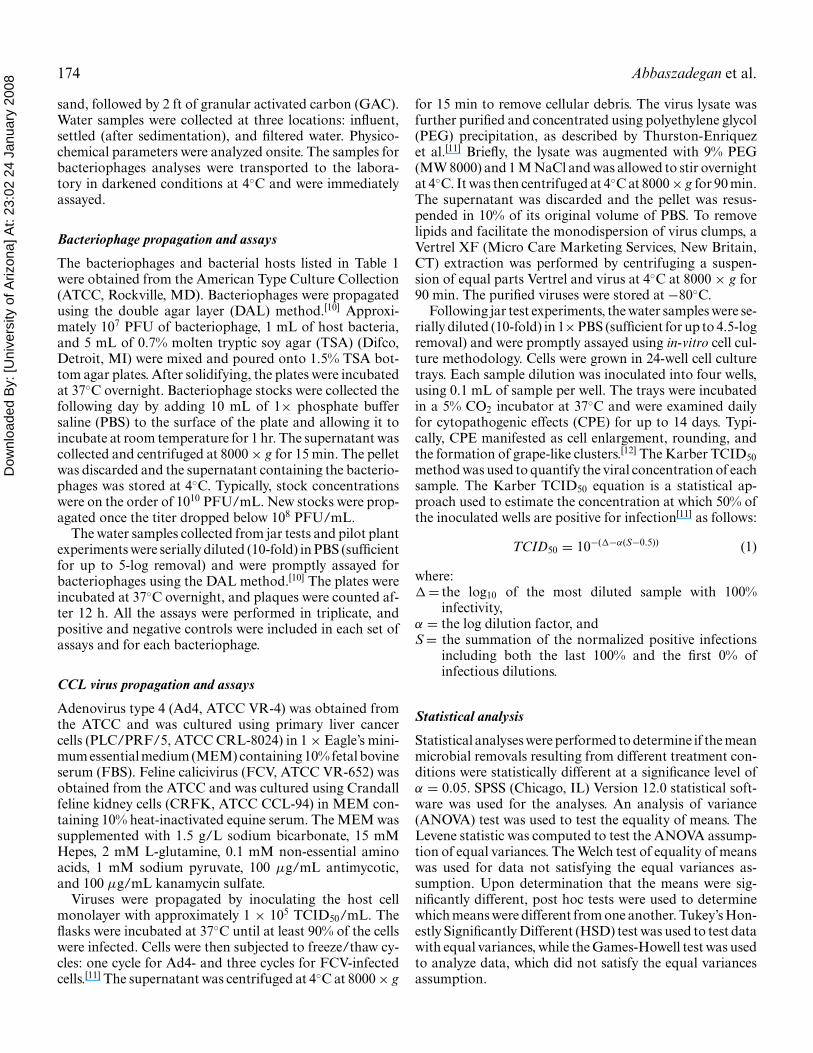
Fig. 3. Average log removal of CCL viruses and bacteriophages at 20 and 40 mg/l of FeCl3 in the jar test. Error bars represent onestandard deviation, and all the data points are an average of 3 experimental runs with duplicate assays.
are removed to a greater extent (during conventional watertreatment processes) than somatic bacteriophages such asPRD-1 and �X-174.[1,2,13]
The main mechanism for virus removal during physicaland chemical separation processes appears to be adsorptionand charge neutralization followed by gravitational separa-tion. Although it is the subject of numerous studies, theadsorption of viruses is a complex process for which theunderlying mechanisms are not fully understood.[14,15] Hy-drophobicity, surface charge, and isoelectric point (IEP) arerecognized as key factors that influence virus adsorption;[15]
however, it appears that no individual factor can adequatelyexplain the mechanism of virus adsorption. For example,MS-2 is more hydrophobic than �X-174,[15,16] which mayhelp to explain the relatively high removal of MS-2; how-ever, this logic can not be applied when comparing the re-moval of MS-2 to PRD-1 (MS-2 was removed to a greaterextent) since PRD-1 is the more hydrophobic of the two.Thus, hydrophobicity alone is unable to account for theobserved virus removals.

Dow
nloa
ded
By:
[Uni
vers
ity o
f Ariz
ona]
At:
23:0
2 24
Jan
uary
200
8 176 Abbaszadegan et al.
Table 3. Average log removal of bacteriophages at different stages of the pilot plant
0.25 gpm (5 gpm/ft2) 0.5 gpm (10 gpm/ft2)
After sedimentation After filtration After sedimentation After filtrationBacteriophage (a) (b) (a) (b)
MS-2 1.6–2.9∗ 4.5–5.1 1.5–1.5 4.5–4.5fr 2.3–2.6 4.3–4.5 1.5–1.6 4.9–4.6PRD-1 0.9–1.3 1.6–2.7 1.4–1.5 2.5–3.5�X-174 0.4–0.4 0.7–0.9 0.8–0.7 1.1–1.3
Note: Log removal by filtration = after filtration (b) – after sedimentation (a).*Average log removals at 20 and 40 mg/l FeCl3, respectively.
Isoelectric point has been suggested to be the dominantfactor controlling virus adsorption.[14] The IEP of fr is rel-atively high (8.9–9.0), meaning that its surface is positivelycharged in most naturally occurring pH ranges (6–8). Thisenables fr to adsorb to the surface of negatively charged par-ticles such as Fe(OH)−4 . In the same pH range, other phagessuch as MS-2 and PRD-1 (IEP of 3.5–3.9 and 3.0–4.2, re-spectively) are negatively charged, thereby allowing them toadsorb to positively charged species such as Fe(OH)+2 . Thehypothesis is that as the absolute value between pH andvirus IEP increases, the magnitude of the surface chargeincreases, thereby improving the efficacy of adsorption.
This supports the observation that MS-2 and fr were re-moved to a greater extent than �X-174 (IEP of 6.6), butdoes not account for the relatively low removal of PRD-1.Although the exact mechanisms cannot be identified as aresult of this study, it is apparent that multiple factors con-tribute to the adsorption and consequent removal of virusesduring physical and chemical treatment processes.
Adenovirus and FCV were removed to a greater extent(1.4 and 1.5 log removal at 40 mg/l of FeCl3, respectively)than PRD-1 and �X-174, suggesting that these bacterio-phages may be appropriate surrogates (process indicators)for both adenovirus and FCV in removal studies. However,MS-2 and fr may not be good surrogates because thesebacteriophages were removed to a greater extent than ade-novirus and FCV.
Pilot plant study
The log removals of the bacteriophages in the pilot plantare presented in Table 3. Of the 4 bacteriophages studied,the highest log removals were observed in case of MS-2and fr (max. of 5.1 and 4.9 log, respectively), and �X-174was removed the least (max. of 1.3 log). The trend in virusremovals (MS-2 and fr > PRD-1 and �X-174) was consis-tent between bench- and pilot-scale testing; however, bench-scale testing seemed to provide a conservative estimate ofthe pilot plant performance.
MS-2 and fr were removed with the greatest efficiencyduring filtration, whereas PRD-1 and �X-174 weremainly removed during sedimentation (Table 3). Duringsedimentation, bacteriophage removal was not influenced
by the increase in coagulant dose (P > 0.1), except MS-2,for which the highest removal of 2.9-log was achieved ata coagulant dose of 40 mg/l FeCl3. Flowrates appeared toinfluence the removal of viruses during sedimentation andfiltration. During sedimentation, better removal of �X-174was achieved at the fast flowrate of 0.5 gpm (10 gpm/ft2),whereas a higher removal of fr was obtained at the slowflowrate of 0.25 gpm (5 gpm/ft2) (P < 0.05). On the otherhand, during filtration, fr and PRD-1 were removed togreater extent at the fast flow rate (P < 0.05), but nostatistically significant difference between the removals ofMS-2 and �X-174 at the different flow rates was observed(P > 0.1).
During drinking water treatment processes, a minimumof 4-log reduction in viruses is required under the SurfaceWater Treatment Rule (SWTR).[17] In this study, MS-2 andfr met the criteria of removal credits even without disin-fection practices. PRD-1 was only removed 3.5 logs duringcoagulation through filtration and did not meet the min-imum requirement; however, disinfection may provide themargin of safety. �X-174 removal was the least, reflectingits resistance to removal or greater survival in water. Theseresults indicate that conventional treatment processes maynot be efficient at removing some viruses. Therefore, ad-ditional measures need to be included in water treatmenttrains to meet the SWTR criteria, suggesting a need forfurther study with advanced processes such as enhancedcoagulation and membrane filtration.
Acknowledgments
This work was supported by the Office of Water, UnitedStates Environmental Protection Agency under contract4C-W010-NAEX. The opinions expressed in this paper arethose of the authors and not necessarily those of the EPA.The mention of product names does not constitute endorse-ment of the companies.
References
[1] Jofre, J.; Olle, E.; Ribas, F.; Vidal, A.; Lucena, F. Potential use-fulness of bacteriophages that infect Bacteroides frageilis as model

Dow
nloa
ded
By:
[Uni
vers
ity o
f Ariz
ona]
At:
23:0
2 24
Jan
uary
200
8 Removal of adenovirus, calicivirus, and bacteriophages 177
organisms for monitoring virus removal in drinking water treatmentplants. Appl. Environ. Microbiol. 1995, 61, 3227–3231.
[2] Gerba, C.P.; Riley, K.R.; Nwachuku, N.; Ryu, H.; Abbaszadegan,M. Removal of Enchephalitozoon intestinalis, calicivirus, and col-iphages by conventional drinking water treatment. J. Environ. Sci.2003, A38, 1259–1268.
[3] Xagoraraki, I.; Harrington, G.W.; Assavasilavasukul, P.; Standridge,J.H. Removal of emerging waterborne pathogens and pathogen in-dicators by pilot-scale conventional treatment. J. Am. Water WorksAssoc. 2004, 96, 102–113.
[4] Crittenden, J.C.; Trussell, R.R.; Hand, D.W.; Howe, K.J.;Tchobanoglous, G. Water Treatment: Principals and Design, 2ndEd. John Wiley: Hoboken, 2005.
[5] Fisher, I.; Kastl, G.; Sathasivan, A.; Chen, P.; van Leeuwen, J.; Daly,R.; Holmes, M. Tuning the enhanced coagulation process to obtainthe best chlorine and THM profiles in the distribution system. WaterSci. Technol., Water Supply. 2004, 4, 235–243.
[6] DeWolf, J.; Dempsey, B.; Taylor, M.; Potter, J. Guidance Manual forCoagulant Changeover, Am. Water Works Assoc. Research Foun-dation, Denver, CO, 2003.
[7] USEPA. Drinking Water Contaminant Candidate List 2. UpdatedJune 2005. 〈http://www.epa.gov/safewater/ccl/ccl2 list.html〉 (ac-cessed Jan., 2006).
[8] Budd, G.C.; Hess, A.F.; Shorney-Darby, H.; Neemann, J.J.; Spencer,C.M.; Bellamy, J.D.; Hargette, P.H. Coagulation applications fornew treatment goals. J. Am. Water Works Assoc. 2004, 96, 102–113.
[9] Volk, C.; Bell, K.; Ibrahim, E.; Verges, D.; Amy, G.; Lechevallier,M. Impact of enhanced and optimized coagulation on removal oforganic matter and its biodegradable fraction in drinking water.Water Res. 1999, 34, 3247–3257.
[10] Adams, M.H. Bacteriophages, Interscience Publishers: New York,1959.
[11] Thurston-Enriquez, J.A.; Haas, C.N.; Jacangelo, J.; Riley, K.;Gerba, C.P. Inactivation of feline calicivirus and adenovirus type40 by UV radiation. Appl. Environ. Microbiol. 2003, 69, 577–582.
[12] Payment, P.; Trudel, M. Methods and Techniques in Virology, MarcelDekker: New York, 1993.
[13] Yahya, M.T.; Galsomies, L.; Gerba, C.P.; Bales, R.C. Survival ofbacteriophages MS-2 and PRD-1 in groundwater. Water Sci. Tech-nol. 1993, 27, 409–412.
[14] Dowd, S.E.; Pillai, S.D.; Wang, S.; Corapcioglu, M.Y. Delineatingthe specific influence of virus isoelectric point and size on virus ad-sorption and transport through sandy soils. Appl. Environ. Micro-biol. 1998, 64, 405–410.
[15] Shields, P.A.; Farrah, S.R. Characterization of virus adsorption byusing DEAE-sepharose and octyl-sepharose. Appl. Environ. Micro-biol. 2002, 68, 3965–3968.
[16] Lytle, C.D.; Routson, L.B. Minimized virus binding for testsof barrier materials. Appl. Environ. Microbiol. 1995, 61, 643–649.
[17] USEPA. National primary drinking water regulations; filtration anddisinfection; turbidity; Giardia lamblia, viruses, Legionella, and het-ereotrophic bacteria. Fed. Reg. 1989, 52(24), 27486.
[18] Schijven, J.F.; Hassanizadeh, S.M.; de Bruin, H.A.M. Column ex-periments to study nonlinear removal of bacteriophages by passagethrough saturated dune sand. J. Contam. Hydrol. 2002, 58, 243–259.
[19] Sokolova, A.; Malfois, M.; Caldentey, J.; Svergun, D.I.; Koch,M.H.J.; Bamford, D.H.; Tuma, R. Solution structure of bacterio-phage PRD-1 vertex complex. J. Biol. Chem. 2001, 276, 46187–46189.
[20] Nader, W. Validation of a UF system for the removal of viruses andscrapie agent. Genet. Eng. News. 1994, 14, 19–22.
[21] Fong, T.T.; Lipp, E.K. Enteric viruses of humans and animals inaquatic environments: health risks, detection, and potential waterquality assessment tools. Microbiol. Molecular Biol. Reviews. 2005,69, 357–371.
[22] Atmar, R.L.; Estes, M.K. Diagnosis of noncultivatable gastroen-teritis viruses, the human caliciviruses. Clin. Microbiol. Rev. 2001,14, 15–37.