Combination of Paclitaxel and MG1 oncolytic virus as ... - CORE
Upload
khangminh22Category
view
1download
0
Blood barriers for oncolytic adenovirus efficacy: study of binding to erythrocytes via CAR and albumin‐
mediated evasion of neutralizing antibodies
Luis Alfonso Rojas Expósito
ADVERTIMENT. La consulta d’aquesta tesi queda condicionada a l’acceptació de les següents condicions d'ús: La difusió d’aquesta tesi per mitjà del servei TDX (www.tdx.cat) i a través del Dipòsit Digital de la UB (diposit.ub.edu) ha estat autoritzada pels titulars dels drets de propietat intel·lectual únicament per a usos privats emmarcats en activitats d’investigació i docència. No s’autoritza la seva reproducció amb finalitats de lucre ni la seva difusió i posada a disposició des d’un lloc aliè al servei TDX ni al Dipòsit Digital de la UB. No s’autoritza la presentació del seu contingut en una finestra o marc aliè a TDX o al Dipòsit Digital de la UB (framing). Aquesta reserva de drets afecta tant al resum de presentació de la tesi com als seus continguts. En la utilització o cita de parts de la tesi és obligat indicar el nom de la persona autora. ADVERTENCIA. La consulta de esta tesis queda condicionada a la aceptación de las siguientes condiciones de uso: La difusión de esta tesis por medio del servicio TDR (www.tdx.cat) y a través del Repositorio Digital de la UB (diposit.ub.edu) ha sido autorizada por los titulares de los derechos de propiedad intelectual únicamente para usos privados enmarcados en actividades de investigación y docencia. No se autoriza su reproducción con finalidades de lucro ni su difusión y puesta a disposición desde un sitio ajeno al servicio TDR o al Repositorio Digital de la UB. No se autoriza la presentación de su contenido en una ventana o marco ajeno a TDR o al Repositorio Digital de la UB (framing). Esta reserva de derechos afecta tanto al resumen de presentación de la tesis como a sus contenidos. En la utilización o cita de partes de la tesis es obligado indicar el nombre de la persona autora. WARNING. On having consulted this thesis you’re accepting the following use conditions: Spreading this thesis by the TDX (www.tdx.cat) service and by the UB Digital Repository (diposit.ub.edu) has been authorized by the titular of the intellectual property rights only for private uses placed in investigation and teaching activities. Reproduction with lucrative aims is not authorized nor its spreading and availability from a site foreign to the TDX service or to the UB Digital Repository. Introducing its content in a window or frame foreign to the TDX service or to the UB Digital Repository is not authorized (framing). Those rights affect to the presentation summary of the thesis as well as to its contents. In the using or citation of parts of the thesis it’s obliged to indicate the name of the author.

UNIVERSITAT DE BARCELONA
FACULTAT DE FARMÀCIA I CIÈNCIES DE L’ALIMENTACIÓ
DEPARTAMENT DE BIOQUÍMICA I BIOLOGIA MOLECULAR
PROGRAMA DE DOCTORAT EN BIOMEDICINA
BLOOD BARRIERS FOR ONCOLYTIC ADENOVIRUS EFFICACY: STUDY OF BINDING TO ERYTHROCYTES VIA
CAR AND ALBUMIN‐MEDIATED EVASION OF NEUTRALIZING ANTIBODIES
LUIS ALFONSO ROJAS EXPÓSITO MARÇ 2017


UNIVERSITAT DE BARCELONA
FACULTAT DE FARMÀCIA I CIÈNCIES DE L’ALIMENTACIÓ
DEPARTAMENT DE BIOQUÍMICA I BIOLOGIA MOLECULAR
PROGRAMA DE DOCTORAT EN BIOMEDICINA
BLOOD BARRIERS FOR ONCOLYTIC ADENOVIRUS EFFICACY: STUDY OF
BINDING TO ERYTHROCYTES VIA CAR AND ALBUMIN‐MEDIATED EVASION OF
NEUTRALIZING ANTIBODIES
LUIS ALFONSO ROJAS EXPÓSITO
2017
Memòria presentada per Luis Alfonso Rojas Expósito per optar al grau de Doctor per la Universitat de
Barcelona
Dr. Ramon Alemany Bonastre Dr. Francesc Viñals Canals Luis Alfonso Rojas Expósito
Director Tutor Autor


Agradecimientos
AGRADECIMIENTOS
Durante esta tesis he iniciado más de diez proyectos, incluyendo el non‐viral delivery de
de genomas de adenovirus oncolíticos (mediante dendrímeros, liposomas, quitosanos,
nanopartículas magnéticas y otras moléculas raras cuyo nombre no recuerdo), la inducción de
expresión de heat‐shock proteins en células de ratón para permitir la replicación del
adenovirus humano, la inserción de mutaciones en el hexón mediante error‐prone PCR para
encontrar mutantes que se neutralicen menos en sangre, la modificación de las regiones
hipervariables para reducir el secuestro por Kupffer, la inserción de una secuencia Kozak
delante de la L4‐100K para aumentar la replicación en células de ratón, la mutagénesis con 5‐
FU para aislar mutantes capaces de replicar en células murinas, la fusión de la PH20 a la
Melitina mediante un linker de FAP para matar los fibroblastos del estroma, la hialuronidasa
de la abeja como alternativa más potente a la PH20 humana, la inserción del motivo ABD en
las regiones hipervariables (que casi descartamos también porque los virus no eran viables),
o la interacción con eritrocitos humanos (alguno más me dejo). Mi afición por destruir los
proyectos que Ramon me iba proponiendo hizo que mis compañeros de grupo me bautizaran
como Project killer (muy majos ellos xD). Por suerte, tal y como me dijo Ramon que ocurriría,
algún proyecto salió adelante y finalmente ha podido salir una tesis. A pesar de las múltiples
frustraciones, haciendo balance general estoy muy contento de esta etapa y de lo mucho que
he aprendido. Todo esto ha sido posible gracias a mucha gente, que en mayor o menor
medida, me ha ayudado en este proceso.
En primer lloc haig d’agrair al Ramon, per transmetre’m paciència i perseverança. Per
involucrar‐te tant amb els teus becaris i tenir sempre bones idees per a projectes. Per ser un
jefe tan proper, tan accessible i tenir sempre “cinc minuts” que després es convertien en dues
hores. Per donar‐nos sempre tanta llibertat, que crec que m’ha ajudat molt a créixer com a
científic. Difícilment hauria pogut tenir un millor director de tesi i mentor.
Me gustaria agradecer también a Carmen San Martín y a Gabriela Condezo por toda la
ayuda y todo el esfuerzo que invirtieron en poner a punto la detección de la albúmina
mediante microscopia electrónica.
Tengo mucho que agradecer también a mis compañeros de grupo, los llamados
“RATgitos”. Empiezo por los que ya no están. Juanjo, gracias por enchufarme en el grupo ya
que si no fuera por ti quizás no me hubieran cogido, ve hablando con tu jefe de Múnich (es
broma xD). Gracias por mil consejos científicos y no científicos, por corregirme la tesis, y por

Agradecimientos
ser mucho más que un hermano y un grandísimo amigo. Sin duda eres un modelo a seguir.
Raulito, tú fuiste mi verdadero maestro durante la tesis, estoy muy orgulloso de ser tu
padawan. Alba, creo sin duda que tú has sido mi mayor apoyo durante toda la tesis, mil gracias
por estar siempre ahí. Marta, quantes coses he après de tu! I quantes vegades m’has ajudat a
l’estabulari i al lab! Sònia, els teus consells m’han ajudat moltíssim (sobretot als congressos!),
ets una crack! Gracias también a Cris (la más fiel admiradora de mis chistes), Edu, Miguel,
Hugo, Jordi y Daniela!
De los actuales RATgitos, ¡tengo que agradecer enormemente al equipo Maquinitas!
Equipaso! Menudas risas han caído con los vicios, aunque fuerais un poco conos (Rafa 21
continues?). Rafa, seguramente hayas sido una de las personas más importantes en mi tesis.
Gracias por tu amistad y por tu inestimable ayuda durante toda la tesis, porque aun siendo yo
bético, siempre estabas dispuesto a ayudarme en cualquier experimento, sobretodo en el
estabulario y con las real times (cuántos estándares me habrás dejado…). Sólo quiero decirte
que ¡P*** SEVILLA, P*** SEVILLAAA! Beto, cuánto he aprendido de ciencia contigo…Eres un
fenómeno y te admiro muchísimo. Marcel, mi más fiel compañero de chistes. Cuánto he
disfrutado trolleandote (jódete). Vuestra amistad es una de las mejores cosas que me llevo de
esta tesis. Ahmed, creo que nunca he conocido un tío tan alegre y con tanta bondad. ¡Ánimo
que ya tienes ese paper y esa tesis! Jana i Martí (aka Mari o Matri), sou el futur del grup!
Llàstima que sigueu lamentables al pàdel, però clar, no es pot tenir tot. Quiero agradecer
también a Sílvia (gran admiradora también de mis chistes que nunca falla en el chat de
Laboratorio), Loli, y a las chicas de VCN, Sara, Ana y Patri.
Tengo mucho que agradecer también a la gente del LRT1. Roser i Mariona, moltíssimes
gràcies per ajudar‐me sempre amb les immunos, tot i que us preguntava sempre les mateixes
chorrades. Gràcies pel dia a dia, per fer sempre sa bereneta, i pel gran viatje a Menorca!
(Mariona, Stroika sucks). Currito, ¡qué grande fue esa feria! Ahí conocimos a Peloma. Qué
pena que no pude probar el rebujito. Gracias además por las múltiples barbacoas de
gambones con Anthony B, y por las salidas a ver al Betis en campos de segunda (a destacar el
diluvio en Palamós viendo el Llagostera din tei). Samuel, ¡AAAAAAAAAH!!! Cada vez que me
acuerdo del dia de la Caipirinha 10X y de la fiesta de “chicos” en tu casa me parto de risa.
¡Enhorabuena por el reciente doctorado! Tampoco quiero alargarme demasiado pero hay más
gente que ha sido importante y que merece una mención. ¡Eskerrik asko a Iratxe! Gracias
también a Ali, Natalia, Elisenda, Joan (grans els oliverars, sorry per abonyegar‐te les cassoles),

Agradecimientos
Jack, Nick, Nadia (por dejarme mil veces la poyata para montar real times), Eric, María, Gabi,
Olga, etc.
A parte, me gustaría agradecer a ciertas personas por méritos o hazañas importantes que
han realizado durante mi estancia en el laboratorio, a pesar de que algunos ya han sido
mencionados:
Especial agradecimiento a Marcel e Iratxe por organizar el PhD day y permitirnos disfrutar
de un día tan mágico, lleno de hadas y princesas Disney (xDDDDD).
Se merece también un agradecimiento a parte Rafa, por iniciar una tradición tan bonita
como la ruta de bares camino a la cena de Navidad. Tradición que ya tiene multitud de
seguidores y que espero que se mantenga muchos años. Agradecimiento también por
supuesto a toda la gente del LRT1 y LRT2 que ha participado en esta caminata, que se hacía
los dorsales, y pegaba ahí las etiquetas de los quintos.
Mención especial al Camallo Vallecano de Bellvitge. A la plantilla formada por Carles, Eric,
Curro, Marcel, Beto, Rashid, Miguel, Jack, y Julián, al polémico periodista W.W. Roncero, y al
presidente Al Zaher III (aunque tuvo muy pocos “valors” vendiendo el club).
Agradecer también a todos los participantes y co‐organizadores de las porras de Eurocopas
y Mundiales (excepto al corrupto de Beto xD). Aunque no estuve ni cerca de ganar ninguna
(ríete Rafa), siempre eran semanas que daban mucha vidilla al laboratorio.
Finalmente tengo que agradecer a mi familia. A mis padres, porque si he podido llegar
hasta aquí ha sido gracias a ellos. A mi hermano (que ya lo he mencionado antes) y Lucía, por
todos los momentos y viajes compartidos: Coma‐ruga, Cabo de Gata, Eslovenia y Croacia… Y
por supuesto a Sonia. Poco puedo decirte que no sepas. Gracias por estar a mi lado en todo
momento.


Table of contents
1
TABLE OF CONTENTS
ABBREVIATIONS ............................................................................................. 9
RESUM ......................................................................................................... 15
SUMMARY ................................................................................................... 19
INTRODUCTION ............................................................................................ 23
1. CANCER VIROTHERAPY .......................................................................................... 28
2. ONCOLYTIC ADENOVIRUSES ................................................................................... 24
2.1. ADENOVIRUS CLASSIFICATION .............................................................................. 29
2.2. ADENOVIRUS VIRION STRUCTURE ......................................................................... 30
2.2.1. The hexon protein ................................................................................................. 31
2.3. GENOME STRUCTURE ............................................................................................ 31
2.4. INFECTIOUS CYCLE ................................................................................................ 32
2.4.1. Binding and entry .................................................................................................. 33
2.4.2. Early gene expression and DNA replication........................................................... 34
2.4.3. Late gene expression and viral assembly .............................................................. 35
2.5. DESIGN OF TUMOR SELECTIVE ONCOLYTIC ADENOVIRUSES: ICOVIR15 AND
ICOVIR15K ............................................................................................................ 36
2.6. CLINICAL EXPERIENCE WITH INTRAVENOUS INJECTION OF ONCOLYTIC
ADENOVIRUSES..................................................................................................... 38
2.7. LIMITATIONS OF ONCOLYTIC ADENOVIRUSES ....................................................... 40
2.7.1. Systemic injection of adenoviruses ....................................................................... 40
2.7.1.1. Adenovirus retention in the liver .................................................................. 40
2.7.1.2. Adenovirus interaction with blood components .......................................... 42
2.7.2. Intratumoral spread of oncolytic adenoviruses .................................................... 44
2.7.3. Immune responses ................................................................................................ 45
2.8. STRATEGIES TO EVADE NEUTRALIZING ANTIBODIES .............................................. 46
2.8.1. Use of rare human serotypes or non‐human adenoviruses .................................. 46
2.8.2. Genetic modifications of adenovirus capsid proteins ........................................... 47
2.8.3. Chemical modifications of adenovirus capsid ....................................................... 48
3. ALBUMIN AS A DRUG CARRIER ............................................................................... 49
3.1. GENERAL CHARACTERISTICS OF ALBUMIN ............................................................. 49
3.2. ALBUMIN PROPERTIES AS A DRUG CARRIER .......................................................... 50
3.2.1. Albumin plasma half‐life ........................................................................................ 50
3.2.2. Albumin accumulation in solid tumors .................................................................. 52
3.3. STRATEGIES TO ACHIEVE ALBUMIN BINDING ........................................................ 53

Table of contents
2
3.3.1. Albumin fusions .................................................................................................... 53
3.3.2. Chemical conjugation ........................................................................................... 54
3.3.3. Nanoparticle albumin bound technology (nab technology) ................................. 54
3.3.4. Incorporation of endogenous albumin ligands ..................................................... 55
3.3.5. Albumin‐binding domains (ABDs) or peptides (ABPs) .......................................... 56
OBJECTIVES ................................................................................................... 59
MATERIALS AND METHODS .......................................................................... 63
1. HANDLING OF BACTERIA ........................................................................................ 65
1.1. PLASMIDIC DNA EXTRACTION FROM BACTERIAL CULTURES .................................. 65
1.1.1. Small scale DNA preparations ............................................................................... 65
1.1.2. Large scale DNA preparations ............................................................................... 65
1.2. HOMOLOGOUS RECOMBINATION IN BACTERIA .................................................... 66
2. CELL CULTURE ........................................................................................................ 67
2.1. HEK293 ................................................................................................................. 67
2.2. TUMOR CELL LINES ............................................................................................... 68
2.3. CELL COUNTING .................................................................................................... 69
2.4. CELL FREEZING AND CRYOPRESERVATION............................................................. 69
2.5. MYCOPLASMA TEST .............................................................................................. 69
2.6. PURIFICATION OF HUMAN ERYTHROCYTES ........................................................... 70
3. RECOMBINANT ADENOVIRUSES ............................................................................. 70
3.1. CONSTRUCTION OF RECOMBINANT ADENOVIRUSES ............................................. 70
3.2. GENERATION OF RECOMBINANT ADENOVIRUSES BY CALCIUM PHOSPHATE
TRANSFECTION ..................................................................................................... 72
3.3. CLONE ISOLATION BY PLAQUE PURIFICATION ASSAY ............................................ 73
3.4. AMPLIFICATION AND PURIFICATION OF ADENOVIRUSES ...................................... 74
3.4.1. Amplification of adenoviruses .............................................................................. 74
3.4.2. Purification of adenoviruses ................................................................................. 75
3.5. TITRATION OF ADENOVIRUSES ............................................................................. 76
3.5.1. Determination of physical particles by spectrophotometry ................................. 76
3.5.2. Determination of physical particles by real‐time PCR .......................................... 76
3.5.3. Determination of functional particles by anti‐hexon staining .............................. 77
3.5.4. Quantification of viral protein by Bradford assay ................................................. 78
3.6. CHARACTERIZATION OF RECOMBINANT ADENOVIRUSES ...................................... 78
3.6.1. Methods for purification of viral DNA .................................................................. 78
3.6.1.1. Purification of viral DNA from infected cells ............................................... 79
3.6.1.2. Purification of viral DNA from purified virus stocks .................................... 79
3.6.2. Digestion of viral DNA with restriction enzymes .................................................. 80

Table of contents
3
3.6.3. Viral DNA sequencing ............................................................................................ 80
4. IN VITRO ASSAYS WITH RECOMBINANT ADENOVIRUSES ........................................ 80
4.1. BINDING ASSAYS TO HUMAN ERYTHROCYTES ....................................................... 80
4.2. DETECTION OF ALBUMIN BINDING BY ELISA .......................................................... 81
4.3. DETECTION OF ALBUMIN BINDING BY IMMUNOELECTRON MICROSCOPY ............. 82
4.4. DETECTION OF ALBUMIN BINDING BY CRYO‐ELECTRON MICROSCOPY .................. 82
4.4.1. Cryo‐electron microscopy ...................................................................................... 82
4.4.2. Three‐dimensional reconstruction and difference mapping ................................. 82
4.5. VIRAL PRODUCTION ASSAYS ................................................................................. 83
4.6. VIRAL INFECTIVITY ASSAYS .................................................................................... 83
4.6.1. Infectivity in the presence of HSA ......................................................................... 84
4.6.2. Infectivity in the presence of HSA and FX .............................................................. 84
4.6.3. Infectivity in the presence of human erythrocytes ............................................... 84
4.7. CYTOTOXICITY ASSAYS .......................................................................................... 84
4.8. ANTIBODY‐MEDIATED NEUTRALIZATION ASSAYS .................................................. 85
4.8.1. Determination of anti‐Ad5 NAbs titer in serum samples ...................................... 85
4.8.2. Transduction in presence of NAbs ......................................................................... 86
4.8.3. Cytotoxicity in presence of NAbs ........................................................................... 86
5. IN VIVO ASSAYS WITH RECOMBINANT ADENOVIRUSES ......................................... 87
5.1. ANIMALS AND CONDITIONS .................................................................................. 87
5.2. SUBCUTANEOUS IMPLANTATION OF TUMOR CELLS AND MONITORING ................ 87
5.3. ADMINISTRATIONS ............................................................................................... 88
5.4. SAMPLE COLLECTION ............................................................................................ 88
5.4.1. Blood or serum for erythrocyte or virus detection ............................................... 88
5.4.2. Serum for biochemical analysis or NAbs titration ................................................. 88
5.4.3. Organ collection ..................................................................................................... 89
5.5. DETECTION OF HUMAN ERYTHROCYTES IN MOUSE BLOOD ................................... 89
5.6. BIODISTRIBUTION ANALYSIS OF ADENOVIRUSES ................................................... 89
5.6.1. Analysis of luciferase activity in organs ................................................................. 89
5.6.2. Quantification of virus genomes in tissue extracts ............................................... 90
5.7. QUANTIFICATION OF CTL‐SPECIFIC IMMUNE RESPONSES BY ELISPOT .................... 90
5.7.1. Isolation of splenocytes ......................................................................................... 90
5.7.2. ELISPOT .................................................................................................................. 91
6. HISTOLOGY ............................................................................................................ 92
6.1. PARAFFIN INCLUSION ........................................................................................... 92
6.2. IMMUNOHISTOCHEMISTRY IN PARAFFINIZED SECTIONS ....................................... 92
7. STATISTICAL ANALYSIS ........................................................................................... 93

Table of contents
4
RESULTS ........................................................................................................ 95
1. ANALYSIS OF THE CAR‐MEDIATED INTERACTION BETWEEN ADENOVIRUS TYPE 5 AND
HUMAN ERYTHROCYTES AND ITS EFFECT ON VIRUS BIOACTIVITY .......................... 97
1.1. IN VITRO CAPACITY OF HUMAN ERYTHROCYTES TO BIND ADENOVIRUS TYPE 5 .... 97
1.2. COMPETITIVE INHIBITION OF CAR TO BLOCK ADENOVIRUS BINDING .................... 98
1.3. IN VITRO TRANSDUCTION OF ADENOVIRUS IN PRESENCE OF HUMAN ERYTHROCYTES
........................................................................................................................... 100
1.4. BLOOD PERSISTENCE OF HUMAN ERYTHROCYTES AFTER INTRAVENOUS
ADMINISTRATION IN NUDE MICE ....................................................................... 101
1.5. SYSTEMIC TRANSDUCTION IN THE PRESENCE AND ABSENCE OF HUMAN
ERYTHROCYTES ................................................................................................... 102
2. GENERATION OF AN ALBUMIN‐BINDING ADENOVIRUS TO EVADE NEUTRALIZATION BY
ANTIBODIES ............................................................................................................... 103
2.1. INSERTION OF ALBUMIN‐BINDING MOIETIES IN ADENOVIRUS CAPSID ............... 103
2.2. COMPARATIVE STUDY OF ABD INSERTION IN HVR1 AND IN HVR5 ...................... 105
2.2.1. Albumin‐binding capacity of ABD‐modified adenoviruses ................................. 105
2.2.2. Viral production assay ......................................................................................... 105
2.2.3. Infectivity in tumor cell lines in presence or absence of HSA ............................. 108
2.2.4. Infectivity in presence or absence of FX and HSA ............................................... 108
2.2.5. Infectivity in presence of neutralizing antibodies ............................................... 110
2.3. CHARACTERIZATION OF THE ABD INSERTION IN THE ABSENCE OF NEUTRALIZING
ANTIBODIES ........................................................................................................ 112
2.3.1. Albumin‐binding study of the ABD‐modified adenovirus ................................... 112
2.3.1.1. Binding to purified albumin and albumin present in serum ...................... 112
2.3.1.2. Analysis of albumin‐binding by electron microscopy techniques .............. 113
2.3.2. In vitro characterization of oncolytic properties ................................................ 114
2.3.2.1. Viral production of ICOVIR15‐ABD ............................................................ 114
2.3.2.2. Cytotoxicity in the presence and absence of HSA ..................................... 116
2.3.3. Blood persistence after systemic administration ............................................... 117
2.3.4. Biodistribution profile after systemic administration......................................... 118
2.3.5. Toxicity profile after systemic administration .................................................... 119
2.3.6. Antitumor efficacy after systemic administration .............................................. 120
2.4. CHARACTERIZATION OF THE ABD INSERTION IN THE PRESENCE OF NEUTRALIZING
ANTIBODIES ........................................................................................................ 120
2.4.1. In vitro evasion of neutralizing antibodies .......................................................... 122
2.4.1.1. Effect of HSA concentration on virus transduction and neutralization ..... 122
2.4.1.2. In vitro neutralization by Ab6982 .............................................................. 125

Table of contents
5
2.4.1.3. In vitro neutralization by anti‐Ad5 mouse serum ...................................... 125
2.4.1.4. In vitro neutralization by anti‐Ad5 pre‐immune human sera .................... 127
2.4.2. Blood persistence in pre‐immune mice after systemic administration .............. 128
2.4.3. Organ transduction in pre‐immune mice after systemic administration ............ 131
2.4.4. Generation of an anti‐Ad5 pre‐immune status in immunodeficient mice to test the
antitumor efficacy ............................................................................................... 133
2.4.4.1. Generation of anti‐Ad5 immune sera ........................................................ 133
2.4.4.2. Passive immunization of nude mice: preliminary test of organ transduction
and antitumor efficacy to evaluate neutralization .................................... 135
2.4.4.3. Passive immunization of nude mice: replication analysis with a reporter
oncolytic adenovirus .................................................................................. 135
2.4.4.4. Passive immunization of nude mice: antitumor efficacy ........................... 141
2.4.5. Immune response against proteins expressed from the viral genome in pre‐
immune mice ....................................................................................................... 148
2.5. READMINISTRATION STUDY OF AN ABD‐MODIFIED ADENOVIRUS ...................... 150
2.5.1. Generation of neutralizing antibodies against an ABD‐modified capsid ............. 151
2.5.2. In vitro evasion of neutralizing antibodies against an ABD‐modified capsid ...... 151
2.5.3. In vivo liver transduction after adenovirus readministration .............................. 153
2.5.4. Immune response against proteins expressed from the adenovirus genome after
readministration .................................................................................................. 153
2.6. COMBINATION OF THE ABD‐INSERTION WITH THE REPLACEMENT OF THE FIBER
SHAFT HSG‐BINDING MOTIF WITH RGD .............................................................. 155
2.6.1. Construction of the oncolytic adenovirus ICOVIR15K‐ABD ................................. 155
2.6.2. Cytotoxicity of ICOVIR15K‐ABD in the presence and absence of HSA................. 157
2.6.3. In vitro evasion of neutralizing antibodies by ICOVIR15K‐ABD ........................... 158
2.6.4. Antitumor activity of ICOVIR15K‐ABD in pre‐immune mice after systemic
administration ..................................................................................................... 159
DISCUSSION ................................................................................................ 161
1. ADENOVIRUS CAR‐MEDIATED INTERACTION WITH HUMAN ERYTHROCYTES DOES
NOT PRECLUDE SYSTEMIC TRANSDUCTION .......................................................... 163
2. ALBUMIN‐BINDING ADENOVIRUSES CIRCUMVENT PRE‐EXISTING NEUTRALIZING
ANTIBODIES ......................................................................................................... 169
CONCLUSIONS ............................................................................................. 187
REFERENCES ................................................................................................ 191
ANNEX ........................................................................................................ 211

Table of contents
6
LIST OF FIGURES Figure 1. Principle of cancer virotherapy .............................................................................................. 26
Figure 2. Adenovirus structure .............................................................................................................. 30
Figure 3. Model of Ad5 hexon trimer structure .................................................................................... 32
Figure 4. Adenovirus genome organization .......................................................................................... 33
Figure 5. In vitro entry pathway of Ad5 ................................................................................................. 34
Figure 6. Schematic representation of the modifications in ICOVIR15 and ICOVIR15K genomes ........ 38
Figure 7. Ad5 interactions with blood components in vivo ................................................................... 41
Figure 8. Crystal structure of human serum albumin ............................................................................ 50
Figure 9. Albumin recycling by FcRn ...................................................................................................... 51
Figure 10. Gp60 and SPARC are responsible for albumin accumulation in tumors .............................. 53
Figure 11. Interaction of a bacterial albumin‐binding domain with human serum albumin ................ 57
Figure 12. Model structure of the fusion of ABD3 from streptococcal protein G (ABD) to the C‐terminal
end of a single chain diabody (scDb) ..................................................................................................... 58
Figure 13. Detection of Ad5 binding to human erythrocytes ................................................................ 99
Figure 14. CAR‐blocking assay to inhibit Ad5 binding to erythrocytes ................................................. 99
Figure 15. Ad5 transduction of tumor cells in the absence or presence of human erythrocytes ....... 100
Figure 16. Blood persistence of human erythrocytes after intravenous injection in nude mice ........ 101
Figure 17. Ad5 biodistribution in the presence or absence of human erythrocytes .......................... 102
Figure 18. Amino acid sequence and length of the albumin‐binding motifs tested in this work ....... 104
Figure 19. Substitution of the hypervariable region loops by the albumin‐binding motifs ................ 104
Figure 20. Albumin‐binding adenoviruses generated in this study ..................................................... 106
Figure 21. Analysis of albumin binding by ICOVIR15‐H1‐ABD and ICOVIR15‐H5‐ABD. ....................... 107
Figure 22. Viral production of the ABD‐modified vectors in HEK293 cells .......................................... 107
Figure 23. Infectivity of the ABD‐modified vectors in tumor cells in the presence or absence of
HSA ...................................................................................................................................................... 109
Figure 24. Infectivity of the ABD‐modified adenoviruses in the presence or absence of FX and HSA
............................................................................................................................................................. 110
Figure 25. Comparative in vitro transduction of ABD‐modified vectors in presence of Ab6982 ........ 111
Figure 26. Analysis of ICOVIR15‐ABD binding to purified HSA and BSA .............................................. 112
Figure 27. Analysis of ICOVIR15‐ABD binding to albumin present in human and mouse serum ........ 113
Figure 28. Detection of albumin binding by immunoelectron microscopy ......................................... 114
Figure 29. Cryo‐electron microscopy difference map showing the location of bound HSA in the
ICOVIR15‐ABD capsid .......................................................................................................................... 115
Figure 30. Viral production of ICOVIR15‐ABD ..................................................................................... 116
Figure 31. In vitro cytotoxicity of ICOVIR15‐ABD in absence or presence of HSA at high and low
concentrations ..................................................................................................................................... 117

Table of contents
7
Figure 32. Competitive blood persistence of ICOVIR15 and ICOVIR15‐ABD after intravenous
administration ...................................................................................................................................... 118
Figure 33. Biodistribution profile of AdGLRGD‐ABD in nude mice after intravenous administration
.............................................................................................................................................................. 119
Figure 34. Toxicity after the intravenous administration of ICOVIR15‐ABD in immunocompetent
mice ...................................................................................................................................................... 121
Figure 35. Antitumor efficacy of ICOVIR15‐ABD in nude mice after intravenous administration ....... 122
Figure 36. Infectivity of adenovirus vectors in the presence of different albumin concentrations .... 123
Figure 37. Escape of Ab6982 neutralization in the presence of different albumin concentrations .... 124
Figure 38. In vitro transduction of AdGLRGD‐ABD in presence of Ab6982 .......................................... 126
Figure 39. Cytotoxicity of ICOVIR15‐ABD in presence of Ab6982 ........................................................ 126
Figure 40. In vitro evasion of anti‐Ad5 neutralizing mouse serum ...................................................... 127
Figure 41. In vitro neutralization analysis after incubation with human sera ..................................... 129
Figure 42. Blood persistence of AdGLRGD and AdGLRGD‐ABD after intravenous administration in naïve
and pre‐immune mice .......................................................................................................................... 131
Figure 43. Viral load in blood 1 hour after injection in naïve and pre‐immune mice .......................... 132
Figure 44. Liver and tumor transduction in naïve and pre‐immune mice after systemic
administration ...................................................................................................................................... 133
Figure 45. Neutralizing activity of anti‐Ad5 mouse sera ...................................................................... 134
Figure 46. Preliminary analysis of liver and tumor transduction after passive immunization of nude
mice ...................................................................................................................................................... 136
Figure 47. Preliminary analysis of antitumor efficacy after passive immunization of nude mice ....... 137
Figure 48. Construction and in vitro characterization of ICOVIR15‐Luc ............................................... 138
Figure 49. In vivo imaging of ICOVIR15‐Luc replication in naïve or passively immunized nude mice 140
Figure 50. Quantification of luciferase expression in naïve or passively immunized nude mice
intravenously injected with ICOVIR15‐Luc ........................................................................................... 142
Figure 51. Antitumor efficacy in naïve and passively immunized nude mice ...................................... 143
Figure 52. Kaplan‐Meier survival curves upon systemic administration of oncolytic adenoviruses in
naïve and passively immunized nude mice .......................................................................................... 144
Figure 53. Adenovirus detection in A549 tumors by real‐time PCR .................................................... 145
Figure 54. Adenovirus detection in A549 tumors by immunohistochemistry ..................................... 146
Figure 55. Adenovirus detection in Sk‐mel28 tumors by immunohistochemistry .............................. 147
Figure 56. Early detection of adenovirus in A549 tumors by real‐time PCR ........................................ 148
Figure 57. Stroma barriers present in A549 subcutaneous tumors ..................................................... 149
Figure 58. Immune response against E1b protein expressed from the viral genome in pre‐immune mice
.............................................................................................................................................................. 150
Figure 59. Neutralizing activity of anti‐ABD‐modified adenovirus serum ........................................... 151
Figure 60. In vitro evasion of anti‐ABD‐modified adenovirus neutralizing mouse serum ................... 152

Table of contents
8
Figure 61. Immune response against E1b protein expressed from the viral genome after
readministration .................................................................................................................................. 154
Figure 62. In vitro evasion of a serum of a mouse primed with AdGL and boosted with ICOVIR15‐
ABD ...................................................................................................................................................... 155
Figure 63. Schematic representation of fiber RGD and fiber RGDK in an ABD‐modified capsid ........ 156
Figure 64. Comparative cytotoxicity of ICOVIR15K‐ABD and ICOVIR15‐ABD in the presence or absence
of HSA .................................................................................................................................................. 157
Figure 65. In vitro transduction of ICOVIR15K‐ABD in presence of Ab6982 ....................................... 158
Figure 66. Antitumor activity and survival upon systemic administration of ICOVIR15K‐ABD in pre‐
immune mice ....................................................................................................................................... 160
Figure 67. Albumin protection conferred by insertion of an albumin‐binding moiety on adenovirus
capsid ................................................................................................................................................... 170
Figure 68. Adenovirus content in tumors over time ........................................................................... 182
LIST OF TABLES
Table 1. Classification of human adenoviruses ..................................................................................... 29
Table 2. Tumor cell lines used in this work ........................................................................................... 81
Table 3. Oligonucleotides used for detection of mycoplasma contamination ..................................... 83
Table 4. Oligonucleotides used for the construction of recombinant adenovirus genomes ................ 85
Table 5. Primers and probe used to quantify adenovirus genomes by real‐time PCR .......................... 95

Abbreviations
9
ABBREVIATIONS % Å
Percentage Årmströng
°C Centigrade degrees ∆24 delta24 mutation, deletion of 24 bp in E1A protein
F microfarad
µg microgram µL microliter µm micrometer
Ohm AAALAC ABD ABP
Association for Assessment and Accreditation of Laboratory Animal Care Albumin‐Binding Domain Albumin‐Binding Peptide
Ad Adenovirus ADP Adenovirus Death Protein ALT Alanine Transaminase AST Aspartate Transaminase ATCC American Type Cell Culture BAC Bak Bax
Bacterial Artificial Chromosome Bcl‐2 homologous antagonist/killer Bcl‐2‐associated X protein
BCA Bicinchoninic Acid Assay bp base pairs BSA Bovine Serum Albumin C4BP Complement Binding Protein‐4 CaCl2 Calcium chloride CAR Coxsackievirus B and Adenovirus Receptor CCE Clarified Cell Extract CD4 and 8 Cluster of differentiation 4 and 8 cDNA complementary DNA CE CFSE
Cell Extract Carboxyfluorescein succinimidyl ester
Cm Chloramphenicol cm centimeter CMV Cytomegalovirus CO2 Carbon dioxide CPE CR
Cytopathic effect Complement Receptors
CRAd Conditionally Replicative Adenovirus CsCl Cesium chloride CTL Cytotoxic T Lymphocyte CTLA‐4 Cytotoxic T‐Lymphocyte‐Associated Protein 4 DAB 3,3'‐Diaminobenzidine DAPI DNA
4',6‐Diamidino‐2‐phenylindole dihydrochloride Deoxyribonucleic acid
DC Dendritic cell ddH2O bi‐distilled water ddDNTP 2',3' dideoxynucleotides DMEM Dulbecco’s Modified Eagle’s Medium

Abbreviations
10
DMSO Dimethyl sulfoxide DNA Deoxyribonucleic Acid dNTP Nucleoside triphosphate EDTA ELISA
Ethylenediaminetetraacetic acid Enzyme‐Linked ImmunoSorbent Assay
ELISPOT Enzyme‐Linked Immunospot Assay FACS Fluorescence Activated Cell Sorting FBS Fetal Bovine Serum FDA Food and Drug Administration FIX Coagulation factor IX FX Coagulation factor X g acceleration of gravity g gram GALV Gibbon Ape Leukemia Virus GM‐CSF Granulocyte Macrophage‐Colony Stimulating Factor h hour H2O2 Hydrogen peroxide HA Hyaluronic acid HCl Chloridric acid HDAC HEK293
Histone deacetylases Human Embryonic Kidney 293
HEPES 4‐(2‐hydroxyethyl)‐1‐piperazineethanesulfonic acid HIV Human Immunodeficiency Virus HRP HSA
Horseradish peroxidase Human Serum Albumin
HSG Heparan‐Sulphate‐Glycosaminoglicans HSV HVR
Herpes Simplex Virus Hypervariable Region
IC50 Inhibitory Concentration 50 IFN Interferon Ig Immunoglobulin IL Interleukin IP Intraperitoneal IT Intratumoral ITR IU
Inverted Terminal Repeats International Units
IV IVIS K KH2PO4
Intravenous In Vivo Imaging System Kozak sequence Monopotassium phosphate
Kan kb
Kanamycin kilobase
KC KCl
Kupffer Cell Potassium chloride
L LITR
Liter Left Inverted Terminal Repeat
LRP LSEC LU
Lipoprotein Receptor‐related Protein Liver Sinusoid Endothelial Cell Light Units
mA mAb
Milliampere monoclonal antibody

Abbreviations
11
mg milligram MHC Major Histocompatibility Complex min minute mL milliliter MLP Major Late Promoter MLU Major Late transcription Unit mm millimeter mm3 cubic millimeter mM millimolar MOI Multiplicity of Infection mRNA MSA
Messenger Ribonucleic Acid Mouse Serum Albumin
MSC Mesenchymal Stem Cell MTT MVA
3‐(4,5‐Dimethylthiazol‐2‐yl)‐2,5‐Diphenyltetrazolium Bromide Modified Vaccinia Ankara
NAbs Neutralizing antibodies NaCl Sodium chloride NaH2PO4
Na2HPO4 Monosodium phosphate Disodium phosphate
NaOH Sodium hydroxide NDV Newcastle Disease Virus NF‐κβ Nuclear factor Kappa‐light‐chain‐enhancer of activated B cells ng nanogram NK Natural Killer nm nanometer OCT Optimum Cutting Temperature compound OD p pA
Optical Density photons polyadenylation sequence
PAMP Pathogen‐Associated Molecular Pattern PBS Phosphate Buffered Saline PCR Polymerase Chain Reaction PEG Polyethylene Glycol p.i. Post‐infection or post‐injection pg PMA
picogram Phorbol Myristate Acetate
pmol picomol PRR Pattern Recognition Receptor PS Penicillin‐Streptomycin PSA Prostate‐Specific Antigen Rb Retinoblastoma RGD RITR
Arginine‐glycine‐aspartic acid Right Inverted Terminal Repeat
RNA Ribonucleic Acid rpm revolutions per minute RPMI Roswell Park Memorial Institute RT Room Temperature RT‐PCR SA
Real‐Time PCR Splicing Acceptor
SD Standard Deviation SDS Sodium dodecyl sulfate

Abbreviations
12
SEM Standard Error of the Mean SFC Spot forming colony SPARC Sr SR
Secreted Protein Acidic and Rich in Cysteine Steradian Scavenger Receptor
Strep Streptomycin TAE Tris‐Acetate‐EDTA TAP Transporter Associated to Antigen Processing TE Tris‐EDTA TGF‐β Transforming Growth Factor‐β TL Track‐Luc cassette (eGFP‐Luciferase) TLP Tripartite Leader TNF Tumor Necrosis Factor TLR Toll‐Like Receptor TP Terminal Protein TRAIL TNF‐related apoptosis‐inducing ligand Tris Tris(hydroxymethyl)aminomethane TU Transducing Unit V Volt or Volume VA Virus‐Associated vp viral particle VSV Vesicular Stomatitis Virus VV Vaccinia Virus WHO wt
World Health Organization wild type
Amino acids
F Phe, phenylalanine S Ser, serine Y Tyr, tyrosine K Lys, lysine W Trp tryptophan
L Leu, leucine P Pro, proline H his, histidine D Asp, aspartic acid R Arg, arginine
I Ile, isoleucine T Thr, threonine Q Gln, glutamine E Glu, glutamic acid G Gly, glycine
M Met, methionine A Ala, alanine N Asn, asparagine C Cys, cysteine V Val, valine
Nucleotides
A adenine T thymine G guanine C cytosine U uracil


RESUM


Resum
17
Els adenovirus oncolítics són agents terapèutics prometedors, degut a la seva capacitat
d’infectar i eliminar selectivament les cèl∙lules tumorals, sense afectar les cèl∙lules normals.
Tot i que la ruta preferida d’administració és la intravenosa per tal d’arribar a totes les
metàstasis, la interacció del virus amb diversos components de la sang provoca la seva
neutralització. Per tant, millorar l’arribada dels virus als tumors per via sistèmica és un aspecte
clau per a l’èxit d’aquesta teràpia. En aquest treball s’ha estudiat la interacció de l’adenovirus
serotip 5 amb els eritròcits humans a través del receptor CAR, la qual es va descriure que
provocava el segrest i la inactivació del virus. Malgrat es va observar unió als eritròcits, aquesta
no va reduir la transducció de cèl∙lules tumorals in vitro. Degut a que els eritròcits murins no
expressen CAR, es van transferir eritròcits humans a ratolins immunodeprimits per tal
d’analitzar l’efecte de la interacció després de la injecció sistèmica. Tot i així, aquesta unió als
eritròcits no va alterar la extravasació ni la transducció del fetge per part del virus, suggerint
que la interacció és reversible i no neutralitzant.
Per altra banda, l’alta prevalença d’anticossos neutralitzants contra l’adenovirus 5 en la
població humana representa un obstacle molt important per la injecció intravenosa d’aquest.
Per protegir l’adenovirus contra els anticossos neutralitzants s’ha inserit un domini d’unió a
albúmina (ABD) a la proteïna principal de la càpside viral, la proteïna hexó. Aquest domini
s’uneix a l’albúmina sèrica, recobrint el virus amb aquesta després de l’administració
sistèmica. Els virus modificats amb ABD són capaços d’unir‐se tant a l’albúmina humana com
a la murina, fet que els permet mantenir la infectivitat i la capacitat replicativa en presència
d’anticossos neutralitzants. Els adenovirus no modificats són completament neutralitzats
després de la administració sistèmica en ratolins pre‐immunes, mentre que els virus
modificats amb ABD mantenen la capacitat de transduïr els òrgans i controlar el creixement
tumoral. Els resultats presentats en aquesta tesi recolzen l’ús d’aquesta estratègia per a
tractar pacients amb adenovirus oncolítics per via sistèmica.
En resum, aquest treball està enfocat en millorar l’arribada dels adenovirus oncolítics als
tumors després de l’administració intravenosa, una de les majors limitacions d’aquesta
teràpia. Els resultats d’aquest treball demostren que mentre que la interacció amb eritròcits
a través de CAR no inactiva el virus, els anticossos neutralitzants sí que representen un
obstacle important per a l’eficàcia del tractament. En aquest sentit, la protecció de la càpside
viral amb albúmina és una aproximació efectiva per a evadir‐los. Aquesta estratègia té
rellevància clínica en l’ús d’adenovirus per via sistèmica, no només en el camp de la viroteràpia
sinó també en la teràpia gènica i en les vacunes basades en adenovirus.


SUMMARY


Summary
21
Cancer virotherapy with oncolytic adenoviruses represents a promising therapeutic
approach due to the capacity of these viruses to infect and selectively kill tumor cells without
damaging normal tissues. Although the intravenous is the preferred route of administration
in order to reach disseminated metastasis, several interactions with blood components cause
the neutralization of the virus. Thus, improving the delivery of such adenoviruses to tumors
by systemic injection is crucial for the success of the therapy. In this work we have studied the
interaction of the adenovirus serotype 5 (Ad5) with human erythrocytes through the receptor
CAR, which was described to sequester and inactivate the virus. Although erythrocyte binding
was observed, it did not reduce viral transduction of tumor cells in vitro. Since mouse
erythrocytes do not express CAR, human erythrocytes were transferred into nude mice to
analyze the impact of erythrocyte binding after systemic administration. However, adenovirus
extravasation and transduction of liver and tumors was not reduced, suggesting that this
binding is reversible and does not neutralize the virus.
On the other hand, the high prevalence of anti‐Ad5 neutralizing antibodies (NAbs) is a
major obstacle for the intravenous administration of adenoviruses. To protect adenovirus
against NAbs we inserted an albumin‐binding domain (ABD) in the main adenovirus capsid
protein, the hexon. This domain binds serum albumin to shield the virus upon systemic
administration. The ABD‐modified adenoviruses bind human and mouse albumin, which allow
them to maintain the infectivity and replication capacity in presence of NAbs. Non‐modified
adenoviruses are completely neutralized after systemic administration in pre‐immune mice,
whereas ABD‐modified viruses preserve the ability to transduce target organs and induce
oncolysis. The data presented in this thesis supports the use of this strategy to treat patients
systemically with oncolytic adenoviruses.
In summary, this thesis focused on improving the intravenous delivery of oncolytic
adenoviruses, which is one of the main limitations of this therapy. The results presented in
this work demonstrate that while erythrocyte binding via CAR does not inactivate the virus,
NAbs represent a major obstacle for efficacy. In this regard, albumin coating of the virus capsid
represents an effective approach to evade pre‐existing NAbs. This strategy has translational
relevance in the use of adenovirus by systemic injection not only for cancer virotherapy, but
also for gene therapy and vaccination.


INTRODUCTION


Introduction
25
1. CANCER VIROTHERAPY
Cancer represents one of the leading causes of morbidity and mortality worldwide.
According to the World Health Organization (WHO), cancer caused 8.8 million deaths in 2015,
which corresponded to 16.6% of all human deaths. Cancer is a genetic disease in which
somatic cells acquire several mutations that overcome cell division control mechanisms,
resulting in uncontrolled proliferation and expansion. This abnormal proliferation leads to the
formation of a primary tumor, which can invade surrounding tissues, and eventually spread
by the lymphatic system to regional lymph nodes or by blood vessels to distant sites in a
process known as metastasis. Conventional cancer treatments include surgery,
chemotherapy, and radiation therapy, with successful therapeutic results in localized tumors
and initial stages of the disease. Nevertheless, these therapies generally induce strong toxic
side effects, and some tumors are refractory to them, especially at advanced stages. Thus,
there is a great necessity to develop new treatments with higher specificity, lower toxicity
profiles, and a different mechanism of action to act upon tumor cells resistant to conventional
therapies.
During the last two decades, novel therapies targeting the immune system have proved to
be encouraging in terms of overcoming the disadvantages of classical therapies. Tumors
acquire different mechanisms during their progression to avoid recognition by the immune
system. These immunotherapies, attempt to overcome these mechanisms and induce the
immune system to destroy tumor cells. Strategies approved by FDA include non‐specific
therapies, such as cytokine therapies (interferon‐alpha, IL‐2, etc.) (Papaioannou et al., 2016),
as well as antigen‐specific therapies, such as cancer vaccines (activated autologous peripheral‐
blood mononuclear cells (PBMCs) for the recognition of Prostatic Acid Phosphatase (PAP)
(Kantoff et al., 2010)), or checkpoint inhibitors (anti‐CTLA4 antibody) (Pardoll, 2012).
However, such therapies demonstrated limited efficacy in cancers from certain origins and in
advanced stages, and the development of novel therapies with alternative anti‐tumor
mechanisms seems mandatory for improving the overall survival of patients.
Cancer virotherapy represents an innovative therapeutic modality with unique
characteristics. Oncolytic viruses have the ability to selectively replicate in neoplastic cells
without damaging normal tissues (Hedley et al., 2006; Russell et al., 2012). The viral progeny
produced in the initial infection is released to the extracellular media where it can infect

Introduction
26
neighboring cells, therefore amplifying the initially administered dose, ideally until the
eradication of the tumor mass (Figure 1).
Figure 1. Principle of cancer virotherapy. The oncolytic virus infects preferably tumor cells and selectively
replicates in them. If a normal cell is infected the replication cycle is aborted. Virus replication leads to cancer
cell lysis and release of the viral progeny. The new generated viruses initiate new replicative cycles, disseminating
throughout the tumor mass until its elimination.
In addition to the direct killing of cancer cells by virus replication, oncolytic viruses can also
mediate the destruction of uninfected cancer cells by indirect mechanisms such as destruction
of tumor blood vessels, bystander killing caused by the expression of therapeutic transgenes
inserted in the oncolytic virus genome, or systemic antitumor immune responses triggered by
viral tumor cell lysis (Russell et al., 2012; Ungerechts et al., 2016). In the last years, research
and clinical translation has specifically focused on the latter, and on the combination of
oncolytic viruses with immunotherapy approaches such as immune checkpoint inhibitors
(Lichty et al., 2014; Rojas et al., 2015; Woller et al., 2014).
The concept of virotherapy emerged at the beginning of the 20th century after the
observation of tumor regressions in patients who had suffered from virus infections or had
been vaccinated (Pack, 1950). Dock and co‐workers described in 1904 a leukemia case that
regressed after an infection with influenza virus (Dock et al., 1904). Also, in 1912 DePace
observed the remission of a cervix tumor after the administration of a rabies vaccine (De Pace,
1912). In 1950s and 1960s, the development of cell and tissue culture systems allowed ex vivo

Introduction
27
virus propagation, and led to the evaluation in humans of different viruses that had been
previously tested in rodents. However, the interest on the field was lost due to poor efficacy
results and toxicity of some viruses. Virotherapy emerged back in the 1990s due to increasing
knowledge in the molecular basis of cancer, virus biology, and the development of genetic
engineering techniques, which permitted a more rational design of oncolytic viruses (Martuza
et al., 1991). Thenceforth, several engineered oncolytic viruses from different families have
been developed and tested in clinical trials including adenoviruses, HSVs (herpes simplex
virus), coxsackieviruses, measles viruses, NDVs (Newcastle disease virus), parvoviruses,
reovirus, VV (vaccinia virus), VSVs (vesicular stomatitis virus), etc. (Russell et al., 2012),
demonstrating excellent tolerability profiles. Until now, evidences of antitumor activity after
single‐agent treatment in clinical trials have only been observed with two oncolytic viruses.
The first one is talimogene laherparepvec (T‐Vec, tradenamed Imlygic®, Amgen), an oncolytic
HSV encoding the granulocyte macrophage‐colony stimulating factor (GM‐CSF). Intratumoral
administrations of this virus resulted in complete regressions in 8 of 50 treated patients with
metastatic melanoma in a phase II clinical trial (Senzer et al., 2009). More recently, a phase III
clinical trial in patients with unresectable stage IIIB‐IV melanoma showed an overall objective
response rate of 26.4% including 10.8% of complete responses (Bartlett et al., 2013).
Importantly, T‐Vec was approved in 2015 for the treatment of melanoma, representing the
first oncolytic virus approved by the US Food and Drug Administration (FDA) for cancer
treatment. The second virus is a vaccinia virus (Wyeth strain) named pexastimogene
devacirepvec or Pexa‐Vec (also known as JX‐594), which also expresses GM‐CSF. Intratumoral
administration of this virus induced objective responses in 3 out of 10 evaluable patients with
unresectable hepatocellular carcinoma (Park et al., 2008). Also, a significant increase in
survival from 6.7 to 14.1 months was achieved in a more recent phase I/II clinical trial in
patients also suffering from hepatocellular carcinoma (Heo et al., 2013).
Generally speaking, although oncolytic viruses have shown to be safe and well tolerated
in human patients, the antitumor efficacy still needs improvement. Furthermore, the majority
of clinical studies with oncolytic viruses, and especially those where evidence of antitumor
activity was observed, have used intratumoral injections. However, systemic delivery will be
required for treatment of metastatic cancer and therefore represents a more desirable option.
Intratumoral injections are not always feasible and are limited to those tumors physically
accessible through clinical palpation or direct imaging. In contrast, the systemic injection
allows the oncolytic virus to reach the multiple metastases in the case of disseminated tumors,
and also results in a more uniform distribution of the virus within the tumor (Wein et al.,

Introduction
28
2003). Unfortunately, clinical trials with intravenous injection of oncolytic viruses have shown
in general worse outcomes. Oncolytic viruses encounter several barriers in the bloodstream
such as liver or spleen sequestration, neutralization by serum factors, or difficulties in tumor
extravasation, which prevent them from infecting tumors (see section 2.7.1). The most
clinically advanced oncolytic viruses delivered by intravenous injection are the vaccinia virus
Pexa‐Vec (JX‐594) and a wild‐type reovirus serotype 3 (Dearing strain) named Reolysin®. A
phase I clinical trial with Pexa‐Vec demonstrated for the first time that the intravenously
administered virus is able to infect and replicate in metastases located in different tissues,
besides dose‐dependent antitumor activity (Breitbach et al., 2011), which led to further
clinical development. On the other hand, Reolysin® has been tested alone or in combination
with chemotherapy via systemic injection, usually observing modest responses as
monotherapy (Ungerechts et al., 2016). A phase I/II clinical trial by intravenous administration
in combination with paclitaxel and carboplatin demonstrated a 70% of objectives responses
(Karapanagiotou et al., 2012). Further results in phase II trials suggest a benefit when
combining reovirus with chemotherapy (Villalona‐Calero et al., 2016), and a phase III clinical
trial demonstrated a significant improvement in overall survival compared to chemotherapy
alone (Gong et al., 2016). Other oncolytic viruses tested by the intravenous route are the
vaccinia virus GL‐ONC1 for the treatment of solid tumors, the parvovirus H‐1PV for
glioblastoma, Newcastle disease viruses for glioblastoma (Russell et al., 2012), and several
oncolytic adenoviruses (see section 2.6.).
In our group, we consider the intravenous administration route more relevant than the
usually employed intratumoral route. Therefore, this thesis has focused on the systemic
administration of oncolytic viruses, specifically of oncolytic adenoviruses.
2. ONCOLYTIC ADENOVIRUSES
Adenoviruses possess several features that make them attractive as oncolytic agents such
as their low pathogenicity in immunocompetent patients, their potent lytic activity which
results in the elimination of the infected cell, and the detailed knowledge of their structure
and replication cycle which facilitates genetic modification to confer potency and selectivity.
In addition, their high replicative capacity permits the obtention of high amounts and highly
concentrated virus preparations (1012‐1013 vp/mL) for its use in the clinical setting (Cody and
Douglas, 2009; Ungerechts et al., 2016).

Introduction
29
2.1. ADENOVIRUS CLASSIFICATION
Adenoviruses are members of the family Adenoviridae, and their name derive from their
isolation from human adenoid cells in 1953 (Rowe et al., 1953). Since then, more than 100
species have been characterized and currently 57 human serotypes have been described,
originally based on their ability to be neutralized by specific animal antisera. These 57 human
serotypes are divided in 7 subgroups (A‐G, subgroup B is further divided into B1 and B2) based
on their hemagglutination properties, oncogenic potential in rodents, DNA homology, and
genomic organization (Table 1). Generally speaking there is a correlation (although imperfect)
between subgroup and tissue tropism, as for instance groups B1, C, and E can cause
respiratory infections, B2 infect the kidney and urinary tract, F cause gastroenteritis, and
several group D serotypes are associated with conjunctivitis (Zhang and Bergelson, 2005). The
human adenovirus serotype 5 (Ad5) (subgroup C) has been the most widely used in the fields
of gene therapy, cancer virotherapy, and vaccination. Ad5 mainly infects epithelial cells from
the respiratory tract, causing mild respiratory symptoms similar to a common cold. Ad5 is the
serotype used in this thesis.
Table 1. Classification of human adenoviruses (adapted from (Hall et al., 2010)) Subgroup Serotype Receptor Usage Associated disease
A 12, 18, 31 CAR Gastroenteritis
B1 3, 7, 16, 21, 50 CD46, CD80, CD86 Respiratory disease
B2 11, 14, 34, 35, 55 CD46, CD80, CD86, Desmoglein‐2 Urinary tract disease
C 1, 2, 5, 6, 57 CAR Respiratory disease
D 8, 9, 10, 13, 15, 17, 19, 20,
22‐30, 32, 33, 36‐39, 42‐49,
51, 53, 54, 56
CAR, Sialic acid, CD46 Keratoconjunctivitis
E 4 CAR Respiratory disease,
Conjunctivitis F 40, 41 CAR Gastroenteritis
G 52 Unknown Gastroenteritis

Introduction
30
2.2. ADENOVIRUS VIRION STRUCTURE
Adenoviruses are non‐enveloped double‐stranded DNA viruses, formed by an icosahedral
capsid with 20 triangular faces and 60‐90 nm of diameter. Each of the triangular faces is
formed by 12 hexon trimers (polypeptide II), and complexes formed by the pentameric penton
base (polypeptide III) and trimeric fiber (polypeptide IV) form the vertices. The fiber protein
emerges from the 12 vertices as an antenna, and is structured in three domains: the N‐
terminal tail which attaches to the penton base, a central shaft, and a C‐terminal globular knob
domain. The fiber and the penton base interact with cellular receptors and determine virus
tropism (Russell, 2009). In addition to these three main capsid proteins, other minority
proteins such as protein pIIIa, pVI, pVIII, and pIX act as cement between hexons. The capsid
protects the double‐stranded viral DNA, which is associated to core proteins pV, pVII, Mu (pX),
and terminal protein (TP) (Figure 2).
Figure 2. Adenovirus structure. (a) Virion structure at 17 Å resolution. The three main capsid proteins are
depicted. The hexon protein (blue) is the most abundant capsid protein and forms the 20 triangles of the
icosahedral capsid. At each vertex, the fiber protein (green) protrudes from the penton base (yellow) (from
(Zhang and Bergelson, 2005). (b) Protein composition of adenovirus capsid. Capsid and minor protein locations
are well defined whereas disposition of core proteins and virus DNA is conjectural (from (Russell, 2009)).

Introduction
31
2.2.1. The hexon protein
The hexon is by far the most abundant structural protein of adenovirus capsid with 240
copies of its trimeric form, which form the 20 triangular faces of the icosahedral capsid. Sixty
hexons associate with the penton base at the 12 apices and are named peripentonal hexons,
whereas remaining hexons on the 20 faces of the icosahedron are designated “groups of nine”
(GONs) (Russell, 2009). Comparison of hexons of different serotypes revealed the presence of
highly conserved regions that play a very important role in capsid conformation, and 7 loops
displayed at the surface of the molecule which are not conserved among serotypes named
hypervariable regions (HVR) (Crawford‐Miksza and Schnurr, 1996) (Figure 3). The conserved
residues at the base confer the pseudo hexagonal shape of the trimeric form and interact with
other capsid proteins, allowing the assembly of the virus capsid. Such structurally relevant
regions rarely allow modifications as any instability in the hexon structure might compromise
correct virion formation. On the other hand, HVRs do not play any structural role nor are
implicated in binding other capsid proteins, and contain the antigenic determinants that
characterize each serotype. The high flexibility and variability of these loops allow several
modifications such as insertion of foreign sequences or interchange between serotypes
without affecting the assembly of the capsid (Alba et al., 2009; Khare et al., 2011a; Roberts et
al., 2006).
2.3. GENOME STRUCTURE
Ad5 genome is a 36 kb linear molecule of double‐stranded DNA. At both ends of the
genome there are the inverted terminal repeats (ITR), which contain the viral DNA replication
origins. The packaging signal, located at 100 bp of the left ITR, is rich in adenine and thymine
and plays an important role on genome encapsidation. Genetic information is organized in
overlapping transcription units on both strands (Figure 4). Extensive splicing leads to the
translation of over 50 proteins, from which 11 are structural virion proteins (Verma and
Weitzman, 2005). Adenovirus genes are classified in three groups according to the time course
of their expression during viral cycle: early (E1A, E1B, E2, E3, and E4), delayed (IX and IVa2),
and the late transcription unit (MLU), whose expression is under the control of the major late
promoter (MLP). The latter is processed into five mRNAs (L1‐L5) which produce the structural
proteins of the capsid. In addition, adenovirus genome also contains the viral‐associated (VA)
genes that codify for two non‐coding RNAs.
Introduction
32
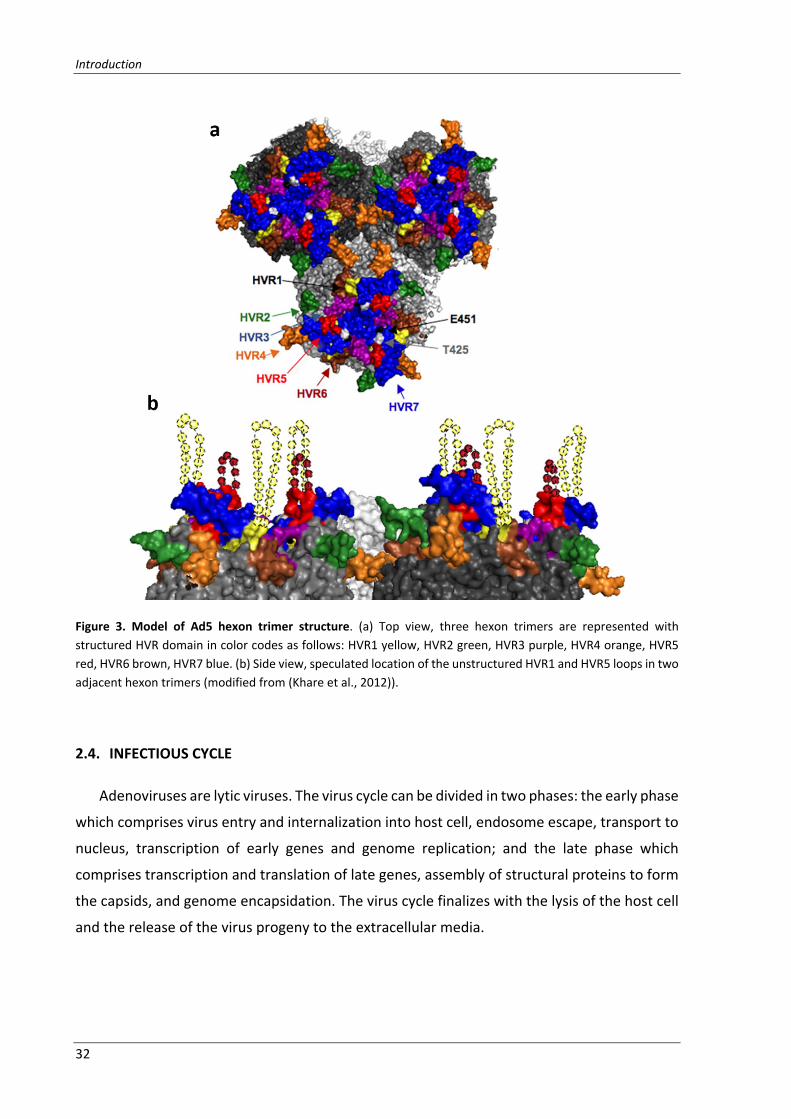
Figure 3. Model of Ad5 hexon trimer structure. (a) Top view, three hexon trimers are represented with
structured HVR domain in color codes as follows: HVR1 yellow, HVR2 green, HVR3 purple, HVR4 orange, HVR5
red, HVR6 brown, HVR7 blue. (b) Side view, speculated location of the unstructured HVR1 and HVR5 loops in two
adjacent hexon trimers (modified from (Khare et al., 2012)).
2.4. INFECTIOUS CYCLE
Adenoviruses are lytic viruses. The virus cycle can be divided in two phases: the early phase
which comprises virus entry and internalization into host cell, endosome escape, transport to
nucleus, transcription of early genes and genome replication; and the late phase which
comprises transcription and translation of late genes, assembly of structural proteins to form
the capsids, and genome encapsidation. The virus cycle finalizes with the lysis of the host cell
and the release of the virus progeny to the extracellular media.

Introduction
33
Figure 4. Adenovirus genome organization. The linear double‐stranded DNA genome is depicted in the center
as a thin line, with the inverted terminal repeats (ITR) at each end. DNA lengths are marked in kb. Transcription
units are shown relative to their position and orientation in the genome. Early genes are indicated by black bars,
intermediate and late genes are indicated by gray bars, and virus‐associated (VA) RNAs are denoted by small
arrows (modified from (Tauber and Dobner, 2001)).
2.4.1. Binding and entry
Initial interaction between Ad5 and host cell occurs through primary interaction between
the fiber knob and the coxsackievirus and adenovirus receptor (CAR) on the cell surface. For
the internalization of the virus, a second interaction is required between an RGD (Arg‐Gly‐Asp)
motif located on the penton base and αVβ3 and αVβ5 integrins (Nemerow and Stewart, 1999).
Alternatively, it was described that liver transduction after intravenous administration occur
by a different mechanism due to virus interaction with blood factors, such as coagulation
factor IX (FIX) and complement binding protein‐4 (C4BP) which bind to fiber knob, or
coagulation factor X (FX) which binds to the hypervariable regions (HVRs) of hexon. Both types
of interactions act as a bridge between the virus and alternative cell receptors on hepatocytes
such as HSGs (heparan sulphate‐glycosaminoglicans) and lipoprotein receptor‐related
proteins (LRPs) (Kalyuzhniy et al., 2008; Shayakhmetov et al., 2005; Waddington et al., 2008).
In addition, direct interaction between Ad5 and HSGs can also occur, and the KKTK91‐94 amino
acidic sequence in the fiber shaft has been postulated as the HSG‐binding motif (Zhang and
Bergelson, 2005).
Binding of adenovirus particles to its cellular receptors triggers clathrin‐dependent,
receptor‐mediated endocytosis (Coughlan et al., 2010). After the virus is internalized,
endosome acidification promotes partial capsid disassembly and escape of virus particle to
the cytoplasm before lysosome formation. Once in the cytoplasm, Ad5 hexon recruits the
molecular motor protein cytoplasmic dynein in a pH‐dependent manner, and the virus particle

Introduction
34
is transported along the microtubules towards the nucleus. This function is critical for efficient
infection, and the role of HVR1 on dynein recruitment was recently described (Scherer and
Vallee, 2015). After reaching the nucleus, the capsid is completely disassembled and the viral
DNA is translocated through the nuclear pore complex for subsequent transcription and
replication (Figure 5).
Figure 5. In vitro entry pathway of Ad5. Steps involved in the in vitro entry pathway of Ad5 are binding to CAR
and integrins, internalization by clathrin‐mediated endocytosis, endosome escape, dynein‐mediated transport
along microtubules, and DNA translocation into nucleus (modified from (Coughlan et al., 2010)).
2.4.2. Early gene expression and DNA replication
E1A is the first transcription unit expressed and its proteins perform several functions. E1A
activates the cell cycle by interacting with Retinoblastoma protein (Rb), which is a tumor
suppressor that inhibits the cell cycle via binding to E2F transcription factor. E1A products are
able to sequester pRb and other members of its family such as p107 and p130, impeding E2F

Introduction
35
sequestration by these proteins, and thus activating the cell cycle and allowing viral DNA
replication (Parreno et al., 2001). In addition, E1A proteins activate the transcription of the
rest of early transcription units (E1B, E2A, E2B, E3, and E4). The deregulation of the cell cycle
caused by E1A results in the accumulation of the tumor suppressor p53, which induces
apoptosis as a cell defense mechanism. To avoid apoptosis, viral protein E1B‐55K directly binds
p53 and induces its degradation, whereas E1B‐19K binds to other proapoptotic proteins such
as Bak and Bax. The genes in E2 region encode for the proteins required for the replication of
the genome including DNA polymerase, preterminal protein, and the single‐stranded DNA‐
binding protein. E3 proteins are responsible for inhibition of the immune response in order to
avoid the destruction of the infected cell before completing the cycle. For instance, E3‐19K
sequesters MHC class I molecules in the endoplasmic reticulum, preventing its transport to
the cell surface. Furthermore, it can also interfere in the loading of peptides onto MHC class I
molecules by binding to the transporter associated to antigen processing (TAP). The E4
transcriptional unit is involved in viral replication, stability and transport of viral mRNA, and
expression of late genes.
2.4.3. Late gene expression and viral assembly
Once DNA replication has been initiated, the transcriptional activity from the major late
promoter (MLP) is intensified due to changes in genome structure and its activation mediated
by viral protein IVa2. The MLP regulates the expression of genes from the major late
transcription unit, which encodes for 15 to 20 different mRNAs derived from a single pre‐
mRNA by differential splicing and polyadenylation. Most late proteins are expressed from
regions L1‐L5, and correspond to structural proteins and proteins involved in virion assembly
and genome packaging (Guimet and Hearing, 2016). Late mRNAs are accumulated in the
cytoplasm and specifically translated in the late phase thanks to the tripartite leader sequence
(TLP). All proteins are translated in the cytoplasm and subsequently transported to the
nucleus. Hexon assembly begins in the cytoplasm and the trimers are then transported to the
nucleus (McConnell and Imperiale, 2004). Once there, hexons associate with the penton base,
the fiber, and minor proteins, and the DNA is incorporated guided by the encapsidation
sequence and DNA‐binding proteins. Capsids are accumulated in the nucleus, which becomes
permeable and intermediate filaments are disaggregated, resulting in a cell round morphology
characteristic of cytopathic effect (CPE). Although the mechanism is not well understood,
expression of ADP (adenovirus death protein) favors the release of the virus progeny to the
extracellular medium.

Introduction
36
2.5. DESIGN OF TUMOR SELECTIVE ONCOLYTIC ADENOVIRUSES: ICOVIR15 AND ICOVIR15K
Contrary to other viruses such as reovirus, Newcastle disease virus (NDV), or vesicular
stomatitis virus (VSV), adenovirus does not have a natural selectivity for tumor cells and
therefore genetic manipulation is required to block replication in normal cells. Three main
strategies have been applied to achieve tumor selectivity: deletion of viral genes that are
complemented by tumor cells, control of the expression of essential viral genes by tumor‐
specific promoters (transcriptional or translational targeting), or modification of capsid
proteins to confer specific or preferential infection of tumor cells (transductional targeting).
ICOVIR15 (Rojas et al., 2010) and ICOVIR15K (Rojas et al., 2012) are two oncolytic adenoviruses
designed in our group that combine the three approaches. These viruses are currently used in
our group as platforms to incorporate and test novel modifications and improvements. A
schematic representation of ICOVIR15 and ICOVIR15K genome with the modifications that
make them tumor‐selective is shown in Figure 6.
Adenoviruses require the host cell machinery for the replication of their genome.
Therefore, mutations or deletions on viral genes involved in activating cell replication or in
inhibiting apoptosis hinder virus replication in normal cells. In contrast, as cell replication is
constantly activated in tumor cells and apoptosis pathways are usually inhibited, viruses
bearing these deletions are able to replicate in cancer cells at normal ratios. In this regard,
ICOVIR15 and ICOVIR15K contain the Δ24 mutation in E1A protein (Fueyo et al., 2000; Heise
et al., 2000), which consists in the deletion of the E1A region responsible for binding and
inactivating Rb. This function is necessary to induce S phase and thus replicate in normal cells
(see section 2.4.2.), whereas it is not required in tumor cells which are in constant replication
(Figure 6b).
As previously explained, E1A is essential for virus replication not only because it induces S
phase in the host cell, but also because it activates the expression of the rest of adenovirus
early genes. Therefore, viral replication can be restricted to tumor cells by controlling E1A
expression with a tumor‐specific promoter. Due to the deregulation of Rb pathway in almost
all tumors, promoters that respond to E2F transcription factor are suitable to achieve
restricted replication in a broad range of tumors. E1A regulation by E2F‐1 promoter confers
selectivity and high potency of transcription (Cascallo et al., 2007; Johnson et al., 2002; Rojas
et al., 2009; Tsukuda et al., 2002). In such adenoviruses, E1A transcription is inhibited in
normal cells due to binding of pRb‐E2F‐HDAC complexes to E2F‐1 promoter. On the contrary,
alteration of Rb pathway in tumor cells results in free E2F, which activates expression of E1A.

Introduction
37
E1A activates the rest of viral genes including E4‐6/7, which forms a complex with E2F and
activates more efficiently the E2F‐1 promoter and E2 promoter, resulting in a positive
feedback loop. In contrast to the insertion of a whole exogenous promoter, ICOVIR15 and
ICOVIR15K incorporate eight E2F responsive sites in the endogenous promoter of E1A (Figure
6a) (Rojas et al., 2010). In addition, a Sp‐1 binding site was also included as both factors
interact cooperatively to activate transcription (Karlseder et al., 1996). These additional boxes
in the promoter enhance E1A expression in tumor cells compared to Adwt, resulting in
increased oncolytic potency. Such modification only increases in 151 bp the adenovirus
genome, allowing the insertion of foreign transgenes without affecting viral replication and
oncolytic potency (Guedan et al., 2010, 2012; Rojas et al., 2010). Hence, these viruses present
a double control of the viral cycle in quiescent cells, and in case that leaky expression of E1A
occurs, the Δ24 deletion impedes the release of E2F from pRb.
The third approach used to limit adenovirus replication to the tumor tissue is the
modification of capsid proteins to achieve preferential infection of tumor cells rather than
normal cells. This strategy includes the ablation of the natural virus tropism (detargeting), and
the redirection of virus infection to tumor‐associated receptors (retargeting). Upon
intravenous administration, adenovirus accumulates mostly in the liver causing toxicity and
reducing the virus bioavailability. Therefore, liver detargeting has been an important subject
in the field over the years. Abrogation of adenovirus interaction with coagulation factor X (FX)
or mutation of the putative heparan sulfate glycosaminoglycan (HSG)‐binding domain KKTK in
the fiber shaft have demonstrated to reduce hepatic transduction (Alba et al., 2010; Smith et
al., 2003; Waddington et al., 2008). However, these modifications also caused a reduced
infection of tumors (Bayo‐Puxan et al., 2006; Gimenez‐Alejandre et al., 2008). With regard to
tumor retargeting, adenoviruses exposing different ligands in different capsid locations such
as fiber, hexon, penton base, or pIX have been described (Coughlan et al., 2010). Specifically,
a widely used ligand to achieve tumor tropism is the RGD‐4C motif (CDCRGDCFC) which targets
RGD‐binding integrins that are overexpressed in tumor cells (Cripe et al., 2001; Dmitriev et al.,
1998; Grill et al., 2001; Nagel et al., 2003; Wesseling et al., 2001). The insertion of this ligand
in the HI‐loop of the fiber knob permits the use of integrins as primary receptors instead of
CAR, which is not highly expressed in tumor cells (Bauerschmitz et al., 2002; Suzuki et al.,
2001). This modification is included in ICOVIR15. In contrast, ICOVIR15K combines a liver
detargeting and a tumor retargeting approach, consisting in the replacement of the KKTK
domain in the fiber shaft with an RGD motif (Figure 6c). This strategy was described by our
group demonstrating improved tumor‐to‐liver ratio in vivo in the context of a non‐replicative

Introduction
38
adenovirus (Bayo‐Puxan et al., 2009). When inserted in an oncolytic platform, ICOVIR15K
showed increased bioavailability after systemic administration and greater antitumor efficacy
in vivo compared to ICOVIR15 (Rojas et al., 2012).
Figure 6. Schematic representation of the modifications in ICOVIR15 and ICOVIR15K genomes. Both oncolytic
adenoviruses contain the modified E1A promoter with four E2F boxes (white squares) and one Sp1 box (red
square), and the truncated E1A‐Δ24 protein to confer selectivity for tumor cells. In ICOVIR15 the RGD motif is
inserted in the HI‐loop of the fiber knob (fiber RGD), whereas in ICOVIR15K is inserted in the fiber shaft replacing
the KKTK domain.
2.6. CLINICAL EXPERIENCE WITH INTRAVENOUS INJECTION OF ONCOLYTIC ADENOVIRUSES
Few clinical trials have been performed by systemic injection of oncolytic adenoviruses. In
fact, until 2010 only two candidates had been tested via intravenous route, ONYX‐015 and

Introduction
39
CG7870. dl152 or ONYX‐015, developed by Onyx pharmaceuticals, was the first engineered
replication selective adenovirus to be used in humans. This virus harbors a deletion of the E1B‐
55K gene making it selective for p53‐deficient tumors (Bischoff et al., 1996). Several trials were
conducted by intravenous injection of ONYX‐015 (some in combination with chemotherapy)
in patients with colorectal tumors, hepatic tumors, and various disseminated solid tumors.
The highest dose administered to a patient in dose‐escalation studies was 2x10¹³ vp on day 1,
followed by weekly doses of 2x10¹¹ vp, resulting in an accumulated dose of 4.2x10¹³ vp
(Nemunaitis et al., 2001). Albeit certain dose‐dependent toxicity effects were observed (fever,
vomits, nausea, or transitory transaminitis), maximum tolerated dose was not reached.
Nevertheless, signs of antitumor efficacy were rarely detected and only some partial
responses, stable diseases, or decrease of tumor markers were observed in some patients
(Habib et al., 2002; Hamid et al., 2003; Nemunaitis et al., 2001; Reid et al., 2002). Despite this
lack of efficacy, evidence of tumor transduction and virus replication was observed. However,
in one study in which a patient died due to disease progression, tissue biopsies revealed that
the majority of the virus was in the liver and the spleen, whereas virus presence in the tumor
was significantly lower (Hamid et al., 2003). Notwithstanding the poor efficacy of ONYX‐015,
the Chinese company Sunway Biotech is commercializing a derivative of this virus named H101
for the intratumoral treatment of head and neck cancer in China. H101 contains an additional
deletion of E3 genes, making it less potent and more immunogenic (Garber, 2006).
The next oncolytic adenovirus tested by systemic injection was CV787, renamed CG7870
after the acquisition of Calydon by Cell Genesys, and now developed by Cold Genesys. This
virus expresses E1A under the control of probasin promoter, and E1B under the control of PSA
promoter, making it selective for prostatic cancer (Yu et al., 1999). Similarly to ONYX‐015, flu‐
like symptoms (fever, fatigue, nausea, vomiting, etc.) were reported in a phase I clinical trial,
but dose‐escalation had to be halted at 6x10¹² vp due to more severe adverse effects (Small
et al., 2006). A secondary peak in plasma of virus genomes suggested active viral replication.
However, a drop on the tumor marker PSA in 3 out of 8 patients treated with the highest dose
was the only reported response.
Currently, more oncolytic adenoviruses are being developed and administered
systemically in clinical trials. ColoAd1 (Enadenotucirev) is an oncolytic adenovirus developed
by Psioxus and generated by “directed evolution”. This process was performed by pooling an
array of adenovirus serotypes (representing subgroups B‐F), passaging the pools at high MOIs
(multiplicity of infection) to promote recombination between serotypes, and then identifying

Introduction
40
and selecting the most potent agents (Kuhn et al., 2008). The selected virus ColoAd1 is a
chimeric Ad11 with a nearly complete E3 region deletion, a smaller deletion in E4 region, and
a chimeric Ad3/Ad11p E2B region. Unlike Ad5, ColoAd1 demonstrated to retain its cancer
killing activity in presence of fresh human blood (Di et al., 2014), making it an interesting
candidate for systemic treatment. Several Phase I/II trials are currently testing ColoAd1 by
intravenous administration in different types of cancer including ovarian cancer, colorectal
cancer, or bladder cancer. Moreover, phase I clinical trials to test the safety and efficacy of
two oncolytic adenoviruses developed by our group (ICOVIR5 and VCN01) by systemic
injection are currently ongoing. ICOVIR5 is a derivative of AdΔ24RGD (Suzuki et al., 2001)
which controls E1A expression by an insulated form of the E2F promoter (Cascallo et al., 2007)
and has been clinically developed from the academia at ICO (Institut Català d’Oncologia).
VCN01 (also known as ICOVIR17K) is a version of ICOVIR15K expressing the PH20
hyaluronidase (Rodriguez‐Garcia et al., 2015), and has been developed clinically by VCN
biosciences.
2.7. LIMITATIONS OF ONCOLYTIC ADENOVIRUSES
Cancer virotherapy with oncolytic adenoviruses presents several limitations, which
account for this lack of efficacy in the clinic, especially after systemic administration. The
bloodstream is a highly hostile environment for the virus and several interactions contribute
to its neutralization and rapid clearance, preventing it from reaching tumors (Figure 7). It was
reported that the adenovirus half‐life after systemic administration was less than 2 minutes
in mice (Alemany et al., 2000) and 10 minutes in humans (Reid et al., 2002). On top of this
rapid clearance, the virus will face additional barriers in the tumor microenvironment such as
limited intratumoral spread and the antiviral immune response
2.7.1. Systemic injection of adenoviruses
2.7.1.1. Adenovirus retention in the liver
Upon the intravenous administration, 90% of the injected dose is rapidly retained in the
liver, causing hepatotoxicity and a significant reduction of the effective dose (Alemany et al.,
2000). Such retention occurs due to the architecture of the liver sinusoids and interactions
with different cell types, including hepatocytes, liver macrophages (Kupffer cells), and liver
sinusoid endothelial cells (LSECs).

Introduction
41
Figure 7. Ad5 interactions with blood components in vivo. 1. Ad5 binding to erythrocytes via CAR, or via CR1
through antibodies and complement traps the virus in the circulation. 2. Ad5 opsonization by natural IgM and/or
complement promotes Kupffer cell uptake via complement receptors or Fc receptors. 3. Interaction with T‐cells.
4. FX binding to Ad5 hexon promotes hepatocyte transduction through HSPGs. 5. Binding of FIX and C4BP to the
fiber knob mediates hepatocyte transduction through HSPGs or LRP. 6. Binding to platelets form aggregates
which are cleared by scavenging macrophages (from (Coughlan et al., 2010)).
Liver reticuloendothelial system, formed by Kupffer cells and LSECs, highly contributes to
adenovirus retention and elimination. Kupffer cells are specialized macrophages lining the
walls of the sinusoids, and due to their location represent the first line of defense against

Introduction
42
viruses. Adenovirus sequestration by Kupffer cells prevents transduction of hepatocytes, and
results in necrosis of these cells, which plays a role in inflammation and triggers innate and
adaptive immune responses to the virus (Liu and Muruve, 2003). The capability of adenovirus
to transduce hepatocytes is not linearly dependent on the injected dose, as there is a
threshold dose around 1011 vp/kg (Manickan et al., 2006) above which Kupffer cells are
eliminated and hepatocytes are massively transduced (Tao et al., 2001). Scavenger receptors
(SRs), which recognize hexon hypervariable regions (HVRs), are responsible for adenovirus
uptake (Haisma et al., 2008; Xu et al., 2008). Specifically, HVR1, HVR2, HVR5, and HVR7, were
proposed to be especially involved in Kupffer cell recognition (Khare et al., 2012). In addition
to recognition by SRs, natural antibodies (IgMs) and complement proteins opsonize the virus
and enhance Kupffer cell‐mediated clearance through Fc receptors or complement receptors
(Khare et al., 2013; Smith et al., 2008). Furthermore, LSECs are also a very important
component of the liver reticuloendothelial system and along with Kupffer cells, contribute to
eliminate the virus from circulation (Ganesan et al., 2011; Piccolo et al., 2013).
The viral dose able to escape Kupffer cells can then infect hepatocytes. Interaction with
natural adenovirus receptors CAR and integrins does not play a relevant role in hepatocyte
transduction after systemic administration. Instead, interaction with several blood proteins
such as complement protein 3 (C3), complement protein 4 (C4), C4 binding protein (C4BP),
coagulation factors VII (FVII), IX (FIX), but mainly coagulation factor X (FX), mediate hepatocyte
transduction by binding to HSPGs and LRPs (Parker et al., 2006; Shayakhmetov et al., 2005).
FX interacts with nanomolar affinity with the HVRs in the hexon, specifically with the loops of
HVR3, HVR5, and HVR7 (Alba et al., 2009).
2.7.1.2. Adenovirus interaction with blood components
Another critical aspect in the intravenous administration of adenovirus is the interaction
with different blood components, including cells and plasma proteins, which typically results
in adenovirus neutralization and clearance from circulation.
Adenoviruses are able to interact with almost all blood cell populations, but in particular,
interaction with erythrocytes has become a major focus of attention in the last years. Human
but not mouse erythrocytes can sequester adenovirus particles directly via CAR, or indirectly
via complement receptor 1 (CR1) after virus opsonization by antibodies and complement
(Carlisle et al., 2009; Seiradake et al., 2009). Such interactions were reported to inactivate Ad5
by preventing extravasation and systemic infection. Therefore, the high concentration of

Introduction
43
erythrocytes in blood would represent a severe barrier for systemic administration of
oncolytic adenoviruses in humans. However, CAR‐bound adenovirus particles cannot be
internalized into erythrocytes due to the lack of integrins on their surface. Previous studies of
adenovirus titration in whole blood reported no loss of adenovirus infectivity despite
erythrocyte binding (Cichon et al., 2003). In addition, proof of adenovirus tumor transduction
in humans after systemic administration has been reported before (Nemunaitis et al., 2001),
and has also been detected in a recent clinical trial with ICOVIR5 (unpublished data). In light
of these facts, in this thesis we question the neutralizing nature of the CAR‐mediated
interaction with human erythrocytes, and hypothesize a reversible binding with no
impairment on adenovirus transduction.
Adenoviruses can also interact with platelets, resulting in a strong thrombocytopenia
between 5 and 24 hours after vector administration, although the mechanism for this
interaction (which was also proposed to be CAR‐mediated) has been controversial (Gupalo et
al., 2011). Furthermore, other cell types such as macrophages, monocytes, or neutrophils can
also sequester opsonized adenovirus particles via Fc receptors and complement receptors
(Cotter et al., 2005; Khare et al., 2013; Lyons et al., 2006).
Besides blood cells, adenovirus also interacts with a variety of plasma proteins. Among
those, neutralizing antibodies (NAbs) represent one of the major limitations for systemic
delivery. Ad5 is a very common pathogen and most of the human population has been
exposed to the virus by natural infection. Although the levels of NAbs vary geographically, the
general prevalence of anti‐Ad5 NAbs is very high, reaching >90% in some locations (Barouch
et al., 2011; Mast et al., 2010). Adenovirus capsid contains three proteins that are accessible
to antibodies: hexon, penton, and fiber. Albeit each of these proteins can be the targeted by
NAbs, the efficiency and the mechanism of neutralization differs considerably. Antibodies
against penton can interfere with viral entry into cells, but their neutralizing capacity is
relatively poor (Wohlfart et al., 1985). Anti‐fiber NAbs can neutralize the virus by cross‐linking
virions into aggregates, and also by inhibiting binding or entry into cells (Wohlfart, 1988).
However, antibodies against hexon are the ones that display the more potent neutralization
activity (Bradley et al., 2012a; Roberts et al., 2006; Roy et al., 2005; Sumida et al., 2005). These
antibodies are directed primarily against the solvent‐exposed HVRs (Bradley et al., 2012b;
Roberts et al., 2006). Unlike anti‐fiber or anti‐penton, anti‐hexon NAbs do not cause
aggregation nor block virus binding to cells, and can neutralize even if added after virion
attachment to cells (Wohlfart, 1988). Thus, such antibodies exert their neutralizing action

Introduction
44
after the virus has been internalized, either by impairing endosome escape or by blocking
dynein‐mediated transport along microtubules to the nucleus. Hence, the high prevalence of
anti‐Ad5 NAbs in humans represents a severe limitation not only for oncolytic adenoviruses,
but also for other adenovirus‐based therapeutic applications such as gene therapy vectors or
vaccines. In addition, the NAbs newly generated after the first adenovirus injection will
efficiently inactivate a second viral dose even in patients with no pre‐existing immunity,
impeding efficient vector re‐administration.
In addition to direct neutralization, antibodies (both NAbs and natural IgMs) not only can
neutralize the virus directly but can also trigger an innate immune response by complement
activation, and by docking the virus particles to Fc receptors of macrophages, monocytes and
neutrophils. This activation of the immune response is not only mediated by binding to
antibodies, but also to direct binding of complement proteins and coagulation factors. In this
regard, the role of FX has been controversial. Whereas a first study described that FX binding
is a mechanism to activate innate immunity against adenovirus (Doronin et al., 2012), a more
recent work claimed that FX shields Ad5 from attack by natural antibodies and complement
(Xu et al., 2013). According to this report, Ad5 recruits FX as a defense mechanism against
complement activation, and such protection is required for efficient intravenous delivery.
Due to the big challenge that represents the intravenous injection of adenovirus, this
thesis was focused on adenovirus interactions on the bloodstream. First, we studied the
interaction of Ad5 with human erythrocytes via CAR. Second, we developed a strategy to avoid
neutralization by antibodies. Nevertheless, oncolytic adenoviruses also face other barriers
which also need addressing.
2.7.2. Intratumoral spread of oncolytic adenoviruses
Propagation of the oncolytic effect within tumors as a consequence of virus replication is
one of the great advantages of virotherapy compared to conventional therapies. Intratumoral
spread is essential in order to generate a significant antitumor effect from an initial low dose.
The difficulty to obtain complete tumor regressions even in animal models indicates an
inefficient virus distribution. Tumors are not only formed by tumor cells but also by the tumor
stroma, including extracellular matrix composed by collagen, hyaluronic acid, elastin, and
proteoglycans, and non‐tumor cells such as fibroblasts. Oncolytic adenoviruses can neither
cross the barriers imposed by extracellular matrix nor replicate in non‐dividing cells,
representing therefore important limitations for intratumoral spread. Our group has

Introduction
45
previously published the expression of hyaluronidase from an oncolytic adenovirus which
degrades hyaluronic acid and significantly improves intratumoral distribution and antitumor
activity (Guedan et al., 2010; Rodriguez‐Garcia et al., 2015). In addition, specific elimination of
tumor fibroblasts is also being addressed in our group by expression of several molecules from
the oncolytic adenovirus (unpublished data).
2.7.3. Immune responses
The intravenous administration of adenoviruses leads to a strong activation of the innate
immune system. Innate immune cells such as macrophages or dendritic cells (DCs) capture the
virus and recognize pathogen‐associated molecular patters (PAMPs) through pattern
recognition receptors (PRRs), such as Toll‐like receptors (TLRs). For instance, TLR9 recognizes
non‐methylated CpG islands in the adenovirus genome. Such interactions trigger the
expression of inflammatory cytokines and chemokines such as IL‐6, TNF‐α, MIP‐2, MIP‐1α, and
type I interferons (IFNs) (Hartman et al., 2008), which recruit neutrophils and NK cells to the
site of infection, as well as monocytes and T cells. Furthermore, DCs travel to draining lymph
nodes where they engage adaptive and memory immune responses which eliminate the
infected cells (Alemany and Cascallo, 2009).
The role of the immune system in cancer virotherapy has been controversial. A more
classic point of view is “virocentrism”, which considers the immune system as an enemy for
oncolytic viruses. According to virocentrics, immunosuppression is a beneficial strategy as it
would allow a more efficient replication of the virus and it would avoid its elimination by the
immune system. Data from clinical trials in which better responses were observed in
immunosuppressed patients support this hypothesis (Kelly and Russell, 2007). On the other
hand, and partly due to the increasing success of cancer immunotherapy, “immunocentrics”
consider the immune system as an ally for the eradication of tumors. The immunogenicity of
the oncolytic viruses might revert the immunosuppressive environment of tumors, and
generate specific antitumor immune responses able to eliminate the tumors (Alemany and
Cascallo, 2009). Of note, the most clinically advanced oncolytic viruses that have
demonstrated efficient antitumor responses (T‐vec, and Pexa‐vec) express the
immunostimulatory molecule GM‐CSF (granulocyte‐macrophage colony‐stimulating factor).
In any case, the high immunogenicity of the virus results in a dominant response against the
virus rather than against the tumor, which is always detrimental for the efficacy of the therapy.

Introduction
46
2.8. STRATEGIES TO EVADE NEUTRALIZING ANTIBODIES
As previously explained, anti‐adenovirus neutralizing antibodies (NAbs) represent a major
hurdle not only for oncolytic adenoviruses, but also for other adenovirus‐based clinical
applications such as vaccination or gene therapy. Therefore, researchers have invested great
efforts in designing strategies to avoid pre‐existing antibodies. Among those, the most
commonly used strategies include the use of alternative serotypes or non‐human
adenoviruses, genetic modifications of capsid proteins to generate pseudotyped or chimeric
vectors, and chemical modifications of the capsid.
2.8.1. Use of rare human serotypes or non‐human adenoviruses
Anti‐Ad5 humoral immunity may be circumvented by using less common serotypes that
have low natural seroprevalence in the general human population, leading to transgene
expression or to a transgene immune response in vaccine applications in the presence of anti‐
Ad5 NAbs (Zaiss et al., 2009). However, other properties besides the seroprevalence should
be considered when selecting a vector, such as tissue tropism, oncolytic activity,
immunogenicity, or toxicity. Utilization of other than species C adenoviruses can be appealing
in virotherapy due to their use of alternative receptors, since CAR has been shown to be down‐
regulated in tumor cells (Li et al., 1999; Wunder et al., 2012). Adenovirus subgroup D, which
has the largest diversity of viruses, has the advantage of showing reduced hepatotropism due
to their low affinity for FX (Waddington et al., 2008). In a study where several alternative
serotypes were analyzed for their ability to spread in the tumor mass, Ad serotype 9 from
subgroup D demonstrated better spreading ability in both CAR negative and positive cancer
cells, suggesting its use for cancer applications (Uchino et al., 2014). Another study compared
the replication efficacy and oncolytic potency of several species B, C, D, and F viruses in B cell
cancer cell lines and primary B cell cancers (Chen et al., 2011). Among those, species D
adenoviruses mediated the most robust killing, and Ad26 and Ad45 significantly reduced
tumor growth after intratumoral administration in lymphoma xenografts. Subgroup B
adenoviruses have also been vectorized due to their tropism profile and low seroprevalence
in humans. These include serotypes 3, 11, 35, and 50 (Alonso‐Padilla et al., 2016), among
which Ad3 has been tested in humans as an oncolytic virus (Hemminki et al., 2012). In addition,
a chimeric oncolytic adenovirus based on serotypes 3 and 11p from subgroup B (ColoAd1) was
generated by “directed evolution”, showing improved potency and selectivity (Kuhn et al.,
2008) (see section 2.6.). Other serotypes such as Ad35, Ad26, and Ad48 were shown to be
more immunogenic than Ad5 in rhesus monkeys (Teigler et al., 2012), a desirable trait for their

Introduction
47
use as vaccine vectors. However, such high immunogenicity might not be desirable for other
applications such as gene therapy vectors, whereas it is less clear for oncolytic adenoviruses.
Some studies have demonstrated unexpected toxicities of some serotypes such as Ad11, Ad3,
and Ad4, at virus titers usually employed for administration of Ad5 (Stone et al., 2007). Of
note, the majority of the studies with alternative serotypes have explored them as vaccine
vectors, and their applicability as oncolytic agents needs further development.
Nevertheless, most humans have NAbs against a range of adenovirus serotypes (Vogels et
al., 2003) and in addition, specific T‐cells against one serotype might crossreact against related
serotypes (Zaiss et al., 2009). An alternative strategy to avoid such crossreactivity is the use of
non‐human adenoviruses. Vectors based on simian (Colloca et al., 2012; Tatsis et al., 2006),
ovine (Hofmann et al., 1999), bovine (Reddy et al., 1999), porcine (Tuboly and Nagy, 2001),
and canine adenoviruses (Perreau and Kremer, 2006) have been tested. The canine adenovirus
serotype 2 (CAV‐2) has been developed in our group as an oncolytic agent to treat canine
patients (Laborda et al., 2014). Nonetheless, the use of non‐human adenoviruses to treat
human tumors is not feasible due to the species‐specificity of adenoviruses, and thus they
have been developed especially as gene therapy and vaccine vectors.
2.8.2. Genetic modifications of adenovirus capsid proteins
An alternative approach to changing the entire vector to a different serotype is to
genetically modify the vector major capsid components or switching them for those of less
seroprevalent or immunogenic serotypes. Fiber modifications or substitutions alone are not
enough to circumvent pre‐existing immunity (Bradley et al., 2012a; Gall et al., 1996; Zaiss et
al., 2009), since the bulk of antibody response to adenoviruses is directed against the hexon
protein (Roberts et al., 2006; Sumida et al., 2005). However, hexon exchange strategies can
easily impact folding and assembly of virus particles, resulting in poor functional titers or
complete failure to rescue viable recombinants. In this regard, hexon swaps between closely
related serotypes are better accepted than between distantly related serotypes (Byrnes,
2016). Replacement of Ad5 hexon gene with the hexon from Ad2 (both species C) resulted
only in partial evasion of anti‐Ad5 serum, probably due to crossreactivity of the NAbs (Gall et
al., 1998). In contrast, Ad5 hexon replacement with hexons from Ad3, Ad6, or Ad12
successfully evaded neutralization in mice immunized with Ad5 (Roy et al., 1998; Wu et al.,
2002; Youil et al., 2002).

Introduction
48
Neutralizing antibodies against Ad5 are mainly directed against the seven hypervariable
regions (HVRs) of hexon and thus, the exchange Ad5 HVRs for those of non‐prevalent
serotypes has also been explored, in contrast to the exchange of the whole hexon protein. An
Ad5 vaccine vector bearing the HVRs from rare Ad48 efficiently evaded NAbs, and generated
CD8+ T cell responses against the expressed simian immunodeficiency virus (SIV) Gag in Ad5
immunized mice and rhesus monkeys (Roberts et al., 2006). Similarly, replacement of HVRs
with those of the low prevalent Ad43 in an Ad5‐based malaria vaccine, resulted in robust T
cell responses against the parasite antigen in mice with high levels of Ad5 NAbs (Bruder et al.,
2012). Despite being subdominant, antifiber Nabs can also contribute to neutralization, and
the combination of hexon HVRs exchange for those of Ad48 together with the exchange of the
fiber knob with that of the chimpanzee adenovirus AdC68, resulted in an even more efficient
neutralization escape (Bradley et al., 2012a).
It is highly likely that these approaches (both the use of alternative serotypes and the
generation of chimeric/pseudotyped vectors) will not prevent the development of humoral
and cellular immune responses after the first administration of the vector. For this reason and
in order to allow efficient multiple dosing, the sequential administration of antigenically
distinct vectors has also been proposed. Heterologous prime‐boosts regimens in the field of
vaccination have been tested combining different serotypes such as Ad5, Ad11, and Ad35
(Lemckert et al., 2005), Ad35 and Ad49 (Thorner et al., 2006), or Ad26 and Ad35 (Zahn et al.,
2012) among others, including even the combination of adenovirus and other types of virus
vectors such as Modified Vaccinia Ankara (MVA) (Draper et al., 2010). In the field of oncolytic
viruses, a protocol of sequential administrations of oncolytic adenovirus and Newcastle
disease virus (NDV) showed a significant benefit in antitumor effect compared to each virus
as a monotherapy, in the immunocompetent Syrian hamster model (Nistal‐Villan et al., 2015).
Nevertheless, an important consideration about these strategies is the necessity to develop
more than one clinical candidate.
2.8.3. Chemical modifications of adenovirus capsid
Another approach to reduce virus neutralization is to “mask” viral epitopes by attaching
chemical polymers or lipid molecules to adenovirus capsid proteins. Polyethylene glycol (PEG)
has been the most used polymer for this purpose, although other polymers such as N‐[2‐
hydroxyl]methacrylamide (HPMA) have also been used (Croyle et al., 2001; Fisher and
Seymour, 2010; Green et al., 2004; O’Riordan et al., 1999; Wortmann et al., 2008). Proteins
on the exterior of Ad surface have abundant free amines, thus PEGylation of Ad capsid can be

Introduction
49
achieved via amine‐mediated covalent bonding, resulting in labeling of all three major capsid
proteins (Choi et al., 2012). Chemical shielding not only can protect the vector from NAbs but
can also reduce Kupffer cell uptake (due to interferences in the interaction with scavenger
receptors (SRs) (Haisma et al., 2009)), hepatotropism (due to evasion of FX binding), and
extend serum half‐life. In addition, this mechanism of protection also reduces innate immune
responses against the virus as evidenced by reduction in plasma levels of IL‐6 after intravenous
injection (Jung et al., 2007), and in antibody‐depenent complement activation (Tian et al.,
2009). However, it has been reported that PEGylation can significantly reduce the
transduction efficiency of adenovirus vectors, due to steric interference of the polymer chains
with attachment to cell receptors (Eto et al., 2005). This issue can be partially overcome by
subsequent addition of tumor‐binding ligands to the distal end of PEG, such as RGD (Eto et al.,
2005), the MAb Herceptin (Kim et al., 2011), or fibrobast growth factor receptor (FGFR) ligands
(Green et al., 2008). Nevertheless, PEG is also immunogenic and can itself induce antibody
responses that neutralize the PEGylated Ad vector, hampering the efficiency of multiple
administrations (Byrnes, 2016; Shimizu et al., 2012). In addition to chemical polymers, some
studies have also demonstrated that adenovirus vectors can also be shielded from antibodies
by liposomes (Steel et al., 2004; Uusi‐Kerttula et al., 2015; Yotnda et al., 2002). However,
unlike genetic modifications, polymers and liposomes are not heritable and therefore,
progeny virions will lack the shielding modification.
As previously explained, the majority of these strategies have been applied to adenovirus
vectors either for gene therapy or for vaccination, and can present certain limitations in
oncolytic adenoviruses. Thus, the design of oncolytic adenoviruses able to circumvent NAbs
remains relatively unexplored. In this thesis, we have developed a novel strategy to
circumvent anti‐Ad NAbs after the systemic administraiton, by using endogenous serum
albumin to mask viral epitopes and shield the adenovirus capsid.
3. ALBUMIN AS A DRUG CARRIER
3.1. GENERAL CHARACTERISTICS OF ALBUMIN
Human serum albumin (HSA) (585 aa, MW 66.5 kDa) is by far the most abundant protein
in blood plasma, with a concentration of 35‐50 mg/mL. Structurally, it is a heart‐shaped α‐
helical protein composed of three homologous domains (I, II, III), each further subdivided in

Introduction
50
two sub‐domains (A and B) formed by 4 and 6 α‐helices, respectively (Larsen et al., 2016)
(Figure 8). Albumin is synthesized predominantly in the liver and is rapidly secreted to
circulation at a rate of 10‐15 g/day. The binding properties and functions of HSA are manifold.
To name a few, it is involved in the regulation of both pH and osmotic pressure in the vascular
system, it acts as a transport vehicle for fatty acids, hormones, metal ions (copper, nickel,
calcium, or zinc), and therapeutic drugs (penicillins, benzodiazepines, sulfonamides, etc.), and
it can also act as an amino acid source to nourish peripheral tissues (Kratz, 2008).
3.2. ALBUMIN PROPERTIES AS A DRUG CARRIER
In the last years, albumin has emerged as a versatile protein carrier for drug targeting and
for improving the pharmacokinetic profile of drugs, including therapeutic proteins, peptides,
and small molecules. Albumin has several traits that make it an ideal candidate as a drug
carrier. Some desirable properties are its high solubility, stability, and biodegradability, along
with its lack of toxicity and immunogenicity. In addition, its extremely long plasma half‐life and
its accumulation in malignant and inflamed tissues are especially relevant for this purpose.
Figure 8. Crystal structure of human serum albumin. The tertiary structure of human serum albumin is
represented. The domains and sub‐domains are shown in purple (IA), red (IB), green (IIA), orange (IIB), blue (IIIA),
and violet (IIIB) (modified from (Larsen et al., 2016)).
3.2.1. Albumin plasma half‐life
Albumin has a particularly long circulatory half‐life of 19 days in humans. This is in part due
to its size, which is above the renal clearance threshold, and to its specific interaction and
recycling by the neonatal Fc receptor (FcRn) (Peters Jr, 1995). With the exception of IgGs, no

Introduction
51
other soluble serum protein is known to exhibit such a long half‐life. Interestingly, the long
half‐life of IgGs (23 days) is also due to FcRn‐mediated recycling (Lencer and Blumberg, 2005).
FcRn is an MHC class I‐related receptor involved in transplacental transport of maternal IgGs
to the fetus and in antigen presentation (Roopenian and Akilesh, 2007). In addition, FcRn also
extends the serum half‐life of albumin and IgG molecules. Specifically, albumin is pinocytosed
along with other serum proteins by many types of cells, and it is transported by endocytic
vesicles which contain FcRn. FcRn shows no affinity for albumin at neutral pH, but binds to it
with high affinity at low pH (Chaudhury et al., 2003). When endosomes are acidified, binding
to FcRn rescues albumin from degradation, diverting it from the lysosomal pathway. Albumin
is then transported back to the extracellular space where it is released at physiologic pH,
thereby prolonging its half‐life (Figure 9).
Figure 9. Albumin recycling by FcRn. Extracellular albumin is pinocytosed along with other serum proteins at
neutral pH (blue shading). Upon endosome acidification (red shading), albumin binds with high affinity to FcRn.
Non‐FcRn bound proteins are sorted to the lysosome for degradation whereas vesicles containing albumin‐FcRn
complexes are recycled back to the cell membrane and upon exposure to the extracellular neutral pH, albumin
is released back to circulation.
Many therapeutic molecules are smaller than the renal filtration threshold, resulting in
their rapid clearance from the bloodstream, which severely limits their therapeutic effect
(Sleep et al., 2013). In this regard, binding to albumin either covalently or non‐covalently, has

Introduction
52
demonstrated to be a useful strategy to significantly increase the plasma half‐life of such
drugs, from minutes to hours or from hours to days. Examples of drugs using the albumin‐
binding strategy for half‐life extension, either already approved or in clinical or pre‐clinical
stages, will be detailed below (section 3.3.).
3.2.2. Albumin accumulation in solid tumors
The fact that albumin is accumulated in solid tumors makes it very attractive as a carrier
for anticancer drugs. Three main factors are responsible for the accumulation of this plasma
protein in tumors. One factor is the enhanced permeability and retention of macromolecules
in relation to passive tumor targeting (EPR effect) (Maeda et al., 2000). The EPR effect is the
property by which high‐molecular‐weight molecules tend to accumulate in tumor tissue much
more than in normal tissue. This effect occurs due to the abnormal vasculature of tumors,
usually conformed by poorly aligned endothelial cells with wide fenestrations. Such leaky and
defective tumor blood vessels are highly permeable for macromolecules, while in the
vasculature of healthy tissue only small molecules can cross the endothelial barrier. The high
intratumoral concentration of macromolecules such as albumin is not only explained by this
enhanced permeability, but also by a high retention. Whereas low‐molecular‐weight
substances can return to circulating blood by diffusion, the lack of a proper lymphatic drainage
in tumors (due to an impaired or absent lymphatic system) cause the accumulation of
macromolecules (Kratz, 2014).
In addition to the EPR effect, a specific albumin transport pathway based on the interplay
of two albumin‐binding proteins (gp60 and SPARC), also contributes to the absorption and
accumulation of albumin in the tumor interstitium. Circulating albumin binds to gp60, located
on the endothelial cell surface, inducing vesicle formation and transcytosis, which transports
albumin through the endothelial cell into the subendothelial space (Schnitzer, 1992). Upon
entering the tumor interstitium, a second albumin‐binding protein, SPARC (Secreted Protein
Acidic and Rich in Cysteine), binds to albumin with high affinity and traps it in the tumor
interstitium (Figure 10).
Finally, it has also been described that the proliferating tumor mass actively sequesters
and digests albumin to cover its high demand for amino acids and metal ions, generally
resulting in hypoalbuminemia, a characteristic feature of patients with advanced solid tumors.

Introduction
53
Figure 10. Gp60 and SPARC are responsible for albumin accumulation in tumors. Circulating albumin is uptaken
by endothelial cells through the gp60‐mediated transcytosis pathway and is retained in the tumor interstitium
by subsequent binding to SPARC.
3.3. STRATEGIES TO ACHIEVE ALBUMIN BINDING
The use of albumin as a carrier protein for treating various diseases (primarily cancer,
rheumatoid arthritis, diabetes, and hepatitis) has resulted in several marketed products and
numerous clinical trials. Such products have been developed with different strategies to
achieve albumin binding, including covalent binding and non‐covalent binding methods.
3.3.1. Albumin fusions
A possible strategy to combine protein‐based drugs with albumin is the genetic fusion to
the N‐ or C‐terminal of albumin, or even to both ends. The gene of the protein of interest is
connected to the albumin gene and the fusion protein is obtained upon expression in a
suitable host (usually yeast). The resulting fusion protein combines the therapeutic activity of
the partner with the long half‐life of albumin. Of note, the proper folding of albumin should
not be altered by the linker or the fused protein, in order to maintain its functionality and long
half‐life (Larsen et al., 2016). In the last decade, a large number of different proteins have

Introduction
54
been genetically fused to albumin, including peptides, cytokines, coagulation factors,
hormones, etc.
Albiglitude (tradenames Eperzan® and Tanzeum®) is a glucagon‐like peptide‐1 agonist
(GLP‐1 agonist) developed by Human Genome Sciences and marketed by GlaxoSmithKline for
the treatment of type II diabetes. It consists of two human GLP‐1 repeats fused to the N‐
terminus of recombinant human albumin (Bush et al., 2009). Data from clinical trials
demonstrated a significant increase in the plasma half‐life of approximately 5 days, compared
to 1‐2 minutes of endogenous GLP‐1. In 2014, Albiglitude was approved for treatment of type
II diabetes. Other albumin‐fused drugs in advanced clinical stages are FIX (rIX‐FP) for the
treatment of hemophilia B (Santagostino et al., 2012), IFNα‐2b (Albuferon®) for the treatment
of hepatitis C (Rustgi, 2009), or the bispecific fusion MM‐111, consisting of an albumin
molecule fused at both N‐ and C‐ terminus with a scFv (single‐chain variable fragment
antibody) directed against HER‐2 and HER‐3 for the treatment of HER‐2 positive tumors
(McDonagh et al., 2012).
3.3.2. Chemical conjugation
A second approach is chemical conjugation of the drug to albumin, usually to either lysines,
tyrosines, or the free SH‐group in the cys34. This strategy usually requires a release
mechanism from albumin, which can be achieved, for instance, with a cleavable linker. Kratz
et al. developed Aldoxorubicin, a maleimide‐modified doxorubicin which covalently
conjugates with cys34 of albumin upon intravenous administration. Aldoxorubicin
accumulates in tumors where the acidic environment cleaves the acid sensitive linker,
releasing doxorubicin to exert its action. Preclinical and advanced clinical studies indicated
reduced adverse effects and improved tumor accumulation and efficacy than native
doxorubicin (Chawla et al., 2015; Graeser et al., 2010; Unger et al., 2007). The chemical
conjugation strategy has also been successfully applied to increase the plasma half‐life of
methotrexate (MTX‐HSA) for cancer treatment (Bolling et al., 2006), and of a peptide with
anti‐HIV activity (Xie et al., 2010), among others.
3.3.3. Nanoparticle albumin‐bound technology (nab technology)
American Bioscience Inc. developed an albumin‐based technology useful for encapsulating
lipophilic drugs into nanoparticles. The principle is based on mixing the drug with serum
albumin in an aqueous solvent and submitting it to high pressure through a jet, to form drug

Introduction
55
albumin nanoparticles ranging 100‐200 nm (Kratz, 2008). This technology was used to
encapsulate the chemotherapeutic agent paclitaxel, generating Abraxane® (nab‐paclitaxel).
This novel formulation of paclitaxel avoids the use of the solvent Cremophor EL, associated
with severe side effects. In addition of being less toxic than its free drug counterpart,
Abraxane® also exhibits higher antitumor activity compared to free paclitaxel (Desai et al.,
2006). Along with the EPR effect, the gp60 and SPARC‐mediated albumin retention
mechanism were proposed as responsible for the enhanced tumor accumulation of
Abraxane®. In fact, clinical data revealed a significant benefit in overall survival in patients
with high SPARC levels treated with the combination of Abraxane® and gemcitabine (Desai et
al., 2009; Von Hoff et al., 2011). In 2005 it was approved for the treatment of metastatic breast
cancer, representing the first albumin‐based drug delivery system to be approved in oncology.
In 2010, it was acquired by Celgene Corporation to be further tested for other types of cancer.
In 2012 it was approved for the first‐line treatment of locally advanced or metastatic non‐
small cell lung carcinoma (NSLC) in combination with carboplatin, and in 2013 for the
treatment of pancreatic cancer in combination with gemcitabine (Kratz, 2014). The high
albumin‐mediated tumor accumulation of this delivery system makes it very attractive to test
other chemotherapeutic agents. Celgene has a portfolio of nab‐technology based drugs
currently in clinical development such as nab‐docetaxel (ABI‐008) for prostate cancer
treatment, or nab‐rapamycin (ABI‐009) for colon and breast tumors (Neil, 2007).
3.3.4. Incorporation of endogenous albumin ligands
Albumin acts as a transporter for a broad variety of molecules such as fatty acids and
therefore contains several fatty acid binding sites. Attachment of a fatty acid to the desired
drug is a feasible way to achieve non‐covalent binding to albumin. This has been applied by
NovoNordisk in the development of Levemir® (insulin detemir) and Victoza® (Liraglutide) for
the treatment of diabetes. Levemir® is a myristic acid modified insulin analog, whereas
Victoza® is a glucagon‐like peptide‐1 agonist (GLP‐1) modified with a palmitic acid. Upon
injection, the fatty acid moiety binds to albumin and slowly dissociates over time, improving
the bioavailability and distribution. Such modifications resulted in a remarkable increase in
half‐life from 4‐6 min to 5‐7 h for insulin, and from 2 min to 11‐15 h for GLP‐1 analog (Sleep
et al., 2013). Successful results in clinical trials (Dornhorst et al., 2007; Pratley et al., 2010) led
to the approval of Levemir® in 2004 for the treatment of type 1 and 2 diabetes, and of Victoza®
in 2009‐2010 for type 2 diabetes.

Introduction
56
3.3.5. Albumin‐binding domains (ABDs) or peptides (ABPs)
Very similarly to this last strategy, non‐covalent reversible binding to albumin can also be
accomplished through protein modules with the capacity to interact with albumin. In this case
and due to their protein origin, they are often genetically fused to the therapeutic protein.
These can include wild‐type proteins with an intrinsic affinity for albumin or molecules
artificially engineered to bind to albumin.
Proteins with albumin‐binding activity have been identified in certain bacteria, such as the
streptococcal protein G. Protein G of Streptococcus is a cell surface receptor containing
tandem repeats of serum albumin‐binding domains (ABDs). Specifically, domain 3 (hereinafter
named ABD) has been extensively studied and characterized. ABD is composed of 46 amino
acids (5‐6 kDa) which form a left‐handed three α‐helix bundle (Kraulis et al., 1996). Structural
data of an ABD homolog in complex with human serum albumin (HSA), revealed that helix two
and three of ABD interacts with domain II of HSA, not interfering with the FcRn binding site
located in domain III (Figure 11). The ABD on its own maintains the albumin‐binding
functionality and is highly stable, not requiring disulfide bonds or metal ions. The affinity for
human albumin has been determined to be in the range of 1‐4 nM (Kontermann, 2009). The
species specificity of the domain has been studied showing high affinity not only for human
but also for mouse, rat, hamster, and baboon albumin, whereas low or no affinity for cow,
rabbit, pig, dog, or sheep albumin (among others) (Falkenberg et al., 1992; Nygren et al.,
1990). This binding to albumin of several species is highly relevant as it facilitates its use in
pre‐clinical and clinical models.
A large number of pre‐clinical studies have demonstrated the applicability of fusion to the
ABD to improve the plasma half‐lives of therapeutic proteins, especially of small antibody
molecules and variants. A HER‐2 binding Fab (Fragment antigen‐binding) derived from the
clinically approved mAb Trastuzumab (Herceptin®) was fused to ABD, resulting in a half‐life in
mice of 20.9 h compared to 2.1 h of the Fab fragment alone (Schlapschy et al., 2007). Similarly,
the same strategy was applied to a bispecific single‐chain diabody (scDb CEA‐CD3) developed
for the retargeting of cytotoxic T cells to CEA‐expressing tumor cells (Figure 12), obtaining an
enhanced half‐life from 5.6 to 27.6 h in mice. Importantly, this prolonged half‐life led to a
strongly increased accumulation of the scDb‐ABD in CEA‐positive tumors in animal models
(Stork et al., 2007). Furthermore, the tumor accumulation of the scDb‐ABD fusion was also
higher than that of a PEGylated scDb derivative which displayed a similar enhanced half‐life,
indicating a specific contribution of albumin in tumor penetration (Stork et al., 2009). Some

Introduction
57
studies have confirmed that the drug‐ABD/albumin interaction survives the FcRn‐mediated
recycling process, and that ABD seems to follow the same kinetics and distribution as serum
albumin itself (Andersen et al., 2011; Kontermann, 2011; Stork et al., 2009). More recent pre‐
clinical studies have described an enhancement in the plasma half‐life and in the antitumor
effect of TRAIL (tumor necrosis factor‐related apoptosis inducing ligand) and anti‐HER2
immunotoxins by fusion to ABD (Guo et al., 2016; Li et al., 2016). The company Affibody has
developed AlbumodTM, a technology for half‐life extension based on the ABD, which offers for
outlicensing.
Figure 11. Interaction of a bacterial albumin‐binding domain with human serum albumin. Crystal structure of
the complex formed by the albumin‐binding domain of protein PAB of Finegoldia magna (homolog to ABD3 of
streptococcal protein G) and HSA. α‐helices 2 and 3 (H2 and H3) of the albumin‐binding domains recognize a site
located in domain II (DII) of HSA, which does not overlap with the binding site of the neonatal Fc‐receptor (FcRn)
located in domain III (DIII) (modified from (Nilvebrant and Hober, 2013)).
Alternatively to bacterial ABDs, some molecules have been specifically engineered to bind
to albumin, including domain antibodies or peptides. Albumin‐binding peptides (ABPs) have
been isolated from random peptide libraries by phage display. Such peptides have the
advantage of being smaller, but on the other hand display lower affinities for albumin. For
example, an 18 amino acid ABP (SA21) obtained from a peptide phage library had an affinity
ranging 266‐467 nM for albumin of different species (Dennis et al., 2002). Still, fusion to an
anticoagulant anti‐tissue factor Fab significantly increased its plasma half‐life 37‐fold to 32.4
h in rabbits and 26‐fold to 10.4 h in mice, representing the 25‐43% of the albumin half‐life in
these models, and maintained its anticoagulant activity.

Introduction
58
Figure 12. Model structure of the fusion of the ABD3 from streptococcal protein G (ABD) to the C‐terminal end
of a single chain diabody (scDb) (from (Stork et al., 2007)).
In the last years, the use of albumin as a carrier protein has emerged as a hot field, and
several approaches and technologies have been developed to attach albumin to drugs. Among
all these strategies, in this thesis we used the albumin‐binding domain of streptococcal protein
G (ABD) and albumin‐binding peptides (ABPs) to promote adenovirus binding to albumin. Such
modifications were tested in our standard oncolytic platforms ICOVIR15, and ICOVIR15K to
protect them from neutralizing antibodies (NAbs) after the systemic administration.

OBJECTIVES

Introduction

Objectives
61
One of the main limitations in the efficacy of adenoviral therapies is the neutralization of
the virus in the bloodstream after the systemic administration. Contrary to previous reports,
human erythrocytes have been described to bind and inactivate Ad5 via CAR. In addition, the
high prevalence of anti‐Ad5 neutralizing antibodies (NAbs) in the human population
represents a significant hurdle for adenovirus‐based gene therapy vectors, oncolytics, and
vaccines.
The general objective of this thesis was to study the interaction of Ad5 with two blood
components: human erythrocytes and NAbs, and due to the significant problem the latter
represent, develop an oncolytic adenovirus able to evade NAbs upon systemic administration
by using serum albumin as a shield. This thesis was carried out in the context of two
independent projects, reflected in two different chapters. Several specific objectives were
planned:
Chapter 1:
‐ To study the CAR‐mediated interaction between Ad5 and human erythrocytes, and
determine its impact on adenovirus bioactivity in vitro and in vivo.
Chapter 2:
‐ To insert albumin‐binding moieties on adenovirus capsid, and confirm their
functionality by demonstrating the ability of the viruses to bind albumin.
‐ To characterize the effect of albumin binding in vitro in the absence of NAbs, and its
impact in vivo on the blood persistence, toxicity, organ transduction, and antitumor
efficacy of the adenovirus after intravenous administration in mice without NAbs.
‐ To assess if albumin binding can protect adenovirus from anti‐Ad5 NAbs in vitro, and
analyze the ability of the virus to transduce target organs and reduce tumor growth in
pre‐immune mouse models after the systemic administration.
‐ To test the ability of albumin‐binding adenoviruses to evade NAbs generated de novo
against their own capsid and therefore, the feasibility of virus readministration.
‐ To study the combination of the albumin‐binding insertion with other adenovirus
modifications such as the replacement of fiber shaft HSG‐binding motif with RGD.


MATERIALS AND METHODS


Materials and methods
65
1. HANDLING OF BACTERIA
In order to obtain enough amounts of plasmid DNA to be easily manipulated, its
amplification in bacteria was required. For this purpose, the plasmid should contain a
replication origin that allows its replication on the desired strain and an antibiotic‐resistance
gene to select the bacteria and avoid contaminations. In this work, the Escherichia coli strain
SW102 has been used with this purpose and to perform homologous recombination.
1.1. PLASMIDIC DNA EXTRACTION FROM BACTERIAL CULTURES
Plasmid DNA was obtained from saturated E.coli cultures grown in LB with antibiotic according
to protocols based on an alkaline lysis with SDS. DNA was prepared at small and large scale.
1.1.1. Small scale DNA preparations
DNA minipreparations were performed following an adapted protocol described by
Birnboim and Doly (Birnboim and Doly, 1979). A colony grown in an LB‐antibiotic dish was
inoculated in 3 mL of LB+antibiotic and incubated overnight at 32ºC. A 2 mL aliquot of the
grown culture was centrifuged at 13000 rpm for 1 minute. The cell pellet was resuspended in
200 µL of pre‐cooled solution 1 (25 mM Tris‐HCl pH 8, 10 mM EDTA, 50 mM glucose, 0.1
mg/mL RNAse). 200 µL of freshly prepared solution 2 (SDS 1%, NaOH 0.2 M) were added and
the mixture was blended by inversion. 200 µL of pre‐cooled solution 3 (3 M potassium acetate,
11.5% acetic acid) were added and the mixture was blended again by inversion until a white
precipitate appeared. The mixture was incubated 5 minutes on ice and centrifuged 15 minutes
at 13000 rpm. The clear supernatant was collected without taking the white pellet
(corresponding to cellular DNA, proteins, and SDS) and 2 volumes of ethanol were added. The
mixture was incubated 15 minutes at room temperature (RT) and the plasmid DNA was
precipitated by centrifugation for 10 minutes at 13000 rpm. The supernatant was discarded
and the DNA pellet was washed with 70% ethanol by centrifugation at 13000 rpm 5 minutes.
The supernatant was discarded again, the pellet was dried and finally, plasmid DNA was
resuspended in 50 µL of TE (10 mM Tris‐HCl, 1 mM EDTA).
1.1.2. Large scale DNA preparations
DNA maxipreparations allow the obtention of large amounts of high purity plasmid DNA
(≥100 µg). Maxipreparations were prepared from 200 mL of saturated bacteria culture using

Materials and methods
66
the commercial kit “PureLinkTM HiPure Plasmid Filter Purification Kits” (Invitrogen, Waltham,
MA, USA), following manufacturer’s instructions.
1.2. HOMOLOGOUS RECOMBINATION IN BACTERIA
Homologous recombinations performed in this work have been conducted in bacteria
using the high efficiency system developed by Richard Stanton (Stanton et al., 2008), who
kindly gave us the plasmid pAdZ5‐CV5‐E3+ in SW102 strain of E.coli. This plasmid contains the
adenovirus type 5 genome (E1‐) as a bacterial artificial chromosome (BAC), with a
chloramphenicol (Cm)‐resistance gene. This system works using phage λ genes Redγβα under
the control of a temperature‐sensitive promoter, which is repressed at 32°C and activated
when bacteria are grown at 42°C. The positive‐negative selection cassette rpsL‐Neo was used
to identify recombinant clones. Plasmid pAdZ5‐CV5‐E3+ was modified in order to obtain
pAdZICOVIR15, pAdZICOVIR15K, pAdZGL, and pAdZGLRGD plasmids, which were used to
introduce the modifications described in this thesis.
Modifications were performed in two steps. In the first step or positive selection, the rpsL‐
Neo cassette was inserted in the target region to modify. For that, a DNA fragment containing
the rpsL‐Neo flanked by homology regions (40 bp) with the recombination sites was generated
by PCR and used to transform the bacteria. After this first recombination step and due to the
incorporation of the rpsL‐Neo, recombinant clones become Kanamycin (Kan) resistant and
Streptomycin (Strep) sensitive (SW102 strain is naturally resistant to Strep), and are selected
in Kanamycin LB plates. In the second step or negative selection, the rpsL‐Neo is substituted
by the desired modification. In this case, a DNA fragment containing the modification flanked
by the same homology regions is also generated by PCR and used to transform the bacteria.
In this case and due to the loss of the rpsL‐Neo, recombinant clones become again Kan
sensitive and Strep resistant and are selected in Streptomycin LB plates.
The procedure that has been followed to perform recombinations is described next. First
of all, glycerinates of the bacteria that contain the plasmid to be modified were scratched with
a sterile pipette tip, inoculated in 5 mL of LB media including Cm + Strep antibiotics (12.5
μg/mL, and 1 mg/mL, respectively), and incubated overnight at 32°C and constant agitation.
Then, 25 mL of LB Cm + Strep were inoculated with 0.5 mL of the previous culture and
incubated 32°C with agitation until it reached an OD measure of 0.5‐0.6 at 600 nm. At this
moment, culture was divided in two Falcon tubes with equal volumes. The expression of the
Red recombinase genes was induced in one of the falcon tubes by incubating it for 15 minutes

Materials and methods
67
at 42°C, and then cultures were incubated on ice 15 minutes more. From that moment,
manipulation was done at 4°C in order to ensure good transformation efficiency. Both cultures
(induced and non‐induced) were centrifuged 5 min at 4000 g at 4°C and supernatant was
discarded. Bacteria pellet was resuspended in 11 mL of cold ddH2O water and centrifugation
was repeated. Three total washing steps were performed and in the last one, the pellet was
resuspended in the remaining water (≤300 μL). Afterwards, 50 μL aliquots of bacteria (both,
from the induced and non‐induced cultures) were transformed by electroporation with 100‐
200 ng of DNA (the fragment which contains rpsL‐neo). Electroporation was performed with
the electroporator machine Electro Cell ManipulatorTM ECM 630 at the following conditions:
50 µF, 1500 V and 125 Ω. Pulses lower than 5 milliseconds were considered correct. Bacteria
were recovered in 1 mL of LB without antibiotics and incubated 70 minutes at 32°C. Finally,
they were plated into LB plates containing Cm and Kan (12.5 μg/mL, and 15 μg/mL,
respectively) and incubated overnight at 32°C. 24 hours later, colonies should be grown and
the ratio between the colonies in the induced culture (recombinant) and the non‐induced one
(control) were compared in order to assess recombination efficiency. Colonies were picked,
minipreparations of DNA were performed and restriction patterns of different clones were
checked. Finally, the correct clone was frozen as a glycerinate.
A similar procedure was followed for the second phase, in which the clones obtained in
the previous step were inoculated in 5 mL of LB Cm+Kan and cultured overnight at 32°C. The
next day, expression of recombinase genes was induced, and the bacteria were made
competent for its transformation by electroporation in the same way described previously.
Again, 100‐200 ng of DNA containing the desired modification were transformed. After the
recovery incubation 100 μL of a 1/25 dilution of the culture were plated in LB agar Cm+Strep
and plates were incubated overnight at 32°C. 24 hours later, colonies were picked,
minipreparations of DNA were performed and clones were checked by the analysis of
restriction pattern and/or sequencing of the recombinant region.
2. CELL CULTURE
2.1. HEK293
Human Embryonic Kidney 293 (HEK293) cells derive from human primary embryonic
kidney cells. This cell line has been transformed with a fragment of the Ad5 genome including

Materials and methods
68
the E1A gene (Graham et al., 1977). 293 cells are highly permissive for the generation and
replication of adenoviruses and are easily transfectable by the calcium phosphate method.
HEK293 are packaging cells and have been used for the generation and amplification of non‐
replicative E1‐deleted adenoviral vectors, and for the generation and functional titration of
oncolytic adenoviruses.
2.2. TUMOR CELL LINES
The different tumor cell lines used in this thesis are summarized in the following table:
Table 2. Tumor cell lines used in this work
Cell line Tumor type Origin species
A549 Lung adenocarcinoma Human
Sk‐mel28 Melanoma Human
NP‐9 Pancreatic Human
NP‐18 Pancreatic Human
A431 Epidermoid carcinoma Human
B16 Melanoma Mouse
B16‐CAR Melanoma Mouse
A549 cell line has been used to amplify the different oncolytic adenoviruses due to its high
efficiency to produce virus. NP‐9 and NP‐18 pancreatic tumor cells line were established by
the Servei de Digestiu de l’Hospital de la Santa Creu i Sant Pau from Barcelona and provided
by the Laboratori d’Investigació Gastrointestinal from the same Hospital (Villanueva et al.,
1998). B16 murine cell line was obtained from Dr. Pablo Sarobe (CIMA, Pamplona). B16CAR
murine cell line was obtained from Dr. Philippe Erbs (Transgene, France). The rest of the cell
lines were obtained from the American Type Cell Culture (ATCC, Manassas, VA, USA). All cell
lines were maintained in Dulbecco’s modified Eagle’s medium (DMEM) supplemented with
5% fetal bovine serum (FBS, Invitrogen Carlsbad, CA, USA) (previously inactivated by heating
at 56°C for 30 minutes) and penicillin‐streptomycin (PS, Gibco‐BRL, Barcelona, Spain) (100
U/mL and 100 μg/mL, respectively), at 37°C and 5% CO2. B16CAR were maintained with 0.5
mg/mL hygromycin (Invivogene, San Diego, CA).

Materials and methods
69
2.3. CELL COUNTING
To determine cell numbers, manual or automatic methods were performed using trypan
blue dying exclusion test. Adherent cells were detached by Trypsin‐EDTA 0.05% (GIBCO RBL)
incubation and were resuspended in fresh medium supplemented with FBS. For the manual
counting, a dilution in which 10 to 100 cells could be counted in each quadrant of the
Neubauer chamber was made. Viable cells in each quadrant were counted and the mean was
calculated. The number of cells per mL was calculated according to the following formula:
Cells/mL =Mean number of viable cells per quadrant×Dilution factor×104
The automatic counting was performed with a cell counter TC20TM (Bio‐Rad, Hercules,
CA, USA) according to the manufacturer’s instructions.
2.4. CELL FREEZING AND CRYOPRESERVATION
Cells were counted as described above and resuspended in cold freezing medium (90%
FBS 10% DMSO) at a final concentration of 5‐10x106 cells/mL depending on the cell line. Cell
suspension was distributed in cryotubes at 1 mL/tube and placed in a container filled with 2‐
propanol for its freezing at ‐80°C during 24 hours. The cryotubes were then stored in a liquid
nitrogen tank. For cell thawing, cells were rapidly moved from the liquid nitrogen to a water
bath at 37°C. Cells were then diluted in pre‐warmed medium and transferred to a 15 mL Falcon
tube. Soft centrifugation at 1000 g was carried out for 5 minutes, and the pellet of cells was
resuspended in fresh medium and plated at high confluence to optimize the recovery.
2.5. MYCOPLASMA TEST
All cell lines were routinely tested for mycoplasma contamination by PCR using the
following oligonucleotides:
Table 3. Oligonucleotides used for detection of mycoplasma contamination
Oligonucleotide Sequence
MICO‐1 5’‐GGCGAATGGGTGAGTAACACG‐3’
MICO‐2 5’‐CGGATAACGCTTGCGACTATG‐3’

Materials and methods
70
As a template for the PCR, medium from cells that had been in overconfluence in absence
of antibiotics for at least 5 days was used. If the result was positive, cells were discarded.
2.6. PURIFICATION OF HUMAN ERYTHROCYTES
Human blood samples were obtained by venipuncture into Lithium‐heparinized tubes
(Greiner Bio‐One, Monroe, NC, USA) with donor consent and approval by the IDIBELL’s Ethics
Committee. After centrifugation at 2000 g for 10 min at room temperature, the plasma and
leukocyte layer were removed by aspiration. Erythrocytes were resuspended in 3 volumes of
PBS and centrifuged again with the same settings. This washout step was repeated 4 times.
Washed erythrocytes were counted in the Neubauer chamber and resuspended in PBS at the
indicated concentrations.
3. RECOMBINANT ADENOVIRUSES
All adenoviruses employed in this thesis derive from the human adenovirus serotype 5
(Ad5). First generation vectors as well as oncolytic adenoviruses were used. Wild‐type human
adenovirus 5 (Adwt) was obtained from ATCC. ICOVIR15, and ICOVIR15K have been previously
described (Rojas et al., 2010, 2012). AdGL is an E1‐deleted first generation vector expressing
the EGFP‐Luciferase fusion protein cassette from pEGFPLuc (Clontech, Mountain View, CA,
USA). AdGLRGD is the same adenovirus vector harboring the RGD (Arg‐Gly‐Asp) domain in the
HI‐loop of the fiber. Both adenovirus vectors had been generated in our group before this
thesis (unpublished data).
3.1. CONSTRUCTION OF RECOMBINANT ADENOVIRUSES
The details of the construction of the recombinant adenoviruses generated in this work
are described below. The oligonucleotide primers used and their sequence are summarized in
Table 4.
ICOVIR15‐H1‐ABD
To insert the ABD in the HVR1 of the hexon, the first step was the amplification of the rpsL‐
neo cassette by PCR from pJetRpsLneo, a plasmid containing the rpsL‐Neo positive‐negative
selection markers cloned into pJet1.2/blunt (GenScript, Wheelock House, Hong Kong). The
specific oligonucleotides used were HVR1rpsLF and HVR1rpsLR. The rpsL‐Neo cassette was

Materials and methods
71
then inserted in the HVR1 of pAdZICOVIR15 plasmid, which contains the genome of ICOVIR15
in the pAdZ backbone. The DNA fragment containing the ABD was generated by PCR using the
following overlapping oligonucleotides: ABDH1F, ABDR1, ABDF2, ABDR2, ABDF3, ABDR3, and
ABDH1R. The generated fragment was used to replace the rpsL‐Neo cassette in the HVR1.
ICOVIR15‐H5‐ABD
In this case, the rpsL‐Neo was amplified with the oligonucleotides HVR5rpsLF and
HVR5rpsLR and inserted in pAdZICOVIR15 plasmid. The ABD was amplified from the genome
of ICOVIR15‐H1‐ABD using oligonucleotides ABDH5F and ABDH5R, and this DNA fragment was
used to replace the rpsL‐Neo in the HVR5.
AdGL‐H1‐ABD
The same DNA fragments used to construct ICOVIR15‐H1‐ABD were used to recombine in
pAdZGL plasmid, which contains the genome of AdGL in the pAdZ backbone.
AdGLRGD‐H1‐ABD
The same DNA fragments used to construct ICOVIR15‐H1‐ABD were used to recombine in
pAdZGLRGD plasmid, which contains the genome of AdGLRGD in the pAdZ backbone.
AdGLRGD‐H5‐ABD
The same DNA fragments used to construct ICOVIR15‐H5‐ABD were used to recombine in
pAdZGLRGD plasmid.
ICOVIR15K‐H1‐ABD
The same DNA fragments used to construct ICOVIR15‐H1‐ABD were used to recombine in
pAdZICOVIR15K plasmid, which contains the genome of ICOVIR15K in the pAdZ backbone.
ICOVIR15‐Luc
The luciferase gene was amplified by PCR from pVK503TrackLuc, a plasmid containing the
genome of an E1‐deleted adenovirus with the GFP and luciferase genes cloned in the E1
region, using the oligonucleotides 3AlucF and 3AlucR. The DNA fragment generated in this PCR
contains the splicing acceptor 3VDE (from the adenovirus protein IIIa), a Kozak sequence to
enhance the translation efficiency, the luciferase gene, and a polyA sequence. To insert the

Materials and methods
72
transgene after the fiber and under the control of the major late promoter (MLP), this DNA
fragment was used to replace the rpsL‐Neo cassette of pAdZICOVIR15‐fi‐rpsL‐Neo, located
after the fiber of oncolytic adenovirus ICOVIR15.
Table 4. Primers used for the construction of recombinant adenovirus genomes
Primer Sequence (5’ 3’)
HVR1rpsLF GCCCTGGCTCCCAAGGGTGCCCCAAATCCTTGCGAATGGGGCCTGGTGATGATGGC
HVR1rpsLR GTAATATTTATACCAGAATAAGGCGCCTGCCCAAATACGTGAGTTCAGAAGAACTCGTCTCAAGAAG
ABDH1F CCCAAGGGTGCCCCAAATCCTTGCGAATGGGATGAAGCTGCTACTGCTCTTGAAATAAACCTAGAAGAAG
AGGACGGCAGCGGATCCCTG
ABDR1 CCCGGTTCGCAAGCACCTTAGCCTCGGCCAGGGATCCGCTGCCCCATTC
ABDF2 GCTTGCGAACCGGGAACTAGACAAATACGGTGTTTCTGATTATTACAAG
ABDR2 CGACGGTTTTGGCATTGTTAATCAAATTCTTGTAATAATCAGAAACACCG
ABDF3 ATGCCAAAACCGTCGAGGGCGTAAAGGCTCTGATCGACGAAATACTTGCG
ABDR3 ATACGTGAGTGCTACCAGACCCGGGTAGGGCCGCAAGTATTTCGTCGATC
ABDH1R CAGAATAAGGCGCCTGCCCAAATACGTGAGTTTTTTGCTGCTCAGCTTGCTCGTCTACTTCGTCTTCGTTGT
CATCGCTACCAGACCCGGG
HVR5rpsLF GAAAGCTAGAAAGTCAAGTGGAAATGCAATTTTTCTCAACTGGCCTGGTGATGATGGC
HVR5rpsLR CTTCTTGACGAGTTCTTCTGACCTAAAGTGGTATTGTACAGTGAAGATGTAGATATAGAAAC
ABDH5F GCTAGAAAGTCAAGTGGAAATGCAATTTTTCTCAACTACTGAGGCAGCCGCAGGCAGCGGATCC
ABDH5R CTATATCTACATCTTCACTGTACAATACCACTTTAGGAGTCAAGTTATCACCATTGCCGCTACCAGACCC
3AlucF CAATTGGTACTAAGCGGTGATGTTTCTGATCAGCCACCATGGAAGACGCCAAAAACAT
3AlucR GACTTGAAATTTTCTGCAATTGAAAAATAAAGTTTATTACACGGCGATCTTTCCGC
3.2. GENERATION OF RECOMBINANT ADENOVIRUSES BY CALCIUM PHOSPHATE
TRANSFECTION
Once the desired modifications have been incorporated into the viral genome, this
recombinant viral DNA needs to be introduced into packaging cells to generate the
adenovirus. For this purpose, HEK293 cells were transfected with the plasmid containing the
viral genome by the calcium phosphate‐based method. For the transfection with pAdZ
plasmids, previous linearization of the viral genome is not required since these plasmids
incorporate a self‐excising system. Once the plasmid enters the cell, the endonuclease I‐SceI
is expressed and releases the viral genome. This system increases the efficiency of the
transfection, as circular DNA is transfected more efficiently than linear DNA. After the

Materials and methods
73
transfection, the viral cycle begins, and after several rounds of replication (about 7 days post‐
transfection), foci of cytopathic effect are clearly seen.
For transfection, monolayers of HEK293 cells seeded in 6‐well plates at a confluence of 60‐
80% were used. For each plasmid to be transfected the following mixture was prepared in a
1.5 mL tube:
‐ 19.5 µL of CaCl2 2 M
‐ 3 µg of DNA
‐ ddH2O up to a final volume of 162 µL
This solution was mixed up softly for 10 seconds, and another 1.5 mL tube containing 162
µL of HBS 2X (NaCl 274 mM, HEPES 50 mM, and NaH2PO4 1.5 mM in H2O, pH adjusted to 6.95‐
7.05 with NaOH) was prepared. The solution containing the DNA was added drop by drop to
the tube containing the HBS while air was being bubbled with a pipette. The mixture was
incubated for 1 minute at RT and added to the cells while shaking softly the plate to allow a
homogenous distribution. 2 hours later calcium phosphate precipitates became visible and at
16 hours post‐transfection the medium was removed and exchanged by fresh medium.
When the foci of cytopathic effect were visible, cells were collected together with the
supernatant (cell extract, CE) and underwent 3 rounds of freeze (‐80°C) and thaw (37°C) to
completely release the viral particles from the cells. This way, we obtain the first viral lysate
(cell extract passage 0, CEp0).
3.3. CLONE ISOLATION BY PLAQUE PURIFICATION ASSAY
In order to have a homogenous stock of each generated recombinant adenovirus, clone
isolation by plaque purification assay was performed with the CEp0. For first generation
vectors, the assay was performed in HEK293 cells as they provide E1A protein allowing the
replication of E1‐deleted adenoviruses. For oncolytic adenoviruses, this assay was performed
in A549 cells in order to avoid the possible recombination between the modified E1 region of
the adenovirus and the wild‐type E1 region of HEK293 cells. Once isolated, the clones were
characterized and those that were correct were amplified for its subsequent use in in vitro and
in vivo assays. The plaque formation assay is based on the infection of cell monolayers with a
bank of serial dilutions made from the CEp0 of the different adenoviruses. The infected cells
are covered with an agarose matrix that allows nutrient and gas exchange with the medium
but does not allow the diffusion of the viral progeny. This way, the viral particles released from

Materials and methods
74
the cells are only able to infect neighboring cells leading to the formation of viral plaques after
several rounds of replication.
First, serial dilutions ranging from 10‐1 to 10‐7 were prepared from the CEp0 in DMEM 5%
FBS. Each well of the 6‐well plate, which contains monolayers of A549 cells at an 80% of
confluence, was infected with 100 µL of each viral dilution during 4 hours at 37°C. Cells were
covered with 3 mL of a 1:1 solution of DMEM 5% FBS and 1% agarose previously prepared at
56°C. Once solidified, 2 mL of fresh medium was added over the agarose matrix. The plates
were incubated at 37°C until plaques appeared at day 5‐8 post infection. At that moment, the
medium was carefully removed and the plaques were picked through the agarose matrix using
a pipette tip. The aspirated agarose/medium was resuspended in 500 µL of DMEM 5% FBS.
3.4. AMPLIFICATION AND PURIFICATION OF ADENOVIRUSES
Amplification and purification allows the obtainment of sufficient amounts of adenovirus
and in the appropriated formulation to be used for in vitro and in vivo assays. The amplification
of the adenovirus is based on the propagation of the virus through culture plates of larger
sizes at each passage and in bigger amounts. The purification of the adenovirus is based on its
separation from the cell debris by ultracentrifugation steps in cesium chloride. Both processes
are described in the following sections.
3.4.1. Amplification of adenoviruses
Amplification of non‐replicative vectors was carried out in HEK293 cells as E1A protein is
indispensable for adenovirus replication, whereas amplification of oncolytic adenoviruses was
performed in A549 cells to avoid recombinations in the E1 region.
The starting material for the amplification of the adenoviruses was the plaque obtained
from the plaque isolation assay. 250 µL of the medium containing the isolated clone were used
to infect a well from a 6‐well plate of the corresponding cells. When the cytopathic effect (CPE)
was completed, the cells were harvested together with the supernatant (CE) and submitted
to 3 rounds of freeze and thaw to release the viral particles from the cells. With the CE
obtained from the 6‐well plate (CEp1), 2 plates of 10 cm were infected. Again, when CPE was
complete (around 72 hours post‐infection) the CE was collected (CEp2). One of these plates
was used to obtain viral DNA by Hirt’s method (detailed in following sections) and the second
one was used to infect 2 plates of 15 cm and continue with the amplification process. The CE
from one of these 15 cm plates contains the sufficient amount of virus to perform in vitro

Materials and methods
75
studies, and was kept at ‐80°C for this use. In order to purify the virus for in vivo usage, the CE
from the other plate was used to infect 20 more 15 cm plates. For this final amplification step,
the CE was collected when the CPE was evident in 90‐100% of the cells but they were not
completely detached from the plate, since the purification is carried out with cell pellets and
the virus present in the supernatant will be lost. At that moment, the cells and the supernatant
were collected in 50 mL Falcon tubes and centrifuged 5 minutes at 1000 g. The supernatant
was discarded (except for 40 mL that were kept at ‐80°C to be used in the purification process)
and the cells from each tube were resuspended and joined into one Falcon tube in an
approximate volume of 10 mL. This CE was kept at ‐80°C until the moment of purification.
3.4.2. Purification of adenoviruses
The purification of the adenoviruses is performed in order to have a viral stock with the
appropriate formulation and concentration to be administered in mice by systemic injection.
The method used in this work for the purification of adenoviruses is based on cesium chloride
(CsCl) density gradients combined with ultracentrifugation to separate viral particles from the
rest of the CE components (cell debris, empty viral capsids, etc.) and to concentrate them. The
buffer‐exchange was performed by dialysis.
In order to release the viral particles from the cells, the pellet obtained in the last
amplification step was submitted to 3 freeze/thaw cycles. The viral extract was centrifuged 5
minutes at 1000 g and the supernatant containing the virus (clarified cell extract, CCE) was
collected. The pellet was resuspended in 10 mL of the supernatant kept from the amplification
process and centrifuged again at the same conditions. This step was repeated 3 times until a
volume of 42 mL of CCE was obtained. This CCE was then loaded onto the CsCl gradients. The
CsCl gradients were prepared in ultracentrifugation tubes (Beckman Coulter, Brea, CA, USA)
using 2 solutions at different concentrations. CsCl solutions B and C were prepared from the
stock solution A (1.5 g/mL). The gradients were carefully prepared adding 2.5 mL of solution
C (1.25 g/mL) dropwise onto 2.5 mL of solution B (1.35 g/mL). Finally, 7 mL of the CCE were
carefully loaded onto each tube containing the gradients. Then the tubes were
ultracentrifuged 2 hours at 10°C and 35000 rpm (SW40 Ti rotor, Beckman). At these
conditions, viral particles are separated from cell debris and appeared as 2 bluish‐white bands
at the interface between 1.25 and 1.35 g/mL layers. The upper band corresponds to empty
viral capsids and was removed by suction. Then, the band of interest was carefully collected
and placed on ice in a 50 mL Falcon tube. For further purification, a second centrifugation step
using a continuous CsCl gradient was necessary. The solution containing the virus was brought

Materials and methods
76
up to 24 mL with the CsCl solution at 1.35 g/mL and distributed into 2 ultracentrifuge tubes.
The second centrifugation was carried out overnight at the same conditions. After this
centrifugation, the solution above the white band was discarded by suction and the band
corresponding to the virus was collected in the smallest volume possible (in order to keep the
virus highly concentrated) and introduced into a dialysis membrane. Three steps of 2 hours
dialysis at 4°C were carried out against 1 L of Tris buffer (Tris‐HCl 20 mM, NaCl 2 mM, and
glycerol 2.5%). After the last dialysis, the virus was separated in aliquots and stored at ‐80ºC.
3.5. TITRATION OF ADENOVIRUSES
3.5.1. Determination of physical particles by spectrophotometry
This method is used to quantify the viral particles (vp) from a purified adenovirus stock
without discrimination between infective or defective particles, and is based on the
determination of the absorbance of viral DNA at a wavelength of 260 nm.
Three different dilutions (1/5, 1/10, and 1/20) of the purified viral stock were prepared in
lysis buffer (Tris 10 mM, EDTA 1 mM, 0.1% SDS, pH 8.0) and incubated for 5‐10 minutes at
56°C. Then, the OD was measured at 260 nm and 280 nm with a spectrophotometer. The final
concentration of the virus was calculated with the following formula, taking into account that
the extinction coefficient of adenoviruses is 1.1x10¹² per OD unit:
vp/mL=OD260 nm×sample dilution×1.1×10¹²
The ratio between the absorbance at 260 nm and 280 nm gives an idea of the integrity of
the purified sample, and should be around 1.4.
3.5.2. Determination of physical particles by real‐time PCR
This technique was used to detect and quantify adenovirus genomes in PBS samples with
or without human erythrocytes, serum samples, and tissue extracts. The PCR was carried out
using the oligonucleotide primers and probe detailed in Table 5, which identify the hexon
region.

Materials and methods
77
Table 5. Primers and probe used to quantify adenovirus genomes by real‐time PCR
Primer Sequence
Ad18852 5’‐ CTTCGATGATGCCGCAGTG‐3’
Ad18918R 5’‐GGGCTCAGGTACTCCGAGG ‐3’
TaqMan probe 5'‐FAM‐TTACATGCACATCTCGGGCCAGGAC‐TAMRA‐3'
A standard curve was prepared by diluting a known number of adenovirus genome copies
in the adequate diluent for each type of sample. For each sample and standard curve point,
the following mixture was prepared in 384‐well plates:
‐ 4 µL of DNA sample [25 ng/µL]
‐ 0.36 µL of Ad18852 [10 µM]
‐ 0.36 µL of Ad18912R [10 µM]
‐ 0.1 µL of TaqMan probe [10 µM]
‐ 5 µL Premix ExTaq 2x
‐ 0.18 µL of ddH2O
The following amplification protocol was followed:
‐ Denaturation 95 ºC for 10 min
‐ 40 cycles:
• 95 ºC for 15 s
• 60 ºC for 1 min
Genome copies were calculated using LightCycler® v4.05 software (Roche, Basel,
Switzerland).
3.5.3. Determination of functional particles by anti‐hexon staining
This method is based on the detection of positive cells for the immunostaining of the viral
protein hexon in monolayers of HEK293 cells infected with serial dilutions of the virus. This
technique allows the determination of functional infective viral particles (Transducing Units,
TU) in purified stocks as well as in cell extract samples.
Serial 1/10 dilutions of the viral stock were prepared per triplicate in 96‐well plates in a
total volume of 100 µL per well of DMEM 5% FBS. Then, 50 µL of a cell suspension at a

Materials and methods
78
concentration of 2x10⁶ cells/mL was added to the wells. After 36 hours incubation at 37°C, the
medium was removed by suction and the cells were dried for 5 minutes at RT. To fix and
permeabilize the cells, 100 µL/well of cold methanol were added and incubated 10 minutes at
‐20°C. The methanol was removed and the wells were washed twice with PBS++ containing
1% BSA. Next, the cells were incubated with a primary antibody against the hexon protein
obtained from the hybridoma 2Hx‐2 (ATCC), diluted 1/5 during 1 hour at 37°C. Afterwards,
cells were washed three times and incubated with an anti‐mouse secondary antibody
conjugated with the fluorochrom Alexa‐488 (Invitrogen) diluted 1/500 for 1 hour. Finally, the
cells were washed thrice and the viral titer was determined by the counting of stained cells
using an inverted fluorescence microscope. To calculate the number of transducing units per
mL the following formula was used:
TU/mL=Mean of positive cells
100 µL×Dilution factor×1000 µL
3.5.4. Quantification of viral protein by Bradford assay
A standard curve was performed with known concentrations of BSA in 96‐well plates. BSA
was serially diluted in ddH2O in order to have 0, 1, 2, 3, 4, 5, and 6 μg of protein in a final
volume of 160 μL. Purified adenovirus samples were diluted in ddH2O depending on the
physical titer previously obtained (dilutions ranging from 1/5 to 1/100) in a volume of 160 μL
(always handling the samples on ice). 40 μL of Bradford reagent were added to the wells and
mixed carefully without making bubbles. After 5 minutes, absorbance at 595 nm was read and
protein concentration was determined taking into account the absorbance of the BSA
standard curve.
3.6. CHARACTERIZATION OF RECOMBINANT ADENOVIRUSES
3.6.1. Methods for purification of viral DNA
In this work, viral DNA has been obtained from two different sources: infected cells or
purified virus stocks. The methods used in each case are detailed below.

Materials and methods
79
3.6.1.1. Purification of viral DNA from infected cells
This method has been used for the analysis and validation of the clones obtained in the
plaque formation assay.
The cell extract of infected cells was harvested and centrifuged 5 minutes at 1000 g. The
supernatant was discarded and the cell pellet resuspended in 1 mL of PBS. The cell suspension
was pelleted again by centrifugation and resuspended in 350 µL of ddH20. 350 µL of Hirt’s
solution 2X (10 mM Tris pH 8.0, 20 mM EDTA, 1.2% SDS, and 200 μg/mL of proteinase K) were
added to the cell suspension and the sample was mixed and incubated for 1 hour at 56°C. 200
µL of NaCl 5 M were added dropwise while vortexing, and the mixture was incubated at 4°C
for 8‐16 hours until a white precipitate (corresponding to the cellular DNA) appeared. In order
to eliminate this cellular DNA, the suspension was centrifuged for 30 minutes at 15000 g and
4°C and the clear supernatant containing the viral DNA was collected. Incubation with RNAse
at a final concentration of 100 µg/µL was carried out for 1 hour at 37°C. Then, a
phenol:chloroform DNA extraction was performed and the DNA was precipitated with ethanol
containing 2% of sodium acetate. Finally, the pellet corresponding to the viral DNA was
resuspended in 50 µL of TE pH 8.0.
3.6.1.2. Purification of viral DNA from purified virus stocks
This method has been used to verify the identity of each generated virus purified stock.
Usually, the starting material has been a 50 µL aliquot of purified virus, containing
approximately 2x10¹⁰ vp, corresponding to 1 µg of viral DNA.
The following was added to this aliquot and incubated 2 hours at 56ºC:
‐ 16 µL of EDTA pH 8.0 [0.5 M]
‐ 20 µL SDS 10%
‐ 8 µL proteinase K [10 mg/mL]
‐ TE pH 8.0 up to 400 µL
After the incubation, a phenol:chloroform DNA extraction was performed and the DNA
was precipitated with ethanol containing 2% of sodium acetate. Finally, the pellet
corresponding to the viral DNA was resuspended in 50 µL of TE pH 8.0.

Materials and methods
80
3.6.2. Digestion of viral DNA with restriction enzymes
As starting material for the digestion of viral DNA with restriction enzymes, 500 ng of DNA
were used. To this amount of DNA, 1 unit of the enzyme, the appropriate buffer to each
enzyme (provided by the manufacturer at 10X), and ddH2O up to the desired final volume
were added. The mixture was incubated normally for 2 hours in a water‐bath at 37°C and then
the samples were resolved in a 1% agarose electrophoresis gel prepared in Tris‐Acetate‐EDTA
(TAE) buffer, together with a molecular weight marker.
3.6.3. Viral DNA sequencing
Sequencing of viral DNA was performed using 200 ng of DNA, 2 µL of the sequencing mix
3.1 (Applied Biosystems, Foster City, CA, USA) containing dNTPs and ddNTPs marked with
different fluorochroms, 2 µL of 5X sequencing buffer, 3.2 pmols of the corresponding
oligonucleotide, and ddH2O up to 10 µL. The conditions of the sequencing reaction were 24
cycles consisting on: 30 seconds at 96°C, 15 seconds at 50°C, and 2 minutes at 60°C. The
sequencing reactions were analyzed with an automatic sequencer at the “Laboratori de
Recerca Translacional 2” of the “Institut Català d’Oncologia” (ICO).
4. IN VITRO ASSAYS WITH RECOMBINANT ADENOVIRUSES
4.1. BINDING ASSAYS TO HUMAN ERYTHROCYTES
All in vitro studies in this thesis were performed in triplicate. AdGL was incubated with
freshly isolated and washed human erythrocytes at the indicated concentrations for 30 min at
37 ºC with constant agitation. For the CAR blocking assay, a preincubation of human
erythrocytes with anti‐CAR antibody from RmcB hybridoma (1/5 dilution, ATCC) was
performed for 1 h at 4 ºC before incubation with AdGL. After the incubation, samples were
centrifuged at 8000 rpm for 10 min at room temperature, the supernatant was separated and
brought up to 1 mL of PBS, and the erythrocyte pellet was resuspended in 1 mL of PBS.
Supernatant and pellet samples were digested with proteinase K and SDS to release the viral
DNA as follows:
‐ 5 µL of sample
‐ 2 µL of proteinase K buffer 2X

Materials and methods
81
‐ 2 µL of proteinase K
‐ 0.5 µL of SDS 4%
‐ 10.5 µL of ddH2O
Mixtures were incubated 45 min at 54 ºC and 10 min at 90 ºC. After the digestion, samples
were diluted 1/20 in ddH2O and adenovirus genome quantification was performed by real‐
time PCR. Two different standard curves were prepared for the supernatant samples and for
the erythrocyte fraction samples. 1.8x10⁸ vp were diluted in 1 ml of PBS (supernatant samples)
or in 1 ml of PBS with 5x10⁹ erythrocytes (erythrocyte fraction samples). Subsequent 1/2
dilutions were performed in PBS or in PBS containing erythrocytes. These standard curve
samples were treated equally as the test samples regarding the digestion with proteinase K
and SDS, and subsequent dilution in ddH2O.
4.2. DETECTION OF ALBUMIN BINDING BY ELISA
Detection of binding to human serum albumin (HSA) and mouse serum albumin (MSA) was
performed following an ELISA protocol adapted from Konig and Skerra (Konig and Skerra,
1998). Incubations were performed 1 h at room temperature followed by three washing steps
with 200 µL of PBS containing 0.1% Tween20, which was also the buffer used to dilute viruses
and antibodies. The 96‐well plate was coated with 200 µL of BSA, HSA, or MSA (Sigma, St.
Louis, MO) at 2 mg/mL diluted in PBS. Alternatively, wells were coated with human or mouse
serum previously diluted 1/25 in PBS. Remaining binding sites on the plastic surface were
blocked with 2 mg/mL of BSA diluted in PBS containing 0.5% Tween20. 25 ng of viral protein
of the purified viruses were added in a volume of 50 µL. For the dose‐dependent binding 0,
0.25, 2.5, or 25 ng of viral protein were used. Viral protein concentration of the purified virus
samples was quantified using Bio Rad Protein assay as described in section 3.5.4. Detection of
viruses in wells coated with purified albumins or human serum was performed with anti‐hexon
antibody from 2Hx‐2 hybridoma supernatant (50 µL per well at a dilution of 1/5, ATCC) and a
polyclonal goat anti‐mouse conjugated with horseradish peroxidase (50 µL per well at a
dilution of 1/2000, Life Technologies, Carlsbad, CA, USA). Detection of viruses in wells coated
with mouse serum was performed with anti‐Ad5 Ab6982 (50 µL per well at a dilution of 1/500,
Abcam, Cambridge, UK) and a polyclonal goat anti‐rabbit conjugated with horseradish
peroxidase (50 µL per well at a dilution of 1/2000, Life Technologies). Wells were stained
adding 100 µL per well of freshly mixed 3,3',5,5'‐Tetramethylbenzidine (Thermo Fisher,
Waltham, MA, USA) peroxidase substrate solution and incubated 15 min with shaking. The

Materials and methods
82
reaction was stopped adding 100 µL of sulfuric acid 2N and the absorbance was measured at
450 nm.
4.3. DETECTION OF ALBUMIN BINDING BY IMMUNOELECTRON MICROSCOPY
Purified samples of ICOVIR15 and ICOVIR15‐ABD were adsorbed for 2 min onto glow‐
discharged, collodion/carbon coated nickel grids. Unspecific binding was blocked by floating
grids on a drop of TBG (30 mM Tris‐HCl pH 8, 150 mM NaCl, 0.1% BSA and 1% gelatin) for 10
min. Grids were then incubated for 30 min with a 1:1 dilution of stock of HSA in TBG (2 mg/mL
in 20 mM Tris‐HCl pH 7.8, 150 mM NaCl), washed in TBG (3x3 min), and incubated for 15 min
on a drop of mouse anti‐HSA (Sigma) diluted 1:500 in TBG. After 4 x 2 min rinses with 0.1%
gelatin in PBS (137 mM NaCl, 2.7 mM KCl, 10 mM Na2HPO4, 1.8 mM KH2PO4 pH 7.4) and 5 min
incubation in TBG, grids were incubated for 15 min in goat anti‐mouse IgG‐gold conjugate
(EM.GAF10, British Biocell International, Cardiff, UK) diluted 1:40 in TBG. Finally, grids were
washed in 0.1% gelatin in PBS (5x2 min), milliQ water (3x2 min), and stained with 2% uranyl
acetate (30 sec). Grids were examined in a JEOL 1230 electron microscope.
4.4. DETECTION OF ALBUMIN BINDING BY CRYO‐ELECTRON MICROSCOPY
4.4.1. Cryo‐electron microscopy
75 μL of ICOVIR15‐ABD (8x10¹¹ vp/mL in 20 mM Tris pH7.8, NaCl 25mM, 2.5% glycerol)
were mixed with 0.75 μL of stock HSA and incubated for 30 min at room temperature to obtain
an ICOVIR‐ABD‐HSA complex for cryo‐EM imaging. Glycerol and free HSA were removed, and
the virus concentrated, by adding 2.3 volumes of 20 mM Tris pH7.8, NaCl 25mM and spinning
at 4°C in a 100,000 MWCO Amicon Ultra centrifugal filter (Millipore, Billerica, MA, USA). Final
virus concentration was 2.4x10¹² vp/mL. Samples were applied to glow discharged Quantifoil
R2/4 300 mesh Cu/Rh grids, vitrified in liquid ethane using a Leica CPC plunger, mounted in a
Gatan 626 cryostage and examined in a FEI Tecnai G2 FEG microscope operating at 200 kV.
Low dose cryo‐EM images were acquired using a 4 K x 4 K Eagle CCD camera, with a
magnification of x50,000 and a defocus range of 0.5‐4 µm, with a nominal sampling rate of
2.16 Å/px in the sample.
4.4.2. Three‐dimensional reconstruction and difference mapping
Image processing and three‐dimensional reconstruction were performed using the
software package Xmipp (de la Rosa‐Trevin et al., 2013). The contrast transfer function (CTF)

Materials and methods
83
parameters of 177 micrographs were determined using CTFFIND4 (Rohou and Grigorieff,
2015). An initial set of 1181 particles manually selected was extracted into 516x516 pixel
boxes, with one pixel corresponding to 2.16 Å in the sample, normalized, and reduced to
256x256 box size (4.35 Å/px) for computational efficiency. Orientation search and 3D
reconstruction were carried out using projection matching in Xmipp. The initial model for the
first refinement iteration was a map calculated from the high resolution cryo‐EM model of
human adenovirus type 5 (HAdV‐C5) (Liu et al., 2010) using the program pdb2mrc (Ludtke et
al., 1999) and low‐pass filtered to 60 Å resolution. A total of 1121 particles were included in
the final 3D map at 19.6 Å resolution, estimated by Fourier shell correlation with the threshold
set at 0.5. Icosahedral symmetry was imposed throughout refinement. A difference map
showing the HVR1, ABD and HSA density was calculated by subtracting the HAdV‐C5 model
from the ICOVIR15‐ABD‐HSA map after filtering both to the same resolution (19.6 Å),
normalizing, and refining the scale of the experimental map. Scale refinement, subtraction
and figure creation were carried out with UCSF Chimera (Pettersen et al., 2004).
4.5. VIRAL PRODUCTION ASSAYS
To perform viral production assays, a known number of cells were seeded into 24‐well
plates in order to have an 80% of confluence at the moment of the infection. Cells were
infected per triplicate at an MOI to allow for 80 to 100% of infection. A549 were infected at
800 vp/cell, and HEK293 cells at 100 vp/cell. 4 hours later, infection media was removed, cells
were washed 3 times with PBS and incubated with fresh medium. At indicated time points (4,
24, 48, and 72 hours post infection), cells and medium (CE) were harvested and subjected to
3 rounds of freeze‐thaw lysis. After the lysis, cell extracts were centrifuged 5 min at 5000 g to
separate cell debris and viral titers were determined according to the anti‐hexon staining
method described in section 3.5.3.
4.6. VIRAL INFECTIVITY ASSAYS
Viral infectivity assays were performed by infecting different cell lines with the non‐
replicative vectors expressing the GFP‐Luciferase fusion protein. Transduction efficiency of the
different capsids was determined by measuring the luciferase activity of the cell lysates 24
hours after infection for HEK293 cells, and 36‐48 hours after infection for tumor cell lines.
Medium was removed and cells were lysed adding 50 µL of Reporter Lysis Buffer 1X (Promega,
Madison, WI, USA) and frozen‐thawed once. Lysates were centrifuged at 4000 rpm for 5
minutes at 4ºC and the luciferase enzyme activity of the supernatant was measured mixing 8

Materials and methods
84
µL of sample with 20 µL of Luciferase Assay Reagent (Promega) in a luminometer (Berthold
Junior, Berthold GmbH&Co, KG, Germany).
4.6.1. Infectivity in the presence of HSA
SK‐mel28 (10,000 cells/well), NP‐9 (15,000 cells/well), A549, B16, or B16‐CAR (30,000
cells/well) were seeded in 96‐well plates. The following day, cells were infected in normal
medium or in human serum albumin‐containing medium (HSA 1mg/mL, Sigma) with the MOIs
indicated in each figure caption in the Results section. Alternatively, A549 cells were also
infected in medium containing 0, 0.01, 0.1, 1, and 10 mg/mL of HSA to test the effect of
albumin concentration. At 36 hours post‐infection luciferase activity was measured as
described above.
4.6.2. Infectivity in the presence of HSA and FX
30,000 A431 cells per well were seeded in 96‐well plates. The day after, cells were washed
with PBS, and incubated with FBS‐free medium in the presence and absence of HSA (1 mg/mL)
and in the presence and absence of FX (12.25 µg/mL, Assaypro, St. Charles, MO, USA). Cells
were infected with 1000 vp/cell. 8 hours after the infection cells were washed with PBS and
incubated with fresh DMEM 10% FBS. 24 hours after the infection luciferase activity was
measured as described above.
4.6.3. Infectivity in the presence of human erythrocytes
700,000 A549 cells per well were seeded in 24‐well plates the day before the assay.
1.8x10⁸ vp of AdGL were incubated with 5x10⁹ freshly isolated and washed human
erythrocytes in 1 mL of PBS, or in 1 mL of PBS without erythrocytes for 30 min at 37 ºC with
constant agitation. After the incubation, samples were centrifuged at 8000 rpm for 10 min at
room temperature, the supernatant was discarded and the erythrocyte pellet was
resuspended in 1 mL of PBS. 30 µL of the resuspended erythrocyte pellet were used to infect
A549 cells. Erythrocytes alone in A549 cells and erythrocytes with AdGL in the absence of A549
cells were used as negative controls. 48 hours after the infection luciferase activity was
measured as described above.
4.7. CYTOTOXICITY ASSAYS
Cytotoxicity analysis in vitro is based on the evaluation of the cell viability after the
exposure of tumor cells to the different adenoviruses. Such analysis was performed by the

Materials and methods
85
quantification of total protein content by the bicinchoninic acid assay (BCA, Pierce
Biotechnology, Rockford, IL, USA). This assay combines the reduction of Cu2+ to Cu1+ by
proteins in an alkaline medium with the highly sensitive and selective colorimetric detection
of the cuprous cation (Cu1+) by bicinchoninic acid. The reaction of two molecules of BCA with
one Cu1+ results in an intense purple‐colored product that exhibits a strong linear absorbance
at 540 nm with increasing protein concentrations.
Cytotoxicity assays were performed by seeding 30,000 A549 per well in a 96‐well plate.
Cells were infected in normal medium or in HSA‐containing medium (50 ng/µL or 1mg/mL)
with serial dilutions of the viruses with the MOIs indicated in the figures in the Results section.
At day 5‐7 post‐infection plates were washed with PBS and incubated with 200 μL of BCA
reagent during 30 minutes at 37ºC. Absorbance was quantified at 540 nm and the MOI
required to produce 50% of growth inhibition (IC50 value) was determined from dose‐response
curves by standard nonlinear regression (GraphPad Prism 5; GraphPad Inc., La Jolla, CA).
4.8. ANTIBODY‐MEDIATED NEUTRALIZATION ASSAYS
4.8.1. Determination of anti‐Ad5 NAbs titer in serum samples
This method is based on the transduction analysis of a reporter adenoviral vector after the
incubation with serial dilutions of a serum sample. The transduction efficiency increases as
the serum is diluted and the percentage of neutralization is calculated in each dilution.
Starting from an initial 1/10 dilution, serial 1/2 dilutions of the serum sample were
prepared per triplicate in 96‐well plates in a total volume of 50 µL per well of DMEM 5% FBS
containing 5x10⁵ TU/mL of the indicated virus. The plate was incubated 1 hour at room
temperature to allow the binding of neutralizing antibodies to the virus. Then, 50 µL of a cell
suspension of HEK293 at a concentration of 2x10⁶ cells/mL was added to the wells,
corresponding to an MOI of 0.25 TU/cell. A positive control of virus and cells without serum,
and a negative control of only cells were also included to calculate the percentage of
neutralization. After 24 hours incubation at 37°C, the luciferase activity of the samples was
measured as described above. To calculate the percentage of neutralization of each serum
dilution the following formula was used:
% neutralization= 1‐LU sample‐LU negative control
LU positive control‐LU negative control×100

Materials and methods
86
The titer of the serum corresponds to the dilution in which the percentage of
neutralization is closest to the 50% value.
4.8.2. Transduction in presence of NAbs
This method was used to test the ability of adenoviruses to transduce target cells in
presence of neutralizing antibodies. It is a variation of the NAbs tittering method described
above. The assay was performed using the anti‐Ad5 neutralizing antibody Ab6982, mouse
serum, or human serum. Serial dilutions of the neutralizing agent were performed in 96‐well
plates as indicated, in a total volume of 50 µL per well of DMEM 5% FBS containing the
different reporter vectors or oncolytic adenoviruses, and containing or not HSA (1 mg/mL).
After 1 hour incubation at room temperature, 50 µL of a cell suspension was added to the
wells (100,000 HEK293, and 30,000 A549 or Sk‐mel28 cells per well) to obtain the desired
multiplicity of infection (10 vp/cell for HEK293 and A549, and 40 vp/cell for Sk‐mel28). A
positive control of virus without neutralizing agent and a negative control of only cells were
also included to calculate the percentage of neutralization. After 24 hours incubation at 37°C,
the luciferase activity of the samples was measured. If oncolytic adenoviruses were used, the
plate was developed by anti‐hexon staining method 36 hours after the infection. The
percentage of neutralization is calculated as described above but the data is represented in
percentage of transduction:
% transduction=100‐% neutralization
Alternatively, the effect of HSA concentration on neutralization escape was also analyzed.
A single 1/36 dilution of Ab6982 was performed in medium containing the different reporter
vectors, and containing 0, 0.01, 0.1, 1, or 10 mg/mL of HSA. The transduction of the vectors in
these conditions in A549 cells was performed as described above.
4.8.3. Cytotoxicity in presence of NAbs
This method was used to test the ability of adenoviruses to kill cancer cells in presence of
neutralizing antibodies. The principle is the same as the transduction assay in presence of
NAbs. Serial dilutions of the neutralizing agent were performed in medium containing the
different oncolytic adenoviruses. After 1 hour incubation at room temperature, cells were
added to the wells to obtain the desired multiplicity of infection. At day 5‐7 post‐infection cell
viability was quantified using the BCA reagent as described in section 4.7.

Materials and methods
87
5. IN VIVO ASSAYS WITH RECOMBINANT ADENOVIRUSES
5.1. ANIMALS AND CONDITIONS
All the animal studies were performed at the IDIBELL facility (AAALAC unit 1155) and
approved by the IDIBELL's Ethical Committee for Animal Experimentation.
In this work, female immunocompetent Balb/C mice were used for the toxicity studies,
female immunodeficient Athymic nu/nu mice were used for the blood persistence analysis,
biodistribution, and antitumor activity experiments; and female immunocompetent C57BL/6
mice were used for the immunization, blood persistence, biodistribution, and ELISPOT
experiments. In all cases, 6‐8 week‐old mice with a body weight between 20 and 30 g were
used.
Animals were housed at a temperature between 22 and 24°C under an artificial circadian
12 hour light/12 hour dark cycle, and received ad libitum standard diet and water. The number
of animals used in each study is indicated in the figure captions.
5.2. SUBCUTANEOUS IMPLANTATIONS OF TUMOR CELLS AND MONITORING
Tumor cells were maintained in 15 cm plates at standard in vitro conditions. At the
moment of implantation, cells were trypsinized, resuspended with DMEM 5% FBS, centrifuged
during 5 minutes at 1000 g, washed with PBS, and counted. Finally, they were resuspended in
an appropriate volume of PBS to obtain the desired cell concentration. The number of cells
per tumor varied depending on the cell line and ranged from 3x10⁶ to 1x10⁷ cells/tumor,
implanted in a volume of 200 µL. Mice were anesthetized with isoflurane before the
implantation of the tumors and shaved in the case of immunocompetent C57BL/6 mice. The
subcutaneous injections were carried out with 29 G hypodermic needles. After tumor
implantation, the appearance of the tumors was monitored by palpation, and when they had
a measurable volume, tumors were measured with a caliper. Tumor volume was calculated
according to the following equation:
V (mm3)=π
6×L×W2
where W and L are the width and the length of the tumor, respectively.

Materials and methods
88
When tumors reached a volume of 100‐200 mm3, animals were randomized into
experimental groups and were treated as corresponded.
5.3. ADMINISTRATIONS
Solutions for the administration in vivo were prepared by diluting purified washed human
erythrocytes, purified viral stocks, or mouse serum in PBS. For the systemic administration, a
maximum volume of 200 µL was injected with hypodermic 29 G needles via tail vein. When
adenovirus and erythrocytes were co‐injected, they were incubated for 30 min at 37 ºC before
the injection. For the intraperitoneal administration, hypodermic 29 G needles were also
employed. Viruses were injected in a volume of 200 µL and the volume of mouse serum
administered depended on the serum titer and is indicated in each experiment in the results
section. The injected doses of adenovirus or erythrocytes are also detailed in each experiment.
5.4. SAMPLE COLLECTION
The obtention of blood samples and organs for their analysis was carried out after killing
the mice by asphyxiation in a CO2 chamber. Alternatively, small blood samples were also taken
by a small cut on the tail without killing the mice.
5.4.1. Blood or serum for erythrocyte or virus detection
Blood samples were collected after a small cut in the mouse tail at the indicated time
points after the systemic administration. For the detection of human erythrocytes, blood
samples were collected in EDTA‐coated capillary tubes to avoid clotting. Erythrocyte detection
was performed by flow cytometry analysis as described below in section 5.5. For the detection
of adenovirus particles, blood samples were collected in capillary tubes without anticoagulant
and allowed to clot. The serum was separated by centrifugation 5 minutes at 6500 g. Virus
genomes were quantified by real‐time PCR as described in section 3.5.2., and adenovirus
transducing units were quantified by anti‐hexon staining method described in section 3.5.3.
5.4.2. Serum for biochemical analysis or NAbs titration
Blood samples were obtained by cardiac puncture with a hypodermic 25 G needle. For the
biochemical analysis, tubes containing heparin (BD Microtainer, PSTTM LH tubes) were used
and the samples were centrifuged 5 min at 6500 g to obtain the serum, which was analyzed
by the “Servei de Bioquímica Clínica Veterinària de la Facultat de Veterinària de la Universitat

Materials and methods
89
Autònoma de Barcelona”. For NAbs tittering, serum samples were heat‐inactivated at 56ºC
for 30 min. Determination of NAbs titer was performed as described in section 4.8.1.
5.4.3. Organ collection
Organs were collected using surgical instruments and washed with saline solution. Organs
were directly imaged with an in vivo bioluminescent imaging machine (IVIS), or divided into
pieces and fixed in formaldehyde 4% for 16 hours for paraffin inclusion, or directly frozen for
DNA extraction. Spleens for ELISPOT analysis were harvested in 15 mL conical Falcon tubes
containing 5 mL of RPMI medium supplemented with 10% FBS and 1% PS for subsequent
isolation of splenocytes.
5.5. DETECTION OF HUMAN ERYTHROCYTES IN MOUSE BLOOD
Freshly isolated and washed human erythrocytes were incubated with carboxyfluorescein
succinimidyl ester (CFSE) at a final concentration of 10 µM for 5 min and protected from light.
After the incubation, PBS + 5% FBS was added up to a volume of 50 mL, and erythrocytes were
centrifuged for 7 min at 2000 g. This washing step was performed three times. After the last
centrifugation, erythrocytes were counted and brought to a concentration of 4x10⁹ cells/mL
in PBS. A total of 8x10⁸ CFSE‐labeled erythrocytes in 200 µL of PBS were intravenously injected
in nude mice. Blood samples were collected from the tail vein at the indicated time points as
described in section 5.4.1. A Gallios™ (Beckman Coulter) cytometer was used for the detection
of CSFE‐labeled human erythrocytes. 100,000 events were analyzed for each sample and
FlowJo v7.6.5 (Tree Star, Inc.) software was used for the analysis of the data.
5.6. BIODISTRIBUTION ANALYSIS OF ADENOVIRUSES
The accumulation and transduction of adenoviruses in the organs was analyzed by two
methods as described below:
5.6.1. Analysis of luciferase activity in organs
The transduction in the different organs was determined by measuring the luciferase
activity after the systemic administration of the non‐replicative reporter vectors, or the
replicative luciferase‐expressing adenovirus ICOVIR15‐Luc.
Three days after vector administration mice received an intraperitoneal injection of 250
µL of D‐Luciferin (15mg/mL; Biosynth, Staad, Switzerland). 10 minutes after Luciferin injection

Materials and methods
90
mice were killed and the organs were harvested for imaging. The IVIS Lumina XR (Caliper Life
Sciences, Hopkinton, MA, USA) machine was employed. Livers were usually exposed for 10
seconds whereas the rest of the organs were exposed for 1 minute. The Living Image v4.0
software was used to quantify the emission of light.
To track the replication in vivo of the reporter adenovirus ICOVIR15‐Luc the IVIS imaging
system was also used. Mice were injected with D‐Luciferin 10 minutes after the injection. After
10 minutes, mice were anesthetized with isoflurane by inhalation using an anesthetic
machine. A 2% dose was used with a constant oxygen flux of 2.5 L/s. Images were taken with
30s‐1min of exposure.
5.6.2. Quantification of virus genomes in tissue extracts
Frozen tumors were ground to a fine powder in liquid nitrogen using a pestle and a mortar,
and DNA was purified using the QIAmp DNA MiniKit (QIAGEN, Valencia, CA, USA) according to
manufacturer’s instructions. The obtained DNA was quantified by measuring the absorbance
at 260 nm in a nanodrop spectrophotometer (NanoDrop technologies) from 1.5 µL of the DNA
solution. All DNA samples were diluted to a concentration of 25 ng/µL with ddH2O. Viral
genome quantification was performed by real‐time PCR as previously described in section
3.5.2.
5.7. QUANTIFICATION OF CTL‐SPECIFIC IMMUNE RESPONSES BY ELISPOT
5.7.1. Isolation of splenocytes
The whole process was carried out in sterile conditions. A cell strainer (40‐100 µm) was
placed in a 6 cm plate and the spleens were mechanically disrupted with the flat portion of a
syringe plunger until no fragments remained. The cell strainer was washed several times with
RPMI 10% FBS 1% PS medium, and the cell suspension was transferred to a clean 15 mL Falcon
tube. Cells were centrifuged at 500 g for 10 minutes and the supernatant was removed. In
order to lyse the red blood cells, the pellet was resuspended in 2 mL of ACK lysis buffer (NH4Cl
150 mM, KHCO3 10 mM, EDTA 1 mM, pH 7.2). The suspension was incubated at RT for 3 min,
the tube was filled with fresh medium, and centrifuged again at 500 g for 10 min. The
supernatant was removed and the cell pellet was resuspended in PBS. Two more washes were
performed with PBS to discard any tissue debris. Finally, the cells were counted with the
automatic cell counter TC20TM and brought to a concentration of 2.5x10⁶ cells/mL.

Materials and methods
91
5.7.2. ELISPOT
CTL‐specific responses were evaluated by anti‐IFN‐γ Enzyme‐linked immunospot assay
(ELISPOT). This method is based on the measurement of the frequency of cytokine‐secreting
cells at a single‐cell level, in response to the stimulation with a specific antigen by employing
the sandwich enzyme‐linked immunosorbent assay (ELISA) technique.
All steps until the development of the plates were performed under sterile conditions. 96‐
well polyvinylidene fluoride membrane ELISPOT plates (Multiscreen plates, Millipore) were
treated with 15 μL of 35% ethanol per well for 1 minute. Then, 5 washes with ddH2O were
performed and plates were incubated overnight at 4°C with a murine IFN‐γ‐specific capture
antibody (clone AN‐18, BD 551309, San Jose, CA, USA) at 4 µg/mL diluted in PBS at a final
volume of 100 µL/well. The day after, the antibody was discarded and plates were washed 5
times with PBS and blocked with 200 µL/well of supplemented RPMI medium for 2 h at RT.
During this incubation, the splenocytes were isolated as explained above and 100 µL of a
2.5x10⁶ cells/mL cell suspension were added to each well (2.5x10⁵ cells/well). The peptide
used for the stimulation of the splenocytes was E1b192 (VNIRNCCYI) and was synthesized by
GenScript USA Inc. (Piscataway, NJ, USA) at 95% purity and re‐suspended following the
manufacturer’s instructions. Stimulation with the peptide was performed at a final
concentration of 1 µM. Phorbol‐myristate‐acetate (PMA at 15 ng/mL) plus ionomycin (250
ng/mL) were used as positive control of stimulation and medium only as negative control, at
100 µL/well. Plates were incubated with the splenocytes for at least 18 hours at 37°C and 5%
CO2. From that moment, sterility was not required. Cells and medium were removed and the
plates were washed 5 times with PBS. Then, the biotinylated anti‐IFNγ secondary antibody
(clone RA‐6A2, BD 55156, San Jose, CA) was added at a concentration of 1 µg/mL in PBS 0.5%
FBS at a final volume of 100 µL/well and incubated for 2 hours at RT. Next, the antibody was
removed and the plates were washed 5 times with PBS. The streptavidin‐ALP (ExtrAvidin Ref
E2636‐2ML Sigma E2636) was then added diluted 1/2500 in PBS 0.5% FBS (100 µL/well) and
incubated for 1 hour at RT. 5 washes with PBS were performed and finally 50 µL/well of the
substrate BCIP/NBT solution (Ref B1911‐100ML, Sigma) were added. Plates were developed
for 15‐30 minutes until spots emerged and the plate was washed with tap water to stop the
reaction. Plates were left to dry overnight and spots were counted using the AID ELISPOT
reader classic (ELR07 de AID GmbH, Straßberg, Germany).

Materials and methods
92
6. HISTOLOGY
6.1. PARAFFIN INCLUSION
Fixation of tissues in formaldehyde was followed by washing them with a saline solution
in order to eliminate the fixative agent. Next, tissues underwent a battery of alcohols of
crescent graduation in order to dehydrate them to allow the penetration of paraffin. Tissues
were submerged for 1 hour in ethanol 70%, for 2 hours in ethanol 96%, and then in new 96%
ethanol overnight. Next day, tissues went through a battery consisting of 3 absolute ethanol
(1.5 hours each) and then were submerged into xylol for 1.5 hours more. Finally, they were
submerged into liquid paraffin at 65°C overnight and the next day they were included in
blocks.
6.2. IMMUNOHISTOCHEMISTRY IN PARAFFINIZED SECTIONS
Paraffin‐embedded blocks were cut into 4‐µm thick sections with a microtome and
deposited into poly‐L‐lysine treated slides. Sections were deparaffinized by subjecting them
to a battery of 4 xylols (10 minutes each), 3 absolute ethanol, 3 96% ethanols, 1 70% ethanol,
and 1 50% ethanol (5 minutes each). The sections were rehydrated by submerging them in
ddH2O. Antigens masked during routine fixation were retrieved by submerging the slides in
sodium citrate solution (pH 6.0) and heating during 12 minutes. Slides were washed for 5
minutes in ddH2O. Next, endogenous peroxidase activity was blocked by three washes with
6% H2O2 (1 x 5 minutes, 2 x 10 minutes), and tissue sections were permeabilized for 10 minutes
in PBS 0.1% Triton X‐100. Sections were blocked in order to reduce unspecific binding with
Normal Goat serum diluted 20% in PBS for 1 hour at RT. All incubations were performed in a
humidity chamber. Primary antibody incubation was performed using an anti‐Ad2/5 E1A
antibody (SC‐430, Santa Cruz, Dallas, TX, USA) diluted 1/200 in PBS 5% goat serum overnight
at 4ºC. After washing thrice with PBS 0.1% Triton X‐100 (1 x 5 minutes, 2 x 10 minutes),
sections were incubated for 1 hour at RT with anti‐mouse Envision+‐System‐HRP (Dako
Laboratories, Glostrup, Denmark). Slides were washed again with PBS 0.1% Triton X‐100 and
developed by covering the sections with the chromogenic substrate DAB+ (Envision Kit, Dako
CYtomation K3468) for 10 minutes. The reaction was stopped by rinsing the slides with tap
water. Finally, the sections were counterstained with hematoxylin, dehydrated again (1
ethanol 7%, 3 ethanol 96%, 3 absolute ethanol, and 4 xylols; 5 minutes per ethanol, 10 minutes
per xylol) and mounted in DPX (VWR International).

Materials and methods
93
7. STATISTICAL ANALYSIS
Graphs and statistic tests were performed with GraphPad Prism software v6.02. Two‐tailed
unpaired Student’s t‐test was used to evaluate the statistical significance between groups. A
log‐rank Mantel‐Cox test was used to determine statistically significant differences between
survival curves. The statistic significance was defined at a p value lower than 0.05.


RESULTS


Results
97
1. ANALYSIS OF THE CAR‐MEDIATED INTERACTION BETWEEN ADENOVIRUS
TYPE 5 AND HUMAN ERYTHROCYTES AND ITS EFFECT ON VIRUS BIOACTIVITY
Carlisle et al. (Carlisle et al., 2009) and Seiradake et al. (Seiradake et al., 2009) described
that unlike mouse erythrocytes, human erythrocytes express the primary type 5 adenovirus
(Ad5) receptor, the coxsackie and adenovirus receptor (CAR). The binding of Ad5 to human
erythrocytes via CAR was described to be a neutralizing interaction, sequestering the virus and
preventing systemic infection. However, the absence of integrins on erythrocyte membrane
precludes internalization of the bound Ad5. In addition, human erythrocytes also present
complement receptor 1 (CR1), which binds Ad5 in the presence of antibodies and complement
(Carlisle et al., 2009). These findings generated great skepticism in the capability of CAR‐
binding adenoviruses to reach specific organs or tumors upon systemic administration in
humans. Although we acknowledge that the CR1‐mediated pathway might inactivate Ad5, we
hypothesized that the interaction of Ad5 with CAR in human erythrocytes could be reversible,
and upon long‐time incubation periods, the virus could be available for infection. In this first
chapter, we studied how this CAR‐mediated interaction affected the ability of the virus to
infect cells and transduce target organs. The results of this chapter are gathered in the
manuscript “Adenovirus coxsackie and adenovirus receptor‐mediated binding to human
erythrocytes does not preclude systemic transduction” enclosed in the annex section.
1.1. IN VITRO CAPACITY OF HUMAN ERTRHOCYTES TO BIND ADENOVIRUS TYPE 5
AdGL is an Ad5‐derived non‐replicative luciferase‐expressing vector (described in
Materials and methods section 3) and was used throughout this chapter. Ad5 binding to
human erythrocytes was first evaluated in vitro using different Ad:erythrocyte ratios. After
incubation of erythrocytes and AdGL, the supernatant and the erythrocyte pellet were
separated by centrifugation, and the Ad5 genomes in each fraction were detected by real‐
time PCR (Figure 13). The theoretical distribution of Ad5 particles in each fraction assuming
no erythrocyte binding (considering the relative volume of the fractions in each condition) was
used to calculate the fold‐clearance in the supernatant. In the first condition we incubated
1x10⁹ vp of AdGL with 1x10⁷ erythrocytes in 1ml of PBS (100 Ad particles per erythrocyte).
After centrifugation, the supernatant represented the 99% of the total volume, whereas the
cell pellet the 1%. Most of the Ad particles were found in the supernatant (98.5% in the
supernatant vs 1.5% in the erythrocyte fraction), indicating that in these conditions
erythrocytes were not able to efficiently pull‐down the viral particles. A second condition was

Results
98
analyzed in which 2x10⁸ vp of AdGL were incubated with 5x10⁹ erythrocytes in 1 mL of PBS
(0.04 Ad particles per erythrocyte). These are clinically relevant conditions (for 5 L of blood it
would correspond to an intravenous dose of 1x10¹² viral particles) and as such have been
analyzed previously (Carlisle et al., 2009). Owing to the high erythrocyte concentration, the
cell pellet occupied the 40% of the total volume. In this case, an 88% of the viral input was in
the erythrocyte fraction, and only a 12% was found in the supernatant, confirming the
published results (Carlisle et al., 2009). This corresponds to an 8‐fold clearance of the
adenovirus content in the supernatant or liquid fraction, indicating that there was an efficient
pulldown of Ad particles to the erythrocyte pellet. As only approximately a 10% of the Ad input
was found free, we next reduced the Ad dose by a further 10% to check if all the viral particles
could be bound to erythrocytes in such conditions. Accordingly, 1.8x10⁸ vp of AdGL were
incubated with 5x10⁹ erythrocytes in 1ml of PBS (0.036 Ad particles per erythrocyte). Indeed,
the 99.6% of the viral input was recovered in the erythrocyte fraction, corresponding to a 95‐
fold clearance of the adenovirus content in the liquid fraction. These data indicate that
erythrocytes bind and pull down Ad5 particles. Nonetheless, taking into account the MOIs
(multiplicity of infection) used, the erythrocyte capacity to bind adenovirus via CAR is limited
since approximately 28 erythrocytes are needed to efficiently pull down one Ad5 particle.
These results match with previous studies that show relatively low levels of CAR on human
erythrocytes (Seiradake et al., 2009). However, using physiological erythrocyte concentrations
and a clinically relevant adenovirus dose (e.g. 1x10¹² vp per patient), 88% of the viral input
dose would bind to erythrocytes.
1.2. COMPETITIVE INHIBITION OF CAR TO BLOCK ADENOVIRUS BINDING
To prove that the interaction is CAR‐mediated and not simply a nonspecific binding, we
used an anti‐CAR antibody to block the interaction. AdGL and human erythrocytes pre‐
incubated or not with anti‐CAR antibody were incubated at an Ad:erythrocyte ratio of 0.04 as
described above. In the absence of anti‐CAR antibody, only a 4.2% of the viral input was found
free, and considering that the supernatant represented the 55% of the total volume, this
represents a 13‐fold clearance of the viral content in this fraction (Figure 14). In contrast,
when human erythrocytes were pre‐incubated with anti‐CAR, the 33% of the viral dose was
found free in the supernatant fraction, representing only a 1.6‐fold clearance. Although the
blocking of CAR was not complete, the Ad5 binding to human erythrocytes was remarkably
reduced by the anti‐CAR antibody, indicating that CAR is responsible for the pulldown of the
virus.

Results
99
Figure 13. Detection of Ad5 binding to human erythrocytes. Human erythrocytes were incubated with AdGL at
the indicated proportions of virus particle per erythrocyte for 30 min at 37 ºC. Samples were centrifuged to
separate the liquid and cell fractions, and Ad5 detection was performed by quantitative real‐time PCR. (a)
Theoretical distribution of Ad5 in each fraction assuming no erythrocyte binding, and considering the relative
volume of each fraction. (b) Experimental distribution obtained in each condition. Data are represented as % of
Ad particles recovered in each fraction. Mean values + SD are shown. *, significant (p<0.05) by two‐tailed
unpaired Student’s t‐test compared with Supernatant.
Figure 14. CAR‐blocking assay to inhibit Ad5 binding to erythrocytes. Human erythrocytes were preincubated
or not with anti‐CAR antibody (RmcB) and then incubated with AdGL. After centrifugation, Ad5 detection in the
liquid and cell fractions was performed by quantitative real‐time PCR. The theoretical distribution of Ad5 in each
fraction assuming no erythrocyte binding, and considering the relative volume of each fraction is shown in the
left side of the graph. Data are represented as % of Ad particles recovered in each fraction. Mean values + SD are
shown. *, significant (p<0.05) by two‐tailed unpaired Student’s t‐test compared with the absence of anti‐CAR.

Results
100
1.3. IN VITRO TRANSDUCTION OF ADENOVIRUS IN PRESENCE OF HUMAN ERYTHROCYTES
To analyze whether erythrocyte binding inactivated Ad5 in vitro, we analyzed the
infectivity of AdGL in A549 cells in the presence or absence of human erythrocytes. To achieve
complete binding, the Ad:erythrocyte ratio used was 0.036. After incubation, the erythrocyte
pellet was separated and used to infect A549 cells. The erythrocyte‐bound Ad5 was kept in
contact with the tumor cells for 48 h before luciferase expression was measured. Erythrocytes
alone with A549 cells as well as erythrocytes with AdGL in the absence of A549 cells were
included as negative conditions. Contrary to what was observed in previous studies (Carlisle
et al., 2009), AdGL displayed the same levels of transduction regardless of the presence of
human erythrocytes (Figure 15), indicating that although Ad5 efficiently binds erythrocytes
via CAR, this binding is reversible and in the long term it does not inhibit the infection of tumor
cells. The discrepancies between our results and the previously published work will be
commented in the discussion chapter.
Figure 15. Ad5 transduction of tumor cells in the absence or presence of human erythrocytes. AdGL was
incubated with or without human erythrocytes (0.036 vp per cell), and after centrifugation, the cell fraction was
used to infect A549 cells. At 48 h after infection, cell transduction was analyzed by luciferase expression.
Erythrocytes alone in A549 cells and erythrocytes with AdGL in the absence of A549 cells were used as negative
controls. Mean + SD. ns, nonsignificant (p<0.05) by two‐tailed unpaired Student’s t‐test.

Results
101
1.4. BLOOD PERSISTENCE OF HUMAN ERYTRHOCYTES AFTER INTRAVENOUS
ADMINISTRATION IN NUDE MICE
Not having observed an inhibition of Ad5 infectivity due to erythrocyte binding in vitro, we
next wanted to address the influence of human erythrocytes on the systemic transduction of
Ad5. This was tested in mice transplanted with human erythrocytes and therefore, we first
wanted to analyze the blood persistence of human erythrocytes in the recipient mice after the
intravenous administration. 8x10⁸ human erythrocytes were labelled with CFSE
(carboxyfluorescein succinimidyl ester) and intravenously injected in nude mice. Blood
samples were collected from the tail vein at the indicated time points to detect the CFSE‐
labeled human erythrocytes by flow cytometry. As shown in Figure 16, human erythrocytes
are rapidly cleared from mouse circulation, as more than the 50% of the injected dose is
eliminated 7 minutes after the transference. However, this erythrocyte clearance was
relatively slow when compared to our historical data of Ad5 clearance in nude mice (Gimenez‐
Alejandre et al., 2008). As the erythrocytes stay much longer than adenovirus particles in
circulation, we believe this model should be sensitive to measure the effect of human
erythrocytes in the systemic transduction of Ad5.
Figure 16. Blood persistence of human erythrocytes after intravenous injection in nude mice. Nude mice were
intravenously injected with 8x10⁸ CFSE‐labeled human erythrocytes. At the indicated time points, blood samples
were collected and human erythrocytes were detected by flow cytometry (n = 3 mice). Results are plotted
including our previous data of Ad5 persistence in nude mice (Gimenez‐Alejandre et al., 2008). Mean + SEM is
shown.

Results
102
1.5. SYSTEMIC TRANSDUCTION IN THE PRESENCE AND ABSENCE OF HUMAN
ERYTHROCYTES
Nude mice bearing A549 xenograft tumors were intravenously injected with 5x10¹⁰ vp of
AdGL alone or previously incubated with 8x10⁸ human erythrocytes. This MOI of 62.5 Ad
particles per erythrocyte was used to have enough adenovirus to bypass Kupffer cells, a barrier
not present in previous in vitro experiments. At 3 days after virus administration, mice were
killed and livers and tumors were harvested for in vivo bioluminescent imaging (IVIS) and Ad
genome detection by real‐time PCR. Luciferase expression analysis showed no significant
differences in liver and tumor transduction between AdGL alone and AdGL + erythrocytes
(Figure 17a). The adenovirus vector was able to transduce efficiently the liver and, to a lesser
extent, the tumors regardless of previous incubation with human erythrocytes. Accordingly,
no significant differences were found in the presence of adenovirus genomes in liver and
tumors in both conditions (Figure 17b), corroborating that human erythrocytes did not
prevent extravasation or organ transduction. These results are also contrary to the previously
published work where the systemic infection and the presence of Ad genomes were
significantly reduced due to erythrocyte binding (Carlisle et al., 2009). These differences will
also be discussed further in the discussion chapter.
Figure 17. Ad5 biodistribution in the presence or absence of human erythrocytes. Nude mice bearing
subcutaneous A549 tumors were injected intravenously with 5x10¹⁰ vp of AdGL previously incubated with or
without 8x10⁸ human erythrocytes. (a) After 3 days, liver and tumors were collected and luciferase activity was
analyzed by bioluminescence imaging. (b) Organs were frozen for subsequent Ad5 genome detection by real‐
time PCR. Livers n = 5 and tumors n = 8‐10. Mean values + SEM are depicted. ns, nonsignificant (p<0.05) by two‐
tailed unpaired Students t‐test.

Results
103
Altogether, our results suggest that human erythrocytes bind Ad5 via CAR, but the viral
particle is not internalized and is eventually released to infect other CAR‐expressing cells. We
therefore believe that human erythrocytes do not represent a major obstacle for systemic
delivery of adenoviruses and consider that efforts should be focused on barriers such as liver
sequestration and neutralizing antibodies.
2. GENERATION OF AN ALBUMIN‐BINDING ADENOVIRUS TO EVADE
NEUTRALIZATION BY ANTIBODIES
The results of this chapter generated the manuscript “Albumin‐binding adenoviruses
circumvent pre‐existing neutralizing antibodies upon systemic delivery” and the patent
“Adenovirus comprising an albumin‐binding moiety” (PCT/EP2015/059593), both enclosed in
the annex section.
2.1. INSERTION OF ALBUMIN‐BINDING MOIETIES IN ADENOVIRUS CAPSID
Although albumin binding has been generally used as a method to increase the plasma
half‐life of drugs, in this work we aimed to use it as a mechanism to protect the adenovirus
against antibody‐mediated neutralization. The modifications designed to achieve albumin
binding were incorporated into the oncolytic adenovirus ICOVIR15 (Rojas et al., 2010) and into
the non‐replicative reporter vector AdGLRGD (described in Materials and methods section 3).
The non‐replicative vectors were used to determine the biodistribution of the novel
modifications, and the oncolytic adenoviruses for assessing the advantages in antitumor
activity.
Among the different strategies to achieve albumin binding, we used the genetic insertion
of small peptide motifs that display high affinity for albumin. Three different albumin‐binding
moieties were tested: the albumin‐binding domain 3 (ABD) from the protein G of
Streptococcus, and two albumin‐binding peptides named SA21 and RB5. The amino acid
sequence of these motifs is shown in Figure 18. The ABD is the most widely used strategy and
has been successfully fused to protein‐based drugs, achieving remarkable half‐life extensions
(Andersen et al., 2011; Guo et al., 2016; Li et al., 2016; Schlapschy et al., 2007; Stork et al.,
2007). ABD binds human and mouse albumin but not bovine albumin, a convenient species‐
specificity in case binding impairs virus replication. However, the insertion of foreign

Results
104
sequences in adenovirus capsid has its limitations. The length of the domain (46 aa) and its
strong secondary structure (3 α‐helix) might compromise the viability of the virus. SA‐21 and
RB‐5 are peptides developed by phage display to selectively bind albumin from multiple
species with high affinity (Dennis et al., 2002). The advantage over the ABD is their short length
(18 and 20 aa respectively) and the fact that they are unstructured. While Dennis et al. studied
the SA‐21 peptide in depth, the effects of RB‐5 fusion were not reported. However, this
peptide was also selected for this work due to its inability to bind BSA.
Figure 18. Amino acid sequence and length of the albumin‐binding motifs tested in this work.
We hypothesized that in order to prevent binding of NAbs, the virus capsid needs to be
efficiently coated with albumin. In order to maximize the number of albumin molecules bound
to the virus, the hexon protein was chosen to insert these motifs, as it is the main and most
abundant protein of the capsid. The hypervariable regions (HVRs) were selected as the
insertion point because the albumin‐binding motifs need to be exposed to the surface. In
addition, these HVRs are the main target of anti‐Ad5 NAbs. Two different locations were
tested, one in the HVR1 and the other in the HVR5, substituting the wild‐type loops as
indicated in Figure 19. To confer flexibility, the motifs were flanked by two flexible GSGS
linkers.
Figure 19. Substitution of the hypervariable region loops by the albumin‐binding motifs. The amino acid
sequence of the HVR1 and HVR5 of Ad5 hexon protein is shown. The residues that were replaced by the albumin‐
binding motifs are shown in red.
Plasmids containing the genomes of the modified adenoviruses were generated by
homologous recombination in bacteria and then transfected into HEK293 cells for virus
generation. However, the transfection of these plasmids did not generate any virus and after

Results
105
five independent transfections, it was concluded that the adenoviruses harboring these
modifications were not viable. A second insertion strategy was tested in which the domains
with the GSGS linkers were inserted in the middle of the loops without deleting any residue
from the wt HVRs. The insertion point in the HVR1 was after the D150 aa and in the HVR5 after
the A274 aa (Figure 20). These new constructions were transfected but only the plasmids that
contained the ABD insertion generated viable viruses. After five more unsuccessful attempts
of generating the viruses containing the SA21 and RB5 insertions, these peptides were
discarded and the project was continued only with the ABD. The modified oncolytic
adenoviruses were named ICOVIR15‐H1‐ABD and ICOVIR15‐H5‐ABD, and the non‐replicative
modified vectors AdGLRGD‐H1‐ABD and AdGLRGD‐H5‐ABD. A schematic representation of
their genome is shown in Figure 20.
2.2. COMPARATIVE STUDY OF ABD INSERTION IN HVR1 AND IN HVR5
Once the viruses were generated, amplified, and purified, a comparative study was
performed to characterize the effects of the ABD insertion in the two different locations. The
capacity to bind albumin, the viral production, the infectivity, and the ability to evade NAbs
were compared.
2.2.1. Albumin‐binding capacity of ABD‐modified adenoviruses
The first feature to analyze was the functionality of the ABD in both locations of the viral
capsid. Detection of binding to human (HSA), mouse (MSA), and bovine serum albumin (BSA)
was performed by ELISA using commercially available purified albumins. ICOVIR15‐H1‐ABD
and ICOVIR15‐H5‐ABD showed positive binding in HSA‐ and MSA‐coated wells (Figure 21).
However, a significantly higher signal was obtained with ICOVIR15‐H1‐ABD than with
ICOVIR15‐H5‐ABD in both HSA and MSA. This could mean that although the domain is
functional in both viruses, the affinity for albumin of the ABD is higher when inserted in the
HVR1. In agreement with the binding pattern of ABD to albumin of different species
(Falkenberg et al., 1992; Nygren et al., 1990), none of the ABD‐modified adenoviruses bound
to BSA. As expected, the parental virus ICOVIR15 did not bind to any albumin.
2.2.2. Viral production assay
To analyze if the insertion of the ABD has some detrimental effect on the viral production
yield, the cell extracts of HEK293 infected with AdGLRGD, AdGLRGD‐H1‐ABD, or AdGLRGD‐H5‐
ABD were tittered at different time points post‐infection (0, 24, 48, and 72 hours). Whereas

Results
106
no differences were observed between AdGLRGD and AdGLRGD‐H5‐ABD at any time‐point,
the AdGLRGD‐H1‐ABD suffered a slight loss of total production yield compared with both
vectors at 48 and 72 hours post‐infection (Figure 22). The production loss was 3‐fold at 48 h
and 5.5‐fold at 72 h compared to AdGLRGD, and 2‐fold at 48 h and 3.5‐fold at 72 h compared
to AdGLRGD‐H5‐ABD. This indicates that while the ABD insertion in the HVR5 does not alter
the viral production yields, the same insertion in the HVR1 causes a certain loss on the
productivity of the virus.
Figure 20. Albumin‐binding adenoviruses generated in this study. Schematic representation of the genomes of
the different adenoviruses harboring the ABD insertion. The main genetic modifications of each virus are
indicated. ICOVIR15 oncolytic backbone contains the modified E1A promoter (including four E2F boxes and one
Sp1 box) and the truncated E1A‐Δ24 protein to confer selectivity for tumor cells, and the RGD motif inserted in
the HI‐loop of the fiber knob to enhance infection of tumor cells. AdGLRGD is an E1‐deleted vector expressing a
GFP‐luciferase fusion protein under the control of a CMV promoter, and also contains the RGD motif in the HI‐
loop of the fiber knob. The ABD was inserted in the hexon HVR1 generating ICOVIR15‐H1‐ABD and AdGLRGD‐H1‐
ABD, or in the HVR5 generating ICOVIR15‐H5‐ABD and AdGLRGD‐H5‐ABD. The insertion point of the ABD in the
HVR1 and HVR5 loops are indicated with a red arrow.

Results
107
Figure 21. Analysis of albumin binding by ICOVIR15‐H1‐ABD and ICOVIR15‐H5‐ABD. ELISA plates were coated
with purified bovine (BSA), human (HSA), or mouse serum albumin (MSA). Binding of ICOVIR15, ICOVIR15‐H1‐
ABD, or ICOVIR15‐H5‐ABD to albumin‐coated wells was detected with anti‐hexon antibody and peroxidase‐
labeled secondary antibody by colorimetric analysis. A mock group with no virus was included as negative
control. Mean + SD is plotted. *, significant (p<0.05) by two‐tailed unpaired Student’s t‐test compared with mock.
#, significant (p<0.05) by two‐tailed unpaired Student’s t‐test compared with ICOVIR15. $, significant (p<0.05) by
two‐tailed unpaired Student’s t‐test compared with ICOVIR15‐H5‐ABD.
Figure 22. Viral production of the ABD‐modified vectors in HEK293 cells. Cell monolayers were infected at 100
vp/cell to achieve a 100% of infection. At the indicated time points (4, 24, 48, and 72 hours post‐infection), cell
extracts were collected and virus production was determined using an anti‐hexon staining method. Mean ± SD
is represented. *, significant (p<0.05) by two‐tailed unpaired Student’s t‐test compared with AdGLRGD. #,
significant (p<0.05) by two‐tailed unpaired Student’s t‐test compared with AdGLRGD‐H5‐ABD.

Results
108
2.2.3. Infectivity in tumor cell lines in presence or absence of HSA
To study not only the effect of the ABD insertion but also the effect of albumin binding in
the infectivity of the viruses, the transduction of the reporter vectors was analyzed in presence
and absence of albumin in a panel of tumor cell lines. The physiological concentration of HSA
(50 mg/ml) is so high that it was unfeasible to use it for in vitro studies. For this reason, a more
cost‐effective concentration of 1 mg/ml was used in most experiments unless indicated
otherwise. In the absence of albumin the three vectors showed similar levels of transduction,
indicating that the ABD insertion per se does not interfere in the infectivity of the viruses
(Figure 23). In general terms, addition of albumin caused a reduction in the transduction
ability of all vectors. However, whereas the non‐modified AdGLRGD only suffered a 2‐fold loss
of transduction in most of the cell lines tested, the ABD‐modified adenoviruses were more
notably affected. Moreover, the loss of infectivity of AdGLRGD‐H1‐ABD due to albumin binding
seemed to be higher than that of AdGLRGD‐H5‐ABD (20‐fold vs 13‐fold in A549, 10‐fold vs 8‐
fold in Sk‐mel28, 10‐fold vs 5‐fold in NP‐9, 6‐fold vs 4‐fold in B16, and 3‐fold vs 2.5‐fold in B16‐
CAR respectively). If albumin binding to the virus capsid interferes in the transduction of the
viruses, the lower affinity for albumin of the HVR5‐ABD construction observed in Figure 21
could explain these differences.
2.2.4. Infectivity in presence or absence of FX and HSA
Our group previously demonstrated that the interaction between Ad5 and coagulation
factor X (FX) not only plays a key role in the transduction of hepatocytes, but also in the
transduction of tumor cells (Gimenez‐Alejandre et al., 2008). Since FX binds to the adenovirus
hexon HVRs, the binding of FX to the ABD‐modified adenoviruses was analyzed in the presence
of albumin. The study by Gimenez‐Alejandre et al. demonstrated that FX increases the
infectivity to tumor cells in vitro and therefore, this was taken as a sign of FX binding. The
infectivity of AdGLRGD, AdGLRGD‐H1‐ABD, and AdGLRGD‐H5‐ABD was determined in the
presence and absence of FX (at the physiological concentration of 12.25 µg/ml) combined with
the presence and absence of HSA (1 mg/ml). A431 cells were used in this study as they are
resistant to Ad5 infection (they express low levels of CAR and integrins) and hence, the effect
of FX on transduction is more noticeable. Indeed, addition of FX significantly enhanced the
transduction of all viruses in the absence of albumin (14‐fold, 6.5‐fold, and 8‐fold for
AdGLRGD, AdGLRGD‐H1‐ABD, and AdGLRGD‐H5‐ABD respectively) (Figure 24). Surprisingly,
the transduction of AdGLRGD was enhanced in the presence of albumin and absence of FX
(albeit lower than with FX only), contrary to what was observed in the other tumor cell lines

Results
109
analyzed in section 2.2.4. On the contrary, the ABD‐modified adenoviruses suffered again a
reduction in transduction due to HSA presence, and this reduction was again higher for the
HVR1‐ABD virus (17‐fold vs 10‐fold). In the condition where both FX and HSA were present,
the transduction of the non‐modified virus increased again to the same levels as with FX only.
Interestingly, in this condition the ABD‐modified vectors behaved in completely opposed
manners. Whereas the transduction levels of AdGLRGD‐H1‐ABD remained as low as with only
HSA, the transduction of AdGLRGD‐H5‐ABD was enhanced to almost the same levels as with
FX only. This suggests that when both FX and HSA are present, the HVR1‐modified virus is
mainly coated with HSA, whereas the virus with the ABD in the HVR5 is mainly coated with FX.
This result supports the hypothesis of the lower affinity for albumin of AdGLRGD‐H5‐ABD.
Figure 23. Infectivity of the ABD‐modified vectors in tumor cells in the presence or absence HSA. A panel of
tumor cell lines was infected with the luciferase‐expressing vectors (100 vp/cell for A549, 400 vp/cell for B16‐
CAR, 600 vp/cell for SK‐mel28 and NP‐9, and 1200 vp/cell for B16) in the presence or absence of HSA. At 36 hours
post‐infection, cell transduction was analyzed by luciferase expression. Mean + SD is shown. *, significant
(p<0.05) by two‐tailed unpaired Student’s t‐test compared with AdGLRGD. #, significant (p<0.05) by two‐tailed
unpaired Student’s t‐test compared with AdGLRGD‐H1‐ABD. $, significant (p<0.05) by two‐tailed unpaired
Student’s t‐test compared with AdGLRGD‐H5‐ABD. &, significant (p<0.05) by two‐tailed unpaired Student’s t‐test
compared with AdGLRGD‐H5‐ABD + HSA.

Results
110
Figure 24. Infectivity of the ABD‐modified adenoviruses in the presence or absence of FX and HSA. A431 cells
were infected with 1000 vp/cell of the luciferase‐expressing vectors in normal medium, in medium containing
either FX (12.25 µg/ml) or HSA (1 mg/ml), or in medium containing both FX and HSA. At 24 hours post‐infection
cell transduction was analyzed by luciferase expression. Mean values + SD are shown. *, significant (p<0.05) by
two‐tailed unpaired Student’s t‐test compared with ‐HSA ‐FX. #, significant (p<0.05) by two‐tailed unpaired
Student’s t‐test compared with +HSA ‐FX. $, significant (p<0.05) by two‐tailed unpaired Student’s t‐test compared
with +HSA +FX.
2.2.5. Infectivity in presence of neutralizing antibodies
Having demonstrated the ability of the ABD‐modified adenoviruses to bind albumin, we
then evaluated if this albumin coating could protect the viruses from NAbs in vitro. For these
studies, the commercial antibody Ab6982 was used. This is a polyclonal neutralizing antibody
against Ad5, which targets several capsid proteins including hexon, fiber, and penton.
AdGLRGD, AdGLRGD‐H1‐ABD, or AdGLRGD‐H5‐ABD in absence or presence of HSA were
incubated with serial dilutions of Ab6982, and then used to infect 293 and Sk‐mel28 cells. The
transduction efficiency in each dilution was calculated as percentage of transduction
compared to a control without Ab6982, which was taken as the 100% infection value for each
vector. Very similar results were obtained in both cell lines (Figure 25). Ab6982 neutralizes the
viruses in a dose‐dependent manner, and high transduction values are only achieved when
the antibody is highly diluted. Addition of albumin did not alter the transduction of the non‐
modified AdGLRGD, which was efficiently neutralized in both conditions. On the contrary,
when albumin was present the transduction of AdGLRGD‐H1‐ABD was remarkably enhanced,

Results
111
reaching high percentages of transduction at low antibody dilutions. However, this albumin‐
mediated protection was not observed with AdGLRGD‐H5‐ABD. Only a minor advantage can
be seen in presence of albumin at certain points with high antibody dilutions, but this gain is
minimal compared to the HVR1‐modified vector. These findings indicate that albumin is able
to protect the virus against neutralization when the ABD is located in the HVR1 but not in the
HVR5. These results indicate that owing to the low affinity of albumin to the ABD in the HVR5,
albumin is displaced from the viral capsid by other molecules such as FX or NAbs. Even though
the loss of production and infectivity is higher with the HVR1‐ABD construction, the protection
provided by albumin in the presence of NAbs was considered of great importance. For this
reason, the construction HVR5‐ABD was discarded and the project was continued only with
the viruses with the ABD insertion in the HVR1. Henceforth, the modified viruses will be named
ICOVIR15‐ABD (for the oncolytic version) and AdGLRGD‐ABD (for the non‐replicative vector).
Figure 25. Comparative in vitro transduction of ABD‐modified vectors in presence of Ab6982. Adenovirus
vectors, in the presence or absence of HSA, were incubated with serial dilutions of the polyclonal neutralizing
antibody Ab6982. After 1 hour incubation, the viruses were used to infect (a) HEK293 cells or (b) Sk‐mel28 cells
at 10 vp/cell or 40 vp/cell respectively. Luciferase expression was analyzed 24 hours after the infection. A control
without Ab6982 was included to obtain the 100 % infection value. Results are represented as % transduction
versus the 100 % infection control. Mean values ± SD are depicted. *, significant (p<0.05) by two‐tailed unpaired
Student’s t‐test compared with all groups.

Results
112
2.3. CHARACTERIZATION OF THE ABD INSERTION IN THE ABSENCE OF NEUTRALIZING
ANTIBODIES
After a first comparison of the two insertion sites, a deeper study of the HVR1‐ABD
construction is now presented. This first section studies the impact of the ABD insertion on
the adenovirus, without considering the detrimental effects of the NAbs on the bioactivity of
the virus. In vitro experiments were performed without NAbs, and in vivo studies were
performed using immunodeficient mice or naïve immunocompetent mice.
2.3.1. Albumin binding study of the ABD‐modified adenovirus
2.3.1.1. Binding to purified albumin and albumin present in serum
Albeit the binding to HSA and MSA has already been demonstrated, a more thorough study
was performed only with the HVR1‐ABD construction. The binding to purified HSA and BSA
was analyzed again by ELISA in a dose‐dependent manner using different amounts of virus
(Figure 26). Positive signal was obtained when adding ICOVIR15‐ABD to HSA‐coated wells and
the intensity of the signal increased with the amount of virus used. When BSA was used
instead of HSA, no signal was observed regardless of the amount of virus added. The parental
virus ICOVIR15 did not bind to HSA or BSA, confirming the previously obtained results.
Figure 26. Analysis of ICOVIR15‐ABD binding to purified HSA and BSA. ELISA plates were coated with purified
BSA, or HSA. Binding of ICOVIR15 or ICOVIR15‐ABD was tested using three different amounts of total viral protein
(0.25, 2.5, and 25 ng). Adenovirus binding to albumin‐coated wells was detected after incubation with anti‐hexon
antibody and peroxidase‐labeled secondary antibody by colorimetric analysis. Mean + SD is plotted. *, significant
(p<0.05) by two‐tailed unpaired Student’s t‐test compared with ICOVIR15.

Results
113
To obtain more insight on the ability of the virus to bind albumin in vivo, we investigated
if ICOVIR15‐ABD could also bind to the albumin present in serum. This study was also
performed by ELISA, coating the wells with diluted human or mouse serum. As shown in Figure
27, ICOVIR15‐ABD was able to bind to albumin in both human and mouse serum in a dose‐
dependent manner, contrary to the non‐modified virus which did not show any binding.
Figure 27. Analysis of ICOVIR15‐ABD binding to albumin present in human and mouse serum. ELISA plates were
coated with human or mouse serum previously diluted 25‐fold. Adenovirus binding was tested using three
different amounts of total viral protein (0.25, 2.5, and 25 ng). Adenovirus detection was performed using an anti‐
hexon antibody and peroxidase‐labeled secondary antibody by colorimetric analysis. BSA‐coated wells were used
as negative control. Mean + SD is plotted. *, significant (p<0.05) by two‐tailed unpaired Student’s t‐test compared
with ICOVIR15.
2.3.1.2. Analysis of albumin binding by electron microscopy techniques
In collaboration with Carmen San Martin from the Centro Nacional de Biotecnologia (CNB‐
CSIC) in Madrid, we further studied the interaction between the viral capsid and albumin using
electron microscopy techniques. First, we analyzed the binding of ICOVIR15‐ABD to HSA by
immunoelectron microscopy. After incubating the viruses with albumin, HSA was stained
using a gold‐labeled secondary antibody, which can be directly visualized using transmission
electron microscopy (TEM). Analysis of ICOVIR15‐ABD revealed specific HSA staining, as black
dots corresponding to the gold‐labeled antibody can be observed surrounding the viral capsids
(Figure 28). In contrast, no specific staining was observed in ICOVIR15 capsids as expected.

Results
114
Figure 28. Detection of albumin binding by immunoelectron microscopy. (a) ICOVIR15 or (b) ICOVIR15‐ABD
were incubated with HSA, and then labeled with anti‐HSA antibody and 10 nm gold particle‐conjugated
secondary antibody. Black dots indicate positive HSA staining. Scale bar, 100 nm.
Second, the interaction between HSA and the capsid was studied using cryo‐electron
microscopy. A map showing the HVR1, ABD and putative HSA was generated by three
dimensional reconstruction. A view from outside the capsid shows additional electron
densities in the vicinity of the HVR1 of all hexons, compatible with bound albumin (Figure
29a). A much closer side view shows the fitting of an albumin molecule into the extra density
between two hexon trimers (Figure 29b). A model of the interaction between the hexon
trimer and HSA was generated by fitting their crystallographic structures into the maps
generated by cryo‐electron microscopy (Figure 29c).
2.3.2. In vitro characterization of oncolytic properties
Once demonstrated the ability of the ABD‐modified adenovirus to bind albumin from both
human and mouse origin, we analyzed the oncolytic properties of ICOVIR15‐ABD in terms of
viral production and cytotoxicity to cancer cells in vitro.
2.3.2.1. Viral production of ICOVIR15‐ABD
To confirm the results obtained with the non‐replicative vectors in the comparative study
of the ABD location (section 2.2.2.), the production of ICOVIR15‐ABD was analyzed again in
293 cells but also in A549 cells at 24, 48, and 72 hours post‐infection, in a single‐round

Results
115
replication experiment. The results of the oncolytic virus corroborate those obtained with the
vectors. In 293 cells, ICOVIR15‐ABD suffers a loss of production of 3.5‐fold and 6‐fold at 48 h
and 72 h post‐infection respectively, compared to the control virus ICOVIR15 (Figure 30a). In
A549 cells the production yields of ICOVIR15‐ABD were also lower than those of ICOVIR15,
although only 2‐3 times lower in all time points (Figure 30b). Since these studies were
performed in the absence of albumin, it is likely that the insertion of the domain is what is
causing the reduction in the production yields of the virus.
Figure 29. Cryo‐electron microscopy difference map showing the location of bound HSA in the ICOVIR15‐ABD
capsid. (a) View from outside the capsid. The four hexon trimers in the capsid asymmetric unit (numbered 1‐4)
are shown in gray. In each hexon, the location of the HVR1 residues is indicated in blue. The difference density
between Ad5 and the ICOVIR15‐ABD‐HSA complex, shown in gold, is present near the HVR1 location in all hexons.
(b) Side view. Close up detail showing the crystallographic structure of a bacterial albumin‐binding domain (red
ribbon) and bound HSA (orange ribbon), fitted to the difference intensity located between two hexon trimers.
Scale bars, 50 Å. (c) Crystallographic structures of hexon (brown) and bound HSA (blue) were fitted to the cryo‐
electron microscopy maps from (b).
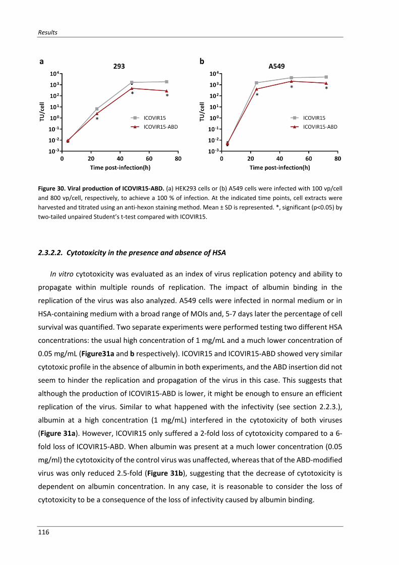
Results
116
Figure 30. Viral production of ICOVIR15‐ABD. (a) HEK293 cells or (b) A549 cells were infected with 100 vp/cell
and 800 vp/cell, respectively, to achieve a 100 % of infection. At the indicated time points, cell extracts were
harvested and titrated using an anti‐hexon staining method. Mean ± SD is represented. *, significant (p<0.05) by
two‐tailed unpaired Student’s t‐test compared with ICOVIR15.
2.3.2.2. Cytotoxicity in the presence and absence of HSA
In vitro cytotoxicity was evaluated as an index of virus replication potency and ability to
propagate within multiple rounds of replication. The impact of albumin binding in the
replication of the virus was also analyzed. A549 cells were infected in normal medium or in
HSA‐containing medium with a broad range of MOIs and, 5‐7 days later the percentage of cell
survival was quantified. Two separate experiments were performed testing two different HSA
concentrations: the usual high concentration of 1 mg/mL and a much lower concentration of
0.05 mg/mL (Figure31a and b respectively). ICOVIR15 and ICOVIR15‐ABD showed very similar
cytotoxic profile in the absence of albumin in both experiments, and the ABD insertion did not
seem to hinder the replication and propagation of the virus in this case. This suggests that
although the production of ICOVIR15‐ABD is lower, it might be enough to ensure an efficient
replication of the virus. Similar to what happened with the infectivity (see section 2.2.3.),
albumin at a high concentration (1 mg/mL) interfered in the cytotoxicity of both viruses
(Figure 31a). However, ICOVIR15 only suffered a 2‐fold loss of cytotoxicity compared to a 6‐
fold loss of ICOVIR15‐ABD. When albumin was present at a much lower concentration (0.05
mg/ml) the cytotoxicity of the control virus was unaffected, whereas that of the ABD‐modified
virus was only reduced 2.5‐fold (Figure 31b), suggesting that the decrease of cytotoxicity is
dependent on albumin concentration. In any case, it is reasonable to consider the loss of
cytotoxicity to be a consequence of the loss of infectivity caused by albumin binding.

Results
117
Figure 31. In vitro cytotoxicity of ICOVIR15‐ABD in absence and presence of HSA at high and low
concentrations. A549 cells were infected with serial dilutions of ICOVIR15 or ICOVIR15‐ABD in the absence or
presence of HSA at (a) 1 mg/ml, or (b) 0.05 mg/ml. At day 5‐7 post‐infection, cell viability was measured by BCA
protein assay. Dose‐response curves are shown along with the IC50 values for each virus and condition. Mean
values ± SD are plotted.
2.3.3. Blood persistence after systemic administration
The ABD has been successfully used to increase the plasma half‐life of therapeutic
proteins, but never tested in the context of a virus. Although our main objective was to protect
the virus against NAbs, we also tested whether albumin binding increased the blood
persistence of the adenovirus. The blood persistence of ICOVIR15 and ICOVIR15‐ABD was
compared by direct competition after intravenous injection of a mixture of both viruses
(2.5x10¹⁰ vp each) in nude mice. Blood samples were collected at different time points. The
genomes of the viruses in serum were detected by PCR of the HVR1, and were distinguished
by the size of the PCR product (199 bp for ICOVIR15, and 361 bp for ICOVIR15‐ABD due to the
ABD insertion). The difference in blood persistence was analyzed comparing the relative
intensity of the bands. Equally intense bands were obtained in the pre‐injection control and 5
min after the injection (Figure 32). From then on, a shift on the intensity of the bands can be
seen, as the band corresponding to ICOVIR15‐ABD becomes more intense than the ICOVIR15
one. One hour after the injection, the extended circulation of ICOVIR15‐ABD was clearly
evident, but at 4 h after injection the level of both viruses dropped notably.

Results
118
Figure 32. Competitive blood persistence of ICOVIR15 and ICOVIR15‐ABD after intravenous administration.
Serum samples of nude mice were collected at the indicated time points after intravenous administration of a
mixture containing ICOVIR15 and ICOVIR15‐ABD (2.5x10¹⁰ vp each, n = 5 mice). PCR amplification of the HVR1
was performed and samples were analyzed by electrophoresis. The upper band corresponds to ICOVIR15‐ABD
and the lower band to ICOVIR15, as the ABD insertion increases the size of the HVR1 from 299 to 361 bp. The gel
shows a standard with several ratios of ICOVIR15‐ABD:ICOVIR15 genomes (0.2, 1, 5, 10, and 50), a pre‐injection
control (t0), a water negative control of the PCR (H2O), and the PCR of the sera of 5 different mice (#1 to #5).
2.3.4. Biodistribution profile after systemic administration
To check if such an increase in viremia enhanced tumor transduction, the biodistribution
of the ABD‐modified adenovirus was analyzed. Subcutaneous tumors of Sk‐mel28 melanoma
cell line were implanted in nude mice and when they reached an appropriate tumor volume,
mice were intravenously injected with PBS or 5x10¹⁰ vp of reporter vectors AdGLRGD or
AdGLRGD‐ABD. Three days after vector injection mice were killed and organs were collected
for in vivo bioluminescence imaging (IVIS). Despite the advantage observed after 1 h in blood
persistence due to ABD insertion, there were no differences in transduction between the
vectors in all the organs analyzed (Figure 33). Only a non‐significant 1.6‐fold loss of liver and
tumor transduction was observed with AdGLRGD‐ABD. Likely, the viremia increase of the
modified virus compensated its lower infectivity in the presence of albumin (Figure 23),
resulting in a final tumor transduction similar to the non‐modified virus.

Results
119
Figure 33. Biodistribution profile of AdGLRGD‐ABD in nude mice after intravenous administration. Nude mice
bearing Sk‐mel28 melanoma tumors were systemically injected with PBS, or 5x10¹⁰ vp of AdGLRGD, or AdGLRGD‐
ABD. Three days after virus administration mice were sacrificed. Comparative luciferase expression in different
organs was measured by in vivo imaging system (IVIS) (n = 5 mice/group). Mean + SEM.
2.3.5. Toxicity profile after systemic administration
The toxicity of ICOVIR15 was previously studied by our group, showing a very low toxicity
profile compared to the Adwt virus (Rojas et al., 2010). Intravenous injection of ICOVIR15
causes a small loss of body weight around day 3‐4, that is quickly rescued to normal levels. In
a similar way, it causes a small increase of liver enzymes (AST and ALT) that is also rapidly
recovered.
To determine the effect of albumin binding in the toxicity of ICOVIR15, Balb/C
immunocompetent mice were injected with PBS or 5x10¹⁰ vp of ICOVIR15 or ICOVIR15‐ABD
via tail vein. Body weight was monitored until day 4 when animals were sacrificed to analyze
the serum levels of liver enzymes (aspartate aminotransferase (AST), and alanine
aminotransferase (ALT)), and other serum markers such as creatinine, lipase, and albumin.
Significant body weight loss was detected at day 3 post‐injection with both viral
treatments (Figure 34a). At this time, ICOVIR15‐treated animals had lost almost a 6% of body
weight whereas mice treated with ICOVIR15‐ABD had lost only a 2.5% (the differences
between the viruses were not significant). At day 4 post‐injection, animals treated with
ICOVIR15‐ABD had completely recovered the body weight to the same levels of PBS‐treated

Results
120
mice. Although mice treated with ICOVIR15 had started to recover, the body weight loss at
this time was still of 4%.
The injection of both oncolytic adenoviruses provoked an increase in the levels of liver
transaminases AST and ALT at day 4 post‐injection. ICOVIR15 raised the levels of AST 7‐fold
compared to a 3‐fold increase caused by ICOVIR15‐ABD, although the differences between
the viruses were not statistically significant. A significant difference was observed in the levels
of ALT as ICOVIR15 caused a 19‐fold increase compared to a 7‐fold caused by ICOVIR15‐ABD
(Figure 34b). Lipase, creatinine, and albumin serum levels were not altered by the injection of
the oncolytic viruses (Figure 34c‐e). In general terms, the ABD insertion reduced even more
the low toxicity profile of oncolytic adenovirus ICOVIR15.
2.3.6. Antitumor efficacy after systemic administration
Next, the antitumor efficacy of ICOVIR15‐ABD was tested in the model of naïve nude mice
bearing melanoma Sk‐mel28 subcutaneous tumors. With this aim, mice were treated with a
single intravenous administration of PBS, or 5x10¹⁰ vp of oncolytic viruses ICOVIR15 or
ICOVIR15‐ABD, and the tumor volume was monitored. Both viruses were able to significantly
reduce the tumor growth compared to the PBS‐treated group (Figure 35). At the end of the
experiment, ICOVIR15‐ABD induced a 2‐fold reduction in tumor volume, compared to a 1.4‐
fold induced by ICOVIR15. Although ICOVIR15‐ABD showed a tendency towards higher
antitumor efficacy, the difference between the oncolytic viruses was not statistically
significant.
Altogether, the results obtained in this chapter indicated that the ABD insertion does not
have an important impact on the oncolytic properties in the absence of neutralizing
antibodies. Whereas in vitro the ABD‐modified virus seems attenuated in the presence of
albumin, its performance in vivo is very similar to that of the parental virus. However, the
conditions studied in this section are always in the absence of NAbs, and such conditions do
not reflect what is expected in the clinic, where a high percentage of human population has
high levels of anti‐Ad5 NAbs.
2.4. CHARACTERIZATION OF THE ABD INSERTION IN THE PRESENCE OF NEUTRALIZING
ANTIBODIES
The following sections will assess the effects of the ABD modification on the adenovirus
bioactivity both in vitro and in vivo in the presence of anti‐Ad5 NAbs. In vitro experiments were

Results
121
performed using commercially available NAbs, as well as anti‐Ad5 pre‐immune sera from both
mouse and human origin. In vivo studies were performed using immunocompetent mice
previously immunized with Ad5 or immunodeficient mice infused with pre‐immune mouse
serum.
Figure 34. Toxicity after the intravenous administration of ICOVIR15‐ABD in immunocompetent mice. (a) Body
weight variation of Balb/C mice injected intravenously with PBS, or 5x10¹⁰ vp of ICOVIR15 or ICOVIR15‐ABD (n =
6‐7 mice). At day 4 post‐injection mice were sacrificed to analyze the serum levels of (b) liver transaminases, (c)
lipase, (d) creatinine, and (e) albumin (n = 4‐5 mice). *, significant (p<0.05) by two‐tailed unpaired Student’s t‐
test compared with PBS. #, significant (p<0.05) by two‐tailed unpaired Student’s t‐test compared with ICOVIR15‐
ABD.

Results
122
Figure 35. Antitumor efficacy of ICOVIR15‐ABD in nude mice after intravenous administration. Nude mice
bearing subcutaneous Sk‐mel28 melanoma tumors were treated with a single intravenous injection of PBS,
ICOVIR15, or ICOVIR15‐ABD at a dose of 5x10¹⁰ vp per mouse (n = 10‐12 tumors/group). Mean values of tumor
volume + SEM are shown. *, significant (p<0.05) by two‐tailed unpaired Student’s t‐test compared with PBS.
2.4.1. In vitro evasion of neutralizing antibodies
2.4.1.1. Effect of HSA concentration on virus transduction and neutralization
As previously explained, the physiological concentration of HSA (50 mg/ml) is too high to
be used for in vitro assays, and a concentration of 1 mg/mL was used instead in most of the
studies. To understand the impact of HSA concentration on the ABD‐modified adenovirus, the
transduction of the vectors was analyzed in the presence and absence of NAbs, using four
different concentrations of HSA (0.01, 0.1, 1, and 10 mg/mL).
As previously observed in Figure 23, albumin negatively interferes in the infectivity of both
adenovirus vectors when NAbs are absent. In this case, albumin reduced the transduction of
the vectors in a dose‐dependent manner in 293 and A549 cells, again affecting much more the
ABD‐modified virus (Figure 36). Unexpectedly, the transduction loss due to albumin binding
was notably lower in 293 cells than in A549 cells (e.g. at 1 mg/ml AdGLRGD‐ABD suffered a
3.4‐fold loss in 293 cells compared to a 24‐fold loss in A549 cells). Remarkably, the
concentration of 10 mg/ml caused a very high inhibition in the transduction of both vectors.
At this albumin concentration, AdGLRGD suffered a 5‐ and 12‐fold loss of transduction, and
the loss of AdGLRGD‐ABD was 27‐ and 91‐fold, in 293 and A549 cells respectively.
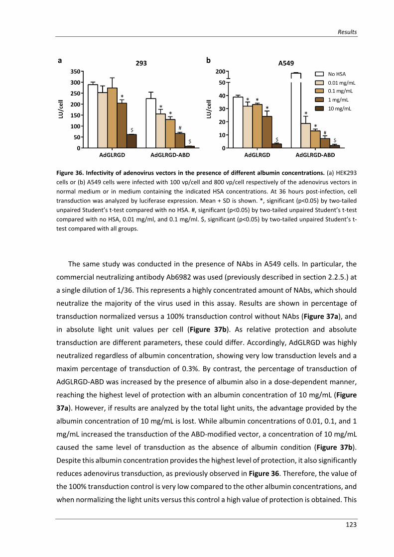
Results
123
Figure 36. Infectivity of adenovirus vectors in the presence of different albumin concentrations. (a) HEK293
cells or (b) A549 cells were infected with 100 vp/cell and 800 vp/cell respectively of the adenovirus vectors in
normal medium or in medium containing the indicated HSA concentrations. At 36 hours post‐infection, cell
transduction was analyzed by luciferase expression. Mean + SD is shown. *, significant (p<0.05) by two‐tailed
unpaired Student’s t‐test compared with no HSA. #, significant (p<0.05) by two‐tailed unpaired Student’s t‐test
compared with no HSA, 0.01 mg/ml, and 0.1 mg/ml. $, significant (p<0.05) by two‐tailed unpaired Student’s t‐
test compared with all groups.
The same study was conducted in the presence of NAbs in A549 cells. In particular, the
commercial neutralizing antibody Ab6982 was used (previously described in section 2.2.5.) at
a single dilution of 1/36. This represents a highly concentrated amount of NAbs, which should
neutralize the majority of the virus used in this assay. Results are shown in percentage of
transduction normalized versus a 100% transduction control without NAbs (Figure 37a), and
in absolute light unit values per cell (Figure 37b). As relative protection and absolute
transduction are different parameters, these could differ. Accordingly, AdGLRGD was highly
neutralized regardless of albumin concentration, showing very low transduction levels and a
maxim percentage of transduction of 0.3%. By contrast, the percentage of transduction of
AdGLRGD‐ABD was increased by the presence of albumin also in a dose‐dependent manner,
reaching the highest level of protection with an albumin concentration of 10 mg/mL (Figure
37a). However, if results are analyzed by the total light units, the advantage provided by the
albumin concentration of 10 mg/mL is lost. While albumin concentrations of 0.01, 0.1, and 1
mg/mL increased the transduction of the ABD‐modified vector, a concentration of 10 mg/mL
caused the same level of transduction as the absence of albumin condition (Figure 37b).
Despite this albumin concentration provides the highest level of protection, it also significantly
reduces adenovirus transduction, as previously observed in Figure 36. Therefore, the value of
the 100% transduction control is very low compared to the other albumin concentrations, and
when normalizing the light units versus this control a high value of protection is obtained. This

Results
124
means that at this albumin concentration, the adenovirus transduction is much lower but it is
more protected from antibody neutralization. This discrepancy in the representation of the
data does not occur with the three lower concentrations, in which more albumin confers more
transduction and protection. In any case, the transduction of AdGLRGD‐ABD in the presence
of Ab6982 was always higher than that of AdGLRGD independently of albumin concentration.
Even in the absence of albumin AdGLRGD‐ABD showed higher transduction and protection
than the non‐modified vector, suggesting that the ABD modification of the HVR1 precluded
binding of some NAbs, likely directed against the wt HVR1. Nevertheless, the working
concentration of albumin in the following in vitro studies was maintained at 1 mg/ml. These
results suggest that physiological albumin concentrations might severely inactivate the ABD‐
modified adenovirus. However, physiological conditions are reflected in the in vivo studies
where the adenovirus is injected systemically in mice. In this context, biodistribution (Figure
33) and antitumor efficacy studies (Figure 35) did not show an inactivation of the ABD‐
modified virus.
Figure 37. Escape of Ab6982 neutralization in the presence of different albumin concentrations. Adenovirus
vectors, in medium containing the indicated HSA concentrations, were incubated with Ab6982 at a single dilution
of 1/32. After 1 hour incubation, the viruses were used to infect A549 cells. Luciferase expression was analyzed
24 hours after the infection. A control without Ab6982 was included to obtain the 100 % infection value. (a)
Results are represented as % transduction versus the 100 % infection control. Mean values + SD are depicted. *,
significant (p<0.05) by two‐tailed unpaired Student’s t‐test compared with no HSA. #, significant (p<0.05) by two‐
tailed unpaired Student’s t‐test compared with no HSA, 0.01 mg/ml, and 0.1 mg/ml. $, significant (p<0.05) by
two‐tailed unpaired Student’s t‐test compared with all groups. (b) Results are represented as total light units per
cell. Mean values + SD are depicted. *, significant (p<0.05) by two‐tailed unpaired Student’s t‐test compared with
no HSA and 10 mg/ml.

Results
125
2.4.1.2. In vitro neutralization by Ab6982
The ability of the ABD‐modified vector to efficiently transduce target cells in the presence
of Ab6982 when coated with albumin has already been demonstrated in section 2.2.5., and in
this last section using a single antibody dilution. However, a more exhaustive study was
performed not only in terms of cell transduction but also of viral replication in tumor cells.
To confirm the evasion results previously obtained, the ability of the vectors to transduce
target cells in the presence of Ab6982 was analyzed. AdGLRGD and AdGLRGD‐ABD in absence
and presence of HSA were incubated with serial dilutions of Ab6982 and used to infect 293
and A549 cells. Very similar results were obtained in both cell lines (Figure 38a and b). We
observed again that AdGLRGD‐ABD was less neutralized than AdGLRGD even in the absence
of albumin. Importanly, a clear increase on the transduction efficiency of the ABD‐modified
vector was observed when albumin was present in the medium. As expected, the non‐
modified vector AdGLRGD was highly neutralized regardless of albumin presence.
The capacity of the viruses to escape neutralization was not only assessed by their
transduction efficiency but also by their cytotoxicity in presence of Ab6982. For this purpose,
the oncolytic adenoviruses ICOVIR15 or ICOVIR15‐ABD in presence or absence of HSA were
incubated with serial dilutions of Ab6982, and used to infect A549 cells. Cell survival was
analyzed 5 days after infection. In absence of HSA both viruses showed similar capacity to kill
tumor cells (Figure 39), and only a small increase of cytotoxicity was observed with ICOVIR15‐
ABD probably due to the certain evasion of NAbs observed in transduction (Figure 38a and b).
Importantly, HSA shifted the cytotoxicity curve of ICOVIR15‐ABD towards higher NAbs
concentrations, whereas it did not protect ICOVIR15 from neutralization.
2.4.1.3. In vitro neutralization by anti‐Ad5 pre‐immune mouse serum
To better understand the potential of the ABD‐adenovirus to escape neutralization in vivo,
the same neutralization tests were performed with serum from mice immunized with Ad5. To
generate the neutralizing serum, C57BL/6 immunocompetent mice were primed
intraperitoneally with 3x10¹⁰ vp of Ad5wt, and boosted intravenously seven days later with
the same dose. Seven days after boosting, mice were killed and the blood was collected to
obtain the serum.

Results
126
Figure 38. In vitro transduction of AdGLRGD‐ABD in presence of Ab6982. Adenovirus vectors, in the presence
or absence of HSA, were incubated with serial dilutions of the polyclonal neutralizing antibody Ab6982. After 1
hour incubation, the viruses were used to infect (a) HEK293 cells or (b) A549 cells at 10 vp/cell. Luciferase
expression was analyzed 24 hours after the infection. A control without Ab6982 was included to obtain the 100
% infection value. Results are represented as % transduction versus the 100 % infection control. Mean values ±
SD. *, significant (p<0.05) by two‐tailed unpaired Student’s t‐test compared with all groups.
Figure 39. Cytotoxicity of ICOVIR15‐ABD in presence of Ab6982. Oncolytic adenoviruses ICOVIR15 or ICOVIR15‐
ABD, in presence or absence of HSA, were incubated with serial dilutions of Ab6982 for 1 hour, and then used to
infect A549 cells. Cell survival was measured at day 5 post‐infection by BCA staining. Mean values ± SD are shown.
*, significant (p<0.05) by two‐tailed unpaired Student’s t‐test compared with all groups.

Results
127
Since mouse serum contains a high concentration of albumin, the absence of albumin
condition could not be analyzed in these studies. On the other hand, HSA was added to the
medium at a concentration of 1 mg/mL in order to maintain a minimum albumin
concentration throughout the dilutions. The evasion of the NAbs present in the serum was
first analyzed in terms of cell transduction in A549 cells. A clear advantage was observed again
with AdGLRGD‐ABD, showing high levels of transduction even at low serum dilutions, where
the non‐modified vector was completely neutralized (Figure 40a). The neutralization escape
in the mouse serum was also tested in terms of cytotoxicity in A549 cells, where ICOVIR15‐
ABD was able to kill the tumor cells more efficiently than ICOVIR15 (Figure 40b),
demonstrating a higher bioactivity in the presence of NAbs.
Figure 40. In vitro evasion of anti‐Ad5 neutralizing mouse serum. (a) Transduction analysis after incubation with
anti‐Ad5 neutralizing mouse serum. AdGLRGD or AdGLRGD‐ABD in the presence of HSA were incubated with
serial dilutions of the serum for 1 hour, and then used to infect A549 cells at 10 vp/cell. Luciferase expression
was analyzed 24 hours after infection. A control without serum was included to obtain the 100 % infection value.
Results are represented as % transduction versus the 100 % infection control. Mean ± SD. *, significant (p<0.05)
by two‐tailed unpaired Student’s t‐test compared with AdGLRGD‐ABD + HSA. (b) Cytotoxicity analysis after
incubation with anti‐Ad5 neutralizing mouse serum. ICOVIR15 or ICOVIR15‐ABD in the presence of HSA were
incubated with serial dilutions of the serum for 1 hour, and then used to infect A549 cells. Cell survival was
measured at day 5 post‐infection by BCA staining. Mean values ± SD. *, significant (p<0.05) by two‐tailed unpaired
Student’s t‐test compared with ICOVIR15 + HSA.
2.4.1.4. In vitro neutralization by anti‐Ad5 pre‐immune human sera
To test whether the ABD‐modified adenovirus could also evade NAbs developed in
humans, sera from six human donors (previously tested by our group for anti‐Ad5

Results
128
seropositivity) were used to test the neutralization escape of the viruses by transduction
analysis in 293 cells. A clear neutralization escape of the ABD‐vector was observed in three of
the six sera analyzed (#1, #2, and #6), whereas no advantage versus the non‐modified vector
was detected in the other three sera (#3, #4, and #5) (Figure 41a). This is observed when
representing the data as % transduction versus the 100% transduction control as explained in
section 2.2.5. However, if the data is not normalized, we observe that in general terms
AdGLRGD‐ABD shows higher transduction levels than AdGLRGD at low serum dilutions, when
the NAbs concentration are higher (Figure 41b), which are the conditions that better mimic
the intravenous administration of the virus. When the sera were highly diluted, the advantage
of the ABD‐modified virus was lost, as previously observed in presence of albumin and
absence of neutralizing antibodies (Figure 23).
2.4.2. Blood persistence in pre‐immune mice after systemic administration
Not having observed an improvement in tumor transduction nor a significant benefit in
antitumor efficacy in naïve mice, we next studied the behavior of the ABD‐adenoviruses in
pre‐immune mouse models. First, the blood persistence was analyzed in both naïve and pre‐
immune immunocompetent mice. To generate anti‐Ad5 NAbs, C57BL/6 mice were immunized
with an intraperitoneal injection of PBS or 3x10¹⁰ vp of Ad5wt (naïve or pre‐immune groups),
and seven days later mice were intravenously injected with 3x10¹⁰ vp of AdGLRGD or
AdGLRGD‐ABD. Serum samples were collected from the tail vein at the indicated time points
to detect adenovirus genomes by real‐time PCR, and transducing units by anti‐hexon staining
method.
In naïve mice, the clearance curve shows an advantage of the ABD‐modified virus in terms
of viral particles (significant differences at 15 min and 1 h post‐injection) but not of
transducing units (Figure 42 left panels). This result matches with the increased blood
persistence at 1 h after injection in nude mice (Figure 32), which did not translate in an
increased organ transduction (Figure 33). However, in pre‐immune mice the advantage of the
ABD‐modified virus was evident not only in circulating viral particles (significant differences at
30 min post‐injection) but also in transducing units (Figure 42 right panels). Although the
clearance curve of transducing units in pre‐immune mice shows higher levels of AdGLRGD‐
ABD versus AdGLRGD, the differences were not statistically significant at any time point.

Results
129

Results
130
Figure 41. In vitro neutralization analysis after incubation with human sera. AdGLRGD or AdGLRGD‐ABD in
presence of HSA were incubated with serial dilutions of human sera from six anonymous donors (#1‐6). After 1
hour incubation, the vectors were used to infect HEK293 cells at 10 vp/cell. Luciferase expression was analyzed
24 hours after infection. A control without serum was included to obtain the 100 % infection value. Results are
represented as (a) % transduction versus the 100 % infection control or (b) total light units per cell. The value of
the positive transduction control without serum is shown (c+). Mean values + SD are depicted. *, significant
(p<0.05) by two‐tailed unpaired Student’s t‐test compared with AdGLRGD + HSA.

Results
131
Figure 42. Blood persistence of AdGLRGD and AdGLRGD‐ABD after intravenous administration in naïve and
pre‐immune mice. C57BL/6 immunocompetent mice were immunized with an intraperitoneal injection of 3x10¹⁰
vp of Ad5wt or PBS. Seven days later mice were intravenously injected with AdGLRGD or AdGLRGD‐ABD (3x10¹⁰
vp, n=5 mice), and serum samples were collected from the tail vein 1 min, 5 min, 15 min, 30 min, and 60 min
after injection. (a) Adenovirus genome quantification was performed by real‐time PCR in serum samples of naïve
(left) and pre‐immune mice (right). (b) Adenovirus transducing units titration in serum samples of naïve (left) and
pre‐immune (right) mice was performed by infection of HEK293 cells followed by antihexon staining. The titers
were normalized to the transduction value of each virus in naïve mouse serum (100% transduction control). *,
significant (p<0.05) by two‐tailed unpaired Student’s t‐test compared to AdGLRGD.
2.4.3. Organ transduction in pre‐immune mice after systemic administration
Next, the effect of anti‐Ad5 NAbs on adenovirus organ transduction after intravenous
administration was analyzed. C57BL/6 mice bearing subcutaneous B16‐CAR melanoma tumors
were immunized with an intraperitoneal injection of PBS or 3x10¹⁰ vp of Ad5wt (naïve or pre‐
immune groups), and seven days later mice received a single intravenous dose of PBS, or
3x10¹⁰ vp of AdGLRGD or AdGLRGD‐ABD.
A blood sample was taken from the tail vein 1 h post‐injection to analyze the amount of
virus in blood, which was confirmed to be significantly higher (2.3‐fold) in both naïve and pre‐

Results
132
immune mice injected with the ABD‐modified vector (Figure 43), compared to those injected
with the control vector. This further supports the results showed in Figures 32 and 42.
Figure 43. Viral load in blood 1 hour after injection in naïve and pre‐immune mice. C57BL/6 immunocompetent
mice previously immunized or not with Ad5wt were intravenously injected with AdGLRGD or AdGLRGD‐ABD
(3x10¹⁰ vp, n=4‐6 mice). Serum samples were collected 1 hour after injection to quantify adenovirus genomes by
real‐time PCR. *, significant (p<0.05) by two‐tailed unpaired Student’s t‐test compared to AdGLRGD in naïve mice.
#, significant (p<0.05) by two‐tailed unpaired Student’s t‐test compared with AdGLRGD in pre‐immune mice.
Three days after vector injection mice were killed and the luciferase expression in livers
and tumors was analyzed by in vivo imaging system (IVIS). In naïve animals, AdGLRGD‐ABD
showed a 9‐ and 7‐fold non‐significant loss of liver and tumor transduction respectively
compared to the non‐modified vector (Figure 44). Importantly, when animals were pre‐
immunized with Ad5wt, the parental AdGLRGD vector suffered a complete neutralization as
the transduction of liver and tumors was completely abolished. On the contrary, AdGLRGD‐
ABD only suffered a 3.9‐ and 1.5‐fold non‐significant loss of liver and tumor transduction
respectively, indicating an efficient protection from anti‐Ad5 NAbs in vivo.

Results
133
Figure 44. Liver and tumor transduction in naïve and pre‐immune mice after systemic administration. C57BL/6
immunocompetent mice bearing subcutaneous B16‐CAR melanoma tumors were immunized with an
intraperitoneal injection of 3x10¹⁰ vp of Ad5wt or PBS, and seven days later mice were intravenously injected
with AdGLRGD or AdGLRGD‐ABD (3x10¹⁰ vp). Three days after virus administration mice were killed and luciferase
activity in liver and tumors was analyzed by in vivo imaging system (IVIS) (livers n = 3‐6, tumors n = 6‐12). Mean
+ SEM is shown. *, significant (p<0.05) by two‐tailed unpaired Student’s t‐test compared with AdGLRGD pre‐
immune.
2.4.4. Generation of an anti‐Ad5 pre‐immune status in immunodeficient mice to test the
antitumor efficacy
After observing the efficient tumor transduction of the ABD‐modified adenovirus in pre‐
immune mice, the next step was to evaluate the antitumor efficacy of ICOVIR15‐ABD in mice
with anti‐Ad5 NAbs. However, since the human adenovirus does not replicate in mouse cells,
an immunodeficient mouse model with engrafted human tumors is required to test the
antitumor activity. The strategy used to generate an anti‐Ad5 pre‐immune status in nude mice
was the passive transfer of neutralizing serum from C57BL/6 mice.
2.4.4.1. Generation of anti‐Ad5 immune sera
C57BL/6 were primed intraperitoneally with 3x10¹⁰ vp of Ad5wt, and boosted
intravenously seven days later with the same dose of the same virus. Seven days later mice
were killed, the blood was collected by intracardiac puncture, and the serum of each mouse

Results
134
was pooled. To analyze the variability in the generation of NAbs, we compared the neutralizing
activity of four batches of pooled serum obtained at different dates during this thesis. The
AdGL vector (a GFP/luciferase expressing vector with a wt capsid) was incubated with serial
1/2 dilutions of the sera, and then used to infect 293 cells. The transduction is compared to a
control without serum (0% neutralization control) to obtain the percentage of neutralization
at each serum dilution. The titer of each serum corresponds to the dilution in which a 50% of
adenovirus neutralization was obtained. Although the four sera displayed high neutralizing
activity, variability was observed in the titer of the sera (serum #0315: 1280‐2560, serum
#0615: >5120, serum #1015: 2560, and serum #0416: 5120) (Figure 45). Of note, serum #0615
displayed a titer more than a 2‐fold higher than serum #0416. This variability in the
neutralizing activity was taken into consideration when transferring the sera to nude mice.
Figure 45. Neutralizing activity of anti‐Ad5 mouse sera. Neutralizing sera were obtained by immunization of
C57BL/6 immunocompetent mice with Ad5wt using a prime and boost regimen. Serum titration was performed
by incubating AdGL with serial 1/2 dilutions of the serum and analyzing the transduction in HEK293 cells using
an MOI of 0.25 TU/cell. A control without serum was included to obtain the 0% neutralization value. The serum
titer corresponds to the dilution required to neutralize the 50% of the virus. Four different serum batches were
compared. Titration curves are shown along with serum titers. Mean + SD.

Results
135
2.4.4.2. Passive immunization of nude mice: preliminary test of organ transduction and
antitumor efficacy to evaluate neutralization
The serum transfer to nude mice was performed via intraperitoneal route 24 hours prior
to virus injection, as previously described (Tsai et al., 2004). In a first test, nude mice bearing
A549 subcutaneous tumors were transferred with PBS (mock), 5µL, or 50 µL of the serum
#0315 (titer of 1280‐2560) diluted in 200 µL of PBS. The next day mice were intravenously
injected with 4.5x10¹⁰ vp of AdGL. Three days after vector injection mice were killed and livers
and tumors were collected for in vivo bioluminescence imaging (IVIS). As expected, the sham
passively immunized mouse with PBS displayed very high luciferase signal in the liver and, to
a lesser extent, in tumors (Figure 46). Passive immunization with 5 µL of serum reduced the
transduction of tumors, but high levels of luciferase signal were still detected in the liver. In
contrast, administration of 50 µL of serum caused a very important drop in the luciferase signal
of liver and tumors, indicating an efficient neutralization of the virus.
Having demonstrated the ability of passively immunized nude mice to neutralize the Ad5,
the antitumor activity of the oncolytic adenoviruses was tested in this mouse model. Nude
mice bearing A549 subcutaneous tumors were sham‐immunized with PBS, or passively
immunized with 50 µL of the serum #0315 intraperitoneally (naïve or pre‐immune groups).
The next day mice were injected with PBS or 4.5x10¹⁰ vp of ICOVIR15, or ICOVIR15‐ABD via tail
vein. To our surprise, the oncolytic activity of ICOVIR15 was not hindered by the passive
immunization of mice (Figure 47). The experiment was stopped at day 21 post‐injection, as
tumors of naïve and pre‐immune mice treated with ICOVIR15 were growing at the same
speed. This result suggests that the virus was not completely neutralized. We hypothesized
that despite not having observed any sign of transduction at day 3, a certain amount of virus
might still be reaching the tumor, being able to replicate and control tumor growth. We
proceeded to test this hypothesis using a reporter oncolytic virus.
2.4.4.3. Passive immunization of nude mice: replication analysis with a reporter oncolytic
adenovirus
To be able to visually detect replication foci several days after virus administration, a
replicative reporter virus is required. In this work we constructed ICOVIR15‐Luc, a reporter
oncolytic adenovirus generated by insertion of the luciferase gene in the genome of ICOVIR15
after the fiber. To drive the expression under the major late promoter (MLP) as a late
expression cassette, the luciferase transgene included the splicing acceptor from the IIIa

Results
136
adenoviral protein, and a polyadenylation sequence (Figure 48a). In this manner, luciferase
expression is linked to the replication of the virus and not just to virus transduction, as it occurs
with the regular CMV‐Luciferase cassette in AdGL and its variations. The in vitro
characterization of ICOVIR15‐Luc was performed by analyzing the luciferase expression
kinetics and the cytotoxicity of the virus.
Figure 46. Preliminary analysis of liver and tumor transduction after passive immunization of nude mice. Nude
mice bearing subcutaneous A549 tumors were injected intraperitoneally with PBS (mock), 5 µL, or 50 µL of anti‐
Ad5 neutralizing mouse serum (serum #0315) diluted in 200 µL of PBS. The next day mice received an intravenous
injection of 4.5x10¹⁰ vp of AdGL (n = 1 mouse per condition). (a) Livers and tumors were imaged for luciferase
expression at day three after vector injection. An overlay of the white image and the luciferase signal is shown.
(b) Quantification of luciferase expression in livers and tumors.

Results
137
Figure 47. Preliminary analysis of antitumor efficacy after passive immunization of nude mice. Nude mice
bearing subcutaneous A549 tumors were passively immunized with an intraperitoneal injection of PBS (naïve),
or 50 µL of anti‐Ad5 neutralizing mouse serum (serum #0315). The next day mice were treated with a single
intravenous injection of PBS or 5x10¹⁰ vp of ICOVIR15, or ICOVIR15‐ABD. Mean values of tumor volume + SEM
are shown (n = 10‐11 tumors/group).
The luciferase expression kinetics of ICOVIR15‐Luc were compared to those of AdGLRGD
in human A549 and canine Dk‐cre cells (Figure 48b). The delayed luciferase expression of
ICOVIR15‐Luc compared to AdGLRGD in A549 cells indicates that the expression occurs in the
late phase of the viral cycle, associated with the replication of the virus. In contrast, since the
human adenovirus cannot replicate in canine cells, the expression of luciferase in Dk‐cre cells
is significantly reduced compared to A549 cells, demonstrating a replication‐dependent
expression. Regarding the cytotoxicity, ICOVIR15‐Luc showed a 7.8‐fold loss compared to
ICOVIR15 (Figure 48c), suggesting that the either the size of the genome or the expression of
the transgene negatively affects the virus life cycle. Despite this cytotoxicity loss, ICOVIR15‐
Luc represents a useful tool to monitor the viral replication by in vivo imaging.

Results
138
Figure 48. Construction and in vitro characterization of ICOVIR15‐Luc. (a) Schematic representation of the
genome of ICOVIR15‐Luc. The luciferase transgene (Luc) was cloned after the fiber gene in the genome of
ICOVIR15. To drive the expression under the control of the major late promoter (MLP), a splicing acceptor (SA)
and a polyadenylation sequence (pA) were cloned before and after the transgene respectively. A Kozak sequence
(K) was also included before the Luc gene to increase the translation efficiency. (b) Luciferase expression kinetics.
A549 cells (left) or Dk‐cre cells (right) were infected with AdGLRGD or ICOVIR15‐Luc at an MOI of 25 TU/cell and
10 TU/cell, respectively. At the indicated time points, cells were lysed and the luciferase activity in the protein
extract was measured with a luminometer. Mean values + SD are depicted. *, significant (p<0.05) by two‐tailed
unpaired Student’s t‐test compared with AdGLRGD. (c) Cytotoxicity. A549 cells were infected with a broad range
of MOIs with ICOVIR15 or ICOVIR15‐Luc. At day 6 post‐infection, cell viability was measured by BCA staining.
Dose‐response curves are shown along with the IC50 values for each virus. Mean values ± SD are plotted.

Results
139
In a second passive immunization test, nude mice bearing subcutaneous A549 tumors
were injected intraperitoneally with 150 µL of PBS or serum #0615 (titer >5120) (naïve or pre‐
immune groups). Since in the first test the neutralization of the virus was not complete, a
higher volume of a more neutralizing serum was used in this case. In addition, the viral dose
was reduced to 4x10¹⁰ vp. The next day after immunization, naïve and passively immunized
mice were injected with 4x10¹⁰ vp of ICOVIR15‐Luc, and a group injected with PBS was also
included as negative control. Luciferase expression in mice was monitored and quantified in
liver and tumors using IVIS imaging (Figure 49 and Figure 50). At day 3 post‐injection, naïve
mice showed a strong luciferase signal in the liver and a much lower signal in tumors. Even
though ICOVIR15‐Luc cannot replicate in normal tissues, a certain level of expression in the
absence of replication is expected (as observed in Figure 48b), and since most of the injected
dose is retained by the liver, a strong signal is generated in this organ. Accordingly, the absence
of replication in the liver causes the signal to rapidly drop over time. On the contrary,
luciferase signal in the tumor increases notably due to active viral replication, confirming the
replication‐dependent luciferase expression of ICOVIR15‐Luc. Same as the PBS‐treated group,
pre‐immune mice transferred with the neutralizing serum did not show any luciferase signal
in the liver nor the tumors throughout the study, suggesting an efficient neutralization of the
virus. However, if the luminescence scale is lowered to levels where the signal in naïve mice
is highly saturated, we observe a focus of replication in a tumor of a pre‐immune mouse that
appears at day 12 post‐injection, and that increases over time (Figure 49b). Figure 50 shows
the quantification of the luciferase signal throughout the study in liver (Figure 50a) and tumors
(Figure 50b), showing positive signal in tumors of pre‐immune mice at day 12 that increases
until day 24 when mice were sacrificed. Individual plotting of tumors indicated that adenoviral
replication occurred in at least two out of four tumors of pre‐immune mice (Figure 50c). This
result indicates that once again, not all the adenovirus dose was neutralized in some mice,
and that a very high amount of NAbs needs to be transferred to neutralize such a high viral
dose. Nonetheless, the differences in virus replication between naïve and pre‐immune mice
were noteworthy, and this level of neutralization was thought to be sufficient to inhibit the
antitumor efficacy of an oncolytic adenovirus.

Results
140

Results
141
Figure 49. In vivo imaging of ICOVIR15‐Luc replication in naïve or passively immunized nude mice. Nude mice
bearing subcutaneous A549 tumors were injected intraperitoneally with 150 µL of PBS, or anti‐Ad5 neutralizing
mouse serum (serum #0615). The next day mice received an intravenous injection of PBS, or 4x10¹⁰ vp of
ICOVIR15‐Luc (n = 2 mice/group). Mice were imaged for luciferase expression at the indicated days after virus
injection using (a) the default luminescence color scale (min = 223, max = 12000), or (b) a much lower
luminescence color scale (min = 20, max = 120). White light images overlaid with luciferase signal are shown.
2.4.4.4. Passive immunization of nude mice: antitumor efficacy
To test the antitumor activity with the new conditions optimized in the previous section,
nude mice bearing subcutaneous A549 tumors were passively immunized with PBS or serum
#0615 (titer >5120). The volume of the serum transferred was further increased to 200 µl to
achieve higher levels of neutralization. The next day mice received a single intravenous
injection of PBS or 4x10¹⁰ vp of ICOVIR15, or ICOVIR15‐ABD. No differences were observed in
antitumor efficacy between the oncolytic viruses in naïve mice. Both viruses were able to
significantly reduce the tumor growth compared to the PBS‐treated group, and at the end of
the study, ICOVIR15 and ICOVIR15‐ABD induced a 2‐fold and 1.7‐fold reduction in tumor
volume respectively (Figure 51a). Importantly, in pre‐immune mice the non‐modified

Results
142
ICOVIR15 was completely inefficacious, but the oncolytic activity of ICOVIR15‐ABD was
preserved. At the end of the study, ICOVIR15‐ABD induced a 1.9‐fold reduction of tumor
volume compared to the PBS‐treated group and a 2.4‐fold reduction compared to the
ICOVIR15‐treated group. The same experiment was conducted in mice bearing Sk‐mel28
melanoma tumors. The stock of serum #0615 was spent in the previous study and to transfer
equivalent levels of NAbs, mice were injected with 400 µl of serum #1015 (titer of 2560). The
outcome was very similar to that in A549 tumors. Both ICOVIR15 and ICOVIR15‐ABD induced
a similar antitumor effect in naïve animals, showing a 1.6 and a 1.8‐fold reduction of tumor
volume respectively. In pre‐immune mice, we observed again no impairment in the antitumor
activity of ICOVIR15‐ABD in presence of neutralizing antibodies, which caused a 1.6‐fold
reduction of tumor volume compared to ICOVIR15, which totally lost its antitumor effect
(Figure 51b).
Figure 50. Quantification of luciferase expression in naïve or passively immunized nude mice intravenously
injected with ICOVIR15‐Luc. Nude mice bearing subcutaneous A549 tumors were injected intraperitoneally with
150 µL of PBS or anti‐Ad5 neutralizing mouse serum (serum #0615). The next day mice received an intravenous
injection of PBS or 4x10¹⁰ vp of ICOVIR15‐Luc (n = 2 mice/group). Quantification of luciferase signal in (a) liver
and (b) tumors. Mean values + SD are depicted. *, significant (p<0.05) by two‐tailed unpaired Student’s t‐test
compared with PBS and ICOVIR15‐Luc naïve. (c) Quantification of the luciferase expression of individual tumors.

Results
143
Figure 51. Antitumor efficacy in naïve and passively immunized nude mice. Nude mice bearing subcutaneous
xenografts of (a) A549 (lung adenocarcinoma) or (b) Sk‐mel28 (melanoma) tumors were injected
intraperitoneally with PBS, or anti‐Ad5 neutralizing mouse serum (200 µL of serum #0615 for mice bearing A549
tumors, and 400 µL of serum #1015 for mice bearing Sk‐mel28 tumors). The day after, mice were treated with a
single intravenous injection of PBS or 4x10¹⁰ vp of ICOVIR15 or ICOVIR15‐ABD. Mean values of tumor volume +
SEM are plotted (n = 10‐13 tumors/group). *, significant (p<0.05) by two‐tailed unpaired Student’s t‐test
compared with PBS (A549 tumors: ICOVIR15 from day 7, ICOVIR15‐ABD from day 13, ICOVIR15‐ABD from day 10;
Sk‐mel28 tumors: ICOVIR15 from day 7, ICOVIR15‐ABD and ICOVIR15‐ABD pre‐immune from day 4). #, significant
(p<0.05) by two‐tailed unpaired Student’s t‐test compared with ICOVIR15 pre‐immune (A549 tumors: ICOVIR15
from day 27, ICOVIR15‐ABD and ICOVIR15‐ABD pre‐immune from day 24; Sk‐mel28 tumors: ICOVIR15 from day
14, ICOVIR15‐ABD and ICOVIR15‐ABD pre‐immune from day 4).
Besides controlling tumor growth, oncolytic adenovirus treatment was also able to
increase mice survival. As shown in Figure 52, treatment with ICOVIR15 significantly increased
the survival of naïve mice compared to the PBS‐treated group in both tumor models, but could
not increase the survival of pre‐immune mice. ICOVIR15‐ABD also increased the survival in
naïve mice compared to PBS treatment. In addition, the treatment with ICOVIR15‐ABD could
also significantly increase the survival of pre‐immune mice compared to the PBS and
ICOVIR15‐treated groups. Overall, these results validate the pre‐immune immunodeficient
mouse model, and demonstrate again the efficient neutralization escape of the ABD‐modified
adenovirus.
At the end of the study, mice were sacrificed and tumors were collected for Ad detection.
A549 tumors were used for Ad genome detection by quantitative real‐time PCR and
immunohistochemical staining of E1A viral protein. Intriguingly, the amount of adenovirus
detected in tumors did not correlate with the tumor volume at the end of the experiment.

Results
144
Quantification of Ad genomes in A549 tumors revealed no differences between naïve and pre‐
immune animals treated with ICOVIR15, which in general contained high amounts of virus
genomes in their tumors (Figure 53). However, certain heterogeneity is observed and some
tumors contained lower viral loads, especially in pre‐immune mice. In addition, we could not
detect ICOVIR15‐ABD genomes in pre‐immune mice, and a very low amount in naïve mice,
neither group being statistically significant versus the PBS. This result was confirmed by the
immune staining of E1A viral protein in paraffin sections of the tumors. Abundant replication
foci were observed in tumors of naïve mice treated with ICOVIR15 (Figure 54). Pre‐immune
mice treated with the same virus showed variability as some tumors showed many E1A‐
stained areas whereas no virus was detected in others. ICOVIR15‐ABD could not be detected
by E1A staining in tumors of neither naïve nor pre‐immune mice.
Figure 52. Kaplan‐Meier survival curves upon systemic administration of oncolytic adenoviruses in naïve and
passively immunized nude mice. Nude mice bearing subcutaneous xenografts of (a) A549 (lung adenocarcinoma)
or (b) Sk‐mel28 (melanoma) tumors were injected intraperitoneally with PBS or anti‐Ad5 neutralizing mouse
serum (200 µL of serum #0615 for mice bearing A549 tumors, and 400 µL of serum #1015 for mice bearing Sk‐
mel28 tumors). The day after, mice were treated with a single intravenous injection with PBS, or 4x10¹⁰ vp of
ICOVIR15 or ICOVIR15‐ABD. Kaplan‐Meier curves are plotted (n = 10‐13 tumors/group). End‐point was
established at 750 mm3 of tumor volume. *, significant (p<0.05) by log‐rank test compared with PBS. #, significant
(p<0.05) by log‐rank test compared with ICOVIR15 pre‐immune.
Detection of E1A by immunohistochemistry was also performed in Sk‐mel28 tumors with
a very similar result. Strong E1A staining was observed in naïve mice treated with ICOVIR15
(Figure 55). Variability was observed again with in pre‐immune mice, showing many
replication areas in some tumors whereas very few were detected in others. ICOVIR15‐ABD

Results
145
was barely detected in tumors of naïve mice whereas tumors of pre‐immune mice showed
some small stained areas.
Figure 53. Adenovirus detection in A549 tumors by real‐time PCR. Nude mice bearing subcutaneous xenografts
of A549 (lung adenocarcinoma) tumors were injected intraperitoneally with 200 µL of PBS, or anti‐Ad5
neutralizing mouse serum (serum #0615). The next day mice were treated with a single intravenous injection
with PBS or 4x10¹⁰ vp of ICOVIR15 or ICOVIR15‐ABD. At day 34 after virus injection, mice were killed and tumors
were collected. Adenovirus genome quantification was performed by real‐time PCR of DNA samples extracted
from frozen tumors. Mean values are represented by horizontal lines within scatterplots. *, significant (p<0.05)
by two‐tailed unpaired Student’s t‐test compared with PBS. #, significant (p<0.05) by two‐tailed unpaired
Student’s t‐test compared with ICOVIR15‐ABD. $, significant (p<0.05) by two‐tailed unpaired Student’s t‐test
compared with ICOVIR15‐ABD pre‐immune.
To understand this lack of correlation between the amount of virus in tumors and the
antitumor efficacy, the same experiment was conducted to detect the adenovirus at a much
earlier time point, when fewer rounds of replication had occurred. Nude mice bearing A549
tumors were passively immunized with 300 µL of serum #0416 (titer of 5120), to achieve the
same levels of neutralization of the last experiments. The next day mice were treated
systemically with PBS or 4x10¹⁰ vp of ICOVIR15, or ICOVIR15‐ABD. Mice were sacrificed at day
11 post‐injection and adenovirus genomes were detected by real‐time PCR, since it is a more
quantitative method than immunohistochemistry. In this case, all tumors of naïve mice
treated with ICOVIR15 contained high levels of adenovirus genomes, which were significantly
higher than in tumors of pre‐immune mice (Figure 56). Unlike in naïve mice, the amount of
ICOVIR15 genomes in pre‐immune mice at day 11 was significantly lower than at day 34. In
fact, there were no significant differences between the PBS and the ICOVIR15 pre‐immune

Results
146
groups. In contrast, ICOVIR15‐ABD genomes could be detected in tumors of both naïve and
pre‐immune mice at higher levels than ICOVIR15 in pre‐immune mice. Nevertheless, the levels
of ICOVIR15‐ABD in naïve and pre‐immune mice were lower compared to those of ICOVIR15
in naïve mice.
Figure 54. Adenovirus detection in A549 tumors by immunohistochemistry. Nude mice bearing subcutaneous
xenografts of A549 (lung adenocarcinoma) tumors were injected intraperitoneally with 200 µL of PBS or anti‐Ad5
neutralizing mouse serum (serum #0615). The next day mice were treated with a single intravenous injection
with PBS or 4x10¹⁰ vp of ICOVIR15, or ICOVIR15‐ABD. At day 34 after virus injection, mice were killed and tumors
were collected. Immunohistochemistry of E1A viral protein was performed in paraffin embedded tumor sections.
Original magnification, 4X.

Results
147
Figure 55. Adenovirus detection in Sk‐mel28 tumors by immunohistochemistry. Nude mice bearing
subcutaneous xenografts of Sk‐mel28 (melanoma) tumors were injected intraperitoneally with 400 µL of PBS, or
anti‐Ad5 neutralizing mouse serum (serum #1015). The next day mice were treated with a single intravenous
injection with PBS or 4x10¹⁰ vp of ICOVIR15, or ICOVIR15‐ABD. At day 32 after virus injection, mice were killed
and tumors were collected. Immunohistochemistry of E1A viral protein was performed in paraffin embedded
tumor sections. Original magnification, 4X.
These differences in the amount of adenovirus genomes at day 11 and day 34 suggest a
quicker clearance of the ABD‐modified virus from tumors, probably related to the lower
production and replication of this virus combined with the stromal barriers present in tumors
(Figure 57). In addition, it can be observed that the neutralization of ICOVIR15 was not
complete (as previously observed with ICOVIR15‐Luc, see Figure 49b) and the small amount

Results
148
of virus that reaches tumors can eventually replicate, albeit is incapable of controlling tumor
growth. This matter will be addressed in depth in the Discussion section.
Figure 56. Early detection of adenovirus in A549 tumors by real‐time PCR. Nude mice bearing subcutaneous
xenografts of A549 (lung adenocarcinoma) tumors were injected intraperitoneally with 300 µL of PBS, or anti‐
Ad5 neutralizing mouse serum (serum #0416). The next day mice were treated with a single intravenous injection
with PBS or 4x10¹⁰ vp of ICOVIR15, or ICOVIR15‐ABD. At day 11 after virus injection, mice were killed and tumors
were collected. Adenovirus genome quantification was performed by real‐time PCR of DNA samples extracted
from frozen tumors (n = 4‐8 tumors/group). Mean values are represented by horizontal lines within scatterplots.
*, significant (p<0.05) by two‐tailed unpaired Student’s t‐test compared with PBS. #, significant (p<0.05) by two‐
tailed unpaired Student’s t‐test compared with ICOVIR15 pre‐immune. $, significant (p<0.05) by two‐tailed
unpaired Student’s t‐test compared with ICOVIR15‐ABD. &, significant (p<0.05) by two‐tailed unpaired Student’s
t‐test compared with ICOVIR15‐ABD pre‐immune.
2.4.5. Immune response against proteins expressed from the viral genome in pre‐immune
mice
Recombinant adenoviruses are also used as vaccine platforms, which also face the
problem of pre‐existing NAbs. Neutralization of the vector impairs transgene expression and
therefore, no immune response can be mounted against the desired antigen. We
hypothesized that the ABD insertion could also be useful in the field of adenovirus‐based
vaccines. To demonstrate this, we analyzed the CTL immune response generated against a
protein expressed from the virus genome in naïve and anti‐Ad5 pre‐immune mice. E1b was
chosen as a model protein, emulating the antigen against which we want to generate
immunity. C57BL/6 mice were immunized with an intraperitoneal injection of 3x10¹⁰ vp of
AdGL. Immunization with this vector allows the generation of anti‐Ad5 NAbs, and being an E1‐

Results
149
deleted vector it avoids generating immunity against E1b, which would interfere in the
experiment outcome. Seven days after immunization mice were injected intravenously with
3x10¹⁰ vp of the oncolytic adenoviruses ICOVIR15 or ICOVIR15‐ABD, both containing E1b. Mice
were sacrificed seven days after, and specific immune responses against E1b192 viral epitope
were assessed by ELISPOT. As expected, mice treated with ICOVIR15 could not generate a very
high immune response against E1b due to the neutralization of the virus by NAbs (Figure 58).
In contrast, the anti‐E1b immune response in mice treated with ICOVIR15‐ABD was
significantly higher (5.5‐fold), indicating that pre‐existing anti‐adenovirus immunity cannot
prevent the immunogenicity of a transgene encoded in an ABD‐modified virus. This
experiment included an additional group that will be explained in the readministration section
(see Figure 61).
Figure 57. Stroma barriers present in A549 subcutaneous tumors. Immunohistochemistry of E1A viral protein
in A549 tumors treated with (a) ICOVIR15 or (b) ICOVIR15‐ABD at day 34 after virus injection. Lower panels show
magnifications where stromal barriers formed by fibroblast can be observed. Upper panels magnification, 4X.
Lower panels magnification, 10X.

Results
150
Figure 58. Immune response against E1b protein expressed from the viral genome in pre‐immune mice.
C57BL/6 mice were immunized with an intraperitoneal injection of the E1‐deleted vector AdGL (3x10¹⁰ vp), and
seven days later were injected intravenously with ICOVIR15 or ICOVIR15‐ABD (3x10¹⁰ vp). Seven days after,
animals were euthanized and splenocytes were isolated. Specific responses against E1b192 peptide were analyzed
by anti‐IFN‐γ ELISpot technique. Mean + SEM is plotted (n = 3 mice/group). *, significant (p<0.05) by two‐tailed
unpaired Student’s t‐test compared with PBS. #, significant (p<0.05) by two‐tailed unpaired Student’s t‐test
compared with AdGL + ICOVIR15.
2.5. READMINISTRATION STUDY OF AN ABD‐MODIFIED ADENOVIRUS
Pre‐existing immunity represents a major obstacle for adenovirus‐based therapies. As we
have observed, pre‐existing anti‐Ad5 NAbs can inhibit the bioactivity of the vector, severely
compromising the efficacy of the treatment. In addition, another major problem is the
readministration of the vector. Even if the patient has no pre‐existing immunity, the first
administration of the virus will generate NAbs and memory cells that will efficiently inactivate
a second viral dose. We have already demonstrated the ability of an ABD‐modified adenovirus
type 5 to successfully evade pre‐existing NAbs due to albumin shielding. The next question
was whether this virus can escape neutralization from NAbs generated against its own capsid.
In this section, we present a preliminary study where the feasibility of readministration of an
ABD‐modified adenovirus is analyzed.

Results
151
2.5.1. Generation of neutralizing antibodies against an ABD‐modified capsid
To study the NAbs generated against an adenovirus bearing the ABD insertion in the HVR1
of the hexon, C57BL/6 immunocompetent mice were immunized with AdGLRGD‐ABD using a
prime and boost regimen as previously explained (see section 2.4.1.3). Two different serum
batches obtained at different dates were analyzed. These sera were tittered in HEK293 cells
against the AdGLRGD‐ABD vector in the absence of albumin (Figure 59). The specific titer of
the serum #0415 could not be determined due to its high neutralizing activity, and at the
highest dilution (5120) the 86% of the virus was neutralized. For this reason, the serum #0515
was tittered using a much higher starting dilution. However, the neutralizing activity of this
serum was even higher, being able to neutralize the 71% of the virus at a dilution of 51,200.
These analyses reveal that mice immunized with an ABD‐modified adenovirus develop a high
neutralizing response against this virus.
Figure 59. Neutralizing activity of anti‐ABD‐modified adenovirus serum. Neutralizing sera were obtained by
immunization of C57BL/6 immunocompetent mice with AdGLRGD‐ABD using a prime and boost regimen. Serum
titration was performed by incubating AdGLRGD‐ABD with serial 1/2 dilutions of the serum and analyzing the
transduction in HEK293 cells using an MOI of 0.25 TU/cell. A control without serum was included to obtain the
0% neutralization value. Two different serum batches were compared. Titration curves are shown. Mean + SD.
2.5.2. In vitro evasion of neutralizing antibodies against an ABD‐modified capsid
We next evaluated if albumin can protect the ABD adenovirus from NAbs generated
against its own capsid. AdGLRGD and AdGLRGD‐ABD, in absence or presence of HSA, were

Results
152
incubated with serial dilutions of serum and used to transduce 293 cells. The absence of HSA
condition was not tested in previous experiments where serum was used, due to the high
mouse albumin content in serum. However, this albumin content will be reduced with each
dilution. For this reason, we included the absence of HSA condition in this case to try to
observe a difference in protection. Surprisingly, the neutralizing activity of this serum against
the parental vector AdGLRGD was significantly lower compared to AdGLRGD‐ABD (Figure 60).
This suggests either that the majority of the NAbs in this serum are targeted against the ABD,
or that those NAbs against the ABD have a very high neutralizing activity. Nevertheless, the
transduction of AdGLRGD‐ABD was remarkably enhanced in the conditions where HSA was
present and the albumin of the serum had been highly diluted. This demonstrates that
albumin can also protect the ABD‐modified adenovirus from the NAbs generated against its
own capsid. However, the high neutralizing activity of these NAbs might still represent a
problem for the readministration of the virus. In order to elucidate this, we proceeded to test
a readministration schedule in vivo.
Figure 60. In vitro evasion of anti‐ABD‐modified adenovirus neutralizing mouse serum. Transduction analysis
after incubation with anti‐ABD‐modified adenovirus neutralizing mouse serum. AdGLRGD or AdGLRGD‐ABD, in
the presence or absence of HSA, were incubated with serial dilutions of the serum for 1 hour, and then used to
infect HEK293 cells at 10 vp/cell. Luciferase expression was analyzed 24 hours after infection. A control without
serum was included to obtain the 100 % infection value. Results are represented as % transduction versus the
100 % infection control. Mean ± SD. *, significant (p<0.05) by two‐tailed unpaired Student’s t‐test compared with
AdGLRGD and AdGLRGD + HSA. #, significant (p<0.05) by two‐tailed unpaired Student’s t‐test compared with
AdGLRGD‐ABD.

Results
153
2.5.3. In vivo liver transduction after adenovirus readministration
To test the feasibility of the readministration in vivo, the liver was used as a reporter organ
for the transduction of the vectors after generating NAbs against their own capsid in
immunocompetent mice. C57BL/6 mice received an intravenous injection of 3x10¹⁰ vp of
ICOVIR15 or ICOVIR15‐ABD, and seven days later another intravenous injection of 3x10¹⁰ vp
of AdGLRGD or AdGLRGD‐ABD, respectively. The luciferase expression in the liver was
analyzed three days later by in vivo imaging system (IVIS). Neither vector displayed detectable
luciferase signal in the liver (data now shown), indicating that both viruses were efficiently
neutralized by the NAbs generated in the first injection. However, our previous results of
passive immunization of nude mice showed that luciferase expression at day 3 post‐injection
is not a reliable method to detect adenovirus neutralization. Albeit most of the virus is
probably neutralized in this case, small levels of liver transduction might be lost using this
readout.
2.5.4. Immune response against proteins expressed from the adenovirus genome after
readministration
The feasibility of the readministration was also tested by detecting the immune response
generated against E1b protein, after generating NAbs against a non‐ABD‐ and an ABD‐
modified adenovirus in immunocompetent mice, as previously performed in section 2.4.5. In
the experiment presented in that section, another group was included to test the
readministration of the ABD‐modified virus. The same results with this additional group is now
presented in Figure 61. C57BL/6 mice were immunized with an intraperitoneal injection of
3x10¹⁰ vp of the E1‐deleted vectors AdGL or AdGL‐ABD, to avoid generating immunity against
E1b. Seven days later, mice immunized with AdGL were injected intravenously with 3x10¹⁰ vp
of ICOVIR15 or ICOVIR15‐ABD, and mice immunized with AdGL‐ABD were injected only with
3x10¹⁰ vp of ICOVIR15‐ABD. Seven days after mice were sacrificed and quantitation of CTL
immune responses against E1b192 viral epitope was performed by ELISPOT (Figure 61). As
expected, the anti‐E1b immune response after the injection of ICOVIR15‐ABD was significantly
lower in mice pre‐injected with AdGL‐ABD than in those pre‐injected with AdGL (2.4‐fold),
indicating that the ABD‐modified adenovirus can evade much more efficiently the NAbs
generated against the wt capsid than those generated against the ABD capsid. Importantly,
we also observed that the immune response generated against ICOVIR15‐ABD in mice pre‐
injected with AdGL‐ABD was 2.3‐fold higher than that generated against ICOVIR15 in mice pre‐
injected with AdGL, although this difference was not statistically significant.

Results
154
Figure 61. Immune response against E1b protein expressed from the viral genome after readministration.
C57BL/6 mice were immunized with an intraperitoneal injection of the E1‐deleted vectors AdGL or AdGL‐ABD
(3x10¹⁰ vp). Seven days later mice immunized with AdGL were injected intravenously with ICOVIR15 or ICOVIR15‐
ABD (3x10¹⁰ vp), and mice immunized with AdGL‐ABD were injected intravenously with ICOVIR15‐ABD (3x10¹⁰
vp). Seven days after, animals were euthanized and splenocytes were isolated. Specific responses against E1b192
peptide were analyzed by anti‐IFN‐γ ELISpot technique. Mean + SEM is plotted (n = 3 mice/group). *, significant
(p<0.05) by two‐tailed unpaired Student’s t‐test compared with PBS. #, significant (p<0.05) by two‐tailed
unpaired Student’s t‐test compared with AdGL + ICOVIR15. $, significant (p<0.05) by two‐tailed unpaired
Student’s t‐test compared with AdGL‐ABD + ICOVIR15‐ABD.
At the end of the study, mice were killed, and the serum of those mice pre‐injected with
AdGL and then treated with ICOVIR15‐ABD was collected and pooled for in vitro neutralization
studies. This situation mimics the ability of the virus to evade the NAbs of a subject with anti‐
Ad5 pre‐immunity that had been treated with an ABD‐modified adenovirus, which would be
the more realistic situation of readministration (due to the high prevalence of anti‐Ad5 NAbs
in humans). AdGLRGD and AdGLRGD‐ABD, in absence or presence of HSA, were incubated
with serial dilutions of the serum and used to transduce 293 cells. In contrast to the serum of
a mouse only immunized with an ABD‐modified adenovirus, the neutralizing activity of this
serum versus the AdGLRGD was very high (titer of 12,800) (Figure 62), even higher than the
other sera previously generated immunizing with the Ad5wt (see Figure 45). In the absence of
albumin, the AdGLRGD‐ABD was more neutralized than the non‐modified vector,
demonstrating again the high neutralizing activity of the NAbs directed against the ABD.

Results
155
However, it is worthy of note the transduction increase of the ABD‐modified vector in the
presence of HSA, showing higher transduction efficiencies than the non‐modified vector.
Figure 62. In vitro evasion of a serum of a mouse primed with AdGL and boosted with ICOVIR15‐ABD. AdGLRGD
or AdGLRGD‐ABD, in the presence or absence of HSA, were incubated with serial dilutions of the serum of a
mouse primed with AdGL and boosted with ICOVIR15‐ABD. After 1 hour incubation, vectors were used to
transduce HEK293 cells at 10 vp/cell. Luciferase expression was analyzed 24 hours after infection. A control
without serum was included to obtain the 100 % infection value. Results are represented as % transduction
versus the 100 % infection control. Mean ± SD. *, significant (p<0.05) by two‐tailed unpaired Student’s t‐test
compared with all groups.
Overall, these results indicate that there is a partial escape from the NAbs generated
against the ABD capsid. Despite the protection is not as efficient as it is against anti‐Ad5 pre‐
existing NAbs, the ABD‐modified adenovirus still presents an advantage in terms of
readministration compared to a non‐modified adenovirus. In any case, these results were
preliminary and a more thorough study including antitumor activity after readministration is
required to draw solid conclusion on this subject.
2.6. COMBINATION OF THE ABD INSERTION WITH THE REPLACEMENT OF THE FIBER SHAFT
HSG‐BINDING MOTIF WITH RGD
2.6.1. Construction of the oncolytic adenovirus ICOVIR15K‐ABD
The heparan sulfate glycosaminoglycan (HSG)‐binding motif KKTK in the shaft of the fiber
plays an important role in the hepatic tropism of the type 5 adenovirus (Bayo‐Puxan et al.,

Results
156
2006; Smith et al., 2003). Our group has previously published the replacement of this KKTK
domain with an RGDK motif (Bayo‐Puxan et al., 2009). In the context of an adenoviral vector
expressing luciferase, this mutation reduced liver transduction and increased tumor
transduction compared with a non‐modified vector after intravenous administration. Thus,
this modification offers simultaneously liver detargeting and tumor targeting properties. In
the context of the oncolytic adenovirus ICOVIR15, the RGDK mutation increased the
bioavailability and the antitumor efficacy, compared with its counterpart presenting the HI‐
loop RGD‐modified fiber (Rojas et al., 2012). For this reason, ICOVIR15K became the standard
oncolytic platform in our group where new modifications are usually tested. Nevertheless, the
location of the RGD in the fiber shaft of ICOVIR15K is very close to the penton base, and
albumin binding to neighboring hexons might compromise the exposure of the motif and
therefore the interaction with cell receptors. For this reason, the ABD insertion was first tested
in the ICOVIR15 platform, where the RGD would be well exposed (Figure 63). In this section,
we studied the compatibility of the ABD insertion with the RGDK mutation. The ABD was
inserted in the HVR1 of the hexon of ICOVIR15K by homologous recombination in bacteria,
and the plasmid was transfected into HEK293 cells for virus generation. ICOVIR15K‐ABD was
successfully rescued, amplified, and purified for in vitro and in vivo studies.
Figure 63. Schematic representation of fiber RGD and fiber RGDK in an ABD‐modified capsid. RGD fiber,
contained in ICOVIR15 and ICOVIR15‐ABD, incorporates the motif CDCRGDCFC (RGD‐4C) in the HI‐loop of the
knob domain of the fiber. In the viruses containing the RGDK fiber, namely ICOVIR15K and ICOVIR15K‐ABD, this
insertion is deleted and the RGD is incorporated replacing the KKTK of the shaft of the fiber. In the latter location,
the RGD exposure might be compromised by the binding of albumin to neighboring hexons.

Results
157
2.6.2. Cytotoxicity of ICOVIR15K‐ABD in the presence and absence of HSA
It was previously observed that in the absence of NAbs albumin binding not only caused a
reduction in cell transduction, but as a consequence, the cytotoxicity of the virus was also
diminished. For this reason, we first analyzed the effect of albumin binding on the cytotoxicity
of ICOVIR15K‐ABD compared to that of ICOVIR15‐ABD. A549 cells were infected with different
MOIs, in normal or in HSA‐containing medium, and 6 days later the percentage of cell survival
was quantified. In the absence of albumin, both oncolytic viruses showed a very similar
cytotoxic activity (Figure 64). As previously observed (see Figure 31), albumin binding caused
a reduction in the cytotoxicity of both ABD‐modified adenoviruses. However, the cytotoxicity
of ICOVIR15K‐ABD was much more diminished by albumin binding than that of ICOVIR15‐ABD,
suffering a 26‐fold versus a 7‐fold reduction respectively. This notable decrease in the
replication capacity of the RGDK‐modified virus suggests an interference of albumin in the
interaction between the RGD and cell integrins, likely causing a reduction in cell transduction
and consequently, in the cytotoxicity of the virus.
Figure 64. Comparative cytotoxicity of ICOVIR15K‐ABD and ICOVIR15‐ABD in the presence or absence of HSA.
A549 cells were infected with serial dilutions of ICOVIR15 or ICOVIR15‐ABD in the absence or presence of HSA at
1 mg/mL. At day 6 post‐infection, cell viability was measured by BCA staining (n = 3). Dose‐response curves are
shown along with the IC50 values for each virus and condition. Mean values ± SD are plotted.

Results
158
2.6.3. In vitro evasion of neutralizing antibodies by ICOVIR15K‐ABD
The ability of the ABD to protect the virus from neutralization was also analyzed in the
RGDK‐modified capsid. The oncolytic adenoviruses, in the presence or absence of albumin,
were incubated with serial dilutions of Ab6982, and then used to transduce 293 cells. Since no
reporter vector was generated with this capsid, the number of transduced cells was
determined by anti‐hexon staining method 36 hours after infection. The evasion of
ICOVIR15K‐ABD was compared to that of ICOVIR15‐ABD, and the parental RGDK virus lacking
the ABD (ICOVIR15K) was also included as control. As shown in Figure 65, the non‐ABD virus
ICOVIR15K was highly neutralized at all the dilutions tested compared to both viruses
containing ABD, which efficiently evaded the NAbs. In addition, there were no differences in
the evasion of neutralization between ICOVIR15‐ABD and ICOVIR15K‐ABD, indicating that the
ABD can also efficiently protect an RGDK‐modified capsid.
Figure 65. In vitro transduction of ICOVIR15K‐ABD in presence of Ab6982. Oncolytic adenoviruses ICOVIR15K,
ICOVIR15‐ABD, and ICOVIR15K‐ABD, were incubated with serial dilutions of the polyclonal neutralizing antibody
Ab6982 in the presence of HSA. After 1 hour incubation, the viruses were used to infect HEK293 cells at 10 vp/cell.
Cell transduction was analyzed at 36 hours post‐infection by anti‐hexon staining method. A control without
Ab6982 was included to obtain the 100 % infection value. Results are represented as % transduction versus the
100 % infection control. Mean values ± SD. *, significant (p<0.05) by two‐tailed unpaired Student’s t‐test
compared with all groups.

Results
159
2.6.4. Antitumor activity of ICOVIR15K‐ABD in pre‐immune mice after systemic
administration
The RGDK mutation represented a significant improvement in the antitumor efficacy of
ICOVIR15 after the systemic administration (Rojas et al., 2012). However, this might not be
the case in ABD‐modified adenoviruses due to the hypothetic interference of albumin with
the interaction of the RGD motif with cell receptors. To analyze such effect, we compared the
antitumor efficacy of ICOVIR15‐ABD and ICOVIR15K‐ABD in mice bearing subcutaneous NP‐18
pancreatic cancer tumors, a tumor model where ICOVIR15K showed a significant advantage
over ICOVIR15 (Rojas et al., 2012). In addition, mice were passively immunized to also test the
ability of ICOVIR15K‐ABD to overcome pre‐existing immunity in vivo. To achieve similar levels
of neutralization as in previous studies, mice were injected intraperitoneally with 300 µL of
serum #0416 (titer of 5120). The next day mice received an intravenous injection of PBS or
4x10¹⁰ vp of ICOVIR15‐ABD, or ICOVIR15K‐ABD. Both oncolytic viruses were able to
significantly reduce the tumor growth compared to the PBS‐treated group, indicating an
efficient escape of NAbs in vivo (Figure 66a). However, no significant differences were
observed in the antitumor efficacy of the viruses. Furthermore, the analysis of survival curves
showed the same result: both viral treatments had a significant effect in prolonging survival
with respect to PBS group, but no differences were observed between the oncolytic viruses
(Figure 66b). These results indicate that the advantage provided by the RGDK fiber is lost in
an ABD‐modified adenovirus. The cytotoxicity loss due to albumin binding was probably
compensated with the increased bioavailability provided by the RGDK, resulting in an
antitumor efficacy similar to ICOVIR15‐ABD. The experiments performed in this thesis did not
clearly reveal the best location of the RGD in an ABD‐modified capsid. More studies are
required to evaluate which is the best modified fiber to combine with the ABD insertion for a
future clinical candidate.

Results
160
Figure 66. Antitumor activity and survival upon systemic administration of ICOVIR15K‐ABD in pre‐immune
mice. Nude mice bearing subcutaneous xenografts of NP‐18 (pancreatic carcinoma) tumors were injected
intraperitoneally with 300 µL of PBS or anti‐Ad5 neutralizing mouse serum (serum #0416). The day after, mice
were treated with a single intravenous injection of PBS or 4x10¹⁰ vp of ICOVIR15‐ABD, or ICOVIR15K‐ABD. (a)
Mean values of tumor volume + SEM are plotted (n = 9‐13 tumors/group). *, significant (p<0.05) by two‐tailed
unpaired Student’s t‐test compared with PBS at end‐point. (b) Kaplan‐Meier curves are plotted. End‐point was
established at 750 mm3 of tumor volume. *, significant (p<0.05) by log‐rank test compared with PBS.

DISCUSSION


Discussion
163
Cancer virotherapy has shown promising clinical results in the last few years. Oncolytic
viruses not only can kill cancer cells directly by lysis, but their replication within tumors can
also elicit effective antitumor immune responses. Successful clinical outcomes are often
attributed to this antitumor immunity. However, to generate such immune reactions the
oncolytic virus needs to reach the tumor site where it will replicate, causing tumor cell lysis
and the release of tumor antigens. For this reason, classic virocentric traits such as efficient
arrival to tumors, good intratumoral spread, and oncolytic potency are still of key importance
(Russell et al., 2012).
The human adenovirus serotype 5 (Ad5) is not only used as an oncolytic agent, but also as
a gene therapy vector, and as a vaccine platform. Generally speaking, the intravenous
injection is usually the preferred administration route, as it allows reaching multiple organs or
disseminated lesions. However, the human bloodstream is a highly hostile environment for
Ad5, where it will encounter multiple barriers that will contribute to its elimination. In this
thesis, we have studied the interaction of Ad5 with two blood components, erythrocytes and
neutralizing antibodies (NAbs). In the first chapter, we analyzed the interaction of Ad5 with
human erythrocytes via the coxsackie and adenovirus receptor (CAR), and its effect on virus
bioactivity. In the second chapter, we developed a strategy to protect adenovirus from NAbs
using serum albumin as a shield.
1. ADENOVIRUS CAR‐MEDIATED INTERACTION WITH HUMAN ERYTHROCYTES
DOES NOT PRECLUDE SYSTEMIC TRANSDUCTION
The ex vivo interaction between adenoviruses and erythrocytes has been known for 50
years, although the molecular and structural basis of this interaction has been a longstanding
enigma. Hemagglutination assays, usually performed for the serotyping of human
adenoviruses, demonstrated that Ad5 interacts with human and rat erythrocytes but not with
those of mouse (Cichon et al., 2003). Nicol et al. demonstrated the importance of the fiber
knob in this interaction, since CAR‐ablated adenoviruses or non‐CAR‐binding serotypes were
unable to agglutinate rat or human red blood cells (Nicol et al., 2004). In addition, another
study showed that binding to human blood cells was not mediated by viral RGD motifs (Lyons
et al., 2006). It was not until 2009 when two groups described that human erythrocytes
express the primary Ad5 receptor, the coxsackie and adenovirus receptor (CAR) (Carlisle et al.,
2009; Seiradake et al., 2009). CAR was first identified in 1997, and has been widely studied

Discussion
164
since then. Among other functions, CAR acts as a mediator of cell adhesion, mostly in the
junctional complexes of epithelial cells in many tissues (Freimuth et al., 2008). The presence
of CAR on mature human erythrocytes was unexpected, and inconsistent with previous
reports (Lyons et al., 2006).
According to the studies performed by Carlisle et al. and Seiradake et al. (Carlisle et al.,
2009; Seiradake et al., 2009), the CAR‐mediated interaction between adenovirus and human
erythrocytes has major consequences for intravenous Ad5 therapeutics. Erythrocytes act as
“circulating viral traps”, sequestering the virus and inhibiting systemic infection and gene
transfer to target cells. This defense mechanism against Ad5 and its inhibition of adenovirus
bioactivity are well accepted on the field, as reflected in several reviews (Alonso‐Padilla et al.,
2016; Coughlan et al., 2010; Khare et al., 2011b). Thus, there is now great skepticism towards
the ability of CAR‐tropic adenoviruses to reach specific organs or tumors upon systemic
administration, independently of neutralizing antibody status. Some authors have claimed the
necessity to develop CAR‐independent retargeting strategies, such as the use of non‐CAR‐
binding fibers or serotypes, or the physical coating of adenovirus particles to avoid erythrocyte
sequestration (Carlisle et al., 2009; Coughlan et al., 2010; Seiradake et al., 2009). However,
Carlisle et al. also demonstrated that the erythrocyte‐bound adenovirus particle cannot be
internalized due to the absence of integrins on erythrocyte membrane. Evidence of
adenovirus reaching tumors after systemic administration in clinical trials has been reported
before (Nemunaitis et al., 2001). Additionally, in a clinical trial with oncolytic adenovirus
ICOVIR‐5 (NCT01864759) injected endovenously in metastatic melanoma patients, we have
detected the presence of the virus in tumor biopsies of patients treated with 1x10¹² and 1x10¹³
vp (unpublished data). Due to the inability of erythrocytes to internalize the adenovirus
particles, and to the results in clinical trials demonstrating tumor infection after systemic
administration, we hypothesized that this CAR‐mediated interaction is probably reversible,
allowing the release of the virus and the subsequent transduction of target cells. This
possibility has already been suggested, albeit not experimentally demonstrated (Lyons et al.,
2006).
In the first chapter of this thesis, we studied the CAR‐mediated interaction between Ad5
and human erythrocytes, and how this interaction affected the ability of the virus to infect
cells and transduce organs. We first demonstrated the role of CAR on this interaction by a
competition assay (Figure 14). Our results confirm the ex vivo binding experiments performed
by other authors (Carlisle et al., 2009; Cichon et al., 2003; Lyons et al., 2006), and the ability

Discussion
165
of human erythrocytes to pull down Ad5 particles after centrifugation. We used three
different Ad:erythrocyte ratios to study the binding (Figure 13). When a high ratio was used
(100 Ad particles per erythrocyte), no adenovirus pull‐down was observed. The ratio of 0.04
(which corresponds to 25 erythrocytes per Ad particle) represents a clinically relevant
condition, as it would correspond to an intravenous dose of 1x10¹² viral particles in 5 L of
blood. Carlisle et al. showed that in these same conditions, the 90% of the viral dose was
erythrocyte‐bound, and accordingly, we found the 88% of the viral dose in the erythrocyte
pellet. A further 10% reduction in the relative amount of Ad particles (Ad:erythrocyte ratio of
0.036, that is 27.7 erythrocytes per Ad particle) resulted in an almost complete pull‐down,
with the 99.6% of the viral dose in the erythrocyte fraction.
Albeit these results show an efficient pull‐down of Ad particles by erythrocytes, some
considerations are worth discussing. First, finding the 90% of the viral input in the erythrocyte
fraction may not imply that the 90% of the virus particles are bound to erythrocytes. The
physiological concentration of erythrocytes in human blood is so high that after centrifugation
the cell pellet usually takes up half the volume of the tube, and therefore, half of the viral dose
would be found in this fraction by random distribution. This fact is not considered by other
authors, which directly correlate the virus presence in erythrocyte fraction with binding. For
this reason, we calculated the fold‐clearance of Ad particles in the supernatant, which we
believe is a more real measure to calculate the Ad pull‐down. In addition, considering the MOIs
used in the pull‐down studies we observed that the erythrocyte capacity to bind Ad5 via CAR
is relatively low (28 erythrocytes would be required to efficiently pull‐down one Ad5 particles),
which is in accordance with the studies that showed low levels of CAR expression in human
erythrocytes (Carlisle et al., 2009; Seiradake et al., 2009). From a clinical point of view,
intravenous administration of 1x10¹² vp would result in an 88% of erythrocyte‐bound Ad
particles. Conditions reflecting other clinically relevant but higher doses such as 1x10¹³ vp
were not tested, but would likely result in a high percentage of non‐bound Ad particles.
The common belief is that erythrocyte binding prevents the adenovirus infection of target
cells. However, due to the inability of erythrocytes to internalize Ad5, the interaction could be
reversible and therefore, not neutralizing. In recent reports, in vitro studies in the presence of
human erythrocytes described a strong inhibition of Ad5 infectivity of A549 cell monolayers
(Carlisle et al., 2009; Lyons et al., 2006). In contrast, previous studies of adenovirus titration in
human blood claimed that the erythrocyte‐bound adenovirus particles were still infective
(Cichon et al., 2003). According to Lyons et al. these discrepancies lie in the concentration of

Discussion
166
erythrocytes, and concentrations lower than 1.7x10⁷ erythrocytes/mL no longer inhibit the
infection. However, we incubated Ad5 with physiological concentrations of human
erythrocytes (5x10⁹/mL) and used the erythrocyte‐bound particles to infect A549 cells,
resulting in a final concentration of 1.5x10⁸ erythrocytes/mL in the well. In these conditions,
we observed the same cell transduction regardless of previous incubation with human
erythrocytes (Figure 15). A possible explanation for these discrepancies is the washout step
of the Ad‐erythrocyte complex from the tumor cells performed by Lyons et al. and by Carlisle
et al. In those studies the Ad‐erythrocyte complex was removed 90 min after adding it to
tumor cells, probably resulting in the removal of bound but not internalized Ad particles, and
causing a significant drop of cell transduction. In contrast, we left the Ad‐erythrocyte complex
in contact with tumor cells for 48 hours. Assuming a reversible binding to CAR, this allowed
the virus to detach from CAR on erythrocytes and attach to CAR on tumor cells. In line with
this observation, no washout step of adenovirus was mentioned by Cichon et al., who had also
described that erythrocyte‐bound particles were still infective. Hence, these results indicate
that although Ad5 efficiently binds to erythrocytes via CAR, this binding is reversible and in
the long term it does not inhibit the infection of tumor cells.
Seiradake et al. analyzed the expression of CAR on erythrocytes of different mammal
species. CAR expression was also found in rat erythrocytes, whereas surprisingly, erythrocytes
of other mammalian species such as mouse, dog, rabbit, or non‐human primates do not
express CAR on their surface (Carlisle et al., 2009; Seiradake et al., 2009). These findings agree
with the previous studies with Ad5 that showed no hemagglutination of mice and rabbit
erythrocytes, and positive hemagglutination with those of rats and humans (Cichon et al.,
2003). These differences in the expression of CAR have raised important questions about the
use of several models to predict the clinical behavior of systemically administered Ad5.
According to some authors, mouse and non‐human primates poorly mimic the environment
that Ad5 will encounter in the human bloodstream, whereas rats provide a better model
regarding systemic Ad5 biodistribution (Carlisle et al., 2009; Seiradake et al., 2009). However,
Seiradake et al. also demonstrated that the levels of CAR in rat erythrocytes were remarkably
higher than in human erythrocytes. We believe this difference in the levels of CAR could also
cause differences in adenovirus systemic biodistribution.
The in vivo impact of erythrocyte binding after systemic injection of Ad5 in mice was
analyzed by Seiradake et al. and Carlisle et al., reaching to the conclusion that the virus can
neither extravasate nor transduce target organs due to erythrocyte sequestration. In these

Discussion
167
studies, two different mouse models were used to test the effect of erythrocyte binding in
vivo. Carlisle et al. used NOD/SCID mice transplanted with washed human erythrocytes by
systemic injection. An extended circulation of Ad5 was observed in those animals that
received human erythrocytes, and the virus was predominantly associated to circulating blood
cells. In contrast, animals without human erythrocytes showed a much quicker clearance, with
the majority of the virus detected in the plasma. Despite this increase in blood persistence,
mice that received human erythrocytes showed 100‐fold less adenovirus genomes in tumors
compared to those without human erythrocytes, indicating that human erythrocytes inhibit
adenovirus extravasation.
In this thesis, we have also tested the erythrocyte transplantation approach, but in nude
mice instead of NOD/SCID. To understand the relevance of this model, we first analyzed the
persistence of the transferred human erythrocytes into the recipient nude mice. Erythrocytes
were labeled with CFSE and detected in the mouse blood by flow cytometry, observing a very
rapid clearance of human erythrocytes from mouse circulation (Figure 16). This method for
measuring human erythrocyte clearance from mouse circulation has been performed before
in NOD/SCID mice, also showing a low persistence (Hu et al., 2011). Hu et al. demonstrated
that this rejection of human erythrocytes in immunodeficient mice is macrophage‐mediated.
NOD‐SCID mouse macrophages showed high phagocytic activity towards human erythrocytes,
and the depletion of macrophages significantly increased the erythrocyte persistence.
Nevertheless, the very low blood persistence of Ad5 should also be considered when
evaluating this model. In fact, we compared the erythrocyte clearance with our historical data
of Ad5 persistence in nude mice (Gimenez‐Alejandre et al., 2008), and observed a remarkably
slower clearance of human erythrocytes (Figure 16). Although we did not measure the blood
persistence of adenovirus when co‐injected with erythrocytes, we believe it is likely that
erythrocyte binding extends the plasma half‐life of the virus as previously reported (Carlisle et
al., 2009). Since adenovirus particles are cleared from circulation quicker than human
erythrocytes, we believe this model should be sensitive enough to detect an alteration of Ad5
biodistribution caused by erythrocyte binding. Accordingly, an efficient adenovirus
sequestration was reported by Carlisle et al. in this model despite the rapid erythrocyte
clearance described by Hu et al. Contrary to Carlisle et al., we observed the same presence of
Ad5 genomes and the same tumor and liver transduction in mouse with or without human
erythrocytes three days after virus injection (Figure 17). Different mouse strains (nude vs
NOD/SCID), but in particular the delayed times of our analysis (72 hours compared to 24
hours) could explain these differences. Likely, due to the reversibility of the CAR‐mediated

Discussion
168
binding, Ad particles are eventually released from human erythrocytes and are able to
extravasate and transduce the liver and the tumors. This effect might be reduced when
analyzing Ad genome presence at such an early time point as 24 hours.
A high MOI of 62.5 Ad particles per erythrocyte was used in our in vivo study in which the
transduction capacity of Ad5 in presence of human erythrocytes was analyzed. The limitations
of the mouse model forced us to use this proportion. First of all, Kupffer cells produce a viral
dose threshold effect. Low doses of virus are efficiently taken up by these liver macrophages
preventing transgene expression, whereas high doses saturate these cells and are able to
productively transduce hepatocytes. Doses of 1‐3x10¹⁰ vp yield barely detectable transgene
expression in nude mice (Tao et al., 2001), and therefore a dose of 5x10¹⁰ vp per mouse was
used. This adenovirus dose along with the maximum intravenously injectable volume of a
solution with physiological concentration of erythrocytes, results in an MOI of 62.5. Likely, a
significant amount of adenovirus particles would not be erythrocyte‐bound in these
conditions. However, considering the quick clearance of adenovirus compared to
erythrocytes, we believe these conditions should be valid to reflect a possible inhibition of
transduction caused by erythrocyte binding. Accordingly, inhibition of adenovirus
extravasation was described by Carlisle et al. also using a relatively high MOI in vivo (20 Ad
particles per erythrocyte). In this published report, SCID mice were used , and a lower viral
dose was possible due to the fact that the threshold dose for Ad5‐mediated gene expression
in the liver of SCID mice is significantly lower than in nude mice (Engler et al., 2004). In our
case, a higher virus dose was required.
Another mouse model used to test the effect of erythrocyte binding via CAR in vivo is the
GATA1‐CAR transgenic mouse, which expresses human CAR on erythrocytes (Asher et al.,
2005). Carlisle et al. reported an 875‐fold loss of liver transduction in this mouse model
compared to isogenic control mice 24 hours after vector administration (Carlisle et al., 2009).
In contrast, Seiradake et al. showed only a 25‐fold decrease in liver viral load at 72 hours post
virus injection in the same mouse model. These data suggest that with time the detrimental
effect of erythrocytes on liver infection is reduced. Even though adenovirus extravasation was
still diminished in this case at 72 hours, the differences in CAR expression between human and
GATA1‐CAR erythrocytes are noteworthy. GATA1‐CAR erythrocytes showed significantly
higher levels of CAR compared to human erythrocytes (Seiradake et al., 2009). In fact, when
compared to other CAR‐expressing cells (HEK293, rat erythrocytes, and GATA1‐CAR
erythrocytes), human erythrocytes showed the lowest levels of CAR expression. We believe

Discussion
169
this difference in the levels of CAR likely account for the discrepancies of our results and the
reported in these mice, as a higher CAR expression probably leads to a stronger and more
permanent binding of the Ad particle.
In conclusion, our results indicate that the interaction between human erythrocytes and
Ad5 via CAR is reversible. Since the virus particle cannot be internalized, it is eventually
released to infect other CAR‐expressing cells. Nevertheless, Carlisle et al. also described that
Ad5 can interact with human erythrocytes via complement receptor 1 (CR1). This is an indirect
interaction which requires the previous opsonization of the adenovirus particle with
antibodies (either anti‐Ad5 neutralizing antibodies or low‐affinity natural IgMs) and
subsequent complement fixation. CR1‐bound Ad5 is then targeted for rapid clearance by
deposition in hepatic and splenic macrophages. In vitro this interaction was described to occur
by incubating Ad5 with human erythrocytes in both human and mouse plasma. However,
when we injected Ad5 with human erythrocytes intravenously in nude mice (where
interactions via CAR and CR1 could occur) no reduction of extravasation or transduction was
observed (Figure 17). Although we acknowledge the CR1‐mediated interaction probably
inactivates adenovirus, we do not believe erythrocytes are the main concern in this situation.
Even if CR1 was absent in erythrocytes the virus would still be inactivated directly by
neutralizing antibodies, or subsequently eliminated by phagocytes via Fc receptor or CR1.
These interactions could be avoided using shielding strategies such as the ABD modification
described in the second chapter of this thesis. In any case, our work was focused on the
interaction with erythrocytes via CAR, which according to our results is not a neutralizing
interaction. Thus, we believe human erythrocytes are not a major threat to systemic Ad5
therapeutics whereas other blood components such as neutralizing antibodies deserve
greater attention.
2. ALBUMIN‐BINDING ADENOVIRUSES CIRCUMVENT PRE‐EXISTING
NEUTRALIZING ANTIBODIES
Among the different barriers Ad5 finds in the human bloodstream, neutralizing antibodies
(NAbs) represent a major obstacle for efficacy. NAbs directed against capsid proteins not only
inhibit cell transduction directly by blocking specific adenovirus motifs, but also opsonize and
target the virus for elimination (Uusi‐Kerttula et al., 2015). In addition, Ad5 is a very common
pathogen and most of the human population has been exposed to the virus by natural

Discussion
170
infection. Although the levels of NAbs vary geographically in humans, the general prevalence
of anti‐Ad5 NAbs is very high, reaching >90% in some locations (Barouch et al., 2011; Mast et
al., 2010). This compromises the potential clinical efficacy of Ad5‐based therapies, not only of
oncolytic viruses but also of gene therapy vectors and vaccines. Studies by Chen et al. already
suggested the screening for pre‐existent anti‐Ad NAbs in each patient, prior to intravascular
adenovirus therapy (Chen et al., 2000b). This is currently being applied in the clinical trial by
intravenous administration of VCN01 oncolytic adenovirus (NCT02045602), which considers
high levels of circulating NAbs in patients as an exclusion criterion. In this second chapter, we
developed a modification on the adenovirus capsid, consisting in the insertion of an albumin‐
binding domain. We hypothesized that upon injection of such adenovirus in the bloodstream,
albumin would shield the viral capsid protecting the virus from NAbs (Figure 67).
Figure 67. Albumin protection conferred by insertion of an albumin‐binding moiety on adenovirus capsid.
Compared to a non‐modified adenovirus (above), the albumin‐binding adenovirus is coated with albumin present
in blood, shielding the virus from neutralizing antibodies (below).

Discussion
171
Among the different strategies to achieve albumin binding we used the genetic insertion
of small peptidic albumin‐binding motifs, which in our opinion offers multiple advantages over
other alternatives. Being a genetic insertion, it allows the preservation of the modification in
the viral progeny unlike chemical shielding of the virus capsid or liposome encapsulation,
which will be lost after the first round of viral replication. Moreover, these conjugation
processes will add batch‐to‐batch heterogeneity in the virus composition, raising important
issues regarding scale‐up and regulatory approval for clinical applications. In this regard, the
high affinity of the domain and the high physiological albumin concentration allow a good
protection just by directly injecting the modified virus in the bloodstream, avoiding any ex vivo
conjugation step with albumin. The direct genetic fusion of whole proteins in adenovirus
capsid has also been proposed (Hedley et al., 2006). Hedley et al. tested the insertion of alpha‐
1‐antitrypsin (A1A) and human albumin (HSA) in pIX to shield the virus from NAbs. A1A could
not provide sufficient coverage to protect the virus, whereas HSA fusion was shown
incompatible with virus production (Hedley et al., 2009). Generally, the insertion of small
peptides is more desirable compared to big proteins in order to avoid viability issues.
However, two of the three small motifs used in this thesis resulted in non‐viable virus,
although a different insertion point was used. Furthermore, the reversible non‐covalent
binding provided by this strategy is also a key issue. Irreversible fusions such as insertion of
proteins can likely compromise the interaction of adenovirus motifs with cellular receptors,
or with other cellular components important for capsid transportation such as dynein.
The use of non‐prevalent human serotypes, non‐human serotypes, or the substitution of
Ad5 HVRs with those of non‐prevalent serotypes has also been explored, especially in the
fields of gene therapy and vaccination (Bruder et al., 2012; Holterman et al., 2004; Lopez‐
Gordo et al., 2014; Roberts et al., 2006; Vogels et al., 2003). In general, alternative serotypes
usually underperform Ad5 vectors (Lemckert et al., 2005; Shiver and Emini, 2004), and imply
GMP production challenges as well. Substitution of Ad5 HVRs with those of rare serotype Ad48
in an Ad‐based vaccine against simian immunodeficiency virus, resulted in efficient escape of
anti‐Ad5 NAbs (Roberts et al., 2006). Nevertheless, Ad5 HVR1 plays a key role in recruiting
dynein for proper capsid transport to nucleus, and when substituted by HVR1 of Ad48 capsid
trafficking was severely affected (Scherer and Vallee, 2015). In addition, albeit showing
efficient escape of anti‐Ad5 NAbs, it is likely that a humoral response against these serotypes
will eventually occur. With our approach we hypothesized that albumin could also protect
from NAbs generated against the modified virus. Although protection against such antibodies

Discussion
172
is not as efficient as against anti‐Ad5wt antibodies, there is a certain evidence of escape,
suggesting the possibility of readministration. This matter will be further discussed below.
The hexon protein was selected for the insertion of the albumin‐binding motifs mainly
because it is the most abundant protein of the adenovirus capsid. As the viral capsid is
composed by 240 trimeric hexons, the albumin‐binding motif will be repeated 720 times on
the virion surface, allowing the binding of more albumin molecules than any other capsid
protein. Therefore, the hexon is the best location to ensure an efficient capsid shielding.
Accordingly, Hedley et al. inserted the streptococcal Albumin‐binding domain (ABD) in the pIX
of adenovirus capsid but such insertion failed to protect the virus from NAbs (Hedley et al.,
2009). As the main protein of the capsid, the proper folding of the hexon is indispensable for
the assembly and functionality of virus particles. The majority of hexon modifications have
been performed in the hypervariable regions (HVRs) of the protein (Alba et al., 2009; Khare et
al., 2011a; Matthews, 2011; Roberts et al., 2006). These regions are poorly conserved loops
without a defined structure, which usually allow the insertion of foreign sequences without
altering the structure of the protein. Importantly, the HVR loops are solvent‐exposed (Rux and
Burnett, 2000), an indispensable trait for the proper interaction between the virus and
albumin in the bloodstream. In addition, albumin would be directly blocking the HVRs of the
hexon which are the main target of anti‐Ad NAbs (Bradley et al., 2012b; Roberts et al., 2006).
Although NAbs directed against the fiber protein have also been described, these are
subdominant and not very efficient on their own (Bradley et al., 2012a; Gall et al., 1996).
Certainly, we demonstrate a very efficient neutralization escape just by modifying the hexon,
despite the fiber protein is probably unprotected by albumin. Altogether, these characteristics
pointed the HVRs of the hexon as the best location for the insertion.
On the other hand, albeit HVRs are more flexible and permissive, their modification can
also be complex and cause deleterious effects on the virus. The HVR5 is in general the most
flexible with respect to incorporation of foreign sequences (Gil‐Hoyos, 2014; Matthews, 2011)
and, since our group also had previous experience in successfully modifying the HVR1, these
two locations were selected for the incorporation of the albumin‐binding motifs. Many groups
have published the replacement of the HVR loop for the desired peptide or antigen (Gil‐Hoyos,
2014; Kalyuzhniy et al., 2008; Lucas et al., 2015; Vigant et al., 2008). However, the replacement
of both HVR1 and HVR5 loops for the albumin‐binding motifs resulted in non‐viable viruses.
For this reason, we decided to change the strategy and insert the foreign motifs in the middle
of the loops without deleting any residue from the wt HVRs. Even using this conservative

Discussion
173
insertion strategy, viruses with the SA‐21 and RB5 peptides were non‐viable and only the
viruses containing the ABD insertions were successfully generated. These results were
completely unexpected. The peptides were initially selected due to their short sequence and
their lack of secondary structure, and even though their effects have not been as extensively
studied as the ABD, we believed they would be better accommodated in the HVRs. Previous
experience of modification of HVRs in our group suggested that the tolerance to modification
is not only related with the length of the inserted motif but also with its sequence, and
probably with the effect that the introduced amino acids have on the conformation of the
protein (Gil‐Hoyos, 2014). The results of this work agree with this view.
The functionality of the ABD was maintained after its insertion in both HVR1 and HVR5.
We demonstrate albumin binding of the modified adenoviruses by ELISA (Figure 21), adapting
a previously published protocol where also an ABD was used (Konig and Skerra, 1998). Our
results add up with the previously published data on the species specificity of the ABD, being
able to bind to human and mouse serum albumin, but not to bovine (Falkenberg et al., 1992;
Nygren et al., 1990). The inability to bind BSA could represent an advantage in the production
process of these viruses when bovine serum is employed to supplement the media. Our results
indicate that the affinity of the ABD for albumin was notably higher in the HVR1 than in the
HVR5. This was reflected in the ELISA where a significantly higher signal was obtained with
both HSA and MSA with the H1‐ABD virus, even though the same amount of both viruses was
used (Figure 21). Also, the deleterious effect on cell transduction caused by albumin binding
(which will be further discussed below) was significantly smaller with the H5‐ABD virus (Figure
23), suggesting a weaker binding to albumin. Transduction studies in presence of FX and HSA
revealed that both viruses could bind FX in the absence of albumin (observed by an increase
of transduction mediated by FX). However, when both FX and HSA were present in the
medium, only the H5‐ABD vector could maintain the increased transduction levels provided
by FX binding, whereas the H1‐ABD vector suffered the transduction loss caused by HSA
binding (Figure 24). These results suggest that in the competition between FX and HSA to bind
to the viral capsid, FX had higher affinity for the H5‐ABD virus whereas HSA had higher affinity
for the H1‐ABD virus. In a similar way, the same competition effect was observed in the
transduction studies with HSA and NAbs. In the absence of HSA both vectors were similarly
neutralized by NAbs. However, HSA could only efficiently protect the H1‐ABD vector from
antibody neutralization, whereas the H5‐ABD vector was highly neutralized by NAbs even in
the presence of HSA (Figure 25). Overall, these comparative studies suggest that the affinity
of HSA for the ABD in the HVR5 is low compared to the HVR1, and can be easily displaced from

Discussion
174
the viral capsid by other molecules with higher affinity such as FX or NAbs. Due to the inability
of the H5‐ABD virus to evade NAbs, this virus was discarded and the project was continued
only with the H1‐ABD virus.
The viruses with the ABD in the HVR1 (the non‐replicative vector AdGLRGD‐ABD and the
oncolytic ICOVIR15‐ABD) were characterized in depth. In the absence of NAbs and albumin,
the insertion did not have a big impact on the virus life cycle. The transduction was very much
the same as the parental vector in most of the cell lines analyzed (Figure 23). A certain
deleterious effect was observed regarding the viral production, which was reduced up to 6‐
fold and 3‐fold in 293 and A549 cells, respectively (Figure 30). Since no albumin was included
in these assays, this loss is likely related to a certain affectation of the hexon structure due to
the insertion of the domain. Even though the production in a single‐round replication
experiment was reduced, cytotoxicity studies with several rounds of replication did not show
any cytotoxicity loss (Figure 31). This might indicate that the amount of virus produced is in
excess and despite the production is lower, it does not affect the capacity of the virus to kill
tumor cells.
On the other hand, conditions without NAbs but with albumin showed a clear impact on
the virus life cycle. Albumin binding caused a reduction in the infectivity of the viruses in all
cell lines tested (Figure 23), in a dose‐dependent manner (Figure 36). High albumin
concentrations close to physiological conditions caused an important inhibition of cell
transduction, not only of the ABD‐modified vector but also of the parental vector (although
the modified vector was more affected). However, such negative effects with high albumin
concentrations were not reflected in vivo with real physiological conditions, as observed with
the transduction of the organs (Figure 33). A possible explanation for this transduction loss
would be that albumin could be blocking the interaction of viral domains with cell receptors.
While the fiber is supposedly not affected by albumin, the penton base will probably be
blocked by an albumin molecule bound to a neighboring hexon. Albeit the viruses used in this
work contain an RGD‐modified fiber, the interaction of cell integrins with the RGD of the
penton base might still be necessary to achieve a proper internalization. However, a most
likely hypothesis is that albumin might be interfering with the capsid transport to the nucleus.
As recently described, HVR1 plays a key role in dynein recruitment for capsid transportation
(Scherer and Vallee, 2015) and therefore, albumin could be blocking dynein binding to hexon.
In this situation, the reversibility nature of the binding represents an important advantage as
dynein could eventually displace albumin, allowing the progression of the viral cycle. This

Discussion
175
reduced infectivity affects the subsequent rounds of replication, causing a reduction in the
cytotoxicity of the virus, also in a dose‐dependent manner (Figure 31). Nonetheless, these
experimental conditions in the presence of albumin but absence of NAbs do not reflect the
clinical problem that represents the high prevalence of pre‐existing immunity against Ad5.
In vitro studies in the presence of anti‐Ad5 NAbs showed a strong inhibition of the
bioactivity of the non‐modified virus, in terms of cell transduction and cytotoxicity. In contrast,
the HSA‐shielded ABD‐modified adenoviruses showed enhanced bioactivity in the presence of
NAbs compared to the parental virus. While in the absence of albumin both viruses showed
very low levels of transduction, in the presence of albumin only the ABD‐modified virus
showed a remarkable increase in the transduction efficiency, demonstrating an albumin‐
mediated protection against NAbs (Figure25 and Figure 38). This albumin‐mediated
protection was also dose‐dependent, and increased with higher albumin concentrations
(Figure 37). The evasion of neutralization observed in terms of cell transduction with the
reporter vector also translated in enhanced cytotoxicity of the oncolytic virus, showing higher
capacity to kill cancer cells in presence of albumin and NAbs (Figure 39). Such protection was
not only observed against commercial Ab6982 but also against NAbs generated in
immunocompetent mice by immunization with Ad5wt (Figure 40). Human sera from six
donors were also tested for neutralization escape of the ABD‐modified vectors. A clear
protection was observed only in three of the sera, whereas no apparent differences between
viruses were observed in the other three (Figure 41a). A possible explanation would be the
differential contribution of anti‐fiber NAbs in the neutralization of the virus. In this direction,
contradictory conclusions have been published: while some studies stated that anti‐fiber NAbs
play a more important role when the response is generated by natural infection than by
vaccination with a recombinant adenovirus (Cheng et al., 2010; Yu et al., 2013), another study
shows that NAb responses to hexon are dominant regardless of the route of exposure (Bradley
et al., 2012a). Nonetheless, the lack of protection observed in three sera was only apparent,
as these neutralization curves do not reflect the differences between viruses that occur at low
dilutions of the serum. The ABD‐modified vector displayed higher transduction levels at low
serum dilutions, in almost all sera analyzed (Figure 41b). However, since these values are very
low compared to the 100% transduction control, the differences are not visible in the
neutralization curves. Although the advantage is restricted to low serum dilutions, these are
the most relevant conditions as the virus will face undiluted serum after the systemic
administration, and the conditions where the serum is highly diluted do not mimic the real in

Discussion
176
vivo situation. Therefore, our results also indicate an escape of anti‐Ad5 NAbs raised by
humans.
We demonstrated as well that the ABD‐modified virus could bind not only to purified
albumins, but also to the albumin present in serum both from human and mouse origin (Figure
27). This fact is relevant as it allows the virus to bind to circulating serum albumin after the
systemic administration, rather than incubating the virus ex vivo with purified albumin prior
to the injection. Also, this result was important to confirm that the virus would bind to albumin
in vivo in our mouse models.
Albumin binding has shown to increase the blood persistence of small drugs (Larsen et al.,
2016), but has never been tested in the context of a whole virus. Here we show a reduced
blood clearance of the ABD‐modified adenovirus in terms of circulating viral particles
(genomes in serum). This was detected in nude mice qualitatively by competition with the
non‐modified virus (Figure 32), and in C57BL/6 mice by direct genome quantification (Figure
42a). Natural immunoglobulins and complement system have been described to influence Ad5
pharmacokinetics, by enhancing clearance by Kupffer cells and by docking the viral particles
to Fc receptors in monocytes and neutrophils (Alonso‐Padilla et al., 2016; Khare et al., 2013;
Xu et al., 2008). Preventing such interactions by albumin protection could likely contribute to
an increased blood persistence. The albumin‐mediated increase in the blood persistence of
small drugs has been usually attributed to albumin recycling through neonatal Fc receptor
(FcRn) (Andersen et al., 2011; Sleep et al., 2013). However, this mechanism seems less
plausible in this case given the large size of the virus particle and the interaction of the virus
capsid with cellular receptors.
Oncolytic adenoviruses derived from ICOVIR15 present low toxicity profiles after the
systemic administration due to their inability to replicate in normal tissue, and their strong
transcriptional control of E1A protein (Guedan et al., 2010; Rodriguez‐Garcia et al., 2015; Rojas
et al., 2010, 2012). Here we analyzed whether the insertion of the ABD caused any change in
the toxicity profile of ICOVIR15. A reduction and a faster recovery of the body weight loss were
observed as well as a reduction in the increase of transaminases levels (Figure 34). A possible
explanation for this lower toxicity could be a lower transduction of the liver. Even though the
differences between vectors were not statistically significant, the values of liver transduction
of AdGLRGD‐ABD were lower especially in C57BL/6 mice. Other toxicity markers such as lipase
or creatinine were not altered, and importantly, the concentration of albumin in the serum
was not reduced by the treatment with the albumin‐binding adenovirus. The reduced toxicity

Discussion
177
of the ABD‐modified virus could allow for the use of higher viral doses to increase the
antitumor effect, although this objective was not pursued in this thesis. The toxicity study was
only performed in naïve mice and the effect of NAbs on adenovirus toxicity was not analyzed
in this work. Various reports demonstrated that pre‐existing NAbs inhibit the toxicity caused
by an intravenously administered oncolytic adenovirus in both mouse and hamster models
(Chen et al., 2000b; Dhar et al., 2009). In this regard, we would expect a similar toxicity of
ICOVIR15‐ABD in naïve and pre‐immune mice, due to the ability of this virus to escape
neutralization by antibodies. From a toxicity point of view, some authors claim pre‐existing
immunity can be beneficial, as it blocks vector spillover from the injection site to other tissues
such as the liver (Chen et al., 2000b; Dhar et al., 2009). While this might be true for therapies
that require a localized injection, it does not apply when the systemic injection is necessary to
reach disseminated metastases. Furthermore, this blockade of virus spillover would be less
relevant in our setting since our oncolytic adenoviruses show a low toxicity profile even after
the intravenous injection of high doses, due to the modifications that make them selective for
cancer cells.
The reduction of transduction observed in vitro due to albumin binding in the absence of
NAbs was not observed in vivo. Experiments in nude mice with no pre‐existing immunity
revealed no significant reduction in the organ transduction of the ABD‐modified vector
compared to the parental virus (Figure 33). In vitro studies with a high albumin concentration
(10 mg/mL) also showed a strong inhibition of the transduction, but this was not observed in
vivo neither, where the physiological albumin concentration in mice is even higher (35
mg/mL). These differences reflect the complexity of an in vivo system where longer
persistence in blood or other interactions seem to compensate for the loss of infectivity. The
organ transduction in nude mice matches with the blood persistence in naïve C57BL/6 mice,
where we observed an increase of circulating virus particles with the ABD‐modified viruses,
but not of circulating transducing units (Figure 42 left panels). The expected outcome of an
enhanced blood persistence of virus particles would be an enhanced tumor transduction.
However, the improved blood persistence was probably compensated with the reduced
transduction observed in vitro, and the tumor transduction was not significantly altered in
naïve mice (Figure 33 and Figure 44 naïve groups). As a consequence of these compensatory
effects, the ABD‐modified oncolytic adenovirus did not show any significant disadvantage nor
advantage regarding antitumor efficacy in mice when NAbs are not present to block the virus
(Figure 35 and Figure 51 naïve groups).

Discussion
178
Apparently, the albumin coating of the viral capsid did not have a great impact in mice
with no pre‐existing humoral immunity. Neither the organ transduction nor the antitumor
activity were significantly altered by the insertion of the ABD. One could believe the ABD
modification causes no significant effect. However, we believe the unaltered results are the
consequence of multiple factors that likely compensate each other. As discussed above, the
albumin‐mediated reduction of cell transduction was probably compensated by the increased
blood persistence, resulting in the same tumor transduction as the parental vector. In
addition, the binding of the virus to FX is another important parameter to consider. FX was
described to mediate liver transduction in vivo by bridging adenovirus hexon with HSGs in
hepatocytes (Kalyuzhniy et al., 2008; Waddington et al., 2008). Ad5 vectors modified not to
bind FX showed a significantly reduced liver transduction (Alba et al., 2009, 2010; Kalyuzhniy
et al., 2008; Vigant et al., 2008; Waddington et al., 2008). Our in vitro results suggest that in
the presence of albumin, FX cannot bind to the virus with the ABD in the HVR1 (Figure 24).
Nevertheless, albeit a certain reduction in liver transduction can be observed (especially in
C57BL/6 mice) (Figure 33 and Figure 44), this difference was never statistically significant.
Additionally and contrary to previous reports (Doronin et al., 2012), it was recently described
that FX binding protects Ad5 from the attack by the classical complement pathway (Xu et al.,
2013). In fact, it was shown that the enhanced liver transduction of Ad5 promoted by FX is
due to its ability to protect the virus rather than bridging the virus to the hepatocyte. Our
results seem to agree with this theory. In our case, albumin is likely playing the protective role
of FX against natural IgMs and complement, and therefore, the ABD‐modified vector showed
no significant reduction in liver transduction even though the virus cannot bind FX.
Furthermore, albumin shows an additional protective capacity compared to FX, which is the
protection against NAbs. The very high concentration of albumin in serum compared to that
of FX might be a key factor for this additional protection. For these reasons, we believe
albumin binding is causing important changes in the interaction of Ad5 with blood
components and cell receptors, although no significant changes can be appreciated in naïve
mice.
Pre‐immune mouse models showed the dramatic effect of NAbs on adenovirus bioactivity
in vivo. Whereas in naïve mice the non‐modified vector displayed high levels of liver and tumor
transduction, this transduction was completely inhibited in mice previously immunized with
Ad5. On the other hand, the protection provided by albumin proved to be remarkably effective
in vivo. In pre‐immune mice, the ABD‐modified vector was able to maintain the same high
levels of liver and tumor transduction as in naïve mice (Figure 44). Again, the transduction

Discussion
179
results agree with the blood persistence, as in pre‐immune C57BL/6 mice we observed not
only an increased number of circulating genomes but also of circulating transducing units of
the vector bearing the ABD (Figure 42 right panels). In our opinion, these are very relevant
results as this new modification allows a very efficient transduction in vivo even with pre‐
existing humoral immunity, a highly probable clinical situation.
The model to study the impact of pre‐existing humoral immunity on the antitumor efficacy
of oncolytic adenoviruses was challenging. Human adenoviruses replicate very poorly in
mouse cells (Jogler et al., 2006), and therefore an immunodeficient mouse grafted with human
tumors is required. However, the lack of CD4 helper T cells in nude mice prevents the proper
generation of antibody responses. Passive immunization with neutralizing serum or purified
neutralizing antibodies represents a feasible option (Chen et al., 2000b; Dhar et al., 2009; Tsai
et al., 2004). To validate the model we analyzed the ability of the passively immunized mice
to block cell transduction and virus replication. For this last purpose we generated ICOVIR15‐
Luc, a reporter oncolytic adenovirus that expresses luciferase under the control of the MLP,
and therefore, this expression is associated to the replication of the virus (Figure 48). The
space optimization on the genome of ICOVIR15 has allowed the insertion of foreign
transgenes without affecting the virus replication (Guedan et al., 2010, 2012; Rojas et al.,
2010). However, contrary to these previous reports we observed a 7.8‐fold loss of cytotoxicity
due to the luciferase insertion. Since larger transgenes have been successfully inserted into
ICOVIR15 genome (Guedan et al., 2012), we believe this loss could be related to the specific
sequence of the transgene rather than to its size. Despite this cytotoxicity loss, ICOVIR15‐Luc
proved to be a useful tool to monitor the viral replication within tumors in vivo, and to validate
the passive immunization approach. The significant inhibition of luciferase expression foci in
tumors indicated that most of the administered virus dose was neutralized by the transferred
serum (Figure 49). We also observed that analysis of transduction at day 3 post‐injection by
luciferase expression cannot detect the arrival of small amounts of virus that can eventually
replicate (Figure 46).
Although the detrimental effect of NAbs on the transduction of adenovirus vectors is
accepted, there is certain controversy regarding the effect of NAbs on the antitumor activity
of oncolytic adenoviruses. Chen et al. transferred purified rabbit anti‐Ad NAbs to a
xenografted mouse model, and observed the antitumor activity of the intravenously
administered oncolytic adenovirus was prevented in a dose‐dependent manner (Chen et al.,
2000b). However, a subsequent study by Tsai et al. showed that the transference of human

Discussion
180
serum could not decrease the antitumor efficacy of the intravenously injected virus, also in a
xenografted mouse model (Tsai et al., 2004). In this last case, the use of human serum into a
mouse environment raise some concerns regarding the incompatibility of the human Fc‐
portion with mouse cells, as previously reviewed (Zaiss et al., 2009). In this work, we used
neutralizing mouse serum to preserve Fc‐dependent effector functions. Our results also
suggest a dose‐dependent inhibition of the antitumor efficacy of the oncolytic adenovirus.
Transference of small volumes of serum did not show differences in the antitumor activity
compared to naïve mice (Figure 47). However, when higher serum volumes were used and
the viral dose was slightly reduced, we observed a complete inhibition of the antitumor
activity of ICOVIR15 in two independent experiments (Figure 51), demonstrating that NAbs
can efficiently block oncolytic adenovirus activity.
The antitumor efficacy studies in passively immunized mice corroborated the efficient
protection provided by albumin shielding in vivo, in two different human tumor xenograft
models (A549 lung adenocarcinoma, and Sk‐mel28 melanoma). Control virus ICOVIR15
displayed a significant reduction of tumor growth in naïve mice, but completely lost its
antitumor activity in passively immunized mice, indicating an efficient neutralization of the
virus. In contrast, ICOVIR15‐ABD efficiently controlled the tumor growth in both naïve and
pre‐immune mice, not showing any signs of inactivation due to NAbs (Figure 51). Additionally,
survival of naïve and pre‐immune mice was increased with ICOVIR15‐ABD treatment, whereas
ICOVIR15 could only increase the survival of naïve mice (Figure 52). To our knowledge, this is
the first report of an intravenously administered oncolytic adenovirus able to generate an
antitumor effect in presence of NAbs in vivo. These results are especially relevant, suggesting
the possible treatment of seropositive human patients with an oncolytic adenovirus by
systemic injection.
Despite passive immunization is useful to test the impact of pre‐existing NAbs on
antitumor efficacy, some limitations of this model should be noted. Circulating pre‐existing
NAbs block the systemically administered virus, impeding its arrival to tumors. However, this
approach does not contemplate the effect of neither NAbs generated de novo against the
virus, nor other important components of the immune system such as pre‐existing or newly
generated cytotoxic T cells. Other possibilities were considered such as the
immunocompetent Syrian hamster model (Thomas et al., 2006). Our group demonstrated the
binding of the ABD to hamster albumin (Martí Farreras, personal communication) as it was
previously described (Falkenberg et al., 1992). Although this model allows evaluating the role

Discussion
181
of a fully functional immune system, the low replication of the human adenovirus in hamster
cell lines is a major drawback. Viral production in hamster cells was described to be 10 to 100‐
fold lower than in the A549 human cell line (Wold and Toth, 2012). This observation was
reproduced in our laboratory and the adenocarcinoma hamster cell line HP‐1 (the most
productive cell line in our hands) showed a 36‐fold less productivity than A549 cells
(Rodriguez‐García, 2015). Despite we have observed a clear antitumor effect after the
intratumor administration many times in this model, antitumor efficacy after intravenous
injection has seldom been observed (Rodriguez‐García, 2015). Furthermore, this marginal
antitumor effect after the systemic administration was observed with our most potent
oncolytic adenovirus ICOVIR‐17K (currently named VCN‐01), notably superior to the ICOVIR15
used in this work. For these reasons, we considered that differences in antitumor activity
between naïve and pre‐immune hamsters treated intravenously with ICOVIR15 or ICOVIR15‐
ABD would hardly be observed in the hamster model.
Adenovirus detection at the end of the antitumor efficacy experiments revealed no
correlation between the adenovirus content in tumors and the antitumor activity observed
with each virus. To explain this lack of correlation, we developed a hypothesis shown in Figure
68, considering the transduction results at day 3 (Figure 44), the virus detection at day 11
(Figure 56) and at the end of the experiments (Figure 53, Figure 54, and Figure 55). The
transduction results demonstrated that ICOVIR15‐ABD could efficiently reach tumors in naïve
and pre‐immune mice, whereas ICOVIR15 could only reach tumors in naïve mice. However, as
observed in the passive immunization tests, not all the adenovirus dose is neutralized. A small
amount of ICOVIR15 can reach tumors in pre‐immune mice, but it is insufficient to control
tumor growth and generate an antitumor effect. Nevertheless, as observed with ICOVIR15‐
Luc, this virus can replicate and increase the amount of adenovirus genomes in tumors. As a
result, a high number of adenovirus genomes are detected at the end of the experiment, albeit
the treatment is inefficacious. Regarding the viruses that displayed significant antitumor
efficacy (ICOVIR15 in naïve mice, and ICOVIR15‐ABD in naïve and pre‐immune mice), the high
levels of virus that reached tumors can replicate since the first days of treatment, and as
tumors are still small, they can efficiently control tumor growth. Although ICOVIR15‐ABD
generates less virus genomes (due to the lowest production and replication observed in vitro),
virus genomes are generated in excess and accumulate in the nucleus, and the virus can lyse
tumor cells. However, as the viruses destroy cancer cells, they find barriers within tumors that
limit their spread and that contribute to their elimination. Immunohistochemistry of both
A549 (Figure 57) and Sk‐mel28 tumors (data not shown) revealed the presence of abundant

Discussion
182
fibroblast barriers, which do not allow the efficient spread of the virus (Figure 57a), and that
can completely surround tumor cells creating compartments within the tumors (Figure 57b).
When ICOVIR15‐ABD finds these barriers, its lower production and replication could result in
a quicker clearance of this virus compared to ICOVIR15. Genome detection at day 11 post‐
injection revealed the levels of ICOVIR15‐ABD (both in naïve and in pre‐immune mice) were
diminishing compared to those of ICOVIR15 in naïve mice, whereas the levels of ICOVIR15 in
pre‐immune mice were already increasing (Figure 56). ICOVIR15 will also find such barriers,
but it will probably take longer for the genomes to disappear from tumors due to the higher
production and replication of this virus. The fact that at day 11 all tumors of naïve mice treated
with ICOVIR15 presented high amounts of genomes, whereas at day 34 some tumors
presented notably lower genome contents (Figure 53), suggest that ICOVIR15 will also be
eventually eliminated from tumors. Figure 68 shows the hypothetical adenovirus content in
tumors of each group over time. The AUC (area under the curve) of each treatment at earlier
stages will determine the antitumor efficacy at later stages. There is a time lag between the
presence of virus in tumors (AUC) and the antitumor efficacy in terms of reduced tumor size,
and therefore, it is important that the high content of virus occurs before tumors reach a
volume close to the maximum allowed tumor size. Other work in our group has confirmed the
lack of correlation between the antitumor efficacy and adenovirus genome content in tumors
(Rafael Moreno, personal communication).
Figure 68. Adenovirus content in tumors over time. The hypothetic evolution of the content of the different
oncolytic adenoviruses in tumors is plotted over time, considering the transduction results at day 3, and the virus
detection in tumors at day 11 and at the end of the experiments. The AUC (area under the curve) at earlier stages
will determine the antitumor efficacy displayed by each virus at later stages.

Discussion
183
Despite the lower replication rates and quicker elimination of the ABD‐modified virus are
not desirable, we need to consider the flaws of the immunodeficient mouse model. The
inability of nude mice to generate an immune response against the virus allows for this long
persistence, observing high amounts of the non‐modified virus at such late days as 32 and 34
after virus injection. However, this long persistence of the virus in tumors would be hindered
in an immunocompetent model, or in a human patient. From a clinical point of view, an
efficient tumor arrival and virus replication in the first days of treatment is critical to disrupt
the immunotolerant state of tumors, and trigger an efficient antitumor immune response. In
addition, tumor debulking caused by this active viral replication will facilitate the elimination
of tumors. However, the eventual generation of an anti‐viral immune response will probably
eliminate the virus from tumors at more advanced days. For these reasons, we strongly
believe the virus replication and antitumor activity observed in the first days of treatment is
more relevant than the one observed in the late days in the immunodeficient mouse model.
The advantage of the ABD modification regarding evasion of NAbs has been demonstrated
in terms of organ transduction and oncolysis, proving its usefulness in the fields of gene
therapy vectors and oncolytic viruses. In addition, adenoviruses are also extensively used as
vaccine platforms against multiple infectious diseases (malaria, influenza, HIV, Ebola, etc.) and
cancer (Barouch, 2010; Majhen et al., 2014; Schuldt and Amalfitano, 2012; Tatsis and Ertl,
2004), which also face the problem of pre‐existing humoral immunity. In contrast to the
immunogenicity of capsid proteins, the immunogenicity of transgenes in adenovirus vectors
depends on their expression and it can be blocked by NAbs (Bruder et al., 2012; Roberts et al.,
2006; Zak et al., 2012). In this work, we used the E1b viral protein emulating a hypothetical
antigen encoded in the adenovirus genome, to test the CTL immune response generated
against E1b in immunocompetent mice previously immunized with Ad5. Indeed, we show a
low anti‐E1b immune response in mice treated with ICOVIR15, due to the neutralization of the
virus by NAbs. In contrast, significantly higher anti‐E1b immune responses were detected in
pre‐immune mice treated with ICOVIR15‐ABD (Figure 58). These results demonstrate the
potential utility of the ABD‐insertion also in the field of adenovirus‐based vaccines.
In addition to pre‐existing humoral immunity, the virus‐specific NAbs generated de novo
after the first administration will hinder repeated administrations of the virus. So far, skeletal
muscle was one of the few tissues where repeated adenovirus administration was successfully
demonstrated, likely due to a lower concentration of NAbs in the muscle than in the serum
(Chen et al., 2000a). In this thesis, we also tested whether the ABD insertion could protect the

Discussion
184
virus against the NAbs generated against its own capsid and therefore, allow for
readministration of the virus. Previous studies have reported the immunogenicity of the big
albumin‐binding region of the streptococcal protein G (BB), containing the specific ABD we
used (Libon et al., 1999; Sjolander et al., 1993, 1997). In these works, BB was used to increase
the immunogenicity of weakly immunogenic peptides. Higher antibody responses against a
respiratory syncytial virus (RSV) protein (Libon et al., 1999) or a Plasmodium falciparum
malaria antigen (Sjolander et al., 1997) were observed if these were fused to BB. This effect is
not observed in our results, since sera of mice treated with an ABD‐modified virus did not
show higher neutralizing activities against Ad5 than sera from non‐modified virus‐treated
mice, indicating that the ABD does not increase the immunogenicity of the rest of the capsid.
Interestingly, these studies reported the generation of specific antibodies against the BB
region in C57BL/6 mice. Accordingly, our immunization studies demonstrated that serum from
C57BL/6 mice immunized with an ABD‐virus had higher neutralizing activity against the ABD‐
virus than against the non‐modified virus, suggesting that specific NAbs against the ABD were
also generated. Furthermore, the low neutralizing activity of sera from ABD‐virus‐treated mice
against the non‐modified virus also suggest that the antibody response against the capsid
proteins is reduced in favor to the response against the ABD, opposite to what is reported in
these studies. We also observed apparent higher titers of NAbs in sera against the ABD virus
compared to sera against the parental virus (tittered with the respective viruses used to
immunize). This could be associated with higher amounts of NAbs against the ABD, or to a
higher neutralizing activity of such NAbs, given that the affinity of the modified HVR1 to dynein
could have been reduced.
Despite these sera against the ABD‐modified virus displayed a high neutralizing activity,
albumin binding to the virus capsid demonstrated a notable protection (Figure 60). However,
whether this level of protection will be enough to obtain a therapeutic effect after a second
administration remains unclear. While liver transduction analysis with IVIS indicated that both
viruses were neutralized after a second administration (although as previously discussed, this
readout method cannot detect small levels of transduction), ELISPOT studies suggested a
higher response against E1b with the ABD‐modified vector than with the control vector, in
mice previously immunized with their respective capsid (Figure 61). In addition, since the
prevalence of anti‐Ad5 NAbs is very high in humans, a more realistic situation would be
immunizing first with Ad5wt (emulating the pre‐existing immunity), and then with an ABD‐
modified virus (emulating the first administration). In vitro neutralization studies using serum
of mice immunized with this pattern demonstrated an advantage of the ABD vector versus the

Discussion
185
control vector in terms of cell transduction (Figure 62). Our group is further investigating the
feasibility of readministration since no final conclusive results were obtained in this thesis. In
any case, although the protection observed after repeated administration is not as efficient as
it is against pre‐existing Ad5 immunity in mice, this should be tested in the clinic as the
response against the ABD in humans is not known.
The combination of the ABD insertion with other previously published modifications is of
interest due to its probably synergistic effects. Hyaluronidase expression or KKTK to RGDK
replacement in the fiber shaft have successfully improved the oncolytic activity of ICOVIR15
(Guedan et al., 2010; Rojas et al., 2012). VCN01, an ICOVIR15‐based adenovirus harboring both
of these modifications is currently being tested in two phase I clinical trials (NCT02045602 and
NCT02045589). In this work, we have tested the compatibility of the RGDK fiber and the ABD
insertion. At the beginning of the project, we chose ICOVIR15 (RGD in the HI‐loop of the fiber
knob) instead of ICOVIR15K (RGD in the fiber shaft) to test the ABD insertion, due to the
possible hiding of the RGD in the shaft by albumin binding (Figure 63). Cytotoxicity studies in
the absence of NAbs seem to support this hypothesis, as in the presence of albumin
ICOVIR15K‐ABD suffers a 5‐fold loss of cytotoxicity compared to ICOVIR15‐ABD (Figure 64). In
contrast, neutralization studies in the presence of NAbs and albumin demonstrated an equally
effective protection of both viruses (Figure 65), and suggested no impairment of cell
transduction due to the RGD location in the shaft. However, to obtain a more reliable result
this assay should be performed by quantifying the luciferase expression of reporter vectors
(rather than counting GFP positive cells), which could not be constructed in this thesis due to
time constraints.
Antitumor efficacy of ICOVIR15K‐ABD was assessed in nude mice with NP‐18 tumors, a
model where ICOVIR15K had shown a significant improvement compared to ICOVIR15 (Rojas
et al., 2012). Nonetheless, when these viruses were bearing the ABD insertion, the advantage
of the RGDK fiber was lost, and no differences between viruses were observed in terms of
antitumor effect (Figure 66a) or in increasing mice survival (Figure 66b). Again, we believe
multiple factors are contributing to the antitumor efficacy outcome, rather than the
combination of ABD and RGDK fiber causes no effect at all. If albumin is really hindering the
exposure of the RGD in the fiber shaft, this loss could be compensated with the increased
bioavailability provided by the RGDK fiber, resulting in a similar antitumor effect in this model.
Blood persistence experiments and more antitumor efficacy studies should be performed to
obtain conclusive results, especially in tumor models where the RGDK fiber does not provide

Discussion
186
a big advantage. An alternative would be the combination of the ABD with the iRGD fiber. The
insertion of the iRGD tumor penetrating peptide in the C terminus of the fiber significantly
increased the intratumoral dissemination and antitumor activity of ICOVIR15K (Puig‐Saus et
al., 2014). Currently, the combination of the ABD with other modifications such as
hyaluronidase expression and the iRGD fiber is being tested in our group.
In conclusion, in this thesis we have analyzed the impact of two blood components on the
bioactivity of the intravenously administered Ad5. We first show that the interaction of Ad5
with human erythrocytes via CAR does not inactivate the virus, supporting the use of CAR‐
tropic adenoviruses as systemic therapies. In contrast, NAbs can significantly inhibit the
therapeutic effect of adenovirus‐based drugs. In this regard, we have designed a modification
consisting in the insertion of an albumin‐binding domain (ABD), which promotes albumin to
shield the adenovirus capsid and protect it from neutralization by antibodies after the
systemic administration. This modification proved to be remarkably effective protecting the
virus against pre‐existing anti‐Ad5 NAbs, which is highly relevant from a clinical point of view
due to the high prevalence of immunity against Ad5 in humans. The feasibility of
readministration as well as the combination of the ABD with other modifications that improve
the efficacy of the oncolytic adenovirus is currently being tested in our group to develop a
possible new clinical candidate.

CONCLUSIONS


Conclusions
189
1. Human erythrocytes bind Ad5 via CAR and efficiently pull‐down the virus at a ratio of
28 erythrocytes per Ad particle.
2. Ad5 binding to human erythrocytes via CAR does not inhibit in vitro cell transduction,
nor in vivo extravasation and organ transduction after the systemic administration in
immunodeficient mice, suggesting that the interaction is reversible.
3. The substitution of HVRs loops by the albumin‐binding moieties always results in non‐
viable viruses, whereas only the insertion of the ABD in the middle of the loops
generates viable viruses.
4. The functionality of the ABD is preserved after its insertion in the viral capsid (HVR1
and HVR5), being able to bind both human and mouse serum albumin, but it is only
able to protect the virus from NAbs when inserted in the HVR1.
5. Albumin binding causes a slight reduction in cell transduction and cytotoxicity in the
absence of NAbs in vitro, in a dose‐dependent manner.
6. ABD‐modified adenoviruses have increased blood persistence after systemic
administration, but maintain the same levels of organ transduction and antitumor
efficacy in immunodeficient naïve mice.
7. The ABD‐modified oncolytic adenovirus shows a reduced toxicity profile after the
systemic administration in immunocompetent naïve mice.
8. In the presence of NAbs, albumin protects the ABD‐modified virus from neutralization
in vitro, in terms of cell transduction and cytotoxicity in a dose dependent manner.
9. In vivo, the protection provided by albumin against NAbs preserves the ABD‐modified
virus capability to transduce target organs, induce antitumor activity or generate
immune responses to expressed proteins in pre‐immune mouse models.
10. The immunogenicity of the ABD results in the generation of highly neutralizing
antibodies against the inserted domain. However, the partial protection provided by
albumin against these NAbs suggests the possibility of virus readministration.

Conclusions
190
11. The combination of the ABD insertion and the RGDK fiber reduced the cytotoxicity in
vitro in absence of NAbs, but maintained the same in vitro protection and antitumor
efficacy in vivo in pre‐immune mice.

REFERENCES


References
193
A
Alba, R., Bradshaw, A.C., Parker, A.L., Bhella, D., Waddington, S.N., Nicklin, S.A., van Rooijen, N., Custers, J., Goudsmit, J., Barouch, D.H., et al. (2009). Identification of coagulation factor (F)X binding sites on the adenovirus serotype 5 hexon: effect of mutagenesis on FX interactions and gene transfer. Blood 114, 965–971.
Alba, R., Bradshaw, A.C., Coughlan, L., Denby, L., McDonald, R.A., Waddington, S.N., Buckley, S.M.K., Greig, J.A., Parker, A.L., Miller, A.M., et al. (2010). Biodistribution and retargeting of FX‐binding ablated adenovirus serotype 5 vectors. Blood 116, 2656–2664.
Alemany, R., and Cascallo, M. (2009). Oncolytic viruses from the perspective of the immune system. Futur. Microbiol 4, 527–536.
Alemany, R., Suzuki, K., and Curiel, D.T. (2000). Blood clearance rates of adenovirus type 5 in mice. J Gen Virol 81, 2605–2609.
Alonso‐Padilla, J., Papp, T., Kajan, G.L., Benko, M., Havenga, M., Lemckert, A., Harrach, B., and Baker, A.H. (2016). Development of Novel Adenoviral Vectors to Overcome Challenges Observed With HAdV‐5‐based Constructs. Mol Ther 24, 6–16.
Andersen, J.T., Pehrson, R., Tolmachev, V., Daba, M.B., Abrahmsen, L., and Ekblad, C. (2011). Extending half‐life by indirect targeting of the neonatal Fc receptor (FcRn) using a minimal albumin binding domain. J Biol Chem 286, 5234–5241.
Asher, D.R., Cerny, A.M., and Finberg, R.W. (2005). The erythrocyte viral trap: transgenic expression of viral receptor on erythrocytes attenuates coxsackievirus B infection. Proc Natl Acad Sci U S A 102, 12897–12902.
B
Barouch, D.H. (2010). Novel adenovirus vector‐based vaccines for HIV‐1. Curr Opin HIV AIDS 5, 386–390.
Barouch, D.H., Kik, S. V, Weverling, G.J., Dilan, R., King, S.L., Maxfield, L.F., Clark, S., Ng’ang’a, D., Brandariz, K.L., Abbink, P., et al. (2011). International seroepidemiology of adenovirus serotypes 5, 26, 35, and 48 in pediatric and adult populations. Vaccine 29, 5203–5209.
Bartlett, D.L., Liu, Z., Sathaiah, M., Ravindranathan, R., Guo, Z., He, Y., and Guo, Z.S. (2013). Oncolytic viruses as therapeutic cancer vaccines. Mol Cancer 12, 103.
Bauerschmitz, G.J., Lam, J.T., Kanerva, A., Suzuki, K., Nettelbeck, D.M., Dmitriev, I., Krasnykh, V., Mikheeva, G. V, Barnes, M.N., Alvarez, R.D., et al. (2002). Treatment of ovarian cancer with a tropism modified oncolytic adenovirus. Cancer Res 62, 1266–1270.
Bayo‐Puxan, N., Cascallo, M., Gros, A., Huch, M., Fillat, C., and Alemany, R. (2006). Role of the putative heparan sulfate glycosaminoglycan‐binding site of the adenovirus type 5 fiber shaft on liver detargeting and knob‐mediated retargeting. J Gen Virol 87, 2487–2495.
Bayo‐Puxan, N., Gimenez‐Alejandre, M., Lavilla‐Alonso, S., Gros, A., Cascallo, M., Hemminki, A., and Alemany, R. (2009). Replacement of adenovirus type 5 fiber shaft heparan sulfate proteoglycan‐binding domain with RGD for improved tumor infectivity and targeting. Hum Gene Ther 20, 1214–1221.

References
194
Birnboim, H.C., and Doly, J. (1979). A rapid alkaline extraction procedure for screening recombinant plasmid DNA. Nucleic Acids Res 7, 1513–1523.
Bischoff, J.R., Kirn, D.H., Williams, A., Heise, C., Horn, S., Muna, M., Ng, L., Nye, J.A., Sampson‐Johannes, A., Fattaey, A., et al. (1996). An adenovirus mutant that replicates selectively in p53‐deficient human tumor cells. Science (80‐. ). 274, 373–376.
Bolling, C., Graefe, T., Lubbing, C., Jankevicius, F., Uktveris, S., Cesas, A., Meyer‐Moldenhauer, W.H., Starkmann, H., Weigel, M., Burk, K., et al. (2006). Phase II study of MTX‐HSA in combination with cisplatin as first line treatment in patients with advanced or metastatic transitional cell carcinoma. Invest New Drugs 24, 521–527.
Bradley, R.R., Lynch, D.M., Iampietro, M.J., Borducchi, E.N., and Barouch, D.H. (2012a). Adenovirus serotype 5 neutralizing antibodies target both hexon and fiber following vaccination and natural infection. J Virol 86, 625–629.
Bradley, R.R., Maxfield, L.F., Lynch, D.M., Iampietro, M.J., Borducchi, E.N., and Barouch, D.H. (2012b). Adenovirus serotype 5‐specific neutralizing antibodies target multiple hexon hypervariable regions. J Virol 86, 1267–1272.
Breitbach, C.J., Burke, J., Jonker, D., Stephenson, J., Haas, A.R., Chow, L.Q., Nieva, J., Hwang, T.H., Moon, A., Patt, R., et al. (2011). Intravenous delivery of a multi‐mechanistic cancer‐targeted oncolytic poxvirus in humans. Nature 477, 99–102.
Bruder, J.T., Semenova, E., Chen, P., Limbach, K., Patterson, N.B., Stefaniak, M.E., Konovalova, S., Thomas, C., Hamilton, M., King, C.R., et al. (2012). Modification of Ad5 hexon hypervariable regions circumvents pre‐existing Ad5 neutralizing antibodies and induces protective immune responses. PLoS One 7, e33920.
Bush, M.A., Matthews, J.E., De Boever, E.H., Dobbins, R.L., Hodge, R.J., Walker, S.E., Holland, M.C., Gutierrez, M., and Stewart, M.W. (2009). Safety, tolerability, pharmacodynamics and pharmacokinetics of albiglutide, a long‐acting glucagon‐like peptide‐1 mimetic, in healthy subjects. Diabetes Obes Metab 11, 498–505.
Byrnes, A.P. (2016). 15 – Antibodies against Adenoviruses. In Adenoviral Vectors for Gene Therapy, pp. 367–390.
C
Carlisle, R.C., Di, Y., Cerny, A.M., Sonnen, A.F., Sim, R.B., Green, N.K., Subr, V., Ulbrich, K., Gilbert, R.J., Fisher, K.D., et al. (2009). Human erythrocytes bind and inactivate type 5 adenovirus by presenting Coxsackie virus‐adenovirus receptor and complement receptor 1. Blood 113, 1909–1918.
Cascallo, M., Alonso, M.M., Rojas, J.J., Perez‐Gimenez, A., Fueyo, J., and Alemany, R. (2007). Systemic toxicity‐efficacy profile of ICOVIR‐5, a potent and selective oncolytic adenovirus based on the pRB pathway. Mol Ther 15, 1607–1615.
Chaudhury, C., Mehnaz, S., Robinson, J.M., Hayton, W.L., Pearl, D.K., Roopenian, D.C., and Anderson, C.L. (2003). The major histocompatibility complex‐related Fc receptor for IgG (FcRn) binds albumin and prolongs its lifespan. J Exp Med 197, 315–322.
Chawla, S.P., Chua, V.S., Hendifar, A.F., Quon, D. V, Soman, N., Sankhala, K.K., Wieland, D.S., and Levitt, D.J. (2015). A phase 1B/2 study of aldoxorubicin in patients with soft tissue sarcoma. Cancer 121, 570–579.

References
195
Chen, C.Y., Senac, J.S., Weaver, E.A., May, S.M., Jelinek, D.F., Greipp, P., Witzig, T., and Barry, M.A. (2011). Species D adenoviruses as oncolytics against B‐cell cancers. Clin Cancer Res 17, 6712–6722.
Chen, P., Kovesdi, I., and Bruder, J.T. (2000a). Effective repeat administration with adenovirus vectors to the muscle. Gene Ther 7, 587–595.
Chen, Y., Yu, D.C., Charlton, D., and Henderson, D.R. (2000b). Pre‐existent adenovirus antibody inhibits systemic toxicity and antitumor activity of CN706 in the nude mouse LNCaP xenograft model: implications and proposals for human therapy. Hum Gene Ther 11, 1553–1567.
Cheng, C., Gall, J.G., Nason, M., King, C.R., Koup, R.A., Roederer, M., McElrath, M.J., Morgan, C.A., Churchyard, G., Baden, L.R., et al. (2010). Differential specificity and immunogenicity of adenovirus type 5 neutralizing antibodies elicited by natural infection or immunization. J Virol 84, 630–638.
Choi, J.W., Lee, J.S., Kim, S.W., and Yun, C.O. (2012). Evolution of oncolytic adenovirus for cancer treatment. Adv. Drug Deliv. Rev. 64, 720–729.
Cichon, G., Boeckh‐Herwig, S., Kuemin, D., Hoffmann, C., Schmidt, H.H., Wehnes, E., Haensch, W., Schneider, U., Eckhardt, U., Burger, R., et al. (2003). Titer determination of Ad5 in blood: a cautionary note. Gene Ther 10, 1012–1017.
Cody, J.J., and Douglas, J.T. (2009). Armed replicating adenoviruses for cancer virotherapy. Cancer Gene Ther 16, 473–488.
Colloca, S., Barnes, E., Folgori, A., Ammendola, V., Capone, S., Cirillo, A., Siani, L., Naddeo, M., Grazioli, F., Esposito, M.L., et al. (2012). Vaccine vectors derived from a large collection of simian adenoviruses induce potent cellular immunity across multiple species. Sci Transl Med 4, 115ra2.
Cotter, M.J., Zaiss, A.K., and Muruve, D.A. (2005). Neutrophils interact with adenovirus vectors via Fc receptors and complement receptor 1. J. Virol. 79, 14622–14631.
Coughlan, L., Alba, R., Parker, A.L., Bradshaw, A.C., McNeish, I.A., Nicklin, S.A., and Baker, A.H. (2010). Tropism‐modification strategies for targeted gene delivery using adenoviral vectors. Viruses 2, 2290–2355.
Crawford‐Miksza, L., and Schnurr, D.P. (1996). Analysis of 15 adenovirus hexon proteins reveals the location and structure of seven hypervariable regions containing serotype‐specific residues. J Virol 70, 1836–1844.
Cripe, T.P., Dunphy, E.J., Holub, A.D., Saini, A., Vasi, N.H., Mahller, Y.Y., Collins, M.H., Snyder, J.D., Krasnykh, V., Curiel, D.T., et al. (2001). Fiber knob modifications overcome low, heterogeneous expression of the coxsackievirus‐adenovirus receptor that limits adenovirus gene transfer and oncolysis for human rhabdomyosarcoma cells. Cancer Res 61, 2953–2960.
Croyle, M.A., Chirmule, N., Zhang, Y., and Wilson, J.M. (2001). “Stealth” adenoviruses blunt cell‐mediated and humoral immune responses against the virus and allow for significant gene expression upon readministration in the lung. J. Virol. 75, 4792–4801.
D
De la Rosa‐Trevin, J.M., Oton, J., Marabini, R., Zaldivar, A., Vargas, J., Carazo, J.M., and Sorzano, C.O. (2013). Xmipp 3.0: an improved software suite for image processing in electron microscopy. J Struct Biol 184, 321–328.

References
196
Dennis, M.S., Zhang, M., Meng, Y.G., Kadkhodayan, M., Kirchhofer, D., Combs, D., and Damico, L.A. (2002). Albumin binding as a general strategy for improving the pharmacokinetics of proteins. J Biol Chem 277, 35035–35043.
De Pace, N. (1912). Sulla scomparsa di un enorme cancro vegetante del collo dell’utero senza cura chirurgica. Ginecologia 9, 82‐89.
Desai, N., Trieu, V., Yao, Z., Louie, L., Ci, S., Yang, A., Tao, C., De, T., Beals, B., Dykes, D., et al. (2006). Increased antitumor activity, intratumor paclitaxel concentrations, and endothelial cell transport of cremophor‐free, albumin‐bound paclitaxel, ABI‐007, compared with cremophor‐based paclitaxel. Clin Cancer Res 12, 1317–1324.
Desai, N., Trieu, V., Damascelli, B., and Soon‐Shiong, P. (2009). SPARC Expression Correlates with Tumor Response to Albumin‐Bound Paclitaxel in Head and Neck Cancer Patients. Transl Oncol 2, 59–64.
Dhar, D., Spencer, J.F., Toth, K., and Wold, W.S. (2009). Pre‐existing immunity and passive immunity to adenovirus 5 prevents toxicity caused by an oncolytic adenovirus vector in the Syrian hamster model. Mol Ther 17, 1724–1732.
Di, Y., Seymour, L., and Fisher, K. (2014). Activity of a group B oncolytic adenovirus (ColoAd1) in whole human blood. Gene Ther 21, 440–443.
Dmitriev, I., Krasnykh, V., Miller, C.R., Wang, M., Kashentseva, E., Mikheeva, G., Belousova, N., and Curiel, D.T. (1998). An adenovirus vector with genetically modified fibers demonstrates expanded tropism via utilization of a coxsackievirus and adenovirus receptor‐independent cell entry mechanism. J Virol 72, 9706–9713.
Dock, G. (1904). The Influence of Complicating Diseases Upon Leukaemia. Am J Med Sci 127, 563‐592.
Dornhorst, A., Luddeke, H.J., Sreenan, S., Koenen, C., Hansen, J.B., Tsur, A., and Landstedt‐Hallin, L. (2007). Safety and efficacy of insulin detemir in clinical practice: 14‐week follow‐up data from type 1 and type 2 diabetes patients in the PREDICTIVE European cohort. Int J Clin Pr. 61, 523–528.
Doronin, K., Flatt, J.W., Di Paolo, N.C., Khare, R., Kalyuzhniy, O., Acchione, M., Sumida, J.P., Ohto, U., Shimizu, T., Akashi‐Takamura, S., et al. (2012). Coagulation factor X activates innate immunity to human species C adenovirus. Science (80‐. ). 338, 795–798.
Draper, S.J., Biswas, S., Spencer, A.J., Remarque, E.J., Capone, S., Naddeo, M., Dicks, M.D.J., Faber, B.W., de Cassan, S.C., Folgori, A., et al. (2010). Enhancing blood‐stage malaria subunit vaccine immunogenicity in rhesus macaques by combining adenovirus, poxvirus, and protein‐in‐adjuvant vaccines. J. Immunol. 185, 7583–7595.
E
Engler, H., Machemer, T., Philopena, J., Wen, S.F., Quijano, E., Ramachandra, M., Tsai, V., and Ralston, R. (2004). Acute hepatotoxicity of oncolytic adenoviruses in mouse models is associated with expression of wild‐type E1a and induction of TNF‐alpha. Virology 328, 52–61.
Eto, Y., Gao, J.Q., Sekiguchi, F., Kurachi, S., Katayama, K., Maeda, M., Kawasaki, K., Mizuguchi, H., Hayakawa, T., Tsutsumi, Y., et al. (2005). PEGylated adenovirus vectors containing RGD peptides on the tip of PEG show high transduction efficiency and antibody evasion ability. J. Gene Med. 7, 604–612.

References
197
F
Falkenberg, C., Bjorck, L., and Akerstrom, B. (1992). Localization of the binding site for streptococcal protein G on human serum albumin. Identification of a 5.5‐kilodalton protein G binding albumin fragment. Biochemistry 31, 1451–1457.
Fisher, K.D., and Seymour, L.W. (2010). HPMA copolymers for masking and retargeting of therapeutic viruses. Adv. Drug Deliv. Rev. 62, 240–245.
Freimuth, P., Philipson, L., and Carson, S.D. (2008). The coxsackievirus and adenovirus receptor. In Group B Coxsackieviruses, (Springer), pp. 67–87.
Fueyo, J., Gomez‐Manzano, C., Alemany, R., Lee, P.S., McDonnell, T.J., Mitlianga, P., Shi, Y.X., Levin, V.A., Yung, W.K., and Kyritsis, A.P. (2000). A mutant oncolytic adenovirus targeting the Rb pathway produces anti‐glioma effect in vivo. Oncogene 19, 2–12.
G
Gall, J., Kass‐Eisler, A., Leinwand, L., and Falck‐Pedersen, E. (1996). Adenovirus type 5 and 7 capsid chimera: fiber replacement alters receptor tropism without affecting primary immune neutralization epitopes. J Virol 70, 2116–2123.
Gall, J.G., Crystal, R.G., and Falck‐Pedersen, E. (1998). Construction and characterization of hexon‐chimeric adenoviruses: specification of adenovirus serotype. J. Virol. 72, 10260–10264.
Ganesan, L.P., Mohanty, S., Kim, J., Clark, K.R., Robinson, J.M., and Anderson, C.L. (2011). Rapid and efficient clearance of blood‐borne virus by liver sinusoidal endothelium. PLoS Pathog 7, e1002281.
Garber, K. (2006). China approves world’s first oncolytic virus therapy for cancer treatment. J Natl Cancer Inst 98, 298–300.
Gil‐Hoyos, R. (2014). Modificación genética de la inmunodominancia epitópica del adenovirus para potenciar la respuesta inmune contra epítopos no virales. (Doctoral thesis). Universitat de Barcelona.
Gimenez‐Alejandre, M., Cascallo, M., Bayo‐Puxan, N., and Alemany, R. (2008). Coagulation factors determine tumor transduction in vivo. Hum Gene Ther 19, 1415–1419.
Gong, J., Sachdev, E., Mita, A.C., and Mita, M.M. (2016). Clinical development of reovirus for cancer therapy: An oncolytic virus with immune‐mediated antitumor activity. World J Methodol 6, 25–42.
Graeser, R., Esser, N., Unger, H., Fichtner, I., Zhu, A., Unger, C., and Kratz, F. (2010). INNO‐206, the (6‐maleimidocaproyl hydrazone derivative of doxorubicin), shows superior antitumor efficacy compared to doxorubicin in different tumor xenograft models and in an orthotopic pancreas carcinoma model. Invest New Drugs 28, 14–19.
Graham, F.L., Smiley, J., Russell, W.C., and Nairn, R. (1977). Characteristics of a human cell line transformed by DNA from human adenovirus type 5. J Gen Virol 36, 59–74.
Green, N.K., Herbert, C.W., Hale, S.J., Hale, A.B., Mautner, V., Harkins, R., Hermiston, T., Ulbrich, K., Fisher, K.D., and Seymour, L.W. (2004). Extended plasma circulation time and decreased toxicity of polymer‐coated adenovirus. Gene Ther. 11, 1256–1263.
Green, N.K., Morrison, J., Hale, S., Briggs, S.S., Stevenson, M., Subr, V., Ulbrich, K., Chandler, L., Mautner, V., Seymour, L.W., et al. (2008). Retargeting polymer‐coated adenovirus to the FGF receptor

References
198
allows productive infection and mediates efficacy in a peritoneal model of human ovarian cancer. J. Gene Med. 10, 280–289.
Grill, J., Van Beusechem, V.W., Van Der Valk, P., Dirven, C.M., Leonhart, A., Pherai, D.S., Haisma, H.J., Pinedo, H.M., Curiel, D.T., and Gerritsen, W.R. (2001). Combined targeting of adenoviruses to integrins and epidermal growth factor receptors increases gene transfer into primary glioma cells and spheroids. Clin Cancer Res 7, 641–650.
Guedan, S., Rojas, J.J., Gros, A., Mercade, E., Cascallo, M., and Alemany, R. (2010). Hyaluronidase expression by an oncolytic adenovirus enhances its intratumoral spread and suppresses tumor growth. Mol Ther 18, 1275–1283.
Guedan, S., Grases, D., Rojas, J.J., Gros, A., Vilardell, F., Vile, R., Mercade, E., Cascallo, M., and Alemany, R. (2012). GALV expression enhances the therapeutic efficacy of an oncolytic adenovirus by inducing cell fusion and enhancing virus distribution. Gene Ther 19, 1048–1057.
Guimet, D., and Hearing, P. (2016). 3 – Adenovirus Replication. In Adenoviral Vectors for Gene Therapy, pp. 59–84.
Guo, R., Guo, W., Cao, L., Liu, H., Liu, J., Xu, H., Huang, W., Wang, F., and Hong, Z. (2016). Fusion of an albumin‐binding domain extends the half‐life of immunotoxins. Int. J. Pharm. 511, 538–549.
Gupalo, E., Buriachkovskaia, L., and Othman, M. (2011). Human platelets express CAR with localization at the sites of intercellular interaction. Virol. J. 8, 456.
H
Habib, N., Salama, H., Abd El Latif Abu Median, A., Isac Anis, I., Abd Al Aziz, R.A., Sarraf, C., Mitry, R., Havlik, R., Seth, P., Hartwigsen, J., et al. (2002). Clinical trial of E1B‐deleted adenovirus (dl1520) gene therapy for hepatocellular carcinoma. Cancer Gene Ther 9, 254–259.
Haisma, H.J., Kamps, J.A., Kamps, G.K., Plantinga, J.A., Rots, M.G., and Bellu, A.R. (2008). Polyinosinic acid enhances delivery of adenovirus vectors in vivo by preventing sequestration in liver macrophages. J Gen Virol 89, 1097–1105.
Haisma, H.J., Boesjes, M., Beerens, A.M., van der Strate, B.W.A., Curiel, D.T., Pluddemann, A., Gordon, S., and Bellu, A.R. (2009). Scavenger Receptor A: A New Route for Adenovirus 5. Mol. Pharm. 6, 366–374.
Hall, K., Blair Zajdel, M.E., and Blair, G.E. (2010). Unity and diversity in the human adenoviruses: exploiting alternative entry pathways for gene therapy. Biochem. J. 431, 321–336.
Hamid, O., Varterasian, M.L., Wadler, S., Hecht, J.R., Benson 3rd, A., Galanis, E., Uprichard, M., Omer, C., Bycott, P., Hackman, R.C., et al. (2003). Phase II trial of intravenous CI‐1042 in patients with metastatic colorectal cancer. J Clin Oncol 21, 1498–1504.
Hartman, Z.C., Appledorn, D.M., and Amalfitano, A. (2008). Adenovirus vector induced innate immune responses: impact upon efficacy and toxicity in gene therapy and vaccine applications. Virus Res 132, 1–14.
Hedley, S.J., Chen, J., Mountz, J.D., Li, J., Curiel, D.T., Korokhov, N., and Kovesdi, I. (2006). Targeted and shielded adenovectors for cancer therapy. Cancer Immunol Immunother 55, 1412–1419.
Hedley, S.J., Krendelshchikov, A., Kim, M.‐H., Chen, J., Hsu, H.‐C., Mountz, J.D., Curiel, D.T., and Kovesdi,

References
199
I. (2009). Assessment of genetic shielding for adenovirus vectors. Open Gene Ther. J. 2, 1–11.
Heise, C., Hermiston, T., Johnson, L., Brooks, G., Sampson‐Johannes, A., Williams, A., Hawkins, L., and Kirn, D. (2000). An adenovirus E1A mutant that demonstrates potent and selective systemic anti‐tumoral efficacy. Nat Med 6, 1134–1139.
Hemminki, O., Diaconu, I., Cerullo, V., Pesonen, S.K., Kanerva, A., Joensuu, T., Kairemo, K., Laasonen, L., Partanen, K., Kangasniemi, L., et al. (2012). Ad3‐hTERT‐E1A, a fully serotype 3 oncolytic adenovirus, in patients with chemotherapy refractory cancer. Mol. Ther. 20, 1821–1830.
Heo, J., Reid, T., Ruo, L., Breitbach, C.J., Rose, S., Bloomston, M., Cho, M., Lim, H.Y., Chung, H.C., Kim, C.W., et al. (2013). Randomized dose‐finding clinical trial of oncolytic immunotherapeutic vaccinia JX‐594 in liver cancer. Nat Med 19, 329–336.
Hofmann, C., Loser, P., Cichon, G., Arnold, W., Both, G.W., and Strauss, M. (1999). Ovine adenovirus vectors overcome preexisting humoral immunity against human adenoviruses in vivo. J Virol 73, 6930–6936.
Holterman, L., Vogels, R., van der Vlugt, R., Sieuwerts, M., Grimbergen, J., Kaspers, J., Geelen, E., van der Helm, E., Lemckert, A., Gillissen, G., et al. (2004). Novel replication‐incompetent vector derived from adenovirus type 11 (Ad11) for vaccination and gene therapy: low seroprevalence and non‐cross‐reactivity with Ad5. J Virol 78, 13207–13215.
Hu, Z., Van Rooijen, N., and Yang, Y.G. (2011). Macrophages prevent human red blood cell reconstitution in immunodeficient mice. Blood 118, 5938–5946.
J
Jogler, C., Hoffmann, D., Theegarten, D., Grunwald, T., Uberla, K., and Wildner, O. (2006). Replication properties of human adenovirus in vivo and in cultures of primary cells from different animal species. J Virol 80, 3549–3558.
Johnson, L., Shen, A., Boyle, L., Kunich, J., Pandey, K., Lemmon, M., Hermiston, T., Giedlin, M., McCormick, F., and Fattaey, A. (2002). Selectively replicating adenoviruses targeting deregulated E2F activity are potent, systemic antitumor agents. Cancer Cell 1, 325–337.
Jung, Y., Park, H.J., Kim, P.H., Lee, J., Hyung, W., Yang, J., Ko, H., Sohn, J.H., Kim, J.H., Huh, Y.M., et al. (2007). Retargeting of adenoviral gene delivery via Herceptin‐PEG‐adenovirus conjugates to breast cancer cells. J. Control. Release 123, 164–171.
K
Kalyuzhniy, O., Di Paolo, N.C., Silvestry, M., Hofherr, S.E., Barry, M.A., Stewart, P.L., and Shayakhmetov, D.M. (2008). Adenovirus serotype 5 hexon is critical for virus infection of hepatocytes in vivo. Proc Natl Acad Sci U S A 105, 5483–5488.
Kantoff, P.W., Higano, C.S., Shore, N.D., Berger, E.R., Small, E.J., Penson, D.F., Redfern, C.H., Ferrari, A.C., Dreicer, R., Sims, R.B., et al. (2010). Sipuleucel‐T immunotherapy for castration‐resistant prostate cancer. N Engl J Med 363, 411–422.
Karapanagiotou, E.M., Roulstone, V., Twigger, K., Ball, M., Tanay, M., Nutting, C., Newbold, K., Gore, M.E., Larkin, J., Syrigos, K.N., et al. (2012). Phase I/II trial of carboplatin and paclitaxel chemotherapy

References
200
in combination with intravenous oncolytic reovirus in patients with advanced malignancies. Clin Cancer Res 18, 2080–2089.
Karlseder, J., Rotheneder, H., and Wintersberger, E. (1996). Interaction of Sp1 with the growth‐ and cell cycle‐regulated transcription factor E2F. Mol Cell Biol 16, 1659–1667.
Kelly, E., and Russell, S.J. (2007). History of oncolytic viruses: genesis to genetic engineering. Mol Ther 15, 651–659.
Khare, R., May, S.M., Vetrini, F., Weaver, E.A., Palmer, D., Rosewell, A., Grove, N., Ng, P., and Barry, M.A. (2011a). Generation of a Kupffer cell‐evading adenovirus for systemic and liver‐directed gene transfer. Mol Ther 19, 1254–1262.
Khare, R., Chen, C.Y., Weaver, E.A., and Barry, M.A. (2011b). Advances and future challenges in adenoviral vector pharmacology and targeting. Curr Gene Ther 11, 241–258.
Khare, R., Reddy, V.S., Nemerow, G.R., and Barry, M.A. (2012). Identification of adenovirus serotype 5 hexon regions that interact with scavenger receptors. J Virol 86, 2293–2301.
Khare, R., Hillestad, M.L., Xu, Z., Byrnes, A.P., and Barry, M.A. (2013). Circulating antibodies and macrophages as modulators of adenovirus pharmacology. J Virol 87, 3678–3686.
Kim, P.H., Sohn, J.H., Choi, J.W., Jung, Y., Kim, S.W., Haam, S., and Yun, C.O. (2011). Active targeting and safety profile of PEG‐modified adenovirus conjugated with herceptin. Biomaterials 32, 2314–2326.
Konig, T., and Skerra, A. (1998). Use of an albumin‐binding domain for the selective immobilisation of recombinant capture antibody fragments on ELISA plates. J Immunol Methods 218, 73–83.
Kontermann, R.E. (2009). Strategies to extend plasma half‐lives of recombinant antibodies. BioDrugs 23, 93–109.
Kontermann, R.E. (2011). Strategies for extended serum half‐life of protein therapeutics. Curr Opin Biotechnol 22, 868–876.
Kratz, F. (2008). Albumin as a drug carrier: design of prodrugs, drug conjugates and nanoparticles. J Control Release 132, 171–183.
Kratz, F. (2014). A clinical update of using albumin as a drug vehicle ‐ a commentary. J Control Release 190, 331–336.
Kraulis, P.J., Jonasson, P., Nygren, P.A., Uhlen, M., Jendeberg, L., Nilsson, B., and Kordel, J. (1996). The serum albumin‐binding domain of streptococcal protein G is a three‐helical bundle: a heteronuclear NMR study. FEBS Lett 378, 190–194.
Kuhn, I., Harden, P., Bauzon, M., Chartier, C., Nye, J., Thorne, S., Reid, T., Ni, S., Lieber, A., Fisher, K., et al. (2008). Directed evolution generates a novel oncolytic virus for the treatment of colon cancer. PLoS One 3, e2409.
L
Laborda, E., Puig‐Saus, C., Rodriguez‐García, A., Moreno, R., Cascalló, M., Pastor, J., and Alemany, R. (2014). A pRb‐responsive, RGD‐modified, and Hyaluronidase‐armed Canine Oncolytic Adenovirus for Application in Veterinary Oncology. Mol. Ther. 22, 986–998.
Larsen, M.T., Kuhlmann, M., Hvam, M.L., and Howard, K.A. (2016). Albumin‐based drug delivery:

References
201
harnessing nature to cure disease. Mol Cell Ther 4, 3.
Lemckert, A.A., Sumida, S.M., Holterman, L., Vogels, R., Truitt, D.M., Lynch, D.M., Nanda, A., Ewald, B.A., Gorgone, D.A., Lifton, M.A., et al. (2005). Immunogenicity of heterologous prime‐boost regimens involving recombinant adenovirus serotype 11 (Ad11) and Ad35 vaccine vectors in the presence of anti‐ad5 immunity. J Virol 79, 9694–9701.
Lencer, W.I., and Blumberg, R.S. (2005). A passionate kiss, then run: exocytosis and recycling of IgG by FcRn. Trends Cell Biol 15, 5–9.
Li, R., Yang, H., Jia, D., Nie, Q., Cai, H., Fan, Q., Wan, L., Li, L., and Lu, X. (2016). Fusion to an albumin‐binding domain with a high affinity for albumin extends the circulatory half‐life and enhances the in vivo antitumor effects of human TRAIL. J Control Release 228, 96–106.
Li, Y., Pong, R.C., Bergelson, J.M., Hall, M.C., Sagalowsky, A.I., Tseng, C.P., Wang, Z., and Hsieh, J.T. (1999). Loss of adenoviral receptor expression in human bladder cancer cells: a potential impact on the efficacy of gene therapy. Cancer Res. 59, 325–330.
Libon, C., Corvaia, N., Haeuw, J.F., Nguyen, T.N., Stahl, S., Bonnefoy, J.Y., and Andreoni, C. (1999). The serum albumin‐binding region of streptococcal protein G (BB) potentiates the immunogenicity of the G130‐230 RSV‐A protein. Vaccine 17, 406–414.
Lichty, B.D., Breitbach, C.J., Stojdl, D.F., and Bell, J.C. (2014). Going viral with cancer immunotherapy. Nat Rev Cancer 14, 559–567.
Liu, Q., and Muruve, D.A. (2003). Molecular basis of the inflammatory response to adenovirus vectors. Gene Ther 10, 935–940.
Liu, H., Jin, L., Koh, S.B., Atanasov, I., Schein, S., Wu, L., and Zhou, Z.H. (2010). Atomic structure of human adenovirus by cryo‐EM reveals interactions among protein networks. Science (80‐. ). 329, 1038–1043.
Lopez‐Gordo, E., Podgorski II, Downes, N., and Alemany, R. (2014). Circumventing antivector immunity: potential use of nonhuman adenoviral vectors. Hum Gene Ther 25, 285–300.
Lucas, T., Benihoud, K., Vigant, F., Schmidt, C.Q., Wortmann, A., Bachem, M.G., Simmet, T., and Kochanek, S. (2015). Hexon modification to improve the activity of oncolytic adenovirus vectors against neoplastic and stromal cells in pancreatic cancer. PLoS One 10, e0117254.
Ludtke, S.J., Baldwin, P.R., and Chiu, W. (1999). EMAN: semiautomated software for high‐resolution single‐particle reconstructions. J Struct Biol 128, 82–97.
Lyons, M., Onion, D., Green, N.K., Aslan, K., Rajaratnam, R., Bazan‐Peregrino, M., Phipps, S., Hale, S., Mautner, V., Seymour, L.W., et al. (2006). Adenovirus type 5 interactions with human blood cells may compromise systemic delivery. Mol Ther 14, 118–128.
M
Maeda, H., Wu, J., Sawa, T., Matsumura, Y., and Hori, K. (2000). Tumor vascular permeability and the EPR effect in macromolecular therapeutics: a review. J Control Release 65, 271–284.
Majhen, D., Calderon, H., Chandra, N., Fajardo, C.A., Rajan, A., Alemany, R., and Custers, J. (2014). Adenovirus‐based vaccines for fighting infectious diseases and cancer: progress in the field. Hum Gene Ther 25, 301–317.

References
202
Manickan, E., Smith, J.S., Tian, J., Eggerman, T.L., Lozier, J.N., Muller, J., and Byrnes, A.P. (2006). Rapid Kupffer cell death after intravenous injection of adenovirus vectors. Mol Ther 13, 108–117.
Martuza, R.L., Malick, A., Markert, J.M., Ruffner, K.L., and Coen, D.M. (1991). Experimental therapy of human glioma by means of a genetically engineered virus mutant. Science (80‐. ). 252, 854–856.
Mast, T.C., Kierstead, L., Gupta, S.B., Nikas, A.A., Kallas, E.G., Novitsky, V., Mbewe, B., Pitisuttithum, P., Schechter, M., Vardas, E., et al. (2010). International epidemiology of human pre‐existing adenovirus (Ad) type‐5, type‐6, type‐26 and type‐36 neutralizing antibodies: Correlates of high Ad5 titers and implications for potential HIV vaccine trials. Vaccine 28, 950–957.
Matthews, Q.L. (2011). Capsid‐incorporation of antigens into adenovirus capsid proteins for a vaccine approach. Mol Pharm 8, 3–11.
McConnell, M.J., and Imperiale, M.J. (2004). Biology of adenovirus and its use as a vector for gene therapy. Hum Gene Ther 15, 1022–1033.
McDonagh, C.F., Huhalov, A., Harms, B.D., Adams, S., Paragas, V., Oyama, S., Zhang, B., Luus, L., Overland, R., Nguyen, S., et al. (2012). Antitumor activity of a novel bispecific antibody that targets the ErbB2/ErbB3 oncogenic unit and inhibits heregulin‐induced activation of ErbB3. Mol Cancer Ther 11, 582–593.
N
Nagel, H., Maag, S., Tassis, A., Nestle, F.O., Greber, U.F., and Hemmi, S. (2003). The alphavbeta5 integrin of hematopoietic and nonhematopoietic cells is a transduction receptor of RGD‐4C fiber‐modified adenoviruses. Gene Ther 10, 1643–1653.
Neil, I. (2007). Nab technology: a drug delivery platform utilizing endothelial gp60 receptor‐based transport and tumor‐derived SPARC for targeting. Drug Deliv Rep 37–41.
Nemerow, G.R., and Stewart, P.L. (1999). Role of alpha(v) integrins in adenovirus cell entry and gene delivery. Microbiol Mol Biol Rev 63, 725–734.
Nemunaitis, J., Cunningham, C., Buchanan, A., Blackburn, A., Edelman, G., Maples, P., Netto, G., Tong, A., Randlev, B., Olson, S., et al. (2001). Intravenous infusion of a replication‐selective adenovirus (ONYX‐015) in cancer patients: safety, feasibility and biological activity. Gene Ther 8, 746–759.
Nicol, C.G., Graham, D., Miller, W.H., White, S.J., Smith, T.A.G., Nicklin, S.A., Stevenson, S.C., and Baker, A.H. (2004). Effect of adenovirus serotype 5 fiber and penton modifications on in vivo tropism in rats. Mol. Ther. 10, 344–354.
Nilvebrant, J., and Hober, S. (2013). The albumin‐binding domain as a scaffold for protein engineering. Comput Struct Biotechnol J 6, e201303009.
Nistal‐Villan, E., Bunuales, M., Poutou, J., Gonzalez‐Aparicio, M., Bravo‐Perez, C., Quetglas, J.I., Carte, B., Gonzalez‐Aseguinolaza, G., Prieto, J., Larrea, E., et al. (2015). Enhanced therapeutic effect using sequential administration of antigenically distinct oncolytic viruses expressing oncostatin M in a Syrian hamster orthotopic pancreatic cancer model. Mol. Cancer 14, 210.
Nygren, P.A., Ljungquist, C., Tromborg, H., Nustad, K., and Uhlen, M. (1990). Species‐dependent binding of serum albumins to the streptococcal receptor protein G. Eur J Biochem 193, 143–148.

References
203
O
O’Riordan, C.R., Lachapelle, A., Delgado, C., Parkes, V., Wadsworth, S.C., Smith, A.E., and Francis, G.E. (1999). PEGylation of adenovirus with retention of infectivity and protection from neutralizing antibody in vitro and in vivo. Hum. Gene Ther. 10, 1349–1358.
P
Pack, G.T. (1950). Note on the experimental use of rabies vaccine for melanomatosis. AMA Arch Derm Syphilol 62, 694–695.
Papaioannou, N.E., Beniata, O. V, Vitsos, P., Tsitsilonis, O., and Samara, P. (2016). Harnessing the immune system to improve cancer therapy. Ann Transl Med 4, 261.
Pardoll, D.M. (2012). The blockade of immune checkpoints in cancer immunotherapy. Nat Rev Cancer 12, 252–264.
Park, B.H., Hwang, T., Liu, T.C., Sze, D.Y., Kim, J.S., Kwon, H.C., Oh, S.Y., Han, S.Y., Yoon, J.H., Hong, S.H., et al. (2008). Use of a targeted oncolytic poxvirus, JX‐594, in patients with refractory primary or metastatic liver cancer: a phase I trial. Lancet Oncol 9, 533–542.
Parker, A.L., Waddington, S.N., Nicol, C.G., Shayakhmetov, D.M., Buckley, S.M., Denby, L., Kemball‐Cook, G., Ni, S., Lieber, A., McVey, J.H., et al. (2006). Multiple vitamin K‐dependent coagulation zymogens promote adenovirus‐mediated gene delivery to hepatocytes. Blood 108, 2554–2561.
Parreno, M., Garriga, J., Limon, A., Albrecht, J.H., and Grana, X. (2001). E1A modulates phosphorylation of p130 and p107 by differentially regulating the activity of G1/S cyclin/CDK complexes. Oncogene 20, 4793–4806.
Perreau, M., and Kremer, E.J. (2006). The conundrum between immunological memory to adenovirus and their use as vectors in clinical gene therapy. Mol Biotechnol 34, 247–256.
Peters Jr, T. (1995). All about albumin: biochemistry, genetics, and medical applications (Academic press).
Pettersen, E.F., Goddard, T.D., Huang, C.C., Couch, G.S., Greenblatt, D.M., Meng, E.C., and Ferrin, T.E. (2004). UCSF Chimera‐‐a visualization system for exploratory research and analysis. J Comput Chem 25, 1605–1612.
Piccolo, P., Vetrini, F., Mithbaokar, P., Grove, N.C., Bertin, T., Palmer, D., Ng, P., and Brunetti‐Pierri, N. (2013). SR‐A and SREC‐I are Kupffer and endothelial cell receptors for helper‐dependent adenoviral vectors. Mol Ther 21, 767–774.
Pratley, R.E., Nauck, M., Bailey, T., Montanya, E., Cuddihy, R., Filetti, S., Thomsen, A.B., Søndergaard, R.E., Davies, M., and 1860‐LIRA‐DPP‐4 Study Group (2010). Liraglutide versus sitagliptin for patients with type 2 diabetes who did not have adequate glycaemic control with metformin: a 26‐week, randomised, parallel‐group, open‐label trial. Lancet 375, 1447–1456.
Puig‐Saus, C., Rojas, L.A., Laborda, E., Figueras, A., Alba, R., Fillat, C., and Alemany, R. (2014). iRGD tumor‐penetrating peptide‐modified oncolytic adenovirus shows enhanced tumor transduction, intratumoral dissemination and antitumor efficacy. Gene Ther 21, 767–774.

References
204
R
Reddy, P.S., Idamakanti, N., Chen, Y., Whale, T., Babiuk, L.A., Mehtali, M., and Tikoo, S.K. (1999). Replication‐defective bovine adenovirus type 3 as an expression vector. J Virol 73, 9137–9144.
Reid, T., Galanis, E., Abbruzzese, J., Sze, D., Wein, L.M., Andrews, J., Randlev, B., Heise, C., Uprichard, M., Hatfield, M., et al. (2002). Hepatic arterial infusion of a replication‐selective oncolytic adenovirus (dl1520): phase II viral, immunologic, and clinical endpoints. Cancer Res 62, 6070–6079.
Roberts, D.M., Nanda, A., Havenga, M.J., Abbink, P., Lynch, D.M., Ewald, B.A., Liu, J., Thorner, A.R., Swanson, P.E., Gorgone, D.A., et al. (2006). Hexon‐chimaeric adenovirus serotype 5 vectors circumvent pre‐existing anti‐vector immunity. Nature 441, 239–243.
Rodriguez‐Garcia, A., Gimenez‐Alejandre, M., Rojas, J.J., Moreno, R., Bazan‐Peregrino, M., Cascallo, M., and Alemany, R. (2015). Safety and efficacy of VCN‐01, an oncolytic adenovirus combining fiber HSG‐binding domain replacement with RGD and hyaluronidase expression. Clin Cancer Res 21, 1406–1418.
Rodriguez‐García, A. (2015). Enhancing the Antitumor Activity of Oncolytic Adenoviruses by Combining Tumor Targeting with Hyaluronidase Expression Or by Increasing the Immunogenicity of Exogenous Epitopes. (Doctoral thesis). Universitat de Barcelona.
Rohou, A., and Grigorieff, N. (2015). CTFFIND4: Fast and accurate defocus estimation from electron micrographs. J Struct Biol 192, 216–221.
Rojas, J.J., Cascallo, M., Guedan, S., Gros, A., Martinez‐Quintanilla, J., Hemminki, A., and Alemany, R. (2009). A modified E2F‐1 promoter improves the efficacy to toxicity ratio of oncolytic adenoviruses. Gene Ther 16, 1441–1451.
Rojas, J.J., Guedan, S., Searle, P.F., Martinez‐Quintanilla, J., Gil‐Hoyos, R., Alcayaga‐Miranda, F., Cascallo, M., and Alemany, R. (2010). Minimal RB‐responsive E1A Promoter Modification to Attain Potency, Selectivity, and Transgene‐arming Capacity in Oncolytic Adenoviruses. Mol Ther 18, 1960–1971.
Rojas, J.J., Gimenez‐Alejandre, M., Gil‐Hoyos, R., Cascallo, M., and Alemany, R. (2012). Improved systemic antitumor therapy with oncolytic adenoviruses by replacing the fiber shaft HSG‐binding domain with RGD. Gene Ther 19, 453–457.
Rojas, J.J., Sampath, P., Hou, W., and Thorne, S.H. (2015). Defining Effective Combinations of Immune Checkpoint Blockade and Oncolytic Virotherapy. Clin. Cancer Res. 21, 5543–5551.
Roopenian, D.C., and Akilesh, S. (2007). FcRn: the neonatal Fc receptor comes of age. Nat Rev Immunol 7, 715–725.
Rowe, W.P., Huebner, R.J., Gilmore, L.K., Parrott, R.H., and Ward, T.G. (1953). Isolation of a cytopathogenic agent from human adenoids undergoing spontaneous degeneration in tissue culture. Proc Soc Exp Biol Med 84, 570–573.
Roy, S., Shirley, P.S., McClelland, A., and Kaleko, M. (1998). Circumvention of immunity to the adenovirus major coat protein hexon. J. Virol. 72, 6875–6879.
Roy, S., Clawson, D.S., Calcedo, R., Lebherz, C., Sanmiguel, J., Wu, D., and Wilson, J.M. (2005). Use of chimeric adenoviral vectors to assess capsid neutralization determinants. Virology 333, 207–214.
Russell, W.C. (2009). Adenoviruses: update on structure and function. J Gen Virol 90, 1–20.
Russell, S.J., Peng, K.W., and Bell, J.C. (2012). Oncolytic virotherapy. Nat Biotechnol 30, 658–670.

References
205
Rustgi, V.K. (2009). Albinterferon alfa‐2b, a novel fusion protein of human albumin and human interferon alfa‐2b, for chronic hepatitis C. Curr Med Res Opin 25, 991–1002.
Rux, J.J., and Burnett, R.M. (2000). Type‐specific epitope locations revealed by X‐ray crystallographic study of adenovirus type 5 hexon. Mol Ther 1, 18–30.
S
Santagostino, E., Negrier, C., Klamroth, R., Tiede, A., Pabinger‐Fasching, I., Voigt, C., Jacobs, I., and Morfini, M. (2012). Safety and pharmacokinetics of a novel recombinant fusion protein linking coagulation factor IX with albumin (rIX‐FP) in hemophilia B patients. Blood 120, 2405–2411.
Scherer, J., and Vallee, R.B. (2015). Conformational changes in the adenovirus hexon subunit responsible for regulating cytoplasmic dynein recruitment. J Virol 89, 1013–1023.
Schlapschy, M., Theobald, I., Mack, H., Schottelius, M., Wester, H.J., and Skerra, A. (2007). Fusion of a recombinant antibody fragment with a homo‐amino‐acid polymer: effects on biophysical properties and prolonged plasma half‐life. Protein Eng Des Sel 20, 273–284.
Schnitzer, J.E. (1992). gp60 is an albumin‐binding glycoprotein expressed by continuous endothelium involved in albumin transcytosis. Am J Physiol 262, H246‐54.
Schuldt, N.J., and Amalfitano, A. (2012). Malaria vaccines: focus on adenovirus based vectors. Vaccine 30, 5191–5198.
Seiradake, E., Henaff, D., Wodrich, H., Billet, O., Perreau, M., Hippert, C., Mennechet, F., Schoehn, G., Lortat‐Jacob, H., Dreja, H., et al. (2009). The cell adhesion molecule “CAR” and sialic acid on human erythrocytes influence adenovirus in vivo biodistribution. PLoS Pathog 5, e1000277.
Senzer, N.N., Kaufman, H.L., Amatruda, T., Nemunaitis, M., Reid, T., Daniels, G., Gonzalez, R., Glaspy, J., Whitman, E., Harrington, K., et al. (2009). Phase II clinical trial of a granulocyte‐macrophage colony‐stimulating factor‐encoding, second‐generation oncolytic herpesvirus in patients with unresectable metastatic melanoma. J Clin Oncol 27, 5763–5771.
Shayakhmetov, D.M., Gaggar, A., Ni, S., Li, Z.Y., and Lieber, A. (2005). Adenovirus binding to blood factors results in liver cell infection and hepatotoxicity. J Virol 79, 7478–7491.
Shimizu, T., Ichihara, M., Yoshioka, Y., Ishida, T., Nakagawa, S., and Kiwada, H. (2012). Intravenous administration of polyethylene glycol‐coated (PEGylated) proteins and PEGylated adenovirus elicits an anti‐PEG immunoglobulin M response. Biol. Pharm. Bull. 35, 1336–1342.
Shiver, J.W., and Emini, E.A. (2004). Recent advances in the development of HIV‐1 vaccines using replication‐incompetent adenovirus vectors. Annu Rev Med 55, 355–372.
Sjolander, A., Stahl, S., and Perlmann, P. (1993). Bacterial Expression Systems Based on a Protein A and Protein G Designed for the Production of Immunogens: Applications to Plasmodium falciparum Malaria Antigens. Immunomethods 2, 79–92.
Sjolander, A., Nygren, P.A., Stahl, S., Berzins, K., Uhlen, M., Perlmann, P., and Andersson, R. (1997). The serum albumin‐binding region of streptococcal protein G: a bacterial fusion partner with carrier‐related properties. J Immunol Methods 201, 115–123.
Sleep, D., Cameron, J., and Evans, L.R. (2013). Albumin as a versatile platform for drug half‐life extension. Biochim Biophys Acta 1830, 5526–5534.

References
206
Small, E.J., Carducci, M.A., Burke, J.M., Rodriguez, R., Fong, L., van Ummersen, L., Yu, D.C., Aimi, J., Ando, D., Working, P., et al. (2006). A phase I trial of intravenous CG7870, a replication‐selective, prostate‐specific antigen‐targeted oncolytic adenovirus, for the treatment of hormone‐refractory, metastatic prostate cancer. Mol Ther 14, 107–117.
Smith, J.S., Xu, Z., Tian, J., Stevenson, S.C., and Byrnes, A.P. (2008). Interaction of systemically delivered adenovirus vectors with Kupffer cells in mouse liver. Hum Gene Ther 19, 547–554.
Smith, T.A., Idamakanti, N., Rollence, M.L., Marshall‐Neff, J., Kim, J., Mulgrew, K., Nemerow, G.R., Kaleko, M., and Stevenson, S.C. (2003). Adenovirus serotype 5 fiber shaft influences in vivo gene transfer in mice. Hum Gene Ther 14, 777–787.
Stanton, R.J., McSharry, B.P., Armstrong, M., Tomasec, P., and Wilkinson, G.W. (2008). Re‐engineering adenovirus vector systems to enable high‐throughput analyses of gene function. Biotechniques 45, 659‐662‐668.
Steel, J.C., Cavanagh, H.M.A., Burton, M.A., and Kalle, W.H.J. (2004). Microsphere‐liposome complexes protect adenoviral vectors from neutralising antibody without losses in transfection efficiency, in‐vitro. J. Pharm. Pharmacol. 56, 1371–1378.
Stone, D., Liu, Y., Li, Z.Y., Tuve, S., Strauss, R., and Lieber, A. (2007). Comparison of adenoviruses from species B, C, E, and F after intravenous delivery. Mol. Ther. 15, 2146–2153.
Stork, R., Muller, D., and Kontermann, R.E. (2007). A novel tri‐functional antibody fusion protein with improved pharmacokinetic properties generated by fusing a bispecific single‐chain diabody with an albumin‐binding domain from streptococcal protein G. Protein Eng Des Sel 20, 569–576.
Stork, R., Campigna, E., Robert, B., Muller, D., and Kontermann, R.E. (2009). Biodistribution of a bispecific single‐chain diabody and its half‐life extended derivatives. J Biol Chem 284, 25612–25619.
Sumida, S.M., Truitt, D.M., Lemckert, A.A., Vogels, R., Custers, J.H., Addo, M.M., Lockman, S., Peter, T., Peyerl, F.W., Kishko, M.G., et al. (2005). Neutralizing antibodies to adenovirus serotype 5 vaccine vectors are directed primarily against the adenovirus hexon protein. J Immunol 174, 7179–7185.
Suzuki, K., Fueyo, J., Krasnykh, V., Reynolds, P.N., Curiel, D.T., and Alemany, R. (2001). A conditionally replicative adenovirus with enhanced infectivity shows improved oncolytic potency. Clin Cancer Res 7, 120–126.
T
Tao, N., Gao, G.P., Parr, M., Johnston, J., Baradet, T., Wilson, J.M., Barsoum, J., and Fawell, S.E. (2001). Sequestration of adenoviral vector by Kupffer cells leads to a nonlinear dose response of transduction in liver. Mol Ther 3, 28–35.
Tatsis, N., and Ertl, H.C. (2004). Adenoviruses as vaccine vectors. Mol Ther 10, 616–629.
Tatsis, N., Tesema, L., Robinson, E.R., Giles‐Davis, W., McCoy, K., Gao, G.P., Wilson, J.M., and Ertl, H.C. (2006). Chimpanzee‐origin adenovirus vectors as vaccine carriers. Gene Ther 13, 421–429.
Tauber, B., and Dobner, T. (2001). Adenovirus early E4 genes in viral oncogenesis. Oncogene 20, 7847–7854.
Teigler, J.E., Iampietro, M.J., and Barouch, D.H. (2012). Vaccination with adenovirus serotypes 35, 26, and 48 elicits higher levels of innate cytokine responses than adenovirus serotype 5 in rhesus monkeys.

References
207
J Virol 86, 9590–9598.
Thomas, M.A., Spencer, J.F., La Regina, M.C., Dhar, D., Tollefson, A.E., Toth, K., and Wold, W.S. (2006). Syrian hamster as a permissive immunocompetent animal model for the study of oncolytic adenovirus vectors. Cancer Res 66, 1270–1276.
Thorner, A.R., Lemckert, A.A.C., Goudsmit, J., Lynch, D.M., Ewald, B.A., Denholtz, M., Havenga, M.J.E., and Barouch, D.H. (2006). Immunogenicity of heterologous recombinant adenovirus prime‐boost vaccine regimens is enhanced by circumventing vector cross‐reactivity. J. Virol. 80, 12009–12016.
Tian, J., Xu, Z., Smith, J.S., Hofherr, S.E., Barry, M.A., and Byrnes, A.P. (2009). Adenovirus activates complement by distinctly different mechanisms in vitro and in vivo: indirect complement activation by virions in vivo. J. Virol. 83, 5648–5658.
Tsai, V., Johnson, D.E., Rahman, A., Wen, S.F., LaFace, D., Philopena, J., Nery, J., Zepeda, M., Maneval, D.C., Demers, G.W., et al. (2004). Impact of human neutralizing antibodies on antitumor efficacy of an oncolytic adenovirus in a murine model. Clin Cancer Res 10, 7199–7206.
Tsukuda, K., Wiewrodt, R., Molnar‐Kimber, K., Jovanovic, V.P., and Amin, K.M. (2002). An E2F‐responsive replication‐selective adenovirus targeted to the defective cell cycle in cancer cells: potent antitumoral efficacy but no toxicity to normal cell. Cancer Res 62, 3438–3447.
Tuboly, T., and Nagy, E. (2001). Construction and characterization of recombinant porcine adenovirus serotype 5 expressing the transmissible gastroenteritis virus spike gene. J Gen Virol 82, 183–190.
U
Uchino, J., Curiel, D.T., and Ugai, H. (2014). Species D human adenovirus type 9 exhibits better virus‐spread ability for antitumor efficacy among alternative serotypes. PLoS One 9, e87342.
Unger, C., Haring, B., Medinger, M., Drevs, J., Steinbild, S., Kratz, F., and Mross, K. (2007). Phase I and pharmacokinetic study of the (6‐maleimidocaproyl)hydrazone derivative of doxorubicin. Clin Cancer Res 13, 4858–4866.
Ungerechts, G., Bossow, S., Leuchs, B., Holm, P.S., Rommelaere, J., Coffey, M., Coffin, R., Bell, J., and Nettelbeck, D.M. (2016). Moving oncolytic viruses into the clinic: clinical‐grade production, purification, and characterization of diverse oncolytic viruses. Mol Ther Methods Clin Dev 3, 16018.
Uusi‐Kerttula, H., Hulin‐Curtis, S., Davies, J., and Parker, A.L. (2015). Oncolytic Adenovirus: Strategies and Insights for Vector Design and Immuno‐Oncolytic Applications. Viruses 7, 6009–6042.
V
Verma, I.M., and Weitzman, M.D. (2005). Gene therapy: twenty‐first century medicine. Annu Rev Biochem 74, 711–738.
Vigant, F., Descamps, D., Jullienne, B., Esselin, S., Connault, E., Opolon, P., Tordjmann, T., Vigne, E., Perricaudet, M., and Benihoud, K. (2008). Substitution of hexon hypervariable region 5 of adenovirus serotype 5 abrogates blood factor binding and limits gene transfer to liver. Mol Ther 16, 1474–1480.
Villalona‐Calero, M.A., Lam, E., Otterson, G.A., Zhao, W., Timmons, M., Subramaniam, D., Hade, E.M., Gill, G.M., Coffey, M., Selvaggi, G., et al. (2016). Oncolytic reovirus in combination with chemotherapy

References
208
in metastatic or recurrent non‐small cell lung cancer patients with KRAS‐activated tumors. Cancer 122, 875–883.
Villanueva, A., Garcia, C., Paules, A.B., Vicente, M., Megias, M., Reyes, G., de Villalonga, P., Agell, N., Lluis, F., Bachs, O., et al. (1998). Disruption of the antiproliferative TGF‐beta signaling pathways in human pancreatic cancer cells. Oncogene 17, 1969–1978.
Vogels, R., Zuijdgeest, D., van Rijnsoever, R., Hartkoorn, E., Damen, I., de Bethune, M.P., Kostense, S., Penders, G., Helmus, N., Koudstaal, W., et al. (2003). Replication‐deficient human adenovirus type 35 vectors for gene transfer and vaccination: efficient human cell infection and bypass of preexisting adenovirus immunity. J Virol 77, 8263–8271.
Von Hoff, D.D., Ramanathan, R.K., Borad, M.J., Laheru, D.A., Smith, L.S., Wood, T.E., Korn, R.L., Desai, N., Trieu, V., Iglesias, J.L., et al. (2011). Gemcitabine plus nab‐paclitaxel is an active regimen in patients with advanced pancreatic cancer: a phase I/II trial. J Clin Oncol 29, 4548–4554.
W
Waddington, S.N., McVey, J.H., Bhella, D., Parker, A.L., Barker, K., Atoda, H., Pink, R., Buckley, S.M., Greig, J.A., Denby, L., et al. (2008). Adenovirus serotype 5 hexon mediates liver gene transfer. Cell 132, 397–409.
Wein, L.M., Wu, J.T., and Kirn, D.H. (2003). Validation and analysis of a mathematical model of a replication‐competent oncolytic virus for cancer treatment: implications for virus design and delivery. Cancer Res 63, 1317–1324.
Wesseling, J.G., Bosma, P.J., Krasnykh, V., Kashentseva, E.A., Blackwell, J.L., Reynolds, P.N., Li, H., Parameshwar, M., Vickers, S.M., Jaffee, E.M., et al. (2001). Improved gene transfer efficiency to primary and established human pancreatic carcinoma target cells via epidermal growth factor receptor and integrin‐targeted adenoviral vectors. Gene Ther 8, 969–976.
Wohlfart, C. (1988). Neutralization of adenoviruses: kinetics, stoichiometry, and mechanisms. J Virol 62, 2321–2328.
Wohlfart, C.E., Svensson, U.K., and Everitt, E. (1985). Interaction between HeLa cells and adenovirus type 2 virions neutralized by different antisera. J Virol 56, 896–903.
Wold, W.S., and Toth, K. (2012). Chapter three‐‐Syrian hamster as an animal model to study oncolytic adenoviruses and to evaluate the efficacy of antiviral compounds. Adv Cancer Res 115, 69–92.
Woller, N., Gurlevik, E., Ureche, C.I., Schumacher, A., and Kuhnel, F. (2014). Oncolytic viruses as anticancer vaccines. Front Oncol 4, 188.
Wortmann, A., Vohringer, S., Engler, T., Corjon, S., Schirmbeck, R., Reimann, J., Kochanek, S., and Kreppel, F. (2008). Fully detargeted polyethylene glycol‐coated adenovirus vectors are potent genetic vaccines and escape from pre‐existing anti‐adenovirus antibodies. Mol Ther 16, 154–162.
Wu, H., Dmitriev, I., Kashentseva, E., Seki, T., Wang, M., and Curiel, D.T. (2002). Construction and characterization of adenovirus serotype 5 packaged by serotype 3 hexon. J. Virol. 76, 12775–12782.
Wunder, T., Schumacher, U., and Friedrich, R.E. (2012). Coxsackie adenovirus receptor expression in carcinomas of the head and neck. Anticancer Res. 32, 1057–1062.

References
209
X
Xie, D., Yao, C., Wang, L., Min, W., Xu, J., Xiao, J., Huang, M., Chen, B., Liu, B., Li, X., et al. (2010). An albumin‐conjugated peptide exhibits potent anti‐HIV activity and long in vivo half‐life. Antimicrob Agents Chemother 54, 191–196.
Xu, Z., Tian, J., Smith, J.S., and Byrnes, A.P. (2008). Clearance of adenovirus by Kupffer cells is mediated by scavenger receptors, natural antibodies, and complement. J Virol 82, 11705–11713.
Xu, Z., Qiu, Q., Tian, J., Smith, J.S., Conenello, G.M., Morita, T., and Byrnes, A.P. (2013). Coagulation factor X shields adenovirus type 5 from attack by natural antibodies and complement. Nat Med 19, 452–457.
Y
Yotnda, P., Chen, D.H., Chiu, W., Piedra, P.A., Davis, A., Templeton, N.S., and Brenner, M.K. (2002). Bilamellar cationic liposomes protect adenovectors from preexisting humoral immune responses. Mol. Ther. 5, 233–241.
Youil, R., Toner, T.J., Su, Q., Chen, M., Tang, A., Bett, A.J., and Casimiro, D. (2002). Hexon gene switch strategy for the generation of chimeric recombinant adenovirus. Hum. Gene Ther. 13, 311–320.
Yu, B., Dong, J., Wang, C., Zhan, Y., Zhang, H., Wu, J., Kong, W., and Yu, X. (2013). Characteristics of neutralizing antibodies to adenovirus capsid proteins in human and animal sera. Virology 437, 118–123.
Yu, D.C., Chen, Y., Seng, M., Dilley, J., and Henderson, D.R. (1999). The addition of adenovirus type 5 region E3 enables calydon virus 787 to eliminate distant prostate tumor xenografts. Cancer Res 59, 4200–4203.
Z
Zahn, R., Gillisen, G., Roos, A., Koning, M., van der Helm, E., Spek, D., Weijtens, M., Grazia Pau, M., Radošević, K., Weverling, G.J., et al. (2012). Ad35 and ad26 vaccine vectors induce potent and cross‐reactive antibody and T‐cell responses to multiple filovirus species. PLoS One 7, e44115.
Zaiss, A.K., Machado, H.B., and Herschman, H.R. (2009). The influence of innate and pre‐existing immunity on adenovirus therapy. J Cell Biochem 108, 778–790.
Zak, D.E., Andersen‐Nissen, E., Peterson, E.R., Sato, A., Hamilton, M.K., Borgerding, J., Krishnamurty, A.T., Chang, J.T., Adams, D.J., Hensley, T.R., et al. (2012). Merck Ad5/HIV induces broad innate immune activation that predicts CD8(+) T‐cell responses but is attenuated by preexisting Ad5 immunity. Proc Natl Acad Sci U S A 109, E3503‐12.
Zhang, Y., and Bergelson, J.M. (2005). Adenovirus receptors. J Virol 79, 12125–12131.


ANNEX


Journal of Controlled Release 237 (2016) 78–88
Contents lists available at ScienceDirect
Journal of Controlled Release
j ourna l homepage: www.e lsev ie r .com/ locate / jconre l
Albumin-binding adenoviruses circumvent pre-existing neutralizingantibodies upon systemic delivery
Luis Alfonso Rojas a, Gabriela N. Condezo b, Rafael Moreno Olié a, Carlos Alberto Fajardo a, Marcel Arias-Badia a,Carmen San Martín b, Ramon Alemany a,⁎a Virotherapy and Gene therapy Group, ProCure Program, Instituto Catalan de Oncología-IDIBELL, L'Hospitalet de Llobregat 08908, Spainb Department of Macromolecular Structure, Centro Nacional de Biotecnología (CNB-CSIC), Madrid 28049, Spain
⁎ Corresponding author at: IDIBELL-Institut Catalàl'Hospitalet 199-203, L'Hospitalet de Llobregat 08907, Bar
E-mail address: [email protected] (R. Aleman
http://dx.doi.org/10.1016/j.jconrel.2016.07.0040168-3659/© 2016 Published by Elsevier B.V.
a b s t r a c t
a r t i c l e i n f oArticle history:Received 21 March 2016Received in revised form 30 June 2016Accepted 2 July 2016Available online 04 July 2016
Recombinant adenoviruses are used as vaccines, gene therapy vectors, and oncolytic viruses. However, the effi-cacy of such therapies is limited by pre-existing neutralizing antibodies (NAbs), especially when the virus is ad-ministered systemically for awider biodistribution or to reachmultiplemetastases. To protect adenovirus againstNAbs we inserted an albumin-binding domain (ABD) in themain adenovirus capsid protein, the hexon. This do-main binds serum albumin to shield the virus upon systemic administration. The ABD-modified adenovirusesbind human and mouse albumin and maintain the infectivity and replication capacity in presence of NAbs. Inpre-immunized mice non-modified viruses are completely neutralized, whereas ABD-modified viruses preservethe ability to transduce target organs, induce oncolysis, or generate immune responses to expressed proteins. Ourresults indicate that albumin coating of the virus capsid represents an effective approach to evade pre-existingNAbs. This strategy has translational relevance in the use of adenovirus for gene therapy, cancer virotherapy,and vaccination.
© 2016 Published by Elsevier B.V.
Keywords:AdenovirusAlbuminAlbumin-binding domainNeutralizing antibodies
1. Introduction
Oncolytic viruses have shown promising clinical results in the lastfew years. Several viruses are currently in phase II and III clinical trials[1], and the modified herpes simplex virus T-vec has recently been ap-proved by the FDA and EMA for melanoma treatment by intratumoraladministration. Nonetheless, in general terms pre-existing humoral im-munity represents a major hurdle for efficacy, not only in cancervirotherapy but also in the fields of gene therapy vectors and vaccina-tion [2,3], specially upon intravenous or systemic administration. Thehuman adenovirus serotype 5 (Ad5) is themost commonly used for ad-enoviral virotherapy, however the prevalence of anti-Ad5 neutralizingantibodies (NAbs) is very high in the general population, reachingN90% in certain geographical locations [4,5]. Several studies have dem-onstrated that Ad5-specific NAbs are directed primarily against the hy-pervariable regions (HVRs) of hexon protein [6,7].
Multiple strategies have been explored to evade NAbs. Chemicalconjugation of capsid with polymers such as polyethyleneglycol or N-(2-hydroxypropyl)methacrylamide shields the virus at the expense of
d'Oncologia, Av Gran Via decelona, Spain.y).
virus infectivity [8,9]. Furthermore, a conjugated product increases thecomplexity of GMP production for clinical application. Use of non-prev-alent human serotypes or simian serotypes usually underperform Ad5vectors [10,11] and imply GMP production challenges as well. Replace-ment of Ad5 HVRs with those from non-prevalent serotypes has shownpromising results in vaccination [12,13], but it has not been tested withoncolytic adenoviruses. In addition, Ad5 HVR1 recruits dynein for prop-er capsid transport to nucleus and when substituted by HVR1 of Ad48capsid trafficking is affected [14].
The use of albumin as a drug carrier is a hot research field, recentlyleading to the approval of Abraxane® and Levemir® to treat cancerand diabetes, respectively [15]. Taking advantage of its long plasmahalf-life, non-covalent interaction with albumin has been shown to re-duce the blood clearance of short-lived drugs [16–18]. Albumin actsalso as a tumor-targeting carrier as it accumulates in solid tumors[15]. We aimed to use albumin binding as a protection mechanismagainst NAbs. To generate an albumin-binding adenovirus we haveused the albumin-binding domain 3 (ABD) from the protein G of Strep-tococcus [19–21]. This domain was inserted in the HVR1 of adenovirushexon, and flanked by two GSGS-linkers to confer flexibility. We gener-ated both an oncolytic adenovirus (ICOVIR15-ABD) and an E1-deletedGFP-Luciferase vector (AdGLRGD-ABD) with ABD. We hypothesizedthat upon injection of the modified virus in the bloodstream albumin

79L.A. Rojas et al. / Journal of Controlled Release 237 (2016) 78–88
would coat the viral capsid, increasing its plasma half-life and shieldingit against circulating NAbs (Fig. 1A).
2. Results
2.1. ABD insertion promotes albumin binding
Parental oncolytic adenovirus ICOVIR15 has been described [22] andwas used as a control. The albumin-binding domain 3 from the proteinG of Streptococcus was inserted in the HVR1 of ICOVIR15 hexon, gener-ating the oncolytic adenovirus ICOVIR15-ABD. The domain was flankedby GSGS-linkers and centered in HVR1 after the D150 amino acid with-out deleting any hexon sequence (Fig. 1B). The insertion was also intro-duced in the reporter non-replicative vector AdGLRGD, obtainingAdGLRGD-ABD.
To demonstrate the functionality of the ABD in adenovirus capsid,we tested the binding of ICOVIR15-ABD to purified human (HSA),mouse (MSA), and bovine serum albumin (BSA) by ELISA. Positive bind-ing was observed when adding ICOVIR15-ABD to HSA- or MSA-coatedwells (Fig. 2A). In agreement with the binding pattern of ABD to albu-min of different species [23,24], the virus did not bind to BSA. As expect-ed, the parental virus ICOVIR15 did not bind to any albumin. The ABD-modified virus was also able to bind to albumin in human and mouseserum in a dose-dependentmanner (Fig. 2B), contrary to the non-mod-ified viruswhich did not show any binding. Binding of ICOVIR15-ABD toHSAwas confirmedby immunoelectronmicroscopy since positive albu-min staining was only observed in ICOVIR15-ABD capsids (Fig. 2C).Cryo-electron microscopy studies showed additional electron densityin the vicinity of the HVR1 of ICOVIR15-ABD, compatible with the pres-ence of bound albumin (Fig. 2D–E).
To understand how the ABD insertion affected the viability of thevirus we studied the virus life cycle in absence of NAbs. We observeda minor loss of ICOVIR15-ABD production yields (2–3 times) comparedto the non-modified ICOVIR15 (Fig. 3A) in A549 cells (e.g. 4900 TU/cellICOVIR15 vs 1500 TU/cell ICOVIR15-ABD at 72 h post-infection, p =0.00083). Infectivity and cytotoxicitywere analyzed in presence and ab-sence of HSA. No differences in infectivitywere observed among vectorsin a panel of tumor cell lines in the absence of albumin. However, albu-min caused a 2-fold loss of transduction in the non-modified vector in
Fig. 1. Design of an oncolytic adenovirus containing an albumin-binding domain (ABD) insertmodified adenovirus ICOVIR15 (left), the ABD-modified virus ICOVIR15-ABD (right) is coatedB, schematic diagram of ABD insertion in ICOVIR15-ABD genome. The ABD 3 from streptocohexon of ICOVIR15 obtaining ICOVIR15-ABD. LITR/RITR, left and right inverted terminal repeatE1A protein with deletion of aminoacids 121–129; L1 to L5, late genes; Fiber RGD, modified fib
most of the cell lines, and an even a higher impairment for the ABD-vec-tor (20-fold in A549, 10-fold in NP-9 and Sk-mel28, 6-fold in B16, and 3-fold in B16-CAR, Fig. 3B). No differences in cytotoxicity were observedamong viruses in absence of HSA. With HSA in the medium, parentalvirus ICOVIR15 suffered a 2-fold loss of cytotoxicity, compared to a 6-fold loss for ICOVIR15-ABD (Fig. 3C). In general terms, these results in-dicate an attenuation of the ABD-modified virus in presence of albumin.
2.2. Albumin binding protects adenovirus from neutralizing antibodies invitro
Having demonstrated the binding of ICOVIR15-ABD to HSA, we test-ed if this binding could protect the virus fromNAbs in vitro. AdGLRGDorAdGLRGD-ABD in absence or presence of HSA, were incubated with se-rial dilutions of the commercial neutralizing antibody Ab6982 (rabbitpolyclonal against Ad5), and then used to transduce A549 cells. Evenin absence of human albumin the modified AdGLRGD-ABD was lessneutralized than the non-modified vector (Fig. 4A), suggesting thatthe ABD modification of the HVR1 precluded binding of some NAbs,likely directed against the wt HVR1. In presence of albumin, the trans-duction efficiency of AdGLRGD-ABD was remarkably enhanced (e.g.8% of transduction of AdGLRGD-ABD vs 70% of transduction ofAdGLRGD-ABD+HSA at 1/64 dilution of Ab6982, p = 0.000011), indi-cating that albuminwas able to protect the virus fromNAbs. Addition ofHSA did not alter the transduction of the non-modified vectorAdGLRGD, which was efficiently neutralized.
In addition, the capacity of viruses to kill cancer cells in presence ofNAbs was also evaluated. For this purpose, ICOVIR15 or ICOVIR15-ABDin absence or presence of HSA were incubated with serial dilutions ofAb6982, and used to infect A549 cells. Cell survival was analyzed5 days after infection. In absence of HSA both viruses showed similar ca-pacity to kill tumor cells (Fig. 4B), and only a small increase of cytotox-icity was observed with ICOVIR15-ABD probably due to the certainevasion of NAbs observed in transduction (Fig. 4A). Importantly, HSAshifted the cytotoxicity curve of ICOVIR15-ABD towards higher NAbsconcentrations (e.g. 75% of cell survival of ICOVIR15-ABD vs 8% of cellsurvival of ICOVIR15-ABD + HSA at 1/32 dilution of Ab6982, p =0.000015), whereas it did not protect ICOVIR15 from neutralization.
ed in the hexon. A, Albumin-protection conferred by ABD insertion. Compared to a non-with albumin present in blood, shielding the virus from neutralizing antibodies (NAbs).ccal protein G is flanked by two GSGS linkers and inserted in the middle of the HVR1 ofs; MLP, major late promoter; E1Ap, modified E1A promoter; E1A-Δ24, mutant version ofer by insertion of the RGD peptide at the HI-loop.

Fig. 2. Adenovirus with ABD insertion binds to human and mouse serum albumin. A, adenovirus binding to BSA-, HSA-, and MSA-coated wells was detected by ELISA with antihexonantibody and peroxidase-labeled secondary antibody by colorimetric analysis. A mock group with no virus was included as negative control. Mean + SD is plotted. *, significant(p b 0.05) by two-tailed unpaired Student's t-test compared to mock group. #, significant (p b 0.05) by two-tailed unpaired Student's t-test compared to ICOVIR15 group. OD, opticdensity. B, adenovirus binding to albumin in human or mouse-serum was also detected by ELISA. Different amounts of viral protein were used. BSA-coated wells were used as negativecontrol. Mean + SD is plotted. *, significant (p b 0.05) by two-tailed unpaired Student's t-test compared to ICOVIR15 group. C, immuno-EM of ICOVIR15 (left) and ICOVIR15-ABD(right) after incubation with HSA, labeled with mouse anti-HSA and 10 nm gold conjugated goat anti-mouse. Black dots indicate positive HSA staining. Scale bar, 100 nm. D, cryo-EMdifference mapping showing the location of bound HSA in the ICOVIR15-ABD capsid. View from outside the capsid. The four hexon trimers in the capsid asymmetric unit (numbered1–4) are shown in gray. In each hexon, the location of the HVR1 flanking residues is indicated in blue. The difference density between Ad5 and the ICOVIR-ABD-HSA complex, shownin gold, is present near the HVR1 location in all hexons. E, side view, close up detail showing the crystallographic structure of a bacterial albumin binding domain (red ribbon) andbound HSA (orange ribbon) fitted to the difference density located between two hexon trimers. Scale bars, 50 Å in (D) and (E). (For interpretation of the references to color in thisfigure legend, the reader is referred to the web version of this article.)
80 L.A. Rojas et al. / Journal of Controlled Release 237 (2016) 78–88

Fig. 3. In vitro characterization of ABD-modified adenoviruses in absence of NAbs. A, viral production of ICOVIR15-ABD. A549 cells were infected with ICOVIR15 or ICOVIR15-ABD and atindicated time points, cell extractswere harvested and titrated. *, significant (p b 0.05) by two-tailed unpaired Student's t-test compared to ICOVIR15. B, transduction assay inpresence andabsence of HSA in tumor cells. A panel of tumor cell lines was infectedwith the indicated luciferase-expressing vectors. At 36 h post-infection cell transduction was analyzed by luciferaseexpression. *, significant (p b 0.05) by two-tailed unpaired Student's t-test compared to AdGLRGD group. #, significant (p b 0.05) by two-tailed unpaired Student's t-test compared toAdGLRGD-ABD group. C, comparative cytotoxicity of ICOVIR15-ABD. A549 cells were infected with serial dilutions of the indicated viruses in presence and absence of HSA. Cell survivalwas measured at day 5 post-infection. IC50 values are shown. Mean values ± SD are plotted. TU, transducing units; LU, light units; vp, viral particles.
81L.A. Rojas et al. / Journal of Controlled Release 237 (2016) 78–88
Evasion of NAbswas also assessedwith serum fromAd5-immunizedC57BL6 mice. The absence of albumin condition could not be analyzedin this case because mouse serum contains albumin. A clear advantagewas observed again with the ABD-modified adenoviruses in terms ofcell transduction (Fig. 4C) and cytotoxicity (Fig. 4D) compared to thenon-modified viruses, which were effectively neutralized by the
Fig. 4. Albumin binding protects adenovirus from neutralizing antibodies in vitro. Transductioneutralizing mice serum (B). AdGLRGD or AdGLRGD-ABD ± HSA were incubated with seexpression was analyzed 24 h after infection. A control without neutralizing agent was includ(C) or anti-Ad5 neutralizing mice serum (D). ICOVIR15 or ICOVIR15-ABD ± HSA were incubasurvival was measured at day 5 post-infection. Mean values ± SD are depicted.
serum of immunized mice (e.g. AdGLRGD 1.7% of transduction vsAdGLRGD-ABD 90% of transduction at 1/16 dilution of serum, p =0.0016; AdGLRGD 117% of cell survival vs AdGLRGD-ABD 1.62% of cellsurvival at 1/32 dilution of serum, p = 0.000015). Similarly, humansera from six donors were used to test neutralization escape of theABD-vector. In general terms, AdGLRGD-ABD showed higher
n analysis after incubation with polyclonal neutralizing antibody Ab6982 (A) or anti-Ad5rial dilutions of the neutralizing agent and then used to infect A549 cells. Luciferaseed to obtain the 100% infection value. Cytotoxicity analysis after incubation with Ab6982ted with serial dilutions of the neutralizing agent and then used to infect A549 cells. Cell

82 L.A. Rojas et al. / Journal of Controlled Release 237 (2016) 78–88
transduction levels thanAdGLRGD at low serumdilutions,which are theconditions that bettermimic the intravenous administration of the virus(Fig. S1). When the sera were highly diluted the advantage of the ABD-modified virus was lost, as previously observed in presence of albuminand absence of neutralizing antibodies (Fig. 3B).
2.3. Blood persistence and biodistribution profile of ABD-modified adenovirusafter systemic administration in naïve mice
The ABD has been successfully used to increase the plasma half-lifeof therapeutic proteins, but never tested in the context of a virus. Wecompared the blood persistence of ICOVIR15-ABD and ICOVIR15 by di-rect competition after intravenous injection of amixture of both viruses(2.5 × 10¹⁰vp each) in nude mice. The genomes of the viruses in serumwere detected by PCRof theHVR1, andwere distinguished by the size ofthe PCR product (199 bp for ICOVIR15 and 361 bp for ICOVIR15-ABD).The difference in blood persistencewas analyzed comparing the relativeintensity of the bands. Equally intense bands were obtained in the pre-injection control and 5 min after the injection (Fig. 5A). From then on, ashift on the intensity of the bands can be seen as the band correspond-ing to ICOVIR15-ABD becomes more intense than the ICOVIR15 one.One hour after the injection the extended circulation of ICOVIR15-ABDwas clearly evident, but at 4 h after injection the level of both virusesdropped notably.
To check if such an increase in viremia enhanced tumor transduc-tion, we analyzed the biodistribution of the ABD-modified adenovirus.Nude mice bearing Sk-mel28 melanoma tumors were intravenouslyinjected with PBS, or 5 × 10¹⁰vp of AdGLRGD, or AdGLRGD-ABD. Three
Fig. 5. Blood persistence, and biodistribution profile of ABD-modified adenovirus in naïve miceindicated timepoints after intravenous administration of amixture containing ICOVIR15 and ICOand sampleswere analyzed by electrophoresis. The upper band corresponds to ICOVIR15-ABDato 361 bp. The gel shows a standardwith several ratios of ICOVIR15-ABD: ICOVIR15 genomes (0and the PCR of the serum samples (#1 to #5). B, biodistribution profile of AdGLRGD-ABD. Nudeviral particles of AdGLRGD, or AdGLRGD-ABD. Three days after virus administration mice wbioluminescence imaging (IVIS) (n = 5 mice). Mean + SEM. sr, steradian.
days after vector injectionmicewere sacrificed and organswere collect-ed for in vivo bioluminescent imaging (IVIS). Despite the advantage ob-served in blood persistence due to ABD insertion, there were nodifferences in transduction among vectors in all the organs analyzed(Fig. 5B). Only a 1.6-fold loss (non-significant) of liver and tumor trans-duction was observed with AdGLRGD-ABD. Likely, the viremia increaseof themodified virus compensated its lower infectivity in presence of al-bumin and absence of NAbs (Fig. 3B), resulting in a final tumor trans-duction similar to the non-modified virus.
2.4. Blood persistence and organ transduction of ABD-modified adenovirusin pre-immune mice after systemic administration
Not having observed higher tumor transduction in naïve mice, westudied the behavior of the ABD-adenoviruses in pre-immune mousemodels. Immunocompetent C57BL6 mice were immunized with an in-traperitoneal injection of PBS or 3 × 10¹⁰vp of Ad5wt (naïve or pre-im-mune groups) to generate anti-Ad5 NAbs, and seven days later micewere injected with 3 × 10¹⁰vp of AdGLRGD or AdGLRGD-ABD. Serumsamples were collected at different time points to detect adenovirus ge-nomes and transducing units (Fig. S2). In naïvemice the clearance curveshows an advantage of the ABD-modified virus in terms of viral particles(e.g. 2 × 10⁷vp/ml of AdGLRGD-ABD vs 8 × 10⁶vp/ml of AdGLRGD at60 min, p = 0.042) but not of transducing units (Fig. S2 left panels).This result adds up with the increased blood persistence at 1 h after in-jection in nude mice (Fig. 5A) which did not translate in an increasedorgan transduction (Fig. 5B). However, in pre-immunemice the advan-tage of the ABD-modified virus was evident not only in circulating viral
. A, blood persistence of ICOVIR15-ABD. Serum samples of nude mice were collected at theVIR15-ABD(2.5× 10¹⁰vp each, n=5mice). PCR amplification of theHVR1wasperformednd the lower band to ICOVIR15 as theABD insertion increases the size of theHVR1 from299.2, 1, 5, 10, and 50), a pre-injection control (t0), a water negative control of the PCR (H2O),mice bearing Sk-mel28melanoma tumorswere systemically injectedwith PBS, or 5 × 10¹⁰
ere sacrificed. Comparative luciferase expression in different organs was measured by

83L.A. Rojas et al. / Journal of Controlled Release 237 (2016) 78–88
particles but also in transducing units (Fig. S2 right panels). To analyzethe impact of pre-immunization in organ transduction C57BL6 micebearing B16-CAR melanoma tumors were immunized with PBS orAd5wt as just described. Seven days later, mice received a single intra-venous dose of PBS, AdGLRGD, or AdGLRGD-ABD at 3 × 10¹⁰vp permouse. Three days after vector injection mice were sacrificed and liverand tumors were harvested for in vivo bioluminescent imaging (IVIS).In naïve animals, AdGLRGD-ABD showed a 9- and 7-fold non-significantloss of liver and tumor transduction respectively (Fig. 6A). Importantly,when animals were pre-immunized the non-modified AdGLRGD vectorsuffered a complete neutralization as the transduction of liver and tu-mors was completely abolished. On the contrary, AdGLRGD-ABD onlysuffered a 3.9- and 1.5-fold non-significant loss of liver and tumor trans-duction respectively, indicating a protection from anti-Ad5 NAbs.
2.5. Oncolytic ABD-modified adenovirus maintains its antitumor activity inpre-immune mice after systemic administration
ICOVIR15-ABD efficacy after systemic administration was comparedwith that of ICOVIR15 in naïve or anti-Ad5 pre-immunemice. To gener-ate a pre-immune status in nude mice, we passively immunized themby an intraperitoneal injection of neutralizing serum from C57BL6
Fig. 6. ABD-modified adenoviruses evade NAbs after systemic administration in pre-immunmelanoma tumors were immunized with an intraperitoneal injection of Ad5wt (3 × 10¹⁰vp) o(3 × 10¹⁰vp). Three days later luciferase activity in liver and tumor was analyzed by biolumgroups; n for tumors is twice that of livers for each group). Mean + SEM is shown. *, significanB, antitumor activity. Nude mice bearing subcutaneous A549 (lung carcinoma) or Sk-mel28 (neutralizing mice serum (pre-immune groups). The next day, mice were injected with an inICOVIR15 and ICOVIR15-ABD pre-immune, n = 12 PBS, ICOVIR15 pre-immune, and ICOVIR15pre-immune, n = 13 ICOVIR15 and ICOVIR15-ABD). Mean values + SEM. *, significant (p b 0two-tailed unpaired Student's t-test compared to ICOVIR15 pre-immune. C, Immune responswith an intraperitoneal injection of the E1-deleted vector AdGL (3 × 10¹⁰vp) and seven daysdays after, animals were euthanized and specific responses against E1b192 peptide were anunpaired Student's t-test compared to PBS. #, significant (p b 0.05) by two-tailed unpaired Stu
mice. The next day, naïve or passively immunized nude mice bearingA549 or Sk-mel28 tumors were injected intravenously with PBS or4 × 1010vp of ICOVIR15 or ICOVIR15-ABD and the tumor volume wasmonitored. In naïve A549-bearing mice no differences were observedin antitumor efficacy among the viruses (Fig. 6B left). Both viruseswere able to significantly reduce the tumor growth compared to thePBS-treated group, and at the end of the study ICOVIR15 andICOVIR15-ABD induced a 2-fold (p = 0.0072) and 1.7-fold (p =0.0019) reduction in tumor volume respectively. In contrast, in pre-im-mune mice the non-modified ICOVIR15 was completely inefficacious,but the oncolytic activity of ICOVIR15-ABD was preserved. At the endof the study, ICOVIR15-ABD induced a 1.9-fold reduction in tumor vol-ume compared to the PBS-treated group (511 vs 970 mm3, p =0.0011) and a 2.4-fold reduction compared to the ICOVIR15-treatedgroup (511 vs 1247 mm3, p = 0.02). A similar result was observed inSk-mel28-bearingmice (Fig. 6B right), where both viruses showed sim-ilar antitumor activity in naïve animals (ICOVIR15 1.6-fold reduction,p = 0.0015; ICOVIR15-ABD 1.8-fold reduction, p = 0.004). In pre-im-mune mice we observed again no impairment of ICOVIR15-ABD,which caused a 1.6-fold reduction of tumor volume compared toICOVIR15 which totally lost its antitumor effect (618 vs 954 mm3,p = 0.016). In both tumor models ICOVIR15-ABD was able to
e mice. A, liver and tumor transduction. C57BL/6 mice bearing subcutaneous B16-CARr vehicle, and 7 days later were injected intravenously with AdGLRGD or AdGLRGD-ABDinescence imaging (IVIS) (livers: n = 3 PBS, n = 6 AdGLRGD-ABD naïve, n = 5 rest oft (p b 0.05) by two-tailed unpaired Student's t-test compared to AdGLRGD pre-immune.melanoma) tumors were injected intraperitoneally with PBS (naïve groups) or anti-Ad5travenous dose of PBS, ICOVIR15, or ICOVIR15-ABD (4 × 10¹⁰vp) (A549 tumors: n = 10-ABD; Sk-mel28 tumors: n = 11 PBS, n = 12 ICOVIR15 pre-immune and ICOVIR15-ABD.05) by two-tailed unpaired Student's t-test compared to PBS. #, significant (p b 0.05) byes against E1b protein expressed from the viral genome. C57BL/6 mice were immunizedlater were injected intravenously with ICOVIR15 or ICOVIR15-ABD (3 × 10¹⁰vp). Sevenalyzed by ELISPOT (n = 3 mice). Mean + SEM. *, significant (p b 0.05) by two-taileddent's t-test compared to ICOVIR15.

84 L.A. Rojas et al. / Journal of Controlled Release 237 (2016) 78–88
significantly increase the survival of naïve and pre-immune animals,whereas ICOVIR15 could only increase the survival of naïve mice (Fig.S3).
2.6. ABD-modified adenovirus can generate immune responses againstexpressed genes in pre-immune mice
Recombinant adenoviruses are also used as vaccine platformswhichalso face the problem of pre-existing NAbs. Neutralization of the vectorimpairs transgene expression and therefore no immune response can bemounted against the desired antigen. To analyze the potential of theABD insertion in the field of vaccination, we measured CTL immune re-sponses against the E1b protein expressed from the viral genome. Togenerate anti-Ad5 NAbs, C57BL/6 mice were immunized with an intra-peritoneal injection of 3 × 10¹⁰vp of the E1-deleted AdGL vector (lack-ing E1b). Seven days later mice were treated with an intravenousinjection of 3 × 10¹⁰vp of ICOVIR15 or ICOVIR15-ABD, both containingE1b. Mice were sacrificed seven days after and specific immune re-sponses against E1b192 viral epitope were assessed by ELISPOT. Specificanti-E1b response showed to be significantly higher (5.5-fold, 814 vs147 SFC/106 splenocytes, p = 0.0015) in pre-immune animals treatedwith ICOVIR15-ABD than in those treated with ICOVIR15 (Fig. 6C).This indicates that pre-existing anti-adenovirus immunity cannot pre-vent the immunogenicity of a viral gene or a transgene encoded in anABD-modified virus.
3. Discussion
The antitumor immune response raised by oncolytic viruses isthought to be a critical mechanism to obtain therapeutic responses[25]. To generate such a response, the oncolytic virus needs to reachthe tumor sitewhere it will replicate, causing tumor cell lysis. Neverthe-less, a high prevalence of anti-viral NAbs is a major barrier to reach tu-mors systemically. NAbs directed against capsid proteins not onlyblock cell transduction but opsonize and target the virus for elimination[2], severely hindering its therapeutic efficacy. Currently, an ongoingclinical trial by intravenous administration of VCN01 oncolytic adenovi-rus (NCT02045602) considers high levels of circulating NAbs in patientsas an exclusion criterion.
Albumin is a promising drug carrier. It is the most abundant plasmaprotein (35–50 g/L human serum) with an extraordinary long half-life(19 days in humans) [17,26], which make it an ideal candidate for thispurpose. Binding to albumin, either by direct fusion or using albumin-binding moieties, has shown to improve the plasma half-life of short-lived drugs from minutes to several hours [18,21,27]. Several albumin-bound molecules have been tested in clinical trials, some of whichhave already been approved [15,18]. Furthermore, albumin has alsobeen used for tumor targeting, as it causes accumulation in solid tumors.The interplay of two albumin-binding proteins, gp60 and SPARC, facili-tates the uptake and retention of albumin in the tumor interstitium [15].Indeed, studies have revealed a significant benefit in overall survival inpatients with high SPARC levels when treated with the combination ofAbraxane® and gemcitabine [28,29].
In the present work, we used the albumin-binding strategy as amethod to reduce the blood clearance of adenovirus and protect itfrom NAbs. Among the various approaches used to achieve albuminbinding, we used the albumin-binding domain (ABD) from the Strepto-coccal protein G [19–21,30]. This genetic insertion confers a reversiblenon-covalent binding that will be maintained in viral progeny, unlikeother strategies such as conjugation. The domain was inserted in theHVR1 of the hexon protein, which is the most abundant protein of thecapsid. HVRs are exposed loops which would facilitate the interactionbetween albumin and the domain. Additionally, albumin would be di-rectly blocking the HVRs of hexon which are the main target of anti-Ad NAbs. There are also NAbs directed against the fiber, but these aresubdominant and not very efficient on their own [6,31]. Certainly, we
demonstrate that adenoviruses with ABD inserted in the HVR1 success-fully evade neutralization. The specific insertion in the viral capsid wasnot straightforward. We also inserted the ABD in the HVR5 and such avirus bound human and mouse albumin less efficiently and it couldnot evade NAbs (data not shown).
The functionality of the ABD was preserved after its insertion in theviral capsid.We demonstrate albumin binding of themodified adenovi-rus by ELISA, adapting a previously published protocol where also anABD was used [32]. Our results add up with the previously publisheddata on the species specificity of the ABD, being able to bind to humanand mouse albumin, but not to bovine [23,24]. The inability to bindBSA could represent an advantage in the production process of these vi-ruses as bovine serum is usually employed to supplement the media.
In vitro studies in absence of NAbs showed that the infectivity of theABD-adenovirus was decreased in presence of HSA. A possible explana-tionwould be that albumin could be blocking the interaction of viral do-mains with cell receptors. While the fiber is supposedly not affected byalbumin, the penton base will probably be blocked by an albumin mol-ecule bound to a neighboring hexon. Albeit the viruses used in thisworkcontain an RGD-modified fiber, the interaction of cell integrins with theRGD of the penton base might still be necessary to achieve a proper in-ternalization. However, a most likely hypothesis is that albumin mightbe interfering with the capsid transport to the nucleus. As recently de-scribed, HVR1 plays a key role in dynein recruitment for capsid trans-portation [14], so albumin could be blocking dynein binding to hexon.An interesting trait is that albumin is not covalently bound to hexon,so the binding is reversible and dynein could eventually displace albu-min. Nonetheless, NAbs-free conditions do not reflect what is expectedin the clinic, where a high percentage of human population has highlevels of anti-Ad5 NAbs. Accordingly, ABD-modified adenovirusesshowed enhanced in vitro bioactivity in presence of NAbs comparedto a non-modified adenovirus, which was highly neutralized. Effectiveshielding was also observed with serum from pre-immune immuno-competent mice and from human donors. The evasion of neutralizationobserved in terms of cell transduction also translated in enhancedoncolysis.
Albumin binding has shown to increase the blood persistence ofsmall drugs, but has never been tested in the context of a virus. Herewe show a reduced blood clearance of the ABD-modified adenovirus.Natural immunoglobulins and complement systemhave beendescribedto influence Ad5 pharmacokinetics, by enhancing clearance by Kupffercells and by docking the viral particles to Fc receptors in monocytesand neutrophils [33–35]. Preventing such interactions by albumin pro-tection likely contributed to an increased blood persistence. Albuminrecycling through neonatal Fc receptor (FcRn) could also contribute toincrease viremia. However, this mechanism seems less plausible giventhe large size of the virus and the interaction of the virus capsidwith cel-lular receptors.
The expected outcomeof such enhanced blood persistencewould bean enhanced tumor transduction, but this was not observed. The im-proved blood persistence was probably compensated with the reducedtransduction observed in vitro, and the tumor transduction was not sig-nificantly altered in naïvemice. Pre-immunemousemodels showed thedramatic effect of NAbs on adenovirus bioactivity in vivo. Transductionof liver and tumorswas completely inhibitedwith the parental virus. Onthe other hand, the protection provided by albumin proved to be re-markably effective in vivo, as no significant reduction in organ transduc-tion was observed with the ABD-adenovirus.
Since human adenoviruses donot replicate inmouse cells, themodelto study the impact of humoral immunity on oncolytic adenoviruseswas challenging. Passive immunization with neutralizing serum or pu-rified neutralizing antibodies represents a feasible option [36,37]. How-ever, a previous study by Tsai et al. showed that the serum did notdecrease the antitumor efficacy of an intravenously administeredoncolytic adenovirus in a xenograft model [37]. Nonetheless, humanserumwas employed in those experiments and, as previously reviewed

85L.A. Rojas et al. / Journal of Controlled Release 237 (2016) 78–88
[38], the human Fc-portion might be incompatible with mouse cells. Inour work, we used neutralizing mice serum, to preserve Fc-dependenteffector functions. Indeed, passive immunization completely inhibitedthe antitumor effect of control virus ICOVIR15, demonstrating thatNAbs can efficiently block oncolytic adenovirus activity. Of note,ICOVIR15-ABD showed the same antitumor activity in naïve and pre-immune mice, demonstrating again a very effective albumin-mediatedprotection.
ABD insertion could be combined with other previously publishedmodifications. Hyaluronidase expression or KKTK to RGDK replacementin the fiber shaft have successfully improved the oncolytic activity ofICOVIR15 [39–41]. VCN01, an ICOVIR15-based adenovirus harboringboth these modifications is currently being tested in two phase I clinicaltrials (NCT02045602 and NCT02045589). The compatibility of the ABDwith these modifications would be of interest. The potential of ABDmodification to allow for repeated administrations of the virus, whichmay be required for therapy, is also an area great interest for futurestudies.
Adenovirus-based vaccines also face the problem of pre-existing hu-moral immunity. In contrast to the immunogenicity of capsid proteins,the immunogenicity of transgenes in adenovirus vectors depends ontheir expression and it can be blocked byNAbs [12,13]. Herewe demon-strate how an ABD-modified adenovirus is also able to generate signifi-cantly higher immune responses against expressed genes than theparental virus in pre-immune mice.
4. Conclusions
ABD insertion in the hexon HVR1 of adenovirus capsid mediatesbinding to both human andmouse albumin. Upon systemic administra-tion of the virus, albumin is able to shield the capsid from NAbs. In ade-novirus pre-immune mice, where a non-modified virus is completelyneutralized, ABD insertion protected the virus allowing same organtransduction and oncolysis as in naïve mice. These results demonstratethat protection of viral capsid with albumin using an albumin-bindingdomain is a useful strategy to overcome pre-existing NAbs.
5. Materials and methods
5.1. Cell lines
HEK293 (human embryonic kidney), A549 (human lung adenocar-cinoma), Sk-mel28 (human melanoma), and B16 (murine melanoma)cells were obtained from the American Type Culture Collection (ATCC,Manassas, VA). B16-CAR (murine melanoma) cell line was obtainedfrom Dr. NM Greenberg (Baylor College of Medicine, Houston, TX).NP-9 (pancreatic adenocarcinoma) cell line was established in our lab-oratory [42]. All tumor cell linesweremaintainedwith Dulbecco'sMod-ified Eagle'sMedium supplementedwith 5% fetal bovine serumat 37 °C,5% CO2. B16-CAR cells were maintained with 0.5 mg/ml hygromycin(Invivogene, San Diego, CA, USA). All cell lines were routinely testedfor mycoplasma.
5.2. Recombinant adenoviruses
ICOVIR15 oncolytic adenovirus has been previously described [22].All genetic modifications were performed following a recombineeringprotocol adapted from Stanton et al. [43] based on homologous recom-bination in bacteria using a positive-negative selection with the rpsL-Neo cassette. AdGL is an E1-deleted first generation vector expressingthe EGFP-Luciferase fusion protein cassette from pEGFPLuc (Clontech,Mountain View, CA, USA). To generate AdGLRGD, the RGD (Arg-Gly-Asp) domain was inserted in the HI-loop of the fiber. ICOVIR15 waspropagated in A549 cells and the replication-deficient AdGL andAdGLRGD were propagated in HEK293 cells. ICOVIR15-ABD andAdGLRGD-ABD were constructed by inserting the albumin-binding
domain (ABD)flankedby twoGSGS-linkers in thehyper-variable region1 (HVR1) of the adenovirus hexon after the D150 aminoacid. ICOVIR15-ABD andAdGLRGD-ABDwere generated by transfection of the plasmidswith calcium phosphate standard protocol in HEK293 cells. ICOVIR15-ABD and AdGLRGD-ABD were plaque-purified and further amplified inA549 andHEK293 cells respectively. Viruseswere purified using cesiumchloride double-gradients.
5.3. Elisa
Detection of binding to human serum albumin (HSA) and mouseserum albumin (MSA) was performed following an ELISA protocoladapted fromKonig and Skerra [32]. Briefly, the 96-well platewas coatedwith BSA, HSA, orMSA (2mg/ml, Sigma, St. Louis,MO), and then blockedwith BSA. Viruses or buffer alone (mock) were added (25 ng of viral pro-tein) and detectionwas performedwith antihexon antibody from 2Hx-2hybridoma supernatant (1/5 dilution, ATCC, Manassas, VA) and poly-clonal goat antimouse HRP (1/500 dilution, Life Technologies, Carlsbad,CA, USA).Wellswere stainedwith 3,3′,5,5′-Tetramethylbenzidine (Ther-mo Fisher, Waltham, MA, USA). The reaction was stopped with sulfuricacid 2 N and the absorbance wasmeasured at 450 nm. Alternatively, de-tection of albumin binding was also performed in serum. Wells werecoated with human or mouse serum (1/25 dilution), and after blocking,different amount of viruses were added (0, 0.25, 2.5, and 25 ng of viralprotein). Detection was performed as described above.
5.4. Immunoelectron microscopy
Samples of ICOVIR15 and ICOVIR15-ABD were adsorbed for 2 minonto glow-discharged, collodion/carbon coated nickel grids. Unspecificbinding was blocked with TBG (30 mMTris-HCl pH 8, 150 mMNaCl,0.1% BSA and 1% gelatin) for 10 min. Grids were then incubated for30 min with a 1:1 dilution of stock (2 mg/ml in 20 mMTris-HClpH 7.8, 150mMNaCl) HSA in TBG, washed in TBG (3 × 3min), and incu-bated for 15min on a drop of mouse anti-HSA (Sigma, St. Louis, MO) di-luted 1:500 in TBG. After four 2 min rinses with 0.1% gelatin in PBS and5 min incubation in TBG, grids were incubated for 15 min in goat anti-mouse IgG-gold conjugate (British Biocell International EM.GAF10) di-luted 1:40 in TBG. Finally, grids were washed in 0.1% gelatin in PBS(5 × 2 min), milliQ water (3 × 2 min), and stained with 2% uranyl ace-tate (30 s). Grids were examined in a JEOL 1230 electron microscope.
5.5. Cryo-electron microscopy
75 μl of ICOVIR15-ABD (8 × 10¹¹vp/ml) were mixed with 0.75 μlof stock HSA and incubated for 30min at room temperature to obtainan ICOVIR-ABD-HSA complex for cryo-EM imaging. Glycerol and freeHSA were removed, and the virus concentrated by adding 2.3 vol-umes of 20 mMTris pH 7.8, NaCl 25 mM and spinning at 4 °C in a100,000 MWCO Amicon Ultra centrifugal filter (Millipore, Billerica,MA, USA). Final virus concentration was 2.4 × 10¹²vp/ml. Sampleswere applied to glow discharged Quantifoil R2/4300 mesh Cu/Rhgrids, vitrified in liquid ethane using a Leica CPC plunger, mountedin a Gatan 626 cryostage and examined in a FEI Tecnai G2 FEGmicro-scope operating at 200 kV. Low dose cryo-EM images were acquiredusing a 4 K × 4 K Eagle CCD camera, with a magnification of ×50000and a defocus range of 0.5–4 μm, with a nominal sampling rate of2.16 Å/px in the sample.
5.6. Three-dimensional reconstruction and difference mapping
Image processing and three-dimensional reconstruction were per-formed using the software Xmipp. The contrast transfer function (CTF)parameters of 177 micrographs were determined using CTFFIND4[44]. An initial set of 1181 particles manually selected was extractedinto 516 × 516 pixel boxes, with one pixel corresponding to 2.16 Å in

86 L.A. Rojas et al. / Journal of Controlled Release 237 (2016) 78–88
the sample, normalized, and reduced to 256 × 256 box size (4.35 Å/px)for computational efficiency. Orientation search and 3D reconstructionwere carried out using projection matching in Xmipp. The initialmodel for the first refinement iteration was a map calculated from thehigh resolution cryo-EM model of human adenovirus type 5 (HAdV-C5) [45] using the program pdb2mrc [46] and low-pass filtered to60 Å resolution. A total of 1121 particles were included in the final 3Dmap at 19.6 Å resolution, estimated by Fourier shell correlation withthe threshold set at 0.5. Icosahedral symmetrywas imposed throughoutrefinement. A difference map showing the HVR1, ABD and HSA densitywas calculated by subtracting the HAdV-C5 model from the ICOVIR15-ABD-HSA map after filtering both to the same resolution (19.6 Å), nor-malizing, and refining the scale of the experimental map. Scale refine-ment, subtraction and figure creation were carried out with UCSFChimera [47].
5.7. Virus production, transduction, and cytotoxicity
Viral production in A549 (800vp/cell) cell extractswas performed asdescribed [22]. For transduction assays, SK-mel28 (10,000 cells/well),NP-9 (15,000 cells/well), A549, B16, and B16-CAR (30,000 cells/well)were seeded in 96-well plates. The following day, cells were infectedin normal medium or in HSA-containing medium (1 mg/ml) with100vp/cell for A549, 400vp/cell for B16, 1200vp/cell for B16-CAR, and600vp/cell for SK-mel28 and NP-9. At 36 h post-infection mediumwas removed and cells were lysed adding 50 μl of Cell Lysis reagent(Promega, Madison, Wi) and frozen-thawed once. Lysates were centri-fuged at 13,000 g for 5 min at 4 °C and the luciferase activity of the su-pernatant wasmeasured using Luciferase Assay Reagent (Promega) in aluminometer (Berthold Junior, Berthold GmbH&Co, KG, Germany). Cy-totoxicity in A549 cells was performed as described [48], in normal orHSA-containing medium (1 mg/ml). IC50 values were determinedfrom dose-response curves by standard nonlinear regression(GraphPad Prism 5; GraphPad Inc., La Jolla, CA).
5.8. Mice immunization and sera collection
In vivo studies were performed at the ICO-IDIBELL facility (Barcelo-na, Spain) AAALAC unit 1155, and approved by IDIBELL's Ethical Com-mittee for Animal Experimentation. Viral solutions were alwaysadministered (intraperitoneally and intravenously) in 200 μl of PBSper animal. Seven-week-old C57BL6 female mice were immunizedwith an intraperitoneal injection of Ad5wt (3 × 10¹⁰vp). For boosting,3 × 1010vp of the same virus were injected intravenously 7 days afterimmunization. Mice were sacrificed 7 days after and blood sampleswere collected by intracardiac puncture and allowed to clot. After cen-trifugation (8000 rpm, 5min), serum sampleswere pooled and subject-ed to heat-treatment (56 °C, 30 min).
5.9. In vitro antibody-mediated neutralization assays
For the infectivity analysis, serial dilutions of the antibody Ab6982(Abcam, Cambridge, UK) or serum were performed in medium ± HSA(1 mg/ml) containing AdGLRGD or AdGLRGD-ABD in 96-well plates.After one hour incubation at room temperature, 30,000 A549 cells/well were added to obtain amultiplicity of infection of 10vp/cell. Twen-ty-four hours after the infection, luciferase activity was analyzed as de-scribed in “in vitro transduction assay”. For the analysis of cytotoxicity,Ab6982 or serum were serially diluted in medium ± HSA containingICOVIR15 or ICOVIR15-ABD. One hour after incubation at room temper-ature, 30,000 A549 cells/well were added to obtain a multiplicity of in-fection of 100vp/cell. At day 5 post-infection cell viability was analyzedas described [48].
5.10. In vivo blood clearance
Seven-week-old Athymic nu/nu female mice were injected intrave-nously with a mixture of ICOVIR15 and ICOVIR15-ABD (ratio 1:1) witha total dose of 5 × 10¹⁰vp. At 5 min, 15 min, 1 h, 4 h, and 24 h post-ad-ministration, blood samples were collected from the tail vein. Theserum fraction was separated and digested with proteinase K and SDSfor 45 min at 54 °C and later for 10 min at 90 °C to release the viralDNA. A PCR of the samples was performed using hexon HVR1-flankingoligonucleotides Ad19121F 5′-CTGGACATGGCTTCCACGTA-3′ andAd19300R 5-‘GCTCGTCTACTTCGTCTTCG-3’, and analyzed by electro-phoresis on a 1% agarose gel. A standard with several ratios ofICOVIR15-ABD:ICOVIR15 genomes (0.2, 1, 5, 10, and 50), the pre-injec-tion 1:1 mixture, and water were included as PCR controls.
5.11. In vivo transduction analysis
Melanoma tumorswere established implanting 1× 10⁷ Sk-mel28, or1 × 106 B16-CAR cells subcutaneously into both flanks of seven-week-old female Athymic nu/nu, or C57BL6 mice respectively. When B16-CAR tumors reached 100 mm3 animals were randomized. C57BL6mice were immunized intraperitoneally with PBS (naïve) or with2 × 1010vp of Ad5wt (pre-immune). Seven days after, animals wereinjected intravenously with PBS, AdGLRGD, or AdGLRGD-ABD(3 × 10¹⁰vp). When Sk-mel28 tumors reached 150 mm3 animals wererandomized, and injected intravenously with PBS, AdGLRGD orAdGLRGD-ABD (5 × 10¹⁰vp). Three days after vector administrationmice received an intraperitoneal injection of 250 μl of D-Luciferin(15mg/ml; Biosynth, Staad, Switzerland) andwere sacrificed to harvestthe organs for bioluminescent imaging. Organswere imaged on the IVISLumina XR (Caliper Life Sciences, Hopkinton, MA) and the Living Imagev4.0 software was used to quantify the emission of light.
5.12. In vivo antitumoural efficacy
Subcutaneous tumors were established by injection of 1 × 10⁷ Sk-mel28, or 5 × 10⁶ A549 cells into the flanks of seven-week-old femaleAthymic nu/nu mice. When tumors reached 150 mm3 (tumors notprogressing were excluded from the study), mice were randomizedand passively immunized with an intraperitoneal injection of 200 μl ofPBS (naïve) or anti-Ad5 neutralizing mice serum (pre-immune). Oneday after, mice were treated intravenously with PBS, ICOVIR15, orICOVIR15-ABD (4 × 10¹⁰vp). Tumor size and mice status were moni-tored thrice per week. Tumor volume was measured with a digital cali-per and defined by the equation V(mm3)= π/6 ×W2 × L, whereW andL are the width and the length of the tumor, respectively.
5.13. Immune responses against E1b protein
Seven-week-old C57BL6 mice were immunized intraperitoneallywith the E1-deleted AdGL vector (3 × 10¹⁰vp). Seven days latermice re-ceived an intravenous administration of ICOVIR15 or ICOVIR15-ABD(3 × 10¹⁰vp). Mice were sacrificed and spleens were collected 7 daysafter virus injection. CTL-specific responses against E1b192 epitopewere evaluated by IFN-γ ELISPOT as described [49].
5.14. Statistical analysis
Two-tailed Student's t-testwas used to evaluate the statistical signif-icance between groups, except for the comparison of survival curveswhere a log-rank test was used.
Acknowledgements
L. A. Rojaswas supported by an IDIBELL grant. C. SanMartínwas sup-ported by BFU2013-41249-P grant. R. Alemany was supported by

87L.A. Rojas et al. / Journal of Controlled Release 237 (2016) 78–88
BIO2011-30299-C02-01 and BIO2014-57716-C2-1-R grants from the“Ministerio de Economía y Competitividad” of Spain, EU contract29002 FP7-PEOPLE-2011-ITN, and 2014SGR364 research grant fromthe ‘Generalitat de Catalunya’. C. San Martín and R. Alemany belong tothe cooperative network BIO2015-68990-REDT. The authors declareno conflict of interest exists.
Appendix A. Supplementary data
Supplementary data to this article can be found online at http://dx.doi.org/10.1016/j.jconrel.2016.07.004.
References
[1] H.L. Kaufman, F.J. Kohlhapp, A. Zloza, Oncolytic viruses: a new class of immunother-apy drugs, Nat. Rev. Drug Discov. 14 (9) (2015) 642–662.
[2] H. Uusi-Kerttula, S. Hulin-Curtis, J. Davies, A.L. Parker, Oncolytic adenovirus: strate-gies and insights for vector design and immuno-oncolytic applications, Viruses 7(11) (2015) 6009–6042.
[3] D.E. Zak, E. Andersen-Nissen, E.R. Peterson, A. Sato, M.K. Hamilton, J. Borgerding, A.T.Krishnamurty, J.T. Chang, D.J. Adams, T.R. Hensley, A.I. Salter, C.A. Morgan, A.C.Duerr, S.C. De Rosa, A. Aderem, M.J. McElrath, Merck Ad5/HIV induces broad innateimmune activation that predicts CD8(+) T-cell responses but is attenuated bypreexisting Ad5 immunity, Proc. Natl. Acad. Sci. U. S. A. 109 (50) (2012)E3503–E3512.
[4] D.H. Barouch, S.V. Kik, G.J. Weverling, R. Dilan, S.L. King, L.F. Maxfield, S. Clark, D.Ng'ang'a, K.L. Brandariz, P. Abbink, F. Sinangil, G. de Bruyn, G.E. Gray, S. Roux, L.G.Bekker, A. Dilraj, H. Kibuuka, M.L. Robb, N.L. Michael, O. Anzala, P.N. Amornkul, J.Gilmour, J. Hural, S.P. Buchbinder, M.S. Seaman, R. Dolin, L.R. Baden, A. Carville,K.G. Mansfield, M.G. Pau, J. Goudsmit, International seroepidemiology of adenovirusserotypes 5, 26, 35, and 48 in pediatric and adult populations, Vaccine 29 (32)(2011) 5203–5209.
[5] T.C. Mast, L. Kierstead, S.B. Gupta, A.A. Nikas, E.G. Kallas, V. Novitsky, B. Mbewe, P.Pitisuttithum, M. Schechter, E. Vardas, N.D. Wolfe, M. Aste-Amezaga, D.R.Casimiro, P. Coplan, W.L. Straus, J.W. Shiver, International epidemiology of humanpre-existing adenovirus (Ad) type-5, type-6, type-26 and type-36 neutralizing anti-bodies: correlates of high Ad5 titers and implications for potential HIV vaccine trials,Vaccine 28 (4) (2010) 950–957.
[6] R.R. Bradley, D.M. Lynch, M.J. Iampietro, E.N. Borducchi, D.H. Barouch, Adenovirusserotype 5 neutralizing antibodies target both hexon and fiber following vaccinationand natural infection, J. Virol. 86 (1) (2012) 625–629.
[7] S.M. Sumida, D.M. Truitt, A.A. Lemckert, R. Vogels, J.H. Custers, M.M. Addo, S.Lockman, T. Peter, F.W. Peyerl, M.G. Kishko, S.S. Jackson, D.A. Gorgone, M.A. Lifton,M. Essex, B.D. Walker, J. Goudsmit, M.J. Havenga, D.H. Barouch, Neutralizing anti-bodies to adenovirus serotype 5 vaccine vectors are directed primarily against theadenovirus hexon protein, J. Immunol. 174 (11) (2005) 7179–7185.
[8] L. Coughlan, R. Alba, A.L. Parker, A.C. Bradshaw, I.A. McNeish, S.A. Nicklin, A.H. Baker,Tropism-modification strategies for targeted gene delivery using adenoviral vectors,Viruses 2 (10) (2010) 2290–2355.
[9] J.M. Prill, V. Subr, N. Pasquarelli, T. Engler, A. Hoffmeister, S. Kochanek, K. Ulbrich, F.Kreppel, Traceless bioresponsive shielding of adenovirus hexon with HPMA copoly-mers maintains transduction capacity in vitro and in vivo, PLoS One 9 (1) (2014),e82716.
[10] A.A. Lemckert, S.M. Sumida, L. Holterman, R. Vogels, D.M. Truitt, D.M. Lynch, A.Nanda, B.A. Ewald, D.A. Gorgone, M.A. Lifton, J. Goudsmit, M.J. Havenga, D.H.Barouch, Immunogenicity of heterologous prime-boost regimens involving recom-binant adenovirus serotype 11 (Ad11) and Ad35 vaccine vectors in the presenceof anti-ad5 immunity, J. Virol. 79 (15) (2005) 9694–9701.
[11] J.W. Shiver, E.A. Emini, Recent advances in the development of HIV-1 vaccines usingreplication-incompetent adenovirus vectors, Annu. Rev. Med. 55 (2004) 355–372.
[12] J.T. Bruder, E. Semenova, P. Chen, K. Limbach, N.B. Patterson, M.E. Stefaniak, S.Konovalova, C. Thomas, M. Hamilton, C.R. King, T.L. Richie, D.L. Doolan, Modificationof Ad5 hexon hypervariable regions circumvents pre-existing Ad5 neutralizing an-tibodies and induces protective immune responses, PLoS One 7 (4) (2012), e33920.
[13] D.M. Roberts, A. Nanda, M.J. Havenga, P. Abbink, D.M. Lynch, B.A. Ewald, J. Liu, A.R.Thorner, P.E. Swanson, D.A. Gorgone, M.A. Lifton, A.A. Lemckert, L. Holterman, B.Chen, A. Dilraj, A. Carville, K.G. Mansfield, J. Goudsmit, D.H. Barouch, Hexon-chimae-ric adenovirus serotype 5 vectors circumvent pre-existing anti-vector immunity,Nature 441 (7090) (2006) 239–243.
[14] J. Scherer, R.B. Vallee, Conformational changes in the adenovirus hexon subunit re-sponsible for regulating cytoplasmic dynein recruitment, J. Virol. 89 (2) (2015)1013–1023.
[15] F. Kratz, A clinical update of using albumin as a drug vehicle— a commentary, J. Con-trol. Release 190 (2014) 331–336.
[16] J.T. Andersen, R. Pehrson, V. Tolmachev, M.B. Daba, L. Abrahmsen, C. Ekblad, Extend-ing half-life by indirect targeting of the neonatal Fc receptor (FcRn) using a minimalalbumin binding domain, J. Biol. Chem. 286 (7) (2011) 5234–5241.
[17] M.S. Dennis, M. Zhang, Y.G. Meng, M. Kadkhodayan, D. Kirchhofer, D. Combs, L.A.Damico, Albumin binding as a general strategy for improving the pharmacokineticsof proteins, J. Biol. Chem. 277 (38) (2002) 35035–35043.
[18] D. Sleep, J. Cameron, L.R. Evans, Albumin as a versatile platform for drug half-life ex-tension, Biochim. Biophys. Acta 1830 (12) (2013) 5526–5534.
[19] M.U. Johansson, I.M. Frick, H. Nilsson, P.J. Kraulis, S. Hober, P. Jonasson, M. Linhult,P.A. Nygren, M. Uhlen, L. Bjorck, T. Drakenberg, S. Forsen, M. Wikstrom, Structure,specificity, and mode of interaction for bacterial albumin-binding modules, J. Biol.Chem. 277 (10) (2002) 8114–8120.
[20] M. Linhult, H.K. Binz, M. Uhlen, S. Hober, Mutational analysis of the interaction be-tween albumin-binding domain from streptococcal protein G and human serum al-bumin, Protein Sci. 11 (2) (2002) 206–213.
[21] R. Stork, D. Muller, R.E. Kontermann, A novel tri-functional antibody fusion proteinwith improved pharmacokinetic properties generated by fusing a bispecific single-chain diabody with an albumin-binding domain from streptococcal protein G, Pro-tein Eng. Des. Sel. 20 (11) (2007) 569–576.
[22] J.J. Rojas, S. Guedan, P.F. Searle, J. Martinez-Quintanilla, R. Gil-Hoyos, F. Alcayaga-Miranda, M. Cascallo, R. Alemany, Minimal RB-responsive E1A promoter modifica-tion to attain potency, selectivity, and transgene-arming capacity in oncolytic ade-noviruses, Mol. Ther. 18 (11) (2010) 1960–1971.
[23] C. Falkenberg, L. Bjorck, B. Akerstrom, Localization of the binding site for streptococ-cal protein G on human serum albumin. Identification of a 5.5-kilodalton protein Gbinding albumin fragment, Biochemistry 31 (5) (1992) 1451–1457.
[24] P.A. Nygren, C. Ljungquist, H. Tromborg, K. Nustad, M. Uhlen, Species-dependentbinding of serum albumins to the streptococcal receptor protein G, Eur. J. Biochem.193 (1) (1990) 143–148.
[25] N.B. Elsedawy, S.J. Russell, Oncolytic vaccines, Expert Rev. Vaccines 12 (10) (2013)1155–1172.
[26] F. Kratz, Albumin as a drug carrier: design of prodrugs, drug conjugates and nano-particles, J. Control. Release 132 (3) (2008) 171–183.
[27] L.J. Holt, A. Basran, K. Jones, J. Chorlton, L.S. Jespers, N.D. Brewis, I.M. Tomlinson,Anti-serum albumin domain antibodies for extending the half-lives of short liveddrugs, Protein Eng. Des. Sel. 21 (5) (2008) 283–288.
[28] N. Desai, V. Trieu, B. Damascelli, P. Soon-Shiong, SPARC expression correlates withtumor response to albumin-bound paclitaxel in head and neck cancer patients,Transl. Oncol. 2 (2) (2009) 59–64.
[29] D.D. Von Hoff, R.K. Ramanathan, M.J. Borad, D.A. Laheru, L.S. Smith, T.E. Wood, R.L.Korn, N. Desai, V. Trieu, J.L. Iglesias, H. Zhang, P. Soon-Shiong, T. Shi, N.V.Rajeshkumar, A. Maitra, M. Hidalgo, Gemcitabine plus nab-paclitaxel is an activeregimen in patients with advanced pancreatic cancer: a phase I/II trial, J. Clin.Oncol. 29 (34) (2011) 4548–4554.
[30] R.E. Kontermann, Strategies to extend plasma half-lives of recombinant antibodies,BioDrugs 23 (2) (2009) 93–109.
[31] J. Gall, A. Kass-Eisler, L. Leinwand, E. Falck-Pedersen, Adenovirus type 5 and 7 capsidchimera: fiber replacement alters receptor tropism without affecting primary im-mune neutralization epitopes, J. Virol. 70 (4) (1996) 2116–2123.
[32] T. Konig, A. Skerra, Use of an albumin-binding domain for the selective immobilisa-tion of recombinant capture antibody fragments on ELISA plates, J. Immunol.Methods 218 (1–2) (1998) 73–83.
[33] J. Alonso-Padilla, T. Papp, G.L. Kajan, M. Benko, M. Havenga, A. Lemckert, B. Harrach,A.H. Baker, Development of novel adenoviral vectors to overcome challenges ob-served with HAdV-5-based constructs, Mol. Ther. 24 (1) (2016) 6–16.
[34] R. Khare, M.L. Hillestad, Z. Xu, A.P. Byrnes, M.A. Barry, Circulating antibodies andmacrophages as modulators of adenovirus pharmacology, J. Virol. 87 (7) (2013)3678–3686.
[35] Z. Xu, J. Tian, J.S. Smith, A.P. Byrnes, Clearance of adenovirus by Kupffer cells is me-diated by scavenger receptors, natural antibodies, and complement, J. Virol. 82 (23)(2008) 11705–11713.
[36] Y. Chen, D.C. Yu, D. Charlton, D.R. Henderson, Pre-existent adenovirus antibody in-hibits systemic toxicity and antitumor activity of CN706 in the nude mouse LNCaPxenograft model: implications and proposals for human therapy, Hum. Gene Ther.11 (11) (2000) 1553–1567.
[37] V. Tsai, D.E. Johnson, A. Rahman, S.F. Wen, D. LaFace, J. Philopena, J. Nery, M. Zepeda,D.C. Maneval, G.W. Demers, R. Ralston, Impact of human neutralizing antibodies onantitumor efficacy of an oncolytic adenovirus in a murine model, Clin. Cancer Res.10 (21) (2004) 7199–7206.
[38] A.K. Zaiss, H.B. Machado, H.R. Herschman, The influence of innate and pre-existingimmunity on adenovirus therapy, J. Cell. Biochem. 108 (4) (2009) 778–790.
[39] S. Guedan, J.J. Rojas, A. Gros, E. Mercade, M. Cascallo, R. Alemany, Hyaluronidase ex-pression by an oncolytic adenovirus enhances its intratumoral spread and sup-presses tumor growth, Mol. Ther. 18 (7) (2010) 1275–1283.
[40] A. Rodriguez-Garcia, M. Gimenez-Alejandre, J.J. Rojas, R. Moreno, M. Bazan-Peregrino, M. Cascallo, R. Alemany, Safety and efficacy of VCN-01, an oncolytic ade-novirus combining fiber HSG-binding domain replacement with RGD and hyaluron-idase expression, Clin. Cancer Res. 21 (6) (2015) 1406–1418.
[41] J.J. Rojas, M. Gimenez-Alejandre, R. Gil-Hoyos, M. Cascallo, R. Alemany, Improvedsystemic antitumor therapy with oncolytic adenoviruses by replacing the fibershaft HSG-binding domain with RGD, Gene Ther. 19 (4) (2012) 453–457.
[42] A. Villanueva, C. Garcia, A.B. Paules, M. Vicente, M. Megias, G. Reyes, P. de Villalonga, N.Agell, F. Lluis, O. Bachs, G. Capella, Disruption of the antiproliferative TGF-beta signalingpathways in human pancreatic cancer cells, Oncogene 17 (15) (1998) 1969–1978.
[43] R.J. Stanton, B.P. McSharry, M. Armstrong, P. Tomasec, G.W.Wilkinson, Re-engineer-ing adenovirus vector systems to enable high-throughput analyses of gene function,Biotechniques 45 (6) (2008) 659–662 (664–658).
[44] A. Rohou, N. Grigorieff, CTFFIND4: fast and accurate defocus estimation from elec-tron micrographs, J. Struct. Biol. 192 (2) (2015) 216–221.
[45] H. Liu, L. Jin, S.B. Koh, I. Atanasov, S. Schein, L. Wu, Z.H. Zhou, Atomic structure ofhuman adenovirus by cryo-EM reveals interactions among protein networks, Sci-ence 329 (5995) (2010) 1038–1043.

88 L.A. Rojas et al. / Journal of Controlled Release 237 (2016) 78–88
[46] S.J. Ludtke, P.R. Baldwin, W. Chiu, EMAN: semiautomated software for high-resolu-tion single-particle reconstructions, J. Struct. Biol. 128 (1) (1999) 82–97.
[47] E.F. Pettersen, T.D. Goddard, C.C. Huang, G.S. Couch, D.M. Greenblatt, E.C. Meng, T.E.Ferrin, UCSF Chimera—a visualization system for exploratory research and analysis,J. Comput. Chem. 25 (13) (2004) 1605–1612.
[48] C. Puig-Saus, L.A. Rojas, E. Laborda, A. Figueras, R. Alba, C. Fillat, R. Alemany, iRGDtumor-penetrating peptide-modified oncolytic adenovirus shows enhanced tumor
transduction, intratumoral dissemination and antitumor efficacy, Gene Ther. 21(8) (2014) 767–774.
[49] A. Rodriguez-Garcia, E. Svensson, R. Gil-Hoyos, C.A. Fajardo, L.A. Rojas, M. Arias-Badia, A.S. Loskog, R. Alemany, Insertion of exogenous epitopes in the E3-19K ofoncolytic adenoviruses to enhance TAP-independent presentation and immunoge-nicity, Gene Ther. 22 (7) (2015) 596–601.


SHORT COMMUNICATION
Adenovirus coxsackie adenovirus receptor-mediated binding tohuman erythrocytes does not preclude systemic transductionLA Rojas1, R Moreno1, H Calderón2,3 and R Alemany1
There is great skepticism in the capability of adenovirus vectors and oncolytic adenoviruses to reach specific organs or tumors uponsystemic administration. Besides antibodies, the presence of CAR (coxsackie and adenovirus receptor) in human erythrocytes hasbeen postulated to sequester CAR-binding adenoviruses, commonly used in gene therapy and oncolytic applications. The use ofnon-CAR-binding fibers or serotypes has been postulated to solve this limitation. Given the lack of integrins in erythrocytes andtherefore of internalization of the CAR-bound virus, we hypothesized that the interaction of adenovirus type 5 (Ad5) with CAR inhuman erythrocytes could be reversible. In this work, we have studied the effects of Ad5 interaction with human erythrocytes viaCAR. Although erythrocyte binding was observed, it did not reduce viral transduction of tumor cells in vitro after long-termincubations. Transplantation of human erythrocytes into nude mice did not reduce Ad5 extravasation and transduction of liver andhuman xenograft tumors after systemic administration. These findings indicate that despite human erythrocytes are able to bind toAd5, this binding is reversible and does not prevent extravasation and organ transduction after systemic delivery. Thus, the poorbioavailability of systemically delivered CAR-binding adenoviruses in humans is likely due to other factors such as liversequestration or neutralizing antibodies.
Cancer Gene Therapy advance online publication, 21 October 2016; doi:10.1038/cgt.2016.50
INTRODUCTIONAdenoviruses are broadly used as gene therapy vectors as well asoncolytic agents for the treatment of cancer. More than 50 humanadenovirus serotypes have been described,1 but particularlyadenovirus type 5 (Ad5) has been the most widely used for theseapplications. In the case of disseminated cancer or when multipleorgans need to be transduced, a systemic delivery of the virus isrequired by injection into the bloodstream. However, clinicalexperience has shown that local delivery has usually out-performed intravenous administration, which has rarely demon-strated therapeutic effects.2–5
Ad5 encounters several important barriers when injectedintravenously that may block transduction. Liver Kupffer cellsand liver sinusoidal endothelial cells efficiently sequester circulat-ing viral particles.1 In addition, human blood represents a hostileenvironment for Ad5 because of multiple neutralizing interactionsthat reduce the bioavailability of the virus. Antibodies thatoccur naturally or develop upon infection directly block capsidinfectivity or enhance its clearance by Fc receptors in phagocytes6
and by complement receptor 1 in erythrocytes.7 Independently ofthe presence of antibodies, human erythrocytes may also bindand inactivate Ad5 directly. Recent studies have demonstratedthat unlike mouse erythrocytes, human erythrocytes express theprimary adenovirus receptor, the Coxsackie and adenovirusreceptor (CAR).7,8 This CAR binding was shown to inhibit Ad5infectivity, contrary to earlier reports of Ad5 titration in thepresence of human blood.9 However, the absence of integrins inerythrocytes precludes internalization of the erythrocyte-boundAd5.7 Therefore, we hypothesized that the interaction of Ad5 with
CAR in human erythrocytes could be reversible, and upon long-term incubation periods, the virus could be available for infection.Our results support this hypothesis.
MATERIALS AND METHODSCell lines and virusesHuman lung adenocarcinoma A549 cells were obtained from theAmerican Type Culture Collection (ATCC, Manassas, VA, USA) andmaintained with Dulbecco’s modified Eagle’s medium supple-mented with 5% fetal bovine serum at 37 °C with 5% CO2. Cellswere routinely tested for mycoplasma. AdGL is an Ad5-derivedE1-deleted first-generation vector expressing the EGFP-luciferasefusion protein cassette from pEGFPLuc (Clontech, Mountain View,CA, USA). AdGL was propagated in human embryonic kidney 293cells and purified using cesium chloride double gradientsaccording to standard techniques.
Isolation of human erythrocytesHuman blood samples were obtained by venipuncture intolithium-heparinized tubes (Greiner Bio-One, Monroe, NC, USA)with donor consent and approval by the IDIBELL's EthicsCommittee. After centrifugation (2000 g for 10 min), plasma andleukocyte layer were removed, and erythrocytes were washedwith phosphate-buffered saline (PBS) four times. Erythrocyteswere resuspended in PBS at the indicated concentrations.
1Virotherapy and Gene therapy Group, ProCure Program, Translational Research Laboratory, Instituto Catalan de Oncología-IDIBELL, Barcelona, Spain; 2Psioxus Therapeutics,Oxford, UK and 3Department of Oncology, University of Oxford, Oxford, UK. Correspondence: Dr R Alemany, Virotherapy and Gene Therapy Group, ProCure Program,Translational Research Laboratory, Instituto Catalan de Oncología-IDIBELL, Av Gran Via de l’Hospitalet 199-203, Via S/N Km2, 7, L’Hospitalet de Llobregat, Barcelona 08907, Spain.E-mail: [email protected] 1 July 2016; revised 22 September 2016; accepted 23 September 2016
Cancer Gene Therapy (2016) 00, 1–4© 2016 Nature America, Inc., part of Springer Nature. All rights reserved 0929-1903/16
www.nature.com/cgt
Quantification of Ad5 binding to human erythrocytesAdGL was incubated with human erythrocytes at the indicatedconcentrations for 30 min at 37 °C with constant agitation. Aftercentrifugation (2000 g for 10 min), the supernatant was separatedand the erythrocyte pellet resuspended in PBS. Supernatant andpellet samples were digested with proteinase K and sodiumdodecyl sulfate for 45 min at 54 °C and later for 10 min at 90 °C torelease the viral DNA. Genome copy levels were quantified by real-time PCR using the oligonucleotides (forward primer: 5'-CTTCGATGATGCCGCAGTG-3' and reverse primer: 5'-GGGCTCAGGTACTCCGAGG-3') and a Taqman probe (5'-FAM-TTACATGCACATCTCGGGCCAGGAC-TAMRA-3'), which identify the hexon region.Genome copies were calculated using LightCycler v.4.05 software(Roche, Basel, Switzerland). For the CAR-blocking assay, apreincubation of human erythrocytes with anti-CAR antibodyfrom RmcB hybridoma (ATCC, Manassas, VA, USA) was performedfor 1 h at 4 °C, before the incubation with Ad5.
In vitro infectivity assayIn total, 1.8x10⁸ vp (viral particles) of AdGL were incubated with5× 109 erythrocytes in 1 ml of PBS or in 1 ml of PBS withouterythrocytes for 30 min at 37 °C with agitation. Samples werecentrifuged, and the erythrocyte pellet was resuspended in 1 ml ofPBS. A measure of 30 μl of the samples were used to infect A549cells in 24-well plates (700 000 cells per well). Erythrocytes alone inA549 cells and erythrocytes with AdGL in the absence of A549 cellswere used as negative controls. At 48 h after infection, the mediumwas removed and cells were lysed adding 200 μl of cell lysisreagent (Promega, Madison, WI, USA) and frozen-thawed once.Lysates were centrifuged at 13 000 g for 5 min at 4 °C, and theluciferase activity of the supernatant was measured using theLuciferase Assay Reagent (Promega) in a luminometer (BertholdJunior, Berthold GmbH & Co. KG, Bad Wildbad, Germany).
In vivo biodistribution studiesIn vivo studies were performed at the ICO-IDIBELL facility(Barcelona, Spain) AAALAC unit 1155, and approved by IDIBELL’sEthical Committee for Animal Experimentation. Subcutaneoustumors were established by injection of 5 × 10⁶ A549 cells into theflanks of 7-week-old male athymic nu/nu mice. When tumorsreached 150 mm3, mice were randomized and injected intrave-nously with 5 × 10¹⁰ vp of AdGL in 200 μl of PBS or with 5 × 10¹⁰ vpof AdGL previously incubated (30 min at 37 °C) with 8 × 10⁸ humanerythrocytes in 200 μl of PBS. Three days after vector administra-tion, mice received an intraperitoneal injection of 250 μl ofD-luciferin (15 mg ml− 1; Biosynth, Staad, Switzerland) and werekilled to harvest liver and tumors for bioluminescent imaging.Organs were imaged on the IVIS Lumina XR (Caliper Life Sciences,Hopkinton, MA, USA), and the Living Image v.4.0 software (CaliperLife Sciences, Hopkinton, MA, USA) was used to quantify theemission of light. Organs were frozen for subsequent adenovirusgenome detection by real-time PCR. Frozen tissues were groundto a fine powder in liquid nitrogen using a pestle and mortar, andDNA was purified following the QIAamp DNA Mini Kit (Qiagen,Valencia, CA, USA) protocol. Adenovirus genomes were quantifiedin DNA-purified samples by real-time PCR as described above.
Blood persistence of human erythrocytes after systemicadministration in nude miceFreshly isolated and washed human erythrocytes were incubatedwith carboxyfluorescein succinidyl ester (Sigma, St Louis, MO, USA)at a final concentration of 10 μM for 5 min protected from light.After washing, erythrocytes were brought to a concentration of4 × 109 cells per ml in PBS. A total of 8 × 10⁸ carboxyfluoresceinsuccinidyl ester-labeled erythrocytes in 200 μl of PBS wereintravenously injected in athymic nu/nu mice. Blood samples
were collected from the tail vein at the indicated time points inEDTA-coated capillary tubes to avoid clotting. Carboxyfluoresceinsuccinidyl ester-labeled human erythrocytes in blood sampleswere detected using Gallios Flow Cytometer (Beckman Coulter,Brea, CA, USA), and the FlowJo software (TreeStar, Ashland, OR,USA) was used for data analysis.
RESULTS AND DISCUSSIONErythrocytes bind adenovirus 5 via CARAdGL is an Ad5-derived nonreplicative luciferase-expressingvector and was used throughout this study. Ad5 binding tohuman erythrocytes was first evaluated in vitro using different Ad:erythrocyte ratios. After incubation of erythrocytes and Ad5, theunbound and the cell-bound fractions were separated bycentrifugation, and the Ad5 genomes were detected by real-time PCR (Figure 1a). The theoretical distribution of Ad5 particlesin each fraction assuming no erythrocyte binding (considering therelative volume of the fractions in each condition) was used tocalculate the fold clearance in the supernatant. In the firstcondition, we incubated 1 × 10⁹ vp of AdGL with 1 × 10⁷ erythro-cytes in 1 ml of PBS (100 Ad particles per erythrocyte). Aftercentrifugation, the supernatant represented the 99% of the totalvolume, whereas the cell pellet the 1%. Most of the Ad particleswere found in the supernatant (98.5% free Ad vs 1.5% erythrocyte-bound), indicating that in this condition erythrocytes were notable to pull down efficiently the viral particles. A second conditionwas analyzed in which 2 × 10⁸ vp of AdGL was incubated with5 × 109 erythrocytes in 1 ml of PBS (0.04 Ad particles pererythrocyte). These are clinically relevant conditions (for 5 L ofblood, it would correspond to an intravenous dose of 1 × 10¹² vp)and as such have been analyzed previously.7 Owing to the higherythrocyte concentration, the cell pellet occupied the 40% of thetotal volume. In this case, an 88% of the viral input waserythrocyte-bound, and only a 12% was found free in thesupernatant, confirming the published results.7 This correspondsto an eightfold clearance of the adenovirus content in thesupernatant or liquid fraction, indicating that there was anefficient pulldown of Ad particles to the erythrocyte pellet. Asonly ~ 10% of the Ad input was found free, we next reduced theAd dose by a further 10% to check if all the viral particles could bebound to erythrocytes in such conditions. Accordingly,1.8 × 108 vp of AdGL was incubated with 5x10⁹ erythrocytes in1 ml of PBS (0.036 Ad particles per erythrocyte). Indeed, the 99.6%of the viral input was recovered in the erythrocyte fraction,corresponding to a 95-fold clearance of the adenovirus content inthe liquid fraction. These data indicate that erythrocytes bind andpull down Ad5 particles, but taking into account the multiplicity ofinfections used, the erythrocyte capacity to bind adenovirus viaCAR is limited as ~ 28 erythrocytes are needed to efficiently pulldown one Ad5 particle. These results match with previous studiesthat show relatively low levels of CAR on human erythrocytes.8
However, using physiological erythrocyte concentrations and aclinically relevant adenovirus dose (e.g. 1 × 10¹² vp per patient),88% of the viral input dose would bind to erythrocytes.To prove that the interaction is CAR-mediated and not simply a
nonspecific binding, we used an anti-CAR antibody to block theinteraction. Adenovirus and human erythrocytes preincubated ornot with anti-CAR antibody were incubated at an Ad:erythrocyteratio of 0.04. In the absence of anti-CAR antibody, only a 4.2% ofthe viral input was found free, and considering that thesupernatant represented the 55% of the total volume, thisrepresents a 13-fold clearance of the viral content in this fraction(Figure 1b). In contrast, when human erythrocytes were pre-incubated with anti-CAR, the 33% of the viral dose was found freein the supernatant fraction, representing only a 1.6-fold clearance.
Human erythrocytes do not inactivate adenovirusLA Rojas et al
2
Cancer Gene Therapy (2016), 1 – 4 © 2016 Nature America, Inc., part of Springer Nature.

Figure 1. (a) Detection of adenovirus type 5 (Ad5) binding to human erythrocytes. Human erythrocytes were incubated with AdGL at theindicated proportions of virus particle per erythrocyte for 30 min at 37 °C. Samples were centrifuged to separate the liquid and cell fractions,and Ad5 detection was performed by quantitative real-time PCR. Data are represented as % of Ad particles recovered in each fraction (n= 3).Mean values± s.d. *Significant (Po0.05) by two-tailed unpaired Student’s t-test compared with free Ad. (b) CAR (coxsackie and adenovirusreceptor)-blocking assay to inhibit Ad5 binding. Human erythrocytes were preincubated or not with anti-CAR antibody (RmcB) and thenincubated with AdGL. After centrifugation, Ad5 detection in the liquid and cell fractions was performed by quantitative real-time PCR. Dataare represented as % of Ad particles recovered in each fraction (n= 3). Mean values± s.d. *Significant (Po0.05) by two-tailed unpairedStudent’s t-test compared with anti-CAR. (c) Ad5-mediated transduction of tumor cells in the absence or presence of human erythrocytes.AdGL was incubated with or without human erythrocytes (0.036 vp per cell), and after centrifugation, the cell fraction was used to infect A549cells. At 48 h after infection, cell transduction was analyzed by luciferase expression. Erythrocytes alone in A549 cells and erythrocytes withAdGL in the absence of A549 cells were used as negative controls. NS, nonsignificant (Po0.05) by two-tailed unpaired Student’s t-test.(d) Systemic transduction of liver and tumor in the presence or absence of erythrocytes. Nude mice bearing subcutaneous A549 (human lungadenocarcinoma) tumors were injected intravenously with 5 × 10¹⁰ vp of AdGL previously incubated with or without 8 × 10⁸ humanerythrocytes. After 3 days, liver and tumors were collected and luciferase activity was analyzed by bioluminescence imaging. Organs werefrozen for subsequent Ad5 genome detection by real-time PCR. (e) Livers n= 5 and tumors n= 8–10. Mean± s.e.m. is shown. NS,nonsignificant (Po0.05) by two-tailed unpaired Student’s t-test. sr, Steradian. (f) Blood persistence of human erythrocytes after intravenousinjection in nude mice. Nude mice were intravenously injected with 8x10⁸ carboxyfluorescein succinidyl ester (CFSE)-labeled humanerythrocytes. At the indicated time points, blood samples were collected and human erythrocytes were detected by flow cytometry (n= 3mice). Results are plotted including our previous data of Ad5 persistence in nude mice.11 Mean± s.e.m. is shown.
Human erythrocytes do not inactivate adenovirusLA Rojas et al
3
© 2016 Nature America, Inc., part of Springer Nature. Cancer Gene Therapy (2016), 1 – 4

CAR-mediated binding to human erythrocytes does not inhibitadenovirus bioactivityTo analyze whether erythrocyte binding inactivated Ad5 in vitro,we analyzed the infectivity of AdGL in A549 cells in the presenceor absence of human erythrocytes. To achieve complete binding,the Ad:erythrocyte ratio used was 0.036. After incubation, theerythrocyte pellet was separated and used to infect A549 cells. Theerythrocyte-bound Ad5 was kept in contact with the tumor cellsfor 48 h before luciferase expression was measured. Previousstudies have shown a strong inhibition of Ad5 infection in A549cells due to erythrocyte sequestration.7,10 Nonetheless, weobserved the same levels of transduction regardless of thepresence of human erythrocytes (Figure 1c). A possible explana-tion for these discrepancies is the washout step of the Ad:erythrocyte complex from the tumor cells. Infection of A549 cellstakes place by interaction with CAR as a primary receptor and withintegrins as secondary or internalizing receptor. However, the lackof integrins in the erythrocyte membrane prevents adenovirusinternalization.7 Similarly, previous experiments, where theerythrocyte–Ad complex was removed 90 min after adding it totumor cells, resulted in the removal of bound but not internalizedAd particles, causing a significant drop in cell transduction. In ourexperiments, the erythrocyte–Ad complex was left in contact withtumor cells for 48 h, and assuming a reversible binding to CAR,this allowed the virus to detach from CAR on erythrocytes andattach to CAR on tumor cells. In line with this observation, aprevious study where erythrocytes were not removed during theincubation with target cells already reported that erythrocyte-bound adenoviruses were still infective.9 Hence, these resultsindicate that although Ad5 efficiently binds to erythrocytes viaCAR, this binding is reversible and in the long term it does notinhibit the infection of tumor cells.Systemic delivery of Ad5-based vectors and oncolytic viruses is
very attractive for gene therapy and cancer virotherapy. We nextevaluated the impact of erythrocyte binding in vivo upon systemicadministration of Ad5. Nude mice bearing A549 xenograft tumorswere intravenously injected with AdGL alone or complexed withhuman erythrocytes. At 3 days after virus administration, micewere killed and livers and tumors were harvested for in vivobioluminescent imaging (IVIS) and Ad genome detection by real-time PCR. Luciferase expression analysis showed no significantdifferences in liver (P= 0.229) and tumor transduction (P= 0.609)among AdGL alone versus AdGL+erythrocytes (Figure 1d). Theadenovirus vector was able to transduce efficiently the liver and,to a lesser extent, the tumors regardless of previous incubationwith human erythrocytes. Accordingly, no significant differenceswere found in the presence of adenovirus genomes in bothconditions, for livers (P= 0.208) and tumors (P= 0.270) (Figure 1e),corroborating that human erythrocytes did not prevent extra-vasation or organ transduction. To understand the relevance ofthese results in the mouse model, we evaluated the persistence ofthe transferred human erythrocytes in the recipient nude mice(Figure 1f). Although human erythrocytes are cleared rapidly frommouse circulation, the kinetics of clearance compared with that ofadenovirus is slow, and therefore the model should be sensitive tomeasure the effect of human erythrocytes in systemic transduc-tion. This method has been used before and an extended Ad5circulation but reduced presence (100-fold) of Ad genomes inlivers of mice transfused with human erythrocytes wereobserved.7 Different tumor models, mouse strains or times ofanalysis could cause differences. An extended blood persistenceand reduced extravasation is likely associated with erythrocytebinding, but without virus internalization, and with a reversiblebinding Ad particles are eventually released from erythrocytes andcan transduce target organs. Indeed, our results show that 72 hafter injection the levels of genome accumulation and organtransduction were the same in mice with and without human
erythrocytes. On the other hand, evidence of impaired livertransduction upon the presence of CAR-expressing erythrocytesin vivo has been also obtained in GATA1-CAR transgenic mice.7,8
Compared with isogenic control mice, GATA1-CAR transgenicmice showed 875-fold lower liver transduction 24 h after vectoradministration.7 A measure of genomes at 72 h in the sametransgenic model showed a 25-fold decrease compared with thatin the control mice.8 These data suggest that with time thedetrimental effect of erythrocytes on liver infection is reduced. Thepresence of much higher levels of CAR in GATA1-CAR erythrocytescompared with that in human erythrocytes likely accounts for thedifference of our results and the reported in these mice.7,8
In summary, our results indicate that human erythrocytes bindAd5 via CAR, but the viral particle is not internalized and iseventually released to infect other CAR-expressing cells. Of note,in an ongoing clinical trial with oncolytic adenovirus ICOVIR-5(NCT01864759) injected endovenously in metastatic melanomapatients, we have detected the presence of the virus in tumorbiopsies of patients treated with 1 × 10¹² and 1× 10¹³ vp(unpublished data). We therefore believe that human erythrocytesdo not represent a major obstacle for systemic delivery ofadenoviruses and consider that efforts should be focused onbarriers such as liver sequestration and neutralizing antibodies.
CONFLICT OF INTERESTThe authors declare no conflict of interest.
ACKNOWLEDGEMENTSLAR was supported by an IDIBELL grant. RA was supported by BIO2011-30299-C02-01and BIO2014-57716-C2-1-R grants from the ‘Ministerio de Economía y Competitivi-dad' of Spain, EU contract 29002 FP7-PEOPLE-2011-ITN and 2014SGR364 researchgrant from the ‘Generalitat de Catalunya’. Co-funded by the European RegionalDevelopment Fund, a way to Build Europe.
REFERENCES1 Khare R, Chen CY, Weaver EA, Barry MA. Advances and future challenges in
adenoviral vector pharmacology and targeting. Curr Gene Ther 2011; 11: 241–258.2 Khuri FR, Nemunaitis J, Ganly I, Arseneau J, Tannock IF, Romel L et al. A controlled
trial of intratumoral ONYX-015, a selectively-replicating adenovirus, in combina-tion with cisplatin and 5-fluorouracil in patients with recurrent head andneck cancer. Nat Med 2000; 6: 879–885.
3 Nemunaitis J, Cunningham C, Buchanan A, Blackburn A, Edelman G, Maples Pet al. Intravenous infusion of a replication-selective adenovirus (ONYX-015) incancer patients: safety, feasibility and biological activity. Gene Therapy 2001; 8:746–759.
4 Small EJ, Carducci MA, Burke JM, Rodriguez R, Fong L, van Ummersen L et al.A phase I trial of intravenous CG7870, a replication-selective, prostate-specificantigen-targeted oncolytic adenovirus, for the treatment of hormone-refractory,metastatic prostate cancer. Mol Ther 2006; 14: 107–117.
5 Nokisalmi P, Pesonen S, Escutenaire S, Sarkioja M, Raki M, Cerullo V et al. Oncolyticadenovirus ICOVIR-7 in patients with advanced and refractory solid tumors.Clin Cancer Res 2010; 16: 3035–3043.
6 Alonso-Padilla J, Papp T, Kajan GL, Benko M, Havenga M, Lemckert A et al.Development of Novel adenoviral vectors to overcome challenges observed withHAdV-5-based constructs. Mol Ther 2016; 24: 6–16.
7 Carlisle RC, Di Y, Cerny AM, Sonnen AF, Sim RB, Green NK et al. Humanerythrocytes bind and inactivate type 5 adenovirus by presenting Coxsackie virus-adenovirus receptor and complement receptor 1. Blood 2009; 113: 1909–1918.
8 Seiradake E, Henaff D, Wodrich H, Billet O, Perreau M, Hippert C et al. The celladhesion molecule 'CAR' and sialic acid on human erythrocytes influenceadenovirus in vivo biodistribution. PLoS Pathog 2009; 5: e1000277.
9 Cichon G, Boeckh-Herwig S, Kuemin D, Hoffmann C, Schmidt HH, Wehnes E et al.Titer determination of Ad5 in blood: a cautionary note. Gene Therapy 2003; 10:1012–1017.
10 Lyons M, Onion D, Green NK, Aslan K, Rajaratnam R, Bazan-Peregrino M et al.Adenovirus type 5 interactions with human blood cells may compromise systemicdelivery. Mol Ther 2006; 14: 118–128.
11 Gimenez-Alejandre M, Cascallo M, Bayo-Puxan N, Alemany R. Coagulation factorsdetermine tumor transduction in vivo. Hum Gene Ther 2008; 19: 1415–1419.
Human erythrocytes do not inactivate adenovirusLA Rojas et al
4
Cancer Gene Therapy (2016), 1 – 4 © 2016 Nature America, Inc., part of Springer Nature.


Copyright © 2022 FDOKUMEN