A new group of cosmopolitan bacteriophages induce a carrier state in the pandemic strain of Vibrio...
11
A new group of cosmopolitan bacteriophages induce a carrier state in the pandemic strain of Vibrio parahaemolyticusRoberto Bastías, Gastón Higuera, Walter Sierralta and Romilio T. Espejo* Instituto de Nutrición y Tecnología de los Alimentos, Universidad de Chile, Santiago, Chile. Summary A clonal population of pathogenic Vibrio para- haemolyticus O3 : K6 serovar has spread in coastal waters, causing outbreaks worldwide since 1996. Bacteriophage infection is one of the main factors affecting bacterial strain concentration in the ocean. We studied the occurrence and properties of phages infecting this V. parahaemolyticus pandemic strain in coastal waters. Analysing 143 samples, phages were found in 13. All isolates clustered in a closely related group of podophages with at least 90% nucleotide sequence identity in three essential genes, despite distant geographical origins. These bacteriophages were able to multiply on the V. parahaemolyticus pan- demic strain, but the impact on host concentration and subsequent growth was negligible. Infected bac- teria continued producing the phage but were not lysogenized. The phage genome of prototype strain VP93 is 43 931 nucleotides and contains 337 bp direct terminal repeats at both ends. VP93 is the first non-Pseudomonas phage related to the FKMV-like subgroup of the T7 supergroup. The lack of a major effect on host growth suggests that these phages exert little control on the propagation of the pan- demic strain in the environment. This form of phage growth can be modelled if phage-sensitive and -resistant cells that convert to each other with a high frequency are present in clonal cultures of pandemic V. parahaemolyticus. Introduction Bacteriophages are highly abundant in the oceans, where they contribute to host mortality and play an important role in shaping the abundance and diversity of marine bacteria (Wommack and Colwell, 2000; Suttle, 2005; Brussaard et al., 2008). Bacteriophages also likely play a role in the occurrence of diarrhoea outbreaks caused by Vibrio para- haemolyticus, a marine bacterium that causes severe diarrhoea when present in seafood in infective doses. The most probable relevant role of phages is reducing the bacterial load by killing the bacteria (Bouvier and Giorgio, 2007), although they can also modify fitness (Zabala et al., 2009) or intervene in evolution by lysogenic conver- sion and lateral transfer (Brüssow et al., 2004). Vibrio parahaemolyticus is a diverse population common in various locations of the littoral, including a minor fraction of pathogenic strains (Depaola et al., 2000; Alam et al., 2003; Hara-kudo et al., 2003; Depaola et al., 2003a,b; Fuenzalida et al., 2006). Among the pathogenic strains, a particular group that is clonal in nature and originally observed in Bangladesh in 1996 has spread worldwide, becoming the first known pandemic of V. parahaemolyti- cus (Nair et al., 2007). At least 65 genes are specifically present in the pandemic strains compared with the non- pandemic strains, including several genes related to pathogenicity in a 80 kb island containing two haemolysin (tdh) genes and a set of genes for the type III secretion system (Izutsu et al., 2008). Bacteriophages, which play a major role in bacterial genomic diversity, may generate variants of this strain (Ogura et al., 2006). Bacteriophages that infect and kill different strains of V. parahaemolyticus have been reported, but there have been no surveys of lytic phages able to infect the pan- demic strain. The abundance of phages against V. para- haemolyticus in inshore marine animals (Baross et al., 1978) and the abundance and genetic richness of vibriophages in oysters and seawater were recently reported (Comeau et al., 2005; 2006). Vibriophages are found in samples of oysters and water at a concentration of 10 4 viruses cm -3 and less than 1 l -1 respectively. Most isolates corresponded to Siphoviridae, followed by Podoviridae. A fair number of V. parahaemolyticus phages have been isolated and characterized in greater detail. VpV262, which has a 46 012 bp genomic sequence, is probably a distant relative of T7 (Hardies et al., 2003). The complete genome sequence of the T4-like, broad-host- range vibriophage KVP40 has also been determined Received 11 August, 2009; accepted 26 November, 2009. *For correspondence. E-mail [email protected]; Tel. (+56) 2 9781426; Fax (+56) 2 2214030. Environmental Microbiology (2010) 12(4), 990–1000 doi:10.1111/j.1462-2920.2009.02143.x © 2010 Society for Applied Microbiology and Blackwell Publishing Ltd
Transcript of A new group of cosmopolitan bacteriophages induce a carrier state in the pandemic strain of Vibrio...

A new group of cosmopolitan bacteriophagesinduce a carrier state in the pandemic strain ofVibrio parahaemolyticusemi_2143 990..1000
Roberto Bastías, Gastón Higuera, Walter Sierraltaand Romilio T. Espejo*Instituto de Nutrición y Tecnología de los Alimentos,Universidad de Chile, Santiago, Chile.
Summary
A clonal population of pathogenic Vibrio para-haemolyticus O3 : K6 serovar has spread in coastalwaters, causing outbreaks worldwide since 1996.Bacteriophage infection is one of the main factorsaffecting bacterial strain concentration in the ocean.We studied the occurrence and properties of phagesinfecting this V. parahaemolyticus pandemic strain incoastal waters. Analysing 143 samples, phages werefound in 13. All isolates clustered in a closely relatedgroup of podophages with at least 90% nucleotidesequence identity in three essential genes, despitedistant geographical origins. These bacteriophageswere able to multiply on the V. parahaemolyticus pan-demic strain, but the impact on host concentrationand subsequent growth was negligible. Infected bac-teria continued producing the phage but were notlysogenized. The phage genome of prototype strainVP93 is 43 931 nucleotides and contains 337 bp directterminal repeats at both ends. VP93 is the firstnon-Pseudomonas phage related to the FKMV-likesubgroup of the T7 supergroup. The lack of a majoreffect on host growth suggests that these phagesexert little control on the propagation of the pan-demic strain in the environment. This form of phagegrowth can be modelled if phage-sensitive and-resistant cells that convert to each other with a highfrequency are present in clonal cultures of pandemicV. parahaemolyticus.
Introduction
Bacteriophages are highly abundant in the oceans, wherethey contribute to host mortality and play an important rolein shaping the abundance and diversity of marine bacteria
(Wommack and Colwell, 2000; Suttle, 2005; Brussaardet al., 2008). Bacteriophages also likely play a role in theoccurrence of diarrhoea outbreaks caused by Vibrio para-haemolyticus, a marine bacterium that causes severediarrhoea when present in seafood in infective doses. Themost probable relevant role of phages is reducing thebacterial load by killing the bacteria (Bouvier and Giorgio,2007), although they can also modify fitness (Zabalaet al., 2009) or intervene in evolution by lysogenic conver-sion and lateral transfer (Brüssow et al., 2004). Vibrioparahaemolyticus is a diverse population common invarious locations of the littoral, including a minor fractionof pathogenic strains (Depaola et al., 2000; Alam et al.,2003; Hara-kudo et al., 2003; Depaola et al., 2003a,b;Fuenzalida et al., 2006). Among the pathogenic strains, aparticular group that is clonal in nature and originallyobserved in Bangladesh in 1996 has spread worldwide,becoming the first known pandemic of V. parahaemolyti-cus (Nair et al., 2007). At least 65 genes are specificallypresent in the pandemic strains compared with the non-pandemic strains, including several genes related topathogenicity in a 80 kb island containing two haemolysin(tdh) genes and a set of genes for the type III secretionsystem (Izutsu et al., 2008). Bacteriophages, which play amajor role in bacterial genomic diversity, may generatevariants of this strain (Ogura et al., 2006).
Bacteriophages that infect and kill different strains of V.parahaemolyticus have been reported, but there havebeen no surveys of lytic phages able to infect the pan-demic strain. The abundance of phages against V. para-haemolyticus in inshore marine animals (Baross et al.,1978) and the abundance and genetic richness ofvibriophages in oysters and seawater were recentlyreported (Comeau et al., 2005; 2006). Vibriophages arefound in samples of oysters and water at a concentrationof 104 viruses cm-3 and less than 1 l-1 respectively. Mostisolates corresponded to Siphoviridae, followed byPodoviridae.
A fair number of V. parahaemolyticus phages havebeen isolated and characterized in greater detail.VpV262, which has a 46 012 bp genomic sequence, isprobably a distant relative of T7 (Hardies et al., 2003). Thecomplete genome sequence of the T4-like, broad-host-range vibriophage KVP40 has also been determined
Received 11 August, 2009; accepted 26 November, 2009. *Forcorrespondence. E-mail [email protected]; Tel. (+56) 29781426; Fax (+56) 2 2214030.
Environmental Microbiology (2010) 12(4), 990–1000 doi:10.1111/j.1462-2920.2009.02143.x
© 2010 Society for Applied Microbiology and Blackwell Publishing Ltd
(Miller et al., 2003). All V. parahaemolyticus phages,except a few filamentous phages of the Inoviridae family,belong to the three families of tailed phages: Myoviridae,Siphoviridae and Podoviridae. These phages are gener-ally species-specific and sometimes strain-specific,although a few demonstrate a broader host range againstVibrio and the related genus Photobacterium (Matsuzakiet al., 2000). Phages against V. parahaemolyticus alsohave been used for typing strains of this species (Kudria-kova et al., 1992); however, they have not been com-monly employed due to an apparent lack of specificity.Temperate phages have been isolated from V. para-haemolyticus using mitomycin C and ultraviolet light asinducing agents (Ohnishi and Nozu, 1986; Koga andKawata, 1991); one study found that 10% of V. para-haemolyticus isolates harbour lysogens (Muramatsu andMatsumoto, 1991). Two of the recovered phages havebeen shown to be generalized transducing phages.
Only three temperate phages have been associatedwith the pandemic strain. The temperate Inoviridaephage f237 is found integrated in most bacterial pan-demic isolates providing its characteristic orf8, an openreading frame (ORF) used as a marker of pandemicstrains (Nasu et al., 2000; Iida et al., 2001). Two othertemperate myovirus phages were recently reported (Lanet al., 2009; Zabala et al., 2009); both belong to a fairlynew group of telomeric phages. One of these phagesenhances the UV sensitivity of the lysogenized host andmay play a significant role in reducing the survival andpropagation capability of the V. parahaemolyticus pan-demic strain in the ocean (Zabala et al., 2009). Weexplored the existence of lytic phages for the pandemicstrain in an initial effort to asses their potential role in thesurvival of the pandemic strain. We found a single cos-mopolitan phage group in widely separated geographicallocations. These phages replicate in the pandemic V.parahaemolyticus strain without evident harm and can
be co-cultured with its host, although they do not lysog-enize the cells. We present a possible mechanism forsuch phage replication.
Results
The presence in finfish and shellfish of phages infectingV. parahaemolyticus pandemic clonal complex isolateswas determined in samples from different geographiclocations. Phages in the samples were enriched by incu-bation in liquid cultures of the pandemic strain PMC 57.5and tested for the formation of plaques on the samebacteria. The PMC 57.5 strain was an isolate chosen asa type strain of the pandemic V. parahaemolyticus iso-lates obtained in Chile (Fuenzalida et al., 2006). Plaque-forming units (pfu) were observed in 13 of 143enrichments from different samples (Table 1). Positivesamples were found in every examined region exceptAntofagasta.
General characterization of V. parahaemolyticusphage isolates
Bacteriophage clones obtained from single plaques weregrown, partially purified, and differentiated based ontheir morphology and nucleic acid properties. Only oneplaque per sample was analysed. The 13 isolates wereresistant to chloroform and their nucleic acid consistedof double-stranded DNA, as indicated by sensitivity toDNase I and DNA restriction enzymes, and by resis-tance to RNase A. After pulsed field gel electrophoresis,the DNAs of the 13 isolates migrated with an apparentsize of 42 kb. Nine different isolates were observedunder the electron microscope; all corresponded toPodoviridae or podophages and consisted of icosahe-dral particles 45 nm in diameter with a short 10-nm-longtail (Fig. 1). No morphological differences were evident
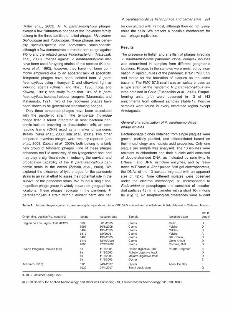
Table 1. Bacteriophages against V. parahaemolyticus pandemic clone PMC 57.5 isolated from shellfish and finfish obtained in Chile and Mexico.
Origin (No. positive/No. negative) Isolate Isolation date Sample Isolation placeRFLPgroupa
Región de Los Lagos Chile (8/123) 5090 30/8/2005 Clams Cailín, D5093 30/8/2005 Clams Yelcho D5489 13/9/2005 Clams Yelcho D5312 5/9/2005 Clams Yelcho A5486 13/9/2005 Clams Isla Chulín, C6119 12/10/2005 Clams Golfo Ancud D7860 27/12/2005 Clams Coronel, B-B D
Puerto Progreso, Mexico (4/6) 5e 11/8/2005 Finfish digestive tract Puerto Progreso B2e 11/8/2005 Robalo digestive tract D4e 11/8/2005 Mojarra digestive tract D6e 11/8/2005 Oyster E
Acapulco (2/10) 512 24/4/2007 Oyster Acapulco Bay F1032 24/4/2007 Small black clam G
a. RFLP obtained using HaeIII.
V. parahaemolyticus VP93 phage and carrier state 991
© 2010 Society for Applied Microbiology and Blackwell Publishing Ltd, Environmental Microbiology, 12, 990–1000
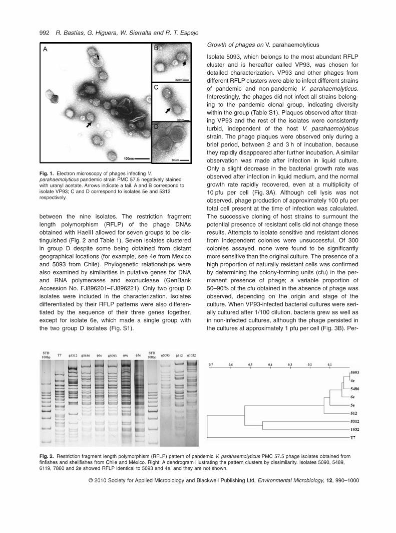
between the nine isolates. The restriction fragmentlength polymorphism (RFLP) of the phage DNAsobtained with HaeIII allowed for seven groups to be dis-tinguished (Fig. 2 and Table 1). Seven isolates clusteredin group D despite some being obtained from distantgeographical locations (for example, see 4e from Mexicoand 5093 from Chile). Phylogenetic relationships werealso examined by similarities in putative genes for DNAand RNA polymerases and exonuclease (GenBankAccession No. FJ896201–FJ896221). Only two group Disolates were included in the characterization. Isolatesdifferentiated by their RFLP patterns were also differen-tiated by the sequence of their three genes together,except for isolate 6e, which made a single group withthe two group D isolates (Fig. S1).
Growth of phages on V. parahaemolyticus
Isolate 5093, which belongs to the most abundant RFLPcluster and is hereafter called VP93, was chosen fordetailed characterization. VP93 and other phages fromdifferent RFLP clusters were able to infect different strainsof pandemic and non-pandemic V. parahaemolyticus.Interestingly, the phages did not infect all strains belong-ing to the pandemic clonal group, indicating diversitywithin the group (Table S1). Plaques observed after titrat-ing VP93 and the rest of the isolates were consistentlyturbid, independent of the host V. parahaemolyticusstrain. The phage plaques were observed only during abrief period, between 2 and 3 h of incubation, becausethey rapidly disappeared after further incubation. A similarobservation was made after infection in liquid culture.Only a slight decrease in the bacterial growth rate wasobserved after infection in liquid medium, and the normalgrowth rate rapidly recovered, even at a multiplicity of10 pfu per cell (Fig. 3A). Although cell lysis was notobserved, phage production of approximately 100 pfu pertotal cell present at the time of infection was calculated.The successive cloning of host strains to surmount thepotential presence of resistant cells did not change theseresults. Attempts to isolate sensitive and resistant clonesfrom independent colonies were unsuccessful. Of 300colonies assayed, none were found to be significantlymore sensitive than the original culture. The presence of ahigh proportion of naturally resistant cells was confirmedby determining the colony-forming units (cfu) in the per-manent presence of phage; a variable proportion of50–90% of the cfu obtained in the absence of phage wasobserved, depending on the origin and stage of theculture. When VP93-infected bacterial cultures were seri-ally cultured after 1/100 dilution, bacteria grew as well asin non-infected cultures, although the phage persisted inthe cultures at approximately 1 pfu per cell (Fig. 3B). Per-
Fig. 1. Electron microscopy of phages infecting V.parahaemolyticus pandemic strain PMC 57.5 negatively stainedwith uranyl acetate. Arrows indicate a tail. A and B correspond toisolate VP93; C and D correspond to isolates 5e and 5312respectively.
Fig. 2. Restriction fragment length polymorphism (RFLP) pattern of pandemic V. parahaemolyticus PMC 57.5 phage isolates obtained fromfinfishes and shellfishes from Chile and México. Right: A dendrogram illustrating the pattern clusters by dissimilarity. Isolates 5090, 5489,6119, 7860 and 2e showed RFLP identical to 5093 and 4e, and they are not shown.
992 R. Bastías, G. Higuera, W. Sierralta and R. T. Espejo
© 2010 Society for Applied Microbiology and Blackwell Publishing Ltd, Environmental Microbiology, 12, 990–1000

sistence after five serial 1/100 dilutions indicated that thephage reproduced in these cultures; otherwise, it shouldhave been diluted out (black triangle, Fig. 3B). The per-sistence of VP93 due to lysogeny was ruled out by theabsence of phage genes in colonies of bacteria growingafter phage infection. When 20 colonies obtained frominfected cultures and purified by two serial cultures in solidmedium were examined by PCR amplification, none werepositive for the putative RNA and DNA phage polymerasegenes of VP93 (see Experimental procedures for details).Additionally, mitomycin C treatment failed to induce phageproduction in cells from the 20 colonies.
Persistence of VP93 in infected cultures
Thorough washing of the cells growing together withVP93 did not eliminate all pfu; approximately 1%remained with the cells. These phages could be eitherinside productively infected cells or tightly associated withthe bacteria. Our overall observations of VP93 growth canbe explained by the presence of two cell types, sensitiveand resistant, at similar concentrations in the originalculture, even in recently cloned cultures. This observationwould result if resistant and sensitive cell types turn intoeach other at similar, high rates. One possible mechanismcould be phase variation. Upon infection, sensitive cellsproduce phages and die, but the growth of resistant cellscontinues unaffected. These resistant cells would,however, generate sensitive cells that can be infected bythe phage, maintaining phage growth and precluding itsdilution upon serial dilution and culturing. Assuming thissituation, the bacterial and bacteriophage populationdynamics were modelled on the equations and computercalculations described by Levin and Bull (2004). Discon-
tinuous lines in Fig. 3A and B show the expected values ifVP93 had a burst size of 1000 with a latent period of18 min and the host bacteria changed from sensitive toresistant or vice versa at a rate of 0.01 per generation.This change could occur by phase variation. Because V.parahaemolyticus undergoes phase variation betweenopaque (op) and translucent (tra) colony morphologies(Enos-Berlage and McCarter, 2000), we explored the pos-sible relationship between phage resistance and the opand tra colony types. No significant differences in resis-tance were observed between the op and tra colonies;both contained 50–90% resistant cells.
VP93 genome
The VP93 genome was sequenced using a combinationof the shotgun and primer walking approaches (GenBankAccession No. FJ 896200). The genome was 43 931nucleotides long with a G+C content of 49.2%, 3.8%higher than the G+C content of the host (Makino et al.,2003). Direct terminal repeats (DTRs) 337 bp in lengthwere observed at the ends. The DTRs were confirmed byprimer walking, but the ends could not be exactly defineddue to the absence of a clear abrupt stop when sequenc-ing the end regions.
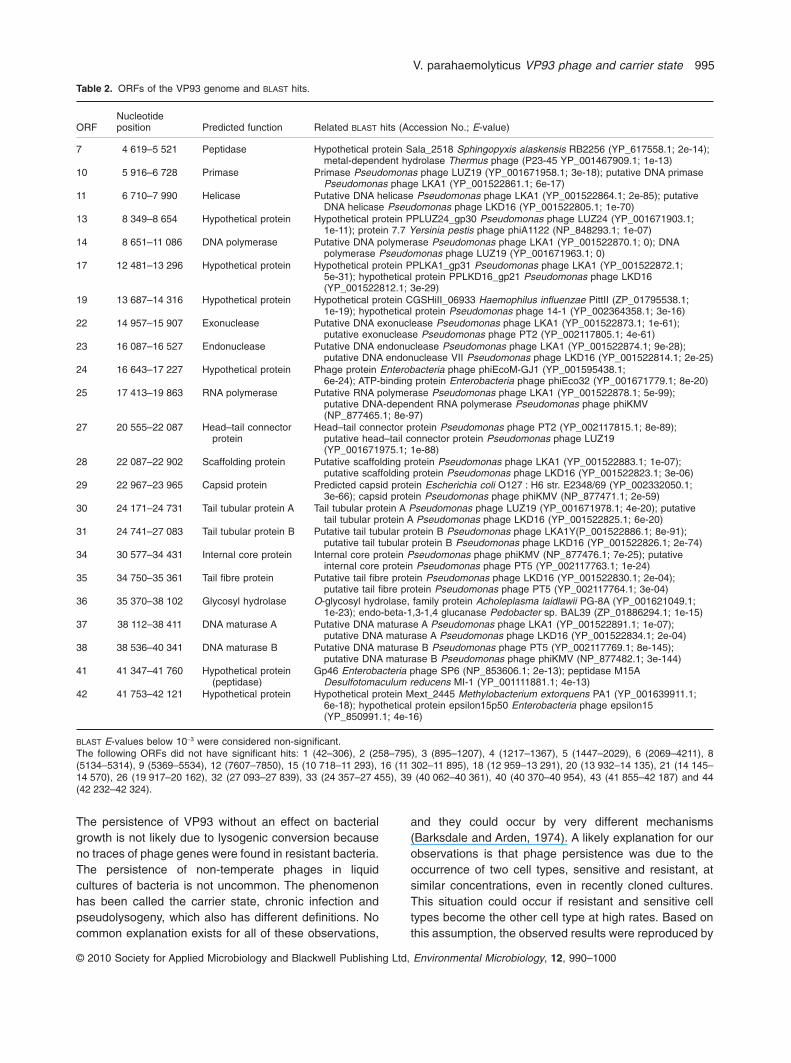
The phage could not be ascribed by nucleotidesequence similarity to any known phage group. However,the genome contained 44 putative ORFs based onArtemis analysis (Rutherford et al., 2000) and 16 of theirputative products (39%) shared significant similarities withproteins of the FKMV subgroup of the T7 supergroup,composed of Pseudomonas phages FKMV, LKD16,LKA1 and LUZ19 (Ceyssens et al., 2006). These genescorrespond to DNA metabolism, structural proteins and
Fig. 3. Growth of VP93 in pandemic V. parahaemolyticus.A. Plaque-forming units (�), cfu (�) and total bacteria calculated from the absorbance (�) after infection of PMC 57.5 with VP93 at moi 10.Lines represent the numbers for pfu, cfu and total cells estimated according to the model based on the existence of resistant and sensitivebacteria in the same bacterial clone, described in Experimental procedures.B. Plaque-forming units (�) and cfu (�) observed after infection, and after the indicated number of subsequent serial cultures of the infectedcells upon 1/100 dilution; (�) indicates the pfu expected after each serial dilution if the phage does not replicate. Continuous anddiscontinuous lines show estimated cfu and pfu, respectively, according to the model based on the existence of resistant and sensitivebacteria in the same bacterial clone described in Experimental procedures.
V. parahaemolyticus VP93 phage and carrier state 993
© 2010 Society for Applied Microbiology and Blackwell Publishing Ltd, Environmental Microbiology, 12, 990–1000
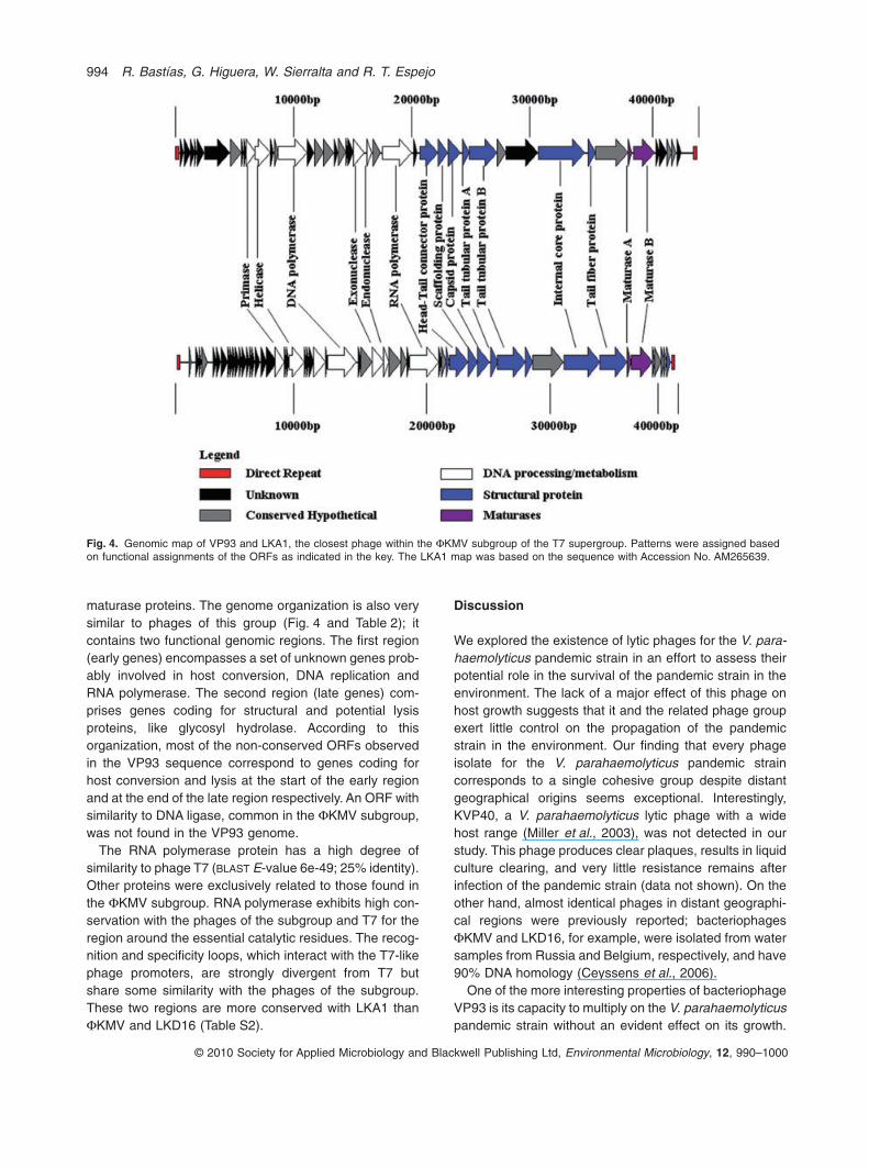
maturase proteins. The genome organization is also verysimilar to phages of this group (Fig. 4 and Table 2); itcontains two functional genomic regions. The first region(early genes) encompasses a set of unknown genes prob-ably involved in host conversion, DNA replication andRNA polymerase. The second region (late genes) com-prises genes coding for structural and potential lysisproteins, like glycosyl hydrolase. According to thisorganization, most of the non-conserved ORFs observedin the VP93 sequence correspond to genes coding forhost conversion and lysis at the start of the early regionand at the end of the late region respectively. An ORF withsimilarity to DNA ligase, common in the FKMV subgroup,was not found in the VP93 genome.
The RNA polymerase protein has a high degree ofsimilarity to phage T7 (BLAST E-value 6e-49; 25% identity).Other proteins were exclusively related to those found inthe FKMV subgroup. RNA polymerase exhibits high con-servation with the phages of the subgroup and T7 for theregion around the essential catalytic residues. The recog-nition and specificity loops, which interact with the T7-likephage promoters, are strongly divergent from T7 butshare some similarity with the phages of the subgroup.These two regions are more conserved with LKA1 thanFKMV and LKD16 (Table S2).
Discussion
We explored the existence of lytic phages for the V. para-haemolyticus pandemic strain in an effort to assess theirpotential role in the survival of the pandemic strain in theenvironment. The lack of a major effect of this phage onhost growth suggests that it and the related phage groupexert little control on the propagation of the pandemicstrain in the environment. Our finding that every phageisolate for the V. parahaemolyticus pandemic straincorresponds to a single cohesive group despite distantgeographical origins seems exceptional. Interestingly,KVP40, a V. parahaemolyticus lytic phage with a widehost range (Miller et al., 2003), was not detected in ourstudy. This phage produces clear plaques, results in liquidculture clearing, and very little resistance remains afterinfection of the pandemic strain (data not shown). On theother hand, almost identical phages in distant geographi-cal regions were previously reported; bacteriophagesFKMV and LKD16, for example, were isolated from watersamples from Russia and Belgium, respectively, and have90% DNA homology (Ceyssens et al., 2006).
One of the more interesting properties of bacteriophageVP93 is its capacity to multiply on the V. parahaemolyticuspandemic strain without an evident effect on its growth.
Fig. 4. Genomic map of VP93 and LKA1, the closest phage within the FKMV subgroup of the T7 supergroup. Patterns were assigned basedon functional assignments of the ORFs as indicated in the key. The LKA1 map was based on the sequence with Accession No. AM265639.
994 R. Bastías, G. Higuera, W. Sierralta and R. T. Espejo
© 2010 Society for Applied Microbiology and Blackwell Publishing Ltd, Environmental Microbiology, 12, 990–1000
The persistence of VP93 without an effect on bacterialgrowth is not likely due to lysogenic conversion becauseno traces of phage genes were found in resistant bacteria.The persistence of non-temperate phages in liquidcultures of bacteria is not uncommon. The phenomenonhas been called the carrier state, chronic infection andpseudolysogeny, which also has different definitions. Nocommon explanation exists for all of these observations,
and they could occur by very different mechanisms(Barksdale and Arden, 1974). A likely explanation for ourobservations is that phage persistence was due to theoccurrence of two cell types, sensitive and resistant, atsimilar concentrations, even in recently cloned cultures.This situation could occur if resistant and sensitive celltypes become the other cell type at high rates. Based onthis assumption, the observed results were reproduced by
Table 2. ORFs of the VP93 genome and BLAST hits.
ORFNucleotideposition Predicted function Related BLAST hits (Accession No.; E-value)
7 4 619–5 521 Peptidase Hypothetical protein Sala_2518 Sphingopyxis alaskensis RB2256 (YP_617558.1; 2e-14);metal-dependent hydrolase Thermus phage (P23-45 YP_001467909.1; 1e-13)
10 5 916–6 728 Primase Primase Pseudomonas phage LUZ19 (YP_001671958.1; 3e-18); putative DNA primasePseudomonas phage LKA1 (YP_001522861.1; 6e-17)
11 6 710–7 990 Helicase Putative DNA helicase Pseudomonas phage LKA1 (YP_001522864.1; 2e-85); putativeDNA helicase Pseudomonas phage LKD16 (YP_001522805.1; 1e-70)
13 8 349–8 654 Hypothetical protein Hypothetical protein PPLUZ24_gp30 Pseudomonas phage LUZ24 (YP_001671903.1;1e-11); protein 7.7 Yersinia pestis phage phiA1122 (NP_848293.1; 1e-07)
14 8 651–11 086 DNA polymerase Putative DNA polymerase Pseudomonas phage LKA1 (YP_001522870.1; 0); DNApolymerase Pseudomonas phage LUZ19 (YP_001671963.1; 0)
17 12 481–13 296 Hypothetical protein Hypothetical protein PPLKA1_gp31 Pseudomonas phage LKA1 (YP_001522872.1;5e-31); hypothetical protein PPLKD16_gp21 Pseudomonas phage LKD16(YP_001522812.1; 3e-29)
19 13 687–14 316 Hypothetical protein Hypothetical protein CGSHiII_06933 Haemophilus influenzae PittII (ZP_01795538.1;1e-19); hypothetical protein Pseudomonas phage 14-1 (YP_002364358.1; 3e-16)
22 14 957–15 907 Exonuclease Putative DNA exonuclease Pseudomonas phage LKA1 (YP_001522873.1; 1e-61);putative exonuclease Pseudomonas phage PT2 (YP_002117805.1; 4e-61)
23 16 087–16 527 Endonuclease Putative DNA endonuclease Pseudomonas phage LKA1 (YP_001522874.1; 9e-28);putative DNA endonuclease VII Pseudomonas phage LKD16 (YP_001522814.1; 2e-25)
24 16 643–17 227 Hypothetical protein Phage protein Enterobacteria phage phiEcoM-GJ1 (YP_001595438.1;6e-24); ATP-binding protein Enterobacteria phage phiEco32 (YP_001671779.1; 8e-20)
25 17 413–19 863 RNA polymerase Putative RNA polymerase Pseudomonas phage LKA1 (YP_001522878.1; 5e-99);putative DNA-dependent RNA polymerase Pseudomonas phage phiKMV(NP_877465.1; 8e-97)
27 20 555–22 087 Head–tail connectorprotein
Head–tail connector protein Pseudomonas phage PT2 (YP_002117815.1; 8e-89);putative head–tail connector protein Pseudomonas phage LUZ19(YP_001671975.1; 1e-88)
28 22 087–22 902 Scaffolding protein Putative scaffolding protein Pseudomonas phage LKA1 (YP_001522883.1; 1e-07);putative scaffolding protein Pseudomonas phage LKD16 (YP_001522823.1; 3e-06)
29 22 967–23 965 Capsid protein Predicted capsid protein Escherichia coli O127 : H6 str. E2348/69 (YP_002332050.1;3e-66); capsid protein Pseudomonas phage phiKMV (NP_877471.1; 2e-59)
30 24 171–24 731 Tail tubular protein A Tail tubular protein A Pseudomonas phage LUZ19 (YP_001671978.1; 4e-20); putativetail tubular protein A Pseudomonas phage LKD16 (YP_001522825.1; 6e-20)
31 24 741–27 083 Tail tubular protein B Putative tail tubular protein B Pseudomonas phage LKA1Y(P_001522886.1; 8e-91);putative tail tubular protein B Pseudomonas phage LKD16 (YP_001522826.1; 2e-74)
34 30 577–34 431 Internal core protein Internal core protein Pseudomonas phage phiKMV (NP_877476.1; 7e-25); putativeinternal core protein Pseudomonas phage PT5 (YP_002117763.1; 1e-24)
35 34 750–35 361 Tail fibre protein Putative tail fibre protein Pseudomonas phage LKD16 (YP_001522830.1; 2e-04);putative tail fibre protein Pseudomonas phage PT5 (YP_002117764.1; 3e-04)
36 35 370–38 102 Glycosyl hydrolase O-glycosyl hydrolase, family protein Acholeplasma laidlawii PG-8A (YP_001621049.1;1e-23); endo-beta-1,3-1,4 glucanase Pedobacter sp. BAL39 (ZP_01886294.1; 1e-15)
37 38 112–38 411 DNA maturase A Putative DNA maturase A Pseudomonas phage LKA1 (YP_001522891.1; 1e-07);putative DNA maturase A Pseudomonas phage LKD16 (YP_001522834.1; 2e-04)
38 38 536–40 341 DNA maturase B Putative DNA maturase B Pseudomonas phage PT5 (YP_002117769.1; 8e-145);putative DNA maturase B Pseudomonas phage phiKMV (NP_877482.1; 3e-144)
41 41 347–41 760 Hypothetical protein(peptidase)
Gp46 Enterobacteria phage SP6 (NP_853606.1; 2e-13); peptidase M15ADesulfotomaculum reducens MI-1 (YP_001111881.1; 4e-13)
42 41 753–42 121 Hypothetical protein Hypothetical protein Mext_2445 Methylobacterium extorquens PA1 (YP_001639911.1;6e-18); hypothetical protein epsilon15p50 Enterobacteria phage epsilon15(YP_850991.1; 4e-16)
BLAST E-values below 10-3 were considered non-significant.The following ORFs did not have significant hits: 1 (42–306), 2 (258–795), 3 (895–1207), 4 (1217–1367), 5 (1447–2029), 6 (2069–4211), 8(5134–5314), 9 (5369–5534), 12 (7607–7850), 15 (10 718–11 293), 16 (11 302–11 895), 18 (12 959–13 291), 20 (13 932–14 135), 21 (14 145–14 570), 26 (19 917–20 162), 32 (27 093–27 839), 33 (24 357–27 455), 39 (40 062–40 361), 40 (40 370–40 954), 43 (41 855–42 187) and 44(42 232–42 324).
V. parahaemolyticus VP93 phage and carrier state 995
© 2010 Society for Applied Microbiology and Blackwell Publishing Ltd, Environmental Microbiology, 12, 990–1000
a model incorporating the following parameters: burst sizeof 1000, latent period of 18 min, phage adsorption rate of10-8, and a conversion rate of sensitivity to resistance andvice versa of 0.01 per generation. The latent period andadsorption rate correspond to the experimentally obtainedvalues. A burst size of 1000 was assumed from the yieldof phage per total bacteria in the culture, considering thatonly 10% of the cells are sensitive (Fig. 3A). The conver-sion rate for host sensitivity was arbitrary, assuming that itcould occur by phase transition. The failure to find arelationship between phage resistance and colony typedoes not rule out resistance and sensitivity occurring byphase variation not associated with the op and tra char-acter. The failure to isolate sensitive and resistant clonesfrom colonies can be explained by high resistance/sensitivity variation. However, this result would beexpected if the rate of change from resistant cell to sen-sitive cell and from sensitive cell to resistant cell occurs ata frequency of 0.01 per generation. In such colonies, eventhose derived from a single resistant or sensitive cell, bothtypes of cells would be present. The modulation of phageinfection by phase variation was previously described inStreptomyces coelicolor (Sumby and Smith, 2003) andHaemophilus influenzae (Zaleski et al., 2005).
Bacteriophage PPO1, which stably coexists withEscherichia coli O157 : H7-like VP93 without lysogenicconversion, is explained by clonal heterogeneity of thehost (Fischer et al., 2004). This phenomenon is notunique; similar observations were later reported withphages of Salmonella spp. (Carey-smith et al., 2006) andFlavobacterium psychrophilum (Middelboe et al., 2009).In all of these cases, the chosen explanation was that onlya subpopulation of the host is susceptible to phage infec-tion, but the cause of the presence of this subpopulationwas not explained. The carrier state in co-habitation asdescribed here, or the replication of the phage withouthurting the host, seems to be advantageous for the per-sistence of both the phage and bacteria in close proximity.Importantly, because phages are isolated or detected byplaque formation, phages that grow without harming theirhosts could occur more frequently in nature than reporteddue to difficulty observing plaques in such cases. Thisphage–bacteria relationship generates a phenomenonanalogous to what happens with ‘non-cultivable’ bacteria;phages like VP93 are not readily detectable unless theyhave another host available for detection that they canlyse with good efficiency. Some of these phages maycorrespond to the large number of putative phagesobserved in culture-independent studies, but without anycultured representative (Breitbart and Rohwer, 2005).
The sequence of VP93 showed that it is related, thoughdistantly, to the FKMV subgroup of phages and it is thefirst non-Pseudomonas phage present in this subgroup.Among the phages in this subgroup VP93 is more closely
related to phage LKA1. We found no similarity betweenVP93 and VpV262, a marine bacteriophage VpV262,which infects V. parahaemolyticus, included in the T7supergroup in spite of lacking RNA polymerase (Hardieset al., 2003).
Experimental procedures
Strains and growth media
Vibrio parahaemolyticus strain PMC 57.5 was previouslycharacterized (Fuenzalida et al., 2006). Bacteria and phagewere grown in synthetic sea water (23.4 g l-1 NaCl, 24.7 g l-1
MgSO4·7H2O, 1.5 g l-1 KCl and 1.43 g l-1 CaCl2·2H2O, pH 6.5)supplemented with 1% Bacto tryptone (Gifco) and 0.5% yeastextract at 37°C with reciprocal shaking.
Bacteriophage isolation and characterization
Shellfish and finfish were collected in the coastal waters of thePacific Ocean off Chile, Region de los Lagos, next to PuertoMontt (41°29′S, 72°24′W) and Antofagasta (23°39′S,70°24′W), and off Mexico, next to Acapulco, Guerrero(16°51′N, 99°52′W) and Puerto Progreso, Yucatán (21°18′N,89°39′W). The fish were kept on ice immediately after collec-tion and processed in the laboratory within 4 h. The soft meatof the shellfish or the digestive tract of the finfish was macer-ated and resuspended in an equal volume of PBS buffer (NaCl0.8%, KCl 0.02%, Na2HPO4 0.14%, KH2PO4 0.024%) andsubsequently centrifuged at 5000 g for 10 min. Supernatant(100 ml) was then used to inoculate an exponentially growingPMC 57.5 culture of ~108 cells per ml and incubated overnightat 37°C with agitation. The culture was pelleted by centrifuga-tion at 5000 g for 10 min and the remaining bacteria wereremoved by filtering (0.22 mm). Phages in the filtrate weredetected by plating 100 ml using the standard method fordouble-layer agar plaque assay. One plaque was picked fromeach positive sample and re-plated two times to ensure clonalphage stocks.
For phage growth, 50 ml of a V. parahaemolyticus PMC 7.5culture (~108 cells per ml, optical density 0.2–0.3 at 600 nm)was infected at a multiplicity of infection (moi) of 10 andincubated overnight. The culture was centrifuged at 5000 gfor 10 min and the supernatant filtrated (0.22 mm) and com-bined with 10 ml of chloroform. Alternatively, strain PMA112,obtained from shellfish, was used as an indicator strain. Forelectron microscopy, this preparation was centrifuged at100 000 g for 50 min and the pellet suspended in 100 ml ofsynthetic sea water. Samples were stained with 1% uranylacetate on grids with carbon-stabilized formvar and observedusing a Phillips CM 100 transmission electron microscope.Only nine isolates were observed: 5090, 5093, 5489, 5312,5486, 1032, 512, 5e and 2e. Phage DNA was extracted frominfected cultures that were centrifuged and filtered asdescribed above, but in this case the phages in the filtratewere precipitated with polyethyleneglycol (PEG-8000) andNaCl at a final concentration of 10% and 1.5 M respectively.The precipitated phage was then centrifuged at 11 000 g for20 min and suspended in synthetic sea water. For DNAextraction, samples were incubated with DNase (2 mg ml-1)
996 R. Bastías, G. Higuera, W. Sierralta and R. T. Espejo
© 2010 Society for Applied Microbiology and Blackwell Publishing Ltd, Environmental Microbiology, 12, 990–1000
and RNase (100 mg ml-1) for 1 h at 37°C. These sampleswere subsequently treated with 500 mg ml-1 proteinase K for15 min at 65°C. Sodium dodecyl sulfate (SDS) was added ata final concentration of 0.5% (w/v). After incubation at 65°Cfor 45 min, the solution was extracted twice with phenol-chloroform. Finally, the DNA was precipitated by adding 1/10volume 3 M sodium acetate (pH 5.0) and two volumes ofabsolute ethanol at -20°C. After the pellet was washed with70% ethanol, it was dissolved in TE buffer (0.01 M Tris,0.001 M EDTA, pH 7.5). Restriction site mapping was per-formed by digesting with the restriction enzyme HaeIII(Promega) according to the manufacturer’s instructions.Fragments were separated by electrophoresis on a 7.5%polyacrylamide gel for 2 h at 70 V using the GeneRuler 1 kbDNA ladder (Fermentas) as a marker. PCR amplification ofthe putative DNA and RNA polymerases and exonucleasewas performed using the following oligonucleotides: TACAGACTATGCCCTGCTG and AGTCTGTGGTGGATGATACCas the forward and reverse primer for DNA polymerase; CGTCAGTGGTACACAAAGG and GTGCAGCTACGTAATGTGGfor RNA polymerase; and GTTAAGACGTTGCCTACTGC andCATAAGGTAGGCGTATCCAG for exonuclease. The primerswere designed from the sequence obtained for the VP93genome. PCR product sequences were deposited asFJ896201–FJ896221. The host range of the phages wasdetermined by standard spot tests (Ceyssens et al., 2006).
Growth curves and phage persistence assays
A one-step growth curve was made by infecting an earlyexponential culture of V. parahaemolyticus PMC 57.5 with thephage at a moi of 10. The absorbance was monitored andsamples were obtained at short time intervals for the deter-mination of pfu and cfu. The pfu was determined after treat-ment with chloroform; plating was performed using thestandard double-layer agar plaque assay. The cfu was deter-mined by plating in soft agar using a described modifiedpour-plate method (Berney et al., 2006). Phage persistencein infected cultures was determined by 1:100 dilution of thestationary-phase cultures in fresh medium and continuing theincubation. Dilution and subsequent incubation until the sta-tionary phase was performed four times in series. The pfuand cfu were determined after each serial culture reachedstationary phase. Phage associated with cells in the lastserial culture was determined by measuring the pfu afterpelleting and washing the bacteria five times with the samevolume of medium. The fraction of resistant cells in colonieswas determined by plating the bacterial suspensions fromeach colony in solid medium with and without VP93. TheVP93 adsorption rate was measured as described by Mudgaland colleagues (2006), except free phage was measuredafter centrifugation instead of filtration.
The presence of the phage genome in resistant bacteriawas determined by testing for the presence of the putativeRNA and DNA phage polymerase genes of VP93 in thebacteria from each of 20 colonies obtained from infectedcultures after two passages (re-picked) in solid medium. Theputative RNA and DNA phage polymerase genes of VP93were assayed by PCR as described above. Positive controlsfor phage detection consisted of bacteria spiked with 0.01 pfuof VP93 per bacteria. The gene tdh of pandemic V. para-
haemolyticus was amplified as a control for PCR amplifica-tion. Mitomycin C treatment was performed by adding freshsolution to obtain a final concentration of 30 ng ml-1 as pre-viously described (Oakey et al., 2002).
Population dynamics
The bacterial and bacteriophage population dynamics weremodelled as described by Levin and Bull (2004). The follow-ing algorithm was used with the indicated parameters:
{Lytic Phage with phase shifting – and mutation to InheritedResistance with VP93 parameters Initial infection}{method definition}
METHOD EULERstarttime = 0STOPTIME = 2.1DT = 0.0001DTOUT = 0.01
{Constant Parameters}
v = 2.0 {Maximum growth rate sensitive S}v1 = 2.0 {Maximum growth rate resistant R1}v2 = 2.0 {Maximum growth rate resistant}d1 = 1e-8 {Maximum adsorption rate parameter}b1 = 1000 {Burst size}k = 0.25 {Monod coefficient}e = 5e-7 {Conversion efficiency}mutR1S = 1e-2 {Rate at which S cells produced R1}mutSR1 = 1e-2 {Rate at which R1 produce S}mu = 1e-10 {Mutation rate to resistance S and to
NR}x = 0.3 {Latent period}m = 1e-3 {Rate mortality P1}
{Variables}
init S = 2.3e7 {Initial density of sensitive bacteria}init R1 = 2.3e7 {Initial density of phenotypically
resistant – phase shift bacteria}init MS = 0 {Initial density of infected S bacteria}init NR = 0 {Initial density of resistant bacteria}init P = 1e9 {Initial density of the phage}init G = 500 {Initial resource}n = s + r1psi = G/(k + G)b = b1*psid = d1*psi
{Delay Variables}
Sx = DELAY(S,x) {Sensitive at t – x}R1x = DELAY(R1,x) {R1 at t – x}MSx = DELAY(MS,x) {S infected with P at t – x}Px = DELAY(P,x) {P1 phage at t – x}
{Equations}
d/dt (G) = -psi*e*(S*v + NR*v2 + R1*v1)d/dt (S) = (1 – mutR1S)*v*S*psi – d*S*P + mutSR1*
R1*psi
V. parahaemolyticus VP93 phage and carrier state 997
© 2010 Society for Applied Microbiology and Blackwell Publishing Ltd, Environmental Microbiology, 12, 990–1000
d/dt (R1) = (1 – mutSR1)*v1*R1*psi + mutR1S*S*psid/dt (MS) = d*S*Pd/dt (NR) = v2*psi*NR + GM/dtd/dt (P) = MSx*b – m*P
{Mutation}
bm = N*mu*DTrm = RANDOM (0, 1)GM = IF rm < bm THEN 1 ELSE 0
The same equations were used to model 1/100 dilutions,except that initial P, S and R1 were those corresponding tothe 1/100 dilution of the previous culture and that STOPTIMEwas 16.
VP93 genome sequencing
Sequencing was performed as described above using theVP93 genome after cloning with the TOPO Shotgun Subclon-ing Kit (Invitrogen) as described by the manufacturer. Mac-rogen (Seoul, Korea) sequenced 161 clones (Seoul, Korea)using M13 forward and reverse primers. Seventeen contigswere obtained and gaps, direct repeats and uncertainties inthe sequence were determined by direct sequencing of thephage genome using appropriate primers. Total sites deter-mined corresponded to a 4.6-fold coverage of the genome.Sequence assembly was performed using the programs inthe Staden Package (http://staden.sourceforge.net/). Thegenome sequence was scanned for potential ORFs usingArtemis (http://www.sanger.ac.uk/Software/Artemis/) (Ruth-erford et al., 2000). Translated ORF sequences were com-pared with known proteins using standard protein–proteinBLASTP (http://blast.ncbi.nlm.nih.gov/) (Altschul et al., 1990).
Acknowledgements
It is a pleasure to thank Pedro Romero from the Instituto deBiotecnología of Universidad Autónoma de México andFernando Puerto from the Instituto Hidegeo Niguchi of Uni-versidad de Yucatán for help isolating the bacteriophagesfrom México. We thank Daniel Castillo and Camila Sorianofor doing part of the modelling and most of the calculation.Roberto Bastías acknowledges scholarship from ProgramaMECE Educación Superior, proyecto UCH 0407. This workwas partially supported by Grant FONDECYT 1076054.
References
Alam, M.J., Miyoshi, S., and Shinoda, S. (2003) Studieson pathogenic Vibrio parahaemolyticus during a warmweather season in the Seto Inland Sea, Japan. EnvironMicrobiol 5: 706–710.
Altschul, S.F., Gish, W., Miller, W., Myers, E.W., and Lipman,D.J. (1990) Basic local alignment search tool. J Mol Biol215: 403–410.
Barksdale, L., and Arden, S.B. (1974) Persisting bacterioph-age infections, lysogeny, and phage conversions. AnnuRev Microbiol 28: 265–299.
Baross, J.A., Liston, J., and Morita, R.Y. (1978) Incidence ofVibrio parahaemolyticus bacteriophages and other Vibrio
bacteriophages in marine samples. Appl Environ Microbiol36: 492–499.
Berney, M., Weilenmann, H.U., Ihssen, J., Bassin, C., andEgli, T. (2006) Specific growth rate determines the sensi-tivity of Escherichia coli to thermal, UVA, and solar disin-fection. Appl Environ Microbiol 72: 2586–2593.
Bouvier, T., and Giorgio, P.A. (2007) Key role of selectiveviral-induced mortality in determining marine bacterialcommunity composition. Environ Microbiol 9: 287–297.
Breitbart, M., and Rohwer, F. (2005) Here a virus, there avirus, everywhere the same virus? Trends Microbiol 13:278–284.
Brussaard, C.P., Wilhelm, S.W., Thingstad, F., Weinbauer,M.G., Bratbak, G., Heldal, M., et al. (2008) Global-scaleprocesses with a nanoscale drive: the role of marineviruses. ISME J 2: 575–578.
Brüssow, H., Canchaya, C., and Hardt, W.D. (2004) Phagesand the evolution of bacterial pathogens: from genomicrearrangements to lysogenic conversion. Microbiol Mol BiolRev 68: 560–602.
Carey-Smith, G.V., Billington, C., Cornelius, A.J., Hudson,J.A., and Heineman, J.A. (2006) Isolation and character-ization of bacteriophages infecting Salmonella spp. FEMSMicrobiol Lett 258: 182–186.
Ceyssens, P.J., Lavigne, R., Mattheus, W., Chibeu, A.,Hertveldt, K., Mast, J., et al. (2006) Genomic analysis ofPseudomonas aeruginosa phages LKD16 and LKA1:establishment of the phiKMV subgroup within the T7 super-group. J Bacteriol 188: 6924–6931.
Comeau, A.M., Buenaventura, E., and Suttle, C.A. (2005) Apersistent, productive, and seasonally dynamic vibrioph-age population within Pacific oysters (Crassostrea gigas).Appl Environ Microbiol 71: 5324–5331.
Comeau, A.M., Chan, A.M., and Suttle, C.A. (2006) Geneticrichness of vibriophages isolated in a coastal environment.Environ Microbiol 8: 1164–1176.
Depaola, A., Kaysner, C.A., Bowers, J., and Cook, D.W.(2000) Environmental investigations of Vibrio para-haemolyticus in oysters after outbreaks in Washington,Texas, and New York (1997 and 1998). Appl Environ Micro-biol 66: 4649–4654.
Depaola, A., Nordstrom, J.L., Bowers, J.C., Wells, J.G.,and Cook, D.W. (2003a) Seasonal abundance of total andpathogenic Vibrio parahaemolyticus in Alabama oysters.Appl Environ Microbiol 69: 1521–1526.
Depaola, A., Ulaszek, J., Kaysner, C.A., Tenge, B.J., Nord-strom, J.L., Wells, J., et al. (2003b) Molecular, serological,and virulence characteristics of Vibrio parahaemolyticusisolated from environmental, food, and clinical sources inNorth America and Asia. Appl Environ Microbiol 69: 3999–4005.
Enos-Berlage, J.L., and McCarter, L.L. (2000) Relation ofcapsular polysaccharide production and colonial cell orga-nization to colony morphology in Vibrio parahaemolyticus.J Bacteriol 182: 5513–5520.
Fischer, C.R., Yoichi, M., Unno, H., and Tanji, Y. (2004) Thecoexistence of Escherichia coli serotype O157 : H7 and itsspecific bacteriophage in continuous culture. FEMS Micro-biol Lett 241: 171–177.
Fuenzalida, L., Hernandez, C., Toro, J., Rioseco, M.L.,Romero, J., and Espejo, R.T. (2006) Vibrio parahaemolyti-
998 R. Bastías, G. Higuera, W. Sierralta and R. T. Espejo
© 2010 Society for Applied Microbiology and Blackwell Publishing Ltd, Environmental Microbiology, 12, 990–1000

cus in shellfish and clinical samples during two large epi-demics of diarrhoea in southern Chile. Environ Microbiol 8:675–683.
Hara-Kudo, Y., Sugiyama, K., Nishibuchi, M., Chowdhury, A.,Yatsuyanagi, J., Ohtomo, Y., et al. (2003) Prevalence ofpandemic thermostable direct hemolysin-producing Vibrioparahaemolyticus O3 : K6 in seafood and the coastalenvironment in Japan. Appl Environ Microbiol 69: 3883–3891.
Hardies, S.C., Comeau, A.M., Serwer, P., and Suttle, C.A.(2003) The complete sequence of marine bacteriophageVpV262 infecting Vibrio parahaemolyticus indicates that anancestral component of a T7 viral supergroup is wide-spread in the marine environment. Virology 310: 359–371.
Iida, T., Hattori, A., Tagomori, K., Nasu, H., Naim, R., andHonda, T. (2001) Filamentous phage associated withrecent pandemic strains of Vibrio parahaemolyticus. EmergInfect Dis 7: 477–478.
Izutsu, K., Kurokawa, K., Tashiro, K., Kuhara, S., Hayashi, T.,Honda, T., and Iida, T. (2008) Comparative genomic analy-sis using microarray demonstrates a strong correlationbetween the presence of the 80-kilobase pathogenicityisland and pathogenicity in Kanagawa phenomenon-positive Vibrio parahaemolyticus strains. Infect Immun 76:1016–1023.
Koga, T., and Kawata, T. (1991) Comparative characteriza-tion of inducible and virulent Vibrio parahaemolyticus bac-teriophages having unique head projections. MicrobiolImmunol 35: 49–58.
Kudriakova, T., Makedonova, L.D., Dudkina, O.S., Degtiarev,B.M., Khaitovich, A.B., Savchenko, B.I., et al. (1992) Thephages of halophilic vibrios and its use. Zh Mikrobiol Epi-demiol Immunobiol 9–10: 5–7 (in Russian).
Lan, S.F., Huang, C.H., Chang, C.H., Liao, W.C., Lin, I.H.,Jian, W.N., et al. (2009) Characterization of a new plasmid-like prophage in a pandemic Vibrio parahaemolyticusO3 : K6 strain. Appl Environ Microbiol 75: 2659–2667.
Levin, B.R., and Bull, J.J. (2004) Population and evolutionarydynamics of phage therapy. Nat Rev Microbiol 2: 166–173.
Makino, K., Oshima, K., Kurokawa, K., Yokoyama, K., Uda,T., Tagomori, K., et al. (2003) Genome sequence of Vibrioparahaemolyticus: a pathogenic mechanism distinct fromthat of V cholerae. Lancet 361: 743–749.
Matsuzaki, S., Inoue, T., Tanaka, S., Koga, T., Kuroda, M.,Kimura, S., and Imai, S. (2000) Characterization of a novelVibrio parahaemolyticus phage, KVP241, and its relativesfrequently isolated from seawater. Microbiol Immunol 44:953–956.
Middelboe, M., Holmfeldt, K., Riemann, L., Nybroe, O., andHaaber, J. (2009) Bacteriophages drive strain diversifica-tion in a marine Flavobacterium: implications for phageresistance and physiological properties. Environ Microbiol11: 1971–1982.
Miller, E.S., Heidelberg, J.F., Eisen, J.A., Nelson, W.C.,Durkin, A.S., Ciecko, A., et al. (2003) Complete genomesequence of the broad-host-range vibriophage KVP40:comparative genomics of a T4-related bacteriophage.J Bacteriol 185: 5220–5233.
Mudgal, P., Breidt, F., Jr, Lubkin, S.R., and Sandeep, K.P.(2006) Quantifying the significance of phage attack on
starter cultures: a mechanistic model for populationdynamics of phage and their hosts isolated from fermentingsauerkraut. Appl Environ Microbiol 72: 3908–3915.
Muramatsu, K., and Matsumoto, H. (1991) Two generalizedtransducing phages in Vibrio parahaemolyticus and Vibrioalginolyticus. Microbiol Immunol 35: 1073–1084.
Nair, G.B., Ramamurthy, T., Bhattacharya, S.K., Dutta, B.,Takeda, Y., and Sack, D.A. (2007) Global dissemination ofVibrio parahaemolyticus serotype O3 : K6 and its serova-riants. Clin Microbiol Rev 20: 39–48.
Nasu, H., Iida, T., Sugahara, T., Yamaichi, Y., Park, K.S.,Yokoyama, K., et al. (2000) A filamentous phage associ-ated with recent pandemic Vibrio parahaemolyticusO3 : K6 strains. J Clin Microbiol 38: 2156–2161.
Oakey, H.J., Cullen, B.R., and Owens, L. (2002) The com-plete nucleotide sequence of the Vibrio harveyi bacterioph-age VHML. J Appl Microbiol 93: 1089–1098.
Ogura, Y., Kurokawa, K., Ooka, T., Tashiro, K., Tobe, T.,Ohnishi, M., et al. (2006) Complexity of the genomic diver-sity in enterohemorrhagic Escherichia coli O157 revealedby the combinational use of the O157 Sakai OligoDNAmicroarray and the Whole Genome PCR scanning. DNARes 13: 3–14.
Ohnishi, T., and Nozu, K. (1986) Induction of phage-like par-ticles from a pathogenic strain of Vibrio parahaemolyticusby mitomycin C. Biochem Biophys Res Commun 141:1249–1253.
Rutherford, K., Parkhill, J., Crook, J., Horsnell, T., Rice, P.,Rajandream, M.A., and Barrell, B. (2000) Artemis:sequence visualization and annotation. Bioinformatics 16:944–945.
Sumby, P., and Smith, M.C. (2003) Phase variation in thephage growth limitation system of Streptomyces coelicolorA3(2). J Bacteriol 185: 4558–4563.
Suttle, C.A. (2005) Viruses in the sea. Nature 437: 356–361.Wommack, K.E., and Colwell, R.R. (2000) Virioplankton:
viruses in aquatic ecosystems. Microbiol Mol Biol Rev 64:69–114.
Zabala, B., Garcia, K., and Espejo, R.T. (2009) Enhancementof UV light sensitivity of a Vibrio parahaemolyticus O3 : K6pandemic strain due to natural lysogenization by a telom-eric phage. Appl Environ Microbiol 75: 1697–1702.
Zaleski, P., Wojciechowski, M., and Piekarowicz, A. (2005)The role of Dam methylation in phase variation of Haemo-philus influenzae genes involved in defence against phageinfection. Microbiology 151: 3361–3369.
Supporting information
Additional Supporting Information may be found in the onlineversion of this article:
Fig. S1. Dendrogram illustrating the pattern clusters basedon the dissimilarity of the nucleotide sequences in PCRamplicons of the putative genes for DNA and RNA poly-merases and exonuclease. Line 0.1 corresponds to changesper nucleotide position.Table S1. Infectivity of phage isolates from different RFLPclusters on diverse V. parahaemolyticus strains. Isolates notshown were not included in the present analysis.
V. parahaemolyticus VP93 phage and carrier state 999
© 2010 Society for Applied Microbiology and Blackwell Publishing Ltd, Environmental Microbiology, 12, 990–1000

Table S2. Sequence of regions around essential catalyticresidues of RNA polymerase. Conserved residues withphage VP93 are in bold. The essential catalytic residues areunderlined.
Please note: Wiley-Blackwell are not responsible for thecontent or functionality of any supporting materials suppliedby the authors. Any queries (other than missing material)should be directed to the corresponding author for the article.
1000 R. Bastías, G. Higuera, W. Sierralta and R. T. Espejo
© 2010 Society for Applied Microbiology and Blackwell Publishing Ltd, Environmental Microbiology, 12, 990–1000