Genotypes Associated with Virulence in Environmental Isolates of Vibrio cholerae
Upload
independentCategory
view
0download
0
V. parahaemolyticus–Induced Immune Responses • JID 2003:187 (1 April) • 1085
M A J O R A R T I C L E
Adaptive and Inflammatory Immune Responsesin Patients Infected with Strains of Vibrioparahaemolyticus
Firdausi Qadri,1 Muhammad Shamsul Alam,1 Mitsuaki Nishibuchi,2 Taufiqur Rahman,1 Nur Haque Alam,1
Jobayer Chisti,1 Seiichi Kondo,3 Junichi Sugiyama,4 Nurul Amin Bhuiyan,1 Minnie M. Mathan,1 David A. Sack,1
and G. Balakrish Nair1
1International Centre for Diarrhoeal Disease Research, Bangladesh, Dhaka, Bangladesh; 2Center for Southeast Asian Studies, Kyoto University,Kyoto, 3School of Pharmaceutical Sciences, Josai University, Saitama, and 4Denkaseiken, Gosen-shi, Niigata, Japan
In patients with diarrhea caused by Vibrio parahaemolyticus, antibody-secreting cell responses to thermostable
direct hemolysin (TDH), lipopolysaccharide (LPS), and whole-cell bacteria were seen. TDH- and LPS-specific
responses were seen in serum samples, and immunoglobulin A antibody responses were observed in stool.
Levels of C-reactive protein and nitric oxide metabolites increased in the systemic circulation at the onset of
illness. Tumor necrosis factor–a and lactoferrin levels were high during the acute stage in mucosal secretions
and in plasma, whereas interleukin-1b levels were high only in mucosal secretions. Duodenal and rectal biopsy
specimens obtained at the onset of illness showed an acute inflammatory response. The lamina propria showed
edema, congestion of blood vessels, and hemorrhage, with an increase in levels of polymorphonuclear neu-
trophils and macrophages. Strains belonging to different serotypes exhibited varying resistance to killing by
serum; the O8:K21 strain was most sensitive. Infection with V. parahaemolyticus results in B cell responses
and an acute inflammatory response that is self-limiting.
Vibrio parahaemolyticus strains are becoming an increas-
ing cause of concern as causative agents of acute gastro-
enteritis in a number of locations, including India [1],
Bangladesh [2, 3], Japan [4], Taiwan [5], and the United
States [6]. The serogroups O3:K6, O4:K68, and O1:KUT
are believed to have acquired pandemic potential [2, 7,
8]. V. parahaemolyticus is responsible mainly for gastro-
Received 25 July 2002; accepted 22 November 2002; electronically published 19March 2003.
Informed consent was obtained from patients and control subjects. This study wasapproved by the ethical review committee of the International Centre for DiarrhoealDisease Research, Bangladesh (ICDDR,B; Dhaka).
Financial support: government of Japan (special research grant to ICDDR,B); Ministryof Education, Science, Sports and Culture, Japan (grant-in-aid for scientific research).ICDDR,B is supported by countries and agencies that share its concern for the healthproblems of developing countries.
Reprints or correspondence: Dr. Firdausi Qadri, ICDDR,B, GPO Box 128, Dhaka 1000,Bangladesh ([email protected]).
The Journal of Infectious Diseases 2003; 187:1085–96� 2003 by the Infectious Diseases Society of America. All rights reserved.0022-1899/2003/18707-0008$15.00
enteritis, although wound infections and septicemia also
may be caused by this organism. V. parahaemolyticus has
been known to cause diarrhea in travelers and army
personnel [9], and it also has been a cause of concern
among immunocompromised individuals, including
those with leukemia, liver disease, and human immu-
nodeficiency virus infection and AIDS [10, 11]. Symp-
toms of V. parahaemolyticus infection usually include
acute, self-limiting diarrhea (caused by consumption of
contaminated food) and, in some cases, septicemia. Only
strains of V. parahaemolyticus with the tdh gene are ca-
pable of causing gastroenteritis [12].
A number of serogroups among the 172 recognized
O:K serotypes of V. parahaemolyticus can cause the dis-
ease. The most important virulence factor identified to
date is thermostable direct hemolysin (TDH) [13].
TDH-related hemolysin also has been identified as a
virulence factor [13, 14]. Enteroinvasiveness of the bac-
teria has been reported in a rabbit model, in which the
organism invaded, colonized, and produced inflamma-
1086 • JID 2003:187 (1 April) • Qadri et al.
tion in the small intestine [15]. However, very little is known
about immunological and inflammatory responses in patients
who have natural infection with the pathogen. In the present
study, we have attempted, to our knowledge for the first time,
to study the mucosal and systemic immune responses in pa-
tients infected with V. parahaemolyticus and the involvement
of different inflammatory components. To examine antigen-
specific mucosal immune responses, we studied the circulating
antibody-secreting cell (ASC) responses, which can be consid-
ered a proxy measure of the mucosal immune response in the
gut [16], and we used stool extract preparations to investigate
intestinal antibody responses [17]. In addition, we studied sys-
temic immune responses to TDH and lipopolysaccharide (LPS)
in serum. Inflammatory responses were evaluated by investi-
gating the levels of the bactericidal protein lactoferrin, oxidant-
mediated defense factor (nitric oxide; NO.), C-reactive protein
(CRP), and the cytokines tumor necrosis factor (TNF)–a and
interleukin (IL)–1b. Responses were studied during the acute
stage of the disease (after the onset of illness) and at different
phases during convalescence. Duodenal and rectal biopsy spec-
imens from patients were also examined to discern changes in
gross histopathology and the contribution of the inflammatory
cells to the pathogenesis of the disease. Responses in patients
were compared with those seen in healthy volunteers for all
study parameters.
SUBJECT, MATERIALS, AND METHODS
Study subjects and clinical evaluation. Twenty-six adults
and 2 children infected with V. parahaemolyticus were enrolled
in the study. They were recruited from the International Centre
for Diarrhoeal Disease Research, Bangladesh (ICDDR,B) hos-
pital in Dhaka between July 2000 and October 2001. The
ICDDR,B has a 2% surveillance system, in which stool samples
from every 50th patient attending the hospital are screened for
enteric pathogens. During the study period, V. parahaemolyticus
was detected in samples from 30 patients. We see 1100,000
diarrhoeal patients at our hospital every year, and, extrapolating
from these data, it can be assumed that many of those patients
are infected with V. parahaemolyticus. For our study, we
screened stool samples from patients who were admitted to the
hospital and who may or may not have been included in the
2% surveillance system. Patients who had characteristic “meat
wash” (reddish and watery) or rice-water diarrhea and met our
study criteria were evaluated. Only those whose stool samples
tested positive for V. parahaemolyticus and who consented to
participate in the study were enrolled. Patients whose stool
samples tested positive (by methods described elsewhere [18])
for other common bacterial pathogens were excluded. The de-
gree of dehydration in the patients (“severe” to “some”) was
assessed by a physician, according to the Denver system [19].
In addition, 20 men and 10 women (age range, 18–45 years)
with no history of diarrhea during the previous 3 months,
whose stool samples tested negative for enteric pathogens, and
who were of socioeconomic backgrounds similar to those of
the patients were randomly recruited from in and around
Dhaka city. Stool and plasma samples collected from 10 patients
with V. cholerae O1 [20] and 10 with S. dysenteriae type 1
infection [21] from July 1999 through September 2001 were
also tested for inflammatory responses, for comparison with
those from patients infected with V. parahaemolyticus.
Bacteriological examination of stool samples and stool oc-
cult blood testing. Watery stools with a characteristic “meat
wash” or rice-water appearance were cultured on thiosulphate
citrate bile-salt sucrose (TCBS) agar (Eiken). Patients with stool
samples from which greenish mucoid colonies were isolated
were recruited for the study. Suspected V. parahaemolyticus
colonies were presumptively identified by a battery of bio-
chemical tests [8] and serotyped by the slide agglutination test,
using commercial antisera against the O and K antigens (Denka
Seiken). Stools were cultured to detect other enteric pathogens,
including V. cholerae [22], enterotoxigenic Escherichia coli [23],
and Salmonella, Shigella, and Campylobacter species [18] and
were examined by direct microscopy for cyst and vegetative forms
of parasites and ova of helminths. The stools of the healthy
control subjects were similarly screened. Stool occult blood was
assayed using the modified guaiac acid procedure [24].
Detection of virulence genes with polymerase chain reaction
(PCR). PCR assays were performed to test for the presence
of the species-specific toxR gene, as well as the 2 virulence genes
tdh and trh [2]. A group-specific PCR was carried out to as-
certain whether a particular isolate belonged to the pandemic
genotype. PCR for the filamentous phage f237 (open-reading
frame [ORF] 8), which is another marker for the pandemic
genotype [8], was also carried out, using procedures described
elsewhere [2].
Sample collection. Samples of venous blood and stool were
collected from patients after the patients had been rehydrated.
This occurred on the second day of hospitalization (day 2), ∼2
days after the onset of diarrhea (day 0); for the purposes of
the study, this was considered to be the acute stage of the illness.
Samples were also collected 5, 12, and 28 days later, during
convalescence (i.e., days 7, 14, and 30 after the onset of the
disease). Samples collected on the day of admission (day 1)
were used to assess changes in stool from patients. For control
specimens, a single sample of blood and another of stool were
collected from healthy subjects. Peripheral blood mononucle-
ar cells (PBMCs) were isolated from blood collected in hepa-
rinized vials (Vacutainer System; Becton Dickinson) by gradi-
ent centrifugation on Ficoll-Isopaque (Pharmacia). Plasma col-
lected from the top of the Ficoll-Isopaque gradient was stored
in aliquots at �20�C until ELISAs were performed. Plasma and
V. parahaemolyticus–Induced Immune Responses • JID 2003:187 (1 April) • 1087
serum separated from blood collected in EDTA-coated sterile
vials and in vials that did not contain any additive, respectively,
were aliquoted and stored at �70�C for sensitive assays. Stool
samples obtained from patients and healthy control subjects
on different study days were frozen immediately at �70�C.
Stool extracts were prepared by mixing stool (1 g of stool in
4 mL of buffer) with PBS containing EDTA (0.05 M), protease
inhibitors, soybean trypsin inhibitor (100 mg/mL), and phenyl-
methylsufonyl fluoride (10 mM) [22]. One milliliter of stool
extract was equal to 0.25 g of stool. Stool extracts were frozen
in aliquots at �70�C.
Biopsy specimens were collected from adult patients from
the second part of the duodenum by a standard endoscopic
procedure, using local anesthetic and biopsy forceps (Mega-
bite endoscopic forceps; Microvasive). The rectal biopsy sam-
ples were taken 10–12 cm from the anus, using a sigmoidoscope
(Olympus).
Antigens. V. parahaemolyticus LPS was extracted by the
hot phenol/water method [25] and purified as described else-
where [26]. Whole-cell (WC) bacterial antigens were prepared
using live V. parahaemolyticus strains grown on TCBS agar,
using a procedure described elsewhere [22]. Clinical V. para-
haemolyticus strains of serotypes in serogroups O1 (strain 003,
serotype O1:K25), O3 (strain 001, serotype O3:K6), O4 (strain
029, serotype O4:K55), O5 (strain 008, serotype O5:KUT), and
O8 (strain 024, serotype O8:K21) were used to prepare anti-
gens for the immunological assays. TDH was purified from V.
parahaemolyticus 4750, a clinical strain isolated in Japan, by a
method described elsewhere [27].
Detection of ASCs in blood. The Ficoll-Isopaque–
separated mononuclear cells (MNCs) were assayed for TDH-,
LPS-, and WC antigen–specific ASCs by a 2-color ELISPOT
technique [16]. Individual wells of poly l-lysine (Sigma)–
treated, nitrocellulose-bottomed 96-well plates (Millititer HA;
Millipore) were coated with 0.1 mL of WC antigen ( 105 � 10
cfu/mL) and LPS (25 mg/mL), layered with 0.5% glutaralde-
hyde, and stored at �20�C until use [22]. Wells were coated
with purified TDH (0.1 mL; 5 mg/mL) and incubated over-
night at 4�C. Samples from patients infected with O1 ( )n p 8
and O3 ( ) V. parahaemolyticus were tested with WC andn p 12
LPS antigens from both homologous strains (i.e., testing of
samples from O1- and O3-infected patients with antigens from
O1 and O3 strains, respectively) and heterologous strains (i.e.,
testing of O1- and O3-infected patients with O3 and O1 strains,
respectively), as well as TDH. Numbers of cells secreting an-
tibodies of the IgA, IgM, and IgG isotypes were determined.
ASC responses to all antigens were studied using PBMCs col-
lected from healthy control subjects at a single time point.
Detection of TDH- and homologous LPS-specific antibodies
in serum and stool. Serum samples collected from patients
at the acute and convalescent stages of infection were tested at
a dilution of 1:200 with homologous LPS (2 mg/mL) and TDH
(1.0 mg/mL) by ELISA, using procedures described elsewhere
[17]. Optical density (OD) was measured kinetically at 450 nm
for 5 min and expressed as mOD per minute [28]. Stool extracts
prepared from samples from patients and healthy control sub-
jects were tested for response to homologous LPS and TDH,
and responses were measured kinetically and expressed as the
specific titer per microgram of total IgA.
Detection of soluble mediators and cytokines. For deter-
mination of CRP concentrations in serum (in mg/dL), a fluo-
rescence polarization immunoassay procedure was carried out
using the Abbott TDx analyzer (Abbot Laboratories). The lac-
toferrin content of plasma and stool was measured using a
commercial EIA kit (Oxis International), and units were ex-
pressed as nanograms per milliliter of total protein in stool
extracts or nanograms per milliliter of plasma. The final prod-
ucts of NO. in vivo are nitrate (NO3�) and nitrite (NO2
�). The
total nitrate and nitrite (designated “NO3�/ NO2
�”) content of
plasma was measured photometrically using a commercial kit
(Cayman Chemical) [21]. NO2�/NO3
� was expressed as a mi-
cromolar concentration in plasma. ELISA kits with amplified-
sensitivity reagents were used to assess the levels of IL-1b and
TNF-a (reagents from Pharmingen and Genzyme, respectively)
in plasma and stool extracts.
Resistance to killing in V. parahaemolyticus strains isolated
from patients. To test the susceptibility of V. parahaemo-
lyticus to human serum, culturing for 2 h of bacteria grown in
Luria broth containing 3% sodium chloride was used. Pooled
human serum prepared from 10 healthy volunteers was used
for the serum bactericidal assays. Bacteria were added to phys-
iological saline to a concentration of ∼ cfu/mL, this51 � 10
solution was added to serum at a level of 10%–95%, and tests
for susceptibility to killing were carried out, using procedures
described elsewhere [29]. The viability of bacteria was deter-
mined immediately before and after incubation for 2 h. Control
aliquots were mixed with serum that had been heated to 56�C
for 30 min. Neat and diluted samples in physiological saline
were plated on TCBS agar in triplicate and incubated overnight.
Each experiment was repeated at least 3 times, and the bacteria
count was expressed as the mean of 3 determinations. Killing
of bacteria was calculated as the percentage of viable bacteria,
in comparison with control vials containing heat-inactivated
serum.
Histopathologic examination. Mucosal punch biopsy
specimens were collected from adult male patients from the
duodenum and from the rectum during the acute stage (day
2; ), early convalescence (day 7; ), and late con-n p 17 n p 17
valescence (day 30; ). Duodenal and rectal biopsy spec-n p 10
imens obtained from 10 healthy adult males were treated as
control samples. Formalin-fixed, paraffin-embedded tissues
from the adult patients were sectioned at 3 mm and stained
1088 • JID 2003:187 (1 April) • Qadri et al.
with hematoxylin-eosin. An enzyme-histochemical staining
procedure was used to detect and quantitate mast cells and
neutrophils by measuring chloroacetate esterase activity using
pararosanilin dye [30]. Coded sections from each specimen
were examined by a histopathologist who was unaware of the
culture report and clinical profile of the patient. For evaluation
of the biopsy specimens, histopathological features described
elsewhere [31] were used.
Statistical analysis. The Wilcoxon signed-rank test and
the Mann-Whitney U test were used, where applicable, for
statistical analyses. was considered to be statisticallyP � .05
significant. Analyses were carried out using SigmaStat (Jandel
Scientific). Data were expressed as median value and inter-
quartile range or as geometric mean (GM) and range (GM �
to ).SEM GM � SEM
RESULTS
Clinical history of study subjects. Of the patients recruited
in the study, 26 were adults (median age, 29 years), and only
2 were children (ages 10 and 11 years). The ratio of men to
women was 25:3. Of the 28 V. parahaemolyticus–infected pa-
tients, 17% suffered from severe dehydration and 45% from
moderate dehydration. No signs of dehydration were recorded
for the remaining patients. Rehydration with intravenous fluid
was needed by 59% of patients (median fluid requirement, 2
L/patient), and all patients received oral rehydration therapy.
The median stool frequency after admission was 18 stools/day,
and the median frequency of vomiting was 6 episodes/day at
the initial stage (day 1). The patients were treated with doxy-
cycline (75%), ciprofloxacin (3%), or erythromycin (7%) or
were not given any antibiotic therapy (15%).
Of the 28 patients recruited in the study, 42% were infected
with strains of the O3:K6 serotype and 21% with strains of the
O1:K25 serotype, both of which belonged to the pandemic
genotype. A mixture of other, nonpandemic serotypes (O1:K56,
O8:K21, O5:KUT, O3:K29, O4:K37, and O4:K55) was also ob-
served. All isolates were shown to have tdh and toxR by PCR.
Of these isolates, 18 strains were shown to be positive by group-
specific PCR and to have ORF-8. V. parahaemolyticus was the
only bacterial pathogen isolated from the patients included in
this study. Microscopic examination of stool revealed the pres-
ence of few ova of Ascaris lumbricoides in 2 patients and both
Giardia and Entamoeba histolytica in 1 patient. No bacterial
pathogens were isolated from stool from healthy control sub-
jects. However, Giardia was isolated from 1 healthy adult, and
A. lumbricoides was isolated from 2 healthy adults.
Occult blood and leukocytes in stool. Examination of stool
collected during the acute stage showed occult blood of mod-
erate to severe grade (2� to 4�) in 68% of patients, occult
blood of mild grade (1�) in 8%, and no occult blood in the
remainder. In the majority of patients, no leukocytes or red
blood cells were found in stool.
Leukocytes in blood. Higher levels of leukocytes in blood
were seen in patients at the acute stage of the disease than in
healthy control subjects (table 1). In adults, the levels remained
high for up to 7 days after the onset of diarrhea but decreased
to levels seen in healthy control subjects by day 30 after onset
(data not shown). Total lymphocyte counts were lower in pa-
tients at the acute stage of illness than in control subjects.
Percentages of polymorphonuclear neutrophils (PMNs) in
blood were high in patients at the acute stage of infection,
compared with convalescence ( to ) or withP p .011 P ! .001
healthy control subjects ( ).P p .027
Mediators in serum and stool. Levels of CRP were higher
in patient serum samples collected during the acute stage of
the infection than in samples collected during convalescence
( ) or in samples from healthy control subjects (P � .001 P �
) (table 2). Approximately 80% of the patients infected with.001
V. parahaemolyticus had a CRP level 17 mg/dL in serum during
the acute stage. The median level was 8.3 mg/dL, which was
higher than that among patients with V. cholerae O1 infection
(4.1 mg/dL; ) and lower than that among patients withP � .045
shigellosis (12.0 mg/dL; ). High levels of lactoferrinP p .04
were seen in plasma and stool samples collected from patients
during the acute stage, compared with samples collected during
convalescence and samples from healthy control subjects
( ). This increase was greater than that seen in patientsP � .001
with S. dysenteriae type 1 infection ( ) but similar toP � .001
that seen in patients with cholera (P was not significant). Levels
of the proinflammatory cytokine TNF-a were high in both
plasma and stool from patients at the acute stage of V. para-
haemolyticus infection, which was similar to the response seen
in patients with shigellosis. TNF-a levels were significantly
higher in stool samples (but not in plasma) from patients with
shigellosis (median, 2280 pg/mL; ) than in samplesP � .001
from patients infected with V. parahaemolyticus infection (me-
dian, 33.1 pg/mL; table 2) at the acute stage. Levels of IL-1b
were high in stool samples from V. parahaemolyticus–infected
patients at the acute stage of infection ( ), comparedP p .001
with convalescence and with stool from healthy control subjects
( ). In patients infected with S. dysenteriae type 1, theP p .002
levels of IL-1b were even more highly elevated (median, 13,000
pg/mL; ) than in patients with V. parahaemolyticusP � .001
(median, 310.9 pg/mL) at the acute stage. No increase in IL-
1b levels was seen in plasma samples from patients with V.
parahaemolyticus infection or from patients with shigellosis.
Levels of the inflammatory cytokines TNF-a and IL-1b were
below the limit of detection in stool and plasma samples from
patients with cholera.
Histopathological examination of tissue sections. He-
matoxylin-eosin–stained sections from duodenal and rectal bi-
V. parahaemolyticus–Induced Immune Responses • JID 2003:187 (1 April) • 1089
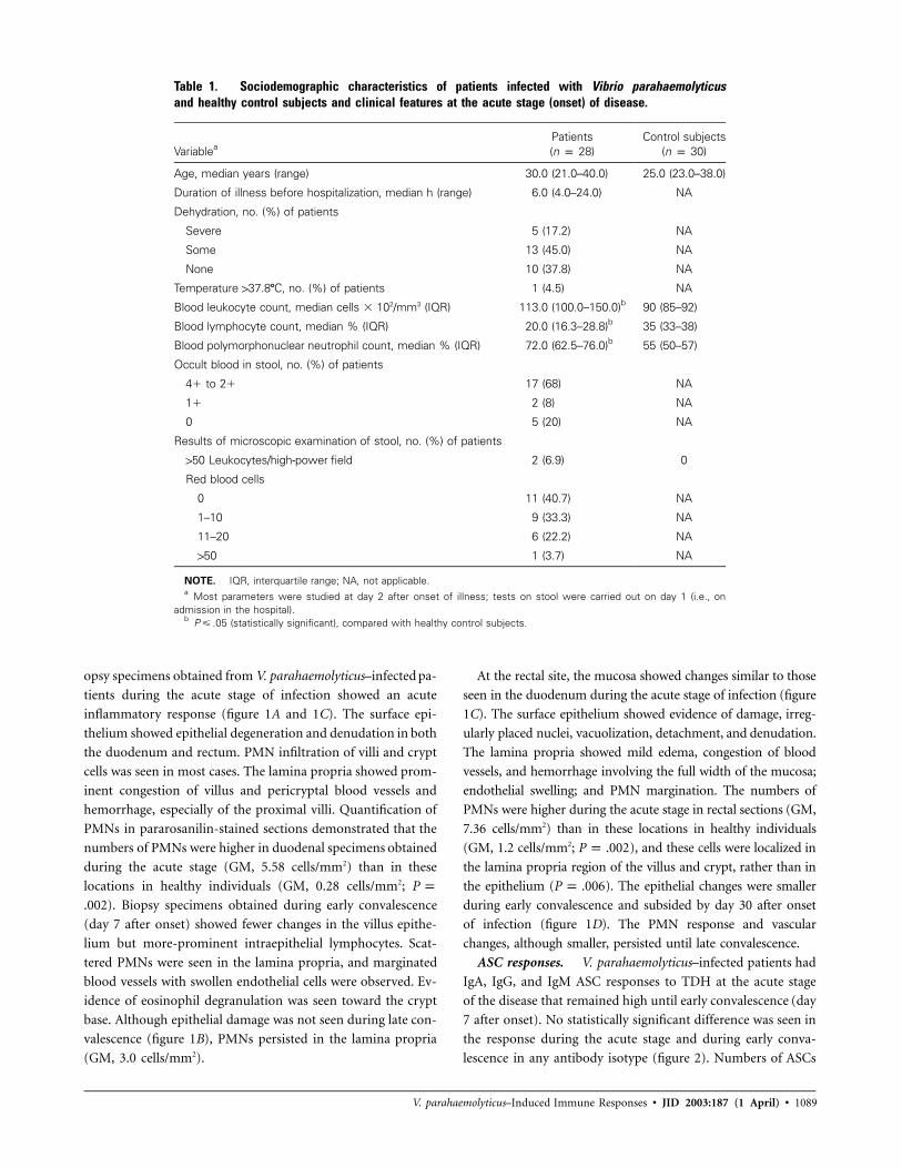
Table 1. Sociodemographic characteristics of patients infected with Vibrio parahaemolyticusand healthy control subjects and clinical features at the acute stage (onset) of disease.
VariableaPatients(n p 28)
Control subjects(n p 30)
Age, median years (range) 30.0 (21.0–40.0) 25.0 (23.0–38.0)
Duration of illness before hospitalization, median h (range) 6.0 (4.0–24.0) NA
Dehydration, no. (%) of patients
Severe 5 (17.2) NA
Some 13 (45.0) NA
None 10 (37.8) NA
Temperature 137.8�C, no. (%) of patients 1 (4.5) NA
Blood leukocyte count, median cells � 102/mm3 (IQR) 113.0 (100.0–150.0)b 90 (85–92)
Blood lymphocyte count, median % (IQR) 20.0 (16.3–28.8)b 35 (33–38)
Blood polymorphonuclear neutrophil count, median % (IQR) 72.0 (62.5–76.0)b 55 (50–57)
Occult blood in stool, no. (%) of patients
4� to 2� 17 (68) NA
1� 2 (8) NA
0 5 (20) NA
Results of microscopic examination of stool, no. (%) of patients
150 Leukocytes/high-power field 2 (6.9) 0
Red blood cells
0 11 (40.7) NA
1–10 9 (33.3) NA
11–20 6 (22.2) NA
150 1 (3.7) NA
NOTE. IQR, interquartile range; NA, not applicable.a Most parameters were studied at day 2 after onset of illness; tests on stool were carried out on day 1 (i.e., on
admission in the hospital).b (statistically significant), compared with healthy control subjects.P � .05
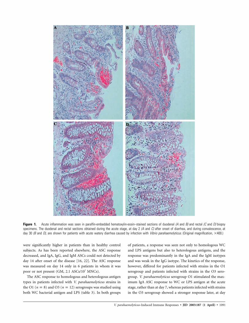
opsy specimens obtained from V. parahaemolyticus–infected pa-
tients during the acute stage of infection showed an acute
inflammatory response (figure 1A and 1C). The surface epi-
thelium showed epithelial degeneration and denudation in both
the duodenum and rectum. PMN infiltration of villi and crypt
cells was seen in most cases. The lamina propria showed prom-
inent congestion of villus and pericryptal blood vessels and
hemorrhage, especially of the proximal villi. Quantification of
PMNs in pararosanilin-stained sections demonstrated that the
numbers of PMNs were higher in duodenal specimens obtained
during the acute stage (GM, 5.58 cells/mm2) than in these
locations in healthy individuals (GM, 0.28 cells/mm2; P p
). Biopsy specimens obtained during early convalescence.002
(day 7 after onset) showed fewer changes in the villus epithe-
lium but more-prominent intraepithelial lymphocytes. Scat-
tered PMNs were seen in the lamina propria, and marginated
blood vessels with swollen endothelial cells were observed. Ev-
idence of eosinophil degranulation was seen toward the crypt
base. Although epithelial damage was not seen during late con-
valescence (figure 1B), PMNs persisted in the lamina propria
(GM, 3.0 cells/mm2).
At the rectal site, the mucosa showed changes similar to those
seen in the duodenum during the acute stage of infection (figure
1C). The surface epithelium showed evidence of damage, irreg-
ularly placed nuclei, vacuolization, detachment, and denudation.
The lamina propria showed mild edema, congestion of blood
vessels, and hemorrhage involving the full width of the mucosa;
endothelial swelling; and PMN margination. The numbers of
PMNs were higher during the acute stage in rectal sections (GM,
7.36 cells/mm2) than in these locations in healthy individuals
(GM, 1.2 cells/mm2; ), and these cells were localized inP p .002
the lamina propria region of the villus and crypt, rather than in
the epithelium ( ). The epithelial changes were smallerP p .006
during early convalescence and subsided by day 30 after onset
of infection (figure 1D). The PMN response and vascular
changes, although smaller, persisted until late convalescence.
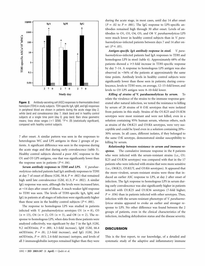
ASC responses. V. parahaemolyticus–infected patients had
IgA, IgG, and IgM ASC responses to TDH at the acute stage
of the disease that remained high until early convalescence (day
7 after onset). No statistically significant difference was seen in
the response during the acute stage and during early conva-
lescence in any antibody isotype (figure 2). Numbers of ASCs
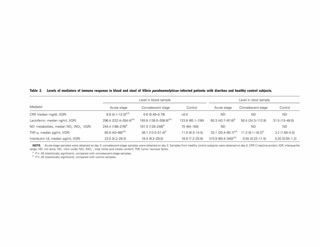
Table 2. Levels of mediators of immune response in blood and stool of Vibrio parahaemolyticus–infected patients with diarrhea and healthy control subjects.
Mediator
Level in blood sample Level in stool sample
Acute stage Convalescent stage Control Acute stage Convalescent stage Control
CRP, median mg/dL (IQR) 8.9 (4.1–12.0)a,b 0.6 (0.49–0.79) !0.5 ND ND ND
Lactoferrin, median ng/mL (IQR) 296.4 (232.4–354.4)a,b 193.6 (138.0–208.8)a,b 123.8 (65.1–156) 65.3 (43.7–97.6)b 50.4 (24.3–112.9) 31.5 (13–48.0)
NO. metabolites, median NO2�/NO3
� (IQR) 244.4 (188–276)b 161.0 (128–248)b 70 (64–160) ND ND ND
TNF-a, median pg/mL (IQR) 65.8 (43–86)a,b 38.1 (13.5–51.4)b 11.0 (8.3–14.5) 33.1 (20.4–60.7)a,b 11.3 (9.1–18.2)b 3.2 (1.60–5.5)
Interleukin-1b, median pg/mL (IQR) 23.0 (5.2–29.2) 16.4 (9.2–29.0) 16.8 (7.2–25.6) 310.9 (60.4–340)a,b 0.55 (0.22–11.5) 0.20 (0.05–1.2)
NOTE. Acute-stage samples were obtained on day 2; convalescent-stage samples were obtained on day 3. Samples from healthy control subjects were obtained on day 0. CRP, C-reactive protein; IQR, interquartilerange; ND, not done; NO., nitric oxide; NO2
�/NO3�, total nitrite and nitrate content; TNF, tumor necrosis factor.
a (statistically significant), compared with convalescent-stage samples.P � .05b (statistically significant), compared with control samples.P � .05
V. parahaemolyticus–Induced Immune Responses • JID 2003:187 (1 April) • 1091
Figure 1. Acute inflammation was seen in paraffin-embedded hematoxylin-eosin–stained sections of duodenal (A and B) and rectal (C and D) biopsyspecimens. The duodenal and rectal sections obtained during the acute stage, at day 2 (A and C) after onset of diarrhea, and during convalescence, atday 30 (B and D), are shown for patients with acute watery diarrhea caused by infection with Vibrio parahaemolyticus. (Original magnification, �400.)
were significantly higher in patients than in healthy control
subjects. As has been reported elsewhere, the ASC response
decreased, and IgA, IgG, and IgM ASCs could not detected by
day 14 after onset of the disease [16, 22]. The ASC response
was measured on day 14 only in 6 patients in whom it was
poor or not present (GM, 2.1 ASCs/107 MNCs).
The ASC response to homologous and heterologous antigen
types in patients infected with V. parahaemolyticus strains in
the O1 ( ) and O3 ( ) serogroups was studied usingn p 8 n p 12
both WC bacterial antigen and LPS (table 3). In both groups
of patients, a response was seen not only to homologous WC
and LPS antigens but also to heterologous antigens, and the
response was predominantly in the IgA and the IgM isotypes
and was weak in the IgG isotype. The kinetics of the response,
however, differed for patients infected with strains in the O1
serogroup and patients infected with strains in the O3 sero-
group. V. parahaemolyticus serogroup O1 stimulated the max-
imum IgA ASC response to WC or LPS antigen at the acute
stage, rather than at day 7, whereas patients infected with strains
in the O3 serogroup showed a stronger response later, at day
1092 • JID 2003:187 (1 April) • Qadri et al.
Figure 2. Antibody-secreting cell (ASC) responses to thermostable directhemolysin (TDH) in study subjects. TDH-specific IgA, IgM, and IgG responsesin peripheral blood are shown in patients during the acute stage (day 2;white bars) and convalescence (day 7; black bars) and in healthy controlsubjects at a single time point (day 0; gray bars). Bars show geometricmeans; lines show ranges (�1 SEM). * (statistically significant),P � .05compared with healthy control subjects.
7 after onset. A similar pattern was seen in the responses to
heterologous WC and LPS antigens in these 2 groups of pa-
tients. A significant difference was seen in the response during
the acute stage and that during early convalescence (table 3).
Healthy control subjects showed a poor ASC response to the
O1 and O3 LPS antigens, one that was significantly lower than
the response seen in patients ( ).P � .04
Serum antibody responses to TDH and LPS. V. parahae-
molyticus–infected patients had IgA antibody responses to TDH
at day 7 of onset of illness (GM, 38.4; ) that remainedP p .002
high until late convalescence (GM, 41.3; ). A similarP p .002
IgG response was seen, although the levels were increased later,
at ∼14 days after onset of illness. A much weaker IgM response
to TDH was seen. The levels of TDH-specific IgA, IgM, and
IgG in patients at all stages of infection were significantly higher
than those seen in the healthy control subjects ( ).P � .001
The response to homologous LPS was studied in patients
infected with V. parahaemolyticus serotypes O1 ( ), O3n p 8
( ), O4 ( ), O5 ( ), and O8 ( ). The re-n p 13 n p 2 n p 3 n p 2
sponse to homologous LPS, when data from these patients were
analyzed collectively, was significant by day 7 in the IgA (GM,
9.2 mOD/min; ; 4.3-fold increase), IgM (GM, 66.4P p .001
mOD/min; ; 2.3-fold increase), and IgG (GM, 26.6P p .01
mOD/min, ; 2.4-fold increase) isotypes, and levels ofP p .015
all 3 immunoglobulin isotypes remained higher than they were
during the acute stage, in most cases, until day 14 after onset
( to ). The IgG response to LPS-specific an-P p .02 P p .001
tibodies remained high through 30 after onset. Levels of an-
tibodies to O1, O3, O4, O5, and O8 V. parahaemolyticus LPS
were much lower in healthy control subjects than in V. para-
haemolyticus–infected patients between days 7 and 14 after on-
set ( ).P � .05
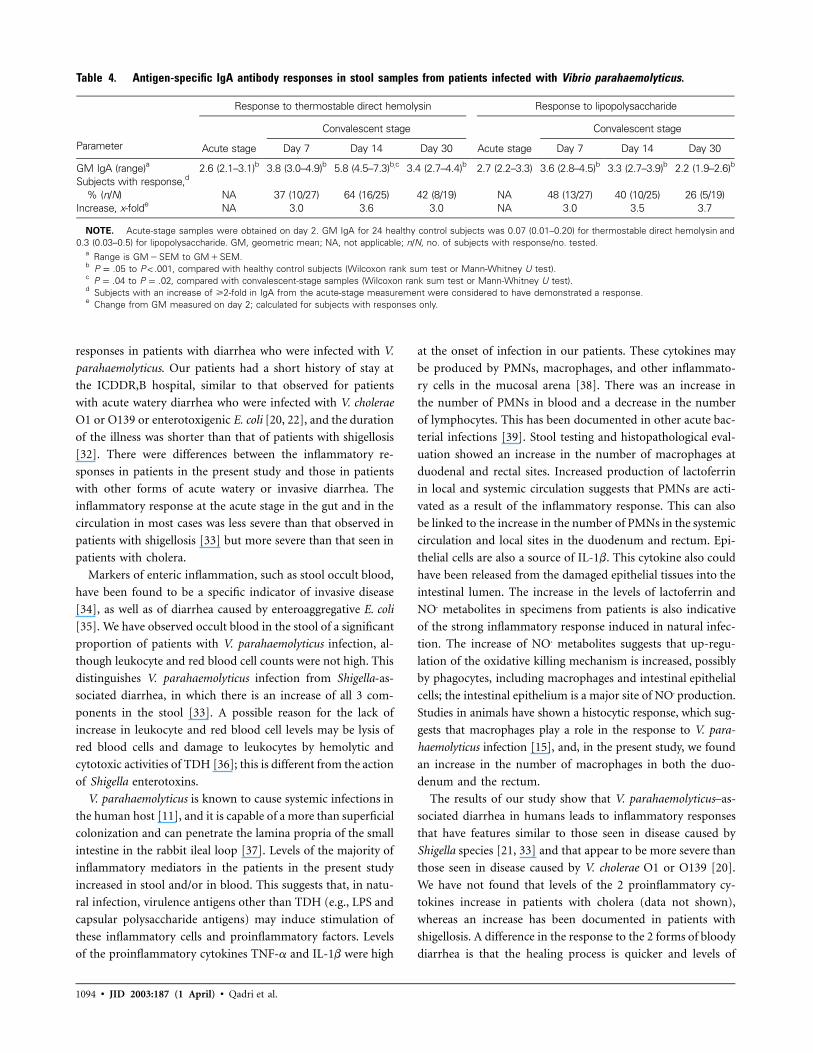
Antigen-specific IgA antibody response in stool. V. para-
haemolyticus–infected patients had IgA responses to TDH and
homologous LPS in stool (table 4). Approximately 68% of the
patients showed a �3-fold increase in TDH-specific response
by day 7–14. A response to homologous LPS antigen was also
observed in ∼56% of the patients at approximately the same
time points. Antibody levels in healthy control subjects were
significantly lower than those seen in patients; during conva-
lescence, levels to TDH were, on average, 12–15-fold lower, and
levels to O3 LPS antigen were 8–10-fold lower.
Killing of strains of V. parahaemolyticus by serum. To
relate the virulence of the strains to the immune response gen-
erated after natural infection, we tested the resistance to killing
by serum of 28 strains of 8 O:K serotypes that were isolated
from patients in this study. Strains of the O1:K25 and O1:K56
serotypes were most resistant and were not killed, even in a
solution containing 95% human serum, whereas others, such
as strains of the O8:K21 and O3:K6 serotypes, were very sus-
ceptible and could be lysed even in a solution containing 20%–
30% serum. In all cases, different isolates, if they belonged to
the same O:K serotype, demonstrated similar susceptibility to
killing by serum.
Relationship between resistance to serum and immune re-
sponse. The cumulative immune response in the 8 patients
who were infected with the serum-resistant strains (i.e., O1:
K25 and O1:K56 serotypes) was compared with that in the 17
patients who were infected with strains that were more sensitive
(i.e., O8:K21, O5:KUT, and O3:K6 serotypes). It appeared that
the more-virulent, serum-resistant strains were those that in-
duced an earlier ASC response to LPS, at day 2 after onset of
infection. The IgA response to homologous LPS in serum dur-
ing early convalescence was also significantly higher in patients
infected with O1:K25 and O1:K56 serotypes (7-fold higher;
) than in patients infected with other serotypes. Thus,P p .036
infection with the serum-resistant phenotype of V. parahaemo-
lyticus strains appeared to evoke an earlier and stronger re-
sponse to LPS. No other difference was found between the 2
groups of patients, even in the clinical characteristics of the
infection, including dehydration status and the disease severity.
DISCUSSION
This is the first report, to our knowledge, of a detailed and
systematic study of the adaptive and inflammatory immune
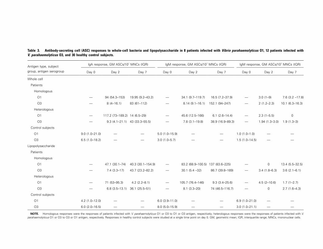
Table 3. Antibody-secreting cell (ASC) responses to whole-cell bacteria and lipopolysaccharide in 8 patients infected with Vibrio parahaemolyticus O1, 12 patients infected withV. parahaemolyticus O3, and 30 healthy control subjects.
Antigen type, subjectgroup, antigen serogroup
IgA response, GM ASCs/107 MNCs (IQR) IgM response, GM ASCs/107 MNCs (IQR) IgM response, GM ASCs/107 MNCs (IQR)
Day 0 Day 2 Day 7 Day 0 Day 2 Day 7 Day 0 Day 2 Day 7
Whole cell
Patients
Homologous
O1 — 94 (54.3–153) 19.95 (9.2–43.2) — 34.1 (9.7–119.7) 16.5 (7.2–37.9) — 3.0 (1–9) 7.6 (3.2 –17.8)
O3 — 8 (4–16.1) 83 (61–112) — 8.14 (9.1–16.1) 152.1 (94–247) — 2 (1.2–2.3) 10.1 (6.3–16.3)
Heterologous
O1 — 117.2 (73–189.2) 14 (6.5–29) — 45.6 (12.5–166) 6.1 (2.6–14.4) — 2.3 (1–5.5) 0
O3 — 9.3 (4.1–21.1) 43 (33.3–55.5) — 7.8 (3.1–19.8) 38.9 (16.9–89.3) — 1.94 (1.3–3.0) 1.9 (1.3–3)
Control subjects
O1 9.0 (1.0–21.0) — — 5.0 (1.0–15.9) — — 1.0 (1.0–1.0) — —
O3 6.5 (1.0–18.2) — — 3.0 (1.0–5.7) — — 1.5 (1.0–14.5) — —
Lipopolysaccharide
Patients
Homologous
O1 — 47.1 (30.1–74) 40.3 (30.1–154.9) — 83.2 (68.9–100.5) 137 (83.6–225) — 0 13.4 (5.5–32.5)
O3 — 7.4 (3.3–17) 43.7 (23.2–82.2) — 30.1 (5.4 –32) 86.7 (39.8–189) — 3.4 (1.8–6.3) 3.6 (2.1–6.1)
Heterologous
O1 — 71 (53–95.3) 4.2 (2.2–8.1) — 105.7 (76.4–146) 9.3 (3.4–25.6) — 4.5 (2–10.6) 1.7 (1–2.7)
O3 — 6.8 (3.5–13.1) 36.1 (25.5–51) — 8.1 (3.3–20) 74 (46.5–116.7) — 0 2.7 (1.6–4.3)
Control subjects
O1 4.2 (1.0–12.0) — — 6.0 (3.9–11.0) — — 6.9 (1.0–21.0) — —
O3 6.0 (2.0–16.5) — — 8.0 (5.0–15.9) — — 3.0 (1.0–21.1) — —
NOTE. Homologous responses were the responses of patients infected with V. parahaemolyticus O1 or O3 to O1 or O3 antigen, respectively; heterologous responses were the responses of patients infected with V.parahaemolyticus O1 or O3 to O3 or O1 antigen, respectively. Responses in healthy control subjects were studied at a single time point on day 0. GM, geometric mean; IQR, interquartile range; MNCs, mononuclear cells.
1094 • JID 2003:187 (1 April) • Qadri et al.
Table 4. Antigen-specific IgA antibody responses in stool samples from patients infected with Vibrio parahaemolyticus.
Parameter
Response to thermostable direct hemolysin Response to lipopolysaccharide
Acute stage
Convalescent stage
Acute stage
Convalescent stage
Day 7 Day 14 Day 30 Day 7 Day 14 Day 30
GM IgA (range)a 2.6 (2.1–3.1)b 3.8 (3.0–4.9)b 5.8 (4.5–7.3)b,c 3.4 (2.7–4.4)b 2.7 (2.2–3.3) 3.6 (2.8–4.5)b 3.3 (2.7–3.9)b 2.2 (1.9–2.6)b
Subjects with response,d
% (n/N) NA 37 (10/27) 64 (16/25) 42 (8/19) NA 48 (13/27) 40 (10/25) 26 (5/19)Increase, x-folde NA 3.0 3.6 3.0 NA 3.0 3.5 3.7
NOTE. Acute-stage samples were obtained on day 2. GM IgA for 24 healthy control subjects was 0.07 (0.01–0.20) for thermostable direct hemolysin and0.3 (0.03–0.5) for lipopolysaccharide. GM, geometric mean; NA, not applicable; n/N, no. of subjects with response/no. tested.
a Range is to .GM � SEM GM � SEMb to , compared with healthy control subjects (Wilcoxon rank sum test or Mann-Whitney U test).P p .05 P ! .001c to , compared with convalescent-stage samples (Wilcoxon rank sum test or Mann-Whitney U test).P p .04 P p .02d Subjects with an increase of �2-fold in IgA from the acute-stage measurement were considered to have demonstrated a response.e Change from GM measured on day 2; calculated for subjects with responses only.
responses in patients with diarrhea who were infected with V.
parahaemolyticus. Our patients had a short history of stay at
the ICDDR,B hospital, similar to that observed for patients
with acute watery diarrhea who were infected with V. cholerae
O1 or O139 or enterotoxigenic E. coli [20, 22], and the duration
of the illness was shorter than that of patients with shigellosis
[32]. There were differences between the inflammatory re-
sponses in patients in the present study and those in patients
with other forms of acute watery or invasive diarrhea. The
inflammatory response at the acute stage in the gut and in the
circulation in most cases was less severe than that observed in
patients with shigellosis [33] but more severe than that seen in
patients with cholera.
Markers of enteric inflammation, such as stool occult blood,
have been found to be a specific indicator of invasive disease
[34], as well as of diarrhea caused by enteroaggregative E. coli
[35]. We have observed occult blood in the stool of a significant
proportion of patients with V. parahaemolyticus infection, al-
though leukocyte and red blood cell counts were not high. This
distinguishes V. parahaemolyticus infection from Shigella-as-
sociated diarrhea, in which there is an increase of all 3 com-
ponents in the stool [33]. A possible reason for the lack of
increase in leukocyte and red blood cell levels may be lysis of
red blood cells and damage to leukocytes by hemolytic and
cytotoxic activities of TDH [36]; this is different from the action
of Shigella enterotoxins.
V. parahaemolyticus is known to cause systemic infections in
the human host [11], and it is capable of a more than superficial
colonization and can penetrate the lamina propria of the small
intestine in the rabbit ileal loop [37]. Levels of the majority of
inflammatory mediators in the patients in the present study
increased in stool and/or in blood. This suggests that, in natu-
ral infection, virulence antigens other than TDH (e.g., LPS and
capsular polysaccharide antigens) may induce stimulation of
these inflammatory cells and proinflammatory factors. Levels
of the proinflammatory cytokines TNF-a and IL-1b were high
at the onset of infection in our patients. These cytokines may
be produced by PMNs, macrophages, and other inflammato-
ry cells in the mucosal arena [38]. There was an increase in
the number of PMNs in blood and a decrease in the number
of lymphocytes. This has been documented in other acute bac-
terial infections [39]. Stool testing and histopathological eval-
uation showed an increase in the number of macrophages at
duodenal and rectal sites. Increased production of lactoferrin
in local and systemic circulation suggests that PMNs are acti-
vated as a result of the inflammatory response. This can also
be linked to the increase in the number of PMNs in the systemic
circulation and local sites in the duodenum and rectum. Epi-
thelial cells are also a source of IL-1b. This cytokine also could
have been released from the damaged epithelial tissues into the
intestinal lumen. The increase in the levels of lactoferrin and
NO. metabolites in specimens from patients is also indicative
of the strong inflammatory response induced in natural infec-
tion. The increase of NO. metabolites suggests that up-regu-
lation of the oxidative killing mechanism is increased, possibly
by phagocytes, including macrophages and intestinal epithelial
cells; the intestinal epithelium is a major site of NO. production.
Studies in animals have shown a histocytic response, which sug-
gests that macrophages play a role in the response to V. para-
haemolyticus infection [15], and, in the present study, we found
an increase in the number of macrophages in both the duo-
denum and the rectum.
The results of our study show that V. parahaemolyticus–as-
sociated diarrhea in humans leads to inflammatory responses
that have features similar to those seen in disease caused by
Shigella species [21, 33] and that appear to be more severe than
those seen in disease caused by V. cholerae O1 or O139 [20].
We have not found that levels of the 2 proinflammatory cy-
tokines increase in patients with cholera (data not shown),
whereas an increase has been documented in patients with
shigellosis. A difference in the response to the 2 forms of bloody
diarrhea is that the healing process is quicker and levels of
V. parahaemolyticus–Induced Immune Responses • JID 2003:187 (1 April) • 1095
inflammatory cytokines decrease more rapidly in TDH-asso-
ciated bacterial infections than in shigellosis. The mediators
may remain elevated for as long as 15–45 days after onset of
illness in shigellosis. Levels of the acute-stage protein, CRP,
increased much more in patients with V. parahaemolyticus in-
fection than in patients with cholera [20] and to a degree com-
parable to that seen in patients with shigellosis [33]. The up-
regulation of NO. metabolites and lactoferrin in patients
infected with V. parahaemolyticus, however, appeared to be
comparable to that seen in the patients with cholera [20].
The inflammatory immune response in V. parahaemolyticus–
induced diarrhea appears to reflect the self-limiting pattern of
the clinical disease. The acute inflammatory response in the cir-
culation and the local secretions at the mucosal surface subsided
and reverted to preinfection conditions very soon, by approxi-
mately day 7 after the onset of infection. This is in keeping with
the action of TDH in IEC-6 cells [40], in which it has been
shown that, once the toxin is removed from the environment,
the cells rapidly recover their original morphology.
Strains of both the pandemic and nonpandemic types were
isolated from our patients. We were not able to discern any
difference between the responses of patients infected by pandemic
strains ( ) and patients infected by nonpandemic strainsn p 18
( ) in clinical characteristics, B cell responses, or inflam-n p 10
matory responses. This suggests that the attributes of the pan-
demic strains, other than virulence in the host, may be respon-
sible for the worldwide spread of infection with these strains.
The strains of V. parahaemolyticus isolated from our patients
showed varying susceptibility to the bactericidal effects of se-
rum. Those belonging to the O1:K25 and O1:K56 serotypes,
which were more resistant, induced a relatively earlier B cell
response. We speculate that the early immune response to these
pathogens could be the result of their increased ability to evade
killing by the host, which allows them to colonize and persist
in higher numbers, resulting in a earlier humoral immunolog-
ical response.
Because V. parahaemolyticus causes secretory diarrhea, the
small intestine has been considered to be the site of action in
the human host. Small-intestine damage was found during au-
topsy of 4 persons who died of V. parahaemolyticus–induced
food poisoning [41]. In the present study, however, we show
that both small-intestine and large-intestine tissues are affected
by the pathogen. We observed epithelial damage, denudation,
and hemorrhage at both sites. Our results suggest that both
locations of the gut are affected by colonization with the path-
ogen. Gastroenteritis caused by V. parahaemolyticus results in
strong systemic and mucosal B cell responses to TDH and LPS.
An IgA ASC response indicates mucosal activation. Both the
antigens also induced an increase in the presence of IgM ASCs,
which suggests that this was a primary response to the antigen.
In addition, TDH also induced strong IgG responses, similar
the responses to cholera toxin reported elsewhere [22]. The IgG
response is also indicative of a systemic component, which is
a possibility; V. parahaemolyticus results in denudation and
damage of the gut epithelium, and antigens can reach the lam-
ina propria. It is possible that TDH can be taken up by Peyer’s
patches and transported within macrophages to the mesenteric
lymph nodes and spleen to induce a systemic response; this
mechanism has also been proposed for cholera toxin [42]. In
addition, TDH may bind to cells via the GT1b ganglioside
receptor [43, 44], which is believed to be a toxin-binding epi-
tope on T cell surfaces [45] and to induce an innate immunity.
Thus, both the mucosal and systemic compartments of the
immune systems are activated.
We do not know whether natural infection due to V. para-
haemolyticus results in an O:K serotype–specific or a TDH-
specific protective immune response. This is the first study to
document the immune response after natural infection, and
more work, especially on mucosal and cellular responses and
on immune modulators, needs to be carried out before the
detailed mechanism of immune response in the host can be
understood.
References
1. Bag PK, Nandi S, Bhadra RK, et al. Clonal diversity among recentlyemerged strains of Vibrio parahaemolyticus O3:K6 associated with pan-demic spread. J Clin Microbiol 1999; 37:2354–7.
2. Bhuiyan NA, Ansaruzzaman M, Kamruzzaman M, et al. Prevalence ofthe pandemic genotype of Vibrio parahaemolyticus in Dhaka, Bang-ladesh, and significance of its distribution across different serotypes. JClin Microbiol 2002; 40:284–6.
3. Gilman RH, Spira WM, Rabbani GH, Al-Mahomod A. Invasive E. coliand V. parahaemolyticus: a rare cause of dysentery in Dacca. Trans RSoc Trop Med Hyg 1980; 74:688–9.
4. Honda S, Matsumoto S, Miwatani T, Honda T. A survey of urease-positive Vibrio parahaemolyticus strains isolated from traveller’s diar-rhea, sea water and imported frozen sea foods. Eur J Epidemiol 1992;8:861–4.
5. Chiou CS, Hsu SY, Chiu SI, Wang TK, Chao CS. Vibrio parahaemo-lyticus serovar O3:K6 as cause of unusually high incidence of food-borne disease outbreaks in Taiwan from 1996 to 1999. J Clin Microbiol2000; 38:4621–5.
6. Daniels NA, MacKinnon L, Bishop R, et al. Vibrio parahaemolyticus in-fections in the United States, 1973–1998. J Infect Dis 2000; 181:1661–6.
7. Okuda J, Ishibashi M, Abbott SL, Janda JM, Nishibuchi M. Analysisof the thermostable direct hemolysin (tdh) gene and the tdh-relatedhemolysin (trh) genes in urease-positive strains of Vibrio parahaemo-lyticus isolated on the West Coast of the United States. J Clin Microbiol1997; 35:1965–71.
8. Matsumoto C, Okuda J, Ishibashi M, et al. Pandemic spread of an O3:K6 clone of Vibrio parahaemolyticus and emergence of related strainsevidenced by arbitrarily primed PCR and toxRS sequence analyses. JClin Microbiol 2000; 38:578–85.
9. Blake PA, Weaver RE, Hollis DG. Diseases of humans (other thancholera) caused by vibrios. Annu Rev Microbiol 1980; 34:341–67.
10. Hsu GJ, Young T, Peng MY, Chang FY, Chou MY. Septicemia causedby Vibrio parahemolyticus: a case report. Zhonghua Yi Xue Za Zhi (Tai-pei) 1993; 52:351–4.
11. Ng TC, Chiang PC, Wu TL, Leu HS. Vibrio parahemolyticus bacteremia:case report. Changgeng Yi Xue Za Zhi 1999; 22:508–14.
1096 • JID 2003:187 (1 April) • Qadri et al.
12. Nishibuchi M, Fasano A, Russell RG, Kaper JB. Enterotoxigenicity ofVibrio parahaemolyticus with and without genes encoding thermostabledirect hemolysin. Infect Immun 1992; 60:3539–45.
13. Nishibuchi M, Kaper JB. Thermostable direct hemolysin gene of Vibrioparahaemolyticus: a virulence gene acquired by a marine bacterium.Infect Immun 1995; 63:2093–9.
14. Xu M, Iida T, Yamamoto K, Takarada Y, Miwatani T, Honda T. Dem-onstration and characterization of simultaneous production of a ther-mostable direct hemolysin (TDH/I) and a TDH-related hemolysin(TRHx) by a clinically isolated Vibrio parahaemolyticus strain, TH3766.Infect Immun 1994; 62:166–71.
15. Chatterjee BD, Mukherjee A, Sanyal SN. Enteroinvasive model of Vibrioparahaemolyticus. Indian J Med Res 1984; 79:151–8.
16. Czerkinsky C, Quiding M, Eriksson K, et al. Induction of specificimmunity at mucosal surfaces: prospects for vaccine development. AdvExp Med Biol 1995; 371B:1409–16.
17. Qadri F, Ahmed F, Karim MM, et al. Lipopolysaccharide- and choleratoxin–specific subclass distribution of B-cell responses in cholera. ClinDiagn Lab Immunol 1999; 6:812–8.
18. World Health Organization (WHO). Programme for control of diar-rhoeal diseases. In: Manual for investigation of acute enteric infections.Rev 1. WHO/CDD/83.3. Geneva: WHO, 1987:9–20.
19. World Health Organization. Diarrhoeal diseases control programme:global activities, 1988–1989. Wkly Epidemiol Rec 1990; 65:289–92.
20. Qadri F, Raqib R, Ahmed F, et al. Increased levels of inflammatorymediators in children and adults infected with Vibrio cholerae O1 andO139. Clin Diagn Lab Immunol 2002; 9:221–9.
21. Raqib R, Mia SM, Qadri F, et al. Innate immune responses in childrenand adults with Shigellosis. Infect Immun 2000; 68:3620–9.
22. Qadri F, Wenneras C, Albert MJ, et al. Comparison of immune re-sponses in patients infected with Vibrio cholerae O139 and O1. InfectImmun 1997; 65:3571–6.
23. Qadri F, Das SK, Faruque AS, et al. Prevalence of toxin types andcolonization factors in enterotoxigenic Escherichia coli isolated duringa 2-year period from diarrheal patients in Bangladesh. J Clin Microbiol2000; 38:27–31.
24. Huicho L, Sanchez D, Contreras M, et al. Occult blood and fecalleukocytes as screening tests in childhood infectious diarrhea: an oldproblem revisited. Pediatr Infect Dis J 1993; 12:474–7.
25. Westphal O, Jann K, Himmelspach K. Chemistry and immunochem-istry of bacterial lipopolysaccharides as cell wall antigens and endo-toxins. Prog Allergy 1983; 33:9–39.
26. Hisatsune K, Kiuye A, Kondo S. Sugar composition of O-antigeniclipopolysaccharides isolated from Vibrio parahaemolyticus. MicrobiolImmunol 1980; 24:691–701.
27. Sakurai J, Matsuzaki A, Miwatani T. Purification and characterizationof thermostable direct hemolysin of Vibrio parahaemolyticus. InfectImmun 1973; 8:775–80.
28. Ryan ET, Butterton JR, Smith RN, Carroll PA, Crean TI, CalderwoodSB. Protective immunity against Clostridium difficile toxin A inducedby oral immunization with a live, attenuated Vibrio cholerae vectorstrain. Infect Immun 1997; 65:2941–9.
29. Qadri F, Haque MA, Hossain A, Azim T, Alam K, Albert MJ. Role ofShigella dysenteriae type 1 slime polysaccharide in resistance to serumkilling and phagocytosis. Microb Pathog 1993; 14:441–9.
30. Jolly S, Detilleux J, Coignoul F, Desmecht D. Enzyme-histochemicaldetection of a chymase-like proteinase within bovine mucosal andconnective tissue mast cells. J Comp Pathol 2000; 122:155–62.
31. Mathan MM, Chandy G, Mathan VI. Ultrastructural changes in theupper small intestinal mucosa in patients with cholera. Gastroenter-ology 1995; 109:422–30.
32. Khan WA, Salam MA, Bennish ML. C reactive protein and prealbuminas markers of disease activity in shigellosis. Gut 1995; 37:402–5.
33. Raqib R, Lindberg AA, Wretlind B, Bardhan PK, Andersson U, An-dersson J. Persistence of local cytokine production in shigellosis inacute and convalescent stages. Infect Immun 1995; 63:289–96.
34. Bardhan PK, Beltinger J, Beltinger RW, Hossain A, Mahalanabis D,Gyr K. Screening of patients with acute infectious diarrhoea: evaluationof clinical features, faecal microscopy, and faecal occult blood testing.Scand J Gastroenterol 2000; 35:54–60.
35. Bouckenooghe AR, Dupont HL, Jiang ZD, et al. Markers of entericinflammation in enteroaggregative Escherichia coli diarrhea in travelers.Am J Trop Med Hyg 2000; 62:711–3.
36. Takeda Y. Thermostable direct hemolysin of Vibrio parahaemolyticus.Pharmacol Ther 1982; 19:123–46.
37. Boutin BK, Townsend SF, Scarpino PV, Twedt RM. Demonstration ofinvasiveness of Vibrio parahaemolyticus in adult rabbits by immuno-fluorescence. Appl Environ Microbiol 1979; 37:647–53.
38. McGee DW, Bamberg T, Vitkus SJ, McGhee JR. A synergistic rela-tionship between TNF-alpha, IL-1 beta, and TGF-beta 1 on IL-6 se-cretion by the IEC-6 intestinal epithelial cell line. Immunology 1995;86:6–11.
39. Myhre EB, Braconier JH, Sjogren U. Automated cytochemical differ-ential leucocyte count in patients hospitalized with acute bacterial in-fections. Scand J Infect Dis 1985; 17:201–8.
40. Fabbri A, Falzano L, Frank C, et al. Vibrio parahaemolyticus thermo-stable direct hemolysin modulates cytoskeletal organization and cal-cium homeostasis in intestinal cultured cells. Infect Immun 1999; 67:1139–48.
41. Okudaira M, Kawamura H, Uemo M, Nakahara Y, et al. Food poison-ing caused by pathogenic halophilic bacterium (Pseudomonas enteritisTakikawa): report of four autopsy cases. Acta Pathol Jpn 1962; 12:299.
42. Ohtomo N, Muraoka T, Tashiro A, Zinnaka Y, Amako K. Size andstructure of the cholera toxin molecule and its subunits. J Infect Dis1976; 133(Suppl):31–40.
43. Takeda Y, Honda T, Miwatani T. Biological activity and receptor ofthermostable direct hemolysin of Vibrio parahaemolyticus [in Japanese].Tanpakushitsu Kakusan Koso 1976; (Suppl):109–20.
44. Raimondi F, Kao JP, Fiorentini C, et al. Enterotoxicity and cytotoxicityof Vibrio parahaemolyticus thermostable direct hemolysin in in vitrosystems. Infect Immun 2000; 68:3180–5.
45. Bukowski JF, Roncarolo MG, Spits H, et al. T cell receptor–dependentactivation of human lymphocytes through cell surface ganglioside GT1b:implications for innate immunity. Eur J Immunol 2000; 30:3199–206.
Copyright © 2022 FDOKUMEN