High-Frequency Rugose Exopolysaccharide Production by Vibrio cholerae
6
APPLIED AND ENVIRONMENTAL MICROBIOLOGY, Nov. 2002, p. 5773–5778 Vol. 68, No. 11 0099-2240/02/$04.000 DOI: 10.1128/AEM.68.11.5773–5778.2002 Copyright © 2002, American Society for Microbiology. All Rights Reserved. High-Frequency Rugose Exopolysaccharide Production by Vibrio cholerae Afsar Ali, Mohammed H. Rashid, and David K. R. Karaolis* Department of Epidemiology and Preventive Medicine, University of Maryland School of Medicine, Baltimore, Maryland 21201 Received 24 June 2002/Accepted 20 August 2002 Vibrio cholerae can shift to a “rugose” phenotype, thereby producing copious exopolysaccharide (EPS), which promotes its environmental survival and persistence. We report conditions that promote high-frequency rugose EPS production (HFRP), whereby cells switch at high frequency (up to 80%) to rugose EPS production. HFRP appeared to be more common in clinical strains, as HFRP was found in 6 of 19 clinical strains (32%) (including classical, El Tor, and non-O1 strains) but in only 1 of 16 environmental strains (6%). Differences were found between strains in rugose colony morphology, conditions promoting HFRP, the frequency of rugose-to-smooth (R-S) cell reversion, and biofilm formation. We propose that rugose EPS and HFRP provide an evolutionary and adaptive advantage to specific epidemic V. cholerae strains for increased persistence in the environment. Cholera, caused by Vibrio cholerae, is a severe, life-threat- ening diarrheal disease which continues to be a scourge throughout much of the world; there have been seven global pandemics recorded since 1817 (15). The explosive epidemic nature and severity of the disease have prompted its listing as a high-priority organism in biodefense research. Epidemic V. cholerae strains can persist for many years in the marine envi- ronment while regularly causing significant cholera epidemics (3, 5, 6, 9, 16, 20, 21); however, the specific mechanism under- lying the adaptation to long-term persistence, particularly that of epidemic strains, has remained a mystery. Several studies have proposed that the persistence of V. cholerae in the envi- ronment is due to a viable but nonculturable state (VBNC) in response to starvation and cold (4, 18, 19, 27); however, it is not known whether the VBNC phenotype promotes persis- tence in regions where cholera is endemic and under condi- tions other than starvation and cold. Alternatively, “rugose” exopolysaccharide (rEPS) production might promote the sur- vival and persistence of V. cholerae in the aquatic environment. A rugose colony phenotype was first reported and described in both sixth (classical biotype) and seventh (El Tor biotype) pandemic strains in the late 1930s by Bruce White (25, 26) and is characterized by wrinkled colonies expressing copious amounts of EPS. Since then, studies have shown that smooth V. cholerae cells under nutrient-limiting conditions can spon- taneously shift at a low frequency to express rEPS and exhibit wrinkled (rugose) colonies (13, 14, 22, 25, 26, 29) that are virulent in rabbit ileal loop models and in human volunteers (14, 17). Although a gene cluster (vps) for the biosynthesis of rEPS and a gene (vpsR) that regulates the expression of several vps biosynthetic genes have been identified (2, 28, 29), the physiology, genetics, and importance of rEPS expression are not well understood and studies on the rugose phenotype have been hampered by the low frequency of switching to the rugose phenotype when available conditions are used. Previously, Morris et al. (14) reported the isolation of only spontaneous rugose variants from a variety of V. cholerae strains after 3 to 4 days of incubation in standard alkaline peptone water (APW) and plating on L agar at 37°C. Wai et al. (22) then reported that V. cholerae El Tor O1 strain TSI-4 was able to spontane- ously shift to a rugose colony morphology under starvation conditions. These investigators isolated a few spontaneous rug- ose variants after smooth colonies were starved in M9 salts for 2 months at 16°C and then plated on L agar at 37°C. Yildiz and Schoolnik (29) showed that, under the nutrient-limiting (car- bon-limiting) conditions they tested (30°C incubation in M9 minimal media supplemented with 0.02% glucose), smooth El Tor strains were able to shift to the rugose colonial variant after 20 days of culture but at a frequency of only 1%. In pursuit of our interests in the factors and processes in- volved in the emergence, pathogenesis, and persistence of ep- idemic V. cholerae, we studied the prevalence, physiology, and role of rEPS production in various V. cholerae strains which either have been described previously (10, 11) or were ob- tained from A. Huq (University of Maryland) and L. Campos (FIOCRUZ, Rio de Janeiro, Brazil). The strains included 19 clinical isolates (O1 and non-O1 strains) and 16 environmental isolates (O1 and non-O1 strains) collected over many years and from several continents. We discovered that a nutrient-poor medium, a modified form of APW, which we have termed APW#3 (1% proteose peptone #3 [Difco], 1% NaCl, pH 8.5), resulted in a high frequency (up to 80%) of N16961 smooth cells shifting to the rugose phenotype. A single smooth colony was inoculated into 3 ml of APW#3 in glass test tubes or 45 ml of APW#3 in 250-ml flasks and incubated statically for 24 to 72 h at 30°C or 37°C. Sterile glass beads (4 mm in diameter) were then added, and the cultures were vortexed to disrupt any aggregates of rugose cells. Following incubation, we observed only a minor decrease in the pH of the culture medium (pH 8.3). Appro- priate dilutions of each culture were plated on Luria-Bertani (LB) agar (Difco), and colonies were counted by standard plate counting to determine the total number of CFU per * Corresponding author. Mailing address: Department of Epidemi- ology and Preventive Medicine, University of Maryland School of Medicine, Baltimore, MD 21201. Phone: (410) 706-4718. Fax: (410) 706-4581. E-mail: [email protected]. 5773 on February 24, 2015 by guest http://aem.asm.org/ Downloaded from
-
Upload
independent -
Category
Documents
-
view
0 -
download
0
Transcript of High-Frequency Rugose Exopolysaccharide Production by Vibrio cholerae
APPLIED AND ENVIRONMENTAL MICROBIOLOGY, Nov. 2002, p. 5773–5778 Vol. 68, No. 110099-2240/02/$04.00�0 DOI: 10.1128/AEM.68.11.5773–5778.2002Copyright © 2002, American Society for Microbiology. All Rights Reserved.
High-Frequency Rugose Exopolysaccharide Production byVibrio cholerae
Afsar Ali, Mohammed H. Rashid, and David K. R. Karaolis*Department of Epidemiology and Preventive Medicine, University of Maryland School of Medicine,
Baltimore, Maryland 21201
Received 24 June 2002/Accepted 20 August 2002
Vibrio cholerae can shift to a “rugose” phenotype, thereby producing copious exopolysaccharide (EPS), whichpromotes its environmental survival and persistence. We report conditions that promote high-frequency rugoseEPS production (HFRP), whereby cells switch at high frequency (up to 80%) to rugose EPS production. HFRPappeared to be more common in clinical strains, as HFRP was found in 6 of 19 clinical strains (32%) (includingclassical, El Tor, and non-O1 strains) but in only 1 of 16 environmental strains (6%). Differences were foundbetween strains in rugose colony morphology, conditions promoting HFRP, the frequency of rugose-to-smooth(R-S) cell reversion, and biofilm formation. We propose that rugose EPS and HFRP provide an evolutionaryand adaptive advantage to specific epidemic V. cholerae strains for increased persistence in the environment.
Cholera, caused by Vibrio cholerae, is a severe, life-threat-ening diarrheal disease which continues to be a scourgethroughout much of the world; there have been seven globalpandemics recorded since 1817 (15). The explosive epidemicnature and severity of the disease have prompted its listing asa high-priority organism in biodefense research. Epidemic V.cholerae strains can persist for many years in the marine envi-ronment while regularly causing significant cholera epidemics(3, 5, 6, 9, 16, 20, 21); however, the specific mechanism under-lying the adaptation to long-term persistence, particularly thatof epidemic strains, has remained a mystery. Several studieshave proposed that the persistence of V. cholerae in the envi-ronment is due to a viable but nonculturable state (VBNC) inresponse to starvation and cold (4, 18, 19, 27); however, it isnot known whether the VBNC phenotype promotes persis-tence in regions where cholera is endemic and under condi-tions other than starvation and cold. Alternatively, “rugose”exopolysaccharide (rEPS) production might promote the sur-vival and persistence of V. cholerae in the aquatic environment.
A rugose colony phenotype was first reported and describedin both sixth (classical biotype) and seventh (El Tor biotype)pandemic strains in the late 1930s by Bruce White (25, 26) andis characterized by wrinkled colonies expressing copiousamounts of EPS. Since then, studies have shown that smoothV. cholerae cells under nutrient-limiting conditions can spon-taneously shift at a low frequency to express rEPS and exhibitwrinkled (rugose) colonies (13, 14, 22, 25, 26, 29) that arevirulent in rabbit ileal loop models and in human volunteers(14, 17). Although a gene cluster (vps) for the biosynthesis ofrEPS and a gene (vpsR) that regulates the expression of severalvps biosynthetic genes have been identified (2, 28, 29), thephysiology, genetics, and importance of rEPS expression arenot well understood and studies on the rugose phenotype havebeen hampered by the low frequency of switching to the rugose
phenotype when available conditions are used. Previously,Morris et al. (14) reported the isolation of only spontaneousrugose variants from a variety of V. cholerae strains after 3 to4 days of incubation in standard alkaline peptone water (APW)and plating on L agar at 37°C. Wai et al. (22) then reportedthat V. cholerae El Tor O1 strain TSI-4 was able to spontane-ously shift to a rugose colony morphology under starvationconditions. These investigators isolated a few spontaneous rug-ose variants after smooth colonies were starved in M9 salts for2 months at 16°C and then plated on L agar at 37°C. Yildiz andSchoolnik (29) showed that, under the nutrient-limiting (car-bon-limiting) conditions they tested (30°C incubation in M9minimal media supplemented with 0.02% glucose), smooth ElTor strains were able to shift to the rugose colonial variantafter 20 days of culture but at a frequency of only �1%.
In pursuit of our interests in the factors and processes in-volved in the emergence, pathogenesis, and persistence of ep-idemic V. cholerae, we studied the prevalence, physiology, androle of rEPS production in various V. cholerae strains whicheither have been described previously (10, 11) or were ob-tained from A. Huq (University of Maryland) and L. Campos(FIOCRUZ, Rio de Janeiro, Brazil). The strains included 19clinical isolates (O1 and non-O1 strains) and 16 environmentalisolates (O1 and non-O1 strains) collected over many years andfrom several continents.
We discovered that a nutrient-poor medium, a modifiedform of APW, which we have termed APW#3 (1% proteosepeptone #3 [Difco], 1% NaCl, pH 8.5), resulted in a highfrequency (up to 80%) of N16961 smooth cells shifting to therugose phenotype. A single smooth colony was inoculated into3 ml of APW#3 in glass test tubes or 45 ml of APW#3 in250-ml flasks and incubated statically for 24 to 72 h at 30°C or37°C. Sterile glass beads (4 mm in diameter) were then added,and the cultures were vortexed to disrupt any aggregates ofrugose cells. Following incubation, we observed only a minordecrease in the pH of the culture medium (pH �8.3). Appro-priate dilutions of each culture were plated on Luria-Bertani(LB) agar (Difco), and colonies were counted by standardplate counting to determine the total number of CFU per
* Corresponding author. Mailing address: Department of Epidemi-ology and Preventive Medicine, University of Maryland School ofMedicine, Baltimore, MD 21201. Phone: (410) 706-4718. Fax: (410)706-4581. E-mail: [email protected].
5773
on February 24, 2015 by guest
http://aem.asm
.org/D
ownloaded from
milliliter and the frequency of rugose cells. On the basis of atleast eleven independent experiments, we found that, after 24to 48 h of growth at 37°C (in glass tubes), approximately 60%of the smooth cells had switched to the rugose phenotype(Table 1 and Fig. 1). Accordingly, we called this phenotypehigh-frequency rugose EPS production (HFRP). Time coursestudies analyzing samples every other hour showed that HFRPwas not detected in N16961 until at least 14 h of incubation, atwhich time we observed a sudden increase in the percentage ofrugose variants (0 to 50%). After this initial HFRP spike thepercentage of rugose cells increased in the culture, resulting inapproximately 60 to 70% rugose cells at 24 h. We found thatHFRP was independent of LuxS, RpoS, and ToxT (data notshown). The mechanism of HFRP is presently being studiedand might be regulated by phase variation. Phase variation canoccur via DNA inversion, DNA recombination, and slipped-strand mispairing and is known to be involved in controllingthe expression of several surface structures of gram-negativebacteria, including fimbriae, flagella, outer membrane pro-teins, lipopolysaccharide, and capsular polysaccharide (8, 12).
In order to determine whether HFRP was found in other V.cholerae strains, we tested various clinical and environmentalisolates. Consistent with previous studies (14), we found a lowfrequency (�0.5%) shift to the rugose phenotype in severalstrains. However, we were surprised to find that 6 of 19 clinicalisolates (32%) and only 1 of 16 nonpathogenic strains (6%)could shift to HFRP under the conditions tested (t test; P �0.05) (Table 1). Of all strains tested, N16961 consistently hadthe highest HFRP in flasks (24 to 38%, 30°C; 42 to 51%, 37°C)and tubes (68 to 74%, 30°C; 60 to 80%, 37°C) (Table 1). Whilethe results revealed that not all epidemic strains showedHFRP, indicating that HFRP is not essential for persistence,our results do suggest that rEPS has a role in V. cholerae andthat HFRP might be more common in clinical strains andmight promote persistence of these specific strains. The asso-ciation of HFRP with clinical epidemic strains is being studiedfurther. Just as many studies rely on in vitro data (in some
cases obtained from a single strain) to draw conclusions on therelevance of a phenotype in vivo or in the natural environment,we propose that HFRP has an important role in nature. Inter-estingly, it is known that persistent bacterial diseases such ascystic fibrosis are caused by only a subpopulation of thePseudomonas aeruginosa species in which specific cells have theability to express copious amounts of EPS and form biofilms inthe lung (7). In the case of V. cholerae, rEPS and HFRPproduction may provide some evolutionary or adaptive advan-tage to that subpopulation of cells in a particular environment.
Interestingly, NCTC 6585, a sixth pandemic (classical bio-type) strain, consistently showed HFRP in flasks (33 to 48%,30°C; 44 to 45%, 37°C). In addition, the sixth pandemic strainAMS20A73 also showed HFRP, albeit at a lower frequency (3to 4%), with HFRP being defined as a �3% shift to the rugosephenotype. A previous study showed that homologous se-quences were found in sixth pandemic (classical biotype)strains; however, these investigators were unable to demon-strate rEPS expression in sixth pandemic strains (29). To dem-onstrate that the rugose classical biotype strain NCTC 6585overexpressed rEPS, we performed transmission electron mi-croscopy (TEM) on ruthenium red-stained thin sections. ForTEM, 2-day-old smooth and rugose colonies on LB agar wereremoved as 0.5-cm2 blocks and then fixed and stained in asolution of 2% glutaraldehyde–0.075% ruthenium red–50 mMlysine monohydrochloride in 0.1 M cacodylate buffer (pH 7.2)for 1 h at room temperature and then for 18 h at 4°C. Sampleswere washed twice in 0.1 M cacodylate buffer (pH 7.2), encasedin 2% molten Noble agar, and postfixed with 1% osmiumtetroxide in 0.1 M cacodylate buffer (pH 7.2) overnight at 4°C.Samples were then dehydrated in 30, 50, 70, and 90% ethanolfor 10 min each time and twice in 100% ethanol for 15 mineach time, followed by two treatments with propylene oxide for15 min each time and then infiltration with a 1:1 solution ofpropylene oxide and Epon for 2 h at room temperature andthen with 3:1 Epon-propylene oxide overnight. Samples werethen placed in pure Epon for 1 h, embedded in Epon, and put
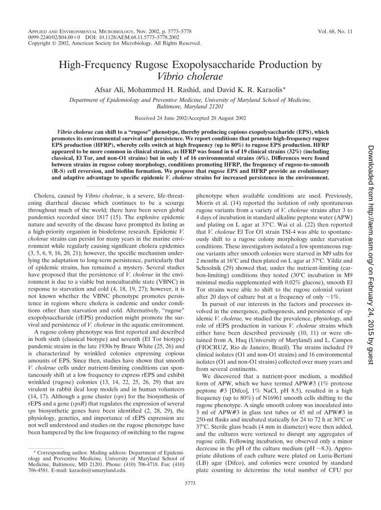
TABLE 1. Frequency of rugose EPS production by V. cholerae strainsa
Strainb Serogroup/biotype Origin Sourcec (year)
% Rugose coloniesd,e,f
Flask Tube
30°C 37°C 30°C 37°C
N16961 O1/E1 Tor Bangladesh C (1971) 31 (1.5) 47 (0.9) 70 (0.7) 74 (1.9)C6709 O1/E1Tor Peru C (1991) 1 23 15 70NCTC 6585 O1/classical India C (1943) 43 (1.4) 44 (0.2) 0 0AMS20A73 O1/classical China C (1945) 3 4 0 0Aldova O37 Czechoslovakia C (1965) 0 1 72 (0.2) 41 (2.8)1803 Non-O1 Brazil C (1992) 0 0 16 871837 O139 Bangladesh C (1992) 0 0.2 0 0 to 2P44 Non-O1 Peru E (2000) 12 0 0 21085-93 O37 Germany E (1993) 0 0 0.1 0141-94 O70 Germany E (1994) 0 0 0.3 0928-93 O6 Argentina E (1993) 0 0.2 0.4 0
a A smooth colony was inoculated into glass tubes containing 3 ml of APW#3 or into flasks containing 45 ml of APW#3, and the cultures were incubated staticallyfor 48 and 72 h at 30°C and 37°C, respectively.
b Listed are only strains showing HFRP or spontaneous rugose colonies under each condition.c C, clinical; E, environmental.d Values represent the average percentages of cells shifting to a rugose colony morphology.e Readings were taken at 72 h for all strains except N16961, for which the reading was taken at 48 h.f Values in parentheses represent standard errors and are shown only for specific strains showing high levels of HFRP.
5774 ALI ET AL. APPL. ENVIRON. MICROBIOL.
on February 24, 2015 by guest
http://aem.asm
.org/D
ownloaded from
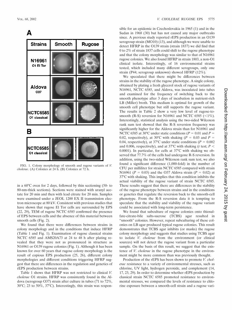
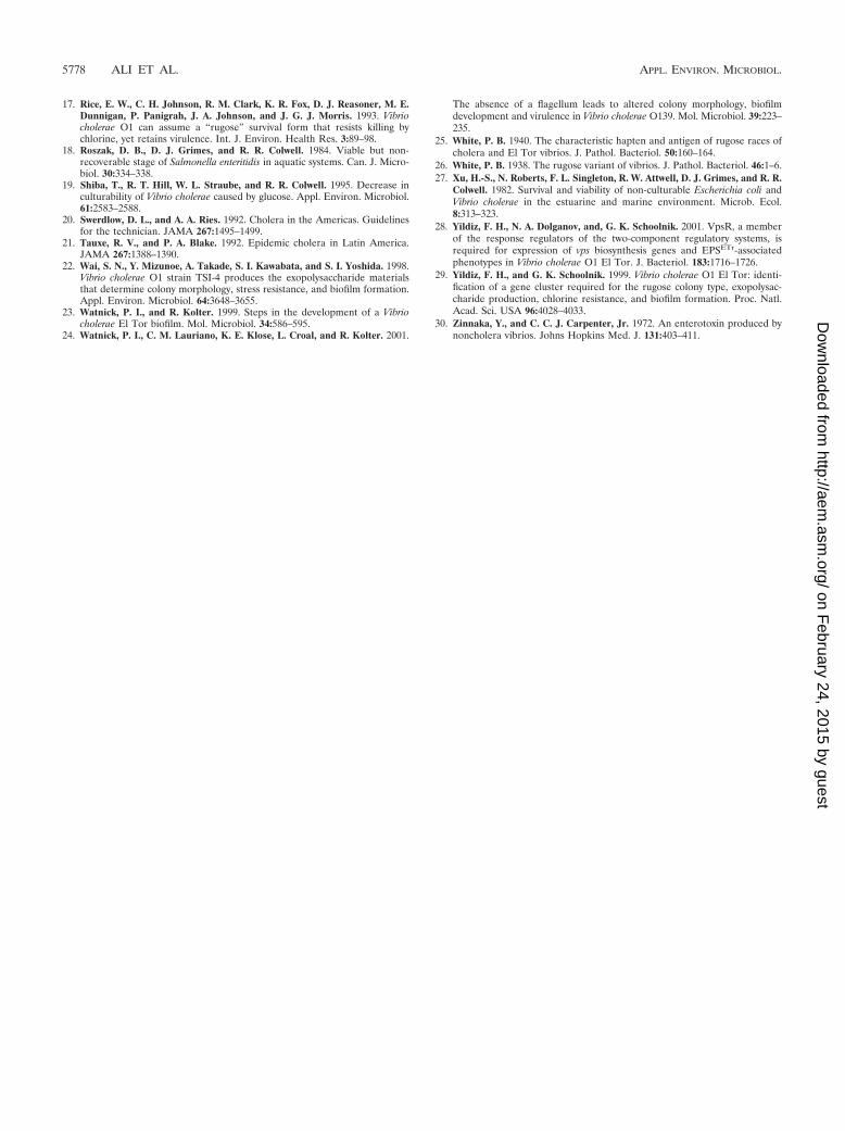
in a 60°C oven for 2 days, followed by thin sectioning (50- to80-nm-thick sections). Sections were stained with uranyl ace-tate for 20 min and then with lead citrate for 20 min. Sampleswere examined under a JEOL 1200 EX II transmission elec-tron microscope at 80 kV. Consistent with previous studies thathave shown that rugose El Tor cells are surrounded by EPS(22, 29), TEM of rugose NCTC 6585 confirmed the presenceof EPS between cells and the absence of this material betweensmooth cells (Fig. 2).
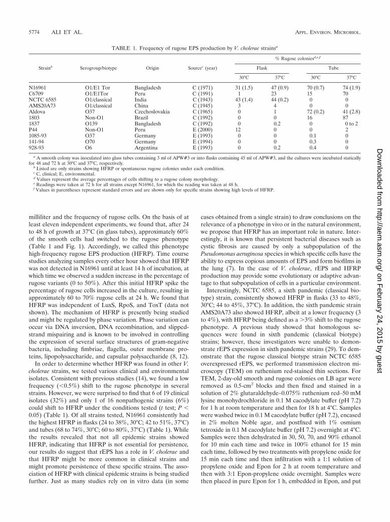
We found that there were differences between strains incolony morphology and in the conditions that induce HFRP(Table 1 and Fig. 1). Examination of rugose classical strainsNCTC 6585 and AMS20A73 at 24 to 48 h after plating re-vealed that they were not as pronounced in structure asN16961 or O139 rugose colonies (Fig. 1). Although it has beenknown for over 60 years that rugose colony morphology is theresult of copious EPS production (25, 26), different colonymorphologies and different conditions triggering HFRP sug-gest that there are differences in the regulation and genetics ofrEPS production between strains.
Table 1 shows that HFRP was not restricted to clinical V.cholerae O1 strains. HFRP was consistently found in the Al-dova (serogroup O37) strain after culture in tubes (71 to 72%,30°C; 23 to 50%, 37°C). Interestingly, this strain was respon-
sible for an epidemic in Czechoslovakia in 1965 (1) and in theSudan in 1968 (30) but has not caused any major outbreakssince. A previous study reported rEPS production in an O139serogroup strain (MO10) (13), and although we were unable todetect HFRP in the O139 strain (strain 1837) we did find that0 to 2% of strain 1837 cells could shift to the rugose phenotypeand that the colony morphology was similar to that of N16961rugose colonies. We also found HFRP in strain 1803, a non-O1clinical isolate. Interestingly, of 16 environmental strainstested, which included many different serogroups, only onestrain (P44; serogroup unknown) showed HFRP (12%).
We speculated that there might be differences betweenstrains in the stability of the rugose phenotype. A single colony,obtained by plating a fresh glycerol stock of rugose variants ofN16961, NCTC 6585, and Aldova, was inoculated into tubesand examined for the frequency of switching back to thesmooth phenotype after 3 days of incubation in nutrient-richLB (Miller) broth. This medium is optimal for growth of thesmooth cell phenotype but still supports the rugose variant.The results in Table 2 show a very low level of rugose-to-smooth (R-S) reversion for N16961 and NCTC 6585 (�1%).Interestingly, statistical analysis using the two-sided Wilcoxonrank sum test showed that the R-S reversion frequency wassignificantly higher for the Aldova strain than for N16961 andNCTC 6585 at 30°C under static conditions (P � 0.01 and P �0.02, respectively), at 30°C with shaking (P � 0.03 and P �0.04, respectively), at 37°C under static conditions (P � 0.002and 0.006, respectively), and at 37°C with shaking (t test; P �0.0001). In particular, for cells at 37°C with shaking we ob-served that 77.7% of the cells had undergone R-S reversion. Inaddition, using the two-sided Wilcoxon rank sum test, we alsofound a significant difference (1,000-fold) in the number ofCFU per milliliter for strain NCTC 6585 compared with strainN16961 (P � 0.03) and the O37 Aldova strain (P � 0.02) at37°C with shaking. This implies that this condition inhibits theactual viability of the rugose variant of strain NCTC 6585.These results suggest that there are differences in the stabilityof the rugose phenotype between strains and in the conditionsor genetics that regulate the reversion back to the smooth-cellphenotype. From the R-S reversion data it is tempting tospeculate that the stability and viability of the rugose variantcould be associated with long-term persistence.
We found that subculture of rugose colonies onto thiosul-fate-citrate-bile salts-sucrose (TCBS) agar resulted in“smooth” colonies. However, repeat subculturing of these col-onies on LB agar produced typical rugose colonies. This resultdemonstrates that TCBS agar inhibits (or masks) the rugosecolony morphology and suggests that studies using TCBS agarto isolate V. cholerae from the environment (or clinicalsources) will not detect the rugose variant from a particularsample. On the basis of this result, we suggest that the exis-tence of V. cholerae in the rugose phenotype in the environ-ment might be more common than was previously thought.
Production of the rEPS has been shown to promote V. chol-erae resistance to a variety of environmental stresses, such aschlorine, UV light, hydrogen peroxide, and complement (14,17, 23, 29). In order to determine whether rEPS production byclassical strain NCTC 6585 promoted resistance to environ-mental stresses, we compared the levels of resistance to chlo-rine exposure between a smooth-cell strain and a rugose vari-
FIG. 1. Colony morphology of smooth and rugose variants of V.cholerae. (A) Colonies at 24 h. (B) Colonies at 72 h.
VOL. 68, 2002 V. CHOLERAE RUGOSE EPS 5775
on February 24, 2015 by guest
http://aem.asm
.org/D
ownloaded from
ant. Chlorine resistance was assayed (four independentexperiments) by using a 1:50 dilution of an overnight culture ofstrain NCTC 6585 smooth and rugose cells in 3 ml of fresh LB(Miller) broth. Cultures were then incubated statically at 37°Cfor 3 h until reaching a density of �2 � 108 CFU/ml; the cellswere then harvested by centrifugation and suspended in phos-phate-buffered saline (pH 7.2) containing 3 mg of free chlorineper liter (sodium hypochlorite; Sigma). After 5 min of expo-sure in 3 mg of chlorine per liter, cultures were serially dilutedand plated on LB agar to determine the number of survivingbacterial cells. Consistent with the findings of rugose El Torstrains (29), we found that the rugose variant of classical strainNCTC 6585 was 10,000-fold more resistant to chlorine (5 minof exposure to 3 mg/liter) than its smooth-cell counterpart.This result demonstrates that the rEPS of classical biotype V.cholerae strains also promotes cell survival and persistence.
Previous studies have reported an association among EPS,motility, and biofilm formation. Traditional motility tests insemisolid media containing 0.3% agar after 4 h at 37°C wereperformed (in triplicate) to compare smooth and rugose vari-ants of N16961. Colony counts of overnight cultures to be usedin motility tests showed no difference between the smooth andrugose variants in the number of CFU per milliliter. Comparedwith the average motility of smooth N16961 (20 mm), we founda 2.5-fold reduction in the motility of the rugose variant (8.5mm). Interestingly, we found that smooth and rugose cellsobtained from fresh colonies were flagellated, as were rugoseplanktonic cells and rugose cells in a biofilm from 48-h LBbroth cultures incubated statically at 37°C (data not shown).These results suggest that V. cholerae cells expressing rEPShave flagella but are less motile than smooth strains under theconditions tested. It might be that the flagellar movement of
FIG. 2. The rugose variant of V. cholerae classical biotype (sixth pandemic) strain NCTC 6585 produces an extracellular glycocalyx. TEM ofruthenium red-stained thin sections of smooth and rugose cells of classical strain NCTC 6585. Micrographs show no stained matrix between smoothcells (left panel) and densely stained matrix between rugose cells (arrow, right panel).
TABLE 2. Rugose stability and frequency of reversion to the smooth cell phenotypea
Strain
% of cells undergoing R-S reversionb at:
30°C 37°C
Static Shaking Static Shaking
N16961 0.8 � 0.2 (5.3 � 108) 0.4 � 0.1 (1.7 � 108) 0.9 � 0.3 (1.7 � 108) 0 (5.8 � 107)NCTC6585 0.9 � 0.2 (4.1 � 108) 0.2 � 0.1 (4.0 � 107) 0.5 � 0.2 (3.9 � 108) 0 (3.4 � 104)Aldova 4.4 � 0.9 (4.6 � 108) 27.5 � 2.3 (1.5 � 108) 27.2 � 2.2 (1.9 � 108) 77.7 � 2.2 (4.1 � 107)
a A single rugose colony was inoculated into LB (Miller) broth and incubated with shaking or statically for three days at 30°C or 37°C. LB was chosen since it providesstable maintenance of rugose and smooth cells (14). Cultures were plated, and colonies were counted to determine the relative numbers of rugose and smooth colonies.Results presented are the mean values of at least four independent experiments.
b The � values represent standard error, and the values in parentheses represent total CFU per milliliter after three days of culture.
5776 ALI ET AL. APPL. ENVIRON. MICROBIOL.
on February 24, 2015 by guest
http://aem.asm
.org/D
ownloaded from
rugose cells is inhibited by copious amounts of EPS or isdefective due to some other mechanism. Although motility isrequired for biofilm formation and motility is impaired in rug-ose variants, this impairment might be offset by the elevatedproduction of the EPS and appears to result in greater biofilm-forming ability than in the smooth variant.
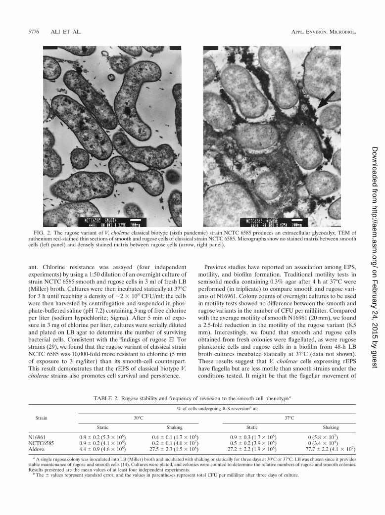
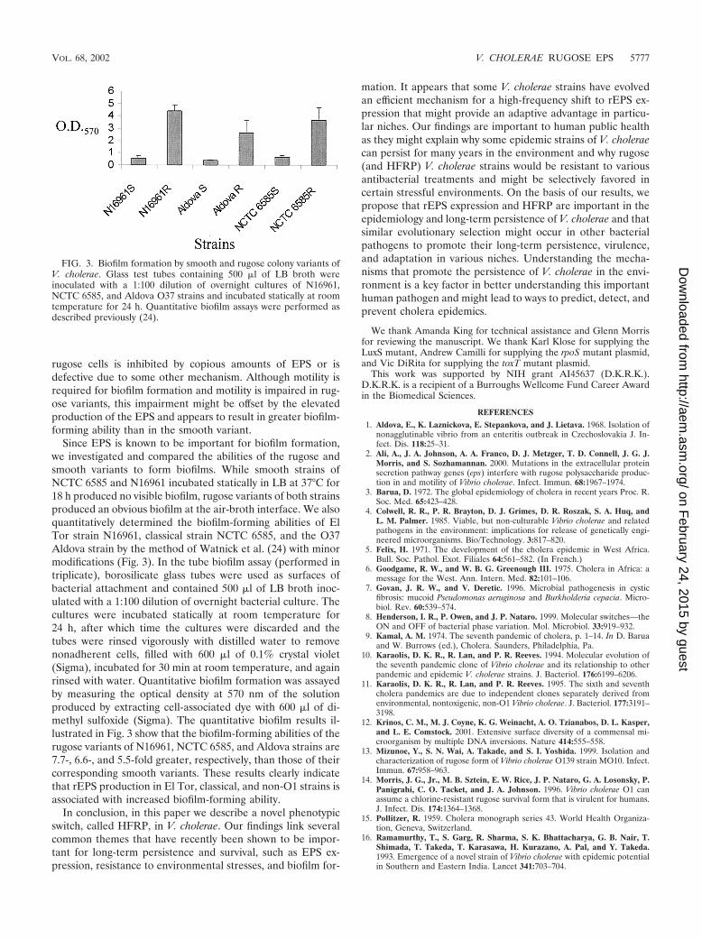
Since EPS is known to be important for biofilm formation,we investigated and compared the abilities of the rugose andsmooth variants to form biofilms. While smooth strains ofNCTC 6585 and N16961 incubated statically in LB at 37°C for18 h produced no visible biofilm, rugose variants of both strainsproduced an obvious biofilm at the air-broth interface. We alsoquantitatively determined the biofilm-forming abilities of ElTor strain N16961, classical strain NCTC 6585, and the O37Aldova strain by the method of Watnick et al. (24) with minormodifications (Fig. 3). In the tube biofilm assay (performed intriplicate), borosilicate glass tubes were used as surfaces ofbacterial attachment and contained 500 �l of LB broth inoc-ulated with a 1:100 dilution of overnight bacterial culture. Thecultures were incubated statically at room temperature for24 h, after which time the cultures were discarded and thetubes were rinsed vigorously with distilled water to removenonadherent cells, filled with 600 �l of 0.1% crystal violet(Sigma), incubated for 30 min at room temperature, and againrinsed with water. Quantitative biofilm formation was assayedby measuring the optical density at 570 nm of the solutionproduced by extracting cell-associated dye with 600 �l of di-methyl sulfoxide (Sigma). The quantitative biofilm results il-lustrated in Fig. 3 show that the biofilm-forming abilities of therugose variants of N16961, NCTC 6585, and Aldova strains are7.7-, 6.6-, and 5.5-fold greater, respectively, than those of theircorresponding smooth variants. These results clearly indicatethat rEPS production in El Tor, classical, and non-O1 strains isassociated with increased biofilm-forming ability.
In conclusion, in this paper we describe a novel phenotypicswitch, called HFRP, in V. cholerae. Our findings link severalcommon themes that have recently been shown to be impor-tant for long-term persistence and survival, such as EPS ex-pression, resistance to environmental stresses, and biofilm for-
mation. It appears that some V. cholerae strains have evolvedan efficient mechanism for a high-frequency shift to rEPS ex-pression that might provide an adaptive advantage in particu-lar niches. Our findings are important to human public healthas they might explain why some epidemic strains of V. choleraecan persist for many years in the environment and why rugose(and HFRP) V. cholerae strains would be resistant to variousantibacterial treatments and might be selectively favored incertain stressful environments. On the basis of our results, wepropose that rEPS expression and HFRP are important in theepidemiology and long-term persistence of V. cholerae and thatsimilar evolutionary selection might occur in other bacterialpathogens to promote their long-term persistence, virulence,and adaptation in various niches. Understanding the mecha-nisms that promote the persistence of V. cholerae in the envi-ronment is a key factor in better understanding this importanthuman pathogen and might lead to ways to predict, detect, andprevent cholera epidemics.
We thank Amanda King for technical assistance and Glenn Morrisfor reviewing the manuscript. We thank Karl Klose for supplying theLuxS mutant, Andrew Camilli for supplying the rpoS mutant plasmid,and Vic DiRita for supplying the toxT mutant plasmid.
This work was supported by NIH grant AI45637 (D.K.R.K.).D.K.R.K. is a recipient of a Burroughs Wellcome Fund Career Awardin the Biomedical Sciences.
REFERENCES
1. Aldova, E., K. Laznickova, E. Stepankova, and J. Lietava. 1968. Isolation ofnonagglutinable vibrio from an enteritis outbreak in Czechoslovakia J. In-fect. Dis. 118:25–31.
2. Ali, A., J. A. Johnson, A. A. Franco, D. J. Metzger, T. D. Connell, J. G. J.Morris, and S. Sozhamannan. 2000. Mutations in the extracellular proteinsecretion pathway genes (eps) interfere with rugose polysaccharide produc-tion in and motility of Vibrio cholerae. Infect. Immun. 68:1967–1974.
3. Barua, D. 1972. The global epidemiology of cholera in recent years Proc. R.Soc. Med. 65:423–428.
4. Colwell, R. R., P. R. Brayton, D. J. Grimes, D. R. Roszak, S. A. Huq, andL. M. Palmer. 1985. Viable, but non-culturable Vibrio cholerae and relatedpathogens in the environment: implications for release of genetically engi-neered microorganisms. Bio/Technology. 3:817–820.
5. Felix, H. 1971. The development of the cholera epidemic in West Africa.Bull. Soc. Pathol. Exot. Filiales 64:561–582. (In French.)
6. Goodgame, R. W., and W. B. G. Greenough III. 1975. Cholera in Africa: amessage for the West. Ann. Intern. Med. 82:101–106.
7. Govan, J. R. W., and V. Deretic. 1996. Microbial pathogenesis in cysticfibrosis: mucoid Pseudomonas aeruginosa and Burkholderia cepacia. Micro-biol. Rev. 60:539–574.
8. Henderson, I. R., P. Owen, and J. P. Nataro. 1999. Molecular switches—theON and OFF of bacterial phase variation. Mol. Microbiol. 33:919–932.
9. Kamal, A. M. 1974. The seventh pandemic of cholera, p. 1–14. In D. Baruaand W. Burrows (ed.), Cholera. Saunders, Philadelphia, Pa.
10. Karaolis, D. K. R., R. Lan, and P. R. Reeves. 1994. Molecular evolution ofthe seventh pandemic clone of Vibrio cholerae and its relationship to otherpandemic and epidemic V. cholerae strains. J. Bacteriol. 176:6199–6206.
11. Karaolis, D. K. R., R. Lan, and P. R. Reeves. 1995. The sixth and seventhcholera pandemics are due to independent clones separately derived fromenvironmental, nontoxigenic, non-O1 Vibrio cholerae. J. Bacteriol. 177:3191–3198.
12. Krinos, C. M., M. J. Coyne, K. G. Weinacht, A. O. Tzianabos, D. L. Kasper,and L. E. Comstock. 2001. Extensive surface diversity of a commensal mi-croorganism by multiple DNA inversions. Nature 414:555–558.
13. Mizunoe, Y., S. N. Wai, A. Takade, and S. I. Yoshida. 1999. Isolation andcharacterization of rugose form of Vibrio cholerae O139 strain MO10. Infect.Immun. 67:958–963.
14. Morris, J. G., Jr., M. B. Sztein, E. W. Rice, J. P. Nataro, G. A. Losonsky, P.Panigrahi, C. O. Tacket, and J. A. Johnson. 1996. Vibrio cholerae O1 canassume a chlorine-resistant rugose survival form that is virulent for humans.J. Infect. Dis. 174:1364–1368.
15. Pollitzer, R. 1959. Cholera monograph series 43. World Health Organiza-tion, Geneva, Switzerland.
16. Ramamurthy, T., S. Garg, R. Sharma, S. K. Bhattacharya, G. B. Nair, T.Shimada, T. Takeda, T. Karasawa, H. Kurazano, A. Pal, and Y. Takeda.1993. Emergence of a novel strain of Vibrio cholerae with epidemic potentialin Southern and Eastern India. Lancet 341:703–704.
FIG. 3. Biofilm formation by smooth and rugose colony variants ofV. cholerae. Glass test tubes containing 500 �l of LB broth wereinoculated with a 1:100 dilution of overnight cultures of N16961,NCTC 6585, and Aldova O37 strains and incubated statically at roomtemperature for 24 h. Quantitative biofilm assays were performed asdescribed previously (24).
VOL. 68, 2002 V. CHOLERAE RUGOSE EPS 5777
on February 24, 2015 by guest
http://aem.asm
.org/D
ownloaded from
17. Rice, E. W., C. H. Johnson, R. M. Clark, K. R. Fox, D. J. Reasoner, M. E.Dunnigan, P. Panigrah, J. A. Johnson, and J. G. J. Morris. 1993. Vibriocholerae O1 can assume a “rugose” survival form that resists killing bychlorine, yet retains virulence. Int. J. Environ. Health Res. 3:89–98.
18. Roszak, D. B., D. J. Grimes, and R. R. Colwell. 1984. Viable but non-recoverable stage of Salmonella enteritidis in aquatic systems. Can. J. Micro-biol. 30:334–338.
19. Shiba, T., R. T. Hill, W. L. Straube, and R. R. Colwell. 1995. Decrease inculturability of Vibrio cholerae caused by glucose. Appl. Environ. Microbiol.61:2583–2588.
20. Swerdlow, D. L., and A. A. Ries. 1992. Cholera in the Americas. Guidelinesfor the technician. JAMA 267:1495–1499.
21. Tauxe, R. V., and P. A. Blake. 1992. Epidemic cholera in Latin America.JAMA 267:1388–1390.
22. Wai, S. N., Y. Mizunoe, A. Takade, S. I. Kawabata, and S. I. Yoshida. 1998.Vibrio cholerae O1 strain TSI-4 produces the exopolysaccharide materialsthat determine colony morphology, stress resistance, and biofilm formation.Appl. Environ. Microbiol. 64:3648–3655.
23. Watnick, P. I., and R. Kolter. 1999. Steps in the development of a Vibriocholerae El Tor biofilm. Mol. Microbiol. 34:586–595.
24. Watnick, P. I., C. M. Lauriano, K. E. Klose, L. Croal, and R. Kolter. 2001.
The absence of a flagellum leads to altered colony morphology, biofilmdevelopment and virulence in Vibrio cholerae O139. Mol. Microbiol. 39:223–235.
25. White, P. B. 1940. The characteristic hapten and antigen of rugose races ofcholera and El Tor vibrios. J. Pathol. Bacteriol. 50:160–164.
26. White, P. B. 1938. The rugose variant of vibrios. J. Pathol. Bacteriol. 46:1–6.27. Xu, H.-S., N. Roberts, F. L. Singleton, R. W. Attwell, D. J. Grimes, and R. R.
Colwell. 1982. Survival and viability of non-culturable Escherichia coli andVibrio cholerae in the estuarine and marine environment. Microb. Ecol.8:313–323.
28. Yildiz, F. H., N. A. Dolganov, and, G. K. Schoolnik. 2001. VpsR, a memberof the response regulators of the two-component regulatory systems, isrequired for expression of vps biosynthesis genes and EPSETr-associatedphenotypes in Vibrio cholerae O1 El Tor. J. Bacteriol. 183:1716–1726.
29. Yildiz, F. H., and G. K. Schoolnik. 1999. Vibrio cholerae O1 El Tor: identi-fication of a gene cluster required for the rugose colony type, exopolysac-charide production, chlorine resistance, and biofilm formation. Proc. Natl.Acad. Sci. USA 96:4028–4033.
30. Zinnaka, Y., and C. C. J. Carpenter, Jr. 1972. An enterotoxin produced bynoncholera vibrios. Johns Hopkins Med. J. 131:403–411.
5778 ALI ET AL. APPL. ENVIRON. MICROBIOL.
on February 24, 2015 by guest
http://aem.asm
.org/D
ownloaded from