High-Frequency Rugose Exopolysaccharide Production by Vibrio cholerae
The Vibrio cholerae Colonization Factor GbpA Possessesa Modular Structure that Governs Binding to DifferentHost SurfacesEdmond Wong1¤a, Gustav Vaaje-Kolstad2, Avishek Ghosh3, Ramon Hurtado-Guerrero1¤b, Peter V.
Konarev4, Adel F. M. Ibrahim1, Dmitri I. Svergun4, Vincent G. H. Eijsink2, Nabendu S. Chatterjee3,
Daan M. F. van Aalten1*
1 Division of Molecular Microbiology, College of Life Sciences, University of Dundee, Dundee, United Kingdom, 2 Department of Chemistry, Biotechnology and Food
Science, Norwegian University of Life Sciences, As, Norway, 3 Division of Biochemistry, National Institute of Cholera and Enteric Diseases, Scheme XM, Beliaghata, Kolkata,
India, 4 European Molecular Biology Laboratory (EMBL), Outstation Hamburg at DESY, Hamburg, Germany
Abstract
Vibrio cholerae is a bacterial pathogen that colonizes the chitinous exoskeleton of zooplankton as well as the humangastrointestinal tract. Colonization of these different niches involves an N-acetylglucosamine binding protein (GbpA) thathas been reported to mediate bacterial attachment to both marine chitin and mammalian intestinal mucin through anunknown molecular mechanism. We report structural studies that reveal that GbpA possesses an unusual, elongated, four-domain structure, with domains 1 and 4 showing structural homology to chitin binding domains. A glycan screen revealedthat GbpA binds to GlcNAc oligosaccharides. Structure-guided GbpA truncation mutants show that domains 1 and 4 ofGbpA interact with chitin in vitro, whereas in vivo complementation studies reveal that domain 1 is also crucial for mucinbinding and intestinal colonization. Bacterial binding studies show that domains 2 and 3 bind to the V. cholerae surface.Finally, mouse virulence assays show that only the first three domains of GbpA are required for colonization. These resultsexplain how GbpA provides structural/functional modular interactions between V. cholerae, intestinal epithelium andchitinous exoskeletons.
Citation: Wong E, Vaaje-Kolstad G, Ghosh A, Hurtado-Guerrero R, Konarev PV, et al. (2012) The Vibrio cholerae Colonization Factor GbpA Possesses a ModularStructure that Governs Binding to Different Host Surfaces. PLoS Pathog 8(1): e1002373. doi:10.1371/journal.ppat.1002373
Editor: Partho Ghosh, University of California San Diego, United States of America
Received August 26, 2011; Accepted September 27, 2011; Published January 12, 2012
Copyright: � 2012 Wong et al. This is an open-access article distributed under the terms of the Creative Commons Attribution License, which permitsunrestricted use, distribution, and reproduction in any medium, provided the original author and source are credited.
Funding: This work was funded by a Wellcome Trust Senior Research Fellowship to DvA (WT087590MA) and an MRC Programme Grant (G0900138) to DvA. Thefunders had no role in study design, data collection and analysis, decision to publish, or preparation of the manuscript.
Competing Interests: The authors have declared that no competing interests exist.
* E-mail: [email protected]
¤a Current address: Division of Molecular Structure, The National Institute for Medical Research, Mill Hill, London, United Kingdom¤b Current address: Institute for Biocomputation and Physics of Complex Systems, University of Zaragoza, Zaragoza, Spain
Introduction
Vibrio cholerae is a Gram-negative bacterial pathogen that causes
excessive watery diarrhea in humans [1,2]. The number of
reported cases of cholera worldwide averages over 100000 per
annum for the last 10 years [1,3], and is presumed to be exceeded
by the number of unreported cases [3,4]. Most of these cases occur
in countries with poor sanitation [5,6]. V. cholerae strains are
classified into more than 200 serogroups, with only the serogroups
O1 and O139 possessing epidemic potential. It has been shown
that the survival of Vibrio cholerae in the intestine is dependent on its
ability to adhere to and colonize cell surfaces [7,8]. In aquatic
environments, attachment to fish, crustacea and algae enables the
bacteria to obtain nutrients, thereby provide a competitive
advantage compared to other free-swimming bacteria [8,9,10].
Adherence to aquatic organisms is believed to involve a different
set of genes and recognition molecules as compared to those used
for intestinal colonization. For example, plankton surface
colonization by the bacteria is more dependent on the mannose
sensitive hemagglutinin (MSHA) [11,12].
Recent studies have suggested that Vibrio cholerae secretes a
protein that mediates adhesion in aquatic environments, e.g. to
plankton, as well as adhesion to human intestinal cells [7,10]. This
protein, GlcNAc binding protein A (GbpA) binds to N-acetylglu-
cosamine (GlcNAc)- containing carbohydrates, such as chitin, and
is secreted by the type 2 secretion system [7]. In addition to chitin,
GbpA has been shown to bind to mucins [13] that also contain
GlcNAc as part of their densely packed network of O-linked
glycans (reviewed in [14,15]). The importance of GbpA for
bacterial colonization has been demonstrated for the O395 [7]
and N16961 Vibrio cholerae strains [16] (representing classical [17]
and El Tor [18] biotypes, respectively).
Here, we have studied the molecular basis of the function of
GbpA as a colonization factor, using a combination of approaches.
Firstly, we determined the three-dimensional structure of GbpA
using a combination of X-ray crystallography and small angle X-
ray scattering, revealing an unusual, elongated four-domain fold.
Two domains appear to be structurally similar to known chitin
binding modules, and we show these domains to be responsible for
the ability of GbpA to bind chitin. The other two domains possess
PLoS Pathogens | www.plospathogens.org 1 January 2012 | Volume 8 | Issue 1 | e1002373
distant structural homology to bacterial pili binding proteins and
serve to bind the V. cholerae surface. Finally, complementation
studies with truncated forms of GbpA identify the domains
responsible for mucin binding and virulence in a V. cholerae mouse
infection model.
Results
GbpA is a multi-domain proteinFull-length GbpA and a range of truncated forms of GbpA (see
Supplemental Table S1 in Text S1 for a summary of all constructs
used in this study) were cloned and expressed in E. coli. The
mature form of the protein (residues 24 to 485, GbpAfl) was
extracted from the periplasm and purified by chromatography.
Two C-terminally truncated forms of mature GbpA, comprising
amino acids 24–203 (GbpAD1), and 24–414 (GbpAD1–3), were also
produced. Two N-terminally truncated forms of GbpA comprising
amino acids 210–414 (GbpAD2–3), and 423–485 (GbpAD4) were
expressed as GST fusion proteins and purified to homogeneity.
Although extensive efforts were made to crystallise full-length
GbpA, GbpAD1–3 was the longest construct that could be
crystallised. The GbpAD1–3 structure was solved by SAD phasing,
and refined against 1.8 A synchrotron diffraction data (Supple-
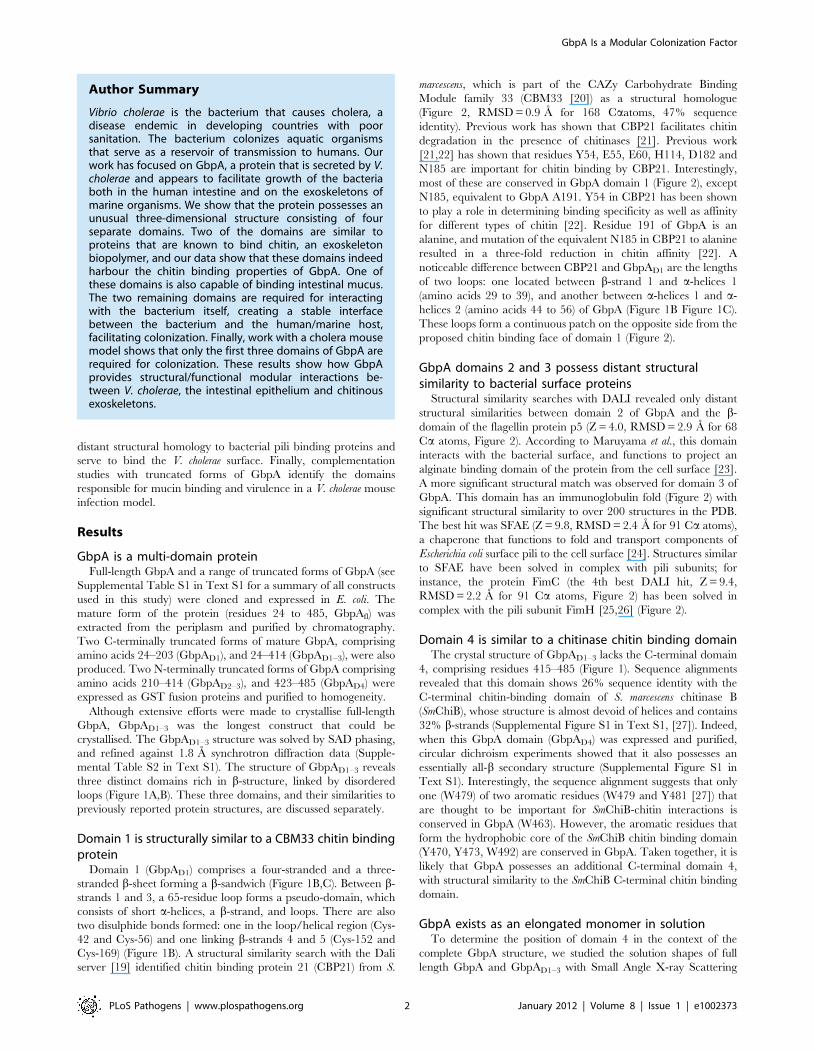
mental Table S2 in Text S1). The structure of GbpAD1–3 reveals
three distinct domains rich in b-structure, linked by disordered
loops (Figure 1A,B). These three domains, and their similarities to
previously reported protein structures, are discussed separately.
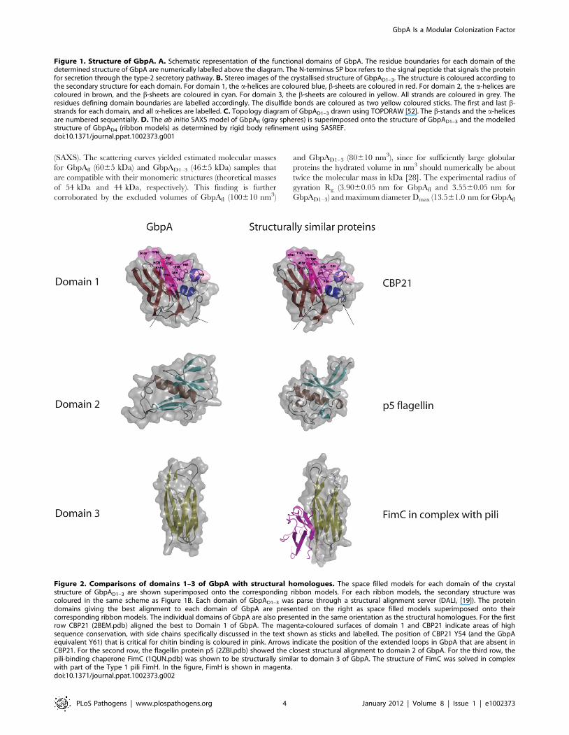
Domain 1 is structurally similar to a CBM33 chitin bindingprotein
Domain 1 (GbpAD1) comprises a four-stranded and a three-
stranded b-sheet forming a b-sandwich (Figure 1B,C). Between b-
strands 1 and 3, a 65-residue loop forms a pseudo-domain, which
consists of short a-helices, a b-strand, and loops. There are also
two disulphide bonds formed: one in the loop/helical region (Cys-
42 and Cys-56) and one linking b-strands 4 and 5 (Cys-152 and
Cys-169) (Figure 1B). A structural similarity search with the Dali
server [19] identified chitin binding protein 21 (CBP21) from S.
marcescens, which is part of the CAZy Carbohydrate Binding
Module family 33 (CBM33 [20]) as a structural homologue
(Figure 2, RMSD = 0.9 A for 168 Caatoms, 47% sequence
identity). Previous work has shown that CBP21 facilitates chitin
degradation in the presence of chitinases [21]. Previous work
[21,22] has shown that residues Y54, E55, E60, H114, D182 and
N185 are important for chitin binding by CBP21. Interestingly,
most of these are conserved in GbpA domain 1 (Figure 2), except
N185, equivalent to GbpA A191. Y54 in CBP21 has been shown
to play a role in determining binding specificity as well as affinity
for different types of chitin [22]. Residue 191 of GbpA is an
alanine, and mutation of the equivalent N185 in CBP21 to alanine
resulted in a three-fold reduction in chitin affinity [22]. A
noticeable difference between CBP21 and GbpAD1 are the lengths
of two loops: one located between b-strand 1 and a-helices 1
(amino acids 29 to 39), and another between a-helices 1 and a-
helices 2 (amino acids 44 to 56) of GbpA (Figure 1B Figure 1C).
These loops form a continuous patch on the opposite side from the
proposed chitin binding face of domain 1 (Figure 2).
GbpA domains 2 and 3 possess distant structuralsimilarity to bacterial surface proteins
Structural similarity searches with DALI revealed only distant
structural similarities between domain 2 of GbpA and the b-
domain of the flagellin protein p5 (Z = 4.0, RMSD = 2.9 A for 68
Ca atoms, Figure 2). According to Maruyama et al., this domain
interacts with the bacterial surface, and functions to project an
alginate binding domain of the protein from the cell surface [23].
A more significant structural match was observed for domain 3 of
GbpA. This domain has an immunoglobulin fold (Figure 2) with
significant structural similarity to over 200 structures in the PDB.
The best hit was SFAE (Z = 9.8, RMSD = 2.4 A for 91 Ca atoms),
a chaperone that functions to fold and transport components of
Escherichia coli surface pili to the cell surface [24]. Structures similar
to SFAE have been solved in complex with pili subunits; for
instance, the protein FimC (the 4th best DALI hit, Z = 9.4,
RMSD = 2.2 A for 91 Ca atoms, Figure 2) has been solved in
complex with the pili subunit FimH [25,26] (Figure 2).
Domain 4 is similar to a chitinase chitin binding domainThe crystal structure of GbpAD1–3 lacks the C-terminal domain
4, comprising residues 415–485 (Figure 1). Sequence alignments
revealed that this domain shows 26% sequence identity with the
C-terminal chitin-binding domain of S. marcescens chitinase B
(SmChiB), whose structure is almost devoid of helices and contains
32% b-strands (Supplemental Figure S1 in Text S1, [27]). Indeed,
when this GbpA domain (GbpAD4) was expressed and purified,
circular dichroism experiments showed that it also possesses an
essentially all-b secondary structure (Supplemental Figure S1 in
Text S1). Interestingly, the sequence alignment suggests that only
one (W479) of two aromatic residues (W479 and Y481 [27]) that
are thought to be important for SmChiB-chitin interactions is
conserved in GbpA (W463). However, the aromatic residues that
form the hydrophobic core of the SmChiB chitin binding domain
(Y470, Y473, W492) are conserved in GbpA. Taken together, it is
likely that GbpA possesses an additional C-terminal domain 4,
with structural similarity to the SmChiB C-terminal chitin binding
domain.
GbpA exists as an elongated monomer in solutionTo determine the position of domain 4 in the context of the
complete GbpA structure, we studied the solution shapes of full
length GbpA and GbpAD1–3 with Small Angle X-ray Scattering
Author Summary
Vibrio cholerae is the bacterium that causes cholera, adisease endemic in developing countries with poorsanitation. The bacterium colonizes aquatic organismsthat serve as a reservoir of transmission to humans. Ourwork has focused on GbpA, a protein that is secreted by V.cholerae and appears to facilitate growth of the bacteriaboth in the human intestine and on the exoskeletons ofmarine organisms. We show that the protein possesses anunusual three-dimensional structure consisting of fourseparate domains. Two of the domains are similar toproteins that are known to bind chitin, an exoskeletonbiopolymer, and our data show that these domains indeedharbour the chitin binding properties of GbpA. One ofthese domains is also capable of binding intestinal mucus.The two remaining domains are required for interactingwith the bacterium itself, creating a stable interfacebetween the bacterium and the human/marine host,facilitating colonization. Finally, work with a cholera mousemodel shows that only the first three domains of GbpA arerequired for colonization. These results show how GbpAprovides structural/functional modular interactions be-tween V. cholerae, the intestinal epithelium and chitinousexoskeletons.
GbpA Is a Modular Colonization Factor
PLoS Pathogens | www.plospathogens.org 2 January 2012 | Volume 8 | Issue 1 | e1002373
GbpA Is a Modular Colonization Factor
PLoS Pathogens | www.plospathogens.org 3 January 2012 | Volume 8 | Issue 1 | e1002373
(SAXS). The scattering curves yielded estimated molecular masses
for GbpAfl (6065 kDa) and GbpAD1–3 (4665 kDa) samples that
are compatible with their monomeric structures (theoretical masses
of 54 kDa and 44 kDa, respectively). This finding is further
corroborated by the excluded volumes of GbpAfl (100610 nm3)
and GbpAD1–3 (80610 nm3), since for sufficiently large globular
proteins the hydrated volume in nm3 should numerically be about
twice the molecular mass in kDa [28]. The experimental radius of
gyration Rg (3.9060.05 nm for GbpAfl and 3.5560.05 nm for
GbpAD1–3) and maximum diameter Dmax (13.561.0 nm for GbpAfl
Figure 2. Comparisons of domains 1–3 of GbpA with structural homologues. The space filled models for each domain of the crystalstructure of GbpAD1–3 are shown superimposed onto the corresponding ribbon models. For each ribbon models, the secondary structure wascoloured in the same scheme as Figure 1B. Each domain of GbpAD1–3 was parse through a structural alignment server (DALI, [19]). The proteindomains giving the best alignment to each domain of GbpA are presented on the right as space filled models superimposed onto theircorresponding ribbon models. The individual domains of GbpA are also presented in the same orientation as the structural homologues. For the firstrow CBP21 (2BEM.pdb) aligned the best to Domain 1 of GbpA. The magenta-coloured surfaces of domain 1 and CBP21 indicate areas of highsequence conservation, with side chains specifically discussed in the text shown as sticks and labelled. The position of CBP21 Y54 (and the GbpAequivalent Y61) that is critical for chitin binding is coloured in pink. Arrows indicate the position of the extended loops in GbpA that are absent inCBP21. For the second row, the flagellin protein p5 (2ZBI.pdb) showed the closest structural alignment to domain 2 of GbpA. For the third row, thepili-binding chaperone FimC (1QUN.pdb) was shown to be structurally similar to domain 3 of GbpA. The structure of FimC was solved in complexwith part of the Type 1 pili FimH. In the figure, FimH is shown in magenta.doi:10.1371/journal.ppat.1002373.g002
Figure 1. Structure of GbpA. A. Schematic representation of the functional domains of GbpA. The residue boundaries for each domain of thedetermined structure of GbpA are numerically labelled above the diagram. The N-terminus SP box refers to the signal peptide that signals the proteinfor secretion through the type-2 secretory pathway. B. Stereo images of the crystallised structure of GbpAD1–3. The structure is coloured according tothe secondary structure for each domain. For domain 1, the a-helices are coloured blue, b-sheets are coloured in red. For domain 2, the a-helices arecoloured in brown, and the b-sheets are coloured in cyan. For domain 3, the b-sheets are coloured in yellow. All strands are coloured in grey. Theresidues defining domain boundaries are labelled accordingly. The disulfide bonds are coloured as two yellow coloured sticks. The first and last b-strands for each domain, and all a-helices are labelled. C. Topology diagram of GbpAD1–3 drawn using TOPDRAW [52]. The b-stands and the a-helicesare numbered sequentially. D. The ab initio SAXS model of GbpAfl (gray spheres) is superimposed onto the structure of GbpAD1–3 and the modelledstructure of GbpAD4 (ribbon models) as determined by rigid body refinement using SASREF.doi:10.1371/journal.ppat.1002373.g001
GbpA Is a Modular Colonization Factor
PLoS Pathogens | www.plospathogens.org 4 January 2012 | Volume 8 | Issue 1 | e1002373

and 11.561.0 nm for GbpAD1–3) ) indicate that the proteins behave
as extended particles. The solution shapes of GbpAfl and GbpAD1–3
were reconstructed ab initio using the program DAMMIF [29], with
good discrepancy factors (x= 1.16 for GbpAfl and x= 1.22 for
GbpAD1–3). For each protein, ten independent reconstructions
produced similar shapes and these were averaged using DAMA-
VER [30]; the ab initio structure is represented in Figure 1D as a
wired mesh. To determine the relative orientations of the domains,
a model of GbpAfl was generated by rigid-body refinement with the
separate GbpA domain structures taken from the domain 1–3
crystal structure (Figure 1B), and the model of domain 4
(Supplemental Figure S1 in Text S1), fitting simultaneously to the
experimental SAXS data of GbpAD1–3 with x= 1.41 and of GbpAfl
with x= 1.53. GbpAfl and GbpAD1–3 adopt a rod shape with the
domains twisted along the long axis (Figure 1D). There are no direct
inter-domain interactions, and the surface of each domain appears
to be completely exposed to the external environment, suggesting a
certain degree of flexibility in the orientation of each domain of
GbpA.
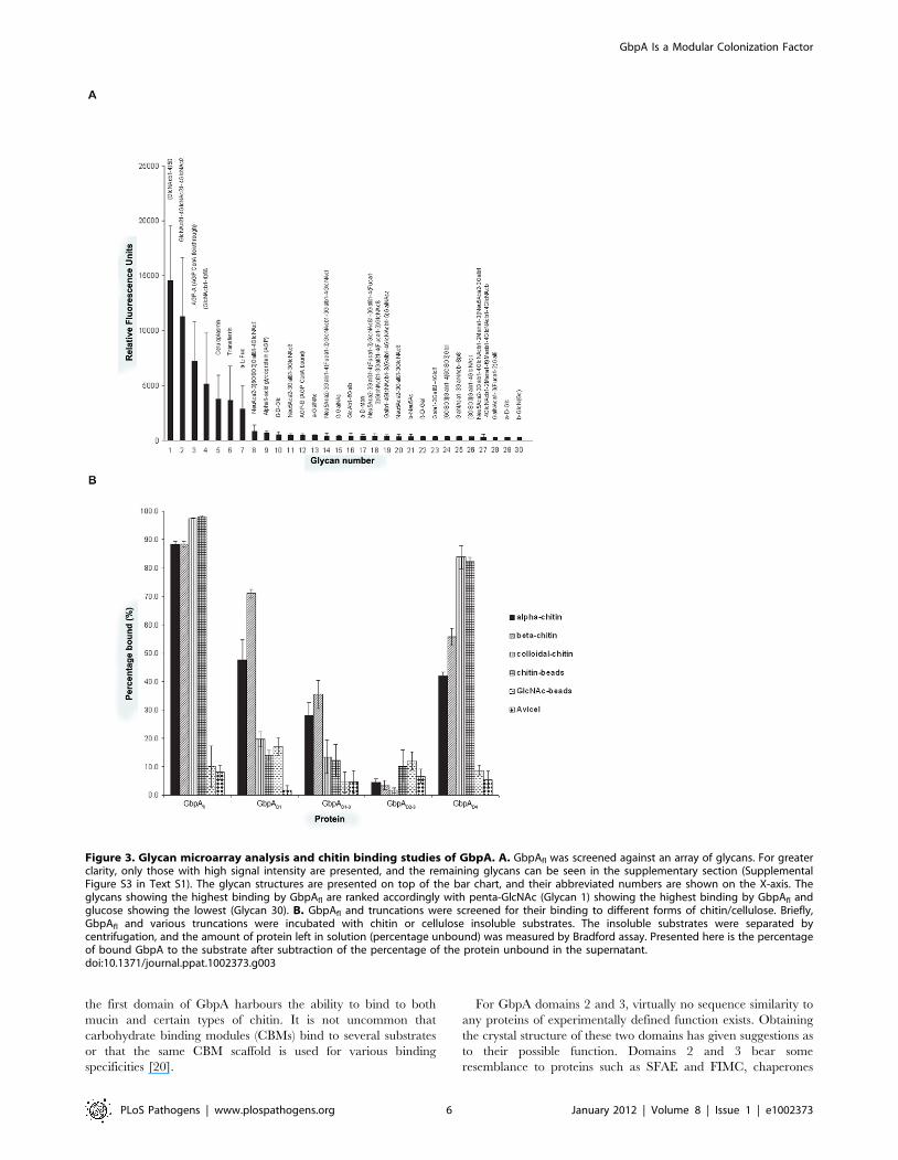
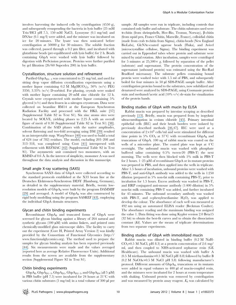
GbpA selectively binds chito-oligosaccharides throughdomains 1 and 4
Previous work has suggested that GbpA binds N-acetylglucosa-
mine (GlcNAc) sugars [7]. To investigate whether this extends to
GlcNAc-containing glycans, we studied the binding specificity of
GbpA by screening a well-established library of mammalian N/O-
linked glycans that also includes a range of linear oligosaccharides
[31,32]. Interestingly, GbpAfl selectively binds chito-oligosaccha-
rides of varying length (Figs. 3A, S5) To quantify chitin binding
and identify the domains responsible, direct binding assays were
carried out with a range of polysaccharides (Figure 3B). GbpAD4
binds to all chitin forms tested with the highest affinity for
amorphous forms of chitin (colloidal chitin and chitin beads).
GbpAD1 shows significant binding only to a- and b-chitin
(Figure 3B). GbpAD2–3 does not bind chitin. For GbpAfl strong
binding to all forms of chitin was observed. None of the GbpA
proteins showed binding to GlcNAc-beads or cellulose. Although
this is in apparent contrast with a previously published report
showing that GbpA-expressing V. cholerae bind to GlcNAc beads
[7], it could also suggest that additional bacterial factors may be
required for the reported GlcNAc binding. Together, these data
show that the two terminal chitin-binding domains in GbpA
endow the protein with the ability to bind different types of
GlcNAc oligomers and polymers.
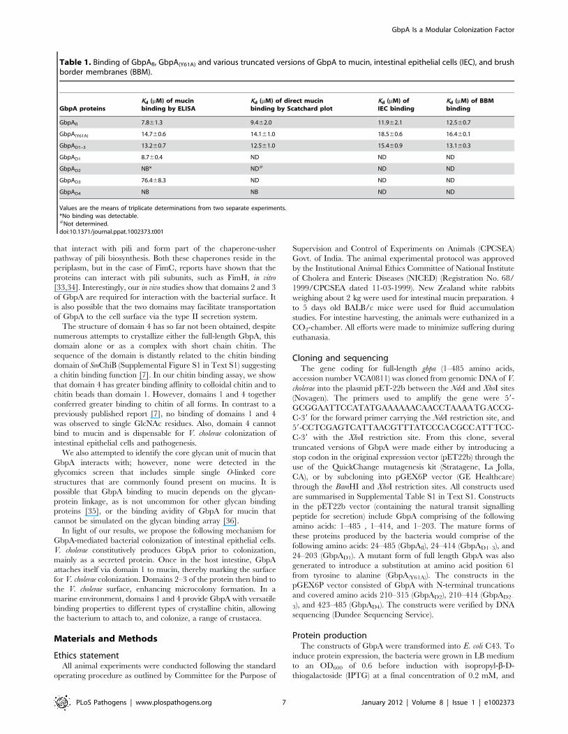
Domain 1 of GbpA is required for mucin bindingV. cholerae GbpA has been shown to interact with intestinal
mucin [13]. To identify the domain(s) involved, GbpAfl and its
truncated variants were tested for their ability to bind to mucin,
mucin coupled to sepharose beads, intestinal epithelial cells and
brush border membranes of the intestine (Table 1). GbpA with a
Tyr61Ala (GbpA(Y61A)) mutation was also tested to assess whether
Tyr61 is important for mucin binding as has been shown for chitin
binding in the case of the equivalent Tyr54 in CBP21.
Interestingly, only GbpAfl, GbpA(Y61A), and GbpAD1–3 bind to
mucin with mM affinity, further confirmed by ELISA for those
domains that tested positive in the initial mucin-binding test. Thus,
domain 1 is essential for mucin binding and domain 4 is
dispensable for mucin binding. Tyr61 on domain 1 of GbpA is
not essential for mucin binding.
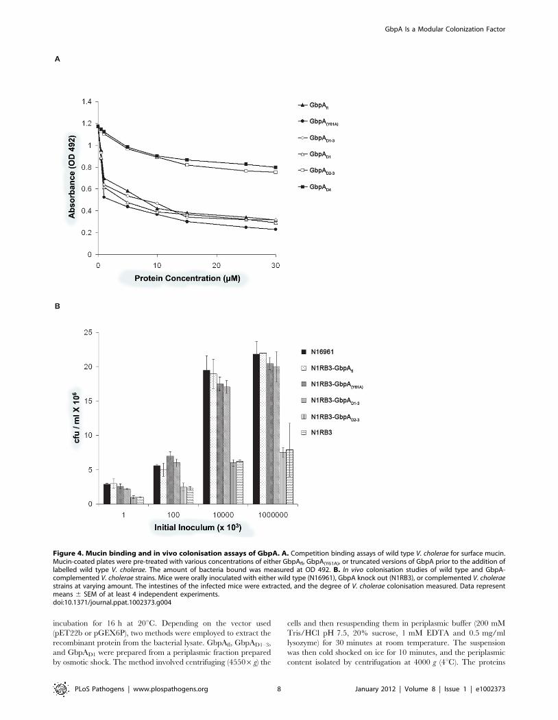
To further investigate mucin binding to domain 1, competitive
mucin binding assays were carried out with recombinant truncated
versions of GbpA and wild type V. cholerae (N16961) (Figure 4A).
Increasing concentrations of purified GbpA variants added to
mucin-coated wells showed varying abilities of the proteins to
prevent bacteria from binding the immobilized mucin. GbpAfl,
GbpA (Y61A), GbpAD1 and GbpAD1–3 could inhibit binding of
N16961 to mucin in a concentration-dependent, saturating
manner (Figure 4A). At saturation, GbpAfl and the GbpA(Y61A)
mutant inhibited binding of V. cholerae to mucin by 83%.
Truncated GbpA variants comprising only domains 2, 3, and/or
4 could not inhibit the binding of V. cholerae to mucin, in agreement
with the direct binding assays.
GbpA domains 1–3 are essential for intestinalcolonization and pathogenesis
In order to understand which domains of GbpA are essential for
colonization, we first studied the surface expression of these
domains in complemented GbpA knockout V. cholerae strain
N16961 (N1RB3 or DgbpA). Immunoblot analysis revealed that the
DgbpA strain complemented with GbpAfl, GbpA(Y61A), or
GbpAD1–3 displays these proteins on the bacterial surface
(Supplemental Figure S2 in Text S1). For the strains comple-
mented with GbpAD1, GbpAD2, GbpAD2–3, or GbpAD4, no
protein could be detected on the bacterial cell surface (Supple-
mental Figure S2 in Text S1). These results suggest that the
presentation of domain 1 on the cell surface is dependent on the
presence of domains 2 and 3.
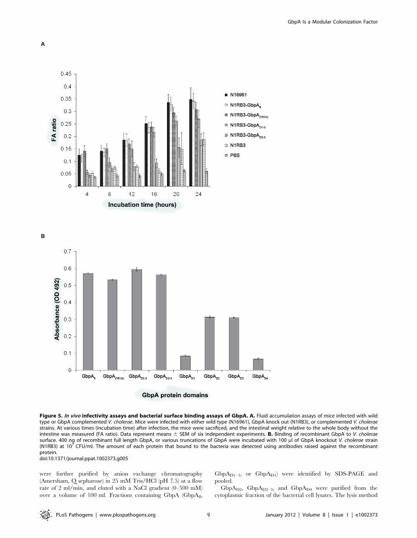
To probe the role of domains 1–3 in intestinal colonization, the
pathogenicity of the complemented strains was analyzed by
studying intestinal fluid accumulation and colonization in a mouse
model (Figure 4B and Figure 5A). All complemented strains were
similar to the wild type strain, with the exception of the GbpAD2–3
complemented strain (N1RB3-GbpAD2–3), which was affected in
its ability to colonize the mice, similar to the DgbpA N16961
control (N1RB3).
Domains 2 and 3 of GbpA interact with V. choleraesurface
As shown earlier, GbpA domains 1 and 4 are mainly responsible
for chitin, mucin and intestinal epithelium binding. However,
GbpA is a secreted protein, and some of the domains must
therefore be capable of interacting with V. cholerae surface to form a
stable host-pathogen interface. To test this, we investigated
binding of recombinant full length, mutant and truncated versions
of GbpA to the DgbpA strain (Figure 5B). The results suggest that
domains 2 and 3, but not domains 1 or 4, are required for binding
to the bacterial surface.
Discussion
Since the discovery of GbpA, little progress has been made
towards an understanding of the molecular properties governing
the protein’s ability to mediate interactions between the bacteria
and the host surfaces, both in the marine environment and the
mammalian host. While it has been reported that the protein binds
to chitin and intestinal epithelia, the chitin specificity was not
probed, nor was it understood how the protein interacts with
intestinal epithelial cells. Here we have reported the crystal and
solution structures of GbpA, showing an unusual 4-domain
elongated structure. Remarkably, the modular domain structure
translates to modular interaction properties with different
substrates/surfaces. For instance, the first domain has a pseudo-
fibronectin type 3 fold that shares considerable similarity with the
chitin-binding protein CBP21. Our chitin binding studies showed
that this domain, like CBP21, binds to a/b chitin. Furthermore,
we were able to demonstrate the interaction of the domain 1 with
mucin, a key surface component of intestinal epithelial cells. Thus,
GbpA Is a Modular Colonization Factor
PLoS Pathogens | www.plospathogens.org 5 January 2012 | Volume 8 | Issue 1 | e1002373
the first domain of GbpA harbours the ability to bind to both
mucin and certain types of chitin. It is not uncommon that
carbohydrate binding modules (CBMs) bind to several substrates
or that the same CBM scaffold is used for various binding
specificities [20].
For GbpA domains 2 and 3, virtually no sequence similarity to
any proteins of experimentally defined function exists. Obtaining
the crystal structure of these two domains has given suggestions as
to their possible function. Domains 2 and 3 bear some
resemblance to proteins such as SFAE and FIMC, chaperones
Figure 3. Glycan microarray analysis and chitin binding studies of GbpA. A. GbpAfl was screened against an array of glycans. For greaterclarity, only those with high signal intensity are presented, and the remaining glycans can be seen in the supplementary section (SupplementalFigure S3 in Text S1). The glycan structures are presented on top of the bar chart, and their abbreviated numbers are shown on the X-axis. Theglycans showing the highest binding by GbpAfl are ranked accordingly with penta-GlcNAc (Glycan 1) showing the highest binding by GbpAfl andglucose showing the lowest (Glycan 30). B. GbpAfl and truncations were screened for their binding to different forms of chitin/cellulose. Briefly,GbpAfl and various truncations were incubated with chitin or cellulose insoluble substrates. The insoluble substrates were separated bycentrifugation, and the amount of protein left in solution (percentage unbound) was measured by Bradford assay. Presented here is the percentageof bound GbpA to the substrate after subtraction of the percentage of the protein unbound in the supernatant.doi:10.1371/journal.ppat.1002373.g003
GbpA Is a Modular Colonization Factor
PLoS Pathogens | www.plospathogens.org 6 January 2012 | Volume 8 | Issue 1 | e1002373
that interact with pili and form part of the chaperone-usher
pathway of pili biosynthesis. Both these chaperones reside in the
periplasm, but in the case of FimC, reports have shown that the
proteins can interact with pili subunits, such as FimH, in vitro
[33,34]. Interestingly, our in vivo studies show that domains 2 and 3
of GbpA are required for interaction with the bacterial surface. It
is also possible that the two domains may facilitate transportation
of GbpA to the cell surface via the type II secretion system.
The structure of domain 4 has so far not been obtained, despite
numerous attempts to crystallize either the full-length GbpA, this
domain alone or as a complex with short chain chitin. The
sequence of the domain is distantly related to the chitin binding
domain of SmChiB (Supplemental Figure S1 in Text S1) suggesting
a chitin binding function [7]. In our chitin binding assay, we show
that domain 4 has greater binding affinity to colloidal chitin and to
chitin beads than domain 1. However, domains 1 and 4 together
conferred greater binding to chitin of all forms. In contrast to a
previously published report [7], no binding of domains 1 and 4
was observed to single GlcNAc residues. Also, domain 4 cannot
bind to mucin and is dispensable for V. cholerae colonization of
intestinal epithelial cells and pathogenesis.
We also attempted to identify the core glycan unit of mucin that
GbpA interacts with; however, none were detected in the
glycomics screen that includes simple single O-linked core
structures that are commonly found present on mucins. It is
possible that GbpA binding to mucin depends on the glycan-
protein linkage, as is not uncommon for other glycan binding
proteins [35], or the binding avidity of GbpA for mucin that
cannot be simulated on the glycan binding array [36].
In light of our results, we propose the following mechanism for
GbpA-mediated bacterial colonization of intestinal epithelial cells.
V. cholerae constitutively produces GbpA prior to colonization,
mainly as a secreted protein. Once in the host intestine, GbpA
attaches itself via domain 1 to mucin, thereby marking the surface
for V. cholerae colonization. Domains 2–3 of the protein then bind to
the V. cholerae surface, enhancing microcolony formation. In a
marine environment, domains 1 and 4 provide GbpA with versatile
binding properties to different types of crystalline chitin, allowing
the bacterium to attach to, and colonize, a range of crustacea.
Materials and Methods
Ethics statementAll animal experiments were conducted following the standard
operating procedure as outlined by Committee for the Purpose of
Supervision and Control of Experiments on Animals (CPCSEA)
Govt. of India. The animal experimental protocol was approved
by the Institutional Animal Ethics Committee of National Institute
of Cholera and Enteric Diseases (NICED) (Registration No. 68/
1999/CPCSEA dated 11-03-1999). New Zealand white rabbits
weighing about 2 kg were used for intestinal mucin preparation. 4
to 5 days old BALB/c mice were used for fluid accumulation
studies. For intestine harvesting, the animals were euthanized in a
CO2-chamber. All efforts were made to minimize suffering during
euthanasia.
Cloning and sequencingThe gene coding for full-length gbpa (1–485 amino acids,
accession number VCA0811) was cloned from genomic DNA of V.
cholerae into the plasmid pET-22b between the NdeI and XhoI sites
(Novagen). The primers used to amplify the gene were 59-
GCGGAATTCCATATGAAAAAACAACCTAAAATGACCG-
C-39 for the forward primer carrying the NdeI restriction site, and
59-CCTCGAGTCATTAACGTTTATCCCACGCCATTTCC-
C-39 with the XhoI restriction site. From this clone, several
truncated versions of GbpA were made either by introducing a
stop codon in the original expression vector (pET22b) through the
use of the QuickChange mutagenesis kit (Stratagene, La Jolla,
CA), or by subcloning into pGEX6P vector (GE Healthcare)
through the BamHI and XhoI restriction sites. All constructs used
are summarised in Supplemental Table S1 in Text S1. Constructs
in the pET22b vector (containing the natural transit signalling
peptide for secretion) include GbpA comprising of the following
amino acids: 1–485 , 1–414, and 1–203. The mature forms of
these proteins produced by the bacteria would comprise of the
following amino acids: 24–485 (GbpAfl), 24–414 (GbpAD1–3), and
24–203 (GbpAD1). A mutant form of full length GbpA was also
generated to introduce a substitution at amino acid position 61
from tyrosine to alanine (GbpA(Y61A)). The constructs in the
pGEX6P vector consisted of GbpA with N-terminal truncations
and covered amino acids 210–315 (GbpAD2), 210–414 (GbpAD2–
3), and 423–485 (GbpAD4). The constructs were verified by DNA
sequencing (Dundee Sequencing Service).
Protein productionThe constructs of GbpA were transformed into E. coli C43. To
induce protein expression, the bacteria were grown in LB medium
to an OD600 of 0.6 before induction with isopropyl-b-D-
thiogalactoside (IPTG) at a final concentration of 0.2 mM, and
Table 1. Binding of GbpAfl, GbpA(Y61A) and various truncated versions of GbpA to mucin, intestinal epithelial cells (IEC), and brushborder membranes (BBM).
GbpA proteinsKd (mM) of mucinbinding by ELISA
Kd (mM) of direct mucinbinding by Scatchard plot
Kd (mM) ofIEC binding
Kd (mM) of BBMbinding
GbpAfl 7.861.3 9.462.0 11.962.1 12.560.7
GbpA(Y61A) 14.760.6 14.161.0 18.560.6 16.460.1
GbpAD1–3 13.260.7 12.561.0 15.460.9 13.160.3
GbpAD1 8.760.4 ND ND ND
GbpAD2 NB* ND# ND ND
GbpAD3 76.468.3 ND ND ND
GbpAD4 NB NB ND ND
Values are the means of triplicate determinations from two separate experiments.*No binding was detectable.#Not determined.doi:10.1371/journal.ppat.1002373.t001
GbpA Is a Modular Colonization Factor
PLoS Pathogens | www.plospathogens.org 7 January 2012 | Volume 8 | Issue 1 | e1002373
incubation for 16 h at 20uC. Depending on the vector used
(pET22b or pGEX6P), two methods were employed to extract the
recombinant protein from the bacterial lysate. GbpAfl, GbpAD1–3,
and GbpAD1 were prepared from a periplasmic fraction prepared
by osmotic shock. The method involved centrifuging (45506g) the
cells and then resuspending them in periplasmic buffer (200 mM
Tris/HCl pH 7.5, 20% sucrose, 1 mM EDTA and 0.5 mg/ml
lysozyme) for 30 minutes at room temperature. The suspension
was then cold shocked on ice for 10 minutes, and the periplasmic
content isolated by centrifugation at 4000 g (4uC). The proteins
Figure 4. Mucin binding and in vivo colonisation assays of GbpA. A. Competition binding assays of wild type V. cholerae for surface mucin.Mucin-coated plates were pre-treated with various concentrations of either GbpAfl, GbpA(Y61A), or truncated versions of GbpA prior to the addition oflabelled wild type V. cholerae. The amount of bacteria bound was measured at OD 492. B. In vivo colonisation studies of wild type and GbpA-complemented V. cholerae strains. Mice were orally inoculated with either wild type (N16961), GbpA knock out (N1RB3), or complemented V. choleraestrains at varying amount. The intestines of the infected mice were extracted, and the degree of V. cholerae colonisation measured. Data representmeans 6 SEM of at least 4 independent experiments.doi:10.1371/journal.ppat.1002373.g004
GbpA Is a Modular Colonization Factor
PLoS Pathogens | www.plospathogens.org 8 January 2012 | Volume 8 | Issue 1 | e1002373
were further purified by anion exchange chromatography
(Amersham, Q sepharose) in 25 mM Tris/HCl (pH 7.5) at a flow
rate of 2 ml/min, and eluted with a NaCl gradient (0–500 mM)
over a volume of 100 ml. Fractions containing GbpA (GbpAfl,
GbpAD1–3, or GbpAD1) were identified by SDS-PAGE and
pooled.
GbpAD2, GbpAD2–3, and GbpAD4 were purified from the
cytoplasmic fraction of the bacterial cell lysates. The lysis method
Figure 5. In vivo infectivity assays and bacterial surface binding assays of GbpA. A. Fluid accumulation assays of mice infected with wildtype or GbpA complemented V. cholerae. Mice were infected with either wild type (N16961), GbpA knock out (N1RB3), or complemented V. choleraestrains. At various times (incubation time) after infection, the mice were sacrificed, and the intestinal weight relative to the whole body without theintestine was measured (FA ratio). Data represent means 6 SEM of six independent experiments. B. Binding of recombinant GbpA to V. choleraesurface. 400 ng of recombinant full length GbpA, or various truncations of GbpA were incubated with 100 ml of GbpA knockout V. cholerae strain(N1RB3) at 107 CFU/ml. The amount of each protein that bound to the bacteria was detected using antibodies raised against the recombinantprotein.doi:10.1371/journal.ppat.1002373.g005
GbpA Is a Modular Colonization Factor
PLoS Pathogens | www.plospathogens.org 9 January 2012 | Volume 8 | Issue 1 | e1002373
involves harvesting the induced cells by centrifugation (4550 g),
and subsequently resuspending the bacteria in lysis buffer (25 mM
Tris/HCl pH 7.5, 150 mM NaCl). Lysozyme (0.1 mg/ml) and
DNAse (0.1 mg/l) were added, and the mixture was incubated on
ice for 20 minutes. The lysate was then sonicated before
centrifugation at 50000 g for 30 minutes. The soluble fraction
was collected, passed through a 0.2 mm filter, and incubated with
glutathione beads (pre-equilibrated with lysis buffer) for 2 h. Beads
containing GbpA were washed with lysis buffer followed by
digestion with PreScission protease. Proteins were further purified
by gel filtration (26/60 Superdex 200) in lysis buffer.
Crystallization, structure solution and refinementPurified GbpAD1–3 was concentrated to 25 mg/ml, and used for
sitting drop vapor diffusion crystallization experiments using a
mother liquor containing 0.2 M Mg(HCO3)2, 50% (w/v) PEG
3350, 3.33% (w/v) D-sorbitol. For phasing, crystals were soaked
with mother liquor containing 20 mM zinc chloride for 12 h.
Crystals were cryoprotected with mother liquor containing 5%
glycerol (v/v) and then frozen in a nitrogen cryostream. Data were
collected on beamline BM14 at the European Synchrotron
Radiation Facility and processed with the HKL suite [37]
(Supplemental Table S2 in Text S1). Six zinc atoms sites were
located by SOLVE, yielding phases to 2.25 A with an overall
figure of merit of 0.30 (Supplemental Table S2 in Text S1) [38].
Combination of the SAD phases with the native amplitudes,
solvent flattening and two-fold averaging using DM [39] resulted
in an interpretable map. WarpNtrace [40] was used to build a total
of 624 (our of 782) residues. The model, excluding 203–208 and
313–318, was completed using Coot [41] interspersed with
refinement with REFMAC [42] (Supplemental Table S2 in Text
S1). The asymmetric unit contained two monomers, with an
RMSD of 0.6 A. In the interest of simplicity, monomer A was used
throughout the data analysis and discussion in this manuscript.
Small angle X-ray scattering (SAXS)Synchrotron SAXS data of GbpA were collected according to
the standard protocols established at the X33 beam line at the
Deutsches Elektronen-Synchrotron DESY (Hamburg, Germany),
as detailed in the supplementary material. Briefly, twenty low-
resolution models of GbpAfl were built by the program DAMMIF
[29] and averaged. A model of GbpAfl was also constructed by
rigid body modelling using the program SASREF [43], employing
the individual GbpA domain structures.
Glycan and chitin binding experimentsRecombinant GbpAfl and truncated forms of GbpA were
screened for glycan binding against a library of 264 natural and
synthetic glycans (100 mM) with amino linkers, and printed onto
chemically-modified glass microscope slides. The facility to carry
out the experiment (Core H, Printed Array Version 2) was kindly
provided by the Consortium of Functional Glycomics (http://
www.functionalglycomics.org). The method used to prepare the
samples for glycan binding analysis has been reported previously
[44]. Six measurements were made and the values averaged
(reported here as average Relative Fluorescence Units). Additional
results from the screen are available from the supplementary
section (Supplemental Figure S2 in Text S1).
Chitin binding experimentsGbpAfl, GbpAD1–3, GbpAD1, GbpAD2–3, and GbpAD4 (all 5 mM)
in PBS buffer (pH 7.5) were incubated for 24 hours at 21uC with
various chitin substrates (5 mg/ml) in a total volume of 300 ml per
sample. All samples were run in triplicate, including controls that
contained only buffer and substrate. The chitin substrates used were
a-chitin (from shrimpshells, Hov-Bio, Tromsø, Norway), b-chitin
(from squid pen, France Chitin, Marseille, France), colloidial chitin
(made from crab a-chitin from Sigma), chitin-beads (New England
BioLabs), GlcNAc-coated agarose beads (Fluka), and Avicel
(microcrystalline cellulose, Sigma). The binding experiment was
carried out in Eppendorf tubes where protein and substrate were
mixed by axial rotation. After incubation, samples were centrifuged
for 5 minutes at 25,5006 g, followed by separation of the pellet
(substrate) and supernatant. The protein concentration of the
supernatant (unbound protein) was estimated using the Bio-Rad
Bradford microassay. The substrate pellets containing bound
protein were washed twice with 1.5 ml of PBS, and subsequently
boiled for four minutes in 50 ml SDS-PAGE sample buffer. After
centrifugation proteins bound to the substrates, now solubilized and
denatured were analyzed by SDS-PAGE, using Coomassie protein-
stain and methanol/acetic acid (10% v/v) de-stain for visualization
of the protein bands.
Binding studies of GbpA with mucin by ELISARabbit mucin was prepared by intestine scraping as described
previously [13]. Briefly, mucin was prepared from by isopyknic
ultracentrifugation in cesium chloride [45]. Primary intestinal
epithelial cells (IEC) and their brush borders were isolated by
previously described methods [46,47]. IEC were used at a
concentration of 26106 cells/ml and were stimulated for different
time points in 5% CO2 at 37uC with recombinant GbpAfl and
truncations of GbpA. 100 ng of rabbit mucin was coated on the
wells of a microtitre plate. The coated plate was kept at 4uCovernight. The unbound mucin was washed with phosphate
buffered saline containing 0.5% tween-20 (PBS-T) the next
morning. The wells were then blocked with 5% milk in PBS-T
for 2 hours. 1–25 mM of recombinant GbpA or its mutant proteins
was prepared in PBS, and then applied into the wells in triplicate.
After 1.5 hours of incubation, unbound proteins were washed with
PBS-T, and anti-GbpA antibody was added to the wells in 1:250
dilution (prepared in 5% non-fat milk containing PBS-T), prior to
incubation for 1.5 hours. Excess antibody was washed in PBS-T,
and HRP conjugated anti-mouse antibody (1:400 dilution) in 5%
non-fat milk containing PBS-T was added, and further incubated
for 45 minutes. The unbound secondary antibody was washed
with PBS-T, and o-phenylenediamine H2O2 was applied to
develop the colour. The absorbance of each well was measured at
492 nm using an automated ELISA reader (Beckman Coulter).
The absorbance reading and the maximum binding was assigned
the value 1. Data fitting was done using Kyplot version 2.0 Beta15
(32 bit) to obtain the best-fit curves and to obtain the dissociation
constant (Kd). Values are the means of triplicate determinations
from two separate experiments.
Binding studies of GbpA with immobilized mucinRabbit mucin was diluted in binding buffer (0.2 M NaH-
CO3+0.5 M NaCl, pH 8.3) at a protein concentration of 2.6 mg/
ml, and then coupled to NHS-activated sepharose resin (GE
Healthcare). The unbound mucin was washed with buffer A
(0.5 M triethanolamine+0.5 M NaCl pH 8.8) followed by buffer B
(0.2 M NaOAc+0.5 M NaCl pH 3.8) following manufacturer’s
protocol. Different amounts of GbpAfl, truncations or its mutants
were added in equal volumes to 400 ml of mucin-coupled resin,
and the mixtures were incubated for 2 hours at room temperature
with shaking. Unbound protein was removed by centrifugation
and was measured by protein assay reagent. Kd was calculated by
GbpA Is a Modular Colonization Factor
PLoS Pathogens | www.plospathogens.org 10 January 2012 | Volume 8 | Issue 1 | e1002373
plotting bound vs. bound/free for each fraction in a Scatchard
plot.
Inhibition of bacterial binding to mucin100 ng of rabbit mucin was coated on the wells of a microtitre
plate as described above. The wells were then blocked with 5% milk
in PBS for 2 hours. Different concentrations of GbpAfl and
truncations of GbpA were applied to the wells in triplicate. After
1.5 hours of incubation unbound proteins were washed with PBS.
V. cholerae N16961O1 El Tor was biotinylated as described
previously [48]. 16104 CFU/ml of biotinylated V. cholerae were
added to each well. After 1.5 hours of incubation, unbound bacteria
were washed with PBS, and HRP-conjugated avidin (1:250 in PBS)
was applied to each well. The unbound HRP conjugated avidin was
washed with PBS, and o-phenylenediamine H2O2 was added. The
absorbance of each well was measured at 490 nm.
Transformation of gbpA and its mutant constructs intoN1RB3 strain
The gbpA knockout strain, N1RB3, was grown overnight as
described previously [13]. Briefly, 100 ml of overnight culture were
inoculated into 100 ml of LB, and incubated at 37uC to A600 = 0.5.
The cells were harvested, and washed five times in total 100 ml of
sucrose buffer (272 mM sucrose, 1 mM HEPES, 10% glycerol,
pH 8). Subsequently, the cells were resuspended in 1/100 of its
original volume in buffer containing 272 mM sucrose and 10%
glycerol. 50 mL of this cell suspension were used for electropora-
tion with 500 ng of each construct. Pulse conditions were as
follows: 2.5 kV voltage, 25 mF capacitance, 200 V resistance, and
time 3–3.5 ms. After electroporation, cells were resuspended in
950 mL of SOC medium, and grown for 1 hour at 37uC. Then
200 mL of each cell suspension was plated on ampicillin-LB agar,
and the plates were incubated for 24 hours at 37uC.
Isolation of the outer membrane proteins (OMP)Bacterial cells were harvested from 150 ml of culture of different
complemented N1RB3 strains [49]. The cells were washed twice
with 0.1 M HEPES (pH 7) before resuspension in 7.5 ml in the
same buffer. Then, each preparation was sonicated (intermittent
pulse of 15 seconds for 18 pulses) whilst maintaining temperature
at 4uC to disrupt the cells. Intact cells were removed by
centrifugation at 70006 g for 10 minutes. Each supernatant was
further centrifuged at 1000006 g for 1 hour, and the pellet was
resuspended in 0.5% (w/v) N-laurylsarcosine-Na salt for 15 min-
utes with gentle agitation. As before, each sample was centrifuged
at 1000006g for 1 hour to collect the outer membrane protein as
pellet. The pellets were resuspended in 0.1 M HEPES.
Detection of GbpA on the surface of V. choleraeTransformation of GbpAfl, truncations of GbpA, and its mutant
constructs into N1RB3 was performed as reported previously [13].
For ELISA surface expression tests, the complemented strains
were grown and fixed, as described previously [50]. The wells were
coated with 107 bacterial cells overnight at 4uC. Wells were
blocked with 5% non-fat milk in PBS-T. After 2 h of blocking,
antibodies against different GbpA truncation mutants were added
to the well at a 1:300 dilution. Excess antibodies were washed off
with PBS-T, and HRP-conjugated anti mouse IgG (1:800) was
added. The reaction was developed with o-phenelynediamine and
H2O2. The absorbance was read at 490 nm.
Intestinal colonization and fluid accumulation assaysFluid accumulation was assayed as described previously [51].
The assay is a technique used to measure the amount of water and
electrolyte accumulation when V. cholerae colonizes the intestinal
and induces severe diarrhea. Briefly, 4 to 5 days old BALB/c mice
were intragastrically inoculated with bacterial inoculum
(16105 CFU) of the appropriate V. cholerae strain. Infected mice
were sacrificed after 4, 8, 12, 16, 20 and 24 hours post-infection in
a CO2-chamber. The mice were weighed, and their entire
intestines were removed. Each of the separated intestines was
weighed, and the FA ratios were calculated as described earlier
[51]: FA ratio = intestinal weight/(whole body weight- intestinal
weight). PBS-fed mice were used as negative control. All animal
experiments were conducted following the guidelines of the
Institutional Animal Ethical Committee.
Binding studies of full length, mutant and truncatedGbpA to N1RB3
N1RB3 was grown overnight as described previously [13]. The
bacteria were washed twice with PBS. The bacteria were fixed
with 0.5% formalin as described previously [50]. After overnight
fixation, the bacteria were again washed twice with PBS, and then
resuspended in PBS. These bacteria were diluted to 108 CFU/ml
(OD600 = 0.1), and 100 mL (107 cells) from this suspension was
coated in each well, and incubated overnight at 4uC. GbpAfl,
truncations of GbpA, and its mutants were applied to the wells at a
fixed concentration of 400 ng. After 1.5 hours of incubation,
polyclonal antibodies against full length, mutant or truncated
versions of GbpA were added to the wells at a concentration 1:300
(v/v) in PBS-T. The amount of recombinant GbpA bound was
detected with HRP-conjugated anti-mouse IgG (H+L) at a
concentration 1:800 and o-phenelynediamine+H2O2. The absor-
bance at 490 nm was measured in a microplate reader (BIORAD
550 CA).
Accession numbersThe Protein Data accession number for the coordinates and
structure factors of GbpA is 2XWX.
Supporting Information
Text S1 Supplementary data. Three figures (S1-3), two
tables (T1-2) and additional Materials and Methods providing
additional data to the experiments described in the main text.
(DOC)
Acknowledgments
We like to thank Dr Aiwu Zhou (CIMR, University of Cambridge) for the
CD spectroscopy, and Dr Ramasubramanian Sundaramoorthy (University
of Dundee) for contributing to structural data analysis. Finally, we like to
thank the European Synchrotron Radiation Facility (Grenoble outstation)
for the time at the beamline BM14 and the DESY (Hamburg outstation)
for the time at beamline X33.
Author Contributions
Conceived and designed the experiments: EW GVK RHG DMFvA NSC
VGHE. Performed the experiments: EW GVK AG PVK AFMI DMFvA.
Analyzed the data: EW GVK AG RHG PVK DIS VGHE NSC DMFvA.
Wrote the paper: EW DIS VGHE NSC DMFvA.
GbpA Is a Modular Colonization Factor
PLoS Pathogens | www.plospathogens.org 11 January 2012 | Volume 8 | Issue 1 | e1002373
References
1. Sack DA, Sack RB, Chaignat CL (2006) Getting serious about cholera.
N Engl J Med 355: 649–651.
2. Sack DA, Sack RB, Nair GB, Siddique AK (2004) Cholera. Lancet 363:
223–233.
3. Fournier JM, Quilici ML (2007) [Cholera]. Presse Med 36: 727–739.
4. von Seidlein L (2006) Editorial: A small step for WHO, a big step for cholera
control. Trop Med Int Health 11: 1773–1774.
5. Griffith DC, Kelly-Hope LA, Miller MA (2006) Review of reported cholera
outbreaks worldwide, 1995–2005. Am J Trop Med Hyg 75: 973–977.
6. Shikanga OT, Mutonga D, Abade M, Amwayi S, Ope M, et al. (2009) High
mortality in a cholera outbreak in western Kenya after post-election violence in
2008. Am J Trop Med Hyg 81: 1085–1090.
7. Kirn TJ, Jude BA, Taylor RK (2005) A colonization factor links Vibrio cholerae
environmental survival and human infection. Nature 438: 863–866.
8. Reen FJ, Almagro-Moreno S, Ussery D, Boyd EF (2006) The genomic code:
inferring Vibrionaceae niche specialization. Nat Rev Microbiol 4: 697–704.
9. Huq A, West PA, Small EB, Huq MI, Colwell RR (1984) Influence of water
temperature, salinity, and pH on survival and growth of toxigenic Vibrio
cholerae serovar 01 associated with live copepods in laboratory microcosms.
Appl Environ Microbiol 48: 420–424.
10. Zampini M, Pruzzo C, Bondre VP, Tarsi R, Cosmo M, et al. (2005) Vibrio
cholerae persistence in aquatic environments and colonization of intestinal cells:
involvement of a common adhesion mechanism. FEMS Microbiol Lett 244:
267–273.
11. Chiavelli DA, Marsh JW, Taylor RK (2001) The mannose-sensitive hemagglu-
tinin of Vibrio cholerae promotes adherence to zooplankton. Appl Environ
Microbiol 67: 3220–3225.
12. Reguera G, Kolter R (2005) Virulence and the environment: a novel role for
Vibrio cholerae toxin-coregulated pili in biofilm formation on chitin. J Bacteriol
187: 3551–3555.
13. Bhowmick R, Ghosal A, Das B, Koley H, Saha DR, et al. (2008) Intestinal
adherence of Vibrio cholerae involves a coordinated interaction between
colonization factor GbpA and mucin. Infect Immun 76: 4968–4977.
14. Jensen PH, Kolarich D, Packer NH (2010) Mucin-type O-glycosylation–putting
the pieces together. FEBS J 277: 81–94.
15. North SJ, Hitchen PG, Haslam SM, Dell A (2009) Mass spectrometry in the
analysis of N-linked and O-linked glycans. Curr Opin Struct Biol 19: 498–506.
16. Meibom KL, Li XB, Nielsen AT, Wu CY, Roseman S, et al. (2004) The Vibrio
cholerae chitin utilization program. Proc Natl Acad Sci U S A 101: 2524–2529.
17. Taylor RK, Miller VL, Furlong DB, Mekalanos JJ (1987) Use of phoA gene
fusions to identify a pilus colonization factor coordinately regulated with cholera
toxin. Proc Natl Acad Sci U S A 84: 2833–2837.
18. Heidelberg JF, Eisen JA, Nelson WC, Clayton RA, Gwinn ML, et al. (2000)
DNA sequence of both chromosomes of the cholera pathogen Vibrio cholerae.
Nature 406: 477–483.
19. Holm L, Kaariainen S, Rosenstrom P, Schenkel A (2008) Searching protein
structure databases with DaliLite v.3. Bioinformatics 24: 2780–2781.
20. Boraston AB, Bolam DN, Gilbert HJ, Davies GJ (2004) Carbohydrate-binding
modules: fine-tuning polysaccharide recognition. Biochem J 382: 769–781.
21. Vaaje-Kolstad G, Horn SJ, van Aalten DM, Synstad B, Eijsink VG (2005) The
non-catalytic chitin-binding protein CBP21 from Serratia marcescens is essential
for chitin degradation. J Biol Chem 280: 28492–28497.
22. Vaaje-Kolstad G, Houston DR, Riemen AH, Eijsink VG, van Aalten DM (2005)
Crystal structure and binding properties of the Serratia marcescens chitin-
binding protein CBP21. J Biol Chem 280: 11313–11319.
23. Maruyama Y, Momma M, Mikami B, Hashimoto W, Murata K (2008) Crystal
structure of a novel bacterial cell-surface flagellin binding to a polysaccharide.
Biochemistry 47: 1393–1402.
24. Knight SD, Choudhury D, Hultgren S, Pinkner J, Stojanoff V, et al. (2002)
Structure of the S pilus periplasmic chaperone SfaE at 2.2 A resolution. Acta
Crystallogr D Biol Crystallogr 58: 1016–1022.
25. Le Trong I, Aprikian P, Kidd BA, Forero-Shelton M, Tchesnokova V, et al.
(2010) Structural basis for mechanical force regulation of the adhesin FimH via
finger trap-like beta sheet twisting. Cell 141: 645–655.
26. Choudhury D, Thompson A, Stojanoff V, Langermann S, Pinkner J, et al.
(1999) X-ray structure of the FimC-FimH chaperone-adhesin complex from
uropathogenic Escherichia coli. Science 285: 1061–1066.
27. van Aalten DM, Synstad B, Brurberg MB, Hough E, Riise BW, et al. (2000)
Structure of a two-domain chitotriosidase from Serratia marcescens at 1.9-Aresolution. Proc Natl Acad Sci U S A 97: 5842–5847.
28. Feigin LA, Svergun DI (1986) Structure analysis by Small-angle X-ray andNeutron scattering. New York: Plenum Press.
29. Franke D, Svergun DI (2009) DAMMIF, a program for rapid ab-initio shapedetermination in small-angle scattering. J Appl Crystallogr 42: 342–346.
30. Volkov VV, Svergun DI (2003) Uniqueness of ab initio shape determination in
small-angle scattering. J Appl Crystallogr 36: 860–864.31. Blixt O, Head S, Mondala T, Scanlan C, Huflejt ME, et al. (2004) Printed
covalent glycan array for ligand profiling of diverse glycan binding proteins. ProcNatl Acad Sci U S A 101: 17033–17038.
32. Alvarez RA, Blixt O (2006) Identification of ligand specificities for glycan-
binding proteins using glycan arrays. Methods Enzymol 415: 292–310.33. Barnhart MM, Pinkner JS, Soto GE, Sauer FG, Langermann S, et al. (2000)
PapD-like chaperones provide the missing information for folding of pilinproteins. Proc Natl Acad Sci U S A 97: 7709–7714.
34. Pellecchia M, Sebbel P, Hermanns U, Wuthrich K, Glockshuber R (1999) Pilus
chaperone FimC-adhesin FimH interactions mapped by TROSY-NMR. NatStruct Biol 6: 336–339.
35. Coombs PJ, Harrison R, Pemberton S, Quintero-Martinez A, Parry S, et al.(2010) Identification of novel contributions to high-affinity glycoprotein-receptor
interactions using engineered ligands. J Mol Biol 396: 685–696.36. Glenn KA, Nelson RF, Wen HM, Mallinger AJ, Paulson HL (2008) Diversity in
tissue expression, substrate binding, and SCF complex formation for a lectin
family of ubiquitin ligases. J Biol Chem 283: 12717–12729.37. Otwinowski Z, Minor W (1997) Methods Enzymology 276: 307–326.
38. Terwilliger TC (2003) SOLVE and RESOLVE: automated structure solutionand density modification. Methods Enzymol 374: 22–37.
39. Cowtan K (1994) Dm: an automated procedure for phase improvement by
density modification. Joint CCP4 and ESF-EACBM Newsletter on ProteinCrystallography 31: 34–38.
40. Perrakis A, Morris R, Lamzin VS (1999) Automated protein model buildingcombined with iterative structure refinement. Nat Struct Biol 6: 458–463.
41. Emsley P, Cowtan K (2004) Coot: model-building tools for molecular graphics.Acta Crystallogr D Biol Crystallogr 60: 2126–2132.
42. Vagin AA, Steiner RA, Lebedev AA, Potterton L, McNicholas S, et al. (2004)
REFMAC5 dictionary: organization of prior chemical knowledge and guidelinesfor its use. Acta Crystallogr D Biol Crystallogr 60: 2184–2195.
43. Petoukhov MV, Svergun DI (2005) Global rigid body modeling of macromo-lecular complexes against small-angle scattering data. Biophys J 89: 1237–1250.
44. Urch JE, Hurtado-Guerrero R, Brosson D, Liu Z, Eijsink VG, et al. (2009)
Structural and functional characterization of a putative polysaccharidedeacetylase of the human parasite Encephalitozoon cuniculi. Protein Sci 18:
1197–1209.45. Mantle M, Allen A (1981) Isolation and characterization of the native
glycoprotein from pig small-intestinal mucus. Biochem J 195: 267–275.46. Dean EA, Isaacson RE (1982) In vitro adhesion of piliated Escherichia coli to
small intestinal villous epithelial cells from rabbits and the identification of a
soluble 987P pilus receptor-containing fraction. Infect Immun 36: 1192–1198.47. Haller D, Russo MP, Sartor RB, Jobin C (2002) IKK beta and phosphatidy-
linositol 3-kinase/Akt participate in non-pathogenic Gram-negative entericbacteria-induced RelA phosphorylation and NF-kappa B activation in both
primary and intestinal epithelial cell lines. J Biol Chem 277: 38168–38178.
48. Sasmal D, Guhathakurta B, Bhattacharya SK, Pal CR, Datta A (1996) Role ofcell-associated N-acetyl-D-glucosamine specific haemagglutinin in the adhesion
of Vibrio cholerae O1 to rabbit intestinal epithelial cells in vitro. FEMSImmunol Med Microbiol 13: 101–105.
49. Filip C, Fletcher G, Wulff JL, Earhart CF (1973) Solubilization of the
cytoplasmic membrane of Escherichia coli by the ionic detergent sodium-laurylsarcosinate. J Bacteriol 115: 717–722.
50. Prieto CI, Rodriguez ME, Bosch A, Chirdo FG, Yantorno OM (2003) Whole-bacterial cell enzyme-linked immunosorbent assay for cell-bound Moraxella
bovis pili. Vet Microbiol 91: 157–168.51. Baselski V, Briggs R, Parker C (1977) Intestinal fluid accumulation induced by
oral challenge with Vibrio cholerae or cholera toxin in infant mice. Infect
Immun 15: 704–712.52. Bond CS (2003) TopDraw: a sketchpad for protein structure topology cartoons.
Bioinformatics 19: 311–312.
GbpA Is a Modular Colonization Factor
PLoS Pathogens | www.plospathogens.org 12 January 2012 | Volume 8 | Issue 1 | e1002373
Copyright © 2022 FDOKUMEN