Indigenous Vibrio cholerae strains from a non-endemic region are pathogenic

.elsevier.com/locate/bba
Biochimica et Biophysica A
Review
Lipopolysaccharides of Vibrio cholerae: III. Biological functions
S.N. Chatterjee a,*, Keya Chaudhuri b
a Saha Institute of Nuclear Physics, 1/AF Bidhannagar, Sector-1, Calcutta-700 064, Indiab Biophysics Division, Indian Institute of Chemical Biology, Jadavpur, Calcutta-700-032, India
Received 15 April 2005; received in revised form 15 August 2005; accepted 15 August 2005
Available online 2 September 2005
Abstract
This review presents the salient features of the biological functions including the (i) endotoxic activities, (ii) antigenic properties, (iii)
immunological responses to and (iv) phage receptor activities of the Vibrio cholerae lipopolysaccharides (LPS). The biological functions of the
capsular polysaccharide (CPS) of V. cholerae have also been discussed briefly as a relevant topic. The roles of LPS and other extracellular
polysaccharides in the (i) intestinal adherence and virulence of the vibrios and (ii) the biofilm formation by the organisms have been analysed on
the basis of the available data. Every effort has been made to bring out, wherever applicable, the lacunae in our knowledge. The need for the
continuous serogroup surveillance and monitoring of the environmental waters and the role of LPS in the designing of newer cholera vaccines has
been discussed briefly in conclusion.
D 2005 Elsevier B.V. All rights reserved.
Keywords: Vibrio cholerae; Lipopolysaccharide (LPS); Capsular polysaccharide (CPS); Endotoxin; Antigen (Ag); Antibody (Ab); Immunoglobulin (Ig); Phage
receptor; Biofilm; Vaccine
1. Introduction
The physical and chemical characterization of the Vibrio
cholerae LPS followed by an in-depth study of the genetics of
its biosynthesis has recently been reviewed [1,2]. The O-Ag
polysaccharide (O-PS) has figured out as an important
constituent of the V. cholerae LPS. These analyses have
pointed out the significant role of V. cholerae LPS in the
causation and spread of the disease, cholera. As a logical
follow up of these studies, the biological functions of the V.
cholerae LPS and of its different constituents, particularly in
relation to the causation and spread of the cholera epidemics,
have naturally drawn our attention. While lipid-A is the key
constituent exhibiting endotoxic properties, the O-PS of V.
cholerae LPS is mainly responsible for its immunogenicity and
production of vibriocidal antibodies in the host, and has led to
the designing of different types of conjugated or non-
conjugated cholera vaccines. An account of these functional
aspects of V. cholerae LPS has been presented in this review.
0925-4439/$ - see front matter D 2005 Elsevier B.V. All rights reserved.
doi:10.1016/j.bbadis.2005.08.005
* Corresponding author. Tel.: +91 33 2334 6118; fax: +91 33 2337 6290.
E-mail address: [email protected] (S.N. Chatterjee).
This review has further taken the opportunity to discuss the role
of V. cholerae LPS (i) in the formation of biofilm that enables
the organism to survive in the hostile natural environments, (ii)
in the intestinal adherence and colonization of the organisms as
an important step in the causation of the disease in the infected
host and (iii) as receptor of cholera phages in the control of
bacterial population in the aquatic environments. The role of
CPS associated with V. cholerae of several serogroups in the
expression of virulence of the organisms has also been briefly
outlined. The abbreviations and nomenclatures used in the
earlier two reviews [1,2] will be used here as such and unless
stated otherwise for any particular case.
2. Endotoxic activities
LPSs of many Gram-negative bacteria were known to
exhibit a wide spectrum of endotoxic activities [3]. In
conformity with this, the LPS of V. cholerae was shown to
exhibit several endotoxic activities, e.g., pyrogenicity, lethality
to mice, local Shwartzmann reaction and limulus lysate
gelation [4,5]. V. cholerae LPS was also shown to exhibit
mitogenic effects and possess adjuvant properties [5]. It
induced in vitro proliferation of murine spleen lymphocytes
cta 1762 (2006) 1 – 16
http://www

S.N. Chatterjee, K. Chaudhuri / Biochimica et Biophysica Acta 1762 (2006) 1–162
and also murine intestinal lymphocytes as measured by the
uptake of (3H) thymidine [5].
2.1. Role of lipid-A
Studies on different Gram-negative bacteria have shown that
among all the chemical constituents of LPS, lipid-A is mostly
responsible for its endotoxic activities [6,7]. Chemical isolation
of lipid-A confirmed that it was the active domain responsible
for the induction of all known pathophysiological LPS effects
[8,9]. Since lipid-A of V. cholerae LPS possesses general
structural similarity with the lipid-A of many other Gram-
negative bacteria [10], it is expected that the V. cholerae lipid-
Awould behave similarly. Lipid-A obtained from five different
strains of V. cholerae and complexed with bovine serum
albumin (BSA) did in fact exhibit the following endotoxic
activities: (i) bone marrow reaction, (ii) limulus lysate gelation,
(iii) pyrogenicity, (iv) mouse/chick embryo lethality, (v) tumor
hemorrhage and (vi) complement inactivation [11]. Experi-
ments conducted in parallel with LPS and lipid-A of these
strains revealed that lipid-A was the major contributing factor
of the endotoxic properties of LPS and that lipid-A–BSA
complexes were comparatively slightly more active than the
parental LPSs in many of the tests carried out [11].
2.2. Role of any constituent chemical group
The specific chemical modification of endotoxins had
proved to be a very efficient tool in recognizing the
participation of a particular group in their toxic activities
[12,13]. In one set of experiments [14], LPS of V. cholerae was
treated separately with succinic anhydride, phthalic anhydride
(both estimated to produce significant decrease in fatty acids,
particularly the ester-linked fatty acids) and dinitrophenyl
ethylene diamine (producing increase in total fatty acid
content) and the resulting changes, if any, of the endotoxic
activities, e.g., lethal activities in chick embryos and mice and
local Shwartzmann reaction in rabbits, were noted. These
studies indicated that the ester-linked fatty acids and in
particular 3-hydroxy lauric acid in LPS of V. cholerae played
a crucial role in eliciting some of its toxic effects. This was in
conformity with the finding that in Gram-negative bacteria the
ester-linked fatty acids were found, in general, as the important
factors determining the toxicity of LPS [3,7,15]. The succinyl
and phthalyl derivatives of LPSs from S. typhimurium and S.
minnesota were less toxic in mice than the original LPS [13].
In E. coli, succinylation and phthalylation of LPSs reduced the
toxicity and pyrogenicity without any considerable loss in
ester-linked fatty acids [12]. Rietschel et al. [16], however,
showed that succinylation of glycolipids from S. minnesota
R595 had no effect on pyrogenicity as well as lethality in
animals. Alkali digested LPSs from V. cholerae O1 with a loss
in ester-linked fatty acids were nontoxic to rabbits and mice
[4]. In a different set of experiments, mutants of V. cholerae
569 B resistant to common antibiotics and neutral and anionic
detergents were isolated [17]. The outer membranes of these
strains showed a significant deficiency in the acylation of lipid-
A. The contents of amide- and ester-linked fatty acids in the
lipid-A of these strains were reduced to 50–56% and 29–37%,
respectively. This defect was specific for lipid-A as there was
no change in the acylation of phospholipids. The reduction in
the fatty acid content of lipid-A was reflected in the endotoxic
properties of LPSs of these mutant strains. LPS from both
mutant strains exhibited markedly low endotoxicity in the
localized Shwartzmann reaction, limulus gelation assay and in
the complement fixation assay. All these studies indicated the
role of fatty acids in the endotoxicity of LPS and ruled out the
possibility of involvement of any particular chemical constit-
uent of LPS in all its endotoxic activities.
2.3. Effect on cell morphology
LPSs from many different enteric bacteria were found to
alter human endothelial cell morphology in vitro in a species-
dependent and dose-dependent manner [18] and were therefore
likely to contribute to the vascular pathology of Gram-negative
infections. Seifert et al. [18] observed that LPSs derived from
V. cholerae cells produced no alteration of human endothelial
cell morphology. But Islam et al. [19] directly added
endotoxins from different enteric pathogens, including V.
cholerae Inaba 569 B, to the neutrophils in suspension and
observed that each one of the endotoxins tested changed the
shape of the neutrophils. The relevance of these findings to the
human infection with V. cholerae is not immediately obvious.
2.4. Effect on neutrophil chemotaxis
While the endotoxins themselves were not found to be
chemotactic [20], they were found to induce chemotactic
activity in plasma by the activation of complement [21].
Bignold et al. [22] showed that endotoxins from various
bacteria, including V. cholerae, inhibited chemotaxis of
neutrophils to IL-8. No endotoxin affected chemotaxis to
formyl peptide or was itself chemotactic for neutrophils. It was
suggested that chemotaxis to IL-8 might be mediated by
cellular mechanisms different from those involved in chemo-
taxis to formyl peptide. Islam et al. [19] directly added
endotoxins from different enteric pathogens, including V.
cholerae Inaba 569 B, to the neutrophils in suspension and
found that each one of them stimulated the neutrophils to
acquire locomotor morphology. Based on the earlier observa-
tions on the responses of neutrophils [23] and monocytes [24]
to chemotactic factors, the authors concluded that the
endotoxins acted as chemotactic factors.
2.5. Effect on haemagglutinating activity of bacterial cells
In cases of several human pathogens, the haemagglutinating
ability of bacterial cells was closely correlated with the
bacterial ability to adhere to the host intestine [25,26]. Alam
et al. [27] found that haemagglutination was a common
function of the polysaccharide moiety of LPSs from important
human enteropathogenic bacteria including V. cholerae O139
Bengal. V. cholerae O139 LPS showed the highest haemag-
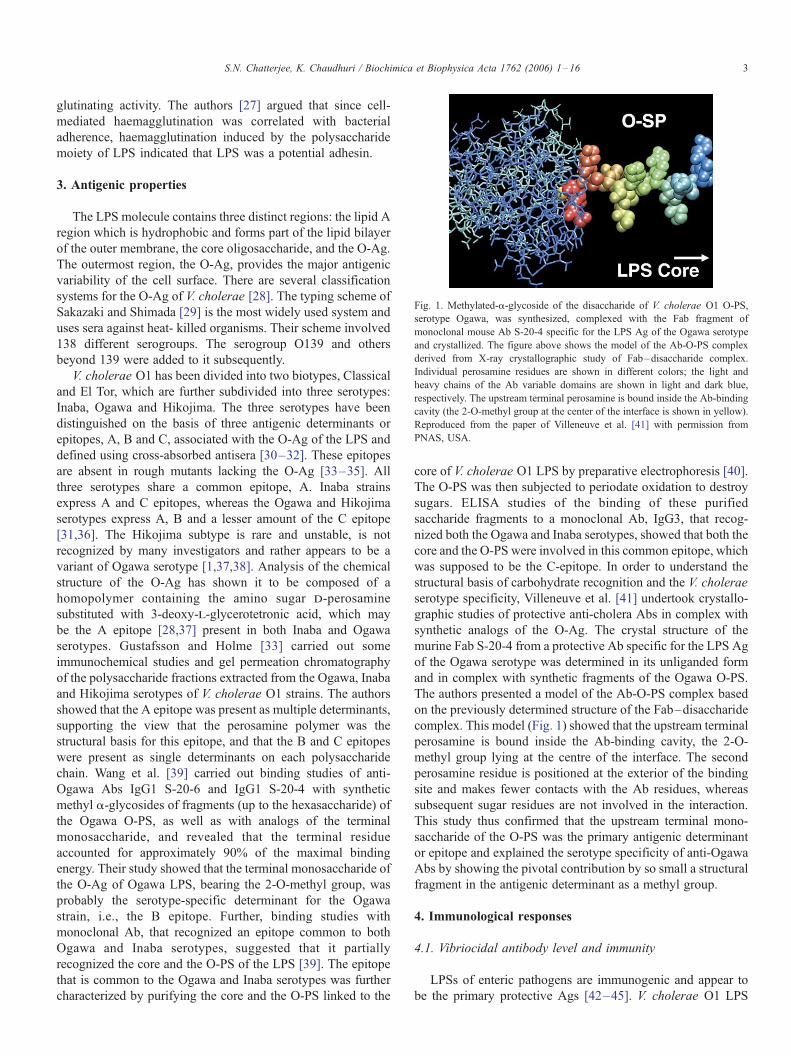
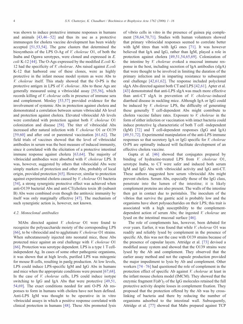
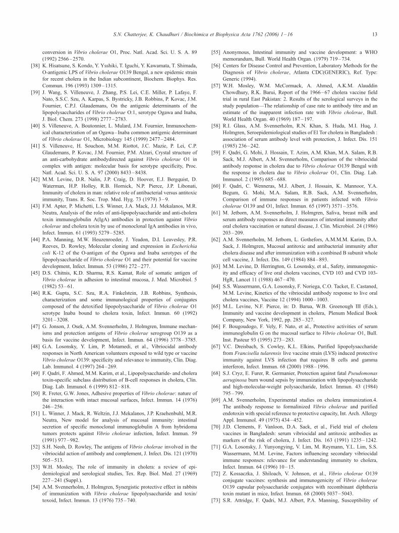
Fig. 1. Methylated-a-glycoside of the disaccharide of V. cholerae O1 O-PS,
serotype Ogawa, was synthesized, complexed with the Fab fragment of
monoclonal mouse Ab S-20-4 specific for the LPS Ag of the Ogawa serotype
and crystallized. The figure above shows the model of the Ab-O-PS complex
derived from X-ray crystallographic study of Fab–disaccharide complex.
Individual perosamine residues are shown in different colors; the light and
heavy chains of the Ab variable domains are shown in light and dark blue,
respectively. The upstream terminal perosamine is bound inside the Ab-binding
cavity (the 2-O-methyl group at the center of the interface is shown in yellow).
Reproduced from the paper of Villeneuve et al. [41] with permission from
PNAS, USA.
S.N. Chatterjee, K. Chaudhuri / Biochimica et Biophysica Acta 1762 (2006) 1–16 3
glutinating activity. The authors [27] argued that since cell-
mediated haemagglutination was correlated with bacterial
adherence, haemagglutination induced by the polysaccharide
moiety of LPS indicated that LPS was a potential adhesin.
3. Antigenic properties
The LPS molecule contains three distinct regions: the lipid A
region which is hydrophobic and forms part of the lipid bilayer
of the outer membrane, the core oligosaccharide, and the O-Ag.
The outermost region, the O-Ag, provides the major antigenic
variability of the cell surface. There are several classification
systems for the O-Ag of V. cholerae [28]. The typing scheme of
Sakazaki and Shimada [29] is the most widely used system and
uses sera against heat- killed organisms. Their scheme involved
138 different serogroups. The serogroup O139 and others
beyond 139 were added to it subsequently.
V. cholerae O1 has been divided into two biotypes, Classical
and El Tor, which are further subdivided into three serotypes:
Inaba, Ogawa and Hikojima. The three serotypes have been
distinguished on the basis of three antigenic determinants or
epitopes, A, B and C, associated with the O-Ag of the LPS and
defined using cross-absorbed antisera [30–32]. These epitopes
are absent in rough mutants lacking the O-Ag [33–35]. All
three serotypes share a common epitope, A. Inaba strains
express A and C epitopes, whereas the Ogawa and Hikojima
serotypes express A, B and a lesser amount of the C epitope
[31,36]. The Hikojima subtype is rare and unstable, is not
recognized by many investigators and rather appears to be a
variant of Ogawa serotype [1,37,38]. Analysis of the chemical
structure of the O-Ag has shown it to be composed of a
homopolymer containing the amino sugar d-perosamine
substituted with 3-deoxy-l-glycerotetronic acid, which may
be the A epitope [28,37] present in both Inaba and Ogawa
serotypes. Gustafsson and Holme [33] carried out some
immunochemical studies and gel permeation chromatography
of the polysaccharide fractions extracted from the Ogawa, Inaba
and Hikojima serotypes of V. cholerae O1 strains. The authors
showed that the A epitope was present as multiple determinants,
supporting the view that the perosamine polymer was the
structural basis for this epitope, and that the B and C epitopes
were present as single determinants on each polysaccharide
chain. Wang et al. [39] carried out binding studies of anti-
Ogawa Abs IgG1 S-20-6 and IgG1 S-20-4 with synthetic
methyl a-glycosides of fragments (up to the hexasaccharide) of
the Ogawa O-PS, as well as with analogs of the terminal
monosaccharide, and revealed that the terminal residue
accounted for approximately 90% of the maximal binding
energy. Their study showed that the terminal monosaccharide of
the O-Ag of Ogawa LPS, bearing the 2-O-methyl group, was
probably the serotype-specific determinant for the Ogawa
strain, i.e., the B epitope. Further, binding studies with
monoclonal Ab, that recognized an epitope common to both
Ogawa and Inaba serotypes, suggested that it partially
recognized the core and the O-PS of the LPS [39]. The epitope
that is common to the Ogawa and Inaba serotypes was further
characterized by purifying the core and the O-PS linked to the
core of V. cholerae O1 LPS by preparative electrophoresis [40].
The O-PS was then subjected to periodate oxidation to destroy
sugars. ELISA studies of the binding of these purified
saccharide fragments to a monoclonal Ab, IgG3, that recog-
nized both the Ogawa and Inaba serotypes, showed that both the
core and the O-PS were involved in this common epitope, which
was supposed to be the C-epitope. In order to understand the
structural basis of carbohydrate recognition and the V. cholerae
serotype specificity, Villeneuve et al. [41] undertook crystallo-
graphic studies of protective anti-cholera Abs in complex with
synthetic analogs of the O-Ag. The crystal structure of the
murine Fab S-20-4 from a protective Ab specific for the LPS Ag
of the Ogawa serotype was determined in its unliganded form
and in complex with synthetic fragments of the Ogawa O-PS.
The authors presented a model of the Ab-O-PS complex based
on the previously determined structure of the Fab–disaccharide
complex. This model (Fig. 1) showed that the upstream terminal
perosamine is bound inside the Ab-binding cavity, the 2-O-
methyl group lying at the centre of the interface. The second
perosamine residue is positioned at the exterior of the binding
site and makes fewer contacts with the Ab residues, whereas
subsequent sugar residues are not involved in the interaction.
This study thus confirmed that the upstream terminal mono-
saccharide of the O-PS was the primary antigenic determinant
or epitope and explained the serotype specificity of anti-Ogawa
Abs by showing the pivotal contribution by so small a structural
fragment in the antigenic determinant as a methyl group.
4. Immunological responses
4.1. Vibriocidal antibody level and immunity
LPSs of enteric pathogens are immunogenic and appear to
be the primary protective Ags [42–45]. V. cholerae O1 LPS
S.N. Chatterjee, K. Chaudhuri / Biochimica et Biophysica Acta 1762 (2006) 1–164
was shown to induce protective immune responses in humans
and animals [43,46–52] and thus its use as a protective
immunogen for cholera vaccine development has been widely
accepted [51,53,54]. The gene clusters that determined the
biosynthesis of the LPS O-Ag of V. cholerae O1, of both the
Inaba and Ogawa serotypes, were cloned and expressed in E.
coli K-12 [44]. The O-Ags expressed by the modified E.coli K-
12 had the specificity of V. cholerae. Abs raised against E.coli
K-12 that harbored one of these clones, were as highly
protective in the infant mouse model system as were Abs to
V. cholerae itself. This study showed that the O-PS is the
protective antigen in LPS of V. cholerae. Abs to these Ags are
generally measured using a vibriocidal assay [55,56], which
records killing of V. cholerae cells in presence of immune sera
and complement. Mosley [53,57] provided evidence for the
involvement of systemic Abs in protection against cholera and
demonstrated a correlation between serum vibriocidal Ab level
and protection against cholera. Elevated vibriocidal Ab levels
were correlated with protection against both V. cholerae O1
colonization and disease [58]. The titer of vibriocidal Abs
increased after natural infection with V. cholerae O1 or O139
[59,60] and after oral or parenteral vaccination [61,62]. The
field trials of vaccines showed that the level of vibriocidal
antibodies in serum was the best measure of induced immunity,
since it correlated with the elicitation of a protective intestinal
immune response against cholera [63,64]. The majority of
vibriocidal antibodies were absorbed with V. cholerae LPS. It
was, however, suggested by others that vibriocidal Abs were
simply markers of protection and other Abs, probably of local
origin, provided protection [65]. However, similar to protection
against experimental cholera caused by V. cholerae O1 bacteria
[54], a strong synergistic protective effect was achieved when
anti-O139 bacterial Abs and anti-CT(cholera toxin )B (subunit
B) Abs were combined even though the antitoxic immunity by
itself was only marginally effective [47]. The mechanism of
such synergistic action is, however, not known.
4.2. Monoclonal antibodies
MAbs directed against V. cholerae O1 were found to
recognize the polysaccharide moiety of the corresponding LPS
[66], to be vibriocidal and to agglutinate V. cholerae O1 strains.
When subcutaneously injected into neonatal mice, these Abs
protected mice against an oral challenge with V. cholerae O1
[66]. Protection was serotype dependent. LPS is a type 1 T cell-
independent Ag. In cases of some other pathogenic organisms,
it was shown that at high levels, purified LPS was mitogenic
for mouse B cells, resulting in panIg production. At low levels,
LPS could induce LPS-specific IgM and IgG Abs in humans
and mice when the appropriate conditions were present [67,68].
In the case of V. cholerae cells, LPS could induce isotype
switching to IgG and IgA Abs that were protective [49,51,
54,69]. The exact conditions needed for anti O-PS Ab res-
ponses to form in humans with cholera have not been defined.
Anti-LPS IgM was thought to be operative in in vitro
vibriocidal assays in which a positive response correlated with
clinical protection in humans [48]. These Abs promoted lysis
of vibrio cells in vitro in the presence of guinea pig comple-
ment [58,64,70,71]. Studies with human volunteers showed
that primary vibriocidal responses seemed to correlate better
with IgM titres than with IgG ones [71]. It was however
believed that IgA and IgG, rather than IgM, played a role in
protection against cholera [49,51,54,65,69]. Colonization of
the intestine by V. cholerae evoked a mucosal immune res-
ponse in the host, including secretion of IgA antibodies (sIgA)
that were thought to be involved in limiting the duration of the
primary infection and in imparting resistance to subsequent
oral challenge [42,61,62]. The response included polyclonal
sIgA Abs directed against both CT and LPS [42,61]. Apter et al.
[43] demonstrated that anti-LPS sIgAwas much more effective
than anti-CT sIgA in prevention of V. cholerae-induced
diarrheal disease in suckling mice. Although IgA or IgG could
be induced by V. cholerae LPS, the difficulty of generating
these generally T cell-dependent Abs might contribute to
cholera vaccine failure rates. Exposure to V. cholerae in the
form of either infection or vaccination with intact bacteria could
induce protective Ig characteristic of both T cell -independent
(IgM) [72] and T cell-dependent responses (IgG and IgA)
[49,51,72]. Experimental manipulation of the anti-LPS immune
responses so that secretory IgA or IgG specific for V. cholerae
O-PS are optimally induced will facilitate development of an
effective cholera vaccine.
Gupta et al. [46] showed that conjugates prepared by
binding of hydrazine-treated LPS from V. cholerae O1,
serotype Inaba, to CT were safer and induced both serum
IgM and IgG Abs with vibriocidal activity and IgG anti-CT.
These authors suggested how serum vibriocidal Abs might
prevent cholera. Serum Abs, especially those of the IgG class,
penetrate into the lumen of the intestine; it is likely
complement proteins are also present. The walls of the intestine
then get in contact due to peristalsis. The inoculum of the
vibrios that survive the gastric acid is probably low and the
organisms have short polysaccharides on their LPS; this trait is
associated with a high susceptibility to the complement-
dependent action of serum Abs; the ingested V. cholerae are
lysed on the intestinal mucosal surface [46].
The role of complements has, however, been debated for
over years. Earlier, it was found that while V. cholerae O1 was
readily and reliably lysed by complement in the presence of
specific Ab, this was not the case with O139 strains because of
the presence of capsular layers. Attridge et al. [73] devised a
modified assay system and showed that the O139 strains were
lysed by the Ab and complement. They observed that the
earlier assay method and not the capsule production provided
the major impediment to lysis by Ab and complement. Other
workers [74–76] had questioned the role of complement in the
protection effect of specific Ab against V. cholerae at least in
the infant mouse cholera model (IMCM). They showed that the
enzymic fragment F(abV)2 of the IgG molecules retained the full
protective activity despite losses in complement fixation. They
proposed that the protection offered by the Ab was by cross-
linking of bacteria and there by reducing the number of
organisms adsorbed to the intestinal wall. Subsequently,
Attridge et al. [77] showed that Mabs prepared against TCP

S.N. Chatterjee, K. Chaudhuri / Biochimica et Biophysica Acta 1762 (2006) 1–16 5
isolated from V. cholerae O1 El Tor were able to provide
biotype-specific protection against experimental cholera in
infant mice, although the Mabs were not lytic in the presence of
complement. They suggested that Abs to TCP protected by
directly blocking colonization of the mucosal surface rather
than any complement -dependent lysis. But Kaper et al. [28]
observed that long-term protection against cholera could be
accomplished even in the absence of a detectable anti-TCP
immune response. In the context of all these observations, the
exact role of complements in the protection against cholera
remains unclear.
4.3. Immunoglobulin subclasses
The Igs, IgA and IgG, have subclasses, which are known to
exhibit different functions. Knowledge about the subclass
distribution of specific antibodies in infection caused by V.
cholerae O1 or O139 is limited. Jetborn et al. [78] had shown
that CT induced responses of the four Ig G subclasses (IgG1,
IgG2, IgG3, IgG4) and the IgA1 subclass in serum of the
cholera vaccinees and patients. A study of North American
volunteers had shown that secondary challenge with V.
cholerae O1 resulted in LPS-specific responses of the IgG1
and IgG3 subclasses [71], where as after primary exposure, the
major response to LPS was of IgG4 Abs. The LPS-specific
IgG1 and IgG3 responses in the North American volunteers
were highly associated with the vibriocidal activity in the IgG
fraction, suggesting that these subclasses might also contribute
to vibriocidal Abs. Qadri et al. [49] made a comparative study
of the subclass distribution of the mucosal and systemic Ab
responses in patients infected with V. cholerae O1 or O139 to
two Ags, LPS and CT. They assessed the Ab secreting cells
(ASC) in the circulation, which served as proxy indicator of the
mucosal immune response. LPS-specific ASCs of both IgA1
and IgA2 subclasses were found, with the IgA1 ASC response
predominating in both V. cholerae O1 and O139-infected
patients. Both groups of cholera patients showed significant
increases in LPS-specific IgG1, IgG2 and IgG3 Abs in plasma.
Again, both groups of patients showed CT-specific ASC
responses of the different IgG and IgA subclasses in the
circulation. The authors showed that despite possessing a
capsule and an LPS structurally different from that of V.
cholerae O1, V. cholerae O139 induced Ab subclasses similar
to those seen in O1 cholera. Further investigations are required
to decide whether the LPS-specific response in the different
subclasses can be used as an alternative marker of immunity
and whether the vaccines against O1 and O139 cholera can be
developed to stimulate Ab subclasses that are likely to offer
protection.
4.4. Antibody assay for encapsulated cells
Recent studies evaluating the usefulness of the vibriocidal
assay for O139 infections have produced conflicting results and
strain-to-strain variability in the sensitivity of the vibriocidal
assay to fully encapsulated O139 strain has been reported
[48,79,80]. The possibility that the CPS might interfere with
complement-mediated killing of the organisms prompted some
workers to develop modified vibriocidal assay methods.
Losonsky et al. [48] established a modified vibriocidal assay
using another O139 target strain, strain 2 L, an unencapsulated
insertion mutant of parent strain AI-1837, which retained the
truncated O side chain. But the modified vibriocidal assay for
fully encapsulated V. cholerae O139 strain AI-1837 and for the
unencapsulated insertion mutant strain 2 L produced a very
modest vibriocidal response in volunteers challenged with V.
cholerae O139 that is not specific. Boutonnier et al. [81], on
the other hand, prepared a conjugate of the polysaccharide
moiety (O-specific polysaccharide plus core) of the LPS of V.
cholerae O139 (pmLPS) and tetanus toxoid (TT) and tested its
immunological properties using BALB/c mice. The conjugate
(pmLPS-TT) elicited high levels of IgG antibodies, peaking 3
months after the first immunization and declining slowly
during the following 5 months. Antibodies elicited by the
conjugate recognized both CPS and LPS from V. cholerae
O139, were vibriocidal and were protective in neonatal mouse
model of cholera infection. The authors claimed that conjuga-
tion of the O139 pmLPS enhanced its immunogenicity and
conferred T-dependent properties to this polysaccharide.
Boutonnier et al. [82] further developed a new method
(microtiter plate assay) for determining the vibriocidal Ab
titer, which was considered equally convenient and efficient for
both V. cholerae O1 and O139 serogroups. Their method was
also found convenient for measuring the activity of animal sera
and mouse MAbs.
A new assay using blocking of the limulus amebocyte lysate
(LAL) reaction in a microtiter plate was developed [83] to
detect Abs to V. cholerae O139 LPS that would be less
susceptible to the confounding effects of the capsule. It was
shown that Abs to V. cholerae blocked the LAL reaction and
that LAL titers were comparable to the vibriocidal titers. Also
blocking of the gel reaction was serotype specific. However,
practical use of this method in field studies required further
investigations. Nandy et al. [84], in a different approach, raised
antisera to the truncated form of O-polysaccharides (TFOP)
linked to the core of O139 LPS and found that anti-TFOP Abs
and their Fab (IgG) fragments induced passive protection
against challenge with colonial variants of encapsulated O139
strains in the suckling mouse model of experimental cholera.
The authors found that such protection was mediated by
inhibition of intestinal colonization.
4.5. Synthetic oligosaccharides
The use of a synthetic, O-PS-based immunogen was found
to eliminate the toxicity problems associated with native LPS.
Chernyak et al. [85] prepared immunogens by linking of BSA
to the chemically synthesized, linker- equipped hexasaccharide
fragment of the O-PS of V. cholerae O1, serotype Ogawa, by
appropriate chemical methods [86,87] (Fig. 2). Conjugates with
different carbohydrate (CHO)-to-carrier (BSA) molar ratios
were tested for immunogenicity and efficacy in mice. All the
conjugates tested were found to be immunogenic and a
correlation was found between vibriocidal activity and protec-

Fig. 2. Chemical structure of the neoglycoconjugate immunogen (CHO [Ogawa terminal hexasaccharide]–BSA) complex, as obtained from the works of Chernyak
et al. [85,86]. BSAwas linked to the chemically synthesized, linker-equipped hexasaccharide fragment of the O-PS of V. cholerae O1 serotype Ogawa. Arrow shows
the 2-O-methyl group in the terminal sugar of the Ogawa serotype, which is replaced by 2-OH group in the Inaba serotype. Three different immunogens (A, B and C)
based on the synthetic Ogawa epitope that varied in number of hexasaccharide residues and were covalently coupled to BSAwere used to test immune responses in
female BALB/c mice. The CHO to BSA molar ratios in the three immunogens were: A, 15.5:1; B, 9.2:1; C, 4.6:1.
S.N. Chatterjee, K. Chaudhuri / Biochimica et Biophysica Acta 1762 (2006) 1–166
tion. The protective capacity of antiserum was evident in serum
from mice immunized with all conjugates, but it was highest in
the groups that received the conjugate with the lowest level of
substitution (conjugate C). The corresponding mice received
fewer immunizations with conjugate C. The level of substitu-
tion and the number of immunizations affected the repertoire
profile of the anti-Ogawa epitope response but the reasons for
this differential protection were not known and required further
investigations. Subsequently, a series of conjugates made from
Inaba di-, tetra- and hexasaccharide and BSA were prepared
[88,89] and found to be immunogenic in mice, inducing IgM
and the T-dependent IgG1 subclasses. But the Inaba-specific
Abs, IgM and IgG1, were neither vibriocidal nor protective in
the infant mouse cholera model [89]. Again the exact reason
for the functional differences between the anti-Inaba and anti-
Ogawa Abs remained to be explained. In contrast to the anti-
Inaba CHO–BSA sera, the secondary, anti-whole LPS sera
were vibriocidal. The authors thus suggested that the Abs
induced by the Inaba CHO–BSA conjugates did not bind with
enough affinity or specificity to native LPS when expressed on
the bacterial surface.
5. Role in the intestinal adhesion and virulence of the
vibrios
Several studies [44,46,52,90] implied that the O-Ag
represented a protective Ag and was involved in the adherence
and colonization of V. cholerae. Fuerst and Perry [91]
demonstrated LPS on sheathed flagella of V. cholerae O1 by
protein A-gold immunoelectron microscopy. The flagellum on
V. cholerae cells was found essential for in vitro attachment
and enhanced initial colonization of the host intestinal surface
in the infant mouse cholera model [92]. The LPS on the
flagellum was thus found to function as a carrier of adhesins
[93,94]. On the other hand, Mukhopadhyay et al. [95] observed
that, in the mouse model, the anti-LPS Abs induced passive
protection through microagglutination and/or immobilization
of vibrios, which did not allow the vibrios to adhere to and
colonize the intestine. The different animal models or cultured
cells may, however, not be completely suitable to evaluate the
factors that are essential for colonization of the human gut. To
get a more realistic picture, Benitez et al. [96] studied the
interactions between V. cholerae O1 and O139 with the highly
differentiated mucin-secreting cells, HT29-18N2, which were
derived from the human colonic adenocarcinoma HT29 cell
line. Choleragenic vibrios were shown to adhere to and
multiply on monolayers of these cells. Their adherence was
partially inhibited by LPS. The authors further showed that the
flagella, an active toxR gene, and the virulence cassette were
not essential for binding. The authors emphasized that the
interactions studied mimicked important events accompanying
intestinal colonization and as such provided a new approach for
studying factors involved in the intestinal colonization of the
vibrios.
Several studies correlated LPS mutations with colonization
defects in V. cholerae [97,98] and other organisms [99–101],
but the mechanism by which LPS mutations decreased
colonization remained unclear. Iredell et al. [102] studied some
wbe::Tn mutants (which were resistant to phages known to use
the O-Ag as their receptor) and tried to explain the role of LPS
in virulence of V. cholerae O1. The authors found that the
mutants were unable to assemble TCP on their surface, but the
major subunit TcpA could be found as an intracellular pool.
These mutants could be complemented back to wild type using
the cloned wbe region implying that the functional TCP
assembly was dependent upon an intact LPS. This was
significant in the background of the finding that TCP was a
protective Ag in animal models [103–105] and has been

S.N. Chatterjee, K. Chaudhuri / Biochimica et Biophysica Acta 1762 (2006) 1–16 7
shown to be an essential colonization factor for both human
and infant mouse, and for both classical and El Tor strains
[103,105,106]. While searching for genes required for coloni-
zation, Chiang and Mekalanos [107] identified manB and wbeL
mutants as colonization-defective strains. They found no defect
in the TCP production of wbe mutants or could detect any
reduction in the TCP expression in a gmd :: Tn 5lac mutant of
O395 strain. The authors accordingly concluded that the
colonization defect associated with wbe mutations was
unrelated to defects in TCP assembly but suggested, on the
other hand, that LPS itself was important for colonization.
They argued that although the exact role of LPS in colonization
was unclear, the possibility that LPS defects might render
bacteria more susceptible to gut-associated bactericidal sub-
stances appeared sensible particularly since LPS was known to
be involved in resistance to antibiotics and complement
mediated killing [108,109].
Nesper et al. [110] isolated bacteriophage K139-resistant
mutant of V. cholerae O1 El Tor having intact O-Ag but altered
core oligosaccharide and also a mutation in the galU gene.
They further isolated another gal mutant (inactivated galE),
which was defective in the catabolism of exogenous galactose
but synthesized an apparently normal LPS. They found that the
galU and a rough LPS mutant (R-LPS), but not the galE
mutant, were defective in colonization, a phenotype also
associated with O-Ag-negative mutants. Their study further
showed that galU and R-LPS mutants were more sensitive to
short chain organic acids, cationic antimicrobial peptides, the
complement system, the bile salts and other hydrophobic
agents, indicating that the outer membrane of these organisms
could not provide an effective barrier function. The O-Ag-
negative strains were also found to be sensitive to complement
and cationic peptides, but displayed significant resistance to
bile salts and short chain organic acids. This study indicated the
involvement of galU in V. cholerae virulence, correlated with
the observed change in LPS structure, and a role for both galU
and galE in the environmental survival of V. cholerae. In a
more recent study, the authors [111] further investigated the
role of LPS O-side chain and CPS of V. cholerae O139 in
intestinal colonization by using genetically engineered mutants.
Their results showed that the loss of LPS O-side chain or CPS
resulted in approximately 30-fold reduction in colonization of
the infant mouse small intestine. Their study further indicated
that in so far as V. cholerae O139 strain was concerned, the
presence of both LPS O-side chain and CPS was important
during the colonization process. On the other hand, Attridge et
al. [112] obtained the bacteriophage JA-1 (which uses the
capsule as the receptor)—mutants having several phenotypes,
with loss of capsule and/or O-Ag from the cell surface, studied
their residual complement resistance and infant mouse coloni-
zation potential and showed that production of O-Ag was of
much greater significance than the presence of capsular
material for both of the aforesaid properties. In the background
of these studies, additional factors (other than the known
colonization factors) involved in the colonization and acid
tolerance of V. cholerae were subsequently identified [113].
Several genes were identified whose activity in colonization
was not previously appreciated [113]. The functions of these
genes included production of factors involved in metabolic
activities, regulation of cellular processes, transport, adaptation
to stress and some unknown functions. These authors identified
nine new factors as crucial for the V. cholerae acid tolerance
response (previously identified to be important for epidemic
spread of cholera) and showed that mutations in the genes,
gshB, hepA and recO resulted in a 1000-fold reduction in
colonization [113].
6. LPS as phage receptor
V. cholerae LPS, like the LPS of many Gram-negative
bacteria, was found to act as the receptor of several cholera
phages [114–116]. Mukherjee’s group IV cholera phage
differentiates the classical and El Tor biotypes of V. cholerae,
the classical ones being sensitive and the El Tor ones resistant
to these phages [117]. The basis of this differentiation was
traced down to the LPSs of the two biotypes. The phage B149was inactivated by the classical LPS but was resistant to the El
Tor LPS [114,115]. Adsorption of cholera phage B149 to
isolated classical LPS followed a first order reaction kinetics,
the 50% phage inactivating concentration of LPS (IC50) being
7 Ag/ml. After treatment of LPS by 0.5% (w/v) sodium
deoxycholate at 37 -C, the LPS largely lost its phage
inactivating capacity and the IC50 value rose to 3.6 mg/ml
[114,115]. This was in conformity with the fact that sodium
deoxycholate dissociated LPS of Gram-negative bacteria into
very small units with subsequent loss of biological activity
[118,119]. When cholate was removed by extensive dialysis,
the phage inactivating capacity of LPS was restored signifi-
cantly, the IC50 value being 570 Ag/ml of LPS. The cholate
alone could not inactivate the phages by any significant degree
[114]. LPS isolated from Inaba or Ogawa serotypes and
classical or El Tor biotypes of V. cholerae showed identical
phage inactivating capacities for the phage CP-T1 [116]. On
the other hand, LPS from a CP-T1 -resistant mutant exhibited
no phage inactivating capacity. The mutant was shown to lack
the O-Ag by bactericidal assays utilizing a MAb directed
against O-Ag side chain of V. cholerae LPS. The absence of O-
Ag in the phage-resistant strain was further confirmed by SDS-
PAGE study of 32P-labelled LPS [116]. Similarly, another
phage, VCII, specific to O1 classical strains found their
receptors in the O-Ags of LPS and the VCII resistant mutants
lacked the O-Ag [35].
Bacteriophage K139 was originally isolated from a V.
cholerae O139 strain and was identified as belonging to the
Kappa phage family [120]. Further analysis revealed that this
phage was widely distributed among clinical El Tor strains and
was also found as a defective prophage in classical O1 strains
[120,121]. K-139 was perhaps the first vibriophage for which
the entire genome was sequenced [122]. The tail fibers were
thought to be involved in receptor binding. The presumed tail
fiber genes of the phage K-139 were sequenced and analyzed,
and two conserved and two variable regions were identified.
Three different tail fiber types were discovered depending on
the different combinations of the variable regions. Since the C-
S.N. Chatterjee, K. Chaudhuri / Biochimica et Biophysica Acta 1762 (2006) 1–168
terminal part of the tail fiber was believed to be involved in
receptor binding [122], it was speculated that the variable
regions of the K-139 phages determined their binding ability to
different O-Ag receptors. Phage binding studies with purified
LPS of different O1 serotypes and biotypes revealed that the
O1 O-Ag served as the phage receptor. Analysis of the LPS of
spontaneous phage-resistant mutants revealed that most of
them synthesized incomplete LPS molecules composed of
either defective O1 O-Ag or core oligosaccharide [121].
Applying hypervirulent phage K139cm9 to O1 El Tor strains,
different phage-resistant mutants were isolated and these were
found to express different LPS mutations. Interestingly several
mutants were found linked not with the O1 O-Ag but with the
core structure. Such mutants indirectly implicated the core
region of the LPS in secondary phage infection steps [121].
Among the O-Ag defective mutants, one mutant was charac-
terized for the loss of O-Ag due to transposition of IS1004 into
the wbeW gene encoding a putative glycosyltransferase. In a
later study [123], one wbeW: IS1004 serum-sensitive mutant
was treated with normal human serum and several survivors
showing precise excision of IS1004, restoring O-Ag biosyn-
thesis and serum resistance were detected. Further, by
screening for phage resistance among clinical isolates and
performing LPS analysis of non-lysogenic strains, one strain
was identified with decreased O-Ag presentation and signifi-
cant reduction in ability to colonize the mouse small intestine.
Several other cholera phages were identified as having
receptors not in the cell wall LPS but in other structures
associated with the organism [124–126]. Any discussion on
these phages and their receptors is beyond the scope of this
review.
7. Biofilm formation and the structure of LPS
Biofilm formation by bacteria is of great importance in
respect of their survival in natural environments and causation
of epidemic outburst of the disease. Biofilm can develop on
abiotic surfaces and generally consists of bacterial cells
entwined in a protective matrix of extracellular polysacchar-
ides. V. cholerae is a natural inhabitant of aquatic ecosystems
and is known to attach to different environmental surfaces.
Adhikari and Chatterjee [127] reported the formation of thick
pellicle on the surface of static liquid cultures of several
mannose-sensitive haemagglutinating strains of V. cholerae El
Tor and found a direct correlation between the formation of a
special type of pili on the bacterial surface and pellicle
formation. Tweedy et al. [128] confirmed the presence of pili
on V. cholerae surface and produced evidence that the Vibrio
strains, which exhibited weaker haemagglutination reaction,
were comparatively poorer in pili formation. In the recent days,
V. cholerae El Tor has been reported to form three-dimensional
biofilm on abiotic surfaces [110,129–132] and on simple static
liquid cultures [133] in agreement with the earlier observations
of Adhikari and Chatterjee [127]. V. cholerae O1 El Tor
N16961 required the MSHA, a type IV pilus, and the flagellum
to associate with abiotic surfaces [129–131] in LB broth,
where as V. cholerae O139 strain M010 depended only on the
flagellum for surface association [132]. For subsequent
development of a three dimensional biofilm, both the strains
required the presence of the vps genes, which are responsible
for the synthesis of an exopolysaccharide-based adhesive
extracellular matrix [129,132,133]. Watnick and Kolter [132]
further reported, using transposon mutagenesis, that the genes
involved in biofilm formation included those encoding (i) the
biosynthesis and secretion of the type IV pilus (MSHA), (ii) the
synthesis of exopolysaccharide and (iii) flagellar motility.
Accordingly, they suggested that the three steps in the process
of biofilm formation were: (i) the type IV pilus and the
flagellum accelerating attachment to the abiotic surface, (ii) the
flagellum mediating spread along the abiotic surface and (iii)
the exopolysaccharide forming the three-dimensional biofilm
architecture. The exopolysaccharide initially forms the so-
called slime layer on the surface of bacteria. The biofilm
formation is normally associated with the change from normal
smooth colony morphology to a rugose one of the bacteria
[134]. The rugose colony morphology was the result of
increased synthesis of the VPS exopolysaccharide [133,135],
and transcriptional regulation of the vps genes, which are
required for the synthesis of the VPS exopolysaccharide, was
altered in these strains [136]. Thus, these variants rapidly
formed biofilms in LB broth that were much thicker than those
formed by smooth-colony variants of V. cholerae. Electron
microscopic examination of the rugose form V. cholerae El Tor
strain TSI-4 revealed thick electron dense exopolysaccharide
materials surrounding polycationic ferritin-stained cells, while
the ferritin-stained material was absent around the translucent
form of the strain TSI-4. Scanning electron microscopic
examination further revealed that the surface of the biofilm
was colonized by the actively dividing rod shaped cells. By
having exopolysaccharide materials, the rugose strains ac-
quired resistance to osmotic and oxidative stress and were
capable of causing human disease [137]. V. cholerae O139
strain M010 was also shown to produce exopolysaccharide
leading to biofilm formation in response to nutrient starvation
with concomitant change from a normal smooth colony
morphology to a rugose one [134]. It was further demonstrated
by immunoelectron microscopy that there was an epitope
common to the exopolysaccharide Ag of V. cholerae O1 strain
TSI-4 (rugose form) and that of O139 strain MO10 [134].
The entire V. cholerae O1 genome sequence being available
[138], a method was developed for the whole genome
characterization of the biofilm phenotype through the use of
microarray-based expression profiling [139]. The important
objectives of study were the differential expression pattern
between the sessile and planktonic populations of the same
culture, the identification of genes selectively expressed during
different stages of biofilm development, identification of genes
differentially expressed during adaptation of a mature biofilm
to various changes in the fluid phase, etc. Hango and Watnick
[140] subsequently identified a transcriptional repressor in V.
cholerae that inhibited exopolysaccharide synthesis and
biofilm development. It was shown that the repressor was the
V. cholerae homologue of E. coli CytR, a protein that represses
nucleoside uptake and catabolism when nucleosides are scarce.
S.N. Chatterjee, K. Chaudhuri / Biochimica et Biophysica Acta 1762 (2006) 1–16 9
The influence of biofilm formation on the structure of LPS
or vice versa among V. cholerae cells is likely to form another
important field of study. In Pseudomonas aeruginosa, studies
had indicated that changes in LPS phenotype affected
adherence properties and influenced biofilm formation [141].
Recently, Nesper et al. [110] studied several aspects including
the resistance to phage K139.cm9 of and biofilm formation by
the different gal U and gal E mutants of V. cholerae El Tor.
Among the spontaneous phage K139.cm9-resistant strains,
they found strains with a rugose colony morphology constitu-
tively synthesizing an exopolysaccharide and producing
biofilm on abiotic surfaces. They introduced galU and galE
mutations into the rugose variant P27459res105 and found that
both mutations yielded smooth colony forms, suggesting that
galU and galE mutants were unable to synthesize the
exopolysaccharide and could not form the biofilm. The
activated carbohydrate moieties, like UDP-glucose and UDP-
galactose, were often involved in the synthesis of different
surface structures of bacteria [110]. Enzymes for the biosyn-
thesis of UDP-glucose and UDP-galactose are UDP-glucose-
pyrophosphorylase, encoded by gal-U, and UDP-glucose-4-
epimerase, encoded by galE [142]. The fact that galU and galE
were found essential for the formation of a biofilm by the
phage-resistant rugose variant suggested that the synthesis of
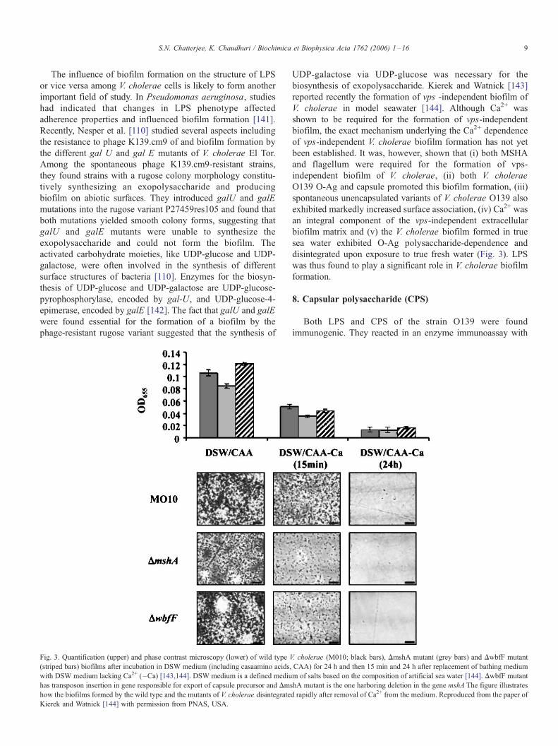
Fig. 3. Quantification (upper) and phase contrast microscopy (lower) of wild type V
(striped bars) biofilms after incubation in DSW medium (including casaamino acids,
with DSW medium lacking Ca2+ (–Ca) [143,144]. DSW medium is a defined mediu
has transposon insertion in gene responsible for export of capsule precursor and Dms
how the biofilms formed by the wild type and the mutants of V. cholerae disintegrate
Kierek and Watnick [144] with permission from PNAS, USA.
UDP-galactose via UDP-glucose was necessary for the
biosynthesis of exopolysaccharide. Kierek and Watnick [143]
reported recently the formation of vps -independent biofilm of
V. cholerae in model seawater [144]. Although Ca2+ was
shown to be required for the formation of vps-independent
biofilm, the exact mechanism underlying the Ca2+ dependence
of vps-independent V. cholerae biofilm formation has not yet
been established. It was, however, shown that (i) both MSHA
and flagellum were required for the formation of vps-
independent biofilm of V. cholerae, (ii) both V. cholerae
O139 O-Ag and capsule promoted this biofilm formation, (iii)
spontaneous unencapsulated variants of V. cholerae O139 also
exhibited markedly increased surface association, (iv) Ca2+ was
an integral component of the vps-independent extracellular
biofilm matrix and (v) the V. cholerae biofilm formed in true
sea water exhibited O-Ag polysaccharide-dependence and
disintegrated upon exposure to true fresh water (Fig. 3). LPS
was thus found to play a significant role in V. cholerae biofilm
formation.
8. Capsular polysaccharide (CPS)
Both LPS and CPS of the strain O139 were found
immunogenic. They reacted in an enzyme immunoassay with
. cholerae (M010; black bars), DmshA mutant (grey bars) and DwbfF mutant
CAA) for 24 h and then 15 min and 24 h after replacement of bathing medium
m of salts based on the composition of artificial sea water [144]. DwbfF mutant
hA mutant is the one harboring deletion in the gene mshA The figure illustrates
d rapidly after removal of Ca2+ from the medium. Reproduced from the paper of
S.N. Chatterjee, K. Chaudhuri / Biochimica et Biophysica Acta 1762 (2006) 1–1610
rabbit Abs generated against heat-killed bacteria [145]. Waldor
et al. [97] carried out immunoblot analysis of either whole cell
lysates or LPS preparations and obtained three electrophoretic
forms of the O139 Ag, i.e., two slowly migrating forms and
one rapidly migrating one that appeared identical to O139 LPS.
All three forms of the Ag shared an epitope defined by an O139
specific MAb. A serum-sensitive non-encapsulated mutant was
isolated that lacked only the slow migrating forms. The slow
migrating forms did not stain with silver whereas the rapidly
migrating forms did, indicating that the former might constitute
highly polymerized O-Ag side chain molecules that were not
covalently bound to core OS and lipid A, i.e., the O-Ag
capsule. This is in conformity with the observations of other
workers [145,146] that the V. cholerae O139 serogroup Ag
includes both the LPS and the CPS.
The presence of capsule on V. cholerae O139 strain
contributed to its virulence in several ways. The CPS made the
strain more resistant to killing by normal human serum and the
loss of capsule was associated with loss of the resistance [147].
Johnson et al. [148] derived an unencapsulated mutant of O139
strain by transposon mutagenesis and showed that it was readily
killed by serum, while the encapsulated one was protected
significantly. The unencapsulated mutant was less virulent in a
mouse model than the encapsulated parent. Another factor
contributing to the virulence of the non-O1 strains of V. cholerae
in general was septicemia [148]. Non-O1 V. cholerae strain
NRT36S produced a polysaccharide capsule that determined
colony morphology, serum resistance and virulence in mice. The
causation of such extraintestinal disease has not been found with
V. cholerae O1 strains. A third reason for the increased virulence
of the encapsulated strains of V. cholerae vis-a-vis the
nonencapsulated ones was that both the LPS and CPS were
important for their colonization of the small intestine of the new
born mouse [146]. There was also evidence that the CPS
mediated adherence to epithelial cells [97]. Using the intestinal
epithelial cell line Caco-2, a clear correlation between the
amount of capsular material expressed and the avidity of binding
to Caco-2 cells was found. In view of all these findings, Johnson
et al. [145] observed that the presence of capsule on O139 strains
had profound implications for vaccine development. There was
already a report of septicemia caused by an O139 strain [149].
Further, in keeping with observations with other non-O1 isolates
[150], sepsis was found to occur in a patient with underlying
liver disease. Since O139 strains might follow the pattern of
other non-O1 strains, the risk of dissemination would be greatest
in persons with chronic underlying illness [150,151]. Thus, the
advisability of administering oral attenuated vaccines that still
carry the capsule to persons who may have underlying illness
may be questioned. Further works addressing these problems
will be of practical importance.
9. Concluding remarks
9.1. Recognition of LPS and activation of host innate immunity
The molecular mechanisms involved in the recognition of
LPS of Gram-negative organisms and the initiation of host
response have been reviewed recently [152–154]. At the
extracellular stage, LPS has to be bound to a transport
molecule, a lipid-binding protein (LBP), which facilitates its
binding to a surface protein, CD14. CD14 then brings LPS
to the proximity of the cell membrane. The LPS-binding
protein, MD-2, then opsonizes LPS to be recognized by
another protein, TLR4, for initiation of signal transduction.
LPS is then briefly released into the lipid bilayer where it
interacts with a complex of receptors, e.g., heat shock
proteins (HSPs) and others, depending upon the cell type.
TLR-mediated signaling activates signal transduction path-
ways (such as NFkh, JNK/p38, NF/IL6 and IRF) that induce
transcription of cytokines (such as TNF-a and the type 1
interferons) and that in turn stimulate immune function and
control expression of a variety of inducible immune response
genes. A recent study has shown that V. cholerae LPS acts
through the TLR 4-MyD88-dependent signaling pathway and
induces INF-a, IL-1h and MIP-3a and significantly lesser
amounts of IFN-h, nitric oxide and IP-10 in macrophages
[155]. Further studies on V. cholerae LPS are required at
least for having a better knowledge of its interaction with the
B cells involving TLRs.
9.2. Serogroup surveillance and monitoring
The structure of the O-PS of V. cholerae of any serogroup
has been found to be unique [1]. The genetic organization
encoding the O-PS biosynthesis is quite susceptible to change,
but the factors responsible for effecting such changes are still
largely unknown [2]. Thus, a new serogroup or any of the
known serogroups may acquire pathogenic potential in
epidemic genetic background and may cause future epidemics.
This situation demands a continuous and strict surveillance and
monitoring of the emergence of either a new serogroup or any
of the known serogroups with pathogenic potential so that
appropriate vaccines can be devised promptly.
Phages are known to play a role in the emergence of
pathogenic clones and may also be involved in territorialism
between different strains of V. cholerae. For example, CT
genes were transferred to non-toxigenic strains through a
lysogenic filamentous phage [126], and the emergence and
dominance of V. cholerae O139 in Bangladesh and India
during 1992–1993 might have involved phages both as a
means of horizontal gene transfer as well as a bacteriocidal
selective mechanism. Faruque et al. [156] monitored the
environmental water samples of Bangladesh for nearly a 3-
year period and found that significantly more of these samples
contained either a phage targeting V. cholerae LPS as its
receptor or a phage-susceptible V. cholerae strain than both.
Interepidemic periods were characterized by water samples
containing cholera phages but no viable bacteria. Faruque et al.
[157] further observed that host-mediated phage amplification
during the cholera epidemic likely contributed to increased
environmental phage abundance, decreased load of environ-
mental V. cholerae, and, hence, the collapse of the epidemic.
The authors thus put forward the important suggestion that
environmental surveillance for vibriophages could be useful in

S.N. Chatterjee, K. Chaudhuri / Biochimica et Biophysica Acta 1762 (2006) 1–16 11
tracking outbreaks, predicting epidemics and anticipating
emergence of new serogroups. Further, vibriophages might
also be employed as biological control agents in cholera
epidemic areas [157].
But any effective surveillance and monitoring of the
aqueous environments demands the availability of rapid
diagnostic tests for cholera. Several such tests were available
for V. cholerae O1 or O139 using LPS Ag [158–161]. A
multistep colloidal gold-based colorimetric immunoassay
known as SMART was also developed for direct detection of
V. cholerae O1 or O139 in stool samples [162,163]. For rapid
detection of V. cholerae O1 or O139, Nato et al. [164]
described the development of a diagnostic test, the one-step
immunochromatographic dipstick test, based on LPS detection
using colloidal gold particles and immunochromatography.
This test was claimed to be of very high specificity and
sensitivity and could provide a simple tool for epidemiological
surveys. Similar simple and rapid test should also be developed
for the different serogroups of V. cholerae. Robert-Pillot et al.
[165] devised a method for improved and specific detection of
V. cholerae in environmental samples by culture of selective
medium and colony hybridization assay with an oligonucleo-
tide probe. The rapid detection of pathogenic vibrios using
biochemical and immunological markers, PCR and DNA
microarrays techniques will be a very challenging task. This
will require use of DNA sequences specific for virulence genes
or vibrio species to build and optimize a DNA microarray chip
that specifically identifies pathogenic isolates of various vibrio
species [166]. The successful development of such a technique
will provide a less time-consuming diagnostic strategy to be
used in the surveillance and monitoring of the estuarine or
environmental water samples.
9.3. LPS and cholera vaccine
Since the day of isolation of V. cholerae by Koch in 1883,
several cholera vaccines have been developed and evaluated
in clinical trials [65,167,168]. The involvement of LPS O-Ag
in the design and preparation of cholera vaccine using
recombinant DNA technology and synthetic carbohydrate
chemistry has been a rather recent and alternative approach.
An attempt to construct cholera vaccine using recombinant
DNA technology used the attenuated Salmonella typhi
vaccine strain Ty21a containing cloned V. cholerae genes
expressing the O-Ag [169–171]. Yet another novel idea for
the design of a carbohydrate based cholera vaccine originated
from the work of Villeneuve et al. [41] and which has been
discussed earlier. Abs specific for the terminal perosamine
could selectively protect against the Ogawa serotype but
failed to recognize the Inaba serotype. Therefore, protective
Abs against both serotypes should, the authors argued, bind to
the inner part of the O-PS and/or sugar residues defining the
core of the LPS molecule. The idea seems to be promising
but remains to be implemented in practice. It was however
proposed by another group of workers [172] that serum IgG
Abs conferred protection against enteric diseases by inactivat-
ing the inoculum on the mucosal surfaces. At the level of
laboratory animals, systemic administration of IgG Abs
specific for the O-PS of V. cholerae O1 was found to protect
neonatal mice against loss of weight and death following
intragastral challenge with V. cholerae O1 [66]. It may be
pertinent to note here the fundamental limitation that natural
infection with V. cholerae does not occur in animals, although
a few of the animal models have yielded useful information
relevant to human disease. Further, the infant mice or the
infant rabbits may be susceptible to infection with V. cholerae
but only for a relatively short time after birth. It is therefore
generally recognized that only the volunteer challenge studies
with V. cholerae can give most useful information about the
human disease.
Cellular cholera vaccines are poor immunogens and have
T cell-independent properties [53,57,173]. Besides, LPS as a
vaccine or in cellular vaccine often exhibits adverse reactions
due to its endotoxic properties. The recipients of the cellular
vaccine usually have high level of IgM anti-LPS Ab for about
6 months. The rapid decline of this IgM vibriocidal activity
explains the short-lived protection conferred by cellular
vaccines [52,70,174]. With a view to eliminating these
undesirable properties of the cellular vaccines, two groups
of workers have produced conjugate vaccines by coupling
Fdetoxified_ LPS to protein carriers [46,83]. Gupta et al. [46]
produced the deacylated LPS (DeALPS) by treatment of LPS
with hydrazine there by reducing the endotoxic properties of
LPS to clinically accepted levels. Conjugate vaccines were
prepared by binding DeALPS from V. cholerae O1, serotype
Inaba, to CT variants, CT-1 and CT-2 (used mainly as carrier
proteins which are also immunogenic), with a spacer and
evaluated (Phase I trial) in healthy volunteers [175]. The
conjugates elicited the highest levels of IgG anti-LPS
vibriocidal Abs, which persisted longer than those elicited
by the whole cell vaccine. The authors, however, expected to
improve further the level of IgG anti-LPS achieved with their
conjugates so far. Although the pmLPS conjugate vaccine
prepared by Boutonnier et al. [81], already discussed in
Section 4.4 of this review, was found protective in the
neonatal mouse model of cholera infection, it also remains to
be evaluated clinically. An earlier study [176] showed that a
V. cholerae O139 CPS-TT conjugate vaccine induced
protection in rabbit ileal loop model of experimental cholera.
Recently, V. cholerae O139 CPS conjugated with a recom-
binant mutant diphtheria toxin was shown to elicit high levels
of serum anti-CPS IgG in mice with vibriocidal activity [72].
The use of chemically synthesized, linker-equipped hexasac-
charide fragment of the O-PS of V. cholerae conjugated to
BSA as an immunogen, as discussed earlier, has been very
promising. But the use of these synthetic conjugates as
vaccine candidates requires further investigation and clinical
evaluation.
The goal of having a long-lasting vaccine effective against
more than one serogroup and/or serotype of V. cholerae still
remains to be achieved. Although the task ahead may be chal-
lenging, it certainly demands greater attention of researchers in
the context of the long-standing global defence problem against
the recurring cholera epidemics.

S.N. Chatterjee, K. Chaudhuri / Biochimica et Biophysica Acta 1762 (2006) 1–1612
Acknowledgements
Authors are most thankful to the many scientists and
particularly to Drs. Paul I. Watnick, P.M. Alzari, J.M.
Fournier, P. Kovac, P.A. Manning, U.H. Stroeher and B.S.
Srivastava for kindly sending reprints of their relevant
publications and materials for our use and also to Dr. Diane
Sullenberger, Executive Editor, PNAS, U.S.A. for granting
permission to reproduce in this review article (i) Fig. 5 of the
paper of Villeneuve et al. [41], copyright (2000) National
Academy of Sciences, USA and (ii) Fig. 2 of the paper of
Kierek and Watnick [144], copyright (2003) National Acad-
emy of Sciences, U.S.A. Sincere thanks are due to Dr. M.
Maiti, Director-Grade Scientist, Indian Institute of Chemical
Biology, Calcutta for helping us in many ways all through.
Thanks are also due to Drs. Sanjay Nag and Raghunath
Chatterjee of Biophysics Division, Indian Institute of Chem-
ical Biology, for rendering technical help during the
preparation of the manuscript.
References
[1] S.N. Chatterjee, K. Chaudhuri, Lipopolysaccharides of Vibrio cholerae:
I. Physical and chemical characterization, Biochim. Biophys. Acta 1639
(2003) 65–79.
[2] S.N. Chatterjee, K. Chaudhuri, Lipopolysaccharides of Vibrio cho-
lerae: II. Genetics of biosynthesis, Biochim. Biophys. Acta 1690
(2004) 93–109.
[3] O. Luderitz, O. Westphal, A.M. Staub, H. Nikaido, Isolation and
chemical and immunological characterization of bacterial lipopolysac-
charides, in: G. Weinbaum, S. Kadis, S.J. Ajl (Eds.), Microbial Toxins,
vol. 4, Academic Press, New York, 1971, pp. 145–233.
[4] S. Raziuddin, Toxic and immunological properties of the lipopolysac-
charides (O-antigens) from Vibrio el-tor, Immunochemistry 15 (1978)
611–614.
[5] S. Kabir, P. Mann, Immunological properties of the cell envelope
components of Vibrio cholerae, J. Gen. Microbiol. 119 (1980) 517–525.
[6] C. Galanos, E.T. Rietschel, O. Luderitz, O. Westphal, Y.B. Kim, D.W.
Watson, Biological activities of lipid A complexed with bovine serum
albumin, Eur. J. Biochem. 31 (1972) 230–233.
[7] E.T. Rietschel, O. Luderitz, W.A. Volk, Nature, type of linkage and
absolute configuration of (hydroxyl) fatty acids in lipopolysaccharides
from Xanthomonas sinesis and related strains, J. Bacteriol. 122 (1975)
1180–1188.
[8] C. Galanos, O. Luderitz, E.T. Reitschel, Synthetic and natural Escher-
ichia coli-free lipid-A express identical endotoxic activities, Eur. J.
Biochem. 148 (1985) 1–5.
[9] O. Westphal, O. Luderitz, Chemilische erforschung von lipopolysacchar-
iden gram-negativen bacterien, Angew. Chem. 66 (1954) 407–417.
[10] K.W. Broady, E.T. Rietschel, O. Luderitz, The chemical structure of
lipid-A component of lipopolysaccharides from Vibrio cholerae, Eur. J.
Biochem. 115 (1981) 463–468.
[11] S. Raziuddin, Structure– function relationship: biological activities of the
lipopolysaccharides and lipid A from Vibrio cholerae, J. Infect. Dis. 140
(1979) 590–595.
[12] F.C. McIntire, M.P. Hargie, J.R. Schenck, R.A. Finley, H.W. Sievert, E.T.
Rietschel, D.L. Rosenstreich, Biologic properties of non toxic derivatives
of a lipopolysaccharide from Escherichia coli K235, J. Immunol. 117
(1976) 674–678.
[13] M.L. Chedid, F. Audibert, C. Bona, C. Damais, F. Parant, D. Parant,
Biological activities of endotoxins detoxified by alkylation, Infect.
Immun. 12 (1975) 714–721.
[14] S. Raziuddin, Biological activities of chemically modified endotoxins
from Vibrio cholerae, Biochim. Biophys. Acta 620 (1980) 193–204.
[15] H. Takada, S. Kotani, in: D.C. Morrison, J.L. Ryan (Eds.),
Bacterial Endotoxic Lipopolysaccharides, CRC Press, Boca Raton,
1992, pp. 107–134.
[16] E.T. Rietschel, C. Galanos, A. Tanaka, E. Ruschmann, O. Luderitz, O.
Westphal, Biological activities of chemically modified endotoxins, Eur.
J. Biochem. 22 (1971) 218–224.
[17] S. Paul, A.K. Sen, N. Banerjee, A.N. Chatterjee, J. Das, Lipid A mutants
of Vibrio cholerae: isolation and partial characterization, Biochem.
Biophys. Res. Commun. 169 (1990) 116–122.
[18] P.S. Seifert, N. Haeffner-Cavaillon, M.D. Appay, M.D. Kazatchkine,
Bacterial lipopolysaccharides alter human endothelial cell morphology in
vitro independent of cytokine secretion, J. Lab. Clin. Med. 118 (1991)
563–569.
[19] L.N. Islam, A.H. Nabi, K.M. Ahmed, N. Sultana, Endotoxins of enteric
pathogens are chemotactic factors for human neutrophils, J. Biochem.
Mol. Biol. 35 (2002) 482–487.
[20] R.A. Proctor, Effects of endotoxins on neutrophils, in: L.J. Berry
(Ed.), Handbook of Endotoxins, vol. 3, Elsevier, Amsterdam, 1985,
pp. 244–259.
[21] K. Sveen, The importance of C5 and the role of the alternative
complement pathway in leukocyte chemotaxis induced in vivo and in
vitro by Bacteroides fragilis lipopolysaccharide, Acta Pathol. Microbiol.
Scand., Sect. B 86 (1978) 93–100.
[22] L.P. Bignold, S.D. Rogers, T.M. Siaw, J. Banisch, Inhibition of
chemotaxis of neutrophil leukocytes to interleukin-8 by endotoxins of
various bacteria, Infect. Immun. 59 (1991) 4255–4258.
[23] J.M. Shields, W.S. Haston, Behaviour of neutrophil leukocytes in
uniform concentration of chemotactic factors: contraction waves, cell
polarity and persistence, J. Cell. Sci. 74 (1985) 75–93.
[24] L.N. Islam, P.C. Wilkinson, Chemotactic factor-induced polarization,
receptor redistribution and locomotion of human blood monocytes,
Immunology 64 (1988) 501–507.
[25] M. Alam, S.-I. Miyoschi, S. Yamamoto, K.L. Tomochika, S. Shinoda,
Expression of virulence related properties by, and intestinal adhesiveness
of, Vibrio mimicus strains isolated from aquatic environments, Appl.
Environ. Microbiol. 62 (1996) 3871–3874.
[26] K. Nagayama, T. Oguchi, M. Arita, T. Honda, Correlation between cell-
associated mannose-sensitive hemagglutination by Vibrio parahaemoly-
ticus and adherence to human colonic cell line Caco-2, FEMS Microbiol.
Lett. 120 (1994) 207–210.
[27] M. Alam, S.-I. Miyoshi, K.-I. Tomochika, S. Shinoda, Hemagglutination
is a novel biological function of lipopolysaccharide (LPS), Clin. Diagn.
Lab. Immunol. 4 (1997) 604–606.
[28] J.B. Kaper, J.G. Morris, M.M. Levine, Cholera, Clin. Microbiol. Rev.
8 (1995) 48–86.
[29] R. Sakazaki, T. Shimada, Serovars of Vibrio cholerae, Jpn. J. Med. Sci.
Biol. 30 (1977) 279–282.
[30] W. Burrows, A.N. Mather, V.G. McGann, S.M. Wagner, Studies on
immunity to Asiatic cholera, J. Infect. Dis. 79 (1946) 159–167.
[31] R. Sakazaki, K. Tamura, Somatic antigen variation in Vibrio cholerae,
Jpn. J. Med. Sci. Biol. 24 (1971) 93–100.
[32] J.W. Redmond, M.J. Korsch, G.D.F. Jackson, Immunochemical studies
of the O-antigens of Vibrio cholerae, partial characterization of an acid-
labile antigenic determinant, Aust. J. Exp. Biol. Med. Sci. 51 (1973)
229–235.
[33] B. Gustaffson, T. Holme, Immunological characterization of Vibrio
choleraeO1 lipopolysaccharide O-side chain and core with monoclonal
antibodies, Infect. Immun. 49 (1985) 275–280.
[34] K. Hisatsune, S. Kondo, Lipopolysaccharides of R-mutants isolated from
Vibrio cholerae, Biochem. J. 185 (1980) 77–81.
[35] H.M. Ward, P.A. Manning, Mapping of chromosomal loci associated
with lipopolysaccharide synthesis and serotype specificity in Vibrio
cholerae O1 by transposon mutagenesis using Tn 5 and Tn 2680, Mol.
Gen. Genet. 218 (1989) 367–370.
[36] J.W. Redmond, The structure of the O-antigenic side chain of the
lipopolysaccharide of Vibrio cholerae 569B (Inaba), Biochim. Biophys.
Acta 584 (1979) 346–352.
[37] U.H. Stroeher, L.E. Karageorgos, R. Morona, P.A. Manning, Serotype
S.N. Chatterjee, K. Chaudhuri / Biochimica et Biophysica Acta 1762 (2006) 1–16 13
conversion in Vibrio cholerae O1, Proc. Natl. Acad. Sci. U. S. A. 89
(1992) 2566–2570.
[38] K. Hisatsune, S. Kondo, Y. Ysshiki, T. Iguchi, Y. Kawamata, T. Shimada,
O-antigenic LPS of Vibrio cholerae O139 Bengal, a new epidemic strain
for recent cholera in the Indian subcontinent, Biochem. Biophys. Res.
Commun. 196 (1993) 1309–1315.
[39] J. Wang, S. Villeneuve, J. Zhang, P.S. Lei, C.E. Miller, P. Lafaye, F.
Nato, S.S.C. Szu, A. Karpas, S. Bystricky, J.B. Robbins, P. Kovac, J.M.
Fournier, C.P.J. Glaudemans, On the antigenic determinants of the
lipopolysaccharides of Vibrio cholerae O:1, serotype Ogawa and Inaba,
J. Biol. Chem. 273 (1998) 2777–2783.
[40] S. Villeneuve, A. Boutonnier, L. Mulard, J.M. Fournier, Immunochem-
ical characterization of an Ogawa– Inaba common antigenic determinant
of Vibrio cholerae O1, Microbiology 145 (1999) 2477–2484.
[41] S. Villeneuve, H. Souchon, M.M. Riottot, J.C. Mazie, P. Lei, C.P.
Glaudemans, P. Kovac, J.M. Fournier, P.M. Alzari, Crystal structure of
an anti-carbohydrate antibodydirected against Vibrio cholerae O1 in
complex with antigen: molecular basis for serotype specificity, Proc.
Natl. Acad. Sci. U. S. A. 97 (2000) 8433–8438.
[42] M.M. Levine, D.R. Nalin, J.P. Craig, D. Hoover, E.J. Bergquist, D.
Waterman, H.P. Holley, R.B. Hornick, N.P. Pierce, J.P. Libonati,
Immunity of cholera in man: relative role of antibacterial versus antitoxic
immunity, Trans. R. Soc. Trop. Med. Hyg. 73 (1979) 3–9.
[43] F.M. Apter, P. Michetti, L.S. Winner, J.A. Mack, J.J. Mekalanos, M.R.
Neutra, Analysis of the roles of anti-lipopolysaccharide and anti-cholera
toxin immunoglobulin A(IgA) antibodies in protection against Vibrio
cholerae and cholera toxin by use of monoclonal IgA antibodies in vivo,
Infect. Immun. 61 (1993) 5279–5285.
[44] P.A. Manning, M.W. Heuzenroeder, J. Yeadon, D.I. Leavesley, P.R.
Reeves, D. Rowley, Molecular cloning and expression in Escherichia
coli K-12 of the O-antigen of the Ogawa and Inaba serotypes of the
lipopolysaccharide of Vibrio cholerae O1 and their potential for vaccine
development, Infect. Immun. 53 (1986) 272–277.
[45] D.S. Chitnis, K.D. Sharma, R.S. Kamat, Role of somatic antigen of
Vibrio cholerae in adhesion to intestinal mucosa, J. Med. Microbiol. 5
(1982) 53–61.
[46] R.K. Gupta, S.C. Szu, R.A. Finkelstein, J.B. Robbins, Synthesis,
characterization and some immunological properties of conjugates
composed of the detoxified lipopolysaccharide of Vibrio cholerae O1
serotype Inaba bound to cholera toxin, Infect. Immun. 60 (1992)
3201–3208.
[47] G. Jonson, J. Osek, A.M. Svennerholm, J. Holmgren, Immune mechan-
isms and protection antigens of Vibrio cholerae serogroup O139 as a
basis for vaccine development, Infect. Immun. 64 (1996) 3778–3785.
[48] G.A. Losonsky, Y. Lim, P. Motamedi, et al., Vibriocidal antibody
responses in North American volunteers exposed to wild type or vaccine
Vibrio cholerae O139: specificity and relevance to immunity, Clin. Diag.
Lab. Immunol. 4 (1997) 264–269.
[49] F. Qadri, F. Ahmed, M.M. Karim, et al., Lipopolysaccharide- and cholera
toxin-specific subclass distribution of B-cell responses in cholera, Clin.
Diag. Lab. Immunol. 6 (1999) 812–818.
[50] R. Freter, G.W. Jones, Adhesive properties of Vibrio cholerae: nature of
the interaction with intact mucosal surfaces, Infect. Immun. 14 (1976)
246–256.
[51] L. Winner, J. Mack, R. Weltzin, J.J. Mekalanos, J.P. Kraehenbuhl, M.R.
Neutra, New model for analysis of mucosal immunity: intestinal
secretion of specific monoclonal immunoglobulin A from hybridoma
tumors protects against Vibrio cholerae infection, Infect. Immun. 59
(1991) 977–982.
[52] S.H. Neoh, D. Rowley, The antigens of Vibrio cholerae involved in the
vibriocidal action of antibody and complement, J. Infect. Dis. 121 (1970)
505–513.
[53] W.H. Mosley, The role of immunity in cholera: a review of epi-
demiological and serological studies, Tex. Rep. Biol. Med. 27 (1969)
227–241 (Suppl.).
[54] A.M. Svennerholm, J. Holmgren, Synergistic protective effect in rabbits
of immunization with Vibrio cholerae lipopolysaccharide and toxin/
toxoid, Infect. Immun. 13 (1976) 735–740.
[55] Anonymous, Intestinal immunity and vaccine development: a WHO
memorandum, Bull. World Health Organ. (1979) 719–734.
[56] Centers for Disease Control and Prevention, Laboratory Methods for the
Diagnosis of Vibrio cholerae, Atlanta CDC(GENERIC), Ref. Type:
Generic (1994).
[57] W.H. Mosley, W.M. McCormack, A. Ahmed, A.K.M. Alauddin
Chowdhury, R.K. Barui, Report of the 1966–67 cholera vaccine field
trial in rural East Pakistan: 2. Results of the serological surveys in the
study population—The relationship of case rate to antibody titre and an
estimate of the inapparent infection rate with Vibrio cholerae, Bull.
World Health Organ. 40 (1969) 187–197.
[58] R.I. Glass, A.M. Svennerholm, R.N. Khan, S. Huda, M.I. Huq, J.
Holmgren, Seroepidemiological studies of El Tor cholera in Bangladesh :
association of serum antibody level with protection, J. Infect. Dis. 151
(1985) 236–242.
[59] F. Qadri, G. Mohi, J. Hossain, T. Azim, A.M. Khan, M.A. Salam, R.B.
Sack, M.J. Albert, A.M. Svennerholm, Comparison of the vibriocidal
antibody response in cholera due to Vibrio cholerae O139 Bengal with
the response in cholera due to Vibrio cholerae O1, Clin. Diag. Lab.
Immunol. 2 (1995) 685–688.
[60] F. Qadri, C. Wenneras, M.J. Albert, J. Hossain, K. Mannoor, Y.A.
Begum, G. Mohi, M.A. Salam, R.B. Sack, A.M. Svennerholm,
Comparison of immune responses in patients infected with Vibrio
cholerae O139 and O1, Infect. Immun. 65 (1997) 3571–3576.
[61] M. Jetborn, A.M. Svennerholm, J. Holmgren, Saliva, breast milk and
serum antibody responses as direct measures of intestinal immunity after
oral cholera vaccination or natural disease, J. Clin. Microbiol. 24 (1986)
203–209.
[62] A.M. Svennerholm, M. Jetborn, L. Gothefors, A.M.M.M. Karim, D.A.
Sack, J. Holmgren, Mucosal antitoxic and antibacterial immunity after
cholera disease and after immunization with a combined B subunit whole
cell vaccine, J. Infect. Dis. 149 (1984) 884–893.
[63] M.M. Levine, D. Herrington, G. Losonsky, et al., Safety, immunogenic-
ity and efficacy of live oral cholera vaccines, CVD 103 and CVD 103-
HgR, Lancet 11 (1988) 467–470.
[64] S.S. Wassermann, G.A. Losonsky, F. Noriega, C.O. Tacket, E. Castaned,
M.M. Levine, Kinetics of the vibriocidal antibody response to live oral
cholera vaccines, Vaccine 12 (1994) 1000–1003.
[65] M.L. Levine, N.F. Pierce, in: D. Barua, W.B. Greenough III (Eds.),
Immunity and vaccine development in cholera, Plenum Medical Book
Company, New York, 1992, pp. 285–327.
[66] F. Bougoudogo, F. Vely, F. Nato, et al., Protective activities of serum
immunoglobulin G on the mucosal surface to Vibrio cholerae O1, Bull.
Inst. Pasteur 93 (1995) 273–283.
[67] V.C. Dreisbach, S. Cowley, K.L. Elkins, Purified lipopolysaccharide
from Francisella tularensis live vaccine strain (LVS) induced protective
immunity against LVS infection that requires B cells and gamma
interferon, Infect. Immun. 68 (2000) 1988–1996.
[68] S.J. Cryz, E. Furer, R. Germanier, Protection against fatal Pseudomonas
aeruginosa burn wound sepsis by immunization with lipopolysaccharide
and high-molecular-weight polysaccharide, Infect. Immun. 43 (1984)
795–799.
[69] A.M. Svennerholm, Experimental studies on cholera immunization.4.
The antibody response to formalinized Vibrio cholerae and purified
endotoxin with special reference to protective capacity, Int. Arch. Allergy
Appl. Immunol. 49 (1975) 434–452.
[70] J.D. Clements, F. Vanloon, D.A. Sack, et al., Field trial of cholera
vaccines in Bangladesh: serum vibriocidal and antitoxic antibodies as
markers of the risk of cholera, J. Infect. Dis. 163 (1991) 1235–1242.
[71] G.A. Losonsky, J. Yunyongying, V. Lim, M. Reymann, Y.L. Lim, S.S.
Wassermann, M.M. Levine, Factors influencing secondary vibriocidal
immune responses: relevance for understanding immunity to cholera,
Infect. Immun. 64 (1996) 10–15.
[72] Z. Kossaczka, J. Shiloach, V. Johnson, et al., Vibrio cholerae O139
conjugate vaccines: synthesis and immunogenicity of Vibrio cholerae
O139 capsular polysaccharide conjugates with recombinant diphtheria
toxin mutant in mice, Infect. Immun. 68 (2000) 5037–5043.
[73] S.R. Attridge, F. Qadri, M.J. Albert, P.A. Manning, Susceptibility of
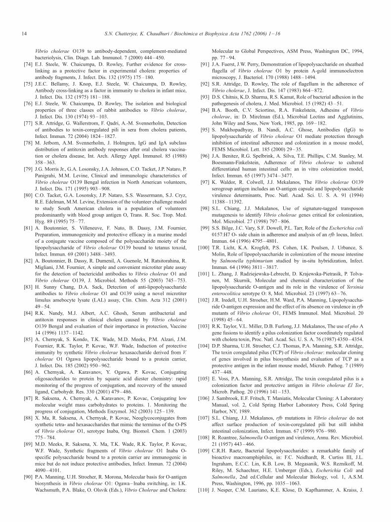
S.N. Chatterjee, K. Chaudhuri / Biochimica et Biophysica Acta 1762 (2006) 1–1614
Vibrio cholerae O139 to antibody-dependent, complement-mediated
bacteriolysis, Clin. Diagn. Lab. Immunol. 7 (2000) 444–450.
[74] E.J. Steele, W. Chaicumpa, D. Rowley, Further evidence for cross-
linking as a protective factor in experimental cholera: properties of
antibody fragments, J. Infect. Dis. 132 (1975) 175–180.
[75] J.E.C. Bellamy, J. Knop, E.J. Steele, W. Chaicumpa, D. Rowley,
Antibody cross-linking as a factor in immunity to cholera in infant mice,
J. Infect. Dis. 132 (1975) 181–188.
[76] E.J. Steele, W. Chaicumpa, D. Rowley, The isolation and biological
properties of three classes of rabbit antibodies to Vibrio cholerae,
J. Infect. Dis. 130 (1974) 93–103.
[77] S.R. Attridge, G. Wallerstrom, F. Qadri, A.-M. Svennerholm, Detection
of antibodies to toxin-coregulated pili in sera from cholera patients,
Infect. Immun. 72 (2004) 1824–1827.
[78] M. Jetborn, A.M. Svennerholm, J. Holmgren, IgG and IgA subclass
distribution of antitoxin antibody responses after oral cholera vaccina-
tion or cholera disease, Int. Arch. Allergy Appl. Immunol. 85 (1988)
358–363.
[79] J.G. Morris Jr., G.A. Losonsky, J.A. Johnson, C.O. Tacket, J.P. Nataro, P.
Panigrahi, M.M. Levine, Clinical and immunologic characteristics of
Vibrio cholerae O139 Bengal infection in North American volunteers,
J. Infect. Dis. 171 (1995) 903–908.
[80] C.O. Tacket, G.A. Losonsky, J.P. Nataro, S.S. Wassermann, S.J. Cryz,
R.E. Edelman, M.M. Levine, Extension of the volunteer challenge model
to study South American cholera in a population of volunteers
predominantly with blood group antigen O, Trans. R. Soc. Trop. Med.
Hyg. 89 (1995) 75–77.
[81] A. Boutonnier, S. Villeneuve, F. Nato, B. Dassy, J.M. Fournier,
Preparation, immunogenicity and protective efficacy in a murine model
of a conjugate vaccine composed of the polysaccharide moiety of the
lipopolysaccharide of Vibrio cholerae O139 bound to tetanus toxoid,
Infect. Immun. 69 (2001) 3488–3493.
[82] A. Boutonnier, B. Dassy, R. Dumenil, A. Guenole, M. Ratsitorahina, R.
Migliani, J.M. Fournier, A simple and convenient microtiter plate assay
for the detection of bactericidal antibodies to Vibrio cholerae O1 and
Vibrio cholerae O139, J. Microbiol. Methods 55 (2003) 745–753.
[83] H. Sunny Chang, D.A. Sack, Detection of anti-lipopolysaccharide
antibodies to Vibrio cholerae O1 and O139 using a novel microtiter
limulus amebocyte lysate (LAL) assay, Clin. Chim. Acta 312 (2001)
49–54.
[84] R.K. Nandy, M.J. Albert, A.C. Ghosh, Serum antibacterial and
antitoxin responses in clinical cholera caused by Vibrio cholerae
O139 Bengal and evaluation of their importance in protection, Vaccine
14 (1996) 1137–1142.
[85] A. Chernyak, S. Kondo, T.K. Wade, M.D. Meeks, P.M. Alzari, J.M.
Fournier, R.K. Taylor, P. Kovac, W.F. Wade, Induction of protective
immunity by synthetic Vibrio cholerae hexasaccharide derived from V.
cholerae O1 Ogawa lipopolysaccharide bound to a protein carrier,
J. Infect. Dis. 185 (2002) 950–962.
[86] A. Chernyak, A. Karavanov, Y. Ogawa, P. Kovac, Conjugating
oligosaccharides to protein by squaric acid diester chemistry: rapid
monitoring of the progress of conjugation, and recovery of the unused
ligand, Carbohydr. Res. 330 (2001) 479–486.
[87] R. Saksena, A. Chernyak, A. Karavanov, P. Kovac, Conjugating low
molecular weight mass carbohydrates to proteins. 1. Monitoring the
progress of conjugation, Methods Enzymol. 362 (2003) 125–139.
[88] X. Ma, R. Saksena, A. Chernyak, P. Kovac, Neoglycoconjugates from
synthetic tetra- and hexasaccharides that mimic the terminus of the O-PS
of Vibrio cholerae O1, serotype Inaba, Org. Biomol. Chem. 1 (2003)
775–784.
[89] M.D. Meeks, R. Saksena, X. Ma, T.K. Wade, R.K. Taylor, P. Kovac,
W.F. Wade, Synthetic fragments of Vibrio cholerae O1 Inaba O-
specific polysaccharide bound to a protein carrier are immunogenic in
mice but do not induce protective antibodies, Infect. Immun. 72 (2004)
4090–4101.
[90] P.A. Manning, U.H. Stroeher, R. Morona, Molecular basis for O-antigen
biosynthesis in Vibrio cholerae O1: Ogawa– Inaba switching, in: I.K.
Wachsmuth, P.A. Blake, O. Olsvik (Eds.), Vibrio Cholerae and Cholera:
Molecular to Global Perspectives, ASM Press, Washington DC, 1994,
pp. 77–94.
[91] J.A. Fuerst, J.W. Perry, Demonstration of lipopolysaccharide on sheathed
flagella of Vibrio cholerae O1 by protein A-gold immunoelectron
microscopy, J. Bacteriol. 170 (1988) 1488–1494.
[92] S.R. Attridge, D. Rowley, The role of flagellum in the adherence of
Vibrio cholerae, J. Infect. Dis. 147 (1983) 864–872.
[93] D.S. Chitnis, K.D. Sharma, R.S. Kamat, Role of bacterial adhesion in the
pathogenesis of cholera, J. Med. Microbiol. 15 (1982) 43–51.
[94] B.A. Booth, C.V. Sciortino, R.A. Finkelstein, Adhesins of Vibrio
cholerae, in: D. Mirelman (Ed.), Microbial Lectins and Agglutinins,
John Wiley and Sons, New York, 1985, pp. 169–182.
[95] S. Mukhopadhyay, B. Nandi, A.C. Ghose, Antibodies (IgG) to
lipopolysaccharide of Vibrio cholerae O1 mediate protection through
inhibition of intestinal adherence and colonization in a mouse model,
FEMS Microbiol. Lett. 185 (2000) 29–35.
[96] J.A. Benitez, R.G. Spelbrink, A. Silva, T.E. Phillips, C.M. Stanley, M.
Boesmann-Finkelstein, Adherence of Vibrio cholerae to cultered
differentiated human intestinal cells: an in vitro colonization model,
Infect. Immun. 65 (1997) 3474–3477.
[97] K. Waldor, R. Colwell, J.J. Mekalanos, The Vibrio cholerae O139
serogroup antigen includes an O-antigen capsule and lipopolysaccharide
virulence determinants, Proc. Natl. Acad. Sci. U. S. A. 91 (1994)
11388–11392.
[98] S.L. Chiang, J.J. Mekalanos, Use of signature-tagged transposon
mutagenesis to identify Vibrio cholerae genes critical for colonization,
Mol. Microbiol. 27 (1998) 797–806.
[99] S.S. Bilge, J.C. Vary, S.F. Dowell, P.L. Tarr, Role of the Escherichia coli
0157:H7 O- side chain in adherence and analysis of an rfb locus, Infect.
Immun. 64 (1996) 4795–4801.
[100] T.R. Licht, K.A. Krogfelt, P.S. Cohen, I.K. Poulsen, J. Urbance, S.
Molin, Role of lipopolysaccharide in colonization of the mouse intestine
by Salmonella typhimurium studied by in-situ hybridization, Infect.
Immun. 64 (1996) 3811–3817.
[101] L. Zhang, J. Radziejewska-Lebrecht, D. Krajewska-Pietrasik, P. Tolva-
nen, M. Skurnik, Molecular and chemical characterization of the
lipopolysaccharide O-antigen and its role in the virulence of Yersinia
enterocolitica serotype O: 8, Mol. Microbiol. 23 (1997) 63–76.
[102] J.R. Iredell, U.H. Stroeher, H.M. Ward, P.A. Manning, Lipopolysaccha-
ride O-antigen expression and the effect of its absence on virulence in rfb
mutants of Vibrio cholerae O1, FEMS Immunol. Med. Microbiol. 20
(1998) 45–64.
[103] R.K. Taylor, V.L. Miller, D.B. Furlong, J.J. Mekalanos, The use of pho A
gene fusions to identify a pilus colonization factor coordinately regulated
with cholera toxin, Proc. Natl. Acad. Sci. U. S. A. 76 (1987) 4350–4354.
[104] D.P. Sharma, U.H. Stroeher, C.J. Thomas, P.A. Manning, S.R. Attridge,
The toxin coregulated pilus (TCP) of Vibrio cholerae: molecular cloning
of genes involved in pilus biosynthesis and evaluation of TCP as a
protective antigen in the infant mouse model, Microb. Pathog. 7 (1989)
437–448.
[105] E. Voss, P.A. Manning, S.R. Attridge, The toxin coregulated pilus is a
colonization factor and protective antigen in Vibrio cholerae El Tor,
Microb. Pathog. 20 (1996) 141–153.
[106] J. Sambrook, E.F. Fritsch, T. Maniatis, Molecular Cloning: A Laboratory
Manual, vol. 2, Cold Spring Harbor Laboratory Press, Cold Spring
Harbor, NY, 1989.
[107] S.L. Chiang, J.J. Mekalanos, rfb mutations in Vibrio cholerae do not
affect surface production of toxin-coregulated pili but still inhibit
intestinal colonization, Infect. Immun. 67 (1999) 976–980.
[108] R. Roantree, Salmonella O-antigen and virulence, Annu. Rev. Microbiol.
21 (1957) 443–466.
[109] C.R.H. Raetz, Bacterial lipopolysaccharides: a remarkable family of
bioactive macroamphiphiles, in: F.C. Neidhardt, R. Curtiss III, J.L.
Ingraham, E.C.C. Lin, K.B. Low, B. Megasanik, W.S. Reznikoff, M.
Riley, M. Schaechter, H.E. Umberger (Eds.), Escherichia Coli and
Salmonella, 2nd edRCellular and Molecular Biology, vol. 1, A.S.M.
Press, Washington, 1996, pp. 1035–1063.
[110] J. Nesper, C.M. Lauriano, K.E. Klose, D. Kapfhammer, A. Kraiss, J.

S.N. Chatterjee, K. Chaudhuri / Biochimica et Biophysica Acta 1762 (2006) 1–16 15
Reidl, Characterization of Vibrio cholerae O1 El Tor galU and galE
mutants: influence on lipopolysaccharide structure, colonization and
biofilm formation, Infect. Immun. 69 (1) (2001) 435–445.
[111] J. Nesper, S. Schild, C.M. Lauriano, A. Kraiss, K.E. Klose, J. Reidl, Role
of Vibrio cholerae O139 surface polysaccharides in intestinal coloniza-
tion, Infect. Immun. 70 (2002) 5990–5996.
[112] S.R. Attridge, A. Fazeli, P.A. Manning, U.H. Stroeher, Isolation and
characterization of bacteriophage-resistant mutants of Vibrio cholerae
O139, Microb. Pathog. 30 (2001) 237–246.
[113] D.S. Merrell, D.L. Hava, A. Camilli, Identification of novel factors
involved in colonization and acid tolerance of Vibrio cholerae, Mol.
Microbiol. 43 (2002) 1471–1491.
[114] M. Maiti, S.N. Chatterjee, Characteristics of a Group IV cholera phage,
J. Gen. Virol. 13 (1971) 327–330.
[115] M. Maiti, P. Sur, S.N. Chatterjee, Aminosugar contents and phage
inactivating properties of lipopolysaccharide from cholera and El Tor
vibrios, Ann. Microbiol. (Inst. Pasteur) 128A (1977) 35–39.
[116] A. Guidolin, P.A. Manning, Bacteriophage CP-T1 of Vibrio cholerae.
Identification of the cell surface receptor, Eur. J. Biochem. 153 (1985)
89–94.
[117] S. Mukherjee, The bacteriophage susceptibility test in differentiating
Vibrio cholerae and Vibrio El Tor, Bull. World Health Org. 28 (1963)
333–336.
[118] E. Ribi, R.L. Anacker, R. Brown, W.T. Haskins, B. Malmgren, K.C.
Milner, J.A. Rudbach, Reaction of endotoxin and surfactants,
J. Bacteriol. 92 (1966) 1493–1509.
[119] A.A. Lindberg, Studies of a receptor for Felix O-1 phage in Salmonella
Minnesota, J. Gen. Microbiol. 48 (1967) 225–233.
[120] J. Riedl, J.J. Mekalanos, Characterization of Vibrio cholerae bacterio-
phage K139 and use of a novel mini-transposon to identify phage-
encoded virulence factor, Mol. Microbiol. 18 (1995) 685–701.
[121] J. Nesper, J. Blah, M. Fountouloakis, J. Reidl, Characterization of the
major control region of Vibrio cholerae bacteriophage K139: immunity,
exclusion and integration, J. Bacteriol. 181 (1999) 2902–2913.
[122] D. Kapfhammer, J. Blass, S. Evers, J. Reidl, Vibrio cholerae phage
K139: complete genome sequence and comparative genomics of related
phages, J. Bacteriol. 184 (2002) 6592–6601.
[123] J. Nesper, D. Kapfhammer, K.E. Klose, H. Merkert, J. Reidl,
Characterization of Vibrio cholerae O1 antigen as the bacteriophage
K139 receptor and identification of IS 1004 insertions aborting O1
antigen, J. Bacteriol. 182 (2000) 5097–5104.
[124] M.J. Albert, N.A. Bhuivan, A. Rahman, A.N. Ghosh, K. Hultenby, A.
Weintraub, S. Nahar, A.K. Kibriya, M. Ansaruzaman, T. Shimada, Phage
specific for Vibrio cholerae O139 Bengal, J. Clin. Microbiol. 34 (1996)
1843–1845.
[125] E.A. Jouravleva, G.A. McDonald, J.W. Marsh, R.K. Taylor, M. Boes-
man-Finkelstein, R.A. Finkelstein, The Vibrio cholerae mannose-
sensitive hemagglutinin is the receptor for a filamentous bacteriophage
from Vibrio cholerae O139, Infect. Immun. 66 (1998) 2535–2539.
[126] M.K. Waldor, J.J. Mekalanos, Lysogenic conversion by a filamentous
phage encoding cholera toxin, Science 272 (1996) 1910–1914.
[127] P.C. Adhikari, S.N. Chatterjee, Fimbriation and pellicle formation of
Vibrio El Tor, Ind. J. Med. Res. 57 (1969) 1897–1901.
[128] J.M. Tweedy, R.W.A. Park, W. Hodgkiss, Evidence for the presence of
fimbriae (pili) on vibrio species, J. Gen. Microbiol. 51 (1968) 235–244.
[129] P.I. Watnick, K.J. Fullner, R. Kolter, A role for the mannose-sensitive
hemagglutinin in biofilm formation by Vibrio cholerae El Tor,
J. Bacteriol. 181 (1999) 3606–3609.
[130] P.I. Watnick, C.M. Lauriano, K.E. Klose, L. Croal, R. Kolter, The
absence of a flagellum leads to altered colony morphology, biofilm
development and virulence in Vibrio cholerae O139, Mol. Microbiol. 39
(2001) 223–235.
[131] D.A. Chiavelli, J.W. Marsh, R.K. Taylor, The mannose-sensitive
hemagglutinin of Vibrio cholerae promotes adherence to zooplankton,
Appl. Environ. Microbiol. 67 (2001) 3220–3225.
[132] P.I. Watnick, R. Kolter, Steps in the development of a Vibrio cholerae
El Tor biofilm, Mol. Microbiol. 34 (1999) 586–595.
[133] F.H. Yildiz, G.K. Schoolnik, Vibrio cholerae O1 El Tor: identification of
a gene cluster required for the rugose colony type, exopolysaccharide
production, chlorine resistance and biofilm formation, Proc. Natl. Acad.
Sci. U. S. A. 96 (1999) 4028–4033.
[134] Y. Mizunoe, S.N. Wai, A. Takade, S.I. Yoshida, Isolation and
characterization of rugose form of Vibrio cholerae O139 strain M010,
Infect. Immun. 67 (1999) 958–963.
[135] S.N. Wai, Y. Mizunoe, A. Takade, S.I. Kawabata, S.I. Yoshida, Vibrio
cholerae O1 strain TSI-4 produces the exopolysaccharide materials that
determine colony morphology, stress resistance and biofilm formation,
Appl. Environ. Microbiol. 64 (1998) 3648–3655.
[136] F.H. Yildiz, N.A. Dolganov, G.K. Schoolnik, VpsR, a member of the
response regulators of the two-component regulatory system, is
required for expression of vps biosynthesis genes and EPS (Etr)-
associated phenotypes in Vibrio cholerae O1 El Tor, J. Bacteriol. 183
(2001) 1716–1726.
[137] G. Morris, M.B. Sztein, E.W. Rice, J.P. Nataro, G.A. Losonsky, P.
Panigrahi, C.O. Tacket, J.A. Johnson, Vibrio cholerae O1 can assume a
chlorine-resistant rugose survival form that is virulent for humans,
J. Infect. Dis. 174 (1996) 1364–1368.
[138] J.F. Heidelberg, J.A. Eisen, W.C. Nelson, R.A. Clayton, M.I. Guinn, R.J.
Dodson, D.H. Haft, E.K. Hickey, J.D. Peterson, L. Umayam, S.R. Gill,
K.E. Nelson, T.D. Read, H. Tettelin, D. Richardson, M.D. Ermolaeva, J.
Vamathevan, S. Bass, H. Quin, I. Dragoi, P. Sellers, L. McDonald, T.
Utterback, R.D. Fleishmann, W.C. Nierman, O. White, DNA sequence of
both chromosomes of the cholera pathogen Vibrio cholerae, Nature 406
(2000) 477–483.
[139] G.K. Schoolnik, M.I. Voskuil, D. Schnappinger, F.H. Yildiz, K. Meibom,
N.A. Dolganov, M.A. Wilson, K.H. Chong, Whole genome DNA
microarray expression analysis of biofilm development by Vibrio
cholerae O1 El Tor, Methods Enzymol. 336 (2001) 3–19.
[140] A.J. Hango, P.I. Watnick, Vibrio cholerae CytR is a repressor of biofilm
development, Mol. Microbiol. 45 (2002) 471–483.
[141] H.L. Rocchetta, L.L. Burrows, J.S. Lam, Genetics of O-antigen
biosynthesis in Pseudomonas aeruginosa, Microbiol. Mol. Biol. Rev.
63 (1999) 523–553.
[142] E.C.C. Lin, Dissimilatory pathways for sugars, polyols and carboxylates,
in: F.C. Neidhardt, R. Curtiss III, J.L. Ingraham, E.C.C. Lin, K.B. Low,
B. Megasanik, W.C. Reznikoff, M. Riley, M. Schaechter, H.E. Umberger
(Eds.), Escherichia Coli and Salmonella: Cellular and Molecular
Biology, 2nd edR, American Society for Microbiology, Washington,
DC, 1996, pp. 307–342.
[143] K. Kierek, P.I. Watnick, Environmental determinants of Vibrio
cholerae biofilm development, Appl. Environ. Microbiol. 69 (2003)
5079–5088.
[144] K. Kierek, P.I. Watnick, The Vibrio cholerae O139 O-antigen polysac-
charide is essential for Ca2+-dependent biofilm development in sea water,
Proc. Natl. Acad. Sci. U. S. A. 100 (2003) 14357–14362.
[145] J.A. Johnson, C.A. Salles, P. Panigrahi, M.J. Albert, A.C. Wright, R.J.
Johnson, J.G. Morris, V. cholerae O139 synonym Bengal is closely
related to V. cholerae El Tor but has important differences, Infect.
Immun. 62 (1994) 2108–2110.
[146] A. Weintraub, G. Widmalm, P.E. Jansson, K. Hultenby, M.J. Albert,
Vibrio cholerae O139 Bengal possesses a capsular polysaccharide which
may confer increased virulence, Microb. Pathog. 16 (1994) 235–241.
[147] L.E. Comstock, D. Maneval, P. Panigrahi, A. Joseph, M.M. Leine, J.B.
Kaper, J.G. Morris, J.A. Johnson, The capsule and O-antigen of Vibrio
cholerae O139 Bengal are associated with a genetic region not present in
Vibrio cholerae O1, Infect. Immun. 63 (1995) 317–323.
[148] J.A. Johnson, P. Panigrahi, J.G. Morris Jr., Non-O1 Vibrio cholerae
NRT36S produces a polysaccharide capsule that determines colony
morphology, serum resistance and virulence in mice, Infect. Immun. 60
(1992) 864–869.
[149] M.V. Jesudason, A.M. Cherian, T.J. John, Blood stream invasion by
Vibrio cholerae O139, Lancet 342 (1993) 431.
[150] S. Safrin, J.G. Morris, M. Adams, V. Pons, R. Jacob, J.E. Conte, Non-O1
Vibrio cholerae bacteremia: a case report and review, Rev. Infect. Dis. 10
(1987) 1012–1017.
[151] P. Panigrahi, S. Srinivas, J.A. Johnson, L.J. DeTolla, Modulation of

S.N. Chatterjee, K. Chaudhuri / Biochimica et Biophysica Acta 1762 (2006) 1–1616
immunity in non-O1 Vibrio cholerae, 92nd Annu. Meet Am. Soc.
Microbiol., 1992 (Abstr. B-316).
[152] C.R.H. Raetz, C. Whitfield, Lipopolysaccharide endotoxins, Annu. Rev.
Biochem. 71 (2002) 635–700.
[153] M. Caroff, D. Karibian, J.-M. Cavaillon, N. Haeffner-Cavaillon,
Structural and functional analysis of bacterial lipopolysaccharides,
Microbes. infect. 4 (2002) 915–926.
[154] S.H. Diks, D.J. Richel, M.P. Peppelenbosch, LPS signal transduction:
the picture is becoming more complex, Curr. Top. Med. Chem. 4
(2004) 1–12.
[155] S.M. Zughaier, S.M. Zimmer, A. Datta, R.W. Carlson, D.S. Stephens,
Differential induction of the Toll-like receptor 4-MyD88-dependent
and -independent signaling pathways by endotoxins, Infect. Immun.
73 (2005) 2940–2950.
[156] S.M. Faruque, I.B. Naser, M.J. Islam, A.S.G. Faruque, A.N. Ghosh, G.B.
Nair, D.A. Sack, J.J. Mekalanos, Seasonal epidemics of cholera inversely
correlate with the prevalence of environmental cholera phages, Proc.
Natl. Acad. Sci. U. S. A. 102 (2005) 1702–1707.
[157] S.M. Faruque, M.J. Islam, Q.S. Ahmad, A.S.G. Faruque, D.A. Sack,
G.B. Nair, J.J. Mekalanos, Self-limiting nature of seasonal cholera
epidemics: role of host-mediated amplification of phage, Proc. Natl.
Acad. Sci. U. S. A. 102 (2005) 6119–6124.
[158] L. Carillo, R.H. Gilman, R.E. Mantle, N. Nunez, J. Watanabe, J. Morou,
V. Quispe, A. Ramirez-Ramos, et al., Rapid detection of Vibrio cholerae
O1 in stools of Peruvian cholera patients by using monoclonal
immunodiagnostic kits, J. Clin. Microbiol. 32 (1994) 856–857.
[159] R.R. Colwell, J.A.K. Hasan, A. Huq, L. Loomis, R.J. Siebeling, M.
Torres, S. Galvez, S. Islam, M.T. Tamplin, D. Bernstein, Development
and evaluation of a rapid, simple, sensitive, monoclonal antibody-based
co-agglutination test for direct determination of Vibrio cholerae O1,
FEMS Microbiol. Lett. 97 (1992) 215–220.
[160] V. Agarwal, M. Biswas, A.A. Pathak, A.M. Sauji, Rapid detection of
Vibrio cholerae O139 in faecal specimens by co-agglutination, Ind.
J. Med. Res. 101 (1995) 55–56.
[161] J.A.K. Hasan, A. Huq, G.B. Nair, S. Garg, A.K. Mukhopadhyay, L.
Loomis, D. Bernstein, R.R. Colwell, Development and testing of
monoclonal antibody-based rapid immunodiagnostic test kits for direct
detection of Vibrio cholerae O139 synonym Bengal, J. Clin. Microbiol.
33 (1995) 2935–2939.
[162] J.A.K. Hasan, A. Huq, M.L. Tamplin, R.J. Siebeling, R.R. Colwell, A
novel kit for rapid detection of Vibrio cholerae O1, J. Clin. Microbiol. 32
(1994) 249–252.
[163] F. Qadri, J.A.K. Hasan, J. Hussain, A. Chowdhury, Y.A. Begum, T.
Azim, L. Loomis, R.B. Sack, M.J. Albert, Evaluation of the monoclonal
antibody-based kit Bengal SMART for rapid detection of Vibrio cholerae
O139 synonym Bengal in stool samples, J. Clin. Microbiol. 33 (1995)
732–734.
[164] F. Nato, A. Boutonnier, M. Rajerison, P. Grosjean, S. Dartevelle, A.
Guenole, N.A. Bhuiyan, D.A. Sack, G.B. Nair, J.M. Fournier, S.
Chanteau, One step immunochromatographic dipstick tests for rapid
detection of Vibrio cholerae O1 and O139 in stool samples, Clin. Diagn.
Lab. Immunol. 10 (2003) 476–478.
[165] A. Robert-Pillot, S. Baron, J. Lesne, J.-M. Fournier, M.L. Quilici,
Improved specific detection of Vibrio cholerae in environmental water
samples by culture on selective medium and colony hybridization assay
with an oligonucleotide probe, FEMSMicrobiol. Ecol. 40 (2002) 39–46.
[166] Anonymous, Research Report 1999–2003. Institut Pasteur, 2003, p. 44.
[167] J.J. Mekalanos, M.K. Waldor, C.L. Gardel, T.S. Coster, J. Kenner, K.P.
Killeen, D.T. Bealtie, A. Trofa, D.N. Taylor, J.C. Sadoff, Live cholera
vaccines: perspectives on their construction and safety, Bull. Inst. Pasteur
93 (1995) 255–262.
[168] M.M. Levine, J.B. Kaper, Live oral cholera vaccine: from principle to
product, Bull. Inst. Pasteur 93 (1995) 243–253.
[169] S.R. Attridge, C. Dearlove, L. Beyer, L. van den Bosch, A. Howles, J.
Hackett, R. Morona, J. Lebrooy, D. Rowley, Characterization and
immunogenicity of EX880, a Salmonella typhi Ty21a-based clone
which produces Vibrio cholerae O-antigen, Infect. Immun. 59 (1991)
5279–5284.
[170] B.D. Forrest, J.T. Labrooy, S.R. Attridge, G. Bochm, L. Beyer, R.
Morona, D.J.C. Shearman, D. Rowley, Immunogenicity of a candidate
live oral typhoid/cholera hybrid vaccine in human, J. Infect. Dis. 159
(1989) 145–146.
[171] C.O. Tacket, B. Forrest, R. Morona, S.R. Attridge, J. Labrooy, B.D. Tall,
M. Reymann, D. Rowley, M.M. Levine, Safety, immunogenicity and
efficacy against cholera challenge in humans of a typhoid-cholera hybrid
vaccine derived from Salmonella typhi Ty21a, Infect. Immun. 58 (1990)
1620–1627.
[172] J.B. Robbins, R. Schneerson, S.C. Szu, Hypothesis: serum IgG antibody
is sufficient to confer protection against infectious diseases by inactivat-
ing the inoculum, J. Infect. Dis. 171 (1995) 1387–1398.
[173] D.A. Sack, J.D. Clemens, S. Huda, J.R. Harris, M.R. Khan, J.
Chakraborty, M. Yunis, J. Gomes, O. Siddique, F. Ahmed, B.A. Kay,
F.P.L. van Loon, M.R. Rao, A.-M. Svennerholm, J. Holmgren,
Antibody responses after immunization with killed oral cholera
vaccines during the 1985 vaccine field trial in Bangladesh, J. Infect.
Dis. 164 (1991) 407–411.
[174] S.C. Szu, R. Gupta, J.B. Robbins, Induction of serum vibriocidal
antibodies by O-specific polysaccharide-protein conjugate vaccines for
prevention of cholera, in: I.K. Wachsmuth, P.A. Blake, O. Olsvik (Eds.),
Vibrio Cholerae and Cholera, American Society for Microbiology,
Washington, DC, 1994, pp. 381–394.
[175] R.K. Gupta, D.N. Taylor, D.A. Bryia, J.B. Robbins, S.S.C. Szu, Phase
I evaluation of Vibrio cholerae O1, serotype Inaba, polysaccharide-
cholera toxin conjugates in adult volunteers, Infect. Immun. 66 (1998)
3095–3099.
[176] J.A. Johnson, A. Joseph, J.G. Morris, Capsular polysaccharide-protein
conjugate vaccines against Vibrio cholerae O139 Bengal, Bull. Inst.
Pasteur 93 (1995) 285–290.
Copyright © 2022 FDOKUMEN