A new Vibrio cholerae sRNA modulates colonization and affects release of outer membrane vesicles
12
A new Vibrio cholerae sRNA modulates colonization and affects release of outer membrane vesicles Tianyan Song, 1 Franziska Mika, 2 Barbro Lindmark, 1 Zhi Liu, 3 Stefan Schild, 4 Anne Bishop, 4 Jun Zhu, 3 Andrew Camilli, 4 Jörgen Johansson, 1 Jörg Vogel 2 and Sun Nyunt Wai 1 * 1 Department of Molecular Biology, Umeå University, SE-901 87 Umeå, Sweden. 2 RNA Biology Group, Max Planck Institute for Infection Biology, 10117 Berlin, Germany. 3 Departments of Microbiology, Physics, and Biology, University of Pennsylvania, Philadelphia, PA 19104, USA. 4 Howard Hughes Medical Institute and the Department of Molecular Biology an Microbiology, Tufts University School of Medicine, Boston, MA 02111, USA. Summary We discovered a new small non-coding RNA (sRNA) gene, vrrA of Vibrio cholerae O1 strain A1552. A vrrA mutant overproduces OmpA porin, and we demon- strate that the 140 nt VrrA RNA represses ompA trans- lation by base-pairing with the 5 region of the mRNA. The RNA chaperone Hfq is not stringently required for VrrA action, but expression of the vrrA gene requires the membrane stress sigma factor, s E , suggesting that VrrA acts on ompA in response to periplasmic protein folding stress. We also observed that OmpA levels inversely correlated with the number of outer membrane vesicles (OMVs), and that VrrA increased OMV production comparable to loss of OmpA. VrrA is the first sRNA known to control OMV formation. More- over, a vrrA mutant showed a fivefold increased ability to colonize the intestines of infant mice as compared with the wild type. There was increased expression of the main colonization factor of V. cholerae, the toxin co-regulated pili, in the vrrA mutant as monitored by immunoblot detection of the TcpA protein. VrrA overproduction caused a distinct reduction in the TcpA protein level. Our findings suggest that VrrA contributes to bacterial fitness in certain stressful environments, and modulates infec- tion of the host intestinal tract. Introduction Vibrio cholerae is a Gram-negative bacterium that causes the acute, severe diarrhoeal disease cholera. Its natural ecosystem includes aquatic environments in endemic locations. Two factors are critical to V. cholerae virulence – cholera toxin (CT) and an intestinal colonization factor known as the toxin co-regulated pilus (TCP). Poorly char- acterized environmental cues influence the expression of CT and TCP in vivo (Faruque et al., 1998). Two sensory proteins, ToxR and TcpP, likely play a role in detection of the environmental signals, and activate the transcription of genes involved in TCP and CT expression through the expression of ToxT (Lee et al., 1999). Outer membrane vesicles (OMVs) are produced by a wide variety of Gram-negative bacteria (Beveridge, 1999) including Vibrio species (Kondo et al., 1993) during their growth. They contain outer membrane proteins, lipopolysaccharides, phospholipids and, as the vesicles are being released from the surface, they entrap some of the underlying periplasm. Different hypotheses have been proposed for the function of OMVs. OMVs have been suggested to promote the adherence, the transfer of bac- terial DNA and the delivery of virulence factors to bacterial or eukaryotic cells (Kuehn and Kesty, 2005; Mashburn- Warren and Whiteley, 2006). We have previously shown that OMVs contribute to the delivery of active ClyA cyto- toxin, a-haemolysin and CNF1 from Escherichia coli to mammalian cells (Wai et al., 2003; Balsalobre et al., 2006; Kouokam et al., 2006). Recently, it was suggested that OMV production is a physiological consequence of Gram-negative bacteria and that OMVs are a component of the matrix of Gram-negative bacterial biofilms (School- ing and Beveridge, 2006). In their study, they found that OMVs from biofilm contained more proteolytic activity than those from planktonic cells. They speculated that OMVs could act as decoys to reduce inimical agents within bio- films before they can attack cells. OMVs are also very promising for different biotechnological applications such as the delivery of antibiotics or as efficient vaccine particles. In contrast to the extensive research on the biological functions of OMVs, very little is known about the Accepted 27 July, 2008. *For correspondence. E-mail sun.nyunt. [email protected]; Tel. (+46) 90 7856704, Fax (+46) 90 772630. Re-use of this article is permitted in accordance with the Creative Commons Deed, Attribution 2.5, which does not permit commercial exploitation. OnlineOpen: This article is available free online at www.blackwell-synergy.com Molecular Microbiology (2008) 70(1), 100–111 doi:10.1111/j.1365-2958.2008.06392.x First published online 15 August 2008 © 2008 The Authors Journal compilation © 2008 Blackwell Publishing Ltd
-
Upload
uni-wuerzburg -
Category
Documents
-
view
5 -
download
0
Transcript of A new Vibrio cholerae sRNA modulates colonization and affects release of outer membrane vesicles

A new Vibrio cholerae sRNA modulates colonization andaffects release of outer membrane vesicles
Tianyan Song,1 Franziska Mika,2 Barbro Lindmark,1
Zhi Liu,3 Stefan Schild,4 Anne Bishop,4 Jun Zhu,3
Andrew Camilli,4 Jörgen Johansson,1 Jörg Vogel2
and Sun Nyunt Wai1*1Department of Molecular Biology, Umeå University,SE-901 87 Umeå, Sweden.2RNA Biology Group, Max Planck Institute for InfectionBiology, 10117 Berlin, Germany.3Departments of Microbiology, Physics, and Biology,University of Pennsylvania, Philadelphia, PA 19104,USA.4Howard Hughes Medical Institute and the Departmentof Molecular Biology an Microbiology, Tufts UniversitySchool of Medicine, Boston, MA 02111, USA.
Summary
We discovered a new small non-coding RNA (sRNA)gene, vrrA of Vibrio cholerae O1 strain A1552. A vrrAmutant overproduces OmpA porin, and we demon-strate that the 140 nt VrrA RNA represses ompA trans-lation by base-pairing with the 5� region of the mRNA.The RNA chaperone Hfq is not stringently required forVrrA action, but expression of the vrrA gene requiresthe membrane stress sigma factor, sE, suggestingthat VrrA acts on ompA in response to periplasmicprotein folding stress. We also observed that OmpAlevels inversely correlated with the number of outermembrane vesicles (OMVs), and that VrrA increasedOMV production comparable to loss of OmpA. VrrA isthe first sRNA known to control OMV formation. More-over, a vrrA mutant showed a fivefold increasedability to colonize the intestines of infant mice ascompared with the wild type. There was increasedexpression of the main colonization factor ofV. cholerae, the toxin co-regulated pili, in the vrrAmutant as monitored by immunoblot detection of theTcpA protein. VrrA overproduction caused a distinctreduction in the TcpA protein level. Our findingssuggest that VrrA contributes to bacterial fitness in
certain stressful environments, and modulates infec-tion of the host intestinal tract.
Introduction
Vibrio cholerae is a Gram-negative bacterium that causesthe acute, severe diarrhoeal disease cholera. Its naturalecosystem includes aquatic environments in endemiclocations. Two factors are critical to V. cholerae virulence– cholera toxin (CT) and an intestinal colonization factorknown as the toxin co-regulated pilus (TCP). Poorly char-acterized environmental cues influence the expression ofCT and TCP in vivo (Faruque et al., 1998). Two sensoryproteins, ToxR and TcpP, likely play a role in detection ofthe environmental signals, and activate the transcriptionof genes involved in TCP and CT expression through theexpression of ToxT (Lee et al., 1999).
Outer membrane vesicles (OMVs) are produced bya wide variety of Gram-negative bacteria (Beveridge,1999) including Vibrio species (Kondo et al., 1993) duringtheir growth. They contain outer membrane proteins,lipopolysaccharides, phospholipids and, as the vesiclesare being released from the surface, they entrap some ofthe underlying periplasm. Different hypotheses have beenproposed for the function of OMVs. OMVs have beensuggested to promote the adherence, the transfer of bac-terial DNA and the delivery of virulence factors to bacterialor eukaryotic cells (Kuehn and Kesty, 2005; Mashburn-Warren and Whiteley, 2006). We have previously shownthat OMVs contribute to the delivery of active ClyA cyto-toxin, a-haemolysin and CNF1 from Escherichia colito mammalian cells (Wai et al., 2003; Balsalobre et al.,2006; Kouokam et al., 2006). Recently, it was suggestedthat OMV production is a physiological consequence ofGram-negative bacteria and that OMVs are a componentof the matrix of Gram-negative bacterial biofilms (School-ing and Beveridge, 2006). In their study, they found thatOMVs from biofilm contained more proteolytic activity thanthose from planktonic cells. They speculated that OMVscould act as decoys to reduce inimical agents within bio-films before they can attack cells. OMVs are also verypromising for different biotechnological applications suchas the delivery of antibiotics or as efficient vaccineparticles.
In contrast to the extensive research on the biologicalfunctions of OMVs, very little is known about the
Accepted 27 July, 2008. *For correspondence. E-mail [email protected]; Tel. (+46) 90 7856704, Fax (+46) 90 772630.Re-use of this article is permitted in accordance with the CreativeCommons Deed, Attribution 2.5, which does not permit commercialexploitation.
� OnlineOpen: This article is available free online at www.blackwell-synergy.com
Molecular Microbiology (2008) 70(1), 100–111 � doi:10.1111/j.1365-2958.2008.06392.xFirst published online 15 August 2008
© 2008 The AuthorsJournal compilation © 2008 Blackwell Publishing Ltd

mechanism and regulation of the formation of OMVs. OMVformation has been suggested to be linked to turgor pres-sure of the cell envelope during bacterial growth (Zhouet al., 1998). Release of OMVs is highly dependent on theenvelope structure. Defects in proteins either linking theouter membrane to the peptidoglycan layer or involved in astructural network between the inner, outer membranesand the peptidoglycan layer result in the shedding of largeamounts of OMVs (McBroom and Kuehn, 2007).
In the past few years, it has become increasingly clearthat small non-coding RNAs (sRNAs) regulate manydiverse cellular processes, including acid resistance andiron homeostasis (Majdalani et al., 2005), and the viru-lence of pathogens (Romby et al., 2006; Toledo-Aranaet al., 2007). A major class of sRNAs in bacteria functionsby base-pairing with target mRNAs, and positively ornegatively regulates translation and/or stability of thesemessages. This class of sRNAs usually requires the RNAchaperone Hfq as a cofactor, which facilitates the interac-tion between sRNAs and target mRNAs (Storz et al.,2004; Valentin-Hansen et al., 2004).
Recent systematic searches (Vogel and Sharma, 2005)revealed that E. coli expresses close to 100 sRNAs, andthe total number of sRNAs in a typical enterobacteriummay well range in the hundreds (Hershberg et al., 2003;Zhang et al., 2004). To date, numerous sRNAs have beenpredicted in V. cholerae (Livny et al., 2005), and several ofthese candidates have been confirmed by Northern blotanalysis. Nine sRNAs have been assigned cellular func-tions in V. cholerae: the homologue of E. coli RyhB sRNA,which is involved in iron utilization (Davis et al., 2005; Meyet al., 2005); MicX sRNA, which negatively regulates anuncharacterized outer membrane protein (OMP) and aperiplasmic component of a peptide ABC transporter(Davis and Waldor, 2007); seven sRNAs, i.e. Qrr1–Qrr4,CsrB–CsrD, which are involved in quorum-sensing regu-lation (Lenz et al., 2004; 2005).
Here we report on the discovery of a new sRNA inV. cholerae, to which we will refer as VrrA (Vibrio regulatoryRNA of ompA). VrrA positively regulates OMV releasethrough downregulation of outer membrane protein OmpA.Inactivation of VrrA resulted in increased colonization ofV. cholerae in the infant mouse colonization assay.
Results
Characterization of a new sRNA, VrrA, in V. cholerae
We became aware of the vrrA gene when analysing amini-Tn5 transposon mutant (SNW6) from a library ofV. cholerae El Tor O1 strain A1552 (Vaitkevicius et al.,2006), which was found to carry a mini-Tn5 insertion in theintergenic region between vc1741 and vc1743 (Fig. 1A).Inspection and sequence comparison with other Vibriostrains of the disrupted region suggested the existence of
a previously unrecognized sRNA gene. We successfullyvalidated this prediction by Northern blot analysis, whichdetected a ~140 nt RNAexpressed from the positive strandin samples of the wild type (Fig. 4) but not of the SNW6mutant strain (data not shown). Subsequent 5′ RACEanalysis of this sRNA species (Fig. 1B) identified the tran-scription start site (+1) shown in Fig. 1A, which is locatedapproximately 140 bp upstream of a putative Rho-independent terminator downstream. The 5′ RACE analy-sis was performed to determine the transcription start (+1)site of the vrrA downstream gene vc1743 (Fig. S1). The+1 site of vc1743 was the same as that of VrrA. Thisindicates that vc1743 would be co-transcribed with vrrA.However, in the Northern blot analysis, the VrrA probenever detected the reaction band larger than 140 nt. Underthe same detection condition, there was no detectablesignal with a vc1743 probe (data not shown). This suggeststhat, although vc1743 can be co-transcribed with vrrA, thelevel of readthrough of the proposed terminator (Fig. 1A) isvery low under the growth conditions that we used in thisstudy. Interestingly, the V. cholerae VrrA promoter regioncontains a sequence that is a perfect match to the previ-ously reported consensus of promoters recognized by thealternative sigma factor, sE (Rhodius et al., 2006; Skovi-erova et al., 2006). Using BLASTN searches, we identifiedvrrA homologues in other Vibrio species, and all of thesegenes show conservation of the sE binding sites in the vrrApromoter region (Fig. 1C). In order to analyse the role ofRpoE in regulation of vrrA expression, we constructed anin-frame deletion rpoE mutant and tested the level of vrrAexpression by Northern blot analysis. The expression ofVrrA was totally abolished in the DrpoE mutant strain(Fig. 1D, left). Furthermore, a cloned copy of vrrA with itspromoter region (plasmid pTS2) was introduced into Sal-monella typhimurium strain SL1344 and its otherwiseisogenic DrpoE mutant strain JVS-01028 (Papenfort et al.,2006), to test the sE requirement in the heterologousbacterial system. S. typhimurium carrying pTS2 expressedVrrA in a manner that was totally dependent on a functionalsE (Fig. 1D, right). Taken together, our results providedconclusive genetic evidence that vrrA expression is directlycontrolled by the sE factor.
OmpA is downregulated by VrrA
To investigate the role of VrrA, we made comparisonsusing the wild-type V. cholerae strain A1552 and the vrrAdeletion strain DNY7. When comparing the whole-cellprotein profiles by SDS-PAGE, we noticed that a protein at34 kDa appeared more abundant in the vrrA mutant incomparison with the wild-type strain A1552 (Fig. 2A,panel I; lanes 2 and 3). The protein was identified as theputative outer membrane porin protein OmpA by massspectrometry analysis. The altered level of OmpA wasfurther confirmed by Western blot analysis using anti-
VrrA controls OmpA expression and virulence 101
© 2008 The AuthorsJournal compilation © 2008 Blackwell Publishing Ltd, Molecular Microbiology, 70, 100–111

102 T. Song et al. �
© 2008 The AuthorsJournal compilation © 2008 Blackwell Publishing Ltd, Molecular Microbiology, 70, 100–111
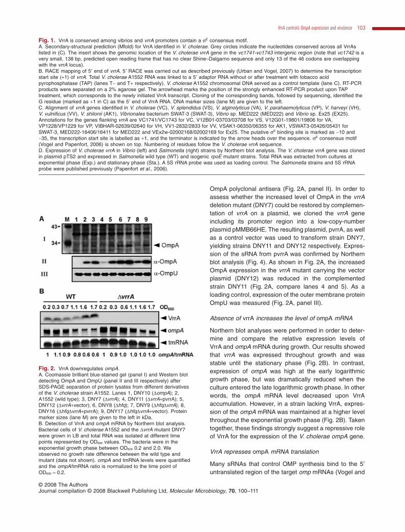
OmpA polyclonal antisera (Fig. 2A, panel II). In order toassess whether the increased level of OmpA in the vrrAdeletion mutant (DNY7) could be restored by complemen-tation of vrrA on a plasmid, we cloned the vrrA geneincluding its promoter region into a low-copy-numberplasmid pMMB66HE. The resulting plasmid, pvrrA, as wellas a control vector was used to transform strain DNY7,yielding strains DNY11 and DNY12 respectively. Expres-sion of the sRNA from pvrrA was confirmed by Northernblot analysis (Fig. 4). As shown in Fig. 2A, the increasedOmpA expression in the vrrA mutant carrying the vectorplasmid (DNY12) was reduced in the complementedstrain DNY11 (Fig. 2A, compare lanes 4 and 5). As aloading control, expression of the outer membrane proteinOmpU was measured (Fig. 2A, panel III).
Absence of vrrA increases the level of ompA mRNA
Northern blot analyses were performed in order to deter-mine and compare the relative expression levels ofVrrA and ompA mRNA during growth. Our results showedthat vrrA was expressed throughout growth and wasstable until the stationary phase (Fig. 2B). In contrast,expression of ompA was high at the early logarithmicgrowth phase, but was dramatically reduced when theculture entered the late logarithmic growth phase. In otherwords, the ompA mRNA level decreased upon VrrAaccumulation. However, in a strain lacking VrrA, expres-sion of the ompA mRNA was maintained at a higher levelthroughout the exponential growth phase (Fig. 2B). Takentogether, these findings strongly suggest a repressive roleof VrrA for the expression of the V. cholerae ompA gene.
VrrA represses ompA mRNA translation
Many sRNAs that control OMP synthesis bind to the 5′untranslated region of the target omp mRNAs (Vogel and
Fig. 1. VrrA is conserved among vibrios and vrrA promoters contain a sE consensus motif.A. Secondary-structural prediction (Mfold) for VrrA identified in V. cholerae. Grey circles indicate the nucleotides conserved across all VrrAslisted in (C). The insert shows the genomic location of the V. cholerae vrrA gene in the vc1741-vc1743 intergenic region (note that vc1742 is avery small, 138 bp, predicted open reading frame that has no clear Shine–Dalgarno sequence and only 13 of the 46 codons are overlappingwith the vrrA locus).B. RACE mapping of 5′ end of vrrA. 5′ RACE was carried out as described previously (Urban and Vogel, 2007) to determine the transcriptionstart site (+1) of vrrA. Total V. cholerae A1552 RNA was linked to a 5′ adaptor RNA without or after treatment with tobacco acidpyrophosphatase (TAP) (lanes T- and T+ respectively). V. cholerae A1552 chromosomal DNA served as a control template (lane C). RT-PCRproducts were separated on a 2% agarose gel. The arrowhead marks the position of the strongly enhanced RT-PCR product upon TAPtreatment, which corresponds to the newly initiated VrrA transcript. Cloning of the corresponding bands, followed by sequencing, identified theG residue (marked as +1 in C) as the 5′ end of VrrA RNA. DNA marker sizes (lane M) are given to the left.C. Alignment of vrrA genes identified in V. cholerae (VC), V. splendidus (VS), V. alginolyticus (VA), V. parahaemolyticus (VP), V. harveyi (VH),V. vulnificus (VV), V. shilonii (AK1), Vibrionales bacterium SWAT-3 (SWAT-3), Vibrio sp. MED222 (MED222) and Vibrio sp. Ex25 (EX25).Annotations for the genes flanking vrrA are VC1741/VC1743 for VC, V12B01-03703/03708 for VS, V12G01-19801/19806 for VA,VP1228/VP1229 for VP, VIBHAR-02639/02640 for VH, VV1-2832/2833 for VV, VSAK1-06350/06355 for AK1, VSWAT3-05426/05431 forSWAT-3, MED222-16406/16411 for MED222 and VEx2w-02002168/02002169 for Ex25. The putative sE binding site is marked as -10 and-35, the transcription start site is labelled as +1, and the terminator is indicated by the arrow heads over the sequence. sE consensus motif(Vogel and Papenfort, 2006) is shown on top. Numbering of residues follow the V. cholerae vrrA sequence.D. Expression of V. cholerae vrrA in Vibrio (left) and Salmonella (right) strains by Northern blot analysis. The V. cholerae vrrA gene was clonedin plasmid pTS2 and expressed in Salmonella wild type (WT) and isogenic rpoE mutant strains. Total RNA was extracted from cultures atexponential phase (Exp.) and stationary phase (Sta.). A 5S rRNA probe was used as loading control. The Salmonella strains and 5S rRNAprobe were published previously (Papenfort et al., 2006).
Fig. 2. VrrA downregulates ompA.A. Coomassie brilliant blue-stained gel (panel I) and Western blotdetecting OmpA and OmpU (panel II and III respectively) afterSDS-PAGE separation of protein lysates from different derivativesof the V. cholerae strain A1552. Lanes 1, DNY10 (DompA); 2,A1552 (wild type); 3, DNY7 (DvrrA); 4, DNY11 (DvrrA+pvrrA); 5,DNY12 (DvrrA+vector); 6, DNY8 (Dhfq); 7, DNY9 (DhfqDvrrA); 8,DNY16 (DhfqDvrrA+pvrrA); 9, DNY17 (DhfqDvrrA+vector). Proteinmarker sizes (lane M) are given to the left in kDa.B. Detection of VrrA and ompA mRNA by Northern blot analysis.Bacterial cells of V. cholerae A1552 and the DvrrA mutant DNY7were grown in LB and total RNA was isolated at different timepoints represented by OD600 values. The bacteria were in theexponential growth phase between OD600 0.2 and 2.0. Weobserved no growth rate difference between the wild type andmutant (data not shown). ompA and tmRNA levels were quantifiedand the ompA/tmRNA ratio is normalized to the time point ofOD600 = 0.2.
VrrA controls OmpA expression and virulence 103
© 2008 The AuthorsJournal compilation © 2008 Blackwell Publishing Ltd, Molecular Microbiology, 70, 100–111

Papenfort, 2006). Bioinformatic predictions of the VrrA–ompA interaction with the RNAhybrid program (Rehms-meier et al., 2004) revealed that a region of VrrA waspartially complementary to nucleotides encompassing theribosome binding site and part of the coding region of theompA mRNA (Fig. 3A). In addition, VrrA homologues fromother Vibrio species also displayed complementarity tothe translation initiation region of the ompA mRNA ofthese strains (Fig. S2). This predicts that VrrA binds to theribosome binding region of the ompA transcript, inhibitingribosome entry and thus destabilizing this mRNA.
In order to assess the possibility that the regulatoryfunction of VrrA on the ompA mRNA was direct, we per-formed gel-shift and RNA footprint experiments, expectingthat VrrA and ompA would form a complex in vitro.Complex formation was observed and the interactioncould be detected upstream and downstream of the ATGin the ompA messenger and in the complementary regionin VrrA (data not shown).
To obtain direct proof of translational control, we per-formed toeprinting assays with ribosomal 30S subunits(Hartz et al., 1988), testing if VrrA prevented formation of
the ternary translation initiation complex (mRNA/30S/tRNAfMet) on the ompA mRNA (Fig. 3B). An ompA mRNAfragment of V. cholerae, encompassing the complete 5′untranslated region (determined by 5′ RACE, Fig. S1) and75 nt of the coding region, was incubated with purified 30Sribosomal subunit in the presence or absence ofuncharged tRNAfMet. Subsequently cDNA was synthesizedfrom a primer binding in the ompA mRNA coding region.This revealed the typical toeprint signal at position +14/+15(relative to the AUG start codon of ompA mRNA). Thissignal was lost if the mRNA was incubated with increasingconcentrations of VrrA prior to 30S binding (Fig. 3B, lanes3–5). Instead a new toeprint pattern representing the vrrAinteraction appeared (Fig. 3B, lanes 5 and 6). SalmonellaMicA RNA served as a control RNA, and failed to inhibit30S binding to the Vibrio ompA Shine–Dalgarno region(Fig. 3B, lane 7). These experiments show that VrrA spe-cifically and directly pairs with the ompA coding region invitro thereby inhibiting ribosome binding.
VrrA reduces the OmpA level in hfq mutant V. cholerae
To date, many of the sRNAs shown to function by base-pairing to complementary mRNA sequences appear torequire involvement of the RNA chaperone protein Hfq(Brennan and Link, 2007). In order to investigate a puta-tive involvement of Hfq for the VrrA-mediated repressionof ompA, hfq mutant derivatives of the wild type and thevrrA mutant strain were constructed (DNY8 and DNY9respectively). In the absence of Hfq, the OmpA level wasstill elevated by the vrrA mutation (Fig. 2A, compare lane6 with lane 7, panels I and II). Furthermore, OmpA syn-thesis was still repressed by VrrA expression fromplasmid, pvrrA, in a strain lacking Hfq (Fig. 2A, comparelane 8 with lane 9). The Northern blot analysis of ompAmRNA showed that the transcript level was threefoldhigher in the Dhfq DvrrA double mutant than in the Dhfqsingle mutant (Fig. 4B, cf. lanes 6 and 7). These dataindicated that the VrrA-mediated regulation of OmpAexpression did occur in the absence of Hfq. Furthermore,the VrrA overexpression caused a great reduction of theompA mRNA level both in the hfq wild-type strain DNY11and in the hfq mutant DNY16 (Fig. 4B, lanes 4 and 8).This suggests strongly that Hfq is not essential for OmpArepression by VrrA although it is also feasible that Hfq canenhance the repression. We also observed that in the hfqmutant the basal OmpA protein level was higher (comparelane 2 with lane 6 in Fig. 2A). The apparent repression byHfq was presumably not strictly dependent on VrrA andcould also be mediated by some other sRNA or by a directinteraction of Hfq with the ompA transcript as previouslyproposed for E. coli (Vytvytska et al., 2000). However,RNA analysis by Northern blot hybridization showed thatthe total level of VrrA was slightly higher in the Dhfq
Fig. 3. VrrA directly regulates ompA mRNA by inhibiting 30Sbinding.A. Interaction between VrrA and ompA mRNA, which was predictedby RNAhybrid program analysis and extended according to thetoeprinting analysis (B).B. Toeprinting analysis on ompA leader RNA (20 nM). The plussymbol ‘+’ and the minus symbol ‘-’ indicate the presence andabsence, respectively, of 30S subunit (20 nM) and fMet initiatortRNA (100 nM). The ompA AUG start codon position is shown.Increasing concentrations of VrrA RNA (lanes 4 and 5: 20 and200 nM) in the reactions inhibit 30S binding, whereas thenon-specific control RNA, MicA (lane 7, 200 nM), does not inhibitthe toeprint.
104 T. Song et al. �
© 2008 The AuthorsJournal compilation © 2008 Blackwell Publishing Ltd, Molecular Microbiology, 70, 100–111

mutant than in the wild-type strain A1552 which suggeststhat the Hfq protein somehow might reduce the stability,and thereby the level, of VrrA or indirectly might affect itsexpression (Fig. 4A, lanes 2 and 6). It has been observedin V. cholerae that there is an increased level of rpoEexpression in the hfq mutant (Ding et al., 2004). Theincreased level of RpoE might promote the increasedexpression of VrrA in V. cholerae.
VrrA promotes OMV production through repressingOmpA synthesis
Changes in the outer membrane protein composition inGram-negative bacteria can result in altered formationand release of OMVs (Sonntag et al., 1978). Given thatOmpA is an abundant outer membrane protein inV. cholerae, we considered that the VrrA regulatory effecton OmpA expression might influence the production ofOMVs. To test this hypothesis, we constructed an ompAmutant derivative and compared its production of OMVswith the wild-type strain. The result suggested that thelack of OmpA led to more production of OMVs (Fig. 5).The OMVs were visualized by electron microscopy andtwo different subpopulations of vesicles were observed,i.e. the smaller vesicle with an average diameter of 50 nm
indicated by white arrows and larger vesicles with anaverage diameter of 150 nm, indicated with black arrows(Fig. 5A). The amount of OMVs released was alsoreflected by the amount of V. cholerae major outer mem-brane protein OmpU (Fig. 5B). As it is a major proteincomponent of the OMVs it was used as a marker in ourcomparison of OMVs from the different strains. In keepingwith the above results, the VrrA-overexpressing strain(DNY11), in which the OmpA level was repressed, pro-duced more OMVs when compared with the DvrrA vectorcontrol strain (DNY12) and the vrrA mutant strain alone. Inaddition, a DompA DvrrA double mutant was constructedto see whether VrrA would have an impact on the OMVproduction in the absence of OmpA. As shown in Fig. 5A
B
C
A
Fig. 4. Northern blot analysis of VrrA (A) and ompA mRNA (B)levels in V. cholerae DvrrA and Dhfq mutants. Bacterial growth andRNA extraction procedures were as described in Experimentalprocedures. The Northern blot procedure was as described earlier(Papenfort et al., 2006). The 5S rRNA (C) was probed as internalcontrol.
Fig. 5. Overexpression of VrrA and deletion of ompA lead toincreased release of OMVs.A. Electron micrographs of negatively stained samples of OMVsisolated from V. cholerae strains: A1552 (WT), DNY7 (DvrrA),DNY11 (DvrrA+pvrrA), DNY12 (DvrrA+vector), DNY10 (DompA) andDNY104 (DompADvrrA). White arrows show small vesicles andblack arrows show large vesicles. The bar represents 200 nm inlength.B. Immunoblot analysis of the OMV preparations, using anti-OmpUantiserum.
VrrA controls OmpA expression and virulence 105
© 2008 The AuthorsJournal compilation © 2008 Blackwell Publishing Ltd, Molecular Microbiology, 70, 100–111

and B, there was no significant difference in the release ofOMV when the single DompA mutant and double DompADvrrA mutant were compared. This is consistent with oursuggestion that the VrrA effect on OMV release is occur-ring through OmpA protein regulation.
VrrA modulates virulence of V. cholerae
It has been shown in E. coli that OmpA protein is utilizedby the bacteria for adhesion to HeLa epithelial cells andCaco-2 colonic epithelial cells (Torres and Kaper, 2003).V. cholerae OmpA shares 47.8% similarity to E. coliOmpA. To test whether OmpA has any influence onV. cholerae virulence, we used the infant mouse infectionmodel to examine the colonization abilities of V. choleraeompA mutant and wild-type strains. Figure 6A shows thatinactivation of ompA resulted in a ~10-fold attenuation inthe ability to colonize the infant mouse small intestine. Asthe vrrA mutant overproduced OmpA compared with thewild type (Fig. 2A), we hypothesized that this mutantwould behave like the wild type or perhaps even be morevirulent in the colonization assay. Indeed, the vrrA mutantshowed an approximately fivefold increase in colonizationability when compared with the wild-type strain (Fig. 6A).These data suggest that OmpA is important for the colo-nization ability of V. cholerae, and that VrrA RNA may beconsidered as a regulator that modulates the virulence ofV. cholerae.
We also attempted to monitor VrrA expression duringV. cholerae infection of the host by quantitative RT-PCRanalysis of RNA from small intestines recovered from theinfant mouse infection model (see Experimental proce-dures for details). However, we were not able to detect theVrrA transcript from infected murine small intestinal homo-genates, although VrrA was detected in RNA from in vitrosamples prepared in parallel (data not shown). Although itremains to be verified, we must consider that VrrA isexpressed at a rather low level in V. cholerae bacteria thatare colonizing the host environment.
Earlier studies showed that TCP, a type IV pilus, isrequired for intestinal colonization (Thelin and Taylor,1996). The TCP causes aggregation of V. cholerae andinduces microcolony formation within the intestine. As vrrAmutant V. cholerae showed increased colonization ability,we were interested to examine whether expression of TCPwas influenced by the inactivation of VrrA. We thereforecultivated V. cholerae strains in a TCP-inducing growthcondition (Iwanaga and Kuyyakanond, 1987) and moni-tored TCP by Western blot analysis using antisera againstthe major subunit, TcpA (23 kDa). As shown in Fig. 6B, theTCP level was elevated in the vrrA mutant and the effectcould be complemented by overexpression of VrrA from aplasmid. Thus, VrrA is either a direct or an indirect regulatorof TCP and the increased ability of vrrA mutant V. choleraein colonization could at least partially be caused by theincreased production of TCP. Our additional analyses sug-
Fig. 6. VrrA affects V. cholerae virulencethrough OmpA and TcpA.A. Approximately 105 bacteria of V. choleraewild type and either vrrA or ompA mutantswere inoculated intragastrically into 6-day-oldCD-1 (Charles River Laboratories) mice. Aftera 20 h period of colonization, intestinalhomogenates were collected and the bacterianumbers were determined.B. V. cholerae vrrA deletion results inincreased TCP expression. Western blotdetecting TcpA after SDS-PAGE analysis onlysates from different derivatives of theV. cholerae strain A1552. A Coomassiebrilliant blue-stained gel verifying that therewas equal sample loading is shown inFig. S3.C. Predicted interaction between tcpA andVrrA. VrrA single stranded region (nt 61–106)was hybridized with tcpA (vc0828 ofV. cholerae strain N16961), using RNAhybrid2.2 online submission (http://bibiserv.techfak.uni-bielefeld.de/rnahybrid/submission.html).Start codon (AUG) of tcpA is marked.Sequences of tcpA and vrrA are in red andgreen respectively.
106 T. Song et al. �
© 2008 The AuthorsJournal compilation © 2008 Blackwell Publishing Ltd, Molecular Microbiology, 70, 100–111
gested that VrrA might interact directly with the tcpAmRNA. In the tcpA mRNA 5′ region including the transla-tion start and Shine–Dalgarno region there is goodsequence complementarity to VrrA as indicated by resultsfrom a prediction using the RNAhybrid program (Fig. 6C).
Discussion
This study describes the discovery of a new V. choleraesRNA (VrrA) that regulates expression of OmpA. VrrAappears to be a direct negative regulator of ompA mRNAand unlike other repressors of OmpA synthesis VrrA doesnot strictly require the RNA chaperone Hfq, suggestinga partly Hfq-independent pathway of ompA mRNArepression. We describe a regulatory role of VrrA in theformation and release of OMVs from V. cholerae and inmodulation of V. cholerae virulence. Based on the obser-vation of the sE consensus binding site, and direct geneticevidence for sE-dependent expression, we propose thatVrrA acts as a regulator mediating sE-related stress.
OmpAis a b-barrel protein in the membrane and is highlyconserved among Gram-negative bacteria (Delcour,2002). The biological properties and functions of OmpAhave been extensively studied in E. coli (Sugawara andNikaido, 1992; Smith et al., 2007). Recently, the E. coliMicA and RseX sRNAs, and the Salmonella MicA andRybB sRNAs, have been demonstrated to downre-gulate OmpA levels by a base-paring mechanism, andtheir functions are Hfq-dependent (Udekwu et al., 2005;Douchin et al., 2006; Figueroa-Bossi et al., 2006;Johansen et al., 2006; Papenfort et al., 2006; Thompsonet al., 2007; Udekwu and Wagner, 2007). Importantly, theVrrA of V. cholerae is not a homologue of MicA, RybB orRseX, and Hfq is not strictly required for VrrA-mediateddownregulation of OmpA. However, it is not ruled out thatHfq can enhance the repression of VrrA on OmpA in asimilar fashion to that of the other OmpA repressors, e.g.like MicA in E. coli (Udekwu et al., 2005). It is noteworthythat a decrease in ompA mRNA signals upon entry intolate log phase is still observed in the absenceof the VrrA RNA (Fig. 2B). We do not yet understandthe molecular nature of the additional regulation of ompA.The observation that V. cholerae OmpA expressionwas elevated in the hfq mutant hints at the existence ofadditional Hfq-dependent OmpA-regulatory sRNAs inV. cholerae. Alternatively, there could be a direct interac-tion between Hfq and the ompA mRNA as been shown tooccur in E. coli (Vytvytska et al., 2000). In addition to VrrAreported here, sRNA RyhB might also have a role inregulating OmpA in V. cholerae, although two researchgroups that characterized RyhB in V. cholerae reportedopposite conclusions on the regulation of ompA by RyhB.Data by Davis et al. (2005) revealed that mutation of theV. cholerae ryhB sRNA resulted in a 1.7-fold elevation of
the ompA transcript when the bacteria were grown inminimal medium supplemented with the iron chelator dipy-ridyl. By comparing ompA transcript levels in wild type,ryhB mutant and hfq mutant V. cholerae, the authors con-cluded that RyhB and Hfq act in conjunction to downregu-late expression of the ompA gene. However, microarraydata by Mey et al. (2005) showed that ompA was 3.4-foldincreased by RyhB. These data imply that OmpAregulationin V. cholerae is complex and that the bacteria exploitmultiple regulation pathways to fine-tune OmpA expres-sion to adapt to different growth environments.
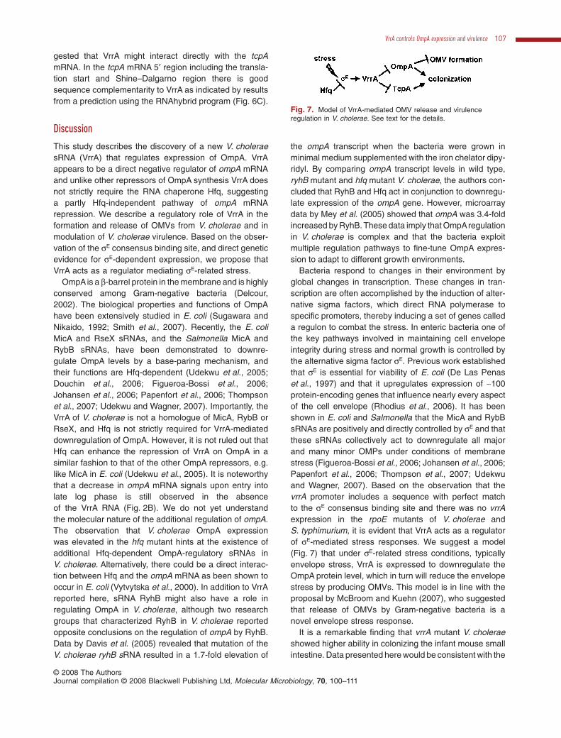
Bacteria respond to changes in their environment byglobal changes in transcription. These changes in tran-scription are often accomplished by the induction of alter-native sigma factors, which direct RNA polymerase tospecific promoters, thereby inducing a set of genes calleda regulon to combat the stress. In enteric bacteria one ofthe key pathways involved in maintaining cell envelopeintegrity during stress and normal growth is controlled bythe alternative sigma factor sE. Previous work establishedthat sE is essential for viability of E. coli (De Las Penaset al., 1997) and that it upregulates expression of ~100protein-encoding genes that influence nearly every aspectof the cell envelope (Rhodius et al., 2006). It has beenshown in E. coli and Salmonella that the MicA and RybBsRNAs are positively and directly controlled by sE and thatthese sRNAs collectively act to downregulate all majorand many minor OMPs under conditions of membranestress (Figueroa-Bossi et al., 2006; Johansen et al., 2006;Papenfort et al., 2006; Thompson et al., 2007; Udekwuand Wagner, 2007). Based on the observation that thevrrA promoter includes a sequence with perfect matchto the sE consensus binding site and there was no vrrAexpression in the rpoE mutants of V. cholerae andS. typhimurium, it is evident that VrrA acts as a regulatorof sE-mediated stress responses. We suggest a model(Fig. 7) that under sE-related stress conditions, typicallyenvelope stress, VrrA is expressed to downregulate theOmpA protein level, which in turn will reduce the envelopestress by producing OMVs. This model is in line with theproposal by McBroom and Kuehn (2007), who suggestedthat release of OMVs by Gram-negative bacteria is anovel envelope stress response.
It is a remarkable finding that vrrA mutant V. choleraeshowed higher ability in colonizing the infant mouse smallintestine. Data presented here would be consistent with the
Fig. 7. Model of VrrA-mediated OMV release and virulenceregulation in V. cholerae. See text for the details.
VrrA controls OmpA expression and virulence 107
© 2008 The AuthorsJournal compilation © 2008 Blackwell Publishing Ltd, Molecular Microbiology, 70, 100–111

suggestion that the increased colonization ability was dueto effects on both OmpA and TCP. We demonstrated thatthe interaction between VrrA and ompA mRNA is direct.Although we do not yet know the exact mechanism under-lying VrrA–TCP interaction, there is possibility that VrrAregulates tcpA by directly binding to the 5′ untranslatedregion of tcpA, as predicted by RNAhybrid program analy-sis (Fig. 6C). Considering these findings, we included TCPin our model of VrrA action as summarized in Fig. 7. At theinitial stage of V. cholerae infection, the sE level would below and consequently also VrrA expression would be at alow level. During such a stage, the bacteria would produceOmpA, TCP and other factors that may contribute to colo-nization of the intestine. When bacterial numbers reach acertain level, or due to the stress from the host, the sE levelelevates, which activates expression of VrrA. After synthe-sis, VrrA will reduce OmpA and TCP production resulting inreduced interaction with the intestinal mucosa. Concomi-tantly, the bacteria would produce more OMVs and therebyreduce the envelope stress (Fig. 7).
In summary, we demonstrated that VrrA positively con-trolled the release of OMVs by negatively controlling theexpression of the outer membrane protein OmpA. More-over, this single small regulatory RNA in V. cholerae mayinfluence the bacterial colonization ability as manifestedusing the mouse intestine model. To the best of ourknowledge, this is the first described case of a singleV. cholerae sRNA that solitarily would affect the virulenceof this bacterium. Because VrrA represses rather thanpromotes virulence in V. cholerae, attenuation of coloni-zation ability by some means affecting VrrA expressioncould be considered as the basis of a strategy for thera-peutic intervention in bacterial pathogenicity.
Experimental procedures
Bacterial strains and growth conditions
Vibrio cholerae strains are derivatives of El Tor Inaba strainA1552 (Yildiz and Schoolnik, 1998). V. cholerae, E. coli andS. typhimurium strains were grown at 37°C in Luria–Bertani(LB) broth supplemented, as appropriate, with carbenicillinat 100 mg ml-1 and chloramphenicol at 25 mg ml-1. For TCPexpression analysis, V. cholerae strains were grown at induc-ing conditions as described previously (Iwanaga and Kuyya-kanond, 1987).
DNA manipulations
In-frame deletions were constructed by the proceduresdescribed previously (Vaitkevicius et al., 2006). Primersequences are summarized in Table S1. Deletion of the vrrA,ompA, hfq, tcpA and rpoE loci in V. cholerae strain A1552resulted in DNY7, DNY10, DNY8, DNY51 and DNY105respectively. vrrA deletion mutant was constructed such that22 nucleotides upstream from and 91 nucleotides of vrrA
were removed from the chromosome. Double deletion of hfqand vrrA in A1552 resulted in DNY9. Double deletion of vrrAand ompA in A1552 resulted in DNY104. A DNA fragment(304 bp) containing the vrrA gene including its putative pro-moter region was amplified from the A1552 genome andcloned into pMMB66HE (Furste et al., 1986) at the HindIII/BamHI sites. The resulting plasmid pvrrA and its vectorcontrol (pMMB66HE) were introduced by transformation intoDNY7, resulting DNY11 and DNY12; and into DNY8, resultingDNY16 and DNY17 respectively.
In the pvrrA construct, the vrrA gene was cloned togetherwith its putative promoter region into the HindIII/BamHI sitesin pMMB66HE and consequently the Ptac promoter inpMMB66HE was located ~100 bp upstream of vrrA’s ownpromoter. We propose that vrrA transcription from pvrrA isdriven by its own promoter instead of the Ptac promoter inpMMB66HE, based on two observations. First, pvrrA wasnot induced by Ptac inducers (e.g. IPTG) in the experiments.Second, the size of vrrA transcript in strain DNY11(DvrrA+pvrrA) was the same as that in the wild-type strain(~140 nt), and we always observed a single band in theNorthern blot analysis (Fig. 4).
The ColE1-based plasmid, pTS2, expressing Vibrio vrrAfrom its own promoter, was constructed based on plasmidpZE12-luc (Lutz and Bujard, 1997). A DNA fragment ofpZE12-luc, which lacks the PLlaco promoter region, was ampli-fied by PCR using Phusion-polymerase (Finnzymes) andprimers JVO-2512 and pLLacOB, digested with XbaI, result-ing the backbone for pTS2. The V. cholerae vrrA gene wasPCR-amplified using primers JVO-2639 and JVO-2640. JVO-2639 binds 100 nt upstream of the +1 site of vrrA and carriesa 5′ monophosphate for cloning; JVO-2640 binds 80 nt down-stream of the vrrA terminator and will add an XbaI site tothe PCR product. Following XbaI digestion, the productwas ligated to the backbone, to yield plasmid pTS2 upontransformation.
RNA preparation and Northern blot analysis
RNA was prepared using Trizol according to the manufactur-er’s instructions (Invitrogen) and quantified on a NanoDropND-1000 Spectrophotometer (NanoDrop Technologies). ForNorthern blotting, 20 mg of total RNA was separated on aformaldehyde:agarose gel prior to blotting as previouslydescribed (Sheehan et al., 1995). The Hybond-N membrane(GE Healthcare) was subsequently hybridized with 32P-labelled gene-specific probes (Table S1). Northern blots wereexposed to a phosphorimager screen and scanned on aStormTM phosphorimager (Molecular Dynamics, USA). Quan-tification was performed using ImageQuantTM software(Molecular Dynamics).
5� RACE analysis
5′ RACE was carried out as described previously (Urban andVogel, 2007) to determine the transcription start sites of vrrA,ompA and vc1743. Total RNA obtained on strain V. choleraeA1552 was used for cDNA generation. Oligo TY2, VC2213-rev and TIS-31 (Table S1) were used as vrrA-, ompA- orvc1743-specific primers in PCR. PCR products were sepa-rated on a 2% agarose gel (Fig. 1B for vrrA and Fig. S1 for
108 T. Song et al. �
© 2008 The AuthorsJournal compilation © 2008 Blackwell Publishing Ltd, Molecular Microbiology, 70, 100–111

ompA and vc1743), gel-eluted and used as template forsequencing.
Toeprinting analysis
Toeprinting reactions were carried out as described (Sharmaet al., 2007) with few modifications. An unlabelled ompAmRNA fragment (0.2 pmol; 176 nt; T7 template amplified withprimers JVO-2784/-2871), and 0.5 pmol of 5′-end-labelledprimer JVO-2871 complementary to the ompA coding regionwere annealed. For inhibition analysis, 0.2 and 2 pmol of VrrARNA (134 nt, T7 template amplified with JVO-2782/-2783)or 2 pmol of control RNA (Salmonella MicA) were added.See the figure legend of Fig. 3 for final concentrations ofother components.
Isolation of OMVs
Outer membrane vesicles were isolated from culture super-natants as previously described (Wai et al., 2003).
SDS-PAGE and Western blot analysis
Protein samples were prepared from equal amount of bacte-ria cells after grown overnight unless otherwise indicated.The standard SDS-PAGE procedure was used (Laemmli,1970). Gels were stained with Coomassie brilliant blue.Western blot analyses were performed as described earlier(Vaitkevicius et al., 2006), using polyclonal anti-OmpA, anti-OmpU and anti-TcpA antisera.
Electron microscopy
Procedures for electron microscopy were essentially asdescribed earlier (Wai et al., 2003).
Infant mouse competition assay
Approximately 105 wild type and either vrrA or ompA mutantswere inoculated intragastrically into 6-day-old CD-1 (CharlesRiver Laboratories) mice. Mice were sacrificed after 20 hand bacteria colonizing the intestines were quantified asdescribed previously (Gardel and Mekalanos, 1996).
Analysis of VrrA expression in the sucklingmouse intestine
For analysis of gene expression in the suckling mouse intes-tine, mice were infected with A1552 as described (Camilli andMekalanos, 1995) with an inoculum of around 105 colony-forming units (cfu) in 50 ml of LB broth. RNA was isolated fromthree (first experiment) or six (second experiment) separatesmall intestines of infected CD-1 mice 24 h post infection.RNA was isolated in parallel from an equal number of sepa-rate in vitro OD600 = 0.5 LB-broth cultures. RNA extraction,removal of chromosomal DNA contamination, random-primed reverse transcription and qPCR were carried out asdescribed previously (Schild et al., 2007).
Mass spectrometry peptide sequencing
Proteins of interest were cut out from gel and analysed atAlphalyse (Denmark) for mass spectrometry.
Acknowledgements
We thank Bernt Eric Uhlin for valuable suggestions andsupport, Carlos Balsalobre for providing OmpA antiserum,Masaaki Iwanaga for providing OmpU antiserum and RonaldK. Taylor for providing TcpA antiserum. We are grateful toKarolis Vaitkevicius for designing primers for hfq deletionconstruct, Akemi Takade and Franziska Seifert for skilfultechnical assistance, Edmund Loh and Jonas Gripenland forkind help in RNA experiments, and Cynthia Sharma and KaiPapenfort for discussions on SigmaE and sRNAs. This workwas supported by grants from the Swedish ResearchCouncil, the Swedish Foundation for International Coopera-tion in Research and Higher Education (STINT), the Facultyof Medicine at Umeå University, and it was performed withinthe Umeå Centre for Microbial Research (UCMR). The workwas carried out in the frame of the European Virtual Institutefor Functional Genomics of Bacterial Pathogens (CEE LSHB-CT-2005-512061).
References
Balsalobre, C., Silvan, J.M., Berglund, S., Mizunoe, Y., Uhlin,B.E., and Wai, S.N. (2006) Release of the type I secretedalpha-haemolysin via outer membrane vesicles fromEscherichia coli. Mol Microbiol 59: 99–112.
Beveridge, T.J. (1999) Structures of gram-negative cell wallsand their derived membrane vesicles. J Bacteriol 181:4725–4733.
Brennan, R.G., and Link, T.M. (2007) Hfq structure, functionand ligand binding. Curr Opin Microbiol 10: 125–133.
Camilli, A., and Mekalanos, J.J. (1995) Use of recombinasegene fusions to identify Vibrio cholerae genes inducedduring infection. Mol Microbiol 18: 671–683.
Davis, B.M., and Waldor, M.K. (2007) RNase E-dependentprocessing stabilizes MicX, a Vibrio cholerae sRNA. MolMicrobiol 65: 373–385.
Davis, B.M., Quinones, M., Pratt, J., Ding, Y., and Waldor,M.K. (2005) Characterization of the small untranslatedRNA RyhB and its regulon in Vibrio cholerae. J Bacteriol187: 4005–4014.
De Las Penas, A., Connolly, L., and Gross, C.A. (1997) ThesigmaE-mediated response to extracytoplasmic stress inEscherichia coli is transduced by RseA and RseB, twonegative regulators of sigmaE. Mol Microbiol 24: 373–385.
Delcour, A.H. (2002) Structure and function of pore-formingbeta-barrels from bacteria. J Mol Microbiol Biotechnol 4:1–10.
Ding, Y., Davis, B.M., and Waldor, M.K. (2004) Hfq is essen-tial for Vibrio cholerae virulence and downregulates sigmaexpression. Mol Microbiol 53: 345–354.
Douchin, V., Bohn, C., and Bouloc, P. (2006) Down-regulation of porins by a small RNA bypasses the essenti-ality of the regulated intramembrane proteolysis proteaseRseP in Escherichia coli. J Biol Chem 281: 12253–12259.
VrrA controls OmpA expression and virulence 109
© 2008 The AuthorsJournal compilation © 2008 Blackwell Publishing Ltd, Molecular Microbiology, 70, 100–111

Faruque, S.M., Albert, M.J., and Mekalanos, J.J. (1998)Epidemiology, genetics, and ecology of toxigenic Vibriocholerae. Microbiol Mol Biol Rev 62: 1301–1314.
Figueroa-Bossi, N., Lemire, S., Maloriol, D., Balbontin, R.,Casadesus, J., and Bossi, L. (2006) Loss of Hfq activatesthe sigmaE-dependent envelope stress response in Sal-monella enterica. Mol Microbiol 62: 838–852.
Furste, J.P., Pansegrau, W., Frank, R., Blocker, H., Scholz,P., Bagdasarian, M., and Lanka, E. (1986) Molecularcloning of the plasmid RP4 primase region in a multi-host-range tacP expression vector. Gene 48: 119–131.
Gardel, C.L., and Mekalanos, J.J. (1996) Alterations in Vibriocholerae motility phenotypes correlate with changes invirulence factor expression. Infect Immun 64: 2246–2255.
Hartz, D., McPheeters, D.S., Traut, R., and Gold, L. (1988)Extension inhibition analysis of translation initiationcomplexes. Meth Enzymol 164: 419–425.
Hershberg, R., Altuvia, S., and Margalit, H. (2003) A survey ofsmall RNA-encoding genes in Escherichia coli. NucleicAcids Res 31: 1813–1820.
Iwanaga, M., and Kuyyakanond, T. (1987) Large productionof cholera toxin by Vibrio cholerae O1 in yeast extractpeptone water. J Clin Microbiol 25: 2314–2316.
Johansen, J., Rasmussen, A.A., Overgaard, M., andValentin-Hansen, P. (2006) Conserved small non-codingRNAs that belong to the sigmaE regulon: role in down-regulation of outer membrane proteins. J Mol Biol 364:1–8.
Kondo, K., Takade, A., and Amako, K. (1993) Release of theouter membrane vesicles from Vibrio cholerae and Vibrioparahaemolyticus. Microbiol Immunol 37: 149–152.
Kouokam, J.C., Wai, S.N., Fallman, M., Dobrindt, U., Hacker,J., and Uhlin, B.E. (2006) Active cytotoxic necrotizing factor1 associated with outer membrane vesicles from uropatho-genic Escherichia coli. Infect Immun 74: 2022–2030.
Kuehn, M.J., and Kesty, N.C. (2005) Bacterial outer mem-brane vesicles and the host–pathogen interaction. GenesDev 19: 2645–2655.
Laemmli, U.K. (1970) Cleavage of structural proteins duringthe assembly of the head of bacteriophage T4. Nature 227:680–685.
Lee, S.H., Hava, D.L., Waldor, M.K., and Camilli, A. (1999)Regulation and temporal expression patterns of Vibriocholerae virulence genes during infection. Cell 99: 625–634.
Lenz, D.H., Mok, K.C., Lilley, B.N., Kulkarni, R.V., Wingreen,N.S., and Bassler, B.L. (2004) The small RNA chaperoneHfq and multiple small RNAs control quorum sensing inVibrio harveyi and Vibrio cholerae. Cell 118: 69–82.
Lenz, D.H., Miller, M.B., Zhu, J., Kulkarni, R.V., and Bassler,B.L. (2005) CsrA and three redundant small RNAs regulatequorum sensing in Vibrio cholerae. Mol Microbiol 58: 1186–1202.
Livny, J., Fogel, M.A., Davis, B.M., and Waldor, M.K. (2005)sRNAPredict: an integrative computational approach toidentify sRNAs in bacterial genomes. Nucleic Acids Res33: 4096–4105.
Lutz, R., and Bujard, H. (1997) Independent and tight regu-lation of transcriptional units in Escherichia coli via theLacR/O, the TetR/O and AraC/I1-I2 regulatory elements.Nucleic Acids Res 25: 1203–1210.
McBroom, A.J., and Kuehn, M.J. (2007) Release of outermembrane vesicles by Gram-negative bacteria is a novelenvelope stress response. Mol Microbiol 63: 545–558.
Majdalani, N., Vanderpool, C.K., and Gottesman, S. (2005)Bacterial small RNA regulators. Crit Rev Biochem Mol Biol40: 93–113.
Mashburn-Warren, L.M., and Whiteley, M. (2006) Specialdelivery: vesicle trafficking in prokaryotes. Mol Microbiol61: 839–846.
Mey, A.R., Craig, S.A., and Payne, S.M. (2005) Character-ization of Vibrio cholerae RyhB: the RyhB regulon and roleof ryhB in biofilm formation. Infect Immun 73: 5706–5719.
Papenfort, K., Pfeiffer, V., Mika, F., Lucchini, S., Hinton, J.C.,and Vogel, J. (2006) SigmaE-dependent small RNAs ofSalmonella respond to membrane stress by acceleratingglobal omp mRNA decay. Mol Microbiol 62: 1674–1688.
Rehmsmeier, M., Steffen, P., Hochsmann, M., and Giegerich,R. (2004) Fast and effective prediction of microRNA/targetduplexes. RNA 10: 1507–1517.
Rhodius, V.A., Suh, W.C., Nonaka, G., West, J., and Gross,C.A. (2006) Conserved and variable functions of the sigmaEstress response in related genomes. PLoS Biol 4: e2.
Romby, P., Vandenesch, F., and Wagner, E.G. (2006) Therole of RNAs in the regulation of virulence-gene ex-pression. Curr Opin Microbiol 9: 229–236.
Schild, S., Tamayo, R., Nelson, E.J., Qadri, F., Calderwood,S.B., and Camilli, A. (2007) Genes induced late in infectionincrease fitness of Vibrio cholerae after release into theenvironment. Cell Host Microbe 2: 264–277.
Schooling, S.R., and Beveridge, T.J. (2006) Membranevesicles: an overlooked component of the matrices ofbiofilms. J Bacteriol 188: 5945–5957.
Sharma, C.M., Darfeuille, F., Plantinga, T.H., and Vogel, J.(2007) A small RNA regulates multiple ABC transportermRNAs by targeting C/A-rich elements inside and upstreamof ribosome-binding sites. Genes Dev 21: 2804–2817.
Sheehan, B., Klarsfeld, A., Msadek, T., and Cossart, P.(1995) Differential activation of virulence gene expressionby PrfA, the Listeria monocytogenes virulence regulator.J Bacteriol 177: 6469–6476.
Skovierova, H., Rowley, G., Rezuchova, B., Homerova, D.,Lewis, C., Roberts, M., and Kormanec, J. (2006) Identifi-cation of the sigmaE regulon of Salmonella entericaserovar Typhimurium. Microbiology 152: 1347–1359.
Smith, S.G., Mahon, V., Lambert, M.A., and Fagan, R.P.(2007) A molecular Swiss army knife: OmpA structure,function and expression. FEMS Microbiol Lett 273: 1–11.
Sonntag, I., Schwarz, H., Hirota, Y., and Henning, U. (1978)Cell envelope and shape of Escherichia coli: multiplemutants missing the outer membrane lipoprotein and othermajor outer membrane proteins. J Bacteriol 136: 280–285.
Storz, G., Opdyke, J.A., and Zhang, A. (2004) ControllingmRNA stability and translation with small, noncodingRNAs. Curr Opin Microbiol 7: 140–144.
Sugawara, E., and Nikaido, H. (1992) Pore-forming activity ofOmpA protein of Escherichia coli. J Biol Chem 267: 2507–2511.
Thelin, K.H., and Taylor, R.K. (1996) Toxin-coregulated pilus,but not mannose-sensitive hemagglutinin, is required forcolonization by Vibrio cholerae O1 El Tor biotype and O139strains. Infect Immun 64: 2853–2856.
110 T. Song et al. �
© 2008 The AuthorsJournal compilation © 2008 Blackwell Publishing Ltd, Molecular Microbiology, 70, 100–111

Thompson, K.M., Rhodius, V.A., and Gottesman, S. (2007)SigmaE regulates and is regulated by a small RNA inEscherichia coli. J Bacteriol 189: 4243–4256.
Toledo-Arana, A., Repoila, F., and Cossart, P. (2007) Smallnoncoding RNAs controlling pathogenesis. Curr OpinMicrobiol 10: 182–188.
Torres, A.G., and Kaper, J.B. (2003) Multiple elements con-trolling adherence of enterohemorrhagic Escherichia coliO157:H7 to HeLa cells. Infect Immun 71: 4985–4995.
Udekwu, K.I., Darfeuille, F., Vogel, J., Reimegård, J., Holm-qvist, E., and Wagner, E.G. (2005) Hfq-dependent regula-tion of OmpA synthesis is mediated by an antisense RNA.Genes Dev 19: 2355–2366.
Udekwu, K.I., and Wagner, E.G. (2007) Sigma E controlsbiogenesis of the antisense RNA MicA. Nucleic Acids Res35: 1279–1288.
Urban, J.H., and Vogel, J. (2007) Translational control andtarget recognition by Escherichia coli small RNAs in vivo.Nucleic Acids Res 35: 1018–1037.
Vaitkevicius, K., Lindmark, B., Ou, G., Song, T., Toma, C.,Iwanaga, M., et al. (2006) A Vibrio cholerae proteaseneeded for killing of Caenorhabditis elegans has a role inprotection from natural predator grazing. Proc Natl AcadSci USA 103: 9280–9285.
Valentin-Hansen, P., Eriksen, M., and Udesen, C. (2004) Thebacterial Sm-like protein Hfq: a key player in RNAtransactions. Mol Microbiol 51: 1525–1533.
Vogel, J., and Papenfort, K. (2006) Small non-coding RNAsand the bacterial outer membrane. Curr Opin Microbiol 9:605–611.
Vogel, J., and Sharma, C.M. (2005) How to find small non-coding RNAs in bacteria. Biol Chem 386: 1219–1238.
Vytvytska, O., Moll, I., Kaberdin, V.R., von Gabain, A., andBlasi, U. (2000) Hfq (HF1) stimulates ompA mRNA decayby interfering with ribosome binding. Genes Dev 14: 1109–1118.
Wai, S.N., Lindmark, B., Soderblom, T., Takade, A., Wester-mark, M., Oscarsson, J., et al. (2003) Vesicle-mediatedexport and assembly of pore-forming oligomers of theenterobacterial ClyA cytotoxin. Cell 115: 25–35.
Yildiz, F.H., and Schoolnik, G.K. (1998) Role of rpoS in stresssurvival and virulence of Vibrio cholerae. J Bacteriol 180:773–784.
Zhang, Y., Zhang, Z., Ling, L., Shi, B., and Chen, R. (2004)Conservation analysis of small RNA genes in Escherichiacoli. Bioinformatics 20: 599–603.
Zhou, L., Srisatjaluk, R., Justus, D.E., and Doyle, R.J. (1998)On the origin of membrane vesicles in gram-negativebacteria. FEMS Microbiol Lett 163: 223–228.
Supporting information
Additional supporting information may be found in the onlineversion of this article.
Please note: Blackwell Publishing are not responsible for thecontent or functionality of any supporting materials suppliedby the authors. Any queries (other than missing material)should be directed to the corresponding author for the article.
VrrA controls OmpA expression and virulence 111
© 2008 The AuthorsJournal compilation © 2008 Blackwell Publishing Ltd, Molecular Microbiology, 70, 100–111