Natural transformation of Vibrio cholerae as a tool - Optimizing the procedure

The three-dimensional structure of the cytoplasmic domains ofEpsF from the type 2 secretion system of Vibrio cholerae
Jan Abendroth1,$, Daniel D. Mitchell1, Konstantin V. Korotkov1, Tanya L. Johnson2, AllisonKreger1, Maria Sandkvist2, and Wim G. J. Hol1,*1Department of Biochemistry, Biomolecular Structure Center, University of Washington, Box357742, Seattle WA 98195, USA2University of Michigan Medical School, Department of Microbiology and Immunology, 1150 WestMedical Center Drive, Ann Arbor, MI 48109, USA
AbstractThe type 2 secretion system (T2SS), a multi-protein machinery that spans both the inner and the outermembranes of Gram-negative bacteria, is used for the secretion of several critically importantproteins across the outer membrane. Here we report the crystal structure of the N-terminalcytoplasmic domain of EpsF, an inner membrane spanning T2SS protein from Vibrio cholerae. Thisdomain consists of a bundle of six anti-parallel helices and adopts a fold that has not been describedbefore. The long C-terminal helix α6 protrudes from the body of the domain and most likely continuesas the first transmembrane helix of EpsF. Two N-terminal EpsF domains form a tight dimer with aconserved interface, suggesting that the observed dimer occurs in the T2SS of many bacteria. Twocalcium binding sites are present in the dimer interface with ligands provided for each site by bothsubunits. Based on this new structure, sequence comparisons of EpsF homologs and localizationstudies of GFP fused with EpsF, we propose that the second cytoplasmic domain of EpsF adopts asimilar fold as the first cytoplasmic domain and that full-length EpsF, and its T2SS homologs, havea three-transmembrane helix topology.
KeywordsGeneral Secretory Pathway; Type 4 Pilin Biogenesis; GspF; XcpS; PilG
1. IntroductionIn many pathogenic as well as non-pathogenic Gram-negative bacteria a diversity of unrelatedproteins is secreted from the periplasm across the outer membrane into the extracellular milieuby the type 2 secretion system (T2SS) (Cianciotto, 2005; Evans et al., 2008; Sandkvist,2001b). The T2SS is also called the terminal branch of the general secretory pathway (Gsp)(Pugsley, 1993) and, in Vibrio species, the extracellular protein secretion (Eps) apparatus(Sandkvist et al., 1997). This sophisticated machinery spans both the inner and the outermembrane and contains 11 to 15 different proteins, with many, if not all, of these proteins
*Corresponding author: ph: 206-685-7044 fx: 206-685-7002, email: [email protected].$Current address: deCODE BioStructures Inc., Bainbridge Island, WA 98110, USAPublisher's Disclaimer: This is a PDF file of an unedited manuscript that has been accepted for publication. As a service to our customerswe are providing this early version of the manuscript. The manuscript will undergo copyediting, typesetting, and review of the resultingproof before it is published in its final citable form. Please note that during the production process errors may be discovered which couldaffect the content, and all legal disclaimers that apply to the journal pertain.
NIH Public AccessAuthor ManuscriptJ Struct Biol. Author manuscript; available in PMC 2010 June 1.
Published in final edited form as:J Struct Biol. 2009 June ; 166(3): 303–315. doi:10.1016/j.jsb.2009.03.009.
NIH
-PA Author Manuscript
NIH
-PA Author Manuscript
NIH
-PA Author Manuscript
present in multiple copies (Filloux, 2004; Johnson et al., 2006). The nomenclature of the T2SSproteins is quite complex. In this paper, proteins from the Eps system from Vibrio species arereferred to as “Eps” followed by a capital letter, while the non-Vibrio T2SS homologs will becalled “Gsp” followed by the same capital letter. Hence, the T2SS homologs of EpsF are calledGspF proteins. GspF will also be used as the name of the family of T2SS inner membraneproteins to which EpsF belongs (See Supplementary Figure 1 for a GspF family sequencealignment).
In V. cholerae, the major virulence factor cholera toxin and several other proteins including asoluble colonization factor, hemagglutinin-protease, lipase and chitinase are secreted by theT2SS across the outer membrane (Hirst and Holmgren, 1987; Kirn et al., 2005; Sandkvist,2001b; Sikora et al., 2007). In enterotoxigenic Escherichia coli (ETEC), the T2SS isresponsible for secretion of heat-labile enterotoxin (Tauschek et al., 2002), a close homologof cholera toxin (Merritt and Hol, 1995; O'Neal et al., 2004; Sixma et al., 1991). Other bacteriasecreting a variety of proteins using the T2SS include important human pathogens such asPseudomonas aeruginosa (Bally et al., 1992), Klebsiella spp. (d'Enfert et al., 1987; Uweh,2006), Legionella pneumophila (Söderberg et al., 2004), Yersinia pestis (Yen et al., 2008),enteropathogenic E. coli (EPEC) and enterohemorrhagic E. coli (EHEC) (Schmidt et al.,1997). Also several plant pathogens, like Erwinia chrysanthemi and E. carotovora,Xanthomonas campestris and X. fastidiosa (Bouley et al., 2001; Cianciotto, 2005; Sandkvist,2001b), contain a T2SS system. Among the different types of protein secretion systemsidentified so far in Gram-negative bacteria, the T2SS is remarkable in that proteins are secretedby the T2SS across the outer membrane in a folded conformation (Bortoli-German et al.,1994; Hardie et al., 1995; Hirst and Holmgren, 1987; Pugsley, 1992; Sandkvist, 2001b).
It has become evident that several components of the T2SS are related to components of thetype 4 pilin biogenesis (T4PB) system (Bally et al., 1992; Craig and Li, 2008; Filloux, 2004;Hobbs and Mattick, 1993; Nunn, 1999; Peabody et al., 2003). Type 4 pili are thin, strongfilaments extending from a wide variety of human bacterial pathogens (Craig et al., 2004;Hansen and Forest, 2006). The T4PB system is responsible for a diversity of functionsincluding pilus assembly and disassembly, protein export, DNA import and phage entry(Burrows, 2005; Mattick, 2002; Nudleman and Kaiser, 2004). Studies of the T2SS proteins aretherefore important for increasing our understanding of critical membrane transportphenomena in many bacterial species, several of which are of great medical importance.
The T2SS can be envisioned as consisting of three major subassemblies (Filloux, 2004; Johnsonet al., 2006; Peabody et al., 2003; Py et al., 2001; Sandkvist, 2001a; Sauvonnet et al., 2000):(i) the outer membrane complex, comprising mainly the crucial multi-subunit secretin EpsD,which is thought to be the pore that opens and closes to allow passage of the secreted proteins;(ii) the pseudopilus, which consists of one major and several minor pseudopilins that may forma retractable plug or piston; and, (iii) an inner membrane platform, containing the cytoplasmic“secretion ATPase” EpsE and the membrane proteins EpsL, EpsM, EpsC and EpsF. The centralprotein of the current paper is the polytopic inner membrane protein EpsF, which previouslywas shown to be indispensable for secretion in V. cholerae (Sandkvist et al., 1997).
GspF interacts with other proteins from the inner membrane platform and is thought to be akey player in the T2SS and T4PB systems (Crowther et al., 2004; Py et al., 2001).Bioinformatics analysis of the amino acid sequence and BlaM-fusion experiments of GspFfrom E. caratovora indicated that this T2SS EpsF homolog crosses the inner membrane threetimes (Thomas et al., 1997). These authors also concluded that the N-terminus of the GspFprotein in E. caratovora is cytoplasmic and the C-terminus periplasmic. This topology wasconfirmed by alkaline phosphatase fusion experiments of the T2SS GspF homolog in P.aeruginosa (Arts et al., 2007). Peabody et al. analyzed a large number of sequences from
Abendroth et al. Page 2
J Struct Biol. Author manuscript; available in PMC 2010 June 1.
NIH
-PA Author Manuscript
NIH
-PA Author Manuscript
NIH
-PA Author Manuscript

members of the superfamily of bacterial inner membrane proteins to which EpsF belongs(Peabody et al., 2003), hereafter called the “GspF/PilG/BfpE superfamily”. The GspF familyrepresents members of the T2SS machinery, whereas the PilG and BfpE families consist ofhomologs from the T4PB system. Members from the entire superfamily display an internalsequence repeat such that the first cytoplasmic domain is related to the second half of the protein(Peabody et al., 2003). In spite of considerable attention for these important bacterial innermembrane proteins, no high resolution structural information has been reported to date for fulllength proteins or domains of members from this superfamily.
In continuation of our studies aimed to unravel the architecture of the T2SS and its componentsin human pathogens and close relatives (Abendroth et al., 2004a; Abendroth et al., 2004b;Abendroth et al., 2005; Korotkov et al., 2006; Korotkov and Hol, 2008; Korotkov et al.,2009; Robien et al., 2003; Yanez et al., 2008a; Yanez et al., 2008b), we report here the crystalstructure of a truncated form of the first N-terminal cytoplasmic domain of V. cholerae EpsF.This truncated domain (hereafter also called “cyto1-EpsF56-171”) appears to adopt a novel fold,is entirely helical and forms a tight dimer with the residues of the dimer interface wellconserved. Two symmetry-related calcium binding sites occupy a dimer with well conservedligand residues across the GspF family. Sequence analysis of the GspF family suggests thatthe second cytoplasmic domain of members of this family adopts the same conformation asthe first cytoplasmic domain. A tentative model of the cytoplasmic domains of a dimer of full-length EpsF is proposed.
2. Materials and methods2.1 Protein expression and purification
DNA encoding for V. cholerae cyto1-EpsF56-171 was amplified by PCR and ligated into apACYC-CT vector using NcoI and NheI restriction sites. The pACYC-CT vector encodes aC-terminal TEV-cleavable His6-tag and provides chloramphenicol resistance. BL21(DE3) E.coli cells were used for protein expression. For expression of native protein, the main culturewas grown in LB medium at 37°C, and induced for 3 hrs with 0.5 mM IPTG. For expressionof Se-Met labeled protein, the main culture was grown in M9 medium. 30 minutes beforeinduction with 0.5 mM IPTG, an amino acid mixture for suppression of methioninebiosynthesis was added essentially as described elsewhere (van Duyne et al., 1993). Cells wereresuspended in buffer A (50 mM Tris-HCl, pH 8, 300 mM NaCl) and lysed with lysozyme andby sonication. The cell debris was pelleted by centrifugation at 20,000 g for 20 minutes. 3 mlof Ni-NTA (Qiagen) were incubated for 30 minutes with the clarified lysate from 1l culture,washed with 30 ml buffer B (20 mM Tris-HCl, pH 8, 300 mM NaCl, 1 mM TCEP), a secondtime with 30 ml buffer B plus 20 mM imidazole, and then eluted with 20 ml buffer B plus 200mM imidazole. The Ni-NTA elution fractions were incubated with TEV protease over nightat 4°C while dialyzing against 10-fold the volume of buffer A. The sample was then passedover 1ml Ni-NTA resin equilibrated with buffer A and washed with another 10ml buffer A.Flowthrough and wash were combined and concentrated to 5ml. The concentrated sample wasfurther purified in buffer B by gel permeation chromatography using Superdex S200 pre-graderesin (Amersham-Pharmacia). Typically, 50 mg protein could be purified per liter culture. Theprotein was concentrated to 10 mg/ml for crystallization experiments.
2.2 Crystallization and data collectionCrystallization conditions were screened for using the commercial screens ProComplex(Qiagen); Index, PEG-Ion, SaltRX (Hampton Research); and Wizard (Emerald BioSystems).Various hits were found, each of them included PEG (400, 3000, 3350, 8000), a divalent cationsuch as Ca2+ or Mg2+, and a buffer pH 6.0-8.0. Conditions were optimized to 12.5% PEG 400,200 mM CaOAc2, 100 mM MES pH 7.0.
Abendroth et al. Page 3
J Struct Biol. Author manuscript; available in PMC 2010 June 1.
NIH
-PA Author Manuscript
NIH
-PA Author Manuscript
NIH
-PA Author Manuscript
For cryo-cooling, the crystals were sequentially transferred in buffers containing 200 mMCaOAc2, 100 mM MES pH 7.0, plus increasing concentrations of PEG 400 and NaCl in thefollowing ratios: 20%/300 mM, 20%/600 mM, 25%/600 mM, 30%/600 mM. For the NaI soak,NaCl was gradually replaced by NaI. The crystals were frozen in the cryostream and screenedfor diffraction. Two cycles of cryo-annealing by blocking the cryostream could reduce themosaicity of the crystals considerably.
Diffraction data of the iodide soak and the high resolution crystal were collected in house usinga MicroMax-007 HF rotating anode (Rigaku) equipped with VariMax HF (Osmic)monochromator and a Saturn 994 (Rigaku) CCD detector. Diffraction data of the native crystalwere collected at beamline 9.2 at SSRL on a Mar 325 CCD detector. Further details are providedin Table 1.
2.3 Data processing and structure solutionGenerally, the diffraction images were indexed, integrated and scaled using the d*TREK suitethrough the Crystal Clear interface (Pflugrath, 1999). For the iodide soak and the native dataset, the strength of the anomalous signal was estimated with SHELXC. The positions of theanomalous atoms were found with SHELXD (Schneider and Sheldrick, 2002). Sites wererefined and phases were calculated with SHARP (Bricogne et al., 2003). Phases were improvedwithin SHARP using SOLOMON (Abrahams et al., 1996) and DM (Cowtan and Zhang,1999). Non-crystallographic symmetry was not applied for initial phasing. ARP/wARP(Perrakis et al., 1999) was used for automatic model building. The structures were manuallyinspected and completed using COOT (Emsley and Cowtan, 2004) and refined withREFMAC5 (Murshudov et al., 1997) using TLS groups defined by the TLSMD server (Painterand Merritt, 2006). All crystallographic calculations were done within the CCP4 suite (Bailey,1994).
Specific procedures and considerations for both data sets for which experimental phaseinformation was derived, were as follows:
(i) NaI soak: Data were collected at λ=1.5418Å. The following atoms contribute to theanomalous scattering of the crystal: 12× I- (f″=6.75 e-), 2× Ca2+ (f″=1.28 e-), 10× Se (f″=1.14e-), and 2× S (f″=0.56 e-). ShelxC indicated a strong anomalous signal over the entire resolutionrange. ShelxD (Schneider and Sheldrick, 2002) could find 16 sites, which in hindsight couldbe identified as iodide and calcium sites. After refinement and phasing with Sharp (Bricogneet al., 2003), and density modification with Solomon/DM (Abrahams et al., 1996; Cowtan andZhang, 1999) within Sharp, ARP/wARP (Perrakis et al., 1999) could build 232 out of 246residues of the asymmetric unit and assign all of them to the sequence.
(ii) Native protein: Data were collected at λ=2.06633 Å to 1.95 Å resolution. ShelxC indicateda significant anomalous signal up to 2.2 Å resolution. ShelxD could find two strong sites(Ca2+, f″=2.13 e-), and 13 weak sites (S from Met and Cys, f″=0.95 e-). After refinement ofthe positions and phasing with Sharp, and density modification with Solomon/DM withinSharp, ARP/wARP could build 228 out of 246 residues.
2.4 Functional StudiesPlasmid construction—DNA encoding EpsF 1-174 (hereafter also called “cyto1-EpsF”)was amplified by PCR and ligated into the pQE30 vector and then moved into the pMMB67vector using EcoRI and SalI restriction enzymes. The resulting construct was named pMMB-cyto1-EpsF. For plasmid GFP-EpsF, primers CGGATCCGCCGCGTTTGAATACA andGCTGCAGACTATGTCGTTTTCGCC were used to amplify epsF flanked by BamHI andPstI restriction sites. After digestion of pMMB66-gfp with BamHI and PstI, gfp-epsF was
Abendroth et al. Page 4
J Struct Biol. Author manuscript; available in PMC 2010 June 1.
NIH
-PA Author Manuscript
NIH
-PA Author Manuscript
NIH
-PA Author Manuscript
constructed. Plasmid EpsF-GFP was constructed first using primersGAATTCGATGCGGGTGACTAAG and GGATCCACGACTCATTAAGTTATTC toamplify epsF flanked by EcoRI and BamHI restriction sites and then moved into pMMB66-Cterminal-gfp following digestion with EcoRI and BamHI.
Bacterial strains and growth conditions—Wild type and epsF mutant strains of V.cholerae were grown to stationary phase at 37°C in LB (Luria broth) supplemented withthymine and 200μg/ml of carbenicillin or in M9 growth medium supplemented with 4%casamino acids and 0.4% glucose. When indicated, over-expression of cyto1-EpsF was inducedwith isopropyl β-D-thiogalactopyranoside (IPTG).
Inhibition of secretion in wild type V. cholerae cells—The cyto1-EpsF construct(pMMB-cyto1-EpsF) was expressed in wild type V. cholerae with and without induction byIPTG, and tested for extracellular protease secretion using a fluorescence based assay (Johnsonet al., 2007). Briefly, supernatants from overnight cultures grown in LB were assayed in 5 mMHEPES pH 7.5 and 0.05 mM N-tert-butoxy-carbonyl-Gln-Ala-Arg-7-amido-4-methyl-coumarin (Sigma-Aldrich, St. Louis MO) for 10 min at 37° C using the excitation and emissionwavelengths 385 nm and 440 nm, respectively.
In vivo crosslinking—Cells of wt and epsF mutant strains of V. cholerae containing eitherpMMB-cyto1-EpsF or control plasmid pMMB67 were induced with IPTG (100 μM finalconcentration). Following centrifugation, cells were resuspended in phosphate buffered salineto OD600 of 6 and subjected to crosslinking. Disthiobis succinimidyl propionate (DSP) (Pierce,Rockford, IL) was added to a final concentration of 0, 250 or 400 μM and the cells wereincubated at room temperature for 60 minutes. The crosslinker was then quenched by addingTris-HCl, pH 8 to a final concentration of 50 mM. Cells were centrifuged and resuspended in500 μl 50 mM Tris-HCl, pH 8 with 100 μg lysozyme and 10 μg DNase. After incubation for10 minutes at room temperature, the suspensions were sonicated (1 sec pulse followed by 1sec rest for 10 sec) and analyzed by SDS-PAGE with no reducing agent and immunoblottingwith anti-EpsF antibodies as described previously (Johnson et al., 2007).
Co-purification of histidine-tagged cyto1-EpsF and EpsL—Cell extracts wereincubated with 10 μl Cobalt-Immobilized Metal Affinity Chromotography Resin (IMACbeads; BD Biosciences, San Jose CA) in binding buffer (50mM Tris-HCl, pH 8.0, 1% TritonX-100) for 2 hours with rocking at 4°C. After incubation, samples were centrifuged, thesupernatant removed and beads were washed three times with binding buffer and once with50mM Tris-HCl, pH 8.0. 20μl of SDS-PAGE sample buffer containing DTT were added tothe beads and boiled for 10 minutes prior to centrifugation. Supernatants were subjected toSDS-PAGE and immunoblotting with anti-EpsL antibody.
Expression of GFP fusion proteins—GFP-EpsF and EpsF-GFP fusions were expressedfrom plasmid pMMB67 in the V. cholerae epsF mutant strain with and without induction byIPTG (20 μM final concentration). Cells were washed and assayed for fluorescence with aBioTek microplate reader (BioTek Instruments, Winooski VT) using the excitation andemission wavelengths 380 nm and 440 nm, respectively.
2.5 Preparation of figuresFor Figures 1 and 7a, sequences were aligned with T-coffee (Notredame et al., 2000).Rendering was done with ESPRIPT (Gouet et al., 1999). Figures 2 and 6 were prepared withPyMol (DeLano, 2002).
Abendroth et al. Page 5
J Struct Biol. Author manuscript; available in PMC 2010 June 1.
NIH
-PA Author Manuscript
NIH
-PA Author Manuscript
NIH
-PA Author Manuscript
3. Results3.1 Structure Determination
An extensive search was required in order to obtain a GspF construct that could be crystallized.Initially, full length EpsF proteins from V. cholerae, V. vulnificus, V. parahaemolyticus, andGspF from ETEC, were expressed and purified in the presence of various detergents. Despitegood expression and homogeneous preparations, as determined by SDS-PAGE and sizeexclusion chromatography, no crystals were obtained. The first cytoplasmic domain of EpsFfrom all four species (spanning residues 1-171 in V. cholerae) could not be crystallized either.As the degree of sequence homology between different species drops significantly betweenresidues ∼ 45 and ∼ 60 (Figure 1), constructs with several N-terminal truncations were madeand tested for crystal growth. Eventually, an N-terminally truncated version of V. choleraeEpsF containing residues 56-171 yielded diffraction quality crystals, but solely in the presenceof calcium ions, whereas the presence of magnesium gave crystals of poor quality.
Crystals of Se-Met cyto1-EpsF56-171 diffracted very well and an initial 1.7 Å resolution dataset could be collected in house. In order to obtain experimental phase information to determinethe structure, anomalous scattering differences were measured at long wavelengths to obtainphases by two independent approaches. Since it was unknown beforehand which phasingmethod would work, the following two additional data sets were collected. First, a crystal ofSe-Met protein was transferred to an iodide-containing buffer and diffraction data to 1.9 Åresolution were collected on a rotating anode generator with λ=1.5418 Å (Table 1). Asufficiently large signal was obtained from the anomalous scatterers iodide and calcium toallow an initial structure determination as described in Materials and Methods. Second, acrystal of native protein was exposed at the SSRL synchrotron using a wavelength of 2.0663Å, and a dataset up to 1.95 Å resolution was collected (Table 1). Here, calcium and sulphurprovided a sufficiently strong anomalous signal such that useful phase information could beextracted (Materials and Methods). Refinement against the 1.7 Å resolution data set yielded afinal structure with an Rwork of 15.6 %, an Rfree of 20.7 % and good geometry (Table 1). Thecrystal structure for cyto1-EpsF56-171 reported in this paper consists of consecutive residuesPhe 57 to Ser 171 for chain A, and residues Ser 62 to Gln 177 for chain B. Residues Ser 171to Gln 177 originate from the vector sequence of the C-terminal TEV-cleavable His6-tag.
3.2 Tertiary and quaternary structure of cyto1-EpsF56-171
The first domain of V. cholerae EpsF adopts an all-helical fold (Figure 2) with its six helicesforming two layers. One layer is made up by helices α1, α2 and α6, and the other by α3, α4and α5, with adjacent helices running anti-parallel to each other. Four of the six helices havea rather similar length of 10 to 15 residues, helix α4 consists of only five residues, whereas thelong C-terminal helix α6 consists of 23 and 30 residues in chains A and B, respectively. Startingfrom residue Lys162, the C-terminal part of helix α6 protrudes from the body of the domain(Figure 2). The high helix content of EpsF is in agreement with the result of CD studies (Collinset al., 2007) on the homolog PilG. Neither of the common structural similarity search programsSSM (Krissinel and Henrick, 2004) or DALI (Holm and Sander, 1993) reported proteins withsignificant structural homology. Therefore, we assume that cyto1-EpsF56-171 has a novel fold.
The cyto1-EpsF56-171 crystals contain two types of dimers with a substantial interface. One isan arrangement in which two α6 helices from different subunits run anti-parallel to each other,making interactions along their entire length (not shown). It is unlikely that this is anarrangement of physiological relevance because of the different directions of the two α6 helicesin this dimer. The α6 helices are each continued by a transmembrane helix (TM helix) whichstarts 2 to 3 residues after the end of helix α6 (see discussion below). If one α6 helix would
Abendroth et al. Page 6
J Struct Biol. Author manuscript; available in PMC 2010 June 1.
NIH
-PA Author Manuscript
NIH
-PA Author Manuscript
NIH
-PA Author Manuscript
continue as a TM helix into the inner membrane, the end of the other α6 helix in this dimerwould be more than 50 Å away from the inner membrane surface.
Two symmetry-related cyto1-EpsF56-171 chains form another dimer with approximatedimensions of 55 by 55 by 35 Å (Figure 2a). There are in total 14 residues involved in intra-dimer contacts, engaging in a mixture of hydrogen bonds and hydrophobic interactions (Figures1 and 2). The interface between the two molecules in the dimer buries 1750 Å2 solventaccessible surface area. This is in the range often observed for physiological dimers (Jones andThornton, 1996). A gap volume index of 2.26 Å, when compared to values for physiologicalrelevant interfaces (Jones and Thornton, 1996), indicates excellent complementarity of theinterface surfaces. Although gel permeation chromatography indicates that cyto1-EpsF56-171
is monomeric in solution (not shown), the high protein concentration in the crystal likelypromotes a concentration-dependent oligomerization of cyto1-EpsF56-171 that may bereflective of its oligomerization within the T2SS in vivo.
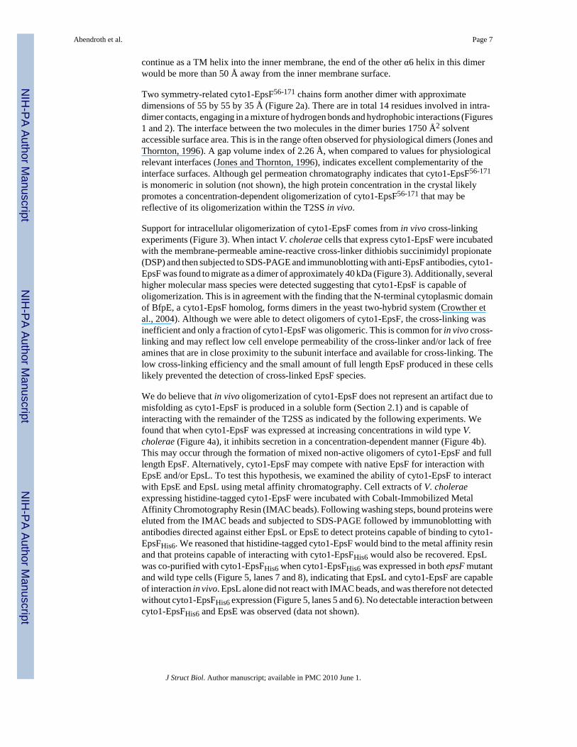
Support for intracellular oligomerization of cyto1-EpsF comes from in vivo cross-linkingexperiments (Figure 3). When intact V. cholerae cells that express cyto1-EpsF were incubatedwith the membrane-permeable amine-reactive cross-linker dithiobis succinimidyl propionate(DSP) and then subjected to SDS-PAGE and immunoblotting with anti-EpsF antibodies, cyto1-EpsF was found to migrate as a dimer of approximately 40 kDa (Figure 3). Additionally, severalhigher molecular mass species were detected suggesting that cyto1-EpsF is capable ofoligomerization. This is in agreement with the finding that the N-terminal cytoplasmic domainof BfpE, a cyto1-EpsF homolog, forms dimers in the yeast two-hybrid system (Crowther etal., 2004). Although we were able to detect oligomers of cyto1-EpsF, the cross-linking wasinefficient and only a fraction of cyto1-EpsF was oligomeric. This is common for in vivo cross-linking and may reflect low cell envelope permeability of the cross-linker and/or lack of freeamines that are in close proximity to the subunit interface and available for cross-linking. Thelow cross-linking efficiency and the small amount of full length EpsF produced in these cellslikely prevented the detection of cross-linked EpsF species.
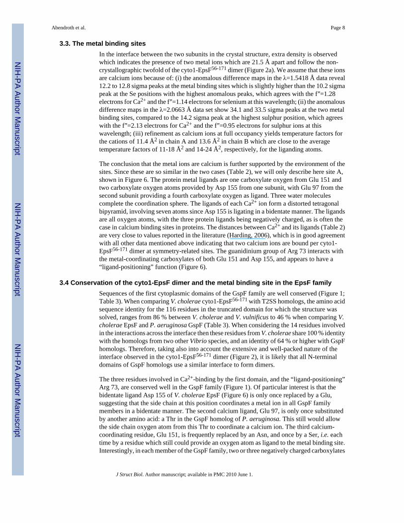
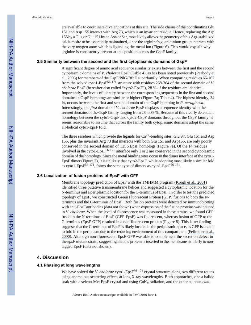
We do believe that in vivo oligomerization of cyto1-EpsF does not represent an artifact due tomisfolding as cyto1-EpsF is produced in a soluble form (Section 2.1) and is capable ofinteracting with the remainder of the T2SS as indicated by the following experiments. Wefound that when cyto1-EpsF was expressed at increasing concentrations in wild type V.cholerae (Figure 4a), it inhibits secretion in a concentration-dependent manner (Figure 4b).This may occur through the formation of mixed non-active oligomers of cyto1-EpsF and fulllength EpsF. Alternatively, cyto1-EpsF may compete with native EpsF for interaction withEpsE and/or EpsL. To test this hypothesis, we examined the ability of cyto1-EpsF to interactwith EpsE and EpsL using metal affinity chromatography. Cell extracts of V. choleraeexpressing histidine-tagged cyto1-EpsF were incubated with Cobalt-Immobilized MetalAffinity Chromotography Resin (IMAC beads). Following washing steps, bound proteins wereeluted from the IMAC beads and subjected to SDS-PAGE followed by immunoblotting withantibodies directed against either EpsL or EpsE to detect proteins capable of binding to cyto1-EpsFHis6. We reasoned that histidine-tagged cyto1-EpsF would bind to the metal affinity resinand that proteins capable of interacting with cyto1-EpsFHis6 would also be recovered. EpsLwas co-purified with cyto1-EpsFHis6 when cyto1-EpsFHis6 was expressed in both epsF mutantand wild type cells (Figure 5, lanes 7 and 8), indicating that EpsL and cyto1-EpsF are capableof interaction in vivo. EpsL alone did not react with IMAC beads, and was therefore not detectedwithout cyto1-EpsFHis6 expression (Figure 5, lanes 5 and 6). No detectable interaction betweencyto1-EpsFHis6 and EpsE was observed (data not shown).
Abendroth et al. Page 7
J Struct Biol. Author manuscript; available in PMC 2010 June 1.
NIH
-PA Author Manuscript
NIH
-PA Author Manuscript
NIH
-PA Author Manuscript
3.3. The metal binding sitesIn the interface between the two subunits in the crystal structure, extra density is observedwhich indicates the presence of two metal ions which are 21.5 Å apart and follow the non-crystallographic twofold of the cyto1-EpsF56-171 dimer (Figure 2a). We assume that these ionsare calcium ions because of: (i) the anomalous difference maps in the λ=1.5418 Å data reveal12.2 to 12.8 sigma peaks at the metal binding sites which is slightly higher than the 10.2 sigmapeak at the Se positions with the highest anomalous peaks, which agrees with the f″=1.28electrons for Ca2+ and the f″=1.14 electrons for selenium at this wavelength; (ii) the anomalousdifference maps in the λ=2.0663 Å data set show 34.1 and 33.5 sigma peaks at the two metalbinding sites, compared to the 14.2 sigma peak at the highest sulphur position, which agreeswith the f″=2.13 electrons for Ca2+ and the f″=0.95 electrons for sulphur ions at thiswavelength; (iii) refinement as calcium ions at full occupancy yields temperature factors forthe cations of 11.4 Å2 in chain A and 13.6 Å2 in chain B which are close to the averagetemperature factors of 11-18 Å2 and 14-24 Å2, respectively, for the liganding atoms.
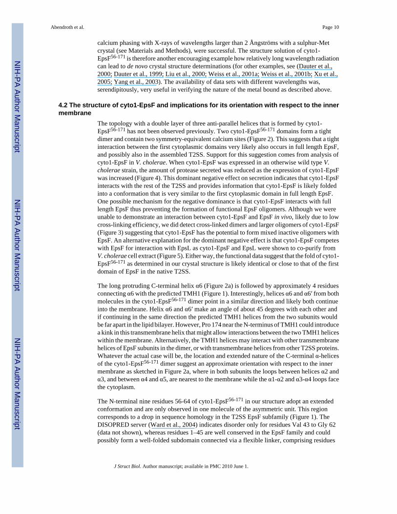
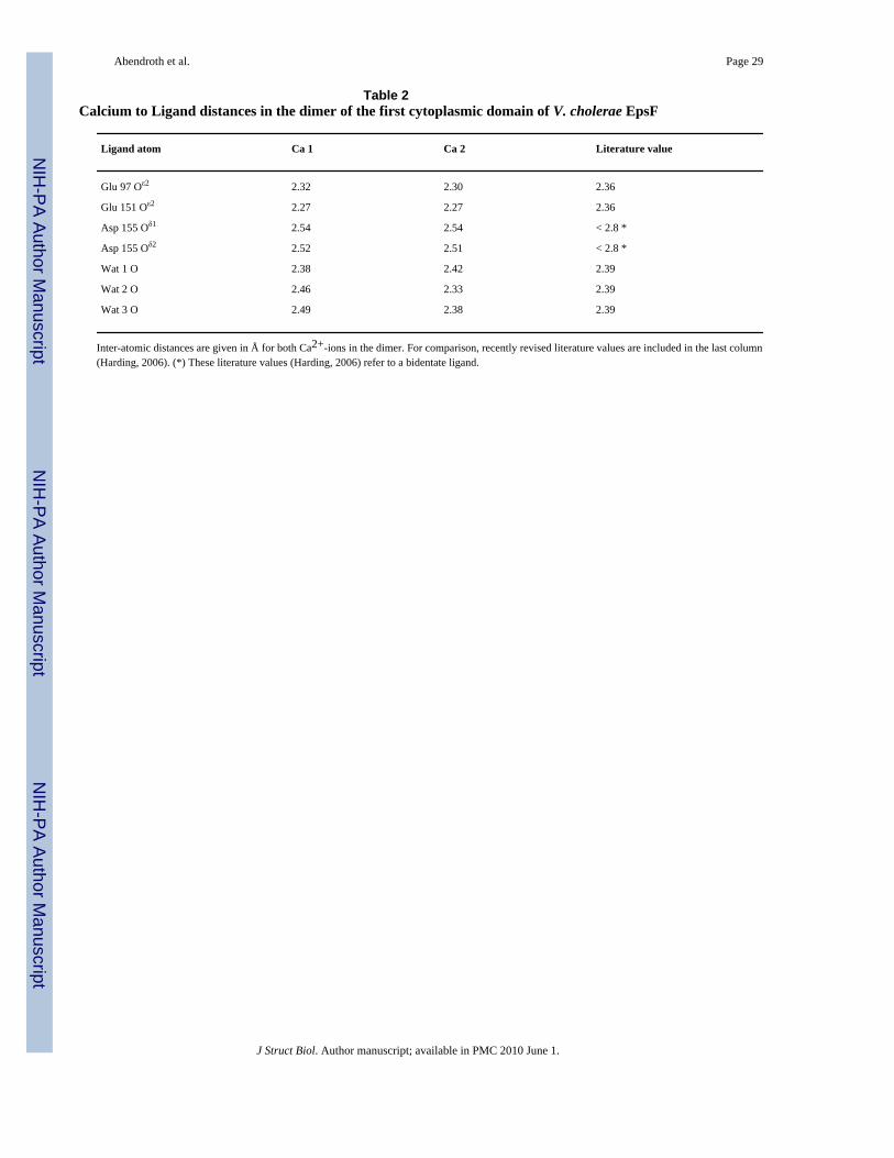
The conclusion that the metal ions are calcium is further supported by the environment of thesites. Since these are so similar in the two cases (Table 2), we will only describe here site A,shown in Figure 6. The protein metal ligands are one carboxylate oxygen from Glu 151 andtwo carboxylate oxygen atoms provided by Asp 155 from one subunit, with Glu 97 from thesecond subunit providing a fourth carboxylate oxygen as ligand. Three water moleculescomplete the coordination sphere. The ligands of each Ca2+ ion form a distorted tetragonalbipyramid, involving seven atoms since Asp 155 is ligating in a bidentate manner. The ligandsare all oxygen atoms, with the three protein ligands being negatively charged, as is often thecase in calcium binding sites in proteins. The distances between Ca2+ and its ligands (Table 2)are very close to values reported in the literature (Harding, 2006), which is in good agreementwith all other data mentioned above indicating that two calcium ions are bound per cyto1-EpsF56-171 dimer at symmetry-related sites. The guanidinium group of Arg 73 interacts withthe metal-coordinating carboxylates of both Glu 151 and Asp 155, and appears to have a“ligand-positioning” function (Figure 6).
3.4 Conservation of the cyto1-EpsF dimer and the metal binding site in the EpsF familySequences of the first cytoplasmic domains of the GspF family are well conserved (Figure 1;Table 3). When comparing V. cholerae cyto1-EpsF56-171 with T2SS homologs, the amino acidsequence identity for the 116 residues in the truncated domain for which the structure wassolved, ranges from 86 % between V. cholerae and V. vulnificus to 46 % when comparing V.cholerae EpsF and P. aeruginosa GspF (Table 3). When considering the 14 residues involvedin the interactions across the interface then these residues from V. cholerae share 100 % identitywith the homologs from two other Vibrio species, and an identity of 64 % or higher with GspFhomologs. Therefore, taking also into account the extensive and well-packed nature of theinterface observed in the cyto1-EpsF56-171 dimer (Figure 2), it is likely that all N-terminaldomains of GspF homologs use a similar interface to form dimers.
The three residues involved in Ca2+-binding by the first domain, and the “ligand-positioning”Arg 73, are conserved well in the GspF family (Figure 1). Of particular interest is that thebidentate ligand Asp 155 of V. cholerae EpsF (Figure 6) is only once replaced by a Glu,suggesting that the side chain at this position coordinates a metal ion in all GspF familymembers in a bidentate manner. The second calcium ligand, Glu 97, is only once substitutedby another amino acid: a Thr in the GspF homolog of P. aeruginosa. This still would allowthe side chain oxygen atom from this Thr to coordinate a calcium ion. The third calcium-coordinating residue, Glu 151, is frequently replaced by an Asn, and once by a Ser, i.e. eachtime by a residue which still could provide an oxygen atom as ligand to the metal binding site.Interestingly, in each member of the GspF family, two or three negatively charged carboxylates
Abendroth et al. Page 8
J Struct Biol. Author manuscript; available in PMC 2010 June 1.
NIH
-PA Author Manuscript
NIH
-PA Author Manuscript
NIH
-PA Author Manuscript
are available to coordinate divalent cations at this site. The side chains of the coordinating Glu151 and Asp 155 interact with Arg 73, which is an invariant residue. Hence, replacing the Asp155 by a Glu, or Glu 151 by an Asn or Ser, most likely allows the geometry of this Arg-stabilizedcalcium site to be essentially maintained, since the arginine's guanidinium group interacts withthe very oxygen atom which is liganding the metal ion (Figure 6). This would explain whyarginine is consistently present at this position across the GspF family.
3.5 Similarity between the second and the first cytoplasmic domains of GspFA significant degree of amino acid sequence similarity exists between the first and the secondcytoplasmic domains of V. cholerae EpsF (Table 4), as has been noted previously (Peabody etal., 2003) for members of the GspF/PilG/BfpE superfamily. When comparing residues 65-162from the solved cyto1-EpsF56-171 structure with residues 268-364 of the second domain of V.cholerae EpsF (hereafter also called “cyto2-EpsF”), 28 % of the residues are identical.Importantly, the levels of identity between the corresponding sequences in the first and seconddomains in GspF homologs are similar or higher (Figure 7a; Table 4). The highest identity, 34%, occurs between the first and second domain of the GspF homolog in P. aeruginosa.Interestingly, the first domain of V. cholerae EpsF displays a sequence identity with thesecond domains of the GspF family ranging from 28 to 39 %. Because of this clearly detectablehomology between the cyto1-GspF and cyto2-GspF domains throughout the GspF family, itseems reasonable to assume that across the family both cytoplasmic domains adopt the sameall-helical cyto1-EpsF fold.
The three residues which provide the ligands for Ca2+-binding sites, Glu 97, Glu 151 and Asp155, plus the invariant Arg 73 that interacts with both Glu 151 and Asp155, are only poorlyconserved in the second domain of T2SS EpsF homologs (Figure 7a). Of the 14 residuesinvolved in the cyto1-EpsF56-171 interface only 1 or 2 are conserved in the second cytoplasmicdomain of the homologs. Since the metal binding sites occur in the dimer interface of the cyto1-EpsF dimer (Figure 2), it is unlikely that cyto2-EpsF, while adopting most likely a similar foldas cyto1-EpsF56-171, forms the same type of dimers as cyto1-EpsF56-171.
3.6 Localization of fusion proteins of EpsF with GFPMembrane topology prediction of EpsF with the TMHMM program (Krogh et al., 2001)identified three putative transmembrane helices and suggested a cytoplasmic location for theN-terminus and a periplasmic location for the C-terminus of EpsF. In order to test the predictedtopology of EpsF, we constructed Green Fluorescent Protein (GFP) fusions to both the N-terminus and the C-terminus of EpsF. Both fusion proteins were detected by immunoblottingwith anti-EpsF antibodies (data not shown) when expression of the fusion proteins was inducedin V. cholerae. When the level of fluorescence was measured in these strains, we found GFPfused to the N-terminus of EpsF (GFP-EpsF) was fluorescent, whereas fusion of GFP to theC-terminus (EpsF-GFP) resulted in a non-fluorescent protein (Figure 8). This latter findingsuggests that the C-terminus of EpsF is likely located in the periplasmic space, as GFP is unableto fold in the periplasm due to the reducing environment of this compartment (Feilmeier et al.,2000). Although non-fluorescent, EpsF-GFP was able to complement the secretion defect inthe epsF mutant strain, suggesting that the protein is inserted in the membrane similarly to non-tagged EpsF (data not shown).
4. Discussion4.1 Phasing at long wavelengths
We have solved the V. cholerae cyto1-EpsF56-171 crystal structure along two different routesusing anomalous scattering effects at long X-ray wavelengths. Both approaches, one a halidesoak with a seleno-Met EpsF crystal and using CuKα radiation, and the other sulphur-cum-
Abendroth et al. Page 9
J Struct Biol. Author manuscript; available in PMC 2010 June 1.
NIH
-PA Author Manuscript
NIH
-PA Author Manuscript
NIH
-PA Author Manuscript
calcium phasing with X-rays of wavelengths larger than 2 Ångströms with a sulphur-Metcrystal (see Materials and Methods), were successful. The structure solution of cyto1-EpsF56-171 is therefore another encouraging example how relatively long wavelength radiationcan lead to de novo crystal structure determinations (for other examples, see (Dauter et al.,2000; Dauter et al., 1999; Liu et al., 2000; Weiss et al., 2001a; Weiss et al., 2001b; Xu et al.,2005; Yang et al., 2003). The availability of data sets with different wavelengths was,serendipitously, very useful in verifying the nature of the metal bound as described above.
4.2 The structure of cyto1-EpsF and implications for its orientation with respect to the innermembrane
The topology with a double layer of three anti-parallel helices that is formed by cyto1-EpsF56-171 has not been observed previously. Two cyto1-EpsF56-171 domains form a tightdimer and contain two symmetry-equivalent calcium sites (Figure 2). This suggests that a tightinteraction between the first cytoplasmic domains very likely also occurs in full length EpsF,and possibly also in the assembled T2SS. Support for this suggestion comes from analysis ofcyto1-EpsF in V. cholerae. When cyto1-EpsF was expressed in an otherwise wild type V.cholerae strain, the amount of protease secreted was reduced as the expression of cyto1-EpsFwas increased (Figure 4). This dominant negative effect on secretion indicates that cyto1-EpsFinteracts with the rest of the T2SS and provides information that cyto1-EpsF is likely foldedinto a conformation that is very similar to the first cytoplasmic domain in full length EpsF.One possible mechanism for the negative dominance is that cyto1-EpsF interacts with fulllength EpsF thus preventing the formation of functional EpsF oligomers. Although we wereunable to demonstrate an interaction between cyto1-EpsF and EpsF in vivo, likely due to lowcross-linking efficiency, we did detect cross-linked dimers and larger oligomers of cyto1-EpsF(Figure 3) suggesting that cyto1-EpsF has the potential to form mixed inactive oligomers withEpsF. An alternative explanation for the dominant negative effect is that cyto1-EpsF competeswith EpsF for interaction with EpsL as cyto1-EpsF and EpsL were shown to co-purify fromV. cholerae cell extract (Figure 5). Either way, the functional data suggest that the fold of cyto1-EpsF56-171 as determined in our crystal structure is likely identical or close to that of the firstdomain of EpsF in the native T2SS.
The long protruding C-terminal helix α6 (Figure 2a) is followed by approximately 4 residuesconnecting α6 with the predicted TMH1 (Figure 1). Interestingly, helices α6 and α6′ from bothmolecules in the cyto1-EpsF56-171 dimer point in a similar direction and likely both continueinto the membrane. Helix α6 and α6′ make an angle of about 45 degrees with each other andif continuing in the same direction the predicted TMH1 helices from the two subunits wouldbe far apart in the lipid bilayer. However, Pro 174 near the N-terminus of TMH1 could introducea kink in this transmembrane helix that might allow interactions between the two TMH1 heliceswithin the membrane. Alternatively, the TMH1 helices may interact with other transmembranehelices of EpsF subunits in the dimer, or with transmembrane helices from other T2SS proteins.Whatever the actual case will be, the location and extended nature of the C-terminal α-helicesof the cyto1-EpsF56-171 dimer suggest an approximate orientation with respect to the innermembrane as sketched in Figure 2a, where in both subunits the loops between helices α2 andα3, and between α4 and α5, are nearest to the membrane while the α1-α2 and α3-α4 loops facethe cytoplasm.
The N-terminal nine residues 56-64 of cyto1-EpsF56-171 in our structure adopt an extendedconformation and are only observed in one molecule of the asymmetric unit. This regioncorresponds to a drop in sequence homology in the T2SS EpsF subfamily (Figure 1). TheDISOPRED server (Ward et al., 2004) indicates disorder only for residues Val 43 to Gly 62(data not shown), whereas residues 1–45 are well conserved in the EpsF family and couldpossibly form a well-folded subdomain connected via a flexible linker, comprising residues
Abendroth et al. Page 10
J Struct Biol. Author manuscript; available in PMC 2010 June 1.
NIH
-PA Author Manuscript
NIH
-PA Author Manuscript
NIH
-PA Author Manuscript
∼45 – ∼60, to the globular cyto1-EpsF56-171 domain. Truncating the N-terminal subdomainfrom residue 1 to 55 appeared to be crucial for crystallization. This is similar to the requirementfor crystallization of another T2SS protein, the secretion ATPase EpsE from V. cholerae(Robien et al., 2003). There, the N1-domain of ∼ 90 residues needed to be removed in orderto obtain crystals. Yet, the N1-domain of EpsE has a critically important function: binding tothe cytoplasmic domain of EpsL, as observed in the crystal structure of the N1-domain of EpsEin complex with the cytoplasmic domain of the inner membrane T2SS protein EpsL. This leadsto membrane association of EpsE and stimulation of its ATPase activity (Abendroth et al.,2005;Camberg et al., 2007;Sandkvist et al., 1995). Similarly, the N-terminal subdomain ofEpsF might be involved in critical interactions and motions during T2SS assembly or function.
4.3 Metal binding sitesTwo symmetry-equivalent metal binding sites are present in the interface of the cyto1-EpsF56-171 dimer (Figure 2). Strong anomalous scattering, a calcium-dependent crystallizationand the geometry of the binding site identified the metals as calcium ions. Notably, the calciumbinding residues are conserved within the GspF family (Figure 1). Whereas cyto1-EpsF56-171 could only be crystallized in the presence of rather high concentrations (200mM)of externally added Ca2+, both protein and crystals were extremely sensitive to the nature ofdivalent ion during crystallization experiments. When calcium salts were replaced even withsmall amounts of strontium, barium, or lanthanide salts, protein in crystallization dropsprecipitated immediately. Moreover, crystals grown in the presence of calcium disintegratedwhen calcium was replaced with strontium, barium or lanthanides. Crystals obtained fromMg2+-containing solutions were far inferior in quality to those obtained from Ca2+-containingbuffer. Undoubtedly, the presence of calcium ions has been critical for crystal stability. Thesignificant degree of conservation of the ligands of the calcium site and of the invariant “Ca-ligand positioning” Arg 73, (Figures 1 and 6) suggests that calcium-binding by EpsF and T2SShomologs may be important during one or more of the many steps in the assembly orfunctioning of the T2SS system. Obviously, the precise nature of the metal bound underphysiological conditions at the putative metal binding sites of EpsF remains to be established.
4.4. Two similar domains and membrane topology of EpsF and its T2SS homologsThe second half of full length V. cholerae EpsF displays a significant degree of sequencehomology with the first cytoplasmic domain with a sequence identity of 28 % for 97 residues.This degree of sequence identity is similar or higher across the GspF family of EpsF homologs(Figure 7a, Table 4) suggesting that full length EpsF and its T2SS homologs contain twocytoplasmic domains of similar fold. This is in agreement with the analysis of Peabody etal., who reported that in the GspF/PilG/BfpE superfamily of membrane proteins a homologyexists between the N-terminal and C-terminal half of members in this large superfamily ofbacterial inner membrane proteins (Peabody et al., 2003).
A transmembrane helix prediction with the program TMHMM (Krogh et al., 2001) for EpsFfrom V. cholerae indicates three transmembrane helices, with residues 171-193 formingTMH1, residues 220-242 TMH2 and residues 370-392 TMH3 (not shown). This is perfectlycompatible with helix α6, observed in the crystal structure of the first cytoplasmic domain ofEpsF, ending at approximately position 168, i.e. just prior to TMH1, and residues ∼260 to∼370 forming the second cytoplasmic domain, i.e. situated between TMH2 and TMH3. Giventhe significant sequence identities between EpsF and its T2SS homologs (Figure 1, Tables 3and 4), and the very similar pattern of transmembrane helix prediction across the GspF family(data not shown), it is likely that the entire GspF family has the same topology with twosimilarly folded cytoplasmic domains and three transmembrane helices. This conclusion is inagreement with the three-transmembrane membrane topology arrived at for E. carotovora
Abendroth et al. Page 11
J Struct Biol. Author manuscript; available in PMC 2010 June 1.
NIH
-PA Author Manuscript
NIH
-PA Author Manuscript
NIH
-PA Author Manuscript
GspF (Thomas et al., 1997) and for P. aeruginosa GspF (Arts et al., 2007) using fusion proteintechniques, and with the GFP fusion studies shown in Figure 8.
4.5 Topology of non-T2SS EpsF homologsThe GspF/PilG/BfpE superfamily has a diversity of members with weak sequence homologybetween the three families, although with detectable similarity between the first and secondhalves of these proteins (Peabody et al., 2003). For BfpE from Enteropathogenic E. coli, a four-transmembrane helix topology has been proposed in the basis of biochemical studies (Blankand Donnenberg, 2001), which is clearly different from the evidence for a three-transmembranehelix topology for the T2SS GspF family, based on: (i) GspF-enzyme fusion studies (Arts etal., 2007; Thomas et al., 1997) (ii) in vivo fluorescence studies on EpsF fusions with GFP(Figure 8); (iii) the topology of the first domain of EpsF (Figure 1) coupled with the significantdegree of sequence identity between the first and second domains of T2SS family members(Table 4).
It might be possible that members of the superfamily do have weak sequence identity butdifferent membrane topologies. After all, among many similarities, substantial differencesbetween the T2SS and the T4PB exist, in particular regarding the composition of the InnerMembrane Complex. In adition, while both PilG and EpsF are required for the function of theT4P and T2S systems, respectively, they may support these systems with slighty differentmechanisms. For example, it has been proposed that in N. menigitidis the GspF homolog PilGcounteracts pilT-mediated pilus retraction (Carbonnelle et al., 2006), yet, retraction of T2Spseudopilus has not been demonstrated, nor does T2SS contain a PilT paralog. Due to thesedifferences it seems best to refrain here from extrapolating the GspF three-transmembrane helixtopology to other members of the GspF/PilG/BfpE superfamily. New structural data will berequired to clarify the intriguing results obtained by different studies so far.
4.6 Full-length EpsF dimersIn order to arrive at a global model of a full-length EpsF dimer with the same conservedinterface between the cyto1-EpsF56-171 domain as observed in our structure (Figure 2), weneed to add to each of the subunits: (i) a second domain of very similar topology, connectedby two anti-parallel transmembrane helices, TMH1 and TMH2, to the first domain, and (ii) atransmembrane helix, TMH3, to the C-terminus of cyto2-EpsF. The orientation of the seconddomains with respect to the membrane is likely to be roughly the same as that of the firstcytoplasmic EpsF domains since the number of residues between the putative α6 and TMH3is similar to the number of residues between the end of helix α1 of cyto1-EpsF and the start ofthe predicted TMH1. The mutual orientation of the first and second domains remains to beestablished, but the requirements of a compact dimer of full length EpsF subunits makes ageneral shape as depicted in Figure 7b a possibility.
It is of interest to compare the approximate model of the V. cholerae EpsF dimer (Figure 7b)with the 22 Å-resolution structure of the N. meningitidis PilG multimer (Collins et al., 2007)obtained from cryo-electron microscopy studies. The dimensions of our global model of thecytoplasmic part of EpsF dimer are, in the plane parallel to the membrane, approximately 70by 80 Å when adding an estimate of the space required for the N-terminal domains absent fromour structure. The model contains four quite similar major domains which are at low resolutionessentially indistinguishable and therefore the model is best compared with the C4 electronmicroscopy reconstruction (Collins et al., 2007). The dimensions parallel to the membrane ofthe electron microscopy reconstruction of PilG are ∼80 × 80 Å which agrees reasonably wellwith our model. The “height” of the EpsF model perpendicular to the membrane extendinginto the cytoplasm is approximately 54 Å, which is slightly less than the 70 Å of the PilGreconstruction. Nevertheless, the EpsF model does suggest that the quaternary structure of the
Abendroth et al. Page 12
J Struct Biol. Author manuscript; available in PMC 2010 June 1.
NIH
-PA Author Manuscript
NIH
-PA Author Manuscript
NIH
-PA Author Manuscript
EpsF-PilG inner membrane protein families is more likely a dimer than a tetramer, optionswhich were both considered possible (Collins et al., 2007).
5. Protein Data Bank accession codesThe atomic coordinates and structure factors have been deposited in the RCSB Protein DataBank and are available under accession code 3C1Q, 2VMA, and 2VMB.
Supplementary MaterialRefer to Web version on PubMed Central for supplementary material.
AcknowledgmentsWe acknowledge Stewart Turley for expert help with data collection. We thank the staff of beamline 9.2 at the StanfordSynchrotron Radiation Lightsource for support during data collection. This research was supported by NIH grantAI34501 to W.G.J.H. from the National Institute of Allergy and Infectious Diseases (NIAID) and by the HowardHughes Medical Institute (HHMI) and by grant AI49294 from NIAID to M.S. The content is solely the responsibilityof the authors and does not necessarily represent the official views of the HHMI, NIAID or the National Institutes ofHealth.
Appendix
Appendix A. Supplementary DataSupplementary data associated with this article can be found in the online version.
ReferencesAbendroth J, Bagdasarian M, Sandkvist M, Hol WGJ. The structure of the cytoplasmic domain of EpsL,
an inner membrane component of the type II secretion system of Vibrio cholerae: An unusual memberof the actin-like ATPase superfamily. Journal of Molecular Biology 2004a;344:619–633. [PubMed:15533433]
Abendroth J, Rice AE, McLuskey K, Bagdasarian M, Hol WGJ. The crystal structure of the periplasmicdomain of the type II secretion system protein EpsM from Vibrio cholerae: The simplest version ofthe ferredoxin fold. Journal of Molecular Biology 2004b;338:585–596. [PubMed: 15081815]
Abendroth J, Murphy P, Sandkvist M, Bagdasarian M, Hol WGJ. The X-ray structure of the type IIsecretion system complex formed by the N-terminal domain of EpsE and the cytoplasmic domain ofEpsL of Vibrio cholerae. Journal of Molecular Biology 2005;348:845–855. [PubMed: 15843017]
Abrahams JP, Buchanan SK, Van Raaij MJ, Fearnley IM, Leslie AG, Walker JE. The structure of bovineF1-ATPase complexed with the peptide antibiotic efrapeptin. Proceedings of the National Academyof Sciences of the United States of America 1996;93:9420–9424. [PubMed: 8790345]
Arts J, de Groot A, Ball G, Durand E, El Khattabi M, Filloux A, Tommassen J, Koster M. Interactiondomains in the Pseudomonas aeruginosa type II secretory apparatus component XcpS (GspF).Microbiology-Uk 2007;153:1582–1592.
Bailey S. The CCP4 Suite - Programs for Protein Crystallography. Acta Crystallogr, Sect D: BiolCrystallogr 1994;50:760–763. [PubMed: 15299374]
Bally M, Filloux A, Akrim M, Ball G, Lazdunski A, Tommassen J. Protein secretion in Pseudomonasaeruginosa: characterization of seven xcp genes and processing of secretory apparatus componentsby prepilin peptidase. Mol Microbiol 1992;6:1121–1131. [PubMed: 1588814]
Blank TE, Donnenberg MS. Novel topology of BfpE, a cytoplasmic membrane protein required for typeIV fimbrial biogenesis in enteropathogenic Escherichia coli. J Bacteriol 2001;183:4435–4450.[PubMed: 11443077]
Bortoli-German I, Brun E, Py B, Chippaux M, Barras F. Periplasmic disulphide bond formation isessential for cellulase secretion by the plant pathogen Erwinia chrysanthemi. Mol Microbiol1994;11:545–553. [PubMed: 8152378]
Abendroth et al. Page 13
J Struct Biol. Author manuscript; available in PMC 2010 June 1.
NIH
-PA Author Manuscript
NIH
-PA Author Manuscript
NIH
-PA Author Manuscript

Bouley J, Condemine G, Shevchik VE. The PDZ domain of OutC and the N-terminal region of OutDdetermine the secretion specificity of the type II out pathway of Erwinia chrysanthemi. J Mol Biol2001;308:205–219. [PubMed: 11327762]
Bricogne G, Vonrhein C, Flensburg C, Schiltz M, Paciorek W. Generation, representation and flow ofphase information in structure determination: recent developments in and around SHARP 2.0. ActaCrystallogr, Sect D: Biol Crystallogr 2003;59:2023–2030. [PubMed: 14573958]
Burrows LL. Weapons of mass retraction. Mol Microbiol 2005;57:878–888. [PubMed: 16091031]Camberg JL, Johnson TL, Patrick M, Abendroth J, Hol WGJ, Sandkvist M. Synergistic stimulation of
EpsE ATP hydrolysis by EpsL and acidic phospholipids. EMBO Journal 2007;26:19–27. [PubMed:17159897]
Carbonnelle E, Helaine S, Nassif X, Pelicic V. A systematic genetic analysis in Neisseria meningitidisdefines the Pil proteins required for assembly, functionality, stabilization and export of type IV pili.Mol Microbiol 2006;61:1510–22. [PubMed: 16968224]
Cianciotto NP. Type II secretion: a protein secretion system for all seasons. Trends Microbiol2005;13:581–588. [PubMed: 16216510]
Collins RF, Saleem M, Derrick JP. Purification and 3-D electron microscopy structure of the Neisseriameningitidis type IV pilus biogenesis protein PilG. J Bacteriol 2007;189:6389–6396. [PubMed:17616599]
Cowtan KD, Zhang KY. Density modification for macromolecular phase improvement. Progress inBiophysics & Molecular Biology 1999;72:245–270. [PubMed: 10581970]
Craig L, Li J. Type IV pili: paradoxes in form and function. Curr Opin Struct Biol 2008;18:267–77.[PubMed: 18249533]
Craig L, Pique ME, Tainer JA. Type IV pilus structure and bacterial pathogenicity. Nat Rev Microbiol2004;2:363–378. [PubMed: 15100690]
Crowther LJ, Anantha RP, Donnenberg MS. The inner membrane subassembly of the enteropathogenicEscherichia coli bundle-forming pilus machine. Mol Microbiol 2004;52:67–79. [PubMed:15049811]
d'Enfert C, Ryter A, Pugsley AP. Cloning and expression in Escherichia coli of the Klebsiellapneumoniae genes for production, surface localization and secretion of the lipoprotein pullulanase.EMBO J 1987;6:3531–3538. [PubMed: 3322811]
Dauter Z, Dauter M, Rajashankar KR. Novel approach to phasing proteins: derivatization by short cryo-soaking with halides. Acta Crystallogr, Sect D: Biol Crystallogr 2000;56:232–237. [PubMed:10666615]
Dauter Z, Dauter M, de La Fortelle E, Bricogne G, Sheldrick GM. Can anomalous signal of sulfur becomea tool for solving protein crystal structures? J Mol Biol 1999;289:83–92. [PubMed: 10339407]
DeLano, WL. The PyMol Molecular Graphics System. 2002. on World Wide Web,http://www.pymol.org
Emsley P, Cowtan K. Coot: model-building tools for molecular graphics. Acta Crystallogr, Sect D: BiolCrystallogr 2004;60:2126–2132. [PubMed: 15572765]
Evans FF, Egan S, Kjelleberg S. Ecology of type II secretion in marine gammaproteobacteria. EnvironMicrobiol 2008;10:1101–7. [PubMed: 18218035]
Feilmeier BJ, Iseminger G, Schroeder D, Webber H, Phillips GJ. Green fluorescent protein functions asa reporter for protein localization in Escherichia coli. J Bacteriol 2000;182:4068–76. [PubMed:10869087]
Filloux A. The underlying mechanisms of type II protein secretion. Biochim Biophys Acta2004;1694:163–179. [PubMed: 15546665]
Gouet P, Courcelle E, Stuart DI, Metoz F. ESPript: analysis of multiple sequence alignments in PostScript.Bioinformatics 1999;15:305–308. [PubMed: 10320398]
Hansen JK, Forest KT. Type IV pilin structures: insights on shared architecture, fiber assembly, receptorbinding and type II secretion. J Mol Microbiol Biotechnol 2006;11:192–207. [PubMed: 16983195]
Hardie KR, Schulze A, Parker MW, Buckley JT. Vibrio spp. secrete proaerolysin as a folded dimerwithout the need for disulphide bond formation. Mol Microbiol 1995;17:1035–1044. [PubMed:8594324]
Abendroth et al. Page 14
J Struct Biol. Author manuscript; available in PMC 2010 June 1.
NIH
-PA Author Manuscript
NIH
-PA Author Manuscript
NIH
-PA Author Manuscript

Harding MM. Small revisions to predicted distances around metal sites in proteins. Acta Crystallogr, SectD: Biol Crystallogr 2006;62:678–682. [PubMed: 16699196]
Hirst TR, Holmgren J. Conformation of protein secreted across bacterial outer membranes: a study ofenterotoxin translocation from Vibrio cholerae. Proc Natl Acad Sci U S A 1987;84:7418–7422.[PubMed: 3478701]
Hobbs M, Mattick JS. Common components in the assembly of type 4 fimbriae, DNA transfer systems,filamentous phage and protein-secretion apparatus: a general system for the formation of surface-associated protein complexes. Mol Microbiol 1993;10:233–243. [PubMed: 7934814]
Holm L, Sander C. Protein structure comparison by alignment of distance matrices. Journal of MolecularBiology 1993;233:123–138. [PubMed: 8377180]
Johnson TL, Scott ME, Sandkvist M. Mapping critical interactive sites within the periplasmic domain ofthe Vibrio cholerae type II secretion protein EpsM. J Bacteriol 2007;189:9082–9. [PubMed:17921296]
Johnson TL, Abendroth J, Hol WGJ, Sandkvist M. Type II secretion: from structure to function. FEMSMicrobiol Lett 2006;255:175–186. [PubMed: 16448494]
Jones S, Thornton JM. Principles of protein-protein interactions. Proceedings of the National Academyof Sciences of the United States of America 1996;93:13–20. [PubMed: 8552589]
Kirn TJ, Jude BA, Taylor RK. A colonization factor links Vibrio cholerae environmental survival andhuman infection. Nature 2005;438:863–6. [PubMed: 16341015]
Korotkov KV, Krumm BE, Bagdasarian M, Hol WGJ. Structural and Functional Studies of EpsC, aCrucial Component of the Type 2 Secretion System from Vibrio cholerae. J Mol Biol 2006;363:311–321. [PubMed: 16978643]
Korotkov KV, Hol WGJ. Structure of the GspK-GspI-GspJ complex from the enterotoxigenicEscherichia coli type 2 secretion system. Nat Struct Mol Biol 2008;15:462–468. [PubMed:18438417]
Korotkov KV, Pardon E, Steyaert J, Hol WGJ. Crystal Structure of the N-Terminal Domain of the SecretinGspD from ETEC Determined with the Assistance of a Nanobody. Structure 2009;17:255–265.[PubMed: 19217396]
Krissinel E, Henrick K. Secondary-structure matching (SSM), a new tool for fast protein structurealignment in three dimensions. Acta Crystallogr, Sect D: Biol Crystallogr 2004;60:2256–2268.[PubMed: 15572779]
Krogh A, Larsson B, von Heijne G, Sonnhammer EL. Predicting transmembrane protein topology witha hidden Markov model: application to complete genomes. J Mol Biol 2001;305:567–580. [PubMed:11152613]
Landau M, Mayrose I, Rosenberg Y, Glaser F, Martz E, Pupko T, Ben-Tal N. ConSurf 2005: the projectionof evolutionary conservation scores of residues on protein structures. Nucleic Acids Res2005;33:W299–302. [PubMed: 15980475]
Liu ZJ, Vysotski ES, Chen CJ, Rose JP, Lee J, Wang BC. Structure of the Ca2+-regulated photoproteinobelin at 1.7 A resolution determined directly from its sulfur substructure. Protein Sci 2000;9:2085–2093. [PubMed: 11152120]
Mattick JS. Type IV pili and twitching motility. Annu Rev Microbiol 2002;56:289–314. [PubMed:12142488]
Merritt EA, Hol WGJ. AB5 toxins. Curr Opin Struct Biol 1995;5:165–171. [PubMed: 7648317]Murshudov GN, Vagin AA, Dodson EJ. Refinement of macromolecular structures by the maximum-
likelihood method. Acta Crystallogr, Sect D: Biol Crystallogr 1997;53:240–255. [PubMed:15299926]
Notredame C, Higgins DG, Heringa J. T-Coffee: A novel method for fast and accurate multiple sequencealignment. Journal of Molecular Biology 2000;302:205–217. [PubMed: 10964570]
Nudleman E, Kaiser D. Pulling together with type IV pili. J Mol Microbiol Biotechnol 2004;7:52–62.[PubMed: 15170403]
Nunn D. Bacterial type II protein export and pilus biogenesis: more than just homologies? Trends CellBiol 1999;9:402–408. [PubMed: 10481178]
Abendroth et al. Page 15
J Struct Biol. Author manuscript; available in PMC 2010 June 1.
NIH
-PA Author Manuscript
NIH
-PA Author Manuscript
NIH
-PA Author Manuscript
O'Neal CJ, Amaya EI, Jobling MG, Holmes RK, Hol WGJ. Crystal structures of an intrinsically activecholera toxin mutant yield insight into the toxin activation mechanism. Biochemistry 2004;43:3772–3782. [PubMed: 15049684]
Painter J, Merritt EA. Optimal description of a protein structure in terms of multiple groups undergoingTLS motion. Acta Crystallogr, Sect D: Biol Crystallogr 2006;62:439–450. [PubMed: 16552146]
Peabody CR, Chung YJ, Yen MR, Vidal-Ingigliardi D, Pugsley AP, Saier MH Jr. Type II protein secretionand its relationship to bacterial type IV pili and archaeal flagella. Microbiology 2003;149:3051–3072.[PubMed: 14600218]
Perrakis A, Morris R, Lamzin VS. Automated protein model building combined with iterative structurerefinement. Nature Structural Biology 1999;6:458–463.
Pflugrath JW. The finer things in X-ray diffraction data collection. Acta Crystallogr, Sect D: BiolCrystallogr 1999;55:1718–1725. [PubMed: 10531521]
Pugsley AP. Translocation of a folded protein across the outer membrane in Escherichia coli. Proc NatlAcad Sci U S A 1992;89:12058–12062. [PubMed: 1465440]
Pugsley AP. The complete general secretory pathway in gram-negative bacteria. Microbiol Rev1993;57:50–108. [PubMed: 8096622]
Py B, Loiseau L, Barras F. An inner membrane platform in the type II secretion machinery of Gram-negative bacteria. EMBO Reports 2001;2:244–248. [PubMed: 11266368]
Robien MA, Krumm BE, Sandkvist M, Hol WGJ. Crystal structure of the extracellular protein secretionNTPase EpsE of Vibrio cholerae. Journal of Molecular Biology 2003;333:657–674. [PubMed:14556751]
Sandkvist M. Biology of type II secretion. Molecular Microbiology 2001a;40:271–283. [PubMed:11309111]
Sandkvist M. Type II secretion and pathogenesis. Infection & Immunity 2001b;69:3523–3535. [PubMed:11349009]
Sandkvist M, Bagdasarian M, Howard SP, DiRita VJ. Interaction between the autokinase EpsE and EpsLin the cytoplasmic membrane is required for extracellular secretion in Vibrio cholerae. EMBO J1995;14:1664–73. [PubMed: 7737119]
Sandkvist M, Michel LO, Hough LP, Morales VM, Bagdasarian M, Koomey M, DiRita VJ. Generalsecretion pathway (eps) genes required for toxin secretion and outer membrane biogenesis in Vibriocholerae. J Bacteriol 1997;179:6994–7003. [PubMed: 9371445]
Sauvonnet N, Vignon G, Pugsley AP, Gounon P. Pilus formation and protein secretion by the samemachinery in Escherichia coli. EMBO J 2000;19:2221–2228. [PubMed: 10811613]
Schmidt H, Henkel B, Karch H. A gene cluster closely related to type II secretion pathway operons ofgram-negative bacteria is located on the large plasmid of enterohemorrhagic Escherichia coli O157strains. FEMS Microbiol Lett 1997;148:265–272. [PubMed: 9084155]
Schneider TR, Sheldrick GM. Substructure solution with SHELXD. Acta Crystallogr, Sect D: BiolCrystallogr 2002;58:1772–1779. [PubMed: 12351820]
Sikora AE, Lybarger SR, Sandkvist M. Compromised outer membrane integrity in Vibrio cholerae TypeII secretion mutants. J Bacteriol 2007;189:8484–95. [PubMed: 17890307]
Sixma TK, Pronk SE, Kalk KH, Wartna ES, van Zanten BAM, Witholt B, Hol WGJ. Crystal structureof a cholera toxin-related heat-labile enterotoxin from E. coli. Nature 1991;351:371–377. [PubMed:2034287]
Söderberg MA, Rossier O, Cianciotto NP. The type II protein secretion system of Legionellapneumophila promotes growth at low temperatures. J Bacteriol 2004;186:3712–3720. [PubMed:15175284]
Tauschek M, Gorrell R, Strugnell R, Robins-Browne R. Identification of a protein secretory pathway forthe secretion of heat-labile enterotoxin by an enterotoxigenic strain of Escherichia coli. Proc NatlAcad Sci U S A 2002;99:7066–7071. [PubMed: 12011463]
Thomas JD, Reeves PJ, Salmond GPC. The general secretion pathway of Erwinia carotovora subspcarotovora: Analysis of the membrane topology of OutC and OutF. Microbiology-Uk 1997;143:713–720.
Uweh, O. Klebsiella Infections. 2006. http://www.emedicine.com/MED/topic1237.htm
Abendroth et al. Page 16
J Struct Biol. Author manuscript; available in PMC 2010 June 1.
NIH
-PA Author Manuscript
NIH
-PA Author Manuscript
NIH
-PA Author Manuscript

van Duyne GD, Standaert RF, Karplus PA, Schreiber SL, Clardy J. Atomic structures of the humanimmunophilin FKBP-12 complexes with FK506 and rapamycin. J Mol Biol 1993;229:105–124.[PubMed: 7678431]
Ward JJ, McGuffin LJ, Bryson K, Buxton BF, Jones DT. The DISOPRED server for the prediction ofprotein disorder. Bioinformatics 2004;20:2138–2139. [PubMed: 15044227]
Weiss MS, Sicker T, Hilgenfeld R. Soft X-rays, high redundancy, and proper scaling: A new procedurefor automated protein structure determination via SAS. Structure 2001a;9:771–777. [PubMed:11566127]
Weiss MS, Sicker T, Djinovic-Carugo K, Hilgenfeld R. On the routine use of soft X-rays inmacromolecular crystallography. Acta Crystallogr, Sect D: Biol Crystallogr 2001b;57:689–695.[PubMed: 11320309]
Xu H, Yang C, Chen LR, Kataeva IA, Tempel W, Lee D, Habel JE, Nguyen D, Pflugrath JW, FerraraJD, Arendall WB, Richardson JS, Richardson DC, Liu ZJ, Newton MG, Rose JP, Wang BC. Awayfrom the edge II: in-house Se-SAS phasing with chromium radiation. Acta Crystallogr, Sect D: BiolCrystallogr 2005;61:960–966. [PubMed: 15983419]
Yanez ME, Korotkov KV, Abendroth J, Hol WGJ. Structure of the minor pseudopilin EpsH from theType 2 Secretion system of Vibrio cholerae. J Mol Biol 2008a;37:91–103.
Yanez ME, Korotkov KV, Abendroth J, Hol WGJ. The crystal structure of a binary complex of twopseudopilins: EpsI and EpsJ from the type 2 secretion system of Vibrio vulnificus. J Mol Biol 2008b;375:471–486. [PubMed: 18022192]
Yang C, Pflugrath JW, Courville DA, Stence CN, Ferrara JD. Away from the edge: SAD phasing fromthe sulfur anomalous signal measured in-house with chromium radiation. Acta Crystallogr, Sect D:Biol Crystallogr 2003;59:1943–1957. [PubMed: 14573949]
Yen YT, Bhattacharya M, Stathopoulos C. Genome-wide in silico mapping of the secretome in pathogenicYersinia pestis KIM. FEMS Microbiol Lett 2008;279:56–63. [PubMed: 18070074]
Abendroth et al. Page 17
J Struct Biol. Author manuscript; available in PMC 2010 June 1.
NIH
-PA Author Manuscript
NIH
-PA Author Manuscript
NIH
-PA Author Manuscript

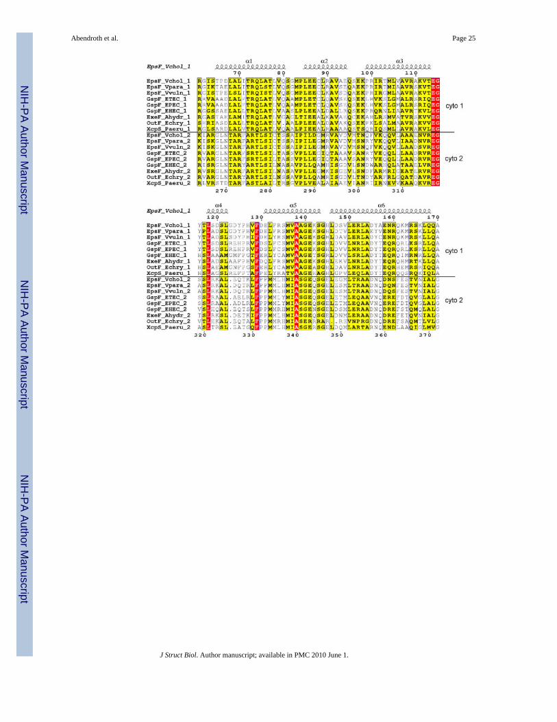
Figure 1. Family sequence alignment of the first cytoplasmic domain of EpsF and selected T2SShomologuesIdentical residues are in red, similar residues according to the Risler matrix are in yellow. Thepredicted transmembrane helix (Met 171 – Val 192) is indicated with a green bar. The extentof the construct is marked with the black arrows above the sequences. The blue bar below thesequences indicates the solvent accessibility of each residue: dark blue is solvent exposed,white is buried. Residues involved in Ca2+-binding are marked with red triangles. Residueswhich are part of the dimer interface are marked with blue stars. Alpha-helical secondarystructure symbols above the cyto1-EpsF sequence represent the observed structure in ourexperimental model.
Abendroth et al. Page 18
J Struct Biol. Author manuscript; available in PMC 2010 June 1.
NIH
-PA Author Manuscript
NIH
-PA Author Manuscript
NIH
-PA Author Manuscript

Figure 2. The dimer of cyto1-EpsF56-171 from V. cholerae(A) Each cyto1-EpsF56-171 subunit consists of a six-helix bundle. The N-terminal residues haveno secondary structure; the second half of the C-terminal helix protrudes away from the bodyof the domain and may extend towards the inner membrane as sketched in the left upper corner.Green spheres indicate calcium ions in the dimer interface. The dimer is shown in three differentorientations related by 90° rotations about two different axes.(B) The interface of the cyto1-EpsF56-171 dimer. In this butterfly figure, the surfaces of the twocyto1-EpsF56-171 domains per dimer are colored in red and blue, respectively. Residuesinvolved in dimer interactions are colored with the same color as the domain they contact.Light colors of the “imprint” indicate residues involved in hydrogen bonds, dark colors indicateresidues involved in hydrogen bonds and hydrophobic interactions. Twelve hydrogen bondsin the dimer interface involve the following residues: (i) Glu 74 and Thr 77 provided by helix
Abendroth et al. Page 19
J Struct Biol. Author manuscript; available in PMC 2010 June 1.
NIH
-PA Author Manuscript
NIH
-PA Author Manuscript
NIH
-PA Author Manuscript
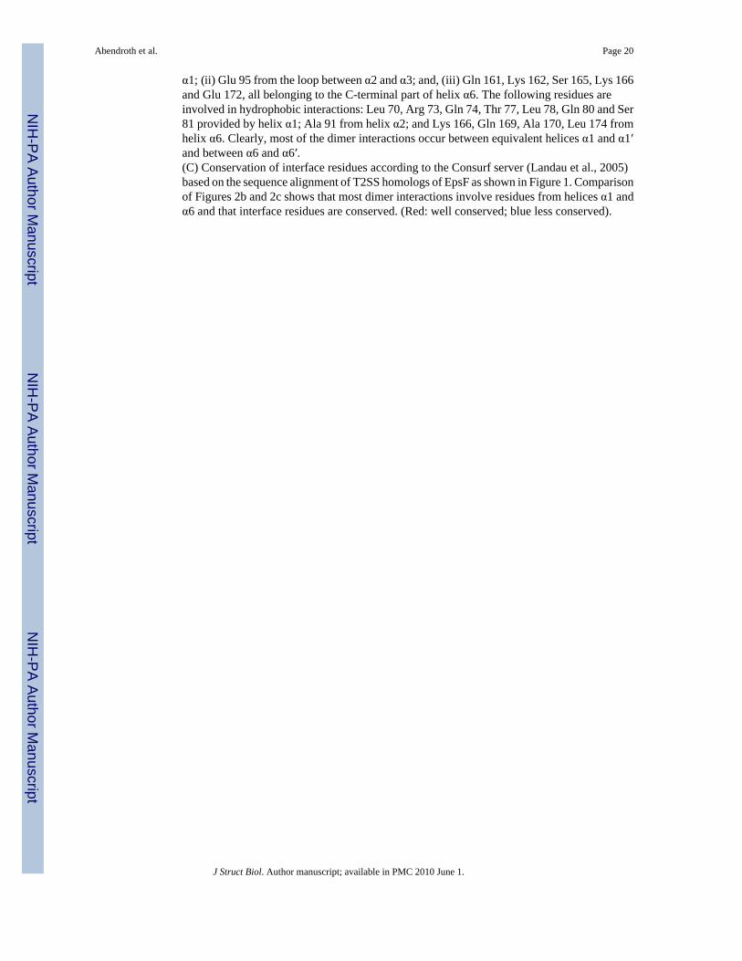
α1; (ii) Glu 95 from the loop between α2 and α3; and, (iii) Gln 161, Lys 162, Ser 165, Lys 166and Glu 172, all belonging to the C-terminal part of helix α6. The following residues areinvolved in hydrophobic interactions: Leu 70, Arg 73, Gln 74, Thr 77, Leu 78, Gln 80 and Ser81 provided by helix α1; Ala 91 from helix α2; and Lys 166, Gln 169, Ala 170, Leu 174 fromhelix α6. Clearly, most of the dimer interactions occur between equivalent helices α1 and α1′and between α6 and α6′.(C) Conservation of interface residues according to the Consurf server (Landau et al., 2005)based on the sequence alignment of T2SS homologs of EpsF as shown in Figure 1. Comparisonof Figures 2b and 2c shows that most dimer interactions involve residues from helices α1 andα6 and that interface residues are conserved. (Red: well conserved; blue less conserved).
Abendroth et al. Page 20
J Struct Biol. Author manuscript; available in PMC 2010 June 1.
NIH
-PA Author Manuscript
NIH
-PA Author Manuscript
NIH
-PA Author Manuscript
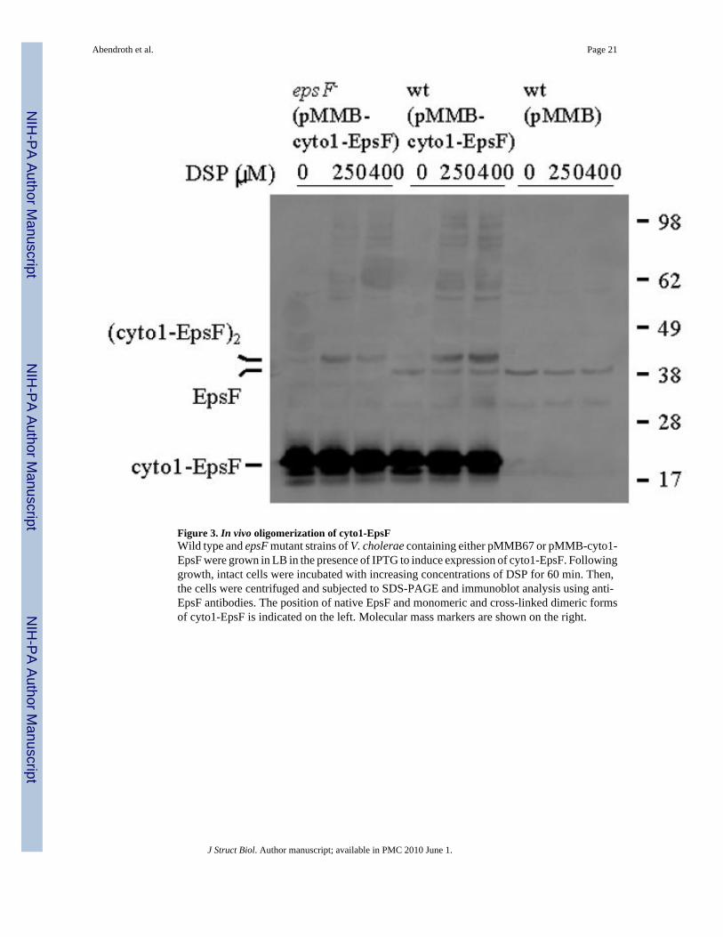
Figure 3. In vivo oligomerization of cyto1-EpsFWild type and epsF mutant strains of V. cholerae containing either pMMB67 or pMMB-cyto1-EpsF were grown in LB in the presence of IPTG to induce expression of cyto1-EpsF. Followinggrowth, intact cells were incubated with increasing concentrations of DSP for 60 min. Then,the cells were centrifuged and subjected to SDS-PAGE and immunoblot analysis using anti-EpsF antibodies. The position of native EpsF and monomeric and cross-linked dimeric formsof cyto1-EpsF is indicated on the left. Molecular mass markers are shown on the right.
Abendroth et al. Page 21
J Struct Biol. Author manuscript; available in PMC 2010 June 1.
NIH
-PA Author Manuscript
NIH
-PA Author Manuscript
NIH
-PA Author Manuscript
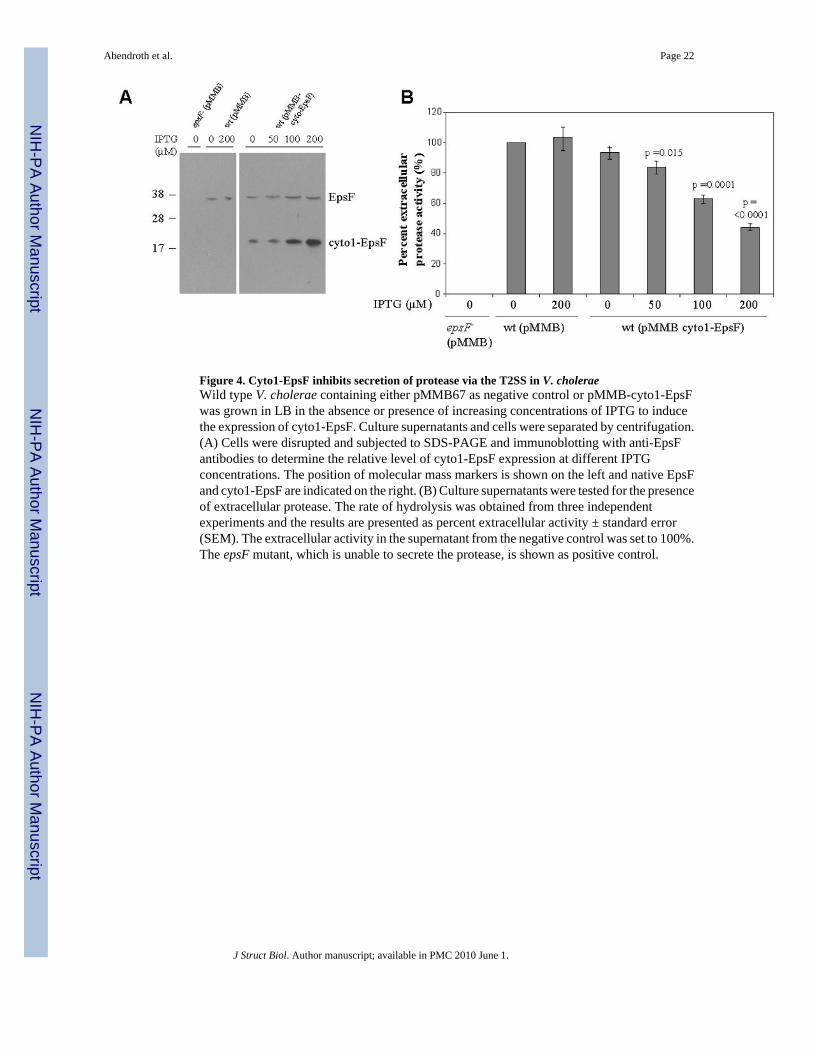
Figure 4. Cyto1-EpsF inhibits secretion of protease via the T2SS in V. choleraeWild type V. cholerae containing either pMMB67 as negative control or pMMB-cyto1-EpsFwas grown in LB in the absence or presence of increasing concentrations of IPTG to inducethe expression of cyto1-EpsF. Culture supernatants and cells were separated by centrifugation.(A) Cells were disrupted and subjected to SDS-PAGE and immunoblotting with anti-EpsFantibodies to determine the relative level of cyto1-EpsF expression at different IPTGconcentrations. The position of molecular mass markers is shown on the left and native EpsFand cyto1-EpsF are indicated on the right. (B) Culture supernatants were tested for the presenceof extracellular protease. The rate of hydrolysis was obtained from three independentexperiments and the results are presented as percent extracellular activity ± standard error(SEM). The extracellular activity in the supernatant from the negative control was set to 100%.The epsF mutant, which is unable to secrete the protease, is shown as positive control.
Abendroth et al. Page 22
J Struct Biol. Author manuscript; available in PMC 2010 June 1.
NIH
-PA Author Manuscript
NIH
-PA Author Manuscript
NIH
-PA Author Manuscript

Figure 5. Interaction of cyto1-EpsF and EpsLEpsL was co-purified with cyto1-EpsFhis6 from V. cholerae cell extracts by metal-affinitychromatography and subjected to SDS-PAGE and immunoblotting with anti-EpsL antibodies.Lanes 1-4 show the level of EpsL detected in cell extracts prior to purification, and lanes 5-8represent EpsL proteins that bound to hexahistidine-tagged cyto1-EpsF. No EpsL was purifiedwhen cyto1-EpsF was not expressed (lanes 5 and 6). Positions of molecular weight markersare shown.
Abendroth et al. Page 23
J Struct Biol. Author manuscript; available in PMC 2010 June 1.
NIH
-PA Author Manuscript
NIH
-PA Author Manuscript
NIH
-PA Author Manuscript

Figure 6. Calcium binding site in the dimer interface of V. cholerae cyto1-EpsF56-171
Calcium is coordinated as an approximate tetragonal bipyramid by Glu 97 from chain A, andGlu 151 and Asp 155 from chain B, and three water molecules. Asp 155 is binding the calciumion in a bidentate manner. Arg 73 forms a salt bridge with both Glu 151 and Asp 155. Glu 151has an additional hydrogen bond with Gln 80. The calcium ion is surrounded by anomalousFourier electron density contoured at 7.5 sigma colored in gold.
Abendroth et al. Page 24
J Struct Biol. Author manuscript; available in PMC 2010 June 1.
NIH
-PA Author Manuscript
NIH
-PA Author Manuscript
NIH
-PA Author Manuscript
Abendroth et al. Page 25
J Struct Biol. Author manuscript; available in PMC 2010 June 1.
NIH
-PA Author Manuscript
NIH
-PA Author Manuscript
NIH
-PA Author Manuscript
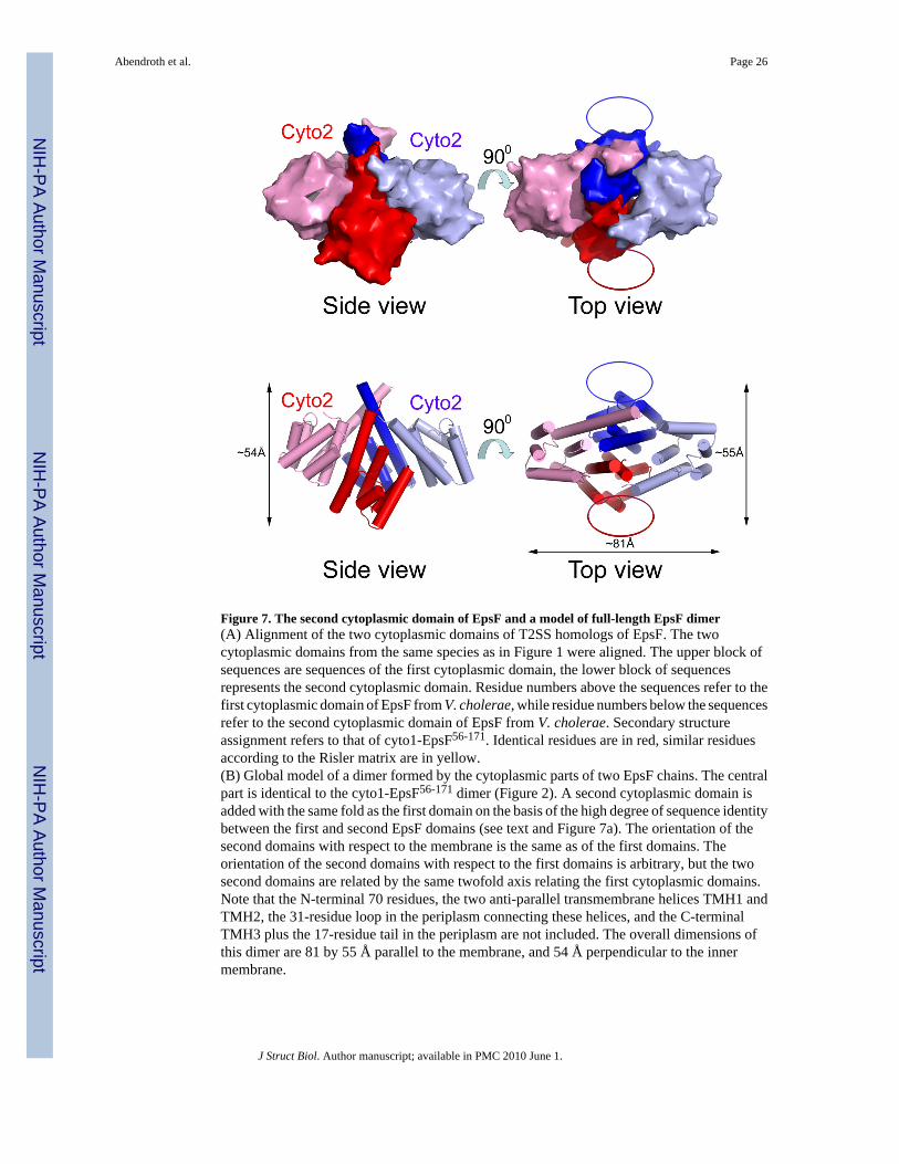
Figure 7. The second cytoplasmic domain of EpsF and a model of full-length EpsF dimer(A) Alignment of the two cytoplasmic domains of T2SS homologs of EpsF. The twocytoplasmic domains from the same species as in Figure 1 were aligned. The upper block ofsequences are sequences of the first cytoplasmic domain, the lower block of sequencesrepresents the second cytoplasmic domain. Residue numbers above the sequences refer to thefirst cytoplasmic domain of EpsF from V. cholerae, while residue numbers below the sequencesrefer to the second cytoplasmic domain of EpsF from V. cholerae. Secondary structureassignment refers to that of cyto1-EpsF56-171. Identical residues are in red, similar residuesaccording to the Risler matrix are in yellow.(B) Global model of a dimer formed by the cytoplasmic parts of two EpsF chains. The centralpart is identical to the cyto1-EpsF56-171 dimer (Figure 2). A second cytoplasmic domain isadded with the same fold as the first domain on the basis of the high degree of sequence identitybetween the first and second EpsF domains (see text and Figure 7a). The orientation of thesecond domains with respect to the membrane is the same as of the first domains. Theorientation of the second domains with respect to the first domains is arbitrary, but the twosecond domains are related by the same twofold axis relating the first cytoplasmic domains.Note that the N-terminal 70 residues, the two anti-parallel transmembrane helices TMH1 andTMH2, the 31-residue loop in the periplasm connecting these helices, and the C-terminalTMH3 plus the 17-residue tail in the periplasm are not included. The overall dimensions ofthis dimer are 81 by 55 Å parallel to the membrane, and 54 Å perpendicular to the innermembrane.
Abendroth et al. Page 26
J Struct Biol. Author manuscript; available in PMC 2010 June 1.
NIH
-PA Author Manuscript
NIH
-PA Author Manuscript
NIH
-PA Author Manuscript

Figure 8. Fusion of GFP to the EpsF C-terminus results in a non-fluorescent proteinV. cholerae epsF mutant strains containing pMMB alone, or expressing either GFP-EpsF orEpsF-GFP were grown in M9 growth medium and carbenicillin without (white bars) and with(grey bars) IPTG at a final concentration of 20 μM to induce expression of the fusion proteins.Following growth, fluorescence was measured using the excitation and emisssion wavelengths380 nm and 440 nm, respectively. Results from three independent experiments are presentedas average relative fluorescence units ± standard error.
Abendroth et al. Page 27
J Struct Biol. Author manuscript; available in PMC 2010 June 1.
NIH
-PA Author Manuscript
NIH
-PA Author Manuscript
NIH
-PA Author Manuscript
NIH
-PA Author Manuscript
NIH
-PA Author Manuscript
NIH
-PA Author Manuscript
Abendroth et al. Page 28
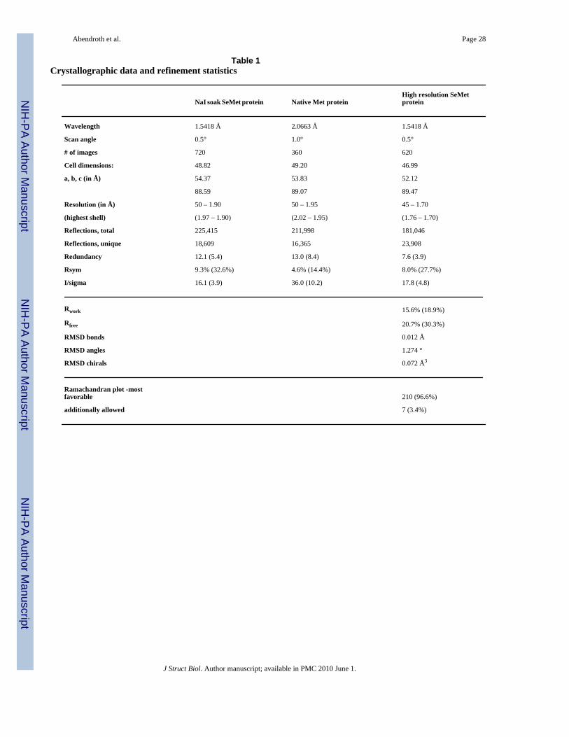
Table 1Crystallographic data and refinement statistics
NaI soak SeMet protein Native Met proteinHigh resolution SeMetprotein
Wavelength 1.5418 Å 2.0663 Å 1.5418 Å
Scan angle 0.5° 1.0° 0.5°
# of images 720 360 620
Cell dimensions: 48.82 49.20 46.99
a, b, c (in Å) 54.37 53.83 52.12
88.59 89.07 89.47
Resolution (in Å) 50 – 1.90 50 – 1.95 45 – 1.70
(highest shell) (1.97 – 1.90) (2.02 – 1.95) (1.76 – 1.70)
Reflections, total 225,415 211,998 181,046
Reflections, unique 18,609 16,365 23,908
Redundancy 12.1 (5.4) 13.0 (8.4) 7.6 (3.9)
Rsym 9.3% (32.6%) 4.6% (14.4%) 8.0% (27.7%)
I/sigma 16.1 (3.9) 36.0 (10.2) 17.8 (4.8)
Rwork 15.6% (18.9%)
Rfree 20.7% (30.3%)
RMSD bonds 0.012 Å
RMSD angles 1.274 °
RMSD chirals 0.072 Å3
Ramachandran plot -mostfavorable 210 (96.6%)
additionally allowed 7 (3.4%)
J Struct Biol. Author manuscript; available in PMC 2010 June 1.
NIH
-PA Author Manuscript
NIH
-PA Author Manuscript
NIH
-PA Author Manuscript
Abendroth et al. Page 29
Table 2Calcium to Ligand distances in the dimer of the first cytoplasmic domain of V. cholerae EpsF
Ligand atom Ca 1 Ca 2 Literature value
Glu 97 Oε2 2.32 2.30 2.36
Glu 151 Oε2 2.27 2.27 2.36
Asp 155 Oδ1 2.54 2.54 < 2.8 *
Asp 155 Oδ2 2.52 2.51 < 2.8 *
Wat 1 O 2.38 2.42 2.39
Wat 2 O 2.46 2.33 2.39
Wat 3 O 2.49 2.38 2.39
Inter-atomic distances are given in Å for both Ca2+-ions in the dimer. For comparison, recently revised literature values are included in the last column(Harding, 2006). (*) These literature values (Harding, 2006) refer to a bidentate ligand.
J Struct Biol. Author manuscript; available in PMC 2010 June 1.

NIH
-PA Author Manuscript
NIH
-PA Author Manuscript
NIH
-PA Author Manuscript
Abendroth et al. Page 30
Table 3Conservation of overall conservation versus contact residues in the first cytoplasmic domains in the T2SS EpsFfamily
T2SS homolog of EpsF in the following species:% identity of EpsF domain 1 (residues
55-168) vs. V. cholerae% identity of 14 dimer contact
residues vs. V. cholerae
Vibrio cholerae 100 100
Vibrio parahaemolyticus 86 100
Vibrio vulnificus 84 100
ETEC 64 86
EPEC 65 86
EHEC 57 64
Aeromonas hydrophila 69 64
Erwinia chrysanthemi 56 64
Pseudomonas aeruginosa 46 71
J Struct Biol. Author manuscript; available in PMC 2010 June 1.

NIH
-PA Author Manuscript
NIH
-PA Author Manuscript
NIH
-PA Author Manuscript
Abendroth et al. Page 31
Table 4Comparison of the amino acid sequence in the first versus the second cytoplasmic domains in the T2SS GspFfamily. (For alignment see Figure 6a)
Species% identity of Domain 1 vs. Domain 2
within each species at left% identity of V. cholerae EpsF Domain 1(Residues 65-162) vs. species Domain 2
Vibrio cholerae 28 28
Vibrio parahaemolyticus 29 29
Vibrio vulnificus 28 29
ETEC 31 31
EPEC 30 32
EHEC 33 34
Aeromonas hydrophila 28 36
Erwinia chrysanthemi 31 35
Pseudomonas aeruginosa 34 39
J Struct Biol. Author manuscript; available in PMC 2010 June 1.
Copyright © 2022 FDOKUMEN