Lipopolysaccharides of Vibrio cholerae
15
Review Lipopolysaccharides of Vibrio cholerae I. Physical and chemical characterization S.N. Chatterjee a, * , Keya Chaudhuri b a Saha Institute of Nuclear Physics, 1/AF Bidhannagar, Sector-1, Calcutta-700 064, India b Biophysics Division, Indian Institute of Chemical Biology, Jadavpur, Calcutta-700 032, India Received 19 March 2003; received in revised form 11 August 2003; accepted 27 August 2003 Abstract Vibrio cholerae is the causative organism of the disease cholera. The lipopolysaccharide (LPS) of V. cholerae plays an important role in eliciting the antibacterial immune response of the host and in classifying the vibrios into some 200 or more serogroups. This review presents an account of our up-to-date knowledge of the physical and chemical characteristics of the three constituents, lipid-A, core-polysaccharide (core-PS) and O-antigen polysaccharide (O-PS), of the LPS of V. cholerae of different serogroups including the disease-causing ones, O1 and O139. The structure and occurrence of the capsular polysaccharide (CPS) on V. cholerae O139 have been discussed as a relevant topic. Similarity and dissimilarity between the structures of LPS of different serogroups, and particularly between O22 and O139, have been analysed with a view to learning their role in the causation of the epidemic form of the disease by avoiding the host defence mechanism and in the evolution of the newer pathogenic strains in future. An idea of the emerging trends of research involving the use of immunogens prepared from synthetic oligosaccharides that mimic terminal epitopes of the O-PS of V. cholerae O1 in the development of a conjugate anti cholera vaccine is also discussed. D 2003 Published by Elsevier B.V. Keywords: Vibrio cholerae; Serogroup; Lipopolysaccharide; Capsular polysaccharide; Structure 1. Introduction The lipopolysaccharide (LPS) of Gram-negative bacteria is composed of two distinct regions, a hydrophilic polar region, the polysaccharide (PS) [consisting of the O-antigen PS (O-PS) and the core-PS], and a hydrophobic lipid portion, lipid-A [1–3]. All Gram-negative bacteria generally possess distinct surface layers such as the outer membrane, the inner or the cytoplasmic membrane and the peptidogly- can. Lipid-A region of the LPS is anchored to the outer leaflet of the outer membrane and the core-PS and O-PS regions project outward. The outer membrane contains both proteins and LPSs and these components interact with the host to elicit the antibacterial immune response. Vibrio cholerae is the causative organism of the disease cholera, which is both an endemic and epidemic disease and is practically the only bacterial pandemic disease known so far. Although V. cholerae LPS has been investigated since late 1960s, researches on these molecules gained momentum in the late 1980s and have been progressing at an accelerating pace since the emergence of the non-O1 strain, V. cholerae O139, which caused an outbreak of the epidemic in the Indian sub-continent around 1992. This review attempts to present an account of our current knowledge of the V. cholerae LPS in relation to its role in the causation and spread of and defense against the disease, cholera. Part 1 of this review presents the physical and chemical characterization of the LPS molecules belonging to V. cholerae of different serogroups, serotypes and biotypes, and will form the background material for elucidation of topics to be presented in Part II, viz, genetics of biosynthesis, biological functions, etc. 2. Taxonomy of V. cholerae The genus Vibrio includes many different species of which few are pathogenic. The classic example of a path- ogenic species of this genus is V. cholerae, others being V. parahaemolyticus, V. alginolyticus, V. mimicus, V. vulnificus 0925-4439/$ - see front matter D 2003 Published by Elsevier B.V. doi:10.1016/j.bbadis.2003.08.004 * Present and corresponding address: Bidhannagar, Sector-1, Block- CF-69, Calcutta-700 064, India Tel.: +91-033-2334-6118; fax: +91-033- 2337-6290. E-mail address: [email protected] (S.N. Chatterjee). www.bba-direct.com Biochimica et Biophysica Acta 1639 (2003) 65 – 79
Transcript of Lipopolysaccharides of Vibrio cholerae

www.bba-direct.com
Biochimica et Biophysica Acta 1639 (2003) 65–79
Review
Lipopolysaccharides of Vibrio cholerae
I. Physical and chemical characterization
S.N. Chatterjeea,*, Keya Chaudhurib
aSaha Institute of Nuclear Physics, 1/AF Bidhannagar, Sector-1, Calcutta-700 064, IndiabBiophysics Division, Indian Institute of Chemical Biology, Jadavpur, Calcutta-700 032, India
Received 19 March 2003; received in revised form 11 August 2003; accepted 27 August 2003
Abstract
Vibrio cholerae is the causative organism of the disease cholera. The lipopolysaccharide (LPS) of V. cholerae plays an important role in
eliciting the antibacterial immune response of the host and in classifying the vibrios into some 200 or more serogroups. This review presents
an account of our up-to-date knowledge of the physical and chemical characteristics of the three constituents, lipid-A, core-polysaccharide
(core-PS) and O-antigen polysaccharide (O-PS), of the LPS of V. cholerae of different serogroups including the disease-causing ones, O1 and
O139. The structure and occurrence of the capsular polysaccharide (CPS) on V. cholerae O139 have been discussed as a relevant topic.
Similarity and dissimilarity between the structures of LPS of different serogroups, and particularly between O22 and O139, have been
analysed with a view to learning their role in the causation of the epidemic form of the disease by avoiding the host defence mechanism and
in the evolution of the newer pathogenic strains in future. An idea of the emerging trends of research involving the use of immunogens
prepared from synthetic oligosaccharides that mimic terminal epitopes of the O-PS of V. cholerae O1 in the development of a conjugate anti
cholera vaccine is also discussed.
D 2003 Published by Elsevier B.V.
Keywords: Vibrio cholerae; Serogroup; Lipopolysaccharide; Capsular polysaccharide; Structure
1. Introduction
The lipopolysaccharide (LPS) of Gram-negative bacteria
is composed of two distinct regions, a hydrophilic polar
region, the polysaccharide (PS) [consisting of the O-antigen
PS (O-PS) and the core-PS], and a hydrophobic lipid
portion, lipid-A [1–3]. All Gram-negative bacteria generally
possess distinct surface layers such as the outer membrane,
the inner or the cytoplasmic membrane and the peptidogly-
can. Lipid-A region of the LPS is anchored to the outer
leaflet of the outer membrane and the core-PS and O-PS
regions project outward. The outer membrane contains both
proteins and LPSs and these components interact with the
host to elicit the antibacterial immune response.
Vibrio cholerae is the causative organism of the disease
cholera, which is both an endemic and epidemic disease and
is practically the only bacterial pandemic disease known so
0925-4439/$ - see front matter D 2003 Published by Elsevier B.V.
doi:10.1016/j.bbadis.2003.08.004
* Present and corresponding address: Bidhannagar, Sector-1, Block-
CF-69, Calcutta-700 064, India Tel.: +91-033-2334-6118; fax: +91-033-
2337-6290.
E-mail address: [email protected] (S.N. Chatterjee).
far. AlthoughV. choleraeLPS has been investigated since late
1960s, researches on these molecules gained momentum in
the late 1980s and have been progressing at an accelerating
pace since the emergence of the non-O1 strain, V. cholerae
O139, which caused an outbreak of the epidemic in the Indian
sub-continent around 1992. This review attempts to present
an account of our current knowledge of the V. choleraeLPS in
relation to its role in the causation and spread of and defense
against the disease, cholera. Part 1 of this review presents the
physical and chemical characterization of the LPS molecules
belonging to V. cholerae of different serogroups, serotypes
and biotypes, and will form the background material for
elucidation of topics to be presented in Part II, viz, genetics of
biosynthesis, biological functions, etc.
2. Taxonomy of V. cholerae
The genus Vibrio includes many different species of
which few are pathogenic. The classic example of a path-
ogenic species of this genus is V. cholerae, others being V.
parahaemolyticus, V. alginolyticus, V. mimicus, V. vulnificus

Table 2
Tests generally used for biotyping of V. cholerae
Tests used Responses of the two biotypes
Classical El Tor
Hemolysis of sheep erythrocytesa � +/�Agglutination of chicken erythrocytes � +
Voges–Proskauer reaction � +
Inhibition by Polymyxin B (50-Ag disk) + �Lysis by Gr. IV cholera phage [8]b + �Lysis by FK cholera phage [9]b + �
a This test is of limited value since both hemolytic and non hemolytic El
Tor strains have been isolated.b This test is dependable provided well characterized bacteriophages are
available from the WHO reference centers.
S.N. Chatterjee, K. Chaudhuri / Biochimica et Biophysica Acta 1639 (2003) 65–7966
and certain non-agglutinable vibrios [4] (previously termed
as NAG vibrios). Sakazaki [5] made a significant contribu-
tion towards classification and characterization of vibrios
and defined the practical basis for the differential diagnosis
of the genus Vibrio and the related genera, viz., Aeromonas,
Plesiomonas and the Enterobacteriaceae. Among the vib-
rios, V. cholerae is a well-defined species on the basis of
biochemical tests and DNA homology studies [6]. V. chol-
erae and the other species belonging to the Vibrio and
related genera can be differentiated for all practical purposes
by a variety of simple tests as shown in Table 1. Further
details of the identification of V. cholerae on the basis of
biochemical and serological tests, DNA probes and PCR
techniques can be found in the recent review article by
Kaper et al. [7].
The species V. cholerae is, however, not homogeneous in
many respects and important distinctions within the species
are made on the basis of serogroup, production of cholera
enterotoxin and potential for epidemic spread. The sero-
grouping is made on the basis of the heat stable O-antigen of
the bacteria. Around 200 serogroups of V. cholerae have
already been identified and more may come to the surface in
future. Previously, all major epidemics of cholera were
caused by the V. cholerae strains belonging to the same
serogroup, the serogroup O1. This idea is no longer valid as
a new serogroup, O139, emerged in 1992 to cause an
epidemic of the disease in the Indian subcontinent. Only
those strains of the two serogroups, O1 and O139, which
produce cholera toxin (CT) are associated with the epidemic
cholera, but there are other strains of these serogroups which
Table 1
Differentiation of V. cholerae from related species
Tests V. cholerae Other Vibrio
species
Enterobacteriaceae
Oxidase1 + + a �String test 2 + +/� �Acid from mannitol3 + +/� +/�Acid from sucrose3 + +/� +/�Lysine decarboxylase4 + +/� +/�Ornithine decarboxylase4 + +/� +/�Growth in 0% NaCl5 + � b �Mol% G+C6 47–49 38–51 38–60
a, except for V. metchnikovii; b, except for V. mimicus; positive response
means: 1, test for the presence in bacteria of certain oxidase that will
catalyze the transport of electrons between donors in bacteria and a redox
dye which is reduced to a deep purple colour; 2, a mucoid string is formed
when an inoculating loop is drawn slowly away from a drop of 0.5%
aqueous solution of sodium deoxycholate in which a 24-h growth of the
organism is suspended. The string is formed because the organisms are
lysed, DNA released and the mixture made viscous; 3, ability of the
organism to ferment the particular sugar added to the growth medium and
produce acid which changes colour of an indicator (phenol red,
bromothymol blue, etc.); 4, ability of the organism to decarboxylate a
particular amino acid added to the growth medium with the liberation of
carbon dioxide and change of colour of the medium to violet; 5, ability of
the organism to grow in the absence of NaCl in the medium; 6, DNA base
composition given in terms of mol% G+C.
do not produce CT, do not produce cholera and are not
involved in the epidemics. Although some strains belonging
to the other serogroups, other than O1 and O139, have
produced occasional outbreaks of cholera, they have so far
not been associated with any large epidemic or extensive
pandemic. In respect of the third classification parameter,
the potential for epidemic spread, the actual determinant of
such potential is still not known and under the circum-
stances, the possession of O1 or O139 antigen may be
considered as at least a marker of such a potential.
V. cholerae O1 strains are divided into two biotypes,
Classical and El Tor. This subdivision is based on several
tests as described in Table 2. Another approach to biotyping
has recently been described on the basis of observed differ-
ences in DNA sequence between genes encoding the toxin-
coregulated pilus (TCP) from Classical and El Tor strains
[10]. Among other recent findings on the differentiation of
Classical and El Tor biotypes, mention may be made of the
differences in the restriction fragment length polymorphisms
(RFLP) in rRNA genes of Classical and El Tor strains
[11,12]. The El Tor biotype was discovered as the causative
agent of the Seventh Pandemic of the disease cholera [13].
However, the properties of this El Tor biotype are not
considered sufficiently distinctive as to claim its identity
as a separate species [14,15]. The O1 serogroup is further
subdivided into three serotypes or subtypes, Inaba, Ogawa
and Hikojima, the names denoting their historical origins.
The basis of this subtyping will be discussed further in a
Table 3
Classification of V. cholerae species into serogroups, serotypes and
biotypes
Serogroups CT production
(no. of strains, %)
Epidemic
spread
Serotypes (no.) Biotypes,
no. (names)
O1 + (>95)a + Inaba, Ogawa,
Hikojima (3)
2 (Classical,
El Tor)
O139 + (>95) + nil 1
Other
Non O1
� (>95)a � nil 1
a More than 95% of the strains produce (+) or do not produce (� )
cholera toxin (CT) and cause (+) or do not cause (� ) epidemic spread of
the disease cholera.

S.N. Chatterjee, K. Chaudhuri / Biochimica et Biophysica Acta 1639 (2003) 65–79 67
later section of this review. It may only be mentioned here
that the Hikojima subtype is not recognized by all workers
and is rare and unstable. A summarized view of the different
serogroups, serotypes and biotypes of V. cholerae is pre-
sented in Table 3.
Fig. 2. Isolated V. cholerae LPS after extensive dialysis and purification.
From Chatterjee et al. [17] � 22,000
3. LPS of V. cholerae
3.1. Physical structure and site of occurrence
LPS as extracted from the surface of V. cholerae cells by
the phenol–water method of Westphal et al. [16] was
directly visualized by electron microscopy [17]. Aggregated
thin sheet like structures of LPS alongside the vibrios could
be seen (Fig. 1). After extensive purification, the LPS
preparation presented much smaller thin sheet like structures
of elongated or rounded shapes (Fig. 2). The purified LPS
preparation was antigenically active, as detected by the
spectrophotometric estimation of protein in the precipitin
(collected by cetrifugation) formed on incubation with O-
antiserum prepared against heat-killed vibrios [17], and
acted as receptor of cholera phage f149 [18–20]. Electron
microscopy revealed the presence of a sheath on the
flagellum of the vibrios [21]. The sheath could be separated
from the core of the flagellum and was of thickness almost
identical to that of the trilamellar structure of the cell wall.
Immunoelectron microscopy of the vibrios revealed the
Fig. 1. Electron micrograph of the V. cholerae O1 cells immediately after
treatment with phenol [16]. The LPSs released appeared as aggregated thin
sheet-like structures of varying sizes and shapes. From Chatterjee et al. [17]
� 12,000.
presence of O-antigen and hence LPS (Fig. 3; see legend
for explanatory notes) on the flagellar sheath and surface of
the vibrios [22].
3.2. Extracellular LPS
Actively growing cells of V. cholerae O1 were shown to
exhibit a novel excretory mechanism (Fig. 4), which includ-
ed (i) bulging out of cell wall outer membrane in localized
areas, (ii) formation of a constricted neck of the bulged-out
portion and (iii) pinching off of the bulged-out portion into
the external medium in the form of membrane vesicles [23].
Subsequently, this was found to be a mechanism of release
of membrane vesicles by Gram-negative bacteria in general
[24]. The membrane vesicles released by V. cholerae were
isolated from the culture filtrate, photographed by electron
microscopy and exhibited a most frequent size ranging
between 400 and 600 A. These extracellular vesicles of V.
cholerae were shown to contain LPS and somatic antigens
[17] besides other macromolecular constituents presumably
derived from the periplasmic space of the bacterial cells.
The membrane vesicles of other Gram-negative bacteria
were shown to contain endotoxins [25], DNA binding
proteins [26] and several other chemicals, and exhibit
several functions including killing of other bacteria
[24,27]. A detailed structural and functional study of the
extracellularly released membrane vesicles of V. cholerae
containing LPS has not been reported so far.
3.3. Chemical composition of lipid-A
On acid hydrolysis (1% acetic acid, 100 jC, 2 h.), the
LPS is normally split off into lipid A and PS moiety. The
lipid-A fraction could be recovered as white waxy solid
soluble in chloroform and insoluble in water and constituted
about 27–30% by weight of V. cholerae LPS [28,29]. Like
Salmonella and other Gram-negative bacterial species [30],
the lipid-A of V. cholerae was found to consist of a sugar
backbone linked with fatty acids.
D-Glucosamine was found as the only amino sugar present
in the lipid A of V. cholerae O1 strains irrespective of
serotype (Ogawa or Inaba) or biotype (Classical or El Tor)

Fig. 3. Ultrastructural expression of the stages in the formation of extracellular membrane vesicles (containing LPS) of V. cholerae cells. From Chatterjee and
Das [23]. Bars represent 0.1 A.
S.N. Chatterjee, K. Chaudhuri / Biochimica et Biophysica Acta 1639 (2003) 65–7968
[29,31]. Similarly the lipid-A of many non-O1 vibrios stud-
ied, including V. cholerae O139, and of some other vibrio
species (V. metchnikovii, V. parahaemolyticus, etc.) contained
only glucosamine as a component sugar, suggesting that the
lipid-A backbone is a glucosamine disaccharide as in cases of
many Gram-negative bacterial LPS [32–34]. In V. cholerae
lipid-A, the ester-bound phosphate group in the D-glucos-
amine backbone appears to be unsubstituted as inEscherichia
coli [35,36], and the glycosidic phosphate group is substitut-
ed with phosphoryl ethanolamine. It differs from Salmonella
lipid-Awhere the ester bound phosphate group is substituted
partially by an L-4-amino-arabinosyl residue [37].
The fatty acid composition of V. cholerae LPS appeared
to be complex. Some 27 fatty acids were detected in the
LPS of V. cholerae 569B (Inaba, O1), of which five were
present in trace amounts and three of structures not
conclusively proven [38] and this finding contrasted inter-
Fig. 4. V. cholerae O1, Ogawa serotype, immunolabelled with anti-B-
determinant monoclonal antibody complexed with protein A and colloidal
gold. The distribution of the gold particles (black dots) over the surface and
the sheathed flagellum of the vibrio represents the distribution of B-antigen
associated with the O-antigen of LPS. Photograph obtained through the
kind courtesy of U.H. Stroeher. � 22,000.
estingly with the relatively simple range (eight fatty acids
only) present in the LPSs of some related vibrios [33].
Further, the most abundant hydroxylated fatty acid was 3-
hydroxy lauric acid (3-OH-C12:0) rather than the more
usual 3-hydroxy myristic acid (3-OH-C14:0) [38]. Hisatsune
et al. [39] studied the fatty acid composition of LPSs of V.
cholerae 35A3 (Inaba), NIH90 (Ogawa) and 4715 (Non-
O1). The three strains presented similar fatty acid compo-
sition with the exceptions of (i) a minor deviation in the
relative proportions of the component fatty acids and (ii)
the presence of odd numbered normal and 3-hydroxy fatty
acids, particularly in the LPS from strain 35A3 (Inaba).
Although C12h:0 was previously reported to be the most
abundant 3-hydroxy fatty acid in LPS from both 569B
(Inaba) and El Tor (Inaba) strains [28,38], the quantity of
C14h:0 was slightly greater than that of C12h:0 in the three
strains studied by them. Raziuddin and Kawasaki [28]
studied only the Inaba type V. cholerae strains and
obtained identical results with the exception of minor
quantitative deviations. According to Broady et al. [40],
the LPSs from the V. cholerae strains 569B (Inaba), 162
(Ogawa), H-11 (NAG, Non-O1) and 95R (rough) pre-
sented an almost identical fatty acid composition. The
LPSs contained smaller amounts of hexadecenoic (C16:1),
stearic (C18:0) and oleic(C18:1) acids as ester-bound. Fur-
ther, the composition and the distribution of the fatty acids
in the lipid-A of O139 LPS were very similar to those of
the lipid-A of O1 V. cholerae strain NIH 41 (Ogawa) LPS
[32]. It thus appeared that barring some minor quantitative
variations in the contents of individual fatty acids, the
underlying general pattern in the fatty acid composition of
LPS of different strains of V. cholerae was that (i) C14:0
and C16:0 were the main non-hydroxy fatty acids and (ii)
C12h:0 and C14h:0 the main 3-hydroxy fatty acids pres-
ent. Some branched-chain fatty acids were occasionally
found only in trace quantities and the presence of 2-
hydroxy fatty acids remains to be confirmed.
Rietschel [41] studied the absolute configuration of 3-
hydroxy fatty acids present in LPSs from various bacterial
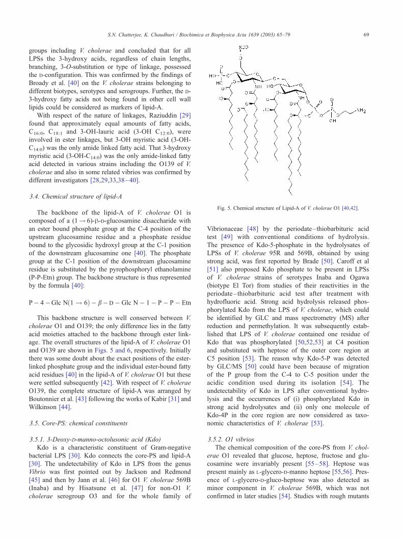
Fig. 5. Chemical structure of Lipid-A of V. cholerae O1 [40,42].
S.N. Chatterjee, K. Chaudhuri / Biochimica et Biophysica Acta 1639 (2003) 65–79 69
groups including V. cholerae and concluded that for all
LPSs the 3-hydroxy acids, regardless of chain lengths,
branching, 3-O-substitution or type of linkage, possessed
the D-configuration. This was confirmed by the findings of
Broady et al. [40] on the V. cholerae strains belonging to
different biotypes, serotypes and serogroups. Further, the D-
3-hydroxy fatty acids not being found in other cell wall
lipids could be considered as markers of lipid-A.
With respect of the nature of linkages, Raziuddin [29]
found that approximately equal amounts of fatty acids,
C16:0, C18:1 and 3-OH-lauric acid (3-OH C12:0), were
involved in ester linkages, but 3-OH myristic acid (3-OH-
C14:0) was the only amide linked fatty acid. That 3-hydroxy
myristic acid (3-OH-C14:0) was the only amide-linked fatty
acid detected in various strains including the O139 of V.
cholerae and also in some related vibrios was confirmed by
different investigators [28,29,33,38–40].
3.4. Chemical structure of lipid-A
The backbone of the lipid-A of V. cholerae O1 is
composed of a (1! 6)-h-D-glucosamine disaccharide with
an ester bound phosphate group at the C-4 position of the
upstream glucosamine residue and a phosphate residue
bound to the glycosidic hydroxyl group at the C-1 position
of the downstream glucosamine one [40]. The phosphate
group at the C-1 position of the downstream glucosamine
residue is substituted by the pyrophosphoryl ethanolamine
(P-P-Etn) group. The backbone structure is thus represented
by the formula [40]:
P� 4�Glc Nð1 ! 6Þ� b�D� Glc N� 1� P� P� Etn
This backbone structure is well conserved between V.
cholerae O1 and O139; the only difference lies in the fatty
acid moieties attached to the backbone through ester link-
age. The overall structures of the lipid-A of V. cholerae O1
and O139 are shown in Figs. 5 and 6, respectively. Initially
there was some doubt about the exact positions of the ester-
linked phosphate group and the individual ester-bound fatty
acid residues [40] in the lipid-A of V. cholerae O1 but these
were settled subsequently [42]. With respect of V. cholerae
O139, the complete structure of lipid-A was arranged by
Boutonnier et al. [43] following the works of Kabir [31] and
Wilkinson [44].
3.5. Core-PS: chemical constituents
3.5.1. 3-Deoxy-D-manno-octolusonic acid (Kdo)
Kdo is a characteristic constituent of Gram-negative
bacterial LPS [30]. Kdo connects the core-PS and lipid-A
[30]. The undetectability of Kdo in LPS from the genus
Vibrio was first pointed out by Jackson and Redmond
[45] and then by Jann et al. [46] for O1 V. cholerae 569B
(Inaba) and by Hisatsune et al. [47] for non-O1 V.
cholerae serogroup O3 and for the whole family of
Vibrionaceae [48] by the periodate– thiobarbituric acid
test [49] with conventional conditions of hydrolysis.
The presence of Kdo-5-phosphate in the hydrolysates of
LPSs of V. cholerae 95R and 569B, obtained by using
strong acid, was first reported by Brade [50]. Caroff et al
[51] also proposed Kdo phosphate to be present in LPSs
of V. cholerae strains of serotypes Inaba and Ogawa
(biotype El Tor) from studies of their reactivities in the
periodate– thiobarbituric acid test after treatment with
hydrofluoric acid. Strong acid hydrolysis released phos-
phorylated Kdo from the LPS of V. cholerae, which could
be identified by GLC and mass spectrometry (MS) after
reduction and permethylation. It was subsequently estab-
lished that LPS of V. cholerae contained one residue of
Kdo that was phosphorylated [50,52,53] at C4 position
and substituted with heptose of the outer core region at
C5 position [53]. The reason why Kdo-5-P was detected
by GLC/MS [50] could have been because of migration
of the P group from the C-4 to C-5 position under the
acidic condition used during its isolation [54]. The
undetectability of Kdo in LPS after conventional hydro-
lysis and the occurrences of (i) phosphorylated Kdo in
strong acid hydrolysates and (ii) only one molecule of
Kdo-4P in the core region are now considered as taxo-
nomic characteristics of V. cholerae [53].
3.5.2. O1 vibrios
The chemical composition of the core-PS from V. chol-
erae O1 revealed that glucose, heptose, fructose and glu-
cosamine were invariably present [55–58]. Heptose was
present mainly as L-glycero-D-manno heptose [55,56]. Pres-
ence of L-glycero-D-gluco-heptose was also detected as
minor component in V. cholerae 569B, which was not
confirmed in later studies [54]. Studies with rough mutants

Fig. 6. Chemical structure of Lipid-A of V. cholerae O139 [43].
S.N. Chatterjee, K. Chaudhuri / Biochimica et Biophysica Acta 1639 (2003) 65–7970
of V. cholerae O1 (95R, a derivative of Ogawa 162) and
with smooth V. cholerae 569B (Inaba) showed that the core
PS was composed of glucose, fructose, heptose (L-glycero-
D-manno heptose), glucosamine and Kdo [54].
The detection of D-fructose in LPS of V. cholerae made
an interesting story. Fructose occurs only rarely as an LPS
component [59]. Kondo et al. [52] demonstrated that,
among the vibrios studied, O1 V. cholerae was the only
group for which all strains contained fructose in LPS. When
LPS of V. cholerae was heated in dilute acetic acid at 100
jC, release of fructose took place [55]. On heating LPS of
Gram-negative enterobacteria under identical conditions,
Kdo is normally released. In the LPS of most Gram-negative
bacteria, Kdo is known to connect the PS moiety with lipid-
A [60]. It was accordingly presumed that in the LPS
molecule of V. cholerae, fructose and not Kdo provided
the connecting link between lipid-A and core-PS [46]. In
some other study [61], it was suspected that fructose was
essentially involved in the antigenicity of LPS because
release of fructose from LPS was accompanied by loss of
antigenicity. This was later ruled out because (a) fructose
was found to be present in the rough strains of V. cholerae
O1 [62] and (b) decrease in antigenicity was not parallel to
the release of fructose and required longer hours of heating
in dilute acetic acid [62]. Further chemical analysis revealed
that D-fructose did not link the core-PS and lipid A but was
present as a branch in the core region [55], which was later
confirmed by others [54].
3.5.3. Non-O1 vibrios
Like O1 vibrios, V. cholerae O139 was reported to
contain glucose, heptose (L-glycero-D-manno heptose), glu-
cosamine, fructose and Kdo in the core-PS region [32]. D-
Fructose was found in a branch linked to 6-position of D-
glucose residue [63]. In V. cholerae O139 strain NRCC
4740, Kdo was found as Kdo-phosphate, as in O1 vibrios,
and it was suggested that Kdo was phosphorylated at C-4
position [63]. Using a different strain of O139 (MO10-T4),
which lacked a capsular polysaccharide (CPS) and produced
a short-chain LPS, biphosphorylated Kdo residues could be
detected, where in addition to the phosphate group in the
usual C-4 position the Kdo residue contained 2-aminoethyl
phosphate (PEtn) at the C-7 position [64]. Although biphos-
phorylated Kdo residue is uncommon to V. cholerae, it was
detected in some strains of enterobacteria [65]. The LPS of
the O139 strain (MO10-T4) contained, in addition, (i) an O-
acetyl group connected to a secondary position, tentatively
O4 of the alpha-linked glucosyl group, and (ii) an additional
putative fructose residue of unknown location on LPS of
some species [64]. The significance of the differences in the
core-PS structures of the two strains of O139 is at present
not clear.
Very few of the other non-O1 vibrios have been exten-
sively studied so far. Since the non-O1 vibrios are composed
of a wide range of serogroups, some variations have been
encountered. Basically, the core-PS of non-O1 vibrios, like
O1 vibrios, contained heptose, glucose, fructose, glucos-
amine, glucosaminitol (in H-11), Kdo and galactose (in H-
11) [52,66]. Fructose was found in some serogroups (O3,
O5, O7 and O8) [67] but not in O6 [52]. Moreover, in a
study conducted on 44 serogroups of non-O1 vibrios,
fructose was not found in as many as nine serogroups (cited
in Ref. [53]). Considerable amount of D-glycero-D-manno-
heptose was found in some strains of serogroup O3 of V.
cholerae in addition to L-glycero-D-mannoheptose [52]. D-
Glycero-D-manno-heptose, which is rarely found in Gram-
negative bacterial LPS [60], was found in significant
amounts in some strains of V. cholerae O3 [52].
3.6. Chemical structure of the core polysaccharide
The chemical structures of the core polysaccharide of V.
cholerae of different serogroups are presented in Fig. 7. The
chemical structures reveal the common features of the
Vibrio cores, viz, the single Kdo residue substituted by a
phosphate residue at the 4-position [52] and the presence of
D-fructose linked to the 6-position of a D-glucose residue
[68] and is located in a branch [62] of the core-PS. Further,
in common with many Gram-negative enteric bacteria [65],
the trisaccharide, a-Hepp-(1-3)-a-Hepp-(1-5)-a-Kdo, is
present in all these vibrio strains, except that the heptose
residues are not phosphorylated in the vibrios (except for the
non-O1 strain H-11).
A comparison of the structures of O1 and O139 core-PS
shows that the two structures are almost identical. The
chemical evidence thus supports the genetic data that
suggested that only the wbe gene cluster (the DNA region
responsible for O-antigen biosynthesis) was altered in the
biogenesis of O139 from O1 [69]. Fig. 7 presents another
interesting feature. The structures of the carbohydrate chains

Fig. 7. Chemical structures of the core-PS of V. cholerae of different serogroups. A, O1 strains 95R (Ogawa) and 569B (Inaba) [54]; B, O139 strain NRCC
4740 [63,64,79]; C, non-O1, strain H-11 [66]; D, O22 strain NRCC 4904 [81,82]; 1, reported to be present by Cox and Perry [79] but not reported by Cox
et al. [81]; 2, detected in strain MO10-T4 by Knirel et al. [64]; 3, not confirmed by Knirel et al. [82] in strain 169-68; 4, detected in strain 169-68 by Knirel
et al. [82].
S.N. Chatterjee, K. Chaudhuri / Biochimica et Biophysica Acta 1639 (2003) 65–79 71
of the lipid A–core region in the LPS of V. cholerae rough
strain 95R (Ogawa) and smooth strain 569B (Inaba) are
identical. Finally, no significant difference is found in the
core-PS structures of the LPSs of V. cholerae O22 and
O139.
3.7. O-PS: chemical constituents
3.7.1. O1 vibrios
The sugar composition of the O-PS of V. cholerae O1
was investigated and a specific amino sugar, 4-amino-4, 6-
dideoxy D-mannose (perosamine), was detected as a com-
ponent [70]. The O-PS of V. cholerae O1 was found to be a
regular a(1! 2)-linked chain of D-perosamine [71], the
amino groups of which were acylated with 3-deoxy-L-
glycero-tetronic acid [72–74]. No difference between
Ogawa and Inaba serotypes in the sugar composition of
O1 strains could be detected at that time [71,72]. Redmond
[75] reported the presence of 4-amino-4-deoxy-L-arabinose
in the LPS of V. cholerae O1 seotype Ogawa and suggested
that this sugar might be a determinant of the Ogawa specific
antigen. However, Kabir [31] could not find 4-amino-4-
deoxy arabinose in the LPS of V. cholerae serotype Ogawa
and ruled out the role of this sugar as a determinant Ogawa
specific antigen. Hisatsune et al. [73] and Ito et al. [74]
reported for the first time that the previously undescribed
sugar, 4-amino-4,6-dideoxy-2-O-methyl mannose (2-O-
methyl perosamine), was present only in the O-PS of the
Ogawa serotype of V. cholerae O1 and proposed that this
sugar might be an important constituent of the Ogawa

S.N. Chatterjee, K. Chaudhuri / Biochimica et Biophysica Acta 1639 (2003) 65–7972
serotype determinant. Ito et al. [74] performed phenotypic
conversion experiments in vitro and found that the sugar
disappeared when the serotype conversion occurred from
Ogawa to Inaba. It was argued that the 2-O-methyl peros-
amine was labile to acid and hence could not be detected
earlier. It is now known that the Ogawa and Inaba serotypes
differ only by a single 2-O-methyl group that is present in
the upstream (nonreducing) terminal perosamine unit (Fig.
8) of the Ogawa O-PS and that is absent in the terminal
perosamine residue of the Inaba O-PS.
3.7.2. Non-O1 vibrios
The most prominent among non-O1 vibrios is V. chol-
erae O139 which, unlike other non-O1 vibrios, was found to
cause the disease cholera with all its clinical manifestations.
Although serologically unrelated to O1, studies [76]
revealed that O139 was biotypically closely related to V.
cholerae O1 El Tor biotype. In terms of pathogenic char-
acteristics and specifically in respect of CT production [77],
the main virulence factor, V. cholerae O139, was indistin-
guishable from El Tor V. cholerae O1 strains. It was
presumed that there were differences between the immune
responses against O1 and O139 strains, which might be of
considerable interest in terms of protection [78]. Analysis of
sugar composition of LPS from V. cholerae O139 revealed a
striking feature in that perosamine, a characteristic sugar
component of the O-PS of V. cholerae O1, was absent [32].
Detailed investigations revealed that the O-antigen of V.
cholerae O139 contained only one unit [63,64,79], unlike
most O-PSs which usually consisted of repeating units, and
was a phosphorylated hexasaccharide consisting of colitose,
galacturonic acid, quinovosamine, galactose and glucos-
amine residues [63,64]. This altered surface polysaccharide
epitope enabled serogroup O139 to avoid previously ac-
Fig. 8. Chemical structure of the O-PS of V. cholerae O1. The O-PS
structures of the two serotypes, Inaba and Ogawa, are the same except that
at the position O-2 of the upstream, terminal perosamine group, R =CH3 in
the Ogawa strain and R =H only in the Inaba strain. n, represents the
number of repeating units, which may be between 12 and 18 [73,74].
quired host immunity to the serogroup O1 strain, and this
contributed significantly to the early success of the organism
in producing epidemic cholera [80].
Another distinguishing feature of V. cholerae O139 is
that it produces, unlike O1, a capsular PS (CPS), which will
be discussed later. Further, the O-PS of V. cholerae O139 is
unique in the sense that it contains colitose, which is
generally not found in vibrios [32]. Only the serogroup
O22 has so far been found to contain colitose [81,82]. V.
cholerae O139 strains collected from four epidemic regions,
viz., Chennai, Vellore, Kolkata and Bangladesh, showed
similar sugar composition [32], indicating that they were
quite homogeneous chemotaxonomically and therefore
belonged to a new chemotype of non-O1 V. cholerae LPS
[32].
Among the other non-O1 vibrios, which consist of some
200 or more serogroups, formerly known as NAG or non-
agglutinating vibrios, O-PS of a few have been studied so
far. The amino sugar, D-perosamine, a characteristic constit-
uent of the O-PS of V. cholerae O1, is generally absent in
non-O1 vibrios, while galactose is present in many of these
serogroups [83,84]. Among other sugars found in LPS of
different non-O1 vibrios are L-rhamnose, heptose, fructose,
glucosamine, galactosamine and N-acetyl-2-amino-2-deoxy-
mannose (ManNAc) [64,83,84]. On the other hand, O-PS of
non-O1 vibrios were found to contain several unusual
sugars, e.g., ascarylose (3,6-dideoxy-L-arabino-hexose),
Sug1 or bacillosamine (2,4-diamino-2,4,6-trideoxy-D-glu-
cose) and fucosamine (2-amino-2,6-dideoxy-L-galactose)
in serogroup O3 [84]; Sug2 (5-acetamidino-7-acetamido-
3,5,7,9-tetradeoxy-L-glycero-beta-L-manno-nonulosonic ac-
id) in serogroup O2 [85]; 2-acetamido-3-(N-formyl-L-alany-
l)amino-2,3-dideoxyglucuronamide (GlcNAc3NAcylAN),
2,3-diacetamido-2,3-dideoxy mannuronamide (ManNAc3-
NAcAN) and 2,3-diacetamido-2,3-dideoxyguluronic acid
(GulNAc3NAcA) in serogroup O8 [86]; 2,4-diacetamido-
2,4,6-trideoxyglucose (QuiNAc4NAc) in both serogroups
O8 and O5 [87] and 2-acetamido-2,6-dideoxy-D-glucose
(QuiNAc) in serogroup O22 and O139 [82].
On the other hand, the sugar composition of the LPS
from serogroup O76 [88] has much in common with that of
the serogroup O1 except that (i) perosamine in the
serogroup O76 is in the L configuration in contrast to the
D configuration of the perosamine in O1, (ii) a small amount
of D-galactose is present in O76 and (iii) the L-perosamine is
N-acylated with an (S)-(+)-2-hydroxy propionyl group in the
serogroup O76. Another serogroup of V. cholerae, O144,
was found to contain homopolymers of D-propionyl-a-L-
perosamine in their O-PS [89].
3.8. Chemical structure of the O-PS
Figs. 8 and 9 present the structures of the O-PS of V.
cholerae of different serogroups and illustrate that the O-PS
structure of any serogroup is distinct. About 12–18 mono-
saccharide repeating units (n = 12–18) are present in the O-

Fig. 9. Chemical structures of the repeat units of O-PS of V. cholerae of different non-O1 serogroups. Abbreviations are as given in the text. (*) Knirel et al.
[82] could not confirm the presence of this residue. References are: O139 [63,79]; O2 [85]; O3 [84]; O8 [86]; O10 [115]; O22 [81,82].
S.N. Chatterjee, K. Chaudhuri / Biochimica et Biophysica Acta 1639 (2003) 65–79 73
PS of V. cholerae O1 [42], which is in accordance with the
high molar ratio of D-perosamine obtained in earlier studies
[55,56]. V. cholerae O139 lacks the ‘‘ladder pattern’’ char-
acteristic of O-antigen producing O1 strains when examined
on SDS-PAGE [90].
Fig. 9 shows that an altered structure from perosamine
homopolymer of V. cholerae O1 is present in the O-PS of
non-O1 vibrios, which generally consists of complex units
containing tri-, tetra-, or pentasaccharide repeating units.
Further, the repeating unit often contains unusual sugars.
The O-PS structures of two different strains of V. cholerae
O22 are more or less similar, except that the position of
the O-acetyl group at h-GalA as shown by Cox et al. [81]
could not be confirmed by the Knirel group [82]. Struc-
tural analysis of the O-PS of LPSs of some other
serogroups of V. cholerae and of some other vibrio
species have by now been completed (not shown in Fig.
9). Of these, mention may be made of O155 [91] and O9
[92] both having pentasaccharide repeating units and O6
[93] and V. mimicus [94] both having tetrasaccharide
repeating units. The important point to be noted here is
that both V. mimicus and O155 contain an immunodomi-
nant group, galactosyl residue substituted with a cyclic
phosphate (D-galactose-4,6-cyclophosphate), in their O-PS
structure in common with the O-PS structure of LPS and
CPS of V. cholerae O139, and that this appears to be the
basis of serological cross-reactivity between O139 and
O155 or V. mimicus.

S.N. Chatterjee, K. Chaudhuri / Biochimica et Biophysica Acta 1639 (2003) 65–7974
The O-PSs of V. cholerae O22 and O139 are similar in
chemical composition and structure and differ only in the
presence of an O-acetylated h-GalA residue in the former
instead of h-Gal-4, 6P in the latter [64] and in the anomeric
configuration of GlcNAc [81]. V. cholerae O22, unlike
O139, lacks a CPS but possesses an O-PS structure com-
parable to that of V. cholerae O139. The similarity in the O-
PS structures of the serogroups O139 and O22 can be
elaborated further. Both serogroups contain the same trisac-
charide, GlcNAc-GalA-QuiNAc, and in both cases, the N-
acetylglucosamine residue is di-substituted at the 3- and 4-
positions. Further, the rare terminal 3, 6-dideoxy-L-xylo
hexose (colitose) residues are present both in O22 and
O139 serogroups. To elaborate the differences in the O-PS
structures of these two serogroups, it may be noted that in
O22, the N-acetylglucosamine has the a-configuration and
not the h-configuration observed previously for O139. The
substituent at the 3-position also differs, being a galactur-
onic acid residue in O22 and a cyclic phosphorylated
galactose in serogroup O139. It may further be noted that
the phosphate group is absent in serogroup O22 and that
such groups are often found to be immunodominant. Bar-
ring the small differences, the striking structural similarity
between the serogroups O22 and O139 provides a chemical
basis for the serological cross-reactions observed between
the strains belonging to these two serogroups. The similarity
further adds support to the existing data suggesting that the
serogroup O22 is the donor strain of the new genetic
material forming the O139 serogroup V. cholerae. This
structural evidence, when considered in conjunction with
the facts that (i) serogroups O22 and O139 are the only
cholera serogroups to produce colitose and (ii) a significant
homology has been found between genes of the wb* clusters
of serogroups O139 and of O22 [95], makes it very likely
that the serogroup O22 is the progenitor strain. The genetic
homology between O139 and O22 will be discussed further
in Part II of this review.
4. The CPS
4.1. Colony morphology and CPS
Many of the non-O1 vibrios are known to produce CPS.
The O1 vibrios have so far not been shown to produce CPS.
The non-O1 strains have been shown to shift between an
encapsulated form with an opaque colony morphology and
an unencapsulated or minimally encapsulated form with
translucent colony morphology. The degree of opacity
correlates with the amount of capsular material which can
be extracted from the cells [96]. V. cholerae O139, a non-O1
strain, was shown to cause large outbreaks of cholera in the
Indian subcontinent in 1992 [80]. Since the O139 strain was
found to be toxigenic and did not agglutinate with either
polyclonal or monoclonal antisera directed against the V.
cholerae O1 antigen, more attention was directed towards
the study of this O139 strain and its various constituents.
The V. cholerae O139 Bengal strain AI-1838 when incu-
bated on agar plates showed two distinct colony morphol-
ogies, translucent and opaque. Both colony types were
tested for agglutination with rabbit anti-O139 antisera and
showed a positive reaction. Subsequently, Johnson et al.
[77] tested eight strains, including AI-1838, belonging to
O139 serogroup and found that all eight strains had mod-
erately opaque colony morphology on initial streaks, but the
translucent sectors and colonies appeared after subculturing.
Similar changes in colony morphology were not found
when more than 100 strains of O1 serogroup were exam-
ined. Comstock et al. [97] showed by TnphoA mutagenesis
that the loss of capsular material was associated with the
loss of opacity of the colony morphology in V. cholerae
O139 Bengal.
4.2. Site of occurrence
The presence of CPS in V. cholerae O139 Bengal was
demonstrated by electron microscopic studies [77,97–99].
Johnson et al. [77] prepared two O139 strains (AI-1855 and
AI-1841) for electron microscopic examination in thin
section by standard methods and stained them with poly-
cationic ferritin. The electron microscopic photographs
showed that bacteria belonging to both strains were sur-
rounded by a relatively thin electron dense capsule. Wein-
traub et al. [98] examined another strain (AI-1838) of O139
vibrio by ultrathin section and electron microscopy and
found that a coarse electron dense band was heterologously
distributed around approximately 30% of the cells. The
thickness of the band was 18–25 nm. The V. cholerae O1
strains (Ogawa) examined under identical conditions lacked
the surrounding electron dense band. Meno et al. [99]
examined a V. cholerae O139 strain by a different technique
(freeze substitution) of electron microscopy and found a
very thin fibrous layer on the outside of the outer membrane
of the cells. In contrast, the mutants of the strain O139,
strain MO10T4 (which lacked the ability to synthesize
capsule) and the strain Bengal-2R1 (which failed to synthe-
size both the capsule and the O-antigen of LPS), were all
found to have lost the surface layer. In addition, the capsule
layer could not be observed on the surface of V. cholerae
strain O1.
4.3. Sugar composition
Kasper et al. [100] showed (by Sephacryl S-300 chro-
matography and chemical analysis of fatty acids and sugars)
that the phenol-water extraction of capsulated bacteria
yielded a mixture of LPS and CPS. In order to purify the
LPS and CPS, Weintraub et al. [98] subjected the lyophi-
lized aqueous phase obtained after phenol-water extraction
of the O139 Bengal strain to an extraction with phenol–
chloroform–petroleum (PCP) ether and obtained two frac-
tions, PCP-soluble fraction (containing LPS) and the PCP-

Fig. 10. Chemical structure (top) and conformation (bottom) of hexasac-
charide obtained by digestion of the capsular polysaccharide of V. cholerae
O139 with a lyase from a bacteriophage specific for O139. The letters a to f
indicate the residue assignments used by the authors [103].
S.N. Chatterjee, K. Chaudhuri / Biochimica et Biophysica Acta 1639 (2003) 65–79 75
insoluble fraction (containing CPS), in the ratio of 1:2. It
was ascertained that the CPS contained no lipid A or fatty
acid. Monosaccharide analyses showed that CPS contained
glucosamine, quinovosamine and 3,6-dideoxy-xylo-hexose.
In addition, small amounts of glucose, galactose and trace of
heptose were found. Gel permeation chromatography of the
CPS showed that this material was of high molecular weight
since it eluted at the void volume on Sephacryl S-300
chromatography. It was established chemically that V. chol-
erae O139 produced a CPS, distinct from the LPS. To
establish the identity of the 3,6-dideoxy-xylo-hexose, it was
analyzed on three different GLC columns [98], one polar
and two nonpolar, on which it showed the same retention
time as an authentic abequose derivative (3,6-dideoxy-D-
xylo-hexose). However, the analysis did not discriminate
between the D and L isomers, i.e., the 3,6-dideoxyhexose
could well be the L-isomer (colitose). Later on, Preston et al.
[101] used two different hydrolysis conditions, viz., 0.5 M
trifluoroacetic acid at 60 jC for 3 h and 1 M trifluoroacetic
acid at 100 jC, 10 h, for the CPS of O139 strain (AI-1837)
and HPLC conditions suitable for neutral and acid sugars,
respectively and demonstrated the presence of 3,6-dideoxy-
xylohexose, quinovosamine, glucosamine, galactose and
galacturonic acid. Subsequently, Knirel et al. [102] pro-
duced a more definite result in respect of the sugar compo-
sition and structure of V. cholerae O139 CPS. These authors
showed that the CPS contained D-galactose, 3,6-dideoxy-L-
xylo-hexose (colitose), 2-acetamido-2-deoxy-D-glucose, 2-
acetamido-2,6-dideoxy-D-glucose (N-acetyl-D-quinovos-
amine), D-galacturonic acid and phosphate.
4.4. Chemical structure
Preston et al. [101] worked out the structure of the V.
cholerae O139 CPS by high performance anion exchange
chromatography and 1H-nuclear magnetic resonance spec-
troscopy. The CPS was found to contain a repeating unit
consisting of six sugar residues, which included one residue
each of N-acetylglucosamine (GlcNAc), N-acetylquinovos-
amine (QuiNAc), galacturonic acid (GalA), galactose and
two residues of 3,6-dideoxy-xylo-hexose (Xylhex). Howev-
er, the workers could not determine unambiguously the
absolute configuration of the monosaccharides and accord-
ingly the residues of 3,6-dideoxy-xylo-hexose could be
designated either as colitose (the L-isomer) or as abequose
(the D-isomer). Subsequently, Knirel et al. [102] studied the
structure of O139 CPS by NMR spectroscopy in combina-
tion with methylation analysis and selective degradations,
including partial acid hydrolysis at pH 3.1 and dephosphor-
ylation with aqueous 48% hydrofluoric acid, and basically
confirmed the CPS structure proposed by Preston et al.
[101]. Knirel et al. [102] could, in addition, specify exactly
the absolute configurations of the constituent monosacchar-
ides and the presence and position of the phosphate group.
Very recently, the hexasaccharide repeating unit has been
isolated from the V. cholerae O139 CPS by digestion with a
polysaccharide lyase derived from a bacteriophage specific
for this serogroup. It specifically cleaves at a single position
of the 4-linked galacturonic acid producing an unsaturated
sugar product, the conformation of which has been studied
by molecular modeling and NMR spectroscopy (Fig. 10)
[103]. The structure has been found to contain a tetrasac-
charide epitope homologous to the human Lewisb blood
group antigen and its conformation is identical or at least
closely similar to that of the intact CPS studied earlier
[101,102].
5. Concluding remarks
5.1. Emerging trends of research and future possibilities
Already some 200 or more serogroups of V. cholerae
have been detected. Studies of these and of any additional
serogroup that may be detected in future would be of
academic nature unless some new strain emerges and causes
severe epidemic or pandemic of the disease cholera. On the
other hand, researches in some new directions have already
been initiated, which may be of great practical use.
Recent works in this line involve the preparation of
synthetic mono- or oligosaccharide fragments that mimick
the terminal mono- or oligosaccharide residues of the O-PS
of V. cholerae LPS and study of their interactions with
monoclonal anti-cholera antibodies with a view to develop-

S.N. Chatterjee, K. Chaudhuri / Biochimica et Biophysica Acta 1639 (2003) 65–7976
ing synthetic carbohydrate based anti-cholera vaccine.
Kenne et al. [104] reported the first synthesis of the
monosaccharide repeating unit of V. cholerae O1, serotype
Inaba. An improved synthesis as well as the crystal structure
of the methyl-a-glycoside of the monomeric repeating unit
common to both Ogawa and Inaba strains were subsequent-
ly reported by Gotoh et al. [105]. Following the discovery
that the O-PSs of the two serotypes, Ogawa and Inaba, differ
in that the terminal 4-N-tetronylated-D-perosaminyl group in
the O-PS of the Ogawa strain is methylated at O-2 [73,74],
the complete terminal sugar of the serotype Ogawa, methyl-
a-glycoside, was synthesized and its crystal structure deter-
mined [106]. This was followed by the synthesis of three
oligosaccharides which imitate the upstream part of the O-
PS of the serotype Ogawa [107] and the analogous trisac-
charide related to the O-PS of the serotype Inaba [108]. As a
further step in the effort to understand protective immunity
to cholera, hexasaccharides representing termini of the O-PS
of the two serotypes of V. cholerae O1 were synthesized in
the form of glycosides whose aglycons allowed linking
these substances to proteins [109]. Further improvement in
the synthesis of similar antigenic compounds has recently
been reported by Ma et al. [110].
Another recent study in this line involved the preparation
of analogs of the methyl-a-glycosides of the terminal
residues of the O-PS of V. cholerae O1 as probes [111] to
study their interaction with anti-V. cholerae O1 antibodies
[112,113]. They differ from the termini of the respective O-
PSs in anomeric or absolute configuration of perosamine,
position of the O-methyl group in D-perosamine and nature
of the N-acyl side chain. These were used to learn more
about the structural requirements for binding with antibodies
and also to find out the structural elements that would
increase binding. Among the interesting results already
achieved include the finding that some antibodies showed
a remarkable tolerance to irregularities or variations in the
structures of the ligands [111]. Some other studies resolved
that the 2-O-methyl group, a small antigenic determinant,
can dictate a highly specific immune response. Wang et al.
[112] carried out binding studies of anti-Ogawa Abs IgG1s
S-20-4, A-20-6 and IgA 2D6 with synthetic methyl a-
glycosides of fragments up to the hexasaccharide, of the
Ogawa O-PS, as well as analogs of the terminal monosac-
charide, and revealed that the terminal residue accounted for
approximately 90% of the maximal binding energy. They
did not react with the corresponding synthetic fragments of
Inaba O-PS. Further insight in to the structural basis of
carbohydrate recognition and particularly V. cholerae sero-
type specificity was obtained from the crystallographic
studies of protective anti-cholera Abs in complex with
synthetic analogs of the O-antigen [42]. The upstream
terminal monosaccharide of the Ogawa O-PS was shown
to be the primary antigenic determinant. The crystallograph-
ic study showed the pivotal contribution by so small a
structural fragment in the antigenic determinant as a methyl
group. Chernyak et al. [114] moved one step forward and
demonstrated induction of protective immunity by synthetic
antigens that mimic the terminal hexasaccharide epitope of
the O-PS of V. cholerae O1, serotype Ogawa, conjugated to
bovine serum albumin with different carbohydrate to carrier
molar ratios. The protective capacity of antiserum was
evident in serum from mice immunized with all conjugates,
but it was highest in the groups that received the conjugate
with the lowest level of substitution. Further investigations
in this line with a view to devising synthetic analogs that
will bind significantly with antibodies derived against V.
cholerae of more than one serogroup may be very chal-
lenging but useful for purposes of vaccine development.
Extension of such studies with the specific object of gaining
insight in molecular and atomic details into the structural
basis of carbohydrate recognition will contribute signifi-
cantly towards the development of a synthetic carbohydrate-
based anti-cholera vaccine.
Acknowledgements
Authors are most thankful to the many scientists, and
particularly to Drs. P. Kovac, A.D. Cox, P.A. Manning, U.H.
Stroeher and A. Weintraub, for sending promptly the
reprints of their important and relevant publications and
thereby making our task easier. Sincere thanks are due to Dr.
M. Maiti, Director-Grade-Scientist, Indian Institute of
Chemical Biology, Calcutta, for helping us in many ways
all through. Thanks are also due to Drs. R. Chatterjee and B.
Bannerjee, Biophysics Division, Indian Institute of Chem-
ical Biology, for rendering technical help during the
preparation of this manuscript.
References
[1] O. Luderitz, A.M. Staub, O. Westphal, Immunochemistry of O and R
antigens of Salmonella and related Enterobacteriaceae, Bacteriol.
Rev. 30 (1966) 192–255.
[2] J. Gmeiner, O. Luderitz, O. Westphal, Biochemical studies on the
lipopolysaccharides of Salmonella R mutants.6. Investigations on
the structure of the lipid A component, Eur. J. Biochem. 7 (1969)
370–379.
[3] O. Luderitz, O. Westphal, A.M. Staub, H. Nikaido, Isolation and
chemical and immunological characterization of bacterial lipopoly-
saccharides, in: G. Weinbaum, S. Kadis, S.J. Ajl (Eds.), Microbial
Toxins, vol. 4, Academic Press, New York, 1971, pp. 145–233.
[4] R. Sakazaki, Proposal of Vibrio alginolyticus for the biotype 2 of
Vibrio parahaemolyticus, Jpn. J. Med. Sci. Biol. 21 (1968) 359–362.
[5] R. Sakazaki, Bacteriology of Vibrio and related organisms, in: D.
Barua, W.B. Greenough III (Eds.), Cholera, Plenum Medical Book
Co., New York, 1992, pp. 37–55.
[6] P. Baumann, A.L. Furniss, J.V. Lee, Genus 1. Vibrio, in: N.R. Kricg,
J.G. Holt (Eds.), Bergey’s Manual of Systematic Bacteriology, vol. 1,
Williams and Wilkins, Baltimore, USA, 1984, pp. 518–538.
[7] J.B. Kaper , J.G. Morris Jr., M.M. Levine, Cholera, Clin. Microbiol.
Rev. 8 (1995) 48–86.
[8] S. Mukherjee, The bacteriophage susceptibility test in differentiating
Vibrio cholerae and Vibrio el tor, Bull. W.H.O. 28 (1963) 333–336.
[9] K. Takeya, T. Otohuji, H. Tokiwa, FK phage for differentiating the

S.N. Chatterjee, K. Chaudhuri / Biochimica et Biophysica Acta 1639 (2003) 65–79 77
Classical and El Tor groups of Vibrio cholerae, J. Clin. Microbiol.
14 (1981) 222–224.
[10] S.P. Keasler, R.H. Hall, Detecting and biotyping Vibrio cholerae O1
with multiplex polymerase chain reaction, Lancet 341 (1993) 1661.
[11] S. Koblavi, F. Grimont, P.A.D. Grimont, Clonal diversity of Vibrio
cholerae O1 evidenced by r-RNA gene restriction patterns, Res.
Microbiol. 141 (1990) 645–657.
[12] T. Popovic, C. Bopp, O. Olsvik, K. Wachsmuth, Epidemiologic
application of a standardized ribotype scheme for Vibrio cholerae
O1, J. Clin. Microbiol. 31 (1993) 2474–2482.
[13] J. Gallut, La septieme pandemic cholerique, Bull. Soc. Pathol. Exot.
(Paris) 64 (1971) 551–560.
[14] R. Hugh, A comparison of Vibrio cholerae Pacini and Vibrio eltor
Pribram, Int. Bull. Bacteriol. Nomencl. Taxon. 15 (1965) 61–68.
[15] R. Hugh, Nomenclature and taxonomy of Vibrio cholerae Pacini
1854 and Vibrio eltor Pribram 1933, Proc. Cholera Res. Symp.
Honululu, Hawaii, US Govt. Printing Office, Washington, 1965,
pp. 1–4.
[16] O. Westphal, O. Luderitz, F. Bister, Uber die extraction von bakter-
ien mit phenol/wasser, Z. Naturforsch. 7b (1952) 148–155.
[17] S.N. Chatterjee, P.C. Adhikari, M. Maiti, C. Raichaudhuri, Pratima
Sur, Growth of Vibrio cholerae cells: biochemical and electron mi-
croscopic study, Ind. J. Exp. Biol. 12 (1974) 35–45.
[18] M. Maiti, S.N. Chatterjee, Characteristics of a Group IV cholera
phage, J. Gen. Virol. 13 (1971) 327–330.
[19] M. Maiti, P. Sur, S.N. Chatterjee, Amino sugar contents and phage
inactivating properties of lipopolysaccharide from cholera and El Tor
vibrios, Ann. Microbiol. (Inst. Pasteur) 128A (1977) 35–39.
[20] S.N. Chatterjee, M. Maiti, Vibriophages and vibriocins: physical,
chemical and biological properties, in: M.A. Lauffer, K. Maramor-
osch (Eds.), Advances in Virus Research, vol. 29, Academic Press,
New York, 1984, pp. 263–312.
[21] J. Das, S.N. Chatterjee, Electron microscopic studies on some ultra-
structural aspects of Vibrio cholerae, Ind. J. Med. Res. 54 (1966)
330–338.
[22] J.A. Fuerst, J.W. Perry, Demonstration of lipopolysaccharide on
sheathed flagella of Vibrio cholerae O1 by protein A-gold immu-
noelectron microscopy, J. Bacteriol. 170 (1988) 1488–1494.
[23] S.N. Chatterjee, J. Das, Electron microscopic observations on the
excretion of cell-wall material by Vibrio cholerae, J. Gen. Microbiol.
49 (1967) 1–11.
[24] Z. Li, A.J. Clarke, T.J. Beveridge, Gram-negative bacteria produce
membrane vesicles which are capable of killing other bacteria,
J. Bacteriol. 180 (1998) 5478–5483.
[25] I.W. Devoe, J.E. Gilchrist, Release of endotoxin in the form of cell
wall blebs during in vitro growth of Neisseria meningitides, J. Exp.
Med. 138 (1973) 1156–1167.
[26] D.W. Dorward, C.F. Garon, Export and intracellular transfer of DNA
via membrane blebs of Neisseria gonorrhoeae, J. Bacteriol. 171
(1989) 2499–2505.
[27] J.L. Kadurugamuwa, T.J. Beveridge, Bacteriolytic effect of mem-
brane vesicles from Pseudomonas aeruginosa on other bacteria in-
cluding pathogens: conceptually new antibiotics, J. Bacteriol. 178
(1996) 2267–2274.
[28] S. Raziuddin, T. Kawasaki, Biochemical studies on the cell wall
lipopolysaccharides (O-antigens) of Vibrio cholerae 569B (Inaba)
and ElTor (Inaba), Biochim Biophys. Acta 431 (1976) 116–126.
[29] S. Raziuddin, Characterization of lipid A and polysaccharide moi-
eties of the lipopolysaccharides from Vibrio cholerae, Biochem. J.
167 (1977) 147–154.
[30] E.Th. Rietschel, H.W. Wollenweaber, H. Brade, U. Zahringer, B.
Linder, G. Barnickel, H. Labischinski, P. Giesbrecht, Structure and
conformation of the lipid A component of lipopolysaccharides, in:
R.A. Proctor (Ed.), Handbook of Endotoxins, vol. 1, Elsevier, Am-
sterdam, 1984, pp. 187–220.
[31] S. Kabir, Characterization of the lipopolysaccharide from Vibrio
cholerae 395 (Ogawa), Infect. Immun. 38 (1982) 1263–1272.
[32] K. Hisatsune, S. Kondo, Y. Isshiki, T. Iguchi, Y. Kawamata, T.
Shimada, O-antigenic LPS of V. cholerae 139 Bengal, a new epi-
demic strain for recent cholera in the Indian subcontinent, Biochem.
Biophys. Res. Commun. 196 (1993) 1309–1315.
[33] E.Th. Rietschel, W.J. Palin, D.W. Watson, Nature and linkage of
fatty acids present in lipopolysaccharides from Vibrio metchinikovii
and Vibrio parahaemolyticus, Eur. J. Biochem. 37 (1973) 116–120.
[34] C.F. Deneke, R.R. Colwell, Lipopolysaccharide and proteins of the
cell envelope of Vibrio marinus, a marine bacterium, Can. J. Micro-
biol. 19 (1973) 1211–1217.
[35] S. Hase, E.Th. Rietschel, Isolation and analysis of the lipid A back-
bone: lipid A structures from lipopolysaccharides from various bac-
terial groups, Eur. J. Biochem. 63 (1976) 101–107.
[36] M. Rosner, J. Tang, I. Barzilay, H.G. Khorana, Structure of the lip-
opolysaccharide from an Escherichia coli heptose-less mutant: I.
Chemical degradations and identification of products, J. Biol. Chem.
254 (1979) 5906–5917.
[37] P.F. Muhlardt, V. Wray, V. Lehmann, A 31P-nuclear-magnetic-reso-
nance study of the phosphate groups in lipopolysaccharide and lipid
A from Salmonella, Eur. J. Biochem. 81 (1977) 193–203.
[38] I.L. Armstrong, J.W. Redmond, The fatty acids present in the lip-
opolysaccharide of Vibrio cholerae 569B (Inaba), Biochim. Bio-
phys. Acta 348 (1974) 302–305.
[39] K. Hisatsune, S. Kondo, T. Kawata, Y. Kishimoto, Fatty acid com-
position of LPS of Vibrio cholerae 35A3 (Inaba), NIH 90 (Ogawa)
and 4715 (Nag), J. Bacteriol. 138 (1979) 288–290.
[40] K.W. Broady, E.T. Rietschel, O. Luderitz, The chemical structure of
the lipid A component of lipopolysaccharides from Vibrio cholerae,
Eur. J. Biochem. 115 (1981) 463–468.
[41] E.Th. Rietschel, Absolute configuration of 3-OH fatty acids present
in LPS from various bacterial groups, Eur. J. Biochem. 64 (1976)
423–428.
[42] S. Villeneuve, H. Souchon, M.M. Riottot, J.C. Mazie, P. Lei, C.P.
Glaudemans, P. Kovac, J.M. Fournier, P.M. Alzari, Crystal structure
of an anti-carbohydrate antibody directed against Vibrio cholerae O1
in complex with antigen: molecular basis for serotype specificity,
Proc. Natl. Acad. Sci. U. S. A. 97 (2000) 8433–8438.
[43] A. Boutonnier, S. Villeneuve, F. Nato, B. Dassy, J.M. Fournier,
Preparation, immunogenicity, and protective efficacy, in a murine
model, of a conjugate vaccine composed of the polysaccharide moi-
ety of the lipopolysaccharide of Vibrio cholerae O139 bound to
tetanus toxoid, Infect. Immun. 69 (2001) 3488–3493.
[44] S.G. Wilkinson, Bacterial lipopolysaccharides: themes and varia-
tions, Prog. Lipid Res. 35 (1996) 283–343.
[45] G.D.F. Jackson, J.W. Redmond, Immunochemical studies of the O-
antigens of Vibrio cholerae: the constitution of a LPS from V. chol-
erae 569B (Inaba), FEBS Lett. 13 (1971) 117–120.
[46] B. Jann, K. Jann, G.O. Beyaert, 2-Amino-2,6-dideoxy-D-glucose (D-
quinovosamine): a constituent of the lipopolysaccharides of Vibrio
cholerae, Eur. J. Biochem. 37 (1973) 531–534.
[47] K. Hisatsune, S. Kondo, K. Kobayashi, Lipopolysaccharides of Vi-
brio cholerae (II) –an immunochemical study on O-antigenic struc-
ture, Jpn. J. Med. Sci. Biol. 31 (1978) 181–184.
[48] K. Hisatsune, S. Kondo, T. Iguchi, M. Machida, S. Asou, M. Ina-
guma, F. Yamamoto, Sugar composition of lipopolysaccharides of
family Vibrionaceae. Absence of 2-keto-3-deoxyoctonate (KDO) ex-
cept in Vibrio parahaemolyticus O6, Microbiol. Immunol. 26 (1982)
649–664.
[49] A. Weissbach, J. Hurwitz, The formation of 2-keto-3-deoxy-hepton-
ic acid in extracts of Escherichia coli B, J. Biol. Chem. 234 (1959)
705–709.
[50] H. Brade, Occurrence of 2-keto-deoxyoctonic acid 5-phosphate in
lipopolysaccharides of Vibrio cholerae Ogawa and Inaba, J. Bacter-
iol. 161 (1985) 795–798.
[51] M. Caroff, S. Lebbar, L. Szabo, Do endotoxins devoid of 3-deoxy-D-
manno-2-octulosonic acid exist? Biochem. Biophys. Res. Com-
mun. 143 (1987) 845–847.

S.N. Chatterjee, K. Chaudhuri / Biochimica et Biophysica Acta 1639 (2003) 65–7978
[52] S. Kondo, T. Iguchi, K. Hisatsune, A comparative study of the sugar
composition of lipopolysaccharides isolated from Vibrio cholerae,
Vibrio albenis and Vibrio metschnikovii, J. Gen. Microbiol. 134
(1988) 1699–1705.
[53] S. Kondo, Y. Haishima, K. Hisatsune, Taxonomic implication of the
apparent undetectability of 3-deoxy-D-manno-2-octulosonate (KDO)
in lipopolysaccharides of the representatives of the family Vibriona-
ceae and the occurrence of Kdo-4-phosphate in their inner core
regions, Carbohydr. Res. 231 (1992) 55–64.
[54] E.V. Vinogradov, K. Bock, O. Holst, H. Brade, The structure of the
lipid A–core region of the lipopolysaccharides from Vibrio cholerae
O1 smooth strain 569B (Inaba) and rough mutant strain 95R (Oga-
wa), Eur. J. Biochem. 233 (1995) 152–158.
[55] K. Hisatsune, M. Hayashi, Y. Haishima, S. Kondo, Relationship
between structure and antigenicity of O1 Vibrio cholerae lipopoly-
saccharides, J. Gen. Microbiol. 135 (1989) 1901–1907.
[56] Y. Haishima, S. Kondo, K. Hisatsune, O-antigen LPS isolated from a
marine Vibrio bioserogroup 1875 possessing an antigenic factor in
common with O1 Vibrio cholerae, J. Gen. Microbiol. 134 (1988)
1827–1833.
[57] S. Raziuddin, Immunochemical studies on the lipopolysacchrides of
Vibrio cholerae: constitution of O-specific side chain and core poly-
saccharide, Infect. Immun. 27 (1980) 211–215.
[58] A.K. Sen, A.K. Mukherjee, B. Guhathakurta, A. Dutta, D. Sasmal,
Structural investigations of the lipopolysaccharide isolated from Vi-
brio cholerae Inaba 569B, Carbohydr. Res. 72 (1979) 191–199.
[59] L. Kenne, B. Lindberg, Bacterial lipopolysaccharides, in: G.O. As-
pinell (Ed.), The Polysaccharides, vol. 2, Academic Press, New
York, 1983, pp. 287–363.
[60] S.G. Wilkinson, Composition and structure of bacterial lipopolysac-
charides, in: I.W. Sutherland (Ed.), Surface Carbohydrates of the
Prokaryotic Cell, Academic Press, New York, 1977, pp. 97–175.
[61] J.W. Redmond, M.J. Korsch, G.D.F. Jackson, Immunochemical
studies to the O-antigens of Vibrio cholerae, Partial characterization
of an acid-labile antigenic determinant, Aust. J. Exp. Biol. Med. Sci.
51 (1973) 229–235.
[62] W. Kaca, L. Brade, E.T. Rietschel, H. Brade, The effect of removal
of D-fructose on the antigenicity of the lipopolysaccharide from a
rough mutant of Vibrio cholerae Ogawa, Carbohydr. Res. 149
(1986) 293–298.
[63] A.D. Cox, J.R. Brisson, V. Varma, M.B. Perry, Structural analysis of
the lipopolysaccharide from Vibrio cholerae O139, Carbohydr. Res.
290 (1996) 43–58.
[64] Y.A. Knirel, G. Widmalm, S.N. Senchenkova, P.E. Jansson, A.
Weintraub, Structural studies on the short-chain lipopolysaccharide
of Vibrio cholerae O139 Bengal, Eur. J. Biochem. 247 (1997)
402–410.
[65] O. Holst, H. Brade, Molecular biochemistry and cellular biology, in:
D.C. Morrison, J.L. Ryan (Eds.), Bacterial Endotoxic Lipopolysac-
charides, vol. 1, CRC Press, Boca Raton, FL, 1992, pp. 135–169.
[66] E.V. Vinogradov, R. Stuike-Prill, K. Bock, O. Holst, H. Brade, The
structure of the carbohydrate backbone of the core-lipid-A region of
the lipopolysaccharide from Vibrio cholerae strain H-11 (non-O1),
Eur. J. Biochem. 218 (1993) 543–554.
[67] K. Hisatsune, S. Kondo, T. Iguchi, M. Machida, S. Asou, M. Ina-
guma, F. Yamamoto, Sugar compositions of lipopolysaccharides of
family Vibrionaceae: absence of 2-keto-3-deoxyoctonate (KDO) ex-
cept in Vibrio parahaemolyticus O6 and Plesiomonas spigelloides,
in: S. Kuwahara, N.F. Pierce (Eds.), Advances in Research on Chol-
era and Related Diarrhea, vol. 1, Maasterus Nighoff Publications,
The Hague, 1983, pp. 59–74.
[68] S. Kondo, T. Watabe, Y. Haishima, K. Hisatsune, Identification of
oligosaccharides consisting of D-glucuronic acid and L-glycero-D-
manno- and D-glycero-D-manno-heptose isolated from Vibrio para-
haemolyticus O2 lipopolysaccharide, Carbohydr. Res. 245 (1993)
353–359.
[69] L.E. Comstock, J.A. Johnson, J.M. Michalski, J.G. Morris Jr., J.B.
Kaper, Cloning and sequence of a region encoding a surface poly-
saccharide of Vibrio cholerae O139 and characterization of the in-
sertion site in the chromosome of Vibrio cholerae O1, Mol.
Microbiol. 19 (1996) 815–826.
[70] J.W. Redmond, The structure of the O-antigenic side chain of the
lipopolysaccharide of Vibrio cholerae 569B (Inaba), Biochim. Bio-
phys. Acta 584 (1979) 346–352.
[71] L. Kenne, B. Lindberg, P. Unger, T. Holme, J. Holmgren, Structural
studies of the Vibrio cholerae O-antigen, Carbohydr. Res. 68 (1979)
C16–C17.
[72] L. Kenne, B. Lindberg, P. Unger, B. Gutafsson, T. Holme, Structural
studies of the Vibrio cholerae O-antigen, Carbohydr. Res. 100
(1982) 341–349.
[73] K. Hisatsune, S. Kondo, Y. Isshiki, T. Iguchi, Y. Haishima, Occur-
rence of 2-O-methyl-N-(3-deoxy-L-glycerotetronyl)-D-perosamine(4-
amino-4,6-dideoxy-D-manno-pyranose) in lipopolysaccharide from
the Ogawa but not from Inaba forms of O1 Vibrio cholerae, Bio-
chem. Biophys. Res. Commun. 190 (1993) 302–307.
[74] T. Ito, T. Higuchi, M. Hirobe, K. Hiramatsu, T. Yokota, Identifica-
tion of a novel sugar, 4-amino-4,6-dideoxy-2-O-methyl mannose in
the LPS of V. cholerae O1 serotype Ogawa, Carbohydr. Res. 256
(1994) 113–128.
[75] J.W. Redmond, The 4-amino sugars present in the lipopolysacchar-
ides of V. cholerae and related Vibrios, Biochim. Biophys. Acta 542
(1978) 378–384.
[76] K.E. Calia, M. Murtagh, M.J. Ferraro, S.B. Calderwood, Compar-
ison of Vibrio cholerae O139 with V. cholerae O1 classical and El
Tor biotypes, Infect. Immun. 62 (1994) 1504–1506.
[77] J.A. Johnson, C.A. Salles, P. Panigrahi, M.J. Albert, A.C. Wright,
R.J. Johnson , J.G. Morris Jr., Vibrio cholerae O139 synonym Ben-
gal is closely related to Vibrio cholerae El Tor but has important
differences, Infect. Immun. 62 (1994) 2108–2110.
[78] M.J. Albert, K. Alam, M. Ansaruzzaman, F. Gadri, R.B. Sack,
Lack of cross-protection against diarrhea due to Vibrio cholerae
O139 (Bengal strain) after oral immunization of rabbits with V.
cholerae O1 vaccine strain CVD103-HgR, J. Infect. Dis. 169
(1994) 230–231.
[79] A.D. Cox, M.B. Perry, Structural analysis of the O-antigen core
region of the lipopolysaccharide from Vibrio cholerae O139, Carbo-
hydr. Res. 290 (1996) 59–65.
[80] T. Ramamurthy, S. Garg, R. Sharma, S.K. Bhattacharya, G.B. Nair,
T. Shimada, T. Takeda, T. Karasawa, H. Kurazano, A. Pal, Y.
Takeda, Emergence of novel strain of Vibrio cholerae with epi-
demic potential in Southern and Eastern India, Lancet 341 (1993)
703–704.
[81] A.D. Cox, J.R. Brisson, P. Thibault, M.B. Perry, Structural analysis
of the lipopolysaccharide from Vibrio cholerae serotype O22, Car-
bohydr. Res. 304 (1997) 191–208.
[82] Y.A. Knirel, S.N. Senchenkova, P.E. Jansson, A. Weintraub, More
on the structure of Vibrio cholerae O22 lipopolysaccharide, Carbo-
hydr. Res. 310 (1998) 117–119.
[83] A.K. Sen, A.K. Mukherjee, Structural studies of a specific polysac-
charide isolated from non-agglutinating vibrios, Carbohydr. Res. 64
(1978) 215–223.
[84] T.A. Chowdhury, P.E. Jansson, B. Lindberg, J. Lindberg, B. Gus-
tafsson, T. Holme, Structural studies of the Vibrio cholerae O:3 O-
antigen polysaccharide, Carbohydr. Res. 215 (1991) 303–314.
[85] L. Kenne, B. Lindberg, E. Schweda, B. Gustafsson, T. Holme, Struc-
tural studies of the O-antigen from Vibrio cholerae O:2, Carbohydr.
Res. 180 (1988) 285–294.
[86] N.A. Kocharova, A.V. Perepelov, G.V. Zatonsky, A.S. Shashkov,
Y.A. Knirel, P.E. Jansson, A. Weintraub, Structural studies of the
O-specific polysaccharide of Vibrio cholerae O8 using solvolysis
with triflic acid, Carbohydr. Res. 330 (2001) 83–92.
[87] S. Kondo, K. Hisatsune, Sugar composition of the polysaccharide
portion of lipopolysaccharides isolated from non-O1 Vibrio cholerae
O2 to O 41, O 44 and O 68,Microbiol. Immunol. 33 (1989) 641–648.

S.N. Chatterjee, K. Chaudhuri / Biochimica et Biophysica Acta 1639 (2003) 65–79 79
[88] S. Kondo, Y. Sano, Y. Isshiki, K. Hisatsune, The O-polysaccharide
chain of the lipopolysaccharide from Vibrio cholerae O76 is a ho-
mopolymer of N-[S-(+)-2-hydroxypropionyl]-alpha-L-perosamine,
Microbiology 142 (1996) 2879–2885.
[89] Y. Sano, S. Kondo, Y. Isshiki, T. Shimada, K. Hisatsune, An N-[(R)-
(� )-2-hydroxypropionyl]-a-L-perosamine homopolymer constitutes
the O-polysaccharide from Vibrio cholerae O144 which has antigen-
ic factor(s) in common with V. cholerae O76, Microbiol. Immunol.
40 (1996) 735–741.
[90] P.A. Manning, U.H. Stroeher, R. Morona, Molecular basis for O-
antigen biosynthesis in Vibrio cholerae O1: Ogawa–Inaba switch-
ing, in: I.K. Wachsmuth, P.A. Blake, O. Olsvik (Eds.), Vibrio chol-
erae and Cholera: Molecular to Global Perspectives, ASM Press,
Washington, DC, 1994, pp. 77–94.
[91] S.N. Senchenkova, G.V. Zatonsky, A.S. Shashkov, Y.A. Knirel, P.E.
Jansson, A. Weintraub, M.J. Albert, Structure of the O-antigen of
Vibrio cholerae O155 that shares a putative D-galactose 4,6-cyclo-
phosphate-associated epitope with V. cholerae O139 Bengal, Eur. J.
Biochem. 245 (1998) 58–62.
[92] N.A. Kocharova, Y.A. Knirel, P.-E. Jansson, A. Weintraub, Structure
of the O-specific polysaccharide of Vibrio cholerae O9 containing 2-
acetamido-2-deoxy-D-galacturonic acid, Carbohydr. Res. 332 (2001)
279–284.
[93] N. Bergstrom, G.B. Nair, A. Weintraub, P.-E. Jansson, Structure of
the O-polysaccharide from the lipopolysaccharide from Vibrio chol-
erae O6, Carbohydr. Res. 337 (2002) 813–817.
[94] C. Landersio, A. Weintraub, M. Ansaruzzaman, M.J. Albert, G.
Widmalm, Structural analysis of the O-antigenic polysaccharide
from Vibrio mimicus N-1990, Eur. J. Biochem. 251 (1998) 986–990.
[95] S. Vimont, S. Dumontier, V. Escuyer, P. Berche, The rfaD locus: a
region of rearrangement in Vibrio cholerae O139, Gene 185 (1997)
43–47.
[96] J.A. Johnson, A. Joseph, P. Panigrahi, J.G. Morris Jr., Non-O1 Vibrio
cholerae NRT36S produces a polysaccharide capsule that deter-
mines colony morphology, serum resistance and virulence in mice,
Infect. Immun. 60 (1992) 864–869.
[97] L.E. Comstock, D. Maneval, P. Panigrahi, A. Joseph, M.M. Leine,
J.B. Kaper., J.G. Morris Jr, J.A. Johnson, The capsule and O anti-
gen of Vibrio cholerae O139 Bengal are associated with a genetic
region not present in Vibrio cholerae O1, Infect. Immun. 63 (1995)
317–323.
[98] A. Weintraub, G. Widmalm, P.E. Jansson, M. Jansson, K. Hultenby,
M.J. Albert, Vibrio cholerae O139 Bengal possesses a capsular pol-
ysaccharide which may confer increased, virulence, Microb. Pathog.
16 (1994) 235–241.
[99] Y. Meno, M.K. Waldor, J.J. Mekalanos, K. Amako, Morphological
and physical characterization of the capsular layer of Vibrio cholerae
O139, Arch. Microbiol. 170 (1998) 339–344.
[100] D.L. Kasper, A. Weintraub, A.A. Lindberg, J. Lonngren, Capsular
polysaccharides and lipopolysaccharides from two Bacteroids fragi-
lis reference strains: chemical and immunochemical characterization,
J. Bacteriol. 153 (1983) 991–997.
[101] L.M. Preston, Q. Xu, J.A. Johnson, A. Joseph, D.R. Maneval, K.
Husain, G.P. Reddy, C.A. Bush, J.G. Morris Jr., Preliminary struc-
ture determination of the capsular polysaccharide of Vibrio cholerae
O139 Bengal AI-1837, J. Bacteriol. 177 (1995) 835–838.
[102] Y.A. Knirel, L. Pardes, P.E. Jansson, A. Weintraub, G. Widmalm,
M.J. Albert, Structure of the capsular polysaccharide of Vibrio chol-
erae O139 synonym Bengal containing D-galactose 4,6-cyclophos-
phate, Eur. J. Biochem. 232 (1995) 391–396.
[103] J. Adeyeye, H.F. Azurmandi, C.J.M. Stroop, S. Sozhamannan, A.L.
Williams, A.M. Adetumbi, J.A. Johnson, C.A. Bush, Conformation
of the hexasaccharide repeating subunit from the V. cholerae O139
capsular polysaccharide, Biochemistry 42 (2003) 3979–3988.
[104] L. Kenne, P. Unger, T. Wehler, Synthesis and nuclear magnetic
resonance studies of some N-acylated methyl 4-amino-4,6-dideoxy-
a-D-mannopyranosides, J. Chem. Soc. Perkin Trans. 1 (1988)
1183–1186.
[105] M. Gotoh, C.N. Barnes, P. Kovac, Improved synthesis and the crystal
structure of methyl 4,6-dideoxy-4-(3-deoxy-L-glycero-tetronamido)-
a-D-mannopyranoside, the methyl a-glycoside of the intercatenary
repeating unit of the O-polysaccharide of Vibrio cholerae O:1, Car-
bohydr. Res. 260 (1994) 203–218.
[106] P. Lei, Y. Ogawa, J.L. Flippen-Anderson, P. Kovac, Synthesis and
crystal structure of methyl 4,6-dideoxy-4-(3-deoxy-L-glycero-tetro-
namido)-2-O-methyl-a-D-mannopyranoside, the methyl-a-glycoside
of the terminal unit, and presumedantigenic determinant, of the O-
specific polysaccharide of Vibrio cholerae O:1, serotype Ogawa,
Carbohydr. Res. 275 (1995) 117–129.
[107] P. Lei, Y. Ogawa, P. Kovac, Synthesis of the methyl a-glycosides of
a di-, tri-,and a tetra-saccharide fragment mimicking the terminus of
the O-polysaccharide of Vibrio cholerae O:1, serotype Ogawa, Car-
bohydr. Res. 281 (1996) 47–60.
[108] P. Lei, Y. Ogawa, P. Kovac, Synthesis of the methyl a-glycoside
of a trisaccharide mimicking the terminus of the O-antigen of
Vibrio cholerae O:1, serotype Inaba, Carbohydr. Res. 279 (1995)
117–131.
[109] Y. Ogawa, P. Lei, P. Kovac, Synthesis of eight glycosides of hex-
asaccharide fragments representing the terminus of the O-polysac-
charide of Vibrio cholerae O:1, serotype Inaba and Ogawa, bearing
aglycons suitable for linking to proteins, Carbohydr. Res. 293 (1996)
173–194.
[110] X. Ma, R. Saksena, A. Chernyak, P. Kovac, Neoglycoconjugates
from synthetic tetra- and hexasaccharides that mimick the terminus
of the O-PS of Vibrio cholerae O:1, serotype Inaba, Org. Biomol.
Chem. 1 (2003) 775–784.
[111] E. Poirot, X. Zhang, N.F. Whittaker, P. Kovac, Synthesis of the L-
manno and some other analogs of the terminal determinants of the O-
PS of Vibrio cholerae O1, Carbohydr. Res. 330 (2001) 7–20.
[112] J. Wang, S. Villeneuve, J. Zhang, P. Lei, C.E. Miller, P. Lafaye, F.
Nato, S.C. Szu, A. Karpas, S. Bystricky, J.B. Robbins, P. Kovac,
J.M. Fournier, C.P.J. Glaudemans, On the antigenic determinants of
the lipopolysaccharides of Vibrio cholerae O:1, serotype Ogawa and
Inaba, J. Biol. Chem. 273 (1998) 2777–2783.
[113] X. Liao, E. Poirot, A.H.C. Chang, X. Zhang, J. Zhang, F. Nato,
J.M. Fournier, P. Kovac, C.P.J. Glaudermans, The binding of syn-
thetic analogs of the upstream, terminal residue of the O-polysac-
charides (O-PS) of Vibrio cholerae O:1 serotypes Ogawa and Inaba
to two murine monoclonal antibodies (MAbs) specific for the
Ogawa lipopolysaccharide (LPS), Carbohydr. Res. 337 (2002)
2437–2442.
[114] A. Chernyak, S. Kondo, T.K. Wade, M.D. Meeds, P.M. Alzari, J.M.
Fournier, R.K. Taylor, P. Kovac, W.F. Wade, Induction of protective
immunity by synthetic Vibrio cholerae hexasaccharide derived from
V. cholerae O1 Ogawa lipopolysaccharide bound to a protein carrier,
J. Infect. Dis. 185 (2002) 950–962.
[115] A. Kjellberg, A. Weintraub, M.J. Albert, G. Widmalm, Structural
analysis of the O-antigenic polysaccharide from Vibrio cholerae
O10, Eur. J. Biochem. 249 (1997) 758–761.