A Genome-Wide Approach to Discovery of Small RNAs Involved in Regulation of Virulence in Vibrio...
16
A Genome-Wide Approach to Discovery of Small RNAs Involved in Regulation of Virulence in Vibrio cholerae Evan S. Bradley, Kip Bodi, Ayman M. Ismail, Andrew Camilli* Howard Hughes Medical Institute and the Department of Molecular Biology and Microbiology, Tufts University School of Medicine, Boston, Massachusetts, United States of America Abstract Small RNAs (sRNAs) are becoming increasingly recognized as important regulators in bacteria. To investigate the contribution of sRNA mediated regulation to virulence in Vibrio cholerae, we performed high throughput sequencing of cDNA generated from sRNA transcripts isolated from a strain ectopically expressing ToxT, the major transcriptional regulator within the virulence gene regulon. We compared this data set with ToxT binding sites determined by pulldown and deep sequencing to identify sRNA promoters directly controlled by ToxT. Analysis of the resulting transcripts with ToxT binding sites in cis revealed two sRNAs within the Vibrio Pathogenicity Island. When deletions of these sRNAs were made and the resulting strains were competed against the parental strain in the infant mouse model of V. cholerae colonization, one, TarB, displayed a variable colonization phenotype dependent on its physiological state at the time of inoculation. We identified a target of TarB as the mRNA for the secreted colonization factor, TcpF. We verified negative regulation of TcpF expression by TarB and, using point mutations that disrupted interaction between TarB and tpcF mRNA, showed that loss of this negative regulation was primarily responsible for the colonization phenotype observed in the TarB deletion mutant. Citation: Bradley ES, Bodi K, Ismail AM, Camilli A (2011) A Genome-Wide Approach to Discovery of Small RNAs Involved in Regulation of Virulence in Vibrio cholerae. PLoS Pathog 7(7): e1002126. doi:10.1371/journal.ppat.1002126 Editor: Howard Ochman, Yale University, United States of America Received January 8, 2011; Accepted May 2, 2011; Published July 14, 2011 Copyright: ß 2011 Bradley et al. This is an open-access article distributed under the terms of the Creative Commons Attribution License, which permits unrestricted use, distribution, and reproduction in any medium, provided the original author and source are credited. Funding: This research was supported by the U.S.A. National Institutes of Health grant AI045746. A. Camilli is a Howard Hughes Medical Institute investigator. The funders had no role in study design, data collection and analysis, decision to publish, or preparation of the manuscript. Competing Interests: The authors have declared that no competing interests exist. * E-mail: [email protected] Introduction Vibrio cholerae is the causative agent of cholera [1], a disease characterized by voluminous secretory diarrhea that is frequently fatal in the absence of treatment [2]. Cholera is endemic in parts of South Asia and Africa and is capable of causing massive epidemics whenever clean drinking water is lacking. While the precise in vivo signals that lead to expression of the pathogenesis program in V. cholerae have not yet been determined, the regulatory events leading to expression of the primary virulence factors, cholera toxin (CTX) and the toxin co-regulated pilus (TCP), have been well studied and the major protein factors in the cascade have been identified [3,4]. Central to transcription of the major virulence factors is production of the AraC family transcriptional activator ToxT [5,6]. ToxT activates production of CTX and TCP by binding to sequences known as toxboxes upstream of the 210 and 235 promoter elements in those operons and stimulating transcription [7,8]. ToxT has also been shown to inhibit expression of the mannose-sensitive hemagglutinin (MSH) pilus, which is an anti-colonization factor, both by stimulating its degradation and inhibiting its transcription [9]. Expression of these and other factors during infection is dynamic [10–12] presumably due to rapidly changing conditions within the small intestine as the infection proceeds. We hypothe- sized that some steps in this dynamic expression may be controlled by ToxT-regulated small non-coding RNAs (sRNAs). Such regulators would have the advantage of being fast acting since an sRNA need only be transcribed in order to function. sRNAs influence a variety of processes in bacteria, mostly at the post-transcriptional level through sRNA-mRNA interactions [13,14]. Processes impacted by sRNA regulators include the DNA damage (SOS) response [15,16], sugar uptake [16], quorum sensing [17,18], expression of outer membrane proteins [19,20] and many others. Recent investigation into the sRNA transcriptome of bacteria has indicated much greater complexity than was previously appreciated [16,21–23]. Given that sRNAs are such ubiquitous regulators of gene expression, we were interested in investigating whether they contributed to virulence factor regulation in V. cholerae. There are several pieces of evidence that suggest the existence of sRNA regulators of virulence in V. cholerae. The major sRNA chaperone Hfq, a protein which many sRNAs act in conjunction with, is required for V. cholerae pathogenesis [24]. In addition, two sRNAs that contribute to virulence were recently discovered. The first regulates the porin OmpA and outer membrane vesicle formation [20] but is not under the control of the virulence regulon, while the second regulates glucose uptake and is a member of the ToxR regulon as it is transcriptionally activated by ToxT downstream of ToxR [25]. To conduct a thorough survey of the possible ToxT-regulated sRNAs, we took a genome-wide approach to discover sRNAs involved in virulence gene regulation by direct cloning and sequencing of sRNA transcripts and by identifying genomic sites bound by purified ToxT. Results Detection of putative ToxT-regulated sRNA transcripts We used direct cloning and deep sequencing of RNA transcripts 50–250 nucleotides in length [16] to compare a culture in which ToxT or an inactive version missing the helix-loop-helix DNA PLoS Pathogens | www.plospathogens.org 1 July 2011 | Volume 7 | Issue 7 | e1002126
-
Upload
independent -
Category
Documents
-
view
4 -
download
0
Transcript of A Genome-Wide Approach to Discovery of Small RNAs Involved in Regulation of Virulence in Vibrio...
A Genome-Wide Approach to Discovery of Small RNAsInvolved in Regulation of Virulence in Vibrio choleraeEvan S. Bradley, Kip Bodi, Ayman M. Ismail, Andrew Camilli*
Howard Hughes Medical Institute and the Department of Molecular Biology and Microbiology, Tufts University School of Medicine, Boston, Massachusetts, United States
of America
Abstract
Small RNAs (sRNAs) are becoming increasingly recognized as important regulators in bacteria. To investigate thecontribution of sRNA mediated regulation to virulence in Vibrio cholerae, we performed high throughput sequencing ofcDNA generated from sRNA transcripts isolated from a strain ectopically expressing ToxT, the major transcriptional regulatorwithin the virulence gene regulon. We compared this data set with ToxT binding sites determined by pulldown and deepsequencing to identify sRNA promoters directly controlled by ToxT. Analysis of the resulting transcripts with ToxT bindingsites in cis revealed two sRNAs within the Vibrio Pathogenicity Island. When deletions of these sRNAs were made and theresulting strains were competed against the parental strain in the infant mouse model of V. cholerae colonization, one, TarB,displayed a variable colonization phenotype dependent on its physiological state at the time of inoculation. We identified atarget of TarB as the mRNA for the secreted colonization factor, TcpF. We verified negative regulation of TcpF expression byTarB and, using point mutations that disrupted interaction between TarB and tpcF mRNA, showed that loss of this negativeregulation was primarily responsible for the colonization phenotype observed in the TarB deletion mutant.
Citation: Bradley ES, Bodi K, Ismail AM, Camilli A (2011) A Genome-Wide Approach to Discovery of Small RNAs Involved in Regulation of Virulence in Vibriocholerae. PLoS Pathog 7(7): e1002126. doi:10.1371/journal.ppat.1002126
Editor: Howard Ochman, Yale University, United States of America
Received January 8, 2011; Accepted May 2, 2011; Published July 14, 2011
Copyright: � 2011 Bradley et al. This is an open-access article distributed under the terms of the Creative Commons Attribution License, which permitsunrestricted use, distribution, and reproduction in any medium, provided the original author and source are credited.
Funding: This research was supported by the U.S.A. National Institutes of Health grant AI045746. A. Camilli is a Howard Hughes Medical Institute investigator.The funders had no role in study design, data collection and analysis, decision to publish, or preparation of the manuscript.
Competing Interests: The authors have declared that no competing interests exist.
* E-mail: [email protected]
Introduction
Vibrio cholerae is the causative agent of cholera [1], a disease
characterized by voluminous secretory diarrhea that is frequently
fatal in the absence of treatment [2]. Cholera is endemic in parts of
South Asia and Africa and is capable of causing massive epidemics
whenever clean drinking water is lacking. While the precise in vivo
signals that lead to expression of the pathogenesis program in V.
cholerae have not yet been determined, the regulatory events leading
to expression of the primary virulence factors, cholera toxin (CTX)
and the toxin co-regulated pilus (TCP), have been well studied and
the major protein factors in the cascade have been identified [3,4].
Central to transcription of the major virulence factors is production
of the AraC family transcriptional activator ToxT [5,6]. ToxT
activates production of CTX and TCP by binding to sequences
known as toxboxes upstream of the 210 and 235 promoter elements
in those operons and stimulating transcription [7,8]. ToxT has also
been shown to inhibit expression of the mannose-sensitive
hemagglutinin (MSH) pilus, which is an anti-colonization factor,
both by stimulating its degradation and inhibiting its transcription
[9]. Expression of these and other factors during infection is
dynamic [10–12] presumably due to rapidly changing conditions
within the small intestine as the infection proceeds. We hypothe-
sized that some steps in this dynamic expression may be controlled
by ToxT-regulated small non-coding RNAs (sRNAs). Such
regulators would have the advantage of being fast acting since an
sRNA need only be transcribed in order to function.
sRNAs influence a variety of processes in bacteria, mostly at the
post-transcriptional level through sRNA-mRNA interactions
[13,14]. Processes impacted by sRNA regulators include the DNA
damage (SOS) response [15,16], sugar uptake [16], quorum sensing
[17,18], expression of outer membrane proteins [19,20] and many
others. Recent investigation into the sRNA transcriptome of
bacteria has indicated much greater complexity than was previously
appreciated [16,21–23]. Given that sRNAs are such ubiquitous
regulators of gene expression, we were interested in investigating
whether they contributed to virulence factor regulation in V. cholerae.
There are several pieces of evidence that suggest the existence of
sRNA regulators of virulence in V. cholerae. The major sRNA
chaperone Hfq, a protein which many sRNAs act in conjunction
with, is required for V. cholerae pathogenesis [24]. In addition, two
sRNAs that contribute to virulence were recently discovered. The
first regulates the porin OmpA and outer membrane vesicle
formation [20] but is not under the control of the virulence
regulon, while the second regulates glucose uptake and is a
member of the ToxR regulon as it is transcriptionally activated by
ToxT downstream of ToxR [25]. To conduct a thorough survey
of the possible ToxT-regulated sRNAs, we took a genome-wide
approach to discover sRNAs involved in virulence gene regulation
by direct cloning and sequencing of sRNA transcripts and by
identifying genomic sites bound by purified ToxT.
Results
Detection of putative ToxT-regulated sRNA transcriptsWe used direct cloning and deep sequencing of RNA transcripts
50–250 nucleotides in length [16] to compare a culture in which
ToxT or an inactive version missing the helix-loop-helix DNA
PLoS Pathogens | www.plospathogens.org 1 July 2011 | Volume 7 | Issue 7 | e1002126
binding domain (DHLH) [11] was expressed from an arabinose
inducible promoter on a plasmid (pToxT or pToxTDHLH). The
highly abundant 5S rRNA and tRNAs present in this size range
were depleted prior to sequencing as described [15]. After
sequencing we removed residual tRNA and rRNA reads and
aligned the remaining reads to the V. cholerae genome. The number
of reads of each unique transcript in each library was normalized
to the number of reads of MtlS, an abundant sRNA [16] that does
not vary between the conditions tested here (data not shown). A
total of 14,578 unique sequences were identified between the two
libraries, of which 13,309 were present in only one library or the
other. Many sequences not shared between the libraries were very
low in abundance and may represent products of random RNA
degradation either in vivo or during preparation of the libraries.
The positions of all reads aligned to the N16916 genome and their
relative abundances in the two libraries is shown in (Table S5).
The short sequencing reads were organized into clusters to provide
an approximation of each putative sRNA sequence. Many of the
1,269 clusters shared between the libraries had large variations in
abundance between the libraries. While this may reflect the true
difference in the sRNA transcriptome between these two strains, to
help us narrow the list of potential sRNAs we sought a method to
determine which sRNAs were directly regulated by ToxT.
Because sRNA promoters share many characteristics with open
reading frame promoters, it seemed reasonable that any sRNA
directly controlled by ToxT would have a ToxT binding site in cis.
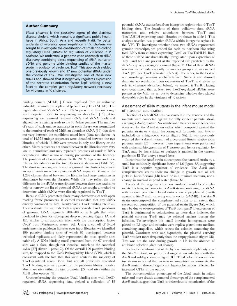
To investigate this we undertook a genome-wide ToxT pulldown
of genomic DNA fragments 200–500 bp in length that were
modified to allow for subsequent deep sequencing (figure 1A and
1B), similar to an approach taken with the transcription factor
CodY from Staphylococcus aureus [26]. Using a cut off of 3-fold
enrichment in pulldown libraries over input libraries, we identified
199 putative binding sites of which 67 overlapped between
technical replicates and likely represented the most specific sites
(table s6). A DNA binding motif generated from the 67 enriched
sites was a close, though not identical, match to the canonical
toxbox [27] (figure 1 panel C). Of the overall 199 putative binding
sites, 64 mapped to the Vibrio Pathogenicity Island (VPI), which is
consistent with the fact that this locus contains the majority of
ToxT-regulated genes. Most, but not all previously described
ToxT binding sites were present in the pulldown library, notably
absent are sites within the tcpA promoter [27] and sites within the
MSH pilus operon [9].
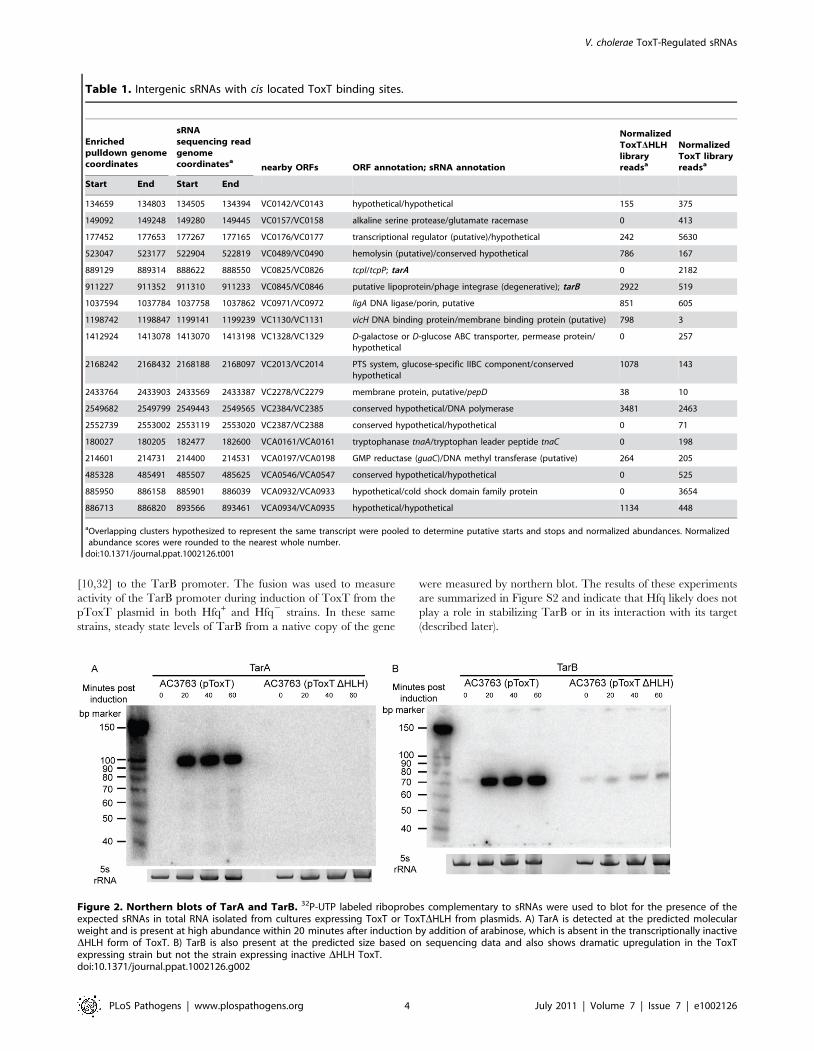
Cross-referencing the putative ToxT binding sites with ToxT-
regulated sRNA sequencing data yielded a collection of 18
potential sRNAs transcribed from intergenic regions with cis ToxT
binding sites. The locations of these pulldown sites, sRNA
transcripts and relative abundance between ToxT and
ToxTDHLH expressing strain libraries are shown in table 1. This
analysis revealed two putative sRNAs within intergenic regions in
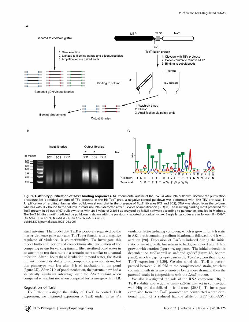
the VPI. To investigate whether these two sRNAs represented
genuine transcripts, we probed for each by northern blot using
total RNA from cultures expressing ToxT or ToxTDHLH. Both
of these sRNAs are dramatically upregulated upon expression of
ToxT and both are present at the expected size predicted by the
sRNA deep sequencing experiment (figure 2). One of these sRNAs
was discovered independently by another group and was named
TarA [25] (for ToxT activated RNA A). The other, to the best of
our knowledge, remains uncharacterized. Since it also showed
dramatic up regulation upon expression of ToxT, and given its
role in virulence (described below), we named it TarB. Having
now determined that at least two ToxT-regulated sRNAs were
present in the VPI, we set out to determine whether they played
detectable roles in the virulence of V. cholerae.
Assessment of sRNA mutants in the infant mouse modelof intestinal colonization
Deletion of each sRNA was constructed in the genome and the
mutants were competed against the fully virulent parental strain
carrying a DlacZ marker. No significant difference in virulence was
observed for the DtarA strain either when competed against the
parental strain or a strain harboring tarA (promoter and toxboxes
included) on a high-copy vector (figure 3A). It was previously
reported that a DtarA mutant had a decreased fitness relative to its
parental strain [25], however, those experiments were performed
with a classical biotype strain of V. cholerae, and hence regulation by
TarA may be less critical or perhaps is masked in the current
pandemic El Tor biotype tested here.
In contrast the DtarB strain outcompetes the parental strain by a
small but statistically significant factor of 1.6 (figure 3A) suggesting
TarB is a negative regulator of virulence. The DtarB and
complemented strains show no change in growth rate or cell
yield in Luria-Bertani (LB) broth or in a minimal medium, nor a
change in survival in pond water (figure S1).
To see if the negative effect on virulence could be comple-
mented in trans, we competed a DtarB strain containing the sRNA
with its own promoter cloned onto a low copy plasmid (ptarB)
against a DtarB strain carrying empty vector (pMMB). The DtarB
strain out-competed the complemented strain to an extent that
exceeds out competition of the parental strain (figure 3A), which
may be due to overexpression of TarB from ptarB. If expression of
TarB is detrimental to colonization, as these data indicate, the
plasmid carrying TarB may be selected against during the
infection. To investigate this, small intestine homogenates were
plated on LB agar and colonies were replica plated onto medium
containing ampicillin, which selects for colonies containing the
plasmid. Consistent with our hypothesis, the plasmid carrying
TarB was lost more frequently than the empty plasmid (figure 3B).
This was not the case during growth in LB in the absence of
antibiotic selection (data not shown).
For further confirmation of the hypercolonization phenotype of
the DtarB mutant, we performed single strain infections with the
DtarB and wildtype strains (Figure 3C). Total colonization in these
two strains indicated that, as seen in competition experiments, the
DtarB mutant showed significant hypercolonization reflected by
increased CFUs in the output.
The out-competition phenotype of the DtarB strain in infant
mice and more drastic attenuated phenotype of the complemented
DtarB strain suggest that TarB is deleterious to colonization of the
Author Summary
Vibrio cholerae is the causative agent of the diarrhealdisease cholera, which remains a significant public healthissue in Africa, South Asia and recently Haiti. To betterunderstand virulence gene regulation in V. cholerae wesought to investigate the contribution of small non-codingregulatory RNAs (sRNAs) to regulation of virulence in V.cholerae. We undertook a genome wide approach to sRNAdiscovery combining direct sequencing of sRNA transcriptcDNA and genome wide binding studies of the masterprotein regulator of virulence, ToxT. This approach yieldedone previously known and 17 new potential sRNAs underthe control of ToxT. We investigated one of these newsRNAs and showed that it negatively regulates expressionof the secreted colonization factor TcpF, adding a newfacet to the complex gene regulatory network necessaryfor virulence in V. cholerae.
V. cholerae ToxT-Regulated sRNAs
PLoS Pathogens | www.plospathogens.org 2 July 2011 | Volume 7 | Issue 7 | e1002126
small intestine. The model that TarB is positively regulated by the
master virulence gene activator ToxT, yet functions as a negative
regulator of virulence, is counterintuitive. To investigate this
model further we performed competitions after incubation of the
competing strains for varying times in filter sterilized pond water in
an attempt to test the strains in a scenario more similar to a natural
infection. After 4 hours (h) of incubation in pond water, the DtarB
mutant retained its ability to outcompete the parental strain, but
this phenotype was lost after 6 h of incubation in the pond
(figure 3D). After 24 h of pond incubation, the parental now had a
statistically significant advantage over the DtarB mutant when
competed in vivo, but not when competed for in vitro growth in LB.
Regulation of TarBTo further investigate the ability of ToxT to control TarB
expression, we measured expression of TarB under an in vitro
virulence factor inducing condition, which is growth for 4 h static
in AKI broth containing sodium bicarbonate followed by 4 h with
aeration [28]. Expression of TarB is induced during the initial
static phase of growth, but returns to background level after 4 h of
growth with aeration (figure 4A, top panel). The initial induction is
dependent on toxT as well as toxR and tcpP/H (figure 4A, bottom
panel), which are genes upstream in the ToxR regulon that induce
ToxT expression [3,4,29]. We also noted that TarB is overex-
pressed between 7–10 fold in the complemented strain, which is
consistent with its in vivo phenotype being more dramatic then the
parental strain in competitions with the DtarB mutant.
We also investigated the role of the RNA chaperone Hfq in
TarB stability and action as many sRNAs that act in conjunction
with Hfq are destabilized in its absence [30,31]. To investigate
expression from the TarB promoter we constructed a transcrip-
tional fusion of a reduced half-life allele of GFP (GFP-ASV)
Figure 1. Affinity purification of ToxT binding sequences. A) Experimental outline of the ToxT in vitro DNA pulldown. Because the purificationprocedure left a residual amount of TEV protease in the His-ToxT prep, a negative control pulldown was performed with 6His-TEV protease. B)Amplification of resulting libraries after pulldowns shows that in the presence of ToxT (libraries BC1 and BC2), DNA was eluted from the column,whereas with TEV bound to the column instead, no DNA is detected after 10 cycles of amplification (BC3). C) The resulting binding motif predicted forToxT present in 66 out of 67 pulldown sites with an E-value of 2.3e14 as analyzed by MEME software according to parameters detailed in Methods.The ToxT binding motif predicted by pulldown is shown with the previously reported canonical toxbox. Single letter codes are as follows; B = C/G/T,D = A/G/T, H = A/C/T, N = A/C/G/T, R = A/G, W = A/T, Y = C/T.doi:10.1371/journal.ppat.1002126.g001
V. cholerae ToxT-Regulated sRNAs
PLoS Pathogens | www.plospathogens.org 3 July 2011 | Volume 7 | Issue 7 | e1002126
[10,32] to the TarB promoter. The fusion was used to measure
activity of the TarB promoter during induction of ToxT from the
pToxT plasmid in both Hfq+ and Hfq2 strains. In these same
strains, steady state levels of TarB from a native copy of the gene
were measured by northern blot. The results of these experiments
are summarized in Figure S2 and indicate that Hfq likely does not
play a role in stabilizing TarB or in its interaction with its target
(described later).
Figure 2. Northern blots of TarA and TarB. 32P-UTP labeled riboprobes complementary to sRNAs were used to blot for the presence of theexpected sRNAs in total RNA isolated from cultures expressing ToxT or ToxTDHLH from plasmids. A) TarA is detected at the predicted molecularweight and is present at high abundance within 20 minutes after induction by addition of arabinose, which is absent in the transcriptionally inactiveDHLH form of ToxT. B) TarB is also present at the predicted size based on sequencing data and also shows dramatic upregulation in the ToxTexpressing strain but not the strain expressing inactive DHLH ToxT.doi:10.1371/journal.ppat.1002126.g002
Table 1. Intergenic sRNAs with cis located ToxT binding sites.
Enrichedpulldown genomecoordinates
sRNAsequencing readgenomecoordinatesa
nearby ORFs ORF annotation; sRNA annotation
NormalizedToxTDHLHlibraryreadsa
NormalizedToxT libraryreadsa
Start End Start End
134659 134803 134505 134394 VC0142/VC0143 hypothetical/hypothetical 155 375
149092 149248 149280 149445 VC0157/VC0158 alkaline serine protease/glutamate racemase 0 413
177452 177653 177267 177165 VC0176/VC0177 transcriptional regulator (putative)/hypothetical 242 5630
523047 523177 522904 522819 VC0489/VC0490 hemolysin (putative)/conserved hypothetical 786 167
889129 889314 888622 888550 VC0825/VC0826 tcpI/tcpP; tarA 0 2182
911227 911352 911310 911233 VC0845/VC0846 putative lipoprotein/phage integrase (degenerative); tarB 2922 519
1037594 1037784 1037758 1037862 VC0971/VC0972 ligA DNA ligase/porin, putative 851 605
1198742 1198847 1199141 1199239 VC1130/VC1131 vicH DNA binding protein/membrane binding protein (putative) 798 3
1412924 1413078 1413070 1413198 VC1328/VC1329 D-galactose or D-glucose ABC transporter, permease protein/hypothetical
0 257
2168242 2168432 2168188 2168097 VC2013/VC2014 PTS system, glucose-specific IIBC component/conservedhypothetical
1078 143
2433764 2433903 2433569 2433387 VC2278/VC2279 membrane protein, putative/pepD 38 10
2549682 2549799 2549443 2549565 VC2384/VC2385 conserved hypothetical/DNA polymerase 3481 2463
2552739 2553002 2553119 2553020 VC2387/VC2388 conserved hypothetical/hypothetical 0 71
180027 180205 182477 182600 VCA0161/VCA0161 tryptophanase tnaA/tryptophan leader peptide tnaC 0 198
214601 214731 214400 214531 VCA0197/VCA0198 GMP reductase (guaC)/DNA methyl transferase (putative) 264 205
485328 485491 485507 485625 VCA0546/VCA0547 conserved hypothetical/hypothetical 0 525
885950 886158 885901 886039 VCA0932/VCA0933 hypothetical/cold shock domain family protein 0 3654
886713 886820 893566 893461 VCA0934/VCA0935 hypothetical/hypothetical 1134 448
aOverlapping clusters hypothesized to represent the same transcript were pooled to determine putative starts and stops and normalized abundances. Normalizedabundance scores were rounded to the nearest whole number.
doi:10.1371/journal.ppat.1002126.t001
V. cholerae ToxT-Regulated sRNAs
PLoS Pathogens | www.plospathogens.org 4 July 2011 | Volume 7 | Issue 7 | e1002126
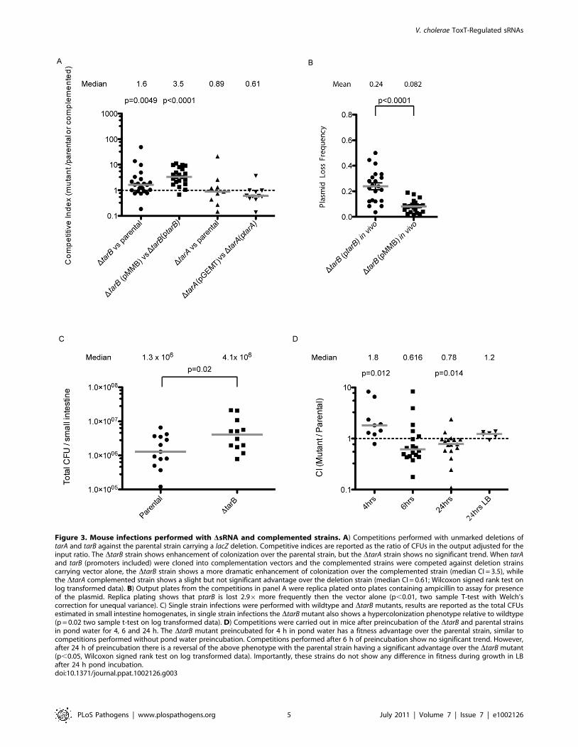
Figure 3. Mouse infections performed with DsRNA and complemented strains. A) Competitions performed with unmarked deletions oftarA and tarB against the parental strain carrying a lacZ deletion. Competitive indices are reported as the ratio of CFUs in the output adjusted for theinput ratio. The DtarB strain shows enhancement of colonization over the parental strain, but the DtarA strain shows no significant trend. When tarAand tarB (promoters included) were cloned into complementation vectors and the complemented strains were competed against deletion strainscarrying vector alone, the DtarB strain shows a more dramatic enhancement of colonization over the complemented strain (median CI = 3.5), whilethe DtarA complemented strain shows a slight but not significant advantage over the deletion strain (median CI = 0.61; Wilcoxon signed rank test onlog transformed data). B) Output plates from the competitions in panel A were replica plated onto plates containing ampicillin to assay for presenceof the plasmid. Replica plating shows that ptarB is lost 2.96 more frequently then the vector alone (p,0.01, two sample T-test with Welch’scorrection for unequal variance). C) Single strain infections were performed with wildtype and DtarB mutants, results are reported as the total CFUsestimated in small intestine homogenates, in single strain infections the DtarB mutant also shows a hypercolonization phenotype relative to wildtype(p = 0.02 two sample t-test on log transformed data). D) Competitions were carried out in mice after preincubation of the DtarB and parental strainsin pond water for 4, 6 and 24 h. The DtarB mutant preincubated for 4 h in pond water has a fitness advantage over the parental strain, similar tocompetitions performed without pond water preincubation. Competitions performed after 6 h of preincubation show no significant trend. However,after 24 h of preincubation there is a reversal of the above phenotype with the parental strain having a significant advantage over the DtarB mutant(p,0.05, Wilcoxon signed rank test on log transformed data). Importantly, these strains do not show any difference in fitness during growth in LBafter 24 h pond incubation.doi:10.1371/journal.ppat.1002126.g003
V. cholerae ToxT-Regulated sRNAs
PLoS Pathogens | www.plospathogens.org 5 July 2011 | Volume 7 | Issue 7 | e1002126
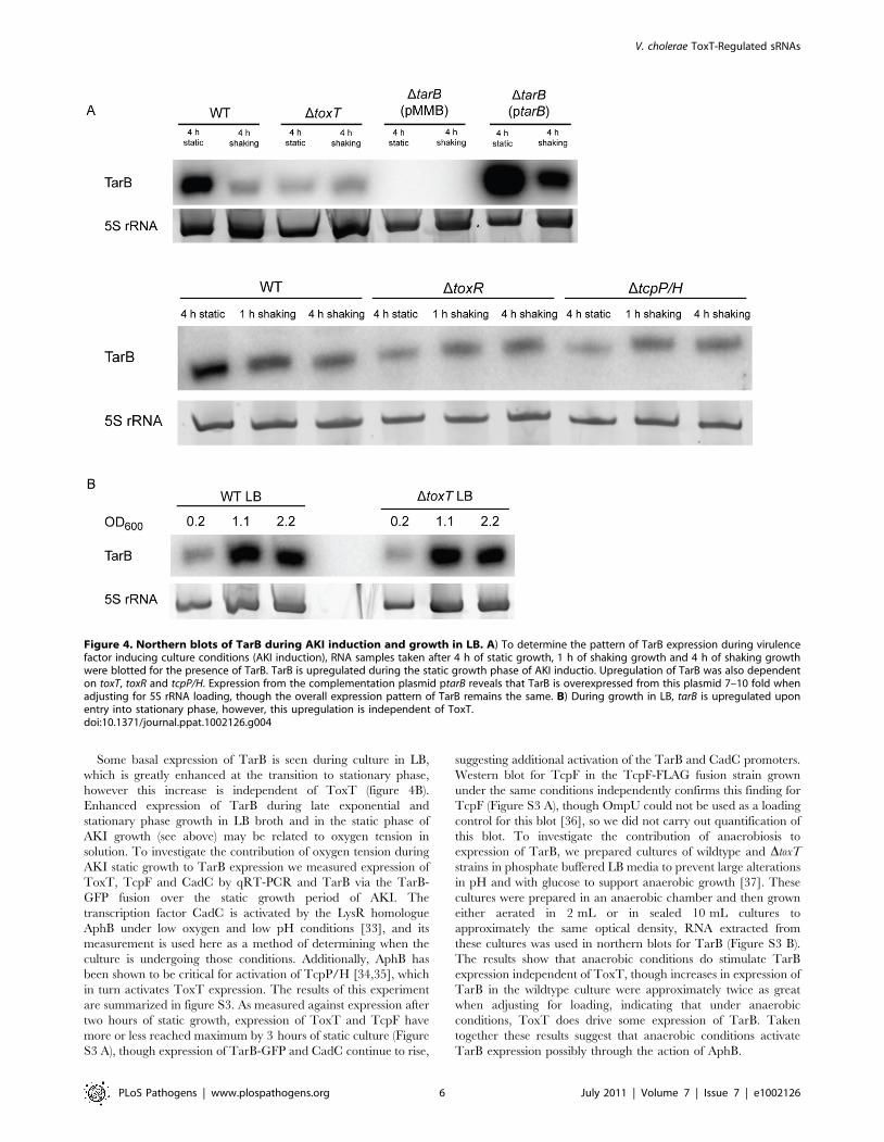
Some basal expression of TarB is seen during culture in LB,
which is greatly enhanced at the transition to stationary phase,
however this increase is independent of ToxT (figure 4B).
Enhanced expression of TarB during late exponential and
stationary phase growth in LB broth and in the static phase of
AKI growth (see above) may be related to oxygen tension in
solution. To investigate the contribution of oxygen tension during
AKI static growth to TarB expression we measured expression of
ToxT, TcpF and CadC by qRT-PCR and TarB via the TarB-
GFP fusion over the static growth period of AKI. The
transcription factor CadC is activated by the LysR homologue
AphB under low oxygen and low pH conditions [33], and its
measurement is used here as a method of determining when the
culture is undergoing those conditions. Additionally, AphB has
been shown to be critical for activation of TcpP/H [34,35], which
in turn activates ToxT expression. The results of this experiment
are summarized in figure S3. As measured against expression after
two hours of static growth, expression of ToxT and TcpF have
more or less reached maximum by 3 hours of static culture (Figure
S3 A), though expression of TarB-GFP and CadC continue to rise,
suggesting additional activation of the TarB and CadC promoters.
Western blot for TcpF in the TcpF-FLAG fusion strain grown
under the same conditions independently confirms this finding for
TcpF (Figure S3 A), though OmpU could not be used as a loading
control for this blot [36], so we did not carry out quantification of
this blot. To investigate the contribution of anaerobiosis to
expression of TarB, we prepared cultures of wildtype and DtoxT
strains in phosphate buffered LB media to prevent large alterations
in pH and with glucose to support anaerobic growth [37]. These
cultures were prepared in an anaerobic chamber and then grown
either aerated in 2 mL or in sealed 10 mL cultures to
approximately the same optical density, RNA extracted from
these cultures was used in northern blots for TarB (Figure S3 B).
The results show that anaerobic conditions do stimulate TarB
expression independent of ToxT, though increases in expression of
TarB in the wildtype culture were approximately twice as great
when adjusting for loading, indicating that under anaerobic
conditions, ToxT does drive some expression of TarB. Taken
together these results suggest that anaerobic conditions activate
TarB expression possibly through the action of AphB.
Figure 4. Northern blots of TarB during AKI induction and growth in LB. A) To determine the pattern of TarB expression during virulencefactor inducing culture conditions (AKI induction), RNA samples taken after 4 h of static growth, 1 h of shaking growth and 4 h of shaking growthwere blotted for the presence of TarB. TarB is upregulated during the static growth phase of AKI inductio. Upregulation of TarB was also dependenton toxT, toxR and tcpP/H. Expression from the complementation plasmid ptarB reveals that TarB is overexpressed from this plasmid 7–10 fold whenadjusting for 5S rRNA loading, though the overall expression pattern of TarB remains the same. B) During growth in LB, tarB is upregulated uponentry into stationary phase, however, this upregulation is independent of ToxT.doi:10.1371/journal.ppat.1002126.g004
V. cholerae ToxT-Regulated sRNAs
PLoS Pathogens | www.plospathogens.org 6 July 2011 | Volume 7 | Issue 7 | e1002126
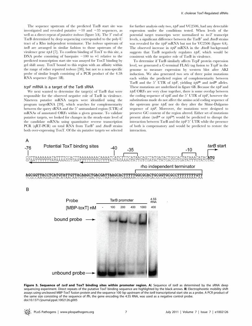
The sequence upstream of the predicted TarB start site was
investigated and revealed putative 210 and 235 sequences, as
well as a direct repeat of putative toxboxes (figure 5A). The 39 end of
TarB determined by deep sequencing corresponded to the poly-U
tract of a Rho independent terminator. The toxboxes upstream of
tarB are arranged in similar fashion to those upstream of the
virulence gene tcpA [7]. To confirm binding of ToxT to this site, a
DNA probe consisting of basepairs 2100 to +1 relative to the
predicted transcription start site was assayed for ToxT binding by
gel shift assay. ToxT bound to this region with an affinity within
the range of other reported toxboxes [38], but not to a non-specific
probe of similar length consisting of a PCR product of the 4.5S
RNA sequence (figure 5B).
tcpF mRNA is a target of the TarB sRNAWe next wanted to determine the target(s) of TarB that were
responsible for the observed negative role of TarB in virulence.
Nineteen putative mRNA targets were identified using the
program targetRNA [39], which searches for complementarity
between the query sRNA and the 59 untranslated region (UTR) of
mRNAs of annotated ORFs within a given genome. To validate
putative targets, we looked for changes in the steady-state level of
the candidate mRNAs using quantitative reverse transcription
PCR (qRT-PCR) on total RNA from TarB+ and DtarB strains
both over-expressing ToxT. Of the six putative targets we selected
for further analysis only two, tcpF and VC2506, had any detectable
expression under the conditions tested. When levels of the
potential target transcripts were normalized to toxT transcript
levels, a significant difference between the TarB+ and DtarB strains
was revealed for the tcpF mRNA but not for VC2506 (figure 6A).
The observed increase in tcpF mRNA in the DtarB background
suggests that TarB negatively regulates tcpF, which would be
consistent with the negative role of TarB in virulence.
To determine if TarB similarly affects TcpF protein expression
level, we generated a C-terminal FLAG tag fusion to TcpF in the
genome to measure expression by western blot after AKI
induction. We also generated two sets of three point mutations
each within the predicted region of complementarity between
TarB and the 59 UTR of tcpF, yielding tcpF* and tarB* alleles.
These mutations are underlined in figure 6B. Because the tcpF and
tcpE ORFs are very close together, there is some overlap between
the coding sequence of tcpE and the 59 UTR of tcpF, however the
substitutions made do not affect the amino acid coding sequence of
the upstream gene tcpE nor do they alter the Shine-Dalgarno
sequence of tcpF. Moreover, the mutations were designed to
preserve GC content of the region altered. Either set of mutations
present alone (tarB* or tcpF*) would be predicted to disrupt the
interaction between TarB and the tcpF 59 UTR while the presence
of both is compensatory and would be predicted to restore the
interaction.
Figure 5. Sequence of tarB and ToxT binding sites within promoter region. A) Sequence of tarB as determined by the sRNA deepsequencing experiment. Direct repeats of the putative ToxT binding sequence are highlighted by the black arrows. B) Electrophoretic mobility shiftassays using uncleaved MBP-ToxT fusion protein and the sequence 100 bp upstream of the tarB transcriptional start site as a probe. A PCR product ofthe same size consisting of the sequence of ffh, the gene encoding the 4.5S RNA, was used as a negative control probe.doi:10.1371/journal.ppat.1002126.g005
V. cholerae ToxT-Regulated sRNAs
PLoS Pathogens | www.plospathogens.org 7 July 2011 | Volume 7 | Issue 7 | e1002126
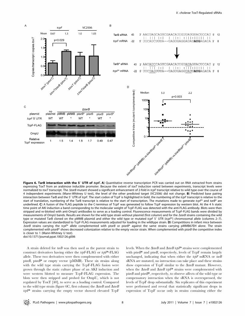
A strain deleted for tarB was then used as the parent strain to
construct derivatives having either the tcpF-FLAG or tcpF*-FLAG
allele. These two derivatives were then complemented with either
ptarB, ptarB* or empty vector (pMMB). These six strains along
with the wild type strain carrying the TcpF-FLAG fusion were
grown through the static culture phase of an AKI induction and
were western blotted to measure TcpF-FLAG expression. The
blots were then stripped and probed for OmpU, which is not
regulated by ToxT [40], to serve as a loading control. Compared
to the wild type strain (figure 6C, first column) the DtarB and DtarB
tcpF* strains carrying the empty vector showed elevated TcpF
levels. When the DtarB and DtarB tcpF* strains were complemented
with ptarB* and ptarB, respectively, levels of TcpF remain largely
unchanged, indicating that when either the tcpF mRNA or tarB
sRNA are mutated, no interaction can take place and these strains
show expression of TcpF similar to the DtarB mutant. However,
when the DtarB and DtarB tcpF* strains were complemented with
ptarB and ptarB*, respectively, to observe affects of the wild type or
compensatory interaction when the sRNA is overexpressed, the
levels of TcpF drop substantially. Six replicates of this experiment
were performed and reveal that statistically significant drops in
expression of TcpF occur only in strains containing either the
Figure 6. TarB interaction with the 59 UTR of tcpF. A) Quantitative reverse transcription PCR was carried out on RNA extracted from strainsexpressing ToxT from an arabinose inducible promoter. Because the extent of toxT induction varied between experiments, transcript levels werenormalized to toxT transcript. The DtarB mutant showed a significant enhancement of 2-fold in tcpF transcript relative to wild type over the course of4 independent experiments (Mann-Whitney U test), the level of the other predicted target (VC2506) did not change. B) Predicted base pairinginteraction between TarB and the 59 UTR of tcpF. The start codon of TcpF is highlighted in bold, the numbering of the tcpF transcript is relative to thestart of translation, numbering of the TarB transcript is relative to the start of transcription. The mutations made to generate tcpF* and tarB* areunderlined. C) A fusion of the FLAG peptide to the C-terminus of TcpF was generated to follow TcpF expression by western blot. At the 4 h statictime point of AKI induction a band corresponding to the molecular weight of TcpF-FLAG was detected with the anti-FLAG antibody. Blots were thenstripped and re-blotted with anti-OmpU antibodies to serve as a loading control. Fluorescence measurements of TcpF-FLAG bands were divided bymeasurements of OmpU bands. Results are shown for the wild type strain without plasmid (first column) and for the DtarB strains containing the wildtype or mutated TarB cloned on the pMMB plasmid and either the wild type or mutated tcpF 59 UTR (tcpF*) chromosomal allele (columns 2–7).Expression values are standardized to TcpF-FLAG measurements adjusted for loading in the wildtype strain. D) Competitions in infant mice betweenDtarB strains carrying the tcpF* allele complemented with ptarB or ptarB* against the same strains carrying pMMB67EH alone. The straincomplemented with ptarB* shows decreased colonization relative to the empty vector strain. When complemented with ptarB the competitive indexis closer to 1 (Mann-Whitney U test).doi:10.1371/journal.ppat.1002126.g006
V. cholerae ToxT-Regulated sRNAs
PLoS Pathogens | www.plospathogens.org 8 July 2011 | Volume 7 | Issue 7 | e1002126
wildtype TcpF target sequence complemented with wildtype TarB
or strains in which the target sequence and sRNA have
compensatory mutations (Figure S4). When these strains were
blotted after the aeration growth phase of AKI induction, no
differences in TcpF expression were visible (data not shown),
which would be expected given the up regulation of tarB during
the static phase but return to basal level of expression during the
aeration phase of AKI induction.
To determine if the interaction of TarB with the 59 UTR of tcpF
was responsible for the phenotype in mice, competitions were
carried out using tcpF* strain derivatives. Competition of the DtarB
tcpF*(ptarB*) strain against the same strain carrying empty vector
yielded the expected result of out-competition by the latter strain,
which lacks tarB* (figure 6D). Competition of the DtarB tcpF*(ptarB)
strain against the same strain with vector alone yielded a
competitive index that was significantly closer to one, which is
expected since neither strain should have an interaction between
sRNA and target. The difference between the two competitive
indices was highly significant (p,0.003).
To determine if the pond water-incubation phenotype of the
DtarB mutant was related to expression of TcpF or TarB in this
environment we carried out experiments to measure TarB and
TcpF levels over the course of pond water incubation, the results
of these experiments are summarized in Figure S5. TcpF
expression was followed through the course of pond water
incubation via the C-terminal FLAG fusion in both the wildtype
and DtarB backgrounds by anti-FLAG western blot. The results
indicate that the wildtype and DtarB mutant show similar levels
of TcpF expression initially, however, over the course of pond
incubation, TcpF levels drop in the wildtype strain, but not the
DtarB strain. Transcription of TarB, as measured by production
of GFP from the TarB promoter-GFP fusion indicates that levels
of TarB expression do not change dramatically over the course
of pond water incubation. Northern blots for TarB expression
over the course of pond water incubation suggest that TarB
steady state levels drop (data not shown), but this may be due to
the observed wholesale degradation of RNAafter increasing
time of incubation in pond water, such that accurate
measurements of TarB expression via northern blot are not
possible. These results indicate that while TarB expression levels
do not vary dramatically over the course of pond water
incubation, TcpF protein levels do drop, and this drop was
absent in the DtarB mutant. This enhanced TcpF expression in
the DtarB mutant may contribute to the phenotype of the DtarB
mutant in vivo after pond water incubation, as over expression of
TcpF in pond water would contribute to metabolic drain prior
to infection.
Discussion
Deep sequencing has allowed the interrogation of processes in
bacteria with unprecedented detail. Here we used two comple-
mentary approaches, deep sequencing of cloned sRNAs and
ToxT-bound DNA fragments, to identify ToxT-regulated sRNAs.
The number of previously estimated ToxT binding sites in the V.
cholerae genome was between 17 and 20 [9,27]. We have now
uncovered what may be a greatly expanded set of targets for ToxT
to coordinate expression of protein coding genes as well as sRNAs.
The results of the pulldown experiment returned regions of a few
hundred basepairs in length that were enriched and many
predicted sites are overlapping, which is due to the size range of
the fragments used in the pulldown and the automated analysis of
the pulldown data. Although many of these sites remain to be
validated we are confident in proposing that the ToxR regulon
encompasses many more transcripts, both protein coding and
otherwise, than was previously thought.
The results of the sRNA deep sequencing reveal the method to
be exquisitely sensitive. Because of our exclusion of larger RNA
transcripts and depletion of tRNA and 5S RNA in the sRNA size
range and the use of Illumina massively parallel sequencing
technology we have achieved tremendous depth of coverage of
potential sRNA genes in V. cholerae [16]. Transcripts represented
by ,40 or more reads could be detected by northern blot (this
study and data not shown). However, transcripts represented by
fewer than ,40 reads, which may represent low abundance
sRNAs, are difficult or impossible to detect by northern blot and
other methods such as qRT-PCR are needed for independent
validation. Of the 18 candidate ToxT-regulated sRNAs we report
here, 11 (including tarB) were not identified as putative sRNAs in
previous sequencing experiments or bioinformatics-based ap-
proaches to sRNA discovery [16,41], displaying the depth of
information that can be gained with high throughput sequencing
technologies and the conditional expression of sRNAs. In
comparison to other methods of sRNA discovery, our approach
has the advantage of being targeted in its search for ToxT-
regulated sRNAs but unbiased in its identification of sRNAs.
Approaches utilizing RNA binding proteins such as Hfq [42,43],
are not exhaustive as the sRNA we report here likely does not
interact with Hfq, though those methods do have the potential to
identify mRNA targets as well as sRNAs. Additionally, this
approach benefits from the vast strides made in high throughput
sequencing recently which generates far more depth of data then
microarray based methods [44], including exact 39 and 59 ends
and unbiased coverage of positive and negative strand sRNAs.
Keeping the latter in mind, this approach can also identify many
potential sense and anti-sense sRNAs [16] overlapping with
protein coding genes although these potential sRNAs are not
discussed here.
In this study we identified a new sRNA member of the ToxR
regulon that fine-tunes expression of a virulence factor also within
the ToxR regulon, thus adding a new facet to the elaborate
virulence gene regulation program in V. cholerae. However, when
placed in the larger context of V. cholerae pathogenesis, it is not
entirely clear why a repressor of an essential virulence factor would
be produced at the same time as the virulence factor it negatively
regulates. The answer may lie in the biphasic nature of V. cholerae
gene expression during intestinal colonization [10,11]. The initial
induction of virulence factors requires ToxR/S- and TcpP/H-
dependent ToxT expression in the intestinal lumen. This is
followed by a more robust activation of the TCP and CTX
operons closer to the epithelial surface of the small intestine, driven
by a positive feedback loop in ToxT expression that is thought to
activated in part by the presence of bicarbonate [28,45].
During AKI induction in vitro in the absence of bicarbonate,
ToxT production is stimulated during static growth but the
transition to aerated growth is required for CTX production [46].
All experiments reported here included bicarbonate in the
medium over the course of the experiment, which is sufficient to
cause CTX production even during static growth [28,47].
Research done on the contribution of anaerobiosis to virulence
gene expression in V. cholerae El Tor isolates has shown stimulation
of VPI gene products [48], and that the AphB protein, which
functions upstream of tcpP/H, is active primarily at low oxygen
tension and low pH [33]. Since TarB expression is greatest during
the static phase of AKI induction, but repressed during aerated
growth even though bicarbonate had been added to induce CTX
and TCP expression prior to aeration, it is tempting to speculate
that TarB expression is enhanced in microaerobic conditions. The
V. cholerae ToxT-Regulated sRNAs
PLoS Pathogens | www.plospathogens.org 9 July 2011 | Volume 7 | Issue 7 | e1002126
experiments we performed under anaerobic conditions also
suggest that oxygen plays a role in TarB expression, though it
may be only one of a host of signals, which act on TarB in vivo.
TarB’s function under low oxygen tension could be to repress
TcpF expression prior to penetration of the mucous barrier of the
small intestine. Upon reaching the epithelial surface, the higher
oxygen tension would contribute to reduced TarB expression,
allowing TcpF to be fully expressed. This would fit with the
proposed role of TcpF in colonization of the epithelium [49]. The
intestinal brush border is a highly vascular structure, commensu-
rate with its role in absorbing nutrients, and it would not be
unreasonable to speculate that the lumenal space adjacent to it
would have greater oxygen tension then the luminal fluid. The
actual oxygen tension of the small intestine may be quite low as
oxygen requiring luciferase reporter systems in bacteria do not
function in the small intestine [50,51]. However, to the best of our
knowledge, oxygen measurements at the brush border have not
been reported.
Other possible factors responsible for controlling TarB expres-
sion could be entry into stationary phase, as increased TarB
expression is observed in V. cholerae grown in LB broth to late
exponential and stationary phase. Also, during AKI induction,
4 hours of growth in static culture corresponds with entry into
stationary phase [46]. Stationary phase regulation of TarB may
also occur via an alternative sigma factor as was observed for the
sRNA VrrA [20], or possibly via CRP-cAMP mediated repression
as carbon sources become depleted [35].
Coordination of TcpF expression by TarB appears to have a
positive effect on colonization if the bacteria are coming from a
resource poor environment, such as contaminated pond water,
and even then, the differences in colonization efficiency of the
DtarB mutant are quite small. In contrast, if the bacteria are grown
in a rich medium prior to infection, overexpression of TcpF in the
DtarB mutant appears to be beneficial. The reasons for this may
relate to the details of the experimental system used here, wherein
immunologically naı̈ve infant mice are used as a host. In contrast,
in nature many hosts in endemic areas will have some level of pre-
existing immunity, and may harbor anti-TcpF antibodies as TcpF
is a known antigenic protein [49]. It is possible that tight repression
of TcpF provides a more pronounced fitness advantage in nature
under different conditions then those used here, which would
explain TarB’s presence among all sequenced isolates of toxigenic
V. cholerae (data not shown). Further insight into the functional role
of TcpF in colonization may shed more light on the necessity of
the TarB-mediated post-transcriptional regulation observed here.
Materials and Methods
Ethics statementAll animal experiments were done in accordance with NIH
guidelines, the Animal Welfare Act and US federal law. The
experimental protocol using animals was approved by Tuft
University School of Medicine’s Institutional Animal Care and
Use Committee. All animals were housed in a centralized and
AAALAC-accredited research animal facility that is fully staffed
with trained husbandry, technical, and veterinary personnel.
Bacterial growth conditionsV. cholerae O1 serogroup El Tor biotype isolate E7946 and
derivatives were grown at 37uC in LB broth with aeration. For
AKI induction, strains were grown in AKI broth (1.5% peptone,
0.4% yeast extract, 0.5% NaCl, 0.3% NaCHO3) statically for 4 h
at 37uC followed by aeration for 4 h 37uC. To induce expression
of cloned genes on plasmids, arabinose was added to 0.04% upon
reaching mid-exponential phase (optical density at 600 nm
[OD] = 0.3). All DNA manipulations were done in E. coli DH5aor derivatives with plasmids maintained with the appropriate
antibiotics.
Strain constructionAll PCR reactions were carried out with EasyA polymerase
according to the manufacturer’s specifications using the indicated
primers, the sequences of which can be found in table S1. The
descriptions of all plasmids used in this study are included in table
S2.
Plasmids pToxT and pToxT DHLH plasmids we constructed
by PCR amplification of the toxT ORF including native RBS from
gDNA from either wildtype V. cholera E6749 or an E6749 strain
carrying an internal deletion of the helix-loop-helix DNA binding
domain [11] using primers NcoI_ToxT_F and XbaI_ToxT_R.
This PCR product was then cloned into the NcoI and XbaI sites of
the pBAD24 plasmid [52] to allow expression of ToxT upon
addition of L-arabinose. Unmarked deletions of chromosomal
genes were constructed by SOE PCR introduced using a
derivative of the pCVD442 allelic exchange vector, pCVD442-
lac which contains the pUC19 LacZ gene and MCS, as described
[53].
Point mutations in the tarB gene were generated by SOE PCR
using primers xbaI_TarB comp_F, TarB_mut_R1 and TarB_
mut_F2 and SacI_TarB_comp_R, using E6749 genomic DNA as
template. PCR products were mixed in a one to one ratio, and
added to a PCR reaction run for 25 cycles at an annealing
temperature of 50uC without primers and the mutated sRNA
sequence plus promoter were amplified with XbaI_TarB_comp_F
and SacI_TarB_comp_R which contain SacI and XbaI restriction
sites which were subsequently used for cloning into pMMB67EH
to generate ptarB*. The wildtype complementation vector ptarB
was generated by cloning a PCR product generated using
XbaI_TarB_comp_F and SacI_TarB_comp_R primers and
genomic DNA as a template.
Point mutations in the tcpF 59 UTR were also generated by SOE
PCR using primers XbaI_TcpF_mut_F1, TcpF_mut_R1,
TcpF_mut_R2 and XbaI_TcpF_mut_R2 using an identical
procedure as above. The final ,2 kb product containing the
mutated tcpF 59 UTR sequence which was subsequently cloned
into the XbaI site of the pCVD442-lac vector which was then
mated into strains of interest. Double crossovers were selected on
10% sucrose plates. Individual double crossovers were screened for
the mutated sequences by sequencing with the TcpF seq primer
and the XbaI_TarB_comp_F primer and confirming double
crossover by streaking on 10% sucrose as well as ampicillin
containing plates to ensure sucrose resistance and ampicillin
sensitivity.
C-terminal FLAG fusions to TcpF were generated by
amplification of the C-terminal 346 bp using the TcpF_qt_F
primer and the TcpF-FLAG_R primer to add the FLAG amino
acid sequence [54], this product was subcloned into Topo pCR2.1
(Invitrogen). The resulting plasmid was cut using KpnI and
EcoRV and the insert containing the C terminus of TcpF with the
FLAG fusion was cloned into a modified pGP704 suicide vector
[55] which contains a chloramphenicol resistance drug marker in
place of an ampicillin marker (pGP704cat). This construct was
then mated into strains of interest and single crossovers were
selected for on chloramphenicol plates at 2 mg/mL. Proper
insertions were confirmed by PCR using the TcpF-FLAG reverse
primer and TcpF seq forward primer. A merodiploid strain was
constructed by plasmid integration resulting in the placement of
GFP(ASV) under the control of one copy of the TarB promoter
V. cholerae ToxT-Regulated sRNAs
PLoS Pathogens | www.plospathogens.org 10 July 2011 | Volume 7 | Issue 7 | e1002126
followed by the native TarB locus downstream of the integrated
plasmid sequence. The plasmid borne fusion was generated by
amplifying the +3 to 2376 positions in the TarB promoter from
E6749 genomic DNA using primers TarB_F and TarB_-300_R
and subcloning the product into pCR2.1 yielding ptarB-300. GFP
was amplified from pGfpmut3.1 plasmid (Clonetech) using primers
Fgfp2 and Rgfp2 which adds a ribosomal binding site and SacI site
at the 59 end and the destabilizing (ASV) [32]C terminal amino
acids and a SmaI site at the 39 end. The GFP(ASV) PCR product
was cloned in a triple ligation with the SacI/EcoRV fragment
from ptarB-300 into pGP704cat digested with SmaI to generate
the transcriptional fusion. The resulting plasmid (pTarB-GFP) was
mated into E6749 strains and single crossovers were selected on
chloramphenicol and confirmed by PCR using primers Rgfp2 and
XbaI_DTarB_R2.
sRNA deep sequencingSingle colonies of strain AC3763 (DtoxT) transformed with
either pToxT or pToxTDHLH plasmids were picked and grown
in LB broth containing streptomycin and ampicillin both at
100 mg/mL overnight. Strains were back diluted from overnight
cultures to an OD of 0.03 in 200 mL LB supplemented with
streptomycin and ampicillin both at 100 mg/mL and were grown
with aeration at 37uC until the strains reached mid-exponential
phase (OD = 0.3). Arabinose was then added to 0.04% to induce
expression of toxT alleles from pToxT plasmids, and induction was
allowed to proceed for 20 minutes prior to RNA extraction. Total
RNA was purified from the bulk culture by phenol/chloroform
extraction and isopropanol precipitation. Cloning and sequencing
of sRNA was carried out as previously described [16], sequences of
the micro RNA cloning linkers (IDT) used are included in table
S4. In order to further decrease tRNA and 5S rRNA in the final
sequenced products, the depletion step described in the previously
published procedure was carried out twice with the addition of an
oligo targeting the serGCC tRNA (59-GCGGTGAGTGAGA-
GATTCGAACTCTC-39). The final cDNA products were
prepared for Illumina Genome Analyzer II sequencing using
Illumina primers 1a, 1b and 1c (table S1) for the first 10 cycles of
PCR, followed by gel purification and Illumina primers 2a and 2b
(table S1) for the final 4 cycles of PCR followed by PCR clean up
(Stratagene). Final products were run on a Bioanalyzer High-
Sensitivity DNA chip (Agilent) prior to Illumina sequencing to
normalize loading of the two samples and ensure quality of the
libraries. The libraries were pooled and placed on one lane of an
Illumina Genome Analyzer IIx paired-end sequencing run at Tufts
University Core Facility. Briefly, a paired-end sequencing run
sequences both the 59 and 39 end of every DNA molecule attached
to the flowcell. The first read is downstream of linker 1 and the
second read is downstream of linker 2 (ToxT library) or linker 3
(DHLH library) so that for every pair, the directionality of the
original RNA molecule could be determined. Sequence reads were
trimmed to remove linker sequences and filtered so that 100% of
the sequenced bases in each read had a minimum quality score of
5 (base call accuracy at least 68%). Reads were aligned to the O1
biovar N16961 genome (NCBI Accession Nos. NC_002505,
NC_002506) using Bowtie (http://bowtie-bio.sourceforge.net).
Reads matching rRNA or tRNA regions were removed from the
alignment, leaving 1,062,048 reads in the ToxTDHLH library and
2,212,216 reads in the ToxT library. Unique transcripts totaled
6,815 for ToxTDHLH and 27,787 for ToxT. The alignments
were then processed to generate a library of clustered transcripts
using the method previously described [16]. This resulted in 3,309
clusters for the ToxTDHLH library and 12,534 clusters for ToxT
library. Clustered reads were output into ‘‘gff’’ format and viewed
using GenomeView (http://genomeview.org). The number of
reads in sRNA clusters were normalized by dividing the number of
reads in that cluster by the ratio of MtlS reads in that library to
total MtlS reads. For example normalized readsToxT = cluster
readsToxT/(MtlSToxT/(MtlSToxT+MtlSToxTDHLH)).
ToxT overexpression and purificationE. coli strain BL21(DE3) was transformed with the plasmid
pMAL-TEV-His-thr-ToxT (table s3). The resulting strain was
grown on LB agar plates containing ampicillin and a single colony
was picked for growth of a 4 mL overnight culture. The overnight
culture was used to inoculate 1 L LB broth containing ampicillin
at 100 mg/mL and was grown with aeration at 37uC. Transcrip-
tion was induced once the culture had reached exponential phase
(OD = 0.5–1) by addition of IPTG to 1 mM. Induction was
allowed to proceed shaking at 20uC for 16 h, after which, cell
pellets were collected by centrifugation and resuspended in 20 mL
lysis buffer (20 mM Tris-HCl pH 8, 2 mM DTT, 1 mM EDTA,
250 mM NaCl) plus Complete protease inhibitors (Roche). Cell
pellets were lysed and the lysate was cleared by centrifugation at
18,000 rpm in a SS34 rotor.
The cleared lysate was then applied to a 5 mL dextrin MBPtrap
column (GE Life sciences). The column was washed with lysis
buffer followed by elution with MBP elution buffer (as lysis buffer,
+1 mM maltose). The elution fractions were subsequently diluted
10-fold with buffer QB1A (20 mM Tris-HCl pH 8.0, 1 mM DTT)
and applied to an 8 mL Source15Q anion exchange column
equilibrated in QB1A. The protein was eluted using a 0 to 20%
gradient of QB1B (20 mM Tris-HCl pH 8.0, 1 M NaCl, 1 mM
DTT) developed over 25 column volumes. The peak fractions
were diluted 5-fold in SB1A buffer (25 mM phosphate buffer
pH 6.0, 1 mM DTT) and applied to a 8 mL Source15S cation
exchange column equilibrated in SB1A. The protein was eluted
using a 15 to 35% gradient QB1B (25 mM phosphate buffer
pH 6.0, 1 mM DTT, 1M NaCl), which resulted in two peaks, the
second peak was known to be a soluble aggregate and was
discarded. The initial peak was split into two aliquots, one of
which was applied to a Superose 12 gel filtration column in EMSA
buffer (10 mM Tris-HCl pH 7.5, 200 mM KCl, 10 mM bME) for
use in mobility shift assays, the other aliquot was cleaved with
TEV protease overnight at 4uC and subsequently diluted 5-fold in
SB1A and applied to a 2 mL Source15S cation exchange column
to separate His-ToxT from the cleaved MBP fusion protein. His-
ToxT was eluted from this column with a 35 to 100% gradient of
SB1B developed over 12 column volumes. Finally, His-ToxT peak
fractions were applied to a Superdex 75 gel filtration column in
EMSA buffer. These final steps did leave a small amount of TEV
protease in the final purified product.
Affinity purification of ToxT binding sequencesGenomic libraries were prepared by centrifuging 10 mL of
overnight growth of wild type (AC53) V. cholerae, washing 26with
TBS and resuspending in 5 mL TBS. To generate gDNA
fragment sizes of 300 to 1,000 bp, the cell pellet was subjected
to four 30 second sonication cycles on ice using a sonicator micro
tip (Branson); each sonication cycle was separated by a 30 second
incubation on ice. After sonication, RNAase A was added to a
concentration of 2 mg/mL, the samples were incubated at 37uCfor 20 min to allow for degradation of RNA. DNA was purified
with 2 rounds of extraction with citrate buffered phenol:chloro-
form (Ambion) followed by a final extraction with chloroform only
and then concentrated by ethanol precipitation. Fragmented DNA
was used to prepare three different bar-coded libraries using
adapters BC1a/BC1b, BC2a/BC2b and BC3a/BC3b (table S4) as
V. cholerae ToxT-Regulated sRNAs
PLoS Pathogens | www.plospathogens.org 11 July 2011 | Volume 7 | Issue 7 | e1002126
described [26]. For the final amplification and purification of bar-
coded libraries, ten PCR reactions were done using linkered and
size selected gDNA as template using primers Olj 139 and 140 and
EasyA polymerase (Stratagene). PCR conditions were as follows,
denaturation for 5 minutes at 95uC, annealing for 30 seconds at
65uC, elongation for 30 seconds at 72uC, cycling back to
denaturation at 95uC for 30 seconds for 15 cycles after which
reactions were pooled and incubated with 50 mL ExoSAP-IT
(USB) at 37uC for 1 h. Final purification of libraries was carried
out by phenol:chloroform extraction and ethanol precipitation and
resuspension of libraries in 100 mL deionized water.
Binding reactions contained 15 mg bar-coded DNA library in a
total volume of 250 mL with 200 nM purified His6-tagged ToxT
purified as above or with His6-tagged TEV protease in EMSA buffer
with 10 mg/mL sheared salmon sperm DNA, 0.3 mg/mL BSA and
10% glycerol. Reactions were allowed to incubate with gentle mixing
at 37uC for 1 h, after which the reaction was added to a
microcentrifuge spin column (Pierce) packed with a 50 mL bead
volume column of HisPur cobalt resin (Pierce) that had been
equilibrated in the above buffer. The reaction was allowed to bind to
the column by mixing gently at 37uC for 1 h. Flow through was then
collected by spinning the column in a microcentrifuge at 3,0006g for
1 minute. The column was washed 36by gentle resuspension of the
bead volume in 250 mL of EMSA buffer with the above additions,
followed by centrifugation. The column was washed an additional 36as above, but in EMSA buffer only. After the final wash, the bead
volume was resuspended in 10 mM Tris-HCl pH 8 and boiled for
5 minutes and allowed to cool to room temperature, then incubated
with proteinase K (5 mg/ml) for 30 minutes at 65uC, followed by
boiling for 5 minutes. After centrifugation for 1 min at 3,0006g, the
resulting 100 ml of the supernatant fluid was purified by using a PCR
purification kit (Qiagen) and then subjected to 10 cycles of PCR
amplification with primers Olj139 and Olj140, repurified, quantified
on the Bioanalyzer high sensitivity DNA chip (Agilent), and subjected
to deep sequencing, along with aliquots of the input libraries prior to
pulldown, using the Illumina Genome Analyzer II on the paired end
setting.
Reads from the Illumina libraries were aligned to the N16961
genome. Sequence alignment and assembly were performed as
described above. After alignment, reads that did not match the
genome were discarded and the sets were normalized so that each
set contained the same number of reads. Alignment positions were
shifted by half their insert length as determined by each mapped
pair, giving the center position of each sequenced DNA molecule.
These positions were then tabulated and used to generate a
coverage map of the genome using a rolling average with window
size of 35 bases. Coverage maps were generated for every sample.
For each genomic DNA and corresponding pulldown sample, an
enrichment map was created, which represented the ratio of the
values from the pulldown sample over that of the genomic DNA
sample. Enrichment maps were then scanned to identify regions
that had more than 36 the average coverage for more than 100
consecutive positions. The false discovery rate (FDR) was then
calculated by performing the same analysis with the control and
pulldown samples switched. At 36 coverage, the FDR was 0.03
and 199 enriched sites were identified totaled between the
libraries, of which 67 were observed in both replicates.
Significance of each enriched region was assessed using two
methods [56]. First, the number of reads in that region in the
control sample was used to generate a Poisson distribution. This
was then used to assess the probability of the same number of
reads occurring in the pulldown sample. Using this method, all
regions identified had a p-value of ,1610298. Second, a Z-score
was found by comparing the proportion of tags in the control
sample to that in the pulldown. All of the regions identified had a
significant difference in the proportion of tags counted between the
control and pulldown samples, with z-scores .7.7. The nucleotide
sequences from the overlapping set were used as a training set for
finding motifs using MEME 4.1.0. We allowed MEME to find
motifs that occurred at least one time in each fragment. The motif
reported in figure 1 panel C is the lowest E-value motif for the 67
sites combined in both libraries.
Mobility shift assaysPrimers TarB promoter R and TarB promoter F were used to
amplify the upstream 100 bp of TarB, predicted to contain
promoter elements and ToxT binding sites to serve as a probe in
the mobility shift assay. The PCR product was purified
(Stratagene) and 3.3 pmoles was end-labeled using T4 Polynucle-
otide Kinase (NEB) and 32P c-ATP according to the manufactures
instructions, and then purified using a Performa DTR spin
cartridge (Edge Biosciences). A negative control probe of similar
size consisting of 4.5S RNA sequence was prepared in parallel.
The binding reaction occurred in 20 mL with 3 nM labeled probe
and varying concentrations of purified MBP-his-thr-ToxT in
EMSA with 10 mM 10 mg/mL sheared salmon sperm DNA,
6 mg/mL BSA, 10% glycerol and 0.002% Orange G dye added.
Binding was allowed to occur for 30 minutes at 30uC followed by
loading of the entire reaction onto a 5% TBE-Polyacrylamide gel,
which was then run at 100 V for 60 minutes. The gel was then
used to directly expose a phosphor screen and the image was read
on a FLA-9000IR using the IP setting.
AKI induction experimentsFor AKI induction experiments, strains were grown overnight
with aeration at 37uC in LB broth containing streptomycin at
100 mg/mL, ampicillin at 50 mg/mL (excluded in the case of the
TcpF C-FLAG integration in the wild type background and TarB-
GFP strains without plasmid) and chloramphenicol at 2 mg/mL.
Overnight cultures were then diluted into prewarmed AKI media
[47] containing 0.3% NaHCO3 and ampicillin at 50 mg/mL (again
excluded for the wild type background strain and TarB-GFP
fusions) to an OD of 0.01. Strains were grown statically in an
incubator at 37C for the indicated times at which culture aliquots
were removed for analysis. After 4 hours of static growth, cultures
were split into 1 mL aliquots and grown shaking at 37C for 4 hours.
Anaerobic growthFor anaerobic growth experiments, overnight cultures were
prepared by inoculation of strains into phosphate buffered LB
media containing 60 mM K2HPO4, 33 mM KH2PO4, 0.5%
glucose and 100 mg/mL streptomycin. These cultures were grown
overnight in an anaerobic chamber and used to subsequently
inoculate either 2 mL aerated cultures or 10 mL cultures in sealed
tubes prepared in the anaerobic chamber to an OD of
approximately 0.01. Aerobic and anaerobic cultures were then
grown in parallel in a shaking 37C incubator to approximately the
same OD and snap frozen on liquid nitrogen and subsequently
used for RNA extraction and northern blots. For each culture the
pH of the media was measured after growth was recorded and
ranged between 6.3 and 6.5 for anaerobic cultures and 6.7 to 6.8
for aerobically grown cultures.
Pond water incubationsFor pond water incubation experiments, strains were grown
overnight on M9 minimal media+glucose plates containing the
proper antibiotics. Overnight growth was resuspended in saline
V. cholerae ToxT-Regulated sRNAs
PLoS Pathogens | www.plospathogens.org 12 July 2011 | Volume 7 | Issue 7 | e1002126
and washed twice. After the final wash, strains were resuspended
in filter-sterilized pond water and inoculated into 2 mL culture
tubes of filter sterilized pond water to an OD of 0.1 and incubated
shaking at 37uC for the indicated times. At those times, culture
aliquots were prepared either for western blot by centrifugation
followed by resuspension in sample buffer and boiling or diluted to
a density of 16103/mL as measured by OD for mouse infections.
ToxT induction experimentsExperiments involving induction of ToxT from the arabinose
inducible plasmid were carried out similarly to those used in sRNA
sequencing experiments. Overnight cultures of the indicated strains
were grown at 37uC overnight in LB containing the appropriate
antibiotics. Overnight cultures were then diluted to an OD of 0.03
in 25 mL of the same media and allowed to grow shaking at 37uC.
Once cultures reached mid-exponential phase (OD = 0.3), arabi-
nose was added to a final concentration of 0.04% and induction was
allowed to proceed for 1 h with 2 mL aliquots of culture taken at the
indicated times and either spun down for western blot analysis or
snap frozen in liquid nitrogen for RNA extraction later.
Northern blotsBetween 2.5–10 mg of total RNA purified using the Ambion
mirVana kit from the indicated cultures was run on 10% TBE-
urea polyacrylamide gels. Prior to transfer, gels were stained with
GelStar (Invitrogen) and scanned on the FLA-9000IR (Fuji) to
assess total RNA loading in each well and to use for normalization
during quantification. RNA was transferred to Hybond N+membranes (Amersham) in 16 TBE using the Mini Trans-Blot
Cell apparatus (Bio-Rad) according to the manufacturer’s
instructions. Blots were prehybridized in Ultrahyb (Ambion) prior
to addition of probe. RNA probes were transcribed from PCR-
derived templates with T7 promoters using 32P-UTP and T7
polymerase (Promega) according to the manufacturer’s instruc-
tions. Ambion Decade ladder labeled with 32P-ATP was run
alongside RNA samples to provide estimations for the sizes of
RNA bands. Hybridzation was carried out at 65uC overnight
followed by washing 36 with low stringency buffer (26SSC+0.05% SDS) wash at room temp, followed by washing 36with high stringency buffer (0.26SSC+0.05% SDS) at 65uC. Blots
were then exposed to phosphor storage screens (Fuji) overnight.
The image was subsequently read on a FLA-9000IR scanner.
When reporting quantification, measurements taken from the
phosphor screen after exposure were divided by fluorescent
measurements of the 5S rRNA taken prior to transfer to normalize
signal for loading using the MultiGage software (Fuji).
qRT-PCRTotal RNA was purified from cultures grown under the indicated
conditions using the mirVana RNA purification kit. Total RNA was
treated with DNAase with the TURBO-DNAfree kit (Ambion)
prior to reverse transcription. cDNA used as template was generated
using iScript complete kit (BioRad) from 2 mg of total RNA using
random hexamers. Quantitative PCR was run using Strategene
Mv3005P equipment and MxPro qPCR software. Each sample was
measured in technical triplicate. In all cases, controls lacking reverse
transcriptase were included to assess DNA contamination, all results
were either below the baseline of detection, or were subtracted from
values obtained with those templates.
Western blotsFor western blot analysis of TcpF and GFP expression, strains
carrying the TcpF C-terminal FLAG allele or the TarB-GFP
fusion were grown under the indicated conditions at which times
2 mL culture aliquots were removed. Culture aliquots were
immediately centrifuged at 10,0006 g for 5 minutes to pellet
cells, and supernatants were removed. Cell pellets were boiled in
50 mL (static timepoints and plasmid induction experiments) or
100 mL (4 h aeration timepoint) of SDS loading buffer (50 mM
Tris-HCl, pH 6.8, 2% SDS, 0.5% bromophenol blue, 10%
glycerol, 100 mM bME). Samples were cooled and a volume
adjusted for differences in OD was loaded on an SDS-
polyacrylamide gel electrophoresis (PAGE) gel and run 90 minutes
at 125 V. Proteins were transferred to a nitrocellulose membrane
at 25 V for 1 h. Membranes were loaded onto the SNAP-ID
Western blotting system (Millipore) and blocked with 16 NAP
blocking agent (G Biosciences) diluted in PBS+0.01% Tween-20.
Primary antibody to the FLAG peptide (Invitrogen) or against
GFP (Abcam) was added to the membrane 1:600 or 1:1200
respectively, diluted in 3 mL 16 NAP block for 10 minutes and
the membrane was washed with 90 mL PBS+0.01% Tween-20.
Secondary antibody (Invitrogen) (Cy5 conjugated goat anti mouse
for anti-FLAG blots or Cy5 conjugated goat anti rabbit for GFP
blots,) was added to the membrane at 1:600 and diluted in 3 mL
16 NAP block for 10 minutes and the membrane was washed
with 90 mL PBS+0.01% Tween-20. Bands were visualized using
the Cy5 setting the FLA-9000IR. After visualization of TcpF-
FLAG, blots were stripped by incubating in 20 mL acid stripping
buffer (25 mM glycine pH 2, 1% SDS) shaking for 30 minutes
followed by washing 26with 20 mL PBS+0.01% Tween-20. After
stripping, blots were reprobed as above with primary anti-OmpU
at 1:600 in 16 NAP block and secondary Cy5 conjugated goat
anti-rabbit (Invitrogen) again in 16 NAP block and scanned on
Cy5 setting on the FLA-9000IR.
Fluorescence measurements were quantified using MultiGage
software (Fuji). Measurements TcpF-FLAG bands, adjusted for
area and background, were divided by fluorescence measurements
of corresponding OmpU bands adjusted for area and background.
Loading-adjusted fluorescence values were then standardized to
wild type expression and reported as fold expression of TcpF
relative to wild type expression. The experiment shown is
representative of six biological replicates.
Mouse infectionsSingle strain infections and competition assays in infant mice,
LB broth and filter sterilized pond water were performed with the
TarB unmarked deletion strain (AC3744) (LacZ+) and wild type
with a lacZ deletion (AC3745) for 24 h as described [57]. Inputs
for competition assays and single strain infections were prepared
by growth overnight on LB plates containing the appropriate
antibiotics followed by resuspension in LB to an approximate
density of 16103/mL as measured by OD, mixing of equal
volumes of either culture (for competition experiments) then
inoculation of infant mice by oral gavage. Samples from pond
water incubations were prepared as described above, mixed in
equal volumes and then used for innocualtion of infant mice.
Immediately after inoculation, input ratios and total CFU were
determined by plating on LB plates containing 5-bromo-4-chloro-
3-indolyl-D-galactopyranoside (X-gal). The target input dose for
all experiments was 105 bacteria/mouse, although over the course
of the experiments doses ranged between 104 and 106. Results are
shown by the competition index (CI), which is the ratio of mutant
CFU to wild type CFU normalized for the input ratio. To show
complementation in trans in all assays in this study, DtarB
derivatives (LacZ+) were complemented with either ptarB or
ptarB* and were competed against the respective isogenic strain
(LacZ2) carrying the pMMB67EH plasmid alone. CIs for these
V. cholerae ToxT-Regulated sRNAs
PLoS Pathogens | www.plospathogens.org 13 July 2011 | Volume 7 | Issue 7 | e1002126
experiments are expressed as the ratio of mutant to complemented
CFU corrected for input. To assess plasmid loss frequency, output
plates were replica plated onto LB agar plates containing
streptomycin and ampicillin at 100 mg/mL and X-Gal at 40 mg/
mL to determine plasmid containing CFUs, and LB agar plates
containing streptomycin and X-Gal to determine total CFUs.
Growth curvesGrowth of strains was determined by measured OD using a Bio-
Tek microplate reader. Cultures grown overnight in LB plus
streptomycin and (ampicillin at 50 mg/mL for complemented
strains) or M9 glucose plus streptomycin and (ampicillin at 50 mg/
mL for complemented strains) were resuspended to an OD of 0.01
in the respective media and pipetted into a 96-well plate in
triplicate. Each growth curve was performed in biological
triplicate. Bacteria were grown with aeration for 17 h at 37uC in
the microplate reader with the OD being read every 17 minutes.
Supporting Information
Figure S1 In vitro analysis of the DtarB mutant andcomplemented strains. A) In vitro competitions in LB between
the DtarB and parental strains show no difference in fitness. In
addition, the DtarB strain complemented with ptarB or containing
empty vector show no significant difference during growth in LB.
The DtarB strain was also competed against wild type for 24 h in
pond water and again the DtarB strain showed no significant
difference in fitness (one sample t-test). B) Shown is the median
value of growth curves performed in biological triplicate with each
individual sample being analyzed in technical triplicate. In either
LB or in M9 minimal medium with glucose, the DtarB mutant
showed no difference in growth rate when compared to the
parental strain. We also measured the growth rate of comple-
mented strains (DtarB [pMMB] and DtarB [ptarB]) in both LB and
M9 minimal media and these also show no changes in growth rate.
(TIF)
Figure S2 The RNA chaperone Hfq likely plays no rolein TarB stability or in its interaction with TcpFtranscript. A) The TarB promoter-GFP fusion was made in
strains deleted for toxT and carrying arabinose inducible ToxT on
a plasmid in both the Hfq+ and Hfq2 backgrounds. These strains
were then used to measure expression from the tarB promoter-gfp
fusion, TarB from an intact native allele, expression of toxT from
the plasmid, and expression of tcpF, which is the target of TarB by
qRT-PCR. Data reported is the relative expression of those
transcripts, adjusted for rpoB in the Hfq+ strain relative to the Hfq2
strain. Although expression of all transcripts were higher at
20 minutes post induction in the Hfq+ strain, there were similar
before induction and after 40 minutes of ToxT induction.
Adjusted for toxT expression, no differences were observed
between Hfq+ and Hfq2 strains for expression of tcpF and gfp
from the tarB promoter-gfp fusion. B) A northern blot for TarB was
carried out on the same RNA samples used in Panel A for qRT-
PCR, the results indicate that there is no large difference in steady
state levels of the TarB sRNA in the Hfq+ and Hfq2 strains,
suggesting that Hfq has no role in stabilizing the sRNA. C) Results
from Panel A were confirmed by western blot for GFP in samples
taken from the same experiment. The results indicate that,
adjusted for loading, the two strains are expressing similar
amounts of GFP from the tarB-gfp fusion prior to induction and
at 40 minutes, indicating the tarB-gfp fusion is activated by
expression of ToxT, as expected.
(TIF)
Figure S3 TarB expression under anaerobic conditions.A) Expression of tcpF, toxT, cadC and gfp from the tarB-gfp fusion.
The toxT and anaerobically upegulated cadC genes were followed
by qRT-PCR over the course of AKI induction. Shown are
median expression values of technical triplicates, adjusted for the
rpoB loading control relative to the two hour time point of AKI
induction. Results indicate that toxT and tcpF have reached near
maximal induction at 3 hours of static growth and expression of
the tarB-gfp fusion and cadC show the most dramatic increases
between 3 and 4 hours. This result was confirmed at the protein
level by western blot of the wildtype strain carrying the TcpF-
FLAG fusion taken through the same AKI induction experiment:
loading was adjusted for OD of the culture instead of OmpU
protein because levels of OmpU change with activation of ToxR.
B) Both wildtype and DtoxT strains were grown in buffered media
containing glucose either in 2 mL culture tubes with aeration
(+O2) or 10 mL sealed culture tubes prepared in an anaerobic
chamber (2O2) at 37uC to early stationary phase. RNA was
extracted and blotted for TarB. The results indicate that TarB is
upregulated under anaerobic growth conditions independent of
toxT when adjusting for loading.
(TIF)
Figure S4 Quantitation of TcpF-FLAG by western blot.Western blotting to quantitate TcpF-FLAG was performed a total
of six times for each strain (including the shown example). For
each experiment, the TcpF-FLAG fluorescence was divided by
OmpU fluorescence and each experimental sample was normal-
ized to the wildtype for that experiment by being set equal to one.
Normalized fluorescence values were log transformed and
evaluated by one sample T-test against one, the normalized
wildtype value. In this analysis only the DtarB (ptarB) strains and
DtarBtcpF* (ptarB*) had mean normalized fluorescence values
significantly different from one.
(TIF)
Figure S5 Expression of TcpF and TarB during pondwater incubations. A) Strains carrying the C-terminal TcpF-
FLAG translational fusion or tarB-gfp (ASV) transcriptional fusion
were incubated in pond water for the indicated amounts of time
then lysed by boiling in SDS-loading buffer. Samples were then
blotted with anti-FLAG and anti-GFP antibodies, loading was
adjusted for OD as OmpU levels and were difficult to detect with
our anti-OmpU antibody during pond water incubation. Levels of
TcpF protein appear to decline over the course of pond water
incubation, this effect was absent in the DtarB mutant. B) Expression
from the TarB promoter as measured by GFP protein expression
from the TarB-GFP fusion as measured by western blot, however,
does not vary greatly over the course of pond water incubation.
(TIF)
Table S1 Oligonucleotides. Sequences of all oligonucleotides
used as primers for PCR in this study.
(DOCX)
Table S2 Plasmids. Information about all plasmids utilized in
this study and their origin.
(DOCX)
Table S3 Strains. The genotypes of all strains of V. cholerae
utilized or constructed for this study.
(DOCX)
Table S4 Linkers used for high throughput sequencing.Sequences of linkers used to ligate sRNAs or gDNA for the
construction of Illumina libraries.
(DOCX)
V. cholerae ToxT-Regulated sRNAs
PLoS Pathogens | www.plospathogens.org 14 July 2011 | Volume 7 | Issue 7 | e1002126
Table S5 sRNA cluster location, abundance and length.For each sRNA cluster, the location aligned to the N16961
genome (reference NC_002505 for chromosome 1, NC_002506
for chromosome 2) is given along with the relative abundance (see
Materials and Methods for calculation), minimum and maximum
predicted length of the putative sRNA.
(XLS)
Table S6 Sites of enrichment in determined by ToxTgenome wide binding assay. Sites calculated to be enriched
(see Materials and Methods for calculation) in Illumina libraries
used in the ToxT pulldown assay versus the libraries sequenced
prior to pulldown. The locations of the enriched sites are given
aligned to the N16961 genome (reference NC_002505 for
chromosome 1, NC_002506 for chromosome 2). The length of
the site, maximum enrichment over background, the position of
maximum enrichment, average coverage and expected coverage
of each site is given in the adjacent columns. Values for Poisson
distributions, Z scores and which technical replicate each site was
found in, are given in the next columns. Finally, sites that overlap
between the libraries are marked, and overlapping sites within
libraries are scored to determine the approximate total enriched
sites.
(XLS)
Author Contributions
Conceived and designed the experiments: ESB AC. Performed the
experiments: ESB AC. Analyzed the data: ESB KB AC. Contributed
reagents/materials/analysis tools: ESB KB AMI. Wrote the paper: ESB
AC.
References
1. Koch R (1883) An address on cholera and its bacillus. BMJ 2: 403–407.
2. Kaper JB, Morris JG, Jr., Levine MM (1995) Cholera. Clin Microbiol Rev 8:
48–86.
3. Childers BM, Klose KE (2007) Regulation of virulence in Vibrio cholerae: theToxR regulon. Future Microbiol 2: 335–344.
4. Higgins DE, DiRita VJ (1994) Transcriptional control of toxT, a regulatory gene
in the ToxR regulon of Vibrio cholerae. Mol Microbiol 14: 17–29.
5. Lowden MJ, Skorupski K, Pellegrini M, Chiorazzo MG, Taylor RK, et al.
(2009) Structure of Vibrio cholerae ToxT reveals a mechanism for fatty acid
regulation of virulence genes. Proc Natl Acad Sci U S A 107: 2860–2865.
6. Higgins DE, Nazareno E, DiRita VJ (1992) The virulence gene activator ToxT
from Vibrio cholerae is a member of the AraC family of transcriptionalactivators. J Bacteriol 174: 6974–6980.
7. Withey JH, DiRita VJ (2006) The toxbox: specific DNA sequence requirements
for activation of Vibrio cholerae virulence genes by ToxT. Mol Microbiol 59:1779–1789.
8. Withey JH, DiRita VJ (2005) Activation of both acfA and acfD transcription by
Vibrio cholerae ToxT requires binding to two centrally located DNA sites in aninverted repeat conformation. Mol Microbiol 56: 1062–1077.
9. Hsiao A, Xu X, Kan B, Kulkarni RV, Zhu J (2009) Direct regulation by the
Vibrio cholerae regulator ToxT to modulate colonization and anticolonizationpilus expression. Infect Immun 77: 1383–1388.
10. Nielsen AT, Dolganov NA, Rasmussen T, Otto G, Miller MC, et al. (2010) A
bistable switch and anatomical site control Vibrio cholerae virulence geneexpression in the intestine. PLoS Pathog 6: e1001102.
11. Lee SH, Hava DL, Waldor MK, Camilli A (1999) Regulation and temporalexpression patterns of Vibrio cholerae virulence genes during infection. Cell 99:
625–634.
12. Schild S, Tamayo R, Nelson EJ, Qadri F, Calderwood SB, et al. (2007) Genesinduced late in infection increase fitness of Vibrio cholerae after release into the
environment. Cell Host Microbe 2: 264–277.
13. Vogel J (2009) A rough guide to the non-coding RNA world of Salmonella. MolMicrobiol 71: 1–11.
14. Vogel J, Papenfort K (2006) Small non-coding RNAs and the bacterial outer
membrane. Curr Opin Microbiol 9: 605–611.
15. Vogel J, Argaman L, Wagner EG, Altuvia S (2004) The small RNA IstR inhibits
synthesis of an SOS-induced toxic peptide. Curr Biol 14: 2271–2276.
16. Liu JM, Livny J, Lawrence MS, Kimball MD, Waldor MK, et al. (2009)Experimental discovery of sRNAs in Vibrio cholerae by direct cloning, 5S/
tRNA depletion and parallel sequencing. Nucleic Acids Res 37: e46.
17. Hammer BK, Bassler BL (2007) Regulatory small RNAs circumvent theconventional quorum sensing pathway in pandemic Vibrio cholerae. Proc Natl
Acad Sci U S A 104: 11145–11149.
18. Lenz DH, Miller MB, Zhu J, Kulkarni RV, Bassler BL (2005) CsrA and three
redundant small RNAs regulate quorum sensing in Vibrio cholerae. Mol
Microbiol 58: 1186–1202.
19. Rasmussen AA, Johansen J, Nielsen JS, Overgaard M, Kallipolitis B, et al. (2009)
A conserved small RNA promotes silencing of the outer membrane proteinYbfM. Mol Microbiol 72: 566–77.
20. Song T, Mika F, Lindmark B, Liu Z, Schild S, et al. (2008) A new Vibrio
choleraesRNA modulates colonization and affects release of outer membranevesicles. Mol Microbiol 70: 100–111.
21. Dornenburg JE, Devita AM, Palumbo MJ, Wade JT (2010) Widespread
Antisense Transcription in Escherichia coli. MBio 1: e00024–10.
22. Sharma CM, Hoffmann S, Darfeuille F, Reignier J, Findeiss S, et al. (2010) The
primary transcriptome of the major human pathogen Helicobacter pylori.
Nature 464: 250–255.
23. Sittka A, Lucchini S, Papenfort K, Sharma CM, Rolle K, et al. (2008) Deep
sequencing analysis of small noncoding RNA and mRNA targets of the global
post-transcriptional regulator, Hfq. PLoS Genet 4: e1000163.
24. Ding Y, Davis BM, Waldor MK (2004) Hfq is essential for Vibrio choleraevirulence and downregulates sigma expression. Mol Microbiol 53: 345–354.
25. Richard AL, Withey JH, Beyhan S, Yildiz F, Dirita VJ (2010) The Vibrio
cholerae virulence regulatory cascade controls glucose uptake through activation
of TarA, a small regulatory RNA. Mol Microbiol 78: 1171–1181.
26. Majerczyk CD, Dunman PM, Luong TT, Lee CY, Sadykov MR, et al. (2010)Direct targets of CodY in Staphylococcus aureus. J Bacteriol 192: 2861–2877.
27. Withey JH, Dirita VJ (2006) The toxbox: specific DNA sequence requirements
for activation of Vibrio cholerae virulence genes by ToxT. Mol Microbiol 59:1779–1789.
28. Iwanaga M, Yamamoto K, Higa N, Ichinose Y, Nakasone N, et al. (1986)
Culture conditions for stimulating cholera toxin production by Vibrio choleraeO1 El Tor. Microbiol Immunol 30: 1075–1083.
29. Krukonis ES, Yu RR, Dirita VJ (2000) The Vibrio cholerae ToxR/TcpP/ToxT
virulence cascade: distinct roles for two membrane-localized transcriptional
activators on a single promoter. Mol Microbiol 38: 67–84.
30. Aiba H (2007) Mechanism of RNA silencing by Hfq-binding small RNAs. CurrOpin Microbiol 10: 134–139.
31. Vecerek B, Moll I, Afonyushkin T, Kaberdin V, Blasi U (2003) Interaction of the
RNA chaperone Hfq with mRNAs: direct and indirect roles of Hfq in ironmetabolism of Escherichia coli. Mol Microbiol 50: 897–909.
32. Andersen JB, Sternberg C, Poulsen LK, Bjorn SP, Givskov M, et al. (1998) New
unstable variants of green fluorescent protein for studies of transient geneexpression in bacteria. Appl Environ Microbiol 64: 2240–2246.
33. Kovacikova G, Lin W, Skorupski K (2010) The LysR-type virulence activator
AphB regulates the expression of genes in Vibrio cholerae in response to low pH
and anaerobiosis. J Bacteriol 192: 4181–4191.
34. Kovacikova G, Skorupski K (1999) A Vibrio cholerae LysR homolog, AphB,cooperates with AphA at the tcpPH promoter to activate expression of the ToxR
virulence cascade. J Bacteriol 181: 4250–4256.
35. Kovacikova G, Skorupski K (2001) Overlapping binding sites for the virulencegene regulators AphA, AphB and cAMP-CRP at the Vibrio cholerae tcpPH
promoter. Mol Microbiol 41: 393–407.
36. Crawford JA, Kaper JB, DiRita VJ (1998) Analysis of ToxR-dependenttranscription activation of ompU, the gene encoding a major envelope protein in
Vibrio cholerae. Mol Microbiol 29: 235–246.
37. Sezonov G, Joseleau-Petit D, D’Ari R (2007) Escherichia coli physiology in
Luria-Bertani broth. J Bacteriol 189: 8746–8749.
38. Yu RR, Dirita VJ (2002) Regulation of gene expression in Vibrio choleraebyToxT involves both antirepression and RNA polymerase stimulation. Mol
Microbiol 43: 119–134.
39. Tjaden B (2008) TargetRNA: a tool for predicting targets of small RNA actionin bacteria. Nucleic Acids Res 36: W109–113.
40. Sperandio V, Giron JA, Silveira WD, Kaper JB (1995) The OmpU outer
membrane protein, a potential adherence factor of Vibrio cholerae. InfectImmun 63: 4433–4438.
41. Livny J, Teonadi H, Livny M, Waldor MK (2008) High-throughput, kingdom-
wide prediction and annotation of bacterial non-coding RNAs. PLoS One 3:
e3197.
42. Christiansen JK, Nielsen JS, Ebersbach T, Valentin-Hansen P, Sogaard-Andersen L, et al. (2006) Identification of small Hfq-binding RNAs in Listeria
monocytogenes. RNA 12: 1383–1396.
43. Sittka A, Lucchini S, Papenfort K, Sharma CM, Rolle K, et al. (2008) Deepsequencing analysis of small noncoding RNA and mRNA targets of the global
post-transcriptional regulator, Hfq. PLoS genetics 4: e1000163.
44. Selinger DW, Cheung KJ, Mei R, Johansson EM, Richmond CS, et al. (2000)RNA expression analysis using a 30 base pair resolution Escherichia coli genome
array. Nat Biotechnol 18: 1262–1268.
45. Abuaita BH, Withey JH (2009) Bicarbonate Induces Vibrio cholerae virulence
gene expression by enhancing ToxT activity. Infect Immun 77: 4111–4120.
V. cholerae ToxT-Regulated sRNAs
PLoS Pathogens | www.plospathogens.org 15 July 2011 | Volume 7 | Issue 7 | e1002126
46. Medrano AI, DiRita VJ, Castillo G, Sanchez J (1999) Transient transcriptional
activation of the Vibrio cholerae El Tor virulence regulator toxT in response toculture conditions. Infect Immun 67: 2178–2183.
47. Iwanaga M, Yamamoto K (1985) New medium for the production of cholera
toxin by Vibrio cholerae O1 biotype El Tor. J Clin Microbiol 22: 405–408.48. Marrero K, Sanchez A, Rodriguez-Ulloa A, Gonzalez LJ, Castellanos-Serra L,
et al. (2009) Anaerobic growth promotes synthesis of colonization factorsencoded at the Vibrio pathogenicity island in Vibrio cholerae El Tor. Res
Microbiol 160: 48–56.
49. Kirn TJ, Taylor RK (2005) TcpF is a soluble colonization factor and protectiveantigen secreted by El Tor and classical O1 and O139 Vibrio cholerae
serogroups. Infect Immun 73: 4461–4470.50. Contag CH, Contag PR, Mullins JI, Spilman SD, Stevenson DK, et al. (1995)
Photonic detection of bacterial pathogens in living hosts. Mol Microbiol 18:593–603.
51. Wiles S, Pickard KM, Peng K, MacDonald TT, Frankel G (2006) In vivo
bioluminescence imaging of the murine pathogen Citrobacter rodentium. InfectImmun 74: 5391–5396.
52. Guzman LM, Belin D, Carson MJ, Beckwith J (1995) Tight regulation,
modulation, and high-level expression by vectors containing the arabinose
PBAD promoter. J Bacteriol 177: 4121–4130.
53. Donnenberg MS, Kaper JB (1991) Construction of an eae deletion mutant of
enteropathogenic Escherichia coli by using a positive-selection suicide vector.
Infect Immun 59: 4310–4317.
54. Terpe K (2003) Overview of tag protein fusions: from molecular and
biochemical fundamentals to commercial systems. Appl Microbiol Biotechnol
60: 523–533.
55. Miller VL, Mekalanos JJ (1988) A novel suicide vector and its use in construction
of insertion mutations: osmoregulation of outer membrane proteins and
virulence determinants in Vibrio cholerae requires toxR. J Bacteriol 170:
2575–2583.
56. Pepke S, Wold B, Mortazavi A (2009) Computation for ChIP-seq and RNA-seq
studies. Nat Methods 6: S22–32.
57. Camilli A, Mekalanos JJ (1995) Use of recombinase gene fusions to identify
Vibrio cholerae genes induced during infection. Mol Microbiol 18: 671–683.
V. cholerae ToxT-Regulated sRNAs
PLoS Pathogens | www.plospathogens.org 16 July 2011 | Volume 7 | Issue 7 | e1002126