UV curable lens production using molecular weight controlled PEEK based acrylic oligomer (Ac-PEEK)
Upload
independentCategory
view
1download
0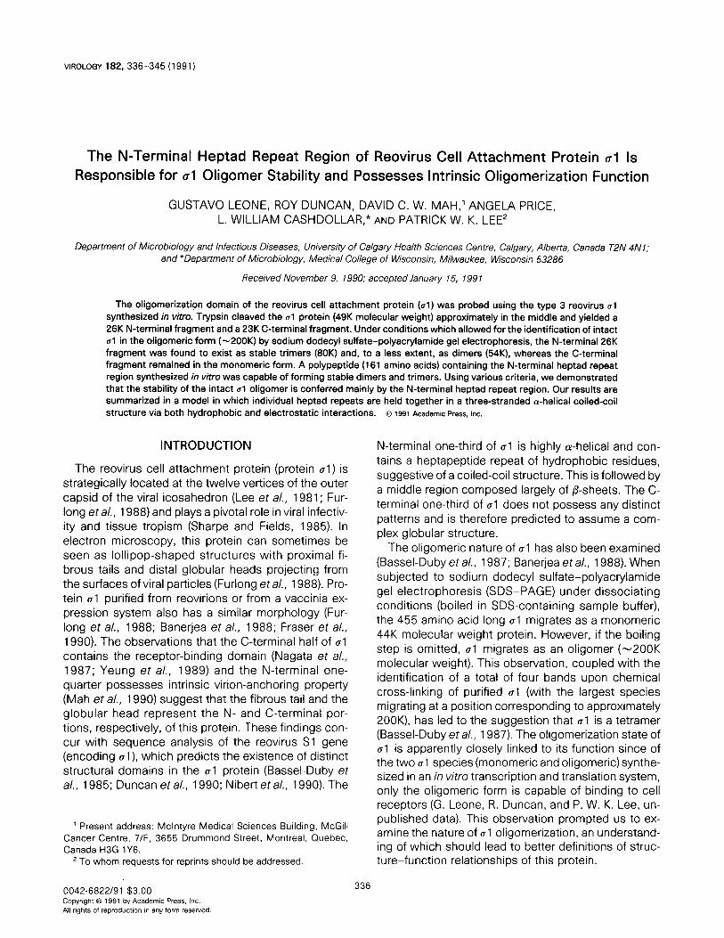
VIROLOGY 182,336-345 (1991)
The N-Terminal Heptad Repeat Region of Reovirus Cell Attachment Protein ~1 Is
Responsible for al Oligomer Stability and Possesses Intrinsic Oligomerization Function
GUSTAV0 LEONE, ROY DUNCAN, DAVID C. W. MAH,’ ANGELA PRICE, L. WILLIAM CASHDOLLAR,” AND PATRICK W. K. LEE*
Department of Microbiology and Infectious Diseases, University of Calgary Health Sciences Centre, Calgary, Alberta, Canada T2N 4N 1; and *Department of Microbiology, Medical College of Wisconsin, Milwaukee, Wisconsin 53286
Received November 9, 1990; accepted January 15, 199 1
The oligomerization domain of the reovirus cell attachment protein (01) was probed using the type 3 reovirus 01 synthesized in vitro. Trypsin cleaved the al protein (49K molecular weight) approximately in the middle and yielded a 26K N-terminal fragment and a 23K C-terminal fragment. Under conditions which allowed for the identification of intact ~1 in the oligomeric form (-200K) by sodium dodecyl sulfate-polyacrylamide gel electrophoresis, the N-terminal 26K fragment was found to exist as stable trimers (80K) and, to a less extent, as dimers (54K), whereas the C-terminal fragment remained in the monomeric form. A polypeptide (161 amino acids) containing the N-terminal heptad repeat region synthesized in v&o was capable of forming stable dimers and trimers. Using various criteria, we demonstrated that the stability of the intact al oligomer is conferred mainly by the N-terminal heptad repeat region. Our results are summarized in a model in which individual heptad repeats are held together in a three-stranded a-helical coiled-coil structure via both hydrophobic and electrostatic interactions. o 1991 Academic PVS, I~C.
INTRODUCTION
The reovirus cell attachment protein (protein ~1) is strategically located at the twelve vertices of the outer capsid of the viral icosahedron (Lee et al., 1981; Fur- long eta/., 1988) and plays a pivotal role in viral infectiv- ity and tissue tropism (Sharpe and Fields, 1985). In electron microscopy, this protein can sometimes be seen as lollipop-shaped structures with proximal fi- brous tails and distal globular heads projecting from the surfaces of viral particles (Furlong et a/., 1988). Pro- tein al purified from reovirions or from a vaccinia ex- pression system also has a similar morphology (Fur- long et a/., 1988; Banerjea et a/., 1988; Fraser et a/., 1990). The observations that the C-terminal half of al contains the receptor-binding domain (Nagata et al., 1987; Yeung et a/., 1989) and the N-terminal one- quarter possesses intrinsic virion-anchoring property (Mah et a/., 1990) suggest that the fibrous tail and the globular head represent the N- and C-terminal por- tions, respectively, of this protein. These findings con- cur with sequence analysis of the reovirus Sl gene (encoding (rl), which predicts the existence of distinct structural domains in the al protein (Bassel-Duby et a/., 1985; Duncan et al., 1990; Nibert et al., 1990). The
’ Present address: McIntyre Medical Sciences Building, McGill Cancer Centre, 7/F, 3655 Drummond Street, Montreal, Quebec, Canada H3G lY6.
’ To whom requests for reprints should be addressed.
N-terminal one-third of al is highly a-helical and con- tains a heptapeptide repeat of hydrophobic residues, suggestive of a coiled-coil structure. This is followed by a middle region composed largely of P-sheets. The C- terminal one-third of 01 does not possess any distinct patterns and is therefore predicted to assume a com- plex globular structure.
The oligomeric nature of al has also been examined (Bassel-Duby et al., 1987; Banerjea et a/., 1988). When subjected to sodium dodecyl sulfate-polyacrylamide gel electrophoresis (SDS-PAGE) under dissociating conditions (boiled in SDS-containing sample buffer), the 455 amino acid long al migrates as a monomeric 44K molecular weight protein. However, if the boiling step is omitted, al migrates as an oligomer (-200K molecular weight). This observation, coupled with the identification of a total of four bands upon chemical cross-linking of purified al (with the largest species migrating at a position corresponding to approximately 200K), has led to the suggestion that al is a tetramer (Bassel-Dubyetal., 1987). The oligomerization state of al is apparently closely linked to its function since of the two al species (monomeric and oligomeric) synthe- sized in an in vitro transcription and translation system, only the oligomeric form is capable of binding to cell receptors (G. Leone, R. Duncan, and P. W. K. Lee, un- published data). This observation prompted us to ex- amine the nature of al oligomerization, an understand- ing of which should lead to better definitions of struc- ture-function relationships of this protein.
0042-6822/91 $3.00 CopyrIght 0 1991 by Academic Press. Inc. All rights of reproductaon in any form reserved.
336
REPEAT REGION OF ul RESPONSIBLE FOR OLIGOMER STABILIW 337
In this report, we used trypsin treatment as an initial step to identify regions that are important for maintain- ing and stabilizing the al oligomeric structure. Of the two fragments generated by such treatment (Yeung et a/., 1989), only the N-terminal fragment, but not the C-terminal fragment, was found to exist as stable trimers (and dimers to a less extent) upon SDS-PAGE analysis. Subsequent in vitro transcription and transla- tion experiments revealed that the N-terminal one-third of al, which harbors the heptapeptide repeat region, possesses intrinsic dimerization and trimerization function. We further demonstrated that ionic interac- tions, in addition to hydrophobic interactions, within the heptapeptide repeat region are also responsible for stabilizing the ul oligomer. These observations have led to a structural model of the oligomerization domain of protein al.
MATERIALS AND METHODS
Plasmid construction
The plasmid (pG4T3) used in the present study was derived from our prokaryotic Sl -gene expression vec- tor pSP4 (Masri et al., 1986) in which the Sl gene con- tained additional sequences at the 3’-end derived from pBR322 (Pstl-EcoRI) during the original subcloning procedure. This plasmid was cleaved with Sstll, which cuts the Sl gene at position 1397, and the synthetic linker
5’-GGCACTGGGGCAT-FTCATCGGTAC-3’
3’-CGCCGTGACCCCGTAAAGTAGC-5’
was added which contained the authentic 3’-terminal Sl gene sequence from the Sstll site to the 3’-end of the gene where a unique Kpnl site was added. A BarnHI-Kpnl fragment was isolated (from the BarnHI site at position 15, which cuts immediately after the ATG initiation codon of al to the newly introduced Kpnl site at the 3’-end of the gene) and ligated to an EcoRI- BamHl translation initiation linker (Pharmacia):
5’-AATTCGGAGGAAAAAAT-TATG-3’
3’-GCCTCCT-llTTTAATACCTAG-5’.
The resultant EcoRI-Kpnl fragment was cloned into the EcoRI-Kpnl site of pGEM-4 (Promega Biotec) to produce pG4T3.
In vitro transcription-translation
The plasmid pG4T3 was linearized with either HindIll or EcoRV restriction endonucleases and used in in vitro transcription reactions to generate full-length or trun- cated Sl mRNAs. The mRNAs were then translated in
vitro in rabbit reticulocyte lysates according to the man- ufacturer’s specifications (Promega). A typical transla- tion mixture contained 50-l 00 ng of RNA and 20 &i of [35S]methionine in a total volume of 25 ~1. After incuba- tion at 37” for 45 min, reactions were stopped by the addition of 200 ~1 phosphate-buffered saline (PBS). Protein sample buffer (5x) was then added (final lx concentration was 50 mn/r Tris, pH 6.8, 1% SDS, 2% P-mercaptoethanol, 10% glycerol, and 0.01% brom- phenol blue) and the mixtures were further incubated at 37” for 30 min (nondissociating condition) or boiled for 5 min (dissociating condition) prior to SDS-PAGE.
Trypsin digestion
Upon completion of the in vitro translation reaction, TLCK-treated trypsin (Sigma) dissolved in PBS (0.05 mg/ml) was added to the translation mixtures to a final concentration of 0.005 mg/ml (unless otherwise stated). After incubation at 37” for 30 min, trypsin inhib- itors (soybean and egg white trypsin inhibitors, Sigma) were added and the mixtures were further incubated in protein sample buffer prior to SDS-PAGE (see above).
lmmunoprecipitation
The monoclonal anti-al antibody G5 has been previously described (Burstin et al., 1982) and shown to interact with the C-terminal tryptic fragment of 01 (Yeung et a/., 1989). The N-terminal specific polyclonal anti-u1 antiserum was prepared in rabbits using the SDS-PAGE-purified trpE-al fusion protein (containing trpE and residues l-l 58 of al) expressed in Esche- richia co/i using the pATH3 vector (Cashdollar et a/., 1989). Aliquots of lysates or trypsin-treated lysates were mixed with an equal volume of appropriate dilu- tions of the antibodies and incubated at room tempera- ture for 1 hr. Fixed Staphylococcus aureus (10% sus- pension) that had been preadsorbed with BSA (5 mg/ ml) was then added to the mixture and incubated for an additional 30 min. lmmunoprecipitates were washed three times with wash buffer (50 mMTris, pH 7.4, 150 mM NaCI, 0.1% SDS, 1% Triton X-l 00) resuspended in 140 ~1 high-pH buffer (50 mM Tris, 0.1 M H,PO,, 2 mM DTT, 0.1% SDS, 6 M urea, pH to 1 1.6 with NaOH), and incubated at 37” for 45 min. Suspensions were then pelleted and supernatants neutralized with 7 ~1 neutralizing solution (0.1 M H,PO,, 1 n/r Tris, pH 7.4). Protein sample buffer was then added to the samples and either incubated at 37” for 30 min (nondissociating condition) or boiled for 5 min (dissociating condition) prior to SDS-PAGE.
SDS-PAGE
Discontinuous SDS-PAGE was performed using the protocol of Laemmli (1970). Both 10 and 12.5% acryl-
338 LEONE ET AL
xc-,
lJ[ailb
12345
FIG. 1. Detection of oligomeric forms of in vitro-translated ~1 and soluble ~1 from reovirus-infected cells. [35S]Methionine-labeled reo- virus-infected cell lysates (S-45) prepared as previously described (Lee er a/., 1981) and [%]methionine-labeled in vitro translation products of the reovirus Sl mRNA in rabbit reticulocyte lysates (Sp6), were precipitated with an anti-01 monoclonal antibody (G5). After being released from the immunoadsorbent, precipitated pro- teins were mixed with protein sample buffer and were either boiled for 5 min (6) or incubated at 37” for 30 min (37) prior to SDS-PAGE. R (lane 1) represents purified [?S]methionine-labeled reovirus.
amide gels were used. Gels containing 35S-labeled pro- teins were fixed and then treated with DMSO-PPO, dried under vacuum, and exposed to Kodak XAR-5 film at -70”.
Gels to be used for band excision were dried under vacuum without prior treatment, and then exposed to film. Developed x-ray film was superimposed on dried gels and bands to be excised were marked with a pouncer. Marked protein bands were excised from gels and rehydrated in Laemmli running buffer. Pro- teins from excised bands were electroeluted into elec- troelution cups in a volume of 200 jJ. Protein sample buffer was added to electroeluted proteins and boiled for 5 min prior to SDS-PAGE.
RESULTS
Generation of al oligomers in vitro
Since some of the following studies on al oligomeri- zation involved the use of genetically truncated protein CT~ , it was necessary to express ~1 in an in vitro system and to ascertain that oligomeric al was indeed gener- ated in such a system. To this end, T3 reovirus Sl mRNA was prepared in vitro using Sp6 RNA polymer- ase and translated in a rabbit reticulocyte lysate. Translation products were then immunoprecipitated with an anti-al antibody (G5) and analyzed by SDS- PAGE under conditions that would not cause the disruption of ~1 oligomers (see Materials and Meth- ods). The results, illustrated in Fig. 1, show that stable ~1 oligomers were indeed produced in vitro (lanes 4 and 5) and their migration rate was identical to that of
authentic ~1 from T3 reovirus-infected cells (lanes 2 and 3). Like the authentic protein, al oligomers synthe- sized in vitro were capable of binding to cell receptors (R. Duncan, G. Leone, and P. W. K. Lee, unpublished observations), and were cleaved by trypsin to yield a well-defined pattern (see below). Some al monomers (44K) were also produced in this system, but they were not precipitable by the anti-al antibody, did not mani- fest cell-binding function, and were highly susceptible to degradation by trypsin or chymotrypsin even when these proteases were used at very low concentrations (data not shown). Subsequent analysis exclusively of the oligomeric form of ~1 was therefore possible.
Trypsin digestion of ~1 oligomers
Previously it was found that trypsin cleaves the ~1 oligomer near the middle to generate two fragments of approximately equal size (Banerjea et al., 1988; Yeung et a/., 1989). The cleavage site has now been deter- mined to be between Arg245 and lle246 (Duncan and Lee, unpublished data). Thus the monomeric forms of the N-terminal fragment (245 amino acids) and the C- terminal fragment (210 amino acids) have molecular weights of approximately 26K and 23K, respectively, corresponding to their migration rates in SDS-PAGE under dissociating conditions (Yeung et a/., 1989). Such a cleavage pattern was also obtained with al synthesized in vitro (Fig. 2, lane 3). An additional minor band of approximately 21 K molecular weight was also observed sometimes. Using N- and C-terminal-specific
R37B
xc -
kJb+-- -8OK
- -!54K *[ ST&*’ -48K
4--izKK --21K
1 2 3
FIG. 2. Trypsin digestion of protein al. In virro-translated [%]me- thionine-labeled 81 protein was digested with 0.005 mg/ml of trypsin as described under Materials and Methods. After the addition of trypsin inhibitors and protein sample buffer, samples were either incubated at 37“ for 15 min (37) or boiled for 5 min (B) prior to SDS- PAGE. R (lane 1) represents purified [35S]methionine-labeled reo- virus. Molecular weights of individual protein bands are indicated at right.

REPEAT REGION OF 01 RESPONSIBLE FOR OLIGOMER STABILITY 339
w.e!e- R 37 BmK54KaK
4BK -23K
32”~,-21K
123456
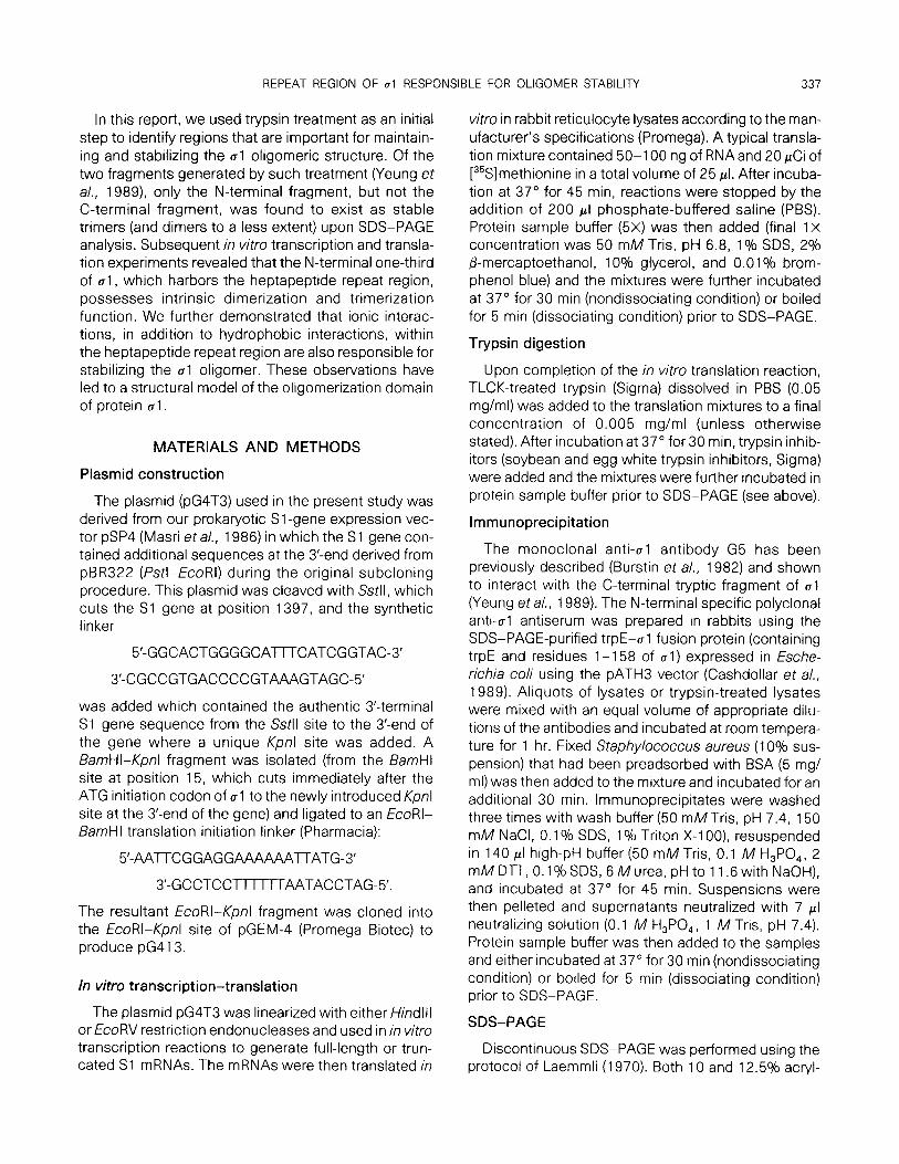
FIG. 3. SDS-PAGE of oligomeric tryptic fragments of ~1. Protein al tryptic digests prepared as described under Materials and Meth- ods were either incubated at 37” for 15 min (37) or boiled for 5 min (B) in protein sample buffer and then subjected to SDS-PAGE. The 80K, 54K, and 48K fragments from the unboiled (37) sample were excised from the gel, eluted, boiled in protein sample buffer, and then reanalyzed by SDS-PAGE (lanes 4. 5, and 6, respectively). R (lane 1) represents purified [%]methionine-labeled reovirus. Molecu- lar weights of individual protein bands are indicated at right.
sera, we have identified the 26K and 21 K tryptic frag- ments to be of N-terminus origin, and the 23K fragment to be of C-terminus origin (Fig. 4).
N-terminal half of 01 is a trimer
To see whether any of the tryptic fragments could be identified in the oligomeric state, trypsin-treated al in SDS-containing sample buffer was incubated at 37” (rather than boiled) prior to SDS-PAGE. It was found that the 26K N-terminal fragment was replaced quanti- tatively by a band migrating at approximately 80K mo- lecular weight (Fig. 2, lane 2). The migration rate of the 23K C-terminal fragment remained unchanged. The 2 1 K N-terminal fragment was absent; instead two faint bands of approximately 48K and 54K molecular weight appeared. Protein bands of 80K, 54K, and 48K molecu- lar weight were excised from a gel, eluted, boiled in sample buffer, and subjected to SDS-PAGE (Fig. 3). Both the 80K and 54K proteins were found to be con- verted to the 26K N-terminal fragment, whereas the 48K protein was converted to the 2 1 K N-terminal frag- ment.
The most reasonable interpretation of these results would be that the 80K and 54K bands represent the trimer and dimer forms, respectively, of the 26K N-ter- minal fragment, and the 48K band represents the dimer of the 21 K N-terminal fragment. The region in the 26K N-terminal fragment that is absent in the 21 K N- terminal fragment may therefore be involved in stabiliz-
ing the third subunit of the 80K N-terminal trimer. The identity of this region is presently unknown.
Titration of trypsin digestion of 01 oligomers
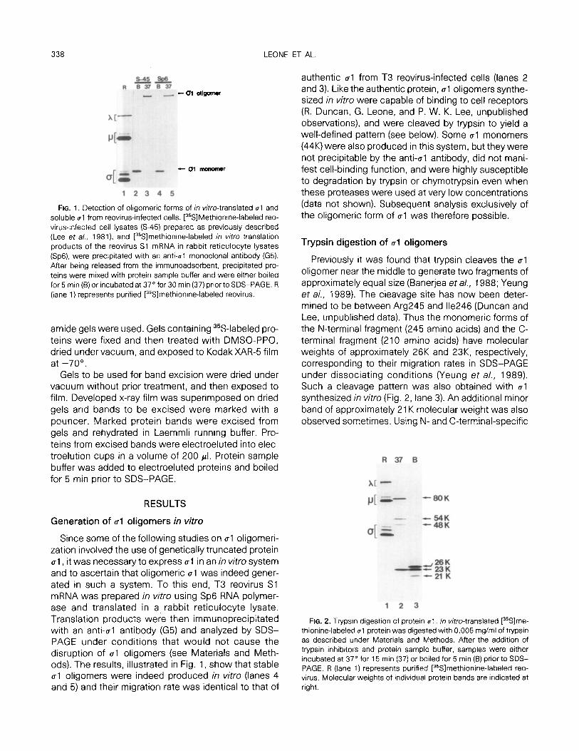
It is important to note that under conditions where the N-terminal tryptic fragment exists as stable trimers (and dimers), the C-terminal fragment was consistently found to be in the monomeric state. This is in contra- diction to the findings of Banerjea et a/. (1988) who reported that both the N- and C-terminal tryptic frag- ments exist as stable oligomers (tetramers) in SDS at 37”. To reconcile such differences, we decided to ex- amine the effects of varying the trypsin concentration on al cleavage pattern. At the lowest concentration (0.005 mg/ml) of trypsin that cleaves al oligomers com- pletely, an identical pattern to that shown in Fig. 2 was observed (Fig. 4A, lane 2). Increasing the concentra- tion of trypsin to 0.05, 0.1, and 0.5 mg/ml, and subse- quently boiling the samples in sample buffer, resulted in the gradual disappearance of the 26K N-terminal fragment with the concomitant appearance, in almost stoichiometric amounts, of a 25K molecular weight band (Fig. 4A, lanes 3-5). Thus the 25K band is a cleav- age product of the 26K band. The 23K C-terminal frag- ment remained unchanged.
When identical samples were instead incubated at 37” in sample buffer prior to SDS-PAGE, both the 26K and 25K bands were replaced by bands with molecular weights of approximately 80K and 75K, respectively (Fig. 4A, lanes 6-9). Again, the mobility of the 23K C- terminal fragment remained unchanged. Radioimmu- noprecipitation of the samples with the N- and C-ter- minus-specific antibodies confirmed that the 25K and 75K proteins, like the 26K and 80K proteins, were of N-terminus origin (Fig. 48) and that the 23K protein was of C-terminal origin (Fig. 4C).
It is possible to explain the discrepancies between our present findings and those reported by Banerjea et al. (1988) on the basis of the methods used for protein al detection. A polyclonal anti-T3 reovirus serum was used by the aforementioned investigators to identify ~1 tryptic fragments on Western blots as opposed to radioimmunoprecipitation with anti-al N- and C-termi- nal-specific antibodies used in our studies. In our hands, polyclonal anti-native al serum is incapable of recognizing the presumably denatured C-terminal tryp- tic fragment on a blot, although the N-terminal frag- ment is easily detectable. These observations, to- gether with the fact that the trypsin concentration (1 mg/ml) used by Banerjea et al. was within the concen- tration range where we found both the 26K and the 25K N-terminal cleavage products, have led us to conclude that the two bands previously identified by these inves-

340 LEONE ET AL.
A R Boil 3x B
R Boil 37c C R Bail 37 %
xc m
d =%‘I: rr---EE!- +%!fi p[ *
c*- *I -48K -48K
FIG. 4. Titration of trypsin digestion of ~1 oligomers. ln vitro-translated [35S]methionine-labeled ~1 was digested with various concentrations [0.005 mg/ml (lanes 2 and 6) 0.05 mg/ml (lanes 3 and 7) 0.1 mg/ml (lanes 4 and 8) and 0.5 mg/ml (lanes 5 and 9)] of trypsin. Tryptic digests were either subjected to SDS-PAGE directly (A), or immunoprecrpitated with the anti-u1 N-terminal-specific serum (B), or with the anti-cl C-terminal-specific antibody (C) prior to SDS-PAGE. All samples were either boiled for 5 min (Boil) or incubated at 37” for 15 min (37’) in protein sample buffer before electrophoresis. R (lane 1) represents purified reovirus. Molecular weights of individual bands are indicated at right.
tigators as N-terminal and C-terminal oligomers corre- spond to our 80K and 75K bands, and are therefore in fact both N-terminal oligomers.
Role of the N-terminal portion in stabilizing ~1 oligomers
To determine the extent of involvement of the N-ter- minal portion in stabilizing the al oligomer, native al and trypsin-treated al were subjected to various treat- ments prior to SDS-PAGE and the relative stability of native al oligomers and N-terminal trimers (80K pro- tein) was compared. Whereas dissociation of oligo- mers by heat was used as a general measure of oligo- mer stability, the effects of pH variation and presence or absence of urea and @-mercaptoethanol were also examined.
Under all conditions tested, native al oligomers and N-terminal trimers behaved identically (Fig. 5). Both al oligomers and N-terminal trimers were stable at 50” in sample buffer but dissociated at 60” (Fig. 5A). Both were stable under alkaline (pH 1 1.6) to mild acidic (pH 6.0) conditions, but dissociated at pH 5.5 (Fig. 5B). Urea (6 IV) apparently had no effect on either oligo- merit species (Fig. 5C). It was previously reported that al oligomers were rendered less stable when the con- centration of P-mercaptoethanol in the sample buffer was reduced (Bassel-Duby et a/., 1987). Consistent with these findings, we observed that both native al oligomers and N-terminal trimers were less stable in the total absence of P-mercaptoethanol (Fig. 5C).
In summary, the stability of the N-terminal trimer was found to be very similar to that of the native al oligo- mer. These data suggest that interactions between al subunits responsible for stabilizing the oligomeric
structure occur mainly, although by no means solely, within the N-terminal half of the al protein.
Involvement of ionic interactions within the N-terminal half of al
The absence of cysteine residues within the N-ter- minal tryptic fragment of al suggests that the stabiliz- ing effect of P-mercaptoethanol could not be directly due to its reducing properties. Indeed, enhanced oligomer stability was not observed when another re- ducing agent, dithiothreitol, was used in place of ,& mercaptoethanol (Fig. 6A).
A less well-characterized property of P-mercap- toethanol is that of chelation (McMichael and Ou, 1977). If /3-mercaptoethanol indeed stabilizes oligo- mers by chelating divalent cations, addition of divalent cations should destabilize oligomers. Indeed, MgCI, at concentrations above 1 mM was found to destabilize N-terminal trimers (Fig. 6B) as well as native al oli- gomers (data not shown). Other divalent cations (zinc and calcium) had a similar effect (data not shown). The effects of divalent cations and P-mercaptoethanol on oligomer stability are clearly antagonistic. Thus the destabilizing effect of 5 mM and 20 ml\/l MgCI, could be reversed by the inclusion of 1 and 5% ,L3-mercap- toethanol, respectively, in the sample buffer (Fig. 6B). Similar results were obtained using EDTA as the chela- tor in place of ,&mercaptoethanol, except that at EDTA concentrations above 5 mM, oligomer stability was in- consistent (data not shown).
The disruption of ~1 oligomers by divalent cations suggests that ionic interactions are involved in stabiliz- ing the N-terminus trimer (and hence the 01 oligomer). Indeed, the distribution of charged residues in the N- terminal coiled-coil region was found to highly favor the

REPEAT REGION OF 01 RESPONSIBLE FOR OLIGOMER STABILITY 341
A TEMP. B PH
C lJ=AIp+J=
naltii trypsin natiw trypdl -- -- native trypsin
375060 37!5060 tL56D50n66051) lJ+p +u -6 -u+B +u -6
w- -w --A -uloligomer
-- WC -- - “itEY
--- --al monomer
t --- WC --- --l N-%zF c-w
FIG. 5. Oligomer stability of intact 01 and of 01 tryptic fragments undervarious conditions. (A) Trypsin-treated (trypsin) and untreated (native) ~1 were incubated in protein sample buffer at 37” (37) 50” (50) or 60” (60) for 30 min prior to SDS-PAGE. (B) Samples were diluted threefold with 50 m/l/rTris buffer at the pHs (1 1.6, 6.0, and 5.0) indicated and incubated at room temperature for 15 min. Protein sample bufferwas then added to the samples which were then further incubated at 37” for 30 min prior to electrophoresis. (C) Samples were incubated in regular protein sample buffer which contained 2% fl-mercaptoethanol (-U+p, control), or in protern sample buffer containing 6 M urea in addition to 2% P-mercaptoethanol (+U), or in protein sample buffer lacking both urea and P-mercaptoethanol (-p), at 37” for 30 min prior to electrophoresis. Native and trypsin lanes in each experiment originate from the same gel.
formation of salt bridges between al subunits (see Discussion). However, the presence of the extended heptad repeat in the same region suggests that hydro- phobic interactions must play a major role in oligomer stabilization. In this regard, the aforementioned ionic interactions are presumed to serve an augmentative function. In agreement with the above hypothesis, tem- perature stability experiments indicated that in the presence of divalent cations (abolishing ionic interac-
tions), oligomers were stable up to a temperature of 20”, but in the absence of divalent cations (maximizing ionic interactions), oligomers were stable up to a tem- perature of 50” (data not shown).
Intrinsic dimerization and trimerization properties of the heptad repeat region of al
It is believed that sequences with heptapeptide re- peats of apolar residues are involved in stabilizing
A I)-me% DlTmM
012515lo
B &me %: 0 1 5
M~mM:ODl1 52Doa1 52020
- r) - - - - c N-timer -.- ---- C- N-timer
FIG. 6. Stability of the N-terminal tryptic trimer in the presence of @-mercaptoethanol, dithiothreitol (DTT), and Mg2+. (A) Trypsin-treated ~1 was Incubated in protean sample buffer containing either fl-mercaptoethanol or DTT at concentrations indicated for 30 min at 37” prior to SDS-PAGE. (B) MgCI, was added to trypsin-treated ~1 to the final concentrations indicated. Samples were then immediately incubated in protein sample buffer containing 0. 1, and 5% @-mercaptoethanol for 30 min at 37” prior to electrophoresis.

342 LEONE ET AL
R 8 37
XI- pc-
I c trimer 54K
a[= arcdimer 38K
-monomer 18K
1 2 3
FIG. 7. SDS-PAGE of an in vitro-translated al polypeptide contain- ing the N-terminal heptapeptide repeat. mRNA encoding the first 161 amino acids of 01 was made and translated as described under Materials and Methods. After the addition of protein sample buffer, the samples were either boiled for 5 min (B) or incubated at 37” for 30 min (37) prior to electrophoresis. R represents the reovirus marker. Molecular weights of individual proteins bands are indicated at right.
coiled-coil structures through hydrophobic interac- tions between the a-helices. In the case of al, the hep- tad repeat region spans the N-terminal one-third of the protein (from residue 28 to 164). It was then of interest to see whether this region possesses intrinsic dimeri- zation and, in view of the trimeric nature of the N-ter- minal tryptic fragment of ~1, trimerization functions.
To this end, the plasmid pG4T3 was treated with the restriction endonuclease EcoRV, which cuts the Sl gene at nucleotide 497 (encoding the N-terminal 161 amino acids). Run-off mRNA transcripts were then pre- pared and translated in vitro, and the ability of the translational products to oligomerize was then deter- mined by SDS-PAGE. The results are shown in Fig. 7. When the samples were boiled in sample buffer prior to SDS-PAGE, closely migrating bands of approximately 18K molecular weight were found (Fig. 7, lane 2), as was predicted from the amino acid sequence. The lack of absolute homogeneity in size of the translational products was due to the absence of a translation stop codon in the mRNA transcripts, necessitating transla- tion termination to occur by the falling off of ribosomes close to, but not precisely at, the 3’ends of the mRNAs. When identical samples were instead incubated at 37” in sample buffer (Fig. 7, lane 3), there was a noticeable decrease in the intensities of the bands migrating at approximately 18K. Concomitantly, two additional bands with estimated molecular weights of 36K and 54K appeared. These two bands corresponded to the dimeric (36K) and trimeric (54K) forms of the EcoRV translation products. Clearly, the dimer was the pre- dominant oligomeric form identified. Whether trimer formation was inefficient or whether trimers were less stable in our SDS-PAGE system is not known. None-
theless, for the first time, direct evidence is presented that demonstrates the intrinsic ability of a heptad re- peat (18 repeats in this case) to form dimers and trimers, an ability that may very well depend on the number of repeats present in the polypeptide.
Heptad repeat imparts oligomer stability
To determine the extent to which the heptad repeat region is involved in stabilizing the N-terminal tryptic trimers, the EcoRV translational products were sub- jected to the various treatments previously applied to the N-terminal tryptic fragment and the native ~1 oligomer. The results are shown in Fig. 8. Both the dimeric and trimeric forms of the heptad repeat were unstable at 50”, a temperature at which the N-terminal tryptic trimer was found to be stable. However, like the N-terminal trimer, heptad dimers and trimers were both stable under alkaline (pH 11.6) to mild acidic (pH 6.0) conditions (but dissociated at pH 5.5) and in 6 M urea, and were destabilized by the absence of P-mercap- toethanol or by the presence of divalent cations in the sample buffer. Again, as observed for the N-terminal tryptic trimer, the destabilizing effect of divalent cations could be neutralized by P-mercaptoethanol.
DISCUSSION
The reovirus cell attachment protein, al, is a ho- mooligomer and has a lollipop morphology. The recep- tor-binding domain of al resides in the C-terminal glob- ular head and the virion-anchoring domain resides in the N-terminal fibrous tail. Sequence analysis of the Sl genes of all three reovirus serotypes concur with such structural and functional domain assignments. The ob-
-
.- .,
iu
I xx_ -- -trimer [SK]
rrrm urn au -dimer [MK]
FIG. 8. Oligomer stability of the N-terminal 161 amino acid long polypeptide. The polypeptide synthesized was subjected to various treatments as described in the legends to Fig. 5 and 6. Protein sam- ple buffer was then added and the samples were incubated at 37” for 30 min prior to SDS-PAGE. Reo represents the reovirus marker. Molecular weights are indicated at right.

REPEAT REGION OF al RESPONSIBLE FOR OLIGOMER STABILITY 343
servation that receptor-binding function is manifested by the oligomeric form, but not by the monomeric form of al, prompted us to examine the region(s) on ~1 that is responsible for the stability and the formation of the ~rl oligomer. Our present analysis of the two tryptic fragments of al has revealed that the stability of the ~1 oligomer is maintained mainly via the N-terminal fi- brous portion, rather than the C-terminal globular por- tion of al. Under nondissociating conditions (preincu- bation in SDS-containing sample buffer at 37” for 30 min), the N-terminal half of al, like intact ~1, migrates in SDS-PAGE as an oligomer (trimer), whereas the C- terminal half migrates as a monomer. We have evi- dence, however, that the latter ttyptic fragment is also a trimer (albeit a less stable one) which dissociates under the assay conditions used in the present study. The additional observation that both the N-terminal tryptic fragment and the full-length 01 oligomer re- spond to temperature and pH changes, and to p-mer- captoethanol in a similar manner, further suggests that the N-terminal half of al plays a major role in stabilizing the ul oligomer.
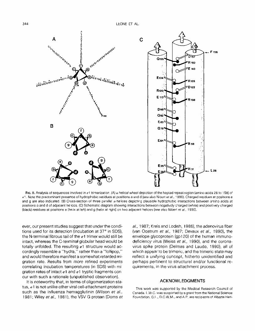
That the N-terminal portion of al possesses oligo- merization potential was first suggested by Bassel- Duby eta/. (1985) from sequence analysis of the type 3 reovirus Sl gene. First of all, the N-terminal one-third of al was found to be highly a-helical. The additional pres- ence, in the same region, of an extended heptad re- peat (a-b-c-d-e-f-g)n, where a and d are character- istically apolar residues, further indicates the propen- sity of this region to adopt a coiled-coil rope-like structure. Such theoretical considerations are clearly compatible with the fibrous morphology of al as re- vealed by electron microscopy (Furlong et al., 1988; Banerjea et al., 1988; Fraser et a/., 1990) and with the present demonstration that the heptad repeat region alone, when synthesized in an in vitro system, is capa- ble of forming dimeric and trimeric structures whose stability is remarkably similar to that of the trimeric N- terminal tryptic fragment or of the native al oligomer. In view of the recent speculations on the structural and functional aspects of the “leucine zipper,” we con- sider our direct demonstration that the heptad repeat possesses intrinsic dimerization and trimerization function to be significant (see also note added in proof). There is little doubt that, in the case of the trimer, apolar residues at positions a and d contribute to hydrophobic interactions that are responsible for holding the three strands together in a coiled-coil con- figuration (Figs. 9A and 9B). In addition, ionic interac- tions were also found to play a role since the dimers and trimers were destabilized by divalent cations but were restabilized by the addition of a chelator. Indeed, an examination of the distribution of charged residues
in positions e and g reveals that when the three cu-he- lices are placed in parallel and in register to each other, residues of opposite charge on adjacent cu-helices in- variably lie in juxtaposition (Fig. 9C) (see also Nibert et a/., 1990). At locations where a corresponding residue of opposite charge is not present on an adjacent a-he- lix, a spatially close residue on the same helix is pres- ent to neutralize the charge. Analysis of analogous re- gions on the al proteins of the other two serotypes reveals a similar, albeit less perfect, distribution pattern of charge residues (data not shown).
It is important to point out that whereas the present study clearly indicates that the 01 oligomer is stabilized mainly via the N-terminal half of the protein, the impli- cations of the intrinsic oligomerizing function of the heptad repeat region on the oligomerization process of the intact al oligomer need to be viewed with caution. Recent evidence suggests that intracellular protein folding and oligomerization are mediated by chaper- ones and that these events are ATP-dependent. In the case of ~1, we have recently observed that whereas the oligomerization of the N-terminal one-third of ~1 occurs spontaneously, that of full-length al is an ATP- dependent event (unpublished data), which further suggests that, as has been reported for a number of oligomeric proteins, the formation of the al oligomer is chaperone-mediated. This would in turn imply that the intrinsic oligomerizing function of the heptad repeat re- gion is necessary, but not sufficient, for intact al oligo- merization, and that a domain(s) downstream of this region must also be involved. We are currently probing the interactions between the three governing factors, namely, the heptad repeat region, a downstream do- main(s), and chaperones, in the al oligomerization pro- cess, the revelation of which may have general and fundamental implications.
Our demonstration that the N-terminal tt-yptic frag- ment is a trimer and the N-terminal heptad repeat re- gion possesses intrinsic trimerization function sug- gests that native protein al is most likely also a trimer, although the possibility of al being a multiple of a trimer (e.g., dimer of a trimer) cannot be ruled out based on the present data alone. Either model contra- dicts a recent suggestion, based on sequence analysis and computer-processed electron microscopy, that ~1 is a tetramer (Fraser et al., 1990). Although the precise reasons for this discrepancy remain to be revealed, we have recently obtained data from biophysical studies and in vitro ~1 assembly experiments that are compati- ble with a trimeric, but not a tetrameric (or a hexa- merit), model of al (unpublished data). A trimeric ~1 would theoretically migrate at a position corresponding to 150K in SDS-PAGE, rather than at -2OOK. How-
344 LEONE ET AL.
0155
FIG. 9. Analysis of sequences involved in 01 trimerization. (A) a-helical wheel depiction of the heptad repeat region (amino acids 28 to 158) of ~1. Note the predominant presence of hydrophobic residues at positions a and d (see also Nibert et al., 1990). Charged residues at positions e and g are also indicated. (B) Cross-section of three parallel a-helices depicting plausible hydrophobic interactions between amino acids at positions a and d of adjacent helices. (C) Schematic diagram showing interactions between negatively charged (white) and positively charged (black) residues at positions e (helix at left) and g (helix at right) on two adjacent helices (see also Nibert et a/., 1990).
ever, our present studies suggest that under the condi- tions used for its detection (incubation at 37” in SDS), the N-terminal fibrous tail of the al trimer would still be intact, whereas the C-terminal globular head would be totally unfolded. The resulting ~1 structure would ac- cordingly resemble a “hydra,” rather than a “lollipop,” and would therefore manifest a somewhat retarded mi- gration rate. Results from more refined experiments correlating incubation temperatures (in SDS) with mi- gration rates of intact al and al tr-yptic fragments con- cur with such a rationale (unpublished observation).
It is noteworthy that, in terms of oligomerization sta- tus, ~1 is not unlike other viral cell-attachment proteins such as the influenza hemagglutinin (Wilson et a/., 1981; Wiley et a/., 1981) the VSV G protein (Doms et
a/., 1987; Kreis and Lodish, 1986) the adenovirus fiber (van Oostrum et a/., 1987; Devaux et al., 1990) the envelope glycoprotein (gpl20) of the human immuno- deficiency virus (Weiss et al., 1990) and the corona- virus spike protein (Delmas and Laude, 1990) all of which appear to be trimeric, and the trimeric state may reflect a unifying concept, hitherto unidentified and perhaps pertinent to structural and/or functional re- quirements, in the virus attachment process.
ACKNOWLEDGMENTS
This work was supported by the Medical Research Council of Canada. L.W.C. was supported by a grant from the National Science Foundation. G.L., D.C.W.M., and A.P. are recipients of Alberta Heri-
REPEAT REGION OF al RESPONSIBLE FOR OLIGOMER STABILITY 345
tage Foundation for Medical Research (AHFMR) Studentships. R.D. is an AHFMR Fellow and P.W.K.L. is an AHFMR Scholar.
Note added in proof. Shortly after submission of our manuscript, Banerjea and Joklik (1990) reported the intrinsic oligomerizing prop- erty of the N-terminal heptad repeat region of 01.
REFERENCES
BANEFUEA. A. C., BRECHLING, K. A., RAY, C. A.. ERICKSON, H., PICKUP, D. T., and JOKLIK, W. K. (1988). High-level synthesis of biologically active reovirus protein 01 in a mammalian expression vector sys- tem. firology 167, 601-612.
BANERIEA, A. C., and JOKLIK, W. K. (1990). Reovirus protein ~1 trans- lated in vitro, as well as truncated derivations of it that lack up to two-thirds of its C-terminal portion, exists as two major tetrameric molecular species that differ in electrophoretic mobility. Virology 179, 460-462.
BASSEL-DUBY, R., JAYASURIYA, A., CHA~ERJEE, D., SONENBERG, N., MAIZEL. J. V., JR., and FIELDS, B. N. (1985). Sequence of reovirus hemagglutinin predicts a coiled-coil structure. Nature (London) 315,421-423.
BASSEL-DUBY, R., NIBERT, M. L., HOMCY, C. J., FIELDS, B. N., and SA- WUTZ, D. G. (1987). Evidence that the ~1 protein of reovirus sero- type 3 is a multimer. J. Viral. 61, 1834-l 841.
BURSTIN. S., SPRIGGS, D. R., and FIELDS, B. N. (1982). Evidence for functional domains on the reovirus type 3 hemagglutinin. Virology 117,146-155.
CASHDOLIAR, L. W., BLAIR, P., and VAN DYNE, S. (1989). Identification of the ~1 S protein in reovirus serotype 2-infected cells with anti- body prepared against a bacterial fusion protein. virology 168, 183-l 86.
DELMAS, B., and LAUDE, H. (1990). Assembly of coronavirus spike protein into trimers and its role in epitope expression. /. Viral. 64, 5367-5375.
DEVAUX, C.. ADRIAN, M., BERTHET-COLOMINAS, C., CUSACK, S., and JACROT, B. (1990). Structure of adenovirus fibre. I. Analysis of crys- tals of fibre from adenovirus serotypes 2 and 5 by electron micros- copy and x-ray crystallography. J. Mol. f3iol. 215, 567-588.
DOMS, R. W., KELLER, D. S., HELENIUS, A., and BALCH, N. E. (1987). Role for adenosine triphosphate in regulating the assembly and transport of vesicular stomatitis virus G protein trimers. J. Ce//Bio/. 105, 1957-1969.
DUNCAN, R., HORNE, D., CASHDOLLAR. L. W., JOKLIK, W. K., and LEE, P. W. K. (1990). Identification of conserved domains in the cell attachment proteins of the three serotypes of reovirus. Virology 174,399-409.
FRASER, R. D. B., FURLONG, D. B., TRUS, B. L., NIBERT, M. L., FIELDS, B. N., and STEVEN, A. C. (1990). Molecular structure of the cell-at- tachment protein of reovirus: Correlation of computer-processed
electron micrographs with sequence-based predictions. /. Viral. 64,2990-3000.
FURLONG, D. B., NIBERT, M. L.. and FIELD, B. N. (1988). Sigma 1 protein of mammalian reoviruses extends from the surfaces of viral particles. J. viral. 62, 246-256.
KREIS, T. E., and LODISH, H. F. (1986). Oligomerization is essential for transport of vesicular stomatitis viral glycoproteins to the cell sur- face. Cell 46, 929-937.
LAEMMLI, U. K. (1970). Cleavage of structural proteins during the assembly of the head of bacteriophage T4. Nature (London) 227, 680-685.
LEE, P. W. K., HAYES, E. C., and JOKLIK, W. K. (1981). Protein 01 is the reovirus cell attachment protein. Virology 108, 156-l 63.
MAH, D. C. W., LEONE, G., JANKOWSKI, J. M., and LEE, P. W. K. (1990). The N-terminal quarter of reovirus cell attachment protein al pos- sesses intrinsic virion-anchoring function. Virology 179, 95-l 03.
MASRI, S. A., NAGATA, L., MAH, D. C. W., and LEE, P. W. K. (1986). Functional expression in fscherichia co/i of cloned reovirus Sl gene encoding the viral cell attachment protein al. Virology 149, 83-90.
MCMICHAEL, J. C., and Ou, J. T. (1977). Metal ion dependence of a heat-modifiable protein from the outer membrane of fscherichia co/i upon sodium dodecyl sulfate-gel electrophoresis. /. Bacterial. 132,314-320.
NAGATA, L., MASRI, S. A., PON, R. T., and LEE, P. W. K. (1987). Analy- sis of functional domains on reovirus cell attachment protein ~1 using cloned Sl gene deletion mutants. Virology 160, 162-l 68.
NIBERT, M. L., DERMODY. T. S., and FIELDS, B. N. (1990). Structure of the reovirus cell attachment protein: A model for the domain orga- nization of ~1. /. Viral. 64, 2976-2989.
SHARPE, A. H., and FIELDS, B. N. (1985). Pathogenesis of viral infec- tions. Basic concepts derived from the reovirus model. N. Engl. /. Med. 312, 486-497.
VAN OOSTRUM, J., SMITH, P. R., MOHRAZ, M., and BURNETT. R. M. (1987). The structure of the adenovirus capsld. Ill. Hexon packing determined from electron micrographs of capsid fragments. 1. Mol. Biol. 198, 73-89.
WEISS, C. D., LEVY, J. A., and WHITE, J. M. (1990). Oligomeric organiza- tion of gpl20 on infectious human immunodeficiency virus type 1 particles. J. Virol. 64, 5674-5677.
WILEY, D. C., WILSON, I. A., and SKEHEL. J. J. (1981). Structural identifi- cation of the antibody-binding sites of Hong Kong influenza hemag- glutinin and their involvement in antigenic variation. Nature (Lon- don) 289, 373-378.
WILSON, I. A., SKEHEL, J. J., and WILEY, D. C. (1981). Structure of the hemagglutinin membrane glycoprotein of influenza virus at 3A res- olution. Nature (London) 289, 368-373.
YEUNG, M. C., LIM, D., DUNCAN, R., SHAHRABADI. M. S., CASHDOLLAR, L. W., and LEE, P. W. K. (1989). The cell attachment proteins of type 1 and type 3 reovirus are differentially susceptible to trypsin and chymotrypsin. Virology 170, 62-70.
Copyright © 2022 FDOKUMEN