Expanded Basal Compartment and Disrupted Barrier in Vocal ...
Upload
independentCategory
view
0download
0
International Immunology, Vol. 19, No. 4, pp. 567–579doi:10.1093/intimm/dxm022
ª The Japanese Society for Immunology. 2007. All rights reserved.For permissions, please e-mail: [email protected]
Comprehensive phenotypic analysis of the gut intra-epithelial lymphocyte compartment: perturbationsinduced by acute reovirus 1/L infection of thegastrointestinal tract
Mantej S. Bharhani, Jasvir S. Grewal, Richard Peppler, Candace Enockson, Lucille London andSteven D. London
Department of Microbiology and Immunology, Medical University of South Carolina, PO Box 250504, 173 Ashley Avenue,Charleston, SC 29425, USA
Keywords: cytokines, IEL, LPL, mucosa, TCRab, TCRcd
Abstract
Intestinal intra-epithelial lymphocytes (IELs) form a highly specialized lymphoid compartment. IELsconsist primarily of T cells that are dispersed as single cells within the epithelial cell layer thatsurrounds the intestinal lumen. These lymphocytes along with lamina propria lymphocytes areconsidered to play an important role in the regulation of immune responses. IELs are heterogeneouswith regard to phenotype, and they contain sub-populations with diverse functions. In our mostrecent study, we found that intra-duodenal inoculation of mice with reovirus serotype 1/strain Lang(reovirus 1/L) induced expression of both germinal center and T cell antigen and CD11c on IELssuggesting these cells to be the recently stimulated cells in gut mucosal tissue. We alsodemonstrated that IELs from these mice when cultured in vitro in the presence of reovirus 1/L-pulsedantigen-presenting cells generated reovirus 1/L-specific MHC-restricted CTL whose function wasmediated utilizing perforin, Fas-FasL and TRAIL mechanisms. This present study providesa comprehensive analysis of the diverse subsets of IELs, which function with other mucosal cells toprovide a strong, protective immunity in a highly regulated fashion inside the microenvironment ofthe intestinal epithelium. We demonstrated that the IEL population contains both thymus-dependent(TD) and thymus-independent (TI) lymphocytes in mice and that a complex phenotype is present whensub-populations are analyzed for TCR, Thy-1, CD4, CD8 and B220 expression in a comprehensivemanner. In reovirus 1/L-inoculated mice, we found a decrease in the TI population and an increase inthe TD population characterized by significant alterations in various sub-populations. This increasewas largely due to an increase in CD41, CD81 and CD4/CD8 double-positive sub-populations of TDIELs. Intracellular cytokine analysis demonstrated induction of IFN-g and an increase in effector/cytotoxic CD8 and CD4 cells after reovirus 1/L infection. These results suggest that TD IELs may playan important role in the clearance of reovirus 1/L infection from gut.
Introduction
Intra-epithelial lymphocytes (IELs) are the first cells to en-counter pathogens that have invaded a host via the intesti-nal, respiratory or genitourinary tract epithelium. IntestinalIELs are found as single cells dispersed between the epithe-lial cells, and based on immunohistology, it has been esti-mated that the IELs of the mouse intestine alone amount toalmost 50% of the total T cell number in all lymphoid organs(1–3). However, IELs are a phenotypically diverse populationand are predominantly T cells (CD3+) with majority of them
being CD8+ at most sites. Both TCRab+ and TCRcd+ cellsare found within IEL populations (2–4). There is increasingevidence utilizing mouse models that two populations of lym-phocytes, which belong to different lineages, populate thegut epithelium. One IEL population [thymus dependent(TD)] is the progeny of precursors, which differentiate in thethymus and migrate after antigenic stimulation from thePeyer’s patches (PP) to the gut epithelium via the hemolym-phatic circuit (3,5–7). The second IEL population differentiates
Correspondence to: S. D. London; E-mail: [email protected] Received 16 August 2006, accepted 31 January 2007
Transmitting editor: G. Trinchieri Advance Access publication 15 March 2007
by guest on July 28, 2014http://intim
m.oxfordjournals.org/
Dow
nloaded from

extrathymically. Although some of this population’s precur-sors may arise and expand in the thymus (8), differentiationmost likely occurs in the gut wall, and this population istermed thymus independent (TI) (3, 5, 9). Intestinal IELs canbe subdivided into two major sub-populations based on theTCR and co-receptor type they express (3, 4). They consistof the TD IELs, which express TCRab together with CD4 orCD8ab as TCR co-receptors, and the TI IELs subset, whichcan be either TCRab+ or TCRcd+ (3, 5, 9). TI TCRcd+ IELs ex-press CD8aa, whereas TI TCRab+ IELs express either formsof CD8 (CD8ab or CD8aa) (10). Both Thy-1+ and Thy-1�
cells are found in the IEL population of normal mice, how-ever, the TD population (TCRab+) is Thy-1+ and the TI popu-lations (TCRcd+ and TCRab+) are considered Thy-1� (11).Since IELs are such a diverse population, there has been
considerable interest in examining the changes in the pro-portions of different sub-populations of IELs after mucosalinfection and defining the role of these sub-populations inregulating the mucosal immune response. The objective ofthis study was to first provide a comprehensive analysis ofthe range of IEL sub-populations present in conventionalmice and then to evaluate changes in the phenotypic hetero-geneity and function of IELs upon acute, gut mucosal stimu-lation. In this study, reovirus serotype 1/strain Lang (reovirus1/L) was utilized, since gastrointestinal infection with reovirus1/L in mice elicits cell-mediated immune responses in thegut as evidenced by the appearance of CD8+ reovirus 1/L-specific precursor CTL among PP lymphocytes and IELs(12–14). However, information is limited regarding theinfection-associated changes in distinct sub-populations ofIELs. Recently, we characterized the expression of activationmarkers and effector mechanisms involved in reovirus 1/L-specific cytotoxicity after intra-duodenal (i.d.) priming ofmice with reovirus 1/L (15). We demonstrated that the major-ity of IELs expressed the phenotype TCRab+Thy-1+CD8+,which was similar to those found in the lamina propria andPP and these cells were cytotoxic and used perforin, Fas-FasL and TRAIL pathways for their cytotoxic effect (15). Thisprompted us to evaluate changes within the different sub-populations of the small intestine IELs after i.d. inoculationof mice with reovirus 1/L to pinpoint which particularcell types among the IEL sub-populations plays a role in thisinfection. Here, we demonstrate that after reovirus 1/Linoculation TCRab+Thy-1+CD4+, TCRab+Thy-1+CD8+ andTCRab+Thy-1+CD4+CD8+ populations were increased signifi-cantly at days 7 and 10 after inoculation, which returned tonormal levels by day 14 coinciding with the course of infec-tion in these mice. Further, intracellular cytokine analysisdemonstrated the production of IFN-c and an increase in ef-fector/cytotoxic CD8 and CD4 cells after reovirus 1/L infec-tion. To our knowledge, this is the first study whichdescribes in detail the changes in various sub-populationsof IELs after a viral infection.
Methods
Animals and virus
Four-week-old female Balb/cJ mice were purchased fromJackson Laboratories (Bar Harbor, ME, USA). Six- to eight-week old mice were used in all experiments. Mice were
maintained under specific pathogen-free conditions in a BL-2 facility and provided sterile food and water ad libitum. Allanimal manipulations were performed in class II biologicalsafety cabinets. Virally primed mice were kept physically iso-lated from all experimental and stock mice.Reovirus 1/L was originally obtained from W. Joklik (Duke
University School of Medicine, Durham, NC, USA). Third pas-sage, gradient-purified stocks were titered by limiting dilutionon L cell monolayers and used in these experiments (16).
Antibodies
The following mAbs were used in this study: anti-TCRab(clone H57-597), anti-TCRcd (clone GL3) and anti-CD4(clone CT-CD4) were purchased from Caltag (San Francisco,CA, USA). Anti-Thy-1.2 (anti-CD90, clone 53-2.1), anti-CD8a(clone 53-6.7), anti-CD8b (clone 53.5.8), anti-B220 (cloneRA3-6B2), anti-CD69 (clone H1.2F3), anti-CD16/CD32 (clone2.4G2), anti-IL-4 (clone 11B11), anti-IL-5 (clone TRFK5), anti-IL-6 (clone MP5-20F3) and anti-IFN-c (clone XMG1.2) werepurchased from PharMingen (San Diego, CA, USA). All anti-bodies were conjugated with FITC, PE, allophycocyanin(APC), PerCp or biotin. Streptavidin-conjugated PE, APCand PerCp (PharMingen) were used as second-stepreagents to identify biotinylated primary antibodies. Each an-tibody was titrated to determine the optimal staining concen-tration for maximal signal.
Immunization
Mice were anesthetized with a 0.15-cc intra-peritoneal dose of20% ketamine (Vetalar 100 mg cc�1; Fort Dodge LaboratoriesInc., Ford Dodge, IA, USA) and 2.0% acepromazine maleate(PromAce 10 mg cc�1; Ayerest Laboratories, New York, NY,USA) and then inoculated i.d. with 3 3 107 Plaque-formingunits (PFU) of reovirus 1/L in 50 ll of injectable grade 0.9%NaCl. Control mice were inoculated as above with the samevolume of 0.9% NaCl.
Isolation of IEL and lamina propria lymphocytes
IEL and lamina propria lymphocytes (LPLs) were isolatedfrom mice with some modifications of a protocol describedpreviously (17). In brief, the intestines from the duodenum tothe ileocaecal junction were removed and flushed with Ca2+-, Mg2+-free HBSS (CMF). PP and mesentery were removedand the intestines were opened longitudinally and cut intosmall pieces. The pieces were stirred three times for 30 minin CMF containing 10 mM HEPES, 25 mM NaHCO3, 2% fetalbovine serum (FBS), 1 mM EDTA and 1 mM dithiothreitol at37�C. The eluted cells from the first two incubations werecollected, passed through 74-l nylon mesh to partially purifythe IELs and kept at 37�C in CO2 incubator for 45–60 min tofacilitate subsequent separation of IELs. The IELs were sep-arated from epithelial cells by centrifugation through a dis-continuous 44/67.5% Percoll (Pharmacia) gradient at 600 3 gfor 20 min. IELs were harvested from the interface betweenthe 44 and 67.5% Percoll layers. For the isolation of LPLs,the EDTA treated intestinal pieces were washed and thendigested for 90 min with RPMI-1640 containing 5% FBS, 10mM HEPES, 25 mM NaHCO3, 100 U of Collagenase type II(Sigma) per milliliter, 0.5 mg of Dispase (BoehringerMannheim) per milliliter and 100 U of DNAse (Boehringer
568 Phenotypic changes in IELs after reovirus infection
by guest on July 28, 2014http://intim
m.oxfordjournals.org/
Dow
nloaded from

Mannheim) per milliliter at 37�C. LPLs were then purifiedthrough a discontinuous 40/67.5% Percoll gradient centri-fuged at 600 3 g for 20 min. LPLs were harvested from theinterface between the 40 and 67.5% Percoll layers.
Multicolor immunofluorescence staining for surface markers
IELs and LPLs (0.5 3 106–1 3 106 per sample) in HBSS–5%FBS containing 0.02% sodium azide were pre-incubated for20 min with anti-CD16/CD32 (Fc block) to block Fc recep-tors to prevent non-specific binding. The cells were then in-cubated with appropriately titered mAbs for 30 min on ice.These antibodies were either directly labeled with FITC,APC, PE and PerCP or were biotinylated. In the case of bioti-nylated antibodies, streptavidin-conjugated PE, APC orPerCP was used as second-step reagents. After staining,the cells were washed with HBSS and fixed in 2% parafor-maldehyde for 20 min at room temperature. Cells were ana-lyzed for cell-surface marker expression using aFACSCalibur flow cytometer (BD Biosciences). Data wereanalyzed using CellQuest software (BD Biosciences).
Intracellular cytokine staining
IELs or LPLs (2 3 106 ml�1) were placed in a 24-well plate(2 ml per well) and stimulated with phorbol myristate acetate(PMA) (50 ng ml�1; Sigma) and ionomycin (500 ng ml�1;Sigma) in the presence of brefeldin A (10 lg ml�1; Sigma)for 4 h at 37�C in a CO2 incubator. Cultured cells were col-lected, incubated for 5 min at 37�C with DNAse (3 mg ml�1)to prevent clumping of the cells. Dead cells were removedby centrifugation at 600 3 g for 20 min at 20�C over a 3 mlFicoll-Paque cushion (Amersham). Viable cells (1.5 3 106
per sample) were suspended in HBSS–5% FBS with sodiumazide and surface stained as detailed above. After surfacestaining, the cells were washed twice with HBSS withoutFBS and sodium azide and fixed with 4% Paraformaldehyde.Fixed cells were washed with HBSS and then permeabilizedwith 0.5% saponin in HBSS–5% FBS for 30 min at room tem-perature in the dark. Cells were then stained for 30 min withmAb diluted in 0.5% saponin at room temperature. Afterwashing, the cells were suspended in HBSS and analyzedusing a FACSCalibur flow cytometer (BD Biosciences). Datawere analyzed using CellQuest software (BD Biosciences).
Reovirus 1/L-specific T lymphocytes
To generate antigen-specific effector cells, IELs were cul-tured in the presence of reovirus 1/L-infected (multiplicity ofinfection of 10), irradiated peritoneal exudate cells (PECs)for 16 h in RPMI–10% FBS as previously described (12).During the last 5 h of stimulation, brefeldin A (10 lg ml�1)was added to the culture. Cells were then harvested andstained for cell-surface markers and intracellular IFN-c ex-pression as described above. Antigen-specific T cells weredefined as the sub-population expressing both the very earlyactivation marker, CD69 and IFN-c as recently described(18).
Statistical analysis
Data were analyzed for statistical significance using one-wayanalysis of variance. Data were considered significant at aP value of <0.05.
Results
Because IELs are such a diverse population, there has beenconsiderable interest in examining the changes in the pro-portions of different sub-populations of IELs after mucosalinfection and defining the role of these sub-populations inregulating the mucosal immune response. In this study,Balb/c mice were inoculated i.d. with reovirus 1/L (infectedmice) or saline (control mice) and were sacrificed on days3, 7, 10 and 14 after inoculation. The intestinal IELs werecollected and the phenotype of the cells was analyzed bymulticolor flow cytometry. Our results demonstrate that al-though the total number of IELs recovered after reovirus 1/Linoculation was not altered significantly (except on day 7 af-ter inoculation), there were significant changes in the pheno-type of various sub-populations of IELs as compared withcontrol, saline-inoculated mice (Table 1).
Reovirus 1/L inoculation alters the expression pattern ofTCR sub-populations in the IEL compartment
Two major populations of T lymphocytes exist in the gutmucosal epithelial compartment based on the expression ofeither TCRcd or TCRab. Figure 1(A) demonstrates the per-centage of IELs and Table 1 shows the absolute number of
Table 1. Changes in the absolute cell number of IEL sub-populations after reovirus 1/L inoculation
Cell type Absolute cell numbers (3106)
Day 3 Day 7 Day 10 Day 14
Control Infected Control Infected Control Infected Control Infected
Total IELs 2.00 6 0.22 2.02 6 0.29 2.03 6 0.21 2.55 6 0.20* 2.14 6 0.19 2.29 6 0.18 2.00 6 0.20 1.89 6 0.32TCRab+ 0.35 6 0.01 0.38 6 0.03 0.37 6 0.04 0.80 6 0.03* 0.36 6 0.03 0.65 6 0.15* 0.34 6 0.03 0.33 6 0.04TCRcd+ 1.11 6 0.06 0.96 6 0.03 1.05 6 0.05 1.16 6 0.30 1.08 6 0.12 1.24 6 0.10 1.09 6 0.10 0.96 6 0.05TCRab+ Thy-1+ 0.09 6 0.01 0.13 6 0.04 0.10 6 0.01 0.53 6 0.07* 0.11 6 0.02 0.29 6 0.04* 0.08 6 0.34 0.11 6 0.03TCRab+ Thy-1� 0.26 6 0.02 0.24 6 0.02 0.26 6 0.06 0.26 6 0.09 0.24 6 0.01 0.35 6 0.11 0.25 6 0.02 0.22 6 0.05TCRcd+ Thy-1+ 0.04 6 0.01 0.05 6 0.01 0.05 6 0.01 0.07 6 0.04 0.05 6 0.01 0.06 6 0.02 0.05 6 0.01 0.05 6 0.01TCRcd+ Thy-1� 1.06 6 0.06 0.90 6 0.08 1.00 6 0.08 1.08 6 0.01 1.05 6 0.05 1.17 6 0.11 1.03 6 0.06 0.90 6 0.07
Control (saline) and reovirus 1/L (i.d. with 3 3 107 PFU) -inoculated mice were sacrificed on days 3, 7, 10 and 14. Various cell populations wereanalyzed by flow cytometry and absolute cell numbers were calculated. The results shown are the mean percentage 6 SD (three independentexperiments with four mice each). *P < 0.05 as compared with control.
Phenotypic changes in IELs after reovirus infection 569
by guest on July 28, 2014http://intim
m.oxfordjournals.org/
Dow
nloaded from

IELs that express TCRcd or TCRab after i.d. inoculation withreovirus 1/L over a 14-day time course. For comparison, thepercentage of IELs that express TCRcd or TCRab in control,saline-inoculated mice are also shown. IELs obtained fromcontrol, saline-inoculated mice were 54.42 6 4.38% TCRcd+
and 18 6 1.43% TCRab+ (Fig. 1A, Table 1). However, follow-ing reovirus 1/L inoculation, there was a significant decreasein the percentage of TCRcd+ IELs on day 3 (48.5 6 1.1%)and day 7 (46.7 6 2.1%) after inoculation. The percentageof TCRcd+ IELs returned to levels comparable to the controlon days 10 and 14 after inoculation (Fig. 1A, Table 1). How-ever, while the percentage of the TCRcd+ IELs decreased,there was no significant change in the absolute number,suggesting that the decrease in the percentage of TCRcd+
IELs may be due to an increase in another IEL population(Table 1). In contrast to the decrease in TCRcd+ IELs, thepercentage of TCRab+ IELs was significantly increased after
reovirus 1/L inoculation, with 31.9 6 1.5% TCRab+ lympho-cytes on day 7 after inoculation and 28.3 6 7.2% on day 10after inoculation (Fig. 1A, Table 1). The percentage ofTCRab+ IELs returned to levels comparable to control, saline-inoculated levels by day 14 after inoculation.
Reovirus 1/L inoculation induced the expression of Thy-1,CD8 and CD4/CD8 in the IEL compartment
Thy-1 antigen expression is a marker of TD T lymphocytes.IELs obtained from control, saline-inoculated mice were 14.46 2.48% Thy-1+ (Fig. 1A). Following reovirus 1/L inoculation,there was a significant increase in the percentage of Thy-1-expressing IELs which peaked on day 7 (32.5 6 3.0%),remained elevated on day 10 (21.3 6 1.9%) andapproached levels comparable to control, saline-inoculatedvalues by day 14 (17.5 6 5.0%) after reovirus 1/L inoculation(Fig. 1A).
Fig. 1. Reovirus 1/L inoculation alters the expression pattern of TCR sub-populations in the IEL compartment. Control (saline) and reovirus 1/L(i.d. with 33 107 PFU) -inoculated mice were sacrificed on days 3, 7, 10 and 14. The results shown for reovirus 1/L-inoculated mice are the meanpercentage6 SD (three independent experiments with four mice each). The results shown for the control group are the average of control groupvalues on days 3, 7, 10 and 14. (A) Expression of TCRcd, TCRab, Thy-1, CD8, CD4 and CD4/CD8 IEL sub-populations after reovirus 1/Linoculation. The results demonstrate the changes in the percentage of TCRcd (dotted gray bar), TCRab (dotted black bar), Thy-1 (dotted whitebar), CD8 (hatched bar), CD4 (open bar) and CD4/CD8 (closed bar) IELs. (B) Phenotypic changes in the Thy-1+ IEL population after reovirus 1/Linoculation. The results demonstrate the changes in the percentage of TCRcd�/TCRab�/Thy-1+ (hatched bar), TCRcd+/Thy-1+ (dotted white bar)and TCRab+/Thy-1+ (dotted black bar) IEL. *P < 0.05 and **P < 0.01 as compared with control group.
570 Phenotypic changes in IELs after reovirus infection
by guest on July 28, 2014http://intim
m.oxfordjournals.org/
Dow
nloaded from

Further analysis demonstrated that Thy-1 was present onthe TCRcd+, TCRab+ and TCRcd�/TCRab� IELs (Fig. 1B,Table 1). In control, saline-inoculated mice, the TCRcd�/TCRab�/Thy-1+, TCRcd+/Thy-1+ and TCRab+/Thy-1+ popula-tions comprised an average 7.09 6 0.93%, 2.56 6 0.52%and 4.96 6 1.08% of IELs, respectively (Fig. 1B). After reovi-rus 1/L inoculation, the percentage and absolute number ofTCRab+/Thy-1+ IELs was significantly increased (P < 0.01)on day 7 (21.3 6 2.7%) and day 10 (13.36 6 1.78%) (Fig.1B, Table 1). While there was a slight increase in theTCRcd+/Thy-1+ population after reovirus 1/L inoculation, thisincrease was not significant as compared with the control,saline-inoculated group. In addition, although in the control,saline-inoculated mice there was a larger percentage ofTCRcd�/TCRab�/Thy-1+ IELs than either TCRcd+/Thy-1+ orTCRab+/Thy-1+ IELs, the percentage of the TCRcd�/TCRab�/Thy-1+ population did not significantly change at any timepoint following reovirus 1/L inoculation.The IEL population in both control, saline-inoculated and
reovirus 1/L-inoculated mice is also comprised of CD8+, CD4+
and CD4/CD8 double-positive (CD4+/CD8+) lymphocytes(Fig. 1A). The percentage of CD8+, CD4+ and CD4+/CD8+
lymphocytes in control, saline-inoculated mice was 62.3 6
5.05%, 4.88 6 1.11% and 2.52 6 0.4%, respectively (Fig.1A). In reovirus 1/L-inoculated mice, the percentage of CD8+
cells decreased significantly (53.34 6 3.29%, P < 0.05) onday 3 after inoculation and then increased on day 7 after in-oculation (66.37 6 6.03%). However, this increase of CD8+
cells was not significant until day 10 after inoculation, when71.37 6 4.42% of the IELs were CD8+ (P < 0.05). The per-centage of CD4+ lymphocytes in reovirus 1/L-inoculated miceincreased significantly on day 3 after inoculation (7.64 6
0.89%, P < 0.05). In addition, the percentage of CD4+/CD8+
cells also increased significantly (P < 0.05) after inoculation,from control levels of 2.52 6 0.4% to 4.7 6 1.31% on day 7and to 3.6 6 0.49% on day 10 after inoculation.
Reovirus 1/L inoculation induced the expression of TCRab+/CD8+ sub-population in the IEL compartment
Because the CD8+ lymphocyte population comprises a largeproportion of the IEL population, both in control, saline-inoculated and reovirus 1/L-inoculated mice, the CD8+ popu-lation was further analyzed for changes in TCRcd and TCRabexpression following reovirus 1/L inoculation (Fig. 2). In con-trol, saline-inoculated mice, the percentage of TCRcd+/CD8+
IELs was 47.27 6 4.48% while the percentage of TCRab+/CD8+ was 13.29 6 2.51%. However, in reovirus 1/L-inoculatedmice the percentage of TCRcd+/CD8+ IELs was significantlydecreased on day 3 (42.79 6 3.1%, P < 0.05) and on day 7(39 6 2.5%, P < 0.01). In addition, in reovirus 1/L-inoculatedmice the percentage of TCRab+/CD8+ IEL population in-creased significantly on day 7 (28.78 6 6.45%, P < 0.05)and on day 10 (26.29 6 5.55%, P < 0.05) after inoculation.
Reovirus 1/L inoculation altered the expression patternof Thy-1+ and Thy-1� TCR sub-populations in the IELcompartment
The above results demonstrated significant changes inTCRab+, TCRcd+ and Thy-1+ populations in the IELs followingreovirus 1/L infection, and these changes warranted furtherinvestigation. Therefore, TCRab+/Thy-1+, TCRab+/Thy-1�,TCRcd+/Thy-1+ and TCRcd+/Thy-1� sub-populations weregated and analyzed for CD4 and CD8 expression.
CD4+, CD8+ and CD4+/CD8+ sub-populations of TCRab+/Thy-1+ IELs
Since we demonstrated an increase in the TCRab+/Thy-1+
IEL population after reovirus 1/L inoculation, we further in-vestigated the expression of CD4 and CD8 on this sub-population by gating on the TCRab+/Thy-1+ IEL population. Incontrol, saline-inoculated mice, the percentage of TCRab+/Thy-1+ IELs-co-expressing CD4 and CD8 was 4.5 6 0.8%,
Fig. 2. Phenotypic changes in theTCRcd+/CD8+ and TCRab+/CD8+ IEL populations after reovirus 1/L inoculation. Control (saline) and reovirus 1/L(i.d. with 3 3 107 PFU) -inoculated mice were sacrificed on days 3, 7, 10 and 14. The results demonstrate the changes in the percentage ofTCRcd+/CD8+ and TCRab+/CD8+ IEL population over time. The results shown for reovirus 1/L-inoculated mice are the mean percentage 6 SD(three independent experiments with four mice each). The results shown for control, saline-inoculated mice are the average of control groupvalues on days 3, 7, 10 and 14. The results demonstrate the changes in the percentage of TCRcd+/CD8+ (dotted grey bar) and TCRab+/CD8+
(dotted black bar) IELs. *P < 0.05 and **P < 0.01 compared with control group.
Phenotypic changes in IELs after reovirus infection 571
by guest on July 28, 2014http://intim
m.oxfordjournals.org/
Dow
nloaded from

the percentage expressing CD4 was 50.3 6 3.05% and thepercentage expressing CD8 was 31.42 6 4.3% (Fig. 3A).However, in reovirus 1/L-inoculated mice, while the percent-age of TCRab+/Thy-1+ IELs-expressing CD4 was significantlyincreased on day 3 (64.0 6 1.7%, P < 0.01), the percentageof TCRab+/Thy-1+ IELs-expressing CD4 was significantly de-creased on day 7 (27.0 6 2.2%, P < 0.01), day 10 (27.4 6
2.9%, P < 0.01) and day 14 (35.2 6 3.2%, P < 0.05) ascompared with control, saline-inoculated mice. In contrast tothe CD4+ sub-population, the percentage of TCRab+/Thy-1+
IELs-expressing CD8 was significantly decreased on day 3(24.4 6 2.0%, P < 0.05). However, after day 3, this sub-population significantly increased (P < 0.01) throughout thestudy period to 58.9 6 3.5%, 53.4 6 2.7% and 51.0 6 2.8%on days 7, 10 and 14, respectively. The percentage of
TCRab+/Thy-1+ IELs-co-expressing CD4 and CD8 alsoincreased significantly to 6.9 6 0.5% (P < 0.05) on day 7and to 12.2 6 0.6% (P < 0.01) on day 10 after inoculation(Fig. 3A).
CD8aa+ and CD8ab+ sub-populations of TCRab+/Thy-1+ andTCRab+/Thy-1� IELs
Since we demonstrated an increase in the TCRab+/Thy-1+/CD8+ IEL population after reovirus 1/L inoculation, we furtherinvestigated the CD8+ subtype composition by gating on ei-ther the TCRab+/Thy-1+ (Fig. 3B) or TCRab+/Thy-1� IELs(Table 2) and analyzing for the expression of CD8aa, CD8aband CD8/B220. In control, saline-inoculated mice, the per-centage of TCRab+/Thy-1+/CD8aa+ IELs was 14.35 6 2.69%and the percentage of TCRab+/Thy-1+/CD8ab+ IELs was
Fig. 3. Reovirus 1/L inoculation altered the expression pattern of Thy-1+ and Thy-1� TCR sub-populations in the IEL compartment. Control (saline)and reovirus 1/L (i.d. with 3 3 107 PFU) -inoculated mice were sacrificed on days 3, 7, 10 and 14. The TCRab+/Thy-1+ population was gated andthen analyzed for (A) CD4 and CD8 expression or (B) CD8aa, CD8ab and CD8/B220 expression. The results shown for reovirus 1/L-inoculatedmice are the mean percentage 6 SD (three independent experiments with four mice each). The results shown for control, saline-inoculated miceare the average of control group values on days 3, 7, 10 and 14. (A) Phenotypic changes in the CD4+ and CD8+ sub-populations of the TCRab+/Thy-1+ IEL population after reovirus 1/L inoculation. TCRab+/Thy-1+/CD4+/CD8+ (hatched bar), TCRab+/Thy-1+/CD4+ (dotted white bar) andTCRab+/Thy-1+/CD8+ (dotted black bar) IELs. (B) Phenotypic changes in the CD8+ sub-population of the TCRab+/Thy-1+ IEL population afterreovirus 1/L inoculation. TCRab+/Thy-1+/CD8aa+ (dotted white bar), TCRab+/Thy-1+/CD8ab+ (dotted black bar), TCRab+/Thy-1+/CD8+/B220�
(open bar) and TCRab+/Thy-1+/CD8+/B220+ (closed bar) IELs. *P < 0.05 and **P < 0.01 compared with control group.
572 Phenotypic changes in IELs after reovirus infection
by guest on July 28, 2014http://intim
m.oxfordjournals.org/
Dow
nloaded from

33.58 6 4.59% (Fig. 3B). However, in reovirus 1/L-inoculatedmice, the percentage of the CD8aa+ sub-population signifi-cantly decreased on day 3 (10.32 6 0.58%, P < 0.01) andreturned to control levels by day 7 (11.54 6 4.23%) after in-oculation as compared with control, saline-inoculated mice(Fig. 3B). However, by day 10, the CD8aa+ sub-populationincreased significantly to 26.3 6 4.3% (P < 0.05) (Fig. 3B).In comparison, after reovirus 1/L inoculation, the CD8ab+
sub-population also decreased significantly on day 3 of in-fection (22.31 6 7.4%, P < 0.05), but then increased signifi-cantly throughout the study period (62.3 6 12%, P < 0.01;47.28 6 6.0%, P < 0.05 and 59.49 6 4.78%, P < 0.01, ondays 7, 10 and 14, respectively).The TCRab+/Thy-1+ IELs were also analyzed for CD8+/
B220� and CD8+/B220+ sub-populations (Fig. 3B). In con-trol, saline-inoculated mice, the percentage of TCRab+/Thy-1+/CD8+/B220� was 30.12 6 6.83% and the percentage ofTCRab+/Thy-1+/CD8+/B220+ was 14.34 6 5.52%. In reovirus1/L-inoculated mice, the percentage of the TCRab+/Thy-1+/CD8+/B220� sub-population increased significantly on day 7(49.44 6 9.67%, P < 0.05), day 10 (51.23 6 9.53%, P <0.05) and day 14 (53.98 6 8.4%, P < 0.05) after inoculation.TCRab+/Thy-1+/CD8+/B220+ sub-population also increasedon day 7 (21.51 6 7.3%), day 10 (23.98 6 5.05%) and day14 (18.53 6 2.6%) after inoculation. However, this increasewas only significant on day 10 (P < 0.05).When TCRab+/Thy-1� IELs were analyzed for CD8 variant
expression, 80.84 6 2.87% of the TCRab+/Thy-1� IELs wereCD8aa+ and 10.27 6 2.37% were CD8ab+ in control, saline-inoculated mice (Table 2). These percentages did not
change significantly following reovirus 1/L infection, with theexception of a significant reduction (73.62 6 2.22%, P <0.05) in the CD8aa+ sub-population on day 3 (Table 2). Simi-larly, percentages of CD8+/B220� (8.78 6 3.12%) and CD8+/B220+ (82.13 6 6.05%) sub-populations in the TCRab+/Thy-1� IELs in control, saline-inoculated mice did not changesignificantly following reovirus 1/L infection except on day10 when the CD8+/B220+ sub-population increased signifi-cantly (89.86 6 0.88%, P < 0.05).
CD8 and B220 sub-populations of TCRcd+/Thy-1+ andTCRcd /Thy-1� IELs
Table 3 shows the phenotypic changes in TCRcd+/Thy-1+
and TCRcd+/Thy-1� IELs. In control, saline-inoculated mice,the TCRcd+ IELs were >90% CD8+ with little to no expressionof CD4 (data not shown). Therefore, we investigated the ex-pression of B220 on the CD8+ sub-population of both theTCRcd+/Thy-1+ and TCRcd+/Thy-1� IELs (Table 3). In control,saline-inoculated mice, the percentage of TCRcd+/Thy-1�/CD8+/B220� and TCRcd+/ Thy-1�/CD8+/B220+ were 1.26 6
0.63% and 91.46 6 1.74%, respectively. After reovirus 1/Linoculation, there was no significant change in the percent-age of either the CD8+/B220+ or CD8+/B220� sub-populationat any time point (Table 3). However, we did observe signifi-cant changes in the phenotype of the TCRcd+/Thy-1+ IELpopulation after reovirus 1/L inoculation. In control, saline-inoculated mice, the percentage of TCRcd+/Thy-1+/CD8+/B220� and TCRcd+/Thy-1+/CD8+/B220+ were 10.84 6 6.26%and 25.0 6 4.73%, respectively. However, after reovirus 1/Linoculation, the CD8+/B220+ sub-population of TCRcd+/
Table 2. Phenotypic changes in CD8aa+, CD8ab+, CD8+/B220� and CD8+/B220+ sub-populations of TCRab+/Thy-1� IELs afterinoculation with reovirus 1/L
Control Infected
Day 3 Day 7 Day 10 Day 14
CD8aa+ 80.84 6 2.87 73.62 6 2.22* 83.6 6 6.3 87.1 6 4.9 78.08 6 3.5CD8ab+ 10.27 6 2.37 8.84 6 1.32 8.6 6 0.7 7.1 6 1.7 14.28 6 1.21CD8+B220� 8.78 6 3.12 9.87 6 1.68 6.97 6 2.74 5.47 6 2.05 3.71 6 0.41CD8+B220+ 82.13 6 6.05 78.16 6 2.8 86.67 6 5.51 89.86 6 0.88* 87.82 6 4.0
Control (saline) and reovirus 1/L (i.d. with 3 3 107 PFU) -inoculated mice were sacrificed on days 3, 7, 10 and 14. The TCRab+/Thy-1� IELpopulation was gated and then analyzed for CD8aa, CD8ab and CD8/B220 expression. The results shown for reovirus 1/L-inoculated mice arethe mean percentage 6 SD (three independent experiments with four mice each). The results shown for control, saline-inoculated mice are theaverage of control group values on days 3, 7, 10 and 14. *P < 0.05 as compared with control.
Table 3. Phenotypic changes in CD8+/B220� and CD8+/B220+ sub-populations of TCRcd+/Thy-1+ and TCRcd+/Thy-1� IELs afterinoculation with reovirus 1/L
Control Infected
Day 3 Day 7 Day 10 Day 14
TCRcd+ Thy-1+ CD8+B220� 10.84 6 6.26 12.27 6 3.95 9.4 6 3.71 3.85 6 0.99 7.11 6 3.4CD8+B220+ 25.0 6 4.73 16.77 6 3.33* 45.03 6 11.4* 61.57 6 17.3* 23.23 6 4.1
TCRcd+ Thy-1� CD8+B220� 1.26 6 0.63 1.67 6 0.63 0.89 6 0.55 0.56 6 0.29 1.6 6 0.6CD8+B220+ 91.46 6 1.74 89.43 6 2.14 90.81 6 2.15 94.35 6 0.56 91.87 6 1.1
Control (saline) and reovirus 1/L (i.d. with 3 3 107 PFU) -inoculated mice were sacrificed on days 3, 7, 10 and 14. The TCRcd+/Thy-1+ andTCRcd+/Thy-1� IEL population was gated and then analyzed for CD8 and B220 expression. The results shown for reovirus 1/L-inoculated miceare the mean percentage 6 SD (three independent experiments with four mice each). The results shown for control, saline-inoculated mice arethe average of control group values on days 3, 7, 10 and 14. *P < 0.05 as compared with control.
Phenotypic changes in IELs after reovirus infection 573
by guest on July 28, 2014http://intim
m.oxfordjournals.org/
Dow
nloaded from

Thy-1+ IELs significantly decreased (16.77 6 3.33%, P <0.05) on day 3 and then significantly increased on bothday 7 (45.03 6 11.4% P < 0.05) and day 10 (61.57 6
17.3% P < 0.05).
The reovirus 1/L-induced increase in the CD4/CD8 double-positive IEL population was due to an increase in the CD4+/CD8ab+ sub-population
In Fig. 1, we demonstrated a significant increase in the per-centage of CD4+/CD8+ IELs on days 7 and 10 after reovirus1/L inoculation. Therefore, to evaluate the aa homodimer ver-sus the ab heterodimer chain of CD8 within this CD4+/CD8+
population, the CD4+/CD8a+ sub-population was gated andanalyzed for the expression of CD8b. In addition, the expres-sion of TCRab was also investigated on this gated population(Fig. 4). In control, saline-inoculated mice, the percentage ofthe CD4+/CD8ab+ population ranged from 30 to 42% and outof these between 13 and 18% were TCRab+. However, asshown in Fig. 4 (lower panels), there was a significant in-crease in the percentage of CD4+/CD8ab+ IELs (37–73%) fol-lowing infection with reovirus 1/L and these IELs werepredominately TCRab+ (32–65%). Although this increasewas observed through day 14 of infection, the maximum in-crease was seen 7 days after inoculation, with 73% of theCD4+/CD8+ IELs expressing the a/b heterodimer and 65%also being TCRab+ (Fig. 4). Therefore, after infection, therewas an almost 2-fold increase in the CD4+/CD8+ popula-tions which can be accounted for in the CD4+/CD8ab+ sub-population.
Reovirus 1/L infection induces a TH1-type response in boththe IEL and LPL populations
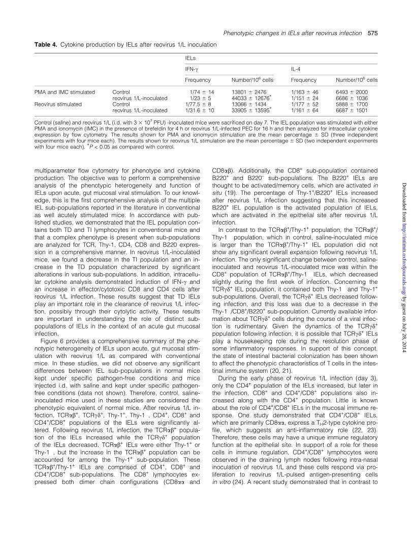
The capacity for cytokine production by the IELs of control,saline-inoculated and reovirus 1/L-inoculated mice was alsoexamined. For cytokine analysis, we focused on day 7 afterinfection, since our results demonstrated maximum pheno-typic changes in the IEL population on that day. Cytokineproduction was assessed by flow cytometry after stimulationwith PMA and ionomycin in the presence of brefeldin A, fa-cilitating cytokine visualization by intracellular flow cytomet-ric cytokine analysis. We evaluated IFN-c and IL-4expression as representative of a TH1 or TH2 profile, respec-tively. The results, shown in Table 4, are expressed in termsof the number of cytokine-positive lymphocytes per 106 cellsand the frequency of cytokine-producing lymphocytes. Thetotal number of IFN-c-positive IELs was significantly in-creased after reovirus 1/L infection, with a frequency of 1/236 5 on day 7 after infection compared with control frequen-cies of 1/74 6 14 (Table 4). No significant change in the totalnumber of IELs-expressing IL-4 was observed after reovirus1/L inoculation (Table 4).Our results demonstrated a significant induction of IFN-c
expression when cultures were stimulated with PMA and ion-omycin. Therefore, to evaluate whether the same increase inIELs-expressing IFN-c was observed after antigen-specificre-stimulation in vitro, we examined cytokine expression fromIELs that were stimulated in vitro with reovirus 1/L-infectedPEC (Table 4). As was observed after culture with PMA andionomycin, there was a significant increase in the frequencyof IFN-c-expressing IELs from a frequency of 1/77.5 in con-trol, saline-inoculated mice to a frequency of 1/31.6 on day7 after reovirus 1/L inoculation after in vitro re-stimulationwith reovirus 1/L-infected PEC. According to a recent study(18), which applied intracellular IFN-c expression to calcu-late effector CTL in spleen cells, the antigen-stimulated IFN-c-producing T cells that co-expressed CD69 (a very earlyactivation marker) were determined to be effector CTL. Weapplied this method to our IEL population and evaluated co-expression of CD69 and IFN-c as a marker for the presenceof effector cells. While it is known that the majority of IELs incontrol, uninfected mice express CD69, we demonstratedthat these CD69+ cells do not significantly express IFN-c(Fig. 5, top). However, in reovirus 1/L-infected mice, therewas a significant increase in the percentage of CD69+/CD8+
(67% infected mice versus 19% control mice) or CD69+/CD4+ IELs (79% infected mice versus 21% control mice) thatwere IFN-c producing (Fig. 5, bottom). These CD4+ or CD8+
IFN-c-producing cells, therefore, fit the profile of effectorcells induced in the intra-epithelial compartment followingreovirus 1/L infection (18).
Discussion
Intestinal IELs form a highly specialized lymphoid compart-ment and along with LPLs are considered to play an impor-tant role in the regulation of immune responses. IELs area phenotypically diverse population with many reportedsub-populations observed under various conditions. In thisstudy, we investigated the IEL population of control (salineinoculated) and reovirus 1/L-inoculated Balb/c mice by
Fig. 4. Phenotypic changes in the CD4+/CD8+ IEL sub-populationafter reovirus 1/L inoculation. Control (saline) and reovirus 1/L (i.d.with 3 3 107 PFU) -inoculated mice were sacrificed on days 7, 10 and14. The CD4+/CD8a+ IEL sub-population was first gated to exclude allother cells, and then analyzed for TCRab and CD8b expression. Theupper panels show control, saline-inoculated mice and the lowerpanels show reovirus 1/L-inoculated mice. The values shown in theright panels are the percentage of the CD4+/CD8+ IEL sub-populationexpressing the ab heterodimeric chains of the CD8 molecule and theTCRab. The results shown represent one of the three independentexperiments (four mice per experiment) demonstrating similar results.
574 Phenotypic changes in IELs after reovirus infection
by guest on July 28, 2014http://intim
m.oxfordjournals.org/
Dow
nloaded from
multiparameter flow cytometry for phenotype and cytokineproduction. The objective was to perform a comprehensiveanalysis of the phenotypic heterogeneity and function ofIELs upon acute, gut mucosal viral stimulation. To our knowl-edge, this is the first comprehensive analysis of the multipleIEL sub-populations reported in the literature in conventionalas well acutely stimulated mice. In accordance with pub-lished studies, we demonstrated that the IEL population con-tains both TD and TI lymphocytes in conventional mice andthat a complex phenotype is present when sub-populationsare analyzed for TCR, Thy-1, CD4, CD8 and B220 expres-sion in a comprehensive manner. In reovirus 1/L-inoculatedmice, we found a decrease in the TI population and an in-crease in the TD population characterized by significantalterations in various sub-populations. In addition, intracellu-lar cytokine analysis demonstrated induction of IFN-c andan increase in effector/cytotoxic CD8 and CD4 cells afterreovirus 1/L infection. These results suggest that TD IELsplay an important role in the clearance of reovirus 1/L infec-tion, possibly through their cytolytic activity. These resultsare important in understanding the role of distinct sub-populations of IELs in the context of an acute gut mucosalinfection.Figure 6 provides a comprehensive summary of the phe-
notypic heterogeneity of IELs upon acute, gut mucosal stim-ulation with reovirus 1/L as compared with conventionalmice. In these studies, we did not observe any significantdifferences between IEL sub-populations in normal micekept under specific pathogen-free conditions and miceinjected i.d. with saline and kept under specific pathogen-free conditions (data not shown). Therefore, control, saline-inoculated mice used in these studies are considered thephenotypic equivalent of normal mice. After reovirus 1/L in-fection, TCRab+, TCRcd+, Thy-1+, Thy-1�, CD4+, CD8+ andCD4+/CD8+ populations of the IELs were significantly al-tered. Following reovirus 1/L infection, the TCRab+ popula-tion of the IELs increased while the TCRcd+ populationof the IELs decreased. TCRab+ IELs were either Thy-1+ orThy-1�, but the increase in the TCRab+ population can beaccounted for among the Thy-1+ sub-population. TheseTCRab+/Thy-1+ IELs are comprised of CD4+, CD8+ andCD4+/CD8+ sub-populations. The CD8+ lymphocytes ex-pressed both dimer chain configurations (CD8aa and
CD8ab). Additionally, the CD8+ sub-population containedB220+ and B220– sub-populations. The B220+ IELs arethought to be activated/memory cells, which are activated insitu (19). The percentage of Thy-1+/B220+ IELs increasedafter reovirus 1/L infection suggesting that this increasedB220+ IEL population is the activated population of IELs,which are activated in the epithelial site after reovirus 1/Linfection.In contrast to the TCRab+/Thy-1+ population, the TCRab+/
Thy-1� population, which in control, saline-inoculated miceis larger than the TCRab+/Thy-1+ IEL population did notshow any significant overall expansion following reovirus 1/Linfection. The only significant change between control, saline-inoculated and reovirus 1/L-inoculated mice was within theCD8+ population of TCRab+/Thy-1� IELs, which decreasedslightly during the first week of infection. Concerning theTCRcd+ IEL population, it contained both Thy-1� and Thy-1+
sub-populations. Overall, the TCRcd+ IELs decreased follow-ing infection, and this loss was due to a decrease in theThy-1�/CD8+/B220+ sub-population. Currently available infor-mation about TCRcd+ cells during the course of a viral infec-tion is rudimentary. Given the dynamics of the TCRcd+
population following infection, it is possible that TCRcd+ IELsplay a housekeeping role during the resolution phase ofsome inflammatory responses. In support of this concept,the state of intestinal bacterial colonization has been shownto affect the phenotypic characteristics of T cells in the intes-tinal immune system (20, 21).During the early phase of reovirus 1/L infection (day 3),
only the CD4+ population of the IELs increased, but later inthe infection, CD8+ and CD4+/CD8+ populations also in-creased along with the CD4+ population. Little is knownabout the role of CD4+/CD8+ IELs in the mucosal immune re-sponse. One study demonstrated that CD4+/CD8+ IELs,which are primarily CD8aa, express a TH2-type cytokine pro-file, which suggests an anti-inflammatory role (22, 23).Therefore, these cells may have a unique immune regulatoryfunction at the epithelial site. In support of a role for thesecells in immune regulation, CD4+/CD8+ lymphocytes wereobserved in the draining lymph nodes following intra-nasalinoculation of reovirus 1/L and these cells respond via pro-liferation to reovirus 1/L-pulsed antigen-presenting cellsin vitro (24). A recent study demonstrated that in contrast to
Table 4. Cytokine production by IELs after reovirus 1/L inoculation
IELs
IFN-c IL-4
Frequency Number/106 cells Frequency Number/106 cells
PMA and IMC stimulated Control 1/74 6 14 13801 6 2476 1/163 6 46 6493 6 2000reovirus 1/L-inoculated 1/23 6 5 44033 6 12676* 1/151 6 24 6686 6 1036
Reovirus stimulated Control 1/77.5 6 8 13066 6 1434 1/177 6 52 5888 6 1700reovirus 1/L-inoculated 1/31.6 6 10 33905 6 13595* 1/161 6 64 6687 6 1501
Control (saline) and reovirus 1/L (i.d. with 3 3 107 PFU) -inoculated mice were sacrificed on day 7. The IEL population was stimulated with eitherPMA and ionomycin (IMC) in the presence of brefeldin for 4 h or reovirus 1/L-infected PEC for 16 h and then analyzed for intracellular cytokineexpression by flow cytometry. The results shown for PMA and ionomycin stimulation are the mean percentage 6 SD (three independentexperiments with four mice each). The results shown for reovirus 1/L stimulation are the mean percentage 6 SD (two independent experimentswith four mice each). *P < 0.05 as compared with control.
Phenotypic changes in IELs after reovirus infection 575
by guest on July 28, 2014http://intim
m.oxfordjournals.org/
Dow
nloaded from

CD8aa+ T cells, which are located mainly within the gut epi-thelium, CD4+ and CD8ab+ T cells are present in the gut epi-thelium and lamina propria and are the progeny of commonprecursors that have proliferated under the same antigenicstimulation (25–27). Since CD4+ T cells comprise a largerproportion of the LPL population than CD8+ T cells, and theadhesive capacity of CD4+ cells to villus microvessels isgreater than that of CD4� cells (28), the increase in CD4+
Tcells in the intra-epithelial compartment 3 days after infectioncould be due to the migration of CD4+ T cells from the lam-ina propria to the epithelium. During the later phase of reovi-rus 1/L infection (days 7 and 10), the CD8+ IELs increasedalong with CD4+ IELs. This is likely due to the migration ofprimed PP CD8+ and CD4+ T cells to the intra-epithelial com-partment through the lamina propria and the proliferation ofCD8ab+ IELs. In this regard, we have recently demonstratedthat reovirus 1/L-primed PP cells adoptively transferred tonormal or reovirus 1/L-inoculated mice, preferentially mi-grated to the intra-epithelial compartment of reovirus1/L-infected mice as compared with normal mice (13). Theliterature also supports the hypothesis that the antigenicstimulation and proliferation of IEL precursors occur mainlyin the PP followed by their rapid emigration to the intra-epithelial compartment and lamina propria through the mes-enteric lymph nodes, thoracic duct and blood circulation(25–27). However, it is also possible that IELs, LPLs and PPlymphocytes upon meeting their cognate antigens in situmay undergo further stimulation and divide. Therefore, wesuggest that the observed increase in TCRab+/Thy-1+ IELsfollowing reovirus 1/L infection is due to migration of primedPP Tcells to the epithelial site. However, the possibility of pro-liferation in situ of TCRab+/Thy-1+ IELs cannot be ruled out.The decrease in reovirus 1/L-induced CD8+ IELs to control
levels 14 days after infection might be due to depletion ofunwanted T cells (CD8aa and CD8ab) via apoptotic death af-ter the clearance of the viral infection. In this study, theCD8aa+ T cells were decreased after reovirus 1/L infection,and we suspect this decrease was the cause of the signifi-cant decrease in the total number of CD8+ T cells on day3 after reovirus 1/L infection. The decrease in the CD8+/B220+
sub-population of TCRcd+/Thy-1+ IELs (CD8aa+/B220+) in ourstudy also demonstrates indirectly that the decrease in theCD8aa+ sub-population of TCRcd+ IELs also contributes tothe decrease in total CD8+ IELs during the early stage of in-fection. There are several possible causes for this depletionof the CD8aa+ sub-population of IELs. Direct viral cytotoxicitymay be playing a role, or some CD8aa+ T cells may also mi-grate to the periphery during reovirus 1/L infection. The pro-portion of CD8aa+ T cells has been shown to increase in theperipheral blood during chronic infection of immunodefi-ciency virus in cats (29). Previous studies have also sug-gested that CD8aa+ IELs were an immature extrathymic T cellsubset that may differentiate through a CD4+/CD8aa+ stageinto mature CD4+ and CD8ab+ T cells in intestinal epithelium(30, 31). If this is the case, the increased CD4+/CD8ab+ sub-population of TCRab+/Thy-1+ IELs after reovirus 1/L infectionmay be the intermediate stage of maturation of extrathymicimmature CD4+/CD8aa+ T cells to mature CD4+ and CD8ab+
IELs, a process which may require antigenic stimulation likethe reovirus 1/L stimulation in this study. The CD4+/CD8aa+
cells may also be intermediate in the extrathymic differentia-tion pathway for local generation of resident CD8aa+ IELs(32). Interestingly, while a vast majority of TCRcd+ IELsexpresses CD8aa but not CD8ab (which are considered todevelop in intestinal epithelial layers independently of a func-tional thymus) (5, 10, 11), we demonstrated a small but sig-nificant CD8ab population of the TCRcd+ IELs (data notshown). This confirmed a recent study that demonstrated this
Fig. 5. Phenotype of IEL-expressing IFN-c after reovirus 1/L in-oculation. Control (saline) and reovirus 1/L (i.d. with 3 3 107 PFU) -inoculated mice were sacrificed on day 7. IELs were then surfacestained with anti-CD8, -CD4 and -CD69 mAbs followed by in-tracellular staining with an anti-IFN-c mAb. The CD8+/CD69+ andCD4+/CD69+ populations were then gated and analyzed for IFN-cexpression. The values shown in the left panels are the percentage ofIELs co-expressing either CD4 or CD8 and CD69 (gated population).The percentage of the gated population expressing IFN-c is shown inthe histogram on the right. The results shown represent one of the twoindependent experiments (four mice per experiment) demonstratingsimilar results.
576 Phenotypic changes in IELs after reovirus infection
by guest on July 28, 2014http://intim
m.oxfordjournals.org/
Dow
nloaded from

functionally active population in athymic nu/nu mice (33).However, we did not see any significant change in the per-centage of TCRcd+/CD8ab+ IELs after reovirus 1/L infection.Gut mucosal infection with reovirus results in the presence
of infectious virions in the intestine for up to 10 days after in-fection, with the titer decreasing 10-fold during this time pe-riod (34). In addition, reovirus 1/L infection results in thegeneration of MHC-restricted, reovirus 1/L-specific, cytolyticTCRab+/Thy-1+ IELs (12). Here, we demonstrate that the in-
crease in the TCRab+/Thy-1+ IELs occurs early after infectionand correlates with viral clearance and suggests that theTCRab+/Thy-1+ IELs may be playing an important role in theclearance of reovirus infection. This role could be executedthrough cytolytic activity or cytokine production. In thesestudies, infection with reovirus 1/L decreased the CD8aa+
IEL sub-population and increased CD8ab+ and CD4+ IEL sub-populations. CD8+ cells provide protection against viruses bytheir cytolytic activity and locally generated TCRab+/CD8aa+
Fig. 6. Summary of the phenotypic changes in distinct sub-populations of IELs after reovirus 1/L infection. The values shown in thin-borderedbox, dashed bordered box, thick-bordered box and double-bordered box and are of control groups on days 3, 7, 10 and 14, respectively. Thevalues shown in shaded thin-bordered box, shaded dashed bordered box, shaded thick-bordered box and shaded double-bordered box areof reovirus 1/L-infected groups on days 3, 7, 10 and 14, respectively. Up arrow shows an increase and down arrow shows a decrease inexpression as compared with control group. *P < 0.05 and **P < 0.01 as compared with control group. The values shown are the percentage ofthe total IELs.
Phenotypic changes in IELs after reovirus infection 577
by guest on July 28, 2014http://intim
m.oxfordjournals.org/
Dow
nloaded from

IELs exert only minimal virus-specific cytotoxicity, while maxi-mum specific killing of virus is mediated by thymically derivedTCRab+/CD8ab+ IELs (13, 35). Therefore, the increase inCD8ab+ IELs in response to reovirus 1/L infection could con-tribute to the CTL responses in the intestinal mucosa.In the absence of deliberate stimulation, the cytokine pro-
file of IELs fails to display normal polarization towards eitherTH1 or TH2 cytokine production despite the fact that theyare morphologically in an active state. In the resting state,IELs produce IFN-c and IL-5 (36), however, after activationthe majority of IELs adopts a TH1 cytokine profile particularlywith respect to inflammatory cytokines (11). In terms of cyto-kine production, reovirus 1/L infection has been shown to in-duce a TH1 response in IELs (37), which includes anti-viralcytokines such as IFN-c and tumor necrosis factor-a (38).The present study extends these results and demonstratesthat the increase in the CD4+ and CD8+ (TCRab+/Thy-1+) IELsub-populations correlated with IFN-c induction by thesecells over time. In addition, our results of antigen-driven in-tracellular IFN-c production, an assay that is used to meas-ure potency of CD4+ and CD8+ IELs after reovirus 1/Linfection, also demonstrated the induction of both effector/cytotoxic CD4+ and CD8+ IELs after reovirus 1/L infection.A recent report demonstrated that IELs recovered from reo-virus 3/D-infected animals are primarily CD4+/CD43+/ICOS+
and secrete IFN-c in response to reovirus 3/D infection andpotentially represent an activated virus-specific response(38, 39). Taken together with our findings that demonstrateactivation of both CD4+ and CD8+ cells, these data supportthe proposal that both CD4+ and CD8+ viral-specific IELsub-populations may play a role in the clearance of reovirus.In conclusion, reovirus 1/L infection was found to alter the
phenotypic profiles of the various IEL sub-populations. Theresults demonstrated increases in CD4+, CD8ab+ and CD4+/CD8ab+ populations of TCRab+/Thy-1+ IELs and a decreasein TCRcd+/Thy-1�/CD8aa+ or TCRab+/Thy-1�/CD8aa+ IELs.The increase in CD4+ and CD8ab cells and decrease inCD8aa cells may partially account for the altered T cell ho-meostasis in the intestinal epithelium following reovirus 1/Linfection. Further, our results demonstrated that the potentialof CD8+ and CD4+ IELs to produce IFN-c was up-regulatedduring reovirus 1/L infection. Taken together, the phenotypeof the IELs after reovirus 1/L infection suggests that the pro-tection against reovirus 1/L is most likely due to the activityof thymus-derived T cells that enter the intestine followingantigenic stimulation. The increase in TCRab+ IELs after reo-virus 1/L infection demonstrates that TCRab+ IELs play animportant role in the immune response against reovirus 1/L.
Acknowledgements
The authors would like to thank the Medical University of SouthCarolina (MUSC) and the Hollings Cancer Center for their support ofthe MUSC Analytical Flow Cytometry Facility. This work was supportedby National Institutes of Health (NIH)/National Institute of DentalCraniofacial Research (NIDCR) grant number 5R01 DE12781 (S.D.L.)and NIH grant number CO6 RR015455 (MUSC).
Abbreviations
APC allophycocyaninCMF Ca2+-, Mg2+-free HBSS
FBS fetal bovine serumi.d. intra-duodenalIEL intra-epithelial lymphocyteLPL lamina propria lymphocyteMUSC Medical University of South CarolinaNIH National Institutes HealthPEC peritoneal exudate cellPMA phorbol myristate acetatePP Peyer’s patchereovirus 1/L reovirus serotype 1/strain LangTD thymus dependentTI thymus independent
References
1 Rocha, B., Vassalli, P. and Guy-Grand, D. 1991. The V betarepertoire of mouse gut homodimeric alpha CD8+ intraepithelialT cell receptor alpha/beta+ lymphocytes reveals a major extra-thymic pathway of T cell differentiation. J. Exp. Med. 173:483.
2 Hayday, A., Theodoridis, E., Ramsburg, E. and Shires, J. 2001.Intraepithelial lymphocytes: exploring the Third Way in immuno-logy. Nat. Immunol. 2:997.
3 Cheroutre, H. 2004. Starting at the beginning: new perspectiveson the biology of mucosal T cells. Annu. Rev. Immunol. 22:217.
4 Cheroutre, H. 2005. IELs: enforcing law and order in the court ofthe intestinal epithelium. Immunol. Rev. 206:114.
5 Guy-Grand, D., Cerf-Bensussan, N., Malissen, B., Malassis-seris,M., Briottet, C. and Vassalli, P. 1991. Two gut intraepithelialCD8+ lymphocyte populations with different T cell receptors:a role for the gut epithelium in T cell differentiation. J. Exp. Med.173:471.
6 Rocha, B., Vassalli, P. and Guy-Grand, D. 1994. Thymic andextrathymic origins of gut intraepithelial lymphocyte populations inmice. J. Exp. Med. 180:681.
7 Guy-Grand, D., Azogui, O., Celli, S. et al. 2003. ExtrathymicT cell lymphopoiesis: ontogeny and contribution to gut intra-epithelial lymphocytes in athymic and euthymic mice. J. Exp. Med.197:333.
8 Lefrancois, L. and Olson, S. 1994. A novel pathway of thymus-directed T lymphocyte maturation. J. Immunol. 153:987.
9 Lin, T., Matsuzaki, G., Yoshida, H. et al. 1994. CD3�CD8+intestinal intraepithelial lymphocytes (IEL) and the extrathymicdevelopment of IEL. Eur. J. Immunol. 24:1080.
10 Lefrancois, L. 1991. Intraepithelial lymphocytes of intestinalmucosa: curiouser and curiouser. Semin. Immunol. 3:99.
11 Beagley, K. W. and Husband, A. J. 1998. Intraepithelial lympho-cytes: origins, distribution, and function. Crit. Rev. Immunol.18:237.
12 London, S. D., Cebra, J. J. and Rubin, D. H. 1989. Intraepitheliallymphocytes contain virus-specific cytotoxic T cell precursorsafter gut mucosal immunization with reovirus serotype 1/Lang.Reg. Immunol. 2:98.
13 Cuff, C. F., Cebra, C. K., Rubin, D. H. and Cebra, J. J. 1993.Developmental relationship between cytotoxic ab T cell receptor-positive intraepithelial lymphocytes and Peyer’s patches lympho-cytes. Eur. J. Immunol. 23:1333.
14 London, S. D., Rubin, D. H. and Cebra, J. J. 1987. Gut mucosalimmunization with reovirus serotype 1/L stimulates viral specificcytotoxic T cell precursors as well as IgA memory cells in Peyer’spatches. J. Exp. Med. 165:830.
15 Bharhani, M., Grewal, J. S., Pilgrim, M. J. et al. 2005. Reovirusserotype 1/strain Lang-stimulated activation of antigen-specificT lymphocytes in Peyer’s patches and distal gut-mucosal sites:activation status and cytotoxic mechanisms. J. Immunol. 174:3580.
16 Rubin, D. H., Kornstein, M. J. and Anderson, A. O. 1985. Reovirusserotype 1 intestinal infection: a novel replicative cycle with ilealdisease. J. Virol. 53:391.
17 Lefrancois, L. 1994. Isolation of mouse small intestinal intra-epithelial lymphocytes. In Coligan, J. E., Kruisbeek, A. M.,Margulies, D. H., Shevak, E. M. and Strober, W., eds, CurrentProtocols in Immunology, Wiley, New York p. 191.
578 Phenotypic changes in IELs after reovirus infection
by guest on July 28, 2014http://intim
m.oxfordjournals.org/
Dow
nloaded from

18 Kumaraguru, U. and Rouse, B. T. 2000. Application of intracellulargamma interferon assay to recalculate the potency of CD8+ T-cellresponses to herpes simplex virus. J. Virol. 74:5709.
19 Lefrancois, L. and Puddington, L. 2001. Basic aspectsof intraepithelial lymphocyte immunobiology. In Ogra, P. L.,Mastecky, J., Lamm, M. E., Strober, W., Bienenstock, J. andMcGhee, J. R., eds, Mucosal Immunology, 2nd edn. Academicpress, San Diego pp 413.
20 Bandeira, A., Mota-Santos, T., Itohara, S., Hiuersser, C., Tone-gawa, S. and Coutinho, A. 1990. Localization of cd T-cells to theintestinal epithelium is independent of normal microbial coloniza-tion. J. Exp. Med. 172:239.
21 Lillehoj, H. S. and Bacon, L. D. 1991. Increase of intestinalintraepithelial lymphocytes expressing CD8 antigen followingchallenge infection with Eimeria acervulina. Avian Dis. 35:294.
22 Fujihashi, K., Yamamoto, M., McGhee, J. R. and Kiyono, H. 1993.Alpha beta Tcell receptor-positive intraepithelial lymphocytes withCD4+, CD8� and CD4+, CD8+ phenotypes from orally immunizedmice provide Th2-like function for B cell responses. J. Immunol.151:6681.
23 Das, G., Augustine, M. M., Das, J., Bottomly, K., Ray, P. and Ray,A. 2003. An important regulatory role for CD4+CD8 alpha alphaT cells in the intestinal epithelial layer in the prevention ofinflammatory bowel disease. Proc. Natl Acad. Sci. USA 100:5324.
24 Periwal, S. B. and Cebra, J. J. 1999. Respiratory mucosalimmunization with reovirus serotype 1/L stimulates virus-specifichumoral and cellular immune responses, including double-positive (CD4(+)/CD8(+)) T cells. J. Virol. 73:7633.
25 Arstila, T., Arstila, T. P., Calbo, S. et al. 2000. Identical T cell clonesare located within the mouse gut epithelium and lamina propriaand circulate in the thoracic duct lymph. J. Exp. Med. 191:823.
26 Kim, S. K., Reed, D. S., Heath, W. R., Carbone, F. and Lefrancois,L. 1997. Activation and migration of CD8 T cells in the intestinalmucosa. J. Immunol. 159:4295.
27 Tamura, A., Soga, H., Yaguchi, K. et al. 2003. Distribution of twotypes of lymphocytes (intraepithelial and lamina-propria-associ-ated) in the murine small intestine. Cell Tissue Res. 313:47.
28 Koseki, S., Miura, S., Fujimori, H. et al. 2001. In situ demonstrationof intraepithelial lymphocyte adhesion to villus microvessels of thesmall intestine. Int. Immunol. 13:1165.
29 Shimojima, M., Miyazawa, T., Kohmoto, M. et al. 1998. Expansionof CD8alpha+beta� cells in cats infected with feline immunode-ficiency virus. J. Gen. Virol. 79:91.
30 Guy-Grand, D. and Vassalli, P. 1993. Gut intraepithelial T lympho-cytes. Curr. Opin. Immunol. 5:247.
31 Guy-Grand, D. and Vassalli, P. 2002. Gut intraepithelial lympho-cyte development. Curr. Opin. Immunol. 14:255.
32 Rozing, J. and de Geus, B. 1995. Changes in the intestinallymphoid compartment throughout life: implications for the localgeneration of intestinal T cells. Int. Rev. Immunol. 12:13.
33 Emoto, Y., Emoto, M., Miyamoto, M., Yoshozawa, I. and Kaufmann,S. H.E. 2004. Functionally active CD8ab+TCRcd intestinalepithelial lymphocytes in athymic nu/nu mice. Int. Immunol.16:111.
34 Fan, J. Y., Boyce, C. S. and Cuff, C. F. 1998. T-helper 1 and T-helper 2 cytokine responses in gut associated lymphoid tissuefollowing reovirus infection. Cell. Immunol. 188:55.
35 Muller, S., Buhler-Jungo, M. and Mueller, C. 2000. Intestinalintraepithelial lymphocytes exert potent protective cytotoxicactivity during an acute virus infection. J. Immunol. 164:1986.
36 Taguchi, T., Aicher, W. K., Fujihasgi, K. et al. 1991. Novel functionfor intraepithelial lymphocytes: murine CD3+, cdTCR T cellsproduce IFN-c and IL-5. J. Immunol. 147:3736.
37 Kagi, D. and Hengartner, H. 1996. Different roles for cytotoxic Tcell in the control of infections with cytopathic versus non-cytopathic viruses. Curr. Opin. Immunol. 8:472.
38 Montufar-Solis, D., Garza, T., Teng, B. B. and Klein, J. R. 2006.Upregulation of ICOS on CD43+ CD4+ murine small intestinalintraepithelial lymphocytes during acute reovirus infection. Bio-chem. Biophys. Res. Commun. 342:782.
39 Wang, H. C., Montufar-Solis, D., Teng, B. B. and Klein, J. R. 2004.Maximum immunobioactivity of murine small intestinal intraepithe-lial lymphocytes resides in a subpopulation of CD43+ T cells.J. Immunol. 173:6294.
Phenotypic changes in IELs after reovirus infection 579
by guest on July 28, 2014http://intim
m.oxfordjournals.org/
Dow
nloaded from
Copyright © 2022 FDOKUMEN