Alzheimer's disease-like perturbations in HIV-mediated ...
23
This is an electronic reprint of the original article. This reprint may differ from the original in pagination and typographic detail. Powered by TCPDF (www.tcpdf.org) This material is protected by copyright and other intellectual property rights, and duplication or sale of all or part of any of the repository collections is not permitted, except that material may be duplicated by you for your research use or educational purposes in electronic or print form. You must obtain permission for any other use. Electronic or print copies may not be offered, whether for sale or otherwise to anyone who is not an authorised user. Jha, Niraj Kumar; Sharma, Ankur; Jha, Saurabh Kumar; Ojha, Shreesh; Chellappan, Dinesh Kumar; Gupta, Gaurav; Kesari, Kavindra Kumar; Bhardwaj, Shanu; Shukla, Shakti D.; Tambuwala, Murtaza M.; Ruokolainen, Janne; Dua, Kamal; Singh, Sandeep Kumar Alzheimer's disease-like perturbations in HIV-mediated neuronal dysfunctions Published in: OPEN BIOLOGY DOI: 10.1098/rsob.200286 Published: 23/12/2020 Document Version Publisher's PDF, also known as Version of record Published under the following license: CC BY Please cite the original version: Jha, N. K., Sharma, A., Jha, S. K., Ojha, S., Chellappan, D. K., Gupta, G., Kesari, K. K., Bhardwaj, S., Shukla, S. D., Tambuwala, M. M., Ruokolainen, J., Dua, K., & Singh, S. K. (2020). Alzheimer's disease-like perturbations in HIV-mediated neuronal dysfunctions: Understanding mechanisms and developing therapeutic strategies: HIV and AD relationship. OPEN BIOLOGY, 10(12), [200286]. https://doi.org/10.1098/rsob.200286
-
Upload
khangminh22 -
Category
Documents
-
view
0 -
download
0
Transcript of Alzheimer's disease-like perturbations in HIV-mediated ...

This is an electronic reprint of the original article.This reprint may differ from the original in pagination and typographic detail.
Powered by TCPDF (www.tcpdf.org)
This material is protected by copyright and other intellectual property rights, and duplication or sale of all or part of any of the repository collections is not permitted, except that material may be duplicated by you for your research use or educational purposes in electronic or print form. You must obtain permission for any other use. Electronic or print copies may not be offered, whether for sale or otherwise to anyone who is not an authorised user.
Jha, Niraj Kumar; Sharma, Ankur; Jha, Saurabh Kumar; Ojha, Shreesh; Chellappan, DineshKumar; Gupta, Gaurav; Kesari, Kavindra Kumar; Bhardwaj, Shanu; Shukla, Shakti D.;Tambuwala, Murtaza M.; Ruokolainen, Janne; Dua, Kamal; Singh, Sandeep KumarAlzheimer's disease-like perturbations in HIV-mediated neuronal dysfunctions
Published in:OPEN BIOLOGY
DOI:10.1098/rsob.200286
Published: 23/12/2020
Document VersionPublisher's PDF, also known as Version of record
Published under the following license:CC BY
Please cite the original version:Jha, N. K., Sharma, A., Jha, S. K., Ojha, S., Chellappan, D. K., Gupta, G., Kesari, K. K., Bhardwaj, S., Shukla, S.D., Tambuwala, M. M., Ruokolainen, J., Dua, K., & Singh, S. K. (2020). Alzheimer's disease-like perturbations inHIV-mediated neuronal dysfunctions: Understanding mechanisms and developing therapeutic strategies: HIVand AD relationship. OPEN BIOLOGY, 10(12), [200286]. https://doi.org/10.1098/rsob.200286

royalsocietypublishing.org/journal/rsob
ReviewCite this article: Jha NK et al. 2020Alzheimer’s disease-like perturbations
in HIV-mediated neuronal dysfunctions:
understanding mechanisms and developing
therapeutic strategies. Open Biol. 10: 200286.https://doi.org/10.1098/rsob.200286
Received: 4 September 2020
Accepted: 27 November 2020
Subject Area:biochemistry/neuroscience/immunology/
molecular biology
Keywords:HIV, Alzheimer’s disease, HAND, microglia,
neuroinflammation, neurotherapeutics
Authors for correspondence:Niraj Kumar Jha
email: [email protected],
Sandeep Kumar Singh
email: [email protected],
© 2020 The Authors. Published by the Royal Society under the terms of the Creative Commons AttributionLicense http://creativecommons.org/licenses/by/4.0/, which permits unrestricted use, provided the originalauthor and source are credited.
Alzheimer’s disease-like perturbationsin HIV-mediated neuronal dysfunctions:understanding mechanisms anddeveloping therapeutic strategies
Niraj Kumar Jha1, Ankur Sharma2, Saurabh Kumar Jha1, Shreesh Ojha3,Dinesh Kumar Chellappan4, Gaurav Gupta5, Kavindra Kumar Kesari6,Shanu Bhardwaj7, Shakti D. Shukla8, Murtaza M. Tambuwala9,Janne Ruokolainen6, Kamal Dua8,10,11 and Sandeep Kumar Singh12,13
1Department of Biotechnology, School of Engineering and Technology (SET), and 2Department of Life Science,School of Basic Science and Research (SBSR), Sharda University, Greater Noida, UP 201310, India3Department of Pharmacology and Therapeutics, College of Medicine and Health Sciences, PO Box 17666,United Arab Emirates University, Al Ain, United Arab Emirates4Department of Life Sciences, School of Pharmacy, International Medical University, Bukit Jalil, Kuala Lumpur57000, Malaysia5School of Phamacy, Suresh Gyan Vihar University, Jagatpura, Mahal Road, Jaipur, India6Department of Applied Physics, School of Science, Aalto University, Espoo 00076, Finland7Department of Biotechnology, HIMT, Greater Noida, CCS University, UP, India8Priority Research Centre for Healthy Lungs, Hunter Medical Research Institute (HMRI) and School of BiomedicalSciences and Pharmacy, University of Newcastle, Callaghan, NSW 2308, Australia9School of Pharmacy and Pharmaceutical Sciences, Ulster University, Coleraine, County Londonderry,BT52 1SA, UK10Discipline of Pharmacy, Graduate School of Health, University of Technology Sydney, Sydney, New South Wales2007, Australia11School of Pharmaceutical Sciences, Shoolini University of Biotechnology and Management Sciences, PO Box 9,Solan, Himachal Pradesh 173229, India12Department of Biomedical Research, Centre of Biomedical Research, SGPGI Campus, Lucknow 226014, UP,India13Biological Science, Indian Scientific Education and Technology Foundation, Lucknow 226002, UP, India
NKJ, 0000-0001-9486-4069; SB, 0000-0002-8059-0287; KD, 0000-0002-7507-1159;SKS, 0000-0002-0022-6240
Excessive exposure to toxic substances or chemicals in the environmentand various pathogens, including viruses and bacteria, is associated withthe onset of numerous brain abnormalities. Among them, pathogens,specifically viruses, elicit persistent inflammation that plays a major role inAlzheimer’s disease (AD) as well as dementia. AD is the most commonbrain disorder that affects thought, speech, memory and ability to executedaily routines. It is also manifested by progressive synaptic impairment andneurodegeneration, which eventually leads to dementia following theaccumulation of Aβ and hyperphosphorylated Tau. Numerous factorscontribute to the pathogenesis of AD, including neuroinflammationassociated with pathogens, and specifically viruses. The human immunodefi-ciency virus (HIV) is often linked with HIV-associated neurocognitivedisorders (HAND) following permeation through the blood–brain barrier(BBB) and induction of persistent neuroinflammation. Further, HIV infectionsalso exhibited the ability to modulate numerous AD-associated factors such asBBB regulators,members of stress-related pathways aswell as the amyloid andTau pathways that lead to the formation of amyloid plaques or neurofibrillarytangles accumulation. Studies regarding the role of HIV in HAND and AD arestill in infancy, and potential link or mechanism between both is not yet estab-lished. Thus, in the present article, we attempt to discuss various molecular

royalsocietypublishing.org/journal/rsobOpen
Biol.10:200286
2
mechanisms that contribute to the basic understanding ofthe role of HIV-associated neuroinflammation in AD andHAND. Further, using numerous growth factors and drugs,we also present possible therapeutic strategies to curb theneuroinflammatory changes and its associated sequels.1. IntroductionAlzheimer’s disease (AD) is the most common neurologicalcomplication, which mainly manifests progressive synapticimpairment and neurodegeneration, following excessive forma-tion and accumulation of amyloid-beta (Aβ) [1,2]. Aβ depositsand hyperphosphorylated Tau (pTau), which interfere withtheneuronal organizationand their function, playaconsiderablerole inADprogression [3].Aβ-pathologyoften involves avarietyof signals that interrupt the homeostasis of neurons [4]. Cur-rently, there are no definite data that can demonstrate acausative relationship between neuronal damage followinghuman immunodeficiency virus (HIV) infections and the onsetof AD. However, available literature indicates that there aresome common factors and pathways modulated in HIV+ andAD patients, thus suggestive of some similarities in these twopathologies. Among numerous pathways, neuroinflammationis shown closely related to these disorders and is considereda crucial factor in their development and progression. It hasbeen reported that HIV regulatory proteins such as trans-activator of transcription (Tat), envelope glycoprotein (Gp120),viral protein R (Vpr) and negative factor (Nef) can directlyinfluence the central nervous system(CNS) andactivateneuroin-flammatory pathways followed by neuronal injury anddysfunction.Additionally, abnormalAβdeposition, a pathologi-cal hallmark of AD has been reported in the individualssuffering from HIV infection. Though the abnormalities associ-ated with Aβ burden are more frequent in the AD brain thanHIV-infected individuals, it has been predominantly observedin younger HIV-infected individuals [5,6].
Additionally, blood–brain barrier (BBB) dysfunction associ-ated with HIV-1 infection is considered another cause ofneuroinflammation in AD. HIV infiltrates macrophages in theCNS by crossing the BBB. The disrupted BBB in HIV patientshas been correlated with toxic Aβ aggregation and otherabnormalities resulting from a failure to sort out Aβ peptides[7]. The virus-induced fusion of macrophages causes the for-mation of giant cells and activation of astrocytes whicheventually causes injuries to different components of thebrain. The most affected areas are the subcortical structuresalong with the limbic structures and basal ganglia, and theverotoxins, including HIV proteins Gp41, Gp120, Tat, Vprand Nef, are accountable for such damage. HIV proteins alsomay cause axonal damage and breakdown of white matter.These injuries cause a decrease in volume of the brain structuressuch as the caudate nucleus and basal ganglia, resulting inatrophy of the brain volume and decline in cognition [8–10].
HIV also leads to HIV-associated neurocognitive disorders(HAND), since it has a propensity to cross the BBB and causeneuroinflammation [11–14]. HAND exhibits a spectrum ofcognitive deficits and typically affects information processingspeed, attention, learning and recall memory among othercognitive functions [15]. HAND also has implications foradherence to antiretroviral (ARV) treatment since it affectsprospective memory [16]. The exact route in which HIVcauses HAND is not yet well known, althoughHIV replication
(potential mechanisms) in the CNS, principally in the basalganglia and the adjacent subcortical white matter, is whereHIV infection is typically observed [17,18].
Among different cell types in the CNS, neurons have theminimal susceptibility to HIV infection; thus the neuronalimpairment is reasonably speculated to result from an infec-tion of neighbouring cells like microglia and macrophages,which exert immune functions in the brain. These infectedcells result in the production of viral proteins that have theability to affect the synapse where communication betweenneurons occurs. Also, the same viral proteins can induce unin-fectedmacrophages, astrocytes andmicroglial cells that resultsin the production of neurotoxins and a variety of inflam-matory molecules, causing further damage to neurons [19].Further, the inflammatory molecules and neurotoxins triggerNMDA receptors and may cause additional damage to theneurons following aggregation of calcium (Ca2+) in the neur-ons which activate the formation of excessive free radicalsthat contribute to oxidative damage. Among other factors,methamphetamine use or abuse and co-infection with hepa-titis C virus (HCV) may aggravate damage caused by HIV,involving activation of macrophages and microglial cells [19].
Like amyloid plaques, neurofibrillary tangles (NFTs)consisting of pTau also occur in people suffering from HIV,particularly in aged individuals [20]. The elevated levels ofTauhavebeen reported tooccurat earlierages in individuals suf-fering from HIV than in healthy individuals. In HIV-infectedindividuals, tau phosphorylation results from viral proteinsand pro-inflammatory cytokines that may impair amyloidosisand precede the development of tau tangles [21]. Higherexpression of pTau in HIV individuals is also correlated withARV treatment [20]. Many comorbid conditions like chronicsubstance abuse independent of the direct consequences ofHIValso lead toHIV transmission, responsiveness and cognitivedifficulties [22]. It is apparent that the linkage and causativemechanisms between neuroinflammation, HIV-CNS neuroin-fections, HAND and AD are still not completely understood.Therefore, it is important to understand the fundamental mol-ecular linkage among these pathologies, which may help inunderstanding pathogenesis and developing therapeutics tar-geting the pathogenesis events along with additional help indiagnosis and prognosis. In the purview of this, herein we sum-marize various underlying mechanisms which contribute toHIV-associated neuroinflammation in HAND and AD usingsynoptic tables and schemes. Additionally, numerous possibletherapeutic strategies are also presented, which may have thepotential to curb these complications and improve quality of life.
2. Human cells involved in HIV-associatedneuronal damage
HIV-1 interacts with different cell types (table 1) in the CNS,including resident macrophages, neurons and astrocytes thatare reported to be involved in neuronal damage [12,26,27]. Inthe CNS, resident macrophages, neurons and astrocytes arethe primary cell targets for HIV infection. In neurodegenerativeprocesses, the rolesofmacrophagesare crucialdue to their resist-ance and sustenance against the cytopathic effects of HIV-1[19,28–33]. In the CNS, there are four major typesof macrophages: choroid-plexus macrophages, meningealmacrophages, perivascular macrophages and microglia[23,34]. Out of these, perivascular macrophages and microglia

Table 1. The role of human cells in HIV-mediated neuronal damage [12,23–25].
neuronal cell associated effectstypes ofinfection
neuron enhances P53 expression restricted
enhances caspase activation
enhances intracellular Ca2+ release
microglia induces viral replication productive
provokes the release of viral proteins including, gp120, Tat and Vpr
increases neurotoxins production and also induces the expression of inflammatory mediators, such
as PDGF and QUIN
astrocyte enhances the production of neurotoxins restricted
downregulates the glutamate uptake
enhances BBB permeability
enhances intracellular release of glutamate and Ca2+
evokes the migration of monocytes into the brain
perivascular
macrophage
triggers viral replication productive
increases neurotoxins production and induces the expression of inflammatory mediators, such as
PDGF and QUIN
provokes the release of viral proteins including gp120, Tat and Vpr
oligodendrocyte enhances cellular apoptosis restricted
enhances intracellular Ca2+ levels
curtails myelin synthesis
royalsocietypublishing.org/journal/rsobOpen
Biol.10:200286
3
are believed to play a crucial role in neuronal damage followingthe release of inflammatory cytokines [23]. Additionally, viralproteins andneurotoxins also take part in the inflammatorypro-cesses, provoking apoptosis and differentiation of astrocytes,and impairing normal neurogenesis [12,24,25]. Further, micro-glial resident cells play a fundamental role in the pathogenesisofHAND, leading todegenerative changes involvingnumerousmechanisms. The glial cells upon HIV infection release factorsand toxins that aggravate neurons and astrocytes [12,35,36].Astrocytes are neuroectodermal-derived cells, which supportthe function and metabolism of neurons, ionic homeostasisinto the CNS, control of the state of the neuronal synapses bythe uptake of neurotransmitters and tissue repair. These arethe important components of the BBB and also regulate theimmune responses in the brain [37–39]. In addition, astrocytescan facilitate the virus to persist in the CNS, which aids inmain-taining low replication ofHIVand establishing a latent infection[40]. Furthermore, in HIV-infected cells, viral factors mayenhance the release of other chemoattractants that recruitmicro-glia and monocytes, resulting in aggravation of the neuronaldamage. Further, cellular factors like interleukin-1β (IL-1β),interferon gamma (IFN-γ) or tumour necrosis factor alpha(TNF-α) have the potential to activate and reactivate viralreplication in latently infected cells [19,41–45].
3. The direct and indirect mechanisms ofHIV induced-neuronal injury
3.1. Direct mechanismsHIV-1 infects CNS involving three different mechanisms(figure 1). In the first mechanism, the virus can directly infect
endothelial cells which express the chemokine receptors(CCR3, CXCR4, DC-SIGN) engaged in HIV-1 entry [40,46].In the second mechanism, the virus may directly cross theimpaired BBB due to increased permeability [45,47]. In thethird mechanism, according to the ‘Trojan horse’ hypothesis,HIV-1 infected monocytes, perivascular macrophages and leu-cocytes cross the BBB and release viral particles, which infectresident cells like microglia and lead to persistent infection.This one is believed to be the main mechanism for entry ofHIV into the brain, similar to other retroviruses andlentiviruses [40].
Several observations advocate that cells like monocytesare infected before leaving the bone marrow [48]. Particularly,proviral DNA has been observed in these cells with no pres-ence of viral proteins, which facilitated dissemination of theHIV-1 infection [48,49]. An increase in a subset of monocytes,including (CD14lowCD16high), plays a significant role in HIV-1 infection [34,50–54]. These cells display intermediate traitsbetween the differentiated cells (dendritic cells and macro-phage) and monocytes [51,53]. Owing to the lower activityof the host restriction factors than the CD14highCD16low
cells, the cells are more liable to HIV replication followingeased permeation through BBB [49–52,54]. Furthermore,viral proteins released into the CNS are believed to induceBBB impairment by enhancing apoptosis and promotingthe invasion of HIV as well as other viruses in the differentcomponents of the brain [45,55–57].
3.2. The indirect mechanismsIn addition to direct mechanisms, HIV-associated neurologicalcomplications and neuroinflammation also involve indirect

pro-inflammatorycytokines
viralproteins
blood
HIV-1monocyte lymphocyte
T CD4+intracellular Ca2+
caspase activationp53 expression
neuron
intracellular Ca2+
cellular apoptosismyelin synthesis
oligodendrocyte
macrophage
astrocyte
microglia
BBB permeabilityintracellular Ca2+
glutamateneurotoxins
glutamate uptake
viral proteins(Gp120, Tat and Vpr)
neurotoxins
viral proteins(Gp120, Tat and Vpr)
neurotoxins
viral replication
basement membrane
activatedmacrophage
astrocyte microglia
activemicroglia
reactiveastrocyte
oligodendrocyte
IL-8, NO and ROS
cytokinesROS, RNS and
neurotoxins
neuronal celldeath and damage
neuron
induce increase activate reduce
(b) cells in HAND(a) HIV-1 entry into the CNS and its effects on neuronal cells
apoptosis
brain
viral replication
2
3
1
5
4
6
Figure 1. Schematic showing the entry mechanisms of HIV-1 into the CNS and its associated effects on neuronal cells that contribute to neuronal damage anddeath. (1) HIV-1 can enter through infected T-cells or monocytes that migrate from the bloodstream to the CNS according to the ‘Trojan horse’ hypothesis. (2) Theincrease in viral proteins and pro-inflammatory cytokines can impair the BBB (epithelial cells) permeability to make virus entry easier. Besides, using infectedepithelial cells, virus can reach the other side through a transcytosis process. (3) Reactive astrocytes can provoke epithelial cell apoptosis, leading to the modificationof BBB permeability through the release of viral proteins such as Tat. (4) The viral protein Tat has a direct effect on neurons and oligodendrocytes, which causeincreased damage and neuronal death. Finally, chronic activation of activated (5) microglia and (6) macrophages causes an increase in the levels of neurotoxins,proinflammatory cytokines, RNA and ROS.
royalsocietypublishing.org/journal/rsobOpen
Biol.10:200286
4
mechanisms such as the infiltration of infected monocytes andlymphocytes in the CNS, release of viral and cellular factorsfrom these infected cells, and infection of the resident cellscaused by viral particles released from infected cells or infiltrat-ing into the CNS [58]. The cells (specifically T-cells andmonocytes) infected with HIV play a crucial role in the releaseof pro-inflammatory cytokines that stimulate microglia andastrocytes. The activated microglia and astrocytes along withperivascular macrophages are engaged in releasing inflamma-tory and neurotoxic mediators, including quinolinic acid(QUIN), nitrogen oxide and platelet-derived growth factor(PDGF), that further lead to neuronal dysfunction and death[45,59].
Despite treatment with ARV agents, a previous study hasreported that the level of cytokines such as CCL3, IL-8, CCL2,IFN-γ, CXCL10 and IL-6 was found to be higher in HIV-1infected individuals in comparison with the uninfected indi-viduals. The higher expressions of cytokines are indicative ofuninterrupted neuroinflammation that is accountable for
promoting HAND-associated encephalopathy [60]. Recently,Vera et al. [61] reported the presence of neuroinflammatorymarkers in neuro-asymptomatic HIV-infected patients,despite the effective control of viraemia. The translocationof the virus from the gut to the bloodstream is believedto cause extensive inflammation and altered integrity ofwhite matter, and this reasonably suggests the role of thebrain–gut axis in the pathogenesis of HAND [62].
4. Detailed mechanisms ofneuroinflammation caused by HIVin the brain
HIV is known to play a key role in depleting cluster ofdifferentiation 4 (CD4) cells, and robustly hampering theimmune responses. Subsequently, it may rise to opportunisticinfections and cause acquired immunodeficiency syndrome

Table 2. The roles of HIV regulatory proteins on neuronal damage.
HIV regulatoryprotein pathological implications on brain references
Tat induces the expression of GAC, GFAP, IL-1β and MCP-1/CCL2 [78–81]
regulates cellular gene expression [82]
enhances the expression of GLUT1 in the hippocampus and cortex; also, enhances leucocyte infiltration [78,82]
upregulates the expression of Cx43 human gene [83]
decreases SYN expression; also reduces GABA in the cortex. [82,84]
interacts with CDK9 and Cyclin T1 [85]
Gp120 activates the release of inflammatory cytokines and toxic substances and accumulation of AβPP [86,87]
decreases the expression of MAP2, LC3 and beclin-1 [88]
Vpr promotes pro-apoptotic and cell-cycle proteins [57]
induces the release of matrix metalloproteinases (neurotoxins) [57]
provokes the release of IL-1β, TNF-α and IL-8 in macrophages [57]
Nef enhances the apoptosis of MVEC; also, enhances the sensitivity of astrocytes to H2O2 [55,89]
provokes astrogliosis and astroglial activation [90]
royalsocietypublishing.org/journal/rsobOpen
Biol.10:200286
5
(AIDS). HIV is occasionally known as a neurotropic virus,although lacking expression of its main receptor CD4 in neur-ons; it cannot directly damage the neuronal tissues [63].Nevertheless, recent phylogenetic analyses showed thatHIV could easily access the CNS during primary infection(within the first two weeks), where it can replicate locallyand get compartmentalized [64]. Thereafter, virus replicationleads to neurotoxicity that is correlated with impaired sen-sory, cognitive and motor function in patients sufferingfrom HIV, and these neuronal abnormalities are collecti-vely termed HAND [11–14]. These conditions are furthercategorized into three groups, based on the severity of thesymptoms, namely, HIV-associated dementia (HAD), mildneurocognitive disorder (MND) and asymptomatic neuro-cognitive impairment (ANI) [15]. Patients suffering fromthese complications exhibit an array of clinical symptomswhich may range from cognitive and motor impairmentto altered mood and behavioural changes to dementia. Theasymptomatic, ANI-HIV+ patients have been reported todisplay greater risk to develop cognitive dysfunctions incomparison with normal patients, and these are consideredto reflect the primary stages of AD [65,66]. The incidencesof HAND have been found to reduce with the successfulestablishment of combination antiretroviral therapy (cART)[12,67]. However, despite the availability of cART, the occur-rence of HAND is drastically increasing nowadays, generallydue to cardiovascular risk factors, increased life expectancy ofpatients, exposure to environmental hazards and neuroin-flammatory changes. Recently, it has been reported thatpatients diagnosed with HAND with mild/ severe cognitiveloss suffer from low quality of life, along with relativelyshorter lifespan [68]. Before the introduction of cART, HADwas reported in 15–20% of HIV+ patients and was considereda focal risk factor [69,70]. However, following the establishmentof this therapy, the total fraction of HAND patients did notshow any discrepancy, but the distribution of the classesshow alteration with an increase in MND and ANI and adecrease in HAD [12]. Evidence from recent studies shows
that neuronal manifestations are becoming more common inthe ageing HIV+ population [14,71,72]. The data from manyclinical trials show poor prediction on the influence of cARTon cognitive dysfunction due to BBB restricted lower pen-etration of the drugs into the CNS. Additionally, some ARVmedicines can cause neurotoxicity and are believed to belinked with a poor prediction on the influence of cART on cog-nitive impairment. Given the available scenario, HAD is alsoconsidered as one of the most common forms of dementia inpeople of less than 40 years of age [14,71,72].
As described previously, HIV uses a mechanism called a‘Trojan horse’ to enter the CNS, and this mechanism consistsof the passage of infected monocytes through the BBB(figure 1) [5,73]. Recently, it has been shown in several clinicalstudies that CD14+CD16+ monocytes are competent to easilytransmigrate through the BBB, and their high numbers arealso reported in HIV-infected patients [5,73]. HIV, once itenters the brain, can damage many cell types, including peri-vascular macrophages, microglia and potentially adult neuralprecursors due to the presence of CD4 receptor on these cells[74,75]. Moreover, HIV replication can also be seen in astro-cytes in a restrictive manner [76]. Due to these reasons, thebrain is sometimes classified as a sanctuary and may serveas a reservoir for HIV [77]. The direct and indirect influencesof HIV infection in the brain cause astrocytes and microglia-induced release of cytokines, chemokines and free radicalsthat result in neuronal dysfunction [12]. In addition, BBB dis-ruption caused by HIV also contributes to further entry/exitof viral proteins and virions.
Numerous HIV regulatory proteins including Tat, Gp120,Vpr and Nef can have direct influences on the nervoussystem, and these viral proteins are accountable for triggeringneuroinflammatory pathways that cause neuronal dysfunc-tion (table 2 and figure 2). The main source of these viralproteins can be infected non-neuronal cells, although thesealso shed from virions [91,92]. Some viral proteins such asVpr and Tat are consistently found in the cerebrospinalfluid (CSF) [91,93,94]. Further, the envelope protein Gp120

Tat
Vpr
IL-1b, GFAP, GAC,MCP-1/CCL2
leukocyte infiltration
GLUT1 expression
SYN and GABAexpression
AbPP accumulation
beclin-1, LC-3 andMAP2
Gp120
Nef
astrocytes sensitivityto H2O2
release of TNF-a,IL-1b and IL-8
release of matrixmetalloproteinases
cell-cycle proteins
pro-apoptoticproteins
apoptosis of MVEC
astroglial activation
astrogliosis
HIV-associated neurocognitive disorders
induce increase stimulate promote reduce
toxic substances
inflammatorycytokines
Figure 2. The scheme shows pathological implications of HIV regulatory proteins in neuronal damage. MVEC, microvascular endothelial cells; SYN, synaptophysin;GABA, gamma-aminobutyric acid; GLUT-1, glucose transporter-1.
royalsocietypublishing.org/journal/rsobOpen
Biol.10:200286
6
has been demonstrated to trigger the release of TNF-α and IL-1β, as well as glutamate, which elicits neuronal apoptosis, asevidenced by numerous ex vivo and in vivo studies [95,96].Similarly, Tat has been found to potentiate glutamate overac-tivation of N-methyl-D-aspartate receptor (NMDA) receptorsand release of cytokines from astrocytes, and potentiate neur-onal apoptosis as well [97–99]. Interestingly, Tat and Gp120-induced apoptosis also accounts for higher Ca2+ levelswhen coupled with excitotoxicity events and activated byglutamate deposition in the extracellular spaces. Patients suf-fering from HIV often have increased levels of glutamate inthe CSF, and this correlates well with both the extent ofbrain atrophy and severity of dementia [100]. Similar to Tatand Gp120, the protein Nef can also trigger cytotoxic effects,though the exact mechanism played by this protein is yet tobe investigated [101].
Furthermore, by regulating microtubule stability, the Vprinduced aggregation of neuronal mitochondria and disruptedaxonal transport [102]. In the meantime, it is consideredthat if the viral load is not checked, there will be a high prob-ability of neuronal dysfunction. Interestingly, HIV-associatedneurodegeneration cannot be correlated fully with cognit-ive deficits, as observed during the early phases of AD[103,104]. In recent studies, cognitive impairments in HADpatients had demonstrated a better correlation with synapticdysfunction than neurodegeneration, which is furtheraccompanied by synaptic loss, degeneration of axons andastrocytosis [105,106]. More studies are required to demon-strate whether neurocognitive deficits are still observed in
patients even when the viral load is well under control.Some cohorts demonstrated that in HIV+ viraemic subjects,there is still a high occurrence of HAND, while otherssuggested that cognition is usually not impaired in individ-uals with no detectable viraemia [66,107,108]. This canpossibly be explained by some possible mechanisms includ-ing (i) toxicity of ARVs, (ii) neuroinflammation, (iii) lack ofproper cART penetration across the BBB, (iv) increasedlongevity of infected people and (v) restricted low-noiseviral replication [109,110]. Further, constant orchestratedinflammatory events may open up the possibility to under-stand the linkage between HIV and AD-associatedneurodegenerative conditions.
5. Mechanisms linking HIV-derivedneuronal damage in the AD brain
With the introduction of cART, AIDS has become a chronicdisease. A substantial number of HIV+ patients over 50–55years of age are prone to age-related diseases [111]. The pla-ques formed by extracellular Aβ peptide deposits have beenreported in patients, specifically before the cART era.Additionally, accelerated ageing such as immunosenescenceis considered an integral part of the natural history of HIVinfection. Specifically, HAND makes an impact on an alreadyage-compromised organ and facilitates the occurring rate ofneurodegenerative conditions. With reference to AD, concernshave been raised on the potential ties between HIV-CNS

royalsocietypublishing.org/journal/rsobOpen
Biol.10:200286
7
infection through various findings highlighting the modu-lation of amyloid and Tau pathways. Many symptomscorrelated with AD pathomechanisms were observed inHIV+ individuals. Moreover, similar observations reportedin the preclinical models represent neuro-AIDS and mimicneuronal dysfunction in HIV (table 3) [112–133]. It has beenreported that CSF features of HIV+ patients, present inHAND, resemble the sign and symptoms akin to the earlyand late stages of AD. For instance, Aβ1–42 levels were foundconsiderably altered in the CSF of HAND patients [134]. How-ever, when comparing CSF with HAND, late-stage AD andage-matched controls, reduced Aβ1–42 levels were observedin HIV+ individuals suffering from neuronal complications[134]. In particular, HIV+ patients without neurological mani-festationsmay have a similar range of Aβ1–42 levels as reportedin non-dementia controls.Mounting evidence indicates that HIV protein/particleexposure to the brain directly or indirectly influences the regu-lation of amyloid and Tau signalling pathways [113,135–137].Recently, neurodegeneration has been noted inmurinemodelsof HIV (Gp120 transgenic mice and HIV-1 transgenic rats).It demonstrates increases in oxidative stress, gliosis, apoptosis,abnormal Aβ formation and phosphorylation of Tau. Further,the viral proteins like Tat affect Aβ synthesis, involvingnumerous mechanisms, including an increase in Aβ synthesisby deregulating structure and function of endolysosomes[135]. Similar to Tat, recombinant Gp120 injected primaryhippocampal cells have demonstrated the promotion ofAβ1–42 secretion [138]. Also, Tat derived from a lentiviralvector exhibited expression in the hippocampus of transgenicmice (AβPP/PS1) and demonstrated an increase in Aβ1–42 for-mation along with a rise in the volume of amyloid plaques[124]. On the other hand, it causes a rise in Aβ aggregationby inhibiting its mediating degradation enzyme, Neprilysin.Moreover, it also enhances BACE1 expression and synthesisof the C99 fragment to accelerate the production of Aβ[113,135,139]. The increased expressions of BACE1(commonly observed with AD) have been reported in HIV+
patients [140].Recently, it has been reported that Tat protein in primary
hippocampal neuronal cultures forms complexes with toxicAβ peptides and potentiates a damaging effect by the for-mation of pores in the membrane [140]. In HIV-1 transgenicrats, the number and volume of amyloid plaques have beenreported to be considerably elevated in the cerebral cortexdue to an increase in amyloid C-terminal fragment C99levels (greater than 5-fold) in the brain of HIV-1 transgenicrats [113]. Likewise, HIV-1 infected cells released p17 (HIV-1 matrix protein) which showed participation in Aβ-inducedneuronal toxicity ascribed to misfolding and aggregationeven when protease inhibitors (PI) are used [141]. Whenp17 was injected into the mouse hippocampus, it wasobserved to colocalize with plaques, phosphorylated Tauand fibril-like structures. In the same study, p17 was furtherdemonstrated to be associated with increased Aβ productionand impairment of cognitive function in experimental tests[141]. Recently, the regulatory effect of Gag polyprotein onAβPP metabolism has been demonstrated in macrophagesand microglia. The Gag enhances Aβ load and associatedneurotoxicity by triggering the activity of secretases. AβPP,on the other hand, mediates antiviral actions by sequesteringGag polyprotein in lipid rafts and limiting the release of HIV-1 [142]. To understand the balance between these two
mechanisms (envision and restriction), and the impact ontoxic Aβ peptide production, further studies are warranted.
The role of Tau protein in HAND pathogenesis is yetto be understood well. However, cognitive abnormalitiesaccompanied by neuronal death and gliosis as a result of Tauhyperphosphorylation have been reported in transgenic mice(10-month-old Gp120 transgenic mice) [112]. Over-activationof glycogen synthase kinase 3β (GSK-3β) is believed to playa key role in such impairment as it is the main enzymeinvolved in Tau phosphorylation. Similarly, higher expressionof cyclin-dependent kinase 5 (Cdk5), another importantenzyme involved in Tau phosphorylation, has also beenshown in HIV-1 transgenic rats along with raised levels ofpTau (p-Thr181, p-Thr231 and p-Ser396), particularly in thehippocampal components [113]. Observations of experimentalmodels therefore demonstrate the linkage between raised pTauand irregular NFTs in HIV+ patients with HAND [20,112,120].
6. Correlation between BBB, HIV and ADpathogenesis
BBB dysfunction is often associated with the pathogenesis ofvarious neurodegenerative conditions, including HAND[143,144]. In AD, the micro-vessel disruption has beenshown to be consistent with disease onset and progression[145–147]. The occurrence of impaired BBB is shown to beassociated with Aβ aggregation in several animal models aswell as in patients suffering from AD [148–150]. The BBBimpairment arising from HIV-1 infection is probably accoun-table for the transmission of the virions from the vascularcompartments. Additionally, it also proved to boost recruit-ment of immune cells and facilitates CNS infection by manyopportunistic microbes [131,143,144]. The interaction betweenBBB and HIV-1 may occur in the neurovascular unit (NVU)cells by engaging viral proteins. Some studies have shownthat by dysregulating gap junctions, HIV-infected astrocytescan damage BBB integrity and impair brain homeostasis[76]. Numerous viral proteins, including Tat, Gp120, Vprand Nef, have been found to be associated with deregulatedmolecular and cellular pathways, and impairing the repairmechanisms, leading to BBB dysfunction [5]. The directregulatory effect of Tat protein on endothelium has also beenshown through multiple cellular routes, such as inhibitionof the Ras pathways, culminating in reduced tight junction(TJ) protein expression and BBB dysfunction [151–153].These effects, mainly triggered by toxic Aβ accumulation inthe brain, highlight a direct involvement of HIV proteinsin Aβ–BBB interaction. Most importantly, Tat also regulatesthe expression of various Aβ associated receptors and trans-porters, which are engaged in the bidirectional movementof peptides across BBB. Recently, it has been shown thatextracellular Tat induces receptor for advanced glycationendproducts (RAGE) activity and results in the activation ofRas/MAPK signalling cascade and agglomeration of Aβ[7,152]. In addition, it also reduces the clearance of Aβ acrossthe endothelial cells and inhibits the synthesis of low-densitylipoprotein receptor-related protein-1 (LRP-1) [152]. Similarto Tat, Gp120 has shown to alter BBB dynamics by regulatingprotein kinase C (PKC) and JAK/STAT signalling. Gp120 alsoincreases monocyte migration, through which it enhances thenumber of HIV-infected monocytes that can cross the BBB toenter the CNS [5,154,155]. On the contrary, recombinant

Table3.Summaryofcommon
neurotoxicmechanismsofAD,observedbetweenexperim
entalm
odels
andHIV+
patients.
pathologicalhallmarks/sym
ptom
sobservations
ininvivo
andinvitroHIVmodels
methods
used
observations
inHIV+
patients
methods
used
references
neurodegeneration
reduction
inNeuN
westernblotting
lossincorticalgrey
andwhitematterofthe
brain
histologicpost-
mortemstudy
[112–114]
altered
neurogenesis
NAalm
ost20–50%neuronaldamageinthefrontalcortex
NA[115,116]
neuroinflammation
increased
expressionofmicroglialIba1andastrocytesGFAP
histology
and
westernblotting
peripheralm
acrophagesinvasionandchemokines
releasecausemassivegliosis
NA[112,113,117]
oxidative
stress
increased
expressionofHIF-1,CYP2E1,NAPDH
oxydase,IkB
andiNOS
westernblotting
higherROSproductionImpairedmitochondrial
dynamics
andglucosemetabolism
NA[113,118]
cognitiveandlearningdeficits
deficitsinLearning
andcognition
morris
waterm
aze
test
diminished
memoryperformances
NA[11,119]
atypicalTau
phosphorylation
higherexpressionofp-Ser396,p-Thr181,p-Ser404
andp-Thr231
westernblotting
increaseinCSFtotaland
phosphorylatedTau
ELISA
[20,112,113,120]
increased
expressionofGSK-3β
contentsandCDK5
westernblotting
frontalcortexdisplay
increased
expressionofGSK3b,
CDK5
andp35
NA[112,113,121]
AβPP
andAβ
synthesis
misprocessing
higheramyloidplaquesgeneration
congoredstaining
higherlevelofCSFAβ
1–42
ELISA
[113,120]
increaseofC99fragm
ent
westernblotting
existenceofamyloidplaquesinbrain
NA[113,122,123]
higherexpressionofAβ
1–42
eLISA
NANA
[124]
activation
ofneuronalcelldeath
pathwaysandapoptosis
increaseofcaspase-3,Bax,pJNK/JNK,Erkcontents
westernblotting
increased
apoptosis
TUNELassay
[99,113,125,126]
increased
apoptosis
tUNELassay
increased
JNK/ERKcontentsandactivities
kinasesassayand
westernblotting
[99,125]
HPAaxisderegulation
higherexpressionofAVP,CRFmRNAandhypothalamicCRF
NAimpairedcytokineproduction,modificationof
glucocorticoid
sensitivityandglucocorticoid
resistance
NA[127,128]
——
adrenalinsufficiency,elevatedplasmaGC
NA[129,130]
blood–brain
barrier(BBB)
HIVinfection
leadstoincreaseleucocytestransmigration
through
metalloproteinasesupregulation
anddownregulation
ofTJsproteins
NAHADpatientsshow
increased
CSF/plasmaalbum
inratio
NA[76,131]
excitotoxicity
astrocytescauseincreaseinglutamatereleaseanddecreaseinglutamate
re-uptake
NAincreased
levels
ofCSFglutamate
ELISA
[100,132,133] royalsocietypublishing.org/journal/rsob
OpenBiol.10:200286
8

royalsocietypublishing.org
9
Gp120 administration showed injury in CNS micro-vesselsthat reveal that Gp120 may directly alter the function ofendothelial cells in the brain and influence BBB dynamics[156]. These mechanisms ultimately lead to the diminishedclearance of Aβ from the interstitial fluid and thus culminatein Aβ deposition, as well as accumulation in the brain.In this context, it is imperative to reasonably speculate andarticulate the intriguing role of the BBB in AD and HANDpathogenesis [150]./journal/rsobOpen
Biol.10:200286
7. Pathological hallmarks of AD: possiblerole of HIV
7.1. Amyloid beta (Aβ)Atypical Aβ build-up is an important trait of AD reported inHIV-infected individuals [120,123]. Abnormalities associatedwith Aβ burden are more frequent in the AD brain than HIV,predominantly in the younger HIV-infected individuals.Ageing is considered as a potential risk for Aβ aggregationin HIV-infected individuals, although recent studies advocatethat HIV and ageing both can influence Aβ aggregation inde-pendently, as well as together [136]. It has been shown that inHIV-infected individuals, the plaques are typically dispersed,and accumulation of Aβ generally occurs in brain somas andextracellular plaques as well as axonal tracks [120,123,157].However, in AD, the plaques are of neurotic occurrence,predominantly in the extracellular spaces [158]. Some neuro-pathological findings demonstrate that Aβ aggregates in HIVcases preferentially in the basal ganglia, frontal lobe and hip-pocampus [123,159]. Though the site of Aβ deposition mayshow a discrepancy in AD brain, it usually tends to arise pri-marily in neocortical areas [158]. There are numerous studiesthat highlight the connection between long-term cART usageand aggregation of Aβ [123,159]. Accumulated Aβ may alsoexist without cognitive impairments in older adults; however,it is widespread and ubiquitous in the AD brain, and it is nota central feature of normal cognitive ageing [160]. The Aβaccumulation develops gradually with reduced neurotoxicityin similar brain areas with healthy ageing as in AD [161].Though Aβ is strongly linked with AD, substantial evidenceis still limited in context to HAND, where Aβ assists as adriving force.
7.2. Hyperphosphorylated Tau (pTau)Tau is a microtubule-associated protein (MAP) that isaccountable for maintaining a normal neuronal network.Hyperphosphorylation of Tau leads to its dissociation frommicrotubules and the dissociated tau forms paired helical fila-ments (PHFs) that eventually aggregate and generate NFTs.NFTs consisting of pTau are another characteristic trait ofAD, specifically in people suffering from HIV [3,162,163].The elevated level of Tau has been reported to occur atearlier ages in individuals suffering from HIV than in healthyindividuals [20]. Even though pTau contents were found tobe irrelevant to the viral levels in the brain, but pTau is oftencorrelated with the activation of microglia [21]. In HIV cases,tau phosphorylation may be initiated by viral proteins aswell as pro-inflammatory cytokines that cause amyloidosisand precede the growth of tau tangles [11]. Higher expressionof pTau has also been shown to be correlated with ARV
treatment [20]. It has been observed that relative to HIV,pTau usually forms in the entorhinal cortex and hippocampus,and later expands to adjacent areas, which represents thephenomenon observedduringnatural ageing andAD [20,164].
7.3. BBB impairmentThe BBB is a biochemical barrier that helps in protectingCNS from potentially damaging substances, including neuro-toxins and drugs. It also protects the neural tissues fromvariations in blood composition and neurotoxins [162]. Thepermeability of the BBB is altered in HIV infection, whichpermits effusion or leakage of toxic elements, such asinfected macrophages from blood to the brain parenchyma.HIV has been reported to influence neuronal endocytosis,which further serves as a key player in impairing the integrityof BBB associated microvascular endothelial cells [165].Further, upregulation of adhesion molecules and HIV-induceddamage of the tight cell junctions facilitate BBBpassage [6]. Thedisrupted BBB has also been correlated with toxic Aβ aggrega-tion in HIV-infected individuals as other abnormalities arisefrom functional failure to sort out the Aβ peptides [7]. Theincreased intracellular Aβ agglomeration in microvascularendothelial cells has also been shown during HIV infection inan in vitro study [166]. The disrupted BBB, which is linkedwith AD pathogenesis, serves both as a reason and mediatorof cerebral Aβ deposition affecting BBB permeability and Aβagglomeration involving a common pathophysiologicalmechanism in AD and HIV cases [7,167].
7.4. CSF markersThe phosphorylated Tau and Aβ concentrations in CSF alsocorrespond with their levels in the brain, though for toxic Aβan opposite correlation exists, indicating a problem that isassociated with its Aβ clearance. The higher expression ofpTau and reduced Aβ level have been reported in the CSF ofindividuals suffering from symptomatic HIV, representingthe phenomenon observed in AD. However, this findinglacks consistency principally for total Tau and pTau[120,168]. In a study, reduced CSF Aβ, but not acceleratedpTau, was observed in an individual suffering with HAND[169]. Conversely, accelerated CSF pTau was also noted inasymptomatic HIV patients as compared to the normalcontrols [170]. Further, this finding also indicates raisedlevels of CSF pTau in HIV-infected older people sufferingfrom HAND. In view of this finding, it is seen that similaritiesexist between HIV+ individuals and AD brain with referenceto CSF Aβ and Tau, although larger disturbances havebeen observed consistently during AD in older people, pre-dominantly in comparison with young adults manifestingneuro-asymptomatic HIV.
8. Risk factors and pathophysiologicalmechanisms of AD induced by HIV
8.1. Genetic predispositionThe apolipoproteins, in particular ε4 allele of apolipoprotein-E(ApoEε4), is known to be one of the major risk factors forAD, which is correlated with elevated Aβ agglomeration,diminished neurocognitive activity, decreased brain volumes

royalsocietypublishing.org/journal/rsobOpen
Biol.10:200286
10
and enhanced systemic progression ofHIV infection [171–173].ApoEε4 susceptibility to HIV infection has been shown to beenhanced in vitro [173]. The greater expression of ApoEε4was shown to be correlated with decreased cognition in HIVcases when compared with age-matched seronegativeApoEε4+ individuals, though many studies did not find ameaningful correlation between ApoEε4 and HAND[172,174]. Another isoform, ApoEμ4, has been shown to dis-play a more stable association with cognitive functioning inAD than in HIV cases, as evidenced by the fact that carrierswith two alleles may have up to 85–90% probability of devel-oping AD by the age of 80. Many risk factors associated withdeveloping AD have also been reported with the ApoEε4risk alleles [171]. Although HIV may influence neurologicalstructure and function, aggravated by pre-existing genetic fac-tors, and then eventually lead to neurodegeneration orcognitive dysfunction following epigenetic changes [175].8.2. Cerebral metabolismEmerging evidence shows that HIV infection in individualscauses disturbances in cerebral metabolism, which signifi-cantly contributes to the development of brain defects andprogression of neurocognitive deficit [6,176,177]. In HIVinfection, there is mitochondrial dysfunction followed by oxi-dative stress via overproduction of reactive oxygen species(ROS), the release of neuroinflammatory markers, neuroim-mune dysfunction, susceptibility to drug toxicities anddevelopment of HAND [6,177,178]. ROS is considered asthe main cause of brain ageing due to oxidative changes aswell as cellular damage that affects the aged brain alongwith impaired insulin signalling [179,180]. Further, glutamateoverproduction, enhanced neuroinflammation and Ca2+ over-load is associated with mitochondrial dysfunction, and allthese contribute to the neurotoxicity [181]. Likewise, pertur-bations in brain mitochondrial activity, oxygen utilizationcapacity and carbohydrate metabolism have also beenimplicated in AD [182,183]. Additionally, the occurrence ofoxidative stress at an early stage of AD promotes andfacilitates the formation of Aβ-plaques and tau tangles [182].
8.3. NeuroinflammationThe dispersal of HIV takes place between infected monocytesto uninfected cerebral microglia and astrocytes, where itactivates inflammatory immune responses by releasingcytokines, chemokines and ROS. Chronic and sustained neu-roinflammation caused by prolonged glial and astrocyteactivation has been reported to culminate in neuronal deathand exhibit correlation with brain defects associated withHIV infections [6,177,178]. The positron emission tomo-graphy (PET) results have also shown functional changesdue to regional microglial activation, consistent with autopsyfindings that demonstrate frontal cortical aggregation ofoxidative damage of macromolecules initiated by ROS inAIDS patients [184,185]. Enhanced glial expression has beenobserved in asymptomatic neuro cases of HIV with substan-tial activation of frontal and parietal components amongpeople with HAD. This demonstrates that excessive glialactivation and neuroinflammation attribute to cognitiveimpairment [186]. PET results also indicated that thesystemic stimulation of microglia occurs in AD, often in con-junction with cognitive impairment [187]. Aβ aggregation also
contributes to astrocyte activation aswell as the onset of inflam-matory reactions and related immunological responses. Inaddition to Aβ accumulation, NFTs induced neuronal degener-ation also provokes neuroinflammation [167].
8.4. NeurotoxicityAn orchestrated reaction of excitotoxicity and apoptosis,which maintains immunological and inflammatory responsesto the virus is potentially accountable for HIV-related braindysfunction [6,177,180]. It has been found that depletion ofT-cells and apoptosis are influenced directly by HIV geneexpression, whereas indirectly by apoptosis in the uninfectedcells. Tat, Gp120 and complementary proteins (such as Fas)are among the substances that have been implicated inHIV-associated neurotoxicity. Tat and Gp120 disrupt theuptake of glutamate by astrocytes, leading to glutamate exci-totoxicity and trigger neuroinflammation and apoptosis.Further, they also result in Ca2+ accumulation and have neu-rotoxic effects of a related kind. Moreover, Tat can promoteastrocytosis and neuronal death and associate with AβPP toenhance Aβ production [124]. Most importantly, viral struc-tures and regulatory proteins also contribute to cerebralmitochondrial damage and BBB dysfunction followingoverproduction of ROS that causes oxidative injury [178,188].
Neurotoxicity may also result from numerous ARV drugsused to treat HIV cases, such as nucleoside analogue reversetranscriptase inhibitors. Some ARV drugs that penetrate theBBB and enter the brain efficiently than others possessmore potential to cope with HIV-associated brain dysfunc-tion [189]. In recent trials, cART-treated HIV patientsexhibited a higher concentration of cerebral Aβ as well aspTau than cART-naive patients [20,123]. There have beencontradictory results, but it seems unlikely that cART tendsto be the major reason for brain dysfunction in most cases[169,190]. Nevertheless, further studies are required oncART-related neurotoxicity; specifically provided ongoingusage of cART in people of old age suffering from HIV andthe probability of emergence of many medications whichare under the different stages of clinical development. Theinflammation and infection of other organ systems outsideof the brain, including liver, gut and vascular systems mayalso represent indirect neurotoxicity. For instance, HIVcauses leaky gut syndrome by damaging and impairing thepermeability of the intestinal lining, allowing microbes andtoxins to enter the blood and reach systemic circulation,which eventually causes neuroinflammation [191]. Further,in response to HIV, hepatic ceramides were correlated withvarious components of the metabolic syndrome, apoptosisand neurodegeneration [192].
8.5. Vascular and metabolic comorbiditiesNumerous comorbidities like chronic substance abuse, oftenindependent of the direct consequences of HIV, lead to HIVtransmission, responsiveness and cognitive difficulties [22].HCV also aggravates HIV-associated neurocognitive damagefollowing similar mechanisms [193,194]. Further, vascularand metabolic conditions such as metabolic syndrome, dia-betes mellitus, vascular injury and obesity are in parallel risewith chronically HIV-infected people age, and there are indi-cations that HIV permits them to improve and flourish[195,196]. These conditions can also have an adverse effect

royalsocietypublishing.org/journal/rsobOpen
Biol.10:200286
11
on neurocognitive function [197,198]. For instance, impairedglucose metabolism which results in hyperglycaemia andhyperinsulinaemia provokes ROS production, tau hyperpho-sphorylation, Aβ accumulation and brain microangiopathy,and altogether these contribute towards a reduction in Aβdegradation and clearance [197]. Hence, vascular, neurologicaldysfunction may be a significant component of HAND causedby HIV, along with the development of vascular comorbid-ities. However, it is still challenging to identify the specificeffect of vascular cognitive dysfunction to HAND. It shouldalso be underlined that vascular risk factors are strongly domi-nant in aged individuals and there is a strong indication thatthese risk factors can be correlated with vascular, neurologicalimpairment, even though there are no distinct cerebrovascularevents [199]. Further, the epidemiological studies also suggestthat these conditions raise the possibility of progression of ADand increase vascular risk in both HIV and AD individuals,and are correlated with higher Aβ burden [198,200–202].Additionally, the flexible complexity of vascular and meta-bolic risk factors may essentially represent therapeutictargets in order to prevent or curtail cognitive impairmentsin HIV-infected individuals.9. Possible mechanisms linking HAND,synaptic degeneration and AD
As described previously, HIV-1 infection of the CNS initiatesfrom the transmigration of HIV-1-infected peripheral bloodmonocytic cells/macrophages across the BBB. Subsequently,microglia and astrocytes become infected and reactivated.The immune-activated and HIV-1-infected microglia/macro-phages release viral proteins (e.g. gp120, Tat, Nef and Vpr),chemokines (e.g. MCP1, CXCL12), cytokines (e.g. IL-1β,TNF-α, IL-6) and other neurotoxic factors. In addition,infected/reactivated astrocytes can also release neurotoxicsubstances and pathogenically increase synaptic activity withincreased transmitter release and impaired glutamate re-uptake. The released neurotoxins and extracellular glutamatecan cause excessive Ca2+ influx, perturbations of energymetabolism and ROS production, leading to the disruptionof normal neuronal function. Most importantly, the releasedviral proteins, cytokines, chemokines and free radicals can trig-germore glial cells andmacrophages. These damaged neuronsmay mark the abnormal synapses with some kind of ‘eat-me’signals, which can be recognized and eliminated by microgliaand/or astrocytes through phagocytotic pathways such as theMerTK, Megf10 and APOE pathway in astrocytes andthe complementary and FKN/CX3CR1pathways inmicroglia.Further, all these mechanisms can contribute to AD-likecharacteristics, including Tau phosphorylation, Aβ produc-tion, oxidative stress and excitotoxicity, and also influenceneuron integrity and CNS homeostasis. It is also observedthat HIV+ patients present high glucocorticoid (cortisol)levels, characteristic of a hypothalamic–pituitary–adrenal(HPA) axis deregulation.Glucocorticoids (GC) and their recep-tors are highly engaged in the etiology of AD. Further, GCand their receptors may modulate/potentiate the develop-ment of HAND and potentially AD. The dysregulation of theHPA axis is observed both in HIV+ individuals and rodentmodels. GC overexposure, along with viral proteins or not, isable to induce the enhancement of Tau phosphorylation, Aβproduction, oxidative stress, excitotoxicity, neuroinflammation
and apoptosis. Through these numerous pathways, HIV-1causes synaptic deficits and neurodegeneration, thus leadingto cognitive impairment and behavioural deficits, and couldalso explain the establishment of HAND in HIV+ patients,and potentially the onset of AD. All these processes lead toneurodegeneration and synaptic deficits/degeneration, andare potentially responsible for cognitive decline observed inHAND patients, all of which could progressively favour thedevelopment of AD (figure 3) [203,204].
10. Therapeutics strategies to combat HIV-mediated neuronal damage
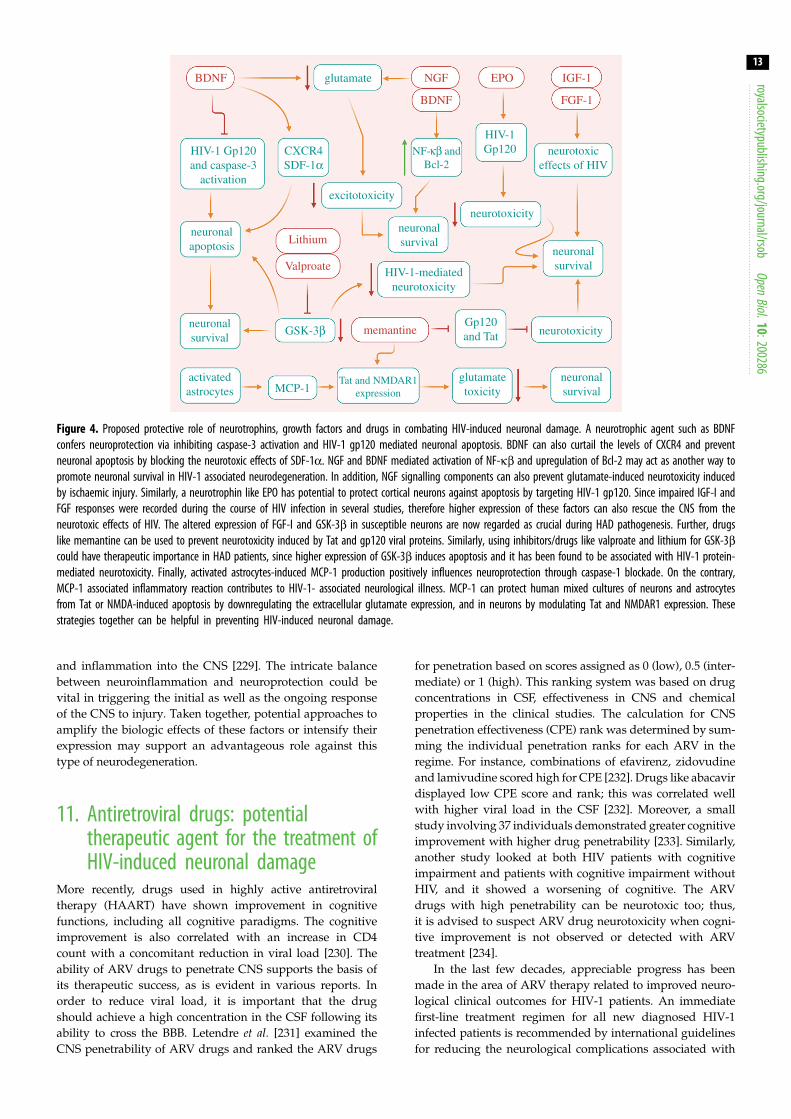
In the above sections, we comprehensively discussed variousunderlying interconnected mechanisms between HIV,neuroinflammation, HAND and AD. Understanding theunderlying mechanisms will help explore various possibletherapeutic strategies and agents, which may be able tocombat these complications. Unfortunately, there are no medi-cations identified so far, and very few studies are available ontherapeutic aspects. Neuroprotective therapies are designedwith a targeted approach to ameliorate damage and improvesurvival as well as the function of neurons. The mechanismsassociated with neuroprotection are classically aimed todiminish the extent of neuronal damage in HIV-1-inducedneuronal dysfunction. It can be considered that agents thatregulate inflammatory and/or cell death pathways andfavourably modulate neurotransmitter function may provideopportunities for pharmacological manipulation duringHIV-1 brain infections, although previous studies whichfocused on anti-inflammatory mechanisms have not demon-strated promising results in attenuating endogenousinflammation and considerable neuroprotection. As a result,a number of studies have recently been conducted to reduceneurotoxicity by blocking or modulating the actions of viralproteins, augmenting the protective action of neurotrophinsand growth factors, or curtailing neuroinflammation triggeredby HIV-1-infected microglia and macrophages (figure 4). Forinstance, the neuroprotective role of brain-derived neuro-trophic factor (BDNF) has recently been observed in HIV-1-mediated neurotoxicity. It appears a potent neurotrophicagent for HIV-1 associated neuronal injury, which confers neu-roprotection via inhibiting caspase-3 activation and HIV-1Gp120 mediated neuronal apoptosis [205]. Moreover, BDNFis also found to reduce the levels of CXC chemokine receptor-4 (CXCR4) and inhibit neuronal apoptosis by blocking theneurotoxic effects of SDF-1α, a ligand for CXCR4. The SDF-1-mediated apoptosis is quantitatively akin to that provokedby Gp120. CXCR4 activation can contribute to the cell deathof a different kind of neuronal population. Consequently,BDNF-mediated neuroprotection occurs by reducing CXCR4level that ultimately leads to the reduced activation of thisreceptor during HIV-1 neuropathogenesis [205]. Recently,activation of nuclear factor kappa beta (NF-κβ) mediatingnerve growth factor (NGF) and BDNF and rise in Bcl-2expression has also been reported to promote neuronalsurvival in HIV-1 associated neurodegeneration [206,207].Additionally, BDNF has also been reported to prevent gluta-mate-induced excitotoxicity through modulation of NMDAreceptors in HIV-1 patients [208]. Similarly, erythropoetin(Epo), a neurotrophin, can also confer neuroprotection againstHIV [209]. A higher dose of Epo for a long duration showed

HIV and neurocognitive disorders HIV and synaptic degeneration
HIV-associated neurocognitive disorders and Alzheimer’s disease
neuroinflammation
microglia
viral proteinschemokinescytokinesfree radicalstoxic substances
oxidative stressAb production Tau phosphorylationexcitotoxicity
oxidative stressCa2+ levelsenergy dyshomeostasis
glutamate
neuroinflammation(TNF-a and IL-6)
cell death (neuronalapoptosis)
oxidative stress (ROS production)
viral proteins
glucocorticoids
neurodegeneration cognitive and behaviour deficits
HPA-axisderegulation
ATP depletion (Na+–K+
pump and EAATs)
AbPP cleavage andprocessing (Ab)
Tau phosphorylation(GSK-3b and CDK5)
excitotoxicity (Ca2+
mobilization)glutamate uptake
HPA-axisderegulation
astrocyte BBB alteration BBB alteration
Figure 3. Schematic showing possible linkage between HAND, synaptic degeneration and AD.
royalsocietypublishing.org/journal/rsobOpen
Biol.10:200286
12
better neuroprotective effect against HIV-1 transmission frommother to infant [210]. It can also protect cortical neuronsagainst apoptosis by targeting HIV-1 Gp120 [211]. Theseobservations suggest that Epo can be considered as a potentialtherapeutic agent for the treatment of HAD [212]. Recently, thepromising role of recombinant human NGF (rhNGF) hasshown to improve the symptoms associated with both HIV-related neuropathy and diabetic polyneuropathy. Substantialevidence demonstrates that NGF signalling may also preventglutamate-induced neurotoxicity caused by ischemic injury.However, in HIV-1-induced neuronal damage, especiallyin the peripheral nervous system, NGF may have significanttherapeutic effects [213–215]. Activation of the insulin-likegrowth factor I (IGF-I) system is another potential approachto treat HAD, as it exhibited neuroprotective action againstneurotoxins [216–218]. Activating IGF-I-stimulated signallingcomponentsmayoffer a potential therapeutic approach to pro-tect susceptible neurons in HAD patients. Earlier, impairedIGF-I responses were reported during the course of HIV infec-tion [216–218]. In HIV-infected patients, reduced levels ofserum IGF-I have been observed particularly in children fail-ure to thrive and individuals displaying wasting syndrome[216]. Reduction in the levels of IGF-I in CNS may aggravateneuronal apoptosis in the course of HIV infection [218].Thus, it can be reasonably argued that activation of the IGF-Isystem or increased utilization of IGF-I-activated pathwaysmay signify a promising treatment approach to rescue neuronssusceptible or vulnerable to injury in HAD patients. Similarly,higher expression of fibroblast growth factor I FGF-I can also
rescue the CNS from the neurotoxic effects of HIV. Alteredexpression of FGF-I and GSK-3β in susceptible neurons canbe considered crucially important for the pathogenesis ofHAD and emergence of therapeutic strategies [219,220].
Furthermore, the Tat and Gp120 mediated neurotoxicitycan be fully blocked by memantine, an NMDA antagonistused well in the treatment of dementia [221,222]. It also ame-liorates hippocampal synaptic transmission in the SCIDmouse model of HIV-1-associated neurologic diseases [223].Recently, the use of inhibitors of GSK-3β in the brain suggestedthat regulation of GSK-3β activity in neurons may be vital forneuroprotection. Higher expressions of GSK-3β induced apop-tosis and showed association with HIV-1 protein-mediatedneurotoxicity [224,225]. As a consequence, pharmacologicalagents like valproate and lithium identified to inhibit GSK-3β activity could be valuable for therapeutic benefits in HADpatients.
The neuroprotective role of monocyte chemoattractantprotein 1 (MCP-1) has recently been observed in HIV andHAD patients [226,227]. Activated astrocytes-induced MCP-1production positively influences neuroprotection through thecaspase-1 blockade. On the contrary,MCP-1 associated inflam-matory reactions contribute to HIV-1-associated neurologicalailments [226,227]. MCP-1 can protect mixed cultures ofneurons and astrocytes from Tat or NMDA-induced apoptosisby downregulating the extracellular glutamate expression,along with modulating Tat and NMDAR1 expression [228].In the case of HAD, MCP-1 may exert a protective as well asa degenerative role as it is coupled with monocyte recruitment
BDNF
BDNF
memantine
Valproate
Lithium
NGF EPO IGF-1
FGF-1
glutamate
HIV-1Gp120 neurotoxic
effects of HIV
neurotoxicity
neurotoxicity
neuronalsurvival
neuronalsurvival
glutamatetoxicity
Tat and NMDAR1expressionMCP-1
activatedastrocytes
neuronalsurvival
neuronalsurvival
neuronalapoptosis
HIV-1 Gp120and caspase-3
activation
HIV-1-mediatedneurotoxicity
excitotoxicity
GSK-3bGp120and Tat
CXCR4SDF-1a
NF-kb andBcl-2
Figure 4. Proposed protective role of neurotrophins, growth factors and drugs in combating HIV-induced neuronal damage. A neurotrophic agent such as BDNFconfers neuroprotection via inhibiting caspase-3 activation and HIV-1 gp120 mediated neuronal apoptosis. BDNF can also curtail the levels of CXCR4 and preventneuronal apoptosis by blocking the neurotoxic effects of SDF-1α. NGF and BDNF mediated activation of NF-κβ and upregulation of Bcl-2 may act as another way topromote neuronal survival in HIV-1 associated neurodegeneration. In addition, NGF signalling components can also prevent glutamate-induced neurotoxicity inducedby ischaemic injury. Similarly, a neurotrophin like EPO has potential to protect cortical neurons against apoptosis by targeting HIV-1 gp120. Since impaired IGF-I andFGF responses were recorded during the course of HIV infection in several studies, therefore higher expression of these factors can also rescue the CNS from theneurotoxic effects of HIV. The altered expression of FGF-I and GSK-3β in susceptible neurons are now regarded as crucial during HAD pathogenesis. Further, drugslike memantine can be used to prevent neurotoxicity induced by Tat and gp120 viral proteins. Similarly, using inhibitors/drugs like valproate and lithium for GSK-3βcould have therapeutic importance in HAD patients, since higher expression of GSK-3β induces apoptosis and it has been found to be associated with HIV-1 protein-mediated neurotoxicity. Finally, activated astrocytes-induced MCP-1 production positively influences neuroprotection through caspase-1 blockade. On the contrary,MCP-1 associated inflammatory reaction contributes to HIV-1- associated neurological illness. MCP-1 can protect human mixed cultures of neurons and astrocytesfrom Tat or NMDA-induced apoptosis by downregulating the extracellular glutamate expression, and in neurons by modulating Tat and NMDAR1 expression. Thesestrategies together can be helpful in preventing HIV-induced neuronal damage.
royalsocietypublishing.org/journal/rsobOpen
Biol.10:200286
13
and inflammation into the CNS [229]. The intricate balancebetween neuroinflammation and neuroprotection could bevital in triggering the initial as well as the ongoing responseof the CNS to injury. Taken together, potential approaches toamplify the biologic effects of these factors or intensify theirexpression may support an advantageous role against thistype of neurodegeneration.
11. Antiretroviral drugs: potentialtherapeutic agent for the treatment ofHIV-induced neuronal damage
More recently, drugs used in highly active antiretroviraltherapy (HAART) have shown improvement in cognitivefunctions, including all cognitive paradigms. The cognitiveimprovement is also correlated with an increase in CD4count with a concomitant reduction in viral load [230]. Theability of ARV drugs to penetrate CNS supports the basis ofits therapeutic success, as is evident in various reports. Inorder to reduce viral load, it is important that the drugshould achieve a high concentration in the CSF following itsability to cross the BBB. Letendre et al. [231] examined theCNS penetrability of ARV drugs and ranked the ARV drugs
for penetration based on scores assigned as 0 (low), 0.5 (inter-mediate) or 1 (high). This ranking system was based on drugconcentrations in CSF, effectiveness in CNS and chemicalproperties in the clinical studies. The calculation for CNSpenetration effectiveness (CPE) rank was determined by sum-ming the individual penetration ranks for each ARV in theregime. For instance, combinations of efavirenz, zidovudineand lamivudine scored high for CPE [232]. Drugs like abacavirdisplayed low CPE score and rank; this was correlated wellwith higher viral load in the CSF [232]. Moreover, a smallstudy involving 37 individuals demonstrated greater cognitiveimprovement with higher drug penetrability [233]. Similarly,another study looked at both HIV patients with cognitiveimpairment and patients with cognitive impairment withoutHIV, and it showed a worsening of cognitive. The ARVdrugs with high penetrability can be neurotoxic too; thus,it is advised to suspect ARV drug neurotoxicity when cogni-tive improvement is not observed or detected with ARVtreatment [234].
In the last few decades, appreciable progress has beenmade in the area of ARV therapy related to improved neuro-logical clinical outcomes for HIV-1 patients. An immediatefirst-line treatment regimen for all new diagnosed HIV-1infected patients is recommended by international guidelinesfor reducing the neurological complications associated with

Table 4. Class, name and CNS penetration of the antiretroviral drugs[239,240].
class of drug name of the drugCNSpenetration
protease inhibitor tipranavir low
fosamprenavir medium
atazanavir medium
saquinavir low
nelfinavir low
lopinavir medium
ritonavir low
darunavir medium
indinavir medium
amprenavir medium
nucleoside reverse
transcriptase inhibitor
tenofovir disoproxil
fumarate
low
abacavir medium
didanosine medium
emtricitabine medium
stavudine medium
lamivudine medium
zidovudine high
entry/fusion inhibitors maraviroc high
enfuvirtide low
non-nucleoside reverse
transcriptase inhibitor
etravirine low
delavirdine high
nevirapine high
efavirenz medium
integrase strand transfer
inhibitor
raltegravir medium
elvitegravir medium
royalsocietypublishing.org/journal/rsobOpen
Biol.10:200286
14
HIV-1infected patients [235,236]. Current ARV therapy ishighly efficient in controlling HIV-1; still, viral replicationcan be found in the CSF among some patients. It has beenfound that ARVs reach different areas of CSF with significantvariability due to the different expression profiles of cellulardrug transporters and the concentrations of few ARVs donot the exceed inhibitory concentration for wild-type HIVreplication in CSF [237,238] (table 4). The main limitation toachieve the HIV-1 eradication from the brain is the subopti-mal concentrations of ARV within this site. Factors likemolecular weight, blood protein binding and lipophilicityinfluence the concentration of drug in the brain tissue[231,241–243]. For instance, while entry and integrase inhibi-tors are able to reach the CNS, the nucleoside/nucleotidereverse transcriptase inhibitors and non-nucleoside reversetranscriptase inhibitors can only partially cross the BBB. Con-versely, the majority of PIs are characterized by a medium/low permeability to the BBB [5,239,244,245]. Furthermore,some cellular transporters like P-gp, MRP4 and MRP5 havethe ability to reduce the intracellular concentration of ARVdrugs which ultimately favours both the emergence of
drug-resistant viruses and their productive infections toother cells [46,56,240,246].
New strategies like the usage of a hypertonic solution ofurea or mannitol [48,49] are currently used to increase the con-centrations of ARV within site. This deed can be achieved byinhibiting the drug efflux transport, while nanoparticles andcell-mediated nanoART may confer other key advantages,such as improved blood half-life and bioavailability, precisedelivery and higher aqueous stability [231]. Different typesof nanoparticles that have been identified for improving theconcentration of ARV are listed below:
1. Lipid nanoparticles have the ability to easily cross the BBB[247,248].
2. Polymeric nanoparticles are able to exploit the interactionwith low-density lipoproteins receptors on the surface ofendothelial cells [239,249].
3. Inorganic nanoparticles such as small size silica with theaddition of polyethylene glycol (PEG) [250].
4. Gold nanoparticles conjugated with cell-penetratingpeptides [251].
It has been recently reported that poly(dl-lactide-co-glycolide) nanoparticles and other nanoparticles increasethe peak concentrations of lopinavir, ritonavir and efavirenz(these drugs are characterized by a low penetration intoCNS) [239,252]. Recently, a CPE that depends on pharmaco-kinetics’ features of various ARV drugs was proposed toestimate the efficacy of ARV treatment in the CSF [238]. How-ever, some contradictory results of this CPE on clinicaloutcomes in HIV-1 infected patients have been reported insome of the studies [169,232,234]. These observations reflectthat further studies are required to prescribe ARV therapyand that the regimens characterized by high CPE scoresmust be carefully chosen. It has been demonstrated that inthe presence of high CPE, there is an acceleration of neurologi-cal disorders [253,254]. For instance, PIs are shown to induceoxidative stress in neuronal cells, while the NNRTI efavirenzcaused toxicity in the cortical neuronal cultures of fetal rats[253–255]. Still in vivo studies are needed to confirm the neuro-toxicity profiles of these drugs for potential applications.
Further, various reports highlighted the use of psychiatricmedication for mood disorders like depression. Many sub-types of antidepressants, including tricyclic antidepressants,serotonin–norepinephrine re-uptake inhibitors and selectiveserotonin re-uptake inhibitors, have been found useful inproviding moderate symptomatic relief [256,257]. Psychosti-mulants may also be useful for apathy and fatigue [258].Psychotic and manic symptoms are less reported in the caseof HIV+ individuals, though a small-scale study with psycho-sis demonstrated a higher occurrence of extrapyramidalsymptoms [259]. Numerous drugs such as mood stabilizers(like lithium) may have concurrent neurotoxic effects, andcarbamazepine may stimulate the same CYP enzyme systemwhich participates in themetabolism of ARVdrugs, and there-fore may cause drug–drug interactions [260,261]. However, ona pharmacological basis, many agents including memantine,nimodipine, selegiline, pentoxifylline and peptide T can beconsidered neuroprotective, although among these numerousagents, only selegiline appears to exhibit potential benefits[262].

royalsocietypublishing.org/journal/rsobOpen
Biol.10:200286
15
12. ConclusionBased on the available literature, it can be concluded that HIV-associated synaptic loss and aetiology of AD and HAND is aninterconnected and orchestrated consequence of numerousneuropathogenic processes triggered by HIV-1. Interactionsbetween HIV-1 and the host cells are believed to play a vitalrole in the pathogenesis of these abnormalities. Several viralproteins (Tat, Gp120, Nef and Vpr), which are released frominfected cells in the nervous system, may impart inductionof synaptic injury and pathogenesis of AD. In addition,these proteins are likely to act in conjunction and cause synap-totoxicity when released from infected cells in the CNS.Further, AD-associated numerous factors such as BBB regula-tors, members of the stress-related pathways as well as theamyloid and Tau pathways appear to augment amyloid pla-ques deposition or NFT accumulation following HIVneuroinfections. Additionally, the HPA axis dysregulationalso showed that when associatedwith HIV infection, it is con-ducive of generating an environment where BBB disruption,neuroinflammation, oxidative stress, excitotoxicity and Aβburden are exacerbated. This, combined with other factors(environmental/genetic), may provide a new insight forunderstanding the pathogenesis, diagnosis and therapeuticsof brain disorders including AD and HAND. The scenarioof replication-independent production of HIV-1 protein isapparently counterintuitive, and the underlying molecularmechanism is yet largely remained unexplored. It is thereforeimperative to explore more in this field. Considering the needfor therapeutics against HIV neuroinfection, unfortunately,still, there is an urgent need for evidence-based medicationsto be identified that would be able to combat these compli-cations. Many studies have recently shown a reduction inneurotoxicity viamodulating the actions of viral proteins, aug-menting the protective action of neurotrophins and growthfactors, or curtailing neuroinflammation triggered by HIV-1-infected microglia and macrophages. The mechanisms associ-ated with neuroprotection are classically aimed to diminishthe extent of neuronal damage inHIV-1-induced synaptic dys-function. Agents that regulate inflammatory and/or cell deathpathways and favourably modulate neurotransmitterfunction may provide opportunities for pharmacologicalmanipulation during HIV-1 brain infections. Altogether, inthe near future, it could be of paramount significance toexplore the molecular mechanisms of HIV neuroinfectionand develop therapeutic strategies.
Data accessibility. This article has no additional data.
Authors’ contributions. A.S., S.K.J., S.O., K.D., D.K.C., G.G. and K.K.K.initiated the draft of the review. N.K.J. and S.K.S. edited the reviewand performed proofreading and final editing. All authors readand approved the final manuscript. S.B., S.D.S., M.M.T. and J.R.contributed in revision of the review.
Competing interests. We declare we have no competing interests.
Funding. We received no funding for this study.
References
1. Jha NK, Jha SK, Sharma R, Kumar D, Ambasta RK,Kumar P. 2018 Hypoxia-induced signaling activationin neurodegenerative diseases: targets for newtherapeutic strategies. J. Alzheimers Dis. 62, 15–38.(doi:10.3233/JAD-170589)
2. Jha NK, Jha SK, Kar R, Ambasta RK, Kumar P. 2014Role of oxidative stress, ER stress and ubiquitinproteasome system in neurodegeneration. MOJ CellSci. Rep. 1, 38–44. (doi:10.15406/mojcsr.2014.01.00010)
3. Jha NK, Jha SK, Kumar D, Kejriwal N, Sharma R,Ambasta RK, Kumar P. 2015 Impact of insulindegrading enzyme and Neprilysin in Alzheimer’sdisease biology: characterization of putativecognates for therapeutic applications. J. AlzheimersDis. 48, 891–917. (doi:10.3233/JAD-150379)
4. Jha SK, Jha NK, Kumar D, Ambasta RK, Kumar P. 2017Linking mitochondrial dysfunction, metabolicsyndrome and stress signaling in neurodegeneration.Biochim. Biophys. Acta Mol. Basis Dis. 1863,1132–1146. (doi:10.1016/j.bbadis.2016.06.015)
5. Zhang YL, Ouyang YB, Liu LG, Chen DX. 2015 Blood-brain barrier and neuro-AIDS. Eur. Rev. Med.Pharmacol. Sci. 19, 4927–4939.
6. Anderson E, Zink W, Xiong H, Gendelman HE. 2002HIV-1-associated dementia: a metabolicencephalopathy perpetrated by virus-infected andimmunecompetent mononuclear phagocytes.J. Acquir. Immune Defic. Syndr. 31, S43–S54.(doi:10.1097/00126334-200210012-00004)
7. Andras IE, Toborek M. 2013 Amyloid betaaccumulation in HIV-1-infected brain: the role ofthe blood brain barrier. IUBMB Life 65, 43–49.(doi:10.1002/iub.1106)
8. Ghafouri M, Amini S, Khalili K, Sawaya BE. 2006HIV-1 associated dementia: symptoms and causes.Retrovirology 3, 28. (doi:10.1186/1742-4690-3-28)
9. Lawrence DM, Major EO. 2002 HIV-1 and the brain:connections between HIV-1-associated dementia,neuropathology and neuroimmunology. MicrobesInfect. 4, 301–308. (doi:10.1016/S1286-4579(02)01542-3)
10. Tucker KA, Robertson KR, Lin W, Smith JK, An H,Chen Y, Aylward SR, Hall CD. 2004 Neuroimaging inhuman immunodeficiency virus infection.J. Neuroimmunol. 157, 153–162. (doi:10.1016/j.jneuroim.2004.08.036)
11. Becker JT et al. 2009 Vascular risk factors, HIVserostatus, and cognitive dysfunction in gay andbisexual men. Neurology 73, 1292–1299. (doi:10.1212/WNL.0b013e3181bd10e7)
12. González-Scarano F, Martín-García J. 2005 Theneuropathogenesis of AIDS. Nat. Rev. Immunol. 5,69–81. (doi:10.1038/nri1527)
13. Clifford DB, Ances BM. 2013 HIV-associatedneurocognitive disorder. Lancet Infect. Dis. 13,976–986. (doi:10.1016/S1473-3099(13)70269-X)
14. Thakur KT, Boubour A, Saylor D, Das M, Bearden DR,Birbeck GL. 2018 Global HIV neurology: acomprehensive review. AIDS 33, 163–184.
15. Antinori A et al. 2007 Updated research nosologyfor HIV-associated neurocognitive disorders.Neurology 69, 1789–1799. (doi:10.1212/01.WNL.0000287431.88658.8b)
16. Blackstone K, Moore D, Woods S, Morgan E, FranklinD, Ellis R. 2012 The CHARTER Group. How‘asymptomatic’ Is HIV-associated asymptomaticneurocognitive impairment? In 19th Conf. onRetroviruses and Opportunistic Infections, Seattle,WA, Abstract 497.
17. Boska MD et al. 2004 Advances in neuroimaging forHIV-1 associated neurological dysfunction: clues tothe diagnosis, pathogenesis and therapeuticmonitoring. Curr. HIV Res. 2, 61–78. (doi:10.2174/1570162043485095)
18. Aylward EH, Brettschneider PD, McArthur JC, HarrisGJ, Schlaepfer TE, Henderer JD, Barta PE, Tien AY,Pearlson GD. 1995 Magnetic resonance imagingmeasurement of gray matter volume reductions inHIV dementia. Am. J. Psychiatry 152, 987–994.(doi:10.1176/ajp.152.7.987)
19. Kaul M, Garden GA, Lipton SA. 2001 Pathways toneuronal injury and apoptosis in HIV-associateddementia. Nature 410, 988–994. (doi:10.1038/35073667)
20. Anthony IC, Ramage SN, Carnie FW, Simmonds P,Bell JE. 2006 Accelerated Tau deposition in thebrains of individuals infected with humanimmunodeficiency virus-1 before and after theadvent of highly active anti-retroviral therapy. Acta

royalsocietypublishing.org/journal/rsobOpen
Biol.10:200286
16
Neuropathol. 111, 529–538. (doi:10.1007/s00401-006-0037-0)21. Smith DB, Simmonds P, Bell JE. 2014 Brain viralburden, neuroinflammation and neurodegenerationin HAART-treated HIV positive injecting drug users.J. Neurovirol. 20, 28–38. (doi:10.1007/s13365-013-0225-3)
22. Grassi MP, Clerici F, Perin C, Zocchetti C, Borella M,Cargnel A, Mangoni A. 1995 HIV infection and druguse: influence on cognitive function. AIDS 9,165–170. (doi:10.1097/00002030-199509020-00008)
23. Williams KC, Hickey WF. 2002 Central nervoussystem damage, monocytes and macrophages, andneurological disorders in AIDS. Annu. Rev. Neurosci.25, 537–562. (doi:10.1146/annurev.neuro.25.112701.142822)
24. Peng H, Sun L, Jia B, Lan X, Zhu B, Wu Y, Zheng J.2011 HIV-1-infected and immune-activatedmacrophages induce astrocytic differentiation ofhuman cortical neural progenitor cells via the STAT3pathway. PLoS ONE 6, e19439. (doi:10.1371/journal.pone.0019439)
25. Hauser KF, Fitting S, Dever SM, Podhaizer EM,Knapp PE. 2012 Opiate drug use and thepathophysiology of neuroAIDS. Curr. HIV Res. 10,435–452. (doi:10.2174/157016212802138779)
26. Thompson KA, Cherry CL, Bell JE, McLean CA. 2011Brain cell reservoirs of latent virus inpresymptomatic HIV-infected individuals.Am. J. Pathol. 179, 1623–1629. (doi:10.1016/j.ajpath.2011.06.039)
27. Liu Y, Liu H, Kim BO, Gattone VH, Li J, Nath A, BlumJ, He JJ. 2004 CD4-independent infection ofastrocytes by human immunodeficiency virus type1: requirement for the human mannose receptor.J. Virol. 78, 4120–4133. (doi:10.1128/JVI.78.8.4120-4133.2004)
28. Coiras M, López-Huertas MR, Pérez-Olmeda M,Alcamí J. 2009 Understanding HIV-1 latencyprovides clues for the eradication of long-termreservoirs. Nat. Rev. Microbiol. 7, 798–812. (doi:10.1038/nrmicro2223)
29. Cassol E, Alfano M, Biswas P, Poli G. 2006Monocyte-derived macrophages and myeloid celllines as targets of HIV-1 replication and persistence.J. Leukoc. Biol. 80, 1018–1130. (doi:10.1189/jlb.0306150)
30. Aquaro S, Bagnarelli P, Guenci T, De Luca A,Clementi M, Balestra E, Caliò R, Perno CF. 2002Long-term survival and virus production in humanprimary macrophages infected by humanimmunodeficiency virus. J. Med. Virol. 68, 479–488.(doi:10.1002/jmv.10245)
31. Koppensteiner H, Brack-Werner R, Schindler M.2012 Macrophages and their relevance inhuman immunodeficiency virus type Iinfection. Retrovirology 9, 82. (doi:10.1186/1742-4690-9-82)
32. Orenstein J, Fox C, Wahl S. 1997 Macrophages as asource of HIV during opportunistic infections.Science 276, 1857–1861. (doi:10.1126/science.276.5320.1857)
33. Coiras M. 2010 HIV-1 latency and eradication oflong-term viral reservoirs. Discov. Med. 9, 185–191.
34. Le Douce V, Herbein G, Rohr O, Schwartz C. 2010Molecular mechanisms of HIV-1 persistence in themonocyte-macrophage lineage. Retrovirology 7, 32.(doi:10.1186/1742-4690-7-32)
35. Zhang D, Hu X, Qian L, O’Callaghan JP, Hong J-S.2010 Astrogliosisi in CNS pathologies: is there arole for microglia? Mol. Neurobiol. 41, 232–241.(doi:10.1007/s12035-010-8098-4)
36. Farina C, Aloisi F, Meinl E. 2007 Astrocytes areactive players in cerebral innate immunity. TrendsImmunol. 28, 138–145. (doi:10.1016/j.it.2007.01.005)
37. Aikaterini A. 2008 Cellular reservoirs of HIV-1 andtheir role in viral persistence. Curr. HIV Res. 6,388–400. (doi:10.2174/157016208785861195)
38. Dong Y, Benveniste EN. 2001 Immune functionof astrocytes. Glia 190, 180–190. (doi:10.1002/glia.1107)
39. Narasipura SD, Henderson LJ, Fu SW, Chen L,Kashanchi F, Al-Harthi L. 2012 Role of β-cateninand TCF/LEF family members in transcriptionalactivity of HIV in astrocytes. J. Virol. 86, 1911–1921.(doi:10.1128/JVI.06266-11)
40. Albright AV, Soldan SS, González-Scarano F. 2003Pathogenesis of human immunodeficiency virus-induced neurological disease. J. Neurovirol. 9,222–227. (doi:10.1080/13550280390194073)
41. Aquaro S, Svicher V, Ronga L, Perno CF, Pollicita M.2008 HIV-1-associated dementia during HAARTtherapy. Recent Pat. CNS Drug Discov. 3, S23–S33.
42. Kohleisen B, Shumay E, Sutter G, Foerster R,Brack-Werner R, Nuesse M, Erfle V. 1999 Stableexpression of HIV-1 Nef induces changes in growthproperties and activation state of human astrocytes.AIDS 13, 2331–2341. (doi:10.1097/00002030-199912030-00004)
43. Wei L, Henderson LJ, Major EO, Al-Harthi L. 2011IFN-γ mediates enhancement of HIV replication inastrocytes by inducing an antagonist of theβ-catenin pathway (DKK1) in a STAT 3-dependentmanner. J. Immunol. 186, 6771–6778. (doi:10.4049/jimmunol.1100099)
44. Mamik MK, Banerjee S, Walseth TF, Hirte R, Tang L,Borgmann K, Ghorpade A. 2011 HIV-1 and IL-1βregulate astrocytic CD38 through mitogen-activatedprotein kinases and nuclear factor-κB signalingmechanisms. J. Neuroinflammation 8, 145. (doi:10.1186/1742-2094-8-145)
45. Trujillo R, Jaramillo-Rangel G, Ortega-Martinez M,Penalva De Oliveira AC, Vidal JE, Bryant J, Gallo RC.2005 International NeuroAIDS: prospects of HIV-1associated neurological complications. Cell Res. 15,962–969. (doi:10.1038/sj.cr.7290374)
46. Mukhtar M, Harley S, Chen P, BouHamdan M, PatelC, Acheampong E, Pomerantz RJ. 2002PrimaryIsolated human brain microvascularendothelial cells express diverse HIV/SIV-associatedchemokine coreceptors and DC-SIGN and L-SIGN.Virology 297, 78–88. (doi:10.1006/viro.2002.1376)
47. Hazleton JE, Berman JW, Eugenin EA. 2010 Novelmechanisms of central nervous system damage inHIVinfection. HIV/AIDS 2, 39–49.
48. Bergamaschi A, Pancino G. 2010 Host hindrance toHIV-1 replication in monocytes and macrophages.Retrovirology 7, 31. (doi:10.1186/1742-4690-7-31)
49. Coleman CM, Wu L. 2009 HIV interactions withmonocytes and dendritic cells: viral latency andreservoirs. Retrovirology 6, 51. (doi:10.1186/1742-4690-6-51)
50. Strazza M, Pirrone V, Wigdahl B, Nonnemacher MR.2011 Breaking down the barrier: the effects of HIV-1 on the blood-brain barrier. Brain Res. 1399,96–115. (doi:10.1016/j.brainres.2011.05.015)
51. Gavegnano C, Schinazi RF. 2010 Antiretroviraltherapy in macrophages: implication for HIVeradication. Antivir. Chem. Chemother. 20, 63–78.(doi:10.3851/IMP1374)
52. Meléndez LM, Colon K, Rivera L, Rodriguez-FrancoE, Toro-Nieves D. 2011 Proteomic analysis of HIV-infected macrophages. J. Neuroimmune Pharmacol.6, 89–106. (doi:10.1007/s11481-010-9253-4)
53. Crowe S, Zhu T, Muller WA. 2003 The contribution ofmonocyte infection and trafficking to viralpersistence,and maintenance of the viral reservoir inHIV infection. J. Leukoc. Biol. 74, 635–641. (doi:10.1189/jlb.0503204)
54. Ellery PJ et al. 2007 The CD16+ monocyte subset ismore harbors HIV-1 in vivo. J. Immunol. 178,6581–6589. (doi:10.4049/jimmunol.178.10.6581)
55. Khan NA, Di Cello F, Stins M, Kim KS. 2007 Gp120-mediated cytotoxicity of human brainmicrovascularendothelial cells is dependent on p38mitogen-activated protein kinase activation.J. Neurovirol. 13, 242–251. (doi:10.1080/13550280701286531)
56. Acheampong E, Parveen Z, Muthoga LW, Kalayeh M,Mukhatar M, Pomerantz RJ. 2005 Humanimmunodeficiency virus type 1 Nef potently inducesapoptosis in primary human brain microvascularendothelial cells via the activation of caspases.J. Virol. 79, 4257–4269. (doi:10.1128/JVI.79.7.4257-4269.2005)
57. Kim AY, Chung RT. 2009 Coinfection with HIV-1 andHCV-a one-two punch. Gastroenterology 137,795–814. (doi:10.1053/j.gastro.2009.06.040)
58. Guha D, Nagilla P, Redinger C, Srinivasan A,Schatten GP, Ayyavoo V. 2012 Neuronal apoptosisby HIV-1Vpr: contribution of proinflammatorymolecular networks from infected target cells.J. Neuroinflammation 9, 138. (doi:10.1186/1742-2094-9-138)
59. Hong S, Banks WA. 2015 Role of the immunesystem in HIV-associated neuroinflammation andneurocognitiveImplications. Brain Behav. Immun.45, 1–12. (doi:10.1016/j.bbi.2014.10.008)
60. Palacio M, Álvarez S, Muñoz-fernández MÁ. 2012HIV-1 infection and neurocognitive impairment inthe current era. Rev. Med. Virol. 22, 33–45. (doi:10.1002/rmv.711)
61. Vera JH, Guo Q, Rabiner I, Matthews P, Gunn R,Winston A. 2014 Neuroinflammation inasymptomatic HIV-infected subjects on effectivecART. In Proc. of the Conf. on Retroviruses andOpportunistic Infections (CROI), Boston, MA, USA,3–6 March 2014. Abstract 486LB.

royalsocietypublishing.org/journal/rsobOpen
Biol.10:200286
17
62. Vera JH et al. 2015 Microbial translocation isassociated with neuroinflammation in HIV-infected subjects on ART. In Proc. of the 22ndConf. on Retroviruses and Opportunistic Infections,Seattle, WA, USA, 23–26 February 2015.Abstract 477.63. Peudenier S, Héry C, Ng KH, Tardieu M. 1991 HIVreceptors within the brain: a study of CD4 andMHC-II on human neurons, astrocytes and microglialcells. Res. Virol. 142, 145–149. (doi:10.1016/0923-2516(91)90051-4)
64. Sturdevant CB, Joseph SB, Schnell G, Price RW,Swanstrom R, Spudich S. 2015 Compartmentalizedreplication of R5T cell-tropic HIV-1 in the centralnervous system early in the course of infection. PLoSPathog. 1, e1004720. (doi:10.1371/journal.ppat.1004720)
65. Ellis R, Langford D, Masliah E. 2007 HIV andantiretroviral therapy in the brain: neuronal injuryand repair. Nat. Rev. Neurosci. 8, 33–44. (doi:10.1038/nrn2040)
66. McArthur JC, Steiner J, Sacktor N, Nath A. 2010Human immunodeficiency virus-associatedneurocognitive disorders: mind the gap.Ann. Neurol. 67, 699–714. (doi:10.1002/ana.22053)
67. Maschke M, Kastrup O, Esser S, Ross B, Hengge U,Hufnagel A. 2000 Incidence and prevalence ofneurological disorders associated with HIV since theintroduction of highly active antiretroviral therapy(HAART). J. Neurol. Neurosurg. Psychiatry 69,376–380. (doi:10.1136/jnnp.69.3.376)
68. Heaton RK et al. 2011 HIV-associated neurocognitivedisorders before and during the era of combinationantiretroviral therapy: differences in rates, nature,and predictors. J. Neurovirol. 17, 3–16. (doi:10.1007/s13365-010-0006-1)
69. McArthur JC, Haughey N, Gartner S, Conant K, PardoC, Nath A, Sacktor N. 2003 Humanimmunodeficiency virus-associated dementia: anevolving disease. J. Neurovirol. 9, 205–221. (doi:10.1080/13550280390194109)
70. McArthur JC et al. 1993 Dementia in AIDS patients:incidence and risk factors. Multicenter AIDS Cohortstudy. Neurology 43, 2245–2252. (doi:10.1212/wnl.43.11.2245)
71. Soontornniyomkij V, Umlauf A, Soontornniyomkij B,Gouaux B, Ellis RJ, Levine AJ, Moore DJ, LetendreSL. 2018 Association of antiretroviral therapy withbrain aging changes among HIV-infected adults.AIDS 32, 2005.
72. Janssen R. 1992 Epidemiology of humanimmunodeficiency virus infection and the neurologiccomplications of the infection. Semin. Neurol. 12,10–17. (doi:10.1055/s-2008-1041152)
73. Williams DW, Anastos K, Morgello S, Berman JW.2015 JAMA and ALCAM are therapeutic targets toinhibit diapedesis across the BBB of CD14CCD16Cmonocytes in HIV-infected individuals. J. Leukoc.Biol. 97, 401–412. (doi:10.1189/jlb.5A0714-347R)
74. Cosenza MA, Zhao ML, Si Q, Lee SC. 2002 Humanbrain parenchymal microglia express CD14 and CD45and are productively infected by HIV-1 in HIV-1
encephalitis. Brain Pathol. 12, 442–455. (doi:10.1111/j.1750-3639.2002.tb00461.x)
75. Rothenaigner I, Kramer S, Ziegler M, Wolff H,Kleinschmidt A, Brack-werner R. 2007 Long-termHIV-1 infection of neural progenitor populations.AIDS 21, 2271–2281. (doi:10.1097/QAD.0b013e3282f12f27)
76. Eugenin EA, Clements JE, Zink MC, Berman JW.2011 Human immunodeficiency virus infection ofhuman astrocytes disrupts blood-brain barrierintegrity by a gap junction-dependent mechanism.J. Neurosci. 31, 9456–9465. (doi:10.1523/JNEUROSCI.1460-11.2011)
77. Hellmuth J, Valcour V, Spudich S. 2015 CNSreservoirs for HIV: implications for eradication.J. Virus Erad. 1, 67–71. (doi:10.1016/S2055-6640(20)30489-1)
78. Nutile-McMenemy N, Elfenbein A, Deleo JA. 2007Minocycline decreases in vitro microglial motility,beta1-integrin, and Kv1.3 channel expression.J. Neurochem. 103, 2035–2046. (doi:10.1111/j.1471-4159.2007.04889.x)
79. Lu S-M et al. 2011 HIV-1 Tat-induced microgliosisand synaptic damage via interactions betweenperipheral and central myeloid cells. PLoS ONE 6,e23915. (doi:10.1371/journal.pone.0023915)
80. Nath A, Conant K, Chen P, Scott C, Major EO. 1999Transient exposure to HIV-1 Tat protein results incytokine production in macrophages and astrocytes.A hit and run phenomenon. J. Biol. Chem. 274,17 098–17 102. (doi:10.1074/jbc.274.24.17098)
81. Zucchini S et al. 2013 Increased excitability in tat-transgenic mice: role of tatin HIV-relatedneurological disorders. Neurobiol. Dis. 55, 110–119.(doi:10.1016/j.nbd.2013.02.004)
82. Berman J, Carvallo L, Buckner CM, Luers A, PrevedelL, Bennett MV, Eugenin EA. 2016 HIV-tat altersConnexin43 expression and trafficking in humanastrocytes: role in NeuroAIDS. J. Neuroinflammation13, 54. (doi:10.1186/s12974-016-0510-1)
83. Fan Y, He JJ. 2016 HIV-1 Tat induces unfoldedprotein response and endoplasmic reticulum stressin astrocytes and causes neurotoxicity through GFAPactivation and aggregation. J. Biol. Chem. 291,22 819–22 829. (doi:10.1074/jbc.M116.731828)
84. Shin AH, Thayer SA. 2013 Human immunodeficiencyvirus-1 protein Tat induces excitotoxic loss ofpresynapticterminals in hippocampal cultures. Mol.Cell. Neurosci. 54, 22–29. (doi:10.1016/j.mcn.2012.12.005)
85. Bai L, Zhu X, Ma T, Wang J, Wang F, Zhang S. 2013The p38 MAPK NF-κB pathway, not the ERKpathway, is involved in exogenous HIV-1 Tat-induced apoptotic cell death in retinal pigmentepithelial cells. Int. J. Biochem. Cell Biol. 31, 1–8.
86. Midde N, Gomez A, Zhu J. 2012 HIV-1 Tat decreasesdopamine transporter cell surface expression andvescicular monoamine transporter-2 function in Ratstriatal synaptosomes. J. Neuroimmune Pharmacol.7, 629–639. (doi:10.1007/s11481-012-9369-9)
87. Chen L, Liu J, Xu C, Keblesh J, Zang W, Xiong H.2011 HIV-1gp120 induces neuronal apoptosisthrough enhancement of 4-aminopyridine-senstive
outward K+ currents. PLoS ONE 6, e25994. (doi:10.1371/journal.pone.0025994)
88. El-Hage N, Podhaizer EM, Sturgill J, Hauser KF. 2011Toll-like receptor expression and activation inastroglia: differential regulation by HIV-1 Tat,gp120, and morphine. Immunol. Investig. 40,498–522. (doi:10.3109/08820139.2011.561904)
89. Lamers S, Fogel G, Singer EJ, Salemi M, Nolan DJ,Huysentruyt LC, McGrath MS. 2012 HIV-1 Nef inmacrophage-mediated disease pathogenesis. Int.Rev. Immunol. 31, 432–450. (doi:10.3109/08830185.2012.737073)
90. Masanetz S, Lehmann MH. 2011 HIV-1 Nef increasesastrocyte sensitivity towards exogenoushydrogenperoxide. Virol. J. 8, 35. (doi:10.1186/1743-422X-8-35)
91. Nath A. 2002 Human immunodeficiency virus (HIV)proteins in neuropathogenesis of HIV dementia.J. Infect. Dis. 7609, 193–198. (doi:10.1086/344528)
92. Churchill MJ, Cowley DJ, Wesselingh SL, Gorry PR,Gray LR. 2015 HIV-1 transcriptional regulation in thecentral nervous system and implications for HIV cureresearch. J. Neurovirol. 21, 290–300. (doi:10.1007/s13365-014-0271-5)
93. Levy DN, Refaeli Y, MacGregor RR, Weiner DB. 1994Serum Vpr regulates productive infection andlatency of human immunodeficiency virus type 1.Proc. Natl Acad. Sci. USA 91, 10 873–10 877.(doi:10.1073/pnas.91.23.10873)
94. Hudson L, Liu J, Nath A, Jones M, Raghavan R,Narayan O, Male D, Everall I. 2000 Detection of thehuman immunodeficiency virus regulatory proteinTat in CNS tissues. J. Neurovirol. 6, 145–155.(doi:10.3109/13550280009013158)
95. Garden GA et al. 2002 Caspase cascades in humanimmunodeficiency virus-associatedneurodegeneration. J. Neurosci. 22, 4015–4024.(doi:10.1523/jneurosci.22-10-04015.2002)
96. Bachis A, Biggio F, Major EO, Mocchetti I. 2009M- and T-tropic HIVs promote apoptosis in ratneurons. J. Neuroimmune Pharmacol. 4, 150–160.(doi:10.1007/s11481-008-9141-3)
97. Haughey NJ, Nath A, Mattson MP, Slevin JT, GeigerJD. 2001 HIV-1 Tat through phosphorylation ofNMDA receptors potentiates glutamateexcitotoxicity. J. Neurochem. 78, 457–467. (doi:10.1046/j.1471-4159.2001.00396.x)
98. King JE, Eugenin EA, Buckner CM, Berman JW. 2006HIV tat and neurotoxicity. Microbes Infect. 8,1347–1357. (doi:10.1016/j.micinf.2005.11.014)
99. Kruman II, Nath A, Mattson MP. 1998 HIV-1 proteinTat induces apoptosis of hippocampal neurons by amechanism involving caspase activation, calciumoverload and oxidative stress. Exp. Neurol. 154,276–288. (doi:10.1006/exnr.1998.6958)
100. Ferrarese C, Aliprandi A, Tremolizzo L, Stanzani L,De Micheli A, Dolara A, Frattola L. 2001 Increasedglutamate in CSF and plasma of patients with HIVdementia. Neurology 57, 671–675. (doi:10.1212/wnl.57.4.671)
101. Sami SA, Cicalese S, Ahooyi TM, Khalili K, Amini S,Sariyer IK. 2017 HIV-1 Nef is released inextracellular vesicles derived from astrocytes:

royalsocietypublishing.org/journal/rsobOpen
Biol.10:200286
18
evidence for Nef-mediated neurotoxicity. Cell DeathDis. 8, e2542. (doi:10.1038/cddis.2016.467)102. Wang Y, Santerre M, Tempera I, Martin K, MukerjeeR, Sawaya BE. 2017 HIV-1 Vpr disruptsmitochondria axonal transport and acceleratesneuronal aging. Neuropharmacology 117, 364–375.(doi:10.1016/j.neuropharm.2017.02.008)
103. Bateman RJ et al. 2012 Clinical and biomarkerchanges in dominantly inherited Alzheimer’sdisease. N. Engl. J. Med. 367, 795–804. (doi:10.1056/NEJMoa1202753)
104. Marcello E, Epis R, Saraceno C, Di Luca M. 2012Synaptic dysfunction in Alzheimer’s disease. Adv.Exp. Med. Biol. 970, 573–601. (doi:10.1007/978-3-7091-0932-8_25)
105. Adle-Biassette H, Chrétien F, Wingertsmann L, HéryC, Ereau T, Scaravilli F, Tardieu M, Gray F. 1999Neuronal apoptosis does not correlate withdementia in HIV infection but is related tomicroglial activation and axonal damage.Neuropathol. Appl. Neurobiol. 25, 123–133. (doi:10.1046/j.1365-2990.1999.00167.x)
106. Avdoshina V, Bachis A, Mocchetti I. 2013 Synapticdysfunction in human immunodeficiency virus type-1-positive subjects: inflammation or impairedneuronal plasticity? J. Intern. Med. 273, 454–465.(doi:10.1111/joim.12050)
107. Lopardo GD, Bissio E, Iannella Mdel C, Crespo AD,Garone DB, Cassetti LI. 2009 Good neurocognitiveperformance measured by the international HIVdementia scale in early HIV-1 infection. J. Acquir.Immune Defic. Syndr. 52, 488–492. (doi:10.1097/QAI.0b013e3181b06348)
108. Cysique LAJ, Maruff P, Brew BJ. 2006 Variable benefitin neuropsychological function in HIV-infectedHAART-treated patients. Neurology 66, 1447–1450.(doi:10.1212/01.wnl.0000210477.63851.d3)
109. Alfahad T, Nath A. 2013 Retroviruses andamyotrophic lateral sclerosis. Antiviral Res. 99,180–187. (doi:10.1016/j.antiviral.2013.05.006)
110. Chakradhar S. 2018 A tale of two diseases: agingHIV patients inspire a closer look at Alzheimer’sdisease. Nat. Med. 24, 376–377. (doi:10.1038/nm0418-376)
111. Milanini B, Valcour V. 2017 Differentiating HIV-associated neurocognitive disorders fromAlzheimer’s disease: an emerging issue in geriatricNeuroHIV. Curr. HIV/AIDS Rep. 14, 123–132. (doi:10.1007/s11904-017-0361-0)
112. Kang YJ, Digicaylioglu M, Russo R, Kaul M, AchimCL, Fletcher L, Masliah E, Lipton SA. 2010Erythropoietin plus insulin-like growth factor-Iprotects against neuronal damage in a murinemodel of human immunodeficiency virus-associatedneurocognitive disorders. Ann. Neurol. 68, 342–352.(doi:10.1002/ana.22070)
113. Cho YE, Lee MH, Song BJ. 2017 Neuronal cell deathand degeneration through increased nitroxidativestress and tau phosphorylation in HIV-1 transgenicrats. PLoS ONE 12, e0169945. (doi:10.1371/journal.pone.0169945)
114. Masliah E, Achim CL, Ge N, DeTeresa R, Terry RD,Wiley CA. 1992 Spectrum of human
immunodeficiency virus-associated neocorticaldamage. Ann. Neurol. 32, 321–329. (doi:10.1002/ana.410320304)
115. Mishra M, Taneja M, Malik S, Khalique H, Seth P.2010 Human immunodeficiency virus type 1 Tatmodulates proliferation and differentiation ofhuman neural precursor cells: implication inNeuroAIDS. J. Neurovirol. 16, 355–367. (doi:10.3109/13550284.2010.513028)
116. Ketzler S, Weis S, Haug H, Budka H. 1990 Loss ofneurons in the frontal cortex in AIDS brains. ActaNeuropathol. 80, 92–94. (doi:10.1007/bf00294228)
117. Peters PJ, Bhattacharya J, Hibbitts S, Dittmar MT,Simmons G, Bell J, Simmonds P, Clapham PR. 2004Biological analysis of human immunodeficiencyvirus type 1 R5 envelopes amplified from brain andlymph node tissues of AIDS patients withneuropathology reveals two distinct tropismphenotypes and identifies envelopes in the brainthat confer an enhanced tropism and fusigenicityfor macrophages. J. Virol. 78, 6915–6926. (doi:10.1128/JVI.78.13.6915-6926.2004)
118. Vignoli AL, Martini I, Haglid KG, Silvestroni L,Augusti-Tocco G, Biagioni S. 2000 Neuronalglycolytic pathway impairment induced by HIVenvelope glycoprotein gp120. Mol. Cell. Biochem.215, 73–80. (doi:10.1023/A:1026590916661)
119. Vigorito M, LaShomb AL, Chang SL. 2007 Spatiallearning and memory in HIV-1 transgenic rats.J. Neuroimmune Pharmacol. 2, 319–328. (doi:10.1007/s11481-007-9078-y)
120. Brew BJ, Pemberton L, Blennow K, Wallin A,Hagberg L. 2005 CSF amyloid b42 and tau levelscorrelate with AIDS dementia complex. Neurology65, 1490–1492. (doi:10.1212/01.wnl.0000183293.95787.b7)
121. Patrick C, Crews L, Desplats P, Dumaop W,Rockenstein E, Achim CL, Everall IP, Masliah E. 2011Increased CDK5 expression in HIV encephalitiscontributes to neurodegeneration via tauphosphorylation and is reversed with Roscovitine.Am. J. Pathol. 178, 1646–1661. (doi:10.1016/j.ajpath.2010.12.033)
122. Esiri MM, Biddolph SC, Morris CS. 1998 Prevalenceof Alzheimer plaques in AIDS. J. Neurol. Neurosurg.Psychiatry 65, 29–33. (doi:10.1136/jnnp.65.1.29)
123. Green DA, Masliah E, Vinters HV, Beizai P, Moore DJ,Achim CL. 2005 Brain deposition of beta-amyloid isa common pathologic feature in HIV positivepatients. AIDS 19, 407–411. (doi:10.1097/01.aids.0000161770.06158.5c)
124. Kim J, Yoon JH, Kim YS. 2013 HIV-1 Tat interactswith and regulates the localization and processingof amyloid precursor protein. PLoS ONE 8, e77972.(doi:10.1371/journal.pone.0077972)
125. Lannuzel A, Tardieu M, Hery C, Barnier JV, Vincent JD,Guibert B, Van Tan H, Gray F. 1997 Humanimmunodeficiency virus type 1 and its coat proteingp120 induce apoptosis and activate JNK and ERKmitogen-activated protein kinases in human neurons.Ann. Neurol. 42, 847–856. (doi:10.1002/ana.410420605)
126. Kaul M, Lipton SA. 1999 Chemokines and activatedmacrophages in HIV gp120-induced neuronal
apoptosis. Proc. Natl Acad. Sci. USA 96, 8212–8216.(doi:10.1073/pnas.96.14.8212)
127. Costa A, Nappi RE, Polatti F, Poma A, Grossman AB,Nappi G. 2000 Stimulating effect of HIV-1 coatprotein gp120 on corticotropin releasing hormoneand arginine vasopressin in the rat hypothalamus:involvement of nitric oxide. Exp. Neurol. 166,376–384. (doi:10.1006/exnr.2000.7502)
128. Chrousos GP, Zapanti ED. 2014 Hypothalamic-pituitary-adrenal axis in HIV infection and disease.Endocrinol. Metab. Clin. North Am. 43, 791–806.(doi:10.1016/j.ecl.2014.06.002)
129. Christeff N, Gherbi N, Mammes O, Dalle MT,Gharakhanian S, Lortholary O, Melchior JC,Nunez EA. 1997 Serum cortisol and DHEAconcentrations during HIV infection.Psychoneuroendocrinology 22, S11–S18. (doi:10.1016/s0306-4530(97)00015-2)
130. Kino T. 2000 AIDS/HPA axis. In Endotext (eds LJ DeGroot, G Chrousos, K Dungan). South Dartmouth,MA: MDText.com Inc. See www.endotext.org
131. Anesten B, Yilmaz A, Hagberg L, Zetterberg H,Nilsson S, Brew BJ, Fuchs D, Price RW, Gisslén M.2016 Blood-brain barrier integrity, intrathecalimmunoactivation and neuronal injury in HIV.Neurol. Neuroimmunol. Neuroinflammation 3, e300.(doi:10.1212/nxi.0000000000000300)
132. Dreyer EB, Lipton SA. 1995 The coat protein gp120of HIV-1 inhibits astrocyte uptake of excitatoryamino acids via macrophage arachidonic acid.Eur. J. Neurosci. 7, 2502–2507. (doi:10.1111/j.1460-9568.1995.tb01048.x)
133. Belmadani A, Zou JY, Schipma MJ, Neafsey EJ,Collins MA. 2001 Ethanol pre-exposure suppressesHIV-1 glycoprotein 120-induced neuronaldegeneration by abrogating endogenousglutamate/Ca2+-mediated neurotoxicity.Neuroscience 104, 769–781. (doi:10.1016/s0306-4522(01)00139-7)
134. Clifford DB, Fagan AM, Holtzman DM, Morris JC,Teshome M, Shah AR, Kauwe JSK. 2009 CSFbiomarkers of Alzheimer disease in HIV-associatedneurologic disease. Neurology 73, 1982–1987.(doi:10.1212/WNL.0b013e3181c5b445)
135. Chen X, Hui L, Geiger NH, Haughey NJ, Geiger JD.2013 Endolysosome involvement in HIV-1transactivator protein-induced neuronal amyloidbeta production. Neurobiol. Aging 34, 2370–2378.(doi:10.1016/j.neurobiolaging.2013.04.015)
136. Ortega M, Ances BM. 2014 Role of HIV in amyloidmetabolism. J. Neuroimmune Pharmacol. 9,483–491. (doi:10.1007/s11481-014-9546-0)
137. Hategan A et al. 2017 HIV Tat protein and amyloid-b peptide form multifibrillar structures that causeneurotoxicity. Nat. Struct. Mol. Biol. 24, 379–386.(doi:10.1038/nsmb.3379)
138. Aksenov MY, Aksenova MV, Mactutus CF, Booze RM.2010 HIV-1 protein-mediated amyloidogenesis inrat hippocampal cell cultures. Neurosci. Lett. 475,174–178. (doi:10.1016/j.neulet.2010.03.073)
139. Daily A, Nath A, Hersh LB. 2006 Tat peptides inhibitneprilysin. J. Neurovirol. 12, 153–160. (doi:10.1080/13550280600760677)

royalsocietypublishing.org/journal/rsobOpen
Biol.10:200286
19
140. Stern AL et al. 2018 BACE1 mediates HIV-associatedand excitotoxic neuronal damage through an APP-dependent mechanism. J. Neurosci. 38, 4288–4300.(doi:10.1523/JNEUROSCI.1280-17.2018)141. Zeinolabediny Y et al. 2017 HIV-1 matrix proteinp17 misfolding forms toxic amyloidogenicassemblies that induce neurocognitive disorders.Sci. Rep. 7, 10313. (doi:10.1038/s41598-017-10875-0)
142. Chai Q, Jovasevic V, Malikov V, Sabo Y, Morham S,Walsh D, Naghavi MH. 2017 HIV-1 counteracts aninnate restriction by amyloid precursor proteinresulting in neurodegeneration. Nat. Commun. 8,1522. (doi:10.1038/s41467-017-01795-8)
143. Atluri VSR, Hidalgo M, Samikkannu T, Kurapati KRV,Jayant RD, Sagar V, Nair MPN. 2015 Effect of humanimmunodeficiency virus on blood–brain barrierintegrity and function: an update. Front. Cell.Neurosci. 9, 212. (doi:10.3389/fncel.2015.00212)
144. Zhao Z, Nelson AR, Betsholtz C, Zlokovic BV. 2015Establishment and dysfunction of the blood-brainbarrier. Cell 163, 1064–1078. (doi:10.1016/j.cell.2015.10.067)
145. Johnson NA, Jahng GH, Weiner MW, Miller BL, ChuiHC, Jagust WJ, Gorno-Tempini ML, Schuff N. 2005Pattern of cerebral hypoperfusion in Alzheimerdisease and mild cognitive impairment measuredwith arterial spin-labeling MR imaging: initialexperience. Radiology 234, 851–859. (doi:10.1148/radiol.2343040197)
146. Marchesi VT. 2011 Alzheimer’s dementia begins as adisease of small blood vessels, damaged byoxidative-induced inflammation and dysregulatedamyloid metabolism: implications for earlydetection and therapy. FASEB J. 25, 5–13. (doi:10.1096/fj.11-0102ufm)
147. Rosenberg GA. 2014 Blood-brain barrierpermeability in aging and Alzheimers disease.J. Prev. Alzheimers Dis. 1, 138–139. (doi:10.14283/jpad.2014.25)
148. Gosselet F, Saint-Pol J, Candela P, Fenart L. 2013Amyloid-b peptides, Alzheimer’s disease and theblood-brain barrier. Curr. Alzheimer Res. 10,1015–1033. (doi:10.2174/15672050113106660174)
149. Montagne A, Zhao Z, Zlokovic BV. 2017 Alzheimer’sdisease: a matter of blood-brain barrier dysfunction?J. Exp. Med. 214, 3151–3169. (doi:10.1084/jem.20171406)
150. Yamazaki Y, Kanekiyo T. 2017 Blood-brain barrierdysfunction and the pathogenesis of Alzheimer’sdisease. Int. J. Mol. Sci. 18, 1965. (doi:10.3390/ijms18091965)
151. Chen L, Choi JJ, Choi YJ, Hennig B, Toborek M. 2012HIV-1 Tat-induced cerebrovascular toxicity isenhanced in mice with amyloid deposits. Neurobiol.Aging 33, 1579–1590. (doi:10.1016/j.neurobiolaging.2011.06.004)
152. Chen Y, Huang W, Jiang W, Wu X, Ye B, Zhou X.2016 HIV-1 Tat regulates occludin and Ab transferreceptor expression in brain endothelial cells viaRho/ROCK signaling pathway. Oxid. Med. Cell.Longev. 2016, 4196572. (doi:10.1155/2016/4196572)
153. Jiang W, Huang W, Chen Y, Zou M, Peng D, Chen D.2017 HIV-1 transactivator protein induces ZO-1 andneprilysin dysfunction in brain endothelial cells viathe ras signaling pathway. Oxid. Med. Cell. Longev.2017, 3160360. (doi:10.1155/2017/3160360)
154. Kanmogne GD, Schall K, Leibhart J, Knipe B,Gendelman HE, Persidsky Y. 2007 HIV-1 gp120compromises blood-brain barrier integrity andenhance monocyte migration across blood-brainbarrier: implication for viral neuropathogenesis.J. Cereb. Blood Flow Metab. 27, 123–134. (doi:10.1038/sj.jcbfm.9600330)
155. Yang B, Akhter S, Chaudhuri A, Kanmogne GD.2009 HIV-1 gp120 induces cytokine expression,leukocyte adhesion and transmigration across theblood-brain barrier: modulatory effects of STAT1signaling. Microvasc. Res. 77, 212–219. (doi:10.1016/j.mvr.2008)
156. Louboutin JP, Reyes BAS, Agrawal L, Maxwell CR,Van Bockstaele EJ, Strayer DS. 2010 Blood-brainbarrier abnormalities caused by exposure to HIV-1gp120–protection by gene delivery of antioxidantenzymes. Neurobiol. Dis. 38, 313–325. (doi:10.1016/j.nbd.2010.02.007)
157. Everall I et al. 2009 Cliniconeuropathologiccorrelates of human immunodeficiency virus in theera of antiretroviral therapy. J. Neurovirol. 15,360–370. (doi:10.3109/13550280903131915)
158. Braak H, Alafuzoff I, Arzberger T, Kretzschmar H, DelTredici K. 2006 Staging of Alzheimer disease-associated neurofibrillary pathology using paraffinsections and immunocytochemistry. ActaNeuropathol. 112, 389–404. (doi:10.1007/s00401-006-0127-z)
159. Nebuloni M, Pellegrinelli A, Ferri A, Bonetto S,Boldorini R, Vago L, Grassi MP, Costanzi G. 2001Beta amyloid precursor protein and patterns of HIVp24 immunohistochemistry in different brain areasof AIDS patients. AIDS 15, 571–575. (doi:10.1097/00002030-200103300-00005)
160. Fjell AM, McEvoy L, Holland D, Dale AM, WalhovdKB. 2014 What is normal in normal aging? Effectsof aging, amyloid and Alzheimer’s disease on thecerebral cortex and the hippocampus. Prog.Neurobiol. 117, 20–40. (doi:10.1016/j.pneurobio.2014.02.004)
161. Piccini A et al. 2005 beta-amyloid is different innormal aging and in Alzheimer disease. J. Biol.Chem. 280, 34 186–34 192. (doi:10.1074/jbc.M501694200)
162. Jha NK, Jha SK, Kar R, Nand P, Swati K, GoswamiVK. 2019 Nuclear factor-kappa β as a therapeutictarget for Alzheimer’s disease. J. Neurochem. 150,113–137. (doi:10.1111/jnc.14687)
163. Jha SK, Jha NK, Kumar D, Sharma R, Shrivastava A,Ambasta RK, Kumar P. 2017 Stress-induced synapticdysfunction and neurotransmitter release inAlzheimer’s disease: can neurotransmitters andneuromodulators be potential therapeutic targets?J. Alzheimers Dis. 57, 1017–1039. (doi:10.3233/JAD-160623)
164. Price JL, Davis PB, Morris JC, White DL. 1991 Thedistribution of tangles, plaques and related
immunohistochemical markers in healthy aging andAlzheimer’s disease. Neurobiol. Aging 12, 295–312.(doi:10.1016/0197-4580(91)90006-6)
165. Banks WA, Ercal N, Price TO. 2006 The blood–brainbarrier in neuroAIDS. Curr. HIV Res. 4, 259–266.(doi:10.2174/157016206777709447)
166. Andras IE, Eum SY, Huang W, Zhong Y, Hennig B,Toborek M. 2010 HIV-1-induced amyloid betaaccumulation in brain endothelial cells is attenuatedby simvastatin. Mol. Cell. Neurosci. 43, 232–243.(doi:10.1016/j.mcn.2009.11.004)
167. Erickson MA, Banks WA. 2013 Blood–brain barrierdysfunction as a cause and consequence ofAlzheimer’s disease. J. Cereb. Blood Flow Metab. 33,1500–1513. (doi:10.1038/jcbfm.2013.135)
168. Gisslen M et al. 2009 Amyloid and tau cerebrospinalfluid biomarkers in HIV infection. BMC Neurol. 9, 63.(doi:10.1186/1471-2377-9-63)
169. Marra CM et al. 2009 Impact of combinationantiretroviral therapy on cerebrospinal fluid HIV RNAand neurocognitive performance. AIDS 23,1359–1366. (doi:10.1097/QAD.0b013e32832c4152)
170. Peluso MJ et al. 2013 Cerebrospinal fluid andneuroimaging biomarker abnormalities suggestearly neurological injury in a subset of individualsduring primary HIV infection. J. Infect. Dis. 207,1703–1712. (doi:10.1093/infdis/jit088)
171. Raber J, Huang Y, Ashford JW. 2004 ApoE genotypeaccounts for the vast majority of AD risk and ADpathology. Neurobiol. Aging 25, 641–650. (doi:10.1016/j.neurobiolaging.2003.12.023)
172. Chang L, Andres M, Sadino J, Jiang CS, Nakama H,Miller E, Ernst T. 2011 Impact of apolipoprotein Eepsilon4 and HIV on cognition and brain atrophy:antagonistic pleiotropy and premature brain aging.Neuroimage 58, 1017–1027. (doi:10.1016/j.neuroimage.2011.07.010)
173. Burt TD et al. 2008 Apolipoprotein (apo) E4enhances HIV-1 cell entry in vitro, and the APOEepsilon4/epsilon4 genotype accelerates HIV diseaseprogression. Proc. Natl Acad. Sci. USA 105,8718–8723. (doi:10.1073/pnas.0803526105)
174. Morgan EE et al. 2013 Apolipoprotein E4 genotypedoes not increase risk of HIV-associatedneurocognitive disorders. J. Neurovirol. 19,150–156. (doi:10.1007/s13365-013-0152-3)
175. Desplats P et al. 2013 Molecular and pathologicinsights from latent HIV-1 infection in the humanbrain. Neurology 80, 1415–1423. (doi:10.1212/WNL.0b013e31828c2e9e)
176. Paul RH, Ernst T, Brickman AM, Yiannoutsos CT, TateDF, Cohen RA, Navia B. 2008 Relative sensitivity ofmagnetic resonance spectroscopy and quantitativemagnetic resonance imaging to cognitive functionamong nondemented individuals infected with HIV.J. Int. Neuropsychol. Soc. 14, 725–733. (doi:10.1017/S1355617708080910)
177. Minghetti L, Visentin S, Patrizio M, Franchini L,Ajmone-Cat MA, Levi G. 2004 Multiple actions ofthe human immunodeficiency virus type-1 Tatprotein on microglial cell functions. Neurochem. Res.29, 965–978. (doi:10.1023/B:NERE.0000021241.90133.89)

royalsocietypublishing.org/journal/rsobOpen
Biol.10:200286
20
178. Mollace V, Nottet HS, Clayette P, Turco MC, MuscoliC, Salvemini D, Perno CF. 2001 Oxidative stress andneuroAIDS: triggers, modulators and novelantioxidants. Trends Neurosci. 24, 411–416. (doi:10.1016/S0166-2236(00)01819-1)179. Harman D, Hendricks S, Eddy DE, Seibold J. 1976Free radical theory of aging: effect of dietary fat oncentral nervous system function. J. Am. Geriatr. Soc.24, 301–307. (doi:10.1111/j.1532-5415.1976.tb06800.x)
180. Mattson MP, Haughey NJ, Nath A. 2005 Cell deathin HIV dementia. Cell Death Differ. 12, 893–904.(doi:10.1038/sj.cdd.4401577)
181. Tian C, Sun L, Jia B, Ma K, Curthoys N, Ding J,Zheng J. 2012 Mitochondrial glutaminase releasecontributes to glutamate-mediated neurotoxicityduring human immunodeficiency virus-1 infection.J. Neuroimmune Pharmacol. 7, 619–628. (doi:10.1007/s11481-012-9364-1)
182. Shi Q, Gibson GE. 2007 Oxidative stress andtranscriptional regulation in Alzheimer disease.Alzheimer Dis. Assoc. Disord. 21, 276–291. (doi:10.1097/WAD.0b013e31815721c3)
183. Nakamura T, Watanabe A, Fujino T, Hosono T,Michikawa M. 2009 Apolipoprotein E4 (1–272)fragment is associated with mitochondrial proteinsand affects mitochondrial function in neuronal cells.Mol. Neurodegener. 4, 35. (doi:10.1186/1750-1326-4-35)
184. Garvey LJ, Pavese N, Politis M, Ramlackhansingh A,Brooks DJ, Taylor-Robinson SD, Winston A. 2014Increased microglia activation in neurologicallyasymptomatic HIV infected patients receivingeffective ART. AIDS 28, 67–72. (doi:10.1097/01.aids.0000432467.54003.f7)
185. Zhang Y, Wang M, Li H, Zhang H, Shi Y, Wei F, LiuD, Liu K, Chen D. 2012 Accumulation of nuclear andmitochondrial DNA damage in the frontal cortexcells of patients with HIV-associated neurocognitivedisorders. Brain Res. 1458, 1–11. (doi:10.1016/j.brainres.2012.04.001)
186. Coughlin JM et al. 2014 Regional brain distributionof translocator protein using [C]DPA-713 PET inindividuals infected with HIV. J. Neurovirol. 20,219–232. (doi:10.1007/s13365-014-0239-5)
187. Edison P et al. 2008 Microglia, amyloid, andcognition in Alzheimer’s disease: an [11C](R)PK11195-PET and [11C]PIB-PET study. Neurobiol.Dis. 32, 412–419. (doi:10.1016/j.nbd.2008.08.001)
188. Wojda U, Salinska E, Kuznicki J. 2008 Calcium ionsin neuronal degeneration. IUBMB Life 60, 575–590.(doi:10.1002/iub.91)
189. Letendre SL, McCutchan JA, Childers ME, Woods SP,Lazzaretto D, Heaton RK, Grant I, Ellis RJ. 2004Enhancing antiretroviral therapy for humanimmunodeficiency virus cognitive disorders. Ann.Neurol. 56, 416–423. (doi:10.1002/ana.20198)
190. Harezlak J et al. 2011 Persistence of HIV-associatedcognitive impairment, inflammation, and neuronalinjury in era of highly active antiretroviraltreatment. AIDS 25, 625–633. (doi:10.1097/QAD.0b013e3283427da7)
191. Brenchley JM et al. 2006 Microbial translocation is acause of systemic immune activation in chronic HIVinfection. Nat. Med. 12, 1365–1371. (doi:10.1038/nm1511)
192. Lyn-Cook Jr LE et al. 2009 Hepatic ceramide maymediate brain insulin resistance andneurodegeneration in type 2 diabetes and non-alcoholic steatohepatitis. J. Alzheimers Dis. 16,715–729. (doi:10.3233/JAD-2009-0984)
193. Devlin KN, Gongvatana A, Clark US, Chasman JD,Westbrook ML, Tashima KT, Navia B, Cohen RA.2012 Neurocognitive effects of HIV, hepatitis C, andsubstance use history. J. Int. Neuropsychol. Soc. 18,68–78. (doi:10.1017/S1355617711001408)
194. Gongvatana A et al. 2011 Clinical contributors tocerebral white matter integrity in HIV-infectedindividuals. J. Neurovirol. 17, 477–486. (doi:10.1007/s13365-011-0055-0)
195. Nix LM, Tien PC. 2014 Metabolic syndrome,diabetes, and cardiovascular risk in HIV. Curr. HIV/AIDS Rep. 11, 271–278. (doi:10.1007/s11904-014-0219-7)
196. El-Sadr WM, Mullin CM, Carr A, Gibert C, RappoportC, Visnegarwala F, Grunfeld C, Raghavan SS. 2005Effects of HIV disease on lipid, glucose and insulinlevels: results from a large antiretroviral-naivecohort. HIV Med. 6, 114–121. (doi:10.1111/j.1468-1293.2005.00273.x)
197. Roriz-Filho SJ, Sá-Roriz TM, Rosset I, Camozzato AL,Santos AC, Chaves ML, Moriguti J, Roriz-Cruz M.2009 (Pre)diabetes, brain aging, and cognition.Biochim. Biophys. Acta 1792, 432–443. (doi:10.1016/j.bbadis.2008.12.003)
198. Yaffe K. 2007 Metabolic syndrome and cognitivedisorders: is the sum greater than its parts?Alzheimer Dis. Assoc. Disord. 21, 167–171. (doi:10.1097/WAD.0b013e318065bfd6)
199. Cohen RA et al. 2009 Vascular and cognitivefunctions associated with cardiovascular disease inthe elderly. J. Clin. Exp. Neuropsychol. 31, 96–110.(doi:10.1080/13803390802014594)
200. Correia SC, Santos RX, Carvalho C, Cardoso S,Candeias E, Santos MS, Oliveira CR, Moreira PI. 2012Insulin signaling, glucose metabolism andmitochondria: major players in Alzheimer’s diseaseand diabetes interrelation. Brain Res. 1441, 64–78.(doi:10.1016/j.brainres.2011.12.063)
201. Izycka-Swieszewska E, Zoltowska A, Rzepko R, GrossM, Borowska-Lehman J. 2000 Vasculopathy andamyloid beta reactivity in brains of patients withacquired immune deficiency (AIDS). FoliaNeuropathol. 38, 175–182.
202. Nelson L, Gard P, Tabet N. 2014 Hypertension andinflammation in Alzheimer’s disease: close partnersin disease development and progression!.J. Alzheimers Dis. 41, 331–343. (doi:10.3233/JAD-140024)
203. Ru W, Tang SJ. 2017 HIV-associated synapticdegeneration. Mol. Brain 10, 40. (doi:10.1186/s13041-017-0321-z)
204. Canet G et al. 2018 HIV neuroinfection andAlzheimer’s disease: similarities and potential links?
Front. Cell. Neurosci. 12, 307. (doi:10.3389/fncel.2018.00307)
205. Bachis A, Major EO, Mocchetti I. 2003 Brain-derivedneurotrophic factor inhibits humanimmunodeficiency virus-1/gp120-mediatedcerebellar granule cell death by preventing gp120internalization. J. Neurosci. 23, 5715–5722. (doi:10.1523/JNEUROSCI.23-13-05715.2003)
206. Macdonald NJ, Perez-Polo JR, Bennett AD,Taglialatela G. 1999 NGF-resistant PC12 cell deathinduced by arachidonic acid is accompanied by adecrease of active PKC zeta and nuclear factorkappa B. J. Neurosci. Res. 57, 219–226. (doi:10.1002/(SICI)1097-4547(19990715)57:2<219::AID-JNR7>3.0.CO;2-C)
207. Ramirez SH, Sanchez JF, Dimitri CA, Gelbard HA,Dewhurst S, Maggirwar SB. 2001 Neurotrophinsprevent HIV Tat-induced neuronal apoptosis via anuclear factor-kappaB (NF-kappaB)-dependentmechanism. J. Neurochem. 78, 874–889. (doi:10.1046/j.1471-4159.2001.00467.x)
208. Brandoli C, Sanna A, De Bernardi MA, Follesa P,Brooker G, Mocchetti I. 1998 Brain-derivedneurotrophic factor and basic fibroblast growthfactor downregulate NMDA receptor function incerebellar granule cells. J. Neurosci. 18, 7953–7961.(doi:10.1523/JNEUROSCI.18-19-07953.1998)
209. Romsi P et al. 2002 Potential neuroprotectivebenefits of erythropoietin during experimentalhypothermic circulatory arrest. J. Thorac. Cardiovasc.Surg. 124, 714–723. (doi:10.1067/mtc.2002.123704)
210. Gassmann M, Heinicke K, Soliz J, Ogunshola OO,Marti HH, Hofer T, Grimm C, Heinicke I, Egli B. 2003Non-erythroid functions of erythropoietin. Adv. Exp.Med. Biol. 543, 323–330. (doi:10.1007/978-1-4419-8997-0_22)
211. Digicaylioglu M, Kaul M, Fletcher L, Dowen R,Lipton SA. 2004 Erythropoietin protectscerebrocortical neurons from HIV-1/gp120-induceddamage. Neurorepor 15, 761–763. (doi:10.1097/00001756-200404090-00004)
212. Digicaylioglu M, Lipton SA. 2001 Erythropoietin-mediated neuroprotection involves cross-talkbetween Jak2 and NF-kappaB signalling cascades.Nature 412, 641–647. (doi:10.1038/35088074)
213. Semkova I, Schilling M, Henrich-Noack P, Rami A,Krieglstein J. 1996 Clenbuterol protects mouse cerebralcortex and rat hippocampus from ischemic damage andattenuates glutamate neurotoxicity in culturedhippocampal neurons by induction of NGF. Brain Res.717, 44–54. (doi:10.1016/0006-8993(95)01567-1)
214. Semkova I, Krieglstein J. 1999 Neuroprotectionmediated via neurotrophic factors and induction ofneurotrophic factors. Brain Res. Brain Res. Rev. 30,176–188. (doi:10.1016/S0165-0173(99)00013-2)
215. Chiaretti A, Piastra M, Polidori G, Di Rocco C, CarestaE, Antonelli A, Amendola T, Aloe L. 2003 Correlationbetween neurotrophic factor expression andoutcome of children with severe traumatic braininjury. Intensive Care Med. 29, 1329–1338. (doi:10.1007/s00134-003-1852-6)

royalsocietypublishing.org/journal/rsobOpen
Biol.10:200286
21
216. Jain S, Golde DW, Bailey R, Geffner ME. 1998Insulin-like growth factor-I resistance. Endocr. Rev.19, 625–646.217. Rondanelli M et al. 2002 Insulin-like growth factor I(IGF-I) and IGF-binding protein 3 response togrowth hormone is impaired in HIV-infectedchildren. AIDS Res. Hum. Retrovir. 18, 331–339.(doi:10.1089/088922202753519106)
218. Ying WJ, Peruzzi F, Lassak A, Del Valle L,Radhakrishnan S, Rappaport J, Khalili K, Amini S,Reiss K. 2003 Neuroprotective effects of IGF-Iagainst TNFalpha-induced neuronal damage in HIV-associated dementia. Virolog 305, 66–76. (doi:10.1006/viro.2002.1690)
219. Everall IP, Trillo-Pazos G, Bell C, Mallory M, SandersV, Masliah E. 2001 Amelioration of neurotoxiceffects of HIV envelope protein gp120 by fibroblastgrowth factor: a strategy for neuroprotection.J. Neuropathol. Exp. Neurol. 60, 293–301. (doi:10.1093/jnen/60.3.293)
220. Everall IP, Bell C, Mallory M, Langford D, Adame A,Rockestein E, Masliah E. 2002 Lithium ameliorates HIV-gp120-mediated neurotoxicity. Mol. Cell. Neurosci. 21,493–501. (doi:10.1006/mcne.2002.1196)
221. Jain KK. 2000 Evaluation of memantine forneuroprotection in dementia. Expert Opin.Investig. Drug 9, 1397–1406. (doi:10.1517/13543784.9.6.1397)
222. Bormann J. 1989 Memantine is a potent blocker ofN-methyl-daspartate (NMDA) receptor channels.Eur. J. Pharmacol. 166, 591–592. (doi:10.1016/0014-2999(89)90385-3)
223. Anderson ER, Gendelman HE, Xiong H. 2004Memantine protects hippocampal neuronal functionin murine human immunodeficiency virus type 1encephalitis. J. Neurosci. 24, 7194–7198. (doi:10.1523/JNEUROSCI.1933-04.2004)
224. Maggirwar SB, Tong N, Ramirez S, Gelbard HA,Dewhurst S. 1999 HIV-1 Tat-mediated activationof glycogen synthase kinase-3beta contributesto Tat-mediated neurotoxicity. J. Neurochem.73, 578–586. (doi:10.1046/j.1471-4159.1999.0730578.x)
225. Tong N, Sanchez JF, Maggirwar SB, Ramirez SH, GuoH, Dewhurst S, Gelbard HA. 2001 Activation ofglycogen synthase kinase 3 beta (GSK-3beta) byplatelet activating factor mediates migration andcell death in cerebellar granule neurons.Eur. J. Neurosci. 13, 1913–1922. (doi:10.1046/j.0953-816x.2001.01572.x)
226. McManus CM, Brosnan CF, Berman JW. 1998Cytokine induction of MIP-1 alpha and MIP-1 betain human fetal microglia. J. Immunol. 160,1449–1455.
227. Wang EJ et al. 2003 Microglia from mice transgenicfor a provirus encoding a monocyte- tropic HIV type1 isolate produce infectious virus and display invitro and in vivo upregulation of lipopolysaccharideinduced chemokine gene expression. AIDS Res. Hum.Retrovir. 19, 755–765. (doi:10.1089/088922203769232557)
228. Eugenin EA, D’Aversa TG, Lopez L, Calderon TM,Berman JW. 2003 MCP-1 (CCL2) protects human
neurons and astrocytes from NMDA or HIV-tat-induced apoptosis. J. Neurochem. 85, 1299–1311.(doi:10.1046/j.1471-4159.2003.01775.x)
229. Kelder W, McArthur JC, Nance-Sproson T, McClernonD, Griffin DE. 1998 Beta-chemokines MCP-1 andRANTES are selectively increased in cerebrospinalfluid of patients with human immunodeficiencyvirus-associated dementia. Ann. Neurol. 44,831–835. (doi:10.1002/ana.410440521)
230. Tozzi V et al. 2007 Persistence of neuropsychologicdeficits despite long-term highly active antiretroviraltherapy in patients with HIV-related neurocognitiveimpairment: prevalence and risk factors. J. Acquir.Immune Defic. Syndr. 45, 174–182. (doi:10.1097/QAI.0b013e318042e1ee)
231. Letendre S et al. 2008 Validation of the CNSpenetration-effectiveness rank for quantifyingantiretroviral penetration into the central nervoussystem. Arch. Neurol. 65, 65–70. (doi:10.1001/archneurol.2007.31)
232. Caniglia EC et al. 2014 HIV-CAUSAL Collaboration.Antiretroviral penetration into the CNS andincidence of AIDS-defining neurologic conditions.Neurology 83, 134–141. (doi:10.1212/WNL.0000000000000564)
233. Cysique LA, Vaida F, Letendre S, Gibson S, ChernerM, Woods SP, McCutchan JA, Heaton RK, Ellis RJ.2009 Dynamics of cognitive change in impairedHIV-positive patients initiating antiretroviral therapy.Neurology 73, 342–348. (doi:10.1212/WNL.0b013e3181ab2b3b)
234. Robertson KR, Su Z, Margolis DM, Krambrink A,Havlir DV, Evans S, Skiest DJ, A5170 Study Team.2010 Neurocognitive effects of treatmentinterruption in stable HIV-positive patients in anobservational cohort. Neurology 74, 1260–1266.(doi:10.1212/WNL.0b013e3181d9ed09)
235. Department of Health and Human Services. 2016Guidelines for the use of antiretroviral agents inHIV-infected adults and adolescents. See https://aidsinfo.nih.gov/contentfiles/lvguidelines/adultandadolescentgl.pdf (accessed on 16 July2016).
236. Nowacek A, Gendelman H. 2009 NanoART,neuroAIDS and CNS drug delivery. Nanomedicine 4,557–574. (doi:10.2217/nnm.09.38)
237. Bleasby K et al. 2006 Expression profiles of 50xenobiotic transporter genes in humans and pre-clinical species: a resource for investigations intodrug disposition. Xenobiotica 36, 963–988. (doi:10.1080/00498250600861751)
238. Ene L, Duiculescu D, Ruta SM. 2011 How much doantiretroviral drugs penetrate into the centralnervous system? J. Med. Life 4, 432–439.
239. Fiandra L, Capetti A, Sorrentino L, Corsi F. 2017Nanoformulated antiretrovirals for penetration ofthe central nervous system: state of the art.J. Neuroimmune Pharmacol. 12, 17–30. (doi:10.1007/s11481-016-9716-3)
240. Saksena NK, Wang B, Zhou L, Soedjono M, Ho YS,Conceicao V. 2010 HIV reservoirs in vivo and newstrategies for possible eradication of HIV from thereservoir sites. HIV/AIDS 2, 103–122.
241. Varatharajan L, Thomas SA. 2009 The transport ofanti-HIV drugs across blood-CNS interfaces:summary of current knowledge andrecommendations for further research. Antivir. Res.82, A99–A109. (doi:10.1016/j.antiviral.2008.12.013)
242. Strazielle N, Ghersi-Egea J-F. 2005 Factors affectingdelivery of antiviral drugs to the brain. Rev. Med.Virol. 15, 105–133. (doi:10.1002/rmv.454)
243. Letendre SL, Ellis RJ, Ances BM, McCutchan JA. 2010Neurologic complications of HIV disease and theirtreatment. Top HIV Med. 18, 45–55.
244. Best BM et al. 2009 Low Atazanavir concentrationsin cerebrospinal fluid. AIDS 23, 83–87. (doi:10.1097/QAD.0b013e328317a702)
245. De Oliveira FTM, do Olival GS, de Oliveira ACP. 2015Central nervous system antiretroviral highpenetration therapy. J. AIDS Clin. Res. 6, 12.
246. Aquaro S et al. 1997 Inhibition of replication of HIVin primary monocyte/macrophages by differentantiviral drugs and comparative efficacy inlymphocytes. J. Leukoc. Biol. 62, 138–143. (doi:10.1002/jlb.62.1.138)
247. Kaur IP, Bhandari R, Bhandari S, Kakkar V. 2008Potential of solid lipid nanoparticles in braintargeting. J. Control. Release 127, 97–109. (doi:10.1016/j.jconrel.2007.12.018)
248. Lai F, Fadda AM, Sinico C. 2013 Liposomes for braindelivery. Exp. Opin. Drug Deliv. 10, 1003–1022.(doi:10.1517/17425247.2013.766714)
249. Saiyed ZM, Gandhi NH, Nair MPN. 2010 Magneticnanoformulation of azidothymidine 50-triphosphatefor targeted delivery across the blood-brain barrier.Int. J. Nanomed. 5, 157–166. (doi:10.2147/IJN.S8905)
250. Liu D, Lin B, Shao W, Zhu Z, Ji T, Yang C. 2014 Invitro and in vivo studies on the transport ofPEGylated silica nanoparticles across the blood–brain barrier. ACS Appl. Mater. Interfaces 6,2131–2136. (doi:10.1021/am405219u)
251. Cheng Y et al. 2014 Blood–brain barrier permeablegold nanoparticles: an efficient delivery platform forenhanced malignant glioma therapy and imaging.Small 29, 5137–5150.
252. Borgmann K, Rao K, Labhasetwar V, Ghorpade A. 2011Efficacy of Tat-conjugated ritonavir-loadednanoparticles in reducing HIV-1 replication inmonocyte-derived macrophages and cytocompatibilitywith macrophages and human neurons. AIDS Res. Hum.Retrovir. 27, 853–862. (doi:10.1089/aid.2010.0295)
253. Akay C et al. 2014 Antiretroviral drugs induceoxidative stress and neuronal damage in the centralnervous system. J. Neurovirol. 20, 39–53. (doi:10.1007/s13365-013-0227-1)
254. Akay-Espinoza C, Stern AL, Nara RL, Panvelker N, LiJ, Jordan-Sciutto K-L. 2017 Differential invitroneurotoxicity of antiretroviral drugs. InProceedings of the Conference on Retroviruses andOpportunistic Infections (CROI), Seattle, WA, USA,13–16 February 2017.
255. Robertson K, Liner J, Meeker RB. 2012 Antiretroviralneurotoxicity. J. Neurovirol. 18, 388–399. (doi:10.1007/s13365-012-0120-3)
256. Rabkin JG, Rabkin R, Wagner G. 1994 Effects offluoxetine on mood and immune status in

royalsocietypublishing.org/jou
22
depressed patients with HIV illness. J. Clin.Psychiatry 55, 92–97.257. Perkins DO, Stern RA, Golden RN, Murphy C,Naftolowitz D, Evans DL. 1994 Mood disorders in HIVinfection: prevalence and risk factors in anonepicenter of the AIDS epidemic. Am. J. Psychiatry151, 233–236. (doi:10.1176/ajp.151.2.299)
258. Hinkin CH, Castellon SA, Hardy DJ, Farinpour R,Newton T, Singer E. 2001 Methylphenidate improvesHIV-1-associated cognitive slowing.
J. Neuropsychiatry Clin. Neurosci. 13, 248–254.(doi:10.1176/jnp.13.2.248)
259. Sewell DD, Jeste DV, Atkinson JH, Heaton RK, HesselinkJR, Wiley C, Thal L, Chandler JL, Grant I. 1994 HIV-associated psychosis: a study of 20 cases. San Diego HIVneurobehavioral research center group. Am. J. Psychiatry151, 237–242. (doi:10.1176/ajp.151.2.237)
260. Parenti DM, Simon GL, Scheib RG, Meyer WA, SzteinMB, Paxton H, DiGioia RA, Schulof RS. 1988 Effect oflithium carbonate in HIV-infected patients with
immune dysfunction. J. Acquir. Immune Defic. Syndr(1988). 1, 119–124.
261. Halman MH, Worth JL, Sanders KM, Renshaw PF,Murray GB. 1993 Anticonvulsant use in thetreatment of manic syndromes in patients with HIV-1 infection. J. Neuropsychiatry Clin. Neurosci. 5,430–434. (doi:10.1176/jnp.5.4.430)
262. Dubé B, Benton T, Cruess DG, Evans DL. 2005Neuropsychiatric manifestations of HIV infection andAIDS. J. Psychiatry Neurosci. 30, 237–246.
r
nal /rsobOpenBiol.10:200286