Expanded Basal Compartment and Disrupted Barrier in Vocal ...
25
Citation: King, R.E.; Ward-Shaw, E.T.; Hu, R.; Lambert, P.F.; Thibeault, S.L. Expanded Basal Compartment and Disrupted Barrier in Vocal Fold Epithelium Infected with Mouse Papillomavirus MmuPV1. Viruses 2022, 14, 1059. https://doi.org/ 10.3390/v14051059 Academic Editor: Sanghyuk Chung Received: 6 April 2022 Accepted: 11 May 2022 Published: 16 May 2022 Publisher’s Note: MDPI stays neutral with regard to jurisdictional claims in published maps and institutional affil- iations. Copyright: © 2022 by the authors. Licensee MDPI, Basel, Switzerland. This article is an open access article distributed under the terms and conditions of the Creative Commons Attribution (CC BY) license (https:// creativecommons.org/licenses/by/ 4.0/). viruses Article Expanded Basal Compartment and Disrupted Barrier in Vocal Fold Epithelium Infected with Mouse Papillomavirus MmuPV1 Renee E. King 1,2 , Ella T. Ward-Shaw 1 , Rong Hu 3 , Paul F. Lambert 1 and Susan L. Thibeault 2, * 1 McArdle Laboratory for Cancer Research, Department of Oncology, University of Wisconsin-Madison, Madison, WI 53705, USA; [email protected] (R.E.K.); [email protected] (E.T.W.-S.); [email protected] (P.F.L.) 2 Department of Surgery, University of Wisconsin-Madison, Madison, WI 53705, USA 3 Department of Pathology and Laboratory Medicine, University of Wisconsin-Madison, Madison, WI 53705, USA; [email protected] * Correspondence: [email protected] Abstract: Laryngeal infection with low-risk human papillomaviruses can cause recurrent respiratory papillomatosis (RRP), a disease with severe effects on vocal fold epithelium resulting in impaired voice function and communication. RRP research has been stymied by limited preclinical models. We recently reported a murine model of laryngeal MmuPV1 infection and disease in immunodeficient mice. In the current study, we compare quantitative and qualitative measures of epithelial prolifer- ation, apoptosis, differentiation, and barrier between mice with MmuPV1-induced disease of the larynx and surrounding tissues and equal numbers of uninfected controls. Findings supported our hypothesis that laryngeal MmuPV1 infection recapitulates many features of RRP. Like RRP, MmuPV1 increased proliferation in infected vocal fold epithelium, expanded the basal compartment of cells, decreased differentiated cells, and altered cell–cell junctions and basement membrane. Effects of MmuPV1 on apoptosis were equivocal, as with RRP. Barrier markers resembled human neoplastic disease in severe MmuPV1-induced disease. We conclude that MmuPV1 infection of the mouse larynx provides a useful, if imperfect, preclinical model for RRP that will facilitate further study and treatment development for this intractable and devastating disease. Keywords: mouse; papillomavirus; larynx; vocal folds; epithelium; recurrent respiratory papillo- matosis; RRP; MmuPV1 1. Introduction Recurrent respiratory papillomatosis (RRP), largely caused by laryngeal infection with low-risk human papillomavirus (HPV) types 6 and 11, remains a rare but highly morbid human health concern without satisfactory treatment options [1]. RRP research has been slowed due to insufficient model systems [2]. In our companion paper [3], we described a novel mouse model of laryngeal papillomavirus infection using Mus musculus papillomavirus (MmuPV1). We found that, while vocal fold infections with MmuPV1 could cause benign disease, progression to cancer was frequent in our model, whereas it is rare in human RRP [4]. Viral gene expression is found in laryngeal tissues in our MmuPV1 infection model, as seen in human RRP [5–7]. However, we found MmuPV1 capsid production in squamous metaplasia only in our mouse model, while low-risk HPV capsid protein is found in sparse, superficial cells in human RRP lesions but is not absent [8]. Characterization of vocal fold epithelial changes induced by MmuPV1 is necessary to assess the utility of laryngeal MmuPV1 infection as a model for human RRP. Vocal folds are paired, layered soft tissue structures within the larynx that contact each other and vibrate to produce voice. Vocal fold epithelium is critical to normal voice production [9] and withstands impact, tensile, and shear forces hundreds of times per second during conversational speech [10–16]. Like other tissue sites with high exposure Viruses 2022, 14, 1059. https://doi.org/10.3390/v14051059 https://www.mdpi.com/journal/viruses
-
Upload
khangminh22 -
Category
Documents
-
view
1 -
download
0
Transcript of Expanded Basal Compartment and Disrupted Barrier in Vocal ...

Citation: King, R.E.; Ward-Shaw, E.T.;
Hu, R.; Lambert, P.F.; Thibeault, S.L.
Expanded Basal Compartment and
Disrupted Barrier in Vocal Fold
Epithelium Infected with Mouse
Papillomavirus MmuPV1. Viruses
2022, 14, 1059. https://doi.org/
10.3390/v14051059
Academic Editor: Sanghyuk Chung
Received: 6 April 2022
Accepted: 11 May 2022
Published: 16 May 2022
Publisher’s Note: MDPI stays neutral
with regard to jurisdictional claims in
published maps and institutional affil-
iations.
Copyright: © 2022 by the authors.
Licensee MDPI, Basel, Switzerland.
This article is an open access article
distributed under the terms and
conditions of the Creative Commons
Attribution (CC BY) license (https://
creativecommons.org/licenses/by/
4.0/).
viruses
Article
Expanded Basal Compartment and Disrupted Barrier in VocalFold Epithelium Infected with Mouse Papillomavirus MmuPV1Renee E. King 1,2 , Ella T. Ward-Shaw 1, Rong Hu 3, Paul F. Lambert 1 and Susan L. Thibeault 2,*
1 McArdle Laboratory for Cancer Research, Department of Oncology, University of Wisconsin-Madison,Madison, WI 53705, USA; [email protected] (R.E.K.); [email protected] (E.T.W.-S.);[email protected] (P.F.L.)
2 Department of Surgery, University of Wisconsin-Madison, Madison, WI 53705, USA3 Department of Pathology and Laboratory Medicine, University of Wisconsin-Madison,
Madison, WI 53705, USA; [email protected]* Correspondence: [email protected]
Abstract: Laryngeal infection with low-risk human papillomaviruses can cause recurrent respiratorypapillomatosis (RRP), a disease with severe effects on vocal fold epithelium resulting in impairedvoice function and communication. RRP research has been stymied by limited preclinical models. Werecently reported a murine model of laryngeal MmuPV1 infection and disease in immunodeficientmice. In the current study, we compare quantitative and qualitative measures of epithelial prolifer-ation, apoptosis, differentiation, and barrier between mice with MmuPV1-induced disease of thelarynx and surrounding tissues and equal numbers of uninfected controls. Findings supported ourhypothesis that laryngeal MmuPV1 infection recapitulates many features of RRP. Like RRP, MmuPV1increased proliferation in infected vocal fold epithelium, expanded the basal compartment of cells,decreased differentiated cells, and altered cell–cell junctions and basement membrane. Effects ofMmuPV1 on apoptosis were equivocal, as with RRP. Barrier markers resembled human neoplasticdisease in severe MmuPV1-induced disease. We conclude that MmuPV1 infection of the mouselarynx provides a useful, if imperfect, preclinical model for RRP that will facilitate further study andtreatment development for this intractable and devastating disease.
Keywords: mouse; papillomavirus; larynx; vocal folds; epithelium; recurrent respiratory papillo-matosis; RRP; MmuPV1
1. Introduction
Recurrent respiratory papillomatosis (RRP), largely caused by laryngeal infectionwith low-risk human papillomavirus (HPV) types 6 and 11, remains a rare but highlymorbid human health concern without satisfactory treatment options [1]. RRP researchhas been slowed due to insufficient model systems [2]. In our companion paper [3], wedescribed a novel mouse model of laryngeal papillomavirus infection using Mus musculuspapillomavirus (MmuPV1). We found that, while vocal fold infections with MmuPV1could cause benign disease, progression to cancer was frequent in our model, whereasit is rare in human RRP [4]. Viral gene expression is found in laryngeal tissues in ourMmuPV1 infection model, as seen in human RRP [5–7]. However, we found MmuPV1capsid production in squamous metaplasia only in our mouse model, while low-risk HPVcapsid protein is found in sparse, superficial cells in human RRP lesions but is not absent [8].Characterization of vocal fold epithelial changes induced by MmuPV1 is necessary to assessthe utility of laryngeal MmuPV1 infection as a model for human RRP.
Vocal folds are paired, layered soft tissue structures within the larynx that contacteach other and vibrate to produce voice. Vocal fold epithelium is critical to normal voiceproduction [9] and withstands impact, tensile, and shear forces hundreds of times persecond during conversational speech [10–16]. Like other tissue sites with high exposure
Viruses 2022, 14, 1059. https://doi.org/10.3390/v14051059 https://www.mdpi.com/journal/viruses
Viruses 2022, 14, 1059 2 of 25
to mechanical forces, vocal fold epithelium is stratified squamous [17]. Vocal folds arevulnerable to HPV6 and 11 infection and are the primary site of lesion growth in RRP [18].
RRP in humans induces changes in vocal fold epithelium at tissue and molecularlevels. Hyperplasia in human RRP produces an irregular epithelial contour and masslesions, which disrupt normal vocal fold vibration and voice production [19]. Healthyvocal fold epithelium is characterized by proliferating cells in the basal and parabasallayers and differentiated suprabasal cells [20–22]. Human RRP lesions are characterizedby an expanded basal compartment, proliferating cells throughout epithelial layers, anddecreased differentiation [23–27]. High-risk HPVs are known to suppress apoptosis ininfected cells [28], and apoptosis may play a role in vocal fold epithelial diseases [29].However, evidence for changes in apoptosis in human RRP lesions is equivocal [30–33].Cell–cell junctions in vocal folds are primarily tight junctions; adherens junctions and gapjunctions are also present [20,34]. RNA-seq experiments in RRP tissues have demonstrateddecreased expression of cell junction and adhesion molecules [35]. Transmission electronmicroscopy (TEM) has demonstrated wide and irregular intercellular spaces in human RRPlesions compared with normal vocal fold epithelium [36–38]. Basement membrane betweenvocal fold epithelium and lamina propria contributes to barrier function of vocal foldmucosa [20,39]. High-risk HPV infection increases production of matrix metalloproteinases(MMPs), thinning the basement membrane, which is a necessary step in progression ofhigh-risk HPV disease to invasive cancer [40]. This phenomenon is less clear in humanRRP. TEM and histology studies have described vocal fold basement membrane in humanRRP lesions as either discontinuous [36,37] or intact [38].
Murine laryngeal anatomy and physiology are grossly similar to those of humansbut differ in several ways [41]. Mice and other rodents have a unique tissue fold calledthe ventral pouch, laryngeal pouch, or laryngeal sac in the ventral aspect of the larynx,superior to the vocal folds [41–47]. All evidence suggests that mouse vocalizations inthe range of human hearing are produced using vocal fold vibration [48–50]. However,routine murine social communication is performed using ultrasonic vocalization [51],which is produced within the larynx, but not via vocal fold vibration [49,52,53]. The mostrecent evidence demonstrates that the ventral pouch is required for ultrasonic vocalizationand suggests that vocal folds contribute by muscular action regulating ventral pouchsize [52]. Like humans, murine vocal folds are layered, and their epithelium is stratifiedsquamous [42,47,54]. Vocal fold epithelium is thinner in mice than in humans, comprising2–4 cell layers rather than 5–10 [20,54]. Likely because of this difference, there is one layerof proliferating basal cells rather than two or three [54–57]. The same markers of basalcells (p63), proliferating cells (Ki67), differentiated cells (cytokeratin 13 (K13)), cell–celljunctions (E-cadherin, β-catenin, zonula occludens 1 (ZO-1)), and basement membrane(laminin, collagen IV) are present in the vocal fold epithelium of humans [20–22,34,58–60]and mice [34,47,54–57,61]. Mouse vocal fold epithelium is also positive for cytokeratin 8(K8), a marker of simple and respiratory epithelium [54]. This is not seen in native adulthuman vocal folds [21,22,60]. Overall, however, vocal fold epithelium is similar betweenhumans and mice. We hypothesize that laryngeal MmuPV1 infection recapitulates featuresof human RRP, specifically increased proliferation, decreased differentiation, altered cell–cell junctions, and thinning of the basement membrane.
Here, we characterize epithelial proliferation, apoptosis, differentiation, and barrierintegrity in vocal fold disease induced by MmuPV1. We compared outcomes betweencontrol and diseased tissues at multiple stages of disease severity from mild dysplasia toinvasive squamous cell carcinoma. We found that, consistent with hyperplasia in humanRRP, the total number of epithelial cells increased in diseased vocal folds and increasedwith disease severity. Proliferating cells increased, while apoptotic cells did not change indiseased vocal folds. Like in human RRP, the basal compartment expanded, and differenti-ation markers decreased in diseased vocal fold epithelium. Cell–cell junctions decreased,as found in human RRP, but other interesting patterns of expression were noted, includingnuclear expression of adherens junction marker E-cadherin and extramembranous, basal

Viruses 2022, 14, 1059 3 of 25
expression of tight junction marker ZO-1. Interestingly, laminin was found within diseasedMmuPV1-infected vocal fold epithelium rather than being limited to the basement mem-brane zone. Overall, our findings demonstrate that laryngeal MmuPV1 infection is a useful,albeit imperfect, model of human RRP.
2. Materials and Methods2.1. Animals, MmuPV1 Infection, Samples, and Pathology Grading
Adult male and female NOD scid gamma (NSG) mice [62] were used for this study.Animal procedures were approved by the University of Wisconsin-Madison InstitutionalAnimal Care and Use Committee (IACUC #M005871) and conducted in accordance with theNational Institutes of Health Guide for the Care and Use of Laboratory Animals [63]. Laryngeswere infected with MmuPV1 under endoscopy with and without vocal fold abrasion.Tissues were collected weeks 1, 2, 4, 8, and 12 post-infection and processed into formalin-fixed, paraffin-embedded (FFPE) slides. Hematoxylin and eosin (H&E)-stained slides weregraded for disease severity in vocal folds, epiglottis and ventral pouch, arytenoids andaryepiglottic folds, proximal trachea, and hypopharynx by a Head and Neck Pathologistblinded to treatment group and timepoint post treatment. The disease was graded as mild,moderate, or severe dysplasia, or invasive cancer. Carcinoma in situ was graded as severedysplasia. Detailed methods for animal experiments, MmuPV1 infection, sample collection,and pathology grading can be found in our companion paper [3].
2.2. Study Design
We compared quantitative and qualitative measures between mice with MmuPV1-induceddysplasia or cancer of the vocal folds, epiglottis/ventral pouch, arytenoids/aryepiglottic folds,proximal trachea, and hypopharynx, and equal numbers of uninfected controls. Samplesrepresented a range of timepoints post infection within each degree of disease severity. Controlanimals were matched to diseased animals by injury status, sex, and timepoint post injuryand/or infection. Control tissue sections were matched with diseased tissue sections in ventral-dorsal location within the larynx. Within diseased tissues, we also compared outcome measuresamong different degrees of disease severity. For proliferation assays, we stained and quantifiedsections from 6 mice per disease stage within each tissue, or the maximum number availableif fewer than 6. For the apoptosis assay and markers of epithelial differentiation and barrier,we stained sections from 3 mice per disease stage within each tissue, or the maximum numberavailable if fewer than 3. Sex, injury status, and timepoint were balanced within each diseasestage for all experiments.
2.3. Immunofluorescence and Immunohistochemistry Staining
FFPE slides were stained following standard protocols for immunohistochemistry(IHC) and immunofluorescent (IF) staining. Briefly, sections were deparaffinized in xyleneand rehydrated in a series of ethanols. For IHC, endogenous peroxidase was quenched in3% hydrogen peroxide in methanol for 10 min. Antigen retrieval was performed by boilingslides in a microwave oven for 20 min in buffer. Antigen retrieval buffer was 10 mM citratepH 6.0 for Ki67 or 1X Tris-EDTA pH 9.0 (Abcam ab93684, Cambridge, United Kingdom)for all other targets. Sections were permeabilized for 20 min in either 0.5% Triton X-100for nuclear targets (p63, Ki67) or 0.2% PBS-Tween for non-nuclear targets (cytokeratins,ZO-1, E-cadherin, laminin). Slides were blocked for 1 h in 5% goat serum, then incubatedin primary antibodies overnight at 4 ◦C. Primary and secondary antibodies are listed inTable 1. Select sections were incubated in 5% goat serum instead of primary antibody as acontrol for background staining. Slides were washed and incubated in secondary antibodiesfor 1 h at room temperature. For IHC, signal was detected with 3,3′-diaminobenzidine(DAB; SK-4100, Vector Laboratories, Inc., Burlingame, CA, USA) for 10 min, and thenslides were counterstained for 1 min with hematoxylin QS (H-3404, Vector Laboratories,Inc.), dehydrated, cleared with xylene, mounted, and coverslipped. For IF, nuclei werecounterstained with 1X Hoechst 33342 (H1399, Fisher Scientific, Hampton, NH, USA) for

Viruses 2022, 14, 1059 4 of 25
10 min, and then slides were mounted and coverslipped with Prolong Diamond mountingmedia (P36970, Fisher Scientific), cured flat at room temperature in the dark for 24 h, andstored at 4 ◦C.
Table 1. Primary and secondary antibodies.
Primary Antibody Species Dilution Supplier Catalog # Application
Ki67 Rabbit 1:50 Abcam ab16667 IHCCytokeratin 8 (K8) Rabbit 1:200 LS Bio, Seattle, WA USA LS-B7928 IF
Cytokeratin 13 (K13) Mouse 1:100 Fisher Scientific MA1-35542 IF
p63 Mouse 1:200 Biocare Medical, Pacheco, CA,USA CM163A IF
Zonula occludens 1 (ZO-1) Mouse 1:200 Fisher Scientific 33-9100 IF
E-cadherin (E-cad) Rabbit 1:200 Cell Signaling Technology,Danvers, MA. USA 3195S IF
Laminin (Lam) Rabbit 1:200 Abcam ab11575 IF
Secondary antibody Species Dilution Supplier Catalog # Application
Horseradish peroxidase (HRP) Goat anti-rabbit 1:500 Jackson Immuno Research Labs,West Grove, PA, USA 111-035-144 IHC
Alexa Fluor 488 Goat anti-rabbit 1:500 Fisher Scientific A-11008 IFCy3 Goat anti-mouse 1:500 Jackson Immuno Research Labs 115-166-003 IF
2.4. TUNEL Staining
Slides were stained for apoptotic cells using terminal deoxynucleotidyl transferase(TdT)-mediated deoxyuridine triphosphate (dUTP)-biotin nick end labeling (TUNEL) usingthe ApopTag Peroxidase In Situ Apoptosis Detection kit (S7100, MilliporeSigma, Burlington,MA, USA). Manufacturer protocols were modified to reduce background staining in mouselaryngeal and respiratory tissues. Briefly, slides were deparaffinized, rehydrated, andpretreated for 15 min with proteinase K (19133, Qiagen, Hilden, Germany) diluted to20 ug/mL. Endogenous peroxidase was quenched in 0.3% hydrogen peroxide in PBSfor 5 min. Sections were washed, incubated in equilibration buffer for 10 s, and thenincubated in working strength TdT enzyme at 37 ◦C for 50 min. TdT enzyme was dilutedto 3:100 with reaction buffer and diH2O (3 ul enzyme, 70 ul reaction buffer, and 27 uLdiH2O per 100 uL). Select sections were incubated in diH2O instead of TdT enzyme as anegative control. The reaction was stopped by agitation followed by 10 min of incubationin working strength stop/wash buffer prepared per manufacturer protocol. Sections werethen washed and incubated in anti-digoxigenin peroxidase for 30 min at room temperature.The signal was detected with DAB (SK-4100, Vector Laboratories, Inc.) for 10 s, and thenslides were counterstained for 30 s with hematoxylin QS (H-3404, Vector Laboratories, Inc.),dehydrated, cleared with xylene, mounted, and coverslipped. To verify the kit activity withprotocol modifications, as a positive control select sections were treated for 10 min withDNase I (EN0523, Fisher Scientific) diluted to 1000 U/mL in 1X buffer containing MgCl2between proteinase K pretreatment and endogenous peroxidase quenching.
2.5. MmuPV1 RNA in Situ Hybridization
In situ hybridization (ISH) for MmuPV1 E4 transcript was completed using RNAscope(2.5 HD Reagent Kit-Brown, 322300, Advanced Cell Diagnostics, Newark, CA, USA) withprobes specific for MmuPV1 E4 (473281) according to the manufacturer’s instructions.
2.6. Image Acquisition
Images were acquired on a Nikon Eclipse Ti2 inverted microscope with NIS Elementssoftware (Nikon, Tokyo, Japan). For quantitative proliferation and apoptosis assays, one30× field was captured of each tissue of interest. This magnification captures a coronalsection of the membranous mouse vocal fold. For other assays, areas of interest werephotographed at 40×. Images were aligned to match tissue locations between diseasedsamples and injury-, sex-, and timepoint-matched controls. Images were adjusted inAdobe Photoshop (Adobe Inc., San Jose, CA, USA) or the GNU Image Manipulation
Viruses 2022, 14, 1059 5 of 25
Program (GIMP Development Team, volunteer group of software developers with nocorporate headquarters) for consistency across slides relative to background fluorescence.For proliferation and apoptosis assays, total epithelial cells and DAB-labeled epithelial cellswere quantified as described below. For IF assays, staining presence, relative intensity, andsubcellular and tissue-level localization were described qualitatively.
2.7. Proliferation, Apoptosis, and Basal Cell Quantification
For proliferation and apoptosis assays, 30× images of Ki67- and TUNEL-stainedtissues were opened in ImageJ (National Institutes of Health, Bethesda, MD, USA). Totalepithelial cells and DAB-labeled cells were enumerated using an automated countingmacro “BrdU Count v2.12.ijm” developed by Dr. David Ornelles (Wake Forest University,Winston-Salem, NC, USA) as previously described [64–67]. Prior to using the macro forthese experiments, settings were adjusted for accuracy in laryngeal and respiratory epitheliain comparison to manual cell counts of 30× images of vocal fold and trachea (Figure S1).To quantify basal cells, total nuclei and p63-positive nuclei were counted in 40× images ofp63- and Hoechst-stained epithelium using the ImageJ macro “RGB fluorescent cell countv1.32.ijm,” also developed by Dr. Ornelles.
2.8. Statistical Analysis
Labeled and unlabeled epithelial cells from the Ki67-stained slides were used for theanalysis of total epithelial cell counts. Proliferation, apoptosis, and basal cell indices werecalculated as percentages of Ki67+, TUNEL+, and p63+ cells, respectively. Data were testedfor normality using the Kolmogorov–Smirnov test. Within each tissue type, difference be-tween control and diseased tissues was tested with the paired T-test or Wilcoxon rank sumtest, as appropriate for distribution. For Ki67 and TUNEL, differences among disease sever-ity levels within a tissue and differences among laryngeal tissues (vocal folds, epiglottis, andarytenoids) were tested with the Kruskal–Wallis test with Dwass–Steel–Critchlow–Fligner(DSCF) pairwise post hoc test. For some analyses, data from laryngeal tissues were pooled.All samples stained for TUNEL were a subset of those stained for Ki67, and serial slideswere stained. We analyzed the reliability of total epithelial cell counts between both stainsusing intraclass correlation coefficients (ICC) in a 2-way, mixed-effects, absolute agreement,single-rater model (called ICC(2,1) or ICC(A,1) by different authors [68–70]). ICCs werecalculated in RStudio 1.4.1717 running R 3.4.3 (R Core Team, Vienna, Austria). All otherstatistical analyses were done with SAS Studio 3.8 running SAS 9.4.1 (SAS Institute, Inc.,Cary, NC, USA) The alpha level for significance was 0.05.
3. Results3.1. Proliferation
Epithelial cells were quantified for assays of proliferation (total number of epithelialcells and Ki67 index, n = 132 images at 30×magnification, 66 diseased and 66 uninfectedcontrols) and apoptosis (TUNEL index, n = 84 images, 42 diseased and 42 controls). A totalof 83 images were available from serial slides stained for both Ki67 and TUNEL. Totalepithelial cell counts (DAB stained + only hematoxylin counterstained, control and dis-eased tissues) were assessed for intrarater reliability between the two assays. Overall,the reliability was excellent, particularly for the larynx and specifically the vocal folds(Table S1).
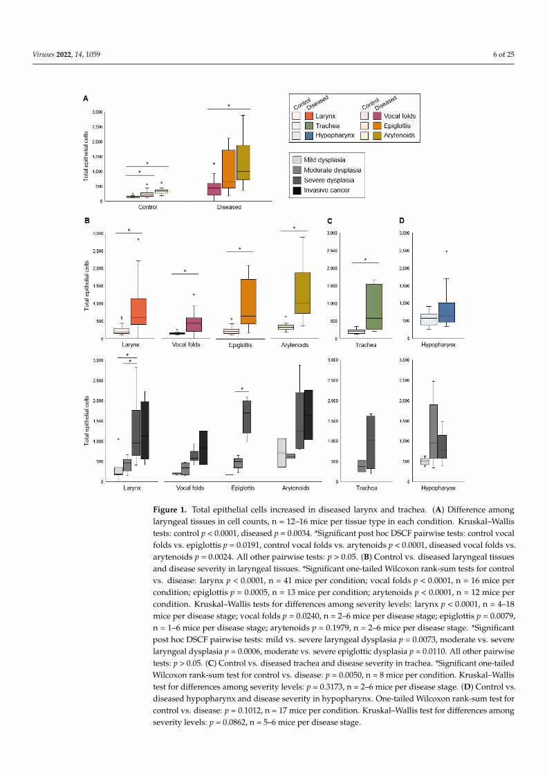
Cell counts differed among laryngeal tissues (Figure 1A). The number of epithelialcells was increased in all diseased laryngeal tissues compared to controls and increasedwith disease severity (Figure 1B). Cell count also increased in diseased proximal trachea butdid not differ by severity (Figure 1C). In the hypopharynx, the median number of epithelialcells did not differ between diseased tissue and uninfected controls (Figure 1D).
Viruses 2022, 14, 1059 6 of 25Viruses 2022, 14, x FOR PEER REVIEW 7 of 28
Figure 1. Total epithelial cells increased in diseased larynx and trachea. (A) Difference among lar-yngeal tissues in cell counts, n = 12–16 mice per tissue type in each condition. Kruskal–Wallis tests: control p < 0.0001, diseased p = 0.0034. *Significant post hoc DSCF pairwise tests: control vocal folds vs. epiglottis p = 0.0191, control vocal folds vs. arytenoids p < 0.0001, diseased vocal folds vs. aryte-noids p = 0.0024. All other pairwise tests: p > 0.05. (B) Control vs. diseased laryngeal tissues and disease severity in laryngeal tissues. *Significant one-tailed Wilcoxon rank-sum tests for control vs. disease: larynx p < 0.0001, n = 41 mice per condition; vocal folds p < 0.0001, n = 16 mice per condition; epiglottis p = 0.0005, n = 13 mice per condition; arytenoids p < 0.0001, n = 12 mice per condition. Kruskal–Wallis tests for differences among severity levels: larynx p < 0.0001, n = 4–18 mice per dis-ease stage; vocal folds p = 0.0240, n = 2–6 mice per disease stage; epiglottis p = 0.0079, n = 1–6 mice per disease stage; arytenoids p = 0.1979, n = 2–6 mice per disease stage. *Significant post hoc DSCF pairwise tests: mild vs. severe laryngeal dysplasia p = 0.0073, moderate vs. severe laryngeal dyspla-sia p = 0.0006, moderate vs. severe epiglottic dysplasia p = 0.0110. All other pairwise tests: p > 0.05. (C) Control vs. diseased trachea and disease severity in trachea. *Significant one-tailed Wilcoxon
Figure 1. Total epithelial cells increased in diseased larynx and trachea. (A) Difference amonglaryngeal tissues in cell counts, n = 12–16 mice per tissue type in each condition. Kruskal–Wallistests: control p < 0.0001, diseased p = 0.0034. *Significant post hoc DSCF pairwise tests: control vocalfolds vs. epiglottis p = 0.0191, control vocal folds vs. arytenoids p < 0.0001, diseased vocal folds vs.arytenoids p = 0.0024. All other pairwise tests: p > 0.05. (B) Control vs. diseased laryngeal tissuesand disease severity in laryngeal tissues. *Significant one-tailed Wilcoxon rank-sum tests for controlvs. disease: larynx p < 0.0001, n = 41 mice per condition; vocal folds p < 0.0001, n = 16 mice percondition; epiglottis p = 0.0005, n = 13 mice per condition; arytenoids p < 0.0001, n = 12 mice percondition. Kruskal–Wallis tests for differences among severity levels: larynx p < 0.0001, n = 4–18mice per disease stage; vocal folds p = 0.0240, n = 2–6 mice per disease stage; epiglottis p = 0.0079,n = 1–6 mice per disease stage; arytenoids p = 0.1979, n = 2–6 mice per disease stage. *Significantpost hoc DSCF pairwise tests: mild vs. severe laryngeal dysplasia p = 0.0073, moderate vs. severelaryngeal dysplasia p = 0.0006, moderate vs. severe epiglottic dysplasia p = 0.0110. All other pairwisetests: p > 0.05. (C) Control vs. diseased trachea and disease severity in trachea. *Significant one-tailedWilcoxon rank-sum test for control vs. disease: p = 0.0050, n = 8 mice per condition. Kruskal–Wallistest for differences among severity levels: p = 0.3173, n = 2–6 mice per disease stage. (D) Control vs.diseased hypopharynx and disease severity in hypopharynx. One-tailed Wilcoxon rank-sum test forcontrol vs. disease: p = 0.1012, n = 17 mice per condition. Kruskal–Wallis test for differences amongseverity levels: p = 0.0862, n = 5–6 mice per disease stage.

Viruses 2022, 14, 1059 7 of 25
In uninfected larynges, Ki67 was expressed in rare basal epithelial cells (Figure 2A).Ki67+ cells were found above the basal layer in diseased laryngeal epithelium (Figure 2A).The proliferation index did not differ by subsite within the larynx (Figure 2B). Quanti-tatively, there were more proliferating epithelial cells in infected larynges than in unin-fected larynges (median 3.4%, IQR 1.3–10.0%, vs. median 0.9%, IQR 0.0–2.2%, p < 0.0001,Figure 2C). The relationship between Ki67 and disease severity was unclear. Groupdifference among disease severity levels was significant in the larynx as a whole whenconsidering pooled data and appeared to be driven by a very high proliferation index inone epiglottis (Figure 2C). Differences by severity were not significant in vocal folds orarytenoids. Proliferation index increased in diseased proximal trachea and hypopharynxbut did not differ by disease severity (Figure 2D–E). Overall, total epithelial cell counts andKi67 staining demonstrated quantifiable hyperplasia and proliferation in MmuPV1-inducedlaryngeal disease.
3.2. Apoptosis
TUNEL+ cells were found primarily in apical epithelium (Figure 3A). The apoptosisindex was low but highly variable in both uninfected (median 2.4%, IQR 0.5–3.2%) anddiseased (median 2.9%, IQR 1.4–5.4%) laryngeal tissues. The number of TUNEL+ cellsdid not differ by subsite within the larynx (Figure 3B). Apoptosis index did not differbetween diseased and control larynges or trachea but increased in diseased hypopharynx(Figure 3C–E). Disease severity was not associated with apoptosis in any tissue.
3.3. Epithelial Differentiation
As expected, normal uninfected vocal fold epithelium was characterized by p63expression in basal cells, K13 in apical cells, and K8 throughout the epithelium (Figure 4). Inmild vocal fold dysplasia induced by MmuPV1 infection, hyperplastic foci with decreasedK13 and suprabasal p63 were found. Cell layers increased in hyperplastic epithelium, andK8 gradually increased from basal to apical layers. These changes were more pronouncedin moderate dysplasia. In severe vocal fold dysplasia, there was an expansion of thebasal compartment. p63 was found in all epithelial layers as well as lining epithelialinvaginations. K13 was severely reduced, as was K8 to a lesser extent, and the expressionof both was limited to apical cells. Squamous cell carcinomas in MmuPV1-infected vocalfolds were characterized by loss of both K13 and K8 in areas of the epithelium. p63-positiveepithelial cells increased from 46.7% ± 7.2% in normal vocal folds to 63.3% ± 10.1% indiseased vocal folds (p = 0.0003, Figure 5A). In the epiglottis/ventral pouch and arytenoidepithelium, the expression of p63, K13, and K8 in mirrored findings in vocal folds exceptfor some suprabasal p63 expression in arytenoids and variable K13 expression in epiglottisat baseline (Figure S2).

Viruses 2022, 14, 1059 8 of 25
Viruses 2022, 14, x FOR PEER REVIEW 9 of 28
Figure 2. Proliferation index increased in diseased larynx, trachea, and hypopharynx. (A) Ki67IHC stain in control and diseased vocal folds, epiglottis, and arytenoids, 30× magnification. (B)Difference among laryngeal tissues in % Ki67+ cells, n = 12-16 mice per tissue type in each condition.Kruskal–Wallis tests: control p = 0.1647, diseased p = 0.6574. (C) Control vs. diseased laryngealtissues and disease severity in laryngeal tissues. * Significant one-tailed Wilcoxon rank-sum tests forcontrol vs. disease: larynx p < 0.0001, n = 41 mice per condition; vocal folds p = 0.0122, n = 16 miceper condition; epiglottis p = 0.0218, n = 13 mice per condition; arytenoids p = 0.0104, n = 12 mice percondition. Kruskal–Wallis tests for differences among severity levels: larynx p = 0.0212, n = 4–18 mice

Viruses 2022, 14, 1059 9 of 25
per disease stage; vocal folds p = 0.3098, n = 2–6 mice per disease stage; epiglottis p = 0.0392, n = 1–6mice per disease stage; arytenoids p = 0.1375, n = 2–6 mice per disease stage. *Significant post hoc DSCFpairwise tests: moderate vs. severe laryngeal dysplasia p = 0.0482, severe laryngeal dysplasia vs. invasivecancer p = 0.0464. All other pairwise tests: p > 0.05. (D) Control vs. diseased trachea and disease severity intrachea. *Significant one-tailed Wilcoxon rank-sum test for control vs. disease: p = 0.0052, n = 8 mice percondition. Kruskal–Wallis test for differences among severity levels: p = 0.3173, n = 2–6 mice per diseasestage. (E) Control vs. diseased hypopharynx and disease severity in hypopharynx. *Significant one-tailedWilcoxon rank-sum test for control vs. disease: p = 0.0137, n = 17 mice per condition. Kruskal–Wallis testfor differences among severity levels: p = 0.3645, n = 5–6 mice per disease stage.
Viruses 2022, 14, x FOR PEER REVIEW 11 of 28
Figure 3. Apoptosis index did not differ in diseased larynx or trachea but increased in diseased hypopharynx. (A) TUNEL stain in control and diseased vocal folds, epiglottis, and arytenoids, 30× magnification. (B) Difference among laryngeal tissues in % TUNEL+ cells, n = 7–11 mice per tissue
Figure 3. Apoptosis index did not differ in diseased larynx or trachea but increased in diseasedhypopharynx. (A) TUNEL stain in control and diseased vocal folds, epiglottis, and arytenoids, 30×magnification. (B) Difference among laryngeal tissues in % TUNEL+ cells, n = 7–11 mice per tissuetype in each condition. Kruskal–Wallis tests: control p = 0.0690, diseased p = 0.8296. (C) Control vs.

Viruses 2022, 14, 1059 10 of 25
diseased laryngeal tissues and disease severity in laryngeal tissues. Two-tailed Wilcoxon rank-sumtests for control vs. disease: larynx p = 0.1794, n = 27 mice per condition; vocal folds p = 0.2493, n = 11mice per condition; epiglottis p = 0.4423, n = 7 mice per condition; arytenoids p = 0.0634, n = 9 mice percondition. Kruskal–Wallis tests for differences among severity levels: larynx p = 0.6510, n = 4–9 mice perdisease stage; vocal folds p = 0.3347, n = 2–3 mice per disease stage; epiglottis p = 0.3189, n = 1–3 miceper disease stage; arytenoids p = 0.0846, n = 2–3 mice per disease stage. (D) Control vs. diseased tracheaand disease severity in trachea. Two-tailed Wilcoxon rank-sum test for control vs. disease: p = 0.4680,n = 6 mice per condition. Kruskal–Wallis test for differences among severity levels: p = 1.0000, n = 2–4mice per disease stage. (E) Control vs. diseased hypopharynx and disease severity in hypopharynx. *Significant two-tailed Wilcoxon rank-sum test for control vs. disease: p = 0.0374, n = 9 mice per condition.Kruskal–Wallis test for differences among severity levels: p = 0.8752, n = 3 mice per disease stage.
Viruses 2022, 14, x FOR PEER REVIEW 13 of 28
Figure 4. Expanded epithelial basal compartment in MmuPV1-induced vocal fold disease. Serial sections of control and diseased vocal folds stained with H&E, MmuPV1 E4 RNAscope ISH, cos-tained p63 (red) and Hoechst (blue) IF, and costained K8 (green), K13 (red), and Hoechst (blue) IF shown both as separate channels and merged. 40× magnification. Arrows: MmuPV1-positive focal dysplasia with suprabasal p63 and reduced K13. Some H&E and RNAscope images reproduced with permission from our companion paper [3].
Figure 4. Expanded epithelial basal compartment in MmuPV1-induced vocal fold disease. Serial
Viruses 2022, 14, 1059 11 of 25
sections of control and diseased vocal folds stained with H&E, MmuPV1 E4 RNAscope ISH, costainedp63 (red) and Hoechst (blue) IF, and costained K8 (green), K13 (red), and Hoechst (blue) IF shown bothas separate channels and merged. 40×magnification. Arrows: MmuPV1-positive focal dysplasia withsuprabasal p63 and reduced K13. Some H&E and RNAscope images reproduced with permissionfrom our companion paper [3].
Viruses 2022, 14, x FOR PEER REVIEW 14 of 28
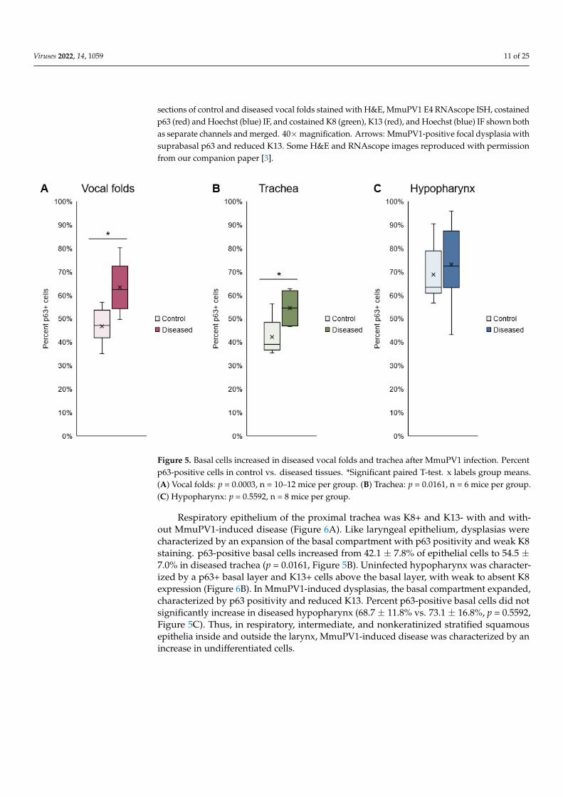
Figure 5. Basal cells increased in diseased vocal folds and trachea after MmuPV1 infection. Percent p63-positive cells in control vs. diseased tissues. *Significant paired T-test. x labels group means. (A) Vocal folds: p = 0.0003, n = 10–12 mice per group. (B) Trachea: p = 0.0161, n = 6 mice per group. (C) Hypopharynx: p = 0.5592, n = 8 mice per group.
Respiratory epithelium of the proximal trachea was K8+ and K13- with and without MmuPV1-induced disease (Figure 6A). Like laryngeal epithelium, dysplasias were char-acterized by an expansion of the basal compartment with p63 positivity and weak K8 staining. p63-positive basal cells increased from 42.1 ± 7.8% of epithelial cells to 54.5 ± 7.0% in diseased trachea (p = 0.0161, Figure 5B). Uninfected hypopharynx was characterized by a p63+ basal layer and K13+ cells above the basal layer, with weak to absent K8 expression (Figure 6B). In MmuPV1-induced dysplasias, the basal compartment expanded, charac-terized by p63 positivity and reduced K13. Percent p63-positive basal cells did not signif-icantly increase in diseased hypopharynx (68.7 ± 11.8% vs. 73.1 ± 16.8%, p = 0.5592, Figure 5C). Thus, in respiratory, intermediate, and nonkeratinized stratified squamous epithelia inside and outside the larynx, MmuPV1-induced disease was characterized by an increase in undifferentiated cells.
Figure 5. Basal cells increased in diseased vocal folds and trachea after MmuPV1 infection. Percentp63-positive cells in control vs. diseased tissues. *Significant paired T-test. x labels group means.(A) Vocal folds: p = 0.0003, n = 10–12 mice per group. (B) Trachea: p = 0.0161, n = 6 mice per group.(C) Hypopharynx: p = 0.5592, n = 8 mice per group.
Respiratory epithelium of the proximal trachea was K8+ and K13- with and with-out MmuPV1-induced disease (Figure 6A). Like laryngeal epithelium, dysplasias werecharacterized by an expansion of the basal compartment with p63 positivity and weak K8staining. p63-positive basal cells increased from 42.1 ± 7.8% of epithelial cells to 54.5 ±7.0% in diseased trachea (p = 0.0161, Figure 5B). Uninfected hypopharynx was character-ized by a p63+ basal layer and K13+ cells above the basal layer, with weak to absent K8expression (Figure 6B). In MmuPV1-induced dysplasias, the basal compartment expanded,characterized by p63 positivity and reduced K13. Percent p63-positive basal cells did notsignificantly increase in diseased hypopharynx (68.7 ± 11.8% vs. 73.1 ± 16.8%, p = 0.5592,Figure 5C). Thus, in respiratory, intermediate, and nonkeratinized stratified squamousepithelia inside and outside the larynx, MmuPV1-induced disease was characterized by anincrease in undifferentiated cells.

Viruses 2022, 14, 1059 12 of 25Viruses 2022, 14, x FOR PEER REVIEW 15 of 28
Figure 6. Expanded epithelial basal compartment in MmuPV1-induced disease in proximal trachea and hypopharynx. Serial sections of control and diseased tissues stained with H&E, MmuPV1 E4 RNAscope ISH, costained p63 (red) and Hoechst (blue) IF, and costained K8 (green), K13 (red), and Hoechst (blue) IF shown both as separate channels and merged. 40× magnification. Some H&E and RNAscope images reproduced with permission from our companion paper [3]. (A) Trachea. (B) Hypopharynx. Arrows indicate transition between normal, nondiseased epithelium and dysplastic, MmuPV1-positive epithelium coinciding with expanded basal compartment.
Figure 6. Expanded epithelial basal compartment in MmuPV1-induced disease in proximal tracheaand hypopharynx. Serial sections of control and diseased tissues stained with H&E, MmuPV1 E4RNAscope ISH, costained p63 (red) and Hoechst (blue) IF, and costained K8 (green), K13 (red), andHoechst (blue) IF shown both as separate channels and merged. 40× magnification. Some H&Eand RNAscope images reproduced with permission from our companion paper [3]. (A) Trachea. (B)Hypopharynx. Arrows indicate transition between normal, nondiseased epithelium and dysplastic,MmuPV1-positive epithelium coinciding with expanded basal compartment.

Viruses 2022, 14, 1059 13 of 25
3.4. Epithelial Barrier
In uninfected vocal folds, laminin labeled the basement membrane below the epithe-lium (Figure 7). The adherens junction marker E-cadherin was expressed in cell membranesthroughout epithelium, and the tight junction marker ZO-1 was expressed in cell mem-branes in more apical cells (Figure 8A). Barrier proteins were slightly disorganized in mildvocal fold dysplasia, with focally decreased E-cadherin and ZO-1 and laminin expressionin epithelial cells (Figure 7). E-cadherin was decreased in moderate dysplasia, and a ZO-1signal appeared in the basal layer around folds of hyperplastic epithelium, with a strongsignal that was not limited to cell membranes. These changes were also observed in severedysplasia (Figure 8A), as was laminin expression throughout the epithelium. In vocal foldcancer induced by MmuPV1, epithelial laminin expression persisted, and cell junction pro-teins became patchy and lost in areas (Figure 7). Barrier protein expression in control anddiseased epithelia of the epiglottis and arytenoids wase similar to vocal folds (Figure S3).
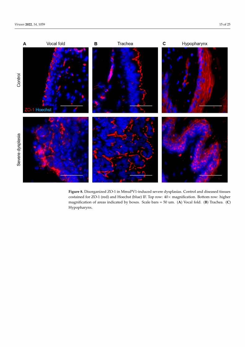
In the uninfected trachea, laminin labeled the basement membrane with some stainingof cilia, E-cadherin labeled cell membranes throughout the epithelium, and ZO-1 formeda distinct line along the luminal surface of cells (Figure 8B, Figure 9A). In dysplasias,E-cadherin expression decreased in the expanding basal compartment. ZO-1 increasedto form two distinct lines in moderate dysplasia and surrounded folds of hyperplasticepithelium in severe dysplasia (Figure 8B, Figure 9A). Changes in laminin with MmuPV1-induced dysplasia mirrored ZO-1, with laminin appearing to support layers of hyperplasticcells (Figure 9A). In control hypopharyngeal epithelium, laminin marked the basementmembrane, and cell junction proteins were membranous, with E-cadherin expressionstronger closer to basal layers and ZO-1 expression stronger in suprabasal and apicallayers (Figure 8C, Figure 9B). In severe dysplasia, laminin was occasionally found inthe epithelium. ZO-1 expression appeared in basal layers and expanded beyond cellmembranes in severe dysplasia (Figure 8C, Figure 9B). E-cadherin expression was decreased(Figure 9B). Nuclear E-cadherin was observed in severe dysplasias and cancers of all tissues,most notably the hypopharynx (Figure 10). Overall, MmuPV1-associated disease wascharacterized by disorganized and mislocalized barrier proteins, with ZO-1 and lamininapparently supporting increased layers and folds of hyperplastic epithelium.

Viruses 2022, 14, 1059 14 of 25Viruses 2022, 14, x FOR PEER REVIEW 17 of 28
Figure 7. Perturbed epithelial barrier in vocal folds with disease induced by MmuPV1. Serial sec-tions of control and diseased vocal folds stained with H&E, MmuPV1 E4 RNAscope ISH, costained laminin (green) and Hoechst (blue) IF, and costained E-cadherin (green), ZO-1 (red), and Hoechst (blue) IF shown both as separate channels and merged. 40× magnification. Arrows: MmuPV1-posi-tive focal dysplasia with epithelial laminin and reduced E-cadherin and ZO-1. Some H&E and RNAscope images reproduced with permission from our companion paper [3].
Figure 7. Perturbed epithelial barrier in vocal folds with disease induced by MmuPV1. Serial sectionsof control and diseased vocal folds stained with H&E, MmuPV1 E4 RNAscope ISH, costained laminin(green) and Hoechst (blue) IF, and costained E-cadherin (green), ZO-1 (red), and Hoechst (blue) IFshown both as separate channels and merged. 40×magnification. Arrows: MmuPV1-positive focaldysplasia with epithelial laminin and reduced E-cadherin and ZO-1. Some H&E and RNAscopeimages reproduced with permission from our companion paper [3].
Viruses 2022, 14, 1059 15 of 25Viruses 2022, 14, x FOR PEER REVIEW 18 of 28
Figure 8. Disorganized ZO-1 in MmuPV1-induced severe dysplasias. Control and diseased tissues costained for ZO-1 (red) and Hoechst (blue) IF. Top row: 40× magnification. Bottom row: higher magnification of areas indicated by boxes. Scale bars = 50 um. (A) Vocal fold. (B) Trachea. (C) Hy-popharynx.
In the uninfected trachea, laminin labeled the basement membrane with some stain-ing of cilia, E-cadherin labeled cell membranes throughout the epithelium, and ZO-1 formed a distinct line along the luminal surface of cells (Figure 8B, Figure 9A). In dyspla-sias, E-cadherin expression decreased in the expanding basal compartment. ZO-1 in-creased to form two distinct lines in moderate dysplasia and surrounded folds of hyper-plastic epithelium in severe dysplasia (Figure 8B, Figure 9A). Changes in laminin with MmuPV1-induced dysplasia mirrored ZO-1, with laminin appearing to support layers of hyperplastic cells (Figure 9A). In control hypopharyngeal epithelium, laminin marked the basement membrane, and cell junction proteins were membranous, with E-cadherin ex-pression stronger closer to basal layers and ZO-1 expression stronger in suprabasal and apical layers (Figure 8C, Figure 9B). In severe dysplasia, laminin was occasionally found in the epithelium. ZO-1 expression appeared in basal layers and expanded beyond cell membranes in severe dysplasia (Figure 8C, Figure 9B). E-cadherin expression was de-creased (Figure 9B). Nuclear E-cadherin was observed in severe dysplasias and cancers of all tissues, most notably the hypopharynx (Figure 10). Overall, MmuPV1-associated dis-ease was characterized by disorganized and mislocalized barrier proteins, with ZO-1 and laminin apparently supporting increased layers and folds of hyperplastic epithelium.
Figure 8. Disorganized ZO-1 in MmuPV1-induced severe dysplasias. Control and diseased tissuescostained for ZO-1 (red) and Hoechst (blue) IF. Top row: 40× magnification. Bottom row: highermagnification of areas indicated by boxes. Scale bars = 50 um. (A) Vocal fold. (B) Trachea. (C)Hypopharynx.

Viruses 2022, 14, 1059 16 of 25Viruses 2022, 14, x FOR PEER REVIEW 19 of 28
Figure 9. Perturbed epithelial barrier in trachea and hypopharynx with disease induced by MmuPV1. Serial sections of control and diseased tissues stained with H&E, MmuPV1 E4 RNAscope ISH, costained laminin (green) and Hoechst (blue) IF, and costained E-cadherin (green), ZO-1 (red), and Hoechst (blue) IF shown both in red channel and merged. 40× magnification. Some H&E and RNAscope images reproduced with permission from our companion paper [3]. (A) Trachea. (B) Hypopharynx. Arrow: basal ZO-1 beyond cell membranes in MmuPV1-positive dysplastic epithe-lium.
Figure 9. Perturbed epithelial barrier in trachea and hypopharynx with disease induced by MmuPV1.Serial sections of control and diseased tissues stained with H&E, MmuPV1 E4 RNAscope ISH,costained laminin (green) and Hoechst (blue) IF, and costained E-cadherin (green), ZO-1 (red),and Hoechst (blue) IF shown both in red channel and merged. 40× magnification. Some H&Eand RNAscope images reproduced with permission from our companion paper [3]. (A) Trachea.(B) Hypopharynx. Arrow: basal ZO-1 beyond cell membranes in MmuPV1-positive dysplasticepithelium.

Viruses 2022, 14, 1059 17 of 25Viruses 2022, 14, x FOR PEER REVIEW 20 of 28
Figure 10. Nuclear E-cadherin in MmuPV1-induced cancers and severe dysplasias. Diseased tissues costained for E-cadherin (green) and Hoechst (blue) IF. Top row: 40× magnification. Bottom row: higher magnification of areas indicated by boxes. Scale bars = 50 um. (A) Vocal fold. (B) Trachea. (C) Hypopharynx.
4. Discussion We compared quantitative and qualitative measures of proliferation, apoptosis, epi-
thelial differentiation, and epithelial barrier between mice with MmuPV1-induced disease of the larynx, proximal trachea, and hypopharynx and equal numbers of uninfected con-trols. Our findings support our hypothesis that laryngeal MmuPV1 infection increase pro-liferation and disrupt differentiation and barriers, characteristic features of human RRP. Similarities and differences between NSG laryngeal epithelial disease associated with MmuPV1 and human RRP are summarized in Table 2.
Table 2. Epithelial similarities and differences between vocal fold disease in NSG mice infected with MmuPV1 and RRP.
Disease feature Mouse Vocal Fold + MmuPV1 Human RRP Hyperplasia Yes Yes [19,71,72]
Increased proliferation Yes Yes [23] Decreased differentiation Yes Yes [24,25]
Decreased apoptosis No Unclear [30–33] Altered barrier Yes Yes [35–37]
Figure 10. Nuclear E-cadherin in MmuPV1-induced cancers and severe dysplasias. Diseased tissuescostained for E-cadherin (green) and Hoechst (blue) IF. Top row: 40× magnification. Bottom row:higher magnification of areas indicated by boxes. Scale bars = 50 um. (A) Vocal fold. (B) Trachea.(C) Hypopharynx.
4. Discussion
We compared quantitative and qualitative measures of proliferation, apoptosis, ep-ithelial differentiation, and epithelial barrier between mice with MmuPV1-induced diseaseof the larynx, proximal trachea, and hypopharynx and equal numbers of uninfected con-trols. Our findings support our hypothesis that laryngeal MmuPV1 infection increaseproliferation and disrupt differentiation and barriers, characteristic features of humanRRP. Similarities and differences between NSG laryngeal epithelial disease associated withMmuPV1 and human RRP are summarized in Table 2.
Table 2. Epithelial similarities and differences between vocal fold disease in NSG mice infected withMmuPV1 and RRP.
Disease Feature Mouse Vocal Fold +MmuPV1 Human RRP
Hyperplasia Yes Yes [19,71,72]Increased proliferation Yes Yes [23]
Decreased differentiation Yes Yes [24,25]Decreased apoptosis No Unclear [30–33]
Altered barrier Yes Yes [35–37]

Viruses 2022, 14, 1059 18 of 25
Consistent with obvious hyperplasia on histology, the total number of epithelial cellsincreased in laryngeal tissues with MmuPV1-induced disease. In uninfected laryngealtissues, the proliferation index (% Ki67 + cells) was about 1%. This is consistent withthe literature reporting very few proliferating cells in adult mouse vocal folds [55,57,73].Proliferation index increased to a median of 3.4% in diseased murine laryngeal tissues,and Ki67-expressing cells were found above the basal layer of the epithelium. A similarexpansion of proliferating cells has been observed in RRP lesions [23] and in MmuPV1-induced lesions of the oral cavity and the anogenital tract [74–77]. Like the larynx, wefound that proliferation and hyperplasia increased in trachea with MmuPV1-induceddisease. In the diseased hypopharynx, however, proliferation increased without resultingin quantifiable hyperplasia. Murine hypopharynx has more epithelial cell layers than thelarynx at baseline (5–7 vs. 2–4 in our observations), so perhaps the difference induced bydisease in hypopharynx was not great enough to be statistically significant.
There was no effect of MmuPV1-induced disease on apoptosis in laryngeal epithelium.High-risk HPV-infected cells resist apoptosis [28]. Studies of apoptosis in RRP tissuescompared with normal laryngeal tissues have found both pro- and anti-apoptotic effects onupstream effectors and inhibitors of apoptosis [30–33]. A common downstream effect ofboth intrinsic and extrinsic apoptosis pathways is DNA fragmentation, which is detectedby TUNEL. One study found no difference in DNA fragmentation between RRP tissues andnormal laryngeal tissues [32]. The relationship between apoptosis in both RRP disease andMmuPV1 laryngeal infection remains unclear based on the literature and our findings. Incontrast to the larynx, apoptosis increased in hypopharynx with MmuPV1-induced disease.MmuPV1 has been shown to increase Bak, an effector of apoptosis, in the skin of medicallyimmunosuppressed mice [78]. Unlike the larynx, the stratified squamous epithelium ofmouse hypopharynx is keratinized [79]. It is possible that the hypopharynx epitheliummore closely resembles skin in the effect of MmuPV1 on apoptotic pathways.
As expected, we observed an expansion of the basal compartment of epithelial cells intissues with MmuPV1-induced disease. These cells were characterized by p63-positivityand the absence of differentiation markers specific to each tissue. Percent basal cellsas a fraction of epithelial cells increased in diseased larynx and trachea. In the larynx,both K13 and K8 marked epithelium above the basal layer as previously shown [54].K13- cells increased in dysplasias, and K8- cells increased in severe dysplasias. A patchyloss of both K13 and K8 was observed in apical layers of laryngeal cancers induced byMmuPV1. Similarly, in tracheal dysplasia, the p63+/K8- compartment was expanded. Inhypopharynx, p63+/K13- cells above the basal layer visibly increased in severe dysplasia.However, like the difference in proliferating Ki67+ cells, the difference in basal p63+ cellswas not statistically significant in quantitative analysis. Decreased K13 and other markers ofepithelial differentiation have been reported in RRP [24,25]. In skin, MmuPV1 increases thebasal cell compartment of infected epithelium and decreases cytokeratin 10 (K10)-positivedifferentiated cells [77]. Thus, our results are consistent with the literature on epithelialdifferentiation in both MmuPV1-induced disease and human RRP.
In MmuPV1-induced laryngeal disease, tight junction marker ZO-1 localized to basalcells of invaginating, hyperplastic epithelium, as opposed to primarily apical cells in normalepithelium. Its expression extended into the cytoplasm, beyond the clear line of membra-nous positivity seen in uninfected cells. In tracheal disease, ZO-1 remained membranousbut was expressed as multiple discrete layers that increased with hyperplasia and diseaseseverity. RNA-seq experiments have shown downregulated epithelial barrier componentsin both high- and low-risk HPV infection, including RRP tissues [35,80]. E6 proteins ofhigh-risk HPVs disrupt tight junctions through direct interaction with ZO-1 [81,82] andother cellular proteins containing a post synaptic density protein (PSD95)/Drosophila disclarge tumor suppressor (DLG)/ZO-1 (PDZ) domain [83]. However, E6 proteins of low-riskHPV types 6 and 11 lack a PDZ-binding motif [83]. ZO-1 has not been specifically studiedin RRP. To our knowledge, the effect of MmuPV1 on cell junctions has not been previouslyexplored. MmuPV1 E6 contains a putative PDZ-binding motif, but not in the extreme

Viruses 2022, 14, 1059 19 of 25
carboxy-terminus like high-risk HPV E6 proteins [84]. A straightforward decrease in ZO-1may not be expected in either RRP or in MmuPV1-induced disease.
We found that membranous E-cadherin expression decreased and became patchy inMmuPV1-induced disease of the larynx. RRP clinical data for E-cadherin are not availablefor comparison. In cervical cancer, E-cadherin protein level is reduced and is inverselycorrelated with high-risk HPV E6 and E7 levels [85]. In genital lesions caused by low-riskHPV6 and 11, the same types that cause RRP, E-cadherin staining is generally reduced,but the level of reduction is heterogeneous across patients [86]. In addition to a reductionin membranous expression, nuclear E-cadherin expression was found in severe laryngealdysplasias and cancers induced by MmuPV1. This has been observed in a number of cancertypes and has been validated by subcellular fractionation, but requires an antibody againstthe cytoplasmic domain rather than the extracellular domain of E-cadherin to be revealedupon immunostaining [87–92]. Nuclear E-cadherin may regulate transcription of manysignaling pathways involved in tumorigenesis [87,89,91].
We hypothesized that MmuPV1 would decrease laminin in laryngeal disease. Lamininhas not been examined in MmuPV1-associated diseases of other tissues. The basementmembrane is disrupted in some TEM studies of RRP lesions [36,37]. The invasion ofepithelial cells through basement membrane is a hallmark of invasive cancer. High-riskHPVs increase many MMPs [40], including types that are known to degrade laminin andother basement membrane components [93,94]. We found that laminin expression wasdisorganized in MmuPV1-induced laryngeal disease but that, contrary to expectations,expression increased in diseased epithelium. Laminins are heterotrimers containing α, β,and γ chains. The primary laminin detected by our antibody is laminin-111 (formerly calledlaminin-1), i.e., α1/β1/γ1 [95,96]. Laminin-111 and laminin-332 (formerly laminin-5) havebeen reported in the vocal fold basement membrane of humans and rodents [61,97–99].Many cancers, including laryngeal squamous cell carcinoma, express laminin-111 and -332throughout the tumor tissue [100–102]. Both laminins contain peptides that promote tumorgrowth [100,101]. In our model, laminin expression in MmuPV1-induced laryngeal diseasewas consistent with its expression in laryngeal cancer.
As summarized in Table 2, the laryngeal epithelial response to MmuPV1-induceddisease grossly resembled findings in RRP, despite the subtle differences discussed above.The major limitation of the present work is the exclusive use of immunocompromised mice.There are important interactions between the immune system and epithelial cells that maybe clinically relevant in RRP patients, but that our experiments could not address. Forexample, cytotoxic CD8+ T cells are decreased in RRP lesions [103,104], and stress keratinK17 transcripts are upregulated [105]. Transcriptomic analysis has revealed molecularsubtypes of RRP associated with clinical course. Patients with more aggressive diseasehad lesions characterized by increased low-risk HPV gene expression, reduced expressionof genes involved in interferon signaling, antigen presentation, and T cell function, fewerCD4+ and CD8+ T cells in lesions, and increased expression of certain cytokeratin genes,including K17 [7]. A recent study using MmuPV1 found a mechanistic link between thesephenomena in skin, demonstrating that MmuPV1 increased K17 in lesions, which preventedCD8+ T cell infiltration via disrupted chemokine signaling and allowed lesion growth [64].As another example, E-cadherin binds Langerhans cells to epithelial cells and is requiredfor Langerhans cell maturation [106,107]. High- and low-risk HPV infections decreaseboth E-cadherin and Langerhans cells in cervical mucosa [86]. RRP lesions have immatureLangerhans cells with a defective response to cytokines [108,109]. Since immunodeficiencyin NSG mice includes a lack of T cells and defective Langerhans cells at baseline, anothermouse strain is required to understand interactions among keratins, cell adhesion molecules,and the immune response to MmuPV1 in the larynx and to generate a model that parallelshuman immune response to low-risk HPVs in RRP. Immunocompromised mouse strains arehighly vulnerable to MmuPV1, but certain immunocompetent strains also develop infectionand disease in various tissues [84,110,111]. This warrants investigation in the larynx. Animmunocompetent animal model of laryngeal papillomavirus infection could be used to
Viruses 2022, 14, 1059 20 of 25
reveal larynx-specific mechanisms of viral immune evasion. For example, the interactionsdiscussed above among keratins, cell junction proteins, and innate and adaptive immunecells in papillomavirus infection could have implications for RRP. However, they have notbeen mechanistically studied in the larynx, which has a unique immunologic milieu [112].With mechanisms of viral immune evasion in the larynx defined, this model could then beused to discover methods to overcome immune evasion and elicit host immune-mediatedresolution of RRP.
5. Conclusions
MmuPV1 increased proliferation in infected laryngeal tissues, expanded the basalcompartment of cells, and decreased differentiated cells. There was no effect of MmuPV1on apoptosis. Tight junction marker ZO-1 was mislocalized rather than decreased. Thesefindings are consistent with findings in RRP lesions and low-risk HPV biology. Theeffects of MmuPV1-induced disease on adherens junction marker E-cadherin and basementmembrane marker laminin resembled human cancers, but the status of these biomarkersin RRP has not been reported. We conclude that MmuPV1 infection of the mouse larynxprovides a useful, if imperfect, preclinical model for RRP.
Supplementary Materials: The following supporting information can be downloaded at: https://www.mdpi.com/article/10.3390/v14051059/s1, Table S1: Intrarater reliability of total epithelial cellcounts; Table S2: Correlations between proliferation and apoptosis indices; Figure S1: Cell countingresults did not differ between manual and automated counting methods; Figure S2: Epithelialdifferentiation markers in epiglottis and arytenoids with disease induced by MmuPV1; Figure S3:Epithelial barrier markers in epiglottis and arytenoids with disease induced by MmuPV1.
Author Contributions: Conceptualization, R.E.K., P.F.L. and S.L.T.; methodology, R.E.K., E.T.W.-S.,R.H., P.F.L. and S.L.T.; validation, R.E.K. and R.H.; formal analysis, R.E.K.; investigation, R.E.K.,E.T.W.-S. and R.H.; resources, R.E.K., E.T.W.-S., P.F.L. and S.L.T.; data curation, R.E.K. and R.H.;writing—original draft preparation, R.E.K.; writing—review and editing, R.E.K., E.T.W.-S., R.H., P.F.L.and S.L.T.; visualization, R.E.K.; supervision, R.E.K., P.F.L. and S.L.T.; project administration, P.F.L.and S.L.T.; funding acquisition, R.E.K., P.F.L. and S.L.T. All authors have read and agreed to thepublished version of the manuscript.
Funding: National Institutes of Health (NIH) P50 DE026787 (Wisconsin Head and Neck SpecializedProgram of Excellence [SPORE]), P30 CA014520 (University of Wisconsin Carbone Cancer Center[UWCCC] Experimental Animal Pathology Lab), P01 CA022443, R35 CA210807, R01 DC004336, T32DC009401, T32 CA090217, and F31 DC018184. The content is solely the responsibility of the authorsand does not necessarily represent the official views of the NIH. The APC was funded by S.L.T.
Institutional Review Board Statement: The animal study protocol was approved by the InstitutionalAnimal Use Committee of the University of Wisconsin-Madison (protocol code M005871).
Data Availability Statement: All relevant data are provided within the article and supplementarymaterials.
Acknowledgments: The authors acknowledge Sierra Raglin in the University of Wisconsin Depart-ment of Surgery Histocore and the UWCCC Experimental Animal Pathology Lab for processinghistology samples; David Ornelles at Wake Forest University for sharing the ImageJ cell countingprograms; and Nathan Welham, Timothy McCulloch, Evie Carchman, and members of the Thibeaultand Lambert labs for valuable feedback and suggestions.
Conflicts of Interest: The authors declare no conflict of interest. The funders had no role in the designof the study; in the collection, analyses, or interpretation of data; in the writing of the manuscript; orin the decision to publish the results.
References1. Benedict, J.J.; Derkay, C.S. Recurrent Respiratory Papillomatosis: A 2020 Perspective. Laryngoscope Investig. Otolaryngol. 2021, 6,
340–345. [CrossRef] [PubMed]2. Doorbar, J. Model Systems of Human Papillomavirus-Associated Disease. J. Pathol. 2016, 238, 166–179. [CrossRef] [PubMed]

Viruses 2022, 14, 1059 21 of 25
3. King, R.E.; Bilger, A.; Rademacher, J.; Ward-Shaw, E.T.; Hu, R.; Lambert, P.F.; Thibeault, S.L. A Novel in Vivo Model of LaryngealPapillomavirus-Associated Disease Using Mus Musculus Papillomavirus. Viruses 2022, 14, 1000. [CrossRef]
4. Donne, A.J.; Hampson, L.; Homer, J.J.; Hampson, I.N. The Role of HPV Type in Recurrent Respiratory Papillomatosis. Int. J.Pediatric Otorhinolaryngol. 2010, 74, 7–14. [CrossRef]
5. Garcia, J.A.; Best, S.R.; Rooper, L.M. HPV RNA In-Situ Hybridization as a Diagnostic Aid in Papillary Laryngeal Lesions.Laryngoscope 2020, 130, 955–960. [CrossRef]
6. Lépine, C.; Voron, T.; Berrebi, D.; Mandavit, M.; Nervo, M.; Outh-Gauer, S.; Péré, H.; Tournier, L.; Teissier, N.; Tartour, E.; et al.Juvenile-Onset Recurrent Respiratory Papillomatosis Aggressiveness: In Situ Study of the Level of Transcription of HPV E6 andE7. Cancers 2020, 12, 2836. [CrossRef]
7. Sievers, C.; Robbins, Y.; Bai, K.; Yang, X.; Clavijo, P.E.; Friedman, J.; Sinkoe, A.; Norberg, S.M.; Hinrichs, C.; Van Waes, C.; et al.Comprehensive Multiomic Characterization of Human Papillomavirus-Driven Recurrent Respiratory Papillomatosis RevealsDistinct Molecular Subtypes. Commun. Biol. 2021, 4, 1416. [CrossRef]
8. Chow, L.T.; Broker, T.R.; Steinberg, B.M. The Natural History of Human Papillomavirus Infections of the Mucosal Epithelia.APMIS 2010, 118, 422–449. [CrossRef]
9. Tse, J.R.; Zhang, Z.; Long, J.L. Effects of Vocal Fold Epithelium Removal on Vibration in an Excised Human Larynx Model. J.Acoust. Soc. Am. 2015, 138, EL60–EL64. [CrossRef]
10. Fitch, J.L.; Holbrook, A. Modal Vocal Fundamental Frequency of Young Adults. Arch. Otolaryngol. 1970, 92, 379–382. [CrossRef]11. Glaze, L.E.; Bless, D.M.; Milenkovic, P.; Susser, R.D. Acoustic Characteristics of Children’s Voice. J. Voice 1988, 2, 312–319.
[CrossRef]12. Gunter, H.E. Modeling Mechanical Stresses as a Factor in the Etiology of Benign Vocal Fold Lesions. J. Biomech. 2004, 37, 1119–1124.
[CrossRef] [PubMed]13. Chan, R.W.; Titze, I.R. Viscoelastic Shear Properties of Human Vocal Fold Mucosa: Measurement Methodology and Empirical
Results. J. Acoust. Soc. Am. 1999, 106, 2008–2021. [CrossRef] [PubMed]14. Gray, S.D.; Alipour, F.; Titze, I.R.; Hammond, T.H. Biomechanical and Histologic Observations of Vocal Fold Fibrous Proteins.
Ann. Otol. Rhinol. Laryngol. 2000, 109, 77–85. [CrossRef] [PubMed]15. Gray, S.D.; Titze, I.R.; Chan, R.; Hammond, T.H. Vocal Fold Proteoglycans and Their Influence on Biomechanics. Laryngoscope
1999, 109, 845–854. [CrossRef] [PubMed]16. Hochman, I.; Hillman, R.E.; Sataloff, R.T.; Zeitels, S.M. Ectasias and Varices of the Vocal Fold: Clearing the Striking Zone. Ann.
Otol. Rhinol. Laryngol. 1999, 108, 10–16. [CrossRef]17. Kashima, H.; Leventhal, B.; Mounts, P.; Hruban, R.H. Sites of Predilection in Recurrent Respiratory Papillomatosis. Ann. Otol.
Rhinol. Laryngol. 1993, 102, 580–583. [CrossRef]18. Benedict, P.A.; Ruiz, R.; Yoo, M.; Verma, A.; Ahmed, O.H.; Wang, B.; Dion, G.R.; Voigt, A.; Merati, A.; Rosen, C.A.; et al.
Laryngeal Distribution of Recurrent Respiratory Papillomatosis in a Previously Untreated Cohort. Laryngoscope 2018, 128, 138–143.[CrossRef]
19. Fortes, H.R.; von Ranke, F.M.; Escuissato, D.L.; Araujo Neto, C.A.; Zanetti, G.; Hochhegger, B.; Souza, C.A.; Marchiori, E.Recurrent Respiratory Papillomatosis: A State-of-the-Art Review. Respir. Med. 2017, 126, 116–121. [CrossRef]
20. Levendoski, E.E.; Leydon, C.; Thibeault, S.L. Vocal Fold Epithelial Barrier in Health and Injury: A Research Review. J. Speech Lang.Hear. Res. 2014, 57, 1679. [CrossRef]
21. Dowdall, J.R.; Sadow, P.M.; Hartnick, C.; Vinarsky, V.; Mou, H.; Zhao, R.; Song, P.C.; Franco, R.A.; Rajagopal, J. Identificationof Distinct Layers within the Stratified Squamous Epithelium of the Adult Human True Vocal Fold. Laryngoscope 2015, 125,E313–E319. [CrossRef] [PubMed]
22. Lungova, V.; Chen, X.; Wang, Z.; Kendziorski, C.; Thibeault, S.L. Human Induced Pluripotent Stem Cell-Derived Vocal FoldMucosa Mimics Development and Responses to Smoke Exposure. Nat. Commun. 2019, 10, 4161. [CrossRef] [PubMed]
23. Bedard, M.C.; Brusadelli, M.G.; Carlile, A.; Ruiz-Torres, S.; Lodin, H.; Lee, D.; Kofron, M.; Lambert, P.F.; Lane, A.; Ameziane, N.;et al. Patient-Derived Organotypic Epithelial Rafts Model Phenotypes in Juvenile-Onset Recurrent Respiratory Papillomatosis.Viruses 2021, 13, 68. [CrossRef] [PubMed]
24. Steinberg, B.M.; Meade, R.; Kalinowski, S.; Abramson, A.L. Abnormal Differentiation of Human Papillomavirus-InducedLaryngeal Papillomas. Arch. Otolaryngol. Head Neck Surg. 1990, 116, 1167–1171. [CrossRef]
25. Wu, R.; Sun, S.; Steinberg, B.M. Requirement of STAT3 Activation for Differentiation of Mucosal Stratified Squamous Epithelium.Mol. Med. 2003, 9, 77–84. [CrossRef]
26. Reppucci, A.D.; Dilorenzo, T.P.; Abramson, A.L.; Steinberg, B.M. In Vitro Modulation of Human Laryngeal Papilloma CellDifferentiation by Retinoic Acid. Otolaryngol. Head Neck Surg. 1991, 105, 528–532. [CrossRef]
27. Vambutas, A.; Di Lorenzo, T.P.; Steinberg, B.M. Laryngeal Papilloma Cells Have High Levels of Epidermal Growth FactorReceptor and Respond to Epidermal Growth Factor by a Decrease in Epithelial Differentiation. Cancer Res. 1993, 53, 910–914.
28. Yuan, C.-H.; Filippova, M.; Duerksen-Hughes, P. Modulation of Apoptotic Pathways by Human Papillomaviruses (HPV):Mechanisms and Implications for Therapy. Viruses 2012, 4, 3831–3850. [CrossRef]
29. Novaleski, C.K.; Carter, B.D.; Sivasankar, M.P.; Ridner, S.H.; Dietrich, M.S.; Rousseau, B. Apoptosis and Vocal Fold Disease:Clinically Relevant Implications of Epithelial Cell Death. J. Speech Lang. Hear. Res. 2017, 60, 1264–1272. [CrossRef]

Viruses 2022, 14, 1059 22 of 25
30. Poetker, D.M.; Sandler, A.D.; Scott, D.L.; Smith, R.J.H.; Bauman, N.M. Survivin Expression in Juvenile-Onset Recurrent RespiratoryPapillomatosis. Ann. Otol. Rhinol. Laryngol. 2002, 111, 957–961. [CrossRef]
31. Niedzielska, G.; Kocki, J. Evaluation of Bcl-2 Gene Expression in Papilloma of Larynx in Children. Int. J. Pediatric Otorhinolaryngol.2000, 53, 25–29. [CrossRef]
32. Manjarrez, M.E.; Ocadiz, R.; Valle, L.; Pacheco, C.; Marroquin, A.; la Torre, C.D.; Selman, M.; Gariglio, P. Detection of HumanPapillomavirus and Relevant Tumor Suppressors and Oncoproteins in Laryngeal Tumors. Clin. Cancer Res. 2006, 12, 6946–6951.[CrossRef] [PubMed]
33. Rodman, R.; Mutasa, S.; Dupuis, C.; Spratt, H.; Underbrink, M. Genetic Dysregulation in Recurrent Respiratory Papillomatosis.Laryngoscope 2014, 124, E320–E325. [CrossRef] [PubMed]
34. Gill, G.A.; Buda, A.; Moorghen, M.; Dettmar, P.W.; Pignatelli, M. Characterisation of Adherens and Tight Junctional Molecules inNormal Animal Larynx; Determining a Suitable Model for Studying Molecular Abnormalities in Human LaryngopharyngealReflux. J. Clin. Pathol. 2005, 58, 1265–1270. [CrossRef] [PubMed]
35. Wu, X.; Xiao, Y.; Zhou, S.; Wang, Y.; Wang, J. Transcriptomic Landscape of Gene Expression Profiles and Pathways in JORRPTumor Tissues and HPV6/11 E6-E7-Overexpressing HNSCC Cell Lines. J. Virol. 2022, 96, e0134221. [CrossRef]
36. Lundquist, P.-G.; Frithiof, L.; Wersäll, J. Ultrastructural Features of Human Juvenile Laryngeal Papillomas. Acta Oto-Laryngol.1975, 80, 137–149. [CrossRef]
37. Horn, T.; Bomholt, A. Ultrastructural Features of the Adult Laryngeal Papilloma. Acta Otolaryngol. 1985, 99, 649–654. [CrossRef]38. Incze, J.S.; Lui, P.S.; Strong, M.S.; Vaughan, C.W.; Clemente, M.P. The Morphology of Human Papillomas of the Upper Respiratory
Tract. Cancer 1977, 39, 1634–1646. [CrossRef]39. Gray, S.D. Cellular Physiology of the Vocal Folds. Otolaryngol. Clin. N. Am. 2000, 33, 679–697. [CrossRef]40. Spurgeon, M.E.; Lambert, P.F. Human Papillomavirus and the Stroma: Bidirectional Crosstalk during the Virus Life Cycle and
Carcinogenesis. Viruses 2017, 9, 219. [CrossRef]41. Thomas, L.B.; Stemple, J.C.; Andreatta, R.D.; Andrade, F.H. Establishing a New Animal Model for the Study of Laryngeal Biology
and Disease: An Anatomic Study of the Mouse Larynx. J. Speech Lang. Hear. Res. 2009, 52, 802–811. [CrossRef]42. Nakano, T.; Muto, H. The “Intermediate Epithelium” Lining the Mouse Larynx. Okajimas Folia Anat. Jpn. 1988, 64, 385–397.
[CrossRef] [PubMed]43. Nakano, T.; Muto, H. Distribution and Probable Functional Role of Taste Buds Located in the Intermediate Epithelium on the
Mouse Arytenoid Region. Okajimas Folia Anat. Jpn. 1986, 63, 81–91. [CrossRef]44. Nakano, T. Ultrastructural Studies of the Transitional Zone in the Nasopharyngeal Epithelium, with Special Reference to the
Keratinizing Process in the Mouse. Cells Tissues Organs 1986, 127, 22–47. [CrossRef] [PubMed]45. Nakano, T.; Muto, H. The Transitional Zone in the Epithelium Lining the Mouse Epiglottis. Cells Tissues Organs 1987, 130, 285–290.
[CrossRef] [PubMed]46. Lungova, V.; Thibeault, S.L. Mechanisms of Larynx and Vocal Fold Development and Pathogenesis. Cell. Mol. Life Sci. 2020, 77,
3781–3795. [CrossRef]47. Yamashita, M.; Bless, D.M.; Welham, N.V. Morphological and Extracellular Matrix Changes Following Vocal Fold Injury in Mice.
Cells Tissues Organs 2010, 192, 262–271. [CrossRef]48. Roberts, L.H. Evidence for the Laryngeal Source of Ultrasonic and Audible Cries of Rodents. J. Zool. 1975, 175, 243–257. [CrossRef]49. Roberts, L.H. The Rodent Ultrasound Production Mechanism. Ultrasonics 1975, 13, 83–88. [CrossRef]50. Pasch, B.; Tokuda, I.T.; Riede, T. Grasshopper Mice Employ Distinct Vocal Production Mechanisms in Different Social Contexts.
Proc. R. Soc. B 2017, 284, 20171158. [CrossRef]51. Sangiamo, D.T.; Warren, M.R.; Neunuebel, J.P. Ultrasonic Signals Associated with Different Types of Social Behavior of Mice. Nat.
Neurosci. 2020, 23, 411–422. [CrossRef] [PubMed]52. Riede, T.; Borgard, H.L.; Pasch, B. Laryngeal Airway Reconstruction Indicates That Rodent Ultrasonic Vocalizations Are Produced
by an Edge-Tone Mechanism. R. Soc. Open Sci. 2017, 4, 170976. [CrossRef] [PubMed]53. Mahrt, E.; Agarwal, A.; Perkel, D.; Portfors, C.; Elemans, C.P.H. Mice Produce Ultrasonic Vocalizations by Intra-Laryngeal Planar
Impinging Jets. Curr. Biol. 2016, 26, R880–R881. [CrossRef] [PubMed]54. Lungova, V.; Verheyden, J.M.; Herriges, J.; Sun, X.; Thibeault, S.L. Ontogeny of the Mouse Vocal Fold Epithelium. Dev. Biol. 2015,
399, 263–282. [CrossRef]55. Mohad, V.; Lungova, V.; Verheyden, J.; Thibeault, S.L. Inactivation of Lats1 and Lats2 Highlights the Role of Hippo Pathway
Effector YAP in Larynx and Vocal Fold Epithelium Morphogenesis. Dev. Biol. 2021, 473, 33–49. [CrossRef]56. Mou, H.; Vinarsky, V.; Tata, P.R.; Brazauskas, K.; Choi, S.H.; Crooke, A.K.; Zhang, B.; Solomon, G.M.; Turner, B.; Bihler, H.; et al.
Dual SMAD Signaling Inhibition Enables Long-Term Expansion of Diverse Epithelial Basal Cells. Cell Stem Cell 2016, 19, 217–231.[CrossRef]
57. Erickson-DiRenzo, E.; Easwaran, M.; Martinez, J.D.; Dewan, K.; Sung, C.K. Mainstream Cigarette Smoke Impacts the MouseVocal Fold Epithelium and Mucus Barrier. Laryngoscope 2021, 131, 2530–2539. [CrossRef]
58. Kurita, T.; Chitose, S.; Sato, K.; Sakazaki, T.; Fukahori, M.; Sueyoshi, S.; Umeno, H. Pathological Mechanisms of LaryngealPapillomatosis Based on Laryngeal Epithelial Characteristics. Laryngoscope Investig. Otolaryngol. 2019, 4, 89–94. [CrossRef]
59. Gartling, G.J.; Sayce, L.; Kimball, E.E.; Sueyoshi, S.; Rousseau, B. A Comparison of the Localization of Integral Membrane Proteinsin Human and Rabbit Vocal Folds. Laryngoscope 2021, 131, E1265–E1271. [CrossRef]

Viruses 2022, 14, 1059 23 of 25
60. Nagle, R.B.; Moll, R.; Weidauer, H.; Nemetschek, H.; Franke, W.W. Different Patterns of Cytokeratin Expression in the NormalEpithelia of the Upper Respiratory Tract. Differentiation 1985, 30, 130–140. [CrossRef]
61. Lungova, V.; Verheyden, J.M.; Sun, X.; Thibeault, S.L. β-Catenin Signaling Is Essential for Mammalian Larynx Recanalization andthe Establishment of Vocal Fold Progenitor Cells. Development 2018, 145, dev157677. [CrossRef] [PubMed]
62. Ito, M.; Hiramatsu, H.; Kobayashi, K.; Suzue, K.; Kawahata, M.; Hioki, K.; Ueyama, Y.; Koyanagi, Y.; Sugamura, K.; Tsuji, K.; et al.NOD/SCID/Γcnull Mouse: An Excellent Recipient Mouse Model for Engraftment of Human Cells. Blood 2002, 100, 3175–3182.[CrossRef] [PubMed]
63. National Research Council (US) Committee for the Update of the Guide for the Care and Use of Laboratory Animals. Guide for theCare and Use of Laboratory Animals, 8th ed.; The National Academies Collection: Reports Funded by National Institutes of Health;National Academies Press (US): Washington, DC, USA, 2011; ISBN 978-0-309-15400-0.
64. Wang, W.; Uberoi, A.; Spurgeon, M.; Gronski, E.; Majerciak, V.; Lobanov, A.; Hayes, M.; Loke, A.; Zheng, Z.-M.; Lambert, P.F.Stress Keratin 17 Enhances Papillomavirus Infection-Induced Disease by Downregulating T Cell Recruitment. PLoS Pathog. 2020,16, e1008206. [CrossRef] [PubMed]
65. Nyman, P.; Buehler, D.; Lambert, P.F. Loss of Function of Canonical Notch Signaling Drives Head and Neck Carcinogenesis. Clin.Cancer Res. 2018, 24, 6308–6318. [CrossRef]
66. Spurgeon, M.E.; Chung, S.-H.; Lambert, P.F. Recurrence of Cervical Cancer in Mice after Selective Estrogen Receptor ModulatorTherapy. Am. J. Pathol. 2014, 184, 530–540. [CrossRef]
67. Spurgeon, M.E.; Cheng, J.; Bronson, R.T.; Lambert, P.F.; DeCaprio, J.A. Tumorigenic Activity of Merkel Cell Polyomavirus TAntigens Expressed in the Stratified Epithelium of Mice. Cancer Res. 2015, 75, 1068–1079. [CrossRef]
68. Koo, T.K.; Li, M.Y. A Guideline of Selecting and Reporting Intraclass Correlation Coefficients for Reliability Research. J. Chiropr.Med. 2016, 15, 155–163. [CrossRef]
69. Shrout, P.E.; Fleiss, J.L. Intraclass Correlations: Uses in Assessing Rater Reliability. Psychol. Bull. 1979, 86, 420–428. [CrossRef]70. McGraw, K.O.; Wong, S.P. Forming Inferences about Some Intraclass Correlation Coefficients. Psychol. Methods 1996, 1, 30–46.
[CrossRef]71. Abramson, A.L.; Steinberg, B.M.; Winkler, B. Laryngeal Papillomatosis: Clinical, Histopathologic and Molecular Studies.
Laryngoscope 1987, 97, 678–685. [CrossRef]72. Syrjänen, S.; Syrjänen, K. HPV-Associated Benign Squamous Cell Papillomas in the Upper Aero-Digestive Tract and Their
Malignant Potential. Viruses 2021, 13, 1624. [CrossRef] [PubMed]73. Griffin, K.; Pedersen, H.; Stauss, K.; Lungova, V.; Thibeault, S.L. Characterization of Intrauterine Growth, Proliferation and
Biomechanical Properties of the Murine Larynx. PLoS ONE 2021, 16, e0245073. [CrossRef] [PubMed]74. Wei, T.; Buehler, D.; Ward-Shaw, E.; Lambert, P.F. An Infection-Based Murine Model for Papillomavirus-Associated Head and
Neck Cancer. MBio 2020, 11, e00908-20. [CrossRef] [PubMed]75. Spurgeon, M.E.; Uberoi, A.; McGregor, S.M.; Wei, T.; Ward-Shaw, E.; Lambert, P.F. A Novel in Vivo Infection Model to Study
Papillomavirus-Mediated Disease of the Female Reproductive Tract. MBio 2019, 10, e00180-19. [CrossRef] [PubMed]76. Blaine-Sauer, S.; Shin, M.-K.; Matkowskyj, K.A.; Ward-Shaw, E.; Lambert, P.F. A Novel Model for Papillomavirus-Mediated Anal
Disease and Cancer Using the Mouse Papillomavirus. MBio 2021, 12, e01611-21. [CrossRef] [PubMed]77. Meyers, J.M.; Uberoi, A.; Grace, M.; Lambert, P.F.; Munger, K. Cutaneous HPV8 and MmuPV1 E6 Proteins Target the NOTCH
and TGF-β Tumor Suppressors to Inhibit Differentiation and Sustain Keratinocyte Proliferation. PLOS Pathog. 2017, 13, e1006171.[CrossRef] [PubMed]
78. Dorfer, S.; Strasser, K.; Schröckenfuchs, G.; Bonelli, M.; Bauer, W.; Kittler, H.; Cataisson, C.; Fischer, M.B.; Lichtenberger, B.M.;Handisurya, A. Mus Musculus Papillomavirus 1 Is a Key Driver of Skin Cancer Development upon Immunosuppression. Am. J.Transplant. 2021, 21, 525–539. [CrossRef]
79. Harkema, J.R.; Carey, S.A.; Wagner, J.G.; Dintzis, S.M.; Liggitt, D. 6-Nose, Sinus, Pharynx, and Larynx. In Comparative Anatomyand Histology, 2nd ed.; Treuting, P.M., Dintzis, S.M., Montine, K.S., Eds.; Academic Press: San Diego, CA, USA, 2018; pp. 89–114.ISBN 978-0-12-802900-8.
80. Klymenko, T.; Gu, Q.; Herbert, I.; Stevenson, A.; Iliev, V.; Watkins, G.; Pollock, C.; Bhatia, R.; Cuschieri, K.; Herzyk, P.; et al.RNA-Seq Analysis of Differentiated Keratinocytes Reveals a Massive Response to Late Events during Human Papillomavirus 16Infection, Including Loss of Epithelial Barrier Function. J. Virol. 2017, 91, e01001-17. [CrossRef]
81. Kranjec, C.; Banks, L. A Systematic Analysis of Human Papillomavirus (HPV) E6 PDZ Substrates Identifies MAGI-1 as a MajorTarget of HPV Type 16 (HPV-16) and HPV-18 Whose Loss Accompanies Disruption of Tight Junctions. J. Virol. 2011, 85, 1757–1764.[CrossRef]
82. Nakagawa, S.; Huibregtse, J.M. Human Scribble (Vartul) Is Targeted for Ubiquitin-Mediated Degradation by the High-RiskPapillomavirus E6 Proteins and the E6AP Ubiquitin-Protein Ligase. Mol. Cell. Biol. 2000, 20, 8244–8253. [CrossRef]
83. Pim, D.; Bergant, M.; Boon, S.S.; Ganti, K.; Kranjec, C.; Massimi, P.; Subbaiah, V.K.; Thomas, M.; Tomaic, V.; Banks, L. HumanPapillomaviruses and the Specificity of PDZ Domain Targeting. FEBS J. 2012, 279, 3530–3537. [CrossRef] [PubMed]
84. Uberoi, A.; Lambert, P.F. Rodent Papillomaviruses. Viruses 2017, 9, 362. [CrossRef] [PubMed]85. Hu, D.; Zhou, J.; Wang, F.; Shi, H.; Li, Y.; Li, B. HPV-16 E6/E7 Promotes Cell Migration and Invasion in Cervical Cancer via
Regulating Cadherin Switch in Vitro and in Vivo. Arch. Gynecol. Obs. 2015, 292, 1345–1354. [CrossRef] [PubMed]
Viruses 2022, 14, 1059 24 of 25
86. Leong, C.-M.; Doorbar, J.; Nindl, I.; Yoon, H.-S.; Hibma, M.H. Deregulation of E-Cadherin by Human Papillomavirus Is NotConfined to High-Risk, Cancer-Causing Types. Br. J. Dermatol. 2010, 163, 1253–1263. [CrossRef] [PubMed]
87. Su, Y.-J.; Chang, Y.-W.; Lin, W.-H.; Liang, C.-L.; Lee, J.-L. An Aberrant Nuclear Localization of E-Cadherin Is a Potent Inhibitor ofWnt/β-Catenin-Elicited Promotion of the Cancer Stem Cell Phenotype. Oncogenesis 2015, 4, e157. [CrossRef]
88. Elston, M.S.; Gill, A.J.; Conaglen, J.V.; Clarkson, A.; Cook, R.J.; Little, N.S.; Robinson, B.G.; Clifton-Bligh, R.J.; McDonald,K.L. Nuclear Accumulation of E-Cadherin Correlates with Loss of Cytoplasmic Membrane Staining and Invasion in PituitaryAdenomas. J. Clin. Endocrinol. Metab. 2009, 94, 1436–1442. [CrossRef]
89. Du, W.; Liu, X.; Fan, G.; Zhao, X.; Sun, Y.; Wang, T.; Zhao, R.; Wang, G.; Zhao, C.; Zhu, Y.; et al. From Cell Membrane to theNucleus: An Emerging Role of E-Cadherin in Gene Transcriptional Regulation. J. Cell. Mol. Med. 2014, 18, 1712–1719. [CrossRef]
90. Chetty, R.; Serra, S. Nuclear E-Cadherin Immunoexpression: From Biology to Potential Applications in Diagnostic Pathology.Adv. Anat. Pathol. 2008, 15, 234–240. [CrossRef]
91. Salahshor, S.; Naidoo, R.; Serra, S.; Shih, W.; Tsao, M.-S.; Chetty, R.; Woodgett, J.R. Frequent Accumulation of Nuclear E-Cadherinand Alterations in the Wnt Signaling Pathway in Esophageal Squamous Cell Carcinomas. Mod. Pathol. 2008, 21, 271–281.[CrossRef]
92. Trillsch, F.; Kuerti, S.; Eulenburg, C.; Burandt, E.; Woelber, L.; Prieske, K.; Eylmann, K.; Oliveira-Ferrer, L.; Milde-Langosch, K.;Mahner, S. E-Cadherin Fragments as Potential Mediators for Peritoneal Metastasis in Advanced Epithelial Ovarian Cancer. Br. J.Cancer 2016, 114, 213–220. [CrossRef]
93. Visse, R.; Nagase, H. Matrix Metalloproteinases and Tissue Inhibitors of Metalloproteinases. Circ. Res. 2003, 92, 827–839.[CrossRef] [PubMed]
94. Singh, B.; Fleury, C.; Jalalvand, F.; Riesbeck, K. Human Pathogens Utilize Host Extracellular Matrix Proteins Laminin andCollagen for Adhesion and Invasion of the Host. FEMS Microbiol. Rev. 2012, 36, 1122–1180. [CrossRef] [PubMed]
95. Orkin, R.W.; Gehron, P.; McGoodwin, E.B.; Martin, G.R.; Valentine, T.; Swarm, R. A Murine Tumor Producing a Matrix ofBasement Membrane. J. Exp. Med. 1977, 145, 204–220. [CrossRef] [PubMed]
96. Futaki, S.; Hayashi, Y.; Yamashita, M.; Yagi, K.; Bono, H.; Hayashizaki, Y.; Okazaki, Y.; Sekiguchi, K. Molecular Basis ofConstitutive Production of Basement Membrane Components: Gene Expression Profiles of Engelbreth-Holm-Swarm Tumor andF9 Embryonal Carcinoma Cells. J. Biol. Chem. 2003, 278, 50691–50701. [CrossRef]
97. Tse, J.R.; Long, J.L. Microstructure Characterization of a Decellularized Vocal Fold Scaffold for Laryngeal Tissue Engineering:Decellularized Vocal Fold Microstructure. Laryngoscope 2014, 124, E326–E331. [CrossRef]
98. Welham, N.V.; Yamashita, M.; Choi, S.H.; Ling, C. Cross-Sample Validation Provides Enhanced Proteome Coverage in Rat VocalFold Mucosa. PLoS ONE 2011, 6, e17754, Correction in PLoS ONE 2011, 6, https://doi.org/10.1371/annotation/369b65bc-2f17-461b-ae3f-90eb0778440d. [CrossRef]
99. Leydon, C.; Imaizumi, M.; Yang, D.; Thibeault, S.L.; Fried, M.P. Structural and Functional Vocal Fold Epithelial Integrity FollowingInjury. Laryngoscope 2014, 124, 2764–2769. [CrossRef]
100. Kikkawa, Y.; Hozumi, K.; Katagiri, F.; Nomizu, M.; Kleinman, H.K.; Koblinski, J.E. Laminin-111-Derived Peptides and Cancer.Cell Adh. Migr. 2013, 7, 150–159. [CrossRef]
101. Tran, M.; Rousselle, P.; Nokelainen, P.; Tallapragada, S.; Nguyen, N.T.; Fincher, E.F.; Marinkovich, M.P. Targeting a Tumor-SpecificLaminin Domain Critical for Human Carcinogenesis. Cancer Res. 2008, 68, 2885–2894. [CrossRef]
102. Meireles Da Costa, N.; Mendes, F.A.; Pontes, B.; Nasciutti, L.E.; Ribeiro Pinto, L.F.; Palumbo Júnior, A. Potential TherapeuticSignificance of Laminin in Head and Neck Squamous Carcinomas. Cancers 2021, 13, 1890. [CrossRef] [PubMed]
103. Bonagura, V.R.; Hatam, L.J.; Rosenthal, D.W.; De Voti, J.A.; Lam, F.; Steinberg, B.M.; Abramson, A.L. Recurrent RespiratoryPapillomatosis: A Complex Defect in Immune Responsiveness to Human Papillomavirus-6 and -11. APMIS 2010, 118, 455–470.[CrossRef] [PubMed]
104. Sobti, A.; Sakellariou, C.; Nilsson, M.; Schwartz, S.; Olofsson, K.; Rydell, R.; Lindstedt, M.; Forslund, O. Immune Delineation ofLaryngeal Papilloma Reveals Enhanced Neutrophil Associated Gene Profile. Eur. J. Immunol. 2021, 51, 2535–2539. [CrossRef][PubMed]
105. DeVoti, J.A.; Rosenthal, D.W.; Wu, R.; Abramson, A.L.; Steinberg, B.M.; Bonagura, V.R. Immune Dysregulation and Tumor-Associated Gene Changes in Recurrent Respiratory Papillomatosis: A Paired Microarray Analysis. Mol. Med. 2008, 14, 608–617.[CrossRef]
106. Tang, A.; Amagai, M.; Granger, L.G.; Stanley, J.R.; Uddy, M.C. Adhesion of Epidermal Langerhans Cells to Keratinocytes Mediatedby E-Cadherin. Nature 1993, 361, 82. [CrossRef]
107. Mayumi, N.; Watanabe, E.; Norose, Y.; Watari, E.; Kawana, S.; Geijtenbeek, T.B.H.; Takahashi, H. E-Cadherin Interactions AreRequired for Langerhans Cell Differentiation. Eur. J. Immunol. 2013, 43, 270–280. [CrossRef]
108. DeVoti, J.; Hatam, L.; Lucs, A.; Afzal, A.; Abramson, A.; Steinberg, B.; Bonagura, V. Decreased Langerhans Cell Responses toIL-36γ: Altered Innate Immunity in Patients with Recurrent Respiratory Papillomatosis. Mol. Med. 2014, 20, 372–380. [CrossRef]
109. Israr, M.; DeVoti, J.A.; Lam, F.; Abramson, A.L.; Steinberg, B.M.; Bonagura, V.R. Altered Monocyte and Langerhans Cell InnateImmunity in Patients with Recurrent Respiratory Papillomatosis (RRP). Front. Immunol. 2020, 11, 336. [CrossRef]
110. Hu, J.; Cladel, N.M.; Budgeon, L.R.; Balogh, K.K.; Christensen, N.D. The Mouse Papillomavirus Infection Model. Viruses 2017, 9,246. [CrossRef]

Viruses 2022, 14, 1059 25 of 25
111. Spurgeon, M.E.; Lambert, P.F. MmuPV1: A New Frontier in Animal Models of Papillomavirus Pathogenesis. J. Virol. 2020, 94,e00002-20. [CrossRef]
112. Thibeault, S.L.; Rees, L.; Pazmany, L.; Birchall, M.A. At the Crossroads: Mucosal Immunology of the Larynx. Mucosal. Immunol.2009, 2, 122–128. [CrossRef] [PubMed]