The Synergistic Antitumor Effect of 5-Fluorouracil Combined ...
Upload
independentCategory
view
2download
0
Nübling et al. Molecular Neurodegeneration 2012, 7:35http://www.molecularneurodegeneration.com/content/7/1/35
RESEARCH ARTICLE Open Access
Synergistic influence of phosphorylation andmetal ions on tau oligomer formation andcoaggregation with α-synuclein at the singlemolecule levelGeorg Nübling1,2, Benedikt Bader1,2, Johannes Levin1,2, Jenna Hildebrandt1, Hans Kretzschmar1 and Armin Giese1*
Abstract
Background: Fibrillar amyloid-like deposits and co-deposits of tau and α-synuclein are found in several commonneurodegenerative diseases. Recent evidence indicates that small oligomers are the most relevant toxic aggregatespecies. While tau fibril formation is well-characterized, factors influencing tau oligomerization and molecularinteractions of tau and α-synuclein are not well understood.
Results: We used a novel approach applying confocal single-particle fluorescence to investigate the influence oftau phosphorylation and metal ions on tau oligomer formation and its coaggregation with α-synuclein at the levelof individual oligomers. We show that Al3+ at physiologically relevant concentrations and tau phosphorylation byGSK-3β exert synergistic effects on the formation of a distinct SDS-resistant tau oligomer species even at nanomolarprotein concentration. Moreover, tau phosphorylation and Al3+ as well as Fe3+ enhanced both formation of mixedoligomers and recruitment of α-synuclein in pre-formed tau oligomers.
Conclusions: Our findings provide a new perspective on interactions of tau phosphorylation, metal ions, and theformation of potentially toxic oligomer species, and elucidate molecular crosstalks between different aggregationpathways involved in neurodegeneration.
Keywords: α-Synuclein, Metal ion, Oligomer, Phosphorylation, Tau, Iron, Aluminium, GSK-3 beta, Alzheimer’s disease,Parkinson’s disease
BackgroundSeveral neurodegenerative diseases comprise neuronal orglial deposits consisting mainly of protein tau, such asAlzheimer’s neurofibrillary tangles (NFTs) or Pick bodies,and are therefore termed “tauopathies“. In Alzheimer’sDisease (AD), tau exhibits pathological hyperphosphoryla-tion [1,2], allowing both histological diagnosis by use oftau antibodies against disease specific phosphorylationsites [3,4] and, to a certain extent, even in vivo diagnosisby determination of the protein’s phosphorylation statusin cerebrospinal fluid [5,6].
* Correspondence: [email protected] for Neuropathology and Prion Research,Ludwig-Maximilians-Universität, Feodor-Lynen-Str. 23, 81377, Munich,GermanyFull list of author information is available at the end of the article
© 2012 Nübling et al.; licensee BioMed CentraCommons Attribution License (http://creativecreproduction in any medium, provided the or
One of the most important tau kinases is GlycogenSynthase Kinase 3β (GSK-3β), which has been shown tocreate AD specific phospho sites on tau in vitro [7], incell culture [8,9] and in vivo [10,11]. Some [12,13] butnot all [14] authors reported increased GSK levels in ADbrains. GSK-3β is colocalized with NFTs [15], and thedistribution of its active form in AD brains coincideswith the appearance of tau pathology [16].Tau phosphorylation by GSK-3β promotes the forma-
tion of paired helical filaments (PHF) in vitro [17-19],though data concerning the relevance of this effect vary[20]. An enhancing impact of GSK-3β on tau aggrega-tion was also demonstrated in cell culture and in vivo[21-23], supporting a possible role of this kinase in ADpathogenesis. Furthermore, phosphorylation influencesmetal ion induced tau aggregation. Several studies
l Ltd. This is an Open Access article distributed under the terms of the Creativeommons.org/licenses/by/2.0), which permits unrestricted use, distribution, andiginal work is properly cited.

Nübling et al. Molecular Neurodegeneration 2012, 7:35 Page 2 of 13http://www.molecularneurodegeneration.com/content/7/1/35
demonstrated that tau phosphorylation enhances Al3+
induced aggregation [24,25] or even is a prerequisite forsuch aggregation [26,27].The influence of aluminium on tau aggregation has
been extensively studied, since the metal ion was shownto induce NFT-like deposits in mammalian brain afterintracerebral injection [28]. Though aluminium levelswere found to be raised in AD hippocampus [29] andthe metal ion was colocalized with NFTs and early taudeposits in brain sections [30,31], its relevance to ADpathogenesis is still unclear, especially due to the incon-sistent outcome of epidemiological studies [32].In vitro studies examining effects of ferric iron (Fe3+)
yielded results resembling those obtained for aluminium.Fe3+ also induces the aggregation of phosphorylatedprotein tau [27], is colocalized with NFTs [30,33,34] andelevated in AD hippocampus and amygdala [35]. Fur-thermore, Fe3+ induces α-synuclein (α-syn) aggregation[36-38].Co-deposits of tau and α-syn have been found in sev-
eral neurodegenerative diseases, and interactions be-tween these two proteins recently gained increasinginterest. α-Syn has been detected in NFTs of AD, pro-gressive supranuclear palsy (PSP) and corticobasal de-generation (CBD) [39], whereas tau was located in Lewybodies of patients with Dementia with Lewy bodies(DLB) [40]. In vitro, tau in solution requires inducerslike heparin for filament formation, whereas the proteinreadily polymerizes in presence of α-syn without indu-cers [41].Furthermore, the minimal α-syn concentration neces-
sary for fibril formation is reduced in presence of tau,and some of the fibrils formed in presence of both pro-teins comprise tau and α-syn segments [41]. Consider-ing that both proteins are located in the cytoplasmiccompartment of neurons, and that minimal concentra-tions of α-syn oligomers can cross-seed tau aggregation[42], interactions of tau and α-syn may be relevant forpathological protein aggregation in neurodegenerativediseases.While established methods of monitoring tau and α-
syn aggregation like Thioflavin T assay or atomic forcemicroscopy yield important insights in fibril formationand the formation of large oligomers, they are not suit-able to directly monitor single protein interactions orinteractions of different proteins. It was demonstratedthat small oligomer species are on-pathway to tau fila-ment formation [43]. Furthermore, it is increasinglyrecognised that prefibrillar small oligomers rather thanthe large NFTs might be responsible for neuronal andsynaptic loss [44-46]. To investigate the influence ofphosphorylation on tau oliomer formation and interac-tions between tau and α-syn, we employed fluorescencecorrelation spectroscopy (FCS) and scanning for
intensely fluorescent targets (SIFT) to investigate the in-fluence of phosphorylation and trivalent metal ions ontau aggregation and its coaggregation with α-syn. Thesemethods allow monitoring of oligomerization processesat the single molecule level even at nanomolar proteinconcentrations [47,48]. Moreover, the possibility to labelproteins with different dyes allows the investigation oftau and α-syn interactions at the level of individualoligomers.
ResultsTau phosphorylation verified by western blot and SIFTMass spectroscopy showed that recombinant protein tauis of high purity and does not contain significantamounts of cleavage products (Figure 1A). Notably,fluorescence labeling of tau with Alexa dyes did not alterthe protein’s electrophoretic mobility, though mass spec-troscopy showed an increase in tau’s molecular weightafter labeling (data not shown). We confirmed in vitrophosphorylation of human recombinant tau by demon-strating a typical band shift of the protein in westernblot (see Figure 1B) [17]. We further evaluated whetherthe SIFT method can be employed to detect phosphory-lated protein tau by labeling with Alexa-488 tagged anti-bodies AT-8 and T46. While T46 detects bothphosphorylated and unphosphorylated tau, the AT-8antibody requires the protein to be phosphorylated atspecific sites (Goedert et al, 1995). The Aβ specific anti-body 6E10 was used as a negative control. SIFT analysisshows that both T46488 and AT-8488, but not 6E10488
bind to phosphorylated tau647 (pTau, Figure 1C), whileonly T46488 binds to mock phosphorylated tau647
(mTau, also see materials and methods section).
Influence of phosphorylation on tau aggregation inducedby aluminium and DMSOOur previous studies revealed that the organic solventDMSO and the metal ion aluminium (Al3+) induce theformation of distinct tau oligomer species [49]. In orderto evaluate the influence of protein phosphorylation onthe effects of these aggregation inducers, we employedGSK-3β to create phosphorylated tau (pTau) and com-pared its aggregation behavior to mock phosphorylatedtau (mTau). Our findings show that oligomerization ofboth pTau and mTau can be induced by 1% DMSO, withmTau showing a higher rate of aggregation than pTau(p< 0.001, Figure 2A). Conversely, in presence of 10 μMAl3+, pTau oligomerization exceeds mTau (p< 0.05),with the overall aggregation level being distinctly highercompared to DMSO for both pTau and mTau (also seeAdditional files 1 and 2).2D histograms of detected aggregates show the forma-
tion of distinct oligomer species in presence of DMSOand Al3+ (Figure 2B). In presence of DMSO, tau protein
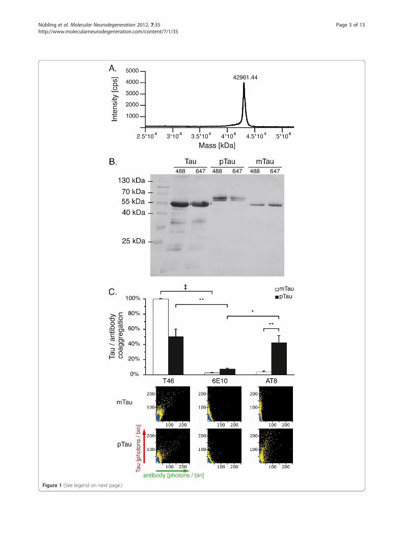
Figure 1 (See legend on next page.)
Nübling et al. Molecular Neurodegeneration 2012, 7:35 Page 3 of 13http://www.molecularneurodegeneration.com/content/7/1/35

(See figure on previous page.)Figure 1 Tau phosphorylation verified by western blot and SIFT analysis. A. Mass spectroscopy showed that recombinant human tau(isoform 5, 42967 Da) is of high purity. B. While mock phosphorylation (mTau) does not influence SDS-PAGE mobility of recombinant tau, atypical band shift is observed upon tau phosphorylation (pTau). C. SIFT analysis showed labeling of pTau oligomers with the phosphorylated tauspecific AT-8 antibody in presence of 1% DMSO indicating antibody binding, while no coaggregation was observed with mTau. Data wasnormalized against the coaggregation level of mTau with the T46 antibody, which does not require tau phosphorylation. 2D histogramsdepicting antibody (x-axis) and protein (y-axis) interactions show coaggregates of T46 with both pTau and mTau, while AT-8 only coaggregateswith pTau. A scheme describing the appearance of mixed aggregates in 2D histograms is included in Figure 3. Upon combining AT-8 and mTau,only DMSO induced tau aggregates are visible along the y-axis, similar to the control antibody 6E10. Measurements were taken from 12independent samples; each sample was measured four times. Levels of significance are displayed as * = p< 0.05; ** = p< 0.01; {=p< 0.001.
Nübling et al. Molecular Neurodegeneration 2012, 7:35 Page 4 of 13http://www.molecularneurodegeneration.com/content/7/1/35
shows only moderate aggregation, while Al3+ enhancesthe formation of larger oligomers with higher fluores-cence intensity.Fluorescence intensity distribution analysis (FIDA) of
tau oligomers induced by DMSO yielded larger aggre-gates for mTau (mean 76 molecules) than for pTau(mean 26 molecules). In presence of Al3+, large tau oli-gomers comprising on average 240 (pTau) and 220(mTau) molecules occurred within 15 minutes, while theoverall aggregation level was higher for pTau (Figure 2A,for aggregation dynamics also see Additional file 3). Thecombination of DMSO and Al3+ increased aggregationlevels compared to Al3+ alone predominantly for mTau(also see Additional files 1 and 2).Furthermore, we examined the SDS stability of oligo-
mers generated by the different enhancers (Figure 2C,D). While DMSO induced aggregates dissolve uponaddition of the ionic detergent SDS in a final concentra-tion of 0.2%, more than 50% of the Al3+ induced oligo-mer signal remains stable 100 min after addition of SDS.In conclusion, Al3+ promotes rapid formation of deter-
gent resistant oligomers, preferably of phosphorylatedprotein tau. Conversely, DMSO induced aggregation isdiminished upon tau phosphorylation.
Influence of tau phosphorylation on the coaggregation ofmonomeric tau and α-synuclein in presence of metal ionsand organic solventsSince the role of tau and α-syn coaggregation is increas-ingly recognized, we investigated the effect of tau488
phosphorylation on its coaggregation with α-syn647. Asprevious work has shown a major influence of Fe3+ onα-syn oligomerization [36-38], we also used Fe3+ at afinal concentration of 10 μM in our assay.In these experiments, 1% DMSO promoted the coag-
gregation of tau and α-syn in a similar manner for pTauand mTau (Figure 3A). On average, coaggregates werecomposed of 11 pTau + 8 α-syn monomers or 15 mTau+12 α-syn monomers, respectively, as determined byFIDA analysis. Fe3+ also had a promotional influence oncoaggregation, which was higher for pTau (p< 0.01, onaverage 53 pTau + 10 α-syn and 39 mTau+ 3 α-synmonomers per oligomer). Upon combining DMSO and
Fe3+, an additional effect exceeding the influence of oneinducer alone can be seen (also see Additional files 4and 5), while no significant difference in aggregationlevels of pTau and mTau was detectable. Similar to ourtau oligomerization experiments, Al3+ was the strongestinducer of tau and α-syn coaggregation, with pTaushowing higher coaggregation activity than mTau (p<0.001, Figure 3A; on average 131 pTau + 115 α-syn and137 mTau + 80 α-syn monomers per oligomer). This dif-ference was still detectable when combining Al3+ withDMSO, though to a lesser extent (p< 0.05). Mixed oli-gomers comprising both tau and α-syn appear scatteredaround the bisectrix of these histograms, while homoge-neous aggregates are located close to the axes, as illu-strated in Figure 3D.Concerning oligomer stability, 0.2% SDS reduced the
number of coaggregates formed in presence of everytested agent (Figure 3B, C). Again, coaggregates formedin presence of metal ions proved to be more resistant toSDS than those induced by DMSO, though to an overalllesser extent than the homogeneous tau aggregates.Oligomer sizes before and after addition of SDS as deter-mined by FIDA analysis are provided in Additional file6. The presence of Al3+-induced, SDS stable mixed oli-gomers was further validated applying gel filtration andfluorescence spectroscopy. In these experiments, Al3+
induced oligomers were separated from monomers bygel filtration. SDS stable mixed tau and α-syn oligomerseluted prior to monomers, as demonstrated by cross-correlation and SIFT-2D analysis (see Additional file 7).Thus, our data show increased tau and α-syn coaggre-
gation after tau phosphorylation in presence of trivalentmetal ions, and that the heterogenic oligomers generatedby Fe3+ and Al3+ are more stable to SDS treatment thanthose formed by DMSO.
Coaggregation of tau oligomers and monomericα-synucleinGiasson et al. showed that tau and α-syn induce eachother’s filament formation, and that some of the fibrilsformed in presence of both proteins comprise tau andα-syn segments [41]. Such findings suggest that thesesegments are either assembled by end to end annealing

Figure 2 (See legend on next page.)
Nübling et al. Molecular Neurodegeneration 2012, 7:35 Page 5 of 13http://www.molecularneurodegeneration.com/content/7/1/35

(See figure on previous page.)Figure 2 Al3+ promotes the formation of SDS stable tau oligomers more efficiently after tau phosphorylation. A. Cross-correlationamplitudes (G (0)) show mild proaggregatory activity of the organic solvent DMSO, which was significantly higher for mTau. Al3+ promotesintense protein oligomerization. Measurements were taken from 15 independent samples; each sample was measured four times. B. 2Dhistograms of detected photons indicate the formation of smaller oligomers in presence of DMSO (mean 76 monomers per oligomer for mTau,26 monomers for pTau) compared to Al3+ (mean 220 to 240 monomers per oligomer). C. Cross correlation amplitude immediately before (T0)and 100 min after (T100) addition of SDS to the aggregation assay. Significant reductions of aggregation levels at T100 are indicated by thesymbols over the error bars, while significant differences between pTau and mTau are indicated by the symbols over the wide bars.Measurements were taken from 7 independent samples. D. Cross correlation amplitude at T100 normalized against T0 shows increased SDS-resistance of Al3+ induced oligomers compared to DMSO. Levels of significance are displayed as * = p< 0.05; ** = p< 0.01; {=p< 0.001.
Nübling et al. Molecular Neurodegeneration 2012, 7:35 Page 6 of 13http://www.molecularneurodegeneration.com/content/7/1/35
of short filament fragments, or that such fragments actas seeds for both proteins. To evaluate the effect of tauphosphorylation on the interaction of tau oligomers withmonomeric α-syn, tau488 was first incubated in presenceof 1% DMSO, 10 μM Fe3+, 10 μM Al3+, and combina-tions of these agents for 90 minutes to generate pre-formed tau oligomers. α-Syn647 monomers were thenadded to the assay and measurements were conducted(Figure 4A).Contrary to the experiments with tau monomers,
DMSO had a stronger effect on the coaggregation ofpre-formed mTau oligomers with monomeric α-syncompared to pTau oligomers (p< 0.05, Figure 4A).Under these conditions, coaggregates contained on aver-age 20 pTau + 2 α-syn monomers or 27 mTau+ 5 α-synmonomers, respectively.pTau oligomer coaggregation with monomeric α-syn
exceeded mTau seven- to ninefold in presence of Fe3+
(p< 0.01; on average 197 pTau + 14 α-syn or 175 mTau+3 α-syn monomers per oligomer) and Al3+ (p< 0.001;on average 151 pTau + 73 α-syn or 154 mTau+ 35 α-synmonomers per oligomer). This difference was markedlyreduced upon combining Fe3+ and 1% DMSO, whichagain yielded a synergistic effect compared to the twoinducers alone (also see Additional files 8 and 9). Thecombination of Al3+ and 1% DMSO also reduced the dif-ference between pTau and mTau to a two-fold excess ofpTau coaggregation (p< 0.01).SDS (0.2%) reduced the number of oligomers formed
in presence of every tested inducer (Figure 4B). Espe-cially aggregates induced by metal ions alone showedenhanced resistance to SDS treatment compared tothose formed in presence of DMSO (Figure 4C).
DiscussionSeveral studies have dealt with the influence of proteinphosphorylation and the metal ion aluminium (Al3+) ontau filament formation so far [17-27]. However, thepathophysiological relevance of these factors is still un-clear. Whereas most studies were mainly focused on taufilament formation, we applied single molecule fluores-cence techniques to investigate the influence of thesefactors on early tau aggregation steps and oligomer
formation. We demonstrate that tau phosphorylationmodulates oligomer formation in presence of Al3+, andthat Al3+ induces the formation of distinct large, SDS re-sistant tau oligomers.We further demonstrate that coaggregation of tau and
α-syn can be observed at the single molecule level, is dif-ferentially modulated by tau phosphorylation, andinduced by trivalent metal ions. Such coaggregation mightbe of pathophysiological relevance, since co-deposits oftau and α-syn have been detected in various neurodegen-erative diseases [39,40].
Influence of tau phosphorylation on electrophoreticmobility and antibody interactionIt has been demonstrated that in vitro phosphorylationof human protein tau by GSK-3β can be verified usingSDS-PAGE by demonstrating a complete band shift ofphosphorylated tau compared to the unphosphorylatedprotein [17]. We verified efficient tau phosphorylationindicated by a complete band shift compared to theunphosporylated protein (Figure 1B). In addition, weinvestigated whether fluorescently labeled antibodies canbe employed in the SIFT method to verify tau phosphor-ylation at nanomolar protein concentrations, allowingfor possible diagnostic implementations of this methodin the future. Our data corroborate that tau phosphoryl-ation was successful and that antibodies can beemployed in the SIFT method to identify proteins andposttranslational modifications such as phosphorylationat nanomolar protein concentrations (Figure 1C). Suchan assay may provide a valuable tool for future diagnos-tic applications e.g. the detection of novel specific CSFbiomarkers of neurodegeneration.
Differential influence of phosphorylation on tauaggregationIn vitro studies have yielded conflicting results regardingthe influence of tau phosphorylation on its assembly tofilaments. To date, both inhibitory and promotionaleffects of GSK-3β mediated phosphorylation on tau fila-ment assembly were found in absence or presence ofpolyanionic aggregation inducers [17-20,50,51]. Notably,for Al3+ induced tau aggregation, phosphorylation was

Figure 3 (See legend on next page.)
Nübling et al. Molecular Neurodegeneration 2012, 7:35 Page 7 of 13http://www.molecularneurodegeneration.com/content/7/1/35
consistently found to be enhancing or, in some experi-ments, even a prerequisite [24-27].However, the methods applied in these studies, such as
electron microscopy, thioflavine fluorescence, laser light
scattering, SDS-PAGE and western blot, are of limited usein directly examining single molecule interactions at theoligomer level. Furthermore, these techniques often re-quire high concentrations of both protein and aggregation

(See figure on previous page.)Figure 3 Al3+ promotes stronger formation of SDS stable tau and α-syn coaggregates than Fe3+, preferably after tau phosphorylation.A. Cross-correlation amplitudes (G (0)) show mild proaggregatory activity of DMSO and Fe3+, while Al3+ promotes intense proteinoligomerization. Combinations of DMSO and metal ions show a synergistic effect especially for mTau. 2D histograms show the formation ofsmaller oligomers induced by DMSO compared to Al3+. B. Cross-correlation amplitude before (T0) and 100 min after (T100) addition of SDS to theaggregation assay. Significant reductions of aggregation levels at T100 are indicated by the symbols over the error bars, while significantdifferences between pTau and mTau are indicated by the symbols over the wide bars. C. Cross correlation amplitude at T100 normalized againstT0 shows increased SDS-resistance of metal ion induced oligomers compared to DMSO. D. Scheme illustrating the appearance of tau and α-synaggregates and coaggregates of the two proteins. Higher fluorescence intensities of detected oligomers indicate increased oligomer size.Measurements were taken from 16 independent samples; each sample was measured four times. Levels of significance are displayed as* = p< 0.05; ** = p< 0.01; {=p< 0.001.
Nübling et al. Molecular Neurodegeneration 2012, 7:35 Page 8 of 13http://www.molecularneurodegeneration.com/content/7/1/35
inducer that might not depict physiological conditions, es-pecially given the fact that tau protein readily polymerizesat high concentrations without any inducer [52,53]. Inaddition, the majority of studies investigating brain Al3+
concentrations did not detect concentrations exceeding 10μg/g brain mass (dry weight) in AD brains [54], which ap-proximately corresponds to 60 μmol/kg (wet weight),while many studies examining the influence of Al3+ on tauaggregation in vitro applied Al3+ concentrations in themilimolar range [25,27,55].In this study, we applied confocal single molecule fluor-
escent techniques to investigate the influence of proteinphosphorylation by GSK-3β on tau oligomer formation atthe single molecule level at nanomolar concentrations. Ex-posure to 10 μM Al3+ induced the rapid formation of largetau oligomers, while DMSO at a final concentration of 1%led to the formation of smaller oligomers. The large Al3+
triggered oligomers contained an average of 220 to 240molecules as shown by FIDA analysis, and were resistantto treatment with 0.2% SDS, which in contrast readily dis-solved the smaller oligomers formed in presence ofDMSO. The oligomer sizes observed here were compar-able with data presented earlier [49]. Interestingly, phos-phorylation by GSK-3β yielded an increase in Al3+
induced tau oligomer formation, while oligomer size wascomparable for pTau and mTau.Thus, our data identify tau phosphorylation and
physiological concentrations of the metal ion Al3+ assynergistic inducers of the formation of SDS resistanttau oligomers even at nanomolar protein concentrations.These findings substantiate the hypothesis that Al3+ mayplay a role in the formation of neurotoxic oligomerseven at early stages of neurodegeneration.
Metal ions and organic solvents enhance tau andα-synunclein coaggregation depending on tau’sphosphorylation statusCo-deposits of tau and α-syn have been found in severalneurodegenerative diseases, including AD, PSP, CBD andDLB [39,40]. Since interactions of different proteins aredifficult to monitor in vitro, only few studies investigatingthe interaction of tau and α-syn have been published so
far. It was demonstrated that in mixed micromolar solu-tions of α-syn and tau, both proteins’ minimal concentra-tions for filament assembly are decreased [41,56].Moreover, Giasson et al. detected filaments that comprisedboth tau and α-syn, proving that the two proteins directlyinteract in mixed solutions [41]. A more recent studyshowed that α-syn fibrils can be taken up by tau overex-pressing cells and induce the formation of phosphorylatedtriton-insoluble tau oligomers [57]. In vivo studies furtherdemonstrated that tau deposits can be found in mice over-expressing pathologic human α-syn mutations [58].In this study, we demonstrate that coaggregation of
tau and α-syn takes place even at nanomolar proteinconcentrations, is strongly induced by trivalent metalions, and differentially modulated by tau’s phosphoryl-ation status. In contrast to the findings of Giasson et al.,we did not detect tau and α-syn coaggregation in the ab-sence of aggregation inducers. This apparent discrepancyis most likely explained by the differences in proteinconcentrations (up to 1000-fold) and the shorter obser-vation time [41]. However, our data demonstrate thattau and α-syn coaggregation occurs even at nanomolarprotein concentrations in presence of aggregation indu-cers. As in our tau aggregation assay, Al3+ had the mostpronounced effect on coaggregation of α-syn with taumonomers and oligomers. Al3+ induces rapid coaggrega-tion of tau and α-syn (see Additional file 3). Fe3+ andDMSO also induce coaggregation of the two proteins,though to an overall lesser extent. In our experiments,tau phosphorylation by GSK-3β strongly enhanced theformation of mixed oligomers induced by Al3+ or Fe3+.Moreover, the mixed oligomers resulting from metal ioninduced coaggregation proved to be more resistant toSDS treatment than those formed in presence of DMSO.We further provide data from FIDA analysis and gel
filtration experiments demonstrating the presence andsize of mixed tau and a-syn oligomers (see Additionalfiles 6 and 7). Earlier studies applying atomic force mico-scropy and SIFT established FIDA-based quantificationof oligomer sizes as a reliable method [38].These results extend the current model of tau and
α-syn interaction at early stages of neurodegeneration.

Figure 4 Monomeric α-syn preferably coaggregates with pTau oligomers in presence of metal ions. A. Cross-correlation amplitudes (G (0))show mild coaggregation of α-syn monomers with pre-formed tau oligomers induced by DMSO and Fe3+, while Al3+ promotes intensecoaggregation. Combinations of DMSO and metal ions show a synergistic effect especially for mTau. 2D histograms show the formation ofsmaller mixed oligomers induced by DMSO compared to the metal ions. B. Cross-correlation amplitude before (T0) and 100 min after (T100)addition of SDS to the aggregation assay. Significant reductions of aggregation levels at T100 are indicated by the symbols over the error bars,while significant differences between pTau and mTau are indicated by the symbols over the wide bars. C. Cross correlation amplitude at T100normalized against T0 shows increased SDS-resistance of metal ion induced oligomers compared to DMSO. Measurements were taken from 20independent samples; each sample was measured four times. Levels of significance are displayed as * = p< 0.05; ** = p< 0.01; {=p< 0.001.
Nübling et al. Molecular Neurodegeneration 2012, 7:35 Page 9 of 13http://www.molecularneurodegeneration.com/content/7/1/35
Nübling et al. Molecular Neurodegeneration 2012, 7:35 Page 10 of 13http://www.molecularneurodegeneration.com/content/7/1/35
As the cytoplasmic concentration of unbound tau andα-syn is low in pre-clinical neurodegeneration, thepresence of pro-aggregatory factors may be crucial inthe generation of early mixed oligomers. We identify tri-valent metal ions and tau phosphorylation by GSK-3β aspotential inducers of such oligomer formation even atnanomolar protein concentrations. A time-dependentincrease of oligomer concentrations may then induceself-propagating and cross-seeding mechanisms asdemonstrated by Giasson and Waxman [41,57].
ConclusionsIn summary, we demonstrate that tau phosphorylationand trivalent metal ions such as Al3+ act together in theformation of distinct SDS resistant tau oligomers. More-over, applying confocal single molecule fluorescencetechniques, we show that, under certain conditions,interactions of tau and α-syn can take place even atnanomolar protein concentrations, and result in the for-mation of mixed oligomers. Notably, the formation ofSDS resistant mixed aggregates was induced by physiolo-gically relevant concentrations of trivalent metal ions,and strongly enhanced by tau phosphorylation. Takinginto account that a considerable amount of soluble tauexists in a phosphorylated state in neurodegenerativediseases [1,2,59], these findings support a crucial role ofspecific metal ions such as Al3+ and Fe3+ in early cyto-plasmic aggregation and coaggregation events.Moreover, our results indicate common pathophysio-
logic mechanisms of both tau aggregation and cross-seeding phenomena, and might explain the coincidenceof tau and α-syn in neuronal deposits. The mixed aggre-gates described here may provide an interlink of differ-ent pathological pathways leading to neurodegeneration,and may serve as promising therapeutic targets for fu-ture drug development. Furthermore, we introduce anovel technique of monitoring post-translational modifi-cations at very low protein concentrations that may pro-vide a powerful diagnostic tool in the future.
MethodsExpression of human proteins tau and α-synucleinProtein tauTau isoform 5 was expressed in E.coli as described previ-ously [49]. In brief, thermocompetent E.coli DE3 (RIL)were transformed, and tau was purified by sterile filtra-tion, cation exchange chromatography and ammoniumacid salt precipitation. The transformation vector was akind gift of Manuela Neumann (ZNP Munich/Zurich).
α-Synucleinα-Synuclein was produced according to established proto-cols [48]. A stock solution containing 1 mg/ml α-synucleinwas prepared in 50 mM tris buffer, pH 7.0.
Protein labelingProteins were labeled with fluorescent dyes Alexa-488-O-succinimidylester and Alexa-647-O-succinimidylester, re-spectively, as described previously [48]. Tau isoform 5 wasincubated with a four-fold molar excess of Alexa-488 orAlexa-647 in 100 mM NaHCO3 for 24 h at roomtemperature. For α-syn, a two-fold molar excess of dyewas used. Antibodies T46 (Invitrogen, Carlsbad, CA), AT-8 (Pierce Endogen, Rockford, IL) and 6E10 (Acris Anti-bodies, Hiddenhausen, Germany) first underwent bufferexchange to PBS pH 7.0. Since antibody concentrationsafter buffer exchange were unknown, concentrations ofAlexa-488-O-succinimidylester were determined empiric-ally. Unbound dye was separated using a PD10 desaltingcolumn. Fluorescently labeled proteins are further referredto as tau488, tau647, and α-syn647.
Phosphorylation of protein tauTau phosphorylation was conducted as described previ-ously [17]. Fluorescently labeled protein tau was incu-bated with Glycogen Synthase Kinase 3β (Sigma, SaintLouis, MO) in a ratio of 0,018 U GSK-3 β / pmol tau inbuffer containing 33 mM tris pH 7.5, 40 mM hepes pH7.64, 100 mM NaCl, 5 mM EGTA, 3 mM MgCl2, 2 mMATP, 180 mM sucrose, 0.13 mM PMSF, 0.67 mM benza-midine, 0.067% mercaptoethanol and 0.02% Brij-35.Samples were incubated for 20 h at 30 °C under constantshaking. As control, fluorescently labeled protein tauwas incubated in buffer containing all components ex-cept for the enzyme, and is further referred to as mockphosphorylated tau (mTau). Protein phosphorylation wasconfirmed by sodium dodecyl sulfate polyacrylamidegel electrophoresis (SDS-PAGE) and western blot usingantibody T46 as described previously [60]. Phosphory-lated protein tau (pTau) featured a typical band shiftcompared to mTau, as described previously [17](Figure 1B).
Aggregation assayStock solutions of tau488, tau647, and α-syn647 were pre-pared for a final concentration of 10 molecules per focalvolume, equivalent to a concentration of 10 nM to 20nM. For coaggregation experiments, a 1:1 ratio of tau488
and α-syn647 was used. Prior to each experiment, tausamples were centrifuged at 100.000 g for 30 min to re-move pre-formed aggregates, and all protein stock solu-tions were then controlled for pre-existing aggregates bySIFT [49]. Only samples free of pre-formed aggregateswere used. All experiments were conducted in 96 wellplates with a cover slide bottom, in a total sample vol-ume of 20 μl per well. Plates were covered with adhesivefilm to obviate evaporation. For aggregation and coag-gregation experiments, protein stock solutions wereadded to wells containing 50 mM tris buffer pH 7.0, 10

Nübling et al. Molecular Neurodegeneration 2012, 7:35 Page 11 of 13http://www.molecularneurodegeneration.com/content/7/1/35
μM AlCl3, 10 μM FeCl3 or 1% dimethyl sulfoxide(DMSO), and measurements were started immediately.For coaggregation of tau488 oligomers with α-syn647
monomers, samples of tau488 were prepared as describedabove and pre-incubated for 90 min. Subsequently, α-syn647 monomers were added and measurements werestarted immediately. To investigate oligomer stability, a10-fold SDS stock solution for a final concentration of0.2% was added to each well after each experiment, andmeasurements were repeated. Each well was measuredfour times, resulting in a total duration of each experi-ment of 60 to 120 minutes, depending on experimentallayout.
Confocal single particle analysisFCS and SIFT were performed on an InsightReader(Evotec-Technologies, Hamburg, Germany) with dualcolor excitation at 488 and 633 nm as described previ-ously [48,49,61]. Data was collected separately for eachexcitation wavelength by two single-photon detectors.Analysis was performed using FCSPP evaluation soft-ware version 2.0 (Evotec-Technologies), allowing auto-correlation, cross-correlation and fluorescence intensitydistribution (FIDA) analysis, and SIFT-2D evaluationsoftware (Evotec-Technologies). For SIFT-2D analysis,photons were added up in time intervals (bins) of 40 μsand illustrated in a 2D scatterplot. Scatterplots depictedin this paper contain all photons of all consecutive mea-surements of one sample. Photon-weighted SIFT aggre-gation and coaggregation analysis was performed asdescribed previously [49]. As cross-correlation detectssmall oligomeric protein aggregates with higher sensitivity,only cross-correlation data is depicted for experiments ontau aggregation and coaggregation with α-syn (for SIFTdata see Additional files 1, 4, and 7).
Mass spectroscopyMass spectroscopy was performed according to estab-lished protocols to verify the purity of tau protein stocksolution [61].
Statistical analysisNormal distribution of data was determined by Shapiro-Wilk test. If normal distribution was confirmed, a two-sided student’s t-test preceded by Levene’s test for equalityof variance was performed. A paired student’s t-test wasused for experiments on SDS-resistance of oligomers. Ifnormal distribution was not confirmed, Mann–WhitneyU-test was performed. Bonferroni-adjustment for multipletesting was done where appropriate. Data is demonstratedas average of all independent samples, and a paired stu-dent’s t-test was performed including all independentsamples. Error bars in figures show the standard error ofthe mean.
Additional files
Additional file 1: Comparison of aggregation levels of pTau andmTau. Comparison of aggregation levels of phosphorylated (pTau) andmock phosphorylated (mTau) protein tau in presence of differentaggregation inducers. SIFT data is presented as ratios (colum / row).Measurements were taken from 15 independent samples, each samplewas measured four times.
Additional file 2: Comparison of aggregation levels of pTau andmTau. Comparison of aggregation levels of phosphorylated (pTau) andmock phosphorylated (mTau) protein tau in presence of differentaggregation inducers. Cross-correlation data is presented as ratios (colum /row). Measurements were taken from 15 independent samples, eachsample was measured four times.
Additional file 3: Kinetics of pTau and mTau aggregation andcoaggregation. Kinetics of pTau and mTau aggregation (tau/tau, 15independent samples) and coaggregation of tau monomers (tau/α-syn,16 independent samples) and oligomers (tauoligo/α-syn, 20independent samples) with monomeric α-synuclein in presence of 1%DMSO, 10 μM Al3+, 10 μM Fe3+ and combinations of metal ions andDMSO. A control measurement depicting the aggregation status in theabsence of inducers was defined as time point “0". While DMSOinduces slow continuous tau aggregation, the protein’s coaggregationwith α-syn reaches an early steady state. Al3+ promotes rapid initialaggregation and coaggregation only for pTau, while mTaucoaggregation proceeds distinctly slower. Fe3+ induced coaggregationis continuous, with pTau proceeding faster than mTau. Uponcombining metal ions and DMSO, both pTau and mTau show rapidaggregation and coaggregation exceeding the single inducers.Levels of significance are displayed as * = p< 0.05; ** = p <0.01;{= p< 0.001.
Additional file 4: Comparison of coaggregation levels of pTauand mTau with α-syn. Comparison of coaggregation levels ofphosphorylated (pTau) and mock phosphorylated (mTau) protein tauwith α-synuclein in presence of different aggregation inducers. SIFTdata is presented as ratios (colum / row). Measurements were takenfrom 16 independent samples, each sample was measured four times.
Additional file 5: Comparison of coaggregation levels of pTauand mTau with α-syn. Comparison of coaggregation levels ofphosphorylated (pTau) and mock phosphorylated (mTau) protein tauwith α-synuclein in presence of different aggregation inducers. Cross-correlation data is presented as ratios (colum / row). Measurementswere taken from 16 independent samples, each sample was measuredfour times.
Additional file 6: Quantification of mixed tau and α-syn oligomersizes. Fluorescence intensity distribution analysis (FIDA) shows the sizeof metal ion induced mixed tau and α-syn oligomers before and afteraddition of SDS. SDS stable mixed oligomers can further bedemonstrated in SIFT-2D analysis.
Additional file 7: Separation of SDS stable mixed tau and α-synoligomers by gel filtration. Mixed tau and α-syn oligomers induced by Al3+
are separated from monomers applying gel filtration. SDS-stable oligomersare eluted before tau and α-syn monomers, as demonstrated by cross-correlation and SIFT analysis.
Additional file 8: Comparison of coaggregation levels of pTau andmTau oligomers with α-syn. Comparison of coaggregation levels ofphosphorylated (pTau) and mock phosphorylated (mTau) tauoligomers with monomeric α-synuclein in presence of differentaggregation inducers. SIFT data is presented as ratios (colum / row).Measurements were taken from 20 independent samples, each samplewas measured four times.
Additional file 9: Comparison of coaggregation levels of pTauand mTau oligomers with α-syn. Comparison of coaggregationlevels of phosphorylated (pTau) and mock phosphorylated (mTau) tauoligomers with monomeric α-synuclein in presence of differentaggregation inducers. Cross-correlation data is presented as ratios(colum / row). Measurements were taken from 20 independentsamples, each sample was measured four times.
Nübling et al. Molecular Neurodegeneration 2012, 7:35 Page 12 of 13http://www.molecularneurodegeneration.com/content/7/1/35
AbbreviationsAD: Alzheimer’s disease; α-syn: α-synuclein; CBD: Corticobasal degeneration;DLB: Dementia with Lewy bodies; DMSO: dimethyl sulfoxide;FCS: Fluorescence correlation spectroscopy; FIDA: fluorescence intensitydistribution analysis; GSK-3β: Glycogen synthase kinase 3β; mTau: Mockphosphorylated tau; NFT: Neurofibrillary tangles; PAGE: Polyacrylamide gelelectrophoresis; PHF: Paired helical filaments; pTau: Phosphorylated tau;PSP: Progressive supranuclear palsy; SDS: Sodium dodecyl sulfate;SIFT: Scanning for intensely fluorescent targets.
Competing interestsThe authors declare that they have no competing interests.
Authors’ contributionsGN1. A, B, C; 2. A, B, C; 3. A, B, BB1. A, B: 2. A, C; 3. B, JL1. A, B; 2. C; 3. B, JH2. A, B, C; 3. A, B,HK1. B; 3. B, AG1. A, B; 2. A, C; 3. B. Author contributions are abbreviated asfollows: 1 = Research project: A. Conception, B. Organization, C. Execution.2 = Statistical Analysis: A. Design, B. Execution, C. Review and Critique.3 =Manuscript Preparation: A. Writing of the first draft, B. Review andCritique. All authors read and approved the final manuscript.
AcknowledgementsWe thank Manuela Neumann for providing the tau expression vector. Wefurther thank Lars Israel for conducting the mass spectroscopy analysis ofrecombinant tau. This work was supported by SFB 596. Parts of this work areelements of one author’s dissertation (Georg Nuebling) presented to theMedical Faculty, LMU Munich, Germany.
Author details1Center for Neuropathology and Prion Research,Ludwig-Maximilians-Universität, Feodor-Lynen-Str. 23, 81377, Munich,Germany. 2Neurologische Klinik und Poliklinik, Klinikum der UniversitätMünchen, Marchioninistr. 15, 81377, Munich, Germany.
Received: 12 March 2012 Accepted: 30 June 2012Published: 23 July 2012
References1. Khatoon S, Grundke-Iqbal I, Iqbal K: Brain levels of microtubule-associated
protein tau are elevated in Alzheimer’s disease: a radioimmuno-slot-blotassay for nanograms of the protein. J Neurochem 1992, 59:750–753.
2. Kopke E, Tung YC, Shaikh S, Alonso AC, Iqbal K, Grundke-Iqbal I:Microtubule-associated protein tau. Abnormal phosphorylation of a non-paired helical filament pool in Alzheimer disease. J Biol Chem 1993,268:24374–24384.
3. Goedert M, Jakes R, Vanmechelen E: Monoclonal antibody AT8 recognisestau protein phosphorylated at both serine 202 and threonine 205.Neurosci Lett 1995, 189:167–169.
4. Buee-Scherrer V, Condamines O, Mourton-Gilles C, Jakes R, Goedert M, Pau B,Delacourte A: AD2, a phosphorylation-dependent monoclonal antibodydirected against tau proteins found in Alzheimer’s disease. Brain Res MolBrain Res 1996, 39:79–88.
5. Buerger K, Ewers M, Pirttila T, Zinkowski R, Alafuzoff I, Teipel SJ, DeBernardis J,Kerkman D, McCulloch C, Soininen H, Hampel H: CSF phosphorylated tauprotein correlates with neocortical neurofibrillary pathology in Alzheimer’sdisease. Brain 2006, 129:3035–3041.
6. Hampel H, Buerger K, Zinkowski R, Teipel SJ, Goernitz A, Andreasen N,Sjoegren M, DeBernardis J, Kerkman D, Ishiguro K, et al: Measurement ofphosphorylated tau epitopes in the differential diagnosis of Alzheimerdisease: a comparative cerebrospinal fluid study. Arch Gen Psychiatry2004, 61:95–102.
7. Reynolds CH, Betts JC, Blackstock WP, Nebreda AR, Anderton BH:Phosphorylation sites on tau identified by nanoelectrospray massspectrometry: differences in vitro between the mitogen-activatedprotein kinases ERK2, c-Jun N-terminal kinase and P38, and glycogensynthase kinase-3beta. J Neurochem 2000, 74:1587–1595.
8. Lovestone S, Reynolds CH, Latimer D, Davis DR, Anderton BH, Gallo JM,Hanger D, Mulot S, Marquardt B, Stabel S, et al: Alzheimer’s disease-likephosphorylation of the microtubule-associated protein tau by glycogensynthase kinase-3 in transfected mammalian cells. Curr Biol 1994,4:1077–1086.
9. Li X, Lu F, Tian Q, Yang Y, Wang Q, Wang JZ: Activation of glycogensynthase kinase-3 induces Alzheimer-like tau hyperphosphorylation inrat hippocampus slices in culture. J Neural Transm 2006, 113:93–102.
10. Spittaels K, Van den Haute C, Van Dorpe J, Geerts H, Mercken M, Bruynseels K,Lasrado R, Vandezande K, Laenen I, Boon T, et al: Glycogen synthase kinase-3beta phosphorylates protein tau and rescues the axonopathy in thecentral nervous system of human four-repeat tau transgenic mice. J BiolChem 2000, 275:41340–41349.
11. Brownlees J, Irving NG, Brion JP, Gibb BJ, Wagner U, Woodgett J, Miller CC:Tau phosphorylation in transgenic mice expressing glycogen synthasekinase-3beta transgenes. Neuroreport 1997, 8:3251–3255.
12. Pei JJ, Tanaka T, Tung YC, Braak E, Iqbal K, Grundke-Iqbal I: Distribution,levels, and activity of glycogen synthase kinase-3 in the Alzheimerdisease brain. J Neuropathol Exp Neurol 1997, 56:70–78.
13. Leroy K, Yilmaz Z, Brion JP: Increased level of active GSK-3beta inAlzheimer’s disease and accumulation in argyrophilic grains and inneurones at different stages of neurofibrillary degeneration. NeuropatholAppl Neurobiol 2007, 33:43–55.
14. Baum L, Hansen L, Masliah E, Saitoh T: Glycogen synthase kinase 3alteration in Alzheimer disease is related to neurofibrillary tangleformation. Mol Chem Neuropathol 1996, 29:253–261.
15. Yamaguchi H, Ishiguro K, Uchida T, Takashima A, Lemere CA, Imahori K:Preferential labeling of Alzheimer neurofibrillary tangles with antisera fortau protein kinase (TPK) I/glycogen synthase kinase-3 beta and cyclin-dependent kinase 5, a component of TPK II. Acta Neuropathol 1996,92:232–241.
16. Pei JJ, Braak E, Braak H, Grundke-Iqbal I, Iqbal K, Winblad B, Cowburn RF:Distribution of active glycogen synthase kinase 3beta (GSK-3beta) inbrains staged for Alzheimer disease neurofibrillary changes. JNeuropathol Exp Neurol 1999, 58:1010–1019.
17. Rankin CA, Sun Q, Gamblin TC: Tau phosphorylation by GSK-3betapromotes tangle-like filament morphology. Mol Neurodegener 2007, 2:12.
18. Liu F, Li B, Tung EJ, Grundke-Iqbal I, Iqbal K, Gong CX: Site-specific effectsof tau phosphorylation on its microtubule assembly activity and self-aggregation. Eur J Neurosci 2007, 26:3429–3436.
19. Wang JZ, Grundke-Iqbal I, Iqbal K: Kinases and phosphatases and tau sitesinvolved in Alzheimer neurofibrillary degeneration. Eur J Neurosci 2007,25:59–68.
20. Voss K, Gamblin TC: GSK-3beta phosphorylation of functionally distinct tauisoforms has differential, but mild effects. Mol Neurodegener 2009, 4:18.
21. Vandebroek T, Vanhelmont T, Terwel D, Borghgraef P, Lemaire K, Snauwaert J,Wera S, Van Leuven F, Winderickx J: Identification and isolation of ahyperphosphorylated, conformationally changed intermediate of humanprotein tau expressed in yeast. Biochemistry 2005, 44:11466–11475.
22. Jackson GR, Wiedau-Pazos M, Sang TK, Wagle N, Brown CA, Massachi S,Geschwind DH: Human wild-type tau interacts with wingless pathwaycomponents and produces neurofibrillary pathology in Drosophila.Neuron 2002, 34:509–519.
23. Engel T, Goni-Oliver P, Lucas JJ, Avila J, Hernandez F: Chronic lithiumadministration to FTDP-17 tau and GSK-3beta overexpressing mice preventstau hyperphosphorylation and neurofibrillary tangle formation, but pre-formed neurofibrillary tangles do not revert. J Neurochem 2006, 99:1445–1455.
24. Li W, Ma KK, Sun W, Paudel HK: Phosphorylation sensitizes microtubule-associated protein tau to Al(3+)-induced aggregation. Neurochem Res1998, 23:1467–1476.
25. Haase C, Stieler JT, Arendt T, Holzer M: Pseudophosphorylation of tau proteinalters its ability for self-aggregation. J Neurochem 2004, 88:1509–1520.
26. Shin RW, Lee VM, Trojanowski JQ: Aluminum modifies the properties ofAlzheimer’s disease PHF tau proteins in vivo and in vitro. J Neurosci 1994,14:7221–7233.
27. Yamamoto A, Shin RW, Hasegawa K, Naiki H, Sato H, Yoshimasu F, Kitamoto T:Iron (III) induces aggregation of hyperphosphorylated tau and itsreduction to iron (II) reverses the aggregation: implications in theformation of neurofibrillary tangles of Alzheimer’s disease. J Neurochem2002, 82:1137–1147.
28. Klatzo I, Wisniewski H, Streicher E: Experimental Production ofNeurofibrillary Degeneration. I. Light Microscopic Observations. JNeuropathol Exp Neurol 1965, 24:187–199.
29. Xu N, Majidi V, Markesbery WR, Ehmann WD: Brain aluminum inAlzheimer’s disease using an improved GFAAS method. Neurotoxicology1992, 13:735–743.

Nübling et al. Molecular Neurodegeneration 2012, 7:35 Page 13 of 13http://www.molecularneurodegeneration.com/content/7/1/35
30. Good PF, Perl DP, Bierer LM, Schmeidler J: Selective accumulation ofaluminum and iron in the neurofibrillary tangles of Alzheimer’s disease:a laser microprobe (LAMMA) study. Ann Neurol 1992, 31:286–292.
31. Walton JR: Aluminum in hippocampal neurons from humans withAlzheimer’s disease. Neurotoxicology 2006, 27:385–394.
32. Krewski D, Yokel RA, Nieboer E, Borchelt D, Cohen J, Harry J, Kacew S,Lindsay J, Mahfouz AM, Rondeau V: Human health risk assessment foraluminium, aluminium oxide, and aluminium hydroxide. J Toxicol EnvironHealth B Crit Rev 2007, 10(Suppl 1):1–269.
33. Smith MA, Harris PL, Sayre LM, Perry G: Iron accumulation in Alzheimerdisease is a source of redox-generated free radicals. Proc Natl Acad Sci US A 1997, 94:9866–9868.
34. Morris CM, Kerwin JM, Edwardson JA: Non-haem iron histochemistry ofthe normal and Alzheimer’s disease hippocampus. Neurodegeneration1994, 3:267–275.
35. Deibel MA, Ehmann WD, Markesbery WR: Copper, iron, and zincimbalances in severely degenerated brain regions in Alzheimer’s disease:possible relation to oxidative stress. J Neurol Sci 1996, 143:137–142.
36. Levin J, Högen T, Hillmer A, Bader B, Schmidt F, Kretzschmar H, Bötzel K,Giese A: Generation of ferric iron links oxidative stress to α-synucleinoligomer formation. J Parkinson’s Dis 2011, 1:205–216.
37. Hillmer AS, Putcha P, Levin J, Hogen T, Hyman BT, Kretzschmar H, McLean PJ,Giese A: Converse modulation of toxic alpha-synuclein oligomers inliving cells by N’-benzylidene-benzohydrazide derivates and ferric iron.Biochem Biophys Res Commun 2010, 391:461–466.
38. Kostka M, Hogen T, Danzer KM, Levin J, Habeck M, Wirth A, Wagner R, Glabe CG,Finger S, Heinzelmann U, et al: Single particle characterization of iron-induced pore-forming alpha-synuclein oligomers. J Biol Chem 2008,283:10992–11003.
39. Takeda A, Hashimoto M, Mallory M, Sundsumo M, Hansen L, Masliah E: C-terminal alpha-synuclein immunoreactivity in structures other than Lewybodies in neurodegenerative disorders. Acta Neuropathol 2000, 99:296–304.
40. Ishizawa T, Mattila P, Davies P, Wang D, Dickson DW: Colocalization of tauand alpha-synuclein epitopes in Lewy bodies. J Neuropathol Exp Neurol2003, 62:389–397.
41. Giasson BI, Forman MS, Higuchi M, Golbe LI, Graves CL, Kotzbauer PT,Trojanowski JQ, Lee VM: Initiation and synergistic fibrillization of tau andalpha-synuclein. Science 2003, 300:636–640.
42. Lasagna-Reeves CA, Castillo-Carranza DL, Guerrero-Muoz MJ, Jackson GR,Kayed R: Preparation and characterization of neurotoxic tau oligomers.Biochemistry 2010, 49:10039–10041.
43. Maeda S, Sahara N, Saito Y, Murayama M, Yoshiike Y, Kim H, Miyasaka T,Murayama S, Ikai A, Takashima A: Granular tau oligomers as intermediatesof tau filaments. Biochemistry 2007, 46:3856–3861.
44. Santacruz K, Lewis J, Spires T, Paulson J, Kotilinek L, Ingelsson M, Guimaraes A,DeTure M, Ramsden M, McGowan E, et al: Tau suppression in aneurodegenerative mouse model improves memory function. Science 2005,309:476–481.
45. Wittmann CW, Wszolek MF, Shulman JM, Salvaterra PM, Lewis J, Hutton M,Feany MB: Tauopathy in Drosophila: neurodegeneration withoutneurofibrillary tangles. Science 2001, 293:711–714.
46. Kimura T, Fukuda T, Sahara N, Yamashita S, Murayama M, Mizoroki T,Yoshiike Y, Lee B, Sotiropoulos I, Maeda S, Takashima A: Aggregation ofdetergent-insoluble tau is involved in neuronal loss but not in synapticloss. J Biol Chem 2010, 285:38692–38699.
47. Bieschke J, Giese A, Schulz-Schaeffer W, Zerr I, Poser S, Eigen M, Kretzschmar H:Ultrasensitive detection of pathological prion protein aggregates by dual-color scanning for intensely fluorescent targets. Proc Natl Acad Sci U S A2000, 97:5468–5473.
48. Giese A, Bader B, Bieschke J, Schaffar G, Odoy S, Kahle PJ, Haass C,Kretzschmar H: Single particle detection and characterization of synucleinco-aggregation. Biochem Biophys Res Commun 2005, 333:1202–1210.
49. Bader B, Nubling G, Mehle A, Nobile S, Kretzschmar H, Giese A: Singleparticle analysis of tau oligomer formation induced by metal ions andorganic solvents. Biochem Biophys Res Commun 2011, 411:190–196.
50. Schneider A, Biernat J, von Bergen M, Mandelkow E, Mandelkow EM:Phosphorylation that detaches tau protein from microtubules (Ser262,Ser214) also protects it against aggregation into Alzheimer paired helicalfilaments. Biochemistry 1999, 38:3549–3558.
51. Sun Q, Gamblin TC: Pseudohyperphosphorylation causing AD-likechanges in tau has significant effects on its polymerization. Biochemistry2009, 48:6002–6011.
52. Crowther RA, Olesen OF, Smith MJ, Jakes R, Goedert M: Assembly ofAlzheimer-like filaments from full-length tau protein. FEBS Lett 1994,337:135–138.
53. Perez M, Valpuesta JM, Medina M, Montejo de Garcini E, Avila J:Polymerization of tau into filaments in the presence of heparin: theminimal sequence required for tau-tau interaction. J Neurochem 1996,67:1183–1190.
54. Andrasi E, Pali N, Molnar Z, Kosel S: Brain aluminum, magnesium andphosphorus contents of control and Alzheimer-diseased patients.J Alzheimers Dis 2005, 7:273–284.
55. Yang LS, Ksiezak-Reding H: Ca2+ and Mg2+ selectively induce aggregatesof PHF-tau but not normal human tau. J Neurosci Res 1999, 55:36–43.
56. Kotzbauer PT, Giasson BI, Kravitz AV, Golbe LI, Mark MH, Trojanowski JQ, Lee VM:Fibrillization of alpha-synuclein and tau in familial Parkinson’s diseasecaused by the A53T alpha-synuclein mutation. Exp Neurol 2004, 187:279–288.
57. Waxman EA, Giasson BI: Induction of Intracellular Tau Aggregation IsPromoted by a-Synuclein Seeds and Provides Novel Insights into theHyperphosphorylation of Tau. J Neurosci 2011, 31:7604–7618.
58. Emmer KL, Waxman EA, Covy JP, Giasson BI: E46K human alpha-synucleintransgenic mice develop Lewy-like and tau pathology associated withage-dependent, detrimental motor impairment. J Biol Chem 2011,286:35104–35118.
59. Ksiezak-Reding H, Liu WK, Yen SH: Phosphate analysis anddephosphorylation of modified tau associated with paired helicalfilaments. Brain Res 1992, 597:209–219.
60. Arai T, Guo JP, McGeer PL: Proteolysis of non-phosphorylated andphosphorylated tau by thrombin. J Biol Chem 2005, 280:5145–5153.
61. Levin J, Giese A, Boetzel K, Israel L, Hogen T, Nubling G, Kretzschmar H, LorenzlS: Increased alpha-synuclein aggregation following limited cleavage bycertain matrix metalloproteinases. Exp Neurol 2009, 215:201–208.
doi:10.1186/1750-1326-7-35Cite this article as: Nübling et al.: Synergistic influence ofphosphorylation and metal ions on tau oligomer formation andcoaggregation with α-synuclein at the single molecule level. MolecularNeurodegeneration 2012 7:35.
Submit your next manuscript to BioMed Centraland take full advantage of:
• Convenient online submission
• Thorough peer review
• No space constraints or color figure charges
• Immediate publication on acceptance
• Inclusion in PubMed, CAS, Scopus and Google Scholar
• Research which is freely available for redistribution
Submit your manuscript at www.biomedcentral.com/submit
Copyright © 2022 FDOKUMEN