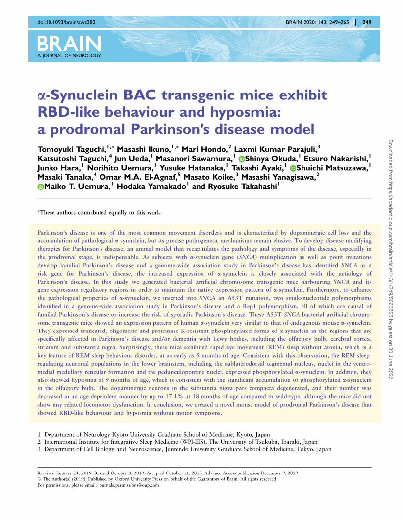
a-Synuclein BAC transgenic mice exhibit RBD-like behaviour ...
17
a-Synuclein BAC transgenic mice exhibit RBD-like behaviour and hyposmia: a prodromal Parkinson’s disease model Tomoyuki Taguchi, 1,\ Masashi Ikuno, 1,\ Mari Hondo, 2 Laxmi Kumar Parajuli, 3 Katsutoshi Taguchi, 4 Jun Ueda, 1 Masanori Sawamura, 1 Shinya Okuda, 1 Etsuro Nakanishi, 1 Junko Hara, 1 Norihito Uemura, 1 Yusuke Hatanaka, 1 Takashi Ayaki, 1 Shuichi Matsuzawa, 1 Masaki Tanaka, 4 Omar M.A. El-Agnaf, 5 Masato Koike, 3 Masashi Yanagisawa, 2 Maiko T. Uemura, 1 Hodaka Yamakado 1 and Ryosuke Takahashi 1 These authors contributed equally to this work. Parkinson’s disease is one of the most common movement disorders and is characterized by dopaminergic cell loss and the accumulation of pathological a-synuclein, but its precise pathogenetic mechanisms remain elusive. To develop disease-modifying therapies for Parkinson’s disease, an animal model that recapitulates the pathology and symptoms of the disease, especially in the prodromal stage, is indispensable. As subjects with a-synuclein gene (SNCA) multiplication as well as point mutations develop familial Parkinson’s disease and a genome-wide association study in Parkinson’s disease has identified SNCA as a risk gene for Parkinson’s disease, the increased expression of a-synuclein is closely associated with the aetiology of Parkinson’s disease. In this study we generated bacterial artificial chromosome transgenic mice harbouring SNCA and its gene expression regulatory regions in order to maintain the native expression pattern of a-synuclein. Furthermore, to enhance the pathological properties of a-synuclein, we inserted into SNCA an A53T mutation, two single-nucleotide polymorphisms identified in a genome-wide association study in Parkinson’s disease and a Rep1 polymorphism, all of which are causal of familial Parkinson’s disease or increase the risk of sporadic Parkinson’s disease. These A53T SNCA bacterial artificial chromo- some transgenic mice showed an expression pattern of human a-synuclein very similar to that of endogenous mouse a-synuclein. They expressed truncated, oligomeric and proteinase K-resistant phosphorylated forms of a-synuclein in the regions that are specifically affected in Parkinson’s disease and/or dementia with Lewy bodies, including the olfactory bulb, cerebral cortex, striatum and substantia nigra. Surprisingly, these mice exhibited rapid eye movement (REM) sleep without atonia, which is a key feature of REM sleep behaviour disorder, at as early as 5 months of age. Consistent with this observation, the REM sleep- regulating neuronal populations in the lower brainstem, including the sublaterodorsal tegmental nucleus, nuclei in the ventro- medial medullary reticular formation and the pedunculopontine nuclei, expressed phosphorylated a-synuclein. In addition, they also showed hyposmia at 9 months of age, which is consistent with the significant accumulation of phosphorylated a-synuclein in the olfactory bulb. The dopaminergic neurons in the substantia nigra pars compacta degenerated, and their number was decreased in an age-dependent manner by up to 17.1% at 18 months of age compared to wild-type, although the mice did not show any related locomotor dysfunction. In conclusion, we created a novel mouse model of prodromal Parkinson’s disease that showed RBD-like behaviour and hyposmia without motor symptoms. 1 Department of Neurology Kyoto University Graduate School of Medicine, Kyoto, Japan 2 International Institute for Integrative Sleep Medicine (WPI-IIIS), The University of Tsukuba, Ibaraki, Japan 3 Department of Cell Biology and Neuroscience, Juntendo University Graduate School of Medicine, Tokyo, Japan doi:10.1093/brain/awz380 BRAIN 2020: 143; 249–265 | 249 Received January 24, 2019. Revised October 8, 2019. Accepted October 11, 2019. Advance Access publication December 9, 2019 ß The Author(s) (2019). Published by Oxford University Press on behalf of the Guarantors of Brain. All rights reserved. For permissions, please email: [email protected] Downloaded from https://academic.oup.com/brain/article/143/1/249/5663565 by guest on 30 June 2022
-
Upload
khangminh22 -
Category
Documents
-
view
3 -
download
0
Transcript of a-Synuclein BAC transgenic mice exhibit RBD-like behaviour ...
a-Synuclein BAC transgenic mice exhibitRBD-like behaviour and hyposmia:a prodromal Parkinson’s disease model
Tomoyuki Taguchi,1,\ Masashi Ikuno,1,\ Mari Hondo,2 Laxmi Kumar Parajuli,3
Katsutoshi Taguchi,4 Jun Ueda,1 Masanori Sawamura,1 Shinya Okuda,1 Etsuro Nakanishi,1
Junko Hara,1 Norihito Uemura,1 Yusuke Hatanaka,1 Takashi Ayaki,1 Shuichi Matsuzawa,1
Masaki Tanaka,4 Omar M.A. El-Agnaf,5 Masato Koike,3 Masashi Yanagisawa,2
Maiko T. Uemura,1 Hodaka Yamakado1 and Ryosuke Takahashi1
�These authors contributed equally to this work.
Parkinson’s disease is one of the most common movement disorders and is characterized by dopaminergic cell loss and the
accumulation of pathological a-synuclein, but its precise pathogenetic mechanisms remain elusive. To develop disease-modifying
therapies for Parkinson’s disease, an animal model that recapitulates the pathology and symptoms of the disease, especially in
the prodromal stage, is indispensable. As subjects with a-synuclein gene (SNCA) multiplication as well as point mutations
develop familial Parkinson’s disease and a genome-wide association study in Parkinson’s disease has identified SNCA as a
risk gene for Parkinson’s disease, the increased expression of a-synuclein is closely associated with the aetiology of
Parkinson’s disease. In this study we generated bacterial artificial chromosome transgenic mice harbouring SNCA and its
gene expression regulatory regions in order to maintain the native expression pattern of a-synuclein. Furthermore, to enhance
the pathological properties of a-synuclein, we inserted into SNCA an A53T mutation, two single-nucleotide polymorphisms
identified in a genome-wide association study in Parkinson’s disease and a Rep1 polymorphism, all of which are causal of
familial Parkinson’s disease or increase the risk of sporadic Parkinson’s disease. These A53T SNCA bacterial artificial chromo-
some transgenic mice showed an expression pattern of human a-synuclein very similar to that of endogenous mouse a-synuclein.
They expressed truncated, oligomeric and proteinase K-resistant phosphorylated forms of a-synuclein in the regions that are
specifically affected in Parkinson’s disease and/or dementia with Lewy bodies, including the olfactory bulb, cerebral cortex,
striatum and substantia nigra. Surprisingly, these mice exhibited rapid eye movement (REM) sleep without atonia, which is a
key feature of REM sleep behaviour disorder, at as early as 5 months of age. Consistent with this observation, the REM sleep-
regulating neuronal populations in the lower brainstem, including the sublaterodorsal tegmental nucleus, nuclei in the ventro-
medial medullary reticular formation and the pedunculopontine nuclei, expressed phosphorylated a-synuclein. In addition, they
also showed hyposmia at 9 months of age, which is consistent with the significant accumulation of phosphorylated a-synuclein
in the olfactory bulb. The dopaminergic neurons in the substantia nigra pars compacta degenerated, and their number was
decreased in an age-dependent manner by up to 17.1% at 18 months of age compared to wild-type, although the mice did not
show any related locomotor dysfunction. In conclusion, we created a novel mouse model of prodromal Parkinson’s disease that
showed RBD-like behaviour and hyposmia without motor symptoms.
1 Department of Neurology Kyoto University Graduate School of Medicine, Kyoto, Japan2 International Institute for Integrative Sleep Medicine (WPI-IIIS), The University of Tsukuba, Ibaraki, Japan3 Department of Cell Biology and Neuroscience, Juntendo University Graduate School of Medicine, Tokyo, Japan
doi:10.1093/brain/awz380 BRAIN 2020: 143; 249–265 | 249
Received January 24, 2019. Revised October 8, 2019. Accepted October 11, 2019. Advance Access publication December 9, 2019
� The Author(s) (2019). Published by Oxford University Press on behalf of the Guarantors of Brain. All rights reserved.
For permissions, please email: [email protected]
Dow
nloaded from https://academ
ic.oup.com/brain/article/143/1/249/5663565 by guest on 30 June 2022
4 Department of Anatomy and Neurobiology, Graduate School of Medical Science, Kyoto Prefectural University of Medicine,Kyoto, Japan
5 Neurological Disorders Research Center, Qatar Biomedical Research Institute (QBRI), Hamad Bin Khalifa University (HBKU),Qatar Foundation, Doha, Qatar
Correspondence to: Hodaka Yamakado, MD, PhD
Department of Neurology, Kyoto University Graduate School of Medicine
54 Shogoin Kawahara-cho, Sakyo-ku, Kyoto 606-8507 Japan
E-mail: [email protected]
Correspondence may also be addressed to: Maiko T. Uemura, MD, PhD
Department of Neurology, Kyoto University Graduate School of Medicine
54 Shogoin Kawahara-cho, Sakyo-ku, Kyoto 606-8507 Japan
E-mail address: [email protected]
Ryosuke Takahashi, MD, PhD
Department of Neurology, Kyoto University Graduate School of Medicine
54 Shogoin Kawahara-cho, Sakyo-ku, Kyoto 606-8507 Japan
E-mail address: [email protected]
Keywords: Parkinson’s disease; alpha-synuclein; RBD; hyposmia; prodromal PD
Abbreviations: �-syn = �-synuclein; BAC = bacterial artificial chromosome; DLB = dementia with Lewy bodies; RBD = rapid eyemovement sleep behaviour disorder; RSWA = REM sleep without atonia; SN = substantia nigra
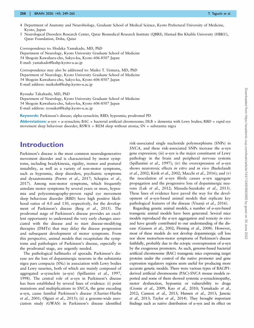
IntroductionParkinson’s disease is the most common neurodegenerative
movement disorder and is characterized by motor symp-
toms, including bradykinesia, rigidity, tremor and postural
instability, as well as a variety of non-motor symptoms,
such as hyposmia, sleep disorders, psychiatric symptoms
and dysautonomia (Poewe et al., 2017; Schapira et al.,
2017). Among non-motor symptoms, which frequently
antedate motor symptoms by several years or more, hypos-
mia and polysomnography-proven rapid eye movement
sleep behaviour disorder (RBD) have high positive likeli-
hood ratios of 4.0 and 130, respectively, for the develop-
ment of Parkinson’s disease (Berg et al., 2015). The
prodromal stage of Parkinson’s disease provides an excel-
lent opportunity to understand the very early changes asso-
ciated with the disease and to start disease-modifying
therapies (DMTs) that may delay the disease progression
and subsequent development of motor symptoms. From
this perspective, animal models that recapitulate the symp-
toms and pathologies of Parkinson’s disease, especially in
the prodromal stage, are urgently needed.
The pathological hallmarks of sporadic Parkinson’s dis-
ease are the loss of dopaminergic neurons in the substantia
nigra pars compacta (SNc) in association with Lewy bodies
and Lewy neurites, both of which are mainly composed of
aggregated �-synuclein (�-syn) (Spillantini et al., 1997,
1998). The central role of �-syn in Parkinson’s disease
has been established by several lines of evidence: (i) point
mutations and multiplications in SNCA, the gene encoding
�-syn, cause familial Parkinson’s disease (Chartier-Harlin
et al., 2001; Olgiati et al., 2015); (ii) a genome-wide asso-
ciation study (GWAS) in Parkinson’s disease identified
risk-associated single nucleotide polymorphisms (SNPs) in
SNCA, and these risk-associated SNPs increase the �-syn
gene expression; (iii) �-syn is the major constituent of Lewy
pathology in the brain and peripheral nervous systems
(Spillantini et al., 1997); (iv) the overexpression of �-syn
shows neurotoxic effects in vitro and in vivo (Baekelandt
et al., 2002; Kirik et al., 2002; Macchi et al., 2016); and (v)
the inoculation of �-syn fibrils causes �-syn aggregate
propagation and the progressive loss of dopaminergic neu-
rons (Luk et al., 2012; Masuda-Suzukake et al., 2013).
These lines of evidence have paved the way for the devel-
opment of �-syn-based animal models that replicate key
pathological features of the disease (Visanji et al., 2016).
Regarding genetic animal models, a number of �-syn-based
transgenic animal models have been generated. Several mice
models reproduced the �-syn aggregation and toxicity in vivo
and have greatly contributed to our understanding of the dis-
ease (Giasson et al., 2002; Fleming et al., 2008). However,
most of these models do not develop dopaminergic cell loss
nor show motor/non-motor symptoms of Parkinson’s disease
faithfully, probably due to the ectopic overexpression of �-syn
by the exogenous promoters. As such, genome-based bacterial
artificial chromosome (BAC) transgenic mice expressing target
proteins under the control of the native promoter and gene
expression regulatory regions seem useful for producing more
accurate genetic models. There were various types of BAC/P1-
derived artificial chromosome (PAC)-SNCA mouse models re-
ported and some of them showed systemic �-synucleinopathy,
motor dysfunction, hyposmia or vulnerability to drugs
(Cronin et al., 2009; Kuo et al., 2010; Yamakado et al.,
2012; Cannon et al., 2013; Hansen et al., 2013; Janezic
et al., 2013; Taylor et al., 2014). They brought important
findings such as native distribution of �-syn and its effect on
250 | BRAIN 2020: 143; 249–265 T. Taguchi et al.
Dow
nloaded from https://academ
ic.oup.com/brain/article/143/1/249/5663565 by guest on 30 June 2022
behaviours. However, there were no mouse models that
showed age-related chronic and selective dopaminergic degen-
eration coupled with multiple prodromal symptoms. Unlike
mouse models, the BAC-SNCA rat model was reported to
show tyrosine hydroxylase (TH)-positive cell loss and hypos-
mia (Nuber et al., 2013). However, the mouse model has an
advantage in handling and genetic engineering. Moreover,
there are no animal models that show RBD phenotypes as a
prodromal symptom in Parkinson’s disease. We previously
generated BAC-SNCA transgenic mice harbouring the entire
wild-type human SNCA and its gene expression regulatory
regions (Yamakado et al., 2012). The mice showed a native
expression pattern of human �-syn and decreased anxiety-like
behaviours but did not recapitulate the pathological changes
of Parkinson’s disease, including dopaminergic cell loss and �-
syn aggregation. To enhance the pathological property of �-
syn, we inserted an A53T point mutation, which is the causa-
tive gene mutation of familial Parkinson’s disease and known
to facilitate �-syn aggregation, into the BAC-SNCA transgenic
construct (Conway et al., 2000; Rodriguez et al., 2015).
Parkinson’s disease patients with A53T mutation showed simi-
lar but accelerated phenotype of idiopathic Parkinson’s disease
in terms of the distribution of Lewy pathology and associated
prodromal non-motor symptoms (Spira et al., 2001). In add-
ition, we also introduced two risk-associated SNPs
(rs11931074 and rs3857059) and a Rep1 dinucleotide
repeat polymorphism into the SNCA promoter region (259
to 261 alleles), all of which increase the risk of developing
sporadic Parkinson’s disease (Maraganore et al., 2006; Satake
et al., 2009; Han et al., 2015). The majority of individuals
have these risk variants, but as the original BAC construct has
minor protective polymorphisms (Maraganore et al., 2006;
Cronin et al., 2009; Satake et al., 2009), we edited the con-
struct to introduce major alleles and eliminate minor protect-
ive factors.
We therefore analysed the pathological phenotypes and
Parkinson’s disease-related motor as well as non-motor
symptoms in this model.
Materials and methods
Animals
All mice used in this study were handled in accordance with thenational guidelines. The mice were maintained at 25�C with 55%humidity on a 12-h light-dark cycle and given free access to foodand drinking water. All procedures performed in this study wereapproved by the Institutional Animal Care and Use Committee,Institute of Laboratory Animals Graduate School of Medicine,Kyoto University (170274) and the University of TsukubaAnimal Care and Use Committee.
A53T BAC-SNCA transgenic mice were generated as previ-ously described (Yamakado et al., 2012) with some modifica-tion. In brief, A53T, rs11931074 (G to T) and rs3857059 (A toG) mutations as well as Rep1 copy number variation (259 to261) were introduced into the human �-syn BAC construct usingthe Red/ET recombination system (K002, Counter-Selection
BAC Modification Kit; Gene Bridges). The construct consists
of P1-derived artificial chromosome (PAC) AF163864 and
BAC AC09748 and contains 28-kb 50- and 50-kb 30-flankingregions in addition to the entire human SNCA (Yamakado
et al., 2012) (Fig. 1A). The resultant vector was injected into
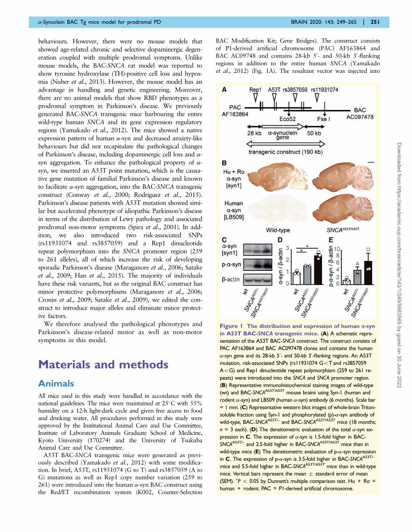
Figure 1 The distribution and expression of human a-syn
in A53T BAC-SNCA transgenic mice. (A) A schematic repre-
sentation of the A53T BAC-SNCA construct. The construct consists of
PAC AF163864 and BAC AC097478 clones and contains the human
�-syn gene and its 28-kb 50- and 50-kb 30-flanking regions. An A53T
mutation, risk-associated SNPs (rs11931074 G5T and rs3857059
A5G) and Rep1 dinucleotide repeat polymorphism (259 to 261 re-
peats) were introduced into the SNCA and SNCA promoter region.
(B) Representative immunohistochemical staining images of wild-type
(wt) and BAC-SNCAA53T/A53T mouse brains using Syn-1 (human and
rodent �-syn) and LB509 (human �-syn) antibody (6 months). Scale bar
= 1 mm. (C) Representative western blot images of whole-brain Triton-
soluble fraction using Syn-1 and phosphorylated (p)-�-syn antibody of
wild-type, BAC-SNCAA53T/– and BAC-SNCAA53T/A53T mice (18 months;
n = 3 each). (D) The densitometric evaluation of the total �-syn ex-
pression in C. The expression of �-syn is 1.5-fold higher in BAC-
SNCAA53T/– and 2.5-fold higher in BAC-SNCAA53T/A53T mice than in
wild-type mice (E) The densitometric evaluation of p-�-syn expression
in C. The expression of p-�-syn is 3.5-fold higher in BAC-SNCAA53T/–
mice and 5.5-fold higher in BAC-SNCAA53T/A53T mice than in wild-type
mice. Vertical bars represent the mean � standard error of mean
(SEM). �P 5 0.05 by Dunnett’s multiple comparison test. Hu + Ro =
human + rodent; PAC = P1-derived artificial chromosome.
�-Synuclein BAC Tg mice model for prodromal PD BRAIN 2020: 143; 249–265 | 251
Dow
nloaded from https://academ
ic.oup.com/brain/article/143/1/249/5663565 by guest on 30 June 2022
the fertilized eggs of C57BL/6J mice. PCR was performed withthe following primer set: SNCA fw 50-ACTTGCTAGGCCACCTGAGA-30, SNCA rv 50-ATGCCAGGTGTTTGGAAAAG-30,followed by electrophoresis on a 2% (w/v) agarose gel. Real-time PCR for SNCA and IL2, a reference gene, were carried outwith the following primer sets: SNCA fw: 50-GGCTGATGCCAACAAGCTGT-30, SNCA rw: 50-GTGGAATTTCGCACAAACCCT-30; IL2 fw: 50-ATAAATTGCCTCCCATGCTGA-30,IL2 rv: 50 -GATGCGAGCTGCATGCTGTA-30.
Immunohistochemical and electronmicroscopic analyses
These methods are described in the Supplementary material.
Cell counting
The number of dopaminergic neurons in the SNc was quanti-fied as previously described (Luk et al., 2012; Tran et al.,2014) with minor modification. The images of every 10th sec-tion of TH-immunostained coronal sections through the entiremidbrain were captured with a microscope (BX43; Olympus).Immunoreactive neurons were counted at 10� magnificationfollowing previously described criteria for each subgroup[dorsal part (SNcd), medial (SNcm), ventral (SNcv), and lateral(SNcl)] (Fu et al., 2012). The number of intact neurons withvisible nuclei was counted.
Sequential extraction
For biochemical analyses, phosphate-buffered saline (PBS)-per-fused mouse brains (3, 9 and 18 months; n = 6, respectively)and human frontal cortices (Supplementary Table 1) werehomogenized in 10 volume of 1% TritonTM lysis buffer [10mM Tris-HCl (pH 7.4), 150 mM NaCl, 1 mM EDTA and 1%(w/v) TritonTM X-100 with protease inhibitor and phosphataseinhibitor mixture] on ice followed by sonication for 5 min witha 30-s interval and centrifugation at 20 400 g for 20 min at4�C. The supernatant was retained as a Triton-soluble frac-tion. The residual pellet was washed twice in 1% Triton lysisbuffer and centrifuged at 20 400 g for 10 min at 4�C. Thepellet was resuspended in 5 volume of 2% sodium dodecylsulphate (SDS) lysis buffer [10 mM Tris-HCl (pH 7.4), 150mM NaCl, 1 mM EDTA, 1% (v/v) TritonTM X-100 and 2%(w/v) SDS with protease inhibitor and phosphatase inhibitormixture] with sonication for 5 min with a 30-s interval andcentrifugation at 20 400 g for 20 min at room temperature.The supernatant was collected as Triton-insoluble fraction.The protein concentration was measured using a bicinchoninicacid (BCA) protein assay kit (23227; Thermo Fisher Scientific).
Preparation of recombinanta-syn monomers andpreformed fibrils
Human �-syn preformed fibrils were generated as previouslydescribed (Masuda-Suzukake et al., 2014; Ihse et al., 2017;Uemura et al., 2018) with minor modification. Escherichia coliBL21 (DE3) (BioDynamics Laboratory) were transformed withplasmid pRK172 encoding the human SNCA cDNA sequence
and incubated in LB medium. The �-syn expression was inducedby 0.1 mM isopropyl b-D-1-thiogalactopyranoside for 4 h. Thebacteria were pelleted by centrifugation at 4000g at 4�C for 5min and lysed with repeated freeze and thaw and by sonication.The lysate was clarified by boiling for 5 min, followed by cen-trifugation at 20 400g at 4�C for 15 min. The supernatant wassubjected to ion exchange using Q Sepharose Fast Flow (GEHealthcare), and �-syn was precipitated with 50% (% satur-ation) ammonium sulphate. Purified �-syn was dialyzed againstdialysis buffer (150 mM KCl, 50 mM Tris-HCl, pH 7.5) andcleared by ultracentrifugation at 186 000g at 4�C for 20 min.The protein concentration was determined using a BCA ProteinAssay kit (Thermo Fisher). Purified �-syn was diluted in dialysisbuffer containing 0.1% (w/v) NaN3 to 7 mg/ml, followed byincubation at 37�C in a shaking incubator (SI-300C; AS ONE)at 1000 rpm for 10 days. The �-syn preformed fibril pellet wasobtained by ultracentrifugation at 186 000g at 20�C for 20 minand stored at –80�C. Before use, the pellet was dissolved in PBS(2 mg/ml) and sonicated for 5 min.
Western blotting
Details are provided in the Supplementary material.
Native polyacrylamide gelelectrophoresis
Western blotting using the native polyacrylamide gel electro-phoresis (PAGE) method was performed in accordance withthe manual of the NativePAGETM Novex� Bis-Tris GelSystem (MAN0000557; Thermo Fisher). In brief, the samplesin the Triton-soluble fraction of mouse midbrain (10 mg) orhuman cortex (20 mg) were mixed with Native PAGETM
sample buffer (4� ) (BN2003; Thermo Fisher) and NativePAGETM 5% G-250 sample additive (BM2004, ThermoFisher). The samples were then subjected to a 4–16% Bis-TrisProtein Gel (BN1002BOX, Thermo Fisher) and electrophor-esed at 150 V at room temperature. During electrophoresis,the cathode buffer was changed from Dark Blue CathodeBuffer (containing 0.02% G-250) to Light Blue CathodeBuffer (containing 0.002% G-250) when the dye front hadmigrated through about one-third of the gel. The sampleswere transferred to a polyvinylidene difluoride (PVDF) mem-brane followed by fixation for 30 min at room temperaturewith 4% (w/v) paraformaldehyde (PFA) in PBS. The excessCoomassie stain was rinsed for 10 min with 50% (v/v) metha-nol. After blocking for 1 h with 5% (w/v) skimmed milk inPBS, the membranes were incubated with anti-�-syn primaryantibody (Supplementary Table 2) overnight at 4�C followedby reaction with horseradish peroxidase-conjugated secondaryantibody (1:5000, NB7574; Novus Biologicals) for 1 h atroom temperature. Immunoreactive bands were detected withdetection reagent (02230; Nacalai Tesque), and the chemilu-minescent signal was detected with Amersham Imager 600 (GEHealthcare).
Filter trap assay
A filter trap assay was performed as previously described(Maesako et al., 2012; Wan and Chung, 2012) with somemodification. The Triton-soluble fraction of mouse midbrain
252 | BRAIN 2020: 143; 249–265 T. Taguchi et al.
Dow
nloaded from https://academ
ic.oup.com/brain/article/143/1/249/5663565 by guest on 30 June 2022
(0.5 mg/ml), �-syn monomer (0.5 ng/ ml) and �-syn preformedfibril (0.5 ng/ml) were subjected to vacuum filtration through a96-well microfiltration apparatus (170-6542; Bio-Rad) con-taining a 200-nm pore cellulose acetate membrane(11020004; ADVANTEC). The resultant membrane wasfixed for 30 min with 4% (w/v) PFA in PBS at room tempera-ture. After blocking for 1 h with 5% (w/v) skimmed milk inPBS, the membranes were incubated with primary antibodies(Supplementary Table 2) overnight at 4�C followed by reac-tion with horseradish peroxidase-conjugated secondary anti-body (1:5000, NB7574; Novus Biologicals) for 1 h at roomtemperature. Immunoreactive bands were detected with detec-tion reagent (02230; Nacalai Tesque), and the chemilumines-cent signal was detected with Amersham Imager 600 (GEHealthcare).
Behavioural tests
Details are provided in the Supplementary material. Details ofthe olfactory test and sleep analysis are provided below.
Electrophysiological recordingsurgery
A connector/electrode for EEG and EMG recordings was im-planted into the skull of each mouse. The two pins of theelectrode for EEG recording were placed over the right cere-bral hemisphere (anterior electrode: 1.26 mm lateral to mid-line, 0 mm anterior to bregma; posterior electrode: 1.26 mmlateral to midline, 5.0 mm posterior to bregma). The stainlesssteel wires (AS633; Cooner Wire) of the electrode for EMGrecording were inserted bilaterally into the neck muscles ofeach mouse, and each electrode was attached to the skullusing dental cement (56818; 3M). After a 1-week recoveryperiod, the animals were moved to a recording cage.
Sleep recording and analyses
For the EEG/EMG recordings, 20- to 55-week-old male micewere used. Cables for signal output were connected to the im-planted electrodes, and the animals were allowed to move freely.Signals were amplified through an amplifier (AB-611J; NihonKoden) and digitized with an analogue-to-digital converter (NIPCIe-6320; National Instruments) and an appropriate softwareprogram (LabView; National Instruments). Animals were allowedat least 1 week to adapt to the recording conditions prior to anyEEG/EMG recording session and handled daily to minimize non-specific stress. During the EEG/EMG recording, the behaviours ofthe animals were monitored by cameras to observe their motoractivity during sleep. Sleep/wake stages were determined by avisual inspection of the EEG/EMG data in 20-s epochs, as pre-viously described (Funato et al., 2016). To determine the ‘EMGvariance’ value, we integrated EMG power over 0.5-s bins andcalculated the arithmetic variance of EMG power over eight con-secutive 0.5-s bins (over 4-s). The median of five consecutive 4-svalues of EMG variance was then taken as the EMG variance ofeach 20-s epoch and used as raw data. We then normalized theREM sleep EMG variance by the same mouse’s non-REM(NREM) sleep EMG variance on the same day at the same ZThour.
Olfactory preference andavoidance test
The olfactory test was performed as previously described(Kobayakawa et al., 2007) with some modification. In brief,the mice were placed in the test cage (width 32.5 cm � depth21.5 cm � height 13 cm). Two pieces of filter paper (2 cm �2 cm) were set at the bottom of the cage (8.5 cm from the longside and 6.5 cm from each short side).
For the olfactory preference test, a test odorant was intro-duced into the filter paper at one side of the cage, and waterwas introduced into the filter paper at the opposite side of thecage. The sides treated with the odorant and water were chan-ged every test. The sniffing time of the odorant was measured.
For the olfactory avoidance test, a bisector was placed in thecage, and two pieces of filter paper were set on opposite sidesof the cage. A test odorant was introduced into the filter paperat one side of the cage, and water was introduced into thefilter paper at the opposite side of the cage. The time spentin the opposite area from the odorant-treated filter paper wasmeasured.
Statistical analyses
Statistical significance was evaluated using a one-way factorialANOVA followed by Dunnett’s post hoc test for multiple com-parison. Student’s t-test was used to compare two groups ofdata. To assess the EEG and EMG data, the non-parametricMann-Whitney U-test was implemented. Statistical significancewas set at \P 5 0.05 or \\P 5 0.01.
Data availability
Data are available from the corresponding author on request.
Results
The native expression patternof human a-syn in the A53TBAC-SNCA transgenic mice
To enhance the aggregation propensity of �-syn, we inserted
A53T, rs11931074 (G to T), and rs3857059 (A to G) point
mutations as well as the Rep1 copy number variation (259 to
261) into the 190-kb BAC-SNCA transgenic construct, which
presumably contains the coding sequence as well as gene ex-
pression regulatory regions (Fig. 1A). The transgenic line was
backcrossed more than 10 generations and maintained on a
C57BL/6J background. The transgenic constructs were in-
serted into the chromosome 9 (Supplementary Fig. 1). The
expression pattern of the transgenic �-syn closely resembled
that of the endogenous rodent �-syn (Fig. 1B). When com-
pared with wild-type mice, heterozygous A53T BAC-SNCA
transgenic (BAC-SNCAA53T/–) mice expressed 1.5-fold more
�-syn, and homozygous A53T BAC-SNCA transgenic (BAC-
SNCAA53T/A53T) mice expressed 2.5-fold more �-syn (Fig. 1C
and D). In addition, phosphorylated �-syn (p-�-syn) was
increased 3.5-fold in BAC-SNCAA53T/– mice and 5.5-fold in
�-Synuclein BAC Tg mice model for prodromal PD BRAIN 2020: 143; 249–265 | 253
Dow
nloaded from https://academ
ic.oup.com/brain/article/143/1/249/5663565 by guest on 30 June 2022

BAC-SNCAA53T/A53T mice (Fig. 1C and E). To avoid potential
off-target effects of genomic transgene insertions, we chose
BAC-SNCAA53T/– mice for further analyses in the present
study.
Increased truncated andTriton-insoluble a-syn in theBAC-SNCAA53T/– mice
We examined a variety of pathological post-translational
modifications of �-syn, including truncation and phosphor-
ylation. Truncated �-syn species enhance �-syn fibril assem-
bly and promote the ability of full-length �-syn to
aggregate (Beyer et al., 2013). In the process of �-syn
fibril formation, conformational changes of �-syn occur,
transforming from soluble oligomers to insoluble fibrils.
We conducted an immunoblot analysis in each brain
region (olfactory bulb, striatum, midbrain, hippocampus
and thalamus) and found that Triton-soluble �-syn was
increased 1.1- to 3.0-fold in each brain region of BAC-
SNCAA53T/– mice compared with wild-type mice (Fig. 2A
and B), and the amount of oligomeric �-syn was increased
in BAC-SNCAA53T/– mice in an age-dependent manner
(Fig. 2A). The amount of Triton-soluble truncated �-syn
was increased up to 13-fold in BAC-SNCAA53T/– mice
(Fig. 2A and C). In addition, BAC-SNCAA53T/– mice also
showed 1.2- to 6.4-fold more Triton-insoluble �-syn in
each brain region than wild-type mice (Fig. 2D and E).
Increased proteinase K-resistanta-syn in the BAC-SNCAA53T/– mice
As abnormally aggregated �-syn in the Lewy bodies is
known to be proteinase K (PK)-resistant (Takeda et al.,
2000; Tanji et al., 2010), we treated mouse brain sections
with PK and immunostained them with p-�-syn antibody.
Whereas PK treatment diminished the immunoreactivity of
p-�-syn in the wild-type mouse brains, PK-resistant p-�-syn
was still abundant in the BAC-SNCAA53T/– mouse brains
(Fig. 3A). PK-resistant p-�-syn was highly expressed in the
cell body of vulnerable regions in �-synucleinopathy,
including the olfactory bulb, anterior olfactory nucleus,
deep layer of the cerebral cortex, amygdala, hippocampus,
SNc and dorsal motor nucleus of the vagus nerve. In add-
ition, abundant punctate staining was observed in the ol-
factory bulb, deep layer of the cerebral cortex and striatum
(Fig. 3B), suggesting that �-syn aggregates in nerve ter-
minals. An immunoelectron microscopic study showed
that immunogold-labelled p-�-syn existed diffusely in the
nucleus and presynaptic regions of neurons of BAC-
SNCAA53T/– mice. In addition, some clusters of immunogold
particles indicating p-�-syn were observed in the presynaptic
regions, probably corresponding to the punctate staining of
p-�-syn on immunohistochemistry. However, no filamentous
structures were observed in the cell bodies or dendrites of p-
�-syn-positive neurons (Fig. 3C).
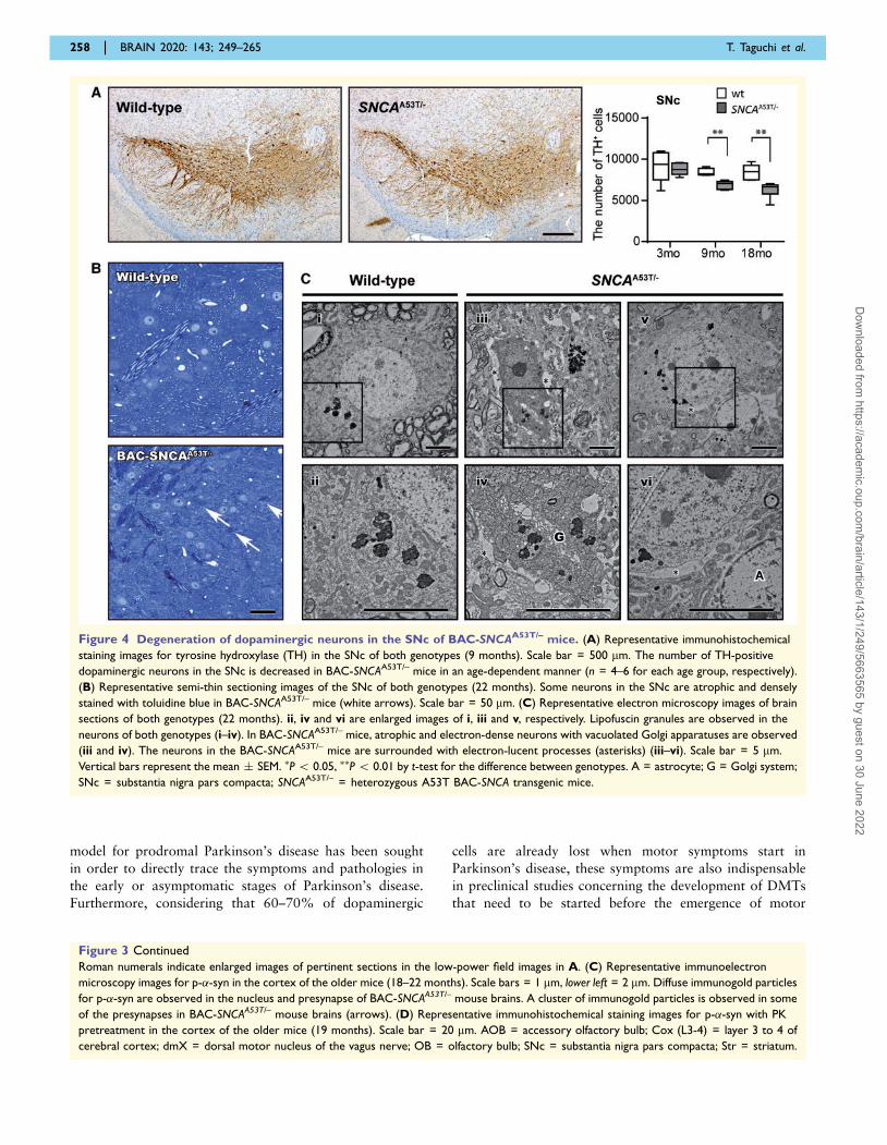
Degeneration of dopaminergicneurons in the substantia nigra parscompacta of BAC-SNCAA53T/– mice
Dopaminergic neuronal loss in the SNc is an important
pathological feature of Parkinson’s disease. We counted
the number of TH-positive neurons in the SNc and found
that it was decreased in the BAC-SNCAA53T/– mice in an
age-dependent manner (Fig. 4A). In contrast, neurons in the
ventral tegmental area were spared (Supplementary Fig. 2).
When the SNc was divided into four subdivisions according
to the morphology, size and distribution of the cell (Fu
et al., 2012), the degree of neuronal loss was more severe
in the SNcd than in the other subdivisions in the SNc
(Supplementary Fig. 3). Semi-thin sections with toluidine
blue staining revealed atrophic and densely stained neurons
in the aged BAC-SNCAA53T/– mice, which is suggestive of
dark cell degeneration (Fig. 4B), originally reported by
Turmaine et al. (2000) as the pathological finding observed
in chronic non-apoptotic neurodegeneration in a
Huntington’s disease mouse model. Electron microscopic
images showed the accumulation of lipofuscin granules in
the neurons of both aged genotypes. However, more atro-
phic and electron-dense neurons were observed in the
BAC-SNCAA53T/– mice [Fig. 4C (i–iv)]. The neurons of
BAC-SNCAA53T/– mice were surrounded by electron-
lucent processes, which were considered to be part of the
foot processes of adjacent astrocytes [Fig. 4C (v and vi)]
(Ikuta et al., 1983).
As neuronal degeneration of �-synucleinopathy starts at
the synaptic terminals (Calo et al., 2016), we investigated
the TH and dopamine transporter (DAT) expression in the
striatum. There was no obvious difference between geno-
types on immunoblot analyses (Supplementary Fig. 4).
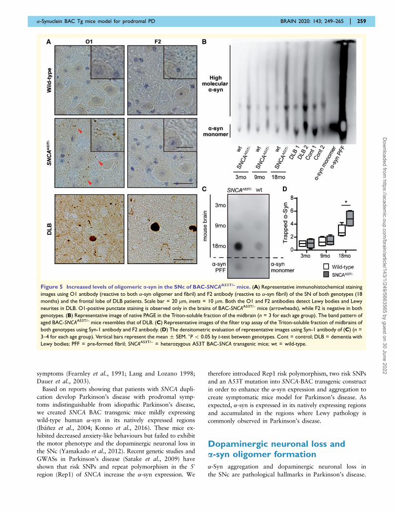
Increased oligomeric a-syn species inthe substantia nigra pars compacta ofBAC-SNCAA53T/– mice
�-Syn oligomers are widely believed to be involved in dopa-
minergic neuronal degeneration (Bengoa-Vergniory et al.,
2017; Mor et al., 2017; Ono, 2017). To examine �-syn
oligomers and fibrils in dopaminergic neurons, we per-
formed immunohistochemistry on tissue sections of the
substantia nigra in BAC-SNCAA53T/– mice using O1 (spe-
cifically reactive with �-syn oligomer and fibril) and F2
antibodies (specifically reactive with �-syn fibril) (Vaikath
et al., 2015). While the F2 antibody detected solid structure
of Lewy bodies and Lewy neurites, O1 detected fine struc-
tures in addition to Lewy bodies and Lewy neurites in the
brains of patients with dementia with Lewy bodies (DLB)
(Fig. 5A). The SNc sections of wild-type mice were not
stained with either antibody, whereas those of BAC-
SNCAA53T/– mice were punctately stained only with O1
antibody (Fig. 5A). In addition, we conducted native
PAGE, which is useful for detecting native protein
254 | BRAIN 2020: 143; 249–265 T. Taguchi et al.
Dow
nloaded from https://academ
ic.oup.com/brain/article/143/1/249/5663565 by guest on 30 June 2022
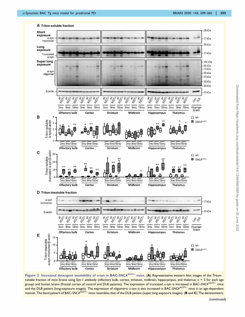
Figure 2 Increased detergent insolubility of a-syn in BAC-SNCAA53T/– mice. (A) Representative western blot images of the Triton-
soluble fraction of mice brains using Syn-1 antibody (olfactory bulb, cortex, striatum, midbrain, hippocampus, and thalamus; n = 5 for each age
group) and human brains (frontal cortex of control and DLB patients). The expression of truncated �-syn is increased in BAC-SNCAA53T/– mice
and the DLB patient (long-exposure images). The expression of oligomeric �-syn is also increased in BAC-SNCAA53T/– mice in an age-dependent
manner. The band pattern of BAC-SNCAA53T/– mice resembles that of the DLB patient (super long exposure images). (B and C) The densitometric
�-Synuclein BAC Tg mice model for prodromal PD BRAIN 2020: 143; 249–265 | 255
(continued)
Dow
nloaded from https://academ
ic.oup.com/brain/article/143/1/249/5663565 by guest on 30 June 2022
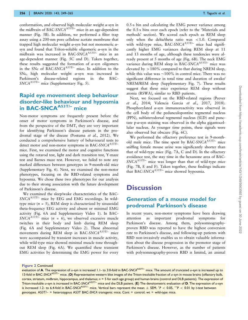
conformation, and observed high molecular weight �-syn in
the midbrain of BAC-SNCAA53T/– mice in an age-dependent
manner (Fig. 5B). In addition, we performed a filter trap
assay using a 200-nm pore cellulose acetate membrane that
trapped high molecular weight �-syn but not monomeric �-
syn and found that Triton-soluble oligomeric �-syn in the
midbrain was increased in BAC-SNCAA53T/– mice in an
age-dependent manner (Fig. 5C and D). Taken together,
these results suggested the formation of �-syn oligomers
in the SNc of BAC-SNCAA53T/– mice. In addition to the
SNc, high molecular weight �-syn was increased in
Parkinson’s disease-related regions in the BAC-
SNCAA53T/– mice (Supplementary Fig. 5).
Rapid eye movement sleep behaviourdisorder-like behaviour and hyposmiain BAC-SNCAA53T/– mice
Non-motor symptoms are frequently present before the
onset of motor symptoms in Parkinson’s disease, and
from the perspective of the DMT, they are very important
for identifying Parkinson’s disease patients in the pro-
dromal stage of the disease (Postuma et al., 2012). We
conducted a comprehensive battery of behavioural tests to
detect motor and non-motor symptoms in BAC-SNCAA53T/–
mice. First, we examined the motor and cognitive functions
using the rotarod test, light and dark transition test, Y-maze
test and Barnes maze test. However, we failed to note any
marked differences between genotypes in 9-month-old mice
(Supplementary Fig. 6). Next, we examined the non-motor
phenotypes, focusing on the RBD-related symptoms and
hyposmia. We chose these two phenotypes for our analysis
due to their strong association with the future development
of Parkinson’s disease.
We examined the sleep/wake characteristics of the BAC-
SNCAA53T/– mice by EEG and EMG recordings. In wild-
type mice (n = 5), REM sleep is characterized by sinusoidal
theta-frequency EEG activity and absent or minimal EMG
activity (Fig. 6A and Supplementary Video 1). In BAC-
SNCAA53T/– mice (n = 6), we observed excessive muscle
twitches in their body and limb during REM sleep
(Fig. 6A and Supplementary Video 2). These abnormal
movements during REM sleep in BAC-SNCAA53T/– mice
were accompanied by transient increases in muscle activity,
while wild-type mice showed minimal muscle tone through-
out REM sleep (Fig. 6A). We quantified these transient
EMG activities by determining the EMG power for every
0.5-s bin and calculating the EMG power variance among
the 0.5-s bins over each epoch (refer to the ‘Materials and
methods’ section). We scored each epoch as REM sleep
only when the delta/theta ratio was 51.0. Compared
with wild-type mice, BAC-SNCAA53T/– mice had signifi-
cantly higher EMG variances during REM sleep at 11
and 13 months of age, although these tendencies were al-
ready present at 5 months of age (Fig. 6B). The neck EMG
variance during REM sleep in BAC-SNCAA53T/– mice was
elevated by 4180% compared to that during NREM sleep,
while this value was �100% in control mice. There was no
significant difference in total time and duration of awake/
NREM/REM sleep (Supplementary Fig. 7). These results
suggest that these mice experience REM sleep without
atonia (RSWA), similar to RBD patients.
Next, we focused on the RBD-related regions (Peever
et al., 2014; Valencia Garcia et al., 2017, 2018).
Phosphorylated �-syn immunoreactivity was observed in
the cell body of the pedunculopontine tegmental nucleus
(PPN), sublaterodorsal tegmental nucleus (SLD) and punc-
tate p-�-syn staining was observed in the alpha gigantocel-
lular nucleus. At younger time points, these signals were
also observed but obscure (Fig. 6C).
We performed the olfactory preference test in 9-month-
old male mice. The time spent by BAC-SNCAA53T/– mice
sniffing female mouse urine was significantly shorter than
that of wild-type mice (Fig. 7A, C and D). In the olfactory
avoidance test, the stay time in the hexanone area of BAC-
SNCAA53T/– mice was longer than that of wild-type mice
(Fig. 7B, E and F). Taken together, these findings indicate
that BAC-SNCAA53T/– mice showed hyposmia.
Discussion
Generation of a mouse model forprodromal Parkinson’s disease
In recent years, non-motor symptoms have been drawing
attention as important prodromal symptoms for
Parkinson’s disease. Among them, polysomnography-
proven RBD was reported to have the highest conversion
rate to Parkinson’s disease, and following-up patients with
RBD non-invasively enables us to obtain valuable informa-
tion about the disease progression in the premotor stage of
Parkinson’s disease. However, as the number of patients
with polysomnography-proven RBD is limited, an animal
Figure 2 Continued
evaluation of A. The expression of �-syn is increased 1.1- to 3.0-fold in BAC-SNCAA53T/– mice. The amount of truncated �-syn is increased up to
13-fold in BAC-SNCAA53T/– mice. (D) Representative western blot images of the Triton-insoluble fraction of �-syn in mouse brains (olfactory bulb,
cortex, striatum, midbrain, hippocampus, and thalamus; n = 5 for each age group) and human brains (control and DLB patients). The expression of
Triton-insoluble �-syn is increased in BAC-SNCAA53T/– mice and the DLB patient. (E) The densitometric evaluation of D. The expression of �-syn
is increased 1.2- to 6.4-fold in BAC-SNCAA53T/– mice. Vertical bars represent the mean � SEM. �P 5 0.05, ��P 5 0.01 by t-test between
genotypes. A53T/– = heterozygous A53T BAC-SNCA transgenic mice; Cont = control; wt = wild-type mice.
256 | BRAIN 2020: 143; 249–265 T. Taguchi et al.
Dow
nloaded from https://academ
ic.oup.com/brain/article/143/1/249/5663565 by guest on 30 June 2022
Figure 3 Increased levels of PK-resistant p-a-syn in the BAC-SNCAA53T/– mice. (A) Representative immunohistochemical staining
images for p-�-syn with proteinase K (PK) pretreatment in brain sections of both genotypes (3 months) in the low-power fields. Scale bar = 500
mm. (B) Representative immunohistochemical staining images for p-�-syn with PK pretreatment in the high-power fields. Scale bar = 20 mm.
�-Synuclein BAC Tg mice model for prodromal PD BRAIN 2020: 143; 249–265 | 257
(continued)
Dow
nloaded from https://academ
ic.oup.com/brain/article/143/1/249/5663565 by guest on 30 June 2022
model for prodromal Parkinson’s disease has been sought
in order to directly trace the symptoms and pathologies in
the early or asymptomatic stages of Parkinson’s disease.
Furthermore, considering that 60–70% of dopaminergic
cells are already lost when motor symptoms start in
Parkinson’s disease, these symptoms are also indispensable
in preclinical studies concerning the development of DMTs
that need to be started before the emergence of motor
Figure 3 Continued
Roman numerals indicate enlarged images of pertinent sections in the low-power field images in A. (C) Representative immunoelectron
microscopy images for p-�-syn in the cortex of the older mice (18–22 months). Scale bars = 1 mm, lower left = 2 mm. Diffuse immunogold particles
for p-�-syn are observed in the nucleus and presynapse of BAC-SNCAA53T/– mouse brains. A cluster of immunogold particles is observed in some
of the presynapses in BAC-SNCAA53T/– mouse brains (arrows). (D) Representative immunohistochemical staining images for p-�-syn with PK
pretreatment in the cortex of the older mice (19 months). Scale bar = 20 mm. AOB = accessory olfactory bulb; Cox (L3-4) = layer 3 to 4 of
cerebral cortex; dmX = dorsal motor nucleus of the vagus nerve; OB = olfactory bulb; SNc = substantia nigra pars compacta; Str = striatum.
Figure 4 Degeneration of dopaminergic neurons in the SNc of BAC-SNCAA53T/– mice. (A) Representative immunohistochemical
staining images for tyrosine hydroxylase (TH) in the SNc of both genotypes (9 months). Scale bar = 500 mm. The number of TH-positive
dopaminergic neurons in the SNc is decreased in BAC-SNCAA53T/– mice in an age-dependent manner (n = 4–6 for each age group, respectively).
(B) Representative semi-thin sectioning images of the SNc of both genotypes (22 months). Some neurons in the SNc are atrophic and densely
stained with toluidine blue in BAC-SNCAA53T/– mice (white arrows). Scale bar = 50 mm. (C) Representative electron microscopy images of brain
sections of both genotypes (22 months). ii, iv and vi are enlarged images of i, iii and v, respectively. Lipofuscin granules are observed in the
neurons of both genotypes (i–iv). In BAC-SNCAA53T/– mice, atrophic and electron-dense neurons with vacuolated Golgi apparatuses are observed
(iii and iv). The neurons in the BAC-SNCAA53T/– mice are surrounded with electron-lucent processes (asterisks) (iii–vi). Scale bar = 5 mm.
Vertical bars represent the mean � SEM. �P 5 0.05, ��P 5 0.01 by t-test for the difference between genotypes. A = astrocyte; G = Golgi system;
SNc = substantia nigra pars compacta; SNCAA53T/– = heterozygous A53T BAC-SNCA transgenic mice.
258 | BRAIN 2020: 143; 249–265 T. Taguchi et al.
Dow
nloaded from https://academ
ic.oup.com/brain/article/143/1/249/5663565 by guest on 30 June 2022
symptoms (Fearnley et al., 1991; Lang and Lozano 1998;
Dauer et al., 2003).
Based on reports showing that patients with SNCA dupli-
cation develop Parkinson’s disease with prodromal symp-
toms indistinguishable from idiopathic Parkinson’s disease,
we created SNCA BAC transgenic mice mildly expressing
wild-type human �-syn in its natively expressed regions
(Ibanez et al., 2004; Konno et al., 2016). These mice ex-
hibited decreased anxiety-like behaviours but failed to exhibit
the motor phenotype and the dopaminergic neuronal loss in
the SNc (Yamakado et al., 2012). Recent genetic studies and
GWASs in Parkinson’s disease (Satake et al., 2009) have
shown that risk SNPs and repeat polymorphism in the 50
region (Rep1) of SNCA increase the �-syn expression. We
therefore introduced Rep1 risk polymorphism, two risk SNPs
and an A53T mutation into SNCA-BAC transgenic construct
in order to enhance the �-syn expression and aggregation to
create symptomatic mice model for Parkinson’s disease. As
expected, �-syn is expressed in its natively expressing regions
and accumulated in the regions where Lewy pathology is
commonly observed in Parkinson’s disease.
Dopaminergic neuronal loss anda-syn oligomer formation
�-Syn aggregation and dopaminergic neuronal loss in
the SNc are pathological hallmarks in Parkinson’s disease.
Figure 5 Increased levels of oligomeric a-syn in the SNc of BAC-SNCAA53T/– mice. (A) Representative immunohistochemical staining
images using O1 antibody (reactive to both �-syn oligomer and fibril) and F2 antibody (reactive to �-syn fibril) of the SN of both genotypes (18
months) and the frontal lobe of DLB patients. Scale bar = 20 mm, insets = 10 mm. Both the O1 and F2 antibodies detect Lewy bodies and Lewy
neurites in DLB. O1-positive punctate staining is observed only in the brains of BAC-SNCAA53T/– mice (arrowheads), while F2 is negative in both
genotypes. (B) Representative image of native PAGE in the Triton-soluble fraction of the midbrain (n = 3 for each age group). The band pattern of
aged BAC-SNCAA53T/– mice resembles that of DLB. (C) Representative images of the filter trap assay of the Triton-soluble fraction of midbrains of
both genotypes using Syn-1 antibody and F2 antibody. (D) The densitometric evaluation of representative images using Syn-1 antibody of (C) (n =
3–4 for each age group). Vertical bars represent the mean � SEM. �P 5 0.05 by t-test between genotypes. Cont = control; DLB = dementia with
Lewy bodies; PFF = pre-formed fibril; SNCAA53T/– = heterozygous A53T BAC-SNCA transgenic mice; wt = wild-type.
�-Synuclein BAC Tg mice model for prodromal PD BRAIN 2020: 143; 249–265 | 259
Dow
nloaded from https://academ
ic.oup.com/brain/article/143/1/249/5663565 by guest on 30 June 2022

Figure 6 RBD-like behaviour in BAC-SNCAA53T/– mice. (A) Typical EEG and EMG findings of wild-type and BAC-SNCAA53T/– mice. Arrows
denote muscle twitches. (B) The REM/NREM ratio of the EMG variance during REM sleep in each BAC-SNCAA53T/– (n = 6) and wild-type (n = 5)
mice. All values are the mean � SEM. �P 5 0.05, ��P 5 0.01, Mann-Whitney U-test. (C) Representative images of p-�-syn immunohistochemical
staining in the field related to RBD of both genotypes (9 months) and younger BAC-SNCAA53T/– mice (3 months). Scale bar = 20 mm. GiA = alpha
gigantocellular nucleus; mo = months; PPN = pedunclopontine tegmental nucleus; SLD = sublaterodorsal tegmental nucleus; SNCAA53T/– =
heterozygous A53T BAC-SNCA transgenic mice.
260 | BRAIN 2020: 143; 249–265 T. Taguchi et al.
Dow
nloaded from https://academ
ic.oup.com/brain/article/143/1/249/5663565 by guest on 30 June 2022

PK-resistant phosphorylated, as well as truncated �-syn,
which are pathological forms of �-syn observed in
Parkinson’s disease brains, were accumulated in the
Parkinson’s disease-related regions in BAC-SNCAA53T/–
mice. However, only small amounts of truncated or insol-
uble �-syn were observed in the midbrain and morpho-
logically abnormal filamentous �-syn aggregates were not
observed even by electron microscopy in BAC-SNCAA53T/–
mice. These results raised a question concerning the
mechanisms underlying dopaminergic cell loss in BAC-
SNCAA53T/– mice. First, a small amount of truncated or
insoluble �-syn in immunoblot might be because the immu-
noblot was performed against whole part of midbrain, not
limited to the SNc, due to the technical difficulties and the
same tendency was previously reported in wild-type
BAC-SNCA trangenic rat (Nuber et al., 2013). Second,
accumulating evidence has shown that not only �-syn fibrils
but also oligomers play a central role in the neuronal de-
generation in Parkinson’s disease (Winner et al., 2011;
Martin et al., 2012). For instance, soluble and lipid-de-
pendent �-syn oligomers were shown to have accumulated
in the brains of patients with Parkinson’s disease and DLB
(Sharon et al., 2003), and the A53T mutation of �-syn was
reported to facilitate its oligomerization in vitro (Conway
et al., 2000). As shown in Fig. 5, oligomeric �-syn species
were increased in the SNc and presumed to be responsible
for the dopaminergic cell loss in BAC-SNCAA53T/– mice.
In vitro, �-syn oligomerization was found to increase cell
toxicity of �-syn (Outeiro et al, 2008). For instance, oligo-
meric �-syn was reported to impair the mitochondrial pro-
tein import, leading to mitochondrial dysfunction and the
production of aberrant reactive oxygen species (Di Maio
et al., 2016). In in vivo studies, the elevation of dopamine
in A53T �-syn transgenic mice was reported to increase
high-angstrom soluble �-syn oligomer, which induced pro-
gressive nigrostriatal degeneration and locomotion disabil-
ity (Mor et al., 2017). Moreover, it was reported that �-syn
oligomerization induced by the dopamine catabolite im-
paired synaptic vesicles function and lead to neurodegen-
eration (Plotegher et al, 2017). These lines of evidence
further support the idea that high molecular weight �-syn
species (oligomers) detected in the midbrain of the BAC-
SNCAA53T/– mice may contribute to the loss of dopamin-
ergic neurons in the SNc.
In Parkinson’s disease patients and �-syn transgenic ani-
mals, nigrostriatal degeneration starts at the synapse in the
striatum and then retrogradely spreads to the cell body in
the SNc (Cheng et al., 2010; Rockenstein et al., 2014). We
examined the presynaptic proteins in dopaminergic nerve
terminals as well as dopaminergic content in the striatum,
and found that the expression level of TH and striatal
dopaminergic content in BAC-SNCAA53T/– mice were not
markedly different from that of wild-type mice, probably
due to the compensatory upregulation of TH protein and
dopaminergic biosynthesis in the remaining neurons.
Similarly, the expression of DAT in BAC-SNCAA53T/–
mice was not significantly decreased compared to wild-
type mice. Again, this is not inconsistent with the mild
dopaminergic cell loss in BAC-SNCAA53T/– mice, because
the expression level of DAT was increased in �-syn over-
expression mice (Bellucci et al., 2011; Yamakado et al.,
2012; Mor et al., 2017) and decreased in �-syn knockout
mice (Fountaine et al., 2007; Bellucci et al., 2011;
Chadchankar et al., 2011). �-Syn was reported to maintain
DAT in the endosomal recycling pathway and to regulate
the cell surface expression (Bellucci et al., 2008, 2011).
Motor and non-motor symptoms inBAC-SNCAA53T/– mice
The BAC-SNCAA53T/– mice did not show motor symptoms
related to Parkinson’s disease, which is reasonable, as the
number of dopaminergic neurons decreased only 17.1% at
18 months of age in BAC-SNCAA53T/– mice compared to
that in wild-type mice.
Figure 7 Hyposmia in the BAC-SNCAA53T/– mice. (A)
Olfactory preference test. (C and D) The duration spent sniffing
female mouse urine was significantly decreased in BAC-SNCAA53T/–
mice. (B) Olfactory avoidance test. (E and F) The duration spent on
the opposite side from the hexanone was significantly decreased in
BAC-SNCAA53T/– mice. The olfactory tests were performed in male
wild-type (9 months; n = 21) and BAC-SNCAA53T/– mice (9 months;
n = 23). Vertical bars represent the mean � SEM. �P 5 0.05, ��P 50.01 by t-test between genotypes. SNCAA53T/– = heterozygous
A53T BAC-SNCA transgenic mice; wt = wild-type mice.
�-Synuclein BAC Tg mice model for prodromal PD BRAIN 2020: 143; 249–265 | 261
Dow
nloaded from https://academ
ic.oup.com/brain/article/143/1/249/5663565 by guest on 30 June 2022

In addition to subtle motor changes, non-motor symp-
toms are important as prodromal symptoms in
Parkinson’s disease. Among them, polysomnography-con-
firmed RBD and hyposmia are particularly important be-
cause they have a high conversion rate to Parkinson’s
disease in the future. Some of the �-syn-based transgenic
mice showed hyposmia or sleep disturbance as non-motor
symptoms (Fleming et al., 2008; Rothman et al., 2013;
McDowll et al., 2014; Zhang et al., 2015), but RBD-like
behaviours were not noted.
In Parkinson’s disease, odour detection and discrimin-
ation are impaired, even before the actual diagnosis
(Goldman and Postuma, 2014). In BAC-SNCAA53T/–
mice, p-�-syn was accumulated along the main and acces-
sory olfactory pathways, probably contributing to hypos-
mia; however, we were unable to specify the main lesions
responsible for hyposmia. In previous studies, transgenic
mice overexpressing �-syn under the Thy1 promoter
showed hyposmia (Fleming et al., 2008) and displayed
PK-resistant �-syn inclusions throughout the olfactory
bulb, including in the external portion of the olfactory nu-
cleus, accessory olfactory bulb and glomerular layer of the
olfactory bulb, similar to BAC-SNCAA53T/– mice. Human
A53T �-syn-overexpressing mice under the prion promoter
also exhibited hyposmia, accompanied by a decreased
number of cholinergic neurons in the mitral cell layer and
the decreased activity of acetylcholinesterase in the olfac-
tory bulb (Zhang et al., 2015). However, the mechanism
underlying hyposmia in Parkinson’s disease is complicated,
and many factors may contribute to hyposmia. BAC-
SNCAA53T/– mice are expected to be useful for achieving
further insight into the mechanisms involved in hyposmia
in Parkinson’s disease.
Several studies have explored the regions responsible for
RBD. In animal studies, glutamatergic neurons within the
SLD in the dorsomedial pons have been shown to activate
glycine/GABA inhibitory neurons in the ventromedial me-
dullary reticular formation (VMRF), including the ventral
gigantocellular nucleus, raphe magnus and alpha giganto-
cellular nuclei. During REM sleep, the inhibitory input
from these VMRF neurons to spinal motor neurons sup-
presses muscle tone, and its decreased activity causes
RSWA, which is a prerequisite for the diagnosis of RBD.
Based on these findings, the neurons in the sublaterodorsal
tegmental nucleus and VMRF are considered to be the re-
gions responsible for RSWA (Valencia et al., 2018). The
PPN is also a candidate region responsible for RBD
(Peever et al., 2014; Valencia et al., 2017). In human stu-
dies incidental Lewy body disease with RBD patients
showed Lewy bodies predominantly in the brainstem,
including the subcoeruleus complex, which is the equivalent
to sublaterodorsal tegmental nucleus in mice. (Uchiyama
et al.,1995; Boeve et al., 2007). In addition, using neuro-
melanin-sensitive imaging, the signal intensity of the locus
subcoeruleus in Parkinson’s disease with RBD was shown
to be more reduced than that in Parkinson’s disease with-
out RBD (Garcıa-Lorenzo et al., 2013). Considering that
the VMRF and subcoeruleus complex are known to be the
initial regions of Lewy pathology in Parkinson’s disease
(Jellinger, 2009), they are major candidate regions respon-
sible for RBD in Parkinson’s disease.
In terms of mouse models for Parkinson’s disease ex-
hibiting sleep abnormalities, wild-type or A53T �-syn
transgenic mice under the Thy1 promoter showed a de-
crease in REM sleep or estimated total sleep time
(Rothman et al., 2013; McDowll et al., 2014), but
RSWA/RBD-like behaviour was not described. The BAC-
SNCAA53T/– mice described in the present study are the
first mouse model for Parkinson’s disease to recapitulate
RSWA associated with p-�-syn accumulation in related re-
gions, such as the sublaterodorsal tegmental nucleus,
VMRF and PPN. BAC-SNCAA53T/– showed RSWA with-
out any disturbance of the sleep construction. However,
they showed a tendency of increased REM sleep at a
younger age when they didn’t show RSWA. It might be
the earlier phenotype of sleep abnormalities followed by
RBD in Parkinson’s disease.
RBD is an important prodromal symptom with very high
conversion rate to �-synucleinopathy in the future.
However, the prevalence of RBD, especially that of the
polysomnography-confirmed cases, is low in the prodromal
stage and is not as high as that of other non-motor symp-
toms such as hyposmia and constipation, even in the symp-
tomatic stage. In BAC-SNCAA53T/– mice, RSWA—a
physiological essential component of RBD—can be detected
but these mice do not necessarily show overt phenotype
such as RBD symptoms in human Parkinson’s disease pa-
tients. The same is true for prodromal symptoms in human
Parkinson’s disease in that multiple �-syn pathology
observed in various regions such as the olfactory bulb,
lower brainstem nuclei and peripheral autonomic nerves
do not necessarily become symptomatic in the early stage.
From these viewpoints, the low prevalence of REM sleep
abnormalities in humans may partly come from the low
sensitivity of detection for RBD-like symptoms. Another
possibility is that in Parkinson’s disease there are multiple
modes of progression in some of which RBD is spared, and
RBD is an uncommon symptom more associated with
increased �-syn expression.
Regional vulnerability and diseaseinitiation in BAC-SNCAA53T/– miceand Parkinson’s disease
The accumulation of PK-resistant p-�-syn was mainly
observed in the limbic system, cerebral cortex, olfactory
bulb and specific brainstem nuclei. Among them, the olfac-
tory bulb and brainstem nuclei, such as the dorsal motor
nucleus of the vagus nerve, raphe nuclei and RBD-related
nuclei, correspond to the early Lewy pathology lesions in
the course of Parkinson’s disease. Despite the substantial
accumulation of p-�-syn in the limbic system and cerebral
cortex, the cognitive function as assessed by Barnes maze
262 | BRAIN 2020: 143; 249–265 T. Taguchi et al.
Dow
nloaded from https://academ
ic.oup.com/brain/article/143/1/249/5663565 by guest on 30 June 2022
and the fear condition were not impaired, possibly due to
regional differences in functional vulnerability to �-syn de-
position in Parkinson’s disease.
In addition, our study provided insight into the initiation
of Parkinson’s disease pathology. It is known that multiple
prodromal symptoms, such as hyposmia and RBD, whose
responsible regions are the olfactory pathway and lower
brainstem nuclei, respectively, are frequently observed sim-
ultaneously (Aguirre-Mardones et al., 2015). Considering
that these two regions are located far away from each
other, this is likely caused by the multifocal initiation of
pathology, rather than the simultaneous propagation
through the nose-to-brain and gut-to-brain pathways.
Indeed, pathological studies have shown the frequent sim-
ultaneous involvement of subcortical and cortical regions in
Parkinson’s disease (Jellinger, 2009). These findings, along
with the fact that BAC-SNCAA53T/– mice present with
hyposmia and RSWA, suggest that the enhanced native
topographical expression pattern of �-syn contributes to
the multifocal pathology in Parkinson’s disease.
ConclusionBAC-SNCAA53T/– mice exhibit mild but age-dependent neu-
rodegeneration and non-motor symptoms of Parkinson’s
disease associated with �-syn pathologies in the regions
that are specifically affected in Parkinson’s disease and/or
DLB. Thus, this mouse model is important as a prodromal
Parkinson’s disease model and can provide insights into the
initiation and progression of the disease, especially in the
prodromal stage of Parkinson’s disease. It may also be
useful for the development of DMTs in preclinical studies
for Parkinson’s disease.
AcknowledgementsWe thank Ms Rie Hikawa, Mr Ryutaro Tamano and Ms
Miki Nakamura for their technical assistance.
FundingWe gratefully acknowledge grant support from the
Ministry of Education, Culture, Sports, Science, and
Technology [R.T., Grant-in-Aid for Scientific Research
(A), JP18H04041; Grant-in-Aid for Scientific Research on
Innovative Areas, JP17H05698], the Integrated
Neurotechnologies for Disease Studies (Brain/MINDS)
from Japan Agency for Medical Research and
Development, AMED (R.T., JP18dm0207020, M.K.,
JP18dm0207024), the Japan Science and Technology
Agency, CREST (R.T., JP17gm0710011), JSPS KAKENHI
(Grant Number JP14616060) and the Mitsubishi founda-
tion (R.T., No. 29125).
Competing interestsThe authors report no competing interests.
Supplementary materialSupplementary material is available at Brain online.
ReferencesAguirre-Mardones C, Iranzo A, Vilas D, Serradell M, Gaig C,
Santamarıa J, Tolosa E. Prevalence and timeline of nonmotor symp-
toms in idiopathic rapid eye movement sleep behavior disorder. J
Neurol 2015; 262: 1568–78.Baekelandt V, Claeys A, Eggermont K, Lauwers E, De Strooper B,
Nuttin B, et al. Characterization of lentiviral vector-mediated gene
transfer in adult mouse brain. Hum Gene Ther 2002; 13: 841–53.Bellucci A, Collo G, Sarnico I, Battistin L, Missale C, Spano P. Alpha-
synuclein aggregation and cell death triggered by energy deprivation
and dopamine overload are counteracted by D2/D3 receptor activa-
tion. J Neurochem 2008; 106: 560–77.
Bellucci A, Navarria L, Falari E, Zaltieri M, Bono F, Collo G, et al.
Redistribution of DAT/�-synuclein complexes visualized by in situ
proximity ligation assay in transgenic mice modeling early
Parkinson’s disease. PLoS One 2011; 6: e27959.
Bengoa-Vergniory N, Robert RF, Wada-Martins R, Alegre-
Abarrategui J. Alpha-synuclein oligomers: a new hope. Acta
Neruopathol 2017; 134: 819–38.
Berg D, Postuma RB, Adler CH, Bloem BR, Chan P, Dubois B, et al.
MDS research criteria for prodromal Parkinson’s disease. Mov
Disord 2015; 30: 1600–11.
Beyer K, Ariza A. Alpha-synuclein posttranslational modification and
alternative splicing as a trigger for neurodegeneration. Mol
Neurobiol 2013; 47: 509–24.
Boeve BF, Dickson DW, Olson EJ, Shepard JW, Silber MH, Ferman
TJ, et al. Insights into REM sleep behavior disorder pathophysiology
in brainstem- predominant Lewy body disease. Sleep Med 2007; 8:
60–4.
Calo L, Wegrzynowicz M, Santivanez-Perez J, Grazia Spillantini M.
Synaptic failure and �-synuclein. Mov Disord 2016; 31: 169–77.Cannon JR, Geghman KD, Tapias V, Sew T, Dail MK, Li C, et al.
Expression of human E46K-mutated alpha-synuclein in BAC-trans-
genic rats replicates early-stage Parkinson’s disease features and en-
hances vulnerability to mitochondrial impairment. Exp Neurol
2013; 240: 44–56.
Chadchankar H, Ihalainen J, Tanila H, Yavich L. Decresed reuptake
of dopamine in the dorsal striatum in the absence of �-synuclein.
Brain Res 2011; 1382:37–44.
Chartier-Harlin M-C, Kachergus J, Roumier C, Mouroux V, Douay
X, Lincoln S, et al. �-synuclein locus duplication as a cause of fa-
milial Parkinson’s disease. Lancet 2001; 364: 1167–9.
Cheng HC, Ulane CM, Burke RE. Clinical progression in Parkinson
disease and the neurobiology of axons. Ann Neurol 2010; 67:715–
25.
Conway KA, Lee SJ, Rochet JC, Ding TT, Williamson RE, Lansbury
PT Jr. Acceleration of oligomerization, not fibrillization, is a shared
property of both alpha-synuclein mutations linked to early-onset
Parkinson’s disease: implications for pathogenesis and therapy.
Proc Natl Acad Sci USA 2000; 97: 571–6.
Cronin KD, Ge D, Manninger P, Linnertz C, Rossoshek A, Orrison
BM, et al. Expansion of the Parkinson disease-associated SNCA-
Rep1 allele upregulates human alpha-synuclein in transgenic
mouse brain. Hum Mol Genet 2009; 18: 3274–85.
�-Synuclein BAC Tg mice model for prodromal PD BRAIN 2020: 143; 249–265 | 263
Dow
nloaded from https://academ
ic.oup.com/brain/article/143/1/249/5663565 by guest on 30 June 2022
Dauer W, Przedborski S. Parkinson’s disease: mechanism and models.
Neuron 2003; 39: 889–909.
Di Maio R, Barrett PJ, Hoffman EK, Barrett CW, Zharikov A, Borah
A, et al. �-Synuclein binds to TOM20 and inhibit mitochondrial
protein import in Parkinson’s disease. Sci Transl Med 2016; 8:
342ra78.
Fearnley JM, Lees AJ. Ageing and Parkinson’s disease: substantia nigra
regional selectivity. Brain 1991; 114: 2283–301.
Fleming SM, Tetreault NA, Mulligan CK, Hutson CB, Masliah E,
Chesselet MF. Olfactory deficits in mice overexpressing human wild-
type �-synuclein. Eur J Neurosci 2008; 28: 247–56.
Fountaine TM, Wade-Martins R. RNA interference-mediated knock-
down of alpha-synuclein human dopaminergic neuroblastoma cells
from MPP( + ) toxicity and reduces dopamine transport. J Neurosci
Res 2007; 85: 351–63.Fu Y, Yuan Y, Halliday G, Rusznak Z, Watson C, Paxinos G. A
cytoarchitectonic and chemoarchitectonic analysis of the dopamine
cell groups in the substantia nigra, ventral tegmental area, and retro-
rubral field in the mouse. Brain Struct Funct 2012; 217: 591–612.
Funato H, Miyoshi C, Fujiyama T, Kanda T, Sato M, Wang Z, et al.
Forward-genetics analysis of sleep in randomly mutagenized mice.
Nature 2016; 539: 378–83.
Garcıa-Lorenzo D, Longo-Dos Santos C, Ewenczyk C, Leu-Semenescu
S, Gallea C, Quattrocchi G, et al. The coeruleus/subcoeruleus com-
plex in rapid eye movement sleep behaviour disorders in Parkinson’s
disease. Brain 2013; 136: 2120–9.
Giasson BI, Duda JE, Quinn SM, Zhang B, Trojanowski JQ, Lee VM
Neuronal alpha-synucleinopathy with severe movement disorder in
mice expressing A53T human alpha-synuclein. Neuron 2002; 34:
521–33.Goldman JG and Postuma R. Premotor and nonmotor features of
Parkinson’s disease. Curr Opin Neurol 2014; 27: 434–41.
Han W, Liu Y, Mi Y, Zhao J, Liu D, Tian Q. Alpha-synuclein (SNCA)
polymorphisms and susceptibility to Parkinson’s disease: a meta-
analysis. Am J Med Genet B Neuropsychiatr Genet 2015; 168B:
123–34.
Hansen C, Bjorklund T, Petit GH, Lundblad M, Murmu RP, Brundin
P, et al. A novel alpha-synuclein-GFP mouse model displays progres-
sive motor impairment, olfactory dysfunction and accumulation of
alpha-synuclein-GFP. Neurobiol Dis 2013; 56: 145–55.
Ibanez P, Bonnet AM, Debarges B, Lohmann E, Tison F, Pollak P,
et al. Causal relation between �-synuclein gene duplication and fa-
milial Parkinson’s disease. Lancet 2004; 364: 1169–71.
Ihse E, Yamakado H, van Wijk XM, Lawrence R, Esko JD, Masliah
E. Cellular internalization of alpha-synuclein aggregates by cell sur-
face heparan sulfate depends on aggregate conformation and cell
type. Sci Rep 2017; 7: 9008.
Ikuta F, Yoshida Y, Ohama E, Oyanagi K, Takeda S, Yamazaki K,
et al. Revised Pathophysiology on BBB Damage: The Edema as an
ingeniously Provided Condition for Cell Motility and Lesion Repair.
Acta Neuropathol Suppl 1983; 8: 103–10.
Janezic S, Threlfell S, Dodson PD, Dowie MJ, Taylor TN, Potgieter D,
et al. Deficits in dopaminergic transmission precede neuron loss and
dysfunction in a new Parkinson model. Proc Natl Acad Sci U S A
2013; 110: E4016–25.
Jellinger KA. A critical evaluation of current staging of alpha-synuclein
pathology in Lewy body disorders. Biochim Biophys Acta 2009;
1792: 730–40.
Kirik D, Rosenblad C, Burer C, Lundberg C, Johansen TE, Muzyczka
N, et al. Parkinson-like neurodegeneration induced by targeted over-
expression of alpha-synuclein in the nigrostriatal system. J Neurosci
2002; 22: 2780–91.
Kobayakawa K, Kobayakawa R, Matsumoto H, Oka Y, Imai T,
Ikawa M, et al. Innate versus learned odour processing in the
mouse olfactory bulb. Nature 2007; 450: 503–8.Konno T, Ross OA, Puschmann A, Dickson DW, Wszolek ZK.
Autosomal dominant Parkinson’s disease caused by SNCA duplica-
tions. Parkisonism Relat Disord 2016; 1: S1–6.
Kuo YM, Li Z, Jiao Y, Gaborit N, Pani AK, Orrison BM, et al.
Extensive enteric nervous system abnormalities in mice transgenic
for artificial chromosomes containing Parkinson disease-associated
alpha-synuclein gene mutations precede central nervous system
changes. Hum Mol Genet 2010; 19: 1633–50.
Lang AE, Lozano AM. Parkinson’s disease. First of two parts. N Engl
J Med 1998; 339: 1044–53.
Luk KC, Kehm V, Carroll J, Zhang B, O’Brien P, Trojanowski JQ,
et al. Pathological �-synuclein transmission initiates Parkinson-
like neurodegeneration in nontransgenic mice. Science 2012; 338:
949–53.Macchi F, Deleersnijder A, Van den Haute C, Munck S, Pottel H,
Michiels A, et al. High-content analysis of �-synuclein aggregation
and cell death in a cellular model of Parkinson’s disease. J Neurosci
Methods 2016; 261: 117–27.
Maesako M, Uemura K, Kubota M, Kuzuya A, Sasaki K, Hayashida
N, et al. Exercise is more effective than diet control in preventing
high fat diet-induced b-amyloid deposition and memory deficit in
amyloid precursor protein transgenic mice. J Biol Chem 2012;
287: 23024–33.
Maraganore DM, de Andrade M, Elbaz A, Farrer MJ, Ioannidis JP,
Kruger R, et al. Collaborative analysis of alpha-synuclein gene pro-
moter variability and Parkinson disease. JAMA 2006; 296: 661–70.Martin ZS, Neugebauer V, Dineley KT, Kayed R, Zhang W, Reese
LC, et al. �-Synuclein oligomers oppose long-term potentiation and
impair memory through a calcineurin-dependent mechanism: rele-
vance to human synucleopathic diseases. J Neurochem 2012; 120:
440–52.
Masuda-Suzukake M, Nonaka T, Hosokawa M, Kubo M, Shimozawa
A, Akiyama H, et al. Pathological alpha-synuclein propagates
through neural networks. Acta Neuropathol Commun 2014; 2: 1–
12.
Masuda-Suzukake M, Nonaka T, Hosokawa M, Oikawa T, Arai T,
Akiyama H, et al. Prion-like spreading of pathological �-synuclein in
brain. Brain 2013; 136: 1128–38.McDowll KA, Shin D, Roos KP, Chesselet MF. Sleep dysfunction and
EEG alterations in mice overexpressing alpha-synuclein. J
Parkinsons Dis 2014; 4: 531–9.Mor DE, Tsika E, Mazzulli JR, Gould NS, Kim H, Doshi S, et al.
Dopamine induces soluble �-synuclein oligomers and nigrostriatal
degeneration. Nat Neurosci 2017; 20: 1560–8.Nuber S, Harmuth F, Kohl Z, Adame A, Trejo M, Schonig K, et al. A
progressive dopaminergic phenotype associated with neurotoxic con-
version of alpha-synuclein in BAC-transgenic rats. Brain 2013; 136:
412–32.
Olgiati S, Thomas A, Quadri M, Breedveld GJ, Graafland J, Eussen H,
et al. Early-onset parkinsonism caused by alpha-synuclein gene trip-
lication: clinical and genetic findings in a novel family. Parkinsonism
Relat Disord 2015; 21: 981–6.
Ono K. The oligomer hypothesis in �-synucleinopathy. Neurochem
Res 2017; 42: 3362–71.
Outeiro TF, Putcha P, Tetzlaff JE, Spoelgen R, Koker M, Carvalho F,
et al. Formation of toxic oligomeric alpha-synuclein species in living
cells. PLoS One 2008; 3: e1867.
Peever J, Luppi PH, Montplaisir J. Breakdown in REM sleep circuitry
underlies REM sleep behaveor disorder. Trends Neurosci 2014;
37:279–88.
Plotegher N, Berti G, Ferrari E, Tessari I, Zanetti M, Lunelli L, et al.
DOPAL derived alpha-synuclein oligomers impair synaptic vesicles
physiological function. Sci Rep 2017; 7: 40699.Poewe W, Seppi K, Tanner CM, Halliday GM, Brundin P, Volkmann
J, et al. Parkinson disease. Nat Rev Dis Primers 2017; 3: 17013.
Postuma RB, Aarsland D, Barone P, Burn DJ, Hawkes CH, Oertel W,
et al. Identifying prodromal Parkinson’s disease: pre-motor disorders
in Parkinson’s disease. Mov Disord 2012; 27: 617–26.Rockenstein E, Nuber S, Overk CR, Ubhi K, Mante M, Patrick C,
et al. Accumulation of oligomer-prone �-synuclein exacerbates syn-
aptic and neuronal degenerateon. Brain 2014; 137: 1496–513.
264 | BRAIN 2020: 143; 249–265 T. Taguchi et al.
Dow
nloaded from https://academ
ic.oup.com/brain/article/143/1/249/5663565 by guest on 30 June 2022
Rodriguez JA, Ivanova MI, Sawaya MR, Cascio D, Reyes FE, Shi D,et al. Structure of the toxic core of �-synuclein from invisible crys-
tals. Nature 2015; 525: 486–90.
Rothman SM, Griffioen KJ, Vranis N, Ladenheim B, Cong WN, Cadet
JL, et al. Neuronal expression of familial Parkinson’s disease A53T�-synuclein causes early motor impairment, reduced anxiety and po-
tential sleep disturbances in mice. J Parkinsons Dis 2013; 3: 215–9.
Satake W, Nakabayashi Y, Mizuta I, Hirota Y, Ito C, Kubo M, et al.
Genome-wide association study identifies common variants at fourloci as genetic risk factors for Parkinson’s disease. Nat Genet 2009;
41: 1303–7.
Schapira A, Chaudhuri RK, Jenner P. Non-motor features ofParkinson disease. Nat Rev Neurosci 2017; 18: 435–50.
Sharon R, Bar-Joseph I, Frosch MP, Walsh DM, Hamilton JA, Selkoe
DJ. The formation of highly soluble oligomers of alpha-synuclein is
regulated by fatty acids and enhanced in Parkinson’s disease.Neuron 2003; 37: 583–95.
Spillantini MG, Crowther RA, Jakes R, Hasegawa M, Goedert M. �-
Synuclein in filamentous inclusions of Lewy bodies from Parkinson’s
disease and dementia with Lewy bodies. Proc Natl Acad Sci 1998.Spillantini MG, Schmidt ML, Lee VM, Trojanowski JQ, Jakes R,
Goedert M. Alpha-synuclein in Lewy bodies. Nature 1997; 388:
839–40.
Spira PJ, Sharpe DM, Halliday G, Cavanagh J, Nicholson GA. Clinicaland pathological features of a Parkinsonian syndrome in a family
with an Ala53Thr alpha-synuclein mutation. Ann Neurol 2001; 49:
313–9.Takeda A, Hashimoto M, Mallory M, Sundsumo M, Hansen L,
Masliah E. C-terminal alpha-synuclein immunoreactivity in struc-
tures other than Lewy bodies in neurodegenerative disorders. Acta
Neuropathol 2000; 99: 296–304.Tanji K, Mori F, Mimura J, Itoh K, Kakita A, Takahashi H, et al.
Proteinase K-resistant �-synuclein is deposited in presynapses in
human Lewy body disease and A53T �-synuclein transgenic mice.
Acta Neuropathol 2010; 120: 145–54.Taylor TN, Potgieter D, Anwar S, Senior SL, Janezic S, Threlfell S,
et al. Region-specific deficits in dopamine, but not norepinephrine,
signaling in a novel A30P alpha-synuclein BAC transgenic mouse.Neurobiol Dis 2014; 62: 193–207.
Tran HT, Chung C, Iba M, Zhang B, Trojanowski JQ, Luk KC, et al.
A-Synuclein immunotherapy blocks uptake and templated
propagation of misfolded �-synuclein and neurodegeneration. CellRep 2014; 7: 2054–65.
Turmaine M, Raza A, Mahal A, Mangiarini L, Bates GP, Davies SW.
Nonapoptotic neurodegeneration in a transgenic mouse model of
Huntington’s disease. Proc Natl Acad Sci U S A 2000; 97: 8093–7.Uchiyama M, Isse K, Tanaka K, Yokota N, Hamamoto H, Aida S,
et al. Incidental Lewy body disease in a patient with REM sleep
behavior disorder. Neurology 1995; 45: 709–12.
Uemura N, Yagi H, Uemura MT, Hatanaka Y, Yamakado H,Takahashi R. Inoculation of �-synuclein preformed fibrils into the
mouse gastrointestinal tract induces Lewy body-like aggregates in
the brainstem via the vagus nerve. Mol Neurodegener 2018; 13: 21.Vaikath NN, Majbour NK, Paleologou KE, Ardah MT, van Dam E,
van de Berg WD, et al. Generation and characterization of novel
conformation-specific monoclonal antibodies for �-synuclein path-
ology. Neurobiol Dis 2015; 79: 81–99.Valencia Garcia S, Brischoux F, Clement O, Libourel PA, Arthaud S,
Lazarus M3, et al. Ventromedial medulla inhibitory neuron inacti-
veation induces REM sleep without atonia and REM sleep behavior
disorder. Nat Commun 2018; 9: 504.Valencia Garcia S, Libourel PA, Lazarus M, Grassi D, Luppi PH, Fort
P. Genetic inactiveation of glutamate neurons in the rat subletrodor-
sal tegmental nucleus recapitulates REM sleep behavior disorder.
Brain 2017; 140: 414–28.Visanji NP, Brotchie JM, Kalia LV, Koprich JB, Tandon A, Watts JC,
et al. �-Synuclein-based animal models of Parkinson’s disease: chal-
lenges and opportunities in a new era. Trends Neurosci 2016; 39:750–62.
Wan OW, Chung KKK. The role of alpha-synuclein oligomerization
and aggregation in cellular and animal models of Parkinson’s dis-
ease. PloS One 2012; 7: e38545.Winner B, Jappelli R, Maji SK, Desplats PA, Boyer L, Aigner S, et al.
In vivo demonstration that �-synuclein oligomers are toxic. Proc
Natl Acad Sci USA 2011; 108: 4194–9.
Yamakado H, Moriwaki Y, Yamasaki N, Miyakawa T, Kurisu J,Uemura K, et al. �-Synuclein BAC transgenic mice as a model for
Parkinson’s disease manifested decreased anxiety-like behavior and
hyperlocomotion. Neurosci Res 2012; 73: 173–7.Zhang S, Xiao Q, Le W. Olfactory dysfunction and neurotransmitter
disturbance in olfactory bulb of transgenic mice expressing human
A53T mutant alpha-synuclein. PLoS One 2015; 10: e0119928.
�-Synuclein BAC Tg mice model for prodromal PD BRAIN 2020: 143; 249–265 | 265
Dow
nloaded from https://academ
ic.oup.com/brain/article/143/1/249/5663565 by guest on 30 June 2022