guidelines post matric scholarships to the students belonging to

St
AaMS
aadgbAaoDfrtttt
I
corrywpvc1p
3
vM
MCNMolecular and Cellular Neuroscience 13, 95–103 (1999)
Article ID mcne.1999.0735, available online at http://www.idealibrary.com on
1CA
ynoretin—A New Protein Belongingo the Synuclein Family
ndrei Surguchov,1,2 Irina Surgucheva,2 Eduardo Solessio,nd Wolfgang Baehr
oran Eye Center, University of Utah Health Science Center, 75 North Medical Drive,alt Lake City, Utah 84132
bTwa(tsaB11dnfe
R
iwsqiKd(htq1wp
-Synuclein, a presynaptic nerve terminal protein, may ben important component of Lewy bodies in Parkinson’sisease, dementia with Lewy bodies, and other neurode-enerative diseases. Additionally, recent genetic studiesased on linkage analysis and cosegregation of A53T and30P missense mutations demonstrated that the-synuclein gene may be responsible for the developmentf at least some cases of familial Parkinson’s disease.espite intense interest in the members of the synuclein
amily, their function(s) and exact role in the diseasesemained unknown. Here we describe a new member ofhe synuclein family, which we term synoretin, and showhat it is expressed in different retinal cells, as well as inhe brain, and it may affect the regulation of signalransduction through activation of the Elk1 pathway.
NTRODUCTION
The phototransduction cascade occurring in photore-eptor cells is a convenient model for understandingther signal transduction processes. To identify newetinal proteins, which may participate directly or indi-ectly in phototransduction, we used the two-hybrideast system (matchmaker) (Fields and Song, 1989),hich identifies expressed proteins based on protein–rotein interactions. Using one of the components in-olved in regulation of phototransduction, guanylateyclase activating protein 2 (GCAP2) (Dizhoor et al.,995) as a bait, we isolated a gene encoding a newrotein that we termed synoretin (SR). This protein
1To whom correspondence should be addressed. Fax: (314) 362–638. E-mail: [email protected].
2
Di
Present address: Department of Ophthalmology, Washington Uni-ersity in St. Louis, Campus Box 8096, 660 S. Euclid Avenue, St. Louis,O 63110.
044-7431/99 $30.00opyright r 1999 by Academic Pressll rights of reproduction in any form reserved.
elongs to a rapidly expanding family of synucleins.he data on physicochemical properties of synucleins asell as their role in neurodegenerative diseases allowed
ssignment of them to a group of prion-like proteinsHeintz and Zoghbi, 1997). However, a recent observa-ion shows that a member of the same family, called byome authors BCSG1 (breast cancer-specific gene 1) (Ji etl., 1997) and by others persyn (Ninkina et al., 1998;uchman et al., 1998) or g-synuclein (Lavedan et al.,998), strongly upregulated in breast cancer (Ji et al.,997) and ovarian tumors (Lavedan et al., 1998). Theseata may indicate that the members of this family areot expressed exclusively in the neural tissues, and theirunction may be more universal than was assumedarlier.
ESULTS AND DISCUSSION
Of 15 clones that were isolated using GCAP2 as a baitn binding domain vector pAS2–1, two identical clones
ere assigned to a synuclein family, based on sequenceimilarity. The analysis of deduced amino acid se-uences (Fig. 1B) showed that the protein contains six
ncomplete repeats of 11 amino acids with a core ofTKEGV. Based on the bovine cDNA sequence, weesigned primers to the 58-end (sense) and 38-end
antisense) of the coding region and amplified it usinguman retina cDNA as a template. The human sequence
hus obtained was almost identical to the bovine se-uence. The only difference we have found is in codon06. In the bovine sequence it is TCA, encoding serine,hereas in the human it is CCA, corresponding toroline. However, subsequent sequencing of human
NAs from different individuals revealed that this sites polymorphic (see below). The protein has highest
95

ss1os(1B(bmpSu
C(iagCa
tnrw
FJsi( e syB MB—s uclei
96 Surguchov et al.
imilarity (84%) to a member of the g-subfamily ofynucleins termed by different authors BCSG1 (Ji et al.,997), persyn (Ninkina et al., 1998; Buchman et al., 1998),r g-synuclein (Lavedan et al., 1998). Also it has lowerimilarity to different members of the synuclein familyMaroteaux et al., 1988, 1991; Ueda et al., 1993; Jakes et al.,994; Iwai et al., 1995; George et al., 1995). Both SR andCSG1 are shorter compared to a- and b-synucleins
127 amino acids vs 140 and 134, respectively) and cane grouped into a g-synuclein subfamily (Fig. 1B). Theain differences are near the C-terminal ends of the
roteins. Cloning of the upstream region of the human
IG. 1. (A) Partial sequences of the human SR gene. In the upstream rohnston et al., 1997; Ahmad, 1995; Yu et al., 1993; Morabito et al., 19ynucleins from various species. The repeats are identified by gray badentifies the antigenic determinant of the anti-SR antibody. The dendrPC/Gene, Intelligenetics, Inc.). According to sequence similarities, thCSG1—breast cancer-specific gene 1; SYNHUM, SYNHUMA, SYNHUynucleins; SYNELFIN—zebra finch synuclein; SYNBOV—bovine syn
R gene was performed by the genome walker approachsing five human genomic libraries obtained from
pl
lontech Laboratories, Inc., and by genomic cloningFig. 1A). The gene lacks a TATA box and containsnstead a MED-1 consensus sequence GCTCCG (Ince etl., 1995; Qian et al., 1997). The upstream region of the SRene contains several retina-specific cis-elements, predictedrx binding sites (Furakawa et al., 1997; Freund et al., 1997),nd consensus for basal transcription factor binding.
To estimate tissue-specific patterns of expression ofhe SR gene, we used a single-stranded antisense oligo-ucleotide probe designed according to the most uniqueegion of SR. Analysis of a multiple-tissue Northern blotith this probe gave a very strong signal with retina
, putative cis-elements are printed white on black (Kikuchi et al., 1993;hen and Zack, 1996). (B) Amino acid sequence alignment of SR ande sixth repeat is present only in a- and g-synucleins. The dotted boxin the lower right was generated on the basis of sequence alignment
nucleins can be grouped into a-, b-, and g-subfamilies. synoret—SR;different forms of human synucleins; SYNRAT1 and SYNRAT2—rat
n.
egion91; Crs. Thogram
oly(A)1 RNA, of a size of approximately 1.0 kb (Fig. 2,ane 9). Lower levels of SR mRNA were found in the

bstctepge(atTsd
W
rofrmmi(1wabtps
Con
Synoretin—A Protein Belonging to the Synuclein Family 97
rain and heart (Fig. 2, lane 7 and 8), and a very weakignal was produced from RNA isolated from otherissues. RT/PCR amplification with SR-specific primersonfirmed the low level of SR expression in tissues otherhan the retina and suggested that this level is higher inmbryonic than in mature tissues (not shown). Polymor-hic variations were found upon analysis of humanenomic DNA isolated from different individuals. Sev-ral silent substitutions and one polymorphism, P106SFig. 1A), were identified. Unlike the A30P mutation in-synucleins linked to Parkinson’s disease, this substitu-
ion is not located in the highly conserved repeat area.his substitution may have serious consequences for thetructure of SR and may be considered a candidate for aisease-causing mutation.
FIG. 1—
The expression of SR in the retina was shown byestern blotting with antipeptide antibodies Ab382
ao
aised to a distinct region of SR (Fig. 1B). The specificityf antibody that we raised to SR was confirmed by theollowing experiments. (1) The mobility of the proteinecognized by this antibody corresponds to approxi-
ately 15 kDa (Fig.3), whereas other synuclein familyembers, according to published data, migrate slower
n agreement with the size of a protein of 19–20 kDaBuchman et al., 1998; Davidson et al., 1998; Jakes et al.,994; Jenco et al., 1998). (2) Staining of our Western blotsith Ab to a-synucleins revealed a single band of
pproximately 20 kDa that migrated slower than theand recognized by Ab382 (not shown). (3) Preincuba-ion of antibodies to SR Ab382 with either antigeniceptide or recombinant SR completely abolished theignal on Western blot and immunostaining (Figs. 4B
tinued
nd 4H). (4) We raised a second Ab (Ab254) to an-ther distinct region of SR located more closely to the

Ct(fIotmswTti
wcrncos(Owsc
F y, 3—skeletal muscle, 4—liver, 5—lung, 6—placenta, 7—brain, 8—heart,9
FECA
98 Surguchov et al.
-terminus of the protein (amino acid 106–120) to confirmhe data on the specificity of staining. This antibodyAb254) possessed a similar pattern of staining, so in allurther experiments we used the first Ab (Ab382).ndeed, the absence of cross-reactivity between SR andther members of the synuclein family is determined byhe selection of antigenic peptide region(s) in their
olecule, since SR and BCSG1 (persyn) have only 7imilar amino acids of 15 in the first antigenic peptide,hile a- and b-synucleins have 4 and 5, respectively.he amino acid sequence of the second antigenic pep-
ide is almost as different, compared to the correspond-ng area of other synucleins, as that of the first peptide.
SR was immunolocalized on retina cross sections asell as on dissociated single retina cells. Immunofluores-
ence microscopy of cross sections of rabbit retinaevealed that SR is present predominantly in the outeruclear layer (ONL) (Fig. 4A), where nuclei of photore-eptor cells are localized. Less intensive staining wasbserved in the inner segment of photoreceptor cells, inome individual cells located in the inner nuclear layerINL), and in Mueller cells (inner plexiform layer; IPL).n cross sections of human retina the pattern of stainingas similar; however, the nerve fiber layer was also
IG. 2. Multiple-tissue Northern blot. Lanes 1—pancreas, 2—kidne—retina.
tained (not shown). Omission of the primary antibodyompletely abolished the staining (Fig. 4B). In indi-
ceC
IG. 3. Identification of SR in the retina and HEK293 extracts (A)lectrophoresis in 12% SDS–PAGE of bovine retinal extracts. Lane 1,oomassie staining; lane 2, Western blot probed with SR antibodyb382. (B) Expression of SR in HEK cells. Lane 1, extract from HEK
ells transfected with the pRCII/CMV vector without insert; lane 2,xtract from HEK cells transfected with SR gene inserted in RCII/MV vector. 20 µg of total protein was loaded in each lane.

Fwtakr
Synoretin—A Protein Belonging to the Synuclein Family 99
IG. 4. Immunolocalization of SR. (A) Cross-section of rabbit retina with 1-nm gold (Mark et al., 1990). (B) Control staining without prim
as stained by primary SR Ab382 and polyclonal anti-rabbit Ab conjugatedary Ab. (C) Turtle photoreceptor cell; double staining by SR (1:100) and
ubulin Ab (1:1000) (DMla for tubulin staining was a gift from Dr. V. Gelfand, Department of Cell and Structural Biology, Urbana, IL. The antibody
gainst pKCa was from Transduction Laboratory, Lexington, KY). (D–H) Bipolar cells. (D) SR Ab alone, (E) double staining by SR and proteininase Ca Ab, (F) SR and tubulin Ab, (G) the same as F, but after the cells were treated with nocadazol, (H) control. SR Ab were preincubated withecombinant human SR before staining.
vCstslnmtSTpbt
canfwi
ttStptedfiom
ctpafea
F wayse SR ee
100 Surguchov et al.
idual mouse bipolar cells [identified by protein kinasea antibody (red color, Fig. 4E)], the SR antibody
tained dendrites and axons as well as the periphery ofhe cell body (Fig. 4D). Interestingly, the pattern oftaining by anti-SR (green color) coincides with theabeling of tubulin (red color) (Fig. 4F). However, afterocadazol treatment, which is known to disassembleicrotubules, tubulin-specific staining disappeared from
he periphery of cell body and synaptic terminal, whileR staining in these parts of the cells persisted (Fig. 4G).he specificity of immunostaining was demonstrated byreincubation of SR antibody (Ab382) with either recom-inant SR (Fig. 4H) or antigenic peptide. In both caseshe staining was completely abolished.
In Fig. 4C the staining of turtle cone photoreceptorells by anti-SR and anti-tubulin is shown. Very intensenti-SR staining in the form of particles was found in theucleus and synaptic area. Less bright SR staining in theorm of separate dots was observed in the myoid area,
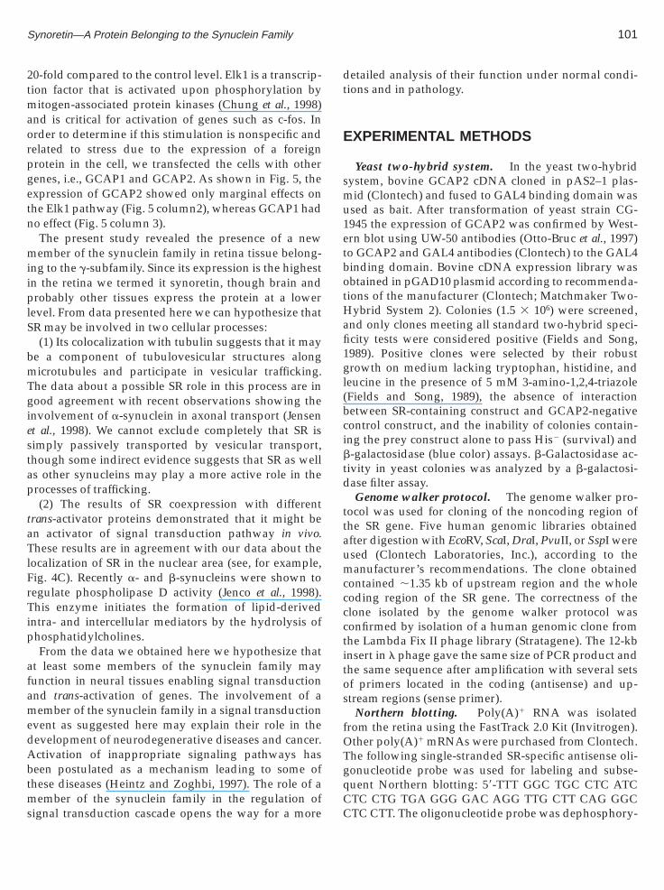
IG. 5. Effect of SR expression on in vitro signal transduction pathxpression on Elk1; 3, effect of GCAP1 expression on Elk1; 4, effect ofxpression on SRE.
here tubulin forms thick bundles (Fig. 4C). Doublemmunoreactivity is seen in the synaptic terminal and in
IE
he area of cone that is located between the nucleus andhe synapses, thus in part colocalizing with GCAP2.ummarizing the data on SR staining, we can concludehat it appears present in two forms in retinal cells:article-like and filament-like. Although the filamen-
ous form of SR colocalizes with microtubules, it mayxist independently, since disassembly of microtubulesoes not eliminate SR filaments. It is not clear if thelamentous form of SR may be attached to other typesf cellular structures, e.g., membranes and neurofila-ents, or if SR is able to form such filaments itself.In order to find a possible function of SR we used
oexpression of SR with trans-activator proteins specifico different signal transduction pathways. This ap-roach allows one to determine if a protein of interestctivates directly or indirectly one of the transcriptionactors. As shown in Fig. 5 (bars 4–6), no effect of SRxpression was observed when coexpressed with trans-ctivators specific for CREB, c-jun, and SRE pathways.
. 1, effect of SR expression on the Elk1 pathway; 2, effect of GCAP2xpression on CREB; 5, effect of SR expression on c-Jun; 6, effect of SR
n contrast, SR expression significantly activated thelk1-specific pathway, increasing luciferase activity about
2tmaorpgetn
miiplS
bmTgiestap
taTlFrTip
afamedAbtms
dt
E
smu1etbotHafi1gl(bcibtd
ttaumcccctitos
fOTgq
Synoretin—A Protein Belonging to the Synuclein Family 101
0-fold compared to the control level. Elk1 is a transcrip-ion factor that is activated upon phosphorylation by
itogen-associated protein kinases (Chung et al., 1998)nd is critical for activation of genes such as c-fos. Inrder to determine if this stimulation is nonspecific andelated to stress due to the expression of a foreignrotein in the cell, we transfected the cells with otherenes, i.e., GCAP1 and GCAP2. As shown in Fig. 5, thexpression of GCAP2 showed only marginal effects onhe Elk1 pathway (Fig. 5 column2), whereas GCAP1 hado effect (Fig. 5 column 3).The present study revealed the presence of a newember of the synuclein family in retina tissue belong-
ng to the g-subfamily. Since its expression is the highestn the retina we termed it synoretin, though brain androbably other tissues express the protein at a lower
evel. From data presented here we can hypothesize thatR may be involved in two cellular processes:(1) Its colocalization with tubulin suggests that it may
e a component of tubulovesicular structures alongicrotubules and participate in vesicular trafficking.
he data about a possible SR role in this process are inood agreement with recent observations showing the
nvolvement of a-synuclein in axonal transport (Jensent al., 1998). We cannot exclude completely that SR isimply passively transported by vesicular transport,hough some indirect evidence suggests that SR as wells other synucleins may play a more active role in therocesses of trafficking.(2) The results of SR coexpression with different
rans-activator proteins demonstrated that it might ben activator of signal transduction pathway in vivo.hese results are in agreement with our data about the
ocalization of SR in the nuclear area (see, for example,ig. 4C). Recently a- and b-synucleins were shown toegulate phospholipase D activity (Jenco et al., 1998).his enzyme initiates the formation of lipid-derived
ntra- and intercellular mediators by the hydrolysis ofhosphatidylcholines.From the data we obtained here we hypothesize that
t least some members of the synuclein family mayunction in neural tissues enabling signal transductionnd trans-activation of genes. The involvement of aember of the synuclein family in a signal transduction
vent as suggested here may explain their role in theevelopment of neurodegenerative diseases and cancer.ctivation of inappropriate signaling pathways haseen postulated as a mechanism leading to some ofhese diseases (Heintz and Zoghbi, 1997). The role of a
ember of the synuclein family in the regulation ofignal transduction cascade opens the way for a more
CC
etailed analysis of their function under normal condi-ions and in pathology.
XPERIMENTAL METHODS
Yeast two-hybrid system. In the yeast two-hybridystem, bovine GCAP2 cDNA cloned in pAS2–1 plas-id (Clontech) and fused to GAL4 binding domain was
sed as bait. After transformation of yeast strain CG-945 the expression of GCAP2 was confirmed by West-rn blot using UW-50 antibodies (Otto-Bruc et al., 1997)o GCAP2 and GAL4 antibodies (Clontech) to the GAL4inding domain. Bovine cDNA expression library wasbtained in pGAD10 plasmid according to recommenda-ions of the manufacturer (Clontech; Matchmaker Two-
ybrid System 2). Colonies (1.5 3 106) were screened,nd only clones meeting all standard two-hybrid speci-city tests were considered positive (Fields and Song,989). Positive clones were selected by their robustrowth on medium lacking tryptophan, histidine, and
eucine in the presence of 5 mM 3-amino-1,2,4-triazoleFields and Song, 1989), the absence of interactionetween SR-containing construct and GCAP2-negativeontrol construct, and the inability of colonies contain-ng the prey construct alone to pass His2 (survival) and-galactosidase (blue color) assays. b-Galactosidase ac-
ivity in yeast colonies was analyzed by a b-galactosi-ase filter assay.Genome walker protocol. The genome walker pro-
ocol was used for cloning of the noncoding region ofhe SR gene. Five human genomic libraries obtainedfter digestion with EcoRV, ScaI, DraI, PvuII, or SspI weresed (Clontech Laboratories, Inc.), according to theanufacturer’s recommendations. The clone obtained
ontained ,1.35 kb of upstream region and the wholeoding region of the SR gene. The correctness of thelone isolated by the genome walker protocol wasonfirmed by isolation of a human genomic clone fromhe Lambda Fix II phage library (Stratagene). The 12-kbnsert in l phage gave the same size of PCR product andhe same sequence after amplification with several setsf primers located in the coding (antisense) and up-tream regions (sense primer).
Northern blotting. Poly(A)1 RNA was isolatedrom the retina using the FastTrack 2.0 Kit (Invitrogen).ther poly(A)1 mRNAs were purchased from Clontech.he following single-stranded SR-specific antisense oli-onucleotide probe was used for labeling and subse-uent Northern blotting: 58-TTT GGC TGC CTC ATC
TC CTG TGA GGG GAC AGG TTG CTT CAG GGCTC CTT. The oligonucleotide probe was dephosphory-
lla
HuaAnScnt(E
warwla2
sawgiKLrtfGTdrnt(tapdpctset
tstrCtmpnwclma(
A
DM
R
A
B
C
C
D
D
F
F
102 Surguchov et al.
ated with calf intestinal alkaline phosphatase, 58-endabeled by [g-32P]ATP with T4 polynucleotide kinasend used for hybridization.
Western blot. To raise Ab382 the peptideKEALKQPVPPQEDE was conjugated to KHL andsed for rabbit immunization. Serum was purified byffinity chromatography and used for immunostaining.fter electrophoresis, proteins were transferred ontoitrocellulose membrane and probed with antibodies toR. The blot was developed using alkaline phosphatase-onjugated secondary antibodies with a mixture ofitroblue tetrazolium and BCIP (Gibco) as substrate or
he ECL system from Pierce. The second Ab to SRAb254) was raised using the peptide PQEDEAAKAE-QVAE to confirm the specificity of the first Ab.Immunolocalization. Cross sections of the retinaere prepared and stained as described earlier (Mark et
l., 1990). To prepare retinal dissociates mice and turtleetinal cells were treated according to Peng and co-orkers (1997). Briefly, the cells were plated onto poly-D-
ysine-coated coverslips, allowed to settle about 30 min,nd fixed in 4% PA in PBS for 10 min or 90% methanol at20°C for 6 min. We used the standard procedure of
taining the cells by incubating coverslips in primaryntibodies (Ab382) and subsequently incubating themith FITC-conjugated goat anti-rabbit IgG or Texas red
oat anti-mouse IgG (1:100) in PBS. Samples were rinsedn PBS and mounted in Vectashield (Vector Laboratories,Y). The immunolabeling was analyzed with a ZeissSM 410 laser scanning confocal microscope. To obtainecombinant SR, the human SR gene was recloned intohe pCALc expression vector (Stratagene) using theollowing strategy. Forward (hsr, 58-TGG GCC CATGA CGT CTT CAA) and reverse (bsras, 58-ACA GGACC ATC TCC CCC ACT CTT GGT) primers wereesigned with engineered NcoI and BamHI sites. In theeverse primer the sequence corresponding to the termi-ation codon TAG was eliminated to ensure in-frame
ranslation of SR with calmodulin binding peptide tagCBP-tag). The sequence corresponding to the SR gene,ogether with engineered restriction nucleases, wasmplified using standard PCR conditions. The PCRroduct obtained was purified as described above andigested with restriction enzymes. Ligation with theCALc vector digested with the same enzymes wasarried out after calf intestine alkaline phosphatasereatment. The recombinant vector, containing the in-erted SR gene, was checked by sequencing and used for
xpression of fusion protein as described by manufac-urer (Stratagene).F
Effect of SR expression on IN VITRO signalransduction pathways. The in vivo activation ofignal transduction pathway by SR was measured usinghe PathDetect in vivo signal transduction pathwayeporting system (Stratagene). The SR gene in pRc/MV2 vector was cotransfected into HEK293 cell cul-
ure with pathway-specific trans-activator pFA-Elk plas-id and luciferase gene in pFR-Luc plasmid (Stratagene).
FC-MEK1 and pFCdbd were used as positive andegative controls, respectively. After the transfectionas completed, cells were suspended in cell lysis buffer
ontaining Triton X-100. Luciferase activity was ana-yzed using luciferin as a substrate according to the
anufacturer’s recommendations. Luminescence wasnalyzed using MLX Microtiter Plate LuminometerDynatech Laboratories, Inc.) in 96-well microtiter plates.
CKNOWLEDGMENTS
We thank Maggie Shaw for preparation of retina cross sections,arin Bronson for DNA sequencing, and Peter Lukasiewicz, Robertark, and Ross Jakes for many helpful suggestions.
EFERENCES
hmad, I. (1995). Mash-1 is expressed during ROD photoreceptordifferentiation and binds an E-box, Eopsin-1 in the rat opsin gene. Dev.Brain Res. 90: 184–189.
uchman, V. L., Adu, J., Pinon, L. G. P., Ninkina, N. N., and Davies,A. M. (1998). Persyn, a member of the synuclein family, influencesneurofilament network integrity. Nat. Neurosci. 1: 101–103.
hen, S., and Zack, D. J. (1996). Ret 4, a positive acting rhodopsinregulatory element identified using a bovine retina in vitro transcrip-tion system. J. Biol. Chem. 271: 28549–28557.
hung, K. C., Gomes, I., Wang, D., Lau, L. F., and Rosner, M. R. (1998).Raf and fibroblast growth factor phosphorylate Elk1 and activatethe serum response element of the immediate early gene pip92 bymitogen-activated protein kinase-independent as well as -depen-dent signaling pathways. Mol. Cell. Biol. 18: 2272–2281.avidson, W. S., Jonas, A., Clayton, D. F., and George, J. M. (1998).Stabilization of a-synuclein secondary structure upon binding tosynthetic membranes. J. Biol. Chem. 273: 9443–9449.izhoor, A., Olshevskaya, E. V., Henzel, W. J., Wong, S. C., Stuls, J. T.,Ankoudinova, I., and Hurley, J. B. (1995). Cloning, sequencing, andexpression of a 24 kDa Ca-binding protein activating photoreceptorguanylyl cyclase. J. Biol. Chem. 270: 25200–25206.
ields, S., and Song, O. (1989). A novel genetic system to detectprotein–protein interactions. Nature 340: 245–247.
reund, C. L., et al. (1997). Cone–rod dystrophy due to mutations in anovel photoreceptor-specific homeobox gene (CRX) essential formaintenance of the photoreceptor. Cell 91: 543–553.
urakawa, T., Morrow, E. M., and Cepko, C. L. (1997). Crx, a novelotx-like homeobox gene, shows photoreceptor-specific expressionand regulates photoreceptor differentiation. Cell 91: 531–541.

G
H
I
I
J
J
J
J
J
K
K
[
M
M
M
M
N
O
P
P
Q
S
[
T
U
Synoretin—A Protein Belonging to the Synuclein Family 103
eorge, J. M., Jin, H, Woods, W. S., and Clayton, D. F. (1995).Characterization of a novel protein regulated during the criticalperiod for song learning in the zebra finch. Neuron 15: 361–372.eintz, N., and Zoghbi, H. (1997). a-Synuclein—A link betweenParkinson and Alzheimer diseases? Nat. Genet. 16: 325–327.
nce, T. A., and Scotto, K. W. (1995). A conserved downstream elementdefines a new class of RNA polymerase II promoters. J. Biol. Chem.270: 30249–30252.
wai, A., Masliah, E., Yoshimoto, M., Ge, N., Flanagan, L., deSilva,H. A. R., Kittel, A., and Satoh, T. (1995). The precursor protein ofnon-A beta component of Alzheimer’s disease amyloid is a presyn-aptic protein of the central nervous system. Neuron 14: 467–475.
akes, R., Spillantini, M. G., and Goedert, M. (1994). Identification oftwo distinct synucleins from human brain. FEBS Lett. 345: 27–32.
enco, J. M., Rawlingson, A., Daniels, B., and Morris, A. J. (1998).Regulation of phospholipase D2: Selective inhibition of mammalianphospholipase D isoenzymes by a- and b-synucleins. Biochemistry37: 4901–4909.
ensen, P. H., Nielson, M. S., Jakes, R., Dotti, C. G., and Goedert, M.(1998). Binding of a-synuclein to brain vesicles is abolished byfamilial Parkinson’s disease mutation. J. Biol. Chem. 41: 26292–26294.
i, H., Liu, Y. E., Jia, T., Wang, M., Liu, J., Xiao, J., Joseph, B. K., Rosen,C., and Shi, Y. E. (1997). Identification of a breast cancer-specificgene, BCSG1, by direct differential cDNA sequencing Cancer Res. 57:759–764.
ohnston, J. P., Farhangfar, F., Aparicio, J. G., Nam, S. H., andApplebury, M. L. (1997). The bovine guanylate cyclase GC-E geneand 58-flanking region. Gene 193: 219–227.
ikuchi, T., Raju, K., Breitman, M. L., and Shinohara, T. (1993). Theproximal promoter of the mouse arrestin gene directs gene expres-sion in photoreceptor cells and contains an evolutionarily conservedretinal factor-binding site. Mol. Cell. Biol. 13: 4400–4408.
rueger, R., Kuhn, W., Muller, T., Woitalla, D., Graeber, M., Kosel, S.,Przuntek, H., Epplen, J. T., Schols, L., Riess, O., et al. (1998). Ala30Promutation in the gene encoding alpha-synuclein in Parkinson’sdisease. Nat. Genet. 18: 106–108.
Letter]Lavedan, C., Leroy, E., Dehejia, A., Buchholtz, S., Dutra, A.,Nussbaum, R. L., and Polymeropoulos, M. (1998). Identification,localization and characterization of the human g-synuclein gene.Hum. Genet. 103, 106–112.ark, R. E., Liu, W. L. S., Kalloniatis, M., Raiguel, S., and vanHaesendonck, E. (1990). Patterns of glutamate immunoreactivity inthe goldfish retina. J. Neurosci. 10: 4006–4034.aroteaux, L., Campanelli, J. T., and Scheller, R. H. (1998). Synuclein:
A neuron specific protein localized to the nucleus and presynapticnerve terminal. J. Neurosci. 8: 2804–2815.Y
Family of proteins transiently associated with neuronal membrane.Brain Res. Mol. Brain Res. 11: 335–343.orabito, M. A., Yu, X., and Barnstable, C. J. (1991). Characterizationof developmentally regulated and retina-specific nuclear proteinbinding to a site in the upstream region of the rat opsin gene. J. Biol.Chem. 266: 9667–9672.inkina, N. N., Alimova-Kost, M. V., Paterson, J. W. E., Delaney, L.,Cohen, B. B., Imreh, S., Gnuchev, N. V., Davies, A. M., and Buchman,V. L. (1998). Organization, expression and polymorphism of thehuman persyn gene. Hum. Mol. Genet. 7: 1417–1424.tto-Bruc, A., Fariss, R. N., Haeseleer, F., Huang, J., Buczylko, J.,Surgucheva, I., Baehr, W., Milam, A. H., and Palcewxki, K. (1997).Localization of guanylate cyclase-activating protein 2 in mamma-lian retinas. Proc. Natl. Acad. Sci. USA 94: 4727–4732.
eng, Y.-W., Rhee, S. G., Yu, W. P., Ho, Y. K., Schoen, T, Chader, G. J.,and Yau, K. W. (1997). Identification of components of a phosphoino-sitide signaling pathway in retinal rod outer segments. Proc. Natl.Acad. Sci. USA 94: 1995–2000.
olymeropoulos, M. H., Lavedan, C., Leroy, E., Ide, S. E., Dehejia, A.,Dutra, A., Pike, B., Root, H., Rubenstein, J., Boyer, R., Stenroos, E. S.,Chandrasekharappa, S., Athanassiadou, A., Papapetropoulos, T.,Johnson, W. G., Lazzarini, A. M., Duvoisin, R. C., DiIorio, G., Golbe,L. I., and Nussbaum, R. L. (1997). Mutation in the alpha-synucleingene identified in families with Parkinson’s disease. Science 276:2045–2047.ian, X., Balestra, E., and Innerarity, T. L. (1997). Two distinctTATA-less promoters direct tissue-specific expression of the ratapo-B editing catalytic polypeptide 1 gene. J. Biol. Chem. 272:18060–18070.
pillantini, M. G., Schmidt, M. L., Lee, V. M. Y., Trojanowski, J. Q.,Jakes, R., and Goedert, M. (1997). Alpha-synuclein in Lewy bodies.Nature 388: 839–840.
Letter]Spillantini, M. G., Crowther, R., Jakes, R., Hasegawa, M., andGoedert, M. (1998). alpha-Synuclein in filamentous inclusions ofLewy bodies from Parkinson’s disease and dementia with Lewybodies. Proc. Natl. Acad. Sci. USA 95: 6469–6473.
akeda, A., Mallory, M., Sunsmo, M., Honer, W., Hansen, L., andMasliah, E. (1998). Abnormal accumulation of NACP/alpha-synuclein in neurodegenerative disorders. Am. J. Pathol. 152:367–372.eda, K., Fukushima, H., Masliah, E., Xia, Y., Iwai, A., Yoshimoto, M.,Otero, D. A. C., Kondo, J., Ihara, Y., Saitoh, T., et al. (1993). Molecularcloning of cDNA encoding an unrecognized component of amyloidin Alzheimer disease. Proc. Natl. Acad. Sci. USA 90: 11282–11286.
u, X., Chung, M., Morabito, M., and Barnstable, C. J. (1993). Sharednuclear protein sites in the upstream region of the rat opsin gene.
aroteaux, L., and Scheller, R. H. (1991). The rat brain synucleins: Biochem. Biophys. Res. Commun. 191: 76–82.
Received October 6, 1998Revised November 23, 1998Accepted January 23, 1999
Copyright © 2022 FDOKUMEN