A generic method for design of oligomer-specific antibodies
13
A Generic Method for Design of Oligomer-Specific Antibodies Kristoffer Bra ¨ nnstro ¨m 1 , Malin Lindhagen-Persson 1 , Anna L. Gharibyan 1 , Irina Iakovleva 1 , Monika Vestling 1 , Mikael E. Sellin 2 , Thomas Bra ¨ nnstro ¨m 3 , Ludmilla Morozova-Roche 1 , Lars Forsgren 4 , Anders Olofsson 1 * 1 Department of Medical Biochemistry and Biophysics, Umea ˚ University, Umea ˚, Sweden, 2 Department of Molecular Biology, Umea ˚ University, Umea ˚, Sweden, 3 Department of Medical Biosciences, Umea ˚ University, Umea ˚, Sweden, 4 Department of Clinical Pharmacology and Clinical Neuroscience, Umea ˚ University, Umea ˚, Sweden Abstract Antibodies that preferentially and specifically target pathological oligomeric protein and peptide assemblies, as opposed to their monomeric and amyloid counterparts, provide therapeutic and diagnostic opportunities for protein misfolding diseases. Unfortunately, the molecular properties associated with oligomer-specific antibodies are not well understood, and this limits targeted design and development. We present here a generic method that enables the design and optimisation of oligomer-specific antibodies. The method takes a two-step approach where discrimination between oligomers and fibrils is first accomplished through identification of cryptic epitopes exclusively buried within the structure of the fibrillar form. The second step discriminates between monomers and oligomers based on differences in avidity. We show here that a simple divalent mode of interaction, as within e.g. the IgG isotype, can increase the binding strength of the antibody up to 1500 times compared to its monovalent counterpart. We expose how the ability to bind oligomers is affected by the monovalent affinity and the turnover rate of the binding and, importantly, also how oligomer specificity is only valid within a specific concentration range. We provide an example of the method by creating and characterising a spectrum of different monoclonal antibodies against both the Ab peptide and a-synuclein that are associated with Alzheimer’s and Parkinson’s diseases, respectively. The approach is however generic, does not require identification of oligomer-specific architectures, and is, in essence, applicable to all polypeptides that form oligomeric and fibrillar assemblies. Citation: Bra ¨nnstro ¨ m K, Lindhagen-Persson M, Gharibyan AL, Iakovleva I, Vestling M, et al. (2014) A Generic Method for Design of Oligomer-Specific Antibodies. PLoS ONE 9(3): e90857. doi:10.1371/journal.pone.0090857 Editor: Sergio T. Ferreira, Federal University of Rio de Janeiro, Brazil Received September 3, 2013; Accepted February 6, 2014; Published March 11, 2014 Copyright: ß 2014 Bra ¨nnstro ¨ m et al. This is an open-access article distributed under the terms of the Creative Commons Attribution License, which permits unrestricted use, distribution, and reproduction in any medium, provided the original author and source are credited. Funding: This work was supported by Parkinsonfonden, Insamlingsstiftelsen Umea ˚ University, Alzheimerfonden, J.C. Kempe, the Swedish Research Council, Magn. Bergvalls stiftelse, O.E. och Edlas stiftelse, FAMY, Torsten So ¨ derbergs stiftelse, Va ¨sterbottens la ¨ns lansting (ALF medel) and the Medical Faculty of Umea ˚ University. The funders had no role in study design, data collection and analysis, decision to publish, or preparation of the manuscript. Competing Interests: The authors have declared that no competing interests exist. * E-mail: [email protected] Introduction The pathological self-assembly of proteins and peptides into amyloid fibrils is the defining characteristic of a group of more than twenty human diseases, including Alzheimer’s disease (AD) and Parkinson’s disease (PD) [1]. Although amyloid fibrils are invariably present in the affected individuals, many studies have shown that soluble oligomeric assemblies, which can either precede amyloid formation or represent a stand-alone entity formed in parallel with the fibrils, exert the most potent detrimental physiological effects [2–11]. However, these oligomers are transient species and frequently only constitute a very minor fraction as compared to the amyloid and the non-aggregated native and precursor forms of the specific protein or peptide. This significantly complicates characterization of oligomers and their selective therapeutic targeting. Intriguingly, antibodies that specifically target oligomeric species have been isolated [11–17]. However, the molecular properties of oligomer-specific antibodies are not well understood, which hinders both directed design as well as optimisation of such antibodies. There is, therefore, an urgent need for a method that can be used to consistently and reliably design oligomer-specific antibodies. Antibodies having the ability to identify structures exclusively present on oligomeric assemblies have previously been demon- strated [11,13,14,18–27]. The term ‘‘oligomer’’ can however, be applied to assemblies ranging from a dimer to much larger protofibrillar structures [28]. Due to this inherent heterogeneity, and the lack of a priori structural information, directed design of oligomer-specific antibodies is not straightforward and is frequent- ly dependent on stochastic events. These limitations hamper development in the field. We have previously shown that the multivalent architecture of IgM antibodies, having 10 independent binding sites, can be used as a selective binder for oligomers due to the exposure of multiple epitopes on the oligomeric assemblies [29]. However, the IgM isotype cannot be recombinantly expressed or genetically modified, and this hampers both its characterisation and its potential therapeutic use. In the present work, we show how a simple divalent binder such as antibodies of the IgG isotype is an interesting alternative. In contrast to the multivalent IgM a divalent interaction importantly facilitate the factors required for oligomer-specificity to be PLOS ONE | www.plosone.org 1 March 2014 | Volume 9 | Issue 3 | e90857
Transcript of A generic method for design of oligomer-specific antibodies

A Generic Method for Design of Oligomer-SpecificAntibodiesKristoffer Brannstrom1, Malin Lindhagen-Persson1, Anna L. Gharibyan1, Irina Iakovleva1,
Monika Vestling1, Mikael E. Sellin2, Thomas Brannstrom3, Ludmilla Morozova-Roche1, Lars Forsgren4,
Anders Olofsson1*
1 Department of Medical Biochemistry and Biophysics, Umea University, Umea, Sweden, 2 Department of Molecular Biology, Umea University, Umea, Sweden,
3 Department of Medical Biosciences, Umea University, Umea, Sweden, 4 Department of Clinical Pharmacology and Clinical Neuroscience, Umea University, Umea,
Sweden
Abstract
Antibodies that preferentially and specifically target pathological oligomeric protein and peptide assemblies, as opposed totheir monomeric and amyloid counterparts, provide therapeutic and diagnostic opportunities for protein misfoldingdiseases. Unfortunately, the molecular properties associated with oligomer-specific antibodies are not well understood, andthis limits targeted design and development. We present here a generic method that enables the design and optimisationof oligomer-specific antibodies. The method takes a two-step approach where discrimination between oligomers and fibrilsis first accomplished through identification of cryptic epitopes exclusively buried within the structure of the fibrillar form.The second step discriminates between monomers and oligomers based on differences in avidity. We show here that asimple divalent mode of interaction, as within e.g. the IgG isotype, can increase the binding strength of the antibody up to1500 times compared to its monovalent counterpart. We expose how the ability to bind oligomers is affected by themonovalent affinity and the turnover rate of the binding and, importantly, also how oligomer specificity is only valid withina specific concentration range. We provide an example of the method by creating and characterising a spectrum of differentmonoclonal antibodies against both the Ab peptide and a-synuclein that are associated with Alzheimer’s and Parkinson’sdiseases, respectively. The approach is however generic, does not require identification of oligomer-specific architectures,and is, in essence, applicable to all polypeptides that form oligomeric and fibrillar assemblies.
Citation: Brannstrom K, Lindhagen-Persson M, Gharibyan AL, Iakovleva I, Vestling M, et al. (2014) A Generic Method for Design of Oligomer-SpecificAntibodies. PLoS ONE 9(3): e90857. doi:10.1371/journal.pone.0090857
Editor: Sergio T. Ferreira, Federal University of Rio de Janeiro, Brazil
Received September 3, 2013; Accepted February 6, 2014; Published March 11, 2014
Copyright: � 2014 Brannstrom et al. This is an open-access article distributed under the terms of the Creative Commons Attribution License, which permitsunrestricted use, distribution, and reproduction in any medium, provided the original author and source are credited.
Funding: This work was supported by Parkinsonfonden, Insamlingsstiftelsen Umea University, Alzheimerfonden, J.C. Kempe, the Swedish Research Council,Magn. Bergvalls stiftelse, O.E. och Edlas stiftelse, FAMY, Torsten Soderbergs stiftelse, Vasterbottens lans lansting (ALF medel) and the Medical Faculty of UmeaUniversity. The funders had no role in study design, data collection and analysis, decision to publish, or preparation of the manuscript.
Competing Interests: The authors have declared that no competing interests exist.
* E-mail: [email protected]
Introduction
The pathological self-assembly of proteins and peptides into
amyloid fibrils is the defining characteristic of a group of more
than twenty human diseases, including Alzheimer’s disease (AD)
and Parkinson’s disease (PD) [1]. Although amyloid fibrils are
invariably present in the affected individuals, many studies have
shown that soluble oligomeric assemblies, which can either
precede amyloid formation or represent a stand-alone entity
formed in parallel with the fibrils, exert the most potent
detrimental physiological effects [2–11]. However, these oligomers
are transient species and frequently only constitute a very minor
fraction as compared to the amyloid and the non-aggregated
native and precursor forms of the specific protein or peptide. This
significantly complicates characterization of oligomers and their
selective therapeutic targeting. Intriguingly, antibodies that
specifically target oligomeric species have been isolated [11–17].
However, the molecular properties of oligomer-specific antibodies
are not well understood, which hinders both directed design as
well as optimisation of such antibodies. There is, therefore, an
urgent need for a method that can be used to consistently and
reliably design oligomer-specific antibodies.
Antibodies having the ability to identify structures exclusively
present on oligomeric assemblies have previously been demon-
strated [11,13,14,18–27]. The term ‘‘oligomer’’ can however, be
applied to assemblies ranging from a dimer to much larger
protofibrillar structures [28]. Due to this inherent heterogeneity,
and the lack of a priori structural information, directed design of
oligomer-specific antibodies is not straightforward and is frequent-
ly dependent on stochastic events. These limitations hamper
development in the field. We have previously shown that the
multivalent architecture of IgM antibodies, having 10 independent
binding sites, can be used as a selective binder for oligomers due to
the exposure of multiple epitopes on the oligomeric assemblies
[29]. However, the IgM isotype cannot be recombinantly
expressed or genetically modified, and this hampers both its
characterisation and its potential therapeutic use.
In the present work, we show how a simple divalent binder such
as antibodies of the IgG isotype is an interesting alternative. In
contrast to the multivalent IgM a divalent interaction importantly
facilitate the factors required for oligomer-specificity to be
PLOS ONE | www.plosone.org 1 March 2014 | Volume 9 | Issue 3 | e90857

determined in a quantitative manner. Through characterising of a
spectrum of monoclonal antibodies, having significantly different
properties, the concept of oligomer-specificity is outlined and we
demonstrate how different parameters affect the efficiency of
selectively binding to oligomers. We expose how the ability to bind
oligomers is affected by the monovalent affinity and the turnover
rate of the binding and, importantly, also how oligomer specificity
is only valid within a specific concentration range. We have
specifically applied the method to identify oligomer-specific
monoclonal antibodies targeting the amyloid b peptide (Ab) and
a-synuclein that are associated with AD and PD, respectively. The
approach is however generic and applicable to all polypeptides
that form oligomeric and fibrillar assemblies.
Results
Step 1: Discriminating between oligomers and amyloidfibrils
The definition of an oligomer-specific antibody implies that it
does not react with the fibrillar or monomeric counterparts of the
same protein or peptide. The first step in the present method is to
find an epitope which exclude binding of the antibody to the
fibrillar form of the polypeptide. To accomplish this, we use the
structural differences between the fibrillar and oligomeric struc-
tures and through identification of a cryptic epitope that is
exclusively buried within the fibrillar architecture, discrimination
can be acquired.
Identifying epitopes having different exposure on Aboligomers and Ab fibrils
For Ab, the N-terminal residues have previously been shown to
be differently exposed between the fibrillar and oligomeric forms
[30,31]. Mice were immunised with the Ab1–42 and hybridomas
were generated according to standard procedures. The antibody
response in mice upon immunisation with Ab frequently results in
antibodies that target the Ab(3–10) epitope, and a number of these
have also been reported in the literature [32]. Using a dot blot
technique – in which an equal amount of fibrils and monomers
from the Ab1–42 are applied to the membrane – we concluded that
the region spanning residue 3–10 in a nice manner can be used to
differentiate between the fibrillar and oligomeric forms. From the
initial pool of clones targeting Ab, two monoclonal IgG antibodies
denoted mAB-O and mAB-M, that both bind within the Ab(3–10)
region, but also in a good way illustrate the subsequent
requirements for oligomer-specificity further presented below,
were selected.
A dot blot analysis of mAB-O and mAB-M is shown in Figure 1
A and B and illustrates the preferential binding to oligomers and
how both antibodies are impaired in binding to the fibrillar fold.
The conformational dependence and fibril specificity of the
monoclonal antibody OC [33] was used to verify the presence
of Ab fibrils (Fig. 1C). The different morphologies of oligomeric
and fibrillar Ab(1–42) were also confirmed by atomic force
microscopy (AFM) (Figure S1).
Identifying epitopes having different exposure on a-synuclein oligomers and a-synuclein fibrils
There is only limited knowledge of the structural properties of
a-synuclein’s oligomeric form, so multiple antibodies where
generated using the complete 140-residue sequence of a-synuclein
as the antigen. Using the dot blot technique - where equal
amounts of a-synuclein fibrils and monomers were applied to a
membrane followed by screening for selective binding to oligomers
- we identified a comparatively broad region in the C-terminal
part of the protein (from Gly111 to Ala140) that is exposed in the
oligomeric form but buried within the fibrillar form.
Two different monoclonal antibodies having the IgG isotype,
that both bind within the Gly111–Ala140 region of a-synuclein,
but also in a nice manner illustrate parameters important for
oligomer-specificity further presented below were selected. These
antibodies, denoted ASyO2 and ASyO5, bind to the Glu131–
Ala140 and Gly111–Tyr125 regions, respectively, and both
readily bind to a-synuclein oligomers but not to a-synuclein fibrils
(Fig. 1 D and E).
A third a-synuclein–specific antibody, denoted ASyM, binds to
the N-terminal stretch spanning Met1–Val15, but this is a
sequence that does not effectively discriminate between oligomers
and fibrils. This antibody was used as a control to expose the equal
amounts of fibrils and oligomers on the membrane (Fig. 1F) as well
as to further expose how differences in binding kinetics in a
dramatic manner may affect oligomer specificity, described below.
The structures of the fibrillar and oligomeric samples of a-
synuclein were also verified using AFM (Figure S1).
Step 2: Discriminating between monomers andoligomers
In the second step of the method, the ability to discriminate
between oligomers and monomers is evaluated. This step
harnesses the effect of avidity, which is defined as a synergistic
increase in binding strength when a multivalent receptor binds to a
multivalent antigen. The impact of avidity and its consequences
for oligomer-specificity is exposed through a comparative analysis
of the above selected spectrum of antibodies, which are all divalent
Figure 1. Dot blot enables probing for structural differencesbetween fibrils and oligomers. An equal amount of fibrils andoligomers from either Ab(1–42) or a-synuclein, as indicated in the figure,were applied to a nitrocellulose membrane. Binding specificity of (A)mAB-O, (B) mAB-M, (C) Ab fibril-specific OC antibody,, (D) ASyO2, (E)ASyO5, and (F) ASyM.doi:10.1371/journal.pone.0090857.g001
Oligomer-Specific Antibodies
PLOS ONE | www.plosone.org 2 March 2014 | Volume 9 | Issue 3 | e90857
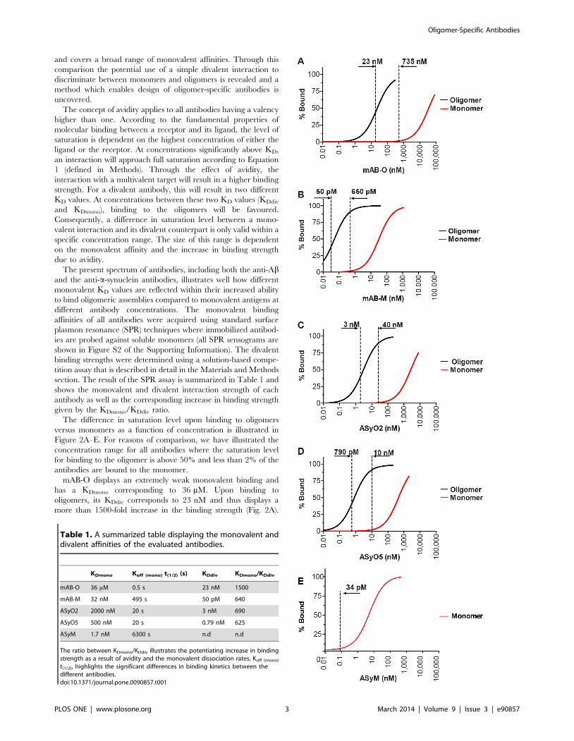
and covers a broad range of monovalent affinities. Through this
comparison the potential use of a simple divalent interaction to
discriminate between monomers and oligomers is revealed and a
method which enables design of oligomer-specific antibodies is
uncovered.
The concept of avidity applies to all antibodies having a valency
higher than one. According to the fundamental properties of
molecular binding between a receptor and its ligand, the level of
saturation is dependent on the highest concentration of either the
ligand or the receptor. At concentrations significantly above KD,
an interaction will approach full saturation according to Equation
1 (defined in Methods). Through the effect of avidity, the
interaction with a multivalent target will result in a higher binding
strength. For a divalent antibody, this will result in two different
KD values. At concentrations between these two KD values (KDdiv
and KDmono), binding to the oligomers will be favoured.
Consequently, a difference in saturation level between a mono-
valent interaction and its divalent counterpart is only valid within a
specific concentration range. The size of this range is dependent
on the monovalent affinity and the increase in binding strength
due to avidity.
The present spectrum of antibodies, including both the anti-Aband the anti-a-synuclein antibodies, illustrates well how different
monovalent KD values are reflected within their increased ability
to bind oligomeric assemblies compared to monovalent antigens at
different antibody concentrations. The monovalent binding
affinities of all antibodies were acquired using standard surface
plasmon resonance (SPR) techniques where immobilized antibod-
ies are probed against soluble monomers (all SPR sensograms are
shown in Figure S2 of the Supporting Information). The divalent
binding strengths were determined using a solution-based compe-
tition assay that is described in detail in the Materials and Methods
section. The result of the SPR assay is summarized in Table 1 and
shows the monovalent and divalent interaction strength of each
antibody as well as the corresponding increase in binding strength
given by the KDmono/KDdiv ratio.
The difference in saturation level upon binding to oligomers
versus monomers as a function of concentration is illustrated in
Figure 2A–E. For reasons of comparison, we have illustrated the
concentration range for all antibodies where the saturation level
for binding to the oligomer is above 50% and less than 2% of the
antibodies are bound to the monomer.
mAB-O displays an extremely weak monovalent binding and
has a KDmono corresponding to 36 mM. Upon binding to
oligomers, its KDdiv corresponds to 23 nM and thus displays a
more than 1500-fold increase in the binding strength (Fig. 2A).
Table 1. A summarized table displaying the monovalent anddivalent affinities of the evaluated antibodies.
KDmono Koff (mono) t(1/2) (s) KDdiv KDmono/KDdiv
mAB-O 36 mM 0.5 s 23 nM 1500
mAB-M 32 nM 495 s 50 pM 640
ASyO2 2000 nM 20 s 3 nM 690
ASyO5 500 nM 20 s 0.79 nM 625
ASyM 1.7 nM 6300 s n.d n.d
The ratio between KDmono/KDdiv illustrates the potentiating increase in bindingstrength as a result of avidity and the monovalent dissociation rates, Koff (mono)
t(1/2), highlights the significant differences in binding kinetics between thedifferent antibodies.doi:10.1371/journal.pone.0090857.t001
Oligomer-Specific Antibodies
PLOS ONE | www.plosone.org 3 March 2014 | Volume 9 | Issue 3 | e90857
The span between .50% bound oligomer and ,2% bound
monomer is between 23 nM and 735 nM.
The binding strength of mAB-M is increased 640-fold as a result
of avidity. Due to the higher monovalent affinity (32 nM),
however, the corresponding concentration range where a satura-
tion level for the oligomer is above 50% while less than 2% of the
antibodies are bound to the monomer becomes very narrow and
only spans between 50 pM and 650 pM (Fig. 2B). The useful
concentration range for mAB-M is, therefore, approximately
1000-fold smaller than for mAB-O.
The potentiating effects of ASyO2 and ASyO5 correspond to
690-fold and 625-fold, respectively, and these antibodies display
monovalent affinities corresponding to 2000 nM and 500 nM,
respectively. The concentration where 50% binding occurs while
the saturation level of binding to the monomer is less than 2%
corresponds to 3–40 nM for ASyO2 and 0.79–10 nM for ASyO5
(Fig. 2C and D).
The divalent binding strength of ASyM, which has a KDmono of
1.7 nM, was too strong to be experimentally determined. A
monovalent level of saturation corresponding to 2% for ASyM is,
however, already reached at 34 pM (Fig. 2E). Below these results
are also presented in a non-logarithmic manner in further
described below, that better illustrates how the width of the useful
‘‘concentration window’’ is dramatically increased simply by
lowering the monovalent affinity.
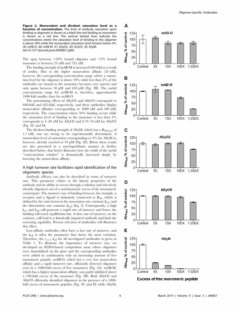
A high turnover rate facilitates rapid identification of theoligomeric species
Antibody efficacy can also be described in terms of turnover
rate. This parameter relates to the kinetic properties of the
antibody and its ability to screen through a solution and selectively
identify oligomers out of a stoichiometric excess of the monomeric
counterpart. The turnover rate of binding between, for example, a
receptor and a ligand is intimately connected to KD, which is
defined by the ratio between the association rate constant (kon) and
the dissociation rate constant (koff) (Eq. 2). Consequently, a high
kon and koff will generate a rapid rate of turnover and hence the
binding will reach equilibrium fast. A slow rate of turnover, on the
contrary, will lead to a kinetically impaired antibody and limit the
screening capability. Present selection of antibodies will illustrates
this effect.
Low-affinity antibodies often have a fast rate of turnover, and
the koff is often the parameter that shows the most variation.
Therefore, the t(1/2) koff for all investigated antibodies is given in
Table 1. To illustrate the importance of turnover rate, we
developed an ELISA-based competition assay where oligomers
were immobilised on the plate and the corresponding antibodies
were added in combination with an increasing amount of free
monomeric peptide. mAB-O, which has a very low monovalent
affinity and a rapid turnover rate, efficiently detected oligomers
even in a 1000-fold excess of free monomers (Fig. 3A). mAB-M,
which has a higher monovalent affinity, was partly inhibited above
a 100-fold excess of the monomer (Fig. 3B). Both ASyO2 and
ASyO5 efficiently identified oligomers in the presence of a 1000-
fold excess of monomeric peptides (Fig. 3C and D) while ASyM,
Figure 2. Monovalent and divalent saturation level as afunction of concentration. The level of antibody saturation uponbinding to oligomers is shown as a black line and binding to monomersis shown as a red line. The vertical dotted lines indicate theconcentrations where the saturation level of binding to the oligomeris above 50% while the monovalent saturation level remains below 2%.(A) mAB-O, (B) mAB-M, (C) ASyO2, (D) ASyO5, (E) ASyM.doi:10.1371/journal.pone.0090857.g002
Oligomer-Specific Antibodies
PLOS ONE | www.plosone.org 4 March 2014 | Volume 9 | Issue 3 | e90857

having a significantly slower turnover rate, in contrast was
effectively inhibited from binding and displayed an essentially
stoichiometric level of inhibition as a result of competition with the
monomer (Fig. 3E).
Dot blots for discriminating between monomers andoligomers can be deceptive
The dot blot technique – which is frequently used for
discriminating between oligomers and monomers – should be
highlighted because it might yield deceptive results if not
performed properly. The effect of avidity, which applies to all
antibodies having a valency higher than one, results in a
potentiation in binding strength as a result of two or more
simultaneous interactions. This also implies that potentiation in
the binding strength will occur if antigens that are immobilized on
a surface are separated by an average distance less than the
distance between the two antigen-binding sites on the antibody.
When using techniques such as dot blot, western blot, or ELISA
for the purpose of discriminating between oligomers and
monomers, this effect must be considered and the antigens must
consequently be diluted accordingly to be separated by an average
distance on the membrane higher than the distance between the
binding sites on the antibody. The multiple epitopes of an
oligomer are close to each other due to the structure of the
oligomer but the average distance between immobilized mono-
mers becomes too far to mediate the effect of avidity and the
potentiated binding strength. The effect is interpreted as an
oligomer-specific interaction. A dot blot of the oligomeric versus
monomeric species is illustrated in Fig. 4A. In this context it should
however be emphasized that dot blot techniques do not reflect the
level of saturation of binding. The ability to remain bound
throughout the assay, including the subsequent washing steps, is
therefore controlled by the dissociation rate of the antibody. This
implies that antibodies having a high affinity, but also a high
turnover rate, i.e. a fast kon and a fast koff, will not necessarily
remain bound throughout the assay. Fig. 4B illustrates the window
of concentration where binding saturation to the oligomer is above
50% while binding to the monomer is below 2% for all evaluated
antibodies (adapted from the results shown in Fig. 2 but now
presented in a non-logarithmic manner). In particular mAB-M,
which has a KD of 32 nM, exemplifies the phenomenon where a
high level of saturation during the initial incubation passes
undetected. During the dot-blot procedure all antibodies are
incubated at 25 nM which regarding mAB-M results in a 44%
level of saturation. However, due to its comparatively high
dissociation rate mAB-M is unable to remain bound throughout
the assay and the result is interpreted as oligomer-specific. Within
the present experiment only the ASyM antibody, as a result of its
slow turnover rate, remains bound throughout the dot blot assay.
Imunohistochemistry on AD brain tissueFor most antibodies, a high binding specificity over non-specific
background binding is often essential, it is therefore important to
emphazise that a low affinity is not necessarily associated with
broad specificity. To illustrate that a high specificity is maintained,
as well as to evaluate their binding pattern to ex vivo material, the
ability of the antibodies created in this study to bind to human
brain-derived Ab and a-synuclein deposits was evaluated using
immunohistochemistry (IHC) Figures 5 and 6.
The hippocampus area from a human AD patient was
evaluated using polyclonal rabbit anti Ab antibody (Fig. 5A–C),
mAB-M (Fig. 5D–F) and mAB-O (Fig. 5G–I). The results show
that the overall background staining of all these antibodies was low
(Fig. 5A,D,G). The results also revealed an intriguing discrepancy
between mAB-M and mAB-O where mAB-M efficiently bound
Ab plaques but no reactivity could be seen with mAB-O (Fig. 5E
and 5H respectively). The reactivity of mAB-M to amyloid plaques
Figure 4. Therapeutic antibody concentrations require very low monovalent affinities to avoid reactivity to monomers. (A)Non-logarithmic illustration of the antibody concentration range where more than 50% of the oligomers are bound while the saturation level to themonomer is below 2%. Note in particular the dramatic difference between mAB-O and mAB-M that both qualify as being oligomer specific accordingto the widely accepted dot blot technique. The bars corresponding to mAB-M as well as ASyM are indicated with arrows to highlight theirappearance. For comparison, the most commonly used range of therapeutic antibody concentrations for AD (20 mM to 250 mM) is indicated in thefigure (grey striped vertical bars). (B) Dot blot analysis where equal amounts of Ab1–42 and a-synuclein oligomers and monomers has been applied ona membrane and probed with mAB-O, mAB-M, ASyO5, ASyO2, and ASyM as indicated in the figure.doi:10.1371/journal.pone.0090857.g004
Figure 3. The rate of turnover controls antibody efficacy.Competition ELISA setup showing the efficacy of the antibodies todetect oligomers in the presence of increasing concentrations ofmonomers. (A) mAB-O. (B) mAB-M. (C) ASyO2. (D) ASyO5. (E) ASyM. Thekinetics of binding for each antibody are given in Table 1.doi:10.1371/journal.pone.0090857.g003
Oligomer-Specific Antibodies
PLOS ONE | www.plosone.org 5 March 2014 | Volume 9 | Issue 3 | e90857

Figure 5. IHC of human hippocampus from an AD-affected individual. IHC was used to illustrate binding specificity and structuralpreferences of mAB-O and mAB-M upon binding to ex vivo material. Hippocampus sections were from a post-mortem AD-affected human brain (A–C)The binding pattern from a polyclonal rabbit anti-Ab antibody illustrating both plaques as well as an intracellular form of Ab that is abundant in thehilar neurons. (D–F) The staining pattern of mAB-M in which Ab plaques near the dentate gyrus (E) are readily stained. (G–I). The binding pattern ofmAB-O illustrates an inability to stain Ab plaques and indicates an alternative structural preference compared to mAB-M. A strong binding to theintracellular form of Ab is observed in the hilus area. The granular cells of the dentate gyrus (B, E, and H) and the neurons of hilus (C, F, and I) areindicated with arrows and represent the highe magnification images of respective selected areas in A, D and G.doi:10.1371/journal.pone.0090857.g005
Figure 6. Immunohistochemistry of human midbrain from a PD-affected individual. To probe the specificity and structural preference ofASyO2 towards ex vivo a-synuclein assemblies, the mesencephalon (midbrain) of the brain of a human PD patient was analysed. The imagesrepresent the substantia nigra area of the midbrain. (A) Polyclonal anti a-synuclein. (B) ASyO5. (C) ASyO2.doi:10.1371/journal.pone.0090857.g006
Oligomer-Specific Antibodies
PLOS ONE | www.plosone.org 6 March 2014 | Volume 9 | Issue 3 | e90857

consequently pinpoints a discrepancy when compared to the dot
blot analysis, shown in Figure 1B. The presence of less condensed
protofibrils or diffuse non-fibrillar plaques, having a more open
architecture, should be considered as possible explanations.
Another reason for this difference might be the fact that the
conditions used to prepare the IHC tissue samples are often harsh
and could possibly induce exposure of previously buried epitopes
on the fibrils.
Nevertheless, the results illustrate how subtle changes in epitope
exposure can have profound impacts on the reactivity’s of the
antibodies. Interestingly all Ab antibodies, including the polyclonal
anti-Ab antibody (Fig. 5A–C), bound to an intracellular form of
Ab that has been previously described but is not currently
associated with disease [34]. The binding to intracellular Ab was
notably stronger for mAB-M (Fig. 5E,F) and mAB-O (Fig. 5H,I)
both in granular neurons of dentate gyrus and in hilar neurons
compared to the polyclonal antibody (Fig. 5B,C).
Immunohistochemistry in PD brain tissuesTo explore the specificity and binding patterns of ASyO2 and
ASyO5, these antibodies were used to probe a tissue sample
isolated from the substantia nigra of the brain from a PD patient
(Fig. 6). The results show that both ASyO5 and ASyO2 specifically
bind to a-synuclein in Lewy bodies and Lewy neurites (Fig. 6B and
C) with a staining pattern similar to that of the polyclonal anti-a-
synuclein antibody AS08-359 (Fig. 6A). Reactivity to deposited a-
synuclein material could indicate the presence of pre-fibrillar
structures in vivo, but we cannot explicitly rule out the possibility
that in vivo and in vitro fibrils might differ in structure or that the
exposure of these epitopes might be caused by the sample
preparation and might, therefore, not be fully representative of the
situation in vivo.
mAB-O efficiently prevents the cytotoxic effect of Ab-oligomers
Oligomer-specific antibodies have therapeutic potential. It is,
therefore, important to verify that antibodies having very low
monovalent affinities are still able to prevent a cytotoxic effect.
Addition of pre-formed Ab oligomers to the human neuroblasto-
ma cell-line SH-SY5Y exerted a cytotoxic effect, and within
48 hours of incubation cell viability was reduced to 60% (Fig. 7).
Addition of mAB-O to the system resulted in an efficient
attenuation of the cytotoxic effect (Fig. 7). Notably, the same
inhibiting effect could also be obtained in the presence of a 100-
fold molar excess of monomeric Ab1–10 peptide. This suggests that
the monomeric form of the antigen did not affect antibody binding
to the oligomeric form. We were unable to perform the same assay
with a-synuclein because the cytotoxic effect from these oligomers
is significantly lower and requires significantly higher protein
concentrations. The required concentration of antibodies could
not be added without unwanted side effects in the cell assay.
Figure 7. Binding to biologically active oligomers of Ab1–42.Isolated Ab1–42 oligomers exert a cytotoxic effect on SH-SY5Y cells, andthe attenuating effect of the oligomer-specific mAB-O antibody wasevaluated. Cells exposed to Ab1–42 oligomers are shown with grey barswhile controls are depicted in black. Addition of mAB-O in a 1:5 molarratio to Ab1–42 fully attenuates the effect. Addition of Ab1–10 monomerat a 100-fold molar excess does not interfere with the attenuatingeffect.doi:10.1371/journal.pone.0090857.g007
Figure 8. Detection of ex vivo-derived oligomeric assemblies.(A) CSF samples from 10 PD patients having a clinical stage of diseasearound 2 according to the Hoehn and Yahr scale [35] and 10 age-matched controls were probed for the presence of a-synucleinoligomers using a homopaired sandwich ELISA based on the ASyO2antibody. The age of each individual is given below the correspondingbar. The concentration of a-synuclein oligomers was determined usinga standard curve of in vitro a-synuclein oligomers based on theirmonomeric protein concentration. (B) A sandwich ELISA setup usingmAB-O and mAB-M to evaluate binding to Ab oligomers derived fromthe well-established and previously described 7PA2 cells [38].doi:10.1371/journal.pone.0090857.g008
Oligomer-Specific Antibodies
PLOS ONE | www.plosone.org 7 March 2014 | Volume 9 | Issue 3 | e90857
Detection of ex vivo-isolated oligomersOligomeric assemblies can be found in the cerebrospinal fluid
(CSF) of both AD and PD patients at concentrations within the
lower picomolar (pM) range. In the present investigation, CSF
samples from PD patients were analysed, and all patients were
classified as being around stage 2 of the disease according to the
Hoehn and Yahr scale [35]. In total, CSF samples from 10 PD
patients and 10 age-matched controls were evaluated. Using a
homopaired sandwich ELISA based on the ASyO2 antibody, a
significant increase in the presence of oligomers was seen in the
group of PD patients (Fig. 8A). In accordance with similar
investigations, oligomeric a-synuclein could be seen in some
controls and there were some individuals with PD who did not
have a-synuclein oligomers in their CSF [36,37].
A similar set of CSF samples was not available for AD during
the time of this investigation. Therefore, to evaluate the ability of
the anti-Ab antibodies to capture oligomers produced by a
eukaryotic cell, we employed the well-established 7PA2 cell-based
system that produces biologically active Ab oligomers [38]. Using
a sandwich-based approach, both mAB-O and mAB-M detected
the oligomers derived from the 7PA2 cell system and this suggested
that exposure of the N-terminal Ab epitope is conserved between
the in vitro and in vivo-derived oligomers (Fig. 8B). Similar results
were also obtained using the recently described yeast-based system
that expresses Ab1–42 [39] (data not shown).
Discussion
It has been more than a decade since the publication of the first
report of cognitive improvement in a mouse model for AD as a
result of passive immunization [40]. Due to the possible beneficial
effect of adopting the same rationale on humans, these initial
findings led to massive efforts from the pharmaceutical industry.
Today more than ten clinical trials are in progress [41,42].
Unfortunately, only modest cognitive improvement has so far been
noted in humans, and individuals treated with the highest
antibody titres have a significantly increased risk of developing
vasogenic oedema and cerebral microhemorrhage [43]. There is,
therefore, a need to better understand the process of passive
immunization and acquire more effective antibodies having fewer
side effects. Interestingly, passive vaccination is currently also
emerging as an approach for other misfolding disorders including
PD, amyotrophic lateral sclerosis, and pathologies associated with
tau assemblies [44–51]. This further emphasizes the need for a
general method to obtain antibodies with optimal properties.
Both cellular and animal models have shown that the fraction of
small oligomeric assemblies – either preceding amyloid formation
or representing a stand-alone entity formed in parallel with the
fibrils – exert the most potent detrimental physiological effects
[3,4,11] [2]. Consequently, these oligomeric assemblies represent a
very interesting target for intervention. Intriguingly, antibodies
have been developed that selectively identify conformational
epitopes that are exclusively exposed in the oligomeric form
[11,13,14,18–27]. These antibodies provide an advantage com-
pared to traditional antibodies because they do not react with the
fibrillar or the monomeric forms, including putative precursor
molecules, and, therefore, achieve a higher effective concentration
toward the intended target. Unfortunately, the molecular proper-
ties associated with oligomer-specific antibodies are not well
understood, and their development depends on stochastic events
that hamper the targeted design and optimisation of these
antibodies.
In the present work, we unveil features that render antibodies
specific for oligomers and show how different properties affect
their efficacy. The technique is easily adapted but, as we describe,
several factors to be considered and extreme binding properties
might be required to reach full capacity. Identifying these
parameters has important therapeutic implications because highly
efficient therapeutic antibodies can now be designed in detail.
The procedure is essentially divided into two steps in which a
linear epitope, exposed within the monomeric and oligomeric forms
but buried within the fibrillar state, can be combined with the effect
of avidity to select for oligomer-specific binding properties.
Identification of an epitope exposed within the oligomeric and
monomeric state but buried within the fibrillar architecture is
comparatively straightforward due to the significant structural
differences that usually exist between oligomers and fibrils.
After selecting for impaired binding to the fibrils, the problem is
reduced to discriminating between monomers (including precursor
molecules) and oligomers. In this work, we show that even a simple
divalent interaction can increase the affinity 1500 times compared
to a monovalent interaction.
At first glance, this rationale implies that all antibodies having a
valency higher than one would prefer binding to a multivalent target
and, therefore, would be oligomer specific. This is, however, an
oversimplification, and although a divalent interaction to a
multivalent antigen causes a significant increase in binding strength
this work describes how additional parameters in the binding need to
be considered to achieve the desired oligomer-specific effect.
According to the fundamental properties of molecular binding,
the level of saturation between a receptor and a ligand is always
dependent on concentration. This implies that the concentration
where oligomeric assemblies can be captured while the level of
monovalent binding is maintained at insignificant levels is only
valid within a certain range. This notion is of central importance
because it implies that although the potentiating factor of avidity
might be identical between high- and low-affinity antibodies their
effective concentration range will differ. This range is dependent
on the monovalent affinity as well as the potentiating factor due to
the effect of avidity. Importantly, lowering the monovalent affinity
will expand the effective concentration range. This is indicated in
Figure 2, but to better illustrate the relative difference Figure 4B
shows the concentration range for all of the antibodies, created in
this work, in a non-logarithmic manner. Note in particular the
large difference between mAB-O and mAB-M that both qualify as
being oligomer specific according to the widely used dot blot
technique shown in Figure 4A emphasizing why dot blot to
discriminate between oligomers and monomers should be used
with great care and always be accompanied with a technique
where saturation levels can be monitored.
Because oligomer-specific antibodies have significant therapeu-
tic potential, it is interesting to relate concentrations to therapeutic
antibody levels. This has been the most well established with
regard to AD, and commonly used serum concentrations of
therapeutic antibodies targeting Ab frequently range from 20 nM
to 250 nM. Note that within this range most traditional antibodies
display a very high level of monovalent saturation, and our results
suggest that essentially only antibodies having very low monova-
lent affinities, such as mAB-O, fulfil the requirements of a selective
binding within this concentration range. The monovalent KD of
mAB-O, corresponding to 36 mM is several thousand-fold higher
than most traditional antibodies.
In this work we further expose how the efficacy by which
antibodies find the thermodynamically most stable interaction
(binding to the oligomer) is dependent on binding kinetics. We also
illustrate how a slow rate of turnover, as clearly shown by ASyM,
impairs the ability to bind the oligomeric target in the presence of
the monomeric counterpart. Notably, the rate of turnover is also
Oligomer-Specific Antibodies
PLOS ONE | www.plosone.org 8 March 2014 | Volume 9 | Issue 3 | e90857

reflected within the dot blot analysis where only the high-affinity
ASyM antibody remains attached to a monovalent target further
emphasizing why dot blots should be used with care and should
always accompanied by a complimentary technique.
Low-affinity binding might intuitively be associated with low
specificity. This assumption is, however, not correct and can be
explained using the thermodynamics of binding energies that have
been described in detail by Kawasaki and co-workers [52]. In
brief, the specificity of the binding is determined by the relative
contribution of hydrophobic and polar interactions. A large
contribution of hydrophobic interactions relative to polar interac-
tions generally decreases the specificity. Because oligomers and
protofibrils in general expose a large proportion of hydrophobic
areas to the solvent, antibodies having a high proportion of the
hydrophobic component might interact with these antigens in a
rather nonspecific manner. A high proportion of hydrophobic
interactions might, when combined with the concept of avidity,
provide a possible molecular explanation for some of the
previously reported generic oligomer-specific antibodies.
The antibodies presented within this work are all directed at
comparatively hydrophilic areas, and their binding is, therefore,
less likely to contain a high hydrophobic component. None of the
antibodies presented within this work have been seen to cross react
with other oligomers.
Because the binding specificity is determined by the choice of
linear epitope, the importance of verifying a correct target should
be stressed. We pinpoint putative structural discrepancies between
in vivo and in vitro-derived assemblies of both Ab and a-synuclein.
Although the methods of sample preparation might affect epitope
exposure, great care should be taken when choosing the epitope of
binding and the initial screening should preferably be performed
on ex vivo-isolated material under non-denaturing conditions.
Obtaining antibodies with high KD values is a central issue, and
in the random generation of hybridomas this is usually a stochastic
event. Due to the generally lower level of affinity maturation
within the murine immune system compared to, for example, that
in rabbits [53], this appears to be fairly easy to accomplish in mice.
We have not been able to successfully perform the corresponding
approach in rabbit due to the significantly higher affinity of the
generated antibodies.
Through the spectrum of different antibodies having vastly
different binding properties, present work describes the proof of
concept of a method to acquire oligomer-specific antibodies. The
design of fully optimized antibodies e.g. for therapeutic purpose, is
however a challenge for the future where importantly the
monovalent affinities should be adjusted to the intended thera-
peutic concentration and where the cryptic epitope exclusively
buried within the fibrils should be chosen with great care. We
explicitly expose the possibility of applying our technique to the
IgG isotype which is amenable to recombinant techniques and
presents an essentially unlimited potential for variability that
facilitates production of antibodies having the desired oligomer-
binding properties. This possibility essentially also eliminates the
dependence on developing antibodies within an animal, which is
an often unpredictable and stochastic process, because optimiza-
tion of affinity can now instead be directly performed on pre-
existing cell lines and sequences from known antibodies using the
versatile techniques available for genetic modification and
recombinant expression of antibodies.
The approach may also be combined with other features e.g.
specific reactivity toward modifications such as the clinically
important pyroglutamate modification of the Ab peptide [54] or
the choice of an IgG subtype that can minimise unwanted Fccreceptor-mediated activation of microglia cells [55]. The tech-
nique also provides opportunities to optimize properties of already
existing monoclonal antibodies, and the same approach could also
be used for the design of antibodies that exclusively target the
fibrillar form if a corresponding epitope only exposed on fibrils but
not oligomers, could be detected.
In conclusion, we show how the effect of avidity from a divalent
antibody in combination with the choice of a cryptic epitope,
exclusively buried within the fibrillar form, can be used to acquire
highly oligomer-specific antibodies. We demonstrate how the
monovalent affinity can be used as a parameter to modulate the
oligomer-specific properties and that a decrease in monovalent
affinity dramatically increases the concentration range over which
oligomers and monomers can be efficiently discriminated. We also
show that high affinity antibodies, having a low turnover rate,
becomes kinetically blocked and impaired to identify the
oligomeric fraction in a stoichiometric excess of the monomeric
counterpart. The presented approach has been demonstrated by
the production of antibodies that target oligomeric assemblies of
Ab and a-synuclein. However, the method is universal and has
translational implications for diagnostic and therapeutic efforts
regarding all protein misfolding disorders associated with oligo-
meric and fibrillar assemblies.
Methods
Immunisation and generation of monoclonal hybridomasMice (BALB/c, female, 8–10 weeks old) were immunised with
either Ab1–42 (Alexotech AB, Umea, Sweden) or the full-length
sequence of human a-synuclein (Alexotech AB). All immunisations
were performed by Agrisera AB (Vannas, Sweden). Generation of
hybridomas was performed by Swedclone AB (Umea, Sweden).
Initial screening of clones and determination of the binding site
was accomplished using standard ELISA techniques where all
peptides were directly immobilised on the surface. Peptide
fragments Ab1–10, Ab1–16, Ab1–20, Ab3–10, and Ab3–40 were
purchased from BioNordika (Stockholm, Sweden). A library
consisting of 15mer peptides synthesised with a five-residue
overlap covering the human a-synuclein protein sequence was
obtained from Innovagen (Lund, Sweden).
Preparation of Ab oligomersRecombinant Ab1–42 (Alexotech AB) was dissolved in 20 mM
NaOH followed by addition of a 106 stock solution of PBS and
adjustment of the pH to 7.4 using HCl. The final Ab concentration
was 100 mM and the sample was incubated for 1 hour at room
temperature (RT) with slow agitation. Isolation of oligomers having
an apparent molecular weight between 25 kDa and 150 kDa, as
determined by a molecular weight standard (Bio-Rad, 151-1901),
was performed by size-exclusion chromatography (SEC) (GE
Superdex-G200 10/300) at 4uC. The concentration of all samples
was determined by absorbance measurements at 280 nm. Yeast-
derived oligomers were prepared according to [39].
Preparation of a-synuclein oligomers and fibrilsa-synuclein oligomers were made as described previously but
with minor modifications [56]. Briefly, lyophilised a-synuclein
(Alexotech AB) was dissolved in 10 mM sodium phosphate buffer
(pH 7.4). Dopamine was added to generate a final concentration
of 73 mM a-synuclein and 1 mM dopamine. The sample was
incubated for 24 hours at 37uC with agitation and separated by
SEC (GE Superdex-G200 10/30, Uppsala, Sweden) in PBS. This
approach results in a stable oligomeric conformation having an
apparent molecular weight around 600 kDa, shown in supporting
information Figure S3. Preparations were aliquoted and kept
Oligomer-Specific Antibodies
PLOS ONE | www.plosone.org 9 March 2014 | Volume 9 | Issue 3 | e90857

frozen until use. Fibrillar a-synuclein was induced through
prolonged incubation at 10 mg/mL in PBS at 37uC, collected
by centrifugation, and washed once in PBS.
Dot blot analysis of Ab fibrils, oligomers, and monomersTo compare the ability of the different antibodies to discrim-
inate between oligomers and fibrils, a specific oligomer fraction
from the sample described above was split into two parts where
one part was subjected to incubation at 37uC for 3 days to induce
fibrils and the remaining sample was frozen at 220uC. Uniform
presence of fibrils was verified using native PAGE and AFM.
Equal amounts of Ab fibrils and oligomers were applied to a
nitrocellulose membrane and air dried. The membrane was
blocked using 5% non-fat milk and incubated with the
corresponding antibodies (25 nM) for 1 hour. All washes of the
membrane were done using PBS containing 0.25% Tween-20
(PBST). Bound antibodies were detected using an anti-mouse
HRP-conjugated secondary antibody at a 1:1500 dilution (GE
Healthcare, Uppsala, Sweden). Detection of the polyclonal OC
antibody was performed with an anti-rabbit HRP-conjugated
secondary antibody (GE Healthcare). Detection used the ECL
prime western blotting detection reagent (GE Healthcare).
Dot blot analysis of a-synuclein was performed according to the
same procedure where equal amounts of a-synuclein fibrils,
oligomers, and monomers were applied to a nitrocellulose
membrane and air dried. The membrane was blocked using 5%
non-fat milk and incubated with the corresponding antibodies
(2 mg/mL) for 1 hour followed by washing in PBST. Bound
antibodies were probed using an anti-mouse HRP-conjugated
secondary antibody at 1:1500 dilution (GE Healthcare).
Affinity determination in solution using competitiveELISA
To illustrate the potentiation in binding strength of a bivalent
interaction occurring in solution, we have used a simple approach
where oligomers and antibodies are probed in solution before
analysis. The setup is based on mixing a constant concentration of
antibody with various concentrations of oligomers ranging from a
substoichiometric ratio to a molar excess of oligomers. The levels of
unbound antibodies can be measured, and the concentration at
which 50% of the antibodies are bound reflects the binding affinity.
In brief, a fixed concentration of antibody was dissolved in PBS
with 0.005% Tween and BSA (1 mg/mL) at RT. Different
concentrations of oligomers were added to the antibody solution in
separate tubes and incubated until equilibrium was reached. These
samples were subsequently transferred to an ELISA plate onto
which the corresponding oligomers had been immobilized.
Antibodies not bound to oligomers in solution could now instead
bind to oligomers on the plate and thus be quantified according to
standard ELISA techniques using a secondary anti-murine
antibody (GE Healthcare). Thus only free antibodies – those not
bound to the oligomers in solution in the initial step – are detected.
The antibody concentrations were quantified by preparing a
standard curve of known concentrations of antibody alone that
binds to the ELISA plate. From the standard curve, it is possible to
determine the amount free antibody in the oligomer samples and
thus determine the amount of antibodies bound to the oligomers.
A binding curve was generated by plotting the concentration of
bound antibody to the oligomer against the concentration of free
oligomers as described in Equation 1 where Bmax is the maximum
binding plateau, X is the free concentration of Ab oligomers, and
KD represents the concentration at which 50% of the divalent
antibodies are bound to the oligomers.
Y~Bmax � X
(KDzX )ð1Þ
We show that the potentiating effect as a result of divalent
binding is between 600 and 1500 fold. This strong potentiation
creates a technical limitation, and affinity determination can only
be performed if the divalent KD is within the range of detection.
This prevents the determination of binding affinity of high-affinity
antibodies and, therefore, the divalent KD of ASyM could not be
determined.
Binding theory describing affinity and avidityThe affinity (KD), which represents the strength of a single
receptor-ligand interaction, is determined by the association rate
of receptor and ligand (kon) and the dissociation rate of the
complex (koff) according to Equation 2.
KD~koff
kon
ð2Þ
KD can be converted to free energy (DG) according to Equation 3
where R is the general gas constant of 1.985 cal/K, T is the
temperature in Kelvin, and KA is the inverse of KD.
DG~{RT ln KA ð3Þ
Avidity is the combined synergistic strength of binding affinities
when several interactions occur simultaneously. This means that
the total binding strength is increased by the sum of the
interactions but is reduced by the loss of entropy as a result of
restriction in free motion. Regarding IgG binding, the maximum
valence of simultaneously occupied sites is two. The difference
between affinity (one interaction) and avidity (in the case of IgG –
a maximum two interactions) can be described theoretically. The
free energy can be separated into two opposing energies, one
favouring association and one opposing it. This is illustrated in
Equation 4 where DGbond is the free energy of all chemical forces
contributing to the association and DGs is the free energy required
to immobilise one subunit in the binding [57].
DG~DGbondzDGs ð4Þ
Free energy is required in a protein-protein interaction due to
the entropy loss caused by immobilisation of one of the proteins.
Before binding, each subunit has three degrees of rotational
freedom and three degrees of translational freedom. In a complex,
one subunit still maintains its three degrees of freedom whereas the
other protein loses its ability to move.
When determining the free energy for a multivalent antibo-
dy:oligomer interaction, DGbond is counted once for every binding
interaction. For an IgG antibody with two binding sites, DGbond
will thus be counted twice. DGs, on the other hand, will only be
counted once because the oligomer is already immobilised after
the first interaction and the second interaction takes place without
loss of entropy. In reality, there is also a small loss of entropy in the
second interaction when the second fragment antigen binding
(Fab) region loses its degrees of rotational freedom. The second
interaction can also be inhibited by structural constraints
depending on immobilisation of the first interaction.
Oligomer-Specific Antibodies
PLOS ONE | www.plosone.org 10 March 2014 | Volume 9 | Issue 3 | e90857

Competition ELISA to probe the efficacy of oligomerbinding in the presence of monomers
Purified oligomers were coated onto Nunc-Immuno MaxiSorp
plates (Nunc, Roskilde, Denmark) at a concentration of 5 mg/mL
in PBS followed by blocking of unbound sites using 5% non-fat
milk dissolved in PBS. Bound murine antibodies were mixed with
a titration of their corresponding epitope in a stoichiometric ratio
as indicated in Figure 3 and were evaluated for their ability to bind
the immobilised oligomers in the presence of the monomeric
peptides corresponding to their epitope. Binding of mAB-O and
mAB-M was competed using different ratios of Ab1–10. ASyO2
binding was competed using the a-synuclein Glu131–Ala140
peptide; ASyO2 binding was competed using the a-synuclein
Gly111–Tyr125 peptide; and ASyM was competed using the a-
synuclein Met1–Val15 peptide. The complexes were incubated for
1 hour, and unbound antibodies were removed through repeated
washing with PBS containing 0.25% Tween-20. Bound murine
antibodies were probed using HRP-conjugated anti-mouse anti-
bodies diluted 1:1500 in blocking buffer (GE Healthcare) and
quantified with EC-blue (Medicago, Uppsala, Sweden).
ImmunohistochemistrySectioned paraffin-embedded tissue samples from human AD
hippocampus or PD midbrain were de-waxed and rehydrated in
an ethanol gradient. Antigens were retrieved in sodium citrate
buffer (pH 6) at 95uC for 1 h. The tissue sections were separately
incubated for 1 h at RT with rabbit polyclonal anti-Ab (AS328,
Agrisera), mAB-O, or mAB-M antibodies for AD hippocampus
sections or with rabbit polyclonal anti-a-synuclein (AS08-358,
Agrisera), ASy-O2, or ASyO5 antibodies for the PD tissue
samples. The immunoreactivity was detected with the anti-mouse
or anti-rabbit IgG Peroxidase Reagent Kit (ImmPRESS, Vector
Laboratories, Inc.) followed by developing with the ImmPACT
AEC Peroxidase Substrate kit (Vector Laboratories, Inc.).
Cytotoxicity assaySH-SY5Y neuroblastoma cells were obtained from the European
Collection of Cell Cultures (Centre for Applied Microbiology and
Research, Wiltshire, UK). Cells were cultured as described in [58].
SEC, performed directly in MEM medium without supplemen-
tary factors, was used for specific isolation of the oligomeric fraction
and removal of any remaining monomeric material. Oligomers were
mixed with the antibody at an Ab:antibody ratio of 5:1 and different
amounts of Ab1–10 (Anaspec, Freemont, CA, USA). Cell viability was
measured after 48 hours using a resazurin reduction test [59].
Cytotoxicity is presented as a percentage of the fluorescence of
control cells incubated with MEM only. Statistical analysis was
performed using GraphPad Prism version 5.01 for Windows
(GraphPad Software, San Diego, CA, USA). Viability of cells
incubated with Ab oligomers in the presence or absence of mAB-O
and Ab1–10 was analysed using one-way ANOVA with Dunnett’s
post-test. Statistical significance was set to a p-value#0.001.
Detection of a-synuclein oligomers in CSF usingsandwich ELISA
The ASyO2 antibody was coated onto black ELISA plates
(Nunc-Immuno MaxiSorp plates, Nunc). Non-specific sites were
blocked with 2% non-fat milk dissolved in PBST. CSF samples
from 10 different individuals having a clinically verified PD
diagnosis as well as 10 age-matched controls were applied in
triplicate. The CSF samples were incubated for 2 h at RT with
slow agitation. Unbound material was removed through extensive
washing with PBST. Bound oligomers were identified using biotin-
labelled ASyO2 antibody. After subsequent washing with PBST,
bound antibodies were detected using Ultra Streptavidin-HRP
(Nordic Biolabs, Stockholm Sweden) and visualized using
chemoluminescence (Supersignal Pico, Nordic Biolabs).
Detection of Ab oligomers derived from the 7PA2 cellline using sandwich ELISA
mAB-O and mAB-M were coated onto an ELISA plate in PBS.
Nonspecific sites were blocked with 2% non-fat milk dissolved in
PBST. Conditioned medium from the well-established 7PA2 cell
line was prepared as described in [38] and applied to the
immobilised antibodies for 1 hour at 4uC. Bound Ab was detected
using a biotin-labelled form of the previously described OMAB
antibody [29] (Agrisera) that binds to the same epitope (Ala3–
Ser8) as both mAB-O and mAB-M. After subsequent washing with
PBST, bound antibodies were detected using Ultra Streptavidin-
HRP (Nordic Biolabs) and visualized using chemluminescence
(Supersignal Pico, Nordic Biolabs).
Ethics statementAll animal experiments were approved by the Umea Ethical
Board of Animal Research and were performed according to the
Declaration of Helsinki (permit number A47-07). Collection and
analysis of human samples was approved by the Ethical
Committee of Umea University and the Regional Ethical Review
Board in Umea, section for Medical Research (approval number
94-135, 19941014), and adhered to the principles of the
Declaration of Helsinki. The information about the project was
given both orally and in written form, and a signed copy was left in
the hospital files. After the death of a patient, the next of kin must
give his or her consent for brain tissue to be used (this cannot be
given prior to the demise of the patients according to our ethical
permit and the standard operating procedures at our hospital).
Given the large distances in our hospital’s area of responsibility
(600 square km), it is not possible for the physician to meet the
next of kin in person to obtain the written consent. However, the
information to make the consent informed was duly noted in the
hospital files with a signed copy, i.e. the next of kin was informed
and gave his or her acknowledgement of this in writing. This
information as well as the date and time of the phone call of the
consent for the autopsy were noted in the hospital files according
to a procedure approved by the Ethical Committee.
Supporting Information
Figure S1 Atomic Force Analysis of Ab and a-synuclein.An aliquot of each sample was diluted in water to approximately
500 nM and applied to freshly cleaved ruby red mica (Goodfellow,
Cambridge, UK). All samples were allowed to adsorb for 30 s.
The mica was then washed with distilled water three times and air-
dried. Analysis was performed using a Nanoscope IIIa multimode
AFMTM (Digital Instruments Santa Barbara, USA) in tapping
mode in air. A silicon probe was oscillated at approximately
300 kHz and images were collected at an optimized scan rate
corresponding to 1–4 Hz. Scale bar = 500 nm (A) Ab1–42
monomer. (B) Ab1–42, oligomers. (C) Ab1–42, fibrils. (D) a-
synuclein, monomer. (E) a-synuclein, oligomers. (F) a-synuclein,
fibrils.
(TIF)
Figure S2 SPR analysis of the monovalent interaction.Antibodies were immobilised at a density of 10 000–15 000 RU
on a CM5 chip (GE Healthcare) using standard amine-coupling
chemistry at pH 5. Determination of monomeric affinity constants
Oligomer-Specific Antibodies
PLOS ONE | www.plosone.org 11 March 2014 | Volume 9 | Issue 3 | e90857

for the anti-Ab antibodies was performed using either Ab(1–40) or
Ab(1–16), in PBS buffer, at a flow rate of 50 ml/min in at 25uC.
SPR sensograms were corrected for non-specific interactions to a
reference surface, and by double referencing. The affinity
constants for ASyM, and ASyO2 were performed in a similar
manner using either full-length a-synuclein or the monomeric
peptide fragment covering the epitope of the specific antibody.
The dissociation constant was determined by fitting the response
at the end of each of the association phases to a single-site binding
isotherm. SPR sensograms acquired through probing immobilised
antibodies towards their corresponding monovalent antigens as
described within material and methods. (A) mAB-M. (B) mAB-O.
(C) ASyM. (D) ASyO2. (E) ASyO5.
(TIF)
Figure S3 Size exclusion chromatography for isolationof a-synuclein oligomers. Lyophilised a-synuclein was dis-
solved at 10 mg/ml in 10 mM sodium phosphate buffer (pH 7.4).
Dopamine was added to generate a final concentration of 73 mM
a-synuclein and 1 mM dopamine. The sample was incubated for
24 hours at 37uC with agitation and separated through size
exclusion chromatography (GE Superdex-G200 10/30, Uppsala,
Sweden) in PBS. The fractions within the borders separated by the
striped lines where used.
(TIF)
Acknowledgments
We would like to thank Jessica Goodman and Susan Lindquist (Whitehead
Institute for Biomedical Research, Cambridge, MA, USA) for supplying
the Ab-expressing yeast strand and Dennis Selkoe (Harvard Medical
School Center for Neurologic Diseases, Brigham & Women’s Hospital,
Boston, MA, USA) for supplying the 7PA2 cell line.
Author Contributions
Conceived and designed the experiments: KB AO MLP. Performed the
experiments: KB AO MLP II AG MV. Analyzed the data: KB AO MLP II
AG MV. Contributed reagents/materials/analysis tools: LMR LF TB MV
MES. Wrote the paper: AO MLP KB AG.
References
1. Sipe JD, Benson MD, Buxbaum JN, Ikeda S, Merlini G, et al. (2012) Amyloid
fibril protein nomenclature: 2012 recommendations from the Nomenclature
Committee of the International Society of Amyloidosis. Amyloid 19: 167–170.
2. Larson ME, Lesne SE (2012) Soluble Abeta oligomer production and toxicity.
J Neurochem 120 Suppl 1: 125–139.
3. Lasagna-Reeves CA, Castillo-Carranza DL, Guerrero-Muoz MJ, Jackson GR,
Kayed R (2010) Preparation and characterization of neurotoxic tau oligomers.
Biochemistry 49: 10039–10041.
4. Lambert MP, Barlow AK, Chromy BA, Edwards C, Freed R, et al. (1998)
Diffusible, nonfibrillar ligands derived from Abeta1–42 are potent central
nervous system neurotoxins. Proc Natl Acad Sci U S A 95: 6448–6453.
5. Walsh DM, Klyubin I, Fadeeva JV, Cullen WK, Anwyl R, et al. (2002) Naturally
secreted oligomers of amyloid beta protein potently inhibit hippocampal long-
term potentiation in vivo. Nature 416: 535–539.
6. Conway KA, Lee SJ, Rochet JC, Ding TT, Williamson RE, et al. (2000)
Acceleration of oligomerization, not fibrillization, is a shared property of both
alpha-synuclein mutations linked to early-onset Parkinson’s disease: implications
for pathogenesis and therapy. Proc Natl Acad Sci U S A 97: 571–576.
7. Nilsberth C, Westlind-Danielsson A, Eckman CB, Condron MM, Axelman K,
et al. (2001) The ‘Arctic’ APP mutation (E693G) causes Alzheimer’s disease by
enhanced Abeta protofibril formation. Nat Neurosci 4: 887–893.
8. Sousa MM, Cardoso I, Fernandes R, Guimaraes A, Saraiva MJ (2001)
Deposition of transthyretin in early stages of familial amyloidotic polyneurop-
athy: evidence for toxicity of nonfibrillar aggregates. Am J Pathol 159: 1993–
2000.
9. Benilova I, Karran E, De Strooper B (2012) The toxic Abeta oligomer and
Alzheimer’s disease: an emperor in need of clothes. Nat Neurosci 15: 349–357.
10. Sehlin D, Englund H, Simu B, Karlsson M, Ingelsson M, et al. (2012) Large
aggregates are the major soluble Abeta species in AD brain fractionated with
density gradient ultracentrifugation. PLoS One 7: e32014.
11. Kayed R, Head E, Thompson JL, McIntire TM, Milton SC, et al. (2003)
Common structure of soluble amyloid oligomers implies common mechanism of
pathogenesis. Science 300: 486–489.
12. Ma QL, Lim GP, Harris-White ME, Yang F, Ambegaokar SS, et al. (2006)
Antibodies against beta-amyloid reduce Abeta oligomers, glycogen synthase
kinase-3beta activation and tau phosphorylation in vivo and in vitro. J Neurosci
Res 83: 374–384.
13. Lambert MP, Velasco PT, Viola KL, Klein WL (2009) Targeting generation of
antibodies specific to conformational epitopes of amyloid beta-derived
neurotoxins. CNS Neurol Disord Drug Targets 8: 65–81.
14. Morgado I, Wieligmann K, Bereza M, Ronicke R, Meinhardt K, et al. (2012)
Molecular basis of beta-amyloid oligomer recognition with a conformational
antibody fragment. Proc Natl Acad Sci U S A.
15. Tomic JL, Pensalfini A, Head E, Glabe CG (2009) Soluble fibrillar oligomer
levels are elevated in Alzheimer’s disease brain and correlate with cognitive
dysfunction. Neurobiol Dis 35: 352–358.
16. Morgado I, Wieligmann K, Bereza M, Ronicke R, Meinhardt K, et al. (2012)
Molecular basis of beta-amyloid oligomer recognition with a conformational
antibody fragment. Proc Natl Acad Sci U S A 109: 12503–12508.
17. Kayed R, Canto I, Breydo L, Rasool S, Lukacsovich T, et al. (2010)
Conformation dependent monoclonal antibodies distinguish different replicating
strains or conformers of prefibrillar Abeta oligomers. Mol Neurodegener 5: 57.
18. O’Nuallain B, Wetzel R (2002) Conformational Abs recognizing a generic
amyloid fibril epitope. Proc Natl Acad Sci U S A 99: 1485–1490.
19. Glabe CG, Kayed R (2006) Common structure and toxic function of amyloidoligomers implies a common mechanism of pathogenesis. Neurology 66: S74–
78.
20. Lee EB, Leng LZ, Zhang B, Kwong L, Trojanowski JQ, et al. (2006) Targetingamyloid-beta peptide (Abeta) oligomers by passive immunization with a
conformation-selective monoclonal antibody improves learning and memoryin Abeta precursor protein (APP) transgenic mice. J Biol Chem 281: 4292–4299.
21. Lambert MP, Velasco PT, Chang L, Viola KL, Fernandez S, et al. (2007)
Monoclonal antibodies that target pathological assemblies of Abeta.J Neurochem 100: 23–35.
22. Leliveld SR, Korth C (2007) The use of conformation-specific ligands and assays
to dissect the molecular mechanisms of neurodegenerative diseases. J Neurosci
Res 85: 2285–2297.
23. Shughrue PJ, Acton PJ, Breese RS, Zhao WQ, Chen-Dodson E, et al. (2010)
Anti-ADDL antibodies differentially block oligomer binding to hippocampal
neurons. Neurobiol Aging 31: 189–202.
24. Haupt C, Morgado I, Kumar ST, Parthier C, Bereza M, et al. (2011) Amyloidfibril recognition with the conformational B10 antibody fragment depends on
electrostatic interactions. J Mol Biol 405: 341–348.
25. Tayebi M, Jones DR, Taylor WA, Stileman BF, Chapman C, et al. (2011)PrP(Sc)-specific antibodies with the ability to immunodetect prion oligomers.
PLoS One 6: e19998.
26. Perchiacca JM, Ladiwala AR, Bhattacharya M, Tessier PM (2012) Structure-based design of conformation- and sequence-specific antibodies against amyloid
beta. Proc Natl Acad Sci U S A 109: 84–89.
27. Arai H, Glabe C, Luecke H (2012) Crystal structure of a conformation-dependent rabbit IgG Fab specific for amyloid prefibrillar oligomers. Biochim
Biophys Acta 1820: 1908–1914.
28. Shankar GM, Walsh DM (2009) Alzheimer’s disease: synaptic dysfunction andAbeta. Mol Neurodegener 4: 48.
29. Lindhagen-Persson M, Brannstrom K, Vestling M, Steinitz M, Olofsson A
(2010) Amyloid-beta oligomer specificity mediated by the IgM isotype–implications for a specific protective mechanism exerted by endogenous auto-
antibodies. PLoS One 5: e13928.
30. Fawzi NL, Ying J, Ghirlando R, Torchia DA, Clore GM (2011) Atomic-resolution dynamics on the surface of amyloid-beta protofibrils probed by
solution NMR. Nature 480: 268–272.
31. Olofsson A, Sauer-Eriksson AE, Ohman A (2006) The solvent protection of
alzheimer amyloid-beta-(1–42) fibrils as determined by solution NMRspectroscopy. J Biol Chem 281: 477–483.
32. Robert R, Lefranc MP, Ghochikyan A, Agadjanyan MG, Cribbs DH, et al.
(2010) Restricted V gene usage and VH/VL pairing of mouse humoral responseagainst the N-terminal immunodominant epitope of the amyloid beta peptide.
Mol Immunol 48: 59–72.
33. Kayed R, Head E, Sarsoza F, Saing T, Cotman CW, et al. (2007) Fibril specific,conformation dependent antibodies recognize a generic epitope common to
amyloid fibrils and fibrillar oligomers that is absent in prefibrillar oligomers. MolNeurodegener 2: 18.
34. Wegiel J, Kuchna I, Nowicki K, Frackowiak J, Mazur-Kolecka B, et al. (2007)
Intraneuronal Abeta immunoreactivity is not a predictor of brain amyloidosis-
beta or neurofibrillary degeneration. Acta Neuropathol 113: 389–402.
35. Hoehn MM, Yahr MD (1967) Parkinsonism: onset, progression and mortality.
Neurology 17: 427–442.
36. Bidinosti M, Shimshek DR, Mollenhauer B, Marcellin D, Schweizer T, et al.(2012) Novel one-step immunoassays to quantify alpha-synuclein: applications
Oligomer-Specific Antibodies
PLOS ONE | www.plosone.org 12 March 2014 | Volume 9 | Issue 3 | e90857

for biomarker development and high-throughput screening. J Biol Chem 287:
33691–33705.37. Park MJ, Cheon SM, Bae HR, Kim SH, Kim JW (2011) Elevated levels of
alpha-synuclein oligomer in the cerebrospinal fluid of drug-naive patients with
Parkinson’s disease. J Clin Neurol 7: 215–222.38. Walsh DM, Klyubin I, Shankar GM, Townsend M, Fadeeva JV, et al. (2005)
The role of cell-derived oligomers of Abeta in Alzheimer’s disease and avenuesfor therapeutic intervention. Biochem Soc Trans 33: 1087–1090.
39. Treusch S, Hamamichi S, Goodman JL, Matlack KE, Chung CY, et al. (2011)
Functional links between Abeta toxicity, endocytic trafficking, and Alzheimer’sdisease risk factors in yeast. Science 334: 1241–1245.
40. Dodart JC, Bales KR, Gannon KS, Greene SJ, DeMattos RB, et al. (2002)Immunization reverses memory deficits without reducing brain Abeta burden in
Alzheimer’s disease model. Nat Neurosci 5: 452–457.41. Lobello K, Ryan JM, Liu E, Rippon G, Black R (2012) Targeting Beta amyloid:
a clinical review of immunotherapeutic approaches in Alzheimer’s disease.
Int J Alzheimers Dis 2012: 628070.42. Delrieu J, Ousset PJ, Caillaud C, Vellas B (2012) ‘Clinical trials in Alzheimer’s
disease’: immunotherapy approaches. J Neurochem 120 Suppl 1: 186–193.43. Salloway S, Sperling R, Gilman S, Fox NC, Blennow K, et al. (2009) A phase 2
multiple ascending dose trial of bapineuzumab in mild to moderate Alzheimer
disease. Neurology 73: 2061–2070.44. Bae EJ, Lee HJ, Rockenstein E, Ho DH, Park EB, et al. (2012) Antibody-aided
clearance of extracellular alpha-synuclein prevents cell-to-cell aggregatetransmission. J Neurosci 32: 13454–13469.
45. Masliah E, Rockenstein E, Mante M, Crews L, Spencer B, et al. (2011) Passiveimmunization reduces behavioral and neuropathological deficits in an alpha-
synuclein transgenic model of Lewy body disease. PLoS One 6: e19338.
46. Valera E, Masliah E (2013) Immunotherapy for neurodegenerative diseases:Focus on alpha-synucleinopathies. Pharmacol Ther.
47. Boutajangout A, Ingadottir J, Davies P, Sigurdsson EM (2011) Passiveimmunization targeting pathological phospho-tau protein in a mouse model
reduces functional decline and clears tau aggregates from the brain. J Neurochem
118: 658–667.
48. Castillo-Carranza DL, Lasagna-Reeves CA, Kayed R (2013) Tau aggregates as
immunotherapeutic targets. Front Biosci (Schol Ed) 5: 426–438.49. Lasagna-Reeves CA, Castillo-Carranza DL, Jackson GR, Kayed R (2011) Tau
oligomers as potential targets for immunotherapy for Alzheimer’s disease and
tauopathies. Curr Alzheimer Res 8: 659–665.50. Danzer KM, Krebs SK, Wolff M, Birk G, Hengerer B (2009) Seeding induced
by alpha-synuclein oligomers provides evidence for spreading of alpha-synucleinpathology. J Neurochem 111: 192–203.
51. Mougenot AL, Nicot S, Bencsik A, Morignat E, Verchere J, et al. (2011) Prion-
like acceleration of a synucleinopathy in a transgenic mouse model. NeurobiolAging.
52. Kawasaki Y, Freire E (2011) Finding a better path to drug selectivity. DrugDiscov Today 16: 985–990.
53. Vilches-Moure JG, Ramos-Vara JA (2005) Comparison of rabbit monoclonaland mouse monoclonal antibodies in immunohistochemistry in canine tissues.
J Vet Diagn Invest 17: 346–350.
54. Frost JL, Liu B, Kleinschmidt M, Schilling S, Demuth HU, et al. (2012) Passiveimmunization against pyroglutamate-3 amyloid-beta reduces plaque burden in
Alzheimer-like transgenic mice: a pilot study. Neurodegener Dis 10: 265–270.55. Adolfsson O, Pihlgren M, Toni N, Varisco Y, Buccarello AL, et al. (2012) An
effector-reduced anti-beta-amyloid (Abeta) antibody with unique abeta binding
properties promotes neuroprotection and glial engulfment of Abeta. J Neurosci32: 9677–9689.
56. Cappai R, Leck SL, Tew DJ, Williamson NA, Smith DP, et al. (2005) Dopaminepromotes alpha-synuclein aggregation into SDS-resistant soluble oligomers via a
distinct folding pathway. FASEB J 19: 1377–1379.57. Doty PM, G(1953) Low molecular weight proteins. 2. Thermodynamics of the
association of insulin molecules. Discuss Faraday Soc 13: 51–58.
58. Lindhagen-Persson M, Vestling M, Reixach N, Olofsson A (2008) Formation ofcytotoxic transthyretin is not dependent on inter-molecular disulphide bridges
commonly found within the amyloid form. Amyloid 15: 240–245.59. O’Brien J, Wilson I, Orton T, Pognan F (2000) Investigation of the Alamar Blue
(resazurin) fluorescent dye for the assessment of mammalian cell cytotoxicity.
Eur J Biochem 267: 5421–5426.
Oligomer-Specific Antibodies
PLOS ONE | www.plosone.org 13 March 2014 | Volume 9 | Issue 3 | e90857