How do we deliver this great customer service - Hawaii Dental ...
Upload
independentCategory
view
4download
0
Highly Efficient System To Deliver Taxanes into Tumor Cells:Docetaxel-Loaded Chitosan Oligomer Colloidal Carriers
Maria V. Lozano,†,‡ Daniel Torrecilla,†,§ Dolores Torres,‡ Anxo Vidal,§
Fernando Domınguez,*,§ and Maria J. Alonso‡
Department of Pharmaceutical Technology, Faculty of Pharmacy, and Department of Physiology, Facultyof Medicine, University of Santiago de Compostela (USC), 15782 Santiago de Compostela, Spain
Received March 22, 2008; Revised Manuscript Received June 11, 2008
Chitosan (CS) colloidal carriers, which consist of an oily core and a CS coating, were developed to facilitate acontrolled intracellular delivery of docetaxel. The systems presented a particle size of <200 nm and a positivesurface charge. As shown by the flow cytometry analysis, fluorescent CS carriers were rapidly internalized byhuman tumor cells. Fluorescence was observed in more than 80% of MCF7 (human breast adenocarcinoma) andalmost 100% of A549 (human lung carcinoma) cells when a 2 h treatment with fluorescent CS carriers wasgiven. A total of 24 h after treatment, docetaxel-loaded CS carriers had an effect on cell proliferation that wassignificantly greater than that of free docetaxel. These results indicate that docetaxel remains fully active upon itsencapsulation into the colloidal carriers and that these systems actively transport docetaxel into cancer cells and,thus, result in a significant increase in its antiproliferative effect.
Introduction
Taxanes (paclitaxel, docetaxel) are potent chemotherapeuticagents that have greatly contributed to improvement in cancerpatient survival and have proven clinical efficacy against a widerange of solid tumors, such as advanced breast, ovarian, ornonsmall cell lung cancer.1–3 Recent reports on antitumoractivity regarding survival in patients with metastatic breastcancer suggest the superiority of docetaxel over paclitaxel.4
Irrespective of their specific activity, these drugs are character-ized by their hydrophobic character and a resulting necessityto use solubilizers for their intravenous administration. So far,Cremophor EL and Tween 80, both of which combined withethanol, have been the only pharmaceutical formulation vehiclesused for administration of paclitaxel and docetaxel, respectively.These vehicles, however, are responsible for severe side effects,which limit the amount of drug that can be safely administered.5,6
To overcome these problems, alternative nanotechnology-basedformulations, which do not require solubilization, have recentlybeen proposed. These formulations consist of nanostructures,such as polymer conjugates, polymeric micelles, liposomes, ornanoparticles.7,8
The rationale behind the nanotechnological approach is theso-called “passive targeting”, which is produced after theintravenous administration of nanosystems and their subsequentaccumulation in the tumor interstitium due to the known“enhanced permeability and retention effect”. This process,which is typical in cancer tissues, enables the achievement ofhigh drug levels in the target site and prevents the indiscriminatebiodistribution of antitumor drugs.9 The potential of nanofor-mulated taxanes was reinforced by the Food and Drug Admin-istration’s approval of Abraxane, which are nanoparticlescontaining albumin-bound paclitaxel.10
Lipid nanosystems based on a core-shell structure consistingof an oil-filled interior with a surrounding polymer layer(nanocapsules) are known to be promising vehicles for thedelivery of hydrophobic drugs like taxanes. Depending on thecoating polymer and on the specific ligands coupled to theirsurface, a variety of exciting possibilities have been described.For example, Leroux et al.11 reported on the potential of longcirculating PEG-decorated lipid nanocapsules as vehicles ofdelivery of docetaxel to solid tumors. This was the first reportto demonstrate that docetaxel physically entrapped in a colloidalcarrier could be passively targeted to neoplastic tissues. Whatis more, nanocapsules showed an enhanced drug deposition inmice tumors when compared to the control formulation (Taxo-tere). Similarly, docetaxel encapsulated in micronized droplets(about 2 µm) of olive oil coated with fibrinogen12 was found tobe more effective than Taxotere for the treatment of melanoma-bearing mice. The enhancement of the antitumoral effect waspartially attributed to the retention of the coated droplets withinthe fibrin-rich tumor microenvironment. A successful targetingwas also achieved by a nanosystem based on oil-encapsulatingPEO-PPO-PEO/PEG nanocapsules conjugated with folic acid.These nanocapsules loaded with paclitaxel showed a significantenhancement of the cellular and apoptotic effect against folatereceptor overexpressing cancer cells when compared with thecommercial formulation (Taxol).13
Previous studies performed by our research group have shownthat CS nanocapsules were able to improve the intestinal, nasal,and ocular transport of poorly absorbed molecules mainly dueto their intimate interaction with the different epithelia.14–16
Previous studies have also suggested that the polymer properties,such as molecular weight, may affect the mode of action of thepolymer. More specifically, it has been indicated that lowmolecular weight CS has a facilitated interaction with epithelialcells when compared to high molecular weight CS.17 In addition,it has been reported that CS oligomers exhibit angioinhibitoryand tumor cell apoptotic properties.18
* To whom correspondence should be addressed. Phone: +34 981582658. Fax: +34 981 574145. E-mail: [email protected].
† Both authors equally contributed to the paper.‡ Department of Pharmaceutical Technology.§ Department of Physiology.
Biomacromolecules 2008, 9, 2186–21932186
10.1021/bm800298u CCC: $40.75 2008 American Chemical SocietyPublished on Web 07/19/2008
Taking this information into account, the aim of this workwas to evaluate the potential of CS oligomer nanocapsules asvehicles for anticancer drugs, using docetaxel as a model drug.
Materials and Methods
Chemicals. Docetaxel (from Fluka) and poloxamer 188 (PluronicF-68) were purchased from Sigma-Aldrich (Spain). Miglyol 812, whichis neutral oil formed by esters of caprylic and capryc fatty acids andglycerol, was donated by Sasol Germany GmbH (Germany). Thesurfactant Epikuron 145V, which is a phosphatidylcholine-enrichedfraction of soybean Lecithin, was donated by Cargill (Spain), andfluorescein-DHPE was supplied by Molecular Probes (Spain). ProtasanCl 113, medium molecular weight CS chloride salt with a deacetylationdegree of 85%, was purchased from FMC Biopolymer Novamatrix(Norway). Sodium nitrite was purchased from Probus (Spain).
Preparation of CS Oligomers. CS oligomers were prepared frommedium molecular weight CS by oxidative degradation using sodiumnitrite (NaNO2) following a previously described procedure.19 Briefly,0.1 mL of NaNO2 (0.1 M) were added to 2 mL of a CS solution (1%w/v) at room temperature under magnetic stirring. Overnight reactionensured complete degradation, and finally, the resulting CS solutionwas freeze-dried and the CS oligomers were obtained. The molecularsize of CS oligomers was verified by size exclusion chromatography(SEC).
Preparation of CS Carriers. Blank CS oligomer carriers wereprepared by the solvent displacement technique following the procedure
described previously.14 Consequently, 40 mg of Epikuron 145V weredissolved in 0.5 mL of ethanol before adding 125 µL of Miglyol 812and 9.5 mL of acetone. This organic phase was immediately pouredinto an aqueous phase composed of CS oligomers (0.05% w/v) andPluronic F-68 (0.25% w/v). The formation of the system wasinstantaneous, which was evident due to the milky appearance of themixture, and provided a CS carriers concentration of 21.75 mg/mL.
The incorporation of docetaxel, or the fluorescent probe in CSoligomer structures, required the previous dissolution of the moleculein ethanol to obtain a final concentration of 2 mg/mL. Next, an aliquotof the stock solution was added to the oily core of the carriers and thesame procedure was followed. The final docetaxel concentrationobtained in CS oligomer carriers was 12.4 µM
CS Carriers Characterization. Size, �-Potential, and Morphol-ogy of CS Oligomer Carriers. Particle size and polydispersion indexwere determined by photon correlation spectroscopy (PCS). Sampleswere diluted to the appropriate concentration with filtered water. Eachanalysis was performed at 25 °C with an angle detection of 90°. The�-potential values were calculated from the mean electrophoreticmobility values, which were determined by laser Doppler anemometry(LDA). Samples were diluted with KCl 1 mM and placed in theelectrophoretic cell, where a potential of (150 mV was established.PCS and LDA analysis were performed using a Zetasizer 3000HS(Malvern Instruments, Malvern, U.K.). Each batch was analyzed intriplicate.
The morphological characterization of the systems was performedusing the transmission electron microscopy technique (TEM, CM12Philips, The Netherlands). Samples were stained with 2% (w/v)phosphotungstic acid solution and placed on copper grids with Formvarfilms for viewing by TEM.
Encapsulation Efficiency of Docetaxel-Loaded CS OligomerCarriers. The encapsulation efficiency of docetaxel in the CS carrierswas determined indirectly by the difference between the total amountof docetaxel in the formulation and the free drug found in thesupernatant of the formulation. Therefore, the total amount of drugwas estimated by dissolving an aliquot of nonisolated docetaxel-loadedCS carriers with acetonitrile. This sample was centrifuged during 20min at 4000 × g and the supernatant was measured with a high-performance liquid chromatography (HPLC) system. The nonencap-sulated drug was determined by the same method following separationof the CS structures from the aqueous medium by ultracentrifugation.
Docetaxel was assayed by a slightly modified version of the methodproposed by Lee et al.20 The HPLC system consisted of an Agilent1100 series instrument equipped with a UV detector set at 227 nm anda reverse phase Zorbax Eclipse XDB- C8 column (4.6 × 150 mm i.d.,pore size 5 µm Agilent, U.S.A.). The mobile phase consisted of amixture of acetonitrile and 0.1% v/v orthophosphoric acid (55:45, v/v)and the flow rate was 1 mL/min. The standard calibration curves ofdocetaxel were linear (r2 > 0.999) in the range of concentrationsbetween 0.3-2 µg/mL.
In Vitro Release Studies. The release studies of docetaxel from CScarriers were performed by incubating a sample of the formulation withacetate buffer (pH ) 5) at an appropriate concentration to ensure sinkconditions. The vials were placed in an incubator at 37 °C withhorizontal shaking. A total of 4 mL of the suspension were collectedand centrifuged by using Amicon Ultra devices (Millipore, Spain) atdifferent time intervals (1, 3, 6, 24, and 48 h). The docetaxel releasedwas calculated indirectly by determining how much of it was left inthe system by processing the isolated CS carriers with acetonitrile beforeHPLC analysis.
Cell Culture, MTT Assay, Flow Cytometry, and Estimation ofGI50. Cell Culture. MCF7 tumor cell line, human breast adenocarci-noma derived, was cultured in Dulbecco′s Modified Eagle′s Medium(DMEM, Sigma), supplemented with 10% (v/v) of fetal bovine serum(FBS, GIBCO - Invitrogen) and 1% (v/v) of L-glutamine, penicillin,and streptomycin solution (GPS, Sigma). The human lung carcinoma-derived A549 tumor cell line was cultured in a 1:1 mixture of DMEMand Ham′s F12 Medium (Sigma) with the same supplements.
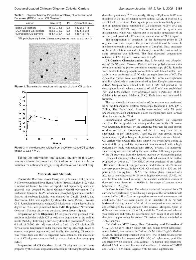
Table 1. Physicochemical Properties of Blank, Fluorescent, andDocetaxel (DCX)-Loaded CS Carriersa
carrier size (nm) PI �-potential (mV)
blank CS carriers 151.2 ( 1.5 0.1 +47.2 ( 1.3DCX-loaded CS carriers 162.3 ( 3.7 0.1 +47.5 ( 3.3fluorescent CS carriers 184.7 ( 3.4 0.1 +38.4 ( 1.6
a PI: polydispersity index. Values are given as mean ( s.d.; n ) 3.
Figure 1. Transmission electron micrograph of blank CS carriers.
Figure 2. In vitro docetaxel release from docetaxel-loaded CS carriers(mean ( s.d.; n ) 3).
Docetaxel-Loaded Chitosan Oligomer Colloidal Carriers Biomacromolecules, Vol. 9, No. 8, 2008 2187

MTT Assay and GI50 Estimation. Cells were plated in amultiwell-96 plate (Iwaki) at 4 × 10³ cells/well, and 24 h later, themedium was changed for the following three treatments: docetaxel,docetaxel-loaded CS carriers, and blank CS carriers. Tetrazolium salt3-(4,5-dimethylthiazol-2-yl)-2,5-diphenyltetrazoliumbromide (MTT, AcrosOrganics) was used for mitochondrial activity evaluation in cell viabilitystudies 24 and 48 h post-treatments. Plates were measured in MicroPlateReader Model 550 (BIO-RAD) and data were processed with Exceland SPSS software. Using absorbance measurements [time zero, (Az),control growth, (C), and test growth in the presence of drug at thevarious concentration levels (Ai)], the percentage growth was calculatedat each of the drug concentration levels.
Growth inhibition of 50% (GI50) was calculated from [(Ai - Az)/(C - Az)] × 100 ) 50, which is the drug concentration resulting in a50% reduction in absorbance in control cells during the drug incubation.
Uptake Studies. Cells were plated in a multiwell-6 plate (Falcon) at5 × 105 cells/well, and 24 h later, the medium was changed for
treatments: fluorescently-labeled CS carriers with fluorescein-derivatizeddihexadecanoylglycerophosphoethanolamine (fluorescein-DHPE, FisherScientific) and fluorescent dye as a control. After 2 h of incubation,cells were washed with acidic phosphate saline buffer (PBS, Sigma),detached, and resuspended in PBS supplemented with 3% (v/v) of FBS.Living cell suspensions were analyzed for green fluorescence by flowcytometry in a FACScan (Becton Dickinson) and fluorescent micros-copy (Leica).
Cell Cycle Analysis. Cell cycle phase distribution was determinedby measuring DNA content by flow cytometry of propidium iodide-stained cells, as described.21 Briefly, whole cell suspensions werewashed in PBS, fixed in 70% ethanol, stained in 50 µg/mL propidiumiodide, 1 mg/mL RNase, 0.1% Triton X-100, and analyzed using theModFit software.
Results and Discussion
The main aim of this project was to evaluate the potential ofCS oligomer carriers of nanometric size, so-called nanocap-sules,14 for the transport of the anticancer drug docetaxel. CSoligomer carriers were proposed because they are supposedlyable to improve the stability of the drug included in their core,as well as promoting a tight interaction with the cancer cellsdue to their CS corona. As a first step, the physicochemical,morphological and encapsulation properties of these systems,as well as the docetaxel in vitro release characteristics, weredetermined. Subsequently, the cell viability assays with breast(MCF-7) and nonsmall lung (A-549) cancer cell lines wereperformed so as to follow the antiproliferative activity of the
Figure 3. Effect on MCF7 and A549 cell proliferation of docetaxel (DCX, dashed line), docetaxel (DCX)-loaded CS carriers (solid line), andblank CS carriers (dotted line) at different incubation times and concentrations. MCF7 after 24 h (A1) and 48 h (A2) and A549 after 24 h (B1)and 48 h (B2). Mean values of four independent experiments. *p < 0.05, DCX-loaded CS carriers vs DCX.
Table 2. Blank CS Carriers, Docetaxel (DCX), and Docetaxel(DCX)-Loaded CS Carriersa
MCF7 A549
24 h 48 h 24 h 48 h
blank CS carriers n.d. n.d. n.d. n.d.DCX 62.5 5.6 36.5 12.8DCX-loaded CS carriers 4.7 2.7 5.3 4.5a GI50 values (growth inhibitory 50, drug concentration resulting in a
50% reduction in absorbance in control cells), expressed in nM, wereestimated as described in Material and Methods. Mean values of fourindependent experiments; n.d.: none of the concentrations tested resultedin a 50% reduction of the absorbance.
2188 Biomacromolecules, Vol. 9, No. 8, 2008 Lozano et al.

encapsulated docetaxel. Additionally, flow cytometry and im-munofluorescence microscopy assays on cell lines pretreatedwith the docetaxel-loaded carriers were carried out to elucidatethe internalization capacity of the systems. Finally, the cellulareffects of CS oligomer carriers in both cell lines wereinvestigated more deeply by cell cycle analysis.
CS Carriers Characterization. CS oligomer carriers wereprepared using the solvent displacement technique previouslyreported.14 The CS coating around the oily nanodroplets wasformed due to the ionic interactions between the negativelycharged tensioactive agent, lecithin, and the positively chargedCS oligomers (MW ) 10 KDa). Blank CS oligomer carriersformed a homogeneous population of a mean particle sizesmaller than 200 nm and a high positive surface charge (Table1). The positive charge was due to the CS oligomer layerdisposed over the hydrophobic core formed by Lecithin andthe oil Miglyol 812.
Docetaxel is a noncharged drug with a very low watersolubility, which makes it an attractive candidate for inclusionin the hydrophobic core of a lipidic system like that of the CSnanocapsules. Furthermore, this reservoir structure allowed anencapsulation efficiency of 72% for docetaxel. Several authorshave also reported high encapsulation efficiencies for docetaxelor paclitaxel in oil containing nanostructures, which is mainlydue to the affinity of these drugs to the core components.11 Asexpected, the encapsulation of docetaxel hardly modified thesize and the charge of blank CS carriers.
The morphological appearance of the systems was observedby transmission electron microscopy. The CS carriers have around shape and a size of less than 200 nm (Figure 1), a resultthat corresponds to that observed by photonic correlationspectroscopy (Table 1).
The results of the in vitro release studies of docetaxel fromCS carriers are presented in Figure 2. The biphasic profile,
Figure 4. Uptake studies of fluorescent CS carriers assessed by flow cytometry in MCF7 and A549 cells. (A) Percentage of stained cells aftera 2 h incubation with fluorescein-DHPE CS carriers, black bars, or free fluorescein-DHPE, white bars. Mean values of three independentexperiments. (B) Fluorescence microscopy image of MCF7 cells treated with fluorescent CS carriers. On the left, the green label correspondsto fluorescent CS carriers loaded with fluorescein-DHPE. On the right, merged imagine of nuclei stained with DAPI (blue) and fluorescein(green). The insert is a magnification of the same image and it shows that the dye was found in vesicles inside the cells.
Docetaxel-Loaded Chitosan Oligomer Colloidal Carriers Biomacromolecules, Vol. 9, No. 8, 2008 2189
typically observed in these types of delivery systems, iscomposed of an initial release phase (50% of drug released in8 h), followed by a second phase characterized by the absenceof drug release. The initial phase could be related to the partitionof the drug between the oily core and the external aqueousrelease medium, whereas the second phase illustrates theimportant affinity of the drug to the oily core. An importantadditional observation from these studies was that the drugdelivered from CS carriers remains stable. These results suggest
that, following in vivo administration, the structures slowlydeliver the encapsulated drug in an active form.
In Vitro Cellular Proliferation and Uptake Studies inHuman Tumor Cell Lines. The results indicate that docetaxel-loaded CS carriers reduced the rate of proliferation of MCF7and A549 human tumor cells with faster kinetics than freedocetaxel. Indeed, at 24 h after treatment, the encapsulateddocetaxel had an effect on cell proliferation that was significantlygreater than that of free docetaxel (Figure 3A1,B1). The GI50
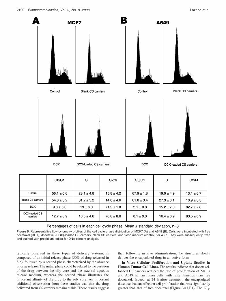
Figure 5. Representative flow cytometry profiles of the cell cycle phase distribution of MCF7 (A) and A549 (B). Cells were incubated with freedocetaxel (DCX), docetaxel (DCX)-loaded CS carriers, blank CS carriers, and fresh medium (control) for 48 h. They were subsequently fixedand stained with propidium iodide for DNA content analysis.
2190 Biomacromolecules, Vol. 9, No. 8, 2008 Lozano et al.

values calculated for docetaxel-loaded carriers (24 h aftertreatment) were predictably lower than those estimated for freedocetaxel both in MCF7 and A549 cell lines (Table 2).
On the other hand, blank CS carriers had no significant effectson the cell growth (Figure 3). Indeed, the differences in viabilityobserved after exposure to this formulation and the nontreatedcells were not statistically significant. These negligible toxicityresults observed for CS carriers, which are consistent with othertoxicity results reported for CS,22,23 could be justified by thelow concentration of CS present in the experimental conditionsof the study.
After 48 h incubation, the effects on cell proliferationobserved for docetaxel-loaded CS carriers and free docetaxel
were similar (Figure 3A2,B2), which suggests that the dif-ferential effect found at 24 h was due to an accelerated uptakeof the encapsulated docetaxel in CS structures. Both docetaxel-loaded CS carriers and free docetaxel had a cytostatic effect,which arrested proliferating cells without noticeable effects oncell viability (data not shown) at any given moment. In theory,the observed antiproliferative effect of docetaxel-loaded CScarriers could be attributed to the uptake of the systems followedby the intracellular delivery of docetaxel. However, the pos-sibility that a certain amount of docetaxel may have beenreleased from the oily reservoir before the uptake took placecannot be ruled out. To provide evidence of the uptake intensityof the CS carriers, a fluorescent dye was encapsulated and its
Figure 6. Confocal microscopy of MCF7 (A) and A549 (B) cells after incubation with free docetaxel (DCX), docetaxel (DCX)-loaded CS carriers,blank CS carriers, and fresh medium (control). Cells were incubated for 48 h with 100 nM of the drug or the equivalent concentration for thecontrols. They were subsequently processed by staining nuclear DNA (DAPI) and R-tubulin microtubules (monoclonal mouse anti-R-tubulinantibody and goat antimouse antibody conjugated with Alexa 488). Micronuclear abnormalities formation and microtubules condensation(arrowhead) are visible in docetaxel-treated cells (DCX and DCX-loaded CS carriers).
Docetaxel-Loaded Chitosan Oligomer Colloidal Carriers Biomacromolecules, Vol. 9, No. 8, 2008 2191

uptake was estimated by flow cytometry. This encapsulatedfluorescent dye was detected inside almost every cell two hours,or possibly sooner, after addition to the cell medium, while thenonencapsulated dye remained mostly excluded (Figure 4).Consequently, these results suggest that the internalization ofthe CS carriers most probably occurs before a significant amountof docetaxel is released from the system.
Fluorescence microscopy showed that the dye localizationinside the cell was in the cytoplasm without staining the nucleus.Moreover, the dye was found in vesicles (Figure 4B), suggestingthat the encapsulated dye had entered the cell via an endocyticpathway. Docetaxel, like most tumor drugs, is not selective oftumors and affects both normal and cancer cells equally.Unfortunately, encapsulation does not make docetaxel moreselective, but it does accelerate its entry into the cell, as wehave shown here. Therefore, tumors rather than normal tissueswill be more exposed to the action of the drug because theyhave a greater blood flow than their normal neighbor tissue.
We postulated that the enhanced antiproliferative effect ofdocetaxel on the tumor cells observed at 24 h post-treatment issubsequently related to rapid uptake by the cells. This improveduptake could be related to a favored interaction of the CS carrierssurface with the cancer cells, which may well be due to the CScoating;24 this was also found for different cell types, such asthose of corneal epithelium.16 Moreover, it has been shown thatthe charge plays a role in passive tumor targeting becausecationic nanoparticles are better at concentrating in the tumorenvironment than similarly designed anionic particles: it wasfound that an increase in the charge content doubled theaccumulation of cationic liposomes in tumor vessels of twodifferent tumor types.25
The next step was to rule out the possibility that the procedureof encapsulation or the mechanism of cellular uptake could affectthe biological activity of docetaxel. Taxanes have been shownto target tubulin causing stabilization of microtubules, whichresults in cell-cycle arrest and apoptosis.26 As a result, attentionwas given to the well-studied effects of docetaxel on tumor cells,such as cell cycle distribution and R-tubulin distribution andaggregation. Flow cytometry analysis of the effects of docetaxelon the cell cycle of the two types of cancer cells showed thatboth docetaxel-loaded CS carriers and free docetaxel were ableto accumulate cells in the G2/M phase (Figure 5), whichsuggests an arrest in the cell cycle, which is consistent with thedata obtained by MTT analysis.
In addition to this, confocal microscopy showed that bothcontrol (without treatment) and blank CS carrier-treated cellshad a more or less homogeneous microtubule network, whichwas clearly present throughout the cell. In contrast, docetaxel-loaded CS carriers and free docetaxel-treated cells presented apattern of unevenly distributed staining with clear aggregationand anchorage of the microtubules into thicker fibers (depoly-merisation was inhibited by docetaxel, therefore, causingaggregation; Figure 6), which again suggests that docetaxel-loaded carriers and free docetaxel cause cytostasis by the samemicrotubule-interfering mechanisms.
Overall, these findings indicate that docetaxel-loaded CScarriers exhibit a higher antiproliferative effect on the twostudied cancer cell lines when compared with that of the freedrug. This took place during the first hours of the study andwas probably related to an early uptake of the systems by thecancer cells. In addition to this, it was also demonstrated thatthe mechanism of action of docetaxel included in the colloidalcarriers was unchanged. Future work on CS carriers will aim
at determining the efficacy of docetaxel entrapped in thesesystems in appropriate tumor models.
Conclusion
In conclusion, it can be stated that CS oligomer carriers area very efficient system for the delivery of docetaxel into humancancer cells. More specifically, CS carriers favor a fast andefficient uptake of the encapsulated drug into tumor cells. Itcan also be stated that encapsulated docetaxel is maintained fullyactive and its mechanism of action unaltered. Consequently, theefficacy of CS carriers for intracellular delivery of docetaxelcombined with their low toxicity points to the potential of thissystem for cancer therapy. Further in vivo studies need to beperformed in order to fully assess the potential of CS carriersin the development of new anticancer agents.
Acknowledgment. This work has been financed by theSpanish Ministry of Science and Technology (SAF 2004-08319-CO2-01) and Consolider Nanobiomed (CSD 2006-00012). Thefirst author acknowledges the fellowship received from theSpanish Government (AP2005-1701). A.V. is an investigatorof the “Ramon y Cajal” Program financed by the Ministry ofScience and Education, Spain. The authors also express theirgratitude to Ramon Novoa Carballal for CS oligomer SECmeasurements. We thank Peter Rees for the English editing.
References and Notes(1) Rowinsky, R. K.; Donehower, R. C. N. Engl. J. Med. 1995, 332, 1004–
1014.(2) Trudeau, M. E.; Eisenhauer, E. A.; Higgins, B. P.; Letendre, F.; Lofters,
W. S.; Norris, B. D.; Vandenberg, T. A.; Delorme, F.; Muldal, A. M.J. Clin. Oncol. 1996, 14, 422–428.
(3) Piccart, M. J.; Gore, M.; Huinink, W. T. B.; Vanoosterom, A.; Verweij,J.; Wanders, J.; Franklin, H.; Bayssas, M.; Kaye, S. J. Natl. CancerInst. 1995, 87, 676–681.
(4) Jones, S. E.; Erban, J.; Overmoyer, B.; Budd, G. T.; Hutchins, L.;Lower, E.; Laufman, L.; Sundaram, S.; Urba, W. J.; Pritchard, K. I.;Mennel, R.; Richards, D.; Olsen, S.; Meyers, M. L.; Ravdin, P. M.J. Clin. Oncol. 2005, 23, 5542–5551.
(5) van Zuylen, L.; Verweij, J.; Sparreboom, A. InVest. New Drugs 2001,19, 125–141.
(6) Engels, F. K.; Mathot, R. A. A.; Verweij, J. Anti-Cancer Drugs 2007,18, 95–103.
(7) Heath, J. R.; Davis, M. E. Annu. ReV. Med. 2008, 59, 251–265.(8) Peer, D.; Karp, J. M.; Hong, S.; Farokhzad, O. C.; Margalit, R.; Langer,
R. Nat. Nanotechnol. 2007, 2, 751–760.(9) Brigger, I.; Dubernet, C.; Couvreur, P. AdV. Drug DeliVery ReV. 2002,
54, 631–651.(10) Green, M. R.; Manikhas, G. M.; Orlov, S.; Afanasyev, B.; Makhson,
A. M.; Bhar, P.; Hawkins, M. J. Ann. Oncol. 2006, 17, 1263–1268.(11) Khalid, M. N.; Simard, P.; Hoarau, D.; Dragomir, A.; Leroux, J. C.
Pharm. Res. 2007, 23, 752–758.(12) Jakate, A. S.; Einhaus, C. M.; DeAnglis, A. P.; Retzinger, G. S.; Desai,
P. B. Cancer Res. 2003, 63, 7314–7320.(13) Bae, K. H.; Lee, Y.; Park, T. G. Biomacromolecules 2007, 8, 650–
656.(14) Prego, C.; Fabre, M.; Torres, D.; Alonso, M. J. Pharm. Res. 2006,
23, 549–556.(15) Prego, C.; Torres, D.; Alonso, M. J. J. Drug DeliVery Sci. Tech. 2006,
16, 331–337.(16) De Campos, A. M.; Sanchez, A.; Gref, R.; Calvo, P.; Alonso, M. J.
Eur. J. Pharm. Sci. 2003, 20, 73–81.(17) Mao, S. R.; Shuai, X. T.; Unger, F.; Simon, M.; Bi, D. Z.; Kissel, T.
Int. J. Pharm. 2004, 281, 45–54.(18) Prashanth, K. V. H.; Tharanathan, R. N. Biochim. Biophys. Acta 2005,
1722, 22–29.(19) Janes, K. A.; Alonso, M. J. J. Appl. Polym. Sci. 2003, 88, 2769–2776.(20) Lee, S. H.; Yoo, S. D.; Lee, K. H. J. Chromatogr., B: Biomed. Sci.
Appl. 1999, 724, 357–363.(21) Vidal, A.; Millard, S. S.; Miller, J. P.; Koff, A. J. Biol. Chem. 2002,
277, 16433–16440.
2192 Biomacromolecules, Vol. 9, No. 8, 2008 Lozano et al.

(22) Prego, C.; Torres, D.; Alonso, M. J. J. Nanosci. Nanotechnol. 2006,6, 1–8.
(23) Garcia-Fuentes, M.; Prego, C.; Torres, D.; Alonso, M. J. Eur. J. Pharm.Sci 2005, 25, 133–143.
(24) Park, J. H.; Kwon, S.; Lee, M.; Chung, H.; Kim, J. H.; Kim, Y. S.;Park, R. W.; Kim, I. S.; Seo, S. B.; Kwon, I. C.; Jeong, S. Y.Biomaterials 2002, 27, 119–126.
(25) Campbell, R. B.; Fukumura, D.; Brown, E. B.; Mazzola, L. M.; Izumi,Y.; Jain, R. K.; Torchilin, V. P.; Munn, L. L. Cancer Res. 2002, 62,6831–6836.
(26) Liang, H. F.; Chen, S. C.; Chen, M. C.; Lee, P. W.; Chen, C. T.;Sung, H. W. Bioconjugate Chem. 2006, 17, 291–299.
BM800298U
Docetaxel-Loaded Chitosan Oligomer Colloidal Carriers Biomacromolecules, Vol. 9, No. 8, 2008 2193
Copyright © 2022 FDOKUMEN