Structure and Stability of Higher-Order Human Telomeric Quadruplexes
Upload
independentCategory
view
3download
0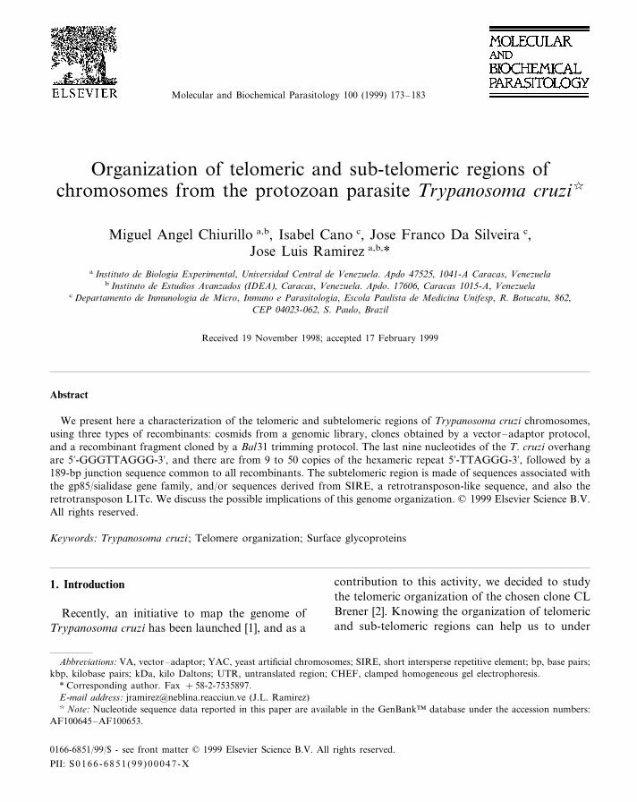
Molecular and Biochemical Parasitology 100 (1999) 173–183
Organization of telomeric and sub-telomeric regions ofchromosomes from the protozoan parasite Trypanosoma cruzi�
Miguel Angel Chiurillo a,b, Isabel Cano c, Jose Franco Da Silveira c,Jose Luis Ramirez a,b,*
a Instituto de Biologia Experimental, Uni6ersidad Central de Venezuela. Apdo 47525, 1041-A Caracas, Venezuelab Instituto de Estudios A6anzados (IDEA), Caracas, Venezuela. Apdo. 17606, Caracas 1015-A, Venezuela
c Departamento de Inmunologia de Micro, Inmuno e Parasitologia, Escola Paulista de Medicina Unifesp, R. Botucatu, 862,CEP 04023-062, S. Paulo, Brazil
Received 19 November 1998; accepted 17 February 1999
Abstract
We present here a characterization of the telomeric and subtelomeric regions of Trypanosoma cruzi chromosomes,using three types of recombinants: cosmids from a genomic library, clones obtained by a vector–adaptor protocol,and a recombinant fragment cloned by a Bal31 trimming protocol. The last nine nucleotides of the T. cruzi overhangare 5%-GGGTTAGGG-3%, and there are from 9 to 50 copies of the hexameric repeat 5%-TTAGGG-3%, followed by a189-bp junction sequence common to all recombinants. The subtelomeric region is made of sequences associated withthe gp85/sialidase gene family, and/or sequences derived from SIRE, a retrotransposon-like sequence, and also theretrotransposon L1Tc. We discuss the possible implications of this genome organization. © 1999 Elsevier Science B.V.All rights reserved.
Keywords: Trypanosoma cruzi ; Telomere organization; Surface glycoproteins
1. Introduction
Recently, an initiative to map the genome ofTrypanosoma cruzi has been launched [1], and as a
contribution to this activity, we decided to studythe telomeric organization of the chosen clone CLBrener [2]. Knowing the organization of telomericand sub-telomeric regions can help us to under
Abbre6iations: VA, vector–adaptor; YAC, yeast artificial chromosomes; SIRE, short intersperse repetitive element; bp, base pairs;kbp, kilobase pairs; kDa, kilo Daltons; UTR, untranslated region; CHEF, clamped homogeneous gel electrophoresis.
* Corresponding author. Fax +58-2-7535897.E-mail address: [email protected] (J.L. Ramirez)� Note: Nucleotide sequence data reported in this paper are available in the GenBank™ database under the accession numbers:
AF100645–AF100653.
0166-6851/99/$ - see front matter © 1999 Elsevier Science B.V. All rights reserved.
PII: S 0166 -6851 (99 )00047 -X

M.A. Chiurillo et al. / Molecular and Biochemical Parasitology 100 (1999) 173–183174
stand mechanisms of gene expression [3], cell cycleregulation [4], and gene recombination [5] occur-ring at those regions. The telomeric and sub-telom-eric regions of blood protozoan parasites such asTrypanosoma brucei [6] and Plasmodium falciparum[7] are specialized in the expression of specificvariant-surface genes that allow the parasite toevade the host immune system. T. cruzi, an obligateintracellular parasite, co-expresses a large numberof surface antigens that seem to prevent an ade-quate immune response [8,9]. Among these anti-gens there is a large multigene family designated asT. cruzi gp85/sialidase superfamily [10,11], whichencodes trypomastigote stage-specific surface gly-coproteins; a subset of these genes are linked to thetelomeres [12–14].
Cloning of telomeric and subtelomeric regions isdifficult; the presence of a single strand overhangand the limited numbers of restriction sites withintheir repetitive sequences cause under-representa-tion or absence of these sequences in most genomiclibraries. Different protocols for cloning telomericsequences have been used, such as flush-end liga-tion after trimming the overhang [15,16], Half-YAC chromosomal complementation [17],amplification protocols combining a specific hex-americ primer, and degenerated oligonucleotides ora vectorette [18,19]. In these protocols the originaltelomeric terminus is lost or modified, and in theamplification protocols recombinants are smalland care must be taken that hexamer repeats canbe found in inverted orientation ([20,21] and thiswork). In the work described in this paper, we havecloned and characterized the T. cruzi telomeric andsubtelomeric regions using some of the protocolscited above and a new vector–adaptor protocoldeveloped by Beck [22]. All T. cruzi telomericrecombinants contain sequences homologous tothe gp85 surface-antigen. We discuss the possiblerole of these genes at the telomere.
2. Materials and methods
2.1. Selection of cosmids containing telomericsequences
The genomic library used in this work was
constructed by Hanke [23] using as cloning vectorthe cosmid Lawrist-7. The inserts were prepared byMboI digestion and physical shearing of DNAfrom reference strain T. cruzi CL Brener [2]. Thelibrary has been organized in filters containing36 864 individual recombinant clones. The filters,kindly donated by Dr J. Hoheisel, were screenedwith the telomeric probe (CCCTAA)3 labeled with[g-32P]ATP using T4 polynucleotide kinase. Afteran additional screening and restriction analysis, weconfirmed the presence of telomeric sequences infive cosmids, whose names and coordinates (inparenthesis) in the filter array were Cosmid 3(37F20), Cosmid 6 (24B7), Cosmid 7 (36G16),Cosmid 8, (35P16) and Cosmid 9 (2M20). Cosmid7 was omitted from further analysis because itsuffered a deletion of the original insert.
Cosmid DNA was isolated using a lithium chlo-ride protocol [24], and subcloned for further map-ping and sequencing analysis. Subcloning was donein pBluescript II KS+and pUC19 vectors, and theDNA sequencing was done by automatic fluores-cent dideoxy termination by Ana-Gen Technolo-gies.
2.2. Cloning of T. cruzi telomeres by the6ector–adaptor protocol
We used the same set of adaptors designed byBeck [22] for the VA protocol (Insert Fig. 1); alloligonucleotides were purchased from OperonTechnologies Inc. The original protocol wasmodified as follows: pBluescript II KS+ vectordigested with HindIII and PstI was ligated to eachone of the six individual adaptors (Fig. 1 step 2).Then, we made two pools of three vector–adaptorseach, and incubated them with high molecularweight T. cruzi DNA at a ratio of 100:1 (vector–adaptors:DNA) (Fig. 1 step 3). After ligation in avolume of 30 ml, T4 DNA ligase was heat inacti-vated at 65°C for 15 min, then the DNA wasdigested with PstI in 100-ml volumereaction (Fig. 1 step 4). After inactivating therestriction enzyme by incubation at 65°C for 15min, the DNA was re-ligated in a large reactionvolume (500 ml) (Fig. 1 step 5). DNA from bothligations was extracted with chloroform-phenol

M.A. Chiurillo et al. / Molecular and Biochemical Parasitology 100 (1999) 173–183 175
(1:1), pooled and precipitated with ethanolovernight at −20°C. The DNA was resuspendedin water and used to electroporate E. coliDH10B electrocompetent cells (Gibco-BRL).
2.3. Screening for 6ector–adaptor telomericrecombinants
White colonies were selected and grown in two96-well microtiter plates filled with LB Ampicillin(100 mg ml−1) medium plus 10% glycerol. Theovernight grown cells were picked with a replica-tor from Nunc® and stamped on Biodyne® nylonfilters. The filters were put on top of LB-agarplates with ampicillin (100 mg ml−1) for 8 h.
Then the filters were laid first on a filter papersoaked in alkaline solution (1.5 M NaCl; 0.5 NNaOH) for 7 min, and second, on another filterpaper embedded in a neutralizing solution (1.5M NaCl; 0.5 M Tris–HCl pH 7.2; 1 mMEDTA) for 7 min. Finally, the filters werewashed with 2×SSC (0.3 M NaCl; 0.03 Msodium citrate) for 5 min, air-dried and bakedfor 2 h at 80°C.
The filters were hybridized with a telomericprobe (CCCTAA)3 labeled with [g-32P]ATP usingT4 polynuclotide kinase. Negative and positivehybridization controls were included. Positiveclones were grown, and plasmid isolation wasdone as for cosmids (see above).
Fig. 1. Vector–adaptor cloning scheme: (1) The vector is double digested with HindIII and PstI; (2) adaptors (with a HindIII site)are ligated to linearized vector in two separated pools of three adaptors each; (3) Vector–adaptors are ligated to intact T. cruziDNA; (4) The ligation product is digested with PstI; (5) The vector–adaptor recombinants are circularized by ligation. Theremaining gap is repaired by the bacterial host. In the insert we show the six adaptors used; adaptor 4 was present in all T. cruzivector–adaptor recombinants.

M.A. Chiurillo et al. / Molecular and Biochemical Parasitology 100 (1999) 173–183176
2.4. Bal31 cloning protocol
We constructed a telomeric-subtelomeric li-brary using the vector pUC18 and T. cruzi ge-nomic fragments obtained by partial digestionwith exonuclease Bal31 [16]. High molecularweight genomic DNA was digested with differentconcentrations of Bal31. After filling of the endswith DNA polymerase I (Klenow fragment), ge-nomic DNA was ligated to pUC18 plasmid lin-earized by SmaI digestion. Then we made anEcoRI digestion, and recombinant moleculeswere circularized by ligation with T4 DNA lig-ase. The ligation mixture was used to transformE. coli DH 5a cells. A 1235-bp recombinant (Tel-CL9) was further characterized.
2.5. CHEF gels
T. cruzi chromosomes were prepared inagarose blocks containing 107 cells ml−1 as de-scribed [25]. Chromosome–sized DNA was sepa-rated in a Biorad CHEF-DR® III System. Blockswere set in 1% DNA Typing Grade Agarose(Gibco-BRL) gel in 0.5×TBE (0.089 M Tris–borate, 0.089 M boric acid and 0.002 M EDTA)buffer. The separation was done in two cycles oflinear gradient of switching times. The first cyclewas from 60 to 120 s for 24 h, and the secondfrom 120 to 180 sec for 6 h. The voltage gradientwas 6 V cm−1 at 14°C. Gels were stained withethidium bromide and transferred to nylon filters(Hybond N®, Amersham).
2.6. Two dimensional (2D) CHEF gels
Blocks containing chromosomal size DNAfrom T. cruzi were placed in 1% low-gellingagarose (Gibco-BRL) gels. Samples were run inthe first direction as described above, then a 4-mm agarose strip containing the resolved chro-mosomal bands was washed with TE (10 mMTris–HCl pH 7.6, 1 mM EDTA) bufferovernight at 4°C. The strip was equilibrated inNotI restriction buffer for 2 h at 4°C. Next, weadded 200–300 U of NotI enzyme and incubatedat 37°C for 6 h. The strip was placed in a wellmade in a 1% agarose-0.5×TBE gel, and run in
a linear gradient of switching times from 30 to70 s, 6 V cm−1 of voltage gradient, for 18 h at14°C. DNA fragments were transferred to nylonfilters by capillary action and hybridized withdifferent probes.
2.7. Probes
Gel-isolated DNA fragments were labeled byrandom hexamer priming incorporating [a-32P]dCTP, 3000 Ci mmol−1, using MegaprimeDNA labeling system® (Amersham). Oligonucle-otides were labeled by T4 polynucleotide kinaseusing [g-32]PdCTP, 3000 Ci mmol−1 (Amer-sham).
2.8. Hybridization conditions
Filters were incubated with the probes in solu-tion containing 0.5 M Na-phosphate pH 7.2, 7%SDS, 1 mM EDTA, 0.1 mg ml−1 E. coli tRNAat 65°C for 18 h. After hybridization, filters werewashed twice at room temperature in 40 mMNa-phosphate, pH 7.2, 0.1% SDS for 15 mineach, and twice with the same buffer at 65°C for15 min each. The hybridization and washingtemperature for telomeric probe (CCCTAA)3 was58°C. Stripping was done by incubating thefilters twice at 90°C in 5 mM Na-phosphate, pH7.2, 0.1% SDS for 30 min.
3. Results
3.1. Cosmids analysis and definition of telomericand subtelomeric regions
Study of cosmids restriction maps and se-quences is summarized in Fig. 2. All cosmidsexhibited 9–50 copies of the hexameric repeat5%-TTAGGG-3% at one end (black blocks Figs. 2and 4), followed by a 189-bp junction sequence(Fig. 4A, B, blocks with vertical bars). The sub-telomeric region carried sequences homologousto gp85/sialidase gene family (Fig. 2 blocks withvertical bars).

M.A. Chiurillo et al. / Molecular and Biochemical Parasitology 100 (1999) 173–183 177
Fig. 2. Restriction maps of cosmids containing T. cruzi telomeric and subtelomeric regions. Cosmid designations are followed by thesize of their inserts in parenthesis. The coding sense of all genes is indicated with arrows. Subclones sequenced are marked with dots,and the numbers indicate their identification. The location of probes pGP85-11 and pDGF-1-611 is shown as open squares. Scalein bp is shown at the bottom.
The insert in Cosmid 8 had at one end 15hexamer repeats (Fig. 2 black blocks) followed bythe 189-bp junction, then we found sequencessharing homology with different regions of thegp85 gene family. From telomere to centromere(Fig. 4A) these sequences were: a region ho-mologous to the 5%UTR of gene gp85 (cloneTt34c1, GenBank M64836) [26], a gene spacersequence found between gp85 pseudogene and theflagellar surface antigen (GenBank M91469) [27],the 3%UTR of gen gp85/Tt34c1, and a sequencehomologous to another member of gene gp85 gene
family (clone SA85-1.2, GenBank M62736) [28].The sequence homologous to gene gp85/SA85-1.2(GenBank AF100647) encoded the C-terminal do-main of gp85 antigen including motif MTVKNVX-LYNR homologous to the general transialidasemotif VTVTNVFLYNR [29]. The coding sequencewas oriented from centromere to telomere andpresented several stop codons. Probe pGP85-811,a 1-kbp EcoRI–Sau3AI fragment containing partof the coding region (Fig. 2, white block), wassubcloned from this cosmid to be used as probe inhybridization experiments described below.

M.A. Chiurillo et al. / Molecular and Biochemical Parasitology 100 (1999) 173–183178
For cosmid 6 we obtained a PstI-subclone con-taining 25 hexamer repeats, the 189-bp junction,and the 5%UTR sequence homologous to gp85/Tt34c1, which was interrupted by a PstI cloningsite. The overall telomere organization was identi-cal to cosmid 8 and VA recombinants group I (seebelow). Further in, at 13-kbp from the copy ofgene gp85, we found a sequence homologous toT. cruzi disperse gene family 1 protein (DGF-1)gene (GenBank M90534) (Fig. 2 block with diag-onal bars). This protein, although not included inthe gp85/sialidase family, is presumed to be lo-cated at the cell surface [30]. From this sequencewe subcloned a 900-bp XhoI fragment within thecoding region (probe pDGF-1-611) (Fig. 2), to beused in hybridization (not shown) and sequencingexperiments.
Cosmid 9 had 20 hexamer repeats, and dis-played the same organization of cosmids 8 and 6.In this case the sequences around the telomericgp85 were subcloned with Sau3AI and PstI. TheSau 3AI subclones and the primer trans (see Fig.4) allowed us to establish the continuity between3%UTR and the gp85 coding next to it. At 14.5kbp from the terminus, we found another se-quence homologous to gp85/SA85-1.2, presentingthe trans-sialidase motif DGGANGDAGSAY-GRVLLPLLLLLGLWGFATA (GenBankM62736), and a sequence homologous to 3%UTRof sequence Tc13 (GenBank M92046) (Fig. 2 dot-ted block) that encodes a chronic stage specificsurface antigen [31].
Due to poor DNA sequence readings at oneend of cosmid 3, we could not determine thenumber of hexamers, but close to these repeatsonce again there was a sequence homologous togp/SA85-1.2 (Fig. 2 blocks with vertical bars).Hybridization experiments using as probe pGP85-811 revealed two additional sequences ho-mologous to gp85/SA85-1.2 at 5.5 and 20 kbpfrom the first copy (Fig. 2).
Although all gp85 related copies found in thecosmids were homologous to gene gp85/SA85-1.2,the homology among them was around 70%. Re-striction maps and sequence analysis indicatedthat the coding sense of all gp85 genes was ori-ented from centromere to telomere.
3.2. 2D-CHEF gel analysis
To determine how frequent pGP85-811 se-quences were associated with telomeric sequences,we did Southern analysis of T. cruzi two-dimen-sional CHEF gels. We first hybridized the filterswith the telomeric probe (CCCTAA)3, and sec-ond, with pGP85-811. A strip run in parallel wasalso transferred and hybridized with the telomericprobe (Fig. 3A). As shown in Fig. 3B, there aremany fragments recognized by the telomericprobe. There is an average of four telomeric sig-nals per band, suggesting either that homologouschromosomes are polymorphic for NotI, or thatwe are in the presence of non-homologous over-lapping chromosomes. We resolved 80 distincttelomeric fragments, many of which showed
Fig. 3. 2D-dimensional CHEF gels of T. cruzi chromosomalbands. (A) One-dimensional chromosomal blot hybridizedwith the telomeric probe (CCCTAA)3 (MW sizes indicatedabove). (B) 2D blot hybridized with the telomeric probe(CCC-TAA)3. (C) 2D blot hybridized with probe pGP85-811. Arrowspoint to some of the fragments coinciding in position in B andC. MW distribution is indicated at the left margin of bothfigures.

M.A. Chiurillo et al. / Molecular and Biochemical Parasitology 100 (1999) 173–183 179
higher hybridization signal intensities. Othertelomeric fragments were clustered at the com-pression zone.
When pGP85-811 was used as probe (Fig. 3C),we observed at least 26 fragments coinciding withthe telomeric signals, within a range of 8–180kbp. Since the probe hybridized with just one ofthe two possible telomeric fragments of a chromo-somal band, we assumed that the distribution ofthese sequences was asymmetric. The hybridiza-tion signal for chromosomal band IX [1], as in thecase with the telomeric probe, was very strong;this suggests multiple copies for one chromosome(aneuploidy).
3.3. Analysis of recombinants obtained by the6ector–adaptor protocol
We selected 190 white colonies and screenedthem with oligonucleotide (CCCTAA)3 labeledwith [g-32P]ATP. Out of the 30 positive colonies,we randomly selected ten recombinants to be se-quenced, confirming in all of them the presence ofthe adaptor and hexamers. This represents a15.8% cloning efficiency, more than twice thevalue obtained for L. dono6ani using the originalVA protocol [1] 22 and Chiurillo et al., unpub-lished. The recombinant clones had the sameadaptor ending in 5%-CCCTAACCC-3%, thus T.cruzi terminus ends in the sequence 5%-GGGT-TAGGG-3% (insert in Fig. 1, and Fig. 4A). Wecould not determine the length of the overhangand there is a possibility that it could be shorterthan nine nucleotides, in which case the ligationcan occur in the opposite strand to that indicatedin Fig. 1. However, in previous experiments wehave found that no ligation of the adaptor occursand no telomeric recombinants were obtained un-less the upper oligonucleotide in the adaptor isphosphorylated, indicating that the overhang isnine nucleotides or longer.
All VA recombinants had from 9 to 50 hexam-eric repeats, followed from telomere to cen-tromere by: the 189 bp consensus sequence foundin cosmids, a sequence homologous to the 5%UTRof gene gp85/Tt34c1 (GenBank M64836), and asequence homologous to the intergenic region be-tween a gp85/pseudogene and the flagellar surface
antigen gene (GenBank M91469) [27] in that or-der. According to the sequences following thisspacer, we divided the VA recombinants in twogroups. The first group (Fig. 4BI) (four recombi-nants) had a sequence homologous to 3%UTR ofgene SA85-1.2 (GenBank M64836), interruptedby the PstI cloning site (see Fig. 2). The secondgroup (six recombinants) had the SIRE element(GenBank Y08881), a middle repetitive retro-transposon-like sequence [32], and the SIRE asso-ciated sequence SZ23 (GenBank AF036147)which contained the PstI cloning site (Fig. 4BII);both sequences were oriented for centromere to-wards telomere. In recombinant VATc22 we alsofound a group of more internally located hexam-ers with inverted orientation
3.4. Analysis of a recombinant obtained by Bal31digestion
Sequence analysis of the selected clone pTel-CL9 showed (forward primer) 80% homologywith sequences found in the 3% UTR of geneTSA-1 (GenBank M58466) [14]. Gene gp85/TSA-1, a member of the gp85 gene family, encodes a T.cruzi stage specific surface glycoprotein of 85kDa. The members of this family that are local-ized at the telomeres are characterized by theirinability to be cloned by conventional methodsand also sensitivity to Bal31 digestion [13,14].
Clone pTel-CL9 presented features of beingtelomeric or subtelomeric, such as Bal31 sensitiv-ity and also hybridization with most T. cruzichromosomal bands (not shown). The blast searchwith the sequence read with the reverse primerhad a match with the T. cruzi non-LTR retro-transposon LcT1 (EMBL X83098) [33].
3.5. Sequence alignment of telomeric andsubtelomeric recombinants
Sequence comparison among all recombinantsrevealed many sequences in common. Fig. 4Ashows the sequence homology match between VArecombinants group 1 and cosmids. In Fig. 4B wepresent a scheme of the organization of VA re-combinants group 1 and 2.

M.A
.C
hiurilloet
al./M
olecularand
Biochem
icalP
arasitology100
(1999)173
–183
180
Fig. 4. (A) Sequence alignment of vector–adaptor recombinants and cosmids. Primer trans allowed us to establish the continuity between vector–adaptor clones andcosmids. The delineated sequences are represented as blocks in B. (B) I. telomere organization of cosmids and vector–adaptor recombinats group 1; II. organizationof VA recombinants group 2. The identity of each box is stated in the figure.

M.A. Chiurillo et al. / Molecular and Biochemical Parasitology 100 (1999) 173–183 181
4. Discussion
The T. cruzi telomeric and subtelomeric se-quences here analyzed have unique features;firstly, they seem to be simple, made of a round ofhexameric repeats, followed by a 189-bp junction,and non-repeated sequences homologous to genegp85/SA85-1.2, the SIRE element, and also theSIRE associated sequence SZ23. Different fromother organisms [34], there are no highly repeatedsequences in the sub-telomeric regions of T. cruzi,and the 189-bp sequence can conveniently be usedas a reference point in mapping studies.
SIRE elements have been implicated in duplica-tion, transposition, and recombination of T. cruziP2 ribosomal gene [32]. T. cruzi CL Brenergenome has nearly 2000 copies of SIRE elementsassociated to the 5% and/or 3%UTR of many genes[32].
In VA recombinants group 1, although thesequences were interrupted by a PstI site withinthe 3%UTR, we could establish a continuity withsequences present in the cosmids (Fig. 4); allcosmids analyzed had telomeres of the first group.
It is likely that group 2 has gp85-associatedsequences after the PstI site within SZ23. In thisregard, the gene organization in this group coin-cides with that of vp85.1 (GenBank AF051695and AF051696), a gene of the gp85/sialidase su-perfamily that has been associated with the recov-ery of virulence in an attenuated T. cruzi CLBrener strain [35]. In the two groups, it looks as ifa gp85 gene copy has been truncated at the telom-ere leaving a ‘molecular scar’ (the intergene se-quence and a 5%UTR next to the junction) of arecombination or transposition event (Fig. 4).
The sense of the genes in cosmids and VArecombinants agreed with the observation madein T. cruzi [36] and L. major (GenBankAE001274) in that departing from a central chro-mosomal region, the sense of genes runs in oppo-site directions towards the telomeres. The Bal31recombinant, in addition to the gp85 homologousgene, harbored a sequence homologous to thenon-LTR retrotransposon L1Tc [33]. This recom-binant must represent the subtelomeric region of achromosome not present in the VA recombinantshere examined. L1Tc can be found in many parts
of the genome and it has been associated withgene mobility [33].
Surface glycoprotein related genes are develop-mentally regulated, and those related to gp85 areco-expressed in the blood forms of the vertebratehost stage [10,12]. The repertoire of gp85-relatedgenes is large; gross calculations place the figurein the hundreds of copies [8,12]. The cDNAclones obtained from normalized libraries revealthat 12.5% of all new recombinants are related tothe trans-sialidase gene family or the mucin genesuperfamily [37].
Recently, Kahn and Wleklinski [8] have shownthat CD4+cells, when primed with a mixture ofsequence variants of a single cloned epitope ofgp85, fail to proliferate or express IFN-g afterbeing challenged by the original epitope. If thesefindings can be demonstrated in vivo, they indi-cate that the generation of variability in gp85 Tcell epitopes is important for the survival of theparasite.
A comparative analysis of different gp85 se-quences (not shown) revealed changes that can beexplained by point mutations, but also there is anassorting of entire blocks of sequences that canonly be explained by gene recombination [28].However, considering that there are many clustersof this family spread throughout the genome,recombination of genes internally located cancause chromosomal rearrangements, an idea notsupported by the stability shown by T. cruzi link-age groups [38]. On the other hand, recombina-tion of genes located at the telomeres can occurwithout seriously affecting the overall gene order.This type of exchange has been documented inPlasmodium falciparum [7], Giardia lamblia [39],and T. brucei [40] but not in T. cruzi. Our resultssuggest a preferred location of gp85 related genesand sequences at T. cruzi telomeres and there is apossibility that new gp85 variants can continu-ously be arising by duplication, mutation, andrecombination of copies that have been trans-posed to the telomeres.
As mentioned above, Trypanosoma brucei, ablood parasite of the same genus, escapes theimmune system by varying surface-antigens [41];for the expression of a new gene variant this hasto be located in a telomeric expression cassette.

M.A. Chiurillo et al. / Molecular and Biochemical Parasitology 100 (1999) 173–183182
Perhaps at the base of the two mechanisms thereare common features such as gene transpositionto the telomeres and generation of new genevariants.
Although there is a possibility that gp85 se-quences may play a mere structural role as sub-telomeres, there are evidences that some of thesetelomeric copies are expressed [12–14,42].
Finally, we found that the last nine nucleotidesof T. cruzi telomeric terminus is 5%-GGGT-TAGGG-3%, this sequence is identical to the onefound by Beck [22] in T. brucei, but differs fromthe termini of L. dono6ani and L. major that endin 5%-GGTTAGGGT-3% Chiurillo, unpublished.Thus, the sequence at the terminus is genus spe-cific, supporting the view that in vivo mechanismsfor generating the overhang and preserving thetelomere are very precise and specific.
Acknowledgements
To Dr Joerg Hoheisel for providing us with thefilters of the T. cruzi CL Brener cosmid libraryand the bacterial cultures containing the telomericrecombinants, Dr Bianca Zingales for the CLBrener strain, Mr. Will Sandoval for graph designand Ian McClure for revising the English. Thiswork received financial support from CONICIT-Venezuela grant S1-95000524 and UNDP/WorldBank/WHO Special Programme for Research andTraining in Tropical Diseases (TDR) grant ID970416, CYTED (Ibero American Project of Bio-technology, Spain) to JLR and FAPESP, SP andPronex (FINEP 7.6.97.1041.00), Brasil to JFDS.
References
[1] Cano MI, Gruber A, Vazquez M, et al. Molecular kary-otype of clone CL Brener chosen for the Trypanosomacruzi genome project. Mol Biochem Parasitol1995;71:273–8.
[2] Zingales B, Pereira MES, Oliveira RP, et al. Trypanosomacruzi genome project: biological characteristics and molec-ular typing of clone CL Brener. Acta Tropica1997;68:159–73.
[3] Gottschling DE, Aparicio OM, Billington BL, ZakianVA. Position effect at S. cere6isiae telomeres: reversiblerepression of. polII transcription. Cell 1990;63:751–62.
[4] Bodnar AG, Ouellette M, Frolkis M, et al. Extension oflife-span by introduction of telomerase into normal hu-man cells. Science 1998;279:349–52.
[5] Dunn B, Szauter P, Pardue ML, Szostak JW. Transfer ofyeast telomeres to linear plasmids by recombination. Cell1984;39:191–201.
[6] Hoijmakers JHJ, Frasch ACC, Bernards A, Borst P,Cross GAM. Novel expression-linked copies of the genesfor variant surface antigens in trypanosomes. Nature1980;284:78–80.
[7] Hernandez-Rivas R, Mattei D, Sterkers Y, Peterson DS,Wellems TE, Scherf A. Expressed var genes are found inPlasmodium falciparum subtelomeric regions. Mol CellBiol 1997;17:604–11.
[8] Kahn SJ, Wlekinski M. The surface glycoproteins ofTrypanosoma cruzi encode a superfamily of variant T cellepitopes. J Immunol 1997;159:4444–51.
[9] Santori FR, Paranhos-Baccala GS, Franco da Silveira J,Ruiz RC, Yoshida N. A recombinant protein based onthe Trypanosoma cruzi metacyclic trypomastigotes 82-kilodalton antigen that induces an effective immune re-sponse to acute infection. Infect Immun 1996;64:1093–9.
[10] Colli W. Trans-sialidase: a unique enzyme activity discov-ered in the protozoan Trypanosoma cruzi. FASEB J1993;7:1257–64.
[11] Schenkman S, Eichinger D, Pereira MEA, NussenzweigV. Structural and functional properties of TrypanosomaTrans-sialidase. Ann Rev Microbiol 1994;40:499–521.
[12] Kahn S, Van Voorhis WC, Eisen H. The major 85-kDasurface antigen of the mammalian form of Trypanosomacruzi is encoded by a large heterogeneous family of simul-taneously expressed genes. J Exp Med 1990;172:589–97.
[13] Peterson DS, Fouts DL, Manning JE. The 85-kd surfaceantigen gene of Trypanosoma cruzi is telomeric and amember of a multigene family. EMBO J 1989;8:3911–6.
[14] Fouts DL, Ruef BJ, Ridley PT, Wrightsman RA, Peter-son DS, Manning JE. Nucleotide sequence and transcrip-tion of a trypomastigote surface antigen of T. cruzi. MolBiochem Parasitol 1991;46:189–200.
[15] De Lange T, Kooter JM, Michels PAM, Borst P. Telom-ere conversion in trypanosomes. Nucleic Acids Res1983;11:8149–65.
[16] Van der Ploeg LHT, Liu AYC, Borst P. Structure of thegrowing telomeres of trypanosomes. Cell 1984;36:459–68.
[17] Cheng JF, Smith CL, Cantor CR. Isolation and charac-terization of a human telomere. Nucleic Acids Res1989;17:6109–27.
[18] Fu G, Barker DC. Rapid cloning of Telomere-associatedsequence using primer-tagged amplification. BioTech-niques 1998;24:386–90.
[19] Riley J, Butler R, Ogilvie D, et al. A novel, rapid methodfor the isolation of terminal sequences from yeast artificialchromosomes (YAC) clones. Nucleic Acids Res1990;18:2887–90.
[20] Ellis J, Crampton J. Characterization of a simple, highlyrepetitive DNA from the parasite Leishmania dono6ani.Mol Biochem Parasitol 1988;29:9–18.

M.A. Chiurillo et al. / Molecular and Biochemical Parasitology 100 (1999) 173–183 183
[21] Weiden M, Osheim YN, Beyer AL, Van der Ploeg LHT.Chromosome structure: DNA nucleotide sequence ele-ments of a subset of the minichromosomes of the proto-zoan Trypanosoma brucei. Mol Cell Biol 1991;11:3823–4.
[22] Beck AE. Telomeres: requirements or creation of a newtelomere in 6i6o and molecular characterization andcloning of telomeric ends by a novel telomere adaptorscheme. Ph.D. Thesis, St. Louis, MO:Washington Univer-sity, Division of Biology and Biomedical Sciences, De-partment of Genetics, 1997.
[23] Hanke J, Sanchez DO, Henriksson J, et al. Mapping theTrypanosoma cruzi genome: analyses of representativecosmid libraries. BioTechniques 1996;21:686–93.
[24] Sambrook J, Fritsch EF, Maniatis T. Molecular Cloning:A Laboratory Manual. Cold Spring Harbor, NY: ColdSpring Harbor Laboratory Press, 1989.
[25] Scholler JK, Reed SG, Stuart K. Molecular karyotype ofspecies and subspecies of Leishmania. Mol Biochem Para-sitol 1986;29:279–93.
[26] Takle GB, Cross GAM. An 85-kilodalton surface antigengene family of Trypanosoma cruzi encodes polypeptideshomologous to bacterial neuramidases. Mol BiochemParasitol 1991;48:185–9.
[27] Takle GB, O’Connor J, Young AJ, Cross GAM. Se-quence homology and absence of mRNA defines a possi-ble pseudogene member of the Trypanosoma cruzigp85/sialidase multigene family. Mol Biochem Parasitol1992;56:117–28.
[28] Kahn S, Colbert TG, Wallace JC, Hoagland NA, EisenH. The major 85-kDa surface antigen of the mammalian-stage forms of Trypanosoma cruzi is a family of sialidases.Proc Natl Acad Sci USA 1991;88:4481–5.
[29] Cross G, Tackle GB. The surface trans-sialidase family ofTrypanosoma cruzi. Ann Rev Microbiol 1993;46:385–411.
[30] Wincker P, Murto-Dovales AC, Goldemberg S. Nucle-otide sequence of a representative member of a Try-panosoma cruzi disperse gene family. Mol BiochemParasitol 1992;55:217–20.
[31] Buschiazzo A, Campetella OE, Macina RA, Salceda S,Frasch ACC, Sanchez DO. Sequence of the gene for aTrypanosoma cruzi protein antigenic during the chronicphase of human Chagas disease. Mol Biochem Parasitol1992;54:125–8.
[32] Vazquez MP, Schijman AG, Levin MJ. A short inter-spersed repetitive element provides a new 3% acceptor sitefor trans-splicing in certain ribosomal P2- protein genesof Trypanosoma cruzi. Mol Biochem Parasitol1994;64:327–36.
[33] Martin F, Maranon C, Olivares M, Alonso C, LopezMC. Characterization of a non-long terminal repeatretrotransposon cDNA (L1TC) from Trypanosoma cruzi :homology of the first ORF with the Ape family of DNArepair enzymes. J Mol Biol 1995;247:49–59.
[34] Zakian VA. Telomeres: beginning to understand the end.Science 1995;270:1601–7.
[35] Weston D, Patel B, Van Voorhis WC. Virulence in Try-panosoma cruzi infection correlates with the expression ofa distinct family of sialidase superfamily genes. MolBiochem Parasitol 1999;98:105–16.
[36] Anderson B, Aslund L, Tammi M, Tran A-N, HoheiselJD, Paterson U. Complete sequence of a 93.4 contig fromchromosome 3 of Trypanosoma cruzi containing a strandswitch region. Gen Res 1998;8:809–16.
[37] Di Noia JM, D’Orso I, Aslund L, Sanchez DO, FraschACC. The Trypanosoma cruzi mucin family is transcribedfrom hundreds of genes having hypervariable regions. JBiol Chem 1998;273:10843–50.
[38] Henriksson J, Aslund L, Petterson U. Karyotype variabil-ity in Trypanosoma cruzi. Parasitol Today 1996;12:109–14.
[39] Adam RD. Chromosome-size variation in Giardia lam-blia : the role of rDNA repeats. Nucleic Acids Res1992;20:3057–61.
[40] Rudenko G, McCulloch R, Dirks-Mulder A, Borst P.Telomere exchange can be an important mechanism ofvariant surface glycoprotein gene switching in Try-panosoma brucei. Mol Biochem Parasitol 1996;80:65–75.
[41] Borst P, Rudenko G. Antigenic variation in Africantrypanosomes. Science 1994;264:1872–4.
[42] Ruef BJ, Dawson BD, Tewari D, Fouts DL, Manning JE.Expression and evolution of members of the Trypanosomacruzi trypomastigote surface antigen multigene family.Mol Biochem Parasitol 1994;63:109–20.
.
Copyright © 2022 FDOKUMEN