
Stabilization of the Cart–Inverted-Pendulum System Using ...
Upload
independentCategory
view
0download
0
LETTERdoi:10.1038/nature12500
Two replication fork maintenance pathways fuseinverted repeats to rearrange chromosomesLingchuan Hu1*, Tae Moon Kim1*, Mi Young Son1, Sung-A Kim1, Cory L. Holland1, Satoshi Tateishi2, Dong Hyun Kim1,P. Renee Yew1, Cristina Montagna3, Lavinia C. Dumitrache1{ & Paul Hasty1
Replication fork maintenance pathways preserve chromosomes, buttheir faulty application at nonallelic repeats could generate rearrange-ments causing cancer, genomic disorders and speciation1–3. Potentialcausal mechanisms are homologous recombination and error-freepostreplication repair (EF-PRR). Homologous recombination repairsdamage-induced DNA double-strand breaks (DSBs) and single-endedDSBs within replication. To facilitate homologous recombination, therecombinase RAD51 and mediator BRCA2 form a filament on the 39DNA strand at a break to enable annealing to the complementarysister chromatid4 while the RecQ helicase, BLM (Bloom syndromemutated) suppresses crossing over to prevent recombination5. Homo-logous recombination also stabilizes6,7 and restarts8,9 replication forkswithout a DSB10,11. EF-PRR bypasses DNA incongruities that impedereplication by ubiquitinating PCNA (proliferating cell nuclear anti-gen) using the RAD6–RAD18 and UBC13–MMS2–RAD5 ubiquitinligase complexes12. Some components are common to both homolog-ous recombination and EF-PRR such as RAD51 and RAD1813,14. Herewe delineate two pathways that spontaneously fuse inverted repeats togenerate unstable chromosomal rearrangements in wild-type mouseembryonic stem (ES) cells. Gamma-radiation induced a BLM-regulatedpathway that selectively fused identical, but not mismatched, repeats.By contrast, ultraviolet light induced a RAD18-dependent pathwaythat efficiently fused mismatched repeats. Furthermore, TREX2 (a39R59 exonuclease) suppressed identical repeat fusion but enhancedmismatched repeat fusion, clearly separating these pathways. TREX2associated with UBC13 and enhanced PCNA ubiquitination in res-ponse to ultraviolet light, consistent with it being a novel member ofEF-PRR. RAD18 and TREX2 also suppressed replication fork stallingin response to nucleotide depletion. Interestingly, replication forkstalling induced fusion for identical and mismatched repeats, implic-ating faulty replication as a causal mechanism for both pathways.
The identical and mismatched repeat reporters (IRR and MRR,Fig. 1a, b) were designed to investigate pathways that rearrange chro-mosomes through repeat fusion. Both reporters contain a 313-base-pair major satellite repeat (MSR) at each junction of an inversionin miniHPRT. These repeats are indirect so repeat fusion restoresminiHPRT to enable survival in hypoxanthine, aminopterin, thymidine(HAT)-selection media by a potential mechanism shown in Fig. 1c. Theonly difference between these reporters is that the MRR 39 repeat con-tains seven mismatches with the longest contiguous homology being 67bases. The IRR and MRR were stably transfected into wild-type AB2.2and IB10 ES cells. About the same number of HAT-resistant coloniesspontaneously grew for both reporters (Fig. 1d, P . 0.85, Student’s t-test),indicating that spontaneous repeat fusion occurred in wild-type cells.
The fused 59 repeat for the MRR was sequenced to determine theswitch location (Fig. 1e, Extended Data Fig. 1). Strand exchange infission yeast predominantly occurred at the palindrome centre afterreplication forks were induced to stall, an event called a U turn9. We
found 6 of 14 switches had this U-turn at the base of a putative hairpin(all green), whereas two occurred at the apex (all orange) and sixoccurred in the stem (green-orange). Thus, strand exchange occurredat multiple locations.
It is possible that the switched strand replicated to the telomere, form-ing a dipericentric (Fig. 1c). Two-colour fluorescence in situ hybridi-zation (FISH) was performed on clones with the IRR and MRR using apericentromeric and telomeric probe. Dipericentrics and chromosomeswith extra pericentromeres and telomeres (EPTs)15 were observed forcells with both reporters (Extended Data Fig. 2a and Extended DataTables 1 and 2). EPTs seemed unstable because the pericentromerenumber and location varied between metaphase spreads from the sameclone, implicating secondary events consistent with breakage–fusion–bridge cycles16. Spectral karyotyping on three MRR clones showed mul-tiple fusion points confirming rearrangement complexity (ExtendedData Table 3). Duplications of chromosome 1 (Fig. 1f, left) and translo-cations between chromosomes 14 and 11 (Fig. 1f, right) or 14 and 13 werefrequently observed from the same clone and even in the same metaphasespread, indicating a role in genome topology17. Two-colour FISH wasperformed on a single clone (clone 18 from Extended Data Tables 2 and 3)with the MRR probe and either chromosome 1 or 14. This analysisrevealed unstable structures because the MRR could be found at eitherchromosomes 1 or 14 (Extended Data Fig. 2b), indicating faulty DNAsynthesis18. Furthermore, the MRR pattern changed from a discrete dotto multiple dots interspersed with chromosomal sequences similar tosegmental duplications described during evolution19. Thus, bothreporters caused unstable and complex rearrangements, yet the causalpathways are not known.
Complex genomic rearrangements could arise from faulty chro-mosome maintenance. Therefore, we tested whether c-radiation orultraviolet light enhanced repeat fusion for wild-type AB2.2 cells withthe IRR or MRR. Exposure to 4 Gy c-radiation induced repeat fusionfor the IRR (Fig. 2a, left, P 5 0.017, Student’s t-test) but not the MRR(Fig. 2a, right, P 5 0.16), whereas exposure to 20 J m22 ultraviolet lighthad the opposite effect on the IRR (Fig. 2b, left, P 5 0.35) and MRR(Fig. 2b right, P 5 0.006). This contrast suggests different pathwaysfused identical and mismatched repeats.
We tested whether homologous recombination proteins fused identicalrepeats because homologous recombination corrects damage caused byc-radiation but not ultraviolet light4. We tested BLM-defective ES cells(blmtm3Brd/tm4Brd, simply called blm2/2)20 because BLM regulates homo-logous recombination through Holliday junction dissolution5. Repeatfusion was significantly higher in blm2/2 cells as compared to AB2.2 cellsfor the IRR (Fig. 2c, compare lanes 1 and 2, P , 0.0001), but not the MRR(Fig. 2c, compare lanes 6 and 7, P 5 0.47). Next we tested blm2/2 cellshaploinsufficient for RAD51 or BRCA2 because BRCA2 enables RAD51filament formation on DNA single stands to mediate strand annealingand Holliday junction formation. We found blm2/2 Rad511/Dex2-4 cells
*These authors contributed equally to this work.
1Department of Molecular Medicine/Institute of Biotechnology, The Barshop Institute for Longevity and Aging Studies, The University of Texas Health Science Center at San Antonio, 15355 Lambda Drive,San Antonio, Texas 78245-3207, USA. 2Institute of Molecular Embryology and Genetics (IMEG), Kumamoto University, Honjo 2-2-1 Kumamoto 860-0811, Japan. 3Department of Genetics, Albert EinsteinCollege of Medicine of Yeshiva University, Bronx, New York 10461, USA. {Present address: Department of Genetics & Tumor Cell Biology M/S 331, St Jude Children’s Research Hospital, 262 Danny ThomasPlace, Memphis, Tennessee 38105, USA.
2 6 S E P T E M B E R 2 0 1 3 | V O L 5 0 1 | N A T U R E | 5 6 9
Macmillan Publishers Limited. All rights reserved©2013

(Extended Data Fig. 3) and blm2/2 brca21/Dex27-n cells (Extended DataFig. 4a) showed reduced repeat fusion (Fig. 2c, compare lane 2 to 3 and4, P , 0.0001). Deleting the remaining Brca2 exon 27 copy (ExtendedData Fig. 4b) further reduced repeat fusion (Fig. 2c, compare lanes 4and 5, P 5 0.049). Thus, BLM suppressed RAD51/BRCA2-mediatedidentical repeat fusion consistent with an homologous-recombination-based pathway (these data do not address the potential role of RAD51/BRCA2 in mismatch repeat fusion).
We tested if EF-PRR fused mismatched repeats because ultravioletlight, but not c-radiation, induced PCNA ubiquitination in mammaliancells21. IB10 ES cells deleted for RAD18 (ref. 22) were analysed. Thesecells showed slightly lower levels of repeat fusion for the IRR as com-pared to IB10 control cells (Fig. 2d, compare lanes 1 and 2, P 5 0.06).This reduction could reflect the nonessential participation of RAD18 inhomologous recombination14. By contrast, RAD18-deletion significantly
0 0.5 1.0
IRR3
MRR2
IRR1
MRR4
1.5
AB2.2IB10
d
1–2PGK
3–8 pA
1–2PGK
1–2PGK
5′
3′
3–8 pA
Palindromicdicentric
1–2 PGK
1–2 PGK
1–2PGK
1–2PGK
3–8 pA
3–8 pA
1–2PGK
1–2PGK
3–8Aq
3–8 pA
1–2PGK
PericentromereTelomere
Template strandsNascent strands
3–8Aq
bMRR
1–2PGK
5′ MSR
1–2PGK 3–8 pA
inverted seq.
IRRa
c
e52
111140178
204246
188
T T A C G G G
52 111
140
178
188
204
246
Ap
exS
tem
Bas
e
14
11
C
C G A C G G G
T G T G A A G
C G T G A G TC G T G A A T
T A C G G G
f
3′ MSR
3–8 pA
5′
3′
5′
3′
3–8Aq
5′
3′
1
12
2
6
2No.
Figure 1 | Inverted repeat fusion. a, b, MiniHPRT reporters. Promoter (PGK)with intron that separates exons 1 and 2 from 3–8. Repeats at inversionjunction. The IRR (a) and MRR (b) differ only in seven 39 repeat mismatches(green vs orange arrow). c, Repeat fusion model. From top to bottom, nascentlagging strand stalls at repeat hairpin and switches to displace complementarytemplate strand to correct miniHPRT and produce a dipericentric. d, Repeatfusion in AB2.2 and IB10 cells. Shown is the ratio of HAT-resistant coloniescompared to IRR. Percentages of HAT-resistant colonies for the IRR in AB2.2and IB10 are 0.02% and 0.14%, respectively. Biological replicates for lanes 1–4:19, 19, 18 and 18, respectively. Error bars, standard error of the mean (s.e.m.).e, Sequence of fused repeats for the MRR in AB2.2 cells (Extended Data Fig. 1).f, Spectral karyotyping analysis on clone 18 (Extended Data Table 3). Left,duplication of chromosome 1; right, translocation of chromosomes 11 and 14.
MRR
6 70
1
2
3
4
5
6
7
8
9
12
1 2
IRR
3 4 5
10
11
AB2.2 blm–/–
blm–/–Rad51+/ ex2-4
blm–/–brca2+/ ex27-n
blm–/–brca2+/ ex27-h/ ex27-h
b
0
2
3
4
1
IRR MRR
0 4 0 4
Gy
AB2.2 no IR no AB2.2 + IR
IRR
0 20
MRR
0
1
2
3
4
5
0 20
J m–2
a
0
1
2
3
4
5
6
IRR
1 2 3 4
AB2.2trex2null
hTX2R167AH188A
e
0
0.2
0.4
0.6
0.8
1.0
MRR
1 2 3 4 5
d
0
0.2
0.4
0.6
0.8
1.0
1.2
1.4
IRR
1 2
MRR
3 4
IB10rad18–/–
AB2.2 no UVAB2.2 + UVtrex2null + UV
20
f
c
Δ
Δ
Δ Δ
Figure 2 | Two pathways enable repeat fusion that depend on sequenceidentity. Shown is the ratio of HAT-resistant colonies transfected with IRR incontrol cells displayed in Fig. 1d. a, Gamma radiation (4 Gy) increases fusionfor the IRR (left) but not MRR (right). Survival fraction, ,10%. Biologicalreplicates for lanes 1–4: 19, 11, 19 and 11, respectively. b, Ultraviolet light(20 J m22) enables fusion for the MRR (right) but not IRR (left). Survivalfraction, ,0.6%. Biological replicates for lanes 1–4: 19, 11, 19 and 11,respectively. c, BLM suppressed repeat fusion for the IRR but not MRR.blm2/2 cells deleted for one copy of Rad51 exons 2–4 (blm2/2 Rad511/Dex2-4
cells), one copy of Brca2 exon 27 (blm2/2 brca21/Dex27-n) or two copies ofBrca2 exon 27 (blm2/2 brca2Dex27-h/Dex27-n). Biological replicates forlanes 1–7: 19, 23, 12, 12, 12, 19 and 23, respectively. d, RAD18 enabledfusion for the MRR more than IRR. Biological replicates: 18 for all lanes.e, f, TREX2 suppressed fusion for the IRR (e) but enabled fusion for the MRR(f). Examined are trex2null cells that express human wild-type TREX2 (hTX2) orhuman TREX2 mutated in the DNA binding domain (R167A) or catalyticdomain (H188A). Biological replicates for lanes 1–4 in e: 19, 19, 20, and 23,respectively, and for lanes 1–5 in f: 19, 21, 21, 21 and 23, respectively. Error barsshow s.e.m. throughout.
RESEARCH LETTER
5 7 0 | N A T U R E | V O L 5 0 1 | 2 6 S E P T E M B E R 2 0 1 3
Macmillan Publishers Limited. All rights reserved©2013

lowered fusion of mismatched repeats (Fig. 2d, compare lanes 3 and 4,P 5 0.0005). The reduction of mismatched repeat fusion is greater thanidentical repeat fusion (P , 0.0001), demonstrating that the role ofRAD18 in fusing mismatched repeats is more prominent than identicalrepeats. These results are consistent with EF-PRR fusing mismatchedrepeats. Yet, RAD18 is an E3 ubiquitin ligase so it could have broadfunction; therefore, mutations in other genes in the poorly understoodEF-PRR pathway should be observed.
TREX2 could be a novel member of EF-PRR. Previously, we ana-lysed trex2null cells and cells that expressed wild-type human TREX2(TREX2WT) and human TREX2 mutated in the catalytic domain(TREX2(H188A)) and DNA-binding domain (TREX2(R167A), ,85%reduction in DNA binding)23,24. We found TREX2 deletion ele-vated levels of spontaneous isochromatid breaks and chromosomalrearrangements24,25. TREX2WT rescued the null phenotype whereasTREX2(H188A) exacerbated this phenotype, suggesting a dominanteffect24. These observations suggested defective DSB repair. However,trex2null cells exhibited increased DSB repair and normal BLM-regulatedsister chromatid exchanges (SCEs)26. Therefore, we proposed that TREX2did not repair DSBs but instead suppressed DSB formation through anunknown pathway, possibly EF-PRR. In support, trex2null cells hadreduced levels of spontaneous SCEs26,27.
TREX2-altered cells were tested for fusion of identical and mis-matched repeats. trex2null and TREX2(H188A)-expressing cells had ele-vated levels of identical repeat fusion as compared to control cells (AB2.2and Trex2hTX2 cells) (Fig. 2e, compare lanes 1 and 3 to 2 and 4, P , 0.05),corroborating our previous observations that homologous recombina-tion is elevated in trex2null cells and that an homologous-recombination-based pathway fuses identical repeats. A similar anti-recombinationeffect on identical repeats was seen for the 39 exonucleases Exo1 andExoVII in Escherichia coli, suggesting that 39 exonuclease activity inhibitsthese fusions28. We also found trex2null and TREX2(H188A)-expressingcells had very low levels of mismatch repeat fusion as compared to AB2.2,Trex2hTX2 and Trex2(R167A) cells (Fig. 2f, compare lanes 1, 3 and 4 to 2and 5, P , 0.0006). Furthermore, TREX2-mediated ultraviolet-light-induced fusion of mismatched repeats (Fig. 2b right panel, P 5 0.003).These data clearly separate the pathways that mediate identical and mis-match repeat fusion and demonstrate sequence identity determinedpathway choice. These data also demonstrate the importance of thecatalytic activity of TREX2 in mediating repeat fusion. Exonuclease acti-vity would predictably remove intermediate 39 mismatches or flaps thatcould occur at the DNA incongruity or during strand exchange and stranddisplacement. Furthermore, these data are consistent with TREX2 beingpart of the EF-PRR machinery.
Three experiments were performed to test if TREX2 is a member ofEF-PRR. First, TREX2 located to the nascent replication strand afterultraviolet light exposure (Extended Data Fig. 5a); thus, it was at theright place at the right time. Second, TREX2 associated with UBC13, butnot MMS2, by glutathione-S-transferase (GST) pull-down (ExtendedData Fig. 5b); UBC13–MMS2 is the E2 heterodimer that polyubiquiti-nates PCNA12,21. In addition, TREX2 associated with UBC13 afterectopic expression in HeLa cells that was enhanced by ultraviolet light(Extended Data Fig. 5c); thus, it associated with the PCNA ubiquitina-tion machinery. Third, we tested the impact TREX2 and RAD18 had onPCNA ubiquitination. As a control we found ultraviolet light, but notc-radiation, enhanced PCNA ubiquitination as previously seen inhuman cells21 (Extended Data Fig. 6a). TREX2 and RAD18 were neededfor efficient PCNA ubiquitination after exposure to ultraviolet light(Extended Data Fig. 6b–d). In addition, cells deleted for both RAD18and TREX2 (Extended Data Fig. 7) showed no further reduction inPCNA ubiquitination, indicating that they are epistatic (ExtendedData Fig. 6b–d). These observations are consistent with TREX2 beingpart of the EF-PRR machinery and implicate RAD18 and TREX2 inreplication fork maintenance.
Potential mechanisms for repeat fusion are faulty DNA repair andfaulty DNA replication2. Repeat fusion could manifest from faulty
DNA repair since c-radiation and ultraviolet light increased fusion.However, the odds that damage actually occurred in or near thereporter sequences is small (even after exposure to agent); thus, theagents could cause a compensatory increase in repair pathways.RAD51, BRCA2 and BLM are involved in both DSB repair and rep-lication fork maintenance6,7,10,11,15,29 so either are possible while directevidence that RAD18 and TREX2 maintain replication forks is lackingin mammalian cells. Therefore, rad182/2 and trex2null cells wereexposed to a brief pulse of low concentration hydroxyurea (0.5 mM,90 min) that depletes nucleotides to stall replication forks withoutcausing DSBs6,7,10,29. We found rad182/2 and trex2null cells had ele-vated levels of stalled replication forks compared to control cells(Fig. 3a, P , 0.0001) similar to depletion of the RAD5 orthologue,HLTF30. We further tested faulty replication as causal for repeat fusionby exposing cells with the IRR or MRR to this mild hydroxyureaconcentration (Fig. 3b). This exposure increased repeat fusion forthe IRR (P 5 0.00025, Student’s t-test) and MRR (P 5 0.0037). Ourobservations suggest a BLM-regulated pathway consistent with homo-logous recombination fused identical repeats whereas a RAD18/TREX2-dependent pathway consistent with EF-PRR fused mis-matched repeats during replicative stress. These pathways are goodcandidates for causing complex rearrangements found in cancer andgenomic disorders in people and chromosomal variation that leads tospecies diversification.
METHODS SUMMARYRepeat fusion assay: the reporters were randomly integrated into ES cells, selectedin HAT and colonies counted (colonies were also counted without selection tocontrol for seeding efficiencies). The percentage of HAT-resistant colonies wasdetermined by dividing the number of HAT-resistant colonies by the number ofcells electroporated multiplied by the seeding efficiency.
10
0
Sta
lled
fork
s (%
)
30
20
HU – + +–
IB10
rad18–/–
AB2.2
trex2null
– + +–
CldU HUIdU b
0
1
3
4
2
0 0.5
IRR MRRmM HU
a
0 0.5
Figure 3 | Hydroxyurea-induced nucleotide depletion. a, RAD18 andTREX2 maintain replication forks. The percentage of stalled replication forksafter hydroxyurea exposure is shown. Experimental design: cells were culturedin IdU (5-iodo-29-deoxyuridine) (20 min) to label nascent strand and thenexposed to hydroxyurea (0.5 mM, 90 min) to stall replication and then culturedin CldU (5-chloro-29-deoxyuridine) (20 min) to label restart. Fibre numberobserved without and with hydroxyurea: IB10 (1,943, 657), rad182/2 (1,180,1,460), AB2.2 (452, 510), trex2null (705, 448). b, The impact of hydroxyurea(0.5 mM, 90 min) on repeat fusion for the IRR (left) and MRR (right). The ratioof HAT-resistant colonies as compared to AB2.2 cells transfected with the IRR(0.05%) is shown. Survival fraction is 100%. Error bars, s.e.m. Biologicalreplicates, 6 for all lanes.
LETTER RESEARCH
2 6 S E P T E M B E R 2 0 1 3 | V O L 5 0 1 | N A T U R E | 5 7 1
Macmillan Publishers Limited. All rights reserved©2013

Online Content Any additional Methods, Extended Data display items and SourceData are available in the online version of the paper; references unique to thesesections appear only in the online paper.
Received 2 January; accepted 22 July 2013.
Published online 8 September 2013.
1. Hastings, P. J., Lupski, J. R., Rosenberg, S. M. & Ira, G. Mechanisms of change ingene copy number. Nature Rev. Genet. 10, 551–564 (2009).
2. Carr, A. M., Paek, A. L. & Weinert, T. DNA replication: failures and inverted fusions.Semin. Cell Dev. Biol. 22, 866–874 (2011).
3. Lee, J. A., Carvalho, C. M. & Lupski, J. R. A. DNA replication mechanism forgenerating nonrecurrent rearrangements associated with genomic disorders. Cell131, 1235–1247 (2007).
4. San Filippo, J., Sung, P. & Klein, H. Mechanism of eukaryotic homologousrecombination. Annu. Rev. Biochem. 77, 229–257 (2008).
5. Wu, L. & Hickson, I. D. The Bloom’s syndrome helicase suppresses crossing overduring homologous recombination. Nature 426, 870–874 (2003).
6. Schlacher, K. et al. Double-strand break repair-independent role for BRCA2 inblocking stalled replication fork degradation by MRE11. Cell 145, 529–542(2011).
7. Schlacher, K., Wu, H. & Jasin, M. A distinct replication fork protection pathwayconnects Fanconi anemia tumor suppressors to RAD51-BRCA1/2. Cancer Cell 22,106–116 (2012).
8. Mizuno, K., Lambert, S., Baldacci, G., Murray, J. M. & Carr, A. M. Nearby invertedrepeats fuse to generate acentric and dicentric palindromic chromosomes by areplication template exchange mechanism. Genes Dev. 23, 2876–2886 (2009).
9. Mizuno, K., Miyabe, I., Schalbetter, S. A., Carr, A. M. & Murray, J. M. Recombination-restarted replication makes inverted chromosome fusions at inverted repeats.Nature 493, 246–249 (2013).
10. Petermann, E., Orta, M. L., Issaeva, N., Schultz, N. & Helleday, T. Hydroxyurea-stalled replication forks become progressively inactivated and require twodifferent RAD51-mediated pathways for restart and repair. Mol. Cell 37, 492–502(2010).
11. Carr, A. M. & Lambert, S. Replication stress-induced genome instability: the darkside of replication maintenance by homologous recombination. J. Mol. Biol.http://dx.doi.org/10.1016/j.jmb.2013.04.023 (30 April 2013).
12. Ulrich, H. D. Regulating post-translational modifications of the eukaryoticreplication clamp PCNA. DNA Repair (Amst.) 8, 461–469 (2009).
13. Falbo, K. B. et al. Involvement of a chromatin remodeling complex in damagetolerance during DNA replication. Nature Struct. Mol. Biol. 16, 1167–1172 (2009).
14. Huang, J. et al. RAD18 transmits DNA damage signalling to elicit homologousrecombination repair. Nature Cell Biol. 11, 592–603 (2009).
15. Kim, T. M. et al. RAD51 mutants cause replication defects and chromosomalinstability. Mol. Cell. Biol. 32, 3663–3680 (2012).
16. Shimizu, N., Shingaki, K., Kaneko-Sasaguri, Y., Hashizume, T. & Kanda, T. When,whereandhow thebridgebreaks: anaphasebridgebreakageplaysacrucial role ingene amplification and HSR generation. Exp. Cell Res. 302, 233–243 (2005).
17. Cavalli, G. & Misteli, T. Functional implications of genome topology. Nature Struct.Mol. Biol. 20, 290–299 (2013).
18. Harada, S., Sekiguchi, N. & Shimizu, N. Amplification of a plasmid bearing amammalian replication initiation region in chromosomal and extrachromosomalcontexts. Nucleic Acids Res. 39, 958–969 (2011).
19. Horvath, J. E. et al. Using a pericentromeric interspersed repeat to recapitulate thephylogeny and expansion of human centromeric segmental duplications. Mol.Biol. Evol. 20, 1463–1479 (2003).
20. Luo, G. et al. Cancer predisposition caused by elevated mitotic recombination inBloom mice. Nature Genet. 26, 424–429 (2000).
21. Motegi, A.et al. Polyubiquitination ofproliferatingcell nuclear antigen byHLTF andSHPRH prevents genomic instability from stalled replication forks. Proc. Natl Acad.Sci. USA 105, 12411–12416 (2008).
22. Tateishi, S. et al.Enhanced genomic instability anddefective postreplication repairin RAD18 knockout mouse embryonic stem cells. Mol. Cell. Biol. 23, 474–481(2003).
23. Chen, M. J., Ma, S. M., Dumitrache, L. C. & Hasty, P. Biochemical and cellularcharacteristics of the 39 R 59 exonuclease TREX2. Nucleic Acids Res. 35,2682–2694 (2007).
24. Dumitrache, L. C., Hu, L. & Hasty, P. TREX2 exonuclease defective cells exhibitdouble-strand breaks and chromosomal fragments but not Robertsoniantranslocations. Mutat. Res. 662, 84–87 (2009).
25. Chen, M. J. et al. Cisplatin depletes TREX2 andcauses Robertsonian translocationsas seen in TREX2 knockout cells. Cancer Res. 67, 9077–9083 (2007).
26. Dumitrache, L. C. et al. Trex2 enables spontaneous sister chromatid exchangeswithout facilitating DNA double-strand break repair. Genetics 188, 787–797(2011).
27. Goldfless, S. J., Morag, A. S., Belisle, K. A., Sutera, V. A. Jr & Lovett, S. T. DNA repeatrearrangements mediated by DnaK-dependent replication fork repair. Mol. Cell21, 595–604 (2006).
28. Dutra, B. E. & Lovett, S. T. Cis and trans-acting effects on a mutational hotspotinvolving a replication template switch. J. Mol. Biol. 356, 300–311 (2006).
29. Sirbu, B. M. et al. Analysis of protein dynamics at active, stalled, and collapsedreplication forks. Genes Dev. 25, 1320–1327 (2011).
30. Blastyak, A., Hajdu, I., Unk, I. & Haracska, L. Role of double-stranded DNAtranslocase activity of human HLTF in replication of damaged DNA. Mol. Cell. Biol.30, 684–693 (2010).
Acknowledgements We thank C. Williams and S. Dodds for technical assistance andthe Molecular Cytogenetic Core at Albert Einstein College of Medicine for help with theexecution of spectral karyotyping and two-colour FISH. This work was supported bythe National Institutes of Health (1 RO1 CA123203-01A1 to P.H., 2P01AG017242-12to P.H. and C.M. P30CA013330 to C.M.) and with support from the Cancer Therapy &Research Center at The University of Texas at San Antonio (CTRC)(P30 CA054174).
Author Contributions L.H., T.M.K., P.R.Y., C.M., L.C.D. and P.H. designed experimentsand interpreted results. L.H., T.M.K., M.Y.S., S.-AK., C.L.H., D.H.K. and PH performedexperiments. S.T. provided the rad182/2 and IB10 ES cells. P.H. wrote the paper withcomments from the other authors.
Author Information Reprints and permissions information is available atwww.nature.com/reprints. The authors declare no competing financial interests.Readers are welcome to comment on the online version of the paper. Correspondenceand requests for materials should be addressed to P.H. ([email protected]).
RESEARCH LETTER
5 7 2 | N A T U R E | V O L 5 0 1 | 2 6 S E P T E M B E R 2 0 1 3
Macmillan Publishers Limited. All rights reserved©2013

METHODSConstruction of the IRR and MRR. The IRR and MRR contain a puromycinphosphotransferase (puro) selection cassette and an HPRT minigene31 (miniHPRT).Puro was positioned 59 to miniHPRT and used to select for stable transfectants.MiniHPRT contains a phosphoglycerate kinase 1 (PGK) promoter32, exons 1 and 2,intron and exons 3–8 with polyadenylation sequences. The 39 half of miniHPRT wasinverted from intronic Xba1. Major satellite repeats (MSRs)33 were positioned atinversion junctions in an indirect orientation. The same MSR sequence (below) islocated at both junctions for the IRR (Fig. 1a, green arrow) and at the 59 junction forthe MRR (Fig. 1b, green arrow) while a divergent MSR (seven mismatches) is locatedat the 39 end for the MRR (Fig. 1b, orange arrow). These mismatches are the onlydifference between the reporters.
MSR sequence, mismatched nucleotides underlined (Fig. 1a, b, green arrow):59-TGGAATATGGCGAGAAAACTGAAAATCATGGAAAATGAGAAATA
CACACTTCAGGACGTGAAATATGGCGAGGAAAACTGAAAAAGGTGGAAAATTTAGAAATGTCCACTGTAGGACGTGGAATATGGCAAGAAAACTGT-AAATCATGGAAAATGAGAAACATCCACTTGACGACTTGAAAAATGACA-AAATCACTAAAAAACATGAAAAATGAGAAATGCACACTGAAGGACCTGGAATATGGCTAGAAAACTGAAAATCACGGAAAATGAGAAATACAAACCTTAGGACTTGAAATATGGCGAGGAAAACT39
MSR sequence, mismatched nucleotides are underlined (Fig. 1b, orange arrow)59-TGGAATATGGCGAGAAAACTGAAAATCATGGAAAATGAGAAATA
CACACTTTAGGACGTGAAATATGGCGAGGAAAACTGAAAAAGGTGGAAAATTTAGAAATGTCCACTTTAGGACGTGGAATATGGCAAGAAAACT-GAAAATCATGGAAAATGAGAAACATCCACTTGACGACTTCAAAAATG-ACGAAATCACTAAAAAACGTGAAAAATGAGAAATGCACACTGAAGGACCTGGAATATGGCGAGAAAACTGAAAATCACGGAAAATGAGAAATACAAACCTTAGGACTTGAAATATGGCGAGGAAAACTG-39
PCR amplification of repeat fusion. PCR amplify fusions with primers 59
(HPRT4) and 39 (HPRT recom Rev) to Xba1. Sequence PCR products with thesame primers.
HPRT4, 59-TCTCAAGCACTGGCCTATGC 39; HPRT recom Rev, 59-AGACAGAATGCTATGCAACC-39.
Conditions: 1 cycle at 98 uC for 10 min, 35 cycles: 98 uC for 1 min, 62 uC for1 min, 72 uC for 20 s.Tissue culture for mouse ES cells. Maintain ES cells in M15 (high glucose DMEMwith 15% fetal bovine serum, 100mM b-mercaptoethanol, 2 mM L-glutamine,3 mg ml21 penicillin, 5 mg ml21 streptomycin, 1,000 U ml21 ESGRO (LIF)) onplastic plates precoated with gelatin (0.1%, ,1 h) and seeded with 2.5 3 106 prim-ary murine embryonic fibroblasts (MEFs, mutated for Hprt and resistant to pur-omycin, exposed to 30 Gy c-irradiation) and incubate at atmospheric O2, 5% CO2,
37 uC. ES cells were also cultured on gelatinized plates without feeders.Repeat fusion assay. Repeat fusion is seen in cells transfected with the IRR orMRR (Figs 1d, 2 and 3b). Transfect ES cells (5 3 106, 800ml PBS) with 5mg of uncutIRR or MRR by electroporation (Bio-Rad Gene Pulsar at 230 V, 500mF). Seed cellsonto 3–6 3.5-cm plates with mitotically inactivated MEFs. Each well is a replicatebecause they remain separate. Add puromycin (3mg ml21) next day. About 100–200puromycin resistant colonies grow for each well. Seven days later, pool puromycin-resistant colonies for each well and passage onto a 3.5-cm plate precoated withgelatin. Three days later passage cells onto a 10-cm plate precoated with gelatin.See below for cell exposure to DNA-damaging agents. For unexposed cells, next dayseed 106 cells onto a gelatin-coated 10-cm plastic plate in M15 supplemented with13 HAT (1 mM sodium hypoxanthine, 4mM aminopterin and 160mM thymidine).Count HAT-resistant colonies 10 days later. To control for seeding efficiencies, seed2,000 cells for each replicate onto a gelatin-coated 3.5-cm plastic plate and culture inM15 without selection. Determine percentage of HAT-resistant colonies by dividingthe number of HAT-resistant colonies by the number of cells electroporated mul-tiplied by the seeding efficiency.
For cells exposed to DNA-damaging agents (c-radiation or ultraviolet light orhydroxyurea (HU)) the protocol is the same for the transfection, selection inpuromycin and expansion of puromycin resistant cells (see earlier). After expan-sion, expose cells to either 4 Gy c-radiation (137Cs at a rate of 0.125 Gy s21, Mark1gamma radiation source from Shepard and Associates) or 20 J m22 ultravioletlight (a dual wavelength ultraviolet transilluminator from Alpha Innotech at arate of 1 J m22 s21) or HU (0.5 mM, 90 min). For c-radiation and ultraviolet light,expose cells directly on the plate after removing media. Then add 10 ml of pre-warmed (37 uC) fresh media and incubated for 48 h. Then seed 106 cells onto agelatin-coated 10-cm plastic plate in M15 supplemented with 13 HAT. CountHAT-resistant colonies 10 days later. To control for seeding efficiencies and sur-vival fraction, seed 2,000 cells for each replicate onto a gelatin-coated 3.5-cmplastic plate and culture in M15 without selection. Survival fraction is ,10%,0.6% or 100% after exposure to c-radiation (4 Gy), ultraviolet (20 J m22) or HU(0.5 mM, 90 min), respectively.
Two-colour FISH with the pericentromeric and telomeric probes. Performtwo-colour FISH (Extended Data Fig. 2a) on HAT-resistant colonies expandedwith the IRR or MRR. Seed cells in HAT selection media on plastic plates pre-coated with gelatin. Next day add fresh media (without HAT). Treat cells withcolcemid (540 nM, 4 h) then trypsinize. Slide preparation: spin cells (180g, for10 min), wash twice in PBS (pH 7.4) and resuspend pellet in 300ml 75 mM KCl,drop-wise, flicking tube. Incubate in a 37 uC water bath (15 min). Add drop-wise300ml methanol/acetic acid (2:1 fixative) while flicking tube, spin 845g, 30 s. Washcells in 300ml 2:1 fixative, drop-wise, flicking tube, spin 850g, 30 s; repeat wash.Hybridization: place slides in methanol overnight, then incubate in 70% forma-mide at 70 uC, place slides in 30% formamide at 37 uC in dark with 500ml per slideof 0.25 mg ml21 pericentromeric (CY-3 59-TGGAATATGGCGAGAAAACTGAAAATCATGGAAAATGAGA-39) and telomeric (6-FAM 59-(CCCTAA)7 39)probes for 15 min, wash in PBS, 10 dips, coverslip in 49,6-diamidino-2-phenylindole(DAPI).Spectral karyotyping. Perform spectral karyotyping (Fig. 1f) as described34 withcommercial spectral karyotyping paint probes from Applied Spectral Imaging. Definerearrangements with nomenclature rules from the International Committee onStandard Genetic Nomenclature for Mice35.Two-colour FISH with the MRR and chromosome 1 or 14 paint. Perform two-colour FISH (Extended Data Fig. 2b) with custom-made chromosome paintprobes specific for murine autosomes 1 and 14 labelled with the SpectrumGreen (Dyomics) using a standard degenerate oligonucleotide primed-polymerasechain reaction (DOP-PCR) protocol (http://atlasgeneticsoncology.org/Deep/ComparCancerCytogID20011.html). Label MRR with Spectrum Orange dUTP(Dyomics) by nick-translation and hybridize to chromosomal preparationsderived from clone 18 (Extended Data Table 3). After overnight hybridization(37 uC), wash slides and counterstain with DAPI and image random fields with aninverted Zeiss Axiovert 200 using fine focusing oil immersion lens (360, numer-ical aperture 1.35). Equip microscope with a Camera Hall 100 and AppliedSpectral Imaging software.Generation of mouse Rad51 targeting vector. Construct mouse Rad51 targetingvector (Extended Data Fig. 3) as described36. Amplify left (59) and right (39)homologous arms with high-fidelity PCR using genomic DNA extracted fromAB2.2 ES cells and iProof DNA polymerase (Bio-Rad Laboratories) in 25ml con-taining 5ml of 53 iProof HF buffer, 0.5ml of 10 mmol l21 deoxynucleotide tripho-sphates, 0.75ml of 4mmol l21 forward and reverse primers (below), 100 ng ofgenomic DNA, and 0.25ml of iProof DNA polymerase.
Left arm primers: Rad51KiLA forward, 59-CACACTCGAGTCCCCTCTACGCTGAGAAGCCGGAGAAAG-39; Rad51KiLA reverse, 59-CACAGCGGCCGCAGGCCACTAAGGCCAGAACTGCAGCTGGCCCTCCCTATCCAC-39.
Right arm primers: Rad51KiRA forward, 59-CACAGCGGCCGCAGGCCTGCGTGGCCGGATTATAGGAATGTCAGCTTCTCATAGAC-39; Rad5KiRAreverse, 59-CACAGTCGACGGTACTGGTTAGTTCATAATGTTGTTCCA-39.
PCR conditions for both arms: 1 cycle: 98 uC for 5 min 35 cycles: 98 uC for 1 min,64.7–70.2 uC gradient for 1 min, 72 uC for 1 min and 30 s. 1 cycle: 72 uC for 10 min.
After amplifying arms, digest left arm (3.9 kb) with SalI and NotI and clone intoa plasmid backbone, pKO, cut with XhoI and NotI. Then, digest right arm (3.0 kb)with XhoI and NotI and clone into the same backbone digested with SalI and NotIto delete Rad51 exons 2–4. Then, clone floxed SAbgeo-miniHPRT (Extended DataFig. 3a) into unique SfiI sites as described36.
Transfect targeting vector (5mg, cut with Pac1) into blm2/2 ES cells (5 3 106
cells in 800ml PBS) by electroporation (Gene Pulser Cuvettes with a 0.4 cm elec-trode gap at 230 V, 500mF with a Gene Pulser Apparatus from Bio-Rad). Afterelectroporation, seed cells onto two 10 cm plates with mitotically inactive MEFs.Next day, add M15 medium containing 13 HAT (0.1 mM hypoxanthine,0.0004 mM aminopterin and 0.016 mM thymidine). Pick HAT-resistant colonies7 days later onto a 96-well plate and maintain in HAT selection. Replica plate tofreeze one plate and use the other to isolate genomic DNA37. Screen for targetedclones with PCR (Extended Data Fig. 3b).
H13F (in miniHPRT): 59-GTAAATGAAAAAATTCTCTTAAACCACAGCACTATTGAG-39 SR3 (outside the right arm): 59-AGCCAGGTATAGTCTCAAAGGAATCTGCAATCC-39.
PCR conditions: 1 cycle: 98 uC for 5 min; 35 cycles: 98 uC for 1 min, 67 uC for1 min, 72 uC for 1 min 30 s; and 1 cycle: 72 uC for 10 min.Cre-mediated deletion of SAbgeo and 59 miniHPRT. Delete SAbgeo and 59 halfof miniHPRT using Cre recombinase to generate Rad511/Dex2-4 cells (ExtendedData Fig. 3c). Expand targeted ES cells in 13 HAT to remove HPRT-negative cellsthat survive due to cross feeding. Removed HAT selection 2 days before transfec-tion and cultured in 13 HT (1 mM sodium hypoxanthine and 160mM thymidine);electroporate 5 3 106 cells in 800ml DPBS with 10mg of pPGKcrepA using a Bio-Rad Gene Pulsar at 230 V, 500mF. After electroporation, seed 200ml onto a 10-cmfeeder plate without selection for 2–4 days to allow time for miniHPRT removal
LETTER RESEARCH
Macmillan Publishers Limited. All rights reserved©2013

and time for degradation of HPRT mRNA and protein. Then seed 4 3 104 cellsonto a 10-cm feeder plate in 10mM 6-thioguanine. Pick 6-thioguanine-resistantcolonies 10 days later. Expand cells in 10mM 6-thioguanine and replica plate.Freeze one plate and use the other to isolate genomic DNA37. Confirmed Cre-mediated deletion with PCR (1.4 kb fragment).
PCR primers. RCF1 (in RAD51 intron 1), 59-GTGCTGAATCTCCTAGAACTG-39; AS2 (in exon cluster 3-8 of miniHPRT), 59-TGTCCCCTGTTGACTGGTCA-39.
PCR conditions: 1 cycle: 98 uC for 5 min, 35 cycles: 98 uC for 1 min, 64 uC for1 min, 72 uC for 30 s. 1 cycle: 72 uC for 10 min.Targeting mouse Brca2 exon 27. Replace the first copy of Brca2 exon 27 withPGKneobpA38 by cloning PGKneobpA into the Sfi1 sites of the Brca2 exon 27deletion targeting vector (Extended Data Fig. 4a)36. Transfect as described forRad51. Use PCR to detect targeted clones (Extended Data Fig. 4a).
PCR primers: NF (in neo), 59-AGCGCATCGCCTTCTATCGCCTTCTTGACG-39; Brca2 intron 27 reverse, 59-CCCCGTCGACCGGAGAGCTAATGGCCTCTACTCCAACG-39. Conditions: 35 cycles of 98 uC for 1 min, 65 uC for1 min, 72 uC for 1 min and 30 s.
Replace the second copy of Brca2 exon 27 with floxed miniHPRT (ExtendedData Fig. 4b)36. Use PCR to detect targeted clones (Extended Data Fig. 4b).
PCR primers: H13F, 59-GTAAATGAAAAAATTCTCTTAAACCACAGCACTATTGAG-39; B27R, 59-CCCCGTCGACCGGAGAGCTAATGGCCTCTACTCCAACG-39. Conditions: 35 cycles of 98 uC for 1 min, 65 uC for 1 min, 72 uC for1 min and 30 s.
Removed the 59 half of miniHPRT by Cre-mediated recombination36 to gene-rate Brca2Dex27-h/Dex27-n cells. Use PCR to detect removal (Extended Data Fig. 4b).
PCR primers: Bi26, 59-TCAATCAAGCAGTCCTCACC-39; H3-8R: 59-TGACCAGTCAACAGGGGACA-39. Conditions: 35 cycles: 98 uC for 1 min,65 uC for 1 min, 72 uC for 45 s.Coimmunoprecipitation of IdU and Myc-TREX2 after exposure to ultravioletlight. TREX2 associates with nascent strand DNA after ultraviolet exposure(Extended Data Fig. 5a). Experiment performed as described10 with minor modi-fications. Transfected HeLa cells with 5mg Myc-TREX2 using FuGENE6 (Roche).Label cells with IdU (5mM, 30 min), treat with 20 J m22 ultraviolet and recoverwith the indicated time. Crosslink cells in formaldehyde (1%, 15 min 24 uC).Remove cytoplasmic protein fraction by incubation in hypotonic buffer (10 mMHEPES, 1.5 mM MgCl2, 10 mM KCl, b-mercaptoethanol, PMSF, ProteaseInhibitor (Roche) for 10 min on ice). Resuspend pellets in nuclear exact buffer(20 mM HEPES, 20% glycerol, 400 mM KCl, 1.5 mM MgCl2, 0.2 mM EDTA,b-mercaptoethanol, PMSF, Protease Inhibitor (Roche)). Dilute nuclear exact protein(50mg) solution with equal volume of immunoprecipitation dilution buffer (20 mMHEPES, 0.2 mM EDTA, 10% glycerol, PMSF, Protease Inhibitor (Roche)) and pre-wash with Protein G Sepharose beads (10ml, 1 h). Remove bead and immunopre-cipitate supernatant by incubating with 1mg of anti-BrdU (mouse anti-BrdU B44) at4 uC overnight. Incubate reaction solution with 20ml Protein G Sepharose beads for3 h at 4 uC and wash beads 4 times with immunoprecipitation wash buffer. Separateimmunoprecipitated proteins with SDS–PAGE gel and blot with anti-Myc (BDBioscience) antibody.TREX2-UBC13 association. TREX2 associates with UBC13 by GST pull-down(Extended Data Fig. 5b). Bind GST–MMS2, GST–UBC13, and GST–TREX2fusion proteins (5mg) to glutathione–Sepharose 4B (GE Healthcare) and incubatewith [35S]methionine-labelled TREX2 (4ml, 1.5 h, 23 uC)39. Wash beads withNETN buffer (50 mM Tris, 250 mM NaCl, 5 mM EDTA, pH 7.5, and 0.1%NP40) and subject to SDS–PAGE and phosphorimager analysis.
TREX2 associates with UBC13 by coimmunoprecipitation in HeLa cells(Extended Data Fig. 5c). Transfect HeLa cells with 5mg Myc-TREX2 and 5mgHA-UBC13 plasmid (48 h) using FuGENE6 (Roche), expose cells to 0 J m22 or20 J m22 ultraviolet as described for the PCNA ubiquitination assay (below).Crosslink cells in formaldehyde (1%, 15 min, 24 uC). Incubate in hypotonic buffer(10 mM HEPES, 1.5 mM MgCl2, 10 mM KCl,b-mercaptoethanol, PMSF, ProteaseInhibitor (Roche)) for 10 min on ice to remove cytoplasmic protein fraction.Resuspend pellets in nuclear exact buffer (20 mM HEPES, 20% glycerol, 400 mMKCl, 1.5 mM MgCl2, 0.2 mM EDTA, b-mercaptoethanol, PMSF, Protease Inhibitor(Roche)). Dilute nuclear exact protein (50mg) solution with equal volumes of immu-noprecipitation dilution buffer (20 mM HEPES, 0.2 mM EDTA, 10% glycerol,PMSF, Protease Inhibitor (Roche)) and incubate with 10ml Protein G Sepharosebeads (1 h). Remove beads and immunoprecipitate supernatant by incubating with2mg anti-Myc (BD Bioscience) or anti-HA (Roche) antibody at 4 uC overnight.Incubate reaction solution with 20ml Protein G Sepharose beads for 3 h. Wash beads4 times with immunoprecipitation wash buffer. Separate immunoprecipitatedproteins by SDS–PAGE gel and blot with anti-Myc or anti-HA antibody.Detection of PCNA ubiquitination with chromatin-bound fraction. RAD18and TREX2 participated in ultraviolet-induced PCNA ubiquitination (Extended
Data Fig. 6). Isolate chromatin-bound fraction as described21,40 with modifications.Briefly, resuspend ,1.53 107 cells in buffer A (10 mM HEPES (pH 7.9), 1.5 mMMgCl2, 10 mM KCl, 0.34 M sucrose, 10% glycerol, 0.1% Triton X-100 and proteaseinhibitor cocktail (Roche)), incubate and rotate 5 min at 4 uC then centrifuge(5,204g, 2 min, 4 uC). Remove soluble fraction. Resuspended pellet in buffer thencentrifuge (5,204g, 3 min, 4 uC). Extract chromatin-bound fraction, resuspend pel-let in buffer B (20 mM Tris-Cl (pH 8.1), 2 mM EDTA (pH 8.0), 500 mM NaCl, 0.1%SDS, 1% Triton X-100 and protease inhibitor cocktail (Roche)), sonicate, treatwith micrococcal nuclease (10 min, 37 uC) and centrifuge (17,948g, 15 min, 4 uC).Immunoprecipitate supernatant containing released chromatin-bound protein.Pre-incubate with protein G Sepharose beads (GE healthcare) (1–2 h, 4 uC) topre-cleaned protein and incubated with 1mg of anti-PCNA antibody (PC10,Santa Cruz Biotechnology) overnight at 4 uC. Precipitate anti-PCNA immune com-plexes with 30ml protein G Sepharose beads for 3 h at 4 uC). Separate protein on10% SDS–PAGE gel and transfer onto PVDF membrane. Use monoclonal antibodiesfor western blot: anti-ubiquitin (P4D1, COVANCE; 1:1,000–2,000) or anti-PCNA(PC10, Santa Cruz Biotechnology; 1:2,000–2,500). Used mouse TrueBlot ULTRA(Anti-mouse Ig HRP, ROCKLAND; 1:1,000–2,500) to minimize IgG signal.Quantify band intensities with ImageJ software (http://rsbweb.nih.gov/ij/).Targeting Trex2 in IB10 cells and rad182/2 cells. Electroporate Trex2 targetingvector (5mg of PacI-cut) (Extended Data Fig. 7)25 into IB10 cells and rad182/2 cellsas described for Rad51.
Primers to detect left arm integration: TX2 LR55 (outside of left arm), 59-TATATTTAGGAGACAAAGTGGCCCTGCCAGAGCTG-39; HATrev (in theHPRT minigene), 59-CATGCGCTTTAGCAGCCCCGCTGGGCACTTGGCGC-39.Conditions: 1 cycle: 98 uC for 5 min 35 cycles: 98 uC for 1 min, 72 uC for 1 min, 72 uCfor 2 min 30 s. 1 cycle: 72 uC for 10 min.
Primers to detect right arm integration: HATfor (in the HPRT minigene), 59-GTAAATGAAAAAATTCTCTTAAACCACAGCACTATTGAG-39; TX2 RR33(outside the right arm), 59-CCTGTTTCACAAATATCAGGACCTGAGTTTGTATCC-39. Conditions: 1 cycle: 98 uC for 5 min 35 cycles: 98 uC for 1 min,63.5 uC for 1 min, 72 uC for 2 min 30 s. 1 cycle: 72 uC for 10 min.
Primers to confirm deletion of TREX2 open reading frame: mTX2For, 59-AAAAGAATTCCCGCCACCATGTCTGAGCCACCCCGGGC-39; mTX2Rev:59-AAAACTCGAGTCAGGCTTCGAGGCTTGGACC-39. Conditions: 1 cycle:98 uC for 5 min 35 cycles: of 98 uC for 1 min, 65 uC for 1 min, 72 uC for 25 min.1 cycle: 72 uC for 10 min.Microfibre analysis. RAD18 and TREX2 enabled replication fork restart (Fig. 3a).Perform DNA fibre analysis as described10,15 with modifications. Pulse-label EScells with IdU (25mM, 20 min), wash twice with medium, expose to HU (0.5 mM,1.5 h), wash twice with medium and pulse-label with CldU (250mM, 20 min). Fixfibres in methanol and acetic acid (3:1) and air-dry. To denature fibres, treat slideswith HCl (2.5 M, 75–80 min) and wash twice with PBS then block 1 h with 1% BSA(bovine serum albumin) 1 0.1% Tween 20. Incubate slides with primary antibodiesagainst CldU (rat anti-BrdU BU1/75[ICR1], Abcam, 1:1,000) and IdU (mouse anti-BrdU B44, 1:750) for 1.5 h. Fix slides with 4% paraformaldehyde and wash thricewith PBS. Apply AlexaFluor 555-conjugated goat anti-rat IgG (Molecular Probes,1:500) and AlexaFluor 488-conjugated goat anti-mouse IgG (Molecular Probes,1:500) to slides for 2 h. Wash slides and mount in Fluoroshield (Sigma) and examine(Axioplan2, Zeiss fluorescent microscope).Statistics. Student’s t-test was used for statistics (two-sided without adjustmentsfor multiple comparisons). The average was the centre value. In all figures thes.e.m. is shown and the number of biological replicates are provided in the legends.
31. Reid, L. H., Gregg, R. G., Smithies, O. & Koller, B. H. Regulatory elements in theintrons of the human HPRT gene are necessary for its expression in embryonicstem cells. Proc. Natl Acad. Sci. USA 87, 4299–4303 (1990).
32. Adra, C. N., Boer, P. H. & McBurney, M. W. Cloning and expression of the mousepgk-1 gene and the nucleotide sequence of its promoter. Gene 60, 65–74 (1987).
33. Guenatri, M., Bailly, D., Maison, C. & Almouzni, G. Mouse centric and pericentricsatellite repeats form distinct functional heterochromatin. J. Cell Biol. 166,493–505 (2004).
34. Montagna,C., Andrechek, E.R., Padilla-Nash,H., Muller,W. J.& Ried, T.Centrosomeabnormalities, recurring deletions of chromosome 4, and genomic amplificationof HER2/neu define mouse mammary gland adenocarcinomas induced bymutant HER2/neu. Oncogene 21, 890–898 (2002).
35. Davisson, M. T. Rules and guidelines for nomenclature of mouse genes. Gene 147,157–160 (1994).
36. Holcomb, V. B. et al. HPRT minigene generates chimeric transcripts as a by-product of gene targeting. Genesis 45, 275–281 (2007).
37. Ramırez-Solis, R. et al. Genomic DNA microextraction: a method to screennumerous samples. Anal. Biochem. 201, 331–335 (1992).
38. Soriano, P.,Montgomery,C., Geske,R.& Bradley, A. Targeteddisruptionof the c-srcproto-oncogene leads to osteopetrosis in mice. Cell 64, 693–702 (1991).
39. Kim, D. H. et al. The CRL4Cdt2 ubiquitin ligase mediates the proteolysis of cyclin-dependent kinase inhibitor Xic1 through a direct association with PCNA. Mol. Cell.Biol. 30, 4120–4133 (2010).
RESEARCH LETTER
Macmillan Publishers Limited. All rights reserved©2013

40. Krijger, P. H. et al. HLTF and SHPRH are not essential for PCNA polyubiquitination,survival and somatic hypermutation: existence of an alternative E3 ligase. DNARepair (Amst.) 10, 438–444 (2011).
41. Friedrich,G.&Soriano,P.Promoter traps inembryonic stemcells: a genetic screento identify and mutate developmental genes in mice. Genes Dev. 5, 1513–1523(1991).
42. Araki, K., Araki, M. & Yamamura, K. Targeted integration of DNA using mutant loxsites in embryonic stem cells. Nucleic Acids Res. 25, 868–872 (1997).
43. Kim, T. M., Choi, Y. J., Ko, J. H. & Hasty, P. High-throughput knock-in coupling genetargeting with the HPRT minigene and Cre-mediated recombination. Genesis 46,732–737 (2008).
44. Donoho, G. et al. Deletion of Brca2 exon 27 causes hypersensitivity to DNAcrosslinks, chromosomal instability, and reduced life span in mice. GenesChromosom. Cancer 36, 317–331 (2003).
45. Morimatsu, M., Donoho, G. & Hasty, P. Cells deleted for Brca2 COOH terminusexhibit hypersensitivity to gamma- radiation and premature senescence. CancerRes. 58, 3441–3447 (1998).
46. Moynahan, M. E., Pierce, A. J. & Jasin, M. BRCA2 is required for homology-directedrepair of chromosomal breaks. Mol. Cell 7, 263–272 (2001).
47. Terai, K., Abbas, T., Jazaeri, A. A. & Dutta, A. CRL4Cdt2 E3 ubiquitin ligasemonoubiquitinates PCNA to promote translesion DNA synthesis. Mol. Cell 37,143–149 (2010).
LETTER RESEARCH
Macmillan Publishers Limited. All rights reserved©2013

Extended Data Figure 1 | Three locations for the switch within a hairpin.There are seven mismatches located at positions 52, 111, 140, 178, 188, 204 and246. This model shows the inverted repeats forming a hairpin to simplyillustrate the location of the switch, although we do not know if hairpins form.a, The switch occurs at the apex of the hairpin before the first mismatch atposition 52 such that the 59 MSR has the same sequence as the orange repeat.
b, The switch occurs in the stem of the hairpin after the first mismatch atposition 52 but before the last mismatch at position 246 such that the 59 MSR isa mixture of both the green and orange repeat. c, The switch occurs at the baseof the hairpin after the last mismatch in position 246 such that the 59 MSR hasthe same sequence as the green repeat.
RESEARCH LETTER
Macmillan Publishers Limited. All rights reserved©2013

Extended Data Figure 2 | Complex chromosomal rearrangements in wild-type cells with the IRR and MRR. a, Two-colour FISH on metaphase spreadsstained with a telomeric probe (green), a MSR probe in the pericentromere(red) and counterstained with DAPI (blue). (1)–(3) Multipericentricchromosomes from cells with the IRR: (1) Typical dipericentric,(2) chromosome with extra pericentromeres and telomeres (EPT)15,(3) segmental duplication with the extra pericentromeres on only onechromatid. (4)–(8) Multipericentric chromosomes from cells with the MRR:(4) typical dipericentric, (5)–(7) EPTs, (8) extra pericentromere on only onechromatid. Chromosomal abnormalities were found for 15/19 (P , 0.0001,Yates-corrected chi-square test) and 18/19 (P , 0.0001) HAT-resistantcolonies transfected with the IRR and MRR, respectively, but none were foundfor non-transfected cells as previously described15. b, Two-colour FISH onnuclei using the MRR as a probe (red) along with either chromosome 1 or 14(green). For some nuclei the MMR associated with chromosome 14 (1) whereasfor others it associated with chromosome 1 (2). Note the MRR is located to bothchromosomes 14 but only one chromosome 1. Thus, the MRR moved todifferent altered chromosomes observed with spectral karyotyping, consistentwith the notion that the MRR is the source of instability. In addition, the size ofthe red dot(s) varied, suggesting continuous nonallelic fusions that couldexpand or contract the number of MRR units. For some nuclei the MRRappeared as a discrete dot, indicating one contiguous array of reporter units(1 and 2, red insets) but for others it was speckled, suggesting arrays of MRRunits were interspersed with chromosomal sequences (3, red inset). For onespeckled cluster a fragment of chromosome 1 surrounded only one red dot,highlighting the complexity of this rearrangement (green inset). The MRRprobe was also found protruding at the edge or outside of some nuclei,indicating these unstable structures could be extruded from the nucleus similarto micronuclei (4, red inset).
LETTER RESEARCH
Macmillan Publishers Limited. All rights reserved©2013

Extended Data Figure 3 | Targeting Rad51 exons 2–4. a, SAbgeo-miniHPRTis used for selection. SAbgeo (green) is a fusion of b-galactosidase andneomycin phosphotransferase and is capable of trapping promoters to improvetargeting efficiency41. A Right element (RE) mutant loxP42 is in the intron (bluegreen arrow). In addition, another RE mutant loxP is 59 to SAbgeo. A FLPrecombination target (FRT) is at the 39 end of miniHPRT36,43. b, Replacing
Rad51 exons 2–4 (exon 2 is the first coding exon) with the SAbgeo-miniHPRTselection cassette. PCR is used to screen G4181HAT-resistant ES cell clones forgene targeting using primers H13F and SR3. c, Removal of SAbgeo, the 59 halfof miniHPRT and a RE mutant loxP by Cre-mediated recombination togenerate Rad511/Dex2-4 cells. Screen 6-thioguanine-resistant clones by PCRusing primers RCF1 and AS2.
RESEARCH LETTER
Macmillan Publishers Limited. All rights reserved©2013

Extended Data Figure 4 | Targeting Brca2 exon 27. There were two genetargeting vectors so we could observe cells deleted for one(blm2/2 Brca21/Dex27-n) and two (blm2/2 brca2Dex27-h/Dex27-n) copies of Brca2exon 27. a, The first targeting vector (Dex27-n) replaced Brca2 exon 27 withneomycin phosphotransferase (neo) and probably generated a severe defectbecause exon 27 was not replaced with a splice donor to ensurepolyadenylation44. This means deletion of the first copy probably caused ahaploinsufficiency. The Brca2 gene after targeting. NF and B27R are PCRprimers used to screen for targeted clones. b, The second targeting vector(Dex27-h) replaced Brca2 exon 27 with miniHPRT that contains a splice donorand polyadenylation sequences. Previously we showed Brca2 exon 26 splicedinto HPRT exon 3 to ensure polyadenylation. Cells mutated with this second
targeting vector produced a truncated BRCA2 protein at normal levels andwere hypersensitive to c-radiation and deficient in homologousrecombination36,45,46 and replication fork maintenance6. Replacing the secondcopy of Brca2 exon 27 with a floxed miniHPRT36 to make Brca2Dex27-h/Dex27-n
cells. H13F and B27R primers were used to screen for targeted clones. Cre-mediated recombination removed the 59 half of miniHPRT. Brca2 exon 26splices into miniHPRT exons 3–8 (grey line) to generate a polyadenylated Brca2transcript that is deleted for exon 2736,45. There is the addition of one amino acidfollowed by a stop codon and this transcript produces a protein at wild-typelevels that associates with RAD51, presumably through the BRC motifs46. Bi26and H3-8R PCR primers were used to screen for Cre-mediated deletion.
LETTER RESEARCH
Macmillan Publishers Limited. All rights reserved©2013

Extended Data Figure 5 | TREX2’s response to ultraviolet light andassociation with UBC13. a, Coimmunoprecipitation of IdU and Myc-TREX2in HeLa cells after exposure to 20 J m22 ultraviolet light. No treatment, NT.
b, GST pull-down of 35S-labelled short isoform wild-type (WT) TREX223.c, Coimmunoprecipitation with Myc-TREX2 and HA-UBC13 in HeLa cellsbefore and 6 h after exposure to 20 J m22 ultraviolet light.
RESEARCH LETTER
Macmillan Publishers Limited. All rights reserved©2013

Extended Data Figure 6 | RAD18 and TREX2 ubiquitinate PCNA.a, Exposure of AB2.2 cells to ultraviolet light, but not c-radiation, inducedPCNA ubiquitination. Immunoprecipitate endogenous PCNA andimmunoblot with anti-ubiquitin (Ub, left), then strip and immunoblot withanti-PCNA (right). PCNA–Ub1 and PCNA–Ub3 are visible; yet, IgG obscuresPCNA–Ub2. In addition, the Ub blot, but not the PCNA blot, reveals apreviously unidentified band between PCNA–Ub1 and PCNA–Ub2. Ultravioletlight, but not c-radiation, increased levels of PCNA–Ub1 and PCNA–Ub3 aspreviously shown in human cells21 (the same was true for the unknownprotein). Survival fraction: 20 J m22, 0.6%; 60 J m22, 0.06%; 5 Gy, 8%; 15 Gy;0.001%. b, Analysis of trex2null and rad182/2 cells and double-mutant cells. Inresponse to 60 J m22 ultraviolet light, trex2null and rad182/2 cells had reducedlevels of PCNA–Ub1 and PCNA–Ub3 and unknown protein as compared toIB10 cells. rad182/2 cells exhibited a marginally greater reduction than trex2null
cells, indicating that RAD18 has a greater role in PCNA ubiquitination. Thedouble-mutant cells failed to show a further reduction, indicating that TREX2and RAD18 are epistatic. Some ubiquitinated PCNA was present in mutantcells, indicating that other proteins ubiquitinate PCNA; similar observations
were made for cells deleted for HLTF and SHPRH40. For example, CRL4Cdt2,independent of RAD18, monoubiquitinates PCNA with and withoutultraviolet-light-induced damage47. c, Bar graph illustrating the reduction ofPCNA–Ub1 and PCNA–Ub3 in trex2null, rad182/2, and double-mutant cells asshown in b, left (immunoprecipitation-PCNA, blot-Ub), after band intensitieswere quantified with ImageJ and normalized for loading with short exposurePCNA. Statistics (t-test) for PCNA–Ub1 and PCNA–Ub3 using threeexperiments (lanes): 1 vs 2 (0.0016, 0.0058), 1 vs 3 (0.0036, 0.0026), 1 vs 4(0.0064, 0.0001), 2 vs 3 (0.0214, 0.0774), 2 vs 4 (0.0310, 0.0486), 3 vs 4 (0.3169,0.1209). d, Bar graph illustrating the reduction of PCNA–Ub1 in trex2null,rad182/2, and double-mutant cells as shown in b, right (immunoprecipitation-PCNA, blot-PCNA), after band intensities were quantified with ImageJ andnormalized for loading with short exposure PCNA. The stripping and re-probing leaves quantification unreliable for PCNA–Ub3 and further work isrequired to clarify the extent to which Ub modification is influenced in thesebackgrounds. Statistics (t-test) for PCNA–Ub1 using three experiments (lanes):1 vs 2 (0.0021), 1 vs 3 (0.0061), 1 vs 4 (0.0460), 2 vs 3 (0.0212), 2 vs 4 (0.0163),3 vs 4 (0.0604).
LETTER RESEARCH
Macmillan Publishers Limited. All rights reserved©2013

Extended Data Figure 7 | Deleting Trex2 in IB10 control and rad182/2
cells. A floxed MiniHPRT36 was used to replace the entire Trex2 codingsequences (located on a single exon)25. Targeted clones were detected usingPCR with TX2 LR55 and HATrev primers for the left arm and HATfor and TX2RR33 primers for the right arm. Removal of the Trex2 coding sequence wasverified by PCR using mTX2For and mTX2Rev primers.
RESEARCH LETTER
Macmillan Publishers Limited. All rights reserved©2013

Extended Data Table 1 | IRR summary
Dipericentric (DP), extra pericentromere and telomere (EPT), metaphase spread (MPS), segmentalduplication (SD).
LETTER RESEARCH
Macmillan Publishers Limited. All rights reserved©2013
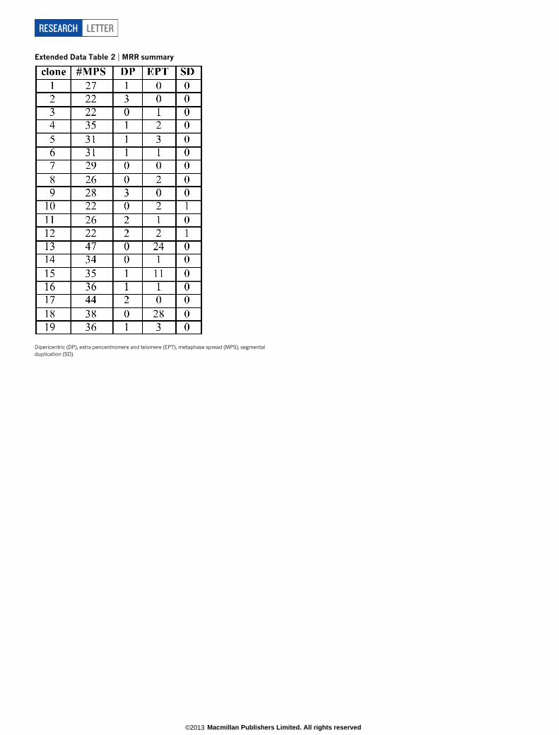
Extended Data Table 2 | MRR summary
Dipericentric (DP), extra pericentromere and telomere (EPT), metaphase spread (MPS), segmentalduplication (SD).
RESEARCH LETTER
Macmillan Publishers Limited. All rights reserved©2013

Extended Data Table 3 | Spectral karyotyping summary
Simple extra pericentromere and telomere (EPT) involves one chromosome. Complex EPT involves more than one chromosome. Other has only one pericentromere.
LETTER RESEARCH
Macmillan Publishers Limited. All rights reserved©2013
Copyright © 2022 FDOKUMEN