Fungal Systematics and Evolution: FUSE 6
126
Sydowia 72 (2020) 171 DOI 10.12905/0380.sydowia72-2020-0271 Published online ?? October 2020 Fungal Systematics and Evolution: FUSE 6 Danny Haelewaters 1,2,3,4,* , Bálint Dima 5 , Abbas I.I. Abdel-Hafiz 6 , Mohamed A. Abdel-Wahab 6 , Samar R. Abul-Ezz 6 , Ismail Acar 7 , Elvira Aguirre-Acosta 8 , M. Catherine Aime 1 , Suheda Aldemir 9 , Muhammad Ali 10 , Olivia Ayala-Vásquez 11 , Mahmoud S. Bakhit 6 , Hira Bashir 10 , Eliseo Battistin 12 , Egil Bendiksen 13 , Rigoberto Castro-Rivera 14 , Ömer Faruk Çolak 15 , André De Kesel 16 , Javier Isaac de la Fuente 17,18 , Ayten Dizkırıcı 9 , Shah Hussain 19 , Gerrit Maarten Jansen 20 , Og ˘uzhan Kaygusuz 21 , Abdul Nasir Khalid 10 , Junaid Khan 19 , Anna A. Kiyashko 22 , Ellen Larsson 23 , César Ramiro Martínez-González 24 , Olga V. Morozova 22 , Abdul Rehman Niazi 10 , Machiel Evert Noordeloos 25 , Thi Ha Giang Pham 26,27 , Eugene S. Popov 22 , Nadezhda V. Psurtseva 22 , Nathan Schoutteten 3 , Hassan Sher 19 , I ˙ brahim Türkekul 28 , Annemieke Verbeken 3 , Habib Ahmad 29 , Najam ul Sehar Afshan 10 , Philippe Christe 30 , Muhammad Fiaz 31 , Olivier Glaizot 30,32 , Jingyu Liu 1 , Javeria Majeed 10 , Wanda Markotter 33 , Angelina Nagy 34 , Haq Nawaz 10 , Viktor Papp 35 , Áron Péter 36 , Walter P. Pfliegler 37 , Tayyaba Qasim 10 , Maria Riaz 10 , Attila D. Sándor 36,38 , Tamara Szentiványi 30,32 , Hermann Voglmayr 39,40 , Nousheen Yousaf 41 & Irmgard Krisai-Greilhuber 42 1 Department of Botany and Plant Pathology, Purdue University, West Lafayette, Indiana 47907, USA 2 Department of Zoology, University of South Bohemia, 370 05 C ˇ eské Budejovice, Czech Republic 3 Research Group Mycology, Department of Biology, Faculty of Sciences, Ghent University, 9000 Ghent, Belgium 4 Operation Wallacea Ltd, Wallace House, Old Bolingbroke, Lincolnshire, PE23 4EX, UK 5 Department of Plant Anatomy, Institute of Biology, Eötvös Loránd University, 1117 Budapest, Hungary 6 Department of Botany and Microbiology, Faculty of Science, Sohag University, Sohag 82524, Egypt 7 Department of Organic Agriculture, Bas ¸kale Vocational School, Van Yüzüncü Yıl University, 65080 Van, Turkey 8 Departamento de Botánica, Instituto de Biología, Universidad Nacional Autónoma de México, CP 04510 Ciudad Universitaria, Ciudad de México, México 9 Department of Molecular Biology and Genetics, Van Yüzüncü Yıl University, 65080 Van, Turkey 10 Fungal Biology and Systematic Research Laboratory, Department of Botany, Quaid-e-Azam Campus, University of the Punjab, Lahore 54590, Pakistan 11 Tecnológico Nacional de México, Instituto Tecnológico de Ciudad Victoria, CP 87010 Ciudad Victoria, Tamaulipas, México 12 Natural History Museum, 36078 Valdagno VI, Italy 13 Norwegian Institute for Nature Research, 0855 Oslo, Norway 14 Instituto Politécnico Nacional, Centro de Investigación en Biotecnología Aplicada, Unidad Tlaxcala, CP 90700 Tepetitla de Lardizábal, Tlaxcala, México 15 Vocational School of Health Services, Süleyman Demirel University, 32260 Isparta, Turkey 16 Meise Botanic Garden, 1860 Meise, Belgium 17 División de Ciencias de la Salud, Universidad de Quintana Roo, CP 77039 Chetumal, Quintana Roo, México 18 Instituto Wozniak, Colonia del Bosque, CP 77019 Chetumal, Quintana Roo, México 19 Center for Plant Sciences and Biodiversity, University of Swat, 19200 Saidu Sharif, Pakistan 20 6703 JC Wageningen, The Netherlands 21 Department of Plant and Animal Production, Atabey Vocational School, Isparta University of Applied Sciences, 32670 Isparta, Turkey 22 Komarov Botanical Institute of the Russian Academy of Sciences, 197376 Saint-Petersburg, Russia 23 Department of Biological and Environmental Sciences, Gothenburg Global Biodiversity Centre, University of Gothenburg, 405 30 Göteborg, Sweden 24 Departamento de Biología, Facultad de Ciencias, Universidad Nacional Autónoma de México, CP 04510 Ciudad Universitaria, Ciudad de México, México 25 Naturalis Biodiversity Center, 2300 RA Leiden, The Netherlands 26 Saint Petersburg State Forestry University, 194021 Saint Petersburg, Russia 27 Joint Russian–Vietnamese Tropical Research and Technological Center, Hanoi, Vietnam 28 Department of Biology, Faculty of Science and Arts, Gaziosmanpas ¸a University, 60010 Tokat, Turkey 29 Department of Genetics, Hazara University, 21300 Mansehra, Pakistan 30 Department of Ecology and Evolution, University of Lausanne, 1015 Lausanne, Switzerland 31 Department of Botany, Hazara University, 21300 Mansehra, Pakistan 32 Museum of Zoology, Palais de Rumine, 1005 Lausanne, Switzerland 33 Centre for Viral Zoonoses, Department of Medical Virology, University of Pretoria, Pretoria 0001, South Africa 34 Hungarian Mycological Society, 1121 Budapest, Hungary
-
Upload
khangminh22 -
Category
Documents
-
view
3 -
download
0
Transcript of Fungal Systematics and Evolution: FUSE 6

Sydowia 72 (2020) 171
DOI 10.12905/0380.sydowia72-2020-0271 Published online ?? October 2020
Fungal Systematics and Evolution: FUSE 6Danny Haelewaters1,2,3,4,*, Bálint Dima5, Abbas I.I. Abdel-Hafiz6, Mohamed A. Abdel-Wahab 6,
Samar R. Abul-Ezz6, Ismail Acar7, Elvira Aguirre-Acosta8, M. Catherine Aime1, Suheda Aldemir9, Muhammad Ali10, Olivia Ayala-Vásquez11, Mahmoud S. Bakhit6, Hira Bashir10, Eliseo Battistin12,
Egil Bendiksen13, Rigoberto Castro-Rivera14, Ömer Faruk Çolak15, André De Kesel16, Javier Isaac de la Fuente17,18, Ayten Dizkırıcı9, Shah Hussain19, Gerrit Maarten Jansen20,
Oguzhan Kaygusuz21, Abdul Nasir Khalid10, Junaid Khan19, Anna A. Kiyashko22, Ellen Larsson23, César Ramiro Martínez-González24, Olga V. Morozova22, Abdul Rehman Niazi10,
Machiel Evert Noordeloos25, Thi Ha Giang Pham26,27, Eugene S. Popov22, Nadezhda V. Psurtseva22, Nathan Schoutteten3, Hassan Sher19, Ibrahim Türkekul28, Annemieke Verbeken3, Habib Ahmad29, Najam ul Sehar Afshan10, Philippe Christe30, Muhammad Fiaz31, Olivier Glaizot30,32, Jingyu Liu1, Javeria Majeed10, Wanda Markotter33, Angelina Nagy34, Haq Nawaz10, Viktor Papp35, Áron Péter36, Walter P. Pfliegler37, Tayyaba Qasim10, Maria Riaz10, Attila D. Sándor36,38, Tamara Szentiványi30,32,
Hermann Voglmayr39,40, Nousheen Yousaf41 & Irmgard Krisai-Greilhuber42
1 Department of Botany and Plant Pathology, Purdue University, West Lafayette, Indiana 47907, USA2 Department of Zoology, University of South Bohemia, 370 05 Ceské Budejovice, Czech Republic
3 Research Group Mycology, Department of Biology, Faculty of Sciences, Ghent University, 9000 Ghent, Belgium4 Operation Wallacea Ltd, Wallace House, Old Bolingbroke, Lincolnshire, PE23 4EX, UK
5 Department of Plant Anatomy, Institute of Biology, Eötvös Loránd University, 1117 Budapest, Hungary6 Department of Botany and Microbiology, Faculty of Science, Sohag University, Sohag 82524, Egypt
7 Department of Organic Agriculture, Baskale Vocational School, Van Yüzüncü Yıl University, 65080 Van, Turkey8 Departamento de Botánica, Instituto de Biología, Universidad Nacional Autónoma de México, CP 04510 Ciudad Universitaria,
Ciudad de México, México9 Department of Molecular Biology and Genetics, Van Yüzüncü Yıl University, 65080 Van, Turkey
10 Fungal Biology and Systematic Research Laboratory, Department of Botany, Quaid-e-Azam Campus, University of the Punjab, Lahore 54590, Pakistan
11 Tecnológico Nacional de México, Instituto Tecnológico de Ciudad Victoria, CP 87010 Ciudad Victoria, Tamaulipas, México12 Natural History Museum, 36078 Valdagno VI, Italy
13 Norwegian Institute for Nature Research, 0855 Oslo, Norway14 Instituto Politécnico Nacional, Centro de Investigación en Biotecnología Aplicada, Unidad Tlaxcala, CP 90700 Tepetitla de
Lardizábal, Tlaxcala, México15 Vocational School of Health Services, Süleyman Demirel University, 32260 Isparta, Turkey
16 Meise Botanic Garden, 1860 Meise, Belgium17 División de Ciencias de la Salud, Universidad de Quintana Roo, CP 77039 Chetumal, Quintana Roo, México
18 Instituto Wozniak, Colonia del Bosque, CP 77019 Chetumal, Quintana Roo, México19 Center for Plant Sciences and Biodiversity, University of Swat, 19200 Saidu Sharif, Pakistan
20 6703 JC Wageningen, The Netherlands21 Department of Plant and Animal Production, Atabey Vocational School, Isparta University of Applied Sciences,
32670 Isparta, Turkey22 Komarov Botanical Institute of the Russian Academy of Sciences, 197376 Saint-Petersburg, Russia
23 Department of Biological and Environmental Sciences, Gothenburg Global Biodiversity Centre, University of Gothenburg, 405 30 Göteborg, Sweden
24 Departamento de Biología, Facultad de Ciencias, Universidad Nacional Autónoma de México, CP 04510 Ciudad Universitaria, Ciudad de México, México
25 Naturalis Biodiversity Center, 2300 RA Leiden, The Netherlands26 Saint Petersburg State Forestry University, 194021 Saint Petersburg, Russia
27 Joint Russian–Vietnamese Tropical Research and Technological Center, Hanoi, Vietnam28 Department of Biology, Faculty of Science and Arts, Gaziosmanpasa University, 60010 Tokat, Turkey
29 Department of Genetics, Hazara University, 21300 Mansehra, Pakistan30 Department of Ecology and Evolution, University of Lausanne, 1015 Lausanne, Switzerland
31 Department of Botany, Hazara University, 21300 Mansehra, Pakistan32 Museum of Zoology, Palais de Rumine, 1005 Lausanne, Switzerland
33 Centre for Viral Zoonoses, Department of Medical Virology, University of Pretoria, Pretoria 0001, South Africa34 Hungarian Mycological Society, 1121 Budapest, Hungary

172 Sydowia 72 (2020)
Haelewaters et al.: FUSE 6
35 Institute of Horticultural Plant Biology, Szent István University, 1118 Budapest, Hungary36 Department of Parasitology and Parasitic Diseases, University of Agricultural Sciences and Veterinary Medicine,
400372 Cluj-Napoca, Romania37 Department of Molecular Biotechnology and Microbiology, University of Debrecen, 4032 Debrecen, Hungary
38 Department of Parasitology and Zoology, University of Veterinary Medicine, 1078 Budapest, Hungary39 Institute of Forest Entomology, Forest Pathology and Forest Protection, Department of Forest and Soil Sciences,
BOKU–University of Natural Resources and Life Sciences, 1190 Vienna, Austria40 Department of Botany and Biodiversity Research, Faculty of Life Sciences, University of Vienna, 1030 Vienna, Austria
41 Department of Botany, Government College University, Lahore, 54000, Pakistan42 Department of Botany and Biodiversity Research, University of Vienna, 1030 Wien, Austria
* e-mail: [email protected]
Haelewaters D., Dima B., Abdel-Hafiz B.I.I., Abdel-Wahab M.A. , Abul-Ezz S.R., Acar I., Aguirre-Acosta E., Aime M.C., Al-demir S., Ali M., Ayala-Vásquez O., Bakhit M.S., Bashir H., Battistin E., Bendiksen E., Castro-Rivera R., Çolak Ö.F., De Kesel A., de la Fuente J.I., Dizkırıcı A., Hussain S., Jansen G.M., Kaygusuz O., Khalid A.N., Khan J., Kiyashko A.A., Larsson E., Martínez-González C.R., Morozova O.V., Niazi A.R., Noordeloos M.E., Pham T.H.G., Popov E.S., Psurtseva N.V., Schoutteten N., Sher H., Türkekul I., Verbeken A., Ahmad H., Afshan N.S., Christe P., Fiaz M., Glaizot O., Liu J., Majeed J., Markotter W., Nagy A., Nawaz H., Papp V., Péter Á., Pfliegler W.P., Qasim T., Riaz M., Sándor A.D., Szentiványi T., Voglmayr H., Yousaf N. & Krisai-Greilhuber I. (2020): Fungal Systematics and Evolution 6. – Sydowia 72: 271–296.
Fungal Systematics and Evolution (FUSE) is one of the journal series to address the “fusion” between morphological data and molecular phylogenetic data and to describe new fungal taxa and interesting observations. This paper is the 6th contribution in the FUSE series—presenting one new genus, twelve new species, twelve new country records, and three new combinations. The new genus is: Pseudozeugandromyces (Laboulbeniomycetes, Laboulbeniales). The new species are: Albatrellopsis flettioides from Pakistan, Aureoboletus garciae from Mexico, Entomophila canadense from Canada, E. frigidum from Sweden, E. porphyroleu-cum from Vietnam, Erythrophylloporus flammans from Vietnam, Marasmiellus boreoorientalis from Kamchatka Peninsula in the Russian Far East, Marasmiellus longistipes from Pakistan, Pseudozeugandromyces tachypori on Tachyporus pusillus (Coleop-tera, Staphylinidae) from Belgium, Robillarda sohagensis from Egypt, Trechispora hondurensis from Honduras, and Tricholoma kenanii from Turkey. The new records are: Arthrorhynchus eucampsipodae on Eucampsipoda africanum (Diptera, Nycteribiidae) from Rwanda and South Africa, and on Nycteribia vexata (Diptera, Nycteribiidae) from Bulgaria; A. nycteribiae on Eucamp-sipoda africanum from South Africa, on Penicillidia conspicua (Diptera, Nycteribiidae) from Bulgaria (the first undoubtful country record), and on Penicillidia pachymela from Tanzania; Calvatia lilacina from Pakistan; Entoloma shangdongense from Pakistan; Erysiphe quercicola on Ziziphus jujuba (Rosales, Rhamnaceae) and E. urticae on Urtica dioica (Rosales, Urticaceae) from Pakistan; Fanniomyces ceratophorus on Fannia canicularis (Diptera, Faniidae) from the Netherlands; Marasmiellus bi-formis and M. subnuda from Pakistan; Morchella anatolica from Turkey; Ophiocordyceps ditmarii on Vespula vulgaris (Hyme-noptera, Vespidae) from Austria; and Parvacoccum pini on Pinus cembra (Pinales, Pinaceae) from Austria. The new combinations are: Appendiculina gregaria, A. scaptomyzae, and Marasmiellus rodhallii. Analysis of an LSU dataset of Arthrorhynchus includ-ing isolates of A. eucampsipodae from Eucampsipoda africanum and Nycteribia spp. hosts, revealed that this taxon is a complex of multiple species segregated by host genus. Analysis of an SSU–LSU dataset of Laboulbeniomycetes sequences revealed sup-port for the recognition of four monophyletic genera within Stigmatomyces sensu lato: Appendiculina, Fanniomyces, Gloeandro-myces, and Stigmatomyces sensu stricto. Finally, phylogenetic analyses of Rhytismataceae based on ITS–LSU ribosomal DNA resulted in a close relationship of Parvacoccum pini with Coccomyces strobi.
Keywords: 1 new genus, 12 new species, 12 new records, 3 new combinations, Agaricomycetes, integrative taxonomy, Laboul-beniomycetes, Leotiomycetes, Pezizomycetes, Rhytismataceae, Sordariomycetes, Stigmatomyces.
With only 138,000 formally described fungal species (Kirk 2019) out of an estimated 2.2–3.8 mil-lion (Hawksworth & Lücking 2017) to 6 million (Taylor et al. 2014), between 97.7 and 93.7% of fun-gal species are left to be characterized. These may be discovered in poorly studied habitats and geo-graphic areas (e.g., tropical rainforests), as molecu-lar novelties, within cryptic taxa, in fungal collec-tions (e.g., new species hidden under current names and in unidentified material), and during studies of plant and insect collections (Hawksworth & Lück-ing 2017, Wijayawardene et al. 2020). This large dis-crepancy between described and undescribed spe-cies needs to be addressed and recent work has
shown that mycologists are nowhere near levelling off the curve in describing new species (Hyde et al. 2020b). Together with other series—Fungal Biodi-versity Profiles (Rossi et al. 2020), Fungal Diversity Notes (Hyde et al. 2020a), Fungal Planet (Crous et al. 2020a), Mycosphere Notes (Pem et al. 2019), New and Interesting Fungi (Crous et al. 2020b)—the Fungal Systematics and Evolution series published by Sydowia contributes to a much-needed accelera-tion of discovery and description of fungal diversity. The present paper is the sixth contribution in the FUSE series published by Sydowia, after Crous et al. (2015), Hernández-Restrepo et al. (2016), Krisai-Greilhuber et al. (2017), Liu et al. (2018), and Song

Sydowia 72 (2020) 173
Haelewaters et al.: FUSE 6
et al. (2019). Altogether, one family, six genera, 67 species, and 22 combinations have been introduced in the FUSE series.
Authors who wish to contribute to the next part in this series, FUSE 7, can e-mail submissions to Danny Haelewaters ([email protected]) or Irmgard Krisai-Greilhuber ([email protected]). Specific Author’s Guidelines for FUSE submissions are available on the website of Sydowia (http://www.sydowia.at/instructions/instructions.htm).
Materials and Methods
Sample collection, isolation, and specimen exami-nation
For the Albatrellopsis study, basidiomata were collected in coniferous forests in the Miandam val-ley of Swat District, Pakistan. Basidiomata were dug out at their base using a knife and photo-graphed in their natural habitat using a Canon Power shot A470 camera (Tokyo, Japan). Macro-morphological characters from fresh basidiomata were noted in the field. Color codes follow Munsell Color Company (1954). Specimens were dried by placing them in front of a hot air fan set at 40–45 °C. Dried specimens were kept at -20 °C for two weeks as a pest-control measure and then deposited at SWAT (herbarium acronyms sensu Thiers continu-ously updated). Microscopic characters of herbari-um specimens were observed using a BM 120 light microscope (BOECO, Hamburg, Germany) with an MVV 3000 camera (Byomic). Tissues were rehydrat-ed using distilled water and mounted in 5 % KOH. Congo red (1 % aqueous solution) was used for staining hyaline structures, whereas Melzer’s rea-gent was used for checking amyloidity of basidio-spores and hyphae. Twenty randomly selected ba-sidiospores, basidia, and hyphae from each availa-ble collection were measured using Piximètre com-puter software (Henriot & Cheypne 2020). Measure-ments are presented as (a−)b−c(−d) with ‘b–c’ repre-senting the 90 % confidence interval, ‘a’ and ‘d’ ex-treme values. ‘Q’ stands for the range of length/width ratio of basidiospores.
Basidiomata of Aureoboletus were collected in the state of Oaxaca, Mexico in forests dominated by oaks (Quercus spp.). Protocols for sampling macro-fungi as described by Lodge et al. (2004) were fol-lowed. The color descriptions were according to Ko-rnerup & Wanscher (1978). Microscopic features from tubes, pileus, and stipe of dried basidiomata were measured at 100× magnification in 5 % KOH,
Melzer’s reagent, and Congo red. The following ab-breviations are used: ‘Q’ for length/width ratio, ‘Lav’ for average length, ‘Wav’ for average width, and ‘n’ for the number of basidiospores measured. At least 30 cystidia, basidia, and basidiospores were meas-ured. Basidiospores were observed using a DSM 950 scanning electron microscope (Zeiss, White Plains, NY). All specimens are deposited at ITCV and MEXU.
For the Entoloma spp. nov. study, collections were photographed in the field. Macroscopic char-acters were noted immediately after collecting. Color codes follow Munsell Soil Color Company (1954) for E. canadense sp. nov. and Kornerup & Wanscher (1978) for E. porphyroleucum sp. nov. Mi-croscopic characters were studied with a Leica DMLS microscope with a drawing tube and a ToupTek Photonics camera (Zhejiang, China); a Zeiss Axioscope A1 microscope with AxioCam 1Cc 3; and a Zeiss Axiophot microscope with DC con-trolled Cree XP-G3 R3 CRI 90+ LED illumination, Plan Neofluar objectives 40×/1.30 Oil, 100×/1.30 Oil (Zeiss), DIC optics, a 12MP ToupTek video camera with SONY Exmor IMX226 CMOS sensor (Tokyo, Japan), and ToupView video & image processing software (ToupTek Photonics). Spores, basidia, and cystidia were observed in squash preparations of small parts of the lamellae in 5 % KOH or 1 % Con-go Red in concentrated NH4OH. Pileipellis was ex-amined on a radial section of the pileus in 5 % KOH. Stipitipellis was examined in 10% Ammonia. Size dimensions are based on measurements of 20 ba-sidiospores, basidia, and cystidia, of which at least 10 structures per collection. Basidiospores were measured without apiculus, and basidia without sterigmata. Basidiospore length × width ratios are reported as Q. Other abbreviations used in Entolo-ma descriptions are ‘Qav’ for the average Q value, ‘L’ for the number of entire lamellae, and ‘l’ for number of lamellulae between each pair of entire lamellae. Collections are deposited at the following herbaria: GB, L, and LE.
Collections of Erythrophylloporus were made in semi-evergreen tropical forests with Fagaceae (Lithocarpus spp.) and Dipterocarpaceae in Viet-nam. Macromorphological features were studied based on fresh collections as well as by the analysis of the photos made in the field. Color codes in the description follow Kornerup & Wanscher (1978). Microscopic characters were studied with a light Zeiss Axioscope A1 microscope with an AxioCam ICc 3 camera and AxioVisionRel.4.6 software (Carl Zeiss, Oberkochen, Germany). Basidiospores, ba-sidia, and hymenial cystidia were observed in

174 Sydowia 72 (2020)
Haelewaters et al.: FUSE 6
squash preparations of small parts of the lamellae in 5 % KOH. The pileipellis was examined on a ra-dial section of the pileus, the stipitipellis on longi-tudinal slice of the stipe in 5 % KOH. Basidiospore dimensions are based on 20 measurements, whereas cystidia and basidia dimensions are based on ob-serving at least 10 structures per collection. Basidia were measured without sterigmata, and the spores without hilum. Basidiospore length × width ratios are reported as ‘Q’. Specimens are deposited at LE.
Russian Marasmiellus basidiomata were sam-pled at the western foothills (ca. 906 m a.s.l.) of the volcano Avachinskaya Sopka at the eastern Kam-chatka Peninsula. Description of basidiomata is based both on notes and photos taken in situ and observations of dried specimens. Color designations follow Kornerup & Wanscher (1978). Microscopic observations were made from dried material mount-ed in 5 % KOH, Congo Red, or Melzer reagent using an Axio Imager A1 light microscope (Carl Zeiss) equipped with differential interference contrast (DIC) optics and a Zeiss AxioCam MRc5 digital camera with AxioVision SE64 version 4.9.1 soft-ware. Basidiospore size was estimated from meas-urements of 60 basidiospores from three basidio-mata; main values represented at least 90 % of the measurements and extreme values are enclosed in parentheses. ‘Q’ is the length/width ratio of basidi-ospores and ‘Qav’ stands for the average Q value. Statistics of hymenial elements and hyphae of pileipellis and caulocystidia are based on measure-ments of at least 10 structures from each of three basidiomata. Drawings were prepared with Ink-scape version 0.91 software (https://inkscape.org/ru/). Ex-type culture LE-BIN 4081 was obtained from spore print of a mature basidioma. After spore germination, the young mycelium was transferred in new Petri plates with beer-wort agar (BWA; beer-wort from brewery “Severnye pivovarni” in Russia, concentration 4 %, agar 20 g/l; Difco, Thermo Fish-er Scientific, Waltham, MA). Culture characteristics were described by standard methods and terminol-ogy (Stalpers 1978). Inoculum plugs (7 mm diam.) were placed mycelium side down in the center of Petri plates (90 mm diam.) containing malt extract agar (MEA; malt extract 15 g/l, Condalab, Madrid, Spain; agar 20 g/l, Difco) and potato dextrose agar (PDA; potato dextrose broth 19.5 g/l, Panreac, Darmstadt, Germany; agar 20 g/l, Difco). Three rep-licates on each medium were incubated for eight weeks in a growth chamber (TS 1/80, Russia) at 25 °C in dark. Linear mycelium extension was re-corded every other day until the plate was covered. Colony radius was measured in four mutually per-
pendicular directions (n=12); standard deviation (SD) was estimated in Excel (Microsoft, Redmond, WA). Extracellular oxidase reactions were tested according to Pointing (1999). The advancing zone and activity of oxidoreductases were studied after 10 days, colony morphology at weeks 4 and 8. Mi-cromorphology was studied under transmitted light using a Zeiss Axio Imager A1 and Axio Scope A1 at week eight. Gymnopus dichrous (Berk. & M.A. Cur-tis) Halling, strain LE-BIN 1134 (USA, North Caro-lina, Jackson County, Highlands, Whiteside Cove Road, on dry tree, 12 July 1999) was used for com-parative study. The holotype is deposited at LE. Ex-type strain LE-BIN 4081 is preserved in the Basidi-omycete Culture Collection of the Komarov Botani-cal Institute of the Russian Academy of Sciences (Saint Petersburg, Russia) as stock cultures in glass tubes on BWA slants, in 2-ml vials under distilled water at 4 °C, and in cryovials on 10 % glycerol at –80 °C (freezing rate 1 °C/min).
Pakistani Marasmiellus basidiomata were col-lected in Ayubia National Park (Khyber Pakh-tunkhwa Province) during the monsoon season in 2016–2017. This area represent one of the moist temperate forests in Pakistan, mostly dominated by conifers including Abies pindraw, Cedrus deodara, and Pinus wallichiana (Pinales Pinaceae), and Tax-us wallichiana (Pinales, Taxaceae), along with broad-leaved oaks (Fagales, Fagaceae, Quercus spp.) (Saima et al. 2009, Raja et al. 2014, Razzaq et al. 2014). Collections were photographed in situ, mor-phologically characterized in the field, vouchered, and dried using a fan heater. Color codes were as-signed following Munsell Color Company (1954). Microscopic characters including basidiospores, basidia, cystidia, pileipellis, and stipitipellis were observed from material mounted in 5 % KOH, Con-go red, and Melzer’s reagent under a CH30 light mi-croscope (Olympus). Line drawings were made free-handed. Specimens are deposited at LAH.
For the Pseudozeugandromyces study, insect hosts were collected with a mouth-operated aspira-tor and immediately stored in 96 % denaturated ethanol. Screening and removal of Laboulbeniales thalli was done at 50× magnification using an Olym-pus SZ61 stereomicroscope (Tokyo, Japan). Thalli were mounted in Amann medium (Benjamin 1971) and slides were sealed with transparent nail var-nish. Insect hosts and microscope slides are depos-ited at BR. Drawings and measurements were made using an Olympus BX51 light microscope with drawing tube, digital camera, and AnalySIS soft-ware (Soft Imaging System GmbH, Münster, Ger-many).

Sydowia 72 (2020) 175
Haelewaters et al.: FUSE 6
For the Robillarda study, senescent and dried leaf litter baits of different plant species—including Eucalyptus rostrata (Myrtales, Myrtaceae), Ficus nitida (Rosales, Moraceae), Phoenix dactylifera (Arecales, Arecaceae), and Phragmites australis (Poales, Poaceae)—were submerged in the Nile river and irrigation canals in Sohag Governorate, Egypt from December 2015 to December 2016. Leaves were baited in plastic mesh bags and collected ran-domly monthly. Collected decaying leaves were placed in clean plastic bags and returned to the laboratory, where they were rinsed first under tap water and then under sterile distilled water. Sam-ples were incubated in Petri plates lined with ster-ile, wet filter paper at room temperature and sprayed with sterile distilled water periodically to avoid drying. Samples were periodically examined using an SZ62 stereomicroscope (Olympus) over 3 months of incubation for the presence of fungal sporulating structures. Fungi were mounted in freshwater and examined under a BX51 compound microscope (Olympus) equipped with DIC optics. Permanent slides were prepared using the double cover-glass method by Volkmann-Kohlmeyer & Kohlmeyer (1996). A herbarium collection of the new Robillar-da species was prepared by drying decaying leaves with fungus material at 60 °C for 24 h and then de-posited at CBS. Single-spore cultures were obtained by cutting open pycnidia with a sterile razor blade. The centrum tissue containing conidia was removed with sterile forceps and placed in sterile freshwater. Small drops of the spore suspension were placed on PDA (Oxoid, Basingstoke, England) and CMA (Ox-oid) media and incubated at 22 °C in dark. Germinated spores were transferred to new plates. Colony characteristics and sporulation were noted after 2–3 weeks of growth. Conidiomata were measured both on leaves collected from the field and in pure culture. Measurements of 30 pycnidia were made under an SZ62 stereomicroscope (Olym-pus) from vertical sections that were prepared using a Leica CM1100 cryostat (Leica Biosystems, Nuss-loch, Germany). Sizes of conidia and conidial ap-pendages were based on 50 measurements in fresh-water.
The Trechispora specimen was collected during an exploratory fungal survey in Cusuco National Park, a Mesoamerican cloud forest in Honduras, be-tween 22 June and 13 July 2019 (details in Haelewa-ters et al. 2020b, Martin et al. 2020). Fresh material was photographed in situ. The specimen was as-signed a HONDURAS19-F collection number and metadata were recorded on site, including data, specific locality, geographic coordinates, substra-
tum, and surrounding habitat notes. Back at Base Camp (located at 1572 m a.s.l.), a rice-sized piece of tissue was removed from the specimen and stored in a 1.5 ml Eppendorf tube with 600 μl of Nuclei Lysis Solution (Promega, Madison, WI) and stored until DNA extraction could be performed. After process-ing, the specimen was dried with silica gel. Exami-nation of microscopic characters was done in Congo Red and Melzer’s reagent using an Olympus CX21 light microscope and a Nikon Eclipse Ni-E fluores-cence microscope (Melville, NY). Measurements of microscopic structures were performed at 100× magnification. At least thirty basidiospores, 20 ba-sidia, and 20 hyphae were measured. Sizes of ba-sidia and basidiospores (excluding ornaments) are presented as follows: (a–)b–c(–d), with ‘b–c’ indicat-ing the 90 % confidence interval, and ‘a’ and ‘d’ rep-resenting extreme values. Drawings were made us-ing a drawing tube at 6000× magnification for ba-sidiopores and at 1500× magnification for other ele-ments. Scanning electron microscope (SEM) images were taken with a JEOL 5800 LV SEM (Peabody, MA).
Tricholoma basidiomata were collected at conif-erous forests in Genç (Bingöl Province, Turkey) in 2018 and photographed with a Canon EOS 60D camera (Tokyo, Japan) equipped with Tokina 100 mm macro lens (New Delhi, India). Specimens were dried, kept in Ziploc bags, and deposited at VPH. Micromorphology of the basidiomata was an-alyzed using a Leica DM500 microscope. Sections of lamellae were mounted in tap water and Melzer’s reagent. Size values reported for basidiospores were based on at least 40 measurements and include the mean length × mean width ± standard deviation and ‘Q’, representing the length-width ratio of basidio-spores. Photographs of basidiospores were taken by field-emission SEM (Zeiss Sigma 300; White Plains, NY) using an accelerating voltage of 10 kV. Other abbreviations used in the description are ‘L’ for the number of entire lamellae, and ‘l’ for number of lamellulae between each pair of entire lamellae.
For the Arthrorhynchus study, bats were cap-tured and screened for ectoparasites in Bulgaria (2017; Sándor et al. 2019), Rwanda (2008), and South Africa (2010–2017). Ectoparasites were stored in 98 % ethanol at –80 °C. Bat flies were screened for the presence of Arthrorhynchus thalli (Ascomycota, Laboulbeniomycetes, Laboulbeni-ales) under 40–50× magnification. Bat fly identifica-tion was based on several keys (Theodor 1957, 1967, 1968, 1973) and bat fly taxonomy follows Dick & Graciolli (2013) and Graciolli & Dick (2018). In ad-dition, African bat flies stored in 70 % ethanol at

176 Sydowia 72 (2020)
Haelewaters et al.: FUSE 6
the entomology collection of the California Acade-my of Sciences (San Fransisco, CA) were screened for Laboulbeniales: Basilia blainvillii blainvillii (Leach, 1817) from Uganda (n=8), Dipseliopoda bi-annulata (Oldroyd, 1953) from Uganda (n=10), Nyc-teribia schmidlii Schiner, 1853 from Uganda (n=1), Penicillidia fulvida (Bigot, 1885) from Kenya (n=2), P. pachymela Speiser 1900 from Tanzania (n=26), and Phthiridium hoogstraali (Theodor, 1957) from Kenya (n=8). Thalli were removed from their bat fly host at the point of attachment using a micropin and mounted in Heinz PVA mounting medium. Voucher slides are deposited at PUL. Mounted specimens were viewed at 200–1000× magnification. Light microscopy photographs of slide-mounted thalli were taken using an Olympus BD40 micro-scope equipped with a 40× phase-contrast lens and Olympus DP71 digital camera and viewed using the Olympus DP Controller software. Line and stipple drawings of thalli were made using photographs as reference material with PITT artist pens (Faber–Castell, Nürnberg, Germany). Drawings were scanned using an HP Scanjet G5040 scanner (Palo Alto, CA) and edited with Photopea (https://www.photopea.com/).
During the exploration of gasteroid fungal di-versity in Pakistan, two collections of Calvatia li-lacina were collected at two different localities. These were Mansehra (975 m a.s.l.) and Deosai Plains (4114 m a.s.l.). Specimens were labelled and morphological features were noted in the field. Col-lections were dried overnight using a fan heater and brought to the laboratory for further analysis. Mi-croscopic study was done by making slides of gleba and peridium. Glebal material was examined mounted in lactophenol, trypan blue, and 5 % KOH. Illustrations of microscopic features were made with the help of a DM750 binocular microscope (Leica Microsystems) with camera lucida attached. Specimens are deposited at LAH.
Basidiomata of Entoloma were collected over several years at the Quaid-i-Azam Campus of the University of the Punjab in Pakistan during the rainy season. Morphological characters were noted from fresh material and photographs; colors follow Munsell Color Company (1954). Specimens were vouchered and dried using fan heater. Microscopic characters were examined based on dried material (lamellae, pileus, stipe) mounted in 2 % KOH, Con-go red, and Melzer’s reagent under an MX4300H light microscope (Meiji Techno). For basidiospores, the notation ‘n/m/p’ indicates that ‘n’ basidiospores were measured from ‘m’ basidiomata of ‘p’ collec-tions. Dimensions of basidiospores are given as
length × width, each as (a–)b–c(–d) with ‘b–c’ indi-cating the 90 % confidence interval, and ‘a’ and ‘d’ representing extreme values; ‘Q’ stands for length/width ratio and ‘Qav’ stands for the average Q value (Liang & Yang 2011). All collections are deposited at LAH.
For the Erysiphe study, phytopathogenic surveys were conducted in Himalayan moist temperate for-ests in Khyber Pakhtunkhwa Province, Pakistan. Plants of Urtica dioica (Urticaceae) and Ziziphus jujuba (Rhamnaceae) infected with powdery mil-dew fungi were collected and photographed in the field. Samples were brought to the lab and photo-graphed under an EMZ-5TR stereomicroscope (Me-iji Techno, Saitama, Japan). Scratch mounts of in-fected portions were prepared in lactophenol. Mi-croscopic examinations were done under a LABOMED (Labo America Inc., Fremont, CA) light microscope and anatomical dimensions of conidia, conidiophores, appressoria (anamorph); chasmoth-ecia, asci, and ascospores (teleomorph) were meas-ured using Scope Image 9.0 (X5) image-processing software (Bioimager, Maple, Ontario, Canada). A JEOL JSM-5910 scanning electron microscope (Peabody, MA) was used for more accurate identifi-cation. Pathogenicity was assessed by pressing a diseased leaf onto young leaves of three asympto-matic, potted plants of Urtica dioica and Ziziphus jujuba. Three non-inoculated plants (for each path-ogen) were used as controls. Plants were maintained at 23 °C and 80 % relative humidity (RH) in a green-house at the Botanical Garden of the University of the Punjab (Lahore, Pakistan). The fungus on the inoculated leaves was morphologically identical to the fungus on the original infected leaves.
For the Fanniomyces study, fly specimens were collected in pitfall traps of dead crayfish (described in De Kesel & Haelewaters 2019) or by hand and screened for the presence of Laboulbeniales thalli (Ascomycota, Laboulbeniomycetes) under 40–50× magnification. Thalli were removed from their host using Minuten Pins (BioQuip, Rancho Dominguez, CA) inserted onto wooden rods. Thalli or groups of thalli were embedded in Amann solution (Benjamin 1971) with the help of a droplet of Hoyer’s medium as described by Haelewaters et al. (2015b). Slides are deposited at FH. Mounted thalli were viewed at 200–1000× magnification using an Olympus BX40 light microscope with Olympus XC50 digital cam-era and MicroSuite Special Edition software 3.1 (Soft Imaging Solutions GmbH). Line and stipple drawings were made using photographs as refer-ence material, with PITT artist pens (Faber–Cas-tell). Drawings were scanned using an HP Scanjet
Sydowia 72 (2020) 177
Haelewaters et al.: FUSE 6
G5040 scanner and then edited with Photopea (htt-ps://www.photopea.com/).
Two specimens of Morchella Dill. ex Pers. were collected in 2015 in the Province of Antalya, Turkey. The morphological features and ecological notes were recorded from young to mature fruiting bodies and ascomata. Ascomata were photographed in their natural habitat. The macro-morphological de-scriptions and images of ascomata were based on fresh material. For micro-morphological structures, the dried ascomata were rehydrated in distilled wa-ter or 3 % KOH, and subsequently stained with Congo Red (to stain cell components) and cotton blue (to check ascospore ornamentation). The fol-lowing abbreviations are used in the description: ‘Lav’ for the average length of all the measured as-cospores, ‘Wav’ for the average width of all the meas-ured ascospores, ‘Q’ for the quotient of length and width of all the measured ascospores, and ‘Qav’ for the average of all calculated Q values for all as-cospores measured. At least thirty mature as-cospores were measured. The collections are depos-ited at the personal fungarium of O. Kaygusuz at Isparta University of Applied Sciences, Turkey.
For the Ophiocordyceps study, macromorpho-logical features were studied on fresh collection as well as by the analysis of photos taken in the field. Micromorphological structures were studied on dried material under a Zeiss Axio Imager.A2 light microscope, equipped with AxioVision Release 4.8.2. software. Measurements were done with a 100× oil immersion objective (1000× magnification). Drawings were produced with the aid of a drawing tube. Observations of microscopic features as well as measurements, and drawings were made from slide preparations stained with 5 % KOH. The spec-imen is deposited at WU.
Fresh material of Parvacoccum pini was collect-ed during a students’ course on management and forest protection in high-altitude afforestations and protective forests, taking place at the Sticklerhütte, Hintermuhr (Salzburg, Austria) in a subalpine stand of Pinus cembra (Pinales, Pinaceae) at ca. 1800 m a.s.l. Dead, corticated branches of Pi. cem-bra still attached to the trees were collected, brought to the laboratory, and checked for the presence of fungi. Study of macromorphology of Parvacoccum pini was done by using a Nikon SMZ 1500 steromi-croscope (Nelville, NY) equipped with a Nikon DS-U2 digital camera. For light microscopy, a Zeiss Axio Imager.A1 compound microscope (Oberkochen, Germany), equipped with DIC optics and a Zeiss Axiocam 506 colour digital camera was used. Mi-croscopic observations of Parvacoccum pini were
made in 3 % KOH except where noted. Images and data were gathered using the following software packages: NIS-Elements D version 3.22.15 (Nikon) or Zeiss ZEN Blue Edition. For certain images of ascomata and conidiomata, stacking software Zerene Stacker version 1.04 (Zerene Systems LLC, Richland, WA) was used. Measurements are report-ed as maxima and minima in parentheses and the range representing the mean plus and minus the standard deviation of a number of measurements given in parentheses. Parvacoccum pini was isolat-ed in pure culture from ascospores as described in Jaklitsch (2009) and grown on 2 % corn meal agar plus 2 % w/v dextrose (CMD). The herbarium speci-men was deposited at WU, and the living culture is maintained in the personal collection of the author.
DNA extraction, PCR amplification, and sequenc-ing
For the Albatrellopsis study, genomic DNA was extracted using the CTAB method of Allen et al. (2006). For molecular phylogenetic analysis, the in-ternal transcribed spacer (ITS) region including was amplified using primers ITS1F (Gardes & Bruns 1993) and ITS4 (White et al. 1990). PCR amplifica-tion followed Khan et al. (2017), with initial dena-turation at 94 °C for 4 min; followed by 40 cycles of denaturation at 94 °C for 1 min, annealing at 55 °C for 1 min, extension at 72 °C for 1 min; and final extension at 72 °C for 10 min. Purification and se-quencing of PCR products was outsourced to the BGI Genomics (Hong Kong). Generated forward and reverse reads were assembled using BioEdit version 7.2.5 (Hall 1999).
For the Aureoboletus study, genomic DNA was extracted from 2–3 mg of tissue using a CTAB method (Doyle & Doyle 1987). DNA was quantified with a NanoDrop ND-1000 spectrophotometer (Thermo Fisher Scientific). Dilutions for each iso-late were prepared resulting in DNA concentration of 20 ng/μl as a basis for PCR amplification. Se-quences were obtained of the nuclear large subunit (LSU) of the ribosomal RNA gene (rDNA) as well as the genes for the RNA polymerase II largest and second largest subunits (rpb1, rpb2). The primer sets used for amplifying these fragments were: LR0R/LR5 for LSU (Vilgalys & Hester 1990, Hopple 1994), RPB1-Af/fRPB1-Cr for rpb1, and bRPB2-6F/bR-PB2-7.1R (sensu Wu et al. 2014). The reaction mix-ture for PCR was prepared in a final volume of 15 μL containing 1× enzyme buffer, 0.8 μm of 0.2 μm dNTPs, 100 ng of DNA extract, 20 pmol of each primer, and 2 units of Taq DNA polymerase (Pro-

178 Sydowia 72 (2020)
Haelewaters et al.: FUSE 6
mega). Cycling conditions were as follows: for LSU; initial denaturation at 96 °C for 2 min; followed by 35 cycles of denaturation at 94 °C for 1 min, anneal-ing at 48 °C for 30 s, extension at 72 °C for 1 min; and final extension at 72 °C for 5 min. For rpb1 and rpb2: same conditions, except for annealing at 52 °C for 30 s. All PCR reactions were carried out in an MJ Research PTC-200 Thermal Cycler (BIO-RAD, Ciu-dad de México, Mexico). The amplifications were verified by electrophoresis in a 1.5 % agarose gel prepared with 1× TAE buffer (Tris Acetate-EDTA), run at 95 V for 1 h. The gel was dyed with GelRed (Biotium, Hayward, CA) and bands were visualized using an INFINITY 3000 transilluminator (Vilber Lourmat, Eberhardzell, Germany). PCR products were purified with the ExoSAP kit (Affymetrix, Santa Clara, CA) and prepared for the sequencing reaction using the Bigdye Terminator version 3.1 kit (Applied Biosystems, Foster City, CA). Sequencing was done with a 3730xl DNA Analyzer (Applied Biosystems) at the Instituto de Biología, Universi-dad Nacional Autónoma de México. Forward and reverse sequence reads were assembled and edited using BioEdit version 7.0.5 (Hall 1999). Consensus sequences were submitted to NCBI GenBank (ac-cession nos. MH337251, MT228976–MT228986).
DNA of Entoloma spp. nov. was extracted from dried herbarium material using the Nucleo-Spin® Plant II kit (Macherey-Nagel, Düren, Germa-ny). The ITS region was amplified with primer sets ITS1F/ITS4, ITS1F/ITS4B, and ITS1F/ITS2 (White et al. 1990, Gardes & Bruns 1993), whereas LSU was amplified with primers LR0R and LR5 (Vilgalys & Hester 1990, Hopple 1994). PCR products were pu-rified with the Fermentas Genomic DNA Purifica-tion Kit (Thermo Fisher Scientific). Purified PCR products were sequenced using the same primers on an ABI model 3130 Genetic Analyzer (Applied Bio-systems) or commercially at LGC Genomics (Berlin, Germany). Alternatively, DNA extraction, PCR am-plification, and Sanger sequencing were performed as part of the Norwegian Barcode of Life project (NorBOL) and followed Larsson et al. (2004, 2018). Chromatograms were checked and edited with the CodonCode Aligner package (CodonCode Corpora-tion, Centerville, MA) and MEGA X (Kumar et al. 2018). Sequence comparison with public and per-sonal databases followed Noordeloos et al. (2017). Newly generated sequences were submitted to Gen-Bank (Tab. 1).
Erythrophylloporus DNA was extracted from herbarium material using NucleoSpin® Plant II kit (Macherey-Nagel, Düren, Germany). The ITS region was amplified with primers ITS1F and ITS4B
(Gardes & Bruns 1993), and translation elongation translation factor 1-α (tef1) with Boletaceae-specif-ic primers EF1-B-F1 and EF1-B-R (Wu et al. 2014). PCR conditions were as follows: for ITS: initial de-naturation at 95 °C for 4 min; then 35 cycles of de-naturation at 94 °C for 1 min, annealing at 52 °C for 1 min, extension at 72 °C for 1 min; and a final ex-tension step of 72 °C for 3 min. For tef1: initial de-naturation at 95 °C for 3 min; then 8 cycles of dena-turation at 98 °C for 20 s, annealing at 60 °C for 40 s, extension at 72 °C for 2 min; then 36 cycles of dena-turing at 98 °C for 20 s, annealing at 53 °C for 90 s, extension at 72 °C for 2 min; and a final extension step of 72 °C for 10 min. PCR products were purified with the Fermentas Genomic DNA Purification Kit (Thermo Fisher Scientific) and sequenced on an Ap-plied Biosystems 3130 Genetic Analyzer. Raw data were edited and assembled in MEGA X (Kumar et al. 2018). Newly generated sequences were deposit-ed in NCBI GenBank (Tab. 1).
Total DNA was extracted from small fragments of dried basidiomata as well as from culture myce-lium of Marasmiellus boreoorientalis sp. nov., using the GeneJET Plant Genomic DNA Purification Mini Kit (Thermo Fisher Scientific) according to the manufacturer’s instructions. PCR amplifications were performed with primer sets ITS1F/ITS4B (Gardes & Bruns 1993) for ITS and LR0R/LR5 (Vil-galys & Hester 1990, Hopple 1994) for LSU. Suc-cessful PCR products were purified with the Gene-JET PCR Purification Kit (Thermo Fisher Scientific) following the manufacturer’s protocol. Sanger se-quencing was performed with an ABI model 3130 Genetic Analyzer (Applied Biosystems). Forward and reverse sequence reads were assembled to ob-tain consensus sequences and ambiguous edges were trimmed. Chromatograms were checked with Chromas version 2.6.6 (https://www.technelysium.com.au). The sequence from the basidioma was aligned with that obtained from the culture to con-firm identity.
For the Pakistani Marasmiellus study, genomic DNA was extracted from lamellae of dried basidi-omata following a modified CTAB method (Lee et al. 1988). The ITS region was amplified using uni-versal primers ITS1F and ITS4 (White et al. 1990, Gardes & Bruns 1993). For PCR, the following cy-cling conditions were used (Saba et al. 2020): initial denaturation at 94 °C for 1 min; 35 cycles of dena-turation at 94 °C for 1 min, annealing at 53 °C for 1 min, and extension at 72 °C for 1 min; followed by final extension at 72 °C for 8 min. Amplified PCR products were purified and sequenced by Tsing Ke Biotech. Forward and reverse sequence reads were

Sydowia 72 (2020) 179
Haelewaters et al.: FUSE 6T
ab. 1
. Det
ails
of
seq
uen
ces
and
iso
late
s in
clu
ded
in
th
e m
olec
ula
r an
alys
is f
or t
he
new
sp
ecie
s an
d i
nte
rest
ing
rep
orts
.
Spec
ies n
ame
ID (i
sola
te, s
trai
n, st
atus
, vou
cher
)Co
untr
y, is
olat
ion
sour
ceSS
UIT
SLS
Urp
b1rp
b2te
f1Re
fere
nce(
s)
Alb
atre
llops
is c
onflu
ens
PV 1
01-9
3 G
B Cz
ech
Repu
blic
AF5
0639
3La
rsso
n &
Lar
sson
(200
3)
Alb
atre
llops
is fl
ettii
DAV
FP:2
7659
Cana
daJF
8995
44M
iller
& B
uyck
(200
2)
Alb
atre
llops
is fl
ettii
398I
F62
USA
AY06
1738
Mill
er &
Buy
ck (2
002)
Alb
atre
llops
is fl
ettii
MIC
H A
HS8
2164
AY62
1802
Alb
ee-S
cott
(200
7)
Alb
atre
llops
is fl
ettio
ides
MM
72Pa
kist
anM
T040
747
This
stud
y
Alb
atre
llops
is fl
ettio
ides
MM
76Pa
kist
anM
T040
748
This
stud
y
Alb
atre
llus a
vella
neus
p816
iU
SAEU
6693
92G
ordo
n M
. & Z
ych
P., u
npub
l.
Alb
atre
llus a
vella
neus
p817
iU
SAEU
6693
93G
ordo
n M
. & Z
ych
P., u
npub
l.
Alb
atre
llus c
itrin
usM
usko
s 850
928
(S)
Swed
enAY
1981
90Ry
man
et a
l. (2
003)
Alb
atre
llus c
itrin
usRy
man
606
1 (U
PS F
-007
387)
Swed
enAY
1981
92Ry
man
et a
l. (2
003)
Alb
atre
llus o
vinu
sFr
anss
on 2
(UPS
F-0
1555
1)Sw
eden
AY19
8203
Rym
an e
t al.
(200
3)
Alb
atre
llus o
vinu
sD
anel
l 11/
8 00
(UPS
F-0
1555
4)Sw
eden
AY19
8198
Rym
an e
t al.
(200
3)
Alb
atre
llus p
icei
philu
sCu
i222
0Ch
ina
DQ
7893
96Cu
i et a
l. (2
008)
Alb
atre
llus p
icei
philu
sCu
i222
0Ch
ina
DQ
7893
97Cu
i et a
l. (2
008)
Alb
atre
llus r
oseu
sSW
AT00
0135
Paki
stan
MF1
1028
5K
han
et a
l. (2
018)
Alb
atre
llus r
oseu
sLA
H35
288
Paki
stan
MF1
1029
7K
han
et a
l. (2
018)
Alb
atre
llus s
imili
sU
SAAY
9635
66Cu
i et a
l. (2
008)
Alb
atre
llus s
ubru
besc
ens
Jaed
erfe
ldt 1
1/10
199
5Sw
eden
AY19
8204
Rym
an e
t al.
(200
3)
Alb
atre
llus s
ubru
besc
ens
Rym
an 6
085
(UPS
F-0
0738
1)Sw
eden
AY19
8208
Rym
an e
t al.
(200
3)
Alb
atre
llus s
ubru
besc
ens
OR9
96Be
lgiu
mK
T947
121
Vadt
hana
rat S
., Lu
myo
ng S
. &
Ras
pé O
., un
publ
.
App
endi
culin
a en
tom
ophi
la [a
s St
igm
atom
yces
ent
omop
hilu
s]D
. Hae
lew
. 106
2cN
ethe
rlan
ds, D
roso
phila
fu
nebr
isM
G95
8014
This
stud
y
App
endi
culin
a en
tom
ophi
la [a
s St
igm
atom
yces
ent
omop
hilu
s]D
. Hae
lew
. 106
3aN
ethe
rlan
ds, D
roso
phila
fu
nebr
isM
H04
0561
Hae
lew
ater
s et a
l. (2
018b
)
App
endi
culin
a gr
egar
ia [a
s St
igm
atom
yces
gre
gari
us]
D. H
aele
w. 1
008a
Sier
ra L
eone
, Dio
psid
ae sp
.M
G43
8348
Hae
lew
ater
s et a
l. (2
019c
)
App
endi
culin
a gr
egar
ia [a
s St
igm
atom
yces
gre
gari
us]
D. H
aele
w. 1
008b
Sier
ra L
eone
, Dio
psid
ae sp
.M
H04
0562
Hae
lew
ater
s et a
l. (2
018b
)
App
endi
culin
a gr
egar
ia [a
s St
igm
atom
yces
gre
gari
us]
LG64
2Si
erra
Leo
ne, D
iops
idae
sp.
MG
6742
25G
oldm
ann
& W
eir (
2018
)
App
endi
culin
a sc
apto
myz
ae [a
s St
igm
atom
yces
]A
F431
758
Wei
r & H
ughe
s (20
02)
Art
hror
hync
hus e
ucam
psip
odae
D. H
aele
w. 1
491a
Bulg
aria
, Nyc
teri
bia
vexa
taM
T241
715
This
stud
y
Art
hror
hync
hus e
ucam
psip
odae
D. H
aele
w. 1
498a
Rwan
da, E
ucam
psip
oda
afri
canu
mM
T235
694
MT2
3571
7Th
is st
udy
Art
hror
hync
hus e
ucam
psip
odae
D. H
aele
w. 1
498b
Rwan
da, E
ucam
psip
oda
afri
canu
mM
T235
695
MT2
3571
8Th
is st
udy
Art
hror
hync
hus e
ucam
psip
odae
D. H
aele
w. 1
499a
Slov
akia
, Nyc
teri
bia
schm
idlii
MT2
3569
6M
T235
719
This
stud
y

180 Sydowia 72 (2020)
Haelewaters et al.: FUSE 6Sp
ecie
s nam
eID
(iso
late
, str
ain,
stat
us, v
ouch
er)
Coun
try,
isol
atio
n so
urce
SSU
ITS
LSU
rpb1
rpb2
tef1
Refe
renc
e(s)
Art
hror
hync
hus n
ycte
ribi
aeD
. Hae
lew
. 148
4bBu
lgar
ia, P
enic
illid
ia
cons
picu
aM
T235
715
This
stud
y
Art
hror
hync
hus n
ycte
ribi
aeD
. Hae
lew
. 148
4cBu
lgar
ia, P
enic
illid
ia
cons
picu
aM
T235
716
This
stud
y
Art
hror
hync
hus n
ycte
ribi
aeD
. Hae
lew
. 150
5aBu
lgar
ia, P
enic
illid
ia
cons
picu
aM
T235
697
MT2
3572
0Th
is st
udy
Art
hror
hync
hus n
ycte
ribi
aeD
. Hae
lew
. 101
5dH
unga
ry, P
enic
illid
ia
cons
picu
aM
G43
8336
MG
4383
63H
aele
wat
ers e
t al.
(201
9c)
Art
hror
hync
hus n
ycte
ribi
aeEd
elen
y_13
.xi.2
014
Hun
gary
, Pen
icill
idia
co
nspi
cua
KY0
9449
6K
Y094
497
Hae
lew
ater
s et a
l. (2
017)
Aur
eobo
letu
s aur
iflam
meu
sD
D97
3U
SAAY
6128
18W
u et
al.
(201
6)
Aur
eobo
letu
s aur
ipor
usBD
CR04
31Co
sta
Rica
AY61
2818
HQ
1618
71D
entin
ger e
t al.
(201
0)
Aur
eobo
letu
s cat
enar
ius
HK
AS
5446
3Ch
ina
NG
0570
93K
T990
890
KT9
9034
8W
u et
al.
(201
6)
Aur
eobo
letu
s dup
licat
opor
usH
KA
S 50
498
Chin
aK
F112
361
KF1
1256
1K
F112
754
Wu
et a
l. (2
014)
Aur
eobo
letu
s for
mos
usG
DG
M44
441
Chin
aN
G05
7082
KT2
9175
1Zh
ang
et a
l. (2
015a
)
Aur
eobo
letu
s gar
ciae
MEX
U 2
9006
, TM
exic
oM
H33
7251
MT2
2897
9M
T228
983
This
stud
y
Aur
eobo
letu
s gar
ciae
MEX
U 3
0133
Mex
ico
MT2
2897
6M
T228
980
MT2
2898
4Th
is st
udy
Aur
eobo
letu
s gar
ciae
MEX
U 3
0134
Mex
ico
MT2
2897
7M
T228
981
MT2
2898
5Th
is st
udy
Aur
eobo
letu
s gar
ciae
MEX
U 3
0135
Mex
ico
MT2
2897
8M
T228
982
MT2
2898
6Th
is st
udy
Aur
eobo
letu
s gen
tilis
MG
372a
Ital
yK
F112
344
KF1
1255
7K
F112
741
Wu
et a
l. (2
014)
Aur
eobo
letu
s inn
ixus
MB0
3104
USA
KF0
3023
9K
F030
239
Nuh
n et
al.
(201
3)
Aur
eobo
letu
s lon
gico
llis
HK
AS
5339
8Ch
ina
KF1
1237
6K
F112
625
KF1
1275
5W
u et
al.
(201
4)
Aur
eobo
letu
s mar
roni
nus
GD
GM
4328
8Ch
ina
NG
0570
40K
T291
753
Zhan
g et
al.
(201
5b)
Aur
eobo
letu
s mir
abili
sH
KA
S 57
776
Chin
aK
F112
360
KF1
1262
4K
F112
743
Wu
et a
l. (2
014)
Aur
eobo
letu
s mor
avic
usM
G37
4aIt
aly
KF1
1242
1K
F112
559
KF1
1274
5W
u et
al.
(201
4)
Aur
eobo
letu
s nep
hros
poru
sH
KA
S 74
929
Chin
aN
G05
7094
KT9
9089
6K
T990
358
Wu
et a
l. (2
016)
Aur
eobo
letu
s pro
ject
ellu
sA
FTO
LID
713
USA
AY68
4158
AY78
8850
AY78
7218
Bind
er &
Hib
bett
(200
7)
Aur
eobo
letu
s que
rcus
-spi
nosa
eG
DG
M 4
3755
Chin
aN
G05
7121
KY0
3996
3K
Y039
958
Zhan
g et
al.
(201
7)
Aur
eobo
letu
s rox
anae
DS6
26-0
7U
SAK
F030
311
KF0
3038
1N
uhn
et a
l. (2
013)
Aur
eobo
letu
s ten
uis
HK
AS
7510
4Ch
ina
KT9
9051
8K
T990
897
KT9
9035
9W
u et
al.
(201
6)
Aur
eobo
letu
s thi
beta
nus
HK
AS
5769
2Ch
ina
KT9
9052
4K
T990
901
KT9
9036
5W
u et
al.
(201
6)
Aur
eobo
letu
s tom
ento
sus
HK
AS
8048
5Ch
ina
KT9
9089
4K
T990
353
Wu
et a
l. (2
016)
Aur
eobo
letu
s vis
cidi
pes
HK
AS
7710
3Ch
ina
KT9
9051
9K
T990
360
Wu
et a
l. (2
016)
Aur
eobo
letu
s vis
cosu
sH
KA
S 53
398
Chin
aK
F112
755
Wu
et a
l. (2
014)
Aur
eobo
letu
s yun
nane
nsis
HK
AS
7505
0Ch
ina
KT9
9052
0K
T990
898
KT9
9036
1W
u et
al.
(201
6)
Aur
eobo
letu
s zan
gii
HK
AS
7475
1Ch
ina
KT9
9052
1K
T990
899
KT9
9036
2W
u et
al.
(201
6)
Aut
oico
myc
es fa
lcat
usU
SA, H
ydro
phili
dae
sp.
MG
6874
07G
oldm
ann
& W
eir (
2018
)
Aut
oico
myc
es re
curv
atus
AW91
1BU
SA, H
ydro
phili
dae
sp.
MG
6874
09G
oldm
ann
& W
eir (
2018
)
Bor
dea
sp.
LG48
3N
amib
ia, S
taph
ylin
idae
sp.
MG
6874
03G
oldm
ann
& W
eir (
2018
)
Calv
atia
bra
silie
nsis
UFR
N-F
ungo
s 303
9Br
azil
MK
6604
93Cr
ous e
t al.
(201
9)

Sydowia 72 (2020) 181
Haelewaters et al.: FUSE 6Sp
ecie
s nam
eID
(iso
late
, str
ain,
stat
us, v
ouch
er)
Coun
try,
isol
atio
n so
urce
SSU
ITS
LSU
rpb1
rpb2
tef1
Refe
renc
e(s)
Calv
atia
caa
tingu
ensi
sU
FRN
Fun
gos 2
945
Braz
ilM
G87
1364
Crou
s et a
l. (2
018)
Calv
atia
can
dida
MJ3
514
Hun
gary
DQ
1126
24La
rsso
n &
Jepp
son
(200
8)Ca
lvat
ia c
hile
nsis
AH
195
09Ch
ileA
J486
965
Crou
s et a
l. (2
018)
Calv
atia
chi
lens
isBA
FC 2
6765
Arg
entin
aA
J486
966
Crou
s et a
l. (2
018)
Calv
atia
cra
niifo
rmis
4205
26M
F003
3Ch
ina
MH
1419
88Cr
ous e
t al.
(201
9)Ca
lvat
ia c
rani
iform
is
4205
26M
F008
4Ch
ina
MG
7196
18Cr
ous e
t al.
(201
9)Ca
lvat
ia c
rani
iform
is61
0723
MF0
044
Chin
aK
Y950
480
Crou
s et a
l. (2
019)
Calv
atia
cre
tace
eaM
J430
2N
orw
ayD
Q11
2598
Lars
son
& Je
ppso
n (2
008)
Calv
atia
cre
tace
aM
J410
5Ic
elan
dD
Q11
2597
Lars
son
& Je
ppso
n (2
008)
Calv
atia
cya
thifo
rmis
JTT1
0U
SAM
F686
508
Crou
s et a
l. (2
018)
Calv
atia
cya
thifo
rmis
AH
252
32Fr
ance
AJ4
8686
4Cr
ous e
t al.
(201
8)Ca
lvat
ia c
yath
iform
isLP
S 77
85b
Para
guay
AJ4
8686
5Cr
ous e
t al.
(201
8)Ca
lvat
ia c
yath
iform
isA
H 2
5225
USA
AJ4
8686
6Cr
ous e
t al.
(201
8)Ca
lvat
ia c
yath
iform
isLl
oyd
3680
3U
SAA
J486
867
Crou
s et a
l. (2
018)
Calv
atia
cya
thifo
rmis
ISC
3695
07U
SAA
J486
868
Crou
s et a
l. (2
018)
Calv
atia
cya
thifo
rmis
HA
JB 2
811
Cuba
AJ4
8686
9Cr
ous e
t al.
(201
8)Ca
lvat
ia c
yath
iform
is18
31-1
833
Braz
ilA
J486
872
Crou
s et a
l. (2
018)
Calv
atia
cya
thifo
rmis
9-VI
-189
0Vi
etna
mA
J486
873
Crou
s et a
l. (2
018)
Calv
atia
cya
thifo
rmis
GFW
(Kre
isel
) leg
. Lop
ez N
ov. 1
990
Fran
ceA
J617
493
Crou
s et a
l. (2
018)
Calv
atia
frag
ilis
Crag
in 5
23 (N
Y)U
SAA
J486
957
Crou
s et a
l. (2
018)
Calv
atia
frag
ilis
AH
252
27Pa
kist
anA
J486
958
Crou
s et a
l. (2
018)
Calv
atia
frag
ilis
AH
241
14A
rgen
tina
AJ4
8695
9Cr
ous e
t al.
(201
8)Ca
lvat
ia fr
agili
sK
560
43A
ustr
alia
AJ4
8696
0Cr
ous e
t al.
(201
8)Ca
lvat
ia fr
agili
sA
H 2
1915
Spai
nA
J486
961
Crou
s et a
l. (2
018)
Calv
atia
frag
ilis
PAD
330
9It
aly
AJ4
8696
2Cr
ous e
t al.
(201
8)Ca
lvat
ia fr
agili
s A
H 2
5228
Mon
golia
AJ4
8696
3Cr
ous e
t al.
(201
8)Ca
lvat
ia fr
agili
sA
AH
252
26G
hana
AJ4
8696
4Cr
ous e
t al.
(201
8)Ca
lvat
ia fr
agili
sSy
dow
941
(M)
USA
AJ4
8687
1Cr
ous e
t al.
(201
8)Ca
lvat
ia fr
agili
sA
H 1
8553
Mex
ico
AJ4
8687
0Cr
ous e
t al.
(201
8)Ca
lvat
ia g
igan
tea
CFRM
FP-
9855
2G
erm
any
AJ6
1749
2G
arga
s A. &
Kru
eger
D.,
unpu
bl.
Calv
atia
hol
othu
rioi
des
LE 2
8740
8Vi
etna
mJQ
7345
47Re
brie
v (2
013)
Calv
atia
leio
spor
aA
N01
4671
(ARI
Z)U
SAEU
8336
52Cr
ous e
t al.
(201
8)Ca
lvat
ia li
laci
naCP
K1
Paki
stan
MN
5449
13Th
is st
udy
Calv
atia
lila
cina
NYG
207
Paki
stan
MN
5449
14Th
is st
udy
Calv
atia
rubr
oflav
a
TEN
N:0
5907
8A
rgen
tina
KY5
5933
5Cr
ous e
t al.
(201
8)Ca
lvat
ia sp
.CP
K4
Paki
stan
MT9
4084
9Th
is st
udy
Calv
atia
turn
eri
MJ5
251
Nor
way
DQ
1125
94La
rsso
n &
Jepp
son
(200
8)Ca
lvat
ia tu
rner
iLa
nge
08-9
5G
reen
land
DQ
1125
96La
rsso
n &
Jepp
son
(200
8)Ca
mpt
omyc
es sp
.D
. Hae
lew
. 122
2bTa
nzan
ia, A
sten
us sp
.M
F314
140
MF3
1414
1H
aele
wat
ers e
t al.
(201
8b)

182 Sydowia 72 (2020)
Haelewaters et al.: FUSE 6Sp
ecie
s nam
eID
(iso
late
, str
ain,
stat
us, v
ouch
er)
Coun
try,
isol
atio
n so
urce
SSU
ITS
LSU
rpb1
rpb2
tef1
Refe
renc
e(s)
Cera
tom
yces
mir
abili
sU
SA, H
ydro
phili
dae
sp.
MG
6794
52G
oldm
ann
& W
eir (
2018
)
Chan
tran
siop
sis s
p.LG
589
Cost
a Ri
ca, C
ucuj
idae
sp.
MG
6874
11G
oldm
ann
& W
eir (
2018
)
Chito
nom
yces
affi
nis
USA
, Dyt
isci
dae
sp.
MG
6792
88G
oldm
ann
& W
eir (
2018
)
Chito
nom
yces
sim
plex
USA
, Lac
coph
ilus
mac
ulos
usJN
1273
89G
oldm
ann
& W
eir (
2012
)
Chito
nom
yces
spin
iger
USA
, Lac
coph
ilus
mac
ulos
usJN
1273
97G
oldm
ann
& W
eir (
2012
)
Chlo
renc
oelia
ver
sifo
rmis
DA
OM
C 25
1598
Cana
da, h
ardw
ood
log
MH
4571
40M
H45
5361
McM
ullin
et a
l. (2
019)
Cocc
omyc
es g
uizh
ouen
sis
439A
Chin
a, P
inus
arm
andi
iJX
3176
77Le
i et a
l. (2
013)
Cocc
omyc
es g
uizh
ouen
sis
439B
Chin
a, P
inus
arm
andi
iJX
3176
78Le
i et a
l. (2
013)
Cocc
omyc
es m
ucro
natu
sR7
3Ch
ina,
Cas
tano
psis
eyr
eiG
U13
8732
Lin Y
.R., W
ang
S.J.,
Gao
X.M
., G
u T.
T., C
hen
J.L. &
Zhe
ng Q
., un
publ
.
Cocc
omyc
es p
inic
ola
HO
U 4
86A
Chin
a, P
inus
arm
andi
iJX
3176
76M
H45
7155
Lei e
t al.
(201
3)
Cocc
omyc
es st
robi
DA
OM
C 25
1575
Cana
da, P
inus
stro
bus
MH
4571
29M
H45
7155
McM
ullin
et a
l. (2
019)
Cocc
omyc
es st
robi
DA
OM
C 25
1589
Cana
da, P
inus
stro
bus
MH
4571
30M
H45
7157
McM
ullin
et a
l. (2
019)
Cocc
omyc
es st
robi
DA
OM
C 25
1937
Cana
da, P
inus
stro
bus
MH
4571
31M
H45
7156
McM
ullin
et a
l. (2
019)
Cocc
omyc
es st
robi
NB-
641A
Cana
da, P
inus
stro
bus
MH
4571
33M
cMul
lin e
t al.
(201
9)
Cocc
omyc
es st
robi
NB-
645C
Cana
da, P
inus
stro
bus
MH
4571
34M
cMul
lin e
t al.
(201
9)
Colp
oma
quer
cinu
mCO
LA
ustr
ia, Q
uerc
us ro
bur
MT7
0724
3M
T707
243
This
stud
y
Core
omyc
es sp
.H
73-1
Swed
en, S
igar
a la
tera
lisK
Y523
236
KY3
5052
5Su
ndbe
rg e
t al.
(201
8)
Core
omyc
es sp
.H
81-1
Den
mar
k, S
igar
a st
riat
aK
Y523
242
KY3
5053
3Su
ndbe
rg e
t al.
(201
8)
Core
omyc
es sp
.H
82-1
Swed
en, S
igar
a la
tera
lisK
Y523
243
KY3
5053
4Su
ndbe
rg e
t al.
(201
8)
Croc
inob
olet
us ru
foau
reus
HK
AS
5342
4Ch
ina
KF1
1220
6W
u et
al.
(201
6)
Dip
lom
itopo
rus c
rust
ulin
usSo
uth
Kor
eaA
F343
320
Kim
et a
l. (2
001)
Dre
chm
eria
gun
nii
OSC
7640
4Le
pido
pter
a (p
upa)
AF3
3952
2Su
ng e
t al.
(200
1)
Dre
chm
eria
sine
nse
CBS
567.
95N
emat
oda
AJ2
9241
7A
F339
545
Sung
et a
l. (2
001)
Ento
lom
a ab
ortiv
umG
DG
M27
313
Chin
aJQ
2915
65H
e et
al.
(201
2)
Ento
lom
a ab
ortiv
umH
MJA
U19
55Ch
ina
JQ28
1483
He
et a
l. (2
012)
Ento
lom
a al
bogr
iseu
m44
75Ca
nada
KJ7
0517
1Be
rube
J.A
., G
adom
ski J
., La
bbe
R., L
eboe
uf R
., G
agne
P.
, Dub
e J.,
et a
l, un
publ
.
Ento
lom
a al
boto
men
tosu
mCU
H:A
M25
3In
dia
KX
9043
54A
char
ya e
t al.
(201
7)
Ento
lom
a al
pinu
mSA
AS7
74 T
ype
Chin
aK
J658
969
He
et a
l. (2
014)
Ento
lom
a at
erH
MLD
1051
Chin
aK
C257
439
Wan
g &
Bau
(201
3)
Ento
lom
a by
ssis
edum
RBG
Kew
K(M
)397
44En
glan
dEU
7842
09Br
ock
et a
l. (2
009)
Ento
lom
a by
ssis
edum
var
. by
ssis
edum
JVG
108
0907
-1Sp
ain
KJ0
0141
3Vi
la e
t al.
(201
4)
Ento
lom
a by
ssis
edum
var
. m
icro
spor
umSA
AS1
828
Chin
aK
U31
2120
He
X.-L
., un
publ
.

Sydowia 72 (2020) 183
Haelewaters et al.: FUSE 6Sp
ecie
s nam
eID
(iso
late
, str
ain,
stat
us, v
ouch
er)
Coun
try,
isol
atio
n so
urce
SSU
ITS
LSU
rpb1
rpb2
tef1
Refe
renc
e(s)
Ento
lom
a by
ssis
edum
var
. m
icro
spor
umSA
AS1
160
Chin
aK
U31
2118
He
X.-L
., un
publ
.
Ento
lom
a by
ssis
edum
var
. m
icro
spor
umJV
G 1
0610
28-3
Spai
nK
J001
409
Vila
et a
l. (2
014)
Ento
lom
a ca
nade
nse
L060
8150
Cana
daM
T940
868
This
stud
y
Ento
lom
a ca
nade
nse
L060
8149
, TCa
nada
MT9
4086
7M
K27
7990
Th
is st
udy
Ento
lom
a ca
nade
nse
[as E
. por
phyr
opha
eum
]iN
atur
alis
t 860
6743
USA
MN
9061
39S.
D. R
usse
ll, u
npub
l.
Ento
lom
a ca
nade
nse
[as E
. sp.
]TR
TC15
6878
Cana
daJN
0210
14D
entin
ger e
t al.
(201
1)
Ento
lom
a ca
nade
nse
[as T
rich
opilu
s po
rphy
roph
aeus
]VH
As0
9/02
USA
EF42
1111
Hof
stet
ter e
t al.
(201
4)
Ento
lom
a ce
trat
umLE
2354
80Ru
ssia
KC8
9845
0M
oroz
ova
et a
l. (2
014)
Ento
lom
a ce
ttoi
WU
115
00, T
Ital
yN
R_13
8020
Kok
kone
n (2
015)
Ento
lom
a co
ncha
tum
SAA
S288
17Ch
ina
KU
3121
03H
e X
.-L.,
unpu
bl.
Ento
lom
a co
ncha
tum
SAA
S137
8Ch
ina
KU
3121
08H
e X
.-L.,
unpu
bl.
Ento
lom
a co
ncha
tum
SAA
S171
2Ch
ina
KU
3121
11H
e X
.-L.,
unpu
bl.
Ento
lom
a cr
emeo
albu
m
O:3
0003
7, T
Finl
and
NR_
1529
33K
okko
nen
(201
5)
Ento
lom
a fla
bella
tum
SAA
S108
0Ch
ina
KU
3121
15H
e X
.-L.,
unpu
bl.
Ento
lom
a fla
bella
tum
SAA
S150
1Ch
ina
KU
3121
16H
e X
.-L.,
unpu
bl.
Ento
lom
a fr
igid
um
0266
-07-
24-2
016
Swed
enM
T940
872
This
stud
y
Ento
lom
a fr
igid
um
0271
-07-
25-2
016
Swed
enM
T940
873
This
stud
y
Ento
lom
a fr
igid
umG
B-02
0230
2 (E
L46-
07),
TSw
eden
MT5
8581
7M
T585
817
This
stud
y
Ento
lom
a fr
igid
um
GB-
0202
314
Swed
enM
T940
871
This
stud
y
Ento
lom
a fr
igid
umG
B-02
0230
3Sw
eden
MT9
4087
0Th
is st
udy
Ento
lom
a fr
igid
um
GB-
0202
316
Swed
enM
T940
874
This
stud
y
Ento
lom
a fr
igid
umG
B-02
0231
5Sw
eden
MT9
4087
5Th
is st
udy
Ento
lom
a fr
igid
um
O-F
-252
043
Nor
way
MT9
4086
9Th
is st
udy
Ento
lom
a fu
scom
argi
natu
m
K(M
)109
022,
TU
nite
d K
ingd
omM
T940
864
This
stud
y
Ento
lom
a fu
scom
argi
natu
m
O-F
-252
055
(KB-
EB 6
7/09
) N
orw
ayM
T940
861
This
stud
y
Ento
lom
a fu
scot
omen
tosu
m
JHP3
48-C
6258
Gre
enla
ndLN
8506
05K
okko
nen
(201
5)
Ento
lom
a fu
scot
omen
tosu
m [a
s E. s
p.]
FA17
766
Icel
and
UDB0
0092
7*Te
ders
oo L
., un
publ
.
Ento
lom
a gr
aphi
tipes
O
-F-7
6067
Nor
way
KU
1272
49W
ehol
t et a
l. (2
015)
Ento
lom
a gr
egar
ium
SAA
S149
3Ch
ina
KU
3121
25H
e X
.-L.,
unpu
bl.
Ento
lom
a in
utile
Arn
olds
364
4 (L
)Th
e N
ethe
rlan
dsM
T940
863
This
stud
y
Ento
lom
a ju
batu
m
K(M
)160
719
Uni
ted
Kin
gdom
MF9
7797
3El
sey
J.J.,
unpu
bl.
Ento
lom
a ju
batu
m
O-F
-249
209
/ TEB
352-
13N
orw
ayM
T878
367
This
stud
y
Ento
lom
a lil
acin
oros
eum
Bo
n811
0012
(L),
TFr
ance
MT9
4086
0Th
is st
udy
Ento
lom
a ne
glec
tum
158/
05Fi
nlan
dLN
8505
57K
okko
nen
(201
5)
Ento
lom
a ne
glec
tum
JV
2002
0Fi
nlan
dLN
8505
58K
okko
nen
(201
5)
Ento
lom
a pe
fiens
e FK
0759
, TBr
azil
KU
6868
90K
arst
edt &
Cap
elar
i (20
17)

184 Sydowia 72 (2020)
Haelewaters et al.: FUSE 6Sp
ecie
s nam
eID
(iso
late
, str
ain,
stat
us, v
ouch
er)
Coun
try,
isol
atio
n so
urce
SSU
ITS
LSU
rpb1
rpb2
tef1
Refe
renc
e(s)
Ento
lom
a ph
aeoc
yath
us
LIP
JVG
960
203,
TSp
ain
KJ0
0141
8Vi
la e
t al.
(201
4)
Ento
lom
a pl
euro
toid
esSA
AS1
215
Chin
aK
U31
2112
He
X.-L
., un
publ
.
Ento
lom
a pl
euro
toid
esSA
AS1
252
Chin
aK
U31
2113
He
X.-L
., un
publ
.
Ento
lom
a pl
euro
toid
esSA
AS1
354
Chin
aK
U31
2114
He
X.-L
., un
publ
.
Ento
lom
a po
rphy
resc
ens
NLN
9880
1211
9 (L
)Ta
sman
iaM
T878
366
This
stud
y
Ento
lom
a po
rphy
role
ucum
LE
3124
90, T
Viet
nam
MT9
4086
2 M
T950
278
This
stud
y
Ento
lom
a po
rphy
roph
aeum
O
-F-2
8791
0N
orw
ayM
T940
866
This
stud
y
Ento
lom
a po
rphy
roph
aeum
W
inke
l 864
(L)
The
Net
herl
ands
MT9
4086
5Th
is st
udy
Ento
lom
a po
rphy
roph
aeum
TU
1206
72Es
toni
aUD
B034
969*
Saar
I., u
npub
l.
Ento
lom
a ra
vine
nse
PSC3
331
Aus
tral
iaK
X38
7622
Catc
hesi
de e
t al.
(201
6)
Ento
lom
a re
duct
umSA
AS1
019
Chin
aK
U31
2117
He
X.-L
., un
publ
.
Ento
lom
a re
duct
umSA
AS1
019
Chin
aK
U31
2123
He
X.-L
., un
publ
.
Ento
lom
a sc
abio
sum
O
-F-2
8800
7N
orw
ayM
T878
365
Saar
I., u
npub
l.
Ento
lom
a se
rice
oniti
dum
TB71
44U
SAEF
4211
08H
ofst
ette
r et a
l. (2
014)
Ento
lom
a af
f. se
rici
oniti
dum
MU
SH14
3-07
Cana
daJN
0210
18D
entin
ger e
t al.
(201
1)
Ento
lom
a sh
ando
ngen
se
HM
LD17
96, T
Chin
aK
C257
440
Wan
g &
Bau
(201
3)
Ento
lom
a sh
ando
ngen
seCU
H: A
M10
9In
dia
KP2
4185
2A
char
ya e
t al.
(201
5)
Ento
lom
a sh
ando
ngen
seLA
H 3
6554
Paki
stan
MT2
5502
2Th
is st
udy
Ento
lom
a sh
ando
ngen
seLA
H 3
6555
Paki
stan
MT2
5294
4Th
is st
udy
Ento
lom
a sh
ando
ngen
seLA
H 3
6556
Paki
stan
MT2
5294
5Th
is st
udy
Ento
lom
a sh
ando
ngen
seLA
H 3
6650
Paki
stan
MT2
5504
1Th
is st
udy
Ento
lom
a sp
.sr
c741
USA
DQ
9746
95Sm
ith e
t al.
(200
7)
Ento
lom
a sp
.SA
AS8
064
Chin
aK
U31
2119
He
X.-L
., un
publ
.
Ento
lom
a sp
.SA
AS0
8300
1Ch
ina
KU
3121
21H
e X
.-L.,
unpu
bl.
Ento
lom
a sp
.SA
AS3
69Ch
ina
KU
3121
04H
e X
.-L.,
unpu
bl.
Ento
lom
a sp
.SA
AS1
220
Chin
aK
U31
2122
He
X.-L
., un
publ
.
Ento
lom
a sp
.CM
13 2
19N
ew C
aled
onia
KY7
7419
9Ca
rric
onde
et a
l. (2
019)
Ento
lom
a sp
. [as
Ric
honi
ella
sp.]
MEL
2321
963
Aus
tral
iaK
P191
922
Lebe
l T. &
Coo
per J
., un
publ
.
Ento
lom
a sp
. [as
unc
ultu
red
Ento
lom
atac
eae]
BH21
04F
Tasm
ania
JF96
0762
Hor
ton
(201
1)
Ento
lom
a un
datu
mLE
3124
17Ru
ssia
MF4
7691
0M
oroz
ova
et a
l. (2
018)
Ento
lom
a un
datu
mJV
G11
1111
8-5
Spai
nK
J001
410
Vila
et a
l. (2
014)
Ento
lom
a un
datu
mJV
G11
1111
8-5
Spai
nK
J001
412
Vila
et a
l. (2
014)
Erys
iphe
ake
biae
MU
MH
<JPN
>:46
49Ja
pan,
Ake
bia
trifo
liata
LC01
0075
Taka
mat
su e
t al.
(201
5)
Erys
iphe
alp
hito
ides
OE2
015P
M1C
SU
nite
d K
ingd
om, W
iste
ria
brac
hybo
trys
KY6
6093
2El
lingh
am (2
017)
Erys
iphe
alp
hito
ides
OE2
015P
MCS
278
Uni
ted
Kin
gdom
, Que
rcus
ro
bur
KY6
6089
0El
lingh
am (2
017)
Erys
iphe
alp
hito
ides
MU
MH
316
9A
rgen
tina,
Que
rcus
robu
rA
B292
702
Taka
mat
su e
t al.
(200
7)

Sydowia 72 (2020) 185
Haelewaters et al.: FUSE 6Sp
ecie
s nam
eID
(iso
late
, str
ain,
stat
us, v
ouch
er)
Coun
try,
isol
atio
n so
urce
SSU
ITS
LSU
rpb1
rpb2
tef1
Refe
renc
e(s)
Erys
iphe
alp
hito
ides
Uni
ted
Kin
gdom
, Wis
teri
a si
nens
isEF
1834
98H
enri
cot &
Coo
k (2
007)
Erys
iphe
alp
hito
ides
MU
MH
261
9Ja
pan,
Que
rcus
den
tata
AB2
9270
0Ta
kam
atsu
et a
l. (2
007)
Erys
iphe
alp
hito
ides
Uni
ted
Kin
gdom
, Sor
bari
a so
rbifo
liaK
C489
094
Den
ton
et a
l. (2
013)
Erys
iphe
alp
hito
ides
MU
MH
144
3Sw
itzer
land
, Que
rcus
robu
rA
B257
431
Taka
mat
su e
t al.
(200
6)
Erys
iphe
alp
hito
ides
MU
MH
773
Lith
uani
a, Q
uerc
us ro
bur
AB2
9271
0Ta
kam
atsu
et a
l. (2
007)
Erys
iphe
alp
hito
ides
MU
MH
631
Switz
erla
nd, Q
uerc
us ro
bur
AB2
9270
8Ta
kam
atsu
et a
l. (2
007)
Erys
iphe
aqu
ilegi
aeO
E201
3PM
37
Uni
ted
Kin
gdom
, A
quile
gia
sp.
KY6
5318
7El
lingh
am (2
017)
Erys
iphe
aqu
ilegi
aeO
E201
4PM
139C
SU
nite
d K
ingd
om, A
quile
gia
vulg
aris
KY6
5319
0El
lingh
am (2
017)
Erys
iphe
auc
ubae
KU
S:F2
9313
Sout
h K
orea
, Auc
uba
japo
nica
KY4
0674
4Ch
o et
al.
(201
7)
Erys
iphe
buh
rii
OE2
015P
M19
6CS
Uni
ted
Kin
gdom
, Sile
ne
dioi
caK
Y660
922
Ellin
gham
(201
7)
Erys
iphe
buh
rii
OE2
015P
M19
4CS
Uni
ted
Kin
gdom
, Sile
ne
dioi
caK
Y660
881
Ellin
gham
(201
7)
Erys
iphe
“ca
rica
e”K
W:5
8355
Ukr
aine
, Car
ica
papa
yaLC
0099
01Ta
kam
atsu
et a
l. (2
015)
Erys
iphe
cas
tane
igen
aK
US:
F287
61So
uth
Kor
ea, C
asta
nea
cren
ata
KY9
2684
6Ch
o et
al.
(201
7)
Erys
iphe
cas
tane
igen
aK
US:
F288
37So
uth
Kor
ea, C
asta
nea
cren
ata
KY9
2684
7Ch
o et
al.
(201
7)
Erys
iphe
cir
caea
eO
E201
5PM
CS21
4U
nite
d K
ingd
om, C
irca
ea
lute
tiana
KY6
6088
2El
lingh
am (2
017)
Erys
iphe
euo
nym
icol
aO
E201
5PM
CS27
7U
nite
d K
ingd
om,
Euon
ymus
sp.
KY6
6088
9El
lingh
am (2
017)
Erys
iphe
euo
nym
icol
aO
E201
5PM
191C
SU
nite
d K
ingd
om,
Euon
ymus
sp.
KY6
6092
5El
lingh
am (2
017)
Erys
iphe
gly
cine
sM
UM
H<J
PN>:
396
Japa
n, D
esm
odiu
m la
xum
LC02
8953
Taka
mat
su e
t al.
(201
5)
Erys
iphe
gly
cine
sM
UM
H<J
PN>:
0396
Japa
n, D
esm
odiu
m la
xum
LC00
9948
Taka
mat
su e
t al.
(201
5)
Erys
iphe
gly
cine
sM
UM
H<J
PN>:
396
Japa
n, D
esm
odiu
m la
xum
LC02
8953
Taka
mat
su e
t al.
(201
5)
Erys
iphe
gly
cine
sM
UM
H<J
PN>:
0396
Japa
n, D
esm
odiu
m la
xum
LC00
9948
Taka
mat
su e
t al.
(201
5)
Erys
iphe
hed
wig
iiO
E201
5PM
CS20
6U
nite
d K
ingd
om, V
ibur
num
op
ulus
KY6
6092
1El
lingh
am (2
017)
Erys
iphe
hed
wig
iiO
E201
5PM
8CS
Uni
ted
Kin
gdom
, Vib
urnu
m
tinus
KY6
6086
9El
lingh
am (2
017)
Erys
iphe
her
acle
iO
E201
3PM
27U
nite
d K
ingd
om, H
erac
leum
sp
hond
yliu
mK
Y660
749
Ellin
gham
(201
7)
Erys
iphe
her
acle
iO
E201
6PM
CS5
Uni
ted
Kin
gdom
, Ant
hris
cus
Sylv
estr
isK
Y661
135
Ellin
gham
(201
7)
Erys
iphe
hyp
ophy
llaM
UM
H 2
361
Japa
n, Q
uerc
us c
risp
ula
AB2
9271
1Ta
kam
atsu
et a
l. (2
007)

186 Sydowia 72 (2020)
Haelewaters et al.: FUSE 6Sp
ecie
s nam
eID
(iso
late
, str
ain,
stat
us, v
ouch
er)
Coun
try,
isol
atio
n so
urce
SSU
ITS
LSU
rpb1
rpb2
tef1
Refe
renc
e(s)
Erys
iphe
hyp
ophy
llaM
UM
H 2
94Ja
pan,
Que
rcus
cus
pida
taA
B292
712
Taka
mat
su e
t al.
(200
7)
Erys
iphe
loni
cera
eO
E201
5PM
141C
SU
nite
d K
ingd
om,
Loni
cera
sp.
KY6
5317
5El
lingh
am (2
017)
Erys
iphe
loni
cera
eO
E201
5PM
CS20
2U
nite
d K
ingd
om, L
onic
era
japo
nica
KY6
6086
8El
lingh
am (2
017)
Erys
iphe
loni
cera
eO
E201
5PM
30CS
Uni
ted
Kin
gdom
, Lon
icer
a pe
ricl
ymen
umK
Y660
891
Ellin
gham
(201
7)
Erys
iphe
mac
leay
aeM
UM
H54
sJa
pan
AB0
1604
8Ta
kam
atsu
et a
l. (1
999)
Erys
iphe
mir
anda
KU
SF31
068
Sout
h K
orea
, Vib
urnu
m
opul
us v
ar. s
arge
ntii
MN
4316
16Br
adsh
aw e
t al.
(202
0)
Erys
iphe
mir
anda
KU
SF31
014
Sout
h K
orea
, Vib
urnu
m
opul
us v
ar. s
arge
ntii
MN
4316
12Br
adsh
aw e
t al.
(202
0)
Erys
iphe
pen
icill
ata
MU
MH
<JPN
>:14
32G
erm
any,
Aln
us in
cana
LC00
9987
Taka
mat
su e
t al.
(201
5)
Erys
iphe
pen
icill
ate
KR:
M36
341
Ger
man
y, A
lnus
glu
tinos
aM
N75
9661
Past
irca
kova
K.,
Ada
mci
kova
K
., M
ikus
ova
P. &
Ada
mci
k S.
, un
publ
.
Erys
iphe
pse
udol
onic
erae
MU
MH
86Co
ccul
us tr
ilobu
sA
B015
915
Taka
mat
su e
t al.
(199
9)
Erys
iphe
pse
udol
onic
erae
MU
MH
<JPN
>:21
07Ja
pan,
Coc
culu
s tri
lobu
sLC
0100
10Ta
kam
atsu
et a
l. (2
015)
Erys
iphe
que
rcic
ola
MU
MH
3230
Thai
land
, Bix
a or
ella
naA
B237
789
Lim
kais
ang
et a
l. (2
006)
Erys
iphe
que
rcic
ola
MU
MH
3231
Thai
land
, Bix
a or
ella
naA
B237
790
Lim
kais
ang
et a
l. (2
006)
Erys
iphe
que
rcic
ola
MU
MH
2606
Thai
land
, Bix
a or
ella
naA
B237
788
Lim
kais
ang
et a
l. (2
006)
Erys
iphe
que
rcic
ola
MU
MH
3210
Mal
aysi
a, C
itrus
sine
nsis
AB2
3779
3Li
mka
isan
g et
al.
(200
6)
Erys
iphe
que
rcic
ola
VPRI
3017
2Ea
st T
imor
, Citr
us li
mon
AB2
3779
1Li
mka
isan
g et
al.
(200
6)
Erys
iphe
que
rcic
ola
VPRI
3017
3Ea
st T
imor
, Citr
us re
ticul
ata
MK
5594
90Fo
nsec
a (2
020)
Erys
iphe
que
rcic
ola
M2
Viet
nam
, Man
gife
ra in
dica
KM
2606
86Th
anh T
am L
.T., M
inh T
hanh
H
., Xua
n H
oat T
., Ngo
c Dun
g P.,
Liem
N.V
., Min
h K
hue N
., Tri
M
.V. &
Viet
Cuo
ng H
., unp
ubl.
Erys
iphe
que
rcic
ola
M3
Viet
nam
, Man
gife
ra in
dica
KM
2606
87Th
anh T
am L
.T., M
inh T
hanh
H
., Xua
n H
oat T
., Ngo
c Dun
g P.,
Liem
N.V
., Min
h K
hue N
., Tri
M
.V. &
Viet
Cuo
ng H
., unp
ubl.
Erys
iphe
que
rcic
ola
JP5
Paki
stan
, Ziz
iphu
sjuj
uba
MN
3941
13Th
is st
udy
Erys
iphe
que
rcic
ola
M1
Viet
nam
, Man
gife
ra in
dica
KM
2606
85Th
anh T
am L
.T., M
inh T
hanh
H
., Xua
n H
oat T
., Ngo
c Dun
g P.,
Liem
N.V
., Min
h K
hue N
., Tri
M
.V. &
Viet
Cuo
ng H
., unp
ubl.
Erys
iphe
que
rcic
ola
M4
Viet
nam
, Man
gife
ra in
dica
KM
2606
88Th
anh T
am L
.T., M
inh T
hanh
H
., Xua
n H
oat T
., Ngo
c Dun
g P.,
Liem
N.V
., Min
h K
hue N
., Tri
M
.V. &
Viet
Cuo
ng H
., unp
ubl.
Erys
iphe
que
rcic
ola
MU
MH
3267
Aus
tral
ia, M
angi
fera
indi
caA
B237
800
Lim
kais
ang
et a
l. (2
006)

Sydowia 72 (2020) 187
Haelewaters et al.: FUSE 6Sp
ecie
s nam
eID
(iso
late
, str
ain,
stat
us, v
ouch
er)
Coun
try,
isol
atio
n so
urce
SSU
ITS
LSU
rpb1
rpb2
tef1
Refe
renc
e(s)
Erys
iphe
que
rcic
ola
HA
L 19
75 F
Thai
land
, Que
rcus
sp.
AB2
9269
2Ta
kam
atsu
et a
l. (2
007)
Erys
iphe
que
rcic
ola
MU
MH
3227
Indo
nesi
a, A
caci
a sp
.A
B237
810
Lim
kais
ang
et a
l. (2
006)
Erys
iphe
que
rcic
ola
MU
MH
3268
Thai
land
, Man
gife
ra in
dica
AB2
3780
1Li
mka
isan
g et
al.
(200
6)
Erys
iphe
que
rcic
ola
MU
MH
195
2Ja
pan,
Que
rcus
cri
spul
aA
B292
688
Taka
mat
su e
t al.
(200
7)
Erys
iphe
schi
zand
rae
MU
MH
<JPN
>:25
82Ru
ssia
, Sch
isan
dra
chin
ensi
sLC
0100
49Ta
kam
atsu
et a
l. (2
015)
Erys
iphe
sym
phor
icar
piO
E201
5PM
186C
SU
nite
d K
ingd
om,
Sym
phor
icar
pos a
lbus
KY6
6086
7El
lingh
am (2
017)
Erys
iphe
syri
ngae
-japo
nica
eM
UM
H 1
736
Switz
erla
nd, S
yrin
ga
vulg
aris
AB2
9545
8Se
ko e
t al.
(200
8, 2
011)
Erys
iphe
syri
ngae
-japo
nica
eM
UM
H<J
PN>:
7020
Aze
rbai
jan,
Syr
inga
vul
gari
sLC
2708
49A
baso
va e
t al.
(201
8)
Erys
iphe
tort
ilis
OE2
014P
M83
CSU
nite
d K
ingd
om, C
ornu
s se
rice
aK
Y660
755
Ellin
gham
(201
7)
Erys
iphe
urt
icae
T-X
JUF
Chin
a, U
rtic
a fis
saK
F582
797
Zhan
g F.
L. &
Li C
.W.,
unpu
bl.
Erys
iphe
urt
icae
OE2
015P
MCS
350
Uni
ted
Kin
gdom
, Urt
ica
dioi
caK
Y660
866
Ellin
gham
(201
7)
Erys
iphe
urt
icae
LAH
3617
3Pa
kist
an, U
rtic
a di
oica
MN
9223
34Th
is st
udy
Erys
iphe
urt
icae
LAH
3615
6 Pa
kist
an, U
rtic
a di
oica
MN
6362
73Th
is st
udy
Erys
iphe
urt
icae
Iran
, Urt
ica
dioi
caA
B104
524
Kho
dapa
rast
et a
l. (2
004)
Erys
iphe
urt
icae
QH
U20
1700
9Ch
ina
MT3
0970
5Ba
i L.,
unpu
bl.
Erys
iphe
urt
icae
HM
NW
AFU
-CF2
0000
16Ch
ina,
Boe
hmer
ia g
raci
lisK
R048
078
Bai L
., un
publ
.
Erys
iphe
wal
lrot
hii
TPU
-172
9Va
ccin
ium
hir
tum
LC01
0088
Taka
mat
su e
t al.
(201
5)
Eryt
hrop
hyllo
poru
s flam
man
sLE
312
491,
TVi
etna
mM
T298
114
MT2
9359
6Th
is st
udy
Eryt
hrop
hyllo
poru
s flam
man
sLE
312
521
Viet
nam
MT8
8167
0Th
is st
udy
Eryt
hrop
hyllo
poru
s fag
icol
aG
aray
215
Mex
ico
MH
6147
16Va
dtha
nara
t et a
l. (2
019)
Eryt
hrop
hyllo
poru
s aur
antia
cus
REH
727
1 (U
SJ),
TCo
sta
Rica
MH
6147
15Va
dtha
nara
t et a
l. (2
019)
Eryt
hrop
hyllo
poru
s pau
cica
rpus
OR
1151
(CM
UB)
, TTh
aila
ndM
H61
4719
Vadt
hana
rat e
t al.
(201
9)
Eryt
hrop
hyllo
poru
s pau
cica
rpus
OR
0689
Thai
land
MH
6147
17Va
dtha
nara
t et a
l. (2
019)
Eryt
hrop
hyllo
poru
s pau
cica
rpus
OR
1135
Thai
land
MH
6147
18Va
dtha
nara
t et a
l. (2
019)
Eryt
hrop
hyllo
poru
s cin
naba
rinu
sG
DG
M 7
0536
, TCh
ina
MH
3740
36M
H37
8802
Zhan
g &
Li (
2018
)
Eryt
hrop
hyllo
poru
s cin
naba
rinu
sG
DG
M 4
6541
Chin
aM
H37
4038
MH
3788
03Zh
ang
& L
i (20
18)
Eryt
hrop
hyllo
poru
s cin
naba
rinu
sG
DG
M 4
4440
Chin
aM
H37
4037
MH
3788
01Zh
ang
& L
i (20
18)
Eryt
hrop
hyllo
poru
s sut
hepe
nsis
SV02
36 (C
MU
B), T
Thai
land
MH
6147
21Va
dtha
nara
t et a
l. (2
019)
Eryt
hrop
hyllo
poru
s sut
hepe
nsis
OR0
615B
Thai
land
MH
6147
20Va
dtha
nara
t et a
l. (2
019)
Euzo
diom
yces
lath
robi
iLG
260B
Gre
ece,
Stap
hylin
idae
sp.
MG
6874
02G
oldm
ann
& W
eir (
2018
)
Fann
iom
yces
bur
diga
lens
isLG
6798
Port
ugal
, Sph
aero
ceri
dae
sp.
MG
6962
99G
oldm
ann
& W
eir (
2018
)
Fann
iom
yces
cer
atop
horu
sD
. Hae
lew
. 113
6hU
SA, F
anni
a ca
nicu
lari
sM
G95
8013
MH
1453
84H
aele
wat
ers e
t al.
(201
8b),
this
stud
y
Fibr
odon
tia a
lba
TNM
F24
944,
TTa
iwan
KC9
2827
4K
C928
275
Yurc
henk
o &
Wu
(201
4)
Fibr
odon
tia g
ossy
pina
AFT
OL-
ID 5
99La
Réu
nion
DQ
2492
74AY
6461
00M
athe
ny P
.B. &
Hib
bett
D.S
., un
publ
.

188 Sydowia 72 (2020)
Haelewaters et al.: FUSE 6Sp
ecie
s nam
eID
(iso
late
, str
ain,
stat
us, v
ouch
er)
Coun
try,
isol
atio
n so
urce
SSU
ITS
LSU
rpb1
rpb2
tef1
Refe
renc
e(s)
Fung
al sp
. 16
GM
17-
33U
SAK
F359
654
Bair
d et
al.
(201
4)
Glio
ceph
alis
hya
lina
DA
OM
229
465
Cana
da, G
lyci
ne m
ax
(soy
bean
)A
H01
2810
Jaco
bs e
t al.
(200
5)
Glo
eand
rom
yces
dic
kii
D. H
aele
w. 1
323b
, TPa
nam
a, T
rich
obiu
s job
lingi
MG
9580
11M
H04
0582
Hae
lew
ater
s et a
l. (2
018b
)
Glo
eand
rom
yces
nyc
teri
biid
arum
D. H
aele
w. 1
334c
Pana
ma,
Meg
isto
poda
ar
anea
MH
0405
34M
H04
0567
Hae
lew
ater
s et a
l. (2
018b
)
Glo
eand
rom
yces
pag
eanu
s f. a
laru
mD
. Hae
lew
. 130
6b, T
Pana
ma,
Tri
chob
ius j
oblin
giM
H04
0541
MH
0405
74H
aele
wat
ers e
t al.
(201
8b)
Glo
eand
rom
yces
pag
eanu
s f. a
laru
mD
. Hae
lew
. 132
2aPa
nam
a, T
rich
obiu
s job
lingi
MH
0405
43M
H04
0577
Hae
lew
ater
s et a
l. (2
018b
)
Glo
eand
rom
yces
pag
eanu
s f. a
laru
mD
. Hae
lew
. 132
7aPa
nam
a, T
rich
obiu
s job
lingi
MH
0405
44M
H04
0578
Hae
lew
ater
s et a
l. (2
018b
)
Glo
eand
rom
yces
pag
eanu
s f.
page
anus
D. H
aele
w. 1
425a
Pana
ma,
Tri
chob
ius
duge
sioi
des
MH
0405
36M
H04
0569
Hae
lew
ater
s et a
l. (2
018b
)
Glo
eand
rom
yces
pag
eanu
s f.
poly
mor
phus
D. H
aele
w. 6
19a
Trin
idad
, Tri
chob
ius j
oblin
giM
H04
0537
KT8
0000
8H
aele
wat
ers e
t al.
(201
5a,
2018
b)
Glo
eand
rom
yces
pag
eanu
s f.
poly
mor
phus
D. H
aele
w. 1
272a
, TPa
nam
a, T
rich
obiu
s du
gesi
oide
sM
H04
0540
MH
0405
73H
aele
wat
ers e
t al.
(201
8b)
Glo
eand
rom
yces
pag
eanu
s f.
poly
mor
phus
D. H
aele
w. 1
315b
Pana
ma,
Tri
chob
ius j
oblin
giM
H04
0542
MH
0405
76H
aele
wat
ers e
t al.
(201
8b)
Glo
eand
rom
yces
stre
blae
f.
sigm
omor
phus
D. H
aele
w. 1
320b
, TPa
nam
a, T
rich
obiu
s job
lingi
MH
0405
45M
H04
0579
Hae
lew
ater
s et a
l. (2
018b
)
Gym
nopu
s alp
inus
TEN
N:F
-558
34U
KD
Q48
0114
Mat
a et
al.
(200
7)
Gym
nopu
s and
rosa
ceus
TFB5
037
Cana
daK
Y026
749
Pete
rsen
& H
ughe
s (20
16)
Gym
nopu
s and
rosa
ceus
CBS
333.
50Fr
ance
MH
8566
50Vu
et a
l. (2
019)
Gym
nopu
s aqu
osus
TFB1
0996
USA
DQ
4499
71M
ata
et a
l. (2
007)
Gym
nopu
s aqu
osus
BRN
M 7
1002
7Cz
ech
Repu
blic
JX53
6170
Ant
onín
et a
l. (2
013)
Gym
nopu
s bic
olor
AWW
116,
TIn
done
sia
(Bal
i)AY
2634
23W
ilson
et a
l. (2
004)
Gym
nopu
s con
fluen
sTE
NN
:F-6
5131
USA
KP7
1028
8H
ughe
s & P
eter
sen
(201
5)
Gym
nopu
s con
fluen
sTE
NN
:F-5
5879
Uni
ted
Kin
gdom
(Sco
tland
)K
P710
302
Hug
hes &
Pet
erse
n (2
015)
Gym
nopu
s dic
hrou
sTE
NN
:F-5
6726
USA
AY25
6702
Mat
a et
al.
(200
4b)
Gym
nopu
s dic
hrou
sTF
B792
0U
SAD
Q45
0007
Mat
a et
al.
(200
7)
Gym
nopu
s dic
hrou
sTE
NN
:F-6
1624
, clo
ne c
10b
USA
JF31
3683
Hug
hes K
.W.,
Pete
rsen
R.H
. &
Lodg
e D
.J., u
npub
l.
Gym
nopu
s dic
hrou
sTE
NN
:F-5
6727
USA
KY0
2665
5Pe
ters
en &
Hug
hes (
2016
)
Gym
nopu
s dic
hrou
s65
068-
1371
4U
SAK
Y242
493
Pete
rsen
& H
ughe
s (20
16)
Gym
nopu
s dic
hrou
s65
069-
1371
5-1
USA
KY2
4249
4Pe
ters
en &
Hug
hes (
2016
)
Gym
nopu
s dic
hrou
s65
069-
1371
5-2
USA
KY2
4249
5Pe
ters
en &
Hug
hes (
2016
)
Gym
nopu
s dic
hrou
s65
070-
1371
6-7
USA
KY2
4249
6Pe
ters
en &
Hug
hes (
2016
)
Gym
nopu
s dic
hrou
s65
070-
1371
6-10
USA
KY2
4249
7Pe
ters
en &
Hug
hes (
2016
)
Gym
nopu
s dic
hrou
s65
070-
1371
6-11
USA
KY2
4249
8Pe
ters
en &
Hug
hes (
2016
)
Gym
nopu
s dic
hrou
s65
071-
1371
7U
SAK
Y242
499
Pete
rsen
& H
ughe
s (20
16)
Gym
nopu
s dic
hrou
s65
071-
1371
7U
SAK
Y242
500
Pete
rsen
& H
ughe
s (20
16)

Sydowia 72 (2020) 189
Haelewaters et al.: FUSE 6Sp
ecie
s nam
eID
(iso
late
, str
ain,
stat
us, v
ouch
er)
Coun
try,
isol
atio
n so
urce
SSU
ITS
LSU
rpb1
rpb2
tef1
Refe
renc
e(s)
Gym
nopu
s aff.
dic
hrou
s ITE
NN
:F-6
7859
USA
KY0
2669
6Pe
ters
en &
Hug
hes (
2016
)
Gym
nopu
s dry
ophi
lus
DU
KE
1934
01Sw
eden
JX53
6159
Ant
onín
et a
l. (2
013)
Gym
nopu
s dry
ophi
lus
KA
12-0
395
Sout
h K
orea
KR6
7345
0K
im e
t al.
(201
5)
Gym
nopu
s dry
ophi
lus
Mus
hroo
mO
bser
ver 2
6801
2M
exic
oK
Y655
332
Rock
efel
ler A
., un
publ
.
Gym
nopu
s dys
odes
KA
12-1
237
Sout
h K
orea
KR6
7349
1K
im e
t al.
(201
5)
Gym
nopu
s dys
odes
TEN
N:F
-611
25U
SAK
Y026
666
Pete
rsen
& H
ughe
s (20
16)
Gym
nopu
s ery
thro
pus
WTU
JFA
1291
0U
SAD
Q44
9998
Mat
a et
al.
(200
7)
Gym
nopu
s ery
thro
pus
6107
23M
F002
4Ch
ina
KY9
5046
0Zh
u M
., un
publ
.
Gym
nopu
s foe
tidus
TEN
N:F
-658
06U
SAK
Y026
682
Pete
rsen
& H
ughe
s (20
16)
Gym
nopu
s foe
tidus
TEN
N:F
-692
80Sl
ovak
iaK
Y026
731
Pete
rsen
& H
ughe
s (20
16)
Gym
nopu
s fol
iiphi
lus
TEN
N:F
-500
13U
SAK
Y026
620
Pete
rsen
& H
ughe
s (20
16)
Gym
nopu
s fol
iiphi
lus
TEN
N:F
-512
21U
SAK
Y026
631
Pete
rsen
& H
ughe
s (20
16)
Gym
nopu
s fus
ipes
TEN
N:F
-692
54Sl
ovak
iaK
Y026
727
Pete
rsen
& H
ughe
s (20
16)
Gym
nopu
s fus
ipes
TEN
N:F
-114
39A
ustr
iaA
F505
777
Mat
a et
al.
(200
4b)
Gym
nopu
s hyb
ridu
sTF
B114
54A
ustr
iaD
Q44
9980
Mat
a et
al.
(200
7)
Gym
nopu
s ind
octo
ides
AWW
125
Indo
nesi
aAY
2634
24W
ilson
et a
l. (2
004)
Gym
nopu
s lux
uria
nsFB
1035
0U
SAA
F505
765
Mat
a et
al.
(200
4a)
Gym
nopu
s lux
uria
nsTE
NN
:F-5
0619
Switz
erla
ndD
Q45
0022
Mat
a et
al.
(200
7)
Gym
nopu
s lux
uria
nsK
UC2
0080
725-
28So
uth
Kor
eaK
M49
6469
Jang
S.,
Jang
Y., L
ee H
.B. &
K
im J.
-J.,
unpu
bl.
Gym
nopu
s mic
rom
phal
oide
sTE
NN
:681
65, T
USA
NR_
1378
64Pe
ters
en &
Hug
hes (
2014
)
Gym
nopu
s oci
orTF
B901
5U
SAD
Q44
9959
Mat
a et
al.
(200
7)
Gym
nopu
s oci
orBR
NM
728
539
Czec
h Re
publ
icJX
5361
67A
nton
ín e
t al.
(201
3)
Gym
nopu
s oci
orTF
B698
1U
nite
d K
ingd
omD
Q44
9968
Mat
a et
al.
(200
7)
Gym
nopu
s per
fora
nsTE
NN
:F-5
0319
Swed
enK
Y026
625
Pete
rsen
& H
ughe
s (20
16)
Gym
nopu
s per
fora
ns su
bsp.
tr
ansa
tlant
icus
AV10
0918
Cana
daK
Y026
743
Pete
rsen
& H
ughe
s (20
16)
Gym
nopu
s per
onat
usTE
NN
:F-5
0540
Swed
enD
Q45
0017
Mat
a et
al.
(200
7)
Gym
nopu
s per
onat
usU
BC F
2840
2Ca
nada
KP4
5402
7Be
rbee
M.,
Bazz
ical
upo
A. &
Le
Ren
ard
L., u
npub
l.
Gym
nopu
s pin
ophi
lus
TEN
N:F
-678
46U
SAK
Y026
695
Pete
rsen
& H
ughe
s (20
16)
Gym
nopu
s pin
ophi
lus
TEN
N:F
-692
12U
SAK
Y026
726
Pete
rsen
& H
ughe
s (20
16)
Gym
nopu
s ram
ulic
ola
GD
GM
:438
84, T
Chin
aN
R_15
8481
Den
g et
al.
(201
6)
Gym
nopu
s seq
uoia
eTE
NN
:F-6
9325
USA
KY0
2674
0Pe
ters
en &
Hug
hes (
2016
)
Gym
nopu
s spo
ngio
sus
TEN
N:F
-596
40U
SAD
Q48
0113
Mat
a et
al.
(200
7)
Gym
nopu
s sub
nudu
sW
RW 0
8-46
2U
SAK
Y026
765
Pete
rsen
& H
ughe
s (20
16)
Her
pom
yces
cha
etop
hilu
sD
. Hae
lew
. 483
bU
SA, P
erip
lane
ta a
mer
ican
aM
G43
8319
MG
4383
50H
aele
wat
ers e
t al.
(201
9c)
Her
pom
yces
cha
etop
hilu
sD
. Hae
lew
. 483
eU
SA, P
erip
lane
ta a
mer
ican
aM
G43
8320
MG
4383
51H
aele
wat
ers e
t al.
(201
9c)
Her
pom
yces
cha
etop
hilu
sD
. Hae
lew
. 602
bU
SA, P
erip
lane
ta a
mer
ican
aK
T800
023
KT8
0000
9H
aele
wat
ers e
t al.
(201
5a)

190 Sydowia 72 (2020)
Haelewaters et al.: FUSE 6Sp
ecie
s nam
eID
(iso
late
, str
ain,
stat
us, v
ouch
er)
Coun
try,
isol
atio
n so
urce
SSU
ITS
LSU
rpb1
rpb2
tef1
Refe
renc
e(s)
Her
pom
yces
cha
etop
hilu
sD
. Hae
lew
. 109
7bPa
nam
a, P
erip
lane
ta
amer
ican
aM
G43
8321
MG
4383
52H
aele
wat
ers e
t al.
(201
9c)
Her
pom
yces
cha
etop
hilu
sD
. Hae
lew
. 109
7cPa
nam
a, P
erip
lane
ta
amer
ican
aM
G43
8322
MG
4383
53H
aele
wat
ers e
t al.
(201
9c)
Her
pom
yces
leur
oles
tisD
ebr_
Ppal
Hun
gary
, Pho
etal
ia p
allid
aM
G43
8323
MG
4383
54H
aele
wat
ers e
t al.
(201
9c)
Her
pom
yces
per
ipla
neta
eD
. Hae
lew
. 602
cU
SA, P
erip
lane
ta a
mer
ican
aK
T800
025
KT8
0001
0H
aele
wat
ers e
t al.
(201
5a)
Her
pom
yces
per
ipla
neta
eD
. Hae
lew
. 602
dU
SA, P
erip
lane
ta a
mer
ican
aM
G43
8327
MG
4383
57H
aele
wat
ers e
t al.
(201
9c)
Her
pom
yces
per
ipla
neta
eD
. Hae
lew
. 118
7dU
SA, P
erip
lane
ta a
mer
ican
aM
G43
8331
MG
4383
59H
aele
wat
ers e
t al.
(201
9c)
Her
pom
yces
per
ipla
neta
eTW
437c
USA
, Per
ipla
neta
am
eric
ana
MG
4383
24M
G43
8355
Hae
lew
ater
s et a
l. (2
019c
)
Her
pom
yces
per
ipla
neta
eTW
448b
USA
, Per
ipla
neta
am
eric
ana
MG
4383
25M
G43
8356
Hae
lew
ater
s et a
l. (2
019c
)
Her
pom
yces
shel
ford
ella
eBu
d_Sl
atH
unga
ry, S
helfo
rdel
la
late
ralis
MG
4383
33M
G43
8361
Hae
lew
ater
s et a
l. (2
019c
)
Her
pom
yces
shel
ford
ella
eD
E_H
erpB
L1H
unga
ry, S
helfo
rdel
la
late
ralis
KT8
0002
6K
T800
011
Hae
lew
ater
s et a
l. (2
015a
)
Her
pom
yces
sp.
H77
-1U
SA, P
arco
blat
ta c
f. la
taK
Y523
239
KY3
5052
9Su
ndbe
rg e
t al.
(201
8)
Her
pom
yces
styl
opyg
aeBu
d_Bo
riH
unga
ry, B
latt
a or
ient
alis
MG
4383
32M
G43
8360
Hae
lew
ater
s et a
l. (2
019c
)
Hes
pero
myc
es v
ires
cens
D. H
aele
w. 3
16a
USA
, Har
mon
ia a
xyri
dis
MG
4383
39K
J842
339
Hae
lew
ater
s et a
l. (2
018b
, 20
19c)
Hes
pero
myc
es v
ires
cens
D. H
aele
w. 3
34b
Net
herl
ands
, Har
mon
ia
axyr
idis
MG
4383
40M
G43
8364
Hae
lew
ater
s et a
l. (2
019c
)
Hes
pero
myc
es v
ires
cens
D. H
aele
w. 4
86c
USA
, Har
mon
ia a
xyri
dis
KT8
0002
9K
T800
014
Hae
lew
ater
s et a
l. (2
015a
)
Hes
pero
myc
es v
ires
cens
D. H
aele
w. 6
55c
Sout
h A
fric
a, C
heilo
men
es
prop
inqu
aK
U57
4866
KU
5748
67H
aele
wat
ers e
t al.
(201
6)
Hes
pero
myc
es v
ires
cens
D. H
aele
w. 6
59b
Sout
h A
fric
a, C
heilo
men
es
prop
inqu
aM
G76
0590
(6
59a)
MG
7453
42H
aele
wat
ers e
t al.
(201
8a)
Hes
pero
myc
es v
ires
cens
D. H
aele
w. 1
174a
Net
herl
ands
, Har
mon
ia
axyr
idis
MG
7605
98M
G74
5345
Hae
lew
ater
s et a
l. (2
018a
)
Hes
pero
myc
es v
ires
cens
D. H
aele
w. 1
188g
USA
, Har
mon
ia a
xyri
dis
MG
4383
41M
G43
8365
Hae
lew
ater
s et a
l. (2
019c
)
Hes
pero
myc
es v
ires
cens
D. H
aele
w. 1
231a
Ital
y, A
dalia
bip
unct
ata
MG
7606
03M
G74
5350
Hae
lew
ater
s et a
l. (2
018a
)
Hes
pero
myc
es v
ires
cens
D. H
aele
w. 1
248b
Ital
y, A
dalia
dec
empu
ncta
taM
G76
0606
MG
7453
53H
aele
wat
ers e
t al.
(201
8a)
Hes
pero
myc
es v
ires
cens
D. H
aele
w. 1
268b
Japa
n, H
arm
onia
axy
ridi
sM
G76
0610
MG
7453
57H
aele
wat
ers e
t al.
(201
8a)
Hes
pero
myc
es v
ires
cens
D. H
aele
w. 1
268d
Japa
n, H
arm
onia
axy
ridi
sM
G76
0611
MG
7453
58H
aele
wat
ers e
t al.
(201
8a)
Hey
deri
a ab
ietis
OSC
6039
2AY
7892
90AY
7892
89W
ang
et a
l. (2
005)
Hou
rang
ia c
heoi
HK
AS
5226
9Ch
ina
KF1
1238
5K
F112
628
Wu
et a
l. (2
014)
Ilyom
yces
sp.
LT17
9U
SA, S
taph
ylin
idae
sp.
MG
6873
97G
oldm
ann
& W
eir (
2018
)
Labo
ulbe
nia
bruc
hii
D. H
aele
w. 1
346b
Pana
ma,
Neo
lem
a ad
unat
aM
N53
0040
MN
3948
43H
aele
wat
ers e
t al.
(201
9b),
Blac
kwel
l et a
l. (2
020)
Labo
ulbe
nia
cala
thi
D. H
aele
w. 1
007a
Net
herl
ands
, Cal
athu
s m
elan
ocep
halu
sM
G43
8342
Hae
lew
ater
s et a
l. (2
019c
)
Labo
ulbe
nia
cris
tata
LG61
3Ta
iwan
, Sta
phyl
inid
ae sp
.M
G69
6296
Gol
dman
n &
Wei
r (20
18)

Sydowia 72 (2020) 191
Haelewaters et al.: FUSE 6Sp
ecie
s nam
eID
(iso
late
, str
ain,
stat
us, v
ouch
er)
Coun
try,
isol
atio
n so
urce
SSU
ITS
LSU
rpb1
rpb2
tef1
Refe
renc
e(s)
Labo
ulbe
nia
pedi
cella
taH
84-1
Swed
en, D
ysch
iriu
s glo
bosu
sK
Y523
244
KY3
5053
7Su
ndbe
rg e
t al.
(201
8)
Labo
ulbe
nia
sp.
D. H
aele
w. 9
71a
Pana
ma,
Sys
tena
sp.
MG
4383
45H
aele
wat
ers e
t al.
(201
9c)
Labo
ulbe
niop
sis t
erm
itari
usD
AH
18
USA
, Ret
icul
iterm
es fl
avip
esAY
2128
10H
enk
et a
l. (2
003)
Labo
ulbe
niop
sis t
erm
itari
usLt
erJP
02Ja
pan,
Ret
icul
iterm
es
sper
atus
MF7
8510
0G
usw
enri
vo e
t al.
(201
8)
Lanm
aoa
asia
tica
HK
AS
6359
2Ch
ina
KM
6051
52W
u et
al.
(201
5)
Loph
oder
miu
m ji
angn
anen
seR4
4Ch
ina
GU
1387
14Li
n Y.R
., Wan
g S.
J., G
ao X
.M.,
Gu
T.T.
, Che
n J.L
. & Z
heng
Q.,
unpu
bl.
Loph
oder
miu
m n
itens
NB-
283-
2KCa
nada
, Pin
us st
robu
sM
H45
7132
McM
ullin
et a
l. (2
019)
Loph
oder
miu
m n
itens
PMM
2-5-
7U
SA, P
inus
mon
ticol
aM
G87
7520
MG
8775
20Sa
las-
Liza
na &
Oon
o (2
018)
Loph
oder
miu
m p
icea
eH
ou e
t al.
55H
Q90
2160
HQ
9021
53Fa
n et
al.
(201
2)
Loph
oder
miu
m p
icea
eZK
53/0
8Cz
ech
Repu
blic
, Pic
ea a
bies
FR83
7918
Kou
kol e
t al.
(201
2)
Lulw
orth
ia g
rand
ispo
raA
FTO
L-ID
424
DQ
5228
55D
Q52
2856
Scho
ch e
t al.
(200
9)
Lulw
orth
ia li
gnoa
rena
ria
PP91
5FJ
1768
48FJ
1769
03Sc
hoch
et a
l. (2
009)
Lyop
hyllu
m d
ecas
tes
Ldec
4Q10
6N
ew Z
eala
ndJN
9839
78Vi
snov
sky
et a
l. (2
011)
Lyop
hyllu
m d
ecas
tes
Ldec
2Q10
6N
ew Z
eala
ndJN
9839
80Vi
snov
sky
et a
l. (2
011)
Lyop
hyllu
m d
ecas
tes
Ldec
7Q10
6N
ew Z
eala
ndJN
9839
81Vi
snov
sky
et a
l. (2
011)
Lyop
hyllu
m d
ecas
tes
Ldec
3Q10
6N
ew Z
eala
ndJN
9839
83Vi
snov
sky
et a
l. (2
011)
Mar
asm
iellu
s bifo
rmis
LAH
364
08Pa
kist
anM
T162
680
This
stud
y
Mar
asm
iellu
s bifo
rmis
LAH
364
09Pa
kist
anM
T162
681
This
stud
y
Mar
asm
iellu
s bifo
rmis
TEN
N:F
-655
86U
SAK
J416
248
Pete
rsen
& H
ughe
s (20
14)
Mar
asm
iellu
s bifo
rmis
TEN
N:F
-651
89U
SAK
J416
249
Pete
rsen
& H
ughe
s (20
14)
Mar
asm
iellu
s bifo
rmis
TEN
N:F
-681
08U
SAK
J416
246
Pete
rsen
& H
ughe
s (20
14)
Mar
asm
iellu
s “bi
form
is”
1014
6(1)
Chin
aK
M08
3047
Tian
H.,
unpu
bl.
Mar
asm
iellu
s “bi
form
is”
1014
6(2)
Chin
aK
M08
3048
Tian
H.,
unpu
bl.
Mar
asm
iellu
s “bi
form
is”
JS16
0901
-64
Sout
h K
orea
MG
4076
76Le
e J.S
., K
im C
., Ch
oi S
.Y. &
Le
e H
.B.,
unpu
bl.
Mar
asm
iellu
s bor
eoor
ient
alis
LE 3
2332
3, T
Russ
iaM
N59
7452
This
stud
y
Mic
rom
phal
e br
assi
cole
nsTE
NN
:F-5
5550
Russ
iaD
Q44
9989
Mat
a et
al.
(200
7)
Mic
rom
phal
e br
assi
cole
nsTE
NN
:F-5
9293
Aus
tria
DQ
4499
90M
ata
et a
l. (2
007)
Mar
asm
iellu
s bru
nnei
grac
ilis
AWW
01In
done
sia
AY26
3434
Wils
on e
t al.
(200
4)
Mar
asm
iellu
s col
lybi
oide
sFB
1008
0Co
sta
Rica
AF5
0577
2M
ata
et a
l. (2
007)
Mar
asm
iellu
s con
fluen
sK
UC2
0140
820A
-21
Sout
h K
orea
KX
5137
43Ja
ng e
t al.
(201
6)
Mar
asm
iellu
s con
fluen
sTE
NN
:F-6
0736
Russ
iaK
P710
289
Hug
hes &
Pet
erse
n (2
015)
Mar
asm
iellu
s con
fluen
sCU
H A
M08
3In
dia
KJ8
1706
5D
utta
et a
l. (2
015)
Mar
asm
iellu
s cyl
indr
icus
[a
s Gym
nopu
s]TE
NN
:F-5
8097
, TCo
sta
Rica
NR_
1194
64Sc
hoch
et a
l. (2
014)
Mar
asm
iellu
s dic
hrou
s = G
ymno
pus
dich
rous
TEN
N:F
-600
29U
SAK
Y026
665
Pete
rsen
& H
ughe
s (20
16)

192 Sydowia 72 (2020)
Haelewaters et al.: FUSE 6Sp
ecie
s nam
eID
(iso
late
, str
ain,
stat
us, v
ouch
er)
Coun
try,
isol
atio
n so
urce
SSU
ITS
LSU
rpb1
rpb2
tef1
Refe
renc
e(s)
Mar
asm
iellu
s dic
hrou
sTE
NN
:F-5
6727
USA
KY0
2665
6Pe
ters
en &
Hug
hes (
2016
)
Mar
asm
iellu
s dis
junc
tus
TEN
N:F
-691
72, T
USA
KJ4
1625
2Pe
ters
en &
Hug
hes (
2014
)
Mar
asm
iellu
s ene
ficol
aTE
NN
:F-6
9123
, TCa
nada
NR_
1376
13Pe
ters
en e
t al.
(201
6)
Mar
asm
iellu
s gib
bosu
sTF
B115
86U
SAD
Q45
0020
M
ata
et a
l. (2
007)
Mar
asm
iellu
s ist
anbu
lens
isK
ATO
_fun
gi_3
596,
TTu
rkey
KX
1847
95Se
sli e
t al.
(201
8)
Mar
asm
iellu
s jun
iper
inus
=
Gym
nopu
s jun
iper
inus
TFB1
0782
Arg
entin
aK
Y026
661
Pete
rsen
& H
ughe
r (20
16)
Mar
asm
iellu
s jun
iper
inus
TEN
N:F
-595
40U
SAAY
2567
08M
ata
et a
l. (2
004b
)
Mar
asm
iellu
s lon
gist
ipes
LAH
359
79, T
Paki
stan
MK
9572
47Th
is st
udy
Mar
asm
iellu
s lon
gist
ipes
LAH
359
80Pa
kist
anM
K95
7248
This
stud
y
Mar
asm
iellu
s lon
gist
ipes
LAH
364
11Pa
kist
anM
K95
7249
This
stud
y
Mar
asm
iellu
s mel
anop
usAW
W54
Indo
nesi
aAY
2634
25W
ilson
et a
l. (2
004)
Mar
asm
iellu
s men
ehun
eD
ED58
66In
done
sia
AY26
3426
Wils
on e
t al.
(200
4)
Mar
asm
iellu
s men
ehun
eTF
B115
87U
SAD
Q45
0043
Mat
a et
al.
(200
7)
Mar
asm
iellu
s mes
oam
eric
anus
TFB1
0411
Cost
a Ri
caD
Q45
0036
M
ata
et a
l. (2
007)
Mar
asm
iellu
s mes
oam
eric
anus
TFB1
1005
, TCo
sta
Rica
NR_
1195
83M
ata
et a
l. (2
007)
Mar
asm
iellu
s mic
rom
phal
eoid
es
TEN
N:F
-681
65, T
USA
NR_
1378
64Pe
ters
en &
Hug
hes (
2014
)
Mar
asm
iellu
s neo
trop
icus
[a
s Gym
nopu
s]TF
B104
16Co
sta
Rica
AF5
0576
9 M
ata
et a
l. (2
007)
Mar
asm
iellu
s non
nullu
s =
Gym
nopu
s non
nullu
sTF
B142
78U
SAK
Y026
701
Pete
rsen
& H
ughe
s (20
16)
Mar
asm
iellu
s non
nullu
sAW
W55
Indo
nesi
aAY
2634
46W
ilson
et a
l. (2
004)
Mar
asm
iellu
s par
vulu
sTE
NN
:F-5
8113
, TCo
sta
Rica
NR_
1195
84M
ata
et a
l. (2
007)
Mar
asm
iellu
s per
onat
usLE
-BIN
1364
Russ
iaK
Y026
755
Pete
rsen
& H
ughe
s (20
16)
Mar
asm
iellu
s per
onat
usLE
-BIN
1898
Russ
iaK
Y026
756
Pete
rsen
& H
ughe
s (20
16)
Mar
asm
iellu
s pol
ygra
mm
us
[as G
ymno
pus]
URM
900
16Br
azil
KY0
7464
1Co
imbr
a et
al.
(201
6)
Mar
asm
iellu
s pol
ygra
mm
us
[as G
ymno
pus]
URM
900
17Br
azil
KY0
7464
2Co
imbr
a et
al.
(201
6)
Mar
asm
iellu
s pse
udol
uxur
ians
TFB1
1711
Dom
inic
an R
epub
licD
Q45
0024
Mat
a et
al.
(200
7)
Mar
asm
iellu
s pse
udol
uxur
ians
TEN
N:F
-681
44, T
USA
NR_
1378
63Pe
ters
en &
Hug
hes (
2014
)
Mar
asm
iellu
s ram
ealis
TEN
N:F
-628
67Sw
eden
JF31
3670
Hug
hes K
.W.,
Pete
rsen
R.H
. &
Lodg
e D
.J., u
npub
l.
Mar
asm
iellu
s ram
ealis
TEN
N:F
-651
32Be
lgiu
mK
J416
235
Pete
rsen
& H
ughe
s (20
14)
Mar
asm
iellu
s ste
vens
onia
e [a
s Gym
nopu
s]PD
D:9
5844
New
Zea
land
HQ
5330
36Jo
hnst
on P
.R. &
Par
k D
., un
publ
.
Mar
asm
iellu
s ste
vens
onia
e [a
s Gym
nopu
s]TF
B757
1N
ew Z
eala
ndD
Q45
0034
Mat
a et
al.
(200
7)
Mar
asm
iellu
s ste
vens
onia
e [a
s Gym
nopu
s]TE
NN
:F-6
1061
New
Zea
land
KJ4
1624
4Pe
ters
en &
Hug
hes (
2014
)

Sydowia 72 (2020) 193
Haelewaters et al.: FUSE 6Sp
ecie
s nam
eID
(iso
late
, str
ain,
stat
us, v
ouch
er)
Coun
try,
isol
atio
n so
urce
SSU
ITS
LSU
rpb1
rpb2
tef1
Refe
renc
e(s)
Mar
asm
iellu
s rod
halli
i [a
s Gym
nopu
s]SF
SU:B
AP
630
São
Tom
é an
d Pr
ínci
peM
F100
981
Des
jard
in &
Per
ry (2
017)
Mar
asm
iellu
s rod
halli
i [a
s Gym
nopu
s]SF
SU:B
AP
627,
TSã
o To
mé
and
Prín
cipe
MF1
0098
2D
esja
rdin
& P
erry
(201
7)
Mar
asm
iellu
s sub
cyat
hifo
rmis
[a
s Gym
nopu
s]U
RM 9
0023
Braz
ilK
Y404
982
Coim
bra
et a
l. (2
016)
Mar
asm
iellu
s sub
cyat
hifo
rmis
[a
s Gym
nopu
s]U
RM 9
0022
Braz
ilK
Y404
983
Coim
bra
et a
l. (2
016)
Mar
asm
iellu
s sub
nudu
sLA
H 3
6410
Paki
stan
MT1
6268
2Th
is st
udy
Mar
asm
iellu
s sub
nudu
sK
UC2
0150
814-
11K
orea
KX
5137
47Ja
ng e
t al.
(201
6)
Mar
asm
iellu
s sub
nudu
sK
UC2
0150
814-
19K
orea
KX
5137
48Ja
ng e
t al.
(201
6)
Mar
asm
iellu
s sub
nudu
sTF
B103
31U
SAD
Q45
0018
Mat
a et
al.
(200
7)
Mar
asm
iellu
s sub
nudu
sTE
NN
:F-6
1138
USA
KY0
2666
7Pe
ters
en &
Hug
hes (
2016
)
Mar
asm
iellu
s tam
blin
gane
nsis
AWW
39In
done
sia
AY26
3450
Wils
on e
t al.
(200
4)
Mar
asm
iellu
s tro
gioi
des
[as G
ymno
pus]
SFSU
:AW
W51
, TIn
done
sia
NR_
1528
84W
ilson
et a
l. (2
004)
Mar
asm
iellu
s tro
gioi
des
[as G
ymno
pus]
AWW
51In
done
sia
AY26
3428
Wils
on e
t al.
(200
4)
Mor
chel
la a
mer
ican
aLI
P:Ph
C73,
TU
SAK
M58
8004
Rich
ard
et a
l. (2
015)
Mor
chel
la a
nato
lica
LIP:
PhC2
33Sp
ain
KM
5879
74Ri
char
d et
al.
(201
5)
Mor
chel
la a
nato
lica
JA-C
usst
a 14
85Sp
ain
KJ8
0210
5Lo
izid
es e
t al.
(201
5)
Mor
chel
la a
nato
lica
OK
A-1
BTu
rkey
MF2
2880
1Th
is st
udy
Mor
chel
la a
nato
lica
OK
A-2
BTu
rkey
MF2
2880
2Th
is st
udy
Mor
chel
la a
ngus
ticep
sM
304
USA
JQ72
3055
Du
et a
l. (2
012b
)
Mor
chel
la d
unal
iiH
T436
Turk
eyJN
0851
17Ta
skın
et a
l. (2
012)
Mor
chel
la d
unen
sis
AH
1833
6Sp
ain
KU
8650
07Lo
izid
es e
t al.
(201
6)
Mor
chel
la e
lata
MR
4In
dia
GQ
2284
69K
anw
al e
t al.
(201
1)
Mor
chel
la e
scul
enta
YAA
S142
6Ch
ina
KM
4859
35Ch
en L
.-J.,
Zhao
Y.-C
. & C
hai
H.-M
., un
publ
.
Mor
chel
la e
xim
iaLI
P:Ph
C70,
TFr
ance
KM
5880
02Ri
char
d et
al.
(201
5)
Mor
chel
la e
xim
ioid
esF1
3064
1Sw
eden
KM
5879
17Ri
char
d et
al.
(201
5)
Mor
chel
la e
xube
rans
M.L
oizi
des A
LV32
10Cy
prus
KU
8650
13Lo
izid
es e
t al.
(201
6)
Mor
chel
la g
alila
eaH
T485
Turk
eyJQ
7230
82D
u et
al.
(201
2b)
Mor
chel
la im
port
una
MS8
79Is
rael
MF1
7063
2Ti
etel
& M
asap
hy (2
018)
Mor
chel
la k
akiic
olor
M28
8Ca
nary
Isla
ndJQ
7230
65D
u et
al.
(201
2b)
Mor
chel
la p
unct
ipes
M83
7U
SAJQ
7230
30D
u et
al.
(201
2b)
Mor
chel
la ru
fobr
unne
aA
LV00
156
Cypr
usJX
2929
73Lo
izid
es e
t al.
(201
5)
Mor
chel
la ru
fobr
unne
aA
LV00
159
Cypr
usJX
2929
74Lo
izid
es e
t al.
(201
5)
Mor
chel
la sc
eptr
iform
isLI
P:Ph
C76,
TU
SAK
M58
8005
Rich
ard
et a
l. (2
015)
Mor
chel
la se
mili
bera
LIP:
PhC
99Fr
ance
KJ1
7432
0M
orea
u et
al.
(201
4)

194 Sydowia 72 (2020)
Haelewaters et al.: FUSE 6Sp
ecie
s nam
eID
(iso
late
, str
ain,
stat
us, v
ouch
er)
Coun
try,
isol
atio
n so
urce
SSU
ITS
LSU
rpb1
rpb2
tef1
Refe
renc
e(s)
Mor
chel
la se
pten
trio
nalis
M9
USA
JQ72
3064
Du
et a
l. (2
012a
)
Mor
chel
la se
xtel
ata
HL1
Chin
aK
X80
9732
Chai
et a
l. (2
017)
Mor
chel
la sn
yder
iJL
F380
2U
SAM
H19
8768
Fran
k J.L
., un
publ
.
Mor
chel
la to
men
tosa
M10
5U
SAJQ
7230
16D
u et
al.
(201
2b)
Mor
chel
la tr
iden
tina
OK
A-T
R-T1
Turk
eyM
K73
4141
Kay
gusu
z et
al.
(201
9)
Mor
chel
la u
lmar
iaLI
P:Ph
C124
, TCa
nada
KM
5879
43Ri
char
d et
al.
(201
5)
Myc
enas
trum
cor
ium
MJ5
467
Swed
enD
Q11
2628
Lars
son
& Je
ppso
n (2
008)
Neo
alba
trel
lus c
aeru
leop
orus
OSC
660
97Ca
nada
KC9
8512
6A
udet
& L
uthe
r (20
15)
Neo
alba
trel
lus c
aeru
leop
orus
DAV
FP 2
8304
Cana
daK
C862
265
Aud
et &
Lut
her (
2015
)
Neo
bole
tus o
bscu
reum
brin
usH
KA
S 89
014
Chin
aK
T990
793
Wu
et a
l. (2
016)
Nyc
tero
myc
es st
rebl
idin
usD
. Hae
lew
. 132
4bPa
nam
a, T
rich
obiu
s job
lingi
MH
0405
54M
H04
0590
Hae
lew
ater
s et a
l. (2
018b
)
Om
phal
otus
illu
dens
TEN
N:F
-545
07U
SAAY
3132
71M
ata
et a
l. (2
004b
)
Om
phal
otus
ole
ariu
sCu
lture
906
1bFr
ance
AY31
3277
Mat
a et
al.
(200
4b)
Om
phal
otus
ole
ariu
sCB
S 16
4.51
Aus
tria
MH
8567
96Vu
et a
l. (2
019)
Om
phal
otus
oliv
asce
nsTE
NN
:F-5
5337
U
SAAY
3132
81M
ata
et a
l. (2
004b
)
Oph
ioco
rdyc
eps d
ipte
rige
naO
phdi
p189
9Co
lom
bia,
Dip
tera
(adu
lt fly
)K
J917
568
Sanj
uan
et a
l. (2
015)
Oph
ioco
rdyc
eps d
itmar
iiA
1481
Aus
tria
, Hym
enop
tera
(V
espu
la v
ulga
ris)
MT8
7836
4M
T937
097
This
stud
y
Oph
ioco
rdyc
eps f
orm
icar
umTN
S F1
8565
Hym
enop
tera
(C
ampo
notu
s sp.
)K
J878
888
Qua
ndt e
t al.
(201
4)
Oph
ioco
rdyc
eps f
orqu
igno
nii
OSC
151
908
Dip
tera
(adu
lt fly
)K
J878
889
Qua
ndt e
t al.
(201
4)
Oph
ioco
rdyc
eps f
orqu
igno
nii
OSC
151
902
Dip
tera
(adu
lt fly
)K
J878
876
Qua
ndt e
t al.
(201
4)
Oph
ioco
rdyc
eps f
orqu
igno
nii
4H4-
2010
dead
fly
in fo
rest
HQ
6621
64O
’Han
lon
R. &
Har
ring
ton
T.J.,
unp
ubl.
Oph
ioco
rdyc
eps f
orqu
igno
nii
UoC
-17.
06.8
4A
J786
562
Sten
srud
et a
l. (2
005)
Oph
ioco
rdyc
eps g
lobi
ceps
MFL
UCC
18-
0495
(T)
Thai
land
, Dip
tera
MH
7258
16M
H72
5830
Xia
o et
al.
(201
9)
Oph
ioco
rdyc
eps g
lobi
ceps
MFL
U 1
8-06
61Th
aila
nd, D
ipte
raM
H72
5816
MH
7258
30X
iao
et a
l. (2
019)
Oph
ioco
rdyc
eps g
rano
spor
aBB
H43
209
(T)
Thai
land
, Hym
enop
tera
(P
olyr
hach
is sp
.)M
H02
8143
MH
0281
56K
hons
anit
et a
l. (2
018)
Oph
ioco
rdyc
eps i
rang
iens
isN
BRC
1014
01Th
aila
nd, H
ymen
opte
ra (a
nt)
JN94
3336
JN94
1427
Scho
ch e
t al.
(201
2)
Oph
ioco
rdyc
eps i
rang
iens
isN
BRC
1013
99Th
aila
nd, H
ymen
opte
ra (a
nt)
JN94
3334
JN94
1425
Scho
ch e
t al.
(201
2)
Oph
ioco
rdyc
eps k
haoy
aien
sis
BBH
4320
5 (T
)Th
aila
nd, H
ymen
opte
ra
(Pol
yrha
chis
arm
ata)
MH
0281
50M
H02
8153
Kho
nsan
it et
al.
(201
8)
Oph
ioco
rdyc
eps m
egac
ucul
laBB
H43
217
(T)
Thai
land
, Hym
enop
tera
(P
olyr
hach
is sp
.)M
H02
8148
MH
0281
62K
hons
anit
et a
l. (2
018)
Oph
ioco
rdyc
eps m
yrm
ecop
hila
TNS
2712
0H
ymen
opte
ra (a
nt)
KJ8
7889
5Q
uand
t et a
l. (2
014)
Oph
ioco
rdyc
eps p
seud
ollo
ydii
MFL
UCC
15-
0689
Hym
enop
tera
(adu
lt an
t)M
F351
725
Xia
o et
al.
(201
7)
Oph
ioco
rdyc
eps s
phec
ocep
hala
NBR
C 10
1416
Thai
land
, Hym
enop
tera
(w
asp)
JN94
3348
JN94
1443
Scho
ch e
t al.
(201
2)

Sydowia 72 (2020) 195
Haelewaters et al.: FUSE 6Sp
ecie
s nam
eID
(iso
late
, str
ain,
stat
us, v
ouch
er)
Coun
try,
isol
atio
n so
urce
SSU
ITS
LSU
rpb1
rpb2
tef1
Refe
renc
e(s)
Oph
ioco
rdyc
eps s
phec
ocep
hala
4169
unkn
own,
Hym
enop
tera
(w
asp)
AF3
2739
0A
rtja
riya
srip
ong
et a
l. (2
001)
Oph
ioco
rdyc
eps t
hana
thon
ensi
sM
FU16
–290
9Th
aila
nd, H
ymen
opte
ra (a
nt)
MF8
5037
6M
F850
377
Xia
o et
al.
(201
7)
Oph
ioco
rdyc
eps t
rice
ntri
NBR
C:10
6968
Japa
n, H
emip
tera
AB9
6841
0A
B968
423
Ban
et a
l. (2
015)
Oph
ioco
rdyc
eps t
rice
ntri
9421
4H
emip
tera
AB0
2737
6A
B027
376
Nik
oh &
Fuk
atsu
(200
0)
Parv
acoc
cum
pin
iAT
CC 6
6185
, TCa
nada
, Pin
us m
ontic
ola
UDB0
3539
1*Sa
ar I.
, unp
ubl.
Parv
acoc
cum
pin
iTR
YA
ustr
ia, P
inus
cem
bra
MT7
0724
4M
T707
244
This
stud
y
Peyr
itsch
iella
ang
olen
sis
LG47
9N
amib
ia, S
taph
ylin
idae
sp.
MG
6873
85G
oldm
ann
& W
eir (
2018
)
Phyl
lopo
rus b
ellu
sH
KA
S 56
763
Chin
aJQ
9671
96Ze
ng e
t al.
(201
3)
Phyl
lopo
rus l
uxie
nsis
HK
AS
7507
7Ch
ina
KF1
1229
8W
u et
al.,
201
6
Poly
andr
omyc
es c
opto
som
alis
D. H
aele
w. 3
13f
Ecua
dor,
Phoe
acia
sp.
KT8
0003
5K
T800
020
Hae
lew
ater
s et a
l. (2
015a
)
Poly
andr
omyc
es c
opto
som
alis
HM
499a
Cana
ry Is
land
s, A
cros
tern
um sp
.M
G43
8347
Hae
lew
ater
s et a
l. (2
019c
)
Poly
pus d
ispa
nsus
NY
7979
Cana
daFJ
4395
16A
udet
(201
0)
Poly
pus d
ispa
nsus
OSC
612
88U
SAEU
8528
10G
ordo
n M
., un
publ
.
Prol
ixan
drom
yces
tria
ndru
sN
agyv
isny
o1H
unga
ry, V
elia
saul
iiLT
1582
94LT
1582
95Pfl
iegl
er e
t al.
(201
6)
Pseu
dotr
icho
lom
a m
etap
odiu
m
MB-
0029
38 /
GB
0066
422
Chin
a / S
wed
enM
F034
220
NG
_060
122
Sánc
hez-
Gar
cía
et a
l. (2
014)
, Re
schk
e et
al.
(201
8)
Pulv
erob
olet
us m
acro
spor
usH
KA
S 57
628,
TCh
ina
KT9
9081
2W
u et
al.
(201
6)
Pyxi
diop
hora
arv
erne
nsis
Net
herl
ands
, Rhi
zoct
onia
so
lani
FJ17
6839
FJ17
6894
Scho
ch e
t al.
(200
9)
Pyxi
diop
hora
mic
rosp
ora
MG
200
Pola
ndM
G43
8334
MG
4383
62H
aele
wat
ers e
t al.
(201
9c)
Pyxi
diop
hora
sp.
AY21
2811
Hen
k et
al.
(200
3)
Pyxi
diop
hora
sp.
IMI-
1989
Cana
da, d
ung
of A
lces
alc
esA
F313
769
Wei
r & B
lack
wel
l (20
01b)
Rhyt
ism
a ac
erin
umH
ou 2
03G
Q25
3100
FJ49
5190
Wan
g et
al.
(200
9), H
ou e
t al.
(201
0)
Rick
ia la
boul
beni
oide
sSR
4sD
enm
ark,
Cyl
indr
oiul
us
punc
tatu
sM
H04
0558
MH
0405
93H
aele
wat
ers e
t al.
(201
8b)
Rick
ia la
boul
beni
oide
sSR
5sD
enm
ark,
Cyl
indr
oiul
us
punc
tatu
sM
N53
0041
MK
5000
60H
aele
wat
ers e
t al.
(201
9a),
Blac
kwel
l et a
l. (2
020)
Rick
ia p
achy
iuli
SR1s
Serb
ia, P
achy
iulu
s hu
ngar
icus
MH
0405
59M
H04
0594
Hae
lew
ater
s et a
l. (2
018b
)
Rick
ia p
achy
iuli
SR8s
Serb
ia, P
achy
iulu
s hu
ngar
icus
MT6
0459
2M
K50
0058
Hae
lew
ater
s et a
l. (2
019a
), th
is st
udy
Rick
ia p
achy
iuli
SR13
sSe
rbia
, Pac
hyiu
lus
hung
aric
usM
N53
0042
MK
5000
59H
aele
wat
ers e
t al.
(201
9a),
Blac
kwel
l et a
l. (2
020)
Rick
ia w
asm
anni
iA
DK
6272
aBe
lgiu
m, M
yrm
ica
sabu
leti
MN
5300
43M
K50
0050
Hae
lew
ater
s et a
l. (2
019a
), Bl
ackw
ell e
t al.
(202
0)
Rick
ia w
asm
anni
iD
E_Ra
k4H
unga
ry, M
yrm
ica
scab
rino
dis
KT8
0003
7K
T800
021
Hae
lew
ater
s et a
l. (2
015a
)
Rugi
bole
tus b
runn
eipo
rus
HK
AS
6858
6Ch
ina
KF1
1219
7W
u et
al.
(201
6)

196 Sydowia 72 (2020)
Haelewaters et al.: FUSE 6Sp
ecie
s nam
eID
(iso
late
, str
ain,
stat
us, v
ouch
er)
Coun
try,
isol
atio
n so
urce
SSU
ITS
LSU
rpb1
rpb2
tef1
Refe
renc
e(s)
Rugi
bole
tus e
xtre
mio
rien
talis
HK
AS
6363
5Ch
ina
KF1
1219
8W
u et
al.
(201
6)
Scut
iger
elli
sii
Stat
es J.
WYE
FU
SAK
J411
956
Alb
ee-S
cott
(200
7)
Scut
iger
elli
sii
JLF1
838
USA
JX41
5333
Fran
k J.L
., un
publ
.
Scut
iger
pes
-cap
rae
QFB
7993
FJ43
9514
Aud
et (2
010)
Scut
iger
pes
-cap
rae
PV 1
53-9
5A
F506
394
Lars
son
& L
arss
on (2
003)
Stig
mat
omyc
es b
orea
lisAW
979
USA
, Par
ydra
bre
vice
psJN
8351
86W
eir A
., un
publ
.
Stig
mat
omyc
es c
ham
aem
yiae
D. H
aele
w. 1
137a
Port
ugal
, cf.
Cham
aem
yia
sp.
MH
0405
64H
aele
wat
ers e
t al.
(201
8b)
Stig
mat
omyc
es c
ham
aem
yiae
D. H
aele
w. 1
137c
Port
ugal
, cf.
Cham
aem
yia
sp.
MH
0405
65H
aele
wat
ers e
t al.
(201
8b)
Stig
mat
omyc
es li
mno
phor
aeD
. Hae
lew
. 180
2cM
alay
sia,
Sar
coph
aga
java
nica
MT3
4179
2M
T341
789
Nur
Alia
h N
.A.,
Liu
J.,
Azm
iera
N. &
Chi
n C.
, unp
ubl.
Stig
mat
omyc
es li
mno
phor
aeD
. Hae
lew
. 180
2dM
alay
sia,
Sar
coph
aga
java
nica
MT3
4179
3M
T341
790
Nur
Alia
h N
.A.,
Liu
J.,
Azm
iera
N. &
Chi
n C.
, unp
ubl.
Stig
mat
omyc
es li
mno
phor
aeD
. Hae
lew
. 180
2eM
alay
sia,
Sar
coph
aga
java
nica
MT3
4179
4M
T341
791
Nur
Alia
h N
.A.,
Liu
J.,
Azm
iera
N. &
Chi
n C.
, unp
ubl.
Stig
mat
omyc
es li
mno
phor
aeAW
785
USA
, Mus
cida
e sp
.A
F407
576
Wei
r & B
lack
wel
l (20
01b)
Stig
mat
omyc
es p
rotr
uden
sA
F298
232
AF2
9823
4W
eir &
Bla
ckw
ell (
2001
a)
Stig
mat
omyc
es ru
gosu
sD
. Hae
lew
. 113
8aPo
rtug
al, P
silo
pa sp
.M
H04
0563
Hae
lew
ater
s et a
l. (2
018b
)
Stig
mat
omyc
es ru
gosu
sA
F431
759
Wei
r & H
ughe
s (20
02)
Subb
arom
yces
sple
nden
sCB
S 35
7.53
(iso
late
AA
1)U
SA, t
rick
ling
filte
r bed
sM
N52
6931
MN
5269
34Bl
ackw
ell e
t al.
(202
0)
Subb
arom
yces
sple
nden
sCB
S 35
7.53
(iso
late
B)
USA
, tri
cklin
g fil
ter b
eds
MN
5269
32M
N52
6935
Blac
kwel
l et a
l. (2
020)
Tetr
amer
onyc
ha sp
.LG
653
Cost
a Ri
ca, C
arab
idae
sp.
MG
6874
12G
oldm
ann
& W
eir (
2018
)
Tett
igom
yces
afr
ican
usLG
652
Sier
ra L
eone
, Gyl
lota
lpa
sp.
MG
6874
05G
oldm
ann
& W
eir (
2018
)
Ther
rya
abie
ticol
aH
OU
447
ACh
ina,
Abi
es sp
.K
P322
574
KP3
2258
0Li
Z.-J
., Li
u W
.-T.,
Ma
H.-Y
. &
Hou
C.-L
., un
publ
.
Ther
rya
fuck
elii
2003
-16/
3N
orw
ay, P
inus
sylv
estr
isJF
7936
69So
lhei
m e
t al.
(201
3)
Ther
rya
fuck
elii
2004
-9/2
Nor
way
, Pin
us sy
lves
tris
JF79
3670
Solh
eim
et a
l. (2
013)
Ther
rya
pini
2004
-9/1
3N
orw
ay, P
inus
sylv
estr
isJF
7936
76So
lhei
m e
t al.
(201
3)
Ther
rya
pini
CBS
177.
56N
ethe
rlan
ds, P
inus
sp.
MH
8575
68K
C312
684
Tian
et a
l. (2
013)
, Vu
et a
l. (2
019)
Trec
hisp
ora
alni
cola
CBS
577.
83U
SAD
Q41
1529
AY63
5768
Mat
heny
P.B
. & H
ibbe
tt D
.S.,
unpu
bl.
Trec
hisp
ora
aran
eosa
GB
KH
L857
0Sw
eden
AF3
4708
4A
F347
084
Lars
son
et a
l. (2
004)
Trec
hisp
ora
coha
eren
sTU
110
332
Esto
nia
UDB0
0824
9*O
rdyn
ets e
t al.
(201
5)
Trec
hisp
ora
coha
eren
sTU
115
568
Esto
nia
UDB0
1642
1*O
rdyn
ets e
t al.
(201
5)
Trec
hisp
ora
confi
nis
GB
KH
L110
64Sw
eden
AF3
4708
1A
F347
081
Lars
son
et a
l. (2
004)
Trec
hisp
ora
cyat
heae
FR 0
2194
42La
Réu
nion
UDB0
2401
4*UD
B024
015*
Ord
ynet
s et a
l. (2
015)
Trec
hisp
ora
cyat
heae
FR 0
2194
43, T
La R
éuni
onUD
B024
016*
UDB0
2401
7*O
rdyn
ets e
t al.
(201
5)
Trec
hisp
ora
cyat
heae
FR 0
2194
46La
Réu
nion
UDB0
2402
0*UD
B024
021*
Ord
ynet
s et a
l. (2
015)
Trec
hisp
ora
echi
nocr
ista
llina
TU 1
1041
4Pa
pua
New
Gui
nea
UDB0
1305
0*UD
B013
050*
Ord
ynet
s et a
l. (2
015)
Trec
hisp
ora
echi
nocr
ista
llina
FR 0
2194
45, T
La R
éuni
onUD
B024
018*
UDB0
2401
9*O
rdyn
ets e
t al.
(201
5)

Sydowia 72 (2020) 197
Haelewaters et al.: FUSE 6Sp
ecie
s nam
eID
(iso
late
, str
ain,
stat
us, v
ouch
er)
Coun
try,
isol
atio
n so
urce
SSU
ITS
LSU
rpb1
rpb2
tef1
Refe
renc
e(s)
Trec
hisp
ora
echi
nocr
ista
llina
FR 0
2194
48La
Réu
nion
UDB0
2402
2*O
rdyn
ets e
t al.
(201
5)
Trec
hisp
ora
echi
nocr
ista
llina
FR 0
2194
49La
Réu
nion
UDB0
2402
3*O
rdyn
ets e
t al.
(201
5)
Trec
hisp
ora
echi
nosp
ora
MA
-Fun
gi 8
2485
, TBr
azil
JX39
2845
JX39
2846
Telle
ria
et a
l. (2
013)
Trec
hisp
ora
fari
nace
aG
B K
HL8
454
Swed
enA
F347
083
AF3
4708
3La
rsso
n et
al.
(200
4)
Trec
hisp
ora
fari
nace
aTU
B 01
1825
Ger
man
yEU
9092
31EU
9092
31K
raus
e et
al.
(201
1)
Trec
hisp
ora
fari
nace
aM
A-F
ungi
794
74Fr
ance
JX39
2855
Telle
ria
et a
l. (2
013)
Trec
hisp
ora
hond
uren
sis
HO
ND
URA
S19-
F016
a, T
Hon
dura
sM
T571
523
MT6
3654
0Th
is st
udy
Trec
hisp
ora
hond
uren
sis
HO
ND
URA
S19-
F016
b, T
Hon
dura
sM
T636
541
This
stud
y
Trec
hisp
ora
hym
enoc
ystis
TL11
112
Den
mar
kUD
B000
778*
Ord
ynet
s et a
l. (2
015)
Trec
hisp
ora
hym
enoc
ystis
GB
KH
L879
5Sw
eden
AF3
4709
0A
F347
090
Lars
son
et a
l. (2
004)
Trec
hisp
ora
inci
saG
B EH
24/9
8Sw
eden
AF3
4708
5A
F347
085
Lars
son
et a
l. (2
004)
Trec
hisp
ora
kavi
nioi
des
GB
KG
N98
1002
Nor
way
AF3
4708
6A
F347
086
Lars
son
et a
l. (2
004)
Trec
hisp
ora
laev
isTU
115
551
Esto
nia
UDB0
1640
6*UD
B016
406*
Ord
ynet
s et a
l. (2
015)
Trec
hisp
ora
mol
lusc
aCF
MR
DLL
2010
-077
USA
JQ67
3209
Br
azee
et a
l. (2
012)
Trec
hisp
ora
mol
lusc
aCF
MR
DLL
2011
-186
USA
KJ1
4068
1 Br
azee
et a
l. (2
012)
Trec
hisp
ora
nive
aG
B G
. Kri
stia
nsen
Nor
way
AY46
3474
AY58
6720
Te
lleri
a M
.T.,
Due
ñas M
. &
Mar
tín M
.P.,
unpu
bl.
Trec
hisp
ora
nive
aG
B 01
0269
4Sw
eden
KU
7470
96K
U74
7089
Telle
ria
M.T
., D
ueña
s M. &
M
artín
M.P
., un
publ
.
Trec
hisp
ora
regu
lari
sG
B K
HL1
0881
Jam
aica
AF3
4708
7 A
F347
087
Lars
son
et a
l. (2
004)
Trec
hisp
ora
sp.
KH
L169
68Br
azil
MH
2907
63M
H29
0763
Chik
owsk
i R.S
., La
rsso
n K
.-H. &
Gib
erto
ni T
.B.,
unpu
bl.
Trec
hisp
ora
sp.
URM
858
84Br
azil
MK
5149
45M
H28
0003
Chik
owsk
i R.S
., La
rsso
n K
.-H. &
Gib
erto
ni T
.B.,
unpu
bl.
Trec
hisp
ora
stev
enso
nii
TU 1
1549
9Es
toni
aUD
B016
467*
Ord
ynet
s et a
l. (2
015)
Trec
hisp
ora
stev
enso
nii
KH
L146
54N
orw
ayM
H29
0762
Chik
owsk
i R.S
., La
rsso
n K
.-H. &
Gib
erto
ni T
.B.,
unpu
bl.
Trec
hisp
ora
stev
enso
nii
MA
-Fun
gi 7
0669
JX39
2841
JX39
2842
Telle
ria
et a
l. (2
013)
Trec
hisp
ora
stev
enso
nii
MA
-Fun
gi 7
0645
JX39
2843
JX39
2844
Telle
ria
et a
l. (2
013)
Trec
hisp
ora
subs
phae
rosp
ora
GB
KH
L851
1Sw
eden
AF3
4708
0A
F347
080
Lars
son
et a
l. (2
004)
Tric
holo
ma
acer
bum
MC0
0-20
4 / G
0188
Slov
enia
/ H
unga
ryLT
0001
34M
K27
8598
Hei
lman
n-Cl
ause
n et
al.
(201
7), V
arga
et a
l. (2
019)
Tric
holo
ma
aest
uans
MC9
4-00
8 D
enm
ark
LT00
0007
Hei
lman
n-Cl
ause
n et
al.
(201
7)
Tric
holo
ma
aest
uans
AL-
ECM
6Ca
nada
MH
8099
50Le
Fait
et a
l. (2
019)
Tric
holo
ma
albu
mM
B-00
3025
/ TF
B137
53Ch
ina
/ USA
MF0
3429
7K
U05
8544
Sánc
hez-
Gar
cía
& M
athe
ny
(201
7), R
esch
ke e
t al.
(201
8)
Tric
holo
ma
anat
olic
umH
D10
58Tu
rkey
MF6
1219
4In
tini e
t al.
(200
3)

198 Sydowia 72 (2020)
Haelewaters et al.: FUSE 6Sp
ecie
s nam
eID
(iso
late
, str
ain,
stat
us, v
ouch
er)
Coun
try,
isol
atio
n so
urce
SSU
ITS
LSU
rpb1
rpb2
tef1
Refe
renc
e(s)
Tric
holo
ma
argy
race
umM
C03-
251
/ AF0
0.07
Slov
akia
/ Fr
ance
LT00
0127
MF3
4496
1H
eilm
ann-
Clau
sen
et a
l. (2
017)
, Cor
riol
& J
arge
at
(201
8)
Tric
holo
ma
argy
race
um22
4464
Nor
th A
mer
ican
MK
6075
04
Russ
ell S
.D. &
Gro
otm
yers
D.,
unpu
bl.
Tric
holo
ma
atro
disc
um46
60-H
RL 1
225
/ MSG
132
Cana
da /
USA
KJ7
0525
4K
U05
8546
Sánc
hez-
Gar
cía
& M
athe
ny
(201
7); B
erub
e J.A
., G
adom
ski
J., L
abbe
R.,
Lebo
euf R
., G
agne
P.,
Dub
e J.,
et a
l.,
unpu
bl.
Tric
holo
ma
aura
ntiu
m10
2121
/ G
0756
Chin
a / H
unga
ryM
F034
300
MK
2786
00Re
schk
e et
al.
(201
8), V
arga
et
al. (
2019
)
Tric
holo
ma
baka
mat
suta
ke
5122
1Ja
pan
AF2
0480
7K
ikuc
hi e
t al.
(200
0)
Tric
holo
ma
boni
i 79
0Tu
rkey
KY1
2110
5 Se
n et
al.
(201
8)
Tric
holo
ma
boni
i M
B-30
1516
Ch
ina
MF0
3424
0Re
schk
e et
al.
(201
8)
Tric
holo
ma
boni
i M
B-30
5154
Chin
aM
F034
321
Resc
hke
et a
l. (2
018)
Tric
holo
ma
boud
ieri
M
B-00
2507
Chin
aM
F034
286
Resc
hke
et a
l. (2
018)
Tric
holo
ma
bufo
nium
C19
AQ
UI /
P62
AQ
UI
Ger
man
yAY
4620
30AY
4620
31Co
man
dini
et a
l. (2
004)
Tric
holo
ma
calig
atum
N
AM
A 2
015-
018
/ PBM
3899
USA
MH
9106
06K
U05
8548
Sánc
hez-
Gar
cía
& M
athe
ny
(201
7); R
usse
ll S.
D.,
Roja
s J.A
. &
Vilg
alys
R.,
unpu
bl.
Tric
holo
ma
cing
ulat
um
MB-
3020
66 /
Rim
03Ch
ina
/ Fra
nce
MF0
3431
0M
F344
963
Corr
iol &
Jar
geat
(201
8),
Resc
hke
et a
l. (2
018)
Tric
holo
ma
filam
ento
sum
M
B-00
0950
/ M
B-00
2942
Chin
aM
F034
280
MF0
3422
2Re
schk
e et
al.
(201
8)
Tric
holo
ma
filam
ento
sum
C-
F359
24Sw
eden
LT00
0165
Hei
lman
n-Cl
ause
n et
al.
(201
7)
Tric
holo
ma
flavo
vire
nsSM
I317
/ TF
B135
53Ca
nada
/ U
SAH
Q65
0740
KU
0585
51K
rana
bett
er e
t al.
(200
9),
Sánc
hez-
Gar
cía
& M
athe
ny
(201
7)
Tric
holo
ma
fron
dosa
e M
C96-
235
/ TA
A14
6369
aD
enm
ark
/ Est
onia
LT00
0023
AM
9464
72Sa
ar e
t al.
(200
9), H
eilm
ann-
Clau
sen
et a
l. (2
017)
Tric
holo
ma
fron
dosa
e M
B-30
1979
Ch
ina
MF0
3424
5Re
schk
e et
al.
(201
8)
Tric
holo
ma
fulv
um
MB-
0010
87 /
GLM
460
34Ch
ina
/ Ger
man
yM
F034
281
AY20
7309
Wal
ther
et a
l. (2
005)
, Res
chke
et
al.
(201
8)
Tric
holo
ma
huro
nens
e K
MS2
48U
SAA
F377
229
Bida
rton
do &
Bru
ns (2
002)
Tric
holo
ma
ilkka
e S_
F513
823
Swed
enN
R159
051
Hei
lman
n-Cl
ause
n et
al.
(201
7)
Tric
holo
ma
imbr
icat
um
KG
P24
USA
DQ
8228
36Re
schk
e et
al.
(201
8)
Tric
holo
ma
inoc
ybeo
ides
M
B-00
3215
/ Vi
lle06
Chin
a / F
ranc
eM
F034
299
MF3
4496
2Co
rrio
l & J
arge
at (2
018)
, Re
schk
e et
al.
(201
8)
Tric
holo
ma
japo
nicu
m
MR2
7Ja
pan
AB0
3690
0M
urat
a H
. (20
00)
Tric
holo
ma
kena
nii
Aca
r 111
4A, T
Turk
eyM
N54
1841
MN
5418
29
This
stud
y

Sydowia 72 (2020) 199
Haelewaters et al.: FUSE 6Sp
ecie
s nam
eID
(iso
late
, str
ain,
stat
us, v
ouch
er)
Coun
try,
isol
atio
n so
urce
SSU
ITS
LSU
rpb1
rpb2
tef1
Refe
renc
e(s)
Tric
holo
ma
kena
nii
Aca
r 111
4B, T
Turk
eyM
N54
1842
MN
5418
30Th
is st
udy
Tric
holo
ma
kena
nii
Aca
r 111
4C, T
Turk
eyM
N54
1843
MN
5418
31Th
is st
udy
Tric
holo
ma
kena
nii
Aca
r 111
4D, T
Turk
eyM
N54
1844
MN
5418
32Th
is st
udy
Tric
holo
ma
kena
nii
Aca
r 111
4E, T
Turk
eyM
N54
1845
MN
5418
33Th
is st
udy
Tric
holo
ma
kena
nii
Aca
r 111
4F, T
Turk
eyM
N54
1846
MN
5418
34Th
is st
udy
Tric
holo
ma
lasc
ivum
M
B-30
3096
Chin
aM
F034
316
Resc
hke
et a
l. (2
018)
Tric
holo
ma
mos
eri
KM
S447
/ G
1986
USA
AF3
7721
1M
K27
8609
Bida
rton
do &
Bru
ns (2
002)
, Va
rga
et a
l. (2
019)
Tric
holo
ma
mos
eri
PP46
G11
USA
EU72
6334
Hyn
es (2
009)
Tric
holo
ma
myo
myc
es
SMI3
23 /
F146
374
Cana
da /
Chin
aFJ
8454
43JN
3893
76K
rana
bett
er e
t al.
(200
9); Y
u X
.-D.,
Den
g H
. & Y
ao Y.
-J.,
unpu
bl.
Tric
holo
ma
myo
myc
es
MIC
H53
128
USA
JN38
9291
Yu X
.-D.,
Den
g H
. & Y
ao Y.
-J.,
unpu
bl.
Tric
holo
ma
myo
myc
es
54-3
49U
SAM
H03
8082
Har
t et a
l. (2
018)
Tric
holo
ma
orir
uben
s M
C03-
243
Slov
akia
LT00
0132
Hei
lman
n-Cl
ause
n et
al.
(201
7)
Tric
holo
ma
pard
inum
D
BG:2
5191
/ K
MS
278
Chin
a / U
SAM
F034
205
U76
462
Resc
hke
et a
l. (2
018)
; Sha
nks
K. &
Vilg
alys
R.,
unpu
bl.
Tric
holo
ma
pess
unda
tum
JV
04-4
82
Den
mar
kLT
0000
32H
eilm
ann-
Clau
sen
et a
l. (2
017)
Tric
holo
ma
plat
yphy
llum
SM
I309
Cana
daFJ
8454
45K
rana
bett
er e
t al.
(200
9)
Tric
holo
ma
port
ento
sum
61
5 / K
MS
591
Japa
n / U
SAA
B036
896
U76
464
Mur
ata
H.,
unpu
bl.;
Shan
ks
K. &
Vilg
alys
R.,
unpu
bl.
Tric
holo
ma
rose
oace
rbum
IK
8811
20Fi
nlan
dLT
0000
72H
eilm
ann-
Clau
sen
et a
l. (2
017)
Tric
holo
ma
sapo
nace
um
TF98
-098
/ TF
B123
28Fr
ance
/ U
SALT
0000
87K
U05
8555
Hei
lman
n-Cl
ause
n et
al.
(201
7), S
ánch
ez-G
arcí
a &
M
athe
ny (2
017)
Tric
holo
ma
scal
ptur
atum
ID
PA
N 7
58 /
AF0
0.53
Pola
nd /
Fran
ceK
M08
5371
MF3
4496
0Co
rrio
l & J
arge
at (2
018)
; Tr
ocha
L.K
. & R
udy
E.M
., un
publ
.
Tric
holo
ma
squa
rrul
osum
CB
S 70
5.84
Belg
ium
MH
8618
19Vu
et a
l. (2
019)
Tric
holo
ma
stip
arop
hyllu
m
MC9
5-11
7 Sw
eden
LT00
0190
Hei
lman
n-Cl
ause
n et
al.
(201
7)
Tric
holo
ma
sulp
hure
um
JMP0
092
USA
EU81
9448
Palm
er e
t al.
(200
8)
Tric
holo
ma
terr
eum
M
ICH
5312
8 / C
594
16Ch
ina
JN38
9295
EU65
3305
Yu X
.-D.,
Den
g H
. & Y
ao Y.
-J.,
unpu
bl.
Tric
holo
ma
terr
eum
M
ICH
5343
1 / C
351
53Ch
ina
JN38
9302
EU65
3302
Yu X
.-D.,
Den
g H
. & Y
ao Y.
-J.,
unpu
bl.
Tric
holo
ma
terr
eum
K
(M)1
2468
1 Ch
ina
EU43
9315
Yu X
.-D.,
Den
g H
. & Y
ao Y.
-J.,
unpu
bl.

200 Sydowia 72 (2020)
Haelewaters et al.: FUSE 6Sp
ecie
s nam
eID
(iso
late
, str
ain,
stat
us, v
ouch
er)
Coun
try,
isol
atio
n so
urce
SSU
ITS
LSU
rpb1
rpb2
tef1
Refe
renc
e(s)
Tric
holo
ma
terr
eum
15
04Ch
ina
KY1
2110
2Se
n et
al.
(201
8)
Tric
holo
ma
tran
smut
ans
4499
Cana
daK
J705
236
Beru
be J.
A.,
Gad
omsk
i J.,
Labb
e R.
, Leb
oeuf
R.,
Gag
ne
P., D
ube
J., e
t al.,
unp
ubl.
Tric
holo
ma
tris
te
JuV5
271F
Esto
nia
LT00
0066
Hei
lman
n-Cl
ause
n et
al.
(201
7)
Tric
holo
ma
tris
te
JHC9
7-16
9 Sw
eden
LT00
0194
Hei
lman
n-Cl
ause
n et
al.
(201
7)
Tric
holo
ma
tris
te
DBG
:226
31Ch
ina
MF0
3427
0H
eilm
ann-
Clau
sen
et a
l. (2
017)
Tric
holo
ma
umbo
natu
m
TRgm
b006
51It
aly
LT00
0114
Hei
lman
n-Cl
ause
n et
al.
(201
7)
Tric
holo
ma
usta
le
MB-
3031
11 /
GLM
460
36Ch
ina
/ Ger
man
yM
F034
317
AY20
7306
Wal
ther
et a
l. (2
005)
, Res
chke
et
al.
(201
8)
Tric
holo
ma
usta
loid
es
68K
Pola
ndK
X03
4212
Hal
ama
et a
l. (2
016)
Tric
holo
ma
vene
natu
m
OU
C993
52 /
KM
S 39
3Ca
nada
/ U
SAD
Q36
7922
U76
463
Dur
all e
t al.
(200
6); S
hank
s K.
& V
ilgal
ys R
., un
publ
.
Tric
holo
ma
viri
diol
ivac
eum
O
TA:6
1887
/ PB
M30
93N
ew Z
eala
ndJX
1786
33JF
7063
17Ba
roni
& M
athe
ny (2
011)
, Te
asda
le e
t al.
(201
3)
Tryb
lidio
psis
mag
nesi
iD
AO
MC
2520
96Ca
nada
, Pic
ea g
lauc
aM
K74
8209
MK
7481
68Ta
nney
& S
eife
rt (2
019)
Tryb
lidio
psis
pin
astr
iCB
S 44
5.71
Slov
akia
, Pic
ea a
bies
JF79
3678
Solh
eim
et a
l. (2
013)
Tryb
lidio
psis
pin
astr
iG
.M. 2
018-
06-0
9.5
Luxe
mbo
urg,
Pic
ea a
bies
MN
0072
33M
arso
n G
., un
publ
.
Tryb
lidio
psis
pin
astr
iH
OU
198
Ger
man
y, Pi
cea
sp.
KC3
1267
8K
C312
680
Wan
g et
al.
(201
4)
Tryb
lidio
psis
sich
uane
nsis
HO
U 3
00Ch
ina,
Pic
ea re
trofl
exa
KC3
1267
7K
C312
679
Wan
g et
al.
(201
4)
Tryb
lidio
psis
sich
uane
nsis
HO
U 3
06Ch
ina,
Pic
ea b
rach
ytyl
aK
C312
676
KC3
1268
3W
ang
et a
l. (2
014)
Tryb
lidio
psis
sine
nsis
HO
U 8
14Ch
ina,
Pic
ea a
sper
ata
KC3
1267
4K
C312
681
Wan
g et
al.
(201
4)
Unc
ultu
red
Gym
nopu
s3_
M36
7Ja
pan
LC01
3344
Kin
oshi
ta e
t al.
(201
6)
Xan
thop
orus
pec
kian
usQ
FB 7
987
Cana
daFJ
4395
13A
udet
(201
0)
Xan
thop
orus
syri
ngae
Rym
an 6
388
(UPS
F-0
1563
3)Sw
eden
AY19
8209
Rym
an e
t al.
(200
3)
Xan
thop
orus
syri
ngae
AY62
1804
Cui e
t al.
(200
8)
Xer
ocep
s ska
man
ius
WTU
975
0U
SAEU
6972
76G
ordo
n M
., un
publ
.
Xer
ocep
s ska
man
ius
p870
iU
SAEU
6972
75G
ordo
n M
., un
publ
.
Xer
ocom
us ru
gose
llus
HK
AS
6829
2Ch
ina
KT9
9068
6K
T991
019
Wu
et a
l. (2
016)
Xer
ocom
us su
btom
ento
sus
VDK
O 0
987
Belg
ium
MG
2126
14Va
dtha
nara
t et a
l. (2
018)
Zodi
omyc
es v
ortic
ella
rius
MG
003
Pola
nd, H
eloc
hare
s obs
curu
sK
T800
038
KT8
0002
2H
aele
wat
ers e
t al.
(201
5a)
Zodi
omyc
es v
ortic
ella
rius
AW81
9U
SA, H
ydro
phili
dae
sp.
AF4
0757
7
Wei
r & B
lack
wel
l (20
01b)
* fr
om U
NIT
E da
taba
se (h
ttps
://un
ite.u
t.ee)

Sydowia 72 (2020) 201
Haelewaters et al.: FUSE 6
assembled and edited using BioEdit version 7.0.9 (Hall 1999). All sequences generated in this study were submitted to GenBank (Tab. 1).
A culture of Robillarda sohagensis sp. nov. was grown in yeast and malt extract with glucose (YMG) broth (4 g yeast extract, 10 g glucose, 10 g malt ex-tract in 1 l H2O) for DNA extraction, using the Ul-traClean Microbial DNA Kit (MoBio Laboratories, Carlsbad, CA). Partial LSU was amplified using primers LR0R and LR7 (Vilgalys & Hester 1990, Hopple 1994). PCR reactions were done in 25-μl re-actions with 20 ng of template DNA, 2.5 μl of 10× Fast buffer, 2.5 mM dNTPs, 0.25 μM of each primer, and 0.125 μl of 5 units/μl SpeedSTAR HS Taq poly-merase (TaKaRa Bio, Shiga, Japan). Cycling param-eters included an initial denaturation step at 98 °C for 2 min; 30 cycles of denaturation at 98 °C for 5 s, annealing at 52 °C for 15 s, and extension at 72 °C for 20 s; and final extension at 72 °C for 1 min. Pu-rification and sequencing were done following Ab-del-Wahab et al. (2009).
DNA extraction of the Trechispora hondurensis sp. nov. specimen was done from a rice-sized piece of tissue that was isolated in the field, using the Wizard Genomic DNA Purification Kit (Promega) following the manufacturers’ instructions. PCR am-plification targeted the internal transcribed spacer (ITS) of the ribosomal RNA (rRNA) gene using primers ITS1F and ITS4 (White et al. 1990, Gardes & Bruns 1993), and the nuclear large subunit of the rRNA gene (LSU) using primers LR0R and LR7 (Vilgalys & Hester 1990, Hopple 1994). PCR condi-tions followed Haelewaters et al. (2020c). Purifica-tion and sequencing using the same primers were outsourced to Genewiz (Plainfield, NJ).
DNA was extracted from six basidiomata of Tricholoma kenanii sp. nov. following Doyle & Doyle (1987) with minor modifications. The ITS and LSU regions were amplified using primer sets N-nc18S10/C26A (Wen et al. 1996) and LR0R/LR5 (Vilgalys & Hester 1990, Hopple 1994), respectively. Amplifications were carried out in 50-μl reactions containing about 30 ng of fungal DNA, 3 μl (ITS)/5 μl (LSU) of each 10 mM primer, 3 μl of 10 mM dNTPs, 5 μl of 10× buffer, 5 μl of 25 mM MgCl2, and 0.3 μl of 5u/ μl Taq DNA polymerase (Thermo Fisher Scientific). Reactions were per-formed in a thermocycler using the following condi-tions: initial denaturation at 95 °C for 3 min; fol-lowed by 35 cycles of denaturation at 94 °C for 40 s, annealing at 50 °C for 45 s, and elongation at 72 °C for 2 min; and final extension at 72 °C for 5 min. PCR success was checked in 1 % TAE agarose gel stained with Gelred dye at 90 V for 30 min; positive
PCR products were sequenced with the same prim-ers, using an ABI 3730XL automated sequencer (Applied Biosystems). All newly generated sequenc-es were submitted to NCBI GenBank under the fol-lowing accession nos.: MN541841–MN541846 (ITS), MN541829–MN541834 (LSU).
DNA was isolated from Arthrorhynchus thalli using the REPLI-g Single Cell Kit (Qiagen, Valencia, CA) with modifications (Haelewaters et al. 2019c). DNA extracts were stored at -20 °C until PCR am-plification. Amplification of SSU and LSU was done using primers NSL1 (5’–GTAGTGTCCTCRCAT-GCTTTTGAC–3’) and NSL2 (5’–AATCYAA-GAATTTCACCTCTGAC–3’) for SSU (Haelewaters et al. 2015a), and LR0R (5’–ACCCGCTGAACTTAA-GC–3’) and LR5 (5’–ATCCTGAGGGAAACTTC–3’) for LSU (Vilgalys & Hester 1990, Hopple 1994). In one case, we were only able to generate a good LSU sequence using two primers combinations: LR0R and LR5, and LIC24R (5’–GAAACCAACAGG-GATTG–3’) and LR3 (5’–GGTCCGTGTTTCAA-GAC–3’) (Vilgalys & Hester 1990, Miadlikowska & Lutzoni 2000). PCR reactions consisted of 12.3 μl of Taq polymerase (Top-Bio, Prague, Czech Republic), 2.5 μL of each 10 μM primer, 6.7 μl of H2O, and 1 μl of template DNA. Amplification reactions were run under the following thermocycler conditions: initial denaturation at 94 °C for 3 min; then 35 cycles of denaturation at 94 °C for 30 sec, annealing at 50 °C for 30 sec, extension at 72 °C for 1 min/1 kb and final extension at 72 °C for 10 min. Purification of suc-cessful PCR products and sequencing were out-sourced to SeqMe (Dobríš, Czech Republic). Gener-ated forward and reverse sequence reads were as-sembled, trimmed, and edited in Sequencher version 4.10.1 (Gene Codes Corporation, Ann Arbor, MI).
For molecular analysis of Calvatia specimens, genomic DNA was extracted from glebal material using the DNeasy Plant Mini Kit (Qiagen) following the manufacturer’s protocol. The ITS and LSU re-gions were amplified using primer pairs ITS1F/ITS4 and LR0R/LR5, respectively (White et al. 1990, Vilgalys & Hester 1990, Gardes & Bruns 1993, Hop-ple 1994). Successfully amplified PCR products were sequenced with the ABI BigDye Terminator Sequencing Kit version 3.1 using the PCR primers. Sequences were read using an AB11373Oxl capil-lary sequencer (Applied Biosystems) at the Harvard University Bauer Core Facility in Cambridge, MA. Forward and reverse sequence reads were assem-bled and edited using Sequencher version 3.0 (Gene Codes Corporation). Sequences generated during this study were submitted to NCBI GenBank (Tab. 1).

202 Sydowia 72 (2020)
Haelewaters et al.: FUSE 6
During the Entoloma study, Genomic DNA was extracted from fresh specimens following a modi-fied CTAB protocol (Lee et al. 1988). The ITS region was amplified using primers ITS1F and ITS4 (White et al. 1990, Gardes & Bruns 1993). Amplifications were done in 25-μl reactions containing 0.125 μl of 5 units/μl EconoTaq DNA Polymerase (Lucigen, Middleton, WI), 2.5 μl 10× Reaction Buffer (Lu-cigen), 0.5 μl dNTPs, 1.25 μl of each 10 μM primer, 14.375 μl of ddH2O, and 5 μl of template DNA. PCR conditions were as follows (sensu Saba et al. 2020): initial denaturation at 94 °C for 1 min; followed by 35 cycles of denaturation at 94 °C for 1 min, anneal-ing at 53 °C for 1 min, and extension at 72 °C for 1 min; and a final extension step at 72 °C for 8 min. PCR products were checked on 1 % agarose gel. Pu-rification of successful PCR products and Sanger sequencing using the same primers were outsourced to Tsing Ke Biotech. Forward and reverse sequence reads were assembled and edited using BioEdit ver-sion 7.0.9 (Hall 1999). All sequences generated in this study were submitted to GenBank (accession numbers MT2529444–MT252945, MT255022, MT255041).
Mycelia and chasmothecia of Erysiphe were used for DNA extraction with the Gene Jet Plant Genomic DNA Purification Mini Kit (Thermo Fish-er Scientific, Waltham, MA) (Andersson et al. 2017). PCR amplification of the ITS region was done using primers PMITS1 (5’–TCGGACTGGCCyAGGGA-GA–3’) and PMITS2 (5’–TCACTCGCCGTTACT-GAGGT–3’) (Cunnington et al. 2003). Purification and sequencing were outsourced to BGI (Shenzhen, China) and Tsing Ke Biotech (Beijing, China). Raw sequence reads were assembled and edited in Bi-oEdit version 7.2.5 (Hall 1999).
During the Fanniomyces study, DNA was isolat-ed from 2–8 thalli from a single host using the Ex-tract-N-Amp Plant PCR Kit (Sigma-Aldrich, St. Louis, MO) or the REPLI-g Single Cell Kit (Qiagen), both with modifications. For the Extract-N-Amp Plant PCR Kit, thalli were placed in a 1.5 ml Ep-pendorf tube with 20 μl of Extraction Solution, fol-lowed by an incubation at 56 °C for up to 1 hour in a Shake ‘N Bake Hybridization Oven (Boekel Scien-tific, Feasterville, PA) as in Haelewaters et al. (2018b). Thalli were manually crushed using a ster-ile micropestle and then incubated at 95 °C for 10 min. After incubation, 60 μl of Dilution Solution was added, bringing the total volume to 80 μl (Haelewaters et al. 2015a). For the REPLI-g Single Cell Kit, thalli were cut in half through the perithe-cium using a #10 surgical blade on disposable Bard-Parker handle (Aspen Surgical, Caledonia, MI),
picked up, and placed in a 0.2 ml PCR tube with 2 μl of PBS solution. After adding 1.5 μl of D2 buffer, the PCR tube was incubated at 65 °C for 30 min. Subsequent steps were according manufacturer’s instructions, except that only half the amounts of listed reagents were used (Haelewaters et al. 2019c). Amplification of the nuclear ribosomal RNA small and large subunits (SSU and LSU) was done using the following primer sets: NSL1/R, NSL1/NSL2, and SL122/NSL2 for SSU (Landvik et al. 1997, Wr-zosek 2000, Haelewaters et al. 2015a); LR0R/LR5 and LIC24R/LR3 for LSU (Vilgalys & Hester 1990, Hopple 1994, Miadlikowska & Lutzoni 2000). PCR reactions consisted of 12.3 μl of 2× PPP Master Mix (Top-Bio), 2.5 μl of both 10 μM primers, 5.7–6.7 μl of H2O, and 1–2 μl of DNA. Amplification reactions were run under the following thermocycler condi-tions: initial denaturation at 94 °C for 3 min; then 35 cycles of denaturation at 94 °C for 30 sec, anneal-ing at 50 °C for 30 sec, extension at 72 °C for 1 min/1 kb; and final extension at 72 °C for 10 min. Purification and Sanger sequencing were out-sourced to SeqMe. Raw sequence reads were assem-bled, trimmed, and edited in Sequencher version 4.10.1 (Gene Codes Corporation).
Total genomic DNA of Morchella was extracted from fresh or dried materials using the ZR Fungal/Bacterial DNA MiniPrep kit (Zymo research, Irvine, CA). PCR of the ITS region was performed using primers ITS1F and ITS4 (White et al. 1990, Gardes & Bruns 1993). Amplifications were done in 25-μl volumes with 12.5 μl of OneTaq Quick-Load 2× Master Mix with Standard Buffer (New England Biolabs, Ipswich, MA), 1 μl of each 10 μM primer, 9.5 μl of ddH2O, and 1 μl of DNA template. Thermal conditions were as follows: initial denaturation at 95 °C for 5 min; 35 cycles of denaturation at 94 °C for 45 s, annealing at 54 °C for 35 s, and extension at 72 °C for 1 min; and final extension at 72 °C for 10 min (Kaygusuz et al. 2019). Sanger sequencing of successful PCR products, with the same primers used in the PCR reactions, was outsourced to Source Bioscience (Berlin, Germany). Raw sequence files were edited with Chromas Lite version 2.1.1 (http://technelysium.com.au/wp/chromas/) and assembled in BioEdit version 7.2.5 (Hall 1999). The edited se-quences were then used for BLAST searches in NCBI GenBank. Newly generated sequences were deposited in GenBank (accession nos. MF228801 and MF228802).
DNA of Ophiocordyceps was amplified directly from samples with the Phire Plant Direct PCR Mas-ter Mix Kit (Thermo Scientific, USA). Amplification of the regions ITS and LSU was done using primer

Sydowia 72 (2020) 203
Haelewaters et al.: FUSE 6
sets ITS1F/ITS4 (White et al. 1990, Gardes & Bruns 1993) and LR0R/LR5 (Vilgalys & Hester 1990, Hop-ple 1994), respectively. PCRs were performed in 20-μl volume containing 7 μl ddH2O, 1 μl of sample (from dilution protocol), 0.5 μM of each primer, and 10 μl of 2× Phire Plant Mastermix PCR Buffer, in-cluding the Phire Hot Start II DNA polymerase. The PCR reactions were performed under the following conditions: initial denaturation at 98 °C for 5 min; followed by 40 cycles of denaturation at 98 °C for 5 s, annealing at 55 °C for 5 s, and extension at 72 °C for 20 s; followed by a final extension at 72 °C for 1 min. PCR products were visualized by gel electro-phoresis in 1.5 % agarose gel stained with ethidium bromide. Sequencing of the amplicons was carried out with the primers used for amplification by Eu-rofins Genomics (Ebersberg, Germany). Chromato-grams were checked and edited with CodonCode Aligner version 4.2.5 (CodonCode Corporation). Newly generated sequences were deposited in Gen-Bank (Tab. 1).
Extraction of genomic DNA from liquid cultures of Parvacoccum pini was performed following Vogl-mayr & Jaklitsch (2011) using the DNeasy Plant Mini Kit (Qiagen, Hilden, Germany). The ITS region and an approximately 1.2-kb fragment of the LSU gene were amplified as a single fragment, with the primers V9G (de Hoog & Gerrits van den Ende 1998) and LR5 (Vilgalys & Hester 1990). DNA was cycle sequenced with the ABI PRISM Big Dye Terminator Cycle Sequencing Ready Reaction Kit version 3.1 (Applied Biosystems), using the same PCR primers in addition to primers ITS4 (White et al. 1990), LR3 (Vilgalys & Hester 1990), and LR2R-A (Voglmayr et al. 2012). Sanger sequencing was performed on an automated DNA sequencer (ABI 3730xl Genetic Analyzer, Applied Biosystems).
Phylogenetic analyses
Generated ITS sequences of Albatrellopsis were subjected to BLAST search in NCBI GenBank and all available sequences of Albatrellopsis and related albatrelloid taxa were downloaded for subsequent phylogenetic analysis (Tab. 1). Diplomitoporus crustulinus (Bres.) Domanski (GenBank accession no. AF343320) was included as outgroup taxon. ModelFinder (Kalyaanamoorthy et al. 2017) was used to select the best-fitting model of nucleotide substitution under the Bayesian information crite-rion (Posada & Buckley 2004), resulting in TIMe+G4 (-lnL=4815.6358). A maximum likelihood (ML) in-ference analysis was performed using IQ-TREE
(Nguyen et al. 2015) and ultrafast bootstrap was done with 10000 replicates (Hoang et al. 2017).
For the Aureoboletus study, newly generated se-quences and closely related sequences downloaded from NCBI GenBank (Tab. 1) were used for molecu-lar phylogenetic analyses. Hourangia cheoi (W.F. Chiu) Xue T. Zhu & Zhu L. Yang, Phylloporus bellus (Massee) Corner, and Xerocomus rugosellus (W.F. Chiu) F.L. Tai were selected as outgroups, following Wu et al. (2016) and Fang et al. (2019). Each locus (LSU, rpb1, rpb2) was aligned using the online ver-sion of MAFFT version 7 (Katoh et al. 2002, 2017; Katoh & Standley 2013). Small manual adjustments to maximize similarity between characters were made in PhyDE (Müller et al. 2005). The aligned datasets were concatenated into a single matrix of 29 taxa and 2286 characters using Mesquite version 3.2 (Maddison & Maddison 2017). The best evolu-tionary model for nucleotide substitution was se-lected using PartitionFinder (Lanfear et al. 2014, 2016; Frandsen et al. 2015). Bayesian inference (BI) was performed using MrBayes version 3.2.6 (Ron-quist et al. 2012). All parameters were unlinked across partitions. The convergence of the different chains was visualized in Tracer version 1.6 (Ram-baut et al. 2014). After removal of 25 % burn-in, TreeAnnotator 1.8.4 (Drummond et al. 2012) was used to infer the maximum clade credibility (MCC) tree with highest product of individual clade poste-rior probabilities (BIPP).
For the Entoloma spp. nov. study, closely related ITS sequences were downloaded from NCBI Gen-Bank and the UNITE database (Abarenkov et al. 2010) and then aligned with MAFFT online version 7, using the E-INS-i option (Katoh & Standley 2013). The alignment was checked and edited in SeaView 4 (Gouy et al. 2010). Phylogenetic analysis was performed in PhyML version 3.0 (Guindon et al. 2010) using the non-parametric Shimodaira-Hasegawa version of the approximate likelihood-ratio test (SH-aLRT) and the GTR+I+Γ evolutionary model of nucleotide substitution. The best-scoring tree was edited in MEGA 7 (Kumar et al. 2016) and Adobe Illustrator CS4.
Previously generated tef1 sequences of Erythro-phylloporus (Zhang & Li 2018, Vadthanarat et al. 2019) were downloaded from NCBI GenBank and supplemented with the newly generated sequence from our Vietnam material. Sequences were aligned with MAFFT online version 7 using the Q-INS-i op-tion (Katoh & Standley 2013). Bayesian inference was performed using MrBayes version 3.2.1 (Ron-quist et al. 2012) under the GTR+I evolutionary model of nucleotide substitution—for two inde-

204 Sydowia 72 (2020)
Haelewaters et al.: FUSE 6
pendent runs, each with 3,000,000 generations. Four chains were run starting from a random starting tree, with a sampling frequency of 100. To check for convergence of Markov chain Monte Carlo (MCMC) analyses and to get estimates of the posterior distri-bution of parameter values, Tracer version 1.7.1 was used (Rambaut et al. 2018). The final tree was edited in Adobe Illustrator CS4 (San Jose, CA).
The BLAST algorithm (Altschul et al. 1990) was used to search for homologous sequences of Maras-miellus boreoorientalis sp. nov. in the NCBI Gen-Bank nr/nt database. Closely related sequences were downloaded and phylogenetic relationships were evaluated based on ITS sequences. Oliveira et al. (2019) observed congruence between the topolo-gies of their ITS and concatenated ITS–LSU trees in Omphalotaceae. Our phylogenetic analysis in-cluded ITS sequences belonging to various sections of Marasmiellus and Gymnopus sensu lato (s.l.); Omphalotus olearius (GenBank accession no. MH856796) was selected as outgroup (Tab. 1). ITS sequences were aligned using MUSCLE (Edgar 2004) and corrected manually if necessary. The GTR+I+G nucleotide substitution model was in-ferred via the FindModel web implementation of ModelTest (http://hiv.lanl.gov/content/sequence/findmodel/findmodel.html). For maximum likeli-hood (ML) analysis, the aligned sequences were loaded in PhyML 3.0 (Guindon et al. 2010) with de-fault settings and 100 rapid bootstrap replicates. An MCMC Bayesian inference (BI) analysis was performed with MrBayes version 3.2.5 (Ronquist & Huelsenbeck 2003) with 10,000,000 generations (sampling frequency every 100 generations, four chains, two independent runs). To check for conver-gence of MCMC analyses and to assess estimates of the posterior distribution of parameter values, Tracer version 1.7.1 was used (Rambaut et al. 2018). We accepted the result, with the effective sample size (ESS) > 200 and the potential scale reduction factor (PSRF) ~ 1. Phylogenetic trees were visual-ized with the Interactive Tree Of Life (iTOL) tool version 4.0 (Letunic & Bork 2019).
Generated ITS sequences of the Pakistani Mar-asmiellus collections were subjected to a BLAST search. Closely related sequences were downloaded from NCBI GenBank (Saba & Khalid 2014, Desjar-din & Perry 2017, Oliveira et al. 2019). Sequences were aligned using MAFFT online version 7 soft-ware (Katoh & Standley 2013). Manual editing was done in BioEdit software version 7.0.9 (Hall 1999). All positions containing gaps were treated as miss-ing data for phylogenetic analysis. Maximum likeli-hood (ML) analysis was performed on the Cipres
Science Gateway (Miller et al. 2010) using RAxML–HPC2 on XSEDE 8.2.10 (Stamatakis 2014) by se-lecting the GTRCAT substitution model. ML boot-strapping was performed with 1000 replicates. The resulting phylogenetic tree was visualized in FigTree1.4.3 (http://tree.bio.ed.ac.uk/software/figtree/) and then exported to Inkscape (Free Soft-ware Foundation, Boston, MA) for editing.
For the Robillarda study, sequences similar to our newly generated LSU sequence were down-loaded from NCBI GenBank and aligned using ClustalX (Thompson et al. 1997). Our final LSU dataset consisted of 27 isolates—including 10 Ro-billarda, 15 representatives of other genera in Spo-rocadaceae, and 2 taxa in Phlogicylindriaceae as outgroup (Tab. 1). We performed maximum parsi-mony (MP), maximum likelihood (ML), and Bayesi-an inference (BI) analyses. MP was performed in PAUP* 4.0 (Swofford 2002) using heuristic searches with random stepwise-addition trees and tree bi-section-reconnection branch swapping; bootstrap analysis was done with 1000 replicates. ML was performed in PAUP* 4.0 using TIMef as the evolu-tionary model of nucleotide substitution, as deter-mined in Modeltest 3.7 (Posada & Crandall 1998) under the Akaike Information Criterion. Posterior probabilities (BIPP) were obtained in MrBayes ver-sion 3.1.2 (Ronquist & Huelsenbeck 2003), under the best-fit model (SYM+G) as selected by Hierar-chical Likelihood Ratio Tests (hLRT) in MrModel-test version 2.2 (Nylander 2004).
For the Trechispora study, two datasets were prepared, a concatenated ITS–LSU dataset and a single-locus dataset with LSU sequences. Sequenc-es of Trechispora were downloaded from NCBI GenBank (https://www.ncbi.nlm.nih.gov/gen bank/) and the UNITE database (Abarenkov et al. 2010), mostly from the studies by Larsson et al. (2004) and Ordynets et al. (2015) and supplemented with a few other more recent sequences (Tab. 1). Se-quences of both loci were aligned using MUSCLE (Edgar 2004) on the Cipres Science Gateway (Miller et al. 2010), and then trimmed using TrimAl version 1.3 (Capella-Gutiérrez et al. 2009) with gap thresh-old (-gt) of 0.6 and minimal coverage (-cons) of 0.5. This resulted in a final ITS alignment with 36 iso-lates and 587 characters of which 289 were constant and 252 were parsimony-informative, and a final LSU alignment with 28 isolates and 860 characters of which 703 were constant and 91 were parsimony-informative. For both datasets, the appropriate model of nucleotide substitution was selected by considering the Akaike Information Criterion (AIC) using ModelFinder (Kalyaanamoorthy et al. 2017).

Sydowia 72 (2020) 205
Haelewaters et al.: FUSE 6
Selected models were GTR+F+R3 (ITS, -lnL=5246.977) and GTR+F+R2 (LSU, -lnL=2884.018). Maximum likelihood (ML) inference was conducted using IQ-TREE (Nguyen et al. 2015) under partitioned models (Chernomor et al. 2016). We ran two analy-ses, for the concatenated ITS–LSU dataset and for LSU alone. Ultrafast bootstrapping was done with 1000 replicates (Hoang et al. 2018). Final trees with ML bootstrap support (MLBS) were visualized in FigTree version 1.4.3 (http://tree.bio.ed.ac.uk/soft ware/figtree/) and edited in Adobe Illustrator 24.1.1.
For the Tricholoma study, we compared newly generated ITS sequences with the NCBI GenBank nr/nt nucleotide database using the BLAST tool. To evaluate the phylogenetic placement of our Turkish specimens, closely related sequences of ITS and LSU were downloaded from GenBank. Phylogenetic analyses were performed for the con-catenated ITS–LSU dataset using Bayesian Infer-ence (BI) in MrBayes version 3.2 (Ronquist et al. 2012). MCMC analyses were run for 1,000,000 gen-erations, saving trees every 1000 generations, un-der the GTR+I+Γ evolutionary model of nucleotide substitution. A conservative burn-in of 25 % was applied after checking for convergence of the log-likelihood curves. A majority rule consensus tree of the remaining trees was calculated. Branch sup-port was determined by BI Posterior Probabilities (BIPP).
Recent studies have proven the usefulness of the LSU region as a secondary barcode for Laboulbeni-omycetes fungi (Haelewaters et al. 2018a, Sundberg et al. 2018, Walker et al. 2018). As a result, for the Arthrorhynchus study, phylogenetic relationships at species-level were inferred by analyzing the LSU dataset by maximum likelihood (ML). The LSU dataset comprised eight LSU sequences, of which seven were newly generated (Tab. 1). Sequences were aligned using MUSCLE version 3.7 (Edgar 2004) on the Cipres Science Gateway (Miller et al. 2010). We then used IQ-TREE version 1.6.7 on the command line, treating the dataset as a single parti-tion (Nguyen et al. 2015, Chernomor et al. 2016). We used the built-in ModelFinder (Kalyaanamoorthy et al. 2017) to select an appropriate model of nucleo-tide substitution under Akaike Information Crite-rion correct for small sample size (AICc). The se-lected model was TPM2u+F (-lnL=1387.689). Ultra-fast bootstrapping was done with 1000 replicates (Hoang et al. 2018). The final tree with ML boot-strap support values was visualized in FigTree version 1.4.3 (http://tree.bio.ed.ac.uk/software/figtree/) and edited in Photopea (https://www.pho topea.com/).
Nucleotide BLAST searches of newly generated Calvatia ITS sequences against the NCBI GenBank nr/nt database were performed for comparison with closely related sequences. Similar sequences were downloaded from GenBank. Mycenastrum corium (Guers.) Desv. (Agaricales, Agaricaceae) was chosen as an outgroup (GenBank accession no. DQ112628). Multiple alignment of the in total forty ITS se-quences was performed using MUSCLE (Edgar 2004) available on the Cipres Science Gateway (Miller et al. 2010). Molecular phylogenetic analysis of the aligned ITS dataset was conducted using MEGA X (Kumar et al. 2018). ML MODELTEST in MEGA X was used to select the best model of nu-cleotide substitution; the selected model was T92+G (Tamura 1992). Rapid bootstrap analysis was per-formed with 1000 replicates.
Newly generated ITS sequences of Entoloma were compared with the NCBI GenBank nr/nt nu-cleotide database using the BLAST tool. Sequences that shared 100% identity with our query sequences and closely related ones from Wang & Bau (2013) and Acharya et al. (2015) were downloaded from Gen-Bank. Lyophyllum decastes (Fr.) Singer was selected as outgroup (sensu He et al. 2012). Sequences were aligned using MAFFT (Katoh et al. 2002) under de-fault settings. Phylogenetic relationships were in-ferred from the aligned ITS dataset by maximum likelihood (ML), maximum parsimony (MP), and Bayesian inference (BI) analyses. ML was done using RAxML–HPC2 version 8.2.10 (Stamatakis 2014) un-der the GTRCAT substitution model. A bootstrap (MLBS) analysis was performed with 1000 repli-cates. MP analysis in PAUP 4.0a167 (Swofford 2002) consisted of 500 stepwise-addition trees using ran-dom sequence addition replicates followed by tree bisection-reconnection (TBR) branch swapping; all equally most parsimonious trees were saved. Ro-bustness of branches was estimated using 1000 boot-strap replicates with TBR branch swapping, a rear-rangement limit of 1000, and MaxTrees of 100. BI was performed using BEAST on XSEDE 1.8.0 (Drummond et al. 2012), with four MCMC chains run for 10 million generations under the GTR+T evolu-tionary model and a strict clock to allow for uniform evolutionary rates across branches. Tracer 1.6 (Ram-baut et al. 2014) was used to check trace plots and effective sample sizes (ESS). After removal of 25 % burn-in for all runs, trees files were combined in LogCombiner 1.8.4 (Drummond et al. 2012). TreeAn-notator 1.8.4 was used to generate consensus trees with 0 % burn-in and to infer the maximum clade credibility (MCC) tree with the highest clade poste-rior probabilities (BIPP).

206 Sydowia 72 (2020)
Haelewaters et al.: FUSE 6
For the Erysiphe study, generated ITS sequences were compared to the NCBI GenBank nr/nt nucleo-tide database with BLAST. Similar sequences were downloaded (Tab. 1). Two ITS datasets were created, one for E. quercicola and one for E. urticae. Se-quences were aligned using MUSCLE version 3.8 (Edgar 2004), which is available on the Cipres Sci-ence Gateway portal (Miller et al. 2010). After align-ment, manual editing was done in BioEdit version 7.2.5 (Hall 1999). Phylogenetic analyses were per-formed through Cipres (Miller et al. 2010). Maxi-mum likelihood inference was done for both data-sets using RAxML-HPC2 version 8.1.11 (Stamata-kis 2014) under the GTRCAT substitution model. A bootstrap (MLBS) analysis was performed with 1000 replicates.
Two FASTA files with newly generated SSU and LSU sequences of Fanniomyces and Stigmatomyces were uploaded to T-BAS version 2.1 (Carbone et al. 2019). We used the “Place Unknowns” tool to place these sequences onto the Laboulbeniomycetes ref-erence tree v2 (Blackwell et al. 2020). We selected the “de novo” option for the RAxML placement, with 500 bootstrap replicates and Rhizopus oryzae (Mucoromycota) as outgroup. The resulting files were downloaded and the aligned SSU and LSU datasets were trimmed to 102 and 62 isolates, re-spectively. We used the command-line version of IQ-TREE to perform a robust maximum likelihood (ML) analysis of the concatenated SSU–LSU data-set, under multiple partitions (Nguyen et al. 2015, Chernomor et al. 2016). We used the IQ-TREE built-in ModelFinder (Kalyaanamoorthy et al. 2017) to select appropriate models of nucleotide substitu-tion under the Akaike Information Criterion (AIC). Models were GTR+F+R3 (SSU, -lnL = 11835.524) and GTR+F+R4 (LSU, -lnL = 10923.759). Ultrafast bootstrapping was done with 1000 replicates (Hoang et al. 2018). The final tree with ML boot-strap support values was visualized in FigTree version 1.4.3 (http://tree.bio.ed.ac.uk/software/figtree/) and edited in Adobe Illustrator version 24.1.1.
For the Morchella study, two sequences of ITS were generated. Twenty-seven related sequences were downloaded from NCBI GenBank for phylo-genetic analyses (Tab. 1). All sequences were aligned by MAFFT version 7.110 (Katoh & Standley 2013). Gyromitra gigas (Krombh.) Cooke (GenBank acces-sion no. MH938669) and Gyromitra ticiniana Littini (MH938674) were selected as outgroup taxa. Phylo-genetic analyses were performed for the ITS dataset by both maximum likelihood (ML) and Bayesian inference (BI) methods. The ML analysis was per-
formed through the Cipres Science Gateway web-portal (Miller et al. 2010) using RAxML version 8.2.10 (Stamatakis 2014), employing the GTRGAM-MA model with 1000 bootstrap (MLBS) replicates and default settings for other parameters. Bayesian inference was carried out using an MCMC approach in MrBayes version 3.2.2 (Ronquist et al. 2012). Two Markov chains were run for 10 million generations, with a tree sampling frequency of 1000. The initial 25 % of trees were excluded as burn-in, and a 50 % majority consensus tree of the remaining trees was then used to calculate posterior probabilities (BIPP). Branch lengths were estimated as mean val-ues over the sampled trees. Topologies of both anal-yses were displayed with FigTree version 1.4.3 (http://tree.bio.ed.ac.uk/software/figtree/); the BI topology was further edited in Adobe Illustrator CS7. Clade names follow Richard et al. (2015), Clowez & Moreau (2018), and Du et al. (2019).
Sequences of Ophiocordyceps spp., and of Drechleria gunnii (Berk.) Spatafora, Kepler & C.A. Quandt and D. sinensi (K.Q. Zhang, L. Cao & Z.Q. Liang) Spatafora & Kepler as outgroup, were aligned with MAFFT online version 7 using the E-INS-i option (Katoh & Standley 2013). The ITS–LSU alignment was checked and edited in SeaView 4 (Gouy et al. 2010). Phylogenetic analysis was per-formed in raxmlGUI 1.5 (Silvestro & Michalak 2012) using the GTRGAMMA nucleotide substitu-tion model and a MLBS analysis with 2000 repli-cates to test the branch support. An additional Bayesian analysis was performed with MrBayes version 3.1.2 (Ronquist & Huelsenbeck 2003) using the GTR+G nucleotide substitution model. Four Markov chains were run for 5,000,000 generations with sampling every 1,000 generations and a 30 % burn-in. The best scoring tree from the ML analysis was edited with MEGA6 (Tamura et al. 2013).
For the Parvacoccum pini study, newly generat-ed ITS–LSU sequences were aligned with selected NCBI GenBank sequences of Rhytismataceae; Chlorencoelia versiformis and Heyderia abietis (Cenangiaceae) were used as outgroup taxa (Tab. 1). Sequence alignments were produced with the serv-er version of MAFFT (http://mafft.cbrc.jp/align ment/server/) and checked and refined using Bi-oEdit version 7.2.6 (Hall 1999). Maximum parsimo-ny (MP) analysis was performed with PAUP version 4.0a167 (Swofford 2002). All molecular characters were unordered and given equal weight, gaps were treated as missing data, and the COLLAPSE com-mand was set to MINBRLEN. MP analysis was done using 1000 replicates of heuristic search with ran-dom addition of sequences and subsequent tree bi-

Sydowia 72 (2020) 207
Haelewaters et al.: FUSE 6
section-reconnection (TBR) branch swapping (MULTREES option in effect, steepest descent op-tion not in effect). Bootstrap analyses with 1000 replicates were performed in the same way but us-ing five rounds of random sequence addition and subsequent branch swapping during each bootstrap replicate. Maximum likelihood (ML) inference was performed with RAxML (Stamatakis 2006) as im-plemented in raxmlGUI 1.3 (Silvestro & Michalak 2012), using the ML + rapid bootstrap setting and the GTRGAMMA substitution model with 1000 bootstrap replicates.
Taxonomy
Basidiomycota, Agaricomycetes, Russulales, Alba-trellaceae
Albatrellopsis flettioides J. Khan, sp. nov. – Figs. 1−2MycoBank no.: MB 834512
D i a g n o s i s . – Different from other Albatrellopsis spe-cies by its grey to pale bluish-grey pileus, greyish white hyme-nium, the pileus margin exceeding the hymenium, and reticu-late to lacerate grey stipe.
H o l o t y p u s . – PAKISTAN. Khyber Pakhtunkhwa Prov-ince, Swat District, Miandam valley, 35°04’07.8”N, 72°35’43.0”E,
2000 m a.s.l., on soil under Pinus wallichiana (Pinales, Pinace-ae), 23 August 2014, leg. J. Khan, MM72 (SWAT-MM72; holo-type). Sequences ex-holotype: MT040747 (ITS).
D e s c r i p t i o n . – B a s i d i o m a t a terrestri-al, solitary. − P i l e u s up to 100 mm in diam., plano-convex to concave, more or less circular in outline; surface smooth, sticky when wet, shiny, grey (10Y6/2) to pale bluish-grey (5B6/2) when young, developing rusty brown (7.5YR7/6) stains with age, not bluing upon handling or bruising; margin arched, exceeding the hymenium, more or less finely velutinous. – C o n t e x t dry, firm, 5−7 mm at mid-radius, continuous with the stipe, under the pileipel-lis greyish white to cream, elsewhere white with light orange to yellowish tinge (7.5YR6/8). − H y -m e n i u m greyish white (7.5GY7/2) with a darker (7.5GY4/2) marginal area, unchanging or slightly turning greyish (7.5GY5/2) when bruised, decurrent ending in reticulation on the stipe, pores angular to irregular, very small, up to 7 per mm; tube layer thin, ≤ 1mm, separable from the context. − S t i p e 50−70 × 30−35 mm, central, terete, unequal with a more or less napiform base, reticulate to lacerate, grey (7.5GY4/2); context solid, fleshy, yellowish red to rusty brown (7.5YR6/8) just beneath the stipiti-
Fig. 1. Albatrellopsis flettioides, collection SWAT-MM72 (holotype). A. Color photo showing context and pore surface. B. Pileus surface showing grey and rusty brown coloration. C. Complete basidioma in natural habitat. D. View of hymenium.

208 Sydowia 72 (2020)
Haelewaters et al.: FUSE 6
pellis, cream yellowish red (2.5YR9/2) towards the center. – S m e l l a n d t a s t e not recorded. – B a -s i d i o s p o r e s (4.2−)4.6−5(−5.4) × (3.4−)3.8−4(−4.4) μm, Q=1.2−1.4, broadly ellipsoid in side view, ovoid in face view, thin-walled, hyaline to very lightly staining in KOH, weakly amyloid. – B a s i d i a
22−25 × 5−7 μm, club shaped to irregular, 4-spored, guttulate. – T r a m a l h y p h a e thin, 2−3 μm wide, hyaline to slightly brownish in KOH, parallel to slightly woven, branched, thin-walled, the hyaline hyphae less congophilous than the darker hyphae. – C o n t e x t u a l h y p h a e ≤ 7 μm wide, hyaline in
Fig. 2. Microscopic features of Albatrellopsis flettioides. A. Basidiospores. B. Basidia. C. Hymenophoral trama. D. Context. E. Pileipellis. Scale bars A–C 4 μm, D–E 7 μm, del. J. Khan.
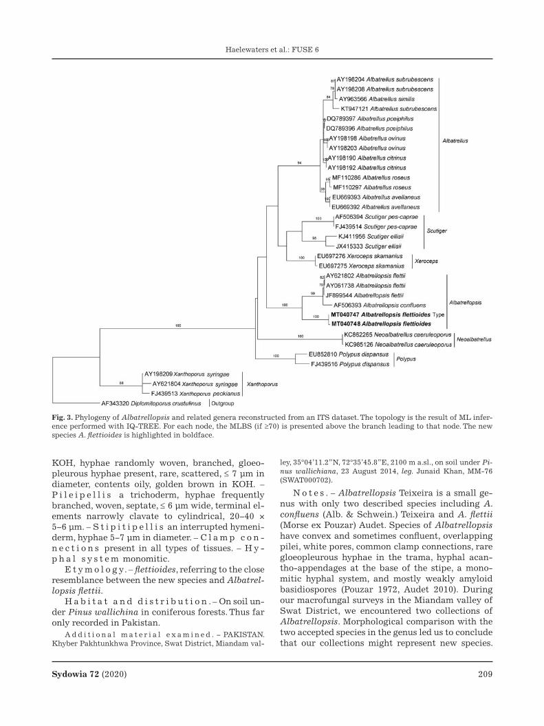
Sydowia 72 (2020) 209
Haelewaters et al.: FUSE 6
KOH, hyphae randomly woven, branched, gloeo-pleurous hyphae present, rare, scattered, ≤ 7 μm in diameter, contents oily, golden brown in KOH. – P i l e i p e l l i s a trichoderm, hyphae frequently branched, woven, septate, ≤ 6 μm wide, terminal el-ements narrowly clavate to cylindrical, 20−40 × 5−6 μm. – S t i p i t i p e l l i s an interrupted hymeni-derm, hyphae 5−7 μm in diameter. – C l a m p c o n -n e c t i o n s present in all types of tissues. – H y -p h a l s y s t e m monomitic.
E t y m o l o g y. – flettioides, referring to the close resemblance between the new species and Albatrel-lopsis flettii.
H a b i t a t a n d d i s t r i b u t i o n . – On soil un-der Pinus wallichina in coniferous forests. Thus far only recorded in Pakistan.
A d d i t i o n a l m a t e r i a l e x a m i n e d . − PAKISTAN. Khyber Pakhtunkhwa Province, Swat District, Miandam val-
ley, 35°04’11.2”N, 72°35’45.8”E, 2100 m a.sl., on soil under Pi-nus wallichiana, 23 August 2014, leg. Junaid Khan, MM-76 (SWAT000702).
N o t e s . – Albatrellopsis Teixeira is a small ge-nus with only two described species including A. confluens (Alb. & Schwein.) Teixeira and A. flettii (Morse ex Pouzar) Audet. Species of Albatrellopsis have convex and sometimes confluent, overlapping pilei, white pores, common clamp connections, rare gloeopleurous hyphae in the trama, hyphal acan-tho-appendages at the base of the stipe, a mono-mitic hyphal system, and mostly weakly amyloid basidiospores (Pouzar 1972, Audet 2010). During our macrofungal surveys in the Miandam valley of Swat District, we encountered two collections of Albatrellopsis. Morphological comparison with the two accepted species in the genus led us to conclude that our collections might represent new species.
Fig. 3. Phylogeny of Albatrellopsis and related genera reconstructed from an ITS dataset. The topology is the result of ML infer-ence performed with IQ-TREE. For each node, the MLBS (if ≥70) is presented above the branch leading to that node. The new species A. flettioides is highlighted in boldface.

210 Sydowia 72 (2020)
Haelewaters et al.: FUSE 6
Our molecular phylogenetic analysis of the ITS dataset supported this conclusion. This dataset con-sisted of 34 sequences, two of which were newly generated (Tab. 1), and 782 characters with 337 par-simony-informative ones. In the resulting ITS tree (Fig. 3), our A. flettioides sequences were placed sis-ter to a clade comprising A. confluens and A. flettii with maximum support.
Morphologically, the species is most similar to A. flettii, which has a grayish to hoary pileus and a matching hyphal anatomy. However, it can be sepa-rated based on its white hymenium that turns apri-cot to salmon upon maturation, and its drying, white, and smooth stipe (Gilbertson & Ryvarden 1986)—contrasting to the whitish grey hymenium, and concolorous, reticulate to lacerate stipe of A. flettioides. Albatrellopsis flettioides is also different in its pileus margin that exceeds the hymenium and the basidiospores that are larger compared to A. flettii (4.2−5.4 × 3.4−4.4 μm vs. 3.5–4 × 2.5–3μm; Morse 1941). The hyphal anatomy (clamped context and tramal hyphae of same size and shape) and ba-sidiospore shape of A. confluens are similar to those of A. flettioides. Albatrellopsis confluens, however, can be distinguished by its pale orange to pinkish buff pileus surface (Zheng et al. 2004).
This report does not only present a new species for science; with no previous records of Albatrellop-sis in Pakistan, it also adds a previously unreported genus to the country’s Funga (sensu Kuhar et al. 2018).
Authors: J. Khan, H. Sher, S. Hussain & A.N. Khalid
Basidiomycota, Agaricomycetes, Boletales, Boleta-ceae
Aureoboletus garciae Ayala-Vásquez & Aguirre-Acosta, sp. nov. – Figs. 4–5MycoBank no.: MB 835042
H o l o t y p u s . – MEXICO. Estado de Oaxaca, Mixistlán de la Reforma municipality, Santa María Mixistlán town, 2211 m a.s.l., 17º08’41”N, 96º05’21”W, in Quercus scytophylla (Fagales, Fagaceae) forest, 23 August 2016, leg. R. Castro-Rive-ra (MEXU 29006; holotype). Sequences ex-holotype: MH337251 (LSU), MT228979 (rpb1), MT228983 (rpb2).
D e s c r i p t i o n . – B a s i d i o m a t a pileate-stipitate, small. – P i l e u s 8–23 mm in diam., con-vex, with furfuraceous surface, dry, vivid blue (20A8-20A6), violet blue (19A6), to light blue (19A5), with some reddish (11A5) tones, margin sterile. – H y m e n o p h o r e adnate, with rounded pores 0.2 – 0.3 mm in diam., bright yellow (4A8) unchanging when cut; tubes 5–7 mm long, concolorous to the
pores. – S t i p e 30–35 × 5–6 mm, cylindrical, with furfuraceous to rimose surface, whitish to pale red (11A4) near the apex, deep red to reddish brown (9D8-9E8) in mature specimens. – C o n t e x t 5 mm thick, whitish, bruising pink (11A4) to pale red (11A3) when young, yellow when mature, context of the stipe whitish with some pale yellow tones in the middle and base. – S m e l l a n d t a s t e indistinct. – B a s i d i o s p o r e s 9–14 × 4–5(–6) μm (Lav=10, Wav=5, Q=1.7, n=35), ellipsoid to fusiform, smooth with or without conspicuous suprahilar depression, hyaline in KOH, yellowish brown in Melzer’s rea-gent, thin-walled. – B a s i d i a 25–49 × 8–9 μm, clavate, 4-spored, sterigmata 3–5 × 0.5–1.0 μm, hya-line, thin-walled. – P l e u r o c y s t i d i a 28–50 × 7–13 μm, clavate, fusoid or mucronated, hyaline in KOH, pale brown in Melzer’s reagent, thin-walled. – C h e i l o c y s t i d i a 50–65 × 7–10 μm, clavate-ventricose to fusoid, hyaline in KOH, brownish yel-low in Melzer’s reagent, thin-walled. – H y m e -n o p h o r a l t r a m a bilateral, composed of a cen-tral stratum with tubulose hyphae, 4–10 μm diam-eter, with gelatinized walls, hyaline, thin-walled; lateral stratum composed of tubulose hyphae, 5–14 μm diameter, hyaline, thin-walled. – P i l e i p e l l i s composed of a trichodermium of tu-bulose, loosely interwoven hyphae, 12–67 × 7.4–15 μm, with cylindrical, mammillate, clavate or subglobose terminal cells, hyaline in KOH, with cel-lular content in Melzer’s reagent, thin-walled. – S t i p i t i p e l l i s 70–81 μm thick, composed of tur-binate, mammillate or pedunculate caulocystidia, 35–67 × 14–19 μm, hyaline in KOH, thick-walled (1.0–1.6 μm).
E t y m o l o g y. – In honor of Dr. Jesús García Jiménez, eminent Mexican mycologist and pioneer in the study of the Mexican boletes.
H a b i t a t a n d d i s t r i b u t i o n . – Solitary under Quercus laurina and Q. scytophylla in oak and montane cloud forests, currently known from Oaxaca, Mexico.
A d d i t i o n a l m a t e r i a l e x a m i n e d . – MEXICO. Estado de Oaxaca, Mixistlán de la Reforma municipality, San-ta María Mixistlán town, 2465 m a.s.l., 17º09’30”N, 96º04’27”W, 4 September 2016, leg. O. Ayala-Vásquez (MEXU-30133, ITCV-873); Ibid., 2211 m a.s.l., 17º08’41”N, 96º05’21”W, 5 Sep-tember 2017, leg. O. Ayala-Vásquez (MEXU-30134, ITCV-983); Ibid., 2211 m a.s.l., 17º08’41”N, 96º05’21”W, 12 September 2017, leg. O. Ayala-Vásquez (ITCV-1022); Ibid., 2230 m a.s.l., 17º08’44.79”N, 96º05’19.38”W, 12 October 2017, leg. O. Ayala-Vásquez (ITCV-1028); Ibid., 2211 m a.s.l., 17º08’41”N, 96º05’21”W, 5 August 2017, leg. O. Ayala-Vásquez (MEXU-29426, ITCV-848); Ibid., 2465 m a.s.l., 17º09’30”N, 96º04’27”W, 24 July 2017, leg. O. Ayala-Vásquez (MEXU-30135, ITCV-886).
N o t e s . – The genus Aureoboletus Pouzar was proposed by Pouzar (1957), with A. gentilis (Quél.)

Sydowia 72 (2020) 211
Haelewaters et al.: FUSE 6
Fig. 4. Aureoboletus garciae, collection MEXU 29006 (holotype). a. Basidiomata. b. Detail of the pileus. c. Detail of the hymeno-phore. d. Detail of the context. e. SEM micrograph of basidiospores. Scale bars a–d 10 mm, e 10 μm.

212 Sydowia 72 (2020)
Haelewaters et al.: FUSE 6
Fig. 5. Microscopic features of Aureoboletus garciae. a. Basidiospores. b. Basidia. c. Pleurocystidia. d. Cheilocystidia. e. Caulocys-tidia. f. Elements of pileipellis. Scale bars 10 μm.

Sydowia 72 (2020) 213
Haelewaters et al.: FUSE 6
Fig. 6. Phylogeny of Aureoboletus reconstructed from a concatenated LSU–rpb1–rpb2 dataset. The MCC topology is the result of Bayesian inference performed with MrBayes. Thick lines represent BIPP ≥0.95. The new species A. garciae is highlighted in boldface.

214 Sydowia 72 (2020)
Haelewaters et al.: FUSE 6
Pouzar as type species. The genus can be found in temperate and subtropical forests (Pouzar 1957; Klofac 2010; Zhang et al. 2014, 2015b). At present, between 33 and 42 species are recognized, depend-ing on the source (Zhang et al. 2019, Species Fungo-rum 2020, Wijayawardene et al. 2020). The genus is distinguished by pileate-stipitate basidiomata; a viscid or sometimes tomentose pileus surface; an adnate hymenium; yellow tubes, with rounded to angular golden pores never bruising when touched; and a central or fusoid stipe that is inflated to acute at the base, with a glabrous to pruinose surface (Klofac 2010, Wu et al. 2016, Bessette et al. 2017). Microscopically, species of Aureoboletus are char-acterized by usually smooth, rarely longitudinally striate, subfusiform basidiospores; light yellow to yellow, bilateral hymenophoral trama with tubu-lose hyphae; and a pileipellis composed of ix-otrichoderm or sometimes a trichoderm (Wu et al. 2016).
The Mexican oak forest possesses a wide diver-sity of boletes (García-Jiménez & Garza-Ocañas 2001), which has been investigated in recent years (García-Jiménez & Garza-Ocañas 2001, Ayala-Vásquez et al. 2018, García-Jiménez et al. 2019). Nevertheless, some areas such as the mountains of south Mexico remain unexplored, with some of the sampled fungi being undescribed.
Features of A. garciae are consistent with those of the genus. The bluish colour of the pileus is the main characteristic that distinguishes A. garciae from other Aureoboletus species. Aureoboletus gar-ciae is associated with Fagaceae—potentially form-ing ectomycorrhizal associations with Quercus lau-rina (a Central American-endemic species) and Q. scytophylla (a Mexican-endemic species). The new species shares its habitat with other Aureoboletus species such as A. innixus (Frost) Halling, A.R. Bessette & Bessette, A. roxanae (Frost) Klofac, and A. russellii (Frost) G. Wu & Zhu L. Yang (Ayala-Vásquez et al. 2018, García-Jiménez et al. 2019). Bayesian inference recovered A. garciae as sister to A. tomentosus G. Wu & Zhu L. Yang with high sup-port (Fig. 6). Morphologically, this species is charac-terized by bright orange-yellow basidiomata, a tomentose pileus surface, context without colour change when bruised, and ovoid basidiospores that are shorter compared to A. garciae (Wu et al. 2016, Zhang et al. 2019). These combined morphological and molecular results confirm the status of A. gar-ciae as a new species.
Authors: O. Ayala-Vásquez, J.I. de la Fuente, C.R. Martínez-González, R. Castro-Rivera & E. Aguirre-Acosta
Basidiomycota, Agaricomycetes, Agaricales, Ento-lomataceae
Entoloma canadense Noordel., G.M. Jansen & Dima, sp. nov. – Fig. 7MycoBank no.: MB 836840
H o l o t y p u s . – Canada. Newfoundland and Labrador Province, Point Armour Historic Site, 51°27’38.16”N, 56°51’34.06”W, 7 September 2005, leg. M.E. Noordeloos, 2005127a (L0608149; holotype). Sequences ex-holotype: MT940867 (ITS), MK277990 (LSU).
D e s c r i p t i o n . – B a s i d i o m a t a medium-sized, tricholomatoid. – P i l e u s 30–40 mm in diam., conico-convex, expanding to plano-convex with low umbo, with deflexed margin, not hygroph-anous, not translucently striate, moderately dark brown with a slight violaceous-purple or plum-colored tinge when very fresh (7.5YR6-5/2-4; 10 YR 2-5/3-4) or 2.5R 5/4. 5YR 4-3/3), entirely very finely squamulose. – L a m e l l a e moderately crowded (L=25–50, l=5–7–9), adnate-emarginate, ventricose, pale cream-colored white then sordid grey-pink with an entire or eroded, concolorous edge. – S t i p e 25–50 × 4–10 mm, cylindrical, entirely fibrillose-striate with pale vinaceous-brown fibrils and flocks, white and smooth at base. – C o n t e x t pale pinkish grey. – S m e l l a n d t a s t e indistinct. – B a s i d i -o s p o r e s (6.0–)6.9–9.2 × 4.6–6.9 μm, average 7.6–8.1 × 5.4–5.8 μm, Q=1.2–1.7, Qav=1.41, heterodiamet-rical 6–8 angled in side-view, thin-walled with rather weak angles. – B a s i d i a 30–37 × 9.5–10.5 μm, clavate, 4-spored, clamped. – L a m e l l a e d g e heterogeneous with scattered cheilocystidia. – C h e i l o c y s t i d i a 32–55 × 8–20 × 5–10 μm, rather variable, the majority lecithiform, sometimes vesiculose, up to 50 μm wide. – H y m e n o p h o r a l t r a m a regular, made up of cylindrical to inflated elements, 100–250 × 5–20 μm wide, also very wide hyphae up to 50 μm present. – P i l e i p e l l i s cutis to trichoderm, cylindrical hyphae 6–20 μm wide, with pale brown intracellular pigment. – C l a m p c o n n e c t i o n s present in hymenium, rare to ab-sent in other tissues.
E t y m o l o g y. – Referring to Canada, where the holotype of the new species was collected.
H a b i t a t a n d d i s t r i b u t i o n . – In a semi-natural grassland, grazed by wildlife, near Abies balsamica (Pinales, Pinaceae) and in a coastal arctic heathland with Empetrum (Ericales, Ericaceae) and dwarf Salix (Malpighiales, Salicaceae). Thus far only known from eastern Canada and the eastern USA.
A d d i t i o n a l m a t e r i a l e x a m i n e d . – CANADA. Newfoundland and Labrador Province, Gros Morne National Park, Killdevil Camp, 49°27’10.01”N, 57°45’13.86”W, 4 Sep-tember 2005, leg. M.E. Noordeloos, 2005121 (L0608150).

Sydowia 72 (2020) 215
Haelewaters et al.: FUSE 6
N o t e s . – Entoloma canadense belongs to the Trichopilus clade of Entoloma P. Kumm. The phylo-genetic placement in this clade (Fig. 8) is morpho-logically supported by the presence of lecythiform cheilocystidia and trichodermal pileipellis. We gen-erated 19 new ITS sequences representing ten spe-
cies in the Trichopilus clade—thereby greatly ex-panding the coverage of this clade in public data-bases. Within Trichopilus, E. canadense is distinct from other described species and placed sister to a clade with E. porphyrescens E. Horak from Tasma-nia and E. porphyroleucum sp. nov. from Vietnam
Fig. 7. Entoloma canadense, collection L0608149 (holotype) a. Basidiomata in situ. b. Basidiospores. c. Cheilocystidia, d. Pileipel-lis. Scale bars a–d 10 μm.

216 Sydowia 72 (2020)
Haelewaters et al.: FUSE 6
(Fig. 8). Morphologically it is distinct from these species because of the minutely squamulose, mod-erately dark to dark brown pileus with violaceous to violaceous-red (plum) tinges when fresh, its small basidiospores, and scattered, in majority lecithi-form cheilocystidia (Fig. 7). Entoloma fuscomargin-atum P.D. Orton [= E. elodes (Fr.) P. Kumm.] has a smoother, micaceous pileus without violaceous tinges, and larger basidiospores. The new species may be misidentified as E. porphyrophaeum (Fr.) P. Karst. [= Trichopilus porphyrophaeus (Fr.) P.D. Or-ton], which is a European species with a similarly colored pileus but differs in a stouter habitus, much larger basidiospores, and cheilocystidia often with
a long, moniliform apex. Entoloma frigidum from northern Sweden is macroscopically similar to E. canadense but has much larger basidiospores. Ento-loma scabinellum Peck, a poorly known species de-scribed from eastern USA, has similar microscopic characters, but there is reasonable doubt that it represents the same species, since it was described as a small Nolanea (Fr.) P. Kumm. (pileus of 12–20 mm in diam., minutely scabrous). Sequences are lacking and the type is unavailable for sequencing, but E. scabinellum may belong to Trichopilus on ac-count of the lecithiform cystidia.
Authors: M.E. Noordeloos, G.M. Jansen & B. Dima
Fig. 8. Phylogeny of Entoloma reconstructed from an ITS dataset. The topology is the result of ML inference performed in PhyML. MLBS values (>60) are shown at the nodes. Sequences newly generated for this study are highlighted in boldface; newly described species are highlighted in blue.

Sydowia 72 (2020) 217
Haelewaters et al.: FUSE 6
Basidiomycota, Agaricomycetes, Agaricales, Ento-lomataceae
Entoloma frigidum Noordel., E. Larss., Bendiksen, G.M. Jansen & Dima, sp. nov. – Fig. 9MycoBank no.: MB 836841
H o l o t y p u s . – SWEDEN. Torne Lappmark, Jukkasjärvi, Latnajavagge, 8 August 2007, leg. E. Larsson, EL46-07 (GB-
0202302; holotype). Sequences ex-holotype: MT585817 (ITS+LSU).
D e s c r i p t i o n . – B a s i d i o m a t a small to medium-sized, tricholomatoid. – P i l e u s 30–50 mm in diam., conical only slightly expanding to conico-convex or convex with umbo, with straight or slightly deflexed margin, not hygrophanous, not translucently striate, uniformly mouse grey or with
Fig. 9. Entoloma frigidum. a. Basidiomata in situ, collection GB-0202302 (holotype). b. Basidiomata in situ, collection EL189-16. c–f. Microstructures, from collection GB-0202302 (holotype). c. Basidiospores. d. Cheilocystidia. e. Pileipellis. f. Stipitipellis. Scale bars 10 μm.

218 Sydowia 72 (2020)
Haelewaters et al.: FUSE 6
a slight porphyry-purple sheen, very finely tomen-tose-radially fibrillose all over, often with a glace impression in parts, not squamulose. – L a m e l l a e normally distant, deeply adnate-emarginate, ven-tricose, pale then pink with slight grey tinge, with more or less concolorous, slightly eroded edge. – S t i p e 60–90 × 3–7 mm, gradually but slightly broadening towards base, more or less concolorous with pileus, base paler, almost white, innately fibril-lose lengthwise. – C o n t e x t firm, pale in inner part, else greyish. – S m e l l a n d t a s t e indis-tinct. – B a s i d i o s p o r e s (10–)11.0–15 × 6.0–9.5 μm, average 13.0 × 8.3 μm, Q=1.3–1.9, Qav =1.48–1.57, with a bumpy-nodulose outline, many of them with a star-formed content. – B a s i d i a 38–68 × 12–15 μm, 4-spored, clavate, clamped. – L a m e l l a e d g e heterogeneous. – C h e i l o c y s t i d i a 36–70 × 6–20 × 5.5–13.5 μm, lageniform to tibiiform, usually with broad basal part, short to long neck, and rounded capitulum, frequently septate, clamped. – P l e u r o -c y s t i d i a absent. – H y m e n o p h o r a l t r a m a regular, made up of cylindrical hyphae, 4–25 μm wide, no clamps seen. – P i l e i p e l l i s a cutis of long, terminal elements, 90–350 × 5–23 μm, with brown intracellular pigment, no clamps seen. – S t i p i t i p e l l i s a cutis of cylindrical hyphae, 4.5–23 μm wide with pale brown intracellular pigment. – Va s c u l a r h y p h a e present in trama and in stipitipellis. – C a u l o c y s t i d i a present at apex of stipe, 38–72 × 11–17 μm, clavate, lageniform, some-times tibiiform. – C l a m p c o n n e c t i o n s pre-sent in hymenium at base of basidia, elsewhere rare or absent.
E t y m o l o g y. – frigidum = cold, referring to the climate in which this species thrives.
H a b i t a t a n d d i s t r i b u t i o n . – In oligo-trophic alpine heath with and Arctostaphylos, Em-petrum (Ericales, Ericaceae), Betula nana (Fagales, Betulaceae), and Salix spp. (Malpighiales, Salicace-ae), and in sandy pine forest with Betula nana, dominated by Empetrum and Vaccinium vitis-idaea (Ericales, Ericaceae). Currently only known from the northernmost parts of Sweden and Norway.
A d d i t i o n a l s p e c i m e n s e x a m i n e d . – Ibid. (L0607966, isotype). – SWEDEN. Lule Lappmark, Jokkmokk, Padjelanta, Ajajaure, 16 August 2016, leg. E. Larsson, EL189-16 (GB-0202303); Ibid., Padjelanta, Ajajure, 16 August 2016, leg. J. Olsson (GB-0202316); Ibid., Padjelanta, Vielggisbakte, 12 August 2016, leg. S. Kuoljok (GB-0202315); Torne Lapp-mark, Jukkasjärvi, Abisko, Latnja, 16 August 2013, leg. P.-A. Moreau, PAM13-23 (GB-0202314). – NORWAY. Finnmark, Karasjok, Basevuovdi N by River Øvre Anarjohka, 20 August 2013, leg. A.-M. Bendiksen, EB 201/13 (O-F-252043).
N o t e s . – Entoloma frigidum, according to our molecular phylogenetic results, belongs to the
Trichopilus clade where it forms the sister species of E. porphyrophaeum (Fig. 8) to which it has a su-perficial resemblance, particularly with regard to the size and shape of the basidiomats, but with a mouse-grey color, sometimes with slightly purplish tinge. In the ITS region, E. frigidum and E. porphy-rophaeum differ by more than 60 substitution and indel positions. Microscopically, E. frigidum can be distinguished by the somewhat larger basidiospores and shape of its cheilocystidia (Fig. 9). As far as we know, this species has a northern boreal-alpine dis-tribution in northernmost Scandinavia. Entoloma fuscotomentosum F.H. Møller, another species with a nordic distribution, seems to prefer coastal habi-tat, and differs by the small basidiospores and grey-brown, fibrillose-subsquamulose pileus. To our knowledge, there are no other morphologically sim-ilar species occurring in the same habitat with which E. frigidum can be confused.
Authors: M.E. Noordeloos, E. Larsson, E. Bendik-sen, G.M. Jansen & B. Dima
Basidiomycota, Agaricomycetes, Agaricales, Ento-lomataceae
Entoloma porphyroleucum O.V. Morozova, Noordel. & Dima, sp. nov. – Fig. 10MycoBank no.: MB 835245
H o l o t y p u s . – VIETNAM. Binh Phuoc Province, Bu Gia Map District, Bu Gia Map National Park, right bank of Dak Ca River, 12°12’15.41”N, 107°12’13.25”E, 370 m a.s.l., on soil, 19 May 2011, leg. O. Morozova (LE 312490; holotype). Se-quences ex-holotype: MT940862 (ITS), MT950278 (LSU).
D e s c r i p t i o n . – B a s i d i o m a t a medium-sized, collybioid to tricholomatoid. – P i l e u s 30–60 mm in diam., conico-convex expanding to plano-convex, with small conical umbo, depressed when old with small acute umbo in the depression, with more or less straight margin, not hygrophanous, not translucently striate, light purplish-brown (8C3, 8D3–4, 9C3, up to 13D4), darker in the center (8E3, 9E4, 10E4–5), densely radially fibrillose in the cent-er, fibrils more spread towards the margin, showing white background. – L a m e l l a e moderately dis-tant, adnexed, adnate-emarginate, ventricose, whit-ish or cream at first, becoming pink and brownish-pink, with white dentate edge. – S t i p e 50–100 × 4–10 mm, cylindrical, distinctly broadened towards the base, white, longitudinally striate, white tomen-tose at base. – C o n t e x t white, unchanging. – S m e l l faint. – Ta s t e not reported. – B a s i d i o -s p o r e s of two types, (i) from 4-spored basidia, small, 7.5–9.5 × 5.5–7 μm, average 8.5 × 6 μm, Q=1.3–1.5, Qav=1.4, heterodiametrical, with 5–6 angles in

Sydowia 72 (2020) 219
Haelewaters et al.: FUSE 6
Fig. 10. Entoloma porphyroleucum, collection LE 312490 (holotype). a. Basidiomata in situ. b. Type locality. c. Basidiospores. d–e. Basidia. f. Cheilocystidia. g. Pileipellis. Scale bars a 1 cm, c–g 10 μm.

220 Sydowia 72 (2020)
Haelewaters et al.: FUSE 6
side-view; and (ii) from 1–2-spored basidia, large, 10–12 × 5.5–8 μm, average 11 × 6.5 μm, Q=1.4–1.9, Qav=1.7, irregular, with poorly defined angles. – B a -s i d i a 22–27 × 8.5–9 μm, 1–4-spored, clavate to cy-lindrical, clampless. – C h e i l o c y s t i d i a 17–31 × 8–17 μm, forming a sterile edge, of intermixed two types: vesiculose to spheropedunculate or lageni-form. – P i l e i p e l l i s a cutis with the transition to a trichoderm of cylindrical to slightly inflated or fusiform hyphae 10–20 μm wide with swollen ter-minal elements with slightly thickened apex and brownish intracellular pigment. – C a u l o c y s t -i d i a absent. – C l a m p c o n n e c t i o n s absent.
E t y m o l o g y. – From Greek ‘πορϕυρεος’ (pur-pureus) and ‘λευκος’ (white, light), referring to the light purple tint of the pileus surface and white stipe.
H a b i t a t a n d d i s t r i b u t i o n . – In small groups on soil in tropical evergreen mixed forests. Known from Vietnam.
N o t e s . – Entoloma porphyroleucum is a re-markable species with purplish-brown, radially fi-brillose pileus and white stipe. In spite of the ab-sence of typical lecithiform cheilocystidia, it is placed in the Trichopilus clade based on molecular phylogenetic data (Fig. 8). Entoloma porphyroleu-cum resembles E. porphyrescens based on its pileus color, the presence of small basidiospores, and the two types of cheilocystidia (Horak 1973). However, E. porphyrescens can be differentiated from the new species by the colored stem the predominantly lecythiform cheilocystidia. In addition, the geo-graphical distribution and habitat of these two spe-cies are completely different. Entoloma porphyres-cens was described from New Zealand from Nothof-agus–Dacrydium–Podocarpus forests; it also fre-quently occurs in Tasmania in wet Eucalyptus and Nothofagus forests (Noordeloos & Gates 2012). Fi-nally, the new species distinctly differs from the Eu-ropean E. porphyrophaeum (Fr.) P. Karst. by its white stipe, small basidiospores, and the absence of lecithiform cheilocystidia (Noordeloos 1992).
Authors: O.V. Morozova, M.E. Noordeloos & B. Dima
Basidiomycota, Agaricomycetes, Boletales, Boleta-ceae
Erythrophylloporus flammans O.V. Morozova, T.H.G. Pham & E.S. Popov, sp. nov. – Fig. 11MycoBank no.: MB 835244
H o l o t y p u s . – VIETNAM. Binh Phuoc Province, Bu Gia Map District, Bu Gia Map National Park, environs of ranger station 2, along road to Bu Gia Map, 12°11’31.3”N,
107°12’16.2”E, 540 m a.sl., on soil in semi-evergreen tropical forests with Fagaceae (Lithocarpus spp.) and Dipterocarpace-ae, 30 November 2018, leg. O. Morozova & E. Popov (LE 312491; holotype). Sequences ex-holotype: MT298114 (ITS), MT293596 (tef1).
D e s c r i p t i o n . – B a s i d i o m a t a small to medium sized, fleshy. – P i l e u s 15–35 mm diam, initially hemispherical, becoming convex to appla-nate, with incurved margin at first, reddish orange, flame red (7A7–8, 7B8, 8A7–8, 8B8), surface dry, ve-lutinose, tomentose or felted, minutely scaled. – L a m e l l a e decurrent, subdistant, thick, up to 4 mm broad, salmon, deep orange, pastel red, red-dish orange (6A4–8, 7A5–7, 8A5–7). – S t i p e 15–30 × 6–8 mm, almost cylindrical, sometimes curved, solid; reddish orange to yellowish red (6A7, 8A7) at apex, orange to vivid yellow at base (up to 3A6–8), punctated with orange, reddish orange to orange red dots; with yellow (2A5–7) basal mycelium. – C o n t e x t light to vivid yellow (3A7–8, 4A7–8),
Fig. 11. Erythrophylloporus flammans, collection LE 312491 (holotype). a. Basidiomata in situ. b. Type locality: Vietnam, Bu Gia Map National Park, the road in a tropical evergreen mixed forest. c. Basidiospores. d. Basidium. e. Pleurocystidia. f. Cheilocystidia. g. Pileipellis. h. Caulocystidia. Scale bars a 1 cm, c–h 10 μm.

Sydowia 72 (2020) 221
Haelewaters et al.: FUSE 6
Fig. 12. Phylogeny of Erythrophylloporus isolates reconstructed from a tef1 dataset. The topology is the result of Bayesian infer-ence performed with MrBayes. For each node, the BIPP (if >0.95) is presented above the branch leading to that node. The scale bar represents the number of nucleotide changes per site.

222 Sydowia 72 (2020)
Haelewaters et al.: FUSE 6
reddish orange under the surface, turning dark blue to blackish blue. Smell weak, taste mild. – B a s i d -i o s p o r e s 6.2–8.2 × 5–6.5 μm, average 7 × 5.7, Q=1.1–1.3, Qav=1.2, broadly ellipsoid, to ovoid, smooth, thin walled, yellowish to yellowish brown in KOH, with a large oil drop, inamyloid. – B a s i d -i a 41–58 × 7.8–10.4 μm, 2–4-spored, subcylindrical to narrowly clavate, clampless. – L a m e l l a edge heterogeneous with scattered cheilocystidia. – C h e i l o c y s t i d i a 50–105 × 8–18 μm, fusiform or lageniform, thin walled, colorless. – P l e u r o c y s t -i d i a 50–115 × 7.5–15 μm, originating in subhyme-nium and hymenophoral trama, broadly fusiform or lageniform, sometimes septate, thin walled, color-less or containing yellowish-brown to brown in KOH pigments. – H y m e n o p h o r a l t r a m a sub-parallel, slightly divergent. – P i l e i p e l l i s a trichoderm made up of palisade of interwoven cy-lindrical cells 4–8 μm broad, with terminal cells 33–66 × 5–11 μm, cylindrical to narrowly lageniform or capitate with slightly thickened walls and brownish yellow in KOH intracellular pigment. – S t i p i t i p e l l i s a caulohymenium in upper part, then trichoderm with abundant caulocystidia, up to 100 μm thick. – C a u l o c y s t i d i a 55–100 × 6–11 μm, narrowly conical, narrowly fusiform or lageni-form, with slightly thickened walls and brownish yellow in KOH intracellular pigment, or hyaline. – C l a m p c o n n e c t i o n s absent.
E t y m o l o g y. – Referring to the bright reddish orange color of the basidiomata, like a flame, ‘flam-mans’ (Latin) – flaming.
H a b i t a t a n d d i s t r i b u t i o n . – Solitary or in groups on naked soil in semi-evergreen tropical forests with Fagaceae (Lithocarpus spp.) and Dip-terocarpaceae of Vietnam.
A d d i t i o n a l m a t e r i a l e x a m i n e d . – VIETNAM. Binh Phuoc Province, Bu Gia Map District, Bu Gia Map Na-tional Park, environs of ranger station 2, path to Dak Ca River, 12°12’3.9”N, 107°12’15.6”E, 420 m a.s.l., 22 May 2011, leg. O. Morozova (LE 312521); Ibid., along road to Bu Gia Map, 12°11’31.3”N, 107°12’16.2”E, 540 m a.s.l., 25 November 2017, leg. E. Popov (LE 312492).
N o t e s . – Genus Erythrophylloporus Ming Zhang & T.H. Li, the lamellate representative of Bo-letaceae, was recently described from China with E. cinnabarinus Ming Zhang & T.H. Li as the type spe-cies (Zhang & Li 2018). Erythrophylloporus is nest-ed within the Pulveroboletus-group-clade and is very distant from the morphologically similar Phyl-loporus (subfamily Xerocomoideae), although both possess lamellae. Erythrophylloporus flammans is a typical representative of the genus, characterized by the lamellate hymenophore, the intense reddish
orange color of the pileus and lamellae, bright yel-low basal mycelium, ovoid, ellipsoid to broadly el-lipsoid basidiospores with non-bacillate surface, and pleurocystidia originating from the subhyme-nium or from hymenophoral trama (Zhang & Li 2018, Vadthanarat et al. 2019). Since the vast major-ity of sequence data available for Erythrophyllopo-rus are tef1, we performed a phylogenetic analysis based on this single gene. According to our tef1 se-quence data, our species is rather distant from other representatives of the genus (p-distance 12–17 %), but it forms a monophyletic clade with them with maximum support (Fig. 12). Morphologically it dif-fers from E. paucicarpus Raspé, Vadthanarat & Lumyong by bluing context (vs. reddening), from E. suthepensis Vadthanarat, Raspé & Lumyong by its larger basidiospores (6–8 × 5–6.5 μm vs. 4.5–6 × 3.5–4.5 μm), and from E. cinnabarinus Ming Zhang & T.H. Li by the absence of yellowish brown pigments in cheilocystidia, and slightly larger (6–8 × 5–6.5 μm vs. 5.5–7 × 4.5–5.5 μm) and more ovoid basidiospores (Qav=1.2 vs. 1.3). The description of Phylloporus coc-cineus Corner from Singapore (Corner 1970) also corresponds to an Erythrophylloporus species as evidenced by the crimson to scarlet lamellate ba-sidiomata with orange to orange-red lamellae and yellow basal mycelium, and broadly ellipsoid to subglobose smooth basidiospores, which are larger (7.5–10 × 6.5–8 μm) compared to E. flammans.
Authors: O.V. Morozova, P.T.H. Giang & E.S. Popov
Basidiomycota, Agaricomycetes, Agaricales, Om-phalotaceae
Marasmiellus boreoorientalis Kiyashko, sp. nov. – Figs. 13–16MycoBank no.: MB 833087
H o l o t y p u s . – RUSSIAN FEDERATION. Kamchatka Territory, Yelizovo District, Nature Park “Volcanoes of Kam-chatka”, special protected area “Nalychevo Nature Park”, W-foothills of Avachinskaya Sopka volcano, thicket of Alnus al-nobetula subsp. fruticose (Fagales, Betulaceae), on alder litter, 53°15’39.5”N, 158°44’35.7”E, 906 m a.s.l., 24 August 2017, leg. N.V. Psurtseva (LE 323323; holotype). Sequences ex-holotype: MN597452 (ITS), MN597444 (LSU).
D e s c r i p t i o n . – B a s i d i o m a t a small, marasmioid or micromphaloid. – P i l e u s up to 20 mm, applanate with straight, deflexed or reflexed margin, sometimes with shallow central depression, radially grooved, occasionally slightly rugulose at center, finely innately fibrillose to subsquamulose, hygrophanous, sudan brown (6D8) to burnt sienna (7D8) or henna (7E8) at central disc, not fading with

Sydowia 72 (2020) 223
Haelewaters et al.: FUSE 6
age, brighten to brownish yellow (5C8) or greyish orange (5B4) towards the margin especially along grooves, margin whitish, becoming wavy and crenu-late with age. – L a m e l l a e narrowly adnate, rath-er distant, L=16–19, with numerous lamellulae, ven-tricose, slightly thickened, whitish, light brown (7D4–5) spotted when old, edge concolourous, sometimes slightly eroded. – S t i p e 18–26 × 1–1.5 mm, usually cylindrical but sometimes flat-tened and with longitudinal groove, tapering to-wards the base, flexible when moist, tough when dry, brown (7E8) or reddish brown (8E8), more or less evenly coloured or darkening to very dark brown (8F5–3) towards the base, with distinct velu-tinous hyaline vesture sometimes weakly developed in the middle part, hairy-tomentose at the base, hairs dark brown. – C o n t e x t very thin, concol-ourous with surface, odour and taste not observed. – B a s i d i o s p o r e s (6.6–)7.1–9.0(–12.7) × 3.4–4.8(–7.3) μm, average 8.6±1.2 × 4.3±0.7 μm, Q=1.6–2.4, Qav=2.0±0.6, varying in shape and size, ellipsoid to lacrymoid or subfusoid in face view, sometimes with broad apiculus, with several drops, thin-walled, smooth, inamyloid. – B a s i d i a 19.8–33.3 × 4.9–6.9 μm, clamped, narrowly clavate to subcapi-tate, (2)4-spored, occasionally with very large ster-igmata (up to 10 μm), with drops, which are some-times yellowish. – B a s i d i o l e s 15.3–30.4 × 3.0–6.2 μm, cylindrical, subclavate or with slightly re-stricted apex. – C h e i l o c y s t i d i a abundant, 14.8–50.5 × 8.2–20.5 μm, broadly clavate to pyriform or spheropedunculate, sometimes with small con-strictions or septate, frequently with thickened up to 1 μm brownish walls. – P l e u r o c y s t i d i a ab-sent or inconspicuous. – P i l e i p e l l i s composed of radially oriented, rarely branched, cylindrical or inflated hyphae lacking diverticulate branchlets, (2.8–)4.8–15.8(–18.1) μm diam., often with thick-ened walls, pigmented, roughly encrusted, encrus-tation brownish, spiral, zebroid to irregular, some-times with solid calluses in profile, terminal cells clavate or broadly fusiform, more rarely tapering, (12.6–)30.5–94.7 × (6.0–)8.3–18.2(–21.2) μm. – S t i p -i t i p e l l i s a cutis composed of cylindrical, thick-walled, pigmented cells (1.9–)2.5–7.6(–8.5) μm in diam., hyphae smooth or encrusted as in pileipellis, sometimes producing roughly encrusted clavate terminal cells. – C a u l o c y s t i d i a abundant, (18.8–)37.2–87.7 × 7.6–13.7 μm, arising as side branches of surface hypha or as differentiated ter-minal cells, thick-walled (up to 1.5 μm), brownish pigmented, more or less cylindrical, flexuous or utriform on long thin stalk. – C l a m p c o n n e c -t i o n s present in all tissues.
C u l t u r e c h a r a c t e r i s t i c s . – M e a n g r o w t h r a t e 2.7 mm/day on BWA and MEA, 2.3 mm/day on PDA, plates covered in 16 days in BWA and MEA, in 18 days on PDA. – M a c r o m o r p h o l -o g y of colonies depending on cultural media, but outlines even and marginal hyphae appressed on all media. – C o l o n y m a t thick woolly-cottony on BWA, thin appressed woolly-filamentous with radi-ally ordered hyphae on MEA and PDA, vaguely zon-ate with tendency to develop brownish-ochraceous pigmentation, on BWA with concentric bands of
Fig. 13. Basidiomata of Marasmiellus boreoorientalis in the field, collection LE 323323 (holotype). Scale bar 10 mm.
Fig 14. Microstructures of Marasmiellus boreoorientalis. a. Basidiospores. b. Basidia and basidioles. c. Cheilocystidia. d. Caulocystidia and encrusted hyphae with terminal stipiti-pellis cells. e. Hyphae of pileipellis. Scale bars 10 μm.

224 Sydowia 72 (2020)
Haelewaters et al.: FUSE 6
thick dense mycelium with pigmented parts, on MEA and PDA with dense center and very thin, transparent mycelial mat at the colony periphery, on MEA with whitish woolly-powdery zone ca. 35 mm around inoculum and small brownish-ochraceous spots at the periphery, on PDA also with pigmented crust ca. 10 mm around inoculum, re-verse bleached on all media, on MEA and PDA with brown spots (especially on PDA). – S m e l l weak, undefinable. – O x i d o r e d u c t a s e s test negative. – M i c r o m o r p h o l o g y not so strongly depend-ing on cultural media as micromorphology; clamps on all hyphae, simple, slightly compressed, some-times sprouting, on BWA aerial hyphae 2.0–2.5 μm
diam., usually rarely septate and branched, some-times with slightly thickened walls, substrate hy-phae 2.0–5.0 μm diam., more often septate and brush-like branched with short irregular branches, on MEA and PDA aerial and substrate mycelia do not clearly separate from each other and look like substrate mycelia on BWA, but on PDA hyphae more vacuolated; crust is formed from hyphae 2.0–2.5 μm in diam. with slightly thickened and pig-mented walls, short-celled and abundantly branched; everywhere on all media hyphae form round to irregular shaped swellings up to 12 μm in diam. with short cylindrical outgrowths, usually in-tercalary, rarely in terminal position; also on all me-dia clusters of brownish crystals are found.
E t y m o l o g y. – boreoorientalis—meaning “north-eastern”—indicating the geographical loca-tion of the holotype, northeastern Russia.
H a b i t a t a n d d i s t r i b u t i o n . – On alder lit-ter (Alnus alnobetula subsp. fruticosa). Only known from the type locality in the foothills of Avachinskaya Sopka volcano, eastern Kamchatka, Russia.
Fig. 15. Pure cultures of Marasmiellus boreoorientalis in 90-mm Petri dishes, after 8 weeks of growth. a. Culture on BWA. b. Cul-ture on MEA. c. Culture on PDA.
N o t e s . – The generic limits and relationships of the two largest genera of Omphalotaceae, Maras-miellus Murrill and Gymnopus (Pers.) Roussel, along with Micromphale Gray, have been discussed for a long time (Mata et al. 2004b, Wilson & Desjar-din 2005, Mešic et al. 2011, Petersen & Hughes 2016, Sesli et al. 2018, Oliveira et al. 2019). Most studies recognized the polyphyletic nature of Gymnopus s.l. as well as the unstable concept of both Marasmiel-lus and Micromphale. Oliveira et al. (2019) carried out a taxonomical revision of Gymnopus, Maras-miellus, and Micromphale on the basis of ITS and combined ITS–LSU phylogenetic analyses. The au-thors transferred species of Gymnopus sect. Vesti-
pedes to Marasmiellus and described a new genus Paragymnopus J.S. Oliveira to accommodate mem-bers of Micromphale sect. Perforantia. To clarify the phylogenetic position of the new species Maras-miellus boreoorientalis, we conducted BI and ML analyses of ITS sequences, resulting in two trees with the same topology (Fig. 17). Our phylogenetic analyses revealed two strongly supported major clades that correspond well to Gymnopus sensu stricto (s.s.) and Marasmiellus, and a third sister clade corresponds to Paragymnopus (sensu Oliveira et al. 2019). The Marasmiellus clade consists of sev-eral linages; M. boreoorientalis is nested in a strong-ly supported subclade with M. dichrous (Berk. & M.A. Curtis) J.S. Oliveira, M. istanbulensis E. Sesli, Antonín & E. Aytaç, M. micromphaloides (R.H. Pe-tersen & K.W. Hughes) J.S. Oliveira, and one uncul-tured specimen that may represent an undescribed taxon. Our new species is clearly distant from the rest of members of the subclade. The ITS sequence of M. boreoorientalis shares 89.53–91.97 % identity (query cover 95–100 %) with sequences of M. di-

Sydowia 72 (2020) 225
Haelewaters et al.: FUSE 6
Fig. 16. Marasmiellus boreoorientalis on PDA, after 8 weeks of growth. a–b. Characteristic swellings of vegetative hyphae of Marasmiellus boreoorientalis. Scale bars 10 μm.
chrous, 91.51 % (query cover 78 %) with M. istan-bulensis, and 89.3 % (query cover 85 %) with M. micromphaloides.
In the field, M. boreoorientalis may resemble Paragymnopus foliiphilus (R.H. Petersen) J.S. Ol-iveira and P. perforans (Hoffm.) J.S. Oliveira, both occurring on litter, because of their rather small fruitbodies with applanate radially grooved pileus and vestured dark brown stipes. The new species
can be distinguished from those species by the ab-sence of a mucoid matrix in its pileipellis. In addi-tion, M. boreoorientalis combines a number of other characteristic features: a brighter and contrastingly colored pileus lacking pinkish tints, slightly larger basidiospores compared to P. foliiphilus and P. per-forans, numerous well-developed cheilocystidia of-ten with thickened brownish walls, roughly en-crusted hyphae of pileipellis sometimes having ven-tricose terminal cells, absence of pileal hairs, its habitat on alder litter. Finally, M. boreoorientalis is phylogenetically very distantly related to both P. fo-liiphilus and P. perforans (Fig. 17).
Among phylogenetically close species, M. di-chrous is more robust and inhabits dead wood of broadleaved trees, mainly Fagus. Further, it differs from M. boreoorientalis in having a dark brown to reddish brown pileus eventually fading to light red-dish-brown or clay overall, an olive brown stipe
base with a tuberculate knob, somewhat larger ba-sidiospores, and diverticulate branchlets on pileipellis hyphae. Marasmiellus dichrous also grows more slowly (mean growth rate on BWA 1.9 mm/day, on MEA 1.5 mm/day, on PDA 1.4 mm/day) and possesses clear differences in colony mor-phology on all media (an appressed to submerged uneven advancing zone, thinner and more pigment-ed mat with a stronger developed crust, unbleached
cultural media, and the absence of swellings with outgrowths). The new species is distinguished from M. micromphaloides by its contrastingly colored cap with orange-brown center and whitish margin; distinctly larger basidiospores; broader cheilocyst-idia; the absence of pileocystidia; and straight, non-branched caulocystidia. Marasmiellus istanbulensis differs from M. boreoorientalis by paler colored ba-sidiomata, shorter basidiospores with Qav=1.7, smooth stipitipellis hyphae, and caespitose growth on bark of dead Quercus petraea wood (Fagales, Fagaceae).
The type locality is situated at the western foot-hills of Avachinskaya Sopka, an active stratovolca-no. The last large eruption occurred in 1945, at which time volcanic ashes fell on a distance of 400 km from the crater. The total volume of an-desite–basalt explosion material was about 0.3 km3 (Melekescev et al. 1994). The vegetation was de-

226 Sydowia 72 (2020)
Haelewaters et al.: FUSE 6
Fig. 17. Phylogeny placement of Marasmiellus boreoorientalis inferred from an ITS dataset. The topology is the result of Bayesian inference performed in MrBayes. For each node, support values (MLBS³85/BIPP≥0.85) are presented above the branch leading to that node. The new species is highlighted in boldface.

Sydowia 72 (2020) 227
Haelewaters et al.: FUSE 6
stroyed over an area of ca. 200 km2 (Grishin 2003). As a result, the contemporary landscapes of this territory are relatively young. The eastern volcanic region of Kamchatka is characterized by a rather poor and uniform vegetation. The upper limit of the forest belt (600–900 m a.s.l.) is composed of mono-dominant thickets of Alnus alnobetula subsp. fruti-cosa and Pinus pumila (Pinales, Pinaceae), which do not form intermixed forest stands. This allows us to presume a wider distribution of M. boreoorienta-lis, at least in Kamchatka. It can be also expected that this species will be found in the continental part of the Russian Far East and, perhaps, in the mountains of Japan.
Authors: A.A. Kiyashko & N.V. Psurtseva
Basidiomycota, Agaricomycetes, Agaricales, Om-phalotaceae
Marasmiellus longistipes Muh. Ali, Niazi & Khalid, sp. nov. – Figs. 18–19MycoBank no.: MB 831702
H o l o t y p u s . – PAKISTAN. Khyber Pakhtunkhwa Prov-ince, Abbottabad District, Ayubia National Park, 33°51’54.83”N, 73°8’19.57”E, 2400 m a.s.l., 15 July 2016, leg. M. Ali & A.R. Niazi, KH 55 (LAH 35979; holotype). Sequences ex-holotype: MK957247 (ITS).
D e s c r i p t i o n . – P i l e u s 18–30 mm in diam., convex or plano-convex, membranous, slightly umbonate, striated, glabrous, creamy (10YR/9) in center with grayish tinge becoming light (2.5Y9/2) towards margin, delicate; margin slightly wavy, uplifted, entire becomes ruptured at maturity; context thin, creamy, 0.5–1.0 mm thick. – L a m e l l a e adnexed, entire, thorough, white (2.5R9/2) at early stage, while pale white (2.5YR8/4) when mature. – S t i p e 70–105 × 3–5 mm, central, equal to sub-equal towards base, non-striated, light brown (10R4/4) towards apex to dark brown (2.5YR3/4) towards base, and entirely pubescent. – B a s i d i o s p o r e s (6.0–)6.5–9.0(–9.5) × (2.0–)3.0–3.5(–4.0) μm, Q=2.0–3.5, Qav=2.6, oblong or narrowly fusiform apiculate, guttulate, smooth, thin-walled, inamyloid, hyaline in 5 % KOH. – B a s i d i a 15–21.5 × 5.6–7.2 μm, narrowly clavate, bisporic to tet-rasporic, thin-walled, hyaline in 5 % KOH. – P l e u -r o c y s t i d i a absent. – C h e i l o c y s t i d i a 21–28 × 5.5–7.0 μm, hyaline and thin-walled, variously shaped, forked, contorted, fusoid or flexuose. – B a -s i d i o l e s 15–22 × 3–6 μm, subclavate to clavate, abundant, thin-walled, hyaline in 5 % KOH. – P i l e i p e l l i s a cutis of hyphae 4–7.5 μm in diam., cylindrical, branched, frequently septate, some slightly constricted at septa, broader hyphae more
constricted at septa, hyaline, terminal elements with rounded ends, clamp connections present. – S t i p i t i p e l l i s hyphae 3.5–5.0 μm in diam., cy-lindrical, parallel, thin-walled and hyaline, septate, clamp connections present, terminal elements nar-rowly rounded. – C a u l o c y s t i d i a 3.0–5.5 μm in diam., sub-cylindrical, slightly contorted, hyaline.
E t y m o l o g y. – A combination of two Latin words, ‘longus’ (= long) and ‘stipes’ (= stipe), refer-ring to the long stipe.
H a b i t a t a n d d i s t r i b u t i o n . – Growing in small bunches on woody litter in pine-dominated moist temperate forests of Pakistan.
A d d i t i o n a l m a t e r i a l e x a m i n e d . – Ibid., 16 July 2017, leg. M. Ali & A.R. Niazi, KH 24A (LAH 35980); Ibid., 12 August 2017, leg. M. Ali & A.R. Niazi, KH 369 (LAH 36411).
N o t e s . – Studies by Wilson & Desjardin (2005), Dutta et al. (2015), and Oliveira et al. (2019) focused on the phylogenetic placement of various “gymnop-oid”, “marasmioid”, and “marasmielloid” taxa (Ag-aricales). They considered Marasmiellus to repre-sent a large group including type species of at least two genera (Collybiopsis Earle and Marasmiellus) and taxa belonging to Gymnopus sect. Vestipedes. In their phylogenetic reconstruction of a combined ITS–LSU dataset, Oliveira et al. (2019) retrieved support for several monophyletic genera: Maras-miellus s.s., Pusillomyces J.S. Oliveira, Connopus R.H. Petersen, “Pallidocephalus”, Rhodocollybia Singer, Lentinula Earle, Gymnopus s.s., Paragym-nopus, Gymnopanella Sand.-Leiva, J.V. McDonald & Thorn, Mycetinis Earle, and Omphalotus Fayod.
The ITS sequences of our Marasmiellus collec-tions KH55, KH24A, and KH369 shared 94 % iden-tity with Marasmiellus confluens (Pers.) J.S. Olivei-ra (GenBank accession nos. KJ817065, KP710289, KX513743) and Marasmiellus stevensoniae (E. Hor-ak) J.S. Oliveira (DQ450034, HQ533036, KJ416244). The aligned ITS dataset consisted of 68 sequences (Tab. 1), including 57 ingroup taxa representing 30 species of Marasmiellus, in addition to eight species of Gymnopus and three species of Omphalotus as outgroup (Moncalvo et al. 2002, Mata et al. 2004b, Saba & Khalid 2014). The resulting ITS tree (Fig. 20) shows that collections KH55, KH24A, and KH369 are retrieved in /Clade A sensu Oliveira et al. (2019). This clade contains the type species, M. juniperinus Murrill, and thus corresponds to Marasmiellus s.s. Our Pakistani collections form a separate mono-phyletic branch with maximum support, close to M. confluens and M. stevensoniae.
Our new species M. longistipes can be differenti-ated from these two species by several morphologi-cal characters. Marasmiellus confluens (Hughes &

228 Sydowia 72 (2020)
Haelewaters et al.: FUSE 6
Peterson 2015) is different from M. longistipes in having a red-brown to dark red-brown pileus; a stipe that is pale or brown, orange-brown, some-times even with a purplish tinge; slightly lacrymoid to ellipsoid or subfusoid basidiospores; and more
elongate cheilocystidia (27.5–70 × 2.8–5.6 μm vs. 21–28 × 5.5–7.0 μm in M. longistipes) with different shapes, including irregularly clavate, cylindrical, and strangulated (Halling 1983, Antonín & Noordeloos 1997, Kerekes & Desjardin 2009, Deng
Fig. 18. Marasmiellus longistipes, basidiomata in situ. A. Collection LAH 35979 (holotype). B. Collection LAH 35980. C–D. Col-lection LAH 36411. Scale bars A–D 1 cm.

Sydowia 72 (2020) 229
Haelewaters et al.: FUSE 6
Fig. 19. Microstructures of Marasmiellus longistipes. A. Basidia. B. Basidiospores. C. Basidioles. D, E. Cheilocystidia. F. Caulocy-tidia. G. Pileipellis. H. Stipitipellis. Scale bars A, D–H 10 μm; B–C 5 μm; del. M. Ali.

230 Sydowia 72 (2020)
Haelewaters et al.: FUSE 6
Fig. 20. Phylogeny of Marasmiellus species reconstructed from a ITS dataset of 57 sequences. The topology is the result of ML inference performed in RAxML. For each node, the MLBS ≥50 is shown above the branch leading to that node. Species collected in this study are highlighted in green; newly generated sequences are highlighted in boldface; ex-type sequences are denoted with “T”. Clade designations are sensu Oliveira et al. (2019: /Clade A, Marasmiellus sensu stricto; /Clade G, Gymnopus sensu stricto; /Clade K, Omphalotus.

Sydowia 72 (2020) 231
Haelewaters et al.: FUSE 6
et al. 2016). Marasmiellus stevensoniae is separated by its reddish-brown pileus; light to dark brown stipe; ellipsoid basidiospores; and longer and broader cheilocystidia (22–40 × 6.5–10 μm) that are also variable in shape from clavate to lageniform or ventricose (Kerekes & Desjardin 2009). In terms of sequence divergence, there are 30 nucleotide poly-morphisms in the ITS among sequences of M. con-fluens and M. longistipes; similarly, there are 27 nu-cleotide polymorphisms among ITS sequences of M. longistipes and M. stevensoniae.
During our phylogenetic study of Marasmiellus, we came across one species of Gymnopus that is part of /Clade A as per Oliveira et al. (2019) and thus needs to be combined in Marasmiellus:
Marasmiellus rodhallii (Desjardin & B.A. Perry) Muh. Ali, Niazi & Khalid, comb. nov. MycoBank no.: MB 837377.
B a s i o n y m . – Gymnopus rodhallii Desjardin & B.A. Per-ry, Mycosphere 8(9): 1372 (2017).
Authors: M. Ali, H. Bashir, A.R. Niazi & A.N. Khalid
Ascomycota, Laboulbeniomycetes, Laboulbeniales, Laboulbeniaceae
Pseudozeugandromyces De Kesel & Haelew., gen nov. MycoBank no.: MB 835916
E t y m o l o g y. – Pseudo- from Greek, referring to the morphological similarities to Zeugandromy-ces.
D e s c r i p t i o n . – R e c e p t a c l e axis com-posed of three superposed cells (I, II, III); cell II higher than broad, pentagonal, apically supporting cells III and VI. – P r i m a r y a p p e n d a g e com-posed of a basal cell carrying two simple antheridi-al branches of superposed cells, each with a single intercalary antheridium. – P e r i t h e c i u m one per thallus, asymmetrical, gradually tapering upwards, with a broad and rounded apex.
Ty p e s p e c i e s . – Pseudozeugandromyces tachypori De Kesel & Haelew.
Pseudozeugandromyces tachypori De Kesel & Haelew., sp. nov. – Fig. 21MycoBank no.: MB 835915
H o l o t y p u s . – BELGIUM. West Flanders Province, Knokke-Heist, Nature reserve Het Zwin, on Tachyporus pusil-lus Gravenhorst, 1806 (Coleoptera, Staphylinidae, Tachypori-nae, Tachyporini), ADK658 [host label], 17 July 1992, leg. G. Haeghebaert, slide BR5020212154341V (2 thalli from abdo-men; holotype).
D e s c r i p t i o n . – T h a l l u s 229–290 μm, am-ber in color. – R e c e p t a c l e 70–83 μm high, its axis composed of superposed cells I, II, and III. – C e l l I 25–35 μm high, 2–3× higher than broad, widest above. – C e l l I I 25–35 μm high, up to 2× higher than broad, pentagonal, apically supporting cells III and VI. – C e l l I I I 12–18 μm high, isodia-metrical, apically carrying the primary appendage. – P r i m a r y a p p e n d a g e up to 80 μm long; com-posed of a single, dark basal cell of 9–10 × 8–10 μm, carrying two simple antheridial branches at the apex. – A n t h e r i d i a l b r a n c h e s simple, very thin, hyaline to amber, composed of a series of 4–6 single, intercalary antheridia with bent and later-ally extending necks. – C e l l V I 30–40 μm high, 2× higher than broad, free, apically supporting cells VII and m. – C e l l s V I I a n d V I together measuring 50–80 μm high. – C e l l s V I I a n d m of similar height, both higher than broad, laterally connected and frequently kinked in dorsal (posterior) direc-tion. – P e r i t h e c i u m 135–173 × 55–88 μm, asym-metrical, with venter, widest in the middle, gradu-ally tapering upwards, apex slightly curved in ante-rior direction, wall cells of unequal height; apex relatively broad and round, with very similar and poorly differentiated ostiolar lips. – A s c o s p o r e s 42 × 5.0 μm.
E t y m o l o g y. – Named after the host on which the fungus was found.
H o s t s a n d d i s t r i b u t i o n . – Thus far only known on Tachyporus pusillus in Belgium.
A d d i t i o n a l m a t e r i a l e x a m i n e d . – BELGIUM. West Flanders Province, Knokke-Heist, Nature reserve Het Zwin, on Tachyporus pusillus, ADK656 [host label], 19 June 1992, leg. G. Haeghebaert, slides BR5020212153313V (4 thalli from left protrochanter) and BR5020212152286V (1 thallus from left procoxa); Ibid., on T. pusillus, ADK1682 [host label], 12 April 1992, leg. G. Haeghebaert, slides BR5020212151258V (3 thalli from metathorax) and BR5020212150220V (3 thalli from metathorax).
N o t e s . – Since 2010, three new genera of La-boulbeniales have been described (Rossi & San-tamaría 2012, Santamaría et al. 2017, Reboleira et al. 2018). These are Opilionomyces Santam., Eng-hoff, Gruber & Reboleira from harvestmen (Opil-iones, Dicranolasmatidae), Rodaucea W. Rossi & Santam. from cholevine beetles (Coleoptera, Leio-didae), and Thaxterimyces Santam., Reboleira & Enghoff from millipedes (Chordeumatida, Metopid-iotrichidae). Elsewhere in this paper, we reinstate Appendiculina Berl. and Fanniomyces T. Majewski, bringing the number of accepted genera in the or-der to 144. Here, we introduce another new genus, Pseudozeugandromyces, to accommodate a species

232 Sydowia 72 (2020)
Haelewaters et al.: FUSE 6
Fig. 21. Pseudozeugandromyces tachypori from Tachyporus pusillus. a. Mature thallus from abdomen, slide BR5020212154341V (holotype). b. Mature thallus from abdomen, slide BR5020212154341V (holotype). c. Mature thallus from left protrochanter, slide BR5020212153313V. d. Mature thallus from left procoxa, slide BR5020212152286V. e. Mature thallus from metathorax, slide BR5020212151258V. Scale bar 50 μm, del. A. De Kesel.

Sydowia 72 (2020) 233
Haelewaters et al.: FUSE 6
that we long thought of as a member of Zeugandro-myces Thaxt. (De Kesel 1997).
Even though P. tachypori is morphologically very similar to Zeugandromyces, it is different in the following characteristics: cell II is higher than broad, the appendage is composed of two antherid-ial branches, and antheridia are not borne in pairs as typically in Zeugandromyces. In addition, the five species of Zeugandromyces have only been re-ported from hosts in the subfamily Paederinae (Co-leoptera, Staphylinidae) (Tavares 1985, Rossi & Bernardi 2018). Pseudozeugandromyces tachypori is described from Tachyporus pusillus, a member of Tachyporinae (Staphylinidae). Interestingly, this subfamily is known to host a wide variety of La-boulbeniales with representatives from eight gen-era, including Clonophoromyces Thaxt., Corethro-myces Thaxt., Dimeromyces Thaxt., Dimorphomy-ces Thaxt., Misgomyces Thaxt., Rickia Cavara, Sme-ringomyces Thaxt., Stichomyces Thaxt. (Frank 1982), and now also Pseudozeugandromyces. Of these, Clonophoromyces, Smeringomyces, and Stichomyces are known only from tachyporine hosts.
The morphologically closely related genus Zeu-gandromyces includes five described species. Spe-cies of Zeugandromyces are recognized by the fol-lowing characteristics: cells I, II, and III are super-posed; cell II is broader than high; cell III is much shorter than cell VI; cell VII and the perithecial ba-sal cells are short; and sessile antheridia are borne in pairs on an unbranched primary appendage (Thaxter 1912, Tavares 1985). Currently, no se-quences are available. Zeugandromyces species are found in North America, South America, southeast-ern and eastern Asia (Thaxter 1931, Sugiyama & Majewski 1985, Try et al. 2017, Rossi & Bernardi 2018).
The new species differs from taxa in Zeugandro-myces (Thaxter 1931, Rossi & Bernardi 2018) by its two slender antheridial branches, non-flattened ba-sal cells of the perithecium, and the conspicuously rounded perithecial apex (Fig. 21). Moreover, P. tachypori lacks deep black pigmentations and as a result can be easily separated from both Z. pseu-domedalis (which has a pigmented cell II) and Z. stilici (Thaxt.) I.I. Tav. (which has a blackish basal cell of the receptaculum). The remaining species – Z. assingii W. Rossi & M. Bernardi, Z. australis Thaxt., and Z. orientalis (Thaxt.) I.I. Tav. – have more robust appendages and tapering perithecial necks with al-most pointed apices.
The host of P. tachypori, Tachyporus pusillus, is a widely distributed species throughout Europe and
Asia, with a preference for managed habitats (Bohác et al. 2005, Sushko 2016). Although T. pusillus is eu-rytopic (Lohse 1964) and fairly common in Belgium (Segers 1986), infected specimens were obtained only in the transition zone between coastal sand dunes and saltmarshes. Thalli of P. tachypori were found on the procoxa and protrochanter, as well as on the abdomen and meso- and metathorax. Thalli on the abdomen of the insect usually have the re-ceptacle closely appressed to the integument. How-ever, due to a dorsal kink in cells VI, VII, and m, the perithecium is positioned perpendicularly with re-spect to the integument. In many cases, the anther-idial branches are also perpendicular to the insect’s integumental surface.
The new genus Pseudozeugandromyces is relat-ed to not only Zeugandromyces but also Stigmato-myces Thaxt. Tavares (1985) hypothesized that Zeu-gandromyces may be ancestral to Stigmatomyces. Molecular phylogenetic analyses are needed to de-termine the evolutionary relationships among these three closely related genera. Of those, only sequenc-es have been generated for species of Stigmatomy-ces s.l. (Weir & Blackwell 2001a, 2001b; Weir & Hughes 2002; Goldmann & Weir 2018; Haelewaters et al. 2018b, 2019c). After a shelf life (fide Fontaine et al. 2012) of 28 years, we opted to formally de-scribe the new taxon based on morphological data to make the data available to other researchers. In doing so, our hope is that fresh collections may be made to support future molecular work.
Authors: A. De Kesel & D. Haelewaters
Ascomycota, Sordariomycetes, Amphisphaeriales, Sporocadaceae
Robillarda sohagensis Abdel-Wahab, Abul-Ezz & Bakhit, sp. nov. – Fig. 22MycoBank no.: MB 835007
D i a g n o s i s . – Different from other species of Robillarda by its larger conidiomata (765–2025 × 270–495 μm), conidia (13–21 × 3–4 μm), and conidial appendages (26–41 μm).
H o l o t y p u s . – EGYPT. Sohag Governorate, Nile river, 26°33’53”N, 31°42’19”E, on decaying submerged leaves of Phoenix dactylifera (Arecales, Arecaceae), 28 January 2015, leg. S.R. Abul-Ezz (CBS H–23861; holotype). Sequences ex-holotype: MT160349 (ITS). Culture ex-holotype: deposited at the microbial culture collection of Sohag University, Egypt (SUMCC 2063).
D e s c r i p t i o n . – Foliicolous. – S e x u a l m o r p h undetermined. – A s e x u a l m o r p h coe-lomycetous; conidiomatastromatic to pycnidioid, scattered to gregarious, occasionally confluent, erumpent to superficial, uni- or plurilocular with as many as three locules, ovoid, subglobose, elongated,

234 Sydowia 72 (2020)
Haelewaters et al.: FUSE 6
765–2025 × 270–495 μm, average 1194 × 402 μm (n=30), dark brown to black; dehiscing by split in the apical wall. – P e r i d i u m 21–60 μm thick, gen-erally thicker at the upper part of the pycnidia than the lower part, forming textura angularis, two-lay-ered; outer layer 5–28 μm thick, consisting of thick-walled, brown to dark-brown cells; inner layer 12–32 μm thick, consisting of thin-walled, hyaline, po-lygonal to flattened cells. – C o n i d i o p h o r e s re-duced to conidiogenous cells lining the cavity of the conidioma. – C o n i d i o g e n o u s c e l l s ampulli-form, hyaline, aseptate, smooth, thin-walled, 3.1–3.9 × 1.7–2.7 μm, proliferating sympodially at the apex. – C o n i d i a 13–21 × 3–4 μm, average 17.4 × 3.5 μm (n=50), composed of a 1-septate conidium body and a separate apical cell modified into a branched ap-pendage; conidium body fusiform-ellipsoid with rounded ends, straight or slightly curved, wall
smooth, slightly constricted at the middle septum, hyaline, guttulate; apical cell 0.8–1.3 × 1.6–1.9 μm, ampulliform to irregular, dividing into 2–3 diver-gent appendages. – A p p e n d a g e s devoid of cell contents, attenuated toward the apex, flexuous, slightly swollen at the tip, 26–41 μm in length, aver-age 31.3 μm (n=50), 1.0–1.5 μm wide at the broadest point; mean conidium body length/width ratio 4.8.
C u l t u r e c h a r a c t e r i s t i c s . – C o l o -n i e s o n P D A reaching 83–85 mm diam. after 3 weeks at 22 °C, dense growth, spreading, circular with entire edge, rough with dense white-grey tufts hyphae, from above white-grey in the center, oliva-ceous grey in outer region, from below dark grey, conidiomata dark-brown to black, scattered, im-mersed to erumpent, stromatic, covered by mycelia. – C o l o n i e s o n C M A reaching 75–80 mm diam. after 3 weeks at 22 °C, sparse mycelium, flat,
Fig. 22. Robillarda sohagensis, collection CBS H-23861 (holotype). A. Pycnidia on leaflet of Phoenix dactylifera (date palm). B–C. Vertical sections through pycnidia. D. Magnified part of the peridium. E–G. Conidiogenous cells attached to conidia. H. Young conidium, in toluidine blue. I–K. Conidia. L–M. Colony on CMA (front and reverse). E–K. Photos from culture. Scale bars A–C 100 μm, D–K 10 μm.

Sydowia 72 (2020) 235
Haelewaters et al.: FUSE 6
spreading, circular with entire edge, from above olivaceous brown, from below dull olivaceous brown, conidiomata drak-brown to black, scat-tered, immersed to erumpent. – C o n i d i o m a t a formed on sterilized Phragmites leaves incubated with pure culture of the fungus, 855–1935 × 405–810 μm, average 1390 × 610 μm (n=35). – C o n i d i a 14–21 × 4–5 μm, average 17.35 × 4.95 μm (n=50).
A p p e n d a g e s b r a n c h e s 40–53 × 1.0–1.5 μm, average 47 × 1.2 μm (n=50).
E t y m o l o g y. – sohagensis, after Sohag Gover-norate in Egypt, where the holotype was collected.
H o s t s a n d d i s t r i b u t i o n . – On decaying submerged leaves of Phoenix dactylifera in the Nile river and irrigation canals. Thus far only known from the type locality in Sohag Governorate, Egypt.
Fig. 23. Phylogeny of Robillarda and representatives of Sporocadaceae reconstructed from an LSU dataset. Two Phlogicylindrium spp. served as outgroup taxa. The topology is the result of a neighbor-joining analysis performed in MEGA X. For each node, MLBS and MPBS (if ≥50) are presented. Thick lines represent BIPP ≥0.95. The new species R. sohagensis is highlighted in red.

236 Sydowia 72 (2020)
Haelewaters et al.: FUSE 6
N o t e s . – Nag Raj (1993) revised the 28 species placed in Robillarda and accepted four species: R. citricola Nag Raj, R. gossypii Erem., R. rhizophorae Kohlm., and R. sessilis Sacc. (type species). He transferred twelve species (five to Pseudorobillarda M. Morelet, one to each of the following genera: Chaetoconis Clem., Ciliochorella Syd., Dilophos-pora Desm., Hyalotiella Papendorf, Neottiospora Desm., Pestalotiopsis Steyaert, and Pseudoneottio-spora Faurel & Schotter) and rejected the twelve other species. Crous et al. (2015) described three new species in the genus: Robillarda africana Crous & Giraldo from South Africa, R. roystoneae Crous & Giraldo from Hong Kong, and R. terrae Crous & Giraldo from India. More recently, R. australiana F. Liu, L. Cai & Crous from Australia (Liu et al. 2019a) and R. mangiferae Thiyag., Wanas., Phookamsak & K.D. Hyde from China (Phookamsak et al. 2019) were described. In total, the number of currently accepted Robillarda species is nine. Only one of these are is marine species (Kohlmeyer 1969).
Our LSU dataset included 515 characters of which 429 were constant and 72 were parsimony-informative. Molecular phylogenetic analyses of this LSU dataset placed R. sohagensis within Robil-larda but distinct from previously described species (Fig. 23). Robillarda sohagensis was retrieved as sis-ter to a clade with R. mangiferae and Robillarda sp. strain MS9788 with maximum support. Our new species is characterized by larger conidiomata, larger conidial dimensions, and longer appendage arms compared to those reported for the nine previ-ously described species in the genus. Robillarda so-hagensis differs from its close relative R. mangiferae by having multiloculate conidiomata that are much larger (765–2025 × 270–495 μm vs. 250–310 × 300–340 μm in R. mangiferae) (Phookamsak et al. 2019). Robillarda mangiferae is associated with leaf blight on Mangifera sp. (Sapindales, Anacardiaceae), whereas R. sohagensis is a freshwater species. Shi-moyama et al. (2018) isolated Robillarda sp. strain MS9788 from submerged decaying stems of Phrag-mites sp. near swamps at Kaw in French Guiana, and studied its ability to produce natural products. However, no morphological details of the fruiting structure of the fungus were provided. As a result, morphological comparison between Robillarda sp. from French Guiana and our newly described R. so-hagensis is not possible.
Key to Robillarda species
1. Unilocular conidiomata ......................................21*. Uni- to plurilocular conidiomata ......................32. Conidiomata dehiscence by ostiole ....................4
2*. Conidiomata dehiscence by a longitudinal split .......................................................................5
3. Conidiomata dehiscence by ostiole ....................63*. Conidiomata dehiscence by a longitudinal split,
conidia hyaline, 13–21 × 3–4 μm ...R. sohagensis4. On leaves of Mangifera, conidia hyaline, 7.5–12 ×
2.5–4.5 μm ...................................... R. mangiferae 4*. On other hosts, conidia hyaline to pale brown .75. Conidia hyaline, 9.5–13.5 × 3–3.5 μm, marine
........................................................R. rhizophorae5*. Conidia hyaline to pale brown or pale oliva-
ceous .....................................................................96. Apical appendage with up to 3 branches ..........86*. Apical appendage with up to 4 branches, conidia
hyaline to pale brown, 11.5–19 × 2.4–3.5 μm ..................................................................R. terrae
7. Text Conidial wall smooth, 10–13 × 2.5–3.5 μm .............................................................. R. africana
7*. Conidial wall verruculose, 9–14 × 3–3.5 μm, on Gossypium hirsutum ........................... R. gossypii
8. Conidial length shorter than 13 μm, 9–13 × 2.5–3.5 μm .................................................... R. sessilis
8*. Conidial length longer than 13 μm, 13–16 × 2.5–3.5 μm ..............................................R. roystoneae
9. Conidia hyaline to pale brown, 8.5–13.5 × 1.5–2 μm ................................................R. australiana
9*. Conidia pale olivaceous, 12–15 × 3–4 μm ..............................................................R. citricola
Authors: S.R. Abul-Ezz, M.S. Bakhit, A.I.I. Ab-del-Hafiz & M.A. Abdel-Wahab
Basidiomycota, Agaricomycetes, Trechisporales, Hydnodontaceae
Trechispora hondurensis Schoutteten & Haelew., sp. nov. – Figs. 24–26Mycobank no.: MB 835787
D i a g n o s i s . – Different from T. echinospora, T. farina-cea, and T. regularis by its poroid to hydnoid hymenophore with coalescing spines; the presence of hyphal cords; ellipsoid, aculeate basidiospores measuring (3.2–)3.67–3.84(–4.0) × (2.5–) 2.76–2.89(–3.0) μm; and the absence of large, encrusted cys-tidia.
H o l o t y p u s . – HONDURAS. Departments of Cortés and Santa Bárbara, Cusuco National Park, Base Camp Transect 1, at 120 m, very decomposed log, 22 June 2019, leg. D. Haelewa-ters & P. Medina-van Berkum, HONDURAS-F016 (GENT; holotype).
D e s c r i p t i o n . – B a s i d i o m a resupinate, effuse, loosely adnate, margin arachnoid to fibril-lose, with formation of distinct white cords. – H y -m e n o p h o r e first fibrillose to subporoid, later becoming hydnoid to partly irpicoid; individual spines fragile, reaching up to 0.8 mm, locally coa-lescing; whitish to cream colored when fresh, pale
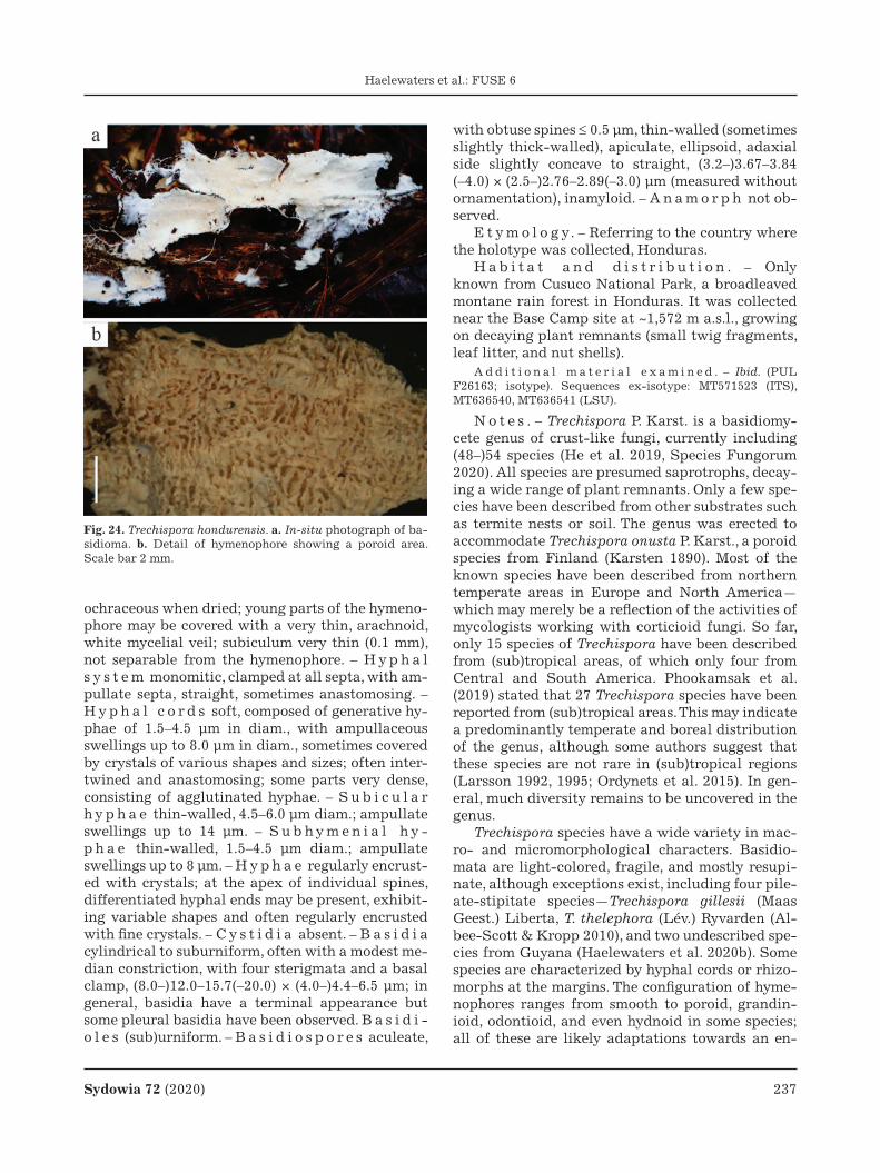
Sydowia 72 (2020) 237
Haelewaters et al.: FUSE 6
ochraceous when dried; young parts of the hymeno-phore may be covered with a very thin, arachnoid, white mycelial veil; subiculum very thin (0.1 mm), not separable from the hymenophore. – H y p h a l s y s t e m monomitic, clamped at all septa, with am-pullate septa, straight, sometimes anastomosing. – H y p h a l c o r d s soft, composed of generative hy-phae of 1.5–4.5 μm in diam., with ampullaceous swellings up to 8.0 μm in diam., sometimes covered by crystals of various shapes and sizes; often inter-twined and anastomosing; some parts very dense, consisting of agglutinated hyphae. – S u b i c u l a r h y p h a e thin-walled, 4.5–6.0 μm diam.; ampullate swellings up to 14 μm. – S u b h y m e n i a l h y -p h a e thin-walled, 1.5–4.5 μm diam.; ampullate swellings up to 8 μm. – H y p h a e regularly encrust-ed with crystals; at the apex of individual spines, differentiated hyphal ends may be present, exhibit-ing variable shapes and often regularly encrusted with fine crystals. – C y s t i d i a absent. – B a s i d i a cylindrical to suburniform, often with a modest me-dian constriction, with four sterigmata and a basal clamp, (8.0–)12.0–15.7(–20.0) × (4.0–)4.4–6.5 μm; in general, basidia have a terminal appearance but some pleural basidia have been observed. B a s i d i -o l e s (sub)urniform. – B a s i d i o s p o r e s aculeate,
with obtuse spines ≤ 0.5 μm, thin-walled (sometimes slightly thick-walled), apiculate, ellipsoid, adaxial side slightly concave to straight, (3.2–)3.67–3.84 (–4.0) × (2.5–)2.76–2.89(–3.0) μm (measured without ornamentation), inamyloid. – A n a m o r p h not ob-served.
E t y m o l o g y. – Referring to the country where the holotype was collected, Honduras.
H a b i t a t a n d d i s t r i b u t i o n . – Only known from Cusuco National Park, a broadleaved montane rain forest in Honduras. It was collected near the Base Camp site at ~1,572 m a.s.l., growing on decaying plant remnants (small twig fragments, leaf litter, and nut shells).
A d d i t i o n a l m a t e r i a l e x a m i n e d . – Ibid. (PUL F26163; isotype). Sequences ex-isotype: MT571523 (ITS), MT636540, MT636541 (LSU).
N o t e s . – Trechispora P. Karst. is a basidiomy-cete genus of crust-like fungi, currently including (48–)54 species (He et al. 2019, Species Fungorum 2020). All species are presumed saprotrophs, decay-ing a wide range of plant remnants. Only a few spe-cies have been described from other substrates such as termite nests or soil. The genus was erected to accommodate Trechispora onusta P. Karst., a poroid species from Finland (Karsten 1890). Most of the known species have been described from northern temperate areas in Europe and North America—which may merely be a reflection of the activities of mycologists working with corticioid fungi. So far, only 15 species of Trechispora have been described from (sub)tropical areas, of which only four from Central and South America. Phookamsak et al. (2019) stated that 27 Trechispora species have been reported from (sub)tropical areas. This may indicate a predominantly temperate and boreal distribution of the genus, although some authors suggest that these species are not rare in (sub)tropical regions (Larsson 1992, 1995; Ordynets et al. 2015). In gen-eral, much diversity remains to be uncovered in the genus.
Trechispora species have a wide variety in mac-ro- and micromorphological characters. Basidio-mata are light-colored, fragile, and mostly resupi-nate, although exceptions exist, including four pile-ate-stipitate species—Trechispora gillesii (Maas Geest.) Liberta, T. thelephora (Lév.) Ryvarden (Al-bee-Scott & Kropp 2010), and two undescribed spe-cies from Guyana (Haelewaters et al. 2020b). Some species are characterized by hyphal cords or rhizo-morphs at the margins. The configuration of hyme-nophores ranges from smooth to poroid, grandin-ioid, odontioid, and even hydnoid in some species; all of these are likely adaptations towards an en-
Fig. 24. Trechispora hondurensis. a. In-situ photograph of ba-sidioma. b. Detail of hymenophore showing a poroid area. Scale bar 2 mm.

238 Sydowia 72 (2020)
Haelewaters et al.: FUSE 6
larged surface promoting spore production (Liu et al. 2019b). The hyphal system is either monomitic, consisting of clamped generative hyphae only (most species), or dimitic, consisting of generative and skeletal hyphae. A typical feature for Trechispora species are the ampullate septa in some of the gen-erative hyphae, visible under the light microscope as swollen hyphae near the septa, although their abundance varies between species. Many Trechis-pora species are characterized by calcium oxalate crystals, often with specific shapes and found at-tached to hyphae in the subiculum and hyphal
cords. Due to their specific shapes and organization in the basidiomata, these crystals are taxonomically informative (Larsson 1994). Basidia (meiosporo-cysts) are four-sterigmate, short, and generally cy-lindrical to clavate. Basidiospores are either thin- or thick-walled, smooth or ornamented, with a vari-ety in shape and length of the spines in the latter type. For some species, conidial stages have been described.
The Honduran collection agrees well with typi-cal microscopic characteristics for Trechispora (e.g., the fragile basidioma and ampullaceous septa).
Fig. 25. Trechispora hondurensis, micromorphological features drawn from the holotype (GENT). a. Structure of an individual spine of the basidioma, with basidia (*1), basidioles (*2), and hyphal ends at the apex encrusted with fine crystals. b. Hyphal cord, non-agglutinated part. c. Basidiospores. Scale bars a–b 20 μm, c 5 μm, del. N. Schoutteten & A. Verbeken.

Sydowia 72 (2020) 239
Haelewaters et al.: FUSE 6
However, based on combined morphological and molecular evidence, it cannot be assigned to any of the currently described species in the genus. Trechis-pora hondurensis clearly shows a light colored ba-sidioma and peripheral hyphal cords. The hymeno-phore is variably organized; young regions are po-roid (sometimes covered by a mycelial veil) while mature regions are hydnoid, with locally coalescing spines. Unlike most species in the genus, this speci-men has more or less differentiated hyphal ends of variable shape in the hymenium positioned at the spinal tips in the basidioma (Fig. 25). These cells are slightly projecting above the basidia and covered with very small roundish calcium oxalate crystals, more or less regularly organized.
Both phylogenetic analyses (ITS–LSU and LSU alone) retrieved Trechispora hondurensis in a max-imum-supported clade with two Trechispora iso-lates from Brazil (KHL16968, URM 85884) (Figs. 27–
28). Micromorphological work on this Brazilian material is unavailable. Given the evolutionary dis-tances among isolates of Trechispora cyatheae Or-dynets, Langer & K.H. Larss., T. echinocrystallina Ordynets, Langer & K.H. Larss., and T. farinacea (Pers.) Liberta, the Brazilian isolates in our trees may represent T. hondurensis—possibly extending the distributional range of this species to South America. The closest phylogenetic relatives of T. hondurensis are T. echinospora Telleria, M. Dueñas, I. Melo & M.P. Martín, T. farinacea, and T. regularis (Murrill) Liberta. In the phylogenetic reconstruc-tion of the LSU dataset (Fig. 28), high support was found for a sister relationship between T. farinacea and T. hondurensis (including Brazilian isolates KHL16968 and URM 85884).
Remarkably, both T. echinospora and T. regularis are described from tropical areas—T. echinospora from Equatorial Guinea in tropical sub-Saharan
Fig. 26. Trechispora hondurensis, scanning electron micrographs. a–b. Encrustations on hyphae. c–d. Basidiospores, which are ellipsoid and aculeate. Scale bars a–b 5 μm, c–d 1 μm.

240 Sydowia 72 (2020)
Haelewaters et al.: FUSE 6
Africa and T. regularis from Jamaica in the Carib-bean (Liberta 1973, Phokaamsak et al. 2019). Trechispora echinospora can be distinguished from T. hondurensis based on its farinaceous to grandin-ioid basidioma; its larger and globose basidiospores (4–5 μm excluding ornamentation) with long, sharp and evenly distributed spines of up to 1 μm in length; the presence of sphaerocytes [often referred to as sphaerocysts in Trechispora literature]; and the absence of hyphal cords and crystals. Trechis-pora regularis is characterized by a poroid hymeno-phore but is easily distinguished from T. honduren-
sis by the presence of many large (up to 60 μm), en-crusted cystidia. In addition to Jamaica where the type was collected, T. regularis has also been report-ed from the USA, Costa Rica, Brazil, and Paraguay (Liberta 1973).
Trechispora farinacea was based on material from Scandinavia [as Hydnum farinaceum Pers. ex Fr.] and is a common species in northern temperate areas around the world. Note that it has also been reported from the Caribbean, South America, and southern Australia (sensu Liberta 1973), although it remains to be proven that these specimens are truly
Fig. 27. Phylogeny of Trechispora isolates reconstructed from a combined ITS–LSU dataset. The topology is the result of ML inference performed with IQ-TREE. For each node, the MLBS (if >60) is presented above/below the branch leading to that node. The new species T. hondurensis is highlighted in grayscale.

Sydowia 72 (2020) 241
Haelewaters et al.: FUSE 6
representative of T. farinacea (Larsson 1995). Much variation has been described in the macromorphol-ogy of T. farinacea, although basidiomata are most-ly smooth to grandinioid or odontioid, often soft and cushion-like, whereas the basidioma of T. hon-durensis is poroid to hydnoid. Micromorphologi-cally, T. farinacea has larger basidiospores, measur-ing 4.0–4.5(–5.0) × 3.3–3.7 μm, and the short spines of its basidioma consist of rather short-celled hy-
phae (Hjortstam et al. 1988). In T. hondurensis, these spines are composed of long-celled hyphae.
Morphologically, the here described T. hondu-rensis is somewhat reminiscent of the temperate species T. verruculosa (G. Cunn.) K.H. Larss. in hav-ing an effused hydnoid basidioma consisting of coa-lescing spines. However, basidiospores of T. verruc-ulosa are slightly cyanophilous and larger, measur-ing (4.5–)4.7–5.2(–5.5) × (3.5–)4.0–4.5 μm (Larsson
Fig. 28. Phylogeny of Trechispora isolates reconstructed from a single-locus dataset including LSU sequences only. The topology is the result of ML inference performed with IQ-TREE. For each node, the MLBS (if >60) is presented above/below the branch leading to that node. The new species T. hondurensis is highlighted in grayscale.

242 Sydowia 72 (2020)
Haelewaters et al.: FUSE 6
1996). Trechispora verruculosa is described from Auckland, New Zealand [as Odontia verruculosa G. H. Cunn.] and has been reported from several coun-tries in Europe and from China. No sequences are currently available for this taxon.
Authors: D. Haelewaters, N. Schoutteten, A. Ver-beken & M.C. Aime
Basidiomycota, Agaricomycetes, Agaricales, Tricho-lomataceae
Tricholoma kenanii I. Acar, S. Aldemir & A. Dizkir-ici, sp. nov. – Figs. 29–31MycoBank no.: MB831905
H o l o t y p u s . – TURKEY. Bingöl Province, Genç, 38°44’54.05”N, 40°33’41.92”E, 1049 m a.s.l., coniferous forest, near Pinus spp., 18 October 2018, leg. I. Acar, VANF 7600 (VPF; holotype). Sequences ex-holotype: MN541841–MN541846 (ITS), MN541829–MN541834 (LSU).
grey-white, smooth, context white, fragile. – B a -s i d i o s p o r e s (6.1–)7.0–9.1 × 4.0–5.5 μm, (n=40 and Q=1.5–1.65), on average 7.44 × 4.72, ellipsoid, thin-walled, smooth, hyaline, guttulate, inamyloid. – B a s i d i a 26.5–36.5 × 6–9 μm, cylindrical-clavate, with 2 or 4 sterigmata, without basal clamp connec-tion. – C h e i l o c y s t i d i a 16.7–28.5 × 5.2–8.0 μm, clavate, irregular, abundant, hyaline, twisted, some-times polymorph. – P l e u r o c y s t i d i a not seen. – P i l e i p e l l i s up to 44 μm, made up of irregular to slightly parallel hyphae, dark brown-pigmented, without clamp connections. – P i l e u s t r a m a l h y p h a e 7–16 μm wide, hyaline to yellow brown. – L a m e l l a r t r a m a parallel, guttulate, hyphae mostly 8–20 μm in diam., thin-walled, hyaline. – S t i p i t i p e l l i s with longitudinally arranged, ap-pressed, parallel hyphae, 8–14 μm in diam., without
Fig. 29. Tricholoma kenanii. a. Basidiomata in natural habitat b. Preserved (dried) basidiomata from the VPF fungarium.
D e s c r i p t i o n . – P i l e u s 15–55 mm in diam., conical when young, then plane, slightly umbonate, outward curved when old, surface radially fibrillose towards margin, scaly, a white zone at the margin, darker to the center, greyish, dark-greyish, blackish brown, whitish towards margin, with white velar remnants on the edges, a cortina when young. – C o n t e x t whitish, thick in the pileus center, thin-ner to the edges. – E d i b i l i t y unknown. – L a -m e l l a e whitish, grey-white, broad, L=60–90, l=1–3, emerginate, shortly with tooth decurrent, edges cre-nate, with lamellulae. – S t i p e 25–40 × 7–10 mm, surface longitudinally fibrillose, cylindrical, some-times curved when mature, basally distinctly clavate, or occasionally bulbous, somewhat rooting, whitish,
clamp connections, thin-walled, hyaline to light yel-lowish. – C a u l o c y s t i d i a not seen.
E t y m o l o g y. – kenanii, in honor of Prof. Dr. Kenan Demirel†, who made significant contribu-tions to mycology in Turkey and died in 2019.
H a b i t a t a n d d i s t r i b u t i o n . – In subal-pine areas in coniferous forests with Pinus sp. of the Eastern Anatolia Region, Turkey.
N o t e s . – The agaric genus Tricholoma (Fr.) Staude was introduced as a section by Fries (1821) and subsequently proposed as a genus by Staude (1857). The genus is mainly characterized by fleshy basidiomata; white, cream to yellowish emarginate lamellae; white spore print; and hyaline, smooth, inamyloid spores (Christensen & Heilmann-Claus-

Sydowia 72 (2020) 243
Haelewaters et al.: FUSE 6
Fig. 30. Tricholoma kenanii, collection VPF Fungi 7600 (holotype). a–b. Basidiospores in distilled water (a) and in Melzer’s reagent (b). c. Basidia. d. Cheilocystidia. e. Pileipellis. f. Pileus tramal hyphae. Scale bars a–d 20 μm, e–f 50 μm.
en 2013, Reschke et al. 2018). The pileal surface, the cellular structure of the pileipellis, the color of the pileus, and the presence vs. absence of clamps are
important characters for species delimitation in the genus (Bon 1984, Singer 1986, Heilmann-Clausen et al. 2017, Reschke et al. 2018). Tricholoma species

244 Sydowia 72 (2020)
Haelewaters et al.: FUSE 6
Fig. 31. Tricholoma kenanii. Field-emission scanning electron micrographs of basidiospores. a. At 25,000K×. b. At 50,000K×.
Fig. 32. Phylogeny of Tricholoma species showing the placement of T. kenanii, reconstructed from a concatenated ITS–LSU data-set. The topology is the result on Bayesian inference performed with MrBayes. Sequences of the newly described species are highlighted in boldface. Only BIPP >0.75 are shown.

Sydowia 72 (2020) 245
Haelewaters et al.: FUSE 6
exhibit very similar microscopic features, and are characterized by hyaline, subglobose to oblong spores; simple pileipellis structures; and a lack of well-differentiated sterile elements, including cys-tidia (Heilmann-Clausen et al. 2017). Around 210 species are currently described in the genus (Wijaya-wardene et al. 2020).
Sequence comparison of newly generated ITS sequences showed—among the top 20 BLAST re-sults—that the new species was most closely related to T. moseri Singer (99.42 % shared identity), T. my-omyces (Pers.) J.E. Lange (98.83–99.71 %), T. terre-um (Schaeff.) P. Kumm. (99.71 %), and T. triste (Scop.) Quél. (99.25–99.85 %). Comparison of the LSU locus led to the following top 20 BLAST re-sults: T. argyraceum (Bull.) Gillet [including syno-nym T. inocybeoides A. Pearson] (98.89–99.22 % shared identity), T. cingulatum (Almfelt ex Fr.) Jacobashch (98.89 %), T. moseri (100 %), T. (cf.) my-omyces (99.66–100 %), T. scalpturatum (Fr.) Quél. (98.89–99.00 %), and T. terreum (98.78–99.66 %). In our phylogenetic tree based on the concatenated ITS–LSU dataset of 64 isolates (Fig. 32), ex-holo-type sequences of T. kenanii forms a separate clade sister to T. moseri with maximum support (BIPP=1). This clade (T. kenanii, T. moseri) is sister to a cluster with representatives of T. bonii, T. myomyces, T. ter-reum, and T. triste.
Tricholoma kenanii is characterized by the typi-cal greyish brown and scaly pileus structure of sect. Terrea; its stipe is markedly clavate and has a slightly rooted structure; basidiospores are (6.1–) 7.0–9.1 × 4.0–5.5 μm, elliptic, globose; and basidia are 26.5–36.5 × 6–9 μm, cylindrical-clavate, without basal clamp connection. Sister species T. moseri is differentiated from T. kenanii by slightly different measurements for basidiospores (6.5–10.6 × 3.3–5.8 μm) and basidia (28–43 × 6.7–8.6 μm). Tricholo-ma moseri is an American species with reports in California and Mexico (type), found particularly under Abies magnifica and Pinus hartwegii (Singer 1989; Shanks 1996, 1997; Bessette et al. 2013). Tricholoma triste can be morphologically separated from the new species by the lack of cystidia, where-as T. kenanii has abundant cheilocystidae. In addi-tion, the pileus color is different between both spe-cies: sometimes almost black in T. triste (Christens-en & Noordeloos 1999) vs. greyish brown in T. kena-nii. Tricholoma myomyces is morphologically easily distinguished from T. kenanii, by its basidiospores (5.0–10.5 × 3.5–6.0 μm), which are ellipsoid to ovoid, and 2-spored basidia (Bessette et al. 2013, Desjar-din et al. 2015).
Authors: I. Acar, S. Aldemir & A. Dizkırıcı
Interesting taxonomical notes, new hosts, and geographical records
Ascomycota, Laboulbeniomycetes, Laboulbeniales, Laboulbeniaceae
Arthrorhynchus eucampsipodae Thaxt., Proc. Am. Acad. Arts Sci. 36: 409 (1901). – Fig. 33b
M a t e r i a l e x a m i n e d . – RWANDA. Northern Prov-ince, Musanze District, Ruhengeri, on female Eucampsipoda africanum Theodor, 1955 (Diptera, Nycteribiidae) collected from female Rousettus aegyptiacus, UP498/D. Haelew. 1498 [bat fly labels], 10 December 2008, leg. W. Markotter, slide PUL F27651; Ibid., on female E. africanum, UP404 [bat fly label], 10 December 2008, slide PUL F27653; Ibid., 10 December 2008, on female E. africanum, UP478 [bat fly label], slide PUL F27652; Ibid., on female E. africanum, UP508 [bat fly label], 11 December 2008, slide PUL F27650; Ibid., on female E. afri-canum, UP541 [bat fly label], 13 December 2008, slide PUL F27647. – SOUTH AFRICA. Limpopo, on female E. africanum collected from male R. aegyptiacus, UP5917 [bat fly label], 26 October 2015, leg. W. Markotter, slide PUL F27649; Ibid., on female E. africanum collected from female R. aegyptiacus, UP7190 [bat fly label], 1 November 2016, slide PUL F27648.
M a t e r i a l s e q u e n c e d . – BULGARIA. Lovech Prov-ince, Chavdartsi village, Mandrata Cave, 43°14’30.9336”N, 24°58’02.3664”E, on female Nycteribia vexata Westwood, 1835 (Diptera, Nycteribiidae) collected from male adult Myotis bly-thii, A1003L [bat fly label] A1003 [bat label], 9 September 2017, leg. A. Sándor & Á. Péter, isolate D. Haelew, 1491a
Fig. 33. Arthrorhynchus species. a. Arthrorhynchus nycteribi-ae. b. Arthrorhynchus eucampsipodae. Scale bars a 200 μm, b 100 μm, del. J. Liu.

246 Sydowia 72 (2020)
Haelewaters et al.: FUSE 6
(3 adult thalli, left metacoxal cavity), MT241715 (LSU). – RWANDA. Northern Province, Musanze District, Ruhengeri, on female E. africanum collected from female R. aegyptiacus, UP498 [bat fly label], 10 December 2008, leg. W. Markotter, isolate D. Haelew. 1498a (2 adult thalli), MT235694 (SSU), MT235717 (LSU); Ibid., isolate D. Haelew. 1498b (2 adult thal-li), MT235695 (SSU), MT235718 (LSU). – SLOVAKIA. Banská Bystrica Region, Nandraž, near Jelšava, on N. schmidlii (Dip-tera, Nycteribiidae) collected from adult female Miniopterus schreibersii, AB04 [bat label], 6 December 2009, leg. M. Ševcík & P. Hohti, isolate D. Haelew. 1499a (9 thalli), MT235696 (SSU), MT235719 (LSU).
H o s t s a n d d i s t r i b u t i o n . – Currently known from temperate and tropical biomes of the Eastern Hemisphere, with records in many coun-tries in Europe (France, Hungary, Italy, Slovakia, Spain, the UK), Africa (Egypt, Gabon, Kenya, Sierra Leone, Tunisia), and Asia (India, Indonesia, Israel, Myanmar, Peninsular Malaysia, Taiwan, Thailand). Reported hosts are Basilia pumila (Scott, 1914), Leptocyclopodia ferrarii (Rondani, 1878), Eucamp-sipoda spp., Nycteribia spp., and Penicillidia du-fourii (Westwood, 1835) (Speiser 1901; Thaxter 1901; Balazuc 1971; Blackwell 1980a, 1980b; Haele-waters et al. 2017; Szentiványi et al. 2018a). Host bats include sedentary tropical, sedentary temper-ate, and migrating species.
N o t e s . – Laboulbeniales are obligate biotroph-ic fungi of arthropods. About 80 % of described La-boulbeniales taxa are known from beetles (Coleop-tera), but these fungi are found on a wide variety of hosts, including ants (Hymenoptera: Formicidae), harvestmen (Opiliones), millipedes (Diplopoda), mites (Acari), true bugs (Hemiptera), and flies (Dip-tera)—including the ectoparasitic bat flies (Enghoff & Santamaría 2015; Haelewaters et al. 2017, 2020a; Santamaría et al. 2017). Bat flies are obligate he-matophagous parasites of bats (Mammalia: Chirop-tera) belonging to two families, Nycteribiidae and Streblidae. Both families show a worldwide distri-bution but nycteribiids have a higher diversity in the Eastern Hemisphere, whereas streblids show a higher diversity in the Western Hemisphere. Cur-rently, about 21 nycteribiid genera with 275 species and 31 streblid genera with 227 species are de-scribed (Dick & Patterson 2006).
Four genera of bat fly-associated Laboulbeni-ales are known: Arthrorhynchus Kolen., Dimeromy-ces Thaxt., Gloeandromyces Thaxt. and Nycteromy-ces Thaxt. (Dogonniuck et al. 2019, Haelewaters et al. 2020a). Arthrorhynchus species are restricted to the Eastern Hemisphere. There are currently four described species in the genus, all parasitizing nyc-teribiid bat flies: Arthrorhynchus acrandros Merola, A. cyclopodiae Thaxt., A. eucampsipodae Thaxt.,
and A. nycteribiae (Peyr.) Thaxt. [although A. acran-dros is disputed (Haelewaters et al. 2017)].
The here presented records of A. eucampsipodae from Bulgaria, Rwanda, and South Africa are new country records. Arthrorhynchus eucampsipodae has thus far been found on E. africanum in Gabon, Rwanda, Sierra Leone and South Africa (Blackwell 1980a, Balazuc 1971, this study). Eucampsipoda af-ricanum has a wide distribution in sub-Saharan Af-rica, where its main bat host R. aegyptiacus occurs. Additionally, E. africanum occasionally parasitizes other cave-dwelling bat species such as Eidolon hel-vum, Hipposideros caffer, H. gigas, and Mi. inflatus (Theodor 1967, Obame-Nkoghe et al. 2016). In a re-cent study, Szentiványi et al. (2018a) focused on bat flies collected from the cave-dwelling bat Mi. schreib-ersii in Europe and found A. eucampsipodae only from N. schmidlii bat flies (23 infected specimens out of 468). This species of Arthrorhynchus seems to have a wide host spectrum but mostly occurs on bat flies associated with cave-dwelling bat species.
We generated 894–523 bp LSU sequences from different isolates of A. eucampsipodae, which share 98.53–100 % identity to one another, and 96.58–98.10 % identity with A. nycteribiae. In the LSU phylogeny (Fig. 34), A. eucampsipodae forms two clades, each clade consisting of isolates sampled from the same host genus (Eucampsipoda, Nyc-teribia). Sequences for the two isolates from E. afri-canum share 100 % identity but this “Eucampsipo-da” clade only received moderate support (MLBS=69). The “Nycteribia” clade receives maxi-mum support and consists of two isolates, one from N. schmidlii (GenBank accession no. MT235719), and the other from N. vexata (MT241715). Although these results may be premature to draw strong con-clusions, they point to A. eucampsipodae being a complex of multiple species segregated by host ge-nus. This presumed speciation pattern is similar to what was recently shown for Hesperomyces vires-cens Thaxt., a parasite of ladybirds in various gen-era (Coleoptera, Coccinellidae) (Haelewaters et al. 2018a). It is clear that the two clades of A. eucamp-sipodae are representative of segregation (specia-tion) but we must incorporate material from E. hy-rtlii (Kolenati, 1856), the bat fly host on which A. eucampsipodae was originally described (Thaxter 1901), before making taxonomic decisions. Until we know what is the relation of A. eucampsipodae s.s. to the two clades uncovered by this study, we refrain from describing new species in the complex.
Authors: D. Haelewaters, J. Liu, T. Szentiványi, W.P. Pfliegler, A.D. Sándor, Á. Péter, W. Markotter, P. Christe, O. Glaizot & M.C. Aime
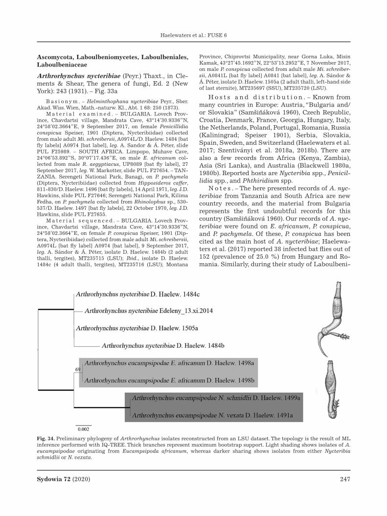
Sydowia 72 (2020) 247
Haelewaters et al.: FUSE 6
Ascomycota, Laboulbeniomycetes, Laboulbeniales, Laboulbeniaceae
Arthrorhynchus nycteribiae (Peyr.) Thaxt., in Cle-ments & Shear, The genera of fungi, Ed. 2 (New York): 243 (1931). – Fig. 33a
B a s i o n y m . – Helminthophana nycteribiae Peyr., Sber. Akad. Wiss. Wien, Math.-naturw. Kl., Abt. 1 68: 250 (1873).
M a t e r i a l e x a m i n e d . – BULGARIA. Lovech Prov-ince, Chavdartsi village, Mandrata Cave, 43°14’30.9336”N, 24°58’02.3664”E, 9 September 2017, on female Penicillidia conspicua Speiser, 1901 (Diptera, Nycteribiidae) collected from male adult Mi. schreibersii, A0974L/D. Haelew. 1484 [bat fly labels] A0974 [bat label], leg. A. Sandor & Á. Péter, slide PUL F25989. – SOUTH AFRICA. Limpopo, Muhave Cave, 24°06’53.892”S, 30°07’17.436”E, on male E. africanum col-lected from male R. aegyptiacus, UP8089 [bat fly label], 27 September 2017, leg. W. Markotter, slide PUL F27654. – TAN-ZANIA. Serengeti National Park, Banagi, on P. pachymela (Diptera, Nycteribiidae) collected from Hipposideros caffer, 811-830/D. Haelew. 1496 [bat fly labels], 14 April 1971, leg. J.D. Hawkins, slide PUL F27646; Serengeti National Park, Kilima Fedha, on P. pachymela collected from Rhinolophus sp., 530-537/D. Haelew. 1497 [bat fly labels], 22 October 1970, leg. J.D. Hawkins, slide PUL F27655.
M a t e r i a l s e q u e n c e d . – BULGARIA. Lovech Prov-ince, Chavdartsi village, Mandrata Cave, 43°14’30.9336”N, 24°58’02.3664”E, on female P. conspicua Speiser, 1901 (Dip-tera, Nycteribiidae) collected from male adult Mi. schreibersii, A0974L [bat fly label] A0974 [bat label], 9 September 2017, leg. A. Sándor & Á. Péter, isolate D. Haelew. 1484b (2 adult thalli, tergites), MT235715 (LSU); Ibid., isolate D. Haelew. 1484c (4 adult thalli, tergites), MT235716 (LSU); Montana
Province, Chiprovtsi Municipality, near Gorna Luka, Misin Kamak, 43°27’45.1692”N, 22°53’15.2952”E, 7 November 2017, on male P. conspicua collected from adult male Mi. schreiber-sii, A0841L [bat fly label] A0841 [bat label], leg. A. Sándor & Á. Péter, isolate D. Haelew. 1505a (2 adult thalli, left-hand side of last sternite), MT235697 (SSU), MT235720 (LSU).
H o s t s a n d d i s t r i b u t i o n . – Known from many countries in Europe: Austria, “Bulgaria and/or Slovakia” (Samšináková 1960), Czech Republic, Croatia, Denmark, France, Georgia, Hungary, Italy, the Netherlands, Poland, Portugal, Romania, Russia (Kaliningrad; Speiser 1901), Serbia, Slovakia, Spain, Sweden, and Switzerland (Haelewaters et al. 2017; Szentiványi et al. 2018a, 2018b). There are also a few records from Africa (Kenya, Zambia), Asia (Sri Lanka), and Australia (Blackwell 1980a, 1980b). Reported hosts are Nycteribia spp., Penicil-lidia spp., and Phthiridium spp.
N o t e s . – The here presented records of A. nyc-teribiae from Tanzania and South Africa are new country records, and the material from Bulgaria represents the first undoubtful records for this country (Samšináková 1960). Our records of A. nyc-teribiae were found on E. africanum, P. conspicua, and P. pachymela. Of these, P. conspicua has been cited as the main host of A. nycteribiae; Haelewa-ters et al. (2017) reported 38 infected bat flies out of 152 (prevalence of 25.0 %) from Hungary and Ro-mania. Similarly, during their study of Laboulbeni-
Fig. 34. Preliminary phylogeny of Arthrorhynchus isolates reconstructed from an LSU dataset. The topology is the result of ML inference performed with IQ-TREE. Thick branches represent maximum bootstrap support. Light shading shows isolates of A. eucampsipodae originating from Eucampsipoda africanum, whereas darker sharing shows isolates from either Nycteribia schmidlii or N. vexata.

248 Sydowia 72 (2020)
Haelewaters et al.: FUSE 6
ales associated with bat flies from Mi. schreibersii bats in eight European countries, Szentiványi et al. (2018a) sampled 144 P. conspicua, of which 33 were infected with A. nycteribiae (prevalence of 22.9 %). In Europe, this fungus is predominantly associated with bat flies parasitizing migratory Mi. schreiber-sii (Moesz 1931; Thaxter 1931; Balcells 1954, 1955; Samšináková 1960; Balazuc 1971; Blackwell 1980a, 1980b; Haelewaters et al. 2017; Szentiványi et al. 2018a).
The majority of bat fly-associated Laboulbeni-ales are reported from the neotropics and Europe (Thaxter 1917, Blackwell 1980a, 1980b, Haelewa-ters et al. 2017, Szentiványi et al. 2018a, Dogonni-uck et al. 2019, Haelewaters & Pfister 2019). African records are rare (Tab. 2), with A. eucampsipodae previously known in Egypt, Gabon, Kenya, Sierra Leone, and Tunisia (Speiser 1901; Thaxter 1901; Balazuc 1971; Blackwell 1980a, 1980b); A. nycterib-iae in Kenya and Zambia (Blackwell 1980a, 1980b); Dimeromyces capensis in South Africa; and Nycter-omyces orientalis in Tanzania (Dogonniuck et al. 2019). Of the 55 African bat flies screened at the en-tomology collection of the California Academy of Sciences, only two specimens of P. pachymela from Tanzania were infected.
Authors: D. Haelewaters, J. Liu, T. Szentiványi, W.P. Pfliegler, A.D. Sándor, Á. Péter, W. Markotter, P. Christe, O. Glaizot & M.C. Aime
Basidiomycota, Agaricomycetes, Agaricales, Agari-caceae
Calvatia lilacina (Mont. & Berk.) Henn., Hedwigia 43(3): 205 (1904). – Figs. 35–37.
B a s i o n y m . – Bovista lilacina Mont. & Berk., London J. Bot. 4: 64 (1845).
Sy n o n y m s . – Globaria lilacina (Mont. & Berk.) Speg., Anal. Soc. Cient. Argent. 9(4): 187 (1880).
Lycoperdon lilacinum (Mont. & Berk.) Speg., Anal. Mus. Nac. Hist. Nat. B. Aires 12(3–6): 252 (1881).
M a t e r i a l e x a m i n e d . – PAKISTAN. Khyber Pak-tunkhwa Province, Mansehra District, Oghi, 975 m a.s.l., on soil under Pinus wallichiana (Pinales, Pinaceae), 1 September 2008, leg. M. Fiaz, NYG207 (LAH 10000082); Autonomous Ter-ritory of Gilgit-Baltistan, Deosai Plains, Deosai National Park, 4114 m a.s.l., in grass, 10 September 2011, leg. A.R. Nia-zi, CPK1 (LAH 10000083).
D e s c r i p t i o n . – B a s i d i o m a t a growing in groups, 60–90 mm wide × 45–60 mm high, subglobose to broadly obpyriform to obovoid, slightly tapering at the base, sometimes forming a pseudo-stipe below. – P e r i d i u m evanescent, vio-laceous brown at maturity; attached to the sub-strate by thick rhizomorphs. – R h i z o m o r p h s brown densely encrusted with the particles of soil. – S u b g l e b a with diaphragm, well-developed, compact, cottony, occupying two-third of basidio-ma, separated from glebal portion by diaphragm. – D i a p h r a g m distinct, greyish brown, slightly de-pressed from the center, center rough, cracked with
Tab. 2. Published records of bat fly-associated Laboulbeniales from the African continent. Records in boldface are newly re-ported here.
Species Country Host
Arthrorhynchus eucampsipodae Egypt Eucampsipoda hyrtlii (Kolenati, 1856)
Arthrorhynchus eucampsipodae Gabon Eucampsipoda africanum Theodor, 1955
Arthrorhynchus eucampsipodae Kenya Nycteribia schmidlii Schiner, 1853
Arthrorhynchus eucampsipodae Rwanda Eucampsipoda africanum Theodor, 1955
Arthrorhynchus eucampsipodae Sierra Leone Eucampsipoda africanum Theodor, 1955
Arthrorhynchus eucampsipodae South Africa Eucampsipoda africanum Theodor, 1955
Arthrorhynchus eucampsipodae Tunisia Nycteribia vexata Westwood, 1835
Arthrorhynchus nycteribiae Kenya Penicillidia fulvida (Bigot, 1885)
Arthrorhynchus nycteribiae Kenya Penicillidia fulvida (Bigot, 1885)
Arthrorhynchus nycteribiae South Africa Eucampsipoda africanum Theodor, 1955
Arthrorhynchus nycteribiae Tanzania Penicillidia pachymela Speiser, 1901
Arthrorhynchus nycteribiae Zambia Penicillidia pachymela Speiser, 1901
Dimeromyces capensis South Africa Brachytarsina africana (Walker, 1849)
Nycteromyces orientalis Tanzania Brachytarsina alluaudi (Falcoz, 1923)

Sydowia 72 (2020) 249
Haelewaters et al.: FUSE 6
Fig. 35. Calvatia lilacina. A. Different views of basidiomata, collection LAH 10000083. B–C. Mature basidioma, collection LAH 10000082. Scar bars 1.7 cm.

250 Sydowia 72 (2020)
Haelewaters et al.: FUSE 6
age after exposure, margins extend outwards, wavy. – G l e b a violaceous brown, lanose, pulverulent with age. – B a s i d i o s p o r e s globose, echinulat-ed, 7.0–8.5 μm in diam. (including echines), 4.5–5.4
μm in diam. (excluding echines), pedicel absent or rudimentary, brown in both water and 5 % KOH mounts. – E u c a p i l l i t i u m Calvatia-type, brown, non-elastic, up to 6.4 μm in diam., with wall thick-
Fig. 36. Calvatia lilacina. A–C, H. Pitted Calvatia-type capillitium. D–G. Echinulate basidiospores. Scale bars A–B 12 μm; C, H 10 μm; D–G 3.2 μm.

Sydowia 72 (2020) 251
Haelewaters et al.: FUSE 6
Fig. 37. Micromorphological features of Calvatia lilacina. A. Echinulate basidiospores. B. Exoperidial elements. C. Pitted capil-litial hyphae. D. Endoperidial hyphae (bar = 10 μm). Scale bars A 4 μm; B, D 10 μm; C 7 μm; del. N. Yousaf.

252 Sydowia 72 (2020)
Haelewaters et al.: FUSE 6
ness up to 1 μm, smooth to slightly encrusted, straight to subundulate, often bulging at some points, frequently branched, tapering at the ends up to 1.2 μm thin; septa rarely seen, hypha broken where septa present; brown in water mounts. – P i t s numerous, small to medium sized, circular. – P a r a -c a p i l l i t i u m scarce, branched, hyaline, septate. – E x o p e r i d i u m composed of subglobose, oval to ellipsoid, elongated to irregular shaped hyphal elements, brown, thick-walled. – E n d o p e r i d i -u m composed of thin-walled, branched and asep-tate (rarely joint-like septa present) brown hyphae.
H a b i t a t a n d d i s t r i b u t i o n . – Known from five continents: North America (USA), South America (Argentina, Brazil, Chile), Europe (Spain), Asia (India, the Philippines, Taiwan), and Oceania (Australia, New Zealand). Growing solitary to gre-garious in forests and grasslands.
N o t e s . – The genus Calvatia Fr. was established by Fries (1849) with C. craniiformis (Schwein.) Fr. as type species. The genus is characterized by medium to large basidiomata that have a reduced to well-defined subgleba. Dehiscence occurs by irregular rupturing of peridium and not through a definite pore (Li 2011, Cortez at al. 2012). Calvatia has been extensively studied throughout the years (Kreisel 1992, 1994; Lange 1990, 1993, 1994; Calonge 1998; Gube 2007; Kasuya & Kotumoto 2008; Cortez & Alves 2012). The number of Calvatia species is 44 thus far (Kirk et al. 2008; Suárez et al. 2009; Alves & Cortez 2013; Alfredo et al. 2014; Crous et al. 2018, 2019; Gunasekaran et al. 2018) but more species are awaiting description (e.g., Yousaf 2014). In Pakistan, the genus is represented by four species: Calvatia ahmadii Khalid & S.H. Iqbal, C. craniiformis, C. cy-athiformis (Bosc) Morgan, and C. excipuliformis var. excipuliformis (Scop.) Perdeck (Ahmad 1956; Khalid & Iqbal 1996, 2004; Ahmad et al. 1997). Cal-vatia lilacina is reported here a new record for the country. This species is characterized by the follow-ing characters: a well-developed sterile base (sub-gleba) of up to 60 mm in diam. with conspicuous diaphragm; fragile peridium that encloses the up-per glebal part, weathering away at maturity; viola-ceous gleba; globose, strongly echinulate basidio-spores; and pitted capillitium.
Calvatia lilacina is morphologically quite dis-tinct but it may be confused with C. cyathiformis and C. fragilis (Quél.) Morgan. Although these three species have been synonymized by several authors in the past due to the presence of violaceous gleba in all three (Bottomley 1948; Zeller & Smith 1964; Liu 1984; Kreisel 1992, 1994; Moyersoen & Demou-lin 1996; Wartchow & Silva 2007; Bates et al. 2009;
Chakraborty et al. 2012), they exhibit striking dif-ferences. The color of basidiomata is one morpho-logical feature by which C. cyathiformis and C. li-lacina can be separated: reddish-brown in C. cy-athiformis vs. violaceous brown in C. lilacina. After rupture of the peridium and dispersal of spores, a cup-shaped structure is left, which is a characteris-tic for C. cyathiformis, but not found in C. lilacina. Finally, a distinct diaphragm separating the upper fertile glebal portion from the lower subgleba be-comes evident after spore dispersal—this is a dis-tinct feature of C. lilacina (Baseia 2003) not found in C. cyathiformis.
Both C. fragilis and C. lilacina have large basidi-omata, violaceous gleba, and a fragile peridium, which breaks away at maturity from the upper por-tion only leaving remnants of the upper glebal por-tion. One striking difference is that C. fragilis lacks a well-developed subgleba and a distinct dia-phragm—both of which are present in C. lilacina. Another difference are the basidiospores, which are finely echinulate in C. fragilis vs. strongly echinu-late in C. lilacina. In the literature, several authors have considered C. cyathiformis, C. fragilis, and C. lilacina as synonyms. However, recent molecular phylogenetic studies suggest that these species are distinct taxa (Crous et al. 2018, 2019). Below, we provide a morphological key of Calvatia species of sect. Hippoperdon to resolve morphological misper-ceptions of these species.
Calvatia lilacina also shares some of its morpho-logically features with C. brasiliensis R.J. Ferreira, R.L. Oliveira, B.D.B. Silva, M.P. Martín & Baseia and C. caatinguensis R.L. Oliveira, R.J. Ferreira, B.D.B. Silva, M.P. Martín & Baseia, both recently described from Brazil (Crous et al. 2018, 2019). Cal-vatia brasiliensis is a similar in its violet-brown gleba, similar to C. lilacina. However, C. brasiliensis has smaller basiodiomata, reduced subgleba, Lyco-perdon-type capillitium, and smaller basidiospores (5.8–6.6 × 5.2–6.5 μm vs. 7.0–8.5 × 7.0–8.5 μm). The second Brazilian species, C. caatinguensis, shares with C. lilacina a well-developed sub-gleba with distinct color band at the apex (diaphragm) and Calvatia-type capillitium. Calvatia caatinguensis can be separated from C. lilacina by its smaller, sub-globose basidiospores (5.4–7.4 × 5.1–6.7 μm vs. 7.0–8.5 × 7.0–8.5 μm).
During our study, two collections of Calvatia were identified as C. lilacina based on a unique combination of morphological features (well-devel-oped sub-gleba, distinct diaphragm, globose basidi-ospores). We also generated ITS sequences that were included in a dataset of similar sequences (Tab. 1).

Sydowia 72 (2020) 253
Haelewaters et al.: FUSE 6
Fig. 38. Phylogeny of Calvatia sect. Hippoperdon reconstructed from an ITS dataset. The topology is the result of ML inference performed with MEGA X. Newly generated sequences are highlighted with black squares. MLBS ≥70 are indicated at the nodes.

254 Sydowia 72 (2020)
Haelewaters et al.: FUSE 6
The final alignment consisted of 691 characters, of which 494 were constant and 101 were parsimony-informative. In the resulting tree, our isolates of C. lilacina were placed in the C. fragilis clade of Crous et al. (2019) (Fig. 38). This clade includes what Crous et al. considered an ex-type for C. fragilis but this is not an actual type collection. We are convinced by
our morphological study that the Pakistani materi-al we investigated represents C. lilacina. This was also confirmed by G. Moreno (personal communica-tion). As a result, we renamed the clades from Crous et al. (2019) (the C. fragilis clade from Crous et al. is C. lilacina, and vice versa). It is obvious that the en-tire group of Calvatia sect. Hippoperdon is in ur-
Fig. 39. Entoloma shandongense. A–C. Basidiomata in situ, multiple collections. Scale bars A 0.4 cm, B–C 0.5 cm.

Sydowia 72 (2020) 255
Haelewaters et al.: FUSE 6
gent need of revision, including morphological in-vestigations of herbarium collections, including the types, thorough phylogenetic study, and interna-tional collaboration.
Key to closely related taxa of Calvatia section Hip-poperdon1. Sub-gleba well-developed ..................................21*. Sub-gleba poorly developed or reduced ...........42. Diaphragm absent .......................C. cyathiformis2*. Diaphragm present .............................................33. Basidiospores subglobose, echinulated, 5.4–7.4 ×
5.1–6.7 μm ..................................C. caatinguensis3*. Basidiospores globose, echinulated, 7.0–8.5 μm
in diam. ................................................ C. lilacina4. Lycoperdon-type capillitium; basidiospores glo-
bose to subglobose, echinulated, 5.8–6.6 × 5.2–6.5 μm .............................................C. brasiliensis
4*. Calvatia-type capillitium; basidiospores glo-bose, finely echinulated, 4.0–5.5 μm in diam. ................................................................ C. fragilis
Authors: N. Yousaf, M. Fiaz, A.R. Niazi, H. Ah-mad & A.N. Khalid
Basidiomycota, Agaricomycetes, Agaricales, Ento-lomataceae
Entoloma shandongense T. Bau & J.R. Wang, Myco-taxon 124: 165–171 (2013). – Figs. 39–40
M a t e r i a l e x a m i n e d . – PAKISTAN. Punjab Province, Lahore District, Lahore, University of the Punjab, Quaid-i-Azam Campus, New Campus Botanical Garden, 31°29’57.98”N, 74°17’59.07”E, 217 m a.s.l., 16 July 2013, leg. T. Qasim & A.N. Khalid, T35 (LAH 36554); Ibid., hostel road along the water channel under Bombax ceiba (Malvales, Malvaceae) and Morus alba (Rosales, Moraceae), 18 July 2018, leg. M. Ali, MA10 (LAH 36555); Ibid., hostel road along the water channel near Main Library at 31°29’57.85”N, 74°18’20.39”E, 15 July 2019, leg. M. Ali, MA15 (LAH 36556); Punjab Province, Dera Ghazi Khan Division, Muzaffargarh District, in the vicinity of Taunsa Barrage, 65 m a.s.l., in soil under hardwood trees, 11 August 2019, leg. M. Ali & H. Nawaz, J1 (LAH 36560).
D e s c r i p t i o n . – B a s i d i o m a t a small, omphalinoid. – P i l e u s 0.5–1.5 cm in diam., convex to deeply umbilicate, infundibuliform with inflexed margins, non-hygrophanous, non-translucent, non-striate margins, irregularly lobed, uplifted and un-dulating when older; surface dry, shiny, silky, fibril-lose, dark blue (7.5PB 2/18) with appressed fibrils, margins equal, entire, involute, cracked irregularly at maturity. – C o n t e x t thin, white (10R 8/1). – L a m e l l a e adnate, slightly decurrent up to 1–2 mm along the stipe, thin, distant, white (10R 8/1), margin entire, lamellulae present alternating with lamellae. – S t i p e 0.7–1.7 × 0.1–0.2 cm, central, un-
equal in diameter, broader upwards gradually ta-pering towards base, fistulose, depressed angular, surface smooth, white tomentose covered with min-ute white hairs, base slightly swollen. – A n n u l u s absent. – Vo l v a absent. – B a s i d i o s p o r e s [30/1/1] (6.5–)7.1–9.8(–10) × (5.8–)6.0–9.2(–9.5) μm, average 8.75 × 6.8 μm, Q=1.05–1.35, Qav=1.29±0.089, subisodiametric, angular with 5–8 irregular angles,
Fig. 40. Microscopic characters of Entoloma shandongense. A. Basidiospsores. B. Basidia. C. Pileipellis. D. Stipitipellis. Scale bars A 5.3 μm, B 10.2 μm, C 20 μm, D 8.2 μm, del. M. Ali.

256 Sydowia 72 (2020)
Haelewaters et al.: FUSE 6
thick walled, apiculate, hyaline with light greenish or bluish tinge in 2 % KOH, non-dextrinoid, non-congophilous; single guttule with contents present. – B a s i d i a (23.6–)24.4–29.5(–30.6) × (7.9–)8.2–9.8(–10.1) μm, average 26.5 × 9 μm, clavate to broadly clavate, thin-walled, smooth, hyaline in 2 % KOH, 2–4-spored, sterigmata with pointed ends, no clamps or short cells found at base. – C h e i l o c y s -t i d i a not found. – P l e u r o c y s t i d i a absent. – P i l e i p e l l i s a cutis made up of hyphae 4.2–12 μm wide, average 8.83 μm, hyaline with bluish tinge, septate, parallel to somewhat irregular in arrange-ment, smooth; terminal elements subclavate, cylin-drical with apical papilla found occasionally, clamp connections absent. – S t i p i t i p e l l i s a cutis made up of hyphae 2.2–4.9 μm wide, average 3.48 μm, thin-walled, hyaline in 2 % KOH, smooth, septate, branched, parallel to overlapping in ar-rangement, clamp connections absent, no modified terminal ends or caulocystidia present.
D i s t r i b u t i o n a n d h a b i t a t . – Known from Poa pratensis (Poales, Poaceae) grassland in China (Wang & Bau 2013), under Ficus religiosa (Rosales, Moraceae) in India (Acharya et al. 2015), and on humus-rich soil or under angiosperm trees in Pakistan (this paper).
N o t e s . – Entolomataceae (Agaricales) is one of the largest euagaric families, with representatives being widely distributed around the world from the arctic to tropical regions. More than 1500 species of Entoloma have been described worldwide (Co-Da-vid et al. 2009, He et al. 2019, Wijayawardene et al. 2020). Despite being diverse in climate, geography, and flora, Pakistan is still understudied with regard to its fungal diversity (Hussain et al. 2018, Saba et al. 2020). The number of Entoloma species reported from Pakistan is only six. These are: Entoloma ce-tratum (Fr.) M.M. Moser, E. gnaphalodes (Berk. &
Broome) E. Horak, E. incanum (Fr.) Hesler, E. mougeotii (Fr.) Hesler, E. papillatum (Bres.) Dennis, and E. sericeum Quél. (Ahmad et al. 1997, Sultana et al. 2011). In this study, one more species is added as a new record for Pakistan, E. shandongense.
Our newly generated ITS sequences of E. shan-dongense were 660–674 bp in length and shared 99.52–98.87% identity with the Chinese (GenBank accession no. KC257440) and Indian (KP241852) se-quences of this species. Phylogenetic analyses of an ITS dataset with 48 isolates placed sequences from Pakistani collections among a clade with E. shan-dongense isolates with maximum support (Fig. 41). Across the ITS region, there were eight nucleotide polymorphisms among existing isolates of E. shan-dongense. Of these, three were substitutions (A–T, C–Y, G–T); the others were deletions (Tab. 3).
The Pakistani material matches morphologically with previous descriptions of E. shandongense, de-scribed from China (Wang & Bau 2013) and later only reported in India (Acharya et al. 2015). Ento-loma shandongense is mainly characterized by om-phalinoid basidiomata, blue pileus, 5–8-angled, nodulose basidiospores, and the lack of all types of cystidia and clamp connections. Whereas many spe-cies of Entoloma have a blue pileus, they are rarely omphalinoid in habitus. Entoloma ater (Hongo) Hongo & Izawa resembles E. shandongense in its omphalinoid basidioma, but it can be separated by its often-purplish pileus and larger basidiospores (Hongo 1958, Li et al. 2009, He et al. 2012). Thus far, neither blue-colored Entoloma species nor species of subgenus Claudopus had been reported from Pa-kistan (Catcheside et al. 2016). Here, we add E. shandongense and a new subgenus of Entoloma (Claudopus) to the Funga of Pakistan.
Authors: M. Ali, T. Qasim, H. Nawaz, A.R. Niazi & A.N. Khalid
Tab. 3. Polymorphic nucleotides from aligned ITS sequences showing variation among isolates of Entoloma shandongense from China (type), India, and Pakistan.
IsolatePosition
14 44 76 99 161 334 509 596
J1, Pakistan A G C A A C T T
MA10, Pakistan A G - T A Y T T
MA15, Pakistan A G - T A Y T T
T35, Pakistan A G - T A Y T T
KP241852, India - - - A - C G -
KC257440, China (type) A G - A A C T -
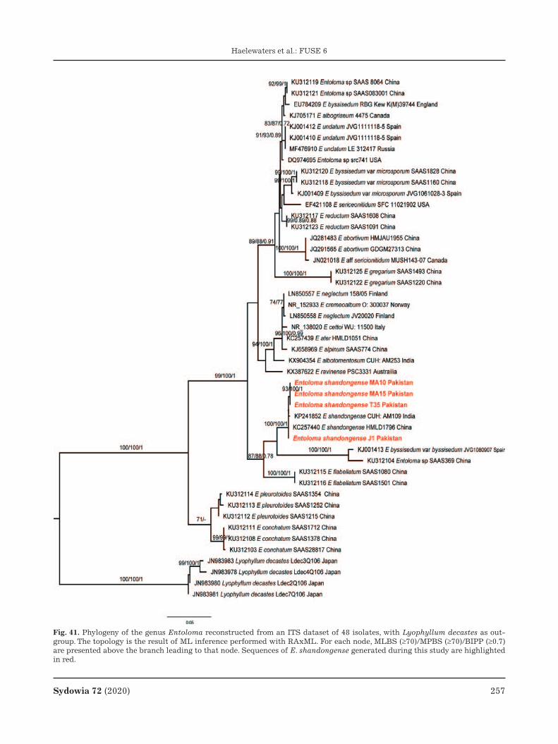
Sydowia 72 (2020) 257
Haelewaters et al.: FUSE 6
Fig. 41. Phylogeny of the genus Entoloma reconstructed from an ITS dataset of 48 isolates, with Lyophyllum decastes as out-group. The topology is the result of ML inference performed with RAxML. For each node, MLBS (≥70)/MPBS (≥70)/BIPP (≥0.7) are presented above the branch leading to that node. Sequences of E. shandongense generated during this study are highlighted in red.

258 Sydowia 72 (2020)
Haelewaters et al.: FUSE 6
Fig. 42. Erysiphe quercicola on Ziziphus jujuba. A. Infected host plant. B. Infection viewed under stereomicroscope. C. Germinat-ing conidium. D. Conidium. E. Foot cells. F. Appressoria. G. Conidiophore. Scale bars A 4 cm, B 8 mm, C–D 8 μm, E 7 μm, F 2 μm, G 12 μm.

Sydowia 72 (2020) 259
Haelewaters et al.: FUSE 6
Ascomycota, Leotiomycetes, Helotiales, Erysip-haceae
Erysiphe quercicola S. Takam. & U. Braun, in Taka-matsu et al., Mycol. Res. 111: 819 (2007). – Fig. 42
M a t e r i a l e x a m i n e d . – PAKISTAN. Khyber Pakh-tunkhwa Province, Hazara Division, Abbottabad, Abbottabad District, Ayubia National Park, Mukshpuri, 34°03’36.72”N, 73°25’50.88”E, 2800 m a.s.l., on leaves of Ziziphus jujuba (Ro-sales, Rhamnaceae), 13 August 2018, leg. N.S. Afshan & J. Ma-jeed, JP#5 (LAH 36141).
D e s c r i p t i o n . – M y c e l i u m amphigenous, persistent, dense, white, forming patches or effuse, epigenous, foliicolous, in patches, evanescent. – H y p h a e about 4–7 μm wide; hyphal appressoria bilobed, distinct in pairs. – C o n i d i o p h o r e s arising from the upper surface of superficial hy-phae, erect, about 80–151 μm. – Fo o t c e l l s cylin-drical, straight, 44–74 × 7–10 μm; followed by 1 longer cell and 2–3 shorter cells. – C o n i d i a cate-nescent, doliiform, subcylindrical to ellipsoid, 24–48 × 16–20 μm.
H a b i t a t a n d d i s t r i b u t i o n . – Known on Acacia spp., Anacardium occidentale, Bixa orella-na, Citrus spp., Hevea brasiliensis, Mangifera indi-ca, and Quercus spp. from many countries in South America, Europe, Africa, Asia, and Australia (Braun & Cook 2012); on Ailanthus excels from India; on Cinnamomum camphora, Delonix regia, and Rumex crispus from Brazil; on Cyclobalanopsis sp. and Nephelium lappaceum from Indonesia and Japan; on Durio zibethinus from Indonesia; and on Mur-raya paniculata from Taiwan (Farr & Rossman 2019).
N o t e s . – The genus Erysiphe consists of 478 species according to Wijayawardene et al. (2020) and belongs to the family Erysiphaceae in Heloti-ales s.l. (sensu Johnston et al. 2019). Morphology and molecular data point at E. quercicola as the identification for our fungus from Z. jujuba leaves. Our initial BLAST search found that our newly generated ITS sequence (GenBank accession no. MN394113) shares 99.69–100 % identity with the top 10 results—all E. quercicola. Our phylogenetic tree based on ITS (Fig. 43) retrieved our isolate in a clustered with 17 other isolates of E. quercicola with high support (BS=96). Inoculated leaves dur-ing pathogenicity test developed symptoms of E. quercicola after 7 days, whereas control plants re-mained symptomless. This report is the first one of a powdery mildew fungus from Z. jujuba in Paki-stan.
Authors: N.S. Afshan, M. Riaz, J. Majeed & A.N. Khalid
Fig. 43. Phylogeny of the genus Erysiphe reconstructed from an ITS dataset of 31 isolates. The topology is the result of ML inference performed with RAxML. For each node, MLBS (if >60) is presented above/below the branch leading to that node. The sequence of E. quercicola generated during this study is highlighted in boldface.

260 Sydowia 72 (2020)
Haelewaters et al.: FUSE 6
Fig. 44. Erysiphe urticae on Urtica dioica. A. Infected host plant. B. Infection viewed under stereomicroscope. C. A Chasmothe-cium. D. Appressoria. E. Immature chasmothecium. F. Chasmothecium (SEM). G. Conidiophore. H. Chasmothecial appendages. I. asci. J. Conidia. Scale bars A 0.75 cm, B 0.08 mm, C 0.03 μm, D 0.07 μm, E 0.03 μm, G 0.07 μm, H 0.05 μm, I 0.07 μm, J 0.14 μm.

Sydowia 72 (2020) 261
Haelewaters et al.: FUSE 6
Ascomycota, Leotiomycetes, Helotiales, Erysip-haceae
Erysiphe urticae (Wallr.) S. Blumer, Beitr. Kryptfl. Schweiz 7(1): 224 (1933). – Figs. 44–45
B a s i o n y m . – Alphitomorpha urticae Wallr., Ann. Wetter. Gesellsch. Ges. Naturk. 4: 238 (1819).
M a t e r i a l e x a m i n e d . – PAKISTAN. Khyber Pakh-tunkhwa Province, Hazara Division, Haripur District, near Khun, 33°51’54.83”, N73°8’19.57”E, 2438 m a.s.l., on leaves of Urtica dioica (Rosales, Urticaceae), 28 October 2018, leg. N.S. Afshan & M. Riaz, MA #08 (LAH 35663); Khyber Pakhtunkh-
wa Province, Hazara Division, Abbottabad District, Ayubia National Park, Mukshpuri, 34°04’N, 73°23’E, 2800 m a.s.l., on leaves of U. dioica, 30 October 2017, leg. N.S. Afshan & M. Riaz, ZM #21 (LAH 36173); Khyber Pakhtunkhwa Province, Hazara Division, Abbottabad District, Ayubia National Park, Khanspur, 34°00’60.00”N, 73°24’59.99”E, 2250 m a.s.l., on leaves of U. dioica, 29 October 2017, leg. N.S. Afshan, JP #20 (LAH 36156).
D e s c r i p t i o n . – M y c e l i u m epiphyllous, forming dense, thick white patches. – H y p h a e hy-aline, thin walled, smooth; hyphal appressoria in opposite pairs multilobed to nipple shaped. – C o n -
Fig. 45. Erysiphe urticae. A. Chasmothecium. B. Asci with ascospores. C. Conidia. D. Appressoria. E. Conidiophore. Scale bars A 0.04 μm, B 0.05 μm, C 0.02 μm, D 0.12 μm, E 0.06 μm, del. M. Riaz.

262 Sydowia 72 (2020)
Haelewaters et al.: FUSE 6
i d i o p h o r e s arising from the upper surface of a hyphal mother cells, 44–105 μm in length. – Fo o t c e l l s cylindrical, somewhat straight, 5–7 × 20–55 μm; followed by 1–2 shorter cells. – C o n i d i a formed singly or in chains, ellipsoid to cylindrical, 17–18 × 34–40 μm. – C h a s m o t h e c i a scattered to gregarious, hyaline when immature to light brown to dark brown at maturity, 125–166 μm in diam.; pe-ridium cells irregularly shaped; appendages numer-ous, mycelioid, branched, 3–5 μm in diam., aseptate, hyaline, thin-walled, smooth. – A s c i 2 –7 per chasmothecium, ellipsoid to ovoid, 26–48 × 60–64 μm, stalked, 2–5-spored. – A s c o s p o r e s ellipsoid to ovoid, 11–17 × 18–25 μm, hyaline.
H a b i t a t a n d d i s t r i b u t i o n . – Known from China, India, Iran, Israel, Lebanon, Russia, Si-beria, Saudi Arabia, South Korea, Sri Lanka, and Turkey (Braun & Cook 2012; Farr & Rossman 2019). Reported host plants are Urtica cannabina L., U. di-oica, U. fissa E. Pritz, U. kioviensis Rogow., U. pilu-lifera L., U. urens L., and outside of Europe also Pilea glaberrima (Blume) Blume (Urticaceae) (Ellis 2020, F.L. Zhang & C.W. Li unpublished). Boehmeria gracilis C.H. Wright (Urticaceae) is listed as the plant host for a Chinese collection of E. urticae in an NCBI GenBank sequence (L. Bai unpublished).
N o t e s . – Our newly generated ITS sequences (GenBank accession nos. MN922334, MN636273) resulting from our work towards the Erysiphaceous Fungi of Ayubia support identification as E. urticae. The sequences shared 99.57–100 % identity with an isolate from the UK (GenBank accession no. KY660866), 99.53–99.65 % with an isolate from China (KF582797), and 98.79 % with isolates from China and Iran (AB104524, KR048078, MT309705), all from either U. dioica, U. fissa, or Boehmeria gra-cilis (Tab. 1). Our ITS-based phylogenetic tree (Fig. 46) retrieved our Pakistani isolates in a cluster with other isolates of E. urticae (BS=99). One of the isolates in this cluster, however, is identified as E. caricae (LC009901). As the only ITS sequence that is available in Genbank for this species, it is unfor-tunate that the authors who generated it, did not provide morphological descriptions (Takamtsu et al. 2015). Erysiphe caricae is reported on members of Caricaceae (Brassicales) whereas known hosts of E. urticae belong to family Urticaceae (Rosales) (Braun & Cook 2012). Our pathogenicity assay on U. dioica shows symptoms of disease after 7 days, whereas control plants remained symptomless. Ery-siphe urticae is reported here as a new country re-cord for Pakistan.
Authors: N.S. Afshan, M. Riaz, J. Majeed & A.N. Khalid
Fig. 46. Phylogeny of the genus Erysiphe reconstructed from an ITS dataset of 36 isolates. The topology is the result of ML inference performed with RAxML. For each node, the ML bootstrap (if >60) is presented above/below the branch leading to that node. Sequences of E. urticae generated during this study are highlighted in boldface.

Sydowia 72 (2020) 263
Haelewaters et al.: FUSE 6
Ascomycota, Laboulbeniomycetes, Laboulbeniales, Laboulbeniaceae
Fanniomyces ceratophorus (Whisler) T. Majewski, Acta Mycol. 8(2): 230 (1972). – Fig. 47
B a s i o n y m . – Stigmatomyces ceratophorus Whisler, My-cologia 60(1): 68 (1968).
M a t e r i a l e x a m i n e d . – THE NETHERLANDS. Gelderland Province, Ede, Oude Kerkplein, 52°02’44.5”N, 5°40’17.6”E, on Fannia canicularis (Linnaeus, 1761) (Diptera, Fanniidae), D. Haelew. 135 [host label], 18 October 2012, leg. J. van Erkelens, slides FH 00313224 (4 thalli from abdominal tergites) and FH 00313225 (8 thalli from abdominal tergites).
M a t e r i a l s e q u e n c e d . – USA. California, Alameda County, Oakland, 37°48’14.2”N, 122°15’55.7”W, on F. canicula-ris, D. Haelew. 1136 [host label], iNaturalist ID 3291539, 24 May 2016, leg. D. Tighe, isolate D. Haelew. 1136h (8 mature thalli, thorax), MG958013 (SSU), MH145384 (LSU).
H o s t s a n d d i s t r i b u t i o n . – Known from Fannia canicularis (lesser house fly) in five conti-nents: North America (USA), South America (Gua-temala), Europe (Czech Republic, Poland, Portugal, Spain), Asia (Lebanon, Turkey), and Oceania (New Zealand) (Whisler 1968; Majewski 1972; Tavares 1985; Hughes et al. 2004; Santamaría 2006; Rossi et al. 2013, 2019; Rossi 2016).
N o t e s . – Stigmatomyces H. Karst. as currently recognized is the second-largest genus of Laboulbe-niales, with 176 described species (Species Fungo-rum 2020). Majewski (1972) established the genus Fanniomyces T. Majewski to accommodate S. cera-tophorus on the basis of the highly branched ap-pendage, which is different from the compact, sin-gle-axis appendage of most species in Stigmatomy-ces. Later, F. burdigalensis Balazuc from France (Balazuc 1979) and F. copromyzae Huldén from Fin-land (Huldén 1983) were added to Fanniomyces. Weir & Rossi (1995) synonymized F. copromyzae un-der F. burdigalensis based on morphological and morphometric data. In the same paper, the authors also considered Fanniomyces as a junior synonym of Stigmatomyces because the latter is variable in the structure of its appendage. Tavares (1985) pre-sented Stigmatomyces oecotheae Thaxt. (Thaxter 1931) as an example of a species with long append-age cells and elongate antheridia. However, without molecular data, it is unknown whether S. oecotheae is representative of Stigmatomyces s.s. Phenotypic plasticity, polymorphism, position-induced mor-phological adaptations, and cryptic diversity in La-boulbeniales can hinder delimitation of natural groups by morphology alone (e.g., Goldmann & Weir 2012; Goldmann et al. 2013; Haelewaters et al. 2018a, 2018b, 2019b; Haelewaters & Pfister 2019).
This study presents the first-ever molecular phy-logenetic analysis focused on Stigmatomyces s.l.
Our two-locus phylogenetic tree retrieved Stigmat-omyces s.l. as paraphyletic, congruent with the pri-or studies of Haelewaters et al. (2018b) and Liu et al. (2020). Four major clades were retrieved, each with maximum support. Clade I included S. burdi-galensis and S. ceratophorus—the type of Fannio-myces. Clade II included S. gregarius, S. scaptomyz-ae, and S. entomophila—the type of Appendiculina. Clade III was comprised of species of Gloeandromy-ces, including the type G. streblae (f. sigmomor-phus). Clade IV included five species of Stigmato-myces.
Fanniomyces was erected to accommodate spe-cies with highly branched appendage and elongate antheridia (Majewski 1972, 1994). This genus is the earliest diverging lineage of the Stigmatomyces complex, represented by clade I in our phylogenetic tree (Fig. 48). Given the results of our phylogenetic reconstruction, Fanniomyces is reinstated here. The appendage structure (Tab. 4) associated with these species appears to be a characteristic feature for this lineage. Fanniomyces ceratophorus is here for the first time reported from the Netherlands. Our sequenced material was sampled in Oakland, Cali-fornia—a city neighboring Berkeley, the type local-ity.
Appendiculina Berl. was erected by Berlese (1889) to correct Peck’s (1885) mistake of describing
Fig. 47. Thalli of Stigmatomyces sensu lato. a. Fanniomyces ceratophorus, mature thallus from slide FH 00313225. b. Gloeandromyces hilleri, mature thallus from holotype slide FH 00313744 (Liu et al. 2020). Shown are cells I, II, and III of the receptacle and the appendage (A). Scale bar 100 μm, del. J. Liu.

264 Sydowia 72 (2020)
Haelewaters et al.: FUSE 6
Tab. 4. Overview of characters of four genera within Stigmatomyces sensu lato, including geographic distribution, host genus and classification up to subsection, and morphological features of the receptacle, appendage, and antheridia.
Clade Genus Species AuthorsGeographic distribution
Host family/-ies (genus/genera)
Host super- family/-ies
Host subsection
Receptacle AppendageAnther-idia
I Fanniomy-ces
burdiga-lensis
Balazuc Europe Sphaeroceridae (Copromyza, Crumomyia)
Sphaerocer-oidea
Acalyptra-tae
Cell II elongated; cells I and VI never in contact
Highly branched
Elongate
Fanniomy-ces
ceratopho-rus
(Whisler) T. Majewski
North and South America, Europe, Asia, Oceania
Fanniidae (Fannia) Muscoidea Calyptratae
II Appendi-culina
entomoph-ila
Peck (Berl.)
North and South America, Caribbean, Europe
Drosophilidae (Drosophila)
Ephydroidea Acalyptra-tae
Un-branched
Short
Appendi-culina
gregaria (W. Rossi) Haelew. & Aime
Africa Diopsidae (Diopsis) Diopsoidea Acalyptra-tae
Appendi-culina
scapto-myzae
(Thaxt.) Haelew. & Aime
North and South America, Europe, Africa
Drosophilidae (Scaptomyza)
Ephydroidea Acalyptra-tae
IV Stigmato-myces
borealis Thaxt. North America Ephydridae (Parydra imitans)
Ephydroidea Acalyptra-tae
Stigmato-myces
chamae-myiae
W. Rossi & M. Leonardi
Europe Chamaemyiidae (Chamae-myia)
Lauxanioidea Acalyptra-tae
Stigmato-myces
limnopho-rae
Thaxt. North and South America, Caribbean, Europe, Africa, Asia
Anthomyiidae (gen. & sp. indet.), Calliphoridae (Calliphora, Lucilia), Muscidae (Dasyphora, Heliographa, Leucomeli-na, Limnophora), Rhiniidae (Fainia, Isomyia, Rhyncomya, Sumatria), Sarcophagidae (Sarcophaga)
Muscoidea (Anthomyi-idae, Muscidae), Oestroidea (Calliphoridae, Rhiniidae, Sarcophagi-dae)
Calyptratae
Stigmato-myces
protrudens Thaxt. North America Ephydridae (Parydra pinguis)
Ephydroidea Acalyptra-tae
Stigmato-myces
rugosus Thaxt. North and South America, Caribbean, Europe, Africa, Asia, Oceania
Ephydridae (Clanoneu-rum, Leptopsilopa, Psilopa)
Ephydroidea Acalyptra-tae
Stigmato-myces
baeri (type)
(Knoch) Peyr.
Europe Muscidae (Musca) Muscoidea Calyptratae
III Gloean-dromyces
dickii Haelew. Central and South America
Streblidae (Trichobius) Hippobos-coidea
Calyptratae Cell II short; cells I and VI always in contact
Fan-like Short, gelati-nousGloean-
dromycesnycteribi-idarum
(Thaxt.) Thaxt.
Caribbean, Central America
Streblidae (Megistopoda, Trichobius)
Gloean-dromyces
pageanus f. alarum
Haelew. Central America Streblidae (Trichobius)
Gloean-dromyces
pageanus f. pageanus
Haelew. Central America Streblidae (Trichobius)
Gloean-dromyces
pageanus f. polymor-phus
Haelew. Central America Streblidae (Trichobius)
Gloean-dromyces
streblae f. sigmomor-phus
Haelew. Central America Streblidae (Trichobius)
Sydowia 72 (2020) 265
Haelewaters et al.: FUSE 6
Appendicularia to accommodate A. entomophila Peck. The genus Appendicularia was illegitimate because it had already been applied to a genus in the plant family Melastomataceae, a genus that re-mains in use even today (Da Rocha et al. 2018). However, by recombining A. entomophila in Stig-matomyces, Thaxter (1896) reduced the monospe-cific genus Appendiculina in synonymy. Since S. en-tomophilus—the former type species of the genus Appendiculina—is represented in clade II of our phylogenetic reconstruction, we here reinstate the genus and add two species to it.
Gloeandromyces (Fig. 47) was erected to accom-modate species with fan-like appendage, consisting of a basal cell that carries two short branches of dichotomously dividing cells, the final cells being antheridial. At maturity, the antheridia become ge-latinous, “losing their identity more or less com-pletely” (Thaxter 1931: 112). Another morphologi-cal character, unnoted by Thaxter in his generic de-scription but consistent among described species of Gloeandromyces is the short cell II (Tavares 1985), allowing cells I and VI to be in contact (Thaxter 1917, 1931; Haelewaters & Pfister 2019; Liu et al. 2020). These morphological features do not occur in any of the species of Fanniomyces of Stigmatomy-ces (Tab. 4). In addition, LSU sequences of the Gloeandromyces species in our final (aligned and trimmed) dataset are 89.24–100 % identical, where-as they are 82.73–85.07 % identical with Fanniomy-ces spp. and 80.34–85.68 % with Stigmatomyces spp. (S. protrudens excluded because of low query coverage). This points at a barcode “gap” of mini-mum 3.56–4.17 % distinguishing inter- from intra-generic variability. We regard Gloeandromyces as an independent radiation on neotropical bat flies (Thaxter 1931, Haelewaters et al. 2018, Haelewaters & Pfister 2019) and the generic description is emended here.
Stigmatomyces s.s. – still the largest genus by far in the Stigmatomyces complex – represents the crown radiation, comprising 171 species (Species Fungorum 2020) and represented in our phyloge-netic reconstruction by clade IV (Fig. 48). This situ-ation needs confirmation by the inclusion of se-quences of Stigmatomyces baeri (Knoch) Peyr, the type species of the genus. As long as these data are not available it is impossible to predict which of the current and potentially more clades in this diverse group should carry the name Stigmatomyces s.s. We recognize that our current sampling is poor (14 of 180 species included in our phylogenetic reconstruc-tion of Stigmatomyces s.l.) but this work is only a starting point to resolve the taxonomy of this clade.
Our taxonomic circumscription of genera within the Stigmatomyces complex results in four mono-phyletic genera. For Appendiculina, Fanniomyces, and Gloeandromyces, the type species are present in the representative clades. Also in Laboulbeniales research, molecular phylogenetic work helps us to better understand evolutionary patterns. In the Stigmatomyces complex, this involves the recogni-tion of a radiation in the Neotropics (Gloeandromy-ces), hinting at a temperate origin for this group, et cetera. The only way to further elaborate on these patterns and to attain a fully stable taxonomy of Stigmatomyces s.l. is by doing more collecting.
Appendiculina Berl., Malpighia 3: 59 (1889).
Sy n o n y m . – Appendicularia Peck, Ann. Rep. N. Y. State Mus. Nat. Hist. 38: 95 (1885), nom. illegit.
N o t e s . – Appendiculina comprises species in clade II (Fig. 48), which is composed of A. ento-mophila (type species) and two more species that were formerly placed in the genus Stigmatomyces (Thaxter 1901, Rossi 1982). Its SSU is 92.93–96.40 % identical with Fanniomyces, 85.51–96.16 % identi-cal with Gloeandromyces, and 83.25–95.71 % iden-tical with Stigmatomyces; compared to other gen-era in Stigmatomyces s.l., unique molecular syna-pomorphies at positions 175, 176, 211, 226, 247, 250, 631, 634, 654, 675, 728.
S p e c i e s i n c l u d e d .
Appendiculina entomophila (Peck) Berl., Mal-pighia 3: 59 (1889).
B a s i o n y m . – Appendicularia entomophila Peck, Ann. Rep. N.Y. St. Mus. nat. Hist. 38: 96 (1885).
Appendiculina gregaria (W. Rossi) Haelew. & Aime, comb. nov. MycoBank no.: MB 835861.
B a s i o n y m . – Stigmatomyces gregarius W. Rossi, Quad. Accad. Naz. Lincei 255: 19 (1982).
Appendiculina scaptomyzae (Thaxt.) Haelew. & Aime, comb. nov. MycoBank no.: MB 835862.
B a s i o n y m . – Stigmatomyces scaptomyzae Thaxt., Proc. Am. Acad. Arts Sci. 36(23): 400 (1901).
Fanniomyces T. Majewski, Acta Mycol. 8(2): 229 (1972). – Fig. 47
N o t e s . – Fanniomyces comprises the two spe-cies in clade I (Fig. 48), F. burdigalensis and F. cera-tophorus (type species). Its LSU is 82.73–85.07 % identical with Gloeandromyces, and 81.63–88.89 % identical with Stigmatomyces; compared to other genera in Stigmatomyces s.l., unique molecular synapomorphies at positions 79, 93, 100, 149, 205, 363, 411, 412, 423, 426, 428, 444, 447, 459, 460, 483, 488, 496, 502, 503, 505, 517, 544, 550, 563, 662. Its

266 Sydowia 72 (2020)
Haelewaters et al.: FUSE 6

Sydowia 72 (2020) 267
Haelewaters et al.: FUSE 6
Fig. 48. Phylogeny of Laboulbeniomycetes isolates reconstructed from a combined SSU–LSU dataset. The topology is the result of ML inference performed with IQ-TREE. For each node, MLBS (if >70) is presented above/below the branch leading to that node. The colored clades are representative of Stigmatomyces sensu lato, including four genus-level lineages, labeled clade I (=Fanniomyces), clade II (=Appendiculata), clade III (=Gloeandromyces), and clade IV (=Stigmatomyces sensu stricto).
SSU is 92.93–96.40% identical with Appendiculina, 93.12–97.62 % identical with Gloeandromyces, and 74.57–97.42 % identical with Stigmatomyces; com-
pared to other genera in Stigmatomyces s.l., unique molecular synapomorphies at positions 56, 266, 639, 671, 726, 827, 837, 840.

268 Sydowia 72 (2020)
Haelewaters et al.: FUSE 6
S p e c i e s i n c l u d e d .
Fanniomyces burdigalensis Balazuc, Revue My-col., Paris 43: 402 (1979).
Fanniomyces ceratophorus (Whisler) T. Majews-ki, Acta Mycol. 8(2): 230 (1972).
B a s i o n y m . – Stigmatomyces ceratophorus Whisler, Mycologia 60(1): 68 (1968).
Gloeandromyces Thaxt., Mem. Am. Acad. Arts Sci. 16: 112 (1931). emend. Haelew. – Fig. 47
D e s c r i p t i o n . – R e c e p t a c l e with three cells; cell I longer than broad, sometimes bent or kinked, in contact with cell VI; cell II short, sepa-rated from cell III by an oblique septum; cell III broadly trapezoidal or triangular. – A p p e n d a g e with basal cell broadly pentagonal, carrying two short, dichotomously dividing branches, terminal cells antheridial; appendage fan-like in structure. – A n t h e r i d i a gelatinous at maturity, distally dis-integrating towards below. – C e l l VI broader than long, obliquely positioned between cells II and VII. – P e r i t h e c i u m with four tiers of vertical wall cells; intraspecific plasticity is observed regarding the presence of perithecial projections, undulations, bumps, or prominences; in certain species this plas-ticity can be linked to growing on a particular posi-tion of the host.
N o t e s . – Gloeandromyces comprises five spe-cies, of which four are included in clade III of our phylogeny (Fig. 48), including the type species, G. streblae (f. sigmomorphus). Its LSU is 82.73–85.07 % identical with Fanniomyces, and 80.34–85.68 % identical with Stigmatomyces; compared to other genera in Stigmatomyces s.l., unique mo-lecular synapomorphies at positions 100, 114, 128, 138, 163, 193, 218, 409, 410, 428, 447, 460, 461, 494, 526, 544, 550, 556, 661. Its SSU is 85.51–96.16 % identical with Appendiculina, 93.12–97.62 % iden-tical with Fanniomyces, and 85.12–97.42 % identi-cal with Stigmatomyces; compared to other genera in Stigmatomyces s.l., unique molecular synapo-morphies at positions 262, 266, 441, 481, 630, 646, 667, 677, 687, 731, 763, 804, 823, 827.
S p e c i e s i n c l u d e d .
Gloeandromyces dickii Haelew., in Haelewaters & Pfister, Fungal Syst. Evol. 3: 22 (2019).
Gloeandromyces hilleri Haelew. & Pfliegler, My-cologia in press (2020).
Gloeandromyces nycteribiidarum (Thaxt.) Thaxt., Mem. Am. Acad. Arts Sci. 16: 113 (1931).
B a s i o n y m . – Stigmatomyces nycteribiidarum Thaxt., Proc. Am. Acad. Arts Sci. 52(10): 702 (1917).
Gloeandromyces pageanus Haelew., in Haelewa-ters et al., Nova Hedwig. 105(3–4): 272 (2017).
Gloeandromyces pageanus f. alarum Haelew., in Haelewaters & Pfister, Fungal Syst. Evol. 3: 26 (2019).
Gloeandromyces pageanus f. pageanus Haelew., in Haelewaters et al., Nova Hedwig. 105(3–4): 272 (2017).
Gloeandromyces pageanus f. polymorphus Haelew., in Haelewaters & Pfister, Fungal Syst. Evol. 3: 28 (2019).
Gloeandromyces streblae (Thaxt.) Thaxt., Mem. Am. Acad. Arts Sci. 16: 113 (1931).
B a s i o n y m . – Stigmatomyces streblae Thaxt., Proc. Am. Acad. Arts Sci. 52(10): 700 (1917).
Gloeandromyces streblae f. sigmomorphus Haelew., in Haelewaters & Pfister, Fungal Syst. Evol. 3: 29 (2019).
Gloeandromyces streblae f. streblae Thaxt., Mem. Am. Acad. Arts Sci. 16: 113 (1931).
Authors: D. Haelewaters, A. De Kesel, J. Liu & M.C. Aime
Basidiomycota, Agaricomycetes, Agaricales, Om-phalotaceae
Marasmiellus biformis (Peck) J.S. Oliveira, in Ol-iveira et al., Mycol. Progr. 18(5): 734 (2019). – Fig. 49
B a s i o n y m . – Marasmius biformis Peck, Bull. N.Y. State Mus. 67: 25 (1904) [1903].
M a t e r i a l e x a m i n e d . – PAKISTAN. Khyber Pakh-tunkhwa Province, Abbottabad District, Ayubia National Park, 33°51’54.83”N, 73°8’19.57”E, 2400 m a.s.l., 15 July 2017, leg. M. Ali & A.R. Niazi, KH 117 (LAH 36408); Ibid., 16 July 2017, leg. M. Ali & A.R. Niazi, KH 122 (LAH 36409).
D e s c r i p t i o n . – P i l e u s 10–28 mm in diam., medium-sized, convex with incurved and entire margin when young, becoming plane or depressed to umbilicate on disc, rugulose sulcate on the margin; surface dry, non-hygrophanous, glabrous, reddish brown (10R4/6) when young to leather brown (10R6/4) when mature. – C o n t e x t white, thin, 0.4–1 mm thick. – L a m e l l a e adnate, sub-distant to close, thin, narrow to moderately broad, white to white grayish or, pale pinkish with age. – S t i p e 20–55 × 2.0–3.5 mm, centrally attached, equal, slen-der, cylindrical, rarely flattened, tough; surface dry, white near cap, tawny (2.5YR) to leather brown (10R6/4) toward the base, densely pubescent, pubes-cence whitish above and dark brown towards base. – B a s i d i o s p o r e s (6.0–) 6.4–7.8(–8.7) × (3.0–)3.1–4.4(–4.5) μm, Q=1.4–1.8, Qav=1.5 μm, slightly ellipsoid to lacrymoid or obovoid, smooth, thin-walled, hya-line, inamyloid, acyanophilous. – B a s i d i a 17.5–30 × 3.5–6.0 μm, clavate, bisporic to tetrasporic, hyaline,

Sydowia 72 (2020) 269
Haelewaters et al.: FUSE 6
Fig. 49. Marasmiellus biformis. A. Basidiomata in situ. B. Basidia. C. Basidiospores. D. Cheilocystidia. E. Caulocystidia. F. Pileipel-lis. G. Stipitipellis. Scale bars A 1 cm; B–C, E–F 5 μm; D, G 10 μm; del. M. Ali.

270 Sydowia 72 (2020)
Haelewaters et al.: FUSE 6
Fig. 50. Marasmiellus subnudus. A. Basidiomata in situ among pine needles. B. Basidia. C. Basidiospores. D. Cheilocystidia. E. Pileipellis. F. Stipitipellis. Scale bars A 1.5 cm, B–C 5 μm, D–F 10 μm, del. M. Ali.

Sydowia 72 (2020) 271
Haelewaters et al.: FUSE 6
thin walled. – P l e u r o c y s t i d i a absent. – C h e i l o c y s t i d i a 21.5–51 μm long, cylindrical, contorted, variously lobed to strangulated, thin walled. – P i l e i p e l l i s a cutis made of cylindric and parallel hyphae, branched, septate, frequently encrusted, 2.0–6.5 μm in diam., with diverticulate or broadly round terminal elements. – S t i p i t i p e l -l i s a layer of parallel and cylindrical hyphae, 2.0–5.5 μm in diam., rarely with inconspicuous encrust-ed, occasionally branched, smooth, thin-walled, fre-quently septate, terminal elements with rounded ends. – C a u l o c y s t i d i a differently shaped, cylin-dric, contorted to strangulated, up to 19–55 μm, hyaline, thin-walled. – C l a m p c o n n e c t i o n s frequently present in all tissues.
H a b i t a t a n d d i s t r i b u t i o n . – Thus far reported from northeastern USA and eastern Asia (Japan). In Pakistan found growing scattered or in bunches on decomposing leaves or needles in pine-dominated moist temperate forests.
N o t e s . – In the ITS tree (Fig. 20), our sequences of Pakistani collections KH117 and KH122 are clus-tered with M. biformis with maximum support. Three sequences of M. “biformis” (GenBank acces-sion nos. KM083047, KM083048, MG407676) form a separate clade (MLBS=84) sister to M. biformis. These are sequences from China (H. Tian unpub-lished) and South Korea (J.S. Lee et al. unpublished) that may be representative of another undescribed species, although no morphological information is available. The Pakistani material resembles M. bi-formis in both macromorphology and microscopic characters (Fig. 49). The only noted differences are the slightly larger pileus (10–28 mm) compared to eastern North American and Japanese collections, and slightly larger caulocystidia (6.0–8.5 μm) com-pared to Japanese material (Halling 1983, 2004; Miy-amoto et al. 1998). North American collections of M. biformis are found mostly on soil and only rarely on decaying leaves or needles (Halling 1983). In con-trast, Miyamoto et al. (1998) studied M. biformis in Japan collected from leaf or needle litter. Our collec-tions were found on decomposing leafy materials.
Authors: M. Ali, H. Bashir, A.R. Niazi & A.N. Khalid
Basidiomycota, Agaricomycetes, Agaricales, Om-phalotaceae
Marasmiellus subnudus (Ellis ex Peck) J.S. Oliveira, in Oliveira et al, Mycol. Progr. 18(5): 735 (2019). – Fig. 50
B a s i o n y m . – Marasmius subnudus Ellis ex Peck., Bull. Torrey Bot. Club 25: 287 (1898).
M a t e r i a l e x a m i n e d . – PAKISTAN. Khyber Pakh-tunkhwa Province, Abbottabad District, Ayubia National Park, 33°51’54.83”N, 73°8’19.57”E, 2,400 m a.s.l., 15 July 2017, leg. M. Ali & A.R. Niazi, KH 317 (LAH36410).
D e s c r i p t i o n . – P i l e u s 15–37 mm in diam., obtusely convex to broadly sub-umbonate becoming broadly convex to plane with a centrally depressed disc; surface dry, glabrous to fibrillose with age, more or less even on the disc, wrinkled towards the margin, very pale brown (10YR7/4). – M a r g i n usually entire, incurved when young, be-coming decurved when mature. – C o n t e x t thin, whitish, tough. – L a m e l l a e adnate to adnexed, subdistant to distant, thin or moderately thick, in-frequently anastomosed, whitish when young, be-coming light pale brown at maturity. – S t i p e 20–70 × 3.0–5.0 mm, cylindrical or somewhat com-pressed, nearly equal or broader towards base, straight, surface dry, slightly pubescent towards base, concolorous with lamellae. – B a s i d i o -s p o r e s (7.7–)8.7–10.9(–11.5) × 3.1–4.1(–4.3) μm, lacrymoid to ellipsoid, smooth, inamyloid, hyaline, thin-walled. – B a s i d i a clavate to subclavate, bisporic to tetrasporic with basal clamps, thin-walled, 20–36.5 × 5.0–11 μm. – P l e u r o c y s t i d i a absent. – C h e i l o c y s t i d i a variously shaped, ventricose-fusoid to somewhat mucronate, occa-sionally fusoid, sometimes lobed, thin walled, hya-line, 27–39(–53) × 7.5–9.0 μm. – P i l e i p e l l i s a cu-tis of loosely arranged cylindric hyphae, thin walled, frequently encrusted; 3.0–7.5 μm in diam. with broadly round, occasionally diverticulate terminal elements. – S t i p i t i p e l l i s a layer of parallel ar-ranged cylindric hyphae, thin-walled, cells 2.0–6.7 μm in diam., with sharply rounded terminal ele-ments. – C l a m p c o n n e c t i o n s frequent in all tissues.
H a b i t a t a n d d i s t r i b u t i o n . – Reported from eastern North America (Morgan 1905), South Korea (Jang et al 2016), and Pakistan (this study). Gregarious or scattered in leaf litter, pine needles, and decaying/dead twigs in forests.
N o t e s . – The newly generated ITS sequence of collection KH 317 shares 99 % identity with M. sub-nudus (GenBank accession nos. KX513747, KX513748). In our ITS-based phylogenetic recon-struction of Marasmiellus, the Pakistani sequence is retrieved among isolates of M. subnudus with strong support: MLBS=95 (Fig. 20). Our collection KH 317 is micromorphologically similar to M. subnudus re-ported from South Korea by Jang et al. (2016). The holotype collection of M. subnudus from eastern North America is different macromorphologically only in its slightly smaller (10–35 mm), cinnamon-

272 Sydowia 72 (2020)
Haelewaters et al.: FUSE 6
brown to cinnamon pileus (Bañares et al. 2007). The Pakistani collection was sampled from leaf litter, pine needles, and decaying twigs in a mixed pine forest (Fig. 50A), which is in line with previous col-lections for this species. Eastern North American and South Korean collections of M. subnudus were also found in litter of leaves and dead twigs in mixed and deciduous forests (Jang et al. 2016).
Authors: M. Ali, H. Bashir, A.R. Niazi & A.N. Khalid
Ascomycota, Pezizomycetes, Pezizales, Morchel-laceae
Morchella anatolica Isıloglu, Spooner, Allı & Solak, Mycologia 102(2): 455 (2010). – Fig. 51
Sy n o n y m . – Morchella lanceolata Clowez & Illescas in Clowez, Bull. Soc. Mycol. France 126(3–4): 282 (2012), nom. inval. (“ad int.”, no diagnosis, no type designated).
M a t e r i a l e x a m i n e d . – TURKEY. Antalya Province, Kemer district, near Beydagları Coastal National Park, P. bru-tia forest, among moss growing on calcareous rocks, 36°35’38.1”N, 30°30’29.4”E, 354 m a.s.l., 17 April 2015, leg. O. Kaygusuz, OKA2001 (OKA-1B); Ibid., 36°35’38.1”N, 30°30’29.7”E, 335 m a.s.l., 18 April 2015, leg. Ö.F. Çolak, ÖFÇ1249 (OKA-2B).
D e s c r i p t i o n . – A s c o m a t a 35–60 mm high. Hymenophore 25–40 mm high, 8–14 mm wide, usually conical to acutely conical when mature, with ribs longitudinally arranged, 0.4–0.9 mm wide, thick, fleshy, occasionally forked, forming large elongate pits. – P i t s short when young but becom-ing vertically elongated on maturity, glabrous, wid-ening and deepening with aging; surface dark lilac-grey when young, dark grayish to pale when ma-ture, especially when exposed to sunlight, hollow when cut longitudinally. – S t i p e 8–15 mm high, 6–8 mm wide, cylindrical or thicker at the base, hol-low; surface puberulent, nearly coarsely granular, lilac-grey towards the base, upper parts whitish, oc-casionally with dark brownish stains. – C o n t e x t elastic. – S m e l l weak to undetermined. – A s -c o s p o r e s (23.5–)25.0–28.5(–29.0) × (13.0–)14.0–16.5(–17.0) μm, Lav × Wav = 26.5 × 15.5 μm, Q = (1.5–) 1.6–1.8(–1.9) μm, Qav = 1.7 μm, ellipsoid, thick-walled, hyaline, distinctively striate lengthwise. – A s c i 250–300 × 16–24 μm, cylindrical to clavate, hyaline, 8–spored, uniseriate, sometimes also biseri-ate, inamyloid. – P a r a p h y s e s 90–210 × 15–35 μm, hyaline, (0–)1–2 septate in the lower half, usually enlarged towards at the apices, cylindrical to nar-rowly clavate, apex sometimes narrowed, obtuse, supcapitae and fusiform apices. – H e t e r o p a r a -
p h y s e s present in some collections, irregular. – A c r o p a r a p h y s e s 65–220 × 17–35 μm, predomi-nantly fusiform to cylindrical, apically rounded or somewhat narrowed, thin-walled, hyaline. – S t i p e c o r t e x a textura globosa, composed of variously sized, irregularly arranged catenulate elements, clavate to capitate, cylindrical, lageniform or am-pulliform, with 1–3 septa, measuring 30–170 × 10–55 μm.
H a b i t a t a n d d i s t r i b u t i o n . – Growing solitary between late March and mid-April at 100–700 m a.s.l., often among damp mosses, in warm, strongly calcareous forests, possibly associated with Pinus brutia Ten. and other Mediterranean vegeta-tion. Thus far only known from Spain (Clowez 2012, as “M. lanceolata”; Richard et al. 2015; Palazon et al. 2017) and Turkey (Isıloglu et al. 2010).
N o t e s . – Recent multilocus DNA studies have demonstrated the existence of ~80 species of Morchella around the world (Loizides 2017), but new species and new transcontinental records have been published since (Baroni et al. 2018, Du et al. 2019, Clowez et al. 2020). Whereas certain species show a high degree of continental endemism or pro-vincialism (O’Donnell et al. 2011; Taskın et al. 2012; Clowez et al. 2014, 2015), phylogenetic studies have revealed that, many others species are found on more than one continent (Taskın et al. 2010, 2016; Du et al. 2012a, 2012b; Ku et al. 2012; Loizides et al. 2015, 2016; Richard et al. 2015; Voitk et al. 2016; Loizides 2017).
In recent molecular phylogenetic studies, three major clades of Morchella can be discriminated: the /Rufobrunnea clade or “white morels” with M. ana-tolica Isıloglu, Spooner, Allı & Solak and M. rufob-runnea Guzmán & F. Tapia; the /Esculenta clade or “yellow morels” with M. esculenta (L.) Pers. and others; and the /Elata clade of “black morels” with M. elata Fr. and others (Guzmán & Tapia 1998, O’Donnell et al. 2011, Taskın et al. 2012, Richard et al. 2015, Loizides 2017, Clowez & Moreau 2018). The /Rufobrunnea clade, which dates back to the late Jurassic, is represented by two distinct species, M. rufobrunnea and M. anatolica (Isıloglu et al. 2010, Taskın et al. 2012). These two species are often found on treeless, disturbed substrates and wood chippings in the Mediterranean basin, as well as under olive trees (Loizides et al. 2015).
In our phylogenetic tree (Fig. 52), the basal-most monophyletic branch was the /Rufobrunnea clade. The /Elata and /Esculenta clades were highly sup-ported as sister clades. The /Esculenta clade (sect. Morchella Dill. ex Pers.) (MLBS=99, BIPP=1.00) consisted of M. americana Clowez & Matherly, M.

Sydowia 72 (2020) 273
Haelewaters et al.: FUSE 6
Fig. 51. Morchella anatolica. a–f. Fresh ascomata, on natural substrate. g. Ascospores. h. Asci. i. Paraphyses. j. Acroparaphyses. k. Stipe hairs. Scala bars a–f 10 mm, g–k 25 μm, del. O. Kaygusuz.
dunensis (Castañera, J.L. Alonso & G. Moreno) Clowez, M. esculenta, M. galilaea Masaphy & Clowez, M. sceptriformis Clowez & Matherly, and M. ulmaria Clowez. The /Elata clade (sect. Dis-
tantes Boud.) (MLBS=96, BIPP=0.99) was composed of M. angusticeps Peck, M. dunalii Boud., M. elata, M. eximia Boud., M. eximioides Jacquet., M. exu-berans Clowez, Hugh Sm. & S. Sm., M. importuna

274 Sydowia 72 (2020)
Haelewaters et al.: FUSE 6
Fig. 52. Phylogeny of Morchella reconstructed from an ITS dataset. The topology is the result of Bayesian inference performed with MrBayes. Gyromitra gigas and G. ticiniana serve as outgroup taxa. For each node, BIPP (if ≥0.95) and MLBS (if ≥80) are shown the branch leading to that node. Branches in bold indicate BIPP≥0.95 and MLBS≥90. Newly generated sequences from Turkey are in boldface.
Sydowia 72 (2020) 275
Haelewaters et al.: FUSE 6
M. Kuo, O’Donnell & T.J. Volk, M. kakiicolor (Clowez & L. Romero) Clowez, L. Romero, P. Alvarado & Loizides, M. punctipes Peck, M. semilibera DC., M. septentrionalis M. Kuo, J.D. Moore & Zordani, M. sextelata M. Kuo, M. snyderi M. Kuo & Methven, M. tomentosa M. Kuo, and M. tridentina Bres. Finally, the /Rufobrunnea clade (sect. Rufobrunnea Clowez & Courtec.) (MLBS=99, BIPP=1.00) was composed of M. anatolica and M. rufobrunnea (Fig. 52). Phylo-genetically, M. anatolica is closely related to but clearly distinct from M. rufobrunnea based on the ITS. Our newly generated sequences of M. anatolica from Turkey clustered with those of collections from Spain. Morchella anatolica was highly sup-ported in both analyses MLBS=99, BIPP=1.00). The results of our ITS phylogeny agree with the topolo-gies by Richard et al. (2015), Loizides et al. (2016), and Clowez & Moreau (2018).
Morchella anatolica is the sister species of M. ru-fobrunnea both morphologically and molecularly. Morphologically, the ascocarp surface of M. anatol-ica is rather rose and purplish to rufescent, similar to M. rufobrunnea. However, the dimensions of the ascomata (up to 60 mm vs. up to 210 mm) and stipe (up to 25 mm vs. 90 mm) of M. anatolica (Isıloglu et al. 2010, Palazon et al. 2017) are much smaller than those of M. rufobrunnea (Guzmán & Tapia 1998, Loizides et al. 2015). The absence of diagonal ribs in M. anatolica and its very few anastomosing longitu-dinal ribs also distinguish it from M. rufobrunnea and other taxa of the genus (Isıloglu et al. 2010). Mi-croscopically, M. anatolica can be easily distin-guished from M. rufobrunnea by its relatively large ascospores (<30 μm) (Isıloglu et al. 2010, Palazon et al. 2017, Clowez & Moreau 2018, this study).
The two species in the /rufobrunnea clade are vastly different in their geographical distribution patterns. Compared to M. rufobrunnea, M. anatolica has a much narrower distribution. Morchella ana-tolica has thus far only been reported from Spain and Turkey (Isıloglu et al. 2010, Clowez 2012, Rich-ard et al. 2015, Palazon et al. 2017). Its known dis-tribution suggests that might be limited to the Med-iterranean area. Our report of M. anatolica is from southern Turkey. It is the second country record, and the first for Antalya Province, which borders Mugla Province where the first Turkish record of M. morchella was reported by Isıloglu et al. (2010). On the other hand, M. rufobrunnea has been reported from the USA, Mexica, Cyprus, Israel, and Austral-ia, and is known as a cosmopolitan species (Guzmán & Tapia 1998, Kuo 2008, Masaphy et al. 2010, Clowez 2012, Kuo et al. 2012, Elliot et al. 2014, Loizides et al. 2015, Richard et al. 2015).
In the original description, M. anatolica was re-ported to occur in forests of Pinus brutia, on stream bed mosses (Isıloglu et al. 2010). The species was later found in Spain, under or in the vicinity of Fraxinus angustifolia Vahl, Fraxinus sp., Nerium oleander L., Olea europaea var. sylvestris (Mill.) Lehr., Phyllirea latifolia L., Quercus faginea Lam., and Quercus sp. (Clowez 2012, Richard et al. 2015, Palazon et al. 2017). The ecology of the collections obtained in our study is similar to the report of Isıloglu et al. (2010). Contrary to M. anatolica, M. rufobrunnea is often found under olive trees (Loizides et al. 2015). A more complete knowledge of the ecology and hosts of M. anatolica and other species could be important from taxonomic and evolutionary points of view. Morchella rufobrunnea is known as a mainly saprophytic species—and was also the first morel to be cultured and grown com-mercially (Ower 1982, Kuo 2008, Loizides 2017). Our observations, along with those presented by Isıloglu et al. (2010), suggest that M. anatolica may also be saprophytic and that culturing may be pos-sible—as a species that is ecologically very close to M. rufobrunnea.
Authors: O. Kaygusuz, –. Türkekul, Ö.F. Çolak & E. Battistin
Ascomycota, Sordariomycetes, Hypocreales, Ophio-cordycipitaceae
Ophiocordyceps ditmarii (Quél.) G.H. Sung, J.M. Sung, Hywel-Jones & Spatafora, in Sung et al., Stud. Mycol. 57: 42 (2007) – Figs. 53–54
B a s i o n y m . – Cordyceps ditmarii Quél. [as “ditmari”], Bull. Soc. bot. Fr. 24: 330 (1878) [1877].
Sy n o n y m s . – Cordyceps forquignonii Quél., C. r. Assoc. Franç. Avancem. Sci. 16(2): 591 (1888).
Ophiocordyceps forquignonii (Quél.) G.H. Sung, J.M. Sung, Hywel-Jones & Spatafora, in Sung et al., Stud. Mycol. 57: 43 (2007).
Polycephalomyces ditmarii Van Vooren & Audibert, Bull. mens. Soc. linn. Lyon 74(7–8): 231 (2005).
M a t e r i a l e x a m i n e d . – AUSTRIA. Steiermark, Hart-berg, Ringwarte, in mixed forest of Picea abies, Abies alba, and Fagus sylvatica, on Vespula vulgaris (Linnaeus, 1758) (Hyme-noptera, Vespidae), 7 September 2019, leg. A. Nagy, DB-2020-09-07-1 (WU-42993).
D e s c r i p t i o n . – A s e x u a l m o r p h . – Sy n n e m a t a numerous or solitary, single or branched, arising from different parts of the host, yellowish white, darkening at the base; each branch terminated by a small subspherical, orange-yellow head with an irregular surface, more or less covered with a fine whitish powder; context of the conidial mass somewhat cottony, homogeneous, pale cream. – P h i a l i d e s produced on the heads, elongated,

276 Sydowia 72 (2020)
Haelewaters et al.: FUSE 6
cylindrical, tapering to the top, 18–25 × 1.6–2.3 μm. – C o n i d i a subellipsoid to subclavate, smooth, hyaline, 3.21–3.34 × 1.37–1.6 μm. – C o n t e x t formed of intertwined, hyaline, segmented hyphae, 3–5 μm wide.
N o t e s . – The sequenced specimen recently found in Austria (WU-42993) is identical to Polycephalomyces ditmarii Van Vooren & Audibert, based on the host preference and the morphological characteristics of the synemmata (Van Vooren & Au-dibert 2005, Matocec et al. 2014, Shrestha et al. 2017). The anamorphic species, P. ditmarii, was de-scribed by Van Vooren & Audibert (2005) based on a
historical specimen collected by Quélet in 1876 in the Jura Mountains (France). In addition to the type locality, P. ditmarii was also reported from the Czech Republic, Germany, Italy, and Poland (Van Vooren & Audibert 2005). However, due to the mis-interpretations of this species, and its teleomorphic stage, further studies are needed to circumscribe its real geographic distribution. Polycephalomyces ditmarii was considered as the potential anamor-phic stage of Cordyceps ditmarii Quél. (Van Vooren & Audibert 2005), a teleomorphic species described from similar host and same locality (Quélet 1877). Based on a systematic revision of Cordyceps s.l.,
Fig. 53. Ophiocordyceps ditmarii, collection WU-42993. Anamorphic synemmata on Vespula vulgaris. a. In situ. b–c. Under ster-eomicroscope.

Sydowia 72 (2020) 277
Haelewaters et al.: FUSE 6
Sung et al. (2007) transferred C. ditmarii to the ge-nus Ophiocordyceps Petch (family Ophiocordycipi-taceae). However, they did not provide sequence data for this species. A molecular study by Kepler et al. (2013) based on a concatenated five-locus data-set retrieved Polycephalomyces as sister to Ophio-cordycipitaceae with low support. It was later con-sidered as the most basal lineage within the family with high support (Quandt et al. 2014). As a result, Quandt et al. (2014) accepted both the anamorph and teleomorph forms present in this clade as Polycephalomyces Kobayasi, based on the inclusion of the type species P. formosus Kobayasi. Kepler et al. (2013) accepted P. ditmarii in Polycephalomyces, although it was not confirmed by phylogenetic evi-dence. Matocec et al. (2014) queried the identity of
P. ditmarii and Ophiocordyceps ditmarii, until firm connection between these two species is demon-strated. Therefore, they considered P. ditmarii as an anamorphic species with Polycephalomyces-type synnemata, which has an unknown sexual state.
Our phylogeny shows that P. ditmarii does not belong to the genus Polycephalomyces (Fig. 55), al-though it has a similar type of synemmata (Figs. 53–54). In our two-locus (ITS–LSU) phylogenetic reconstruction, P. ditmarii forms a separate lineage in the Ophiocordyceps sphecocephala (Klotzsch ex Berk.) G.H. Sung, J.M. Sung, Hywel-Jones & Spata-fora group, which is characterized by Hymenostil-be- and Stilbella-like anamorphs (Hywel-Jones 1995, Araújo & Hughes et al. 2017, Qu et al. 2018). Furthermore, the ITS and LSU sequences of our
Fig. 54. Ophiocordyceps ditmarii, collection WU-42993. Microscopic features of synemmata. a. Conidia. b. Phialides. c. Contex-tual hyphae from the stipe. Scale bars a 5 μm, b–c 10 μm, leg. V. Papp.

278 Sydowia 72 (2020)
Haelewaters et al.: FUSE 6
specimen (WU-42993) collected on a wasp species (Fig. 53) is identical with sequences of O. forquigno-nii (Quél.) G.H. Sung, J.M. Sung, Hywel-Jones & Spatafora, a species on dipteran hosts (Fig. 55). Our results suggest that P. ditmarii is the anamorphic stage of O. ditmarii, and this species is identical to O. forquignonii. The identity of these two species is also confirmed by the observations of Van Vooren & Audibert (2006), who concluded that O. forquigno-nii is probably a synonym of O. ditmarii based on
morphological examinations. However, without molecular data, in their study, they treated the two species separately by their host preference. Further type and molecular studies are needed both on sex-ual and asexual morphs to estimate the taxonomic relationships among the O. sphecocephala group and other Ophiocordyceps species parasitizing hy-menopteran and hemipteran insects.
As far as we know, there are no specimens of O. ditmarii and O. forquinonii known in Austria. How-
Fig. 55. Phylogeny of the Ophiocordyceps sphecocephala clade derived from ML analysis based on an ITS–LSU dataset. MLBS >50 and BIPP >0.7 are shown at the nodes. Isolates are color-coded by host substrate.

Sydowia 72 (2020) 279
Haelewaters et al.: FUSE 6
ever, there remains a bit of uncertainty because some specimens could still be present in major her-baria—such as the Museum of Natural History in Vienna—because these collections have not yet been included in the Database of Austrian Fungi, which is run by the Austrian Mycological Society (http://www.austria.mykodata.net). Some records may also be hidden in the thirteen specimens of Sphaeria sphecocephala Klotzsch ex Berk. currently present in the database. At least in five of these, wasps are mentioned as substrate. None of them has been se-quenced. The years of these reports span from 1969 to 2006. Wasps parasitized by Ophiocordyceps seem to be rare or are probably also overlooked. Two fur-ther Austrian finds of cordycipitaceous fungi on Diphyus Kriechbaumer 1890 cave wasps (Hyme-noptera, Ichneumonidae) belong to Polycephalomy-ces ramosus (Peck) Mains (WU-42995, GenBank ac-cession no. MT937090) and Lecanicillium coprophi-lum Lei Su, Hua Zhu & C. Qin (WU-42994, MT940859).
Authors: V. Papp, B. Dima, I. Krisai-Greilhuber & A. Nagy
Ascomycota, Leotiomycetes, Rhytismatales, Rhytis-mataceae
Parvacoccum pini R.S. Hunt & A. Funk, Mycotaxon 33: 52 (1988). – Figs. 56–57
M a t e r i a l e x a m i n e d . – AUSTRIA. Salzburg, Lungau, Muhr, Sticklerhütte, 1800 m a.s.l., on dead corticated branch of Pinus cembra (Pinales, Pinaceae), 18 June 2019, leg. H. Vogl-mayr (WU 40042, culture TRY).
D e s c r i p t i o n . – A s c o m a t a apothecial, 0.7–2.1 mm diam., 0.6–0.7 mm high, erumpent from host bark, solitary to gregarious, sessile, more or less circular from above, depressed in the centre when dry. – O u t e r s u r f a c e black, shiny, areo-lated by a reticulum of fine cracks, often with ad-herent bark remnants, with the excipular covering opening by irregular splits when hydrated, expos-ing the yellow hymenial surface. – C o v e r i n g e x -c i p u l u m 40–110 μm thick in transverse section, more or less consistent in thickness, outermost layer highly melanised, intergrading to less melanized (subhyaline) cells toward the inner surface, of textura globulosa-angularis composed of thick-walled, dark brown to subhyaline cells 3–12 μm wide, walls 1.0–2.5 μm thick. – B a s a l l a y e r black, 40–100 μm thick, of textura intricata com-posed of dark brown hyphae 3–6 μm wide, mixed with remnants of host tissue. – M e d u l l a r y e x -c i p u l u m 100–300 μm thick, of textura globulosa-angularis composed of thin-walled, hyaline to pale
brown cells 3–16 μm wide. – S u b h y m e n i u m 40–170 μm thick, of textura intricata of hyaline hy-phae 2.0–4.5 μm diam., with a darker pigmented zone towards the medullary excipulum. – P a r a -p h y s e s (130–)135–160(–175) × 1.5–2.5 μm (n=15), filiform, hyaline, smooth, aseptate, unbranched, with sinuous to circinate tips, often with subapical vesicular inflations up to 4.5 μm wide. – A s c i (dead) (110–)115–137(–150) × (7–)8–10(–11) μm (n=23), thin-walled, clavate, with a basal crozier, a long stipe and an obtuse inamyloid apex, (living and dead) containing 8 obliquely uniseriate to ir-regularly biseriate ascospores. – A s c o s p o r e s (20–)21–25(–29) × (2.5–)3.0–3.5(–4) μm, l/w=(5.8–)6.2–8.0(–9.2) (n=40), fusiform, hyaline, aseptate, thin-walled, multiguttulate, with narrowly rounded to subacute ends, within asci (in water) surrounded by a gel sheath becoming indistinct in released as-cospores, occasionally with faint globular gelati-nous appendages in water and 3 % KOH. – C o n -i d i o m a t a 150–600 μm diam. (n=30), developing close to the ascomata, solitary to aggregated, circu-lar to polyangular in outline, black, with tubercu-lar-wrinkled surface, erumpent from bark and opening by irregular splits. In vertical section wall 20–80 μm thick at base, plurilocular, loculi 80–140 μm diam. – C o n i d i o m a t a l t i s s u e s textura globulosa-angularis composed of dark brown to greenish cells 5–10 μm wide. – C o n i d i o g e n o u s c e l l s (10–)16–23(–27) × 3–4(–4.7) μm (n=23), lining the basal parts of the wall of the loculi, enteroblas-tic, phialidic, irregularly lageniform to cylindrical, hyaline, thin-walled, smooth. – C o n i d i a (18–)33–44(–48) × (3.0–)3.3–4.0(–4.5) μm, l/w=(5.4–)8.8–12.5(–14) (n=30), curved to falcate, hyaline, smooth, aseptate, multiguttulate, with narrowly rounded tapering ends.
C u l t u r e c h a r a c t e r i s t i c s . – O n C M D very slow-growing, reaching 15 mm diam. after 2 months of growth at 22 °C, surface blackish brown, without aerial mycelium, covered in brownish mu-cous exudates, margins wavy, secreting diffusible pigments that stain the agar pale yellow, reverse black.
H a b i t a t a n d d i s t r i b u t i o n . – On dead corticated branches of Pinus monticola (type host) and P. cembra still attached to the tree. Only known from two localities in British Columbia, Canada (type) and Austria (this study); probably widespread but overlooked on species of Pinus subgen. Strobus.
N o t e s . – Parvacoccum is a monotypic genus in the Rhytismataceae family (Wijayawardene et al. 2020). Its single species, Pa. pini, was described from a Pinus monticola (Pinaceae) that was killed

280 Sydowia 72 (2020)
Haelewaters et al.: FUSE 6
Fig. 56. Parvacoccum pini, collection WU 40042. Sexual morph. a–c. Apothecia in face view (a, b dry, c rehydrated). d–f. Apothe-cia in vertical section. g. Covering excipulum. h. Medulla. i. Subhymenium. j. Basal tissues with host cells. k. Paraphyses. l. Para-physis tips. m. Immature asci and paraphyses. n, o. Mature asci. p, q. Ascus apices. r. Ascus apices and paraphyses tips. s. Crozier at ascus base. t. Ascus apex with ascospores surrounded by gel sheath. u, v. Ascospores; arrows denoting faint gel sheath and terminal appendages. All microstructures in dead state. All in 3 % KOH, except h, i, q in Lugol’s solution after 3 % KOH pretreat-ment; s in Congo Red; t, u in water. Scale bars a 2 mm; b, c 500 μm; d, e 200 μm; f 50 μm; g–o, q, r; 10 μm; p, q, s–t 5 μm.

Sydowia 72 (2020) 281
Haelewaters et al.: FUSE 6
Fig. 57. Parvacoccum pini, collection WU 40042). Asexual morph. a–c. Conidiomata in face view (a, b dry, c rehydrated). d, e. Plurilocular coniodioma in vertical section. f. Basal conidiomatal tissue. g–l. Conidiogenous cells with conidia; in l showing enteroblastic phialidic conidiation. m–r. Conidia. s. Culture on CMD (2 months, 22 °C). All microstructures in dead state and mounted in 3 % KOH. Scale bars a 300 μm; b, c 200 μm; d 50 μm; e 30 μm; f–k, m–r 10 μm; l 5 μm.

282 Sydowia 72 (2020)
Haelewaters et al.: FUSE 6
by blister rust (Cronartium ribicola), in British Co-lumbia, western Canada (Hunt & Funk 1988). Until this study, only two collections of Pa. pini were known from the type locality (DAVFP 23419 and DAVFP 23420 [holotype]). The ITS sequence from the ex-type strain of Pa. pini (ATCC:66185, UNITE accession no. UDB035391) is identical to that of the here reported Austrian strain. However, the Austri-an and Canadian material differ in ascus and as-cospore sizes, which are distinctly larger in the Aus-trian collection compared to those given by Hunt & Funk (1988): Asci: 110–150 × 7–11 μm (Austrian) vs. 71–116 × 4.0–6.5 (Canadian). Ascospores: 20–29 ×
2.5–4.0 (Austrian) vs. 16–18 × 2 μm (Canadian). In addition, the paraphyses are wider in the Austrian collection (1.5–2.5 vs. 0.7–1.5 μm). The illustrations provided by Hunt & Funk (1988) indicate that the studied ascomata may have been immature. This material should be re-examined, which was not possible for this study due to loan problems gener-ated by the COVID-19 pandemic. No asexual morph was mentioned in the original description of Pa. pini, but the asexual morph here reported matches those of related Rhytismataceae.
The Austrian find is remarkable, considering it was found on another continent from the type col-
Fig. 58. Phylogeny of Rhytismataceae reconstructed from a combined ITS–LSU dataset (2224 characters, of which 362 parsimo-ny-informative), with Chlorencoelia versiformis and Heyderia abietis (Cenangiaceae) as outgroup taxa. The position of Parvacoc-cum pini is highlighted in boldface. For each node, the MPBS/MLBS values (if >50) are presented above or below the branch leading to that node.

Sydowia 72 (2020) 283
Haelewaters et al.: FUSE 6
lection. The host of the Austrian collection, Pi. cem-bra, is a member of Pinus subgen. Strobus and close-ly related to the type host, Pi. monticola. It appears unlikely that Pi. pini was introduced to Europe from North America, considering the occurrence of Pi. cembra in the timberline of the alpine and subalpine zone in remote, undisturbed mountainous areas (Caudullo & de Rigo 2016). Therefore, Pa. pini may be rare but widely distributed on additional related pine species in the boreonemoral temperate zone. It may have been overlooked due to its inconspicuous ascomata and little sampled hosts. In this regard, particularly Pi. pumila and Pi. sibirica, close rela-tives of Pi. cembra, should be considered—they largely bridge the distributional gap by their range from Siberia to the northern Far East Asia. Alterna-tively, Pa. pini may have been accidentally intro-duced from North America with living plants of the type hosts, Pi. monticola, or other closely related potential hosts, e.g. the Eastern North American Pi. strobus, which may have remained unnoticed. How-ever, Pa. pini has not been reported from Pi. strobus in eastern North America, and it is unknown wheth-er it occurs on this host. Additional evidence is nec-essary to clarify this distribution gap.
Our MP and ML analyses (Fig. 58) support that current morphology-based generic concepts within Rhytismataceae do not agree with molecular phylo-genetic relationships (Lantz et al. 2011). Genera like Coccomyces, Lophodermium, and Therrya are re-trieved as polyphyletic, indicating the need for fu-ture generic reclassification, which, however, is hampered by the lack of (i) multilocus data and (ii) sequences for generic type species (e.g. in Coccomy-ces) (Johnston et al. 2019). ITS–LSU data alone are unsuitable for generic classification, as topologies of nodes with no to moderate support change sig-nificantly depending on the taxon selection includ-ed in the analysis (data not shown). In the phyloge-netic tree (Fig. X3), Parvacoccum pini groups with Coccomyces strobi with high support and forms a highly supported clade with Therrya (T. fuckelii, T. pini) and Coccomyces guizhouensis, which all occur on pine twigs. Coccomyces strobi is a common branch saprotroph and needle endophyte of Pi. strobus that likely plays a role in natural twig prun-ing (McMullin et al. 2019), and Pa. pini may occupy a similar niche. Currently, no sequence data are available for the generic type species, Coccomyces coronatus, but its ecology and morphology indicate that it may not be closely related to the Coccomy-ces/Parvacoccum clade. If this hypothesis is sup-ported, C. strobi may be combined in Parvacoccum.
Author: H. Voglmayr
Acknowledgments
The editor is grateful to Paul Kirk (Royal Bo-tanic Gardens, Kew) and Ronald H. Petersen (Uni-versity of Tennessee, Department of Ecology & Evo-lutionary Biology, Knoxville, TN) for nomenclatural input and helpful advice. The Aureoboletus authors thank Berenit Mendoza Garfías (LaNa-Bio, Insti-tuto de Biología, Universidad Nacional Autónoma de México) for technical support with the scanning electron microscope. O. Ayala-Vásquez and J.I. de la Fuente thank CONACYT for scholarships. C.R. Martínez-González acknowledges Laura Márquez and Nelly Lopez (LaNa-Bio) for sequencing, and María Eugenia Muñiz Díaz de Leon (Facultad de Ciencias, Universidad Nacional Autónoma de Méx-ico) for providing access to the Laboratory of Mo-lecular Biology. The Entoloma spp. nov. authors thank Andrus Voitk and the Newfoundland Myco-logical Society for inviting M.E. Noordeloos to a foray in Killdevil Camp, Canada in 2005, during which several new Entoloma species were discov-ered. The Rijksherbariumfonds Dr. E. Kits van Wa-veren (Leiden, The Netherlands) provided funding for type studies and molecular studies in Entoloma, and travel expenses for M.E. Noordeloos, which is greatly acknowledged. Naturvårdsverket, The Swedish Taxonomy Initiative, ArtDatabanken, SLU, Uppsala, and Göran Gustafssons Stiftelse provided funding to E. Larsson for the inventory of the alpine mountain regions in Sweden. The work of B. Dima was partly supported by the ELTE Institu-tional Excellence Program financed by the National Research, Development and Innovation Office (NK-FIH-1157-8/2019-DT) in Hungary. The research of O.V. Morozova and E.S. Popov (Entoloma spp. nov. and Erythophylloporus studies) was conducted in the framework of a research project of the Komarov Botanical Institute of the Russian Academy of Sci-ences (no. AAAA-A19-119080990059-1) using equipment of its Core Facility Centre ‘Cell and Mo-lecular Technologies in Plant Science’ with the fin�nancial support of Russian Foundation for Basic Research (project no. 20-04-00349). The Marasmiel-lus authors express their appreciation to Ekaterina F. Malysheva and Vera F. Malysheva for valuable comments and help in molecular phylogenetic anal-yses. The Marasmiellus boreoorientalis study was done using equipment of the Core Facilities Center “Cell and Molecular Technologies in Plant Science” at the Komarov Botanical Institute of the Russian Academy of Sciences (Saint Petersburg, Russia) in the framework of AAAA-A19-119020890079-6 State Research Project. The Pakistani Marasmiellus

284 Sydowia 72 (2020)
Haelewaters et al.: FUSE 6
authors are grateful to Najam-ul-Sehar Afshan (Centre for Undergraduate studies, University of the Punjab, Lahore, Pakistan), Junaid Khan (Centre for Plant Sciences and Biodiversity, University of Swat, Mingora, Pakistan), and Samira Fatemi (De-partment of Botany and Plant Pathology, Purdue University, West Lafayette, IN) for suggestions and improvements to the text. The Pseudozeugandro-myces study was partly supported by the Agency for Innovation by Science and Technology of the Flem-ish Government (Agentschap voor Innovatie door Wetenschap en Technologie, IWT). Fieldwork for the Robillarda study was supported by Sohag Univer-sity. The Trechispora authors thank Operation Wal-lacea Ltd., which has supported long-term biodi-versity surveys in Cusuco National Park since 2004. D. Haelewaters is grateful to the volunteers, scien-tists, field assistants, local guides, cooks, and sup-port staff. Expediciones y Servicios Ambientales Cusuco is acknowledged for providing logistical support; Instituto de Conservación Forestal issued the research permit (DE MP 005-2020). N. Schoutteten is grateful to the Research Foundation – Flanders for his Fundamental Research Fellow-ship (grant 11E0420N). Myriam de Haan (Meise Bo-tanic Garden, Belgium) is thanked for providing SEM images of T. hondurensis. M.C. Aime is sup-ported by the USDA National Institute of Food and Agriculture Hatch project 1010662. The Tricholoma authors are supported by the Van Yüzüncü Yıl Uni-versity (Scientific Research Projects: FYL-2018-6864). The Arthrorhynchus authors want to thank the staff and students from the Biosurveillance and Ecology of Emerging Zoonoses Research Group in the Centre for Viral Zoonoses of University of Pre-toria, Centre for Emerging Zoonotic and Parasitic Diseases at the National Institute for Communica-ble Diseases, who assisted with field work pertain-ing to this research project. We are thankful to Ra-chel Diaz-Bastin and Christopher C. Grinter for helping with access to bat flies and their metadata in the collection of the California Academy of Sci-ences; to Martin Ševcík and Peter Hohti for the ma-terial collected in Slovakia; to Levente Barti and István Csosz for assistance during fieldwork in Bul-garia; and to Oldrich Nedved for funding a sequenc-ing plate for this project at the University of South Bohemia. This work was supported in part by: a Harvard University Herbaria Fernald Fund grant (to D. Haelewaters); a János Bolyai Research Schol-arship from the Hungarian Academy of Science (to A.D. Sandor), the ÚNKP 19-4-ÁTE-10 New Na-tional Excellence Program of the Hungarian Minis-try for Innovation and Technology (to A.D. Sandor),
and NKFIH 132794 (to A.D. Sandor); the Collegium Talentum grant of the Sapientia Hungariae Foun-dation (to Á. Péter); the National Research Founda-tion of South Africa under grant nos. UID 92524, 85756, and 91496 (to W. Markotter); and the South African Research Chair in Animal Infectious Dis-eases (Zoonoses), grant no. 98339 (to W. Markotter). Permission to conduct research in Bulgaria was granted by the Bulgarian Ministry of Environment and Water (no. 718/24.08.2017). Permission to con-duct research in South Africa was obtained under Section 20 of the Animal Disease Act (No. 35 of 1984) from the Department of Agriculture, Forestry and Fisheries. Fieldwork was conducted with the approval of the University of Pretoria Animal Eth-ics committee (projects EC054-14 and EC059-14), and collecting permits were obtained from the De-partment of Economic Development, Environment and Tourism: Limpopo Province Directorate: Wild-life Trade and Regulation permit CPM006806. The Calvatia authors are thankful to Sana Jabeen (De-partment of Botany, Division of Science and Tech-nology, University of Education, Lahore, Pakistan) for critically reviewing the manuscript. The Ento-loma authors are grateful to the University of the Punjab (Lahore, Pakistan) and the Higher Educa-tion Commission Pakistan for providing financial assistance. The Erysiphe authors are supported by the University of the Punjab (Lahore, Pakistan) un-der research project no. D/6470/Est-1, entitled “Systematic study of some fungal pathogens on wild plants of Khyber Pakhtunkhwa, Pakistan” (2019–2020). The Fanniomyces authors wish to thank Damon Tighe for keeping an eye out for fly specimens with thalli of Laboulbeniales. The Morchella authors would like to acknowledge fi-nancial support from the Republic of Turkey Minis-try of Agriculture and Forestry, General Directorate of Agricultural Research and Policies (project TA-GEM14/AR-GE/40). The Ophiocordyceps authors thank Hermann Voglmayr and Kesiban Karasungur for help with cultivation and sequencing. The cordy-cipitaceous sequences on Diphyus cave wasps were generated as part of the ABOL project and support-ed by the Austrian Ministry of Science. Erhard Christian is thanked for providing parasitized Diphyus specimens. The work of V. Papp was sup-ported by the Ministry for Innovation and Technol-ogy within the framework of the Higher Education Institutional Excellence Program (NK-FIH-1159-6/2019) in the scope of plant breeding and plant protection research of Szent István Uni-versity. The Parvacoccum author thanks H.-O. Baral for helpful comments and providing literature.

Sydowia 72 (2020) 285
Haelewaters et al.: FUSE 6
References
Abarenkov K., Nilsson R.H., Larsson K.-H., Alexander I., Eberhardt U., Erland S., et al. (2010) The UNITE database for molecular identification of fungi – recent updates and future perspectives. New Phytologist 186(2): 281–285.
Abasova L.V., Aghayeva D.N., Takamatsu S. (2018) Notes on powdery mildews of the genus Erysiphe from Azerbaijan. Current Research in Environmental and Applied Mycolo-gy 8: 30–53.
Abdel-Wahab M.A., Nagahama T., Abdel-Aziz F.A. (2009) Two new Corollospora species and one new anamorph based on morphological and molecular data. Mycoscience 50: 147–155.
Acharya K., Paloi S., Dutta A.K., Bera I. (2015) Entoloma shandongense T. Bau & J.R. Wang (Agaricales, Entolo-mataceae): A new distributional record from India. Check List 11(4): 1683.
Acharya K., Paloi S., Sikder R., Dutta A.K., Roy A. (2017) En-toloma albotomentosum (Entolomataceae): First report from India based on morphological and molecular (ITS sequence) data. Current Research in Environmental & Ap-plied Mycology 7(1): 40–47.
Ahmad S. (1956) Fungi of Pakistan. Biological Society of Pa-kistan. Biologia 8: 123–150.
Ahmad S., Iqbal S.H., Khalid A.N. (1997) Fungi of Pakistan. Sultan Ahmad Mycological Society of Pakistan, Depart-ment of Botany University of the Punjab, Lahore, Pakistan.
Albee-Scott S.R. (2007) The phylogenetic placement of the Leucogastrales, including Mycolevis siccigleba (Cribbe-aceae), in the Albatrellaceae using morphological and mo-lecular data. Mycological Research 111: 653−662.
Albee-Scott S., Kropp B.R. (2010) A phylogenetic study of Trechispora thelephora. Mycotaxon 114: 395–399.
Alfredo D.S., Rodrigues A.C.M., Baseia I.G. (2014) Calvatia nodulata, a new gasteroid fungus from Brazilian semiarid region. Journal of Mycology 2014: 697602. doi: 10.1155/2014/697602
Allen G.C., Flores-Vergara M.A., Krasynanski S., Kumar S., Thompson W.F. (2006) A modified protocol for rapid DNA isolation from plant tissues using cetyltrimethylammoni-um bromide. Nature Protocols 1: 2320–2325.
Altschul S.F., Gish W., Miller W., Myers E.W., Lipman D.J. (1990) Basic local alignment search tool. Journal of Mo-lecular Biology 215: 403–410.
Alves C.R., Cortez V.G. (2013) Calvatia guzmanii sp. nov. (Aga-ricaceae, Basidiomycota) from Paraná State, Brazil. Phy-totaxa 85(2): 35–40.
Andersson M., Turesson H., Nicolia A., Fält A.S., Samuelsson M., Hofvander P. (2017) Efficient targeted multiallelic mu-tagenesis in tetraploid potato (Solanum tuberosum) by transient CRISPR-Cas9 expression in protoplasts. Plant Cell Reports 36(1): 117–128.
Antonín V., Noordeloos M.E. (1997) A monograph of Maras-mius, Collybia and related genera in Europe. Part 2: Col-lybia, Gymnopus, Rhodocollybia, Crinipellis, Chaetocala-thus, and additions to Marasmiellus. Libri Botanici 17: 1–256.
Antonín V, Sedlák P, Tomšovský M. (2013) Taxonomy and phy-logeny of European Gymnopus subsection levipedes (Ba-sidiomycota, Omphalotaceae). Persoonia 31: 179–187.
Araújo J.P.M., Hughes D.P. (2017) The fungal spore: myrmeco-philous Ophiocordyceps as a case study. In: The fungal community: its organization and role in the ecosystem (Dighton J, White JM, eds). CRC Press, Boca Raton, FL.
Artjariyasripong S., Mitchell J.I., Hywel-Jones N.L., Jones E.B.G. (2001) Relationship of the genus Cordyceps and re-lated genera, based on parsimony and spectral analysis of partial 18S and 28S ribosomal gene sequences. Mycosci-ence 42(6): 503–517.
Audet S. (2010) Essai de découpage systématique du genre Scutiger (Basidiomycota): Albatrellopsis, Albatrellus, Polyporoletus, Scutiger et description de six nouveaux genres. Mycotaxon 111: 431−464.
Audet S., Luther B.S. (2015) Neoalbatrellus subcaeruleoporus sp. nov. (Scutigeraceae) from western North America. My-cotaxon 130: 1191−1202.
Ayala-Vásquez O., Valenzuela R., Aguirre-Acosta C.E., Ray-mundo T., García-Jiménez J. (2018) Species of Boletaceae (Boletales, Basidiomycota) with ornamented spores from temperate forests at the state of Oaxaca, Mexico. Studies in Fungi 3: 271–292.
Baird R., Stokes C.E., Wood-Jones A., Watson C., Alexander M., Taylor G., et al. (2014) A molecular clone and culture in-ventory of the root fungal community associated with eastern hemlock in Great Smoky Mountains National Park. Southeastern Naturalist 13(6): 219–237.
Balazuc J. (1971) Notes sur les Laboulbéniales. III. Rectifica-tions, synonymies et mises au point (suite). Bulletin men-suel de la Société linnéenne de Lyon 40: 211–216.
Balazuc J. (1979) Recherches sur les Laboulbéniomycètes. IV. Descriptions de cinq espèces de Laboulbéniales. Revue Mycologique 43: 393–404.
Balcells R.E. (1954) Quirópteros de cuevas catalanas: Cam-paña de 1952-53. Speleon 5: 105–110.
Balcells R.E. (1955) Datos para el estudio de la fauna pupí-para de los quirópteros en España. Speleon 6: 287–312.
Ban S., Sakane T., Nakagiri A. (2015) Three new species of Ophiocordyceps and overview of anamorph types in the genus and the family Ophiocordyceptaceae. Mycological Progress 14: 1017.
Bañares Á., Antonín V., Moreno G. (2007) Gymnopus beltrani-ae, a new species of section vestipedes (Agaricales) from the Canary Islands (Spain). Persoonia 19: 255–259.
Baroni T.J., Beug M.W., Cantrell S.A., Clements T.A., Iturriaga T., Læssøe T., Rojas M.E.H., Aguilar F.M., Quispe M.O., Lodge D.J., O’Donnell K. (2018) Four new species of Morchella from the Americas. Mycologia 110(6): 1205–1221.
Baroni T.J., Matheny P.B. (2011) A re-evaluation of gasteroid and cyphelloid species of Entolomataceae from eastern North America. Harvard Papers in Botany 16: 293–310.
Baseia I.G. (2003) Contribution to the study of the genus Cal-vatia (Lycoperdaceae) in Brazil. Mycotaxon 88: 107–112.
Bates S.T., Roberson R.W., Desjardin D.E. (2009) Arizona gas-teroid fungi I: Lycoperdaceae (Agaricales, Basidiomycota). Fungal Diversity 37: 153–207.
Benjamin R.K. (1971) Introduction and supplement to Roland Thaxter’s contribution towards a monograph of the La-boulbeniaceae. Bibliotheca Mycologica 30: 1–155.
Bessette A.E., Bessette A.R., Roody W.C., Trudell S.A. (2013) Tricholomas of North America: a mushroom field guide. University of Texas Press, Austin.
Bessette A., Roody W.C., Bessette A.R. (2017) The boletes of eastern North America: a color guide to the flesh and pored mushrooms. Syracuse University Press, New York.
Bidartondo M.I., Bruns T.D. (2002) Fine-level mycorrhizal specificity in the Monotropoideae (Ericaceae): specificity for fungal species groups. Molecular Ecology 11(3): 557–569.

286 Sydowia 72 (2020)
Haelewaters et al.: FUSE 6
Binder M., Hibbett D.S. (2006) Molecular systematics and bio-logical diversification of Boletales. Mycologia 98: 971–981.
Blackwell M. (1980a) Incidence, host specificity, distribution, and morphological variation in Arthrorhynchus nycterib-iae and A. eucampsipodae (Laboulbeniomycetes). Mycolo-gia 72: 143–158.
Blackwell M. (1980b) Developmental morphology and taxo-nomic characters of Arthrorhynchus nycteribiae and A. eucampsipodae (Laboulbeniomycetes). Mycologia 72: 159–168.
Blackwell M., Haelewaters D., Pfister D.H. (2020) Laboulbeni-omycetes: Evolution, natural history, and Thaxter’s final word. Mycologia 112, in press. doi: 10.1080/00275514.2020. 1718442
Bohác J., Frouz J., Syrovátka O. (2005) Communities of car-abids and staphylinids in seminatural and drained peat meadows in southern Bohemia. Ekológia (Bratislava) 24(3): 292–304.
Bon M. (1984) Les Tricholomes de France et d’Europe occiden-tale. Paul Lechevalier, Paris.
Bottomley A.M. (1948) Gasteromycetes of South Africa. Bothalia 4: 473–810.
Bradshaw M., Braun U., Wang S., Liu S., Feng J., Shin H.D., Tobin P.C. (2020) Phylogeny and taxonomy of powdery mildew on Viburnum species. Mycologia 112(3): 616–632.
Braun U., Cook R.T.A. (2012) Taxonomic manual of Erysip-hales (powdery mildews). CBS Biodiversity series 11: 1–707.
Brazee N.J., Lindner D.L., Fraver S., D’Amato A.W., Milo A.M. (2012) Wood-inhabiting, polyporoid fungi in aspen-domi-nated forests managed for biomass in the US Lake States. Fungal Ecology 5(5): 600–609.
Brock P.M., Döring H., Bidartondo M.I. (2009) How to know unknown fungi: the role of a herbarium. New Phytologist 181(3): 719–724.
Calonge F.D. (1998) Gasteromycetes: Lycoperdales, Nidulari-ales, Phallales, Sclerodermatales, Tulostomatales. Vol. 3. Flora Mycologica Iberica 3: 1–271.
Capella-Gutiérrez S., Silla-Martínez J.M., Gabaldon T. (2009) TrimAl: a tool for automated alignment trimming in large-scale phylogenetic analyses. Bioinformatics 25: 1972–1973.
Carbone I., White J.B., Miadlikowska J., Arnold A.E., Miller M.A., Magain N., U’Ren J.M., Lutzoni F. (2019) T-BAS ver-sion 2.1: Tree-Based Alignment Selector toolkit for evolu-tionary placement of DNA sequences and viewing align-ments and specimen metadata on curated and custom trees. Microbiology Resource Announcements 8: e00328-19.
Carriconde F., Gardes M., Bellanger J.-M., Letellier K., Gi-gante S., Gourmelon V., et al. (2019) Host effects in high ectomycorrhizal diversity tropical rainforests on ultra-mafic soils in New Caledonia. Fungal Ecology 39: 201–212.
Catcheside P.S., Vonow H.P., Catcheside D.E.A. (2016) Entolo-ma ravinense (Agaricales, Basidiomycota), a new species from South Australia. Journal of the Adelaide Botanic Gardens 29: 41–51.
Caudullo G., de Rigo, D. (2016) Pinus cembra in Europe: dis-tribution, habitat, usage and threats. In: European atlas of forest tree species (eds. San-Miguel-Ayanz, J., de Rigo, D., Caudullo, G., Houston Durrant, T., Mauri, A.), Publications Office of the European Union, Luxembourg: 120–121.
Chai H., Chen L., Chen W., Zhao Q., Zhang X., Su K., Zhao Y. (2017) Characterization of mating-type idiomorphs sug-gests that Morchella importuna, Mel-20 and M. sextelata are heterothallic. Mycological Progress 16: 743–752.
Chakraborty N., Pradhan P., Dutta A.K., Acharya K. (2012) Calvatia cyathiformis: new record from West Bengal. Sci-ence & Culture 78(3–4): 161–163.
Chernomor O., von Haeseler A., Minh B.Q. (2016) Terrace aware data structure for phylogenomic inference from su-permatrices. Systematic Biology 65(6): 997–1008.
Cho S.E., Zhao T.T., Lee S.H., Lee S.Y., Shin H.D. (2017) First report of powdery mildew caused by Erysiphe aucubae on Aucuba japonica in Korea. Plant Disease 101(6): 1033.
Christensen M., Heilmann-Clausen J. (2013) The genus Tricho-loma. Fungi of northern Europe, vol. 4. Svampetryk, Tilst, Denmark.
Christensen M., Noordeloos M.E. (1999) Notulae ad floram agaricinam neerlandicam—XXXVI. Tricholoma. Persoo-nia 17: 295–317.
Clowez P. (2012) Les morilles, une nouvelle approche mondiale du genre Morchella. Bulletin de la Société Mycologique de France 126(3–4): 199–376.
Clowez P., Alvarado P., Becerra M., Bilbao T., Moreau P.A. (2014) Morchella fluvialis sp. nov. (Ascomycota, Pezizales): A new but widespread morel in Spain. Boletín de la Socie-dad Micológica de Madrid 38(2): 251–260.
Clowez P., Bellanger J.M., de la Osa L.R., Moreau P.A. (2015) Morchella palazonii sp. nov. (Ascomycota, Pezizales): une nouvelle morille méditerranéenne. Clé des Morchella sect. Morchella en Europe. Documents Mycologiques 36: 71–84.
Clowez P., Marcos Martinez J., Sanjaume R., Marques G., Bel-langer J.M., Moreau P.A. (2020) A survey of half-free mo-rels in Spain reveals a new species: Morchella iberica sp. nov. (Ascomycota, Pezizales). Ascomycete.org 12(1), in press.
Clowez P., Moreau P.A. (2018). Les spores de morilles en mi-croscopie optique. Documents Mycologiques 37: 23–38.
Co-David D.L.V., Langeveld D., Noordeloos M.E. (2009) The molecular phylogeny and spore evolution of Entolomata-ceae. Persoonia 23: 147–176.
Coimbra V.R.M., Pinheiro F.G.B., Wartchow F., Gibertoni T.B. (2015) Studies on Gymnopus sect. Impudicae (Omphalota-ceae, Agaricales) from Northern Brazil: two new species and notes on G. montagnei. Mycological Progress 14(11): 110.
Comandini O., Haug I., Rinaldi A.C., Kuyper T.W. (2004) Unit-ing Tricholoma sulphureum and T. Bufonium. Mycological Research 108: 1162–1171.
Corner E.J.H. (1970) Phylloporus Quél. and Paxillus Fr. in Ma-laya and Borneo. Nova Hedwigia 20: 793–822.
Corriol G., Jargeat P. (2018) Albomagister virgineus sp. nov., a second species of Albomagister in Europe. Current Re-search in Environmental & Applied Mycology 8: 162–171.
Cortez V.G., Alves C.R. (2012) Type study of Calvatia lachno-derma from Brazil. Mycosphere 3(6): 894–898.
Cortez V.G., Baseia, I.G., da Silveira R.M.B. (2012) Gasteroid mycobiota of Rio Grande do Sul, Brazil: Calvatia, Gastro-pila and Langermannia (Lycoperdaceae). Kew Bulletin 67(3): 471–482.
Crous P.W., Carnegie A.J., Wingfield M.J., Sharma R., Mughini G., Noordeloos M.E., et al. (2019) Fungal Planet descrip-tion sheets: 868–950. Persoonia 42: 291–473.
Crous P.W., Carris L.M., Giraldo A., Groenewald J.Z., Hawks-worth D.L., Hernández-Restrepo M., et al. (2015) The gen-era of fungi-fixing the application of the type species of generic names–G 2: Allantophomopsis, Latorua, Mac-rodiplodiopsis, Macrohilum, Milospium, Protostegia, Py-ricularia, Robillarda, Rotula, Septoriella, Torula, and Wo-jnowicia. IMA Fungus 6: 163–198.

Sydowia 72 (2020) 287
Haelewaters et al.: FUSE 6
Crous P.W., Luangsa-ard J.J., Wingfield M.J., Carnegie A.J., Hernández-Restrepo M., Lombard L., et al. (2018) Fungal Planet description sheets: 785–867. Persoonia 41: 238–417.
Crous P.W., Schumacher R.K., Wingfield M.J., Lombard L., Gi-raldo A., Christensen M., et al. (2015) Fungal systematics and evolution: FUSE 1. Sydowia 67: 81–118.
Crous P.W., Wingfield M.J., Chooi Y.-H., Gilchrist C.L.M., Lacey E., Pitt J.I., et al. (2020a) Fungal Planet description sheets: 1042–1111. Persoonia 44: 301–459.
Crous P.W., Wingfield M.J., Schumacher R.K., Akulov A., Bul-gakov T.S., Carnegie A.J., et al. (2020b) New and Interesting Fungi. 3. Fungal Systematics and Evolution 6: 157–231.
Cubeta M.A., Echandi E., Abernethy T., Vilgalys R. (1991) Characterization of anastomosis groups of binucleate Rhizoctonia species using restriction analysis of an ampli-fied ribosomal RNA gene. Phytopathology 81: 1395–1400.
Cui B.K., Wang Z., Dai Y.C. (2008) Albatrellus piceiphilus sp. nov. on the basis of morphological and molecular charac-ters. Fungal Diversity 28: 41−48.
Cunnington J.H., Takamatsu S., Lawrie A.C., Pascoe I.G. (2003) Molecular identification of anamorphic powdery mildews (Erysiphales). Australasian Plant Pathology 32(3): 421–428.
Da Rocha M.J.R., Guimarães P.J., Michelangeli F.A., Nogueira Batista J.A. (2018) Taxonomy of Marcetieae: A new neo-tropical tribe of Melastomataceae. International Journal of Plant Sciences 179(1): 50–74.
de Hoog G.S., Gerrits van den Ende A.H.G. (1998) Molecular diagnostics of clinical strains of filamentous basidiomy-cetes. Mycoses 41: 183–189.
De Kesel A. (1997) Contribution towards the study of the spec-ificity of Laboulbeniales (Fungi, Ascomycetes), with par-ticular reference to the transmission, habitat preference and host-range of Laboulbenia slackensis. PhD thesis, University of Antwerp, Antwerp, Belgium.
De Kesel A., Haelewaters D. (2019) Laboulbeniales (Fungi, As-comycota) of cholevine beetles (Coleoptera, Leiodidae) in Belgium and the Netherlands. Sterbeeckia 35: 60–66.
Deng S.F., Li T.H., Jiang Z.D., Song, B. (2016) Gymnopus ram-ulicola sp. nov., a pinkish species from southern Chi-na. Mycotaxon 131(3): 663–670.
Dentinger B.T.M., Ammirati J.F., Both E.E., Desjardin D.E., Halling R.E., Henkel T.W., et al. (2010) Molecular phyloge-netics of porcini mushrooms (Boletus section Boletus). Molecular Phylogenetics and Evolution 57: 1276–1292.
Dentinger B.T., Didukh M.Y., Moncalvo J.-M. (2011) Compar-ing COI and ITS as DNA barcode markers for mushrooms and allies (Agaricomycotina). Plos One 6(9): e25081.
Denton G.J., Denton J.O., Cook R.T.A. (2013) First report of powdery mildew on Sorbaria. New Disease Report 28(15): 2044–0588.
Desjardin D.E., Perry B.R. (2017) The gymnopoid fungi (Ba-sidiomycota, Agaricales) from the Republic of São Tomé and Príncipe, West Africa. Mycosphere 8(9): 1317–1391.
Desjardin D.E., Wood M.G., Stevens F.A. (2015) California mushrooms: the comprehensive identification guide. Tim-ber Press, Portland, Oregon.
Dick C.W., Graciolli G. (2013) Checklist of world Streblidae (Diptera: Hippoboscoidea). https://www.researchgate.net/publication/322578987_checklist_of_world_streblidae_diptera_hippoboscoidea (accessed 23 March 2020).
Dick C.W., Patterson B.D. (2006) Bat flies: Obligate ectopara-sites of bats. In: Micromammals and macroparasites (eds. Morand S., Krasnov B.R., Poulin R.), Springer, Tokyo, Ja-pan: 179–194.
Dogonniuck A.E., Squires T.J., Weir A. (2019) Studies on Di-morphomyceteae: I. New species of Nycteromyces and Di-meromyces (Laboulbeniales) on bat flies (Streblidae). My-cologia 111: 118–126.
Doyle J.J., Doyle J.L. (1987) A rapid DNA isolation procedure for small quantities of fresh leaf tissue. Phytochemical Bulletin 19: 11–15.
Drummond A.J., Suchard M.A., Xie D., Rambaut A. (2012) Bayesian phylogenetics with BEAUti and the BEAST 1.7. Molecular Biology and Evolution 29: 1969–1973.
Du X.-H., Zhao Q., O’Donnell K., Rooney A.P., Yang Z.L. (2012a) Multigene molecular phylogenetics reveals true morels (Morchella) are especially species-rich in China. Fungal Genetics and Biology 49: 455–469.
Du X.-H., Zhao Q., Yang Z.L., Hansen K., Taskın H., Büyükalaca S., Dewsbury D., Moncalvo J.M., Douhan G.W., Robert V.A.R.G, Crous P.W., Rehner S.A., Rooney A.P., Sink S., O’Donnell K. (2012b) How well do ITS rDNA sequences differentiate species of true morels (Morchella)? Mycologia 104(6): 1351–1368.
Du X.-H., Wu D.M., He G.Q., Wie W., Xu N., Li T.L. (2019) Six new species and two new records of Morchella in China using phylogenetic and morphological analyses. Mycolo-gia 111(5): 287–870.
Durall D.M., Gamiet S., Simard S.W., Kudrna L., Sakakibara S.M. (2006) Effects of clearcut logging and tree species composition on the diversity and community composition of epigeous fruit bodies formed by ectomycorrhizal fungi. Canadian. Journal of Botany 84: 966–980.
Dutta A.K., Wilson A.W., Antonín V., Acharya K. (2015) Taxo-nomic and phylogenetic study of gymnopoid fungi from eastern India. I. Mycological Progress 14: 1–18.
Edgar R.C. (2004) MUSCLE: multiple sequence alignment with high accuracy and high throughput. Nucleic Acids Research 32(5): 1792–1797.
Ellingham O. (2017) Increasing accuracy of powdery mildew (Ascomycota, Erysiphales) identification using previously untapped DNA regions. Doctoral thesis, University of Reading, Berkshire, England.
Elliott T.F., Bougher N.L., O’Donnell K., Trappe J.M. (2014) Morchella australiana sp. nov., an apparent Australian en-demic from New South Wales and Victoria. Mycologia 106(1): 113–118.
Ellis W.N. (2020) Plant parasites of Europe. Erysiphe urticae (Wallroth) Blumer, 1933. https://bladmineerders.nl/para-sites/fungi/ascomycota/pezizomycotina/leotiomycetes/er-ysiphales/erysiphaceae/erysiphe/erysiphe-urticae/ (ac-cessed 19 August 2020)
Enghoff H., Santamaría S. (2015) Infectious intimacy and con-taminated caves—three new species of ectoparasitic fungi (Ascomycota: Laboulbeniales) from blaniulid millipedes (Diplopoda: Julida) and inferences about their transmittal mechanisms. Organisms Diversity & Evolution 15: 249–263.
Fan, L., Lin, Y.R., Wang, S., Hou, C.L. (2012) Two species of Li-rula on Abies from Yunnan, Southwest China. Mycological Progress 11: 279–286.
Fang J., Wu G., Zhao K. (2019) Aureoboletus rubellus, a new species of bolete from Jiangxi Province, China. Phytotaxa 420: 72–78.
Farr D.F., Rossman A.Y. (2019) Fungal Databases, U.S. Nation-al Fungus Collections, ARS, USDA. https://nt.ars-grin.gov/fungaldatabases/ (accessed 15 October 2019)
Fonseca W. L. (2019) Diversidade de Erysiphe spp., agentes eti-ológicos do oídio do cajueiro. Doctoral thesis, Universi-dade Federal do Ceará, Fortaleza, Brazil.

288 Sydowia 72 (2020)
Haelewaters et al.: FUSE 6
Fontaine B., Perrard A., Bouchet P. (2012) 21 years of shelf life between discovery and description of new species. Current Biology 22(22): R943–R944.
Frandsen P.B., Calcott B., Mayer C., Lanfear R. (2015) Auto-matic selection of partitioning schemes for phylogenetic analyses using iterative k-means clustering of site rates. BMC Evolutionary Biology 15(1): 1.
Frank J.H. (1982) The parasites of the Staphylinidae (Coleop-tera). Bulletin Florida Agricultural Experiment Stations 824(1–7): 1–118.
Fries E.M. (1821) Systema mycologicum, Vol. 1. Ex Officina Berlingiana, Lund & Greifswald.
Fu-li Z., Ju Z., Jie X., Ya-jie L., Xiao-ying S., Li Z., et al. (2014) Identification and phylogenic analysis of powdery mildew fungus on Urtica triangularis in Urumchi City of China. Acta Agriculturae Boreali-Sinica 29: 145–148.
García-Jiménez J., Garza-Ocañas F. (2001) Conocimiento de los hongos de la familia Boletaceae de México. Ciencia UANL 4: 336–343.
García-Jiménez J., Garza-Ocañas F., de la Fuente J. I., Saldivar A. E., Ayala-Vásquez O. (2019) Three new records of Aure-oboletus Pouzar (Boletaceae, Boletales) from Mexico. Check List 15(5): 759–765.
Gardes M., Bruns T.D. (1993) ITS primers with enhanced spec-ificity for basidiomycetes – application to the identifica-tion of mycorrhizae and rusts. Molecular Ecology 2(2): 113–118.
Gilbertson R.L., Ryvarden L. (1986) North American poly-pores. Vol. 1. Fungiflora AS, Oslo, Norway.
Goldmann L., Weir A. (2012) Position specificity in Chitono-myces (Ascomycota, Laboulbeniomycetes) on Laccophilus (Coleoptera, Dytiscidae): a molecular approach resolves a century-old debate. Mycologia 104: 1143–1158.
Goldmann L., Weir A. (2018) Molecular phylogeny of the La-boulbeniomycetes (Ascomycota). Fungal Biology 122(2–3): 87–100.
Goldmann L., Weir A., Rossi W. (2013) Molecular analysis re-veals two new dimorphic species of Hesperomyces (Asco-mycota, Laboulbeniomycetes) parasitic on the ladybird Coleomegilla maculata (Coleoptera, Coccinellidae). Fun-gal Biology 117: 807–813.
Gouy M., Guindon S., Gascuel O. (2010) SeaView version 4: a multiplatform graphical user interface for sequence align-ment and phylogenetic tree building. Molecular Biolology and Evolution 27: 221–224.
Graciolli G., Dick C.W. (2018) Checklist of world Nycteribii-dae (Diptera: Hippoboscoidea). https://www.researchgate.net/publication/322579074_checklist_of_world_nycteribii dae_diptera_hippoboscoidea (accessed 23 March 2020).
Grishin S.Yu. (2003) The largest eruptions of XX century in Kamchatka and Kurils and their influence on vegetation [in Russian]. Proceedings of the Russian Geography Soci-ety 135(3): 19–28.
Gube M. (2007) The gleba development of Langermannia gi-gantea (Batsch: Pers.) Rostk. (Basidiomycetes) compared to other Lycoperdaceae, and some systematic implica-tions. Mycologia 99(3): 396–405.
Guindon S., Dufayard J.F., Lefort V., Anisimova M., Hordijk W., Gascuel O. (2010) New algorithms and methods to esti-mate Maximum-likelihood phylogenies: assessing the per-formance of PhyML 3.0. Systematic Biology 59(3): 307–321.
Gunasekaran S., Chinnarajan R., Parasnis, A. (2018) Notes on Indian species of Calvatia and Langermannia including Calvatia natarajanii sp. nov. Phytotaxa 362(2): 160–172.
Guswenrivo I., Sato H., Fujimoto I., Yoshimura T. (2018) First record of the termite ectoparasite Laboulbeniopsis termi-tarius Thaxter in Japan. Mycoscience 59(3): 247–251.
Guzmán G., Tapia F. (1998) The known morels in Mexico, a description of a new blushing species, Morchella rufob-runnea, and new data on M. guatemalensis. Mycologia 90(4): 705–714.
Haelewaters D., Boer P., Báthori F., Rádai Z., Reboleira A.S.P.S., Tartally A., Pfliegler W.P., De Kesel A., Nedved O. (2019a) Studies of Laboulbeniales on Myrmica ants (IV): host-related diversity and thallus distribution patterns of Rickia wasmannii. Parasite 26: 29.
Haelewaters D., De Kesel A., Gorczak M., Bao K., Gort G., Zhao S.Y., Pfister D.H. (2019b) Laboulbeniales (Ascomycota) of the Boston Harbor Islands II (and other localities): Species parasitizing Carabidae, and the Laboulbenia flagellata species complex. Northeastern Naturalist 25(sp9): 110–149.
Haelewaters D., De Kesel A., Pfister D.H. (2018a) Integrative taxonomy reveals hidden species within a common fungal parasite of ladybirds. Scientific Reports 8: 15966.
Haelewaters D., Dick C.W., Cocherán Pittí K.P., Dittmar K., Patterson B.D. (2020a) Bats, bat flies, and fungi: Exploring uncharted waters. In: 50 years of bat research. Founda-tions and new frontiers: fascinating life sciences (eds. Lim B.K., Fenton M.B., Brigham R.M., Mistry S., Kurta A., Gil-lam E.H., Russell A., Ortega J.), Springer Nature.
Haelewaters D., Gorczak M., Pfliegler W.P., Tartally A., Tischer M., Wrzosek M., Pfister D.H. (2015a) Bringing Laboulbeni-ales into the 21st century: enhanced techniques for extrac-tion and PCR amplification of DNA from minute ectopar-asitic fungi. IMA Fungus 6: 363–372.
Haelewaters D., Minnaar I.A., Clusella-Trullas S. (2016) First finding of the parasitic fungus Hesperomyces virescens (Laboulbeniales) on native and invasive ladybirds (Co-leoptera, Coccinellidae) in South Africa. Parasite 23: 5.
Haelewaters D., Page R.A., Pfister D.H. (2018b) Laboulbeni-ales hyperparasites (Fungi, Ascomycota) of bat flies: Inde-pendent origins and host associations. Ecology and Evolu-tion 8: 8396–8418.
Haelewaters D., Pfister D.H. (2019) Morphological species of Gloeandromyces (Ascomycota, Laboulbeniales) evaluated using single-locus species delimitation methods. Fungal Systematics and Evolution 3: 204–218.
Haelewaters D., Pfliegler W.P., Gorczak M., Pfister D.H. (2019c) Birth of an order: comprehensive molecular phylogenetic study reveals that Herpomyces (Fungi, Laboulbeniomy-cetes) is not part of Laboulbeniales. Molecular Phyloge-netics and Evolution 133: 286–301.
Haelewaters D., Pfliegler W.P., Szentiványi T., Földvári M., Sándor A.D., Barti L., Camacho J.J., Gort G., Estók P., Hill-er T., Dick C.W., Pfister D.H. (2017) Parasites of parasites of bats: Laboulbeniales (Fungi: Ascomycota) on bat flies (Diptera: Nycteribiidae) in central Europe. Parasites & Vectors 10: 96.
Haelewaters D., Schoutteten N., Medina-van Berkum P., Mar-tin T.E., Verbeken A., Aime M.C. (2020b) Pioneering a fun-gal inventory at Cusuco National Park, Honduras. Journal of Mesoamerican Biology, in review.
Haelewaters D., Toome-Heller M., Albu S., Aime M.C. (2020c) Red yeasts from leaf surfaces and other habitats: three new species and a new combination of Symmetrospora (Pucciniomycotina, Cystobasidiomycetes). Fungal System-atics and Evolution 5: 187–196.
Haelewaters D., Zhao S.Y., De Kesel A., Handlin R.E., Royer I.R., Farrell B.D., Pfister D.H. (2015b) Laboulbeniales (As-

Sydowia 72 (2020) 289
Haelewaters et al.: FUSE 6
comycota) of the Boston Harbor Islands I: Species para-sitizing Coccinellidae and Staphylinidae, with comments on typification. Northeastern Naturalist 22: 459–477.
Halama M., Wazny R., Czosnykowska-Łukacka M., Dobosz T. (2016) Tricholoma ustaloides (Agaricales, Basidiomycota) in Poland. Polish Botanical Journal 61: 173–180.
Hall T.A. (1999) BioEdit: a user-friendly biological sequence alignment editor and analysis program for windows 95/98/ NT. Nucleic Acids Symposium Series 41: 95–98.
Halling R.E. (1983) The genus Collybia (Agaricales) in the northeastern United States and adjacent Canada. Mycolo-gia Memoir 8: 1–148.
Halling R.E. (2004) Gymnopus section Vestipedes. http://www.nybg.org/bsci/res/col/vestiped.html (accessed 31 August 2020).
Hart B.T.N., Smith J.E., Luoma D.L., Hatten J.A. (2018) Recov-ery of ectomycorrhizal fungus communities fifteen years after fuels reduction treatments in ponderosa pine forests of the Blue Mountains, Oregon. Forest Ecology and Man-agement 422: 11–22.
Hawksworth D.L., Lücking R. (2017) Fungal diversity revisit-ed: 2.2 to 3.8 million species. Microbiology Spectrum 5(4): FUNK-0052–2016. doi: 10.1128/microbiolspec.FUNK-0052-2016
He X.-L., Li T.-H., Jiang Z.-D., Shen Y.-H. (2012) Four new species of Entoloma s.l. (Agaricales) from southern China. Mycological Progress 11(4): 915–925.
He X.-L., Peng W.-H., Gan B.-C. (2015) Morphological and molecular evidence for a new species in Entoloma subge-nus Claudopus from Sichuan Province, southwest Chi-na. Mycoscience 56(3): 326–331.
He M.Q., Zhao R.L., Hyde K.D., Begerow D., Kemler M., Yurk-ov A., et al. (2019) Notes, outline and divergence times of Basidiomycota. Fungal Diversity 99: 105–367.
Heilmann-Clausen J., Christensen M., Frøslev T.G., Kjøller R. (2017) Taxonomy of Tricholoma in northern Europe based on ITS sequence data and morphological characters. Per-soonia 38: 38–57.
Henk D.A., Weir A., Blackwell M. (2003) Laboulbeniopsis ter-mitarius, an ectoparasite of termites newly recognized as a member of the Laboulbeniomycetes. Mycologia 95(4): 561–564.
Henricot B., Cook R.T.A. (2007) New report of powdery mil-dew on Wisteria in the UK. Plant Pathology 57(2): 374.
Henriot A., Cheype J.-L. (2020) Piximètre: La mesure de di-mensions sur images. Version 5.10. http://www.piximetre.fr/ (accessed 20 August 2020).
Hernández-Restrepo M., Schumacher R.K., Wingfield M.J., Ahmad I., Cai L., Duong T.A., et al. (2016) Fungal system-atics and evolution: FUSE 2. Sydowia 68: 193–230.
Hjortstam K., Larsson K.-H., Ryvarden L. (1988) The Cortici-aceae of North Europe. Vol. 8. Phlebiella, Thanatephorus – Ypsilonidium. Fungiflora, Oslo, Norway.
Hoang D.T., Chernomor O., von Haeseler A., Minh B.Q., Vinh L.S. (2017) UFBoot2: Improving the ultrafast bootstrap approximation. Molecular Biology and Evolution 35: 518–522.
Hofstetter V., Redhead S.A., Kauff F., Moncalvo J.-M., Matheny P.B., Vilgalys R. (2014) Taxonomic revision and examina-tion of ecological transitions of the Lyophyllaceae (Basidi-omycota, Agaricales) based on a multigene. Cryptogamie, Mycologie 24: 399–425.
Hongo T. (1958) Notes on Japanese larger fungi (12). Journal of Japanese Botany 33: 41–48.
Hopple J.S. Jr. (1994) Phylogenetic investigations in the genus Coprinus based on morphological and molecular charac-ters. PhD thesis, Duke University, Durham, NC.
Horak E. (1973) Fungi Agaricini Novazelandiae I–V. Beihefte zur Nova Hedwigia 43: 1–200.
Horton B.M. (2011) Eucalypt decline and ectomycorrhizal fun-gal community ecology of Eucalyptus delegatensis forest, Tasmania, Australia. PhD thesis, University of Tasmania, Hobart, Australia.
Hou, C.L., Trampe, T., Piepenbring, M. (2010) A new species of Rhytisma causes tar spot on Comarostaphylis arbutoides (Ericaceae) in Panama. Mycopathologia 169: 225–229.
Hughes K.W., Petersen R.H. (2015) Transatlantic disjunctions in fleshy fungi. III. Gymnopus confluens. MycoKeys 9: 37–63.
Hughes M., Weir A., Gillen B., Rossi W. (2004) Stigmatomyces from New Zealand and New Caledonia: New records, new species, and two new host families. Mycologia 96: 834–844.
Huldén L. (1983) Laboulbeniales (Ascomycetes) of Finland and adjacent parts of the U.S.S.R. Karstenia 23: 31–136.
Hunt R.S., Funk A. (1988) Parvacoccum pini gen. et sp. nov (Rhytismataceae) on western white pine. Mycotaxon 33: 51–55.
Hussain S., Jabeen S., Khalid A.N., Ahmad H., Afshan N.S., Sher H., Pfister D.H. (2018) Underexplored regions of Pa-kistan yield five new species of Leucoagaricus. Mycologia 110(2): 387–400.
Hyde K.D., Dong Y., Phookamsak R., Jeewon R., Bhat D.J, Jones E.B.G., et al. (2020a) Fungal diversity notes 1151–1276: taxonomic and phylogenetic contributions on gen-era and species of fungal taxa. Fungal Diversity 100: 5–277.
Hyde, K.D., Jeewon, R., Chen, Y., Bhunjun C.S., Calabon M.S., Jiang H.-B., et al. (2020b) The numbers of fungi: is the de-scriptive curve flattening? Fungal Diversity, in press. doi: https://doi.org/10.1007/s13225-020-00458-2
Hynes M.M. (2009) Ectomycorrhizal fungi in a northern Cali-fornia Quercus–Pinus woodland: hyphal characterization and roles in carbon, nitrogen, and potassium transfers. Doctoral dissertation, University of California, Davis.
Hywel-Jones N. (1995) Cordyceps sphecocephala and a Hyme-nostilbe sp. infecting wasps and bees in Thailand. Myco-logical Research 99(2): 154–158.
Intini M., Dogan H.H, Riva A. (2003) Tricholoma anatolicum spec. nov.: a new member of the matsutake group. Micolo-gia e Vegetatione Mediterranea 18: 135–142.
Isıloglu M., Allı H., Spooner B.M., Solak M.H. (2010) Morchel-la anatolica (Ascomycota), a new species from southwest-ern Anatolia, Turkey. Mycologia 102(2): 455–458.
Jacobs K., Holtzman K., Seifert K.A. (2005) Morphology, phy-logeny and biology of Gliocephalis hyalina, a biotrophic contact mycoparasite of Fusarium species. Mycologia 97(1): 111–120.
Jaklitsch W.M. (2009) European species of Hypocrea Part I. The green-spored species. Studies in Mycology 63: 1–91.
Jang S., Jang Y., Lim Y.W., Kim C., Ahn B.J., Lee S.S., Kim J.J. (2016) Phylogenetic identification of Korean Gymnopus spp. and the first report of 3 species: G. iocephalus, G. pol-ygrammus, and G. subnudus. Mycobiology 44(3): 131–136.
Johnston P.R., Quijada L., Smith C.A., Baral H.-O., Hosoya T., Baschien C., et al. (2019) A multigene phylogeny toward a new phylogenetic classification of Leotiomycetes. IMA Fungus 10: 1.
Kalamees K., Vaasma M. (1981) Macromycetes of Kamchatka. I. Folia Cryptogamica Estonica 16: 1–8.

290 Sydowia 72 (2020)
Haelewaters et al.: FUSE 6
Kalamees K., Vaasma M. (1993) Mycobiota of alpine and sub-alpine sites of Kamchatka. Arctic and alpine Mycology 3. Biblioteca Mycologica 150: 121–131.
Kalyaanamoorthy S., Minh B.Q., Wong T.K., von Haeseler A., Jermiin, L. S. (2017) ModelFinder: fast model selection for accurate phylogenetic estimates. Nature methods 14(6): 587–589.
Kanwal H.K., Acharya K., Ramesh G., Reddy M.S. (2011) Mo-lecular characterization of Morchella species from the Western Himalayan region of India. Current Microbiology 62(4): 1245–1252.
Karstedt F., Capelari M. (2017) A new species of Entoloma subgenus Trichopilus from Atlantic Forest Region (Brazil). Sydowia 69: 23–28.
Kasuya T., Katumoto K. (2008) Notes on Japanese Lycoper-daceae. 4. Validation of Japanese giant puffball, Calvatia nipponica. Mycoscience 49(4): 271–275.
Katoh K., Misawa K., Kuma K., Miyata T. (2002) MAFFT: a novel method for rapid multiple sequence alignment based on fast Fourier transform. Nucleic Acids Research 30: 3059–3066.
Katoh K., Rozewicki J., Yamada K.D. (2017) MAFFT online service: multiple sequence alignment, interactive sequence choice and visualization. Briefings in Bioinformatics 20: 1160–1166.
Katoh K., Standley D.M. (2013) MAFFT multiple sequence alignment software version 7: improvements in perfor-mance and usability. Molecular Biology and Evolution 30: 772–780.
Kaygusuz O., Çolak Ö.F., Türkekul I. (2019) Morchella triden-tina (Ascomycota) from southwestern Turkey based on morphological and molecular evidences. Current Research in Environmental & Applied Mycology 9(1): 103–112.
Kepler R.M., Ban S., Nakagiri A., Bischoff J., Hywel-Jones N., Owensby C.A., Spatafora J.W. (2013) The phylogenetic placement of hypocrealean insect pathogens in the genus Polycephalomyces: an application of One Fungus One Name. Fungal Biology 117: 611–622.
Kerekes J.F., Desjardin D.E. (2009). A monograph of the genera Crinipellis and Moniliophthora from Southeast Asia in-cluding a molecular phylogeny of the nrITS region. Fungal Diversity 37: 101–152.
Khalid A.N., Iqbal S.H. (1996) New Gasteromycetes from Pa-kistan. Sultania 1(2): 97–102.
Khalid A.N., Iqbal S.H. (2004) Calvatia ahmadii sp. nov., from Pakistan. Pakistan Journal of Botany 36(3): 669–672.
Khan J., Sher H., Bussmann R.W., Hart R., Khalid A.N. (2018) Albatrellus roseus sp. nov. (Albatrellaceae; Basidiomyco-ta), the first representative of the genus from Pakistan. Mycoscience 59: 12–17.
Khan J., Sher H., Naseer A., Khalid A.N. (2017) Descolea quercina (Bolbitiaceae), a new species from moist temper-ate forests in Pakistan. MycoKeys 27: 65.
Khodaparast S.A., Hedjaroude G.A., Takamatsu S. (2004) Phy-logenetic relationships between Iranian isolates of Micro-sphaera and Erysiphe s. lat. based on rDNA internal tran-scribed spacers sequences. Rostaniha 4(2): 93–104.
Khonsanit A., Luangsa-ard J.J., Thanakitpipattana D., Kob-moo N., Piasai O. (2019) Cryptic species within Ophio-cordyceps myrmecophila complex on formicine ants from Thailand. Mycological Progress 18(1–2): 147–161.
Kikuchi K., Matsushita N., Guerin-Laguette A., Ohta A., Su-zuki K. (2000) Detection of Tricholoma matsutake by spe-cific ITS primers. Mycological Research 104: 1427–1430.
Kim C.S., Jo J.W., Kwag Y.N., Sung G.H., Lee S.G., Kim S.Y., Shin C.H., Han S.K. (2015) Mushroom flora of Ulleung-gun and a newly recorded Bovista species in the Republic of Korea. Mycobiology 43(3): 239–257.
Kim S.Y., Park S.Y., Jung H.S. (2001) Phylogenetic classifica-tion of Antrodia and related genera based on ribosomal RNA internal transcribed spacer sequences. Journal of Mi-crobiology and Biotechnology 11(3): 475–481.
Kinoshita A., Ogura Tsujita Y., Umata H., Sato H., Hashimoto T., Yukawa T. (2016) How do fungal partners affect the evo-lution and habitat preferences of mycoheterotrophic plants? A case study in Gastrodia. American Journal of Botany 103(2): 207–220.
Kirk P.M. (2019) Catalogue of Life. http://www.catalogueof-life.org (accessed 16 July 2020).
Kirk P.M., Cannon P.F., Minter D.W., Staplers J.A. (2008) Ains-worth & Bisby’s Dictionary of Fungi, 10th ed. CABI, Wall-ingford, UK.
Kiyashko A.A., Psurtseva N.V. (2017) Basidial macromycetes of the Kamchatka peninsula [in Russian]. In: Conservation of biodiversity of Kamchatka and coastal waters: Materi-als of the XVIII international scientific conference, dedi-cated to the 70th anniversary of P. A. Khomentovsky´s birthday: 97–101.
Klofac W. (2010) The genus Aureoboletus, a world-wide survey. A contribution to a monographic treatment. Österreichi-sche Zeitschrift für Pilzkunde 19:133–174.
Kokkonen K. (2015) A survey of boreal Entoloma with empha-sis on the subgenus Rhodopolia. Mycological Progress 14: 1–116.
Kornerup A., Wanscher J.H. (1978) Methuen handbook of col-ours, 3rd ed. Eyre Methuen, London, UK.
Koukol, O., Kolarik, M., Kolarova, Z., Baldrian, P. (2012) Di-versity of foliar endophytes in wind-fallen Picea abies trees. Fungal Diversity 54: 69–77.
Kranabetter J.M., Friesen J., Gamiet S., Kroeger P. (2009) Epi-geous fruiting bodies of ectomycorrhizal fungi as indica-tors of soil fertility and associated nitrogen status of bo-real forests. Mycorrhiza 19: 535–548.
Krause C., Garnica S., Bauer R., Nebel M. (2011) Aneuraceae (Metzgeriales) and tulasnelloid fungi (Basidiomycota)–a model for early steps in fungal symbiosis. Fungal Biology 115(9): 839–851.
Kreisel H. (1992) An emendation and preliminary survey of the genus Calvatia (Gasteromycetidae). Persoonia 14: 431–439.
Kreisel H. (1994) Studies in the Calvatia complex (Basidiomy-cetes) 2. Feddes Repertorium 105(5–6): 369–376.
Krisai-Greilhuber I., Chen Y., Sana J., Madrid H., Marincowitz S., Razaq A., et al. (2017) Fungal systematics and evolu-tion: FUSE 3. Sydowia 69: 229–264.
Kuhar F., Furci G., Drechsler-Santos E.R., Pfister D.H. (2018) Delimitation of Funga as a valid term for the diversity of fungal communities: the Fauna, Flora & Funga proposal (FF&F). IMA Fungus 9(2): 71–74.
Kumar S., Stecher G., Li M., Knyaz C., Tamura K. (2018) MEGA X: Molecular Evolutionary Genetics Analysis across computing platforms. Molecular Biology and Evo-lution 35: 1547–1549.
Kuo M. (2008) Morchella tomentosa, a new species from west-ern North America, and notes on M. rufobrunnea. Myco-taxon 105: 441–446.
Kuo M., Dewsbury D.R., O’Donnell K., Carter M.C., Rehner S.A., Moore J.D., et al. (2012) Taxonomic revision of true morels (Morchella) in Canada and the United States. My-cologia 104(5): 1159–1177.
Sydowia 72 (2020) 291
Haelewaters et al.: FUSE 6
Landvik S., Egger K.N., Schumacher T. (1997) Towards a sub-ordinal classification of the Pezizales. Nordic Journal of Botany 17: 403–418.
Lanfear R., Calcott B., Kainer D., Mayer C., Stamatakis A. (2014). Selecting optimal partitioning schemes for phylog-enomic datasets. BMC Evolutionary Biology 14(1): 82.
Lanfear R., Frandsen P.B., Wright A.M., Senfeld T., Calcott B. (2016) PartitionFinder 2: new methods for selecting parti-tioned models of evolution for molecular and morphologi-cal phylogenetic analyses. Molecular Biology and Evolu-tion 34(3): 772–773.
Lange M. (1990) Arctic Gasteromycetes II. Calvatia in Green-land, Svalbard and Iceland. Nordic Journal of Botany 9(5): 525–546.
Lange M. (1993) Classifications in the Calvatia group. Blyttia 51(3–4): 141–144.
Lange M. (1994) Calvatia subcretacea, a synonym of C. arctica. Sydowia 46: 126–129.
Lantz H., Johnston P.R., Park D., Minter D.W. (2011) Molecular phylogeny reveals a core clade of Rhytismatales. Mycolo-gia 103: 57–74.
Larsson K.-H. (1992) The genus Trechispora (Corticiaceae, Basidiomycetes). PhD thesis, University of Göteborg, Sweden.
Larsson K.-H. (1994) Poroid species in Trechispora and the use of calcium oxalate crystals for species identification. Mycological Research 98: 1153–1172.
Larsson K.-H. (1995) Taxonomy of Trechispora farinacea and proposed synonyms I. Species with a grandinioid or hyd-noid hymenophore. Symbolae Botanicae Upsaliensis 30: 101–118.
Larsson K.-H. (1996) New species and combinations in Trechispora (Corticiaceae, Basidiomycotina). Nordic Jour-nal of Botany 16(1): 83–98.
Larsson E., Jeppson M. (2008) Phylogenetic relationships among species and genera of Lycoperdaceae based on ITS and LSU sequence data from north European taxa. Myco-logical Research 112(1): 4–22.
Larsson E., Larsson K.H. (2003) Phylogenetic relationships of russuloid basidiomycetes with emphasis on aphyllopho-ralean taxa. Mycologia 95: 1037−1065.
Larsson K.-H., Larsson E., Kõljalg U. (2004) High phyloge-netic diversity among corticioid homobasidiomycetes. My-cological Research 108(9): 983–1002.
Lee S.B., Milgroom M.G., Taylor J.W. (1988) A rapid, high yield mini-prep method for isolation of total genomic DNA from fungi. Fungal Genetics Reports 35(1): 23.
LeFait A., Gailey J., Kernaghan G. (2019) Fungal species selec-tion during ectomycorrhizal grazing by Collembola. Sym-biosis 78: 87–95.
Lei, R.H., Sun, K.Y., Zhang, Q., Hou, C.L. (2013) Coccomyces pinicola sp. nov. on Pinus armandii from China. Mycotax-on 123: 121–128.
Letunic I., Bork P. (2019) Interactive Tree Of Life (iTOL) v4: recent updates and new developments. Nucleic Acids Re-search 47(1): 256–259.
Li D.W. (2011) Five trillion basidiospores in a fruiting body of Calvatia gigantea. Mycosphere 2(4): 457–462.
Li C.-H., Li T.-H., Shen Y.-H. (2009) Two new blue species of Entoloma (Basidiomycetes, Agaricales) from South China. Mycotaxon 107: 405–412.
Liang J.F., Yang Z.L. (2011) A new species of Lepiota (Agaricace-ae) from southwestern China. Mycotaxon 117: 359–363.
Liberta A. (1973) The genus Trechispora (Basidiomycetes, Cor-ticiaceae). Canadian Journal of Botany 51(10): 1871–1892.
Limkaisang S., Cunnington J.H., Wui L.K., Salleh B., Sato Y., Divarangkoon R., et al. (2006) Molecular phylogenetic analyses reveal a close relationship between powdery mil-dew fungi on some tropical trees and Erysiphe alphitoides, an oak powdery mildew. Mycoscience 47(6): 327–335.
Liu B. (1984) The Gasteromycetes of China: Beihafte zur Nova Hedwigia 76: 1–135.
Liu F., Bonthond G., Groenewald J.Z., Cai L., Crous P.W. (2019a) Sporocadaceae, a family of coelomycetous fungi with appendage-bearing conidia. Studies in Mycology 92: 287–415.
Liu J., Haelewaters D., Pfliegler W.P., Page R.A., Dick C.W., Aime M.C. (2020) A new species of Gloeandromyces from Ecuador and Panama revealed by morphology and phylo-genetic reconstruction, with a discussion of secondary barcodes in Laboulbeniomycetes taxonomy. Mycologia 112, in press. doi: 10.1080/00275514.2020.1781496
Liu S.-L., Ma H.-X., He S.-H., Dai Y.-C. (2019b) Four new cor-ticioid species in Trechisporales (Basidiomycota) from East Asia and notes on phylogeny of the order. MycoKeys 48: 97–113.
Liu L.-N., Razaq A., Atri N.S., Bau T., Belbahri L., Bouket A.C., et al. (2018) Fungal systematics and evolution: FUSE 4. Sydowia 70: 211–286
Lodge D.J., Ammirati J.F., Dell T.O., Mueller G.M. (2004) Ter-restrial and lignicolous macrofungi: collecting and de-scribing macrofungi. In: Biodiversity of fungi. Inventory and monitoring methods (eds. Mueller G., Bills G. F., Foster M.S.), Elsevier Academic Press, California: 128–158.
Lohse G.A. (1964) Staphylinidae I (Micropeplinae bis Tachy-porinae). In: Die Käfer Mitteleuropas. Band 4 (eds. Freude H., Harde K.W., Lohse G.A.). Goecke & Evers Verlag, Kre-feld, Germany: 1–264.
Loizides M. (2017) Morels: the story so far. Field Mycology 18(2): 42–53.
Loizides M., Alvarado P., Clowez P., Moreau P.A., Romero L., Palazon A. (2015) Morchella tridentina, M. rufobrunnea and M. kakiicolor: A study of three poorly known Mediter-ranean morels, with nomenclatural updates in section Dis-tantes. Mycological Progress 14(3): 1–18.
Loizides M., Bellanger J.M., Lowez P., Richard F., Moreau P.A. (2016) Combined phylogenetic and morphological studies of true morels (Pezizales, Ascomycota) in Cyprus reveal significant diversity, including Morchella arbutiphila and M. disparilis spp. nov. Mycological Progress 15(4): 39.
Maddison W.P., Maddison D.R. (2017) Mesquite: a modular system for evolutionary analysis. Version 3.31. http://mes-quiteproject.org (accessed 2 October 2019).
Majewski T. (1972) Rare and new Laboulbeniales from Poland. Acta Mycologica 7(2): 269–277.
Martin T.E., McCann N.P., Jones S.E.I., Kolby J.E., Downing R., Green S., et al. (2020) A review of the ecological value of Cusuco National Park: an urgent call for conservation ac-tion in a highly threatened Mesoamerican cloud forest. Journal of Mesoamerican Biology, in review.
Masaphy S., Zabari L., Goldberg D., Jander-Shagug G. (2010) The complexity of Morchella systematics: a case of the yellow morel from Israel. Fungi 3: 14–18.
Mata J.L., Halling R.E., Petersen R.H. (2004a) New species and mating system reports in Gymnopus (Agaricales) from Costa Rica. Fungal Divers. 16: 113–129.
Mata J.L., Hughes K.W., Petersen R.H. (2004b) Phylogenetic placement of Marasmiellus juniperinus. Mycoscience 45: 214–221.
292 Sydowia 72 (2020)
Haelewaters et al.: FUSE 6
Mata J.L., Hughes K.W., Petersen R.H. (2007) An investigation of/omphalotaceae (Fungi: Euagarics) with emphasis on Gymnopus. Sydowia 58: 191–289.
Matocec N., Kušan I., Ozimec R. (2014) The genus Polycepha-lomyces (Hypocreales) in the frame of monitoring Veter-nica cave (Croatia) with a new segregate genus Perenni-cordyceps. Ascomycete.org 6: 125–133.
McMullin D.R., Tanney J.B., McDonald K.P., Miller J.D. (2019) Phthalides produced by Coccomyces strobi (Rhytismata-ceae, Rhytismatales) isolated from needles of Pinus stro-bus. Phytochemistry Letters 29: 17–24.
Melekescev I.V., Braytseva O.A., Dvigalo V.N., Bazanova L.I. (1994) Eruptions of Avacha volcano, Kamchatka. Attempt of modern interpretation and classification for long-term prediction of the types and parameters of future erup-tions. Part 2 (1926–1991) [in Russian]. Volcanology and Seismology 16(2): 93–114.
Mešic A., Tkalcec Z., Deng Ch.-Y., Li T.-H., Pleše B., Cetkovic H. (2011) Gymnopus fuscotramus (Agaricales), a new spe-cies from southern China. Mycotaxon 117: 321–330.
Miadlikowska J., Lutzoni F. (2000) Phylogenetic revision of the genus Peltigera (lichen-forming Ascomycota) based on morphological, chemical, and large subunit nuclear ribo-somal DNA data. International Journal of Plant Sciences 161: 925–958.
Miller S.L., Buyck B. (2002) Molecular phylogeny of the genus Russula in Europe with a comparison of modern infrage-neric classifications. Mycological Research 106: 259−276.
Miller M.A., Pfeiffer W., Schwartz T. (2010) Creating the CIP-RES Science Gateway for inference of large phylogenetic trees. In: 2010 Proceedings of the Gateway Computing En-vironments Workshop (GCE), New Orleans, LA: 1–8.
Miyamoto T., Igarashi T., Takahashi K. (1998) Collybia biformis and C. pinastris: new to Japan. Mycoscience 39: 205–209.
Moesz G. (1931) Mykologiai közlemények VIII. Mykologische Mitteilungen VIII. Botanikai Közlemenyek 28:161–175.
Moncalvo J.M., Vilgalys R., Redhead S.A, Johnson J.E., James T.Y., Aime M.C., et al. (2002) One hundred and seventeen clades of Euagarics. Molecular Phylogenetics and Evolu-tion 23: 357–400.
Moreau P.A., Bellanger J.M., Clowez P., Courtecuisse R., Hans-en K., Knudsen H., O’Donnell K., Richard F. (2014). (2289) Proposal to conserve the name Morchella semilibera against Phallus crassipes, P. gigas and P. undosus (Asco-mycota). Taxon 63: 677–678.
Morgan A.P. (1905) North American species of Marasmius. Journal of Mycology 11(5): 201–212.
Morozova O.V., Noordeloos M.E., Popov E.S., Alexandrova A.V. (2018) Three new species within the genus Entoloma (Ba-sidiomycota, Agaricales) with clamped basidia and a ser-rulatum-type lamellae edge, and their phylogenetic posi-tion. Mycological Progress 17(3): 381–392.
Morozova O.V., Noordeloos M.E., Vila J. (2014) Entoloma sub-genus Leptonia in boreal-temperate Eurasia: towards a phylogenetic species concept. Persoonia 32: 141–169.
Morse E.E. (1941) A new polypore in Washington. Mycologia 33: 506–509.
Moyersoen B., Demoulin V. (1996) Les gastéromycètes de Corse: taxonomie, écologie, chorologie. Lejeunia 152: 1–130.
Müller K., Quandt D., Müller J., Neinhuis C. (2005) PhyDE – Phylogenetic data editor. http://www.phyde.de (accessed 4 October 2019).
Munsell Color Company. (1954) Munsell Soil Color Charts. Baltimore, Maryland.
Murrill W.O. (1915) Marasmiellus in Agaricales, Agaricaceae. North American Flora 9(4): 201–296.
Nag Raj T.R. (1993) Coelomycetous anamorphs with append-age-bearing conidia. Mycologue Publications, Waterloo, Ontario, Canada.
Nguyen L.-T., Schmidt H.A., Von Haeseler A., Minh B.Q. (2015) IQ-TREE: A fast and effective stochastic algorithm for es-timating maximum likelihood phylogenies. Molecular Bi-ology and Evolution 32: 268–274.
Nikoh N., Fukatsu T. (2000) Interkingdom host jumping un-derground: phylogenetic analysis of entomoparasitic fungi of the genus Cordyceps. Molecular Biology and Evolution 17(4): 629–638.
Noordeloos M.E. (1992) Entoloma s.l. In: Fungi Europaei, Vol. 5. Giovanna Biella, Saronno, Italy.
Noordeloos M.E., Gates G.M. (2012) The Entolomataceae of Tasmania. Fungal Diversity Research Series 22. Springer, New York.
Noordeloos M.E., Dima B., Weholt Ø., Eidissen S.E., Lorås J., Brandrud T.E. (2017) Entoloma chamaemori (Entolomata-ceae, Basidiomycota)–a new boreal species, with isolated phylogenetic position. Phytotaxa 298(3): 289–295.
Nuhn M.E., Binder M., Taylor A.F.S., Halling R.E., Hibbett D.S. (2013) Phylogenetic overview of the Boletineae. Fun-gal Biology 117: 479–511.
Nylander J.A.A. (2004) MrModeltest v2. Evolutionary Biology Center, Uppsala University, Uppsala, Sweden.
O’Donnell K., Rooney A.P., Mills G.L., Kuo M., Weber N.S., Rehner S.A. (2011) Phylogeny and historical biogeography of true morels (Morchella) reveals an early Cretaceous ori-gin and high continental endemism and provincialism in the Holarctic. Fungal Genetics and Biology 48(3): 252–265.
Obame-Nkoghe J., Rahola N., Bourgarel M., Yangari P., Prugnolle F., Maganga G.D., et al. (2016) Bat flies (Diptera: Nycteribiidae and Streblidae) infesting cave-dwelling bats in Gabon: Diversity, dynamics and potential role in Polychromophilus melanipherus transmission. Parasites & Vectors 9: 333.
Oliveira J.J.S., Vargas-Isla R., Cabral T.S., Rodrigues D.P., Ishi-kawa N.K. (2019) Progress on the phylogeny of the Om-phalotaceae: Gymnopus s. str., Marasmiellus s. str., Par-agymnopus gen. nov. and Pusillomyces gen. nov. Mycologi-cal Progress 18: 713–739.
Ordynets A., Larsson K.H., Langer E. (2015) Two new Trechis-pora species from La Réunion Island. Mycological Pro-gress 14: 1–11.
Ower R. (1982) Notes on the development of the morel asco-carp: Morchella esculenta. Mycologia 74(1): 142–144.
Palazon A., Pereira J., Olivera M. (2017) Morchella anatolica Isıloglu, Spooner, Allı & Solak at the Cadiz province (Southwestern of Iberian Peninsula). Micobotánica-Jaén XII(3): 1–6.
Palmer J.M., Lindner D.L., Volk T.J. (2008) Ectomycorrhizal characterization of an American chestnut (Castanea dentata)-dominated community in Western Wisconsin. Mycorrhiza 19: 27–36.
Peck C.H. (1885) Report of the botanist. Annual Report of the New York State Museum of Natural History 38: 77–138.
Pem D., Jeewon R., Bhat D.J., Doilom M., Boonmee S., Hong-sanan S., Promputtha I., Xu J.C., Hyde K.D. (2019) Myco-sphere notes 275–324: A morpho-taxonomic revision and typification of obscure Dothideomycetes genera (incertae sedis). Mycosphere 10(1): 1115–1246.
Petersen R.H., Hughes K.W. (2014) New North American spe-cies of Gymnopus. North American Fungi 9(3): 1–22.

Sydowia 72 (2020) 293
Haelewaters et al.: FUSE 6
Petersen R.H., Hughes K.W. (2016) Micromphale sect. Per-forantia (Agaricales, Basidiomycetes): Expansion and phylogenetic placement. MycoKeys 18: 1–122.
Petersen R., Hughes K., Voitk A. (2014) Gymnopus eneficola—species nova from Newfoundland. Omphalina 5(5): 5–12.
Pfliegler W.P., Fekete J., Báthori F., Tartally A. (2016) First Cen-tral European record of the fungus Prolixandromyces tri-andrus Santam. (Ascomycota: Laboulbeniales), a parasite of veliid bugs (Heteroptera: Veliidae), with notes on its bi-ology and DNA barcoding. Aquatic Insects 37(3): 215–223.
Phookamsak R., Hyde K.D., Jeewon R., Bhat D.J., Jones E.B.G., Maharachchikumbura S.S.N., et al. (2019) Fungal diversi-ty notes 929–1035: taxonomic and phylogenetic contribu-tions on genera and species of fungi. Fungal Diversity 95: 1–273.
Pointing S.B. (1999) Qualitative methods for the determina-tion of lignocellulolytic enzyme production by tropical fungi. Fungal Diversity 2: 17–33.
Posada D., Buckley T.R. (2004) Model selection and model av-eraging in phylogenetics. Advantages of Akaike informa-tion criterion and Bayesian approaches over likelihood ratio tests. Systematic Biology 53: 793–808.
Posada D., Crandall K.A. (1998) MODELTEST: testing the model of DNA substitution. Bioinformatics 14: 817–818.
Pouzar Z. (1957) Nova genera macromycetum. I. Ceská Mykologie 11: 48–50.
Pouzar Z. (1972) Contribution to the knowledge of the genus Albatrellus (Polyporaceae) I. A conspectus of species of the North Temperate Zone. Ceská Mykologie 26(4): 194–200.
Qu J.-J., Yu L.-Q., Zhang J., Han Y.-F., Zou X. (2018) A new entomopathogenic fungus, Ophiocordyceps ponerus sp. nov., from China. Phytotaxa 343(2): 116–126.
Quandt C.A., Kepler R.M., Gams W., Araújo J.P.M., Ban S., Evans H.C., et al. (2014) Phylogenetic-based nomenclatural pro-posals for Ophiocordycipitaceae (Hypocreales) with new combinations in Tolypocladium. IMA Fungus 5: 121–134.
Quélet L. (1877) Quelques espèces de champignons nouvelle-ment observées dans le Jura, dans les Vosges et aux envi-rons de Paris. Bulletin de la Société Botanique de France 24: 317–332.
Raja R., Bokhari T.Z., Younis U., Dasti A.A. (2014) Multivariate analysis of vegetation in wet temperate forests of Paki-stan. IOSR Journal of Pharmacy and Biological Sciences 9: 54−59.
Rambaut A., Drummond A.J., Xie D., Baele G. Suchard M.A. (2018) Posterior summarisation in Bayesian phylogenetics using Tracer 1.7. Systematic Biology 67(5): 901–904.
Rambaut A., Suchard M.A., Xie D., Drummond A.J. (2014) Tracer v1.6. http://beast.bio.ed.ac.uk/Tracer (accessed 22 August 2020).
Razzaq A., Khalid A.N., Niazi A.R., Ilyas S. (2014) Russula foetenoides sp. nov. (Basidiomycota, Agaricales) – a new species from Pakistan. Sydowia 66(2): 289−298.
Reboleira A.S.P., Enghoff H., Santamaría S. (2018) Novelty upon novelty visualized by rotational scanning electron micrographs (rSEM): Laboulbeniales on the millipede or-der Chordeumatida. Plos One 13(11): e0206900.
Rebriev Y.A. (2013) Calvatia holothurioides sp. nov. from Viet-nam. Mikologiya i Fitopatologiya 47(1): 21–23.
Reschke K., Popa F., Yang Z.L., Kost G. (2018) Diversity and taxonomy of Tricholoma species from Yunnan, China, and notes on species from Europe and North America. Mycolo-gia 110(6): 1–29.
Richard F., Bellanger J.M., Clowez P., Hansen H., O’Donnell K., Urban A., Sauve M., Courtecuisse R., Moreau P.A. (2015)
True morels (Morchella, Pezizales) of Europe and North America: evolutionary relationships inferred from multi-locus data and a unified taxonomy. Mycologia 107(2): 359–383.
Ronquist F., Huelsenbeck J.P. (2003) MrBayes3: Bayesian phy-logenetic inference under mixed models. Bioinformatics 19: 1572–1574.
Ronquist F., Teslenko M., Van der Mark P., Ayres D.L., Darling A., Höhna S., Larget B., Liu L., Suchard M.A., Huelsen-beck J.P. (2012) MrBayes 3.2: efficient Bayesian phyloge-netic inference and model choice across a large model space. Systematic Biology 61: 539–542.
Rossi W. (1982) Laboulbeniali della Sierra Leone (Ascomycet-es). Accademia nazionale dei Lincei, Quaderno 255: 9–22.
Rossi W. (2016) Contribution to the knowledge of the Laboul-beniales (Ascomycota) from Turkey. Webbia 71: 265–276.
Rossi W., Bernardi M. (2018) New species and new records of Laboulbeniales (Ascomycota) from Thailand. Mycosphere 9(6): 1151–1172.
Rossi W., Das K., Hembrom M.E., Santamaría S., Parihar A., Ghosh A., et al. (2020) Fungal Biodiversity Profiles 91–100. Cryptogamie, Mycologie 41(4): 69–107.
Rossi W., Santamaría S., Andrade R. (2013) Notes on the La-boulbeniales (Ascomycota) parasitic on Diptera from Por-tugal and other countries. Plant Biosystems 147: 730–742.
Rossi W., Vávra J.C., Barták M. (2019) New species and new records of Laboulbeniales (Ascomycota) from the Czech Republic and Slovakia. Nova Hedwigia 109: 149–159.
Rossi W., Santamaría S. (2012) Rodaucea, a new genus of the Laboulbeniales. Mycologia 104(3): 785–788.
Ryman S., Fransson P., Johannesson H., Danell E. (2003) Alba-trellus citrinus sp. nov., connected to Picea abies on lime rich soils. Mycological Research 107: 1243−1246.
Saar I., Poldmaa K., Koljalg U. (2009) The phylogeny and tax-onomy of genera Cystoderma and Cystodermalla (Agari-cales) based on nuclear ITS and LSU sequences. Myco-logical Progress 8: 59–73.
Saba M., Haelewaters D., Pfister D.H., Khalid A.N. (2020) New species of Pseudosperma (Agaricales, Inocybaceae) from Pakistan revealed by morphology and multi-locus phylogenetic reconstruction. MycoKeys 69: 1–31.
Saba M., Khalid A.N. (2014) New reports of Gymnopus from Pakistan based on ITS sequences. Mycotaxon 129(1): 63–72
Saima S., Dasti A.A., Hussain F., Wazir S.M., Malik S.A. (2009) Floristic composition along an 18–km long transects in Ayubia National Park District Abbottabad, Pakistan. Pa-kistan Journal of Botany 41(5): 2115–2127.
Salas-Lizana, R., Oono, R. (2018) A comparative analysis of Lophodermium fissuratum, sp. nov., found in haploxylon pine needles in the Pacific Northwest, and other Lopho-dermium endophytes. Mycologia 110: 797–810.
Samšináková A. Prispevek k poznáni entomofytnich hub na muchulovitych (Nycteribiidae). Beiträge zur Kenntnis der entomophagen Pilze auf den Nycteribiiden. Zoologické Listy 9: 237–238.
Sánchez-García M., Matheny P.B. (2017) Is the switch to an ectomycorrhizal state an evolutionary key innovation in mushroom-forming fungi? A case study in the Tricholo-matineae (Agaricales). Evolution 71(1): 51–65.
Sánchez-García M., Matheny P.B., Palfner G., Lodge J.D. (2014) Deconstructing the Tricholomataceae (Agaricales) and introduction of the new genera Albomagister, Cor-neriella, Pogonoloma and Pseudotricholoma. Taxon 63(5): 993–1007.

294 Sydowia 72 (2020)
Haelewaters et al.: FUSE 6
Sándor A.D., Corduneanu A., Péter Á., Mihalca A.D., Barti L., Csosz I., Szoke K., Hornok S. (2019) Bats and ticks: Host selection and seasonality of bat-specialist ticks in Eastern Europe. Parasites & Vectors 12: 605.
Sanjuan T.I., Franco-Molano A.E., Kepler R.M., Spatafora J.W., Tabima J., Vasco-Palacios A.M., Restrepo S. (2015) Five new species of entomopathogenic fungi from the Am-azon and evolution of neotropical Ophiocordyceps. Fun-gal Biology 119(10): 901–916.
Santamaría S. (2006) New or interesting Laboulbeniales (Fungi, Ascomycota) from Spain, V. Nova Hedwigia 82: 349–363.
Santamaría S., Enghoff H., Gruber J., Reboleira A.S.P.S. (2017) First Laboulbeniales from harvestmen: the new genus Opilionomyces. Phytotaxa 305(4): 285–292.
Schoch CL, Robbertse B, Robert V, Vu D, Cardinali G, Irinyi L, et al. (2014) Finding needles in haystacks: linking scien-tific names, reference specimens and molecular data for Fungi. Database 2014: bau061. doi: 10.1093/database/bau061
Schoch C.L., Seifert K.A., Huhndorf S., Robert V., Spouge J.L., Levesque C.A., Chen W., Fungal Barcoding Consortium. (2012) Nuclear ribosomal internal transcribed spacer (ITS) region as a universal DNA barcode marker for Fungi. Proceedings of the National Academy of Sciences USA 109(16): 6241–6246.
Schoch C.L., Sung G.H., Lopez-Giráldez F., Townsend J.P., Mi-adlikowska J., Hofstetter V., et al. (2009) The Ascomycota tree of life: a phylum-wide phylogeny clarifies the origin and evolution of fundamental reproductive and ecological traits. Systematic Biology 58(2): 224–239.
Segers R. (1986) Catalogus Staphylinidarum Belgicae (Co-leoptera). Studiedocumenten van het Koninklijk Belgisch Instituut voor Natuurwetenschappen 32: 1–104.
Seko Y., Bolay A., Kiss L., Heluta V., Grigaliunaite B., Taka-matsu S. (2008) Molecular evidence in support of recent migration of a powdery mildew fungus on Syringa spp. into Europe from East Asia. Plant Pathology 57(2): 243–250.
Seko Y., Heluta V., Grigaliunaite B., Takamatsu S. (2011) Mor-phological and molecular characterization of two ITS groups of Erysiphe (Erysiphales) occurring on Syringa and Ligustrum (Oleaceae). Mycoscience 52(3): 174–182.
Sen I., Allı H., Çöl B. (2018) Tricholoma bonii, A new record for Turkish mycota and notes on its taxonomic status based on morphological and Mmolecular evidence. Turkish Jour-nal of Life Sciences 3: 200–204.
Sesli E., Antonín V., Hughes K.W. (2018) Marasmiellus istan-bulensis (Omphalotaceae), a new species from Belgrade For-est (Istanbul-Turkey). Plant Biosystems 152(4): 666–673.
Shanks K.M. (1996) New species of Tricholoma from Califor-nia and Oregon. Mycologia 88(3): 497–508.
Shanks K.M. (1997) The Agaricales (gilled fungi) of California. 11. Tricholomataceae II. Tricholoma. Mad River Press, Eu-reka, California.
Shimoyama T., Miyoshi M., Nehira T., Motojima A., Oikawa T., Laurence O., Igarashi Y. (2018) Two new secondary metab-olites from a fungus of the genus Robillarda. The Journal of Antibiotics 71: 432–437.
Shrestha B., Tanaka E., Hyun M.W., Han J.G., Kim C.S., Jo J.W., Han S.K., Oh J., Sung J.M., Sung G.H. (2017) Mycosphere Essay 19. Cordyceps species parasitizing hymenopteran and hemipteran insects. Mycosphere 8(9): 1424–1442.
Silvestro D., Michalak I. (2012) raxmlGUI: a graphical front-end for RAxML. Organisms Diversity and Evolution 12: 335–337.
Singer R. (1989) New taxa and new combinations of Agari-cales. Fieldiana 21: 20–21.
Singer R. (1986) The Agaricales in modern taxonomy. 4th ed. Koeltz Scientific Books, Oberreifenberg, Germany.
Smith M.E., Douhan G.W., Rizzo D.M. (2007) Ectomycorrhizal community structure in a xeric Quercus woodland based on rDNA sequence analysis of sporocarps and pooled roots. New Phytologist 174(4): 847–863.
Solheim, H., Torp, T.B., Hietala, A.M. (2013) Characterization of the ascomycetes Therrya fuckelii and T. pini fruiting on Scots pine branches in Nordic countries. Mycological Pro-gress 12: 37–44.
Song J., Liang J.-F., Mehrabi-Koushki M., Krisai-Greilhuber I., Ali B., Bhatt V.K. (2019) Fungal Systematics and Evolu-tion: FUSE 5. Sydowia 71: 141–245.
Species Fungorum. (2020) http://www.speciesfungorum.org/Names/Names.asp (accessed 22 August 2020).
Speiser P. (1901) Zur Kenntniss der geographischen Verbrei-tung der Ascomyceten-Gattung Helminthophana Pey-ritsch. Berichte der Deutschen Botanischen Gesellschaft 18: 498–500.
Stalpers J.A. (1978) Identification of wood-inhabiting Aphyl-lophorales in pure culture. Studies in Mycology 16: 1–248.
Stamatakis E. (2006) RAxML-VI-HPC: maximum likelihood-based phylogenetic analyses with thousands of taxa and mixed models. Bioinformatics 22: 2688–2690.
Stamatakis A. (2014) RAxML version 8: A tool for Phyloge-netic Analysis and Post-Analysis of Large Phylogenies. Bioinformatics 30: 1312–1313.
Staude F. (1857) Die Schwämme Mitteldeutschlands, in beson-dere des Herzogthums. Coburg, Germany.
Stensrud Ø., Hywel-Jones N.L., Schumacher T. (2005) Towards a phylogenetic classification of Cordyceps: ITS nrDNA se-quence data confirm divergent lineages and paraphyly. Mycological Research 109(1): 41–56.
Suárez V., Wright J., Calonge F.D. (2009) Calvatia oblongispora sp. nov. from Brazil, with close affinities to C. sporocristata from Costa Rica. Mycotaxon 108(1): 323–327.
Sugiyama K., Majewski T. (1985) The Laboulbeniomycetes (Ascomycotina) of Peninsular Malaysia II. Transactions of the Mycological Society of Japan 26: 449–462.
Sultana K., Rauf C.A., Riaz A., Naz F., Irshad G., Ul Haque M.I. (2011) Checklist of Agarics of Kaghan Valley-1. Pakistan Journal of Botany 43(3): 1777–1787.
Sundberg H., Kruys Å., Bergsten J., Ekman S. (2018) Position specificity in the genus Coreomyces (Laboulbeniomycetes, Ascomycota). Fungal Systematics and Evolution 1: 217–228.
Sung G.H., Hywel-Jones N.L., Sung J.M., Luangsa-ard J.J., Shrestha B., Spatafora J.W. (2007) Phylogenetic classifica-tion of Cordyceps and the clavicipitaceous fungi. Studies in Mycology 57: 5–59.
Sung G.H., Spatafora J.W., Zare R., Hodge K.T., Gams W. (2001) A revision of Verticillium sect. Prostrata. II. Phylo-genetic analyses of SSU and LSU nuclear rDNA sequenc-es from anamorphs and teleomorphs of the Clavicipitace-ae. Nova Hedwigia 72(3–4): 311–328.
Sushko G. (2016) Species composition and zoogeography of the rove beetles (Coleoptera: Staphylinidae) of raised bogs of Belarus. North-Western Journal of Zoology 12(2): 220–229.
Swo•ord D.L. (2002) PAUP* 4.0b10: Phylogenetic analysis us-ing parsimony (*and other methods). Sinauer, Sunderland, MA.
Szentiványi T., Haelewaters D., Pfliegler W.P., Clément L., Christe P., Glaizot O. (2018a) Laboulbeniales (Fungi: Asco-mycota) infection of bat flies (Diptera: Nycteribiidae)

Sydowia 72 (2020) 295
Haelewaters et al.: FUSE 6
from Miniopterus schreibersii across Europe. Parasites & Vectors 11: 395.
Szentiványi T., Kravchenko K., Vlaschenko A., Estok, P. (2018b) First record of Laboulbeniales (Fungi: Ascomycota) infec-tion on bat flies (Diptera: Nycteribiidae) from the Cauca-sus region. Folia Entomologica Hungarica 79: 195–201.
Takamatsu S., Hirata T., Sato Y., Nomura Y. (1999) Phyloge-netic relationships of Microsphaera and Erysiphe section Erysiphe (powdery mildews) inferred from the rDNA ITS sequences. Mycoscience 40(3): 259–268.
Takamatsu S., Limkaisang S., Kom-un S., Bolay A., To-anun C. (2006) Identity of a powdery mildew fungus occurring on Paeonia and its relationship with Erysiphe hypophylla on oak. Mycoscience 47(6): 367–373.
Takamatsu S., Braun U., Limkaisang S., Kom-Un S., Sato Y., Cunnington J.H. (2007) Phylogeny and taxonomy of the oak powdery mildew Erysiphe alphitoides sensu lato. My-cological Research 111(7): 809–826.
Takamatsu S., Ito H., Shiroya Y., Kiss L., Heluta V. (2015) First comprehensive phylogenetic analysis of the genus Erysip-he (Erysiphales, Erysiphaceae) I. The Microsphaera line-age. Mycologia 107(3): 475–489.
Tamura K., Nei M., and Kumar S. (2004) Prospects for infer-ring very large phylogenies by using the neighbor-joining method. Proceedings of the National Academy of Sciences USA 101: 11030–11035.
Tanney, J.B., Seifert, K.A. (2019) Tryblidiopsis magnesii sp. nov. from Picea glauca in Eastern Canada. Fungal System-atics and Evolution 4: 13–20.
Taskın H., Büyükalaca S., Dogan H.H., Rehner S.A., O’Donnell K. (2010) A multigene molecular phylogenetic assessment of true morels (Morchella) in Turkey. Fungal Genetics and Biology 47: 672–682.
Taskın H., Büyükalaca S., Hansen K., O’Donnell K. (2012) Multilocus phylogenetic analysis of true morels (Morchel-la) reveals high levels of endemics in Turkey relative to other regions of Europe. Mycologia 104(2): 446–461.
Taskın H., Dogan H.H., Büyükalaca S., Clowez P., Moreau P.A., O’Donnell K. (2016) Four new morel (Morchella) species in the elata subclade (M. sect. Distantes) from Turkey. Myco-taxon 131(2): 467–482.
Tavares I.I. (1985) Laboulbeniales (Fungi, Ascomycetes). Myc-ologia Memoir 9: 1–627.
Taylor D.L., Hollingsworth T.N., McFarland J.W., Lennon N.J., Nusbaum C., Ruess R.W. (2014) A first comprehensive cen-sus of fungi in soil reveals both hyperdiversity and fine-scale niche partitioning. Ecological Monographs 84: 3–20.
Teasdale S.E., Beulke A.K., Guy P.L., Orlovich D.A. (2013) En-vironmental barcoding of the ectomycorrhizal fungus Cor-tinarius. Fungal Diversity 58: 299–310.
Telleria M.T., Melo I., Dueñas M., Larsson K.-H., Martín M.P. (2013) Molecular analyses confirm Brevicellicium in Trechisporales. IMA Fungus 4(1): 21–28.
Thaxter R. (1901) Preliminary diagnosis of new species of La-boulbeniaceae. III. Proceedings of the American Academy of Arts and Sciences 36: 397–414.
Thaxter R. (1912) New or critical Laboulbeniales from the Ar-gentine. Proceedings of the American Academy of Arts and Sciences 48: 155–223.
Thaxter R. (1917) New Laboulbeniales, chiefly dipterophilous American species. Proceedings of the American Academy of Arts and Sciences 52: 649–721.
Thaxter R. (1931) Contribution towards a monograph of the Laboulbeniaceae. Part V. Memoirs of the American Acad-emy of Arts and Sciences 16: 1–435.
Theodor O. (1957) The Nycteribiidae of the Ethiopian region and Madagascar. Parasitology 47: 3–4.
Theodor O. (1967) An illustrated catalogue of the Rothschild collection of Nycteribiidae in the British Museum (Natural History), with keys and short descriptions for the identifi-cation of subfamilies, genera, species and subspecies. Brit-ish Museum (Natural History), London.
Theodor O. (1968) New species and new records of Nycteribii-dae from the Ethiopian, Oriental and Pacific Regions. Par-asitology 58: 247–276.
Theodor O. (1973) New species and new records of Diptera Pupipara. II. Species from Asia and Africa. Journal of Medical Entomology 10: 556–569.
Thiers B. (continuously updated) Index Herbariorum: A global directory of public herbaria and associated staff. New York Botanic Garden’s Virtual Herbarium. http://sweet-gum.nybg.org/ih/ (accessed 14 July 2020)
Thompson J.D., Gibson T.J., Plewniak F., Jeanmougin F., Hig-gins D.G. (1997) The ClustalX windows interface: flexible strategies for multiple sequence alignment aided by qual-ity analysis tools. Nucleic Acids Research 25: 4876–4882.
Tian, H.Z., Yang, Z., Wang, S., Hou, C.L., Piepenbring, M. (2013) A new species and phylogenetic data of Nematococcomy-ces. Botany 91: 592–596.
Tietel Z., Masaphy S. (2018) Aroma-volatile profile of black morel (Morchella importuna) grown in Israel. Journal of the Science of Food and Agriculture 98: 346–353.
Try Y., Kong V., Rossi W. (2017) First records of Laboulbeniales (Ascomycota) from Cambodia. Webbia 72(2): 287–293.
Vadthanarat S., Amalfi M., Halling R.E., Bandala V., Lumyong S., Raspé O. (2019) Two new Erythrophylloporus species (Boletaceae) from Thailand, with two new combinations of American species. MycoKeys 55: 29–57.
Vadthanarat S., Raspé O., Lumyong S. (2018) Phylogenetic af-finities of the sequestrate genus Rhodactina (Boletaceae), with a new species, R. rostratispora from Thailand. My-coKeys 29: 63–80.
Van Vooren N., Audibert C. (2005) Révision du complexe “Cordyceps sphecocephala” 1re partie: les guêpes végé-tales. Bulletin Mensuel de la Société Linnéenne de Lyon 74(7–8): 221–254.
Van Vooren N., Audibert C. (2006) Révision du complexe “Cordyceps sphecocephala” 2e partie: les mouches végé-tales. Bulletin Mensuel de la Société Linnéenne de Lyon 75: 225–237.
Varga T., Krizsán K., Földi C., Dima B. (2019) Megaphylogeny resolves global patterns of mushroom evolution. Nature Ecology & Evolution 3: 668–678.
Vila J., Caballero F., Carbó J., Alvarado P., Català S., Higelmo M.A., Llimona X. (2014) Preliminary morphologic and mo-lecular study of the Entoloma rusticoides group (Agaricales – Basidiomycota). Revista Catalana de Micologia 35: 65–99.
Vilgalys R., Hester M. (1990) Rapid genetic identification and mapping of enzymatically amplified DNA from several Cryptococcus species. Journal of Bacteriology 172(8): 4238–4246.
Visnovsky S.B., Cummings N., Guerin-Laguette A., Wang Y., Yamada A., Kobayashi H., Kawai M., Pitman A.R. (2014) Detection of the edible ectomycorrhizal fungus Lyophyl-lum shimeji colonising seedlings of cultivated conifer spe-cies in New Zealand. Mycorrhiza 24(6): 453–463.
Voglmayr H., Jaklitsch W.M. (2011) Molecular data reveal high host specificity in the phylogenetically isolated genus Massaria (Ascomycota, Massariaceae). Fungal Diversity 46: 133–170.

296 Sydowia 72 (2020)
Haelewaters et al.: FUSE 6
Voglmayr H., Rossman A.Y., Castlebury L.A., Jaklitsch W. (2012) Multigene phylogeny and taxonomy of the genus Melanconiella (Diaporthales). Fungal Diversity 57: 1–44.
Voitk A., Beug M., O’Donnell K., Burzynski M. (2016) Two new species of true morels from Newfoundland and Labrador: cosmopolitan Morchella eohespera and parochial M. lau-rentiana. Mycologia 108(1): 31–37.
Volkmann-Kohlmeyer B, Kohlmeyer J. (1996) How to prepare truly permanent microscope slides. Mycologist 10: 107–108.
Vu D., Groenewald M., De Vries M., Gehrmann T., Stielow B., Eberhardt U., et al. (2019) Large-scale generation and analysis of filamentous fungal DNA barcodes boosts cov-erage for kingdom fungi and reveals thresholds for fungal species and higher taxon delimitation. Studies in Mycolo-gy 92: 135–154.
Walker M.J., Dorrestein A., Camacho J.J., Meckler L.A., Silas K.A., Hiller T., Haelewaters D. (2018) A tripartite survey of hyperparasitic fungi associated with ectoparasitic flies on bats (Mammalia: Chiroptera) in a neotropical cloud forest in Panama. Parasite 25: 19.
Walther G., Garnica S., Weiss M. (2005) The systematic rele-vance of conidiogenesis modes in the gilled Agaricales. Mycological Research 109: 525-544.
Wang J.R., Bau T. (2013) A new species and a new record of the genus Entoloma from China. Mycotaxon 124: 165–171.
Wang, M.M., Jin, L.T., Jiang, C.X., Hou, C.L. (2009) Rhytisma huangshanense sp. nov. described from morphological and molecular data. Mycotaxon 108: 73–82.
Wang, S., Cannon, P., Li, Z.J., Hou, C.L. (2014) Multigene phy-logenetic analysis detects cryptic species of Tryblidiopsis in China. Mycologia 106: 95–104.
Wang, Z., Binder, M., Hibbett, D.S. (2005) Life history and sys-tematics of the aquatic discomycete Mitrula (Helotiales, Ascomycota) based on cultural, morphological, and mo-lecular studies. American Journal of Botany 92: 1565–1574.
Wartchow F., Silva S. (2007) Primeira ocorrência de Calvatia cyathiformis (Basidiomycota) em caatinga, Estado de Per-nambuco, Brasil. Sitientibus Série Ciências Biológicas 7: 176–177.
Weholt Ø., Eidissen S.E., Lorås J., Alvarado P. (2015) Entoloma graphitipes – a species new to Northern Europe. Karstenia 55(1–2): 19–24.
Weir A., Blackwell M. (2001a) Extraction and PCR amplifica-tion of DNA from minute ectoparasitic fungi. Mycologia 93(4): 802–806.
Weir A., Blackwell M. (2001b) Molecular data support the La-boulbeniales as a separate class of Ascomycota, Laboulbe-niomycetes. Mycological Research 105(10): 1182–1190.
Weir A., Hughes M. (2002) The taxonomic status of Corethro-myces bicolor from New Zealand, as inferred from mor-phological, developmental, and molecular studies. Myco-logia 94(3): 483–493.
Weir A., Rossi W. (1995) Laboulbeniales parasitic on British Diptera. Mycological Research 99: 841–849.
Wen J., Zimmer E.A. (1996) Phylogeny and biogeography of Panax L. (the ginseng genus, Araliaceae): inferences from ITS sequences of nuclear ribosomal DNA. Molecular Phy-logenetics and Evolution 6: 167–177.
Whisler H.C. (1968) Experimental studies with a new species of Stigmatomyces (Laboulbeniales). Mycologia 60: 65–75.
White T.J., Bruns T., Lee S., Taylor J. (1990) Amplification and direct sequencing of fungal ribosomal RNA genes for phy-logenetics. In: PCR protocols: a guide to methods and ap-
plications (eds. Innis M.A., Gelfand D.H., Sninsky J.J., White T.J.), Academic Press, New York: 315–322.
Wijayawardene N.N., Hyde K.D., Al-Ani L.K.T., Tedersoo L., Haelewaters D., Rajeshkumar K.C., et al. (2020) Outline of Fungi and fungus-like taxa. Mycosphere 11: 1060–1456.
Wilson A.W., Desjardin D.E. (2005) Phylogenetic relationships in the gymnopoid and marasmioid fungi (Basidiomycetes, Euagarics clade). Mycologia 97(3): 667–679.
Wilson A.W., Desjardin D.E., Horak E. (2004) Agaricales of In-doneisa. 5. The genus Gymnopus from Java and Bali. Sydowia 56: 137–210.
Wrzosek M. (2000) Taksonomia i filogeneza Mucorales (Zygo-mycetes) w swietle analiz morfometrycznych oraz wybra-nych markerów molekularnych. [Taxonomy and phyloge-ny of Mucorales (Zygomycetes) in the light of morphomet-rical and selected molecular markers analyses]. PhD dis-sertation, University of Warsaw, Poland.
Wu G., Feng B., Xu J., Zhu X.T., Li Y.C., Zeng N.K., Hosen M.I., Yang Z.L. (2014) Molecular phylogenetic analyses redefine seven major clades and reveal 25 new generic lineages in the fungal family Boletaceae. Fungal Diversity 69: 93–115.
Wu G., Li Y.C., Zhu X.T., Zhao K., Han L.H., Cui Y.Y., Li F., Xu J.P, Yang Z.L. (2016) One hundred noteworthy boletes from China. Fungal Diversity 81: 25–188.
Xiao Y.-P., Hongsanan S., Hyde K.D., Brooks S., Xie N., Long F.-Y., Wen T.-C. (2019) Two new entomopathogenic species of Ophiocordyceps in Thailand. MycoKeys 47: 53–74.
Xiao Y.-P., Wen T.-C., Hongsanan S., Sun J.-Z., Hyde K.D. (2017) Introducing Ophiocordyceps thanathonensis, a new species of entomogenous fungi on ants, and a reference specimen for O. pseudolloydii. Phytotaxa 328(2): 115–126.
Yousaf N. (2014) Taxonomy, diversity and phylogeny of some gasteroid fungi from Pakistan. PhD thesis, University of the Punjab, Lahore, Pakistan.
Yurchenko E., Wu S.H. (2014) Fibrodontia alba sp. nov. (Ba-sidiomycota) from Taiwan. Mycoscience 55(5): 336–343.
Zeller S.M., Smith A.H. (1964) The genus Calvatia in North America. Lloydia 27: 148–180.
Zeng N.K., Tang L.P., Li Y.C., Tolgor B., Zhu X.T., Zhao Q., Yang Z.L. (2013) The genus Phylloporus (Boletaceae, Boletales) from China: morphological and multilocus DNA sequence analyses. Fungal Diversity 58: 73–101.
Zhang M., Li T.H. (2018) Erythrophylloporus (Boletaceae, Bo-letales), a new genus inferred from morphological and mo-lecular data from subtropical and tropical China. Myco-systema 37(9): 1111–1126.
Zhang M., Li T.H., Song B. (2014) A new slender species of Aureoboletus from southern China. Mycotaxon 128: 195–202.
Zhang M., Li T.H., Wang C.Q., Song B., Xu J. (2015a) Aureobo-letus formosus, a new bolete species from Hunan Province of China. Mycological Progress 14(12): 1–7.
Zhang M., Li T.H., Xu J., Song B. (2015b) A new violet brown Aureoboletus (Boletaceae) from Guangdong of China. My-coscience 56: 481–485.
Zhang M, Li T.H, Nuhn M.E, Tserin T, Song B. (2017) Aureobo-letus quercus-spinosae, a new species from Tibet of China. Mycoscience 58: 192–196.
Zhang M., Li T.H., Wang C.Q., Zeng N.K., Deng W.Q. (2019) Phylogenetic overview of Aureoboletus (Boletaceae, Bole-tales), with descriptions of six new species from China. MycoKeys 61: 111–145.
Zheng H.D., Liu P.G., Wang X.H., Yu F.Q. (2004) Four new re-cords in the genus Albatrellus (Aphyllophorales, Albatrel-laceae) from China. Mycotaxon 90: 291−299.