43063 3503 dermato-anti fungal
13
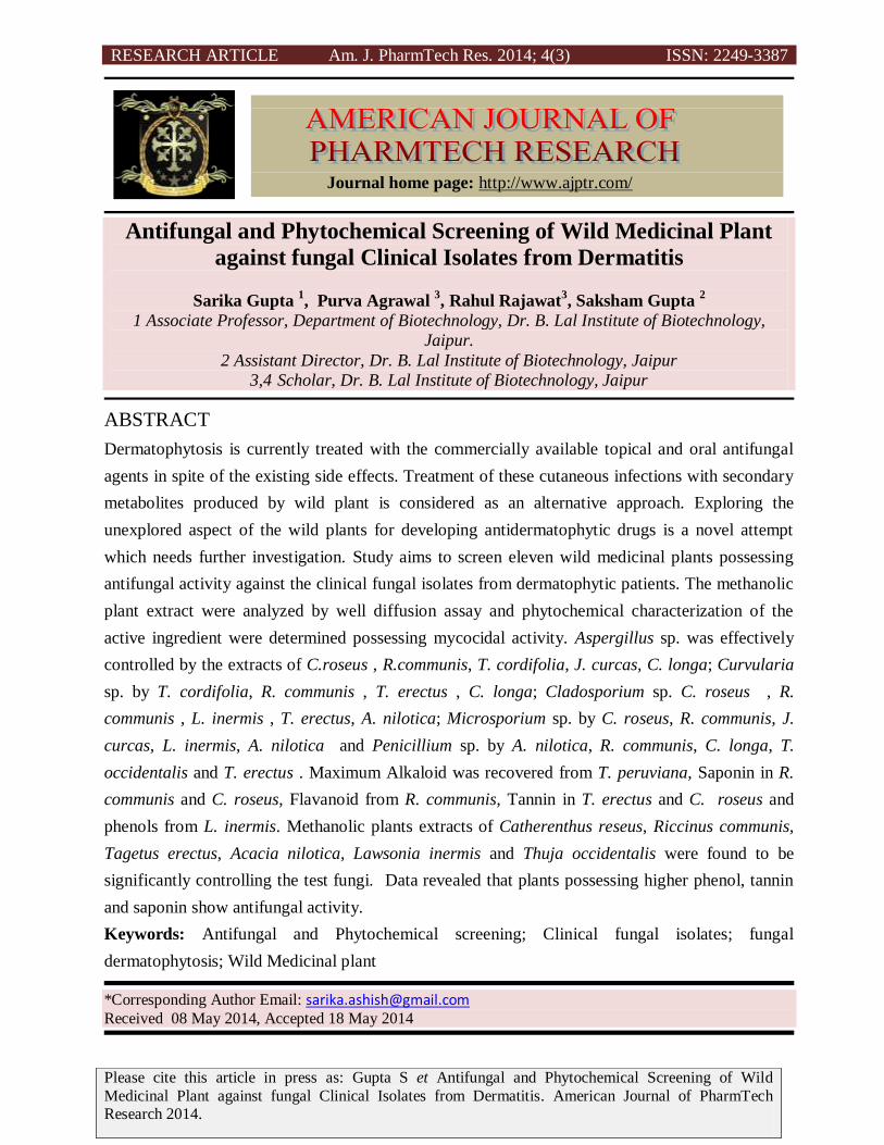
RESEARCH ARTICLE Am. J. PharmTech Res. 2014; 4(3) ISSN: 2249-3387 Please cite this article in press as: Gupta S et Antifungal and Phytochemical Screening of Wild Medicinal Plant against fungal Clinical Isolates from Dermatitis. American Journal of PharmTech Research 2014. Antifungal and Phytochemical Screening of Wild Medicinal Plant against fungal Clinical Isolates from Dermatitis Sarika Gupta 1 , Purva Agrawal 3 , Rahul Rajawat 3 , Saksham Gupta 2 1 Associate Professor, Department of Biotechnology, Dr. B. Lal Institute of Biotechnology, Jaipur. 2 Assistant Director, Dr. B. Lal Institute of Biotechnology, Jaipur 3,4 Scholar, Dr. B. Lal Institute of Biotechnology, Jaipur ABSTRACT Dermatophytosis is currently treated with the commercially available topical and oral antifungal agents in spite of the existing side effects. Treatment of these cutaneous infections with secondary metabolites produced by wild plant is considered as an alternative approach. Exploring the unexplored aspect of the wild plants for developing antidermatophytic drugs is a novel attempt which needs further investigation. Study aims to screen eleven wild medicinal plants possessing antifungal activity against the clinical fungal isolates from dermatophytic patients. The methanolic plant extract were analyzed by well diffusion assay and phytochemical characterization of the active ingredient were determined possessing mycocidal activity. Aspergillus sp. was effectively controlled by the extracts of C.roseus , R.communis, T. cordifolia, J. curcas, C. longa; Curvularia sp. by T. cordifolia, R. communis , T. erectus , C. longa; Cladosporium sp. C. roseus , R. communis , L. inermis , T. erectus, A. nilotica; Microsporium sp. by C. roseus, R. communis, J. curcas, L. inermis, A. nilotica and Penicillium sp. by A. nilotica, R. communis, C. longa, T. occidentalis and T. erectus . Maximum Alkaloid was recovered from T. peruviana, Saponin in R. communis and C. roseus, Flavanoid from R. communis, Tannin in T. erectus and C. roseus and phenols from L. inermis. Methanolic plants extracts of Catherenthus reseus, Riccinus communis, Tagetus erectus, Acacia nilotica, Lawsonia inermis and Thuja occidentalis were found to be significantly controlling the test fungi. Data revealed that plants possessing higher phenol, tannin and saponin show antifungal activity. Keywords: Antifungal and Phytochemical screening; Clinical fungal isolates; fungal dermatophytosis; Wild Medicinal plant *Corresponding Author Email: [email protected] Received 08 May 2014, Accepted 18 May 2014 Journal home page: http://www.ajptr.com/
Transcript of 43063 3503 dermato-anti fungal
RESEARCH ARTICLE Am. J. PharmTech Res. 2014; 4(3) ISSN: 2249-3387
Please cite this article in press as: Gupta S et Antifungal and Phytochemical Screening of Wild
Medicinal Plant against fungal Clinical Isolates from Dermatitis. American Journal of PharmTech Research 2014.
Antifungal and Phytochemical Screening of Wild Medicinal Plant
against fungal Clinical Isolates from Dermatitis
Sarika Gupta 1, Purva Agrawal
3, Rahul Rajawat
3, Saksham Gupta
2
1 Associate Professor, Department of Biotechnology, Dr. B. Lal Institute of Biotechnology,
Jaipur.
2 Assistant Director, Dr. B. Lal Institute of Biotechnology, Jaipur
3,4 Scholar, Dr. B. Lal Institute of Biotechnology, Jaipur
ABSTRACT
Dermatophytosis is currently treated with the commercially available topical and oral antifungal
agents in spite of the existing side effects. Treatment of these cutaneous infections with secondary
metabolites produced by wild plant is considered as an alternative approach. Exploring the
unexplored aspect of the wild plants for developing antidermatophytic drugs is a novel attempt
which needs further investigation. Study aims to screen eleven wild medicinal plants possessing
antifungal activity against the clinical fungal isolates from dermatophytic patients. The methanolic
plant extract were analyzed by well diffusion assay and phytochemical characterization of the
active ingredient were determined possessing mycocidal activity. Aspergillus sp. was effectively
controlled by the extracts of C.roseus , R.communis, T. cordifolia, J. curcas, C. longa; Curvularia
sp. by T. cordifolia, R. communis , T. erectus , C. longa; Cladosporium sp. C. roseus , R.
communis , L. inermis , T. erectus, A. nilotica; Microsporium sp. by C. roseus, R. communis, J.
curcas, L. inermis, A. nilotica and Penicillium sp. by A. nilotica, R. communis, C. longa, T.
occidentalis and T. erectus . Maximum Alkaloid was recovered from T. peruviana, Saponin in R.
communis and C. roseus, Flavanoid from R. communis, Tannin in T. erectus and C. roseus and
phenols from L. inermis. Methanolic plants extracts of Catherenthus reseus, Riccinus communis,
Tagetus erectus, Acacia nilotica, Lawsonia inermis and Thuja occidentalis were found to be
significantly controlling the test fungi. Data revealed that plants possessing higher phenol, tannin
and saponin show antifungal activity.
Keywords: Antifungal and Phytochemical screening; Clinical fungal isolates; fungal
dermatophytosis; Wild Medicinal plant
*Corresponding Author Email: [email protected]
Received 08 May 2014, Accepted 18 May 2014
Journal home page: http://www.ajptr.com/
Gupta et. al., Am. J. PharmTech Res. 2014; 4(3) ISSN: 2249-3387
755 www.ajptr.com
INTRODUCTION
The main objective of the treatment is eradication of the dermatophyte fungal infection from
various sites of the body as scalp, beard, skin, or nails, etc. These infections are typically treated
based on clinical appearance, with use of potassium hydroxide (KOH) microscopy for
confirmatory evidence in questionable cases, or the mandatory use of a confirmatory test (KOH
microscopy, fungal culture) in the case of fungal dermatophytic infection.1 Choice of topical or
oral therapy depends on site of the infection. Currently, no topical antifungal class has been shown
to be better than any other, but additional studies need to be performed.2
Issues of safety of oral
therapy should be considered, although there is low incidence of adverse events in the immuno
competent population.7 Length of time of treatment varies, depending on the agent chosen, from 2
to 8 weeks. Safety issues, cost, and differences in duration of treatment may influence choice of
agent. Systemic antifungals are the mainstay of therapy, with limited efficacy information to
choose one over another.17
Griseofulvin,13
the oldest available oral agent, and newer agents such as
terbinafine and itraconazole, which can be effective when used for shorter periods, are all first-line
options.12
Both ketoconazole and fluconazole have been studied for tinea infection and are
effective. Fluconazole, limited by adverse effects, and ketoconazole, which is hepatotoxic, are
typically reserved as second-line agents.17
Ketoconazole may cause severe liver injury and adrenal
insufficiency. In July 2013, the US Food and Drug Administration (FDA) recommended that oral
ketoconazole should only be used for life-threatening fungal infections where alternative
treatments are not available or tolerated, and when the potential benefits of treatment outweigh the
risks. Its use is contraindicated in patients with liver disease. If used, liver and adrenal function
should be monitored before and during treatment.31
This recommendation does not apply to topical
formulations of ketoconazole.
Identification of some wild medicinal plants against clinically isolated fungi, carry out their phyto-
chemical analysis, test the in vitro antifungal activities with respect to conventional antifungal
drugs. Medicinal plants synthesize a vast array of secondary metabolites that are important for
human life.9 The particular medication and duration of treatment depends on the location of the
infection. Scalp infections usually require treatment with an oral antifungal medication. Infections
of other areas of the skin can be treated with topical antifungal medications. [CDC]
http://www.cdc.gov/fungal/dermatophytes/treatment.html This gives us impetus to analyse the
antifungal activity of the wild medicinal plants against the clinically isolated fungi from fungal
dermatophytic infection. Wild medicinal plants are selected during the study conduct as for their
Gupta et. al., Am. J. PharmTech Res. 2014; 4(3) ISSN: 2249-3387
www.ajptr.com 756
easy availability in abundance and no other significant economical benefit. The study also dealt
with the photochemical screening of the plant extracts for the detection of the active ingredients
responsible for antifungal property. The active ingredient can further be used as recommendation
of the study conduct for pre-clinical trials.
MATERIALS AND METHODS
Test Plants:
Eleven plant were used for the analysis of their antifungal activity as Cucurma longa (Haldi),
Thuja occidentalis (Morpankhi), Murraya koenigii (Meetha Neem), Lawsonia inermis (Mehndi),
Acacia nilotica (Babool), Tagetus erectus (Gainda), Thevatia peruviana (Kaner), Riccinus
communis (Arandi), Catherenthus roseus (Sadabahar), Tinospora cordifolia (Neemgiloya) and
Jatropha curcas (Ratanjot). Fresh leaves were collected washed thoroughly 2-3 times with running
tap water and once with sterile distilled water, air dried at room temperature on a sterile blotter and
used for preparation of extracts.19
(we have used wild plants that are commonly and easily
available in Rajasthan)
Solvent extraction:
The dried powdered leaves were subjected to methanolic extraction by Soxhlet method. Plant
extract were prepared by 15 grams fine powder of leaves was filled in the thimble and extracted
successively with methanol for 48 hours at 55oC. All the solvent extracts were concentrated using
rotary flash evaporator under reduced pressure. The extracts were preserved in airtight brown
bottle until further use.10, 6
(Individual plant leaves were dried and the leaf powder was used for
solvent extraction by soxhlet method using methanol. The plant extracts were then used for
antifungal activity and phytochemical characterization)
Test fungi:
Fungal cultures were collected from Dr. B. Lal Clinical Laboratory, Jaipur reported to have fungal
dermatophytic infection and were subjected to culture on Sabouraud Dextrose Agar medium
(SDA). The plants extracts were used against clinically isolated five test fungi as A. flavus,
Curvularia sp., Microsporum sp., Cladosporum sp. and Penicillium sp. for antifungal activity
assay.
Antifungal activity assay by Well Diffusion technique
Antifungal activity were performed by well diffusion method on SDA medium with respect to
positive control (Itracanezole) (R: reference) and negative control (C) as solvent (methanol). The
sample (S) and positive control 5mg w/v per well. Samples showing activity index >1 represent
Gupta et. al., Am. J. PharmTech Res. 2014; 4(3) ISSN: 2249-3387
757 www.ajptr.com
significant control of pathogens. The plates were incubated at 25±1o C for seven days and ten
replicates were maintained for each treatment. The zone of inhibition of mycelial growth was
determined by antibiotic zone scale (Hi-media).12, 30
Phytochemical Screening of Plant Extract
All the plant extract were subjected for phytochemical screening by quantitative analysis of
alkaloids, flavonoids, saponins, phenols and tannins.
Alkaloid determination
5 g of the sample was weighed into a 250 ml beaker and 200 ml 20% acetic acid in ethanol was
added and covered to stand for 4 h. this was filtered and the extract was concentrated using a water
bath to one quarter of the original volume. Concentrated ammonium hydroxide was added drop
wise to the extract until the precipitation was complete. The whole solution was allowed to settle
and the precipitation was collected by filtration and weighed.24, 15
Flavanoid determination
10 g of the sample was extracted repeatedly with 100 ml of 80% aqueous methanol at room
temperature. The whole solution was filtered through Whatmann filter paper No.1. The filtrate was
later transferred into a crucible and evaporated to dryness over a water bath and weighed.6
Saponin determination
20g of plant sample was dispersed in 200 ml of 20% ethanol. The suspension was heated over a
hot water bath for 4 h with continuous stirring at about 55ºC. The mixture was filtered and the
residue re-extracted with another 200 ml of 20% ethanol. The combined extracts were reduced to
40 ml over water bath at about 90ºC. The concentrate was transferred into a 250 ml separating
funnel and 20 ml of diethyl ether was added and shaken vigorously. The aqueous layer was
recovered while the ether layer was discarded. The purification process was repeated. 60 ml of
normal butanol extracts were washed twice with 10 ml of 5% aqueous sodium chloride. The
remaining solution was heated in a water bath. After evaporation the sample were dried in the oven
into a constant weight. The saponin content was calculated in percentage.21
Determination of total phenol content
The total phenolic content of the plant extract was determined by using Folin-Ciocalteu reagent
following a slightly modified method of Ainsworth.28
Gallic acid was used as a reference standard
for plotting calibration curve. A volume of 0.5 mL of the plant extract (100 µg/mL) was mixed
with 2 mL of the Folin-Ciocalteu reagent (diluted 1:10 with de-ionized water) and were neutralized
Activity index = Zone of inhibition of sample
Zone of inhibition of reference
Gupta et. al., Am. J. PharmTech Res. 2014; 4(3) ISSN: 2249-3387
www.ajptr.com 758
with 4 mL of sodium carbonate solution (7.5%, w/v). The reaction mixture was incubated at room
temperature for 30 min with intermittent shaking for color development. The absorbance of the
resulting blue color was measured at 765 nm using double beam UV-VIS spectrophotometer
(Systronics 119). The total phenolic contents were determined from the linear equation of a
standard curve prepared with gallic acid. The content of total phenolic compounds expressed as
mg/g gallic acid equivalent (GAE) of dry extract.
Determination of tannins
500 mg of the sample was weighed into 100 ml bottle; 50 ml of distilled water was added and
shaken for 1 h in a shaker. This was filtered into a 50 ml volumetric flask and made up to the mark.
Then 5 ml of the filtrate was pipette out into a tube and mixed with 3 ml of 0.1 M FeCl3 in 0.1 N
HCl and 0.008 M potassium ferrocyanide. The absorbance was measured in a spectrophotometer at
120 nm wavelength within 10 min. A blank sample was prepared and read at the same wavelength.
A standard was prepared using tannin acid to get 100 ppm and measured.32
RESULTS AND DISCUSSION
Development of synthetic products to control plant diseases has become difficult because of strict
requirements of their efficacy, selectivity, toxicology and general impact on the environment.
Consequently, there is an increasing interest in evaluating other mechanisms of control including
the effects of plant metabolites on plant pathogens.26
Several higher plants and their constituents
have shown success in plant disease control and proved to be harmless and non phytotoxic unlike
chemical fungicides.30
Indiscriminate use of chemical not only hazardous to living beings but
adversely affects the microbial population present in the ecosystem.8
Alternative to this effect, to control plant diseases, plant products are gaining prominence as
fungicides and bactericides.8 Antifungal compounds from higher plants are advantageous over
synthetic fungicides due to their easily biodegradable nature.30
The clinical samples (150)
suspected for fungal dermatophytic infection were collected from Dr. B. Lal clinical Laboratory.
During the study conduct the relative percent occurrence (RPO) of among various fungi maximum
incidence was shown by Aspergillus sp. followed by Trichophyton sp., Fusarium sp., Microsporum
sp. and Cladosporium sp.1
Antifungal Activity of Methanolic Plant Extracts (During the study conduct methanolic plant
extract were used)
In the present investigation, eleven methanolic plant extracts were used for their antifungal activity
against 5 test fungi clinically isolated from dermatophytic infected sites. The activity index was
Gupta et. al., Am. J. PharmTech Res. 2014; 4(3) ISSN: 2249-3387
759 www.ajptr.com
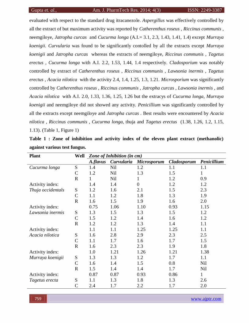
evaluated with respect to the standard drug itracanezole. Aspergillus was effectively controlled by
all the extract of but maximum activity was reported by Catherenthus roseus , Riccinus communis ,
neemgiloye, Jatropha curcas and Cucurma longa (A.I.= 3.1, 2.3, 1.43, 1.41, 1.4) except Murraya
koenigii. Curvularia was found to be significantly contolled by all the extracts except Murraya
koenigii and Jatropha curcas whereas the extracts of neemgiloye, Riccinus communis , Tagetus
erectus , Cucurma longa with A.I. 2.2, 1.53, 1.44, 1.4 respectively. Cladosporium was notably
controlled by extract of Catherenthus roseus , Riccinus communis , Lawsonia inermis , Tagetus
erectus , Acacia nilotica with the activity 2.4, 1.4, 1.25, 1.3, 1.21. Microsporium was significantly
controlled by Catherenthus roseus , Riccinus communis , Jatropha curcas , Lawsonia inermis , and
Acacia nilotica with A.I. 2.0, 1.33, 1.36, 1.25, 1.26 but the extracys of Cucurma longa, Murraya
koenigii and neemgiloye did not showed any activity. Penicillium was significantly controlled by
all the extracts except neemgiloye and Jatropha curcas . Best results were encountered by Acacia
nilotica , Riccinus communis , Cucurma longa, thuja and Tagetus erectus (1.38, 1.26, 1.2, 1.15,
1.13). (Table 1, Figure 1)
Table 1 : Zone of inhibition and activity index of the eleven plant extract (methanolic)
against various test fungus.
Plant Well Zone of Inhibition (in cm)
A.flavus Curvularia Microsporum Cladosporum Penicillium
Cucurma longa S 1.4 Nil 1.2 1.1 1.1
C 1.2 Nil 1.3 1.5 1
R 1 Nil 1 1.2 0.9
Activity index: 1.4 1.4 0 1.2 1.2
Thuja occidentals S 1.2 1.6 2.1 1.5 2.3
C 1.1 1.2 1.8 1.3 1.9
R 1.6 1.5 1.9 1.6 2.0
Activity index: 0.75 1.06 1.10 0.93 1.15
Lawsonia inermis S 1.3 1.5 1.3 1.5 1.2
C 1.5 1.2 1.4 1.6 1.2
R 1.2 1.2 1.3 1.4 1.1
Activity index: 1.1 1.1 1.25 1.25 1.1
Acacia nilotica S 1.6 2.8 2.9 2.3 2.5
C 1.1 1.7 1.6 1.7 1.5
R 1.6 2.3 2.3 1.9 1.8
Activity index: 1.0 1.21 1.26 1.21 1.38
Murraya koenigii S 1.3 1.3 1.2 1.7 1.1
C 1.6 1.4 1.5 0.8 Nil
R 1.5 1.4 1.4 1.7 Nil
Activity index: 0.87 0.87 0.93 0.86 1
Tagetus erecta
S 1.1 1.3 1.8 1.3 2.6
C 2.4 1.7 2.2 1.7 2.0
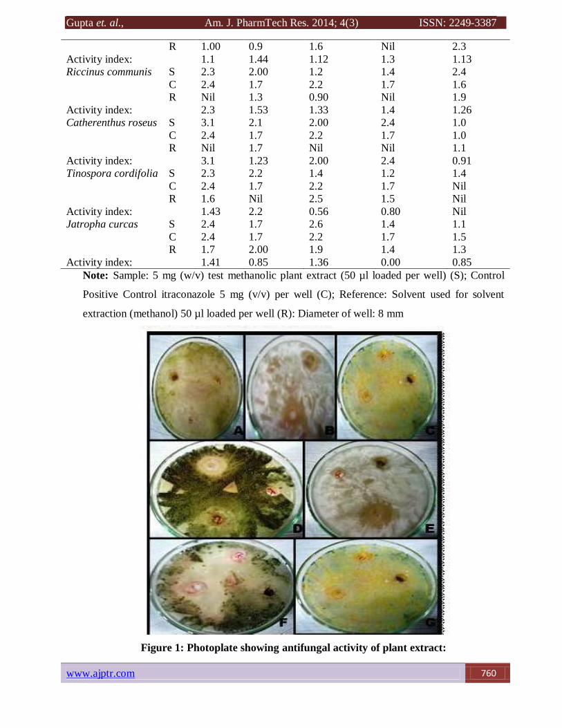
Gupta et. al., Am. J. PharmTech Res. 2014; 4(3) ISSN: 2249-3387
www.ajptr.com 760
R 1.00 0.9 1.6 Nil 2.3
Activity index: 1.1 1.44 1.12 1.3 1.13
Riccinus communis
S 2.3 2.00 1.2 1.4 2.4
C 2.4 1.7 2.2 1.7 1.6
R Nil 1.3 0.90 Nil 1.9
Activity index: 2.3 1.53 1.33 1.4 1.26
Catherenthus roseus S 3.1 2.1 2.00 2.4 1.0
C 2.4 1.7 2.2 1.7 1.0
R Nil 1.7 Nil Nil 1.1
Activity index: 3.1 1.23 2.00 2.4 0.91
Tinospora cordifolia S 2.3 2.2 1.4 1.2 1.4
C 2.4 1.7 2.2 1.7 Nil
R 1.6 Nil 2.5 1.5 Nil
Activity index: 1.43 2.2 0.56 0.80 Nil
Jatropha curcas S 2.4 1.7 2.6 1.4 1.1
C 2.4 1.7 2.2 1.7 1.5
R 1.7 2.00 1.9 1.4 1.3
Activity index: 1.41 0.85 1.36 0.00 0.85
Note: Sample: 5 mg (w/v) test methanolic plant extract (50 µl loaded per well) (S); Control
Positive Control itraconazole 5 mg (v/v) per well (C); Reference: Solvent used for solvent
extraction (methanol) 50 µl loaded per well (R): Diameter of well: 8 mm
Figure 1: Photoplate showing antifungal activity of plant extract:
Gupta et. al., Am. J. PharmTech Res. 2014; 4(3) ISSN: 2249-3387
761 www.ajptr.com
Comparative results of methanolic plant extracts of Pimpinella anisum L. (Apiaceae) and Illicium
verum Hook. f. (Illiciaceae), were reported for effective control the dermatophytic fungal
infections as compared to the standard drugs. This was tested for their potential antifungal
activities.33
Antidermatophytic activities of five selected medicinal plants (leaves) viz. Euphorbia
balsamifera Ait, Mitracarpus scaber Zucc, Pergularia tomentosa L, Streospermum kunthianum
Cham and Holarrhena floribunda was screened for treatment of dermatophytoses29
Antidermatophytic activity of dichloromethane and methanol extracts of whole plant of Allamanda
cathertica was evaluated.22
Whereas in a study conduct extract of quercifolia with four different
solvents such as ethanol, methanol, acetone, di-ethyl ether and water, were used to extract the
bioactive compounds from the rhizome of D.quercifolia23
Anogeissus leiocarpus and Terminalia
avicennioides.20
The efficacy of ethanol and distilled water extracts of Azadirachta indica, Jatropha curcas,
Jatropha gossypifolia, Cassia alata, Anacardium occidentale and Aloe vera was determined.1 The
activity of the methanolic extracts of the 5 selected plants was determined against different
pathogenic fungus.11
Crude methanol extracts from leaves of Cassia alata, Cassia fistula and
Cassia tora were investigated for their antifungal activities.25
These results support the plant oils
can be used to cure mycotic infections and plant oils may have role as pharmaceutical and
preservatives.3
Compared to ketoconazole used as standard antifungal the compound isolate could
be considered as a promising antidermatophytic agent.26
Phytochemical Screening of Plant Extract
Plants are reservoir of biological active compounds to combat various pathogens containing
alkaloids, flavonoids, saponins, phenols and tannins. The mode of action of extracts was
determined on cell wall and enzyme production of fungi.34
In the present study the plant extracts
were prepared and used for their antifungal property to detect their bioefficacy against clinically
isolated fungi. Plant extracts (methanolic) were subjected to phytochemical screening as the
quantitative analysis of Alkaloid, Saponin, Flavanoid, Tannin and Phenol through
spectrophotometric analysis. Maximum Alkaloid was found in Methanolic extract of Thevatia
peruviana (1.27 gm) followed by Tagetus erectus (0.98 gm) and Neem giloye (0.36gm). Saponin
was found highest in extract of Riccinus communis and Catherenthus roseus (0.68gm)
subsequently by Thuja (0.36 gm). Maximum amount of Flavanoid was recovered in extract of
Riccinus communis (0.72 gm), Jatropha (0.51 gm) and Neemgiloye (0.37 gm). Maximum Tannin
was found in methanolic extract of Tagetus erectus and Catherenthus roseus (0.71 gm) followed
by Jatropha curcas (0.64 gm) and Neemgiloye (0.58 gm). Amount of phenols was recovered
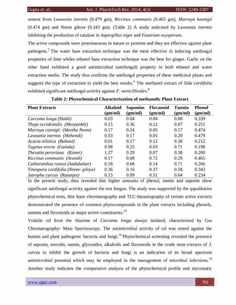
Gupta et. al., Am. J. PharmTech Res. 2014; 4(3) ISSN: 2249-3387
www.ajptr.com 762
utmost from Lawsonia inermis (0.479 gm), Riccinus communis (0.465 gm), Murraya koenigii
(0.474 gm) and Neem giloye (0.343 gm). (Table 2) A study indicated by Lawsonia inermis
inhibiting the production of catalase in Aspergillus niger and Fusarium oxysporum.
The active compounds were proteinaceous in nature or proteins and they are effective against plant
pathogens.5
The water base extraction technique was the most effective in inducing antifungal
properties of lime whiles ethanol base extraction technique was the best for ginger. Garlic on the
other hand exhibited a good antimicrobial (antifungal) property in both ethanol and water
extraction media. The study thus confirms the antifungal properties of these medicinal plants and
suggests the type of extraction to yield the best results.4 The methanol extract of Sida cordifolia
exhibited significant antifungal activity against F. verticillioides.8
Table 2: Phytochemical Characterization of methanolic Plant Extract
Plant Extracts Alkaloid
(gm/ml)
Saponins
(gm/ml)
Flavanoid
(gm/ml)
Tannin
(gm/ml)
Phenol
(gm/ml)
Curcuma longa (Haldi) 0.03 0.04 0.04 0.00 0.339
Thuja occidentalis (Morpankhi) 0.15 0.36 0.12 0.07 0.251
Murraya coenigii (Meetha Neem) 0.17 0.24 0.05 0.17 0.474
Lawsonia inermis (Mehendi) 0.03 0.17 0.05 0.20 0.479
Acacia nilotica (Babool) 0.01 0.17 0.12 0.38 0.212
Tagetus erecta (Gainda) 0.98 0.25 0.03 0.71 0.198
Thevatia peruviana (Kaner) 1.27 0.29 0.07 0.38 0.200
Riccinus communis (Arandi) 0.17 0.68 0.72 0.29 0.465
Catharanthus roseus (Sadabahar) 0.18 0.68 0.14 0.71 0.266
Tinospora cordifolia (Neem- giloye) 0.36 0.16 0.37 0.58 0.343
Jatropha curcas (Ratanjot) 0.13 0.09 0.51 0.64 0.234
In the present study, data revealed that higher amounts of phenol, tannin and saponin show
significant antifungal activity against the test fungus. The study was supported by the qqualitative
phytochemical tests, thin layer chromatography and TLC-bioautography of certain active extracts
demonstrated the presence of common phytocompounds in the plant extracts including phenols,
tannins and flavonoids as major active constituents.19
Volatile oil from the rhizome of Curcuma longa always isolated, characterized by Gas
Chromatography- Mass Spectroscopy. The antimicrobial activity of oil was tested against the
human and plant pathogenic bacteria and fungi.16
Phytochemical screening revealed the presence
of saponin, steroids, tannin, glycosides, alkaloids and flavonoids in the crude stem extracts of J.
curcas to inhibit the growth of bacteria and fungi is an indication of its broad spectrum
antimicrobial potential which may be employed in the management of microbial infections.18
Another study indicates the comparative analysis of the phytochemical profile and mycostatic
Gupta et. al., Am. J. PharmTech Res. 2014; 4(3) ISSN: 2249-3387
763 www.ajptr.com
activity of leaf and flower extracts of Tagetes erecta Linn and Tagetes patula Linn in ethanol.
Phytochemical screening has indicated the presence of alkaloids, flavonoids, steroids, tannins and
phenolic compounds as the major secondary metabolites in extracts of both species has revealed
the inhibitory effect of all the extracts on growth of Candida albicans, Aspergillus niger,
Saccharomyces cerevisiae and Aspergillus flavus. T. erecta leaf extract showed highest anti-fungal
activity among all the four extracts tested.27
Modern analytical spectroscopies of high intrinsic
dimensionality can provide rapid accurate microbial characterization techniques, but only when
combined with appropriate chemometrics.
Leaf extract (methanolic) of Catherenthus roseus (Sadabahar), Riccinus communis (Arandi),
Tagetus erectus (Gainda), Acacia nilotica (Babool), Lawsonia inermis (Mehndi) and Thuja
occidentalis (Morpankhi) serves as a potent plant against clinically isolated fungi from mycotic
dermatitis. It was interpreted that the plants possessing higher amounts of phenol, tannin and
saponin shows significant antifungal activity against the test fungus.
CONCLUSION
The management of dermatophytic infections needs personal hygiene, awareness of infection,
proper diagnosis and medication. At present there are a large number of antidermatophytic drugs
available commercially. With increasing incidence of fungal infection, microbial resistance to the
existing drugs, cost and side effects, there is a need for an antifungal drug that can overcome all
these limitations. out of eleven plants Catherenthus roseus (Sadabahar), Riccinus communis
(Arandi), Tagetus erectus (Gainda), Acacia nilotica (Babool), Lawsonia inermis (Mehndi) and
Thuja occidentalis (Morpankhi) remains to be an unexhausted source of bioactive compounds and
a boon to the medical field. It was interpreted that the plants possessing higher amounts of phenol,
tannin and saponin shows significant antifungal activity against the test fungus. Screening of plants
of wild nature can be a novel approach for obtaining potential lead molecules for clinical trials and
later treatment of dermatomycosis as compared to the standard drugs.
ACKNOWLEDGEMENT
We are grateful to the Department of Science and Technology, Government of Rajasthan for
providing financial support for the study. The authors wish to acknowledge the kind assistance and
contribution of the following for providing samples for the case study of Dermatophillic infection
are provided by Dr. B. Lal. Clinical laboratory, Jaipur under the guidance of Dr. B. Lal Gupta and
the case study under the guidance of Dr. Rishi Bhargava, Girdhar Hospital, Malviya Nagar, Jaipur
and Dr. U.S. Agrawal, S.M.S. Hospital, Jaipur.
Gupta et. al., Am. J. PharmTech Res. 2014; 4(3) ISSN: 2249-3387
www.ajptr.com 764
REFERENCES
1. Gupta S and Gupta B L. Evaluation of the incidences of dermatophillic infection in
Rajasthan: Case studies from Rajasthan, India International Journal of Medicine and
Medical Sciences. 2013; 5(5): 229-232.
2. Rotta I, Sanchez A, Gonçalves P R. Efficacy and safety of topical antifungal in the
treatment of dermatomycosis: a systematic review. Br J Dermatol. 2012; 166: 927-933.
3. Aly R, Maibach H I, Bagatell F K, Dittmar W, Hänel H, Falanga V, Leyden J J,
Roth H L, Stoughton R B, and Willis I. Ciclopirox olamine lotion 1%: bioequivalence to
ciclopirox olamine cream 1% and clinical efficacy in tinea pedis. Clin Ther. 1989; 11(3):
290-303.
4. Ansari MM. Control of sheath blight of rice by plant extracts. Indian Phyto. 1995; 48(3):
268-270.
5. Barton H, Martin F, Anthony F and Sheldon W. The ocular manifestations of Wegener's
granulomatosis: The American Journal of Medicine. 1977; 63(1): 131-141.
6. Boham A B, Kocipai D C. Flavonoid and condensed tannins from leaves of Hawaiian
vaccinum vaticulum and vicalycimum. Pracific Sci. 1994: 48: 458-463.
7. Chang, C H, Young-Xu Y, Kurth T. The safety of oral antifungal treatments for superficial
dermatophytosis and onychomycosis: a meta-analysis. Am J Med. 2007; 120:791-798.
8. Constantinou J, Tao L W, Vajpayee A. Clinical review of corneal ulcers resulting in
evisceration and nucleation in elderly population. Graefes Arch Clin Exp Ophthalmol.
2009; 247(10):1389-93.
9. David M W, Lior B, Michael R, Rossen H and Adi M. Late-onset laser in situ
keratomileusis–related corneal Ulcer. Cornea. 2009; 28:586-588.
10. Dzoyem, J. P., Tangmouo, J. G., Kechia, F. A., Lontsi, D., Etoa, F. X. and Lohoue, P. J. In
vitro Antidermatophytic Activity of Diospyros crassiflora Hiern (Ebenaceae). Sudanese
Journal of Dermatology. 2006; 4(1): 10-15.
11. Fardos, MB. Antifungal activity of some medicinal plants used in Jeddah, Saudi Arabia.
Faculty of Sciences, Biology Department, King Abdel Aziz University. Mycopath. 2009;
7(1): 51-57.
12. Gonzalez U, Seaton T and Bergus G. Systemic antifungal therapy for tinea capitis in
children. Cochrane Database Syst Rev. 2007; (4):CD004685.
13. Gupta A K, Cooper E A, Bowen J E. Meta-analysis: griseofulvin efficacy in the treatment
of tinea capitis. J Drugs Dermatol. 2008; 7:369-372.
Gupta et. al., Am. J. PharmTech Res. 2014; 4(3) ISSN: 2249-3387
765 www.ajptr.com
14. Adejumo T. O. and Bamidele B. S. Control of dermatophyte-causing agents (Trichophyton
mentagrophytes and Trichophyton rubrum) using six medicinal plants. Journal of
Medicinal Plants Research. 2009; 3(11): 906-913.
15. Harborne J B. Phytochemical methods. Chapman and Hall. 1973; London, p. 113
16. Helen M P A, Prinitha, Jaya Sree S, Madoen Abisha SM, Anoop Jacob. Phytochemical
characterization and Antimicrobial activity of oil and solvent extracts of Curcuma longa.
Research Journal of Pharmaceutical, Biological and Chemical Sciences. 2012: 3(3): 49-55.
17. Higgins E M, Fuller L C and Smith C H. Guidelines for the management of tinea capitis.
British Association of Dermatologists. Br J Dermatol. 2000; 143:53-58.
18. Igbinosa O, Igbinosa E O and Aiyegoro O A. Antimicrobial activity and phytochemical
screening of stem bark extracts from Jatropha curcas (Linn)O. African Journal of
Pharmacy and Pharmacology. 2009; 3(2): 058-062.
19. Khan Z S and Nasreen S. Phytochemical analysis, antifungal activity and mode of action of
methanol extracts of plants against pathogens. Journal of Agricultural Technology. 2010;
6(4): 793-805.
20. Mann A, Banso A and Clifford L C. An antifungal property of crude plant extracts from
Anogeissus leiocarpus and Terminalia Avicennioides. Tanzania Journal of Health Research.
2008; 10(1): 34-38.
21. Nahapetian A and Bassiri A. Changes in concentration and interrelationship of phytate, P,
mg, Cu, Zn, in wheat during maturation. J. Agric. Food Chem. 1975; 32: 1179-1182.
22. Nahar A S A, Islam M N and Alam M S. Studies on antidermatophytic effect of Allamanda
cathertica.A Journal of the Bangladesh Pharmacological Society (BDPS) Bangladesh J
Pharmacol. 2010; 5: 5-7.
23. Nejad, B S and Deokule S S. Anti-dermatophytic activity of Drynaria quercifolia (L.) J.
Smith. Jundishapur Journal of Microbiology. 2009; 2(1): 25-30.
24. Obadoni B O and Ochuko P O. Phytochemical studies and comparative efficacy of the
crude extracts of some homeostatic plants in Edo and Delta States of Nigeria. Global J.
Pure Appl. Sci. 2001; 8: 203-208.
25. Phongpaichit S, Pujenjob N, Rukachaisirikul V and Ongsakul M. Antifungal activity from
leaf extracts of Cassia alata L., Cassia fistula L. and Cassia tora L. Songklanakarin
Journal of Science and Technology. 2004; 26 (5):741-748.
26. Pinto C M F, Maffia L A, Casali V W D and Cardoso A A. In vitro effect of plant leaf
extracts on mycelial growth and sclerotial germination of Sclerotium cepivorum. J. of
Phytopathology. 1998; 146: 421-425.
Gupta et. al., Am. J. PharmTech Res. 2014; 4(3) ISSN: 2249-3387
www.ajptr.com 766
27. Ramya R and Bhat S K. Comparative Evaluation of Mycostatic Effect of Tagetes spp.
Indian Journal of applied research. 2013; 3(7): 546-548.
28. Ainsworth E A and Gillespie KM. Estimation of total phenolic content and other oxidation
substrates in plant tissues using Folin- Ciocalteu reagent. Nat Protoc. 2007; 2(4):875–877.
29. Shinkafi S A and Manga S B. Isolation of Dermatophytes and Screening of selected
Medicinal Plants used in the treatment of Dermatophytoses. International Research Journal
of Microbiology. 2011; 2(1) : 40-48.
30. Srivastava A K anf Lal B. Studies on biofungicidal properties of leaf extract of some
plants. Indian Phyto. 1996; 50(3): 408-411.
31. US Food and Drug Administration. FDA drug safety communication: FDA limits usage of
Nizoral (ketoconazole) oral tablets due to potentially fatal liver injury and risk of drug
interactions and adrenal gland problems. July 2013. http://www.fda.gov/ (last accessed 29
July 2013).
32. Van-Burden T P and Robinton W C. Formation of complexes between protein and tannin
acid. J. Agric. Food Chem. 1981; 1: 77-82.
33. Yazdani D, Rezazadeh Sh, Amin Gh., Zainal A M A, Shahnazi S and Jamalifar H.
Antifungal Activity of Dried Extracts of Anise (Pimpinella anisum L.) and Star anise
(Illicium verum Hook. f.) Against Dermatophyte and Saprophyte Fungi . J Medicinal
Plants. 2009; 8,(5): 24-29.
34. Gupta S., Kulshreshth K. and Datta A. Bio-Control of Clinical Fungal Isolates associated
with fungal keratitis using medicinal plant extracts. Int J Pharm Pharma Sci 2012; 4 (4):
544-547.
AJPTR is
Peer-reviewed
bimonthly
Rapid publication
Submit your manuscript at: [email protected]