14- Renoprotective, anti-oxidative and anti-apoptotic-2014
10
RESEARCH Open Access Renoprotective, anti-oxidative and anti-apoptotic effects of oral low-dose quercetin in the C57BL/6J model of diabetic nephropathy Isabele BS Gomes 1 , Marcella L Porto 2 , Maria Carmen LFS Santos 3 , Bianca P Campagnaro 3 , Thiago MC Pereira 3 , Silvana S Meyrelles 2 and Elisardo C Vasquez 2,3* Abstract Background: Diabetic nephropathy (DN) is one of the major causes of end-stage renal disease in diabetic patients. Increasing evidence from studies in the rodents has suggested that this disease is associated with increased oxidative stress due to hyperglycemia. In the present study, we evaluated the renoprotective, anti-oxidative and anti-apoptotic effects of the flavonoid quercetin in C57BL/6J model of DN. Methods: DN was induced by streptozotocin (STZ, 100 mg/kg/day, for 3 days) in adult C57BL/6J mice. Six weeks later, mice were divided into the following groups: diabetic mice treated with quercetin (DQ, 10 mg/kg/day, 4 weeks), diabetic mice treated with vehicle (DV) or non-treated non-diabetic (ND) mice. Results: Quercetin treatment caused a reduction in polyuria (~45%) and glycemia (~35%), abolished the hypertriglyceridemia and had significant effects on renal function including, decreased proteinuria and high plasma levels of uric acid, urea and creatinine, which were accompanied by beneficial effects on the structural changes of the kidney including glomerulosclerosis. Flow cytometry showed a decrease in oxidative stress and apoptosis in DN mice. Conclusion: Taken together, these data show that quercetin effectively attenuated STZ-induced cytotoxicity in renal tissue. This study provides convincing experimental evidence and perspectives on the renoprotective effects of quercetin in diabetic mice and outlines a novel therapeutic strategy for this flavonoid in the treatment of DN. Keywords: Quercetin, Diabetic nephropathy, Oxidative stress, Apoptosis, Flavonoids Introduction Diabetic nephropathy (DN) is one of the most important microvascular complications of diabetes mellitus [1-3] and is the largest single cause of end stage renal disease [4,5], that leads to a decrease in quality of life and an in- creased risk of mortality [6]. Recent data indicate that reactive oxygen species (ROS) play a pivotal role in the pathophysiology of DN [7-9]. Persistent hyperglycemia is the main determinant of initiation, promotion and sus- tentation of DN and it contributes to oxidative stress via two major mechanisms, augmentation of ROS [7,10-13] and the attenuation of antioxidative mechanisms through glycation of antioxidant enzymes [1,12,14]. The development of experimental models of DN has provided a valid approach to characterize its pathogenesis and to create new possibilities for the diagnosis and treat- ment of this disease. In this regard, the pancreatic islet cell toxin streptozotocin (STZ) has been widely used to induce diabetes in rodents [1,15,16], mainly in rat models. Like- wise in rat models, the mouse has become attractive for the understanding of human diseases due to the simi- larities in physiology and to the advantage that its genome can be easily manipulated [17] to produce models of com- plex genetic diseases such as atherosclerosis [18-20], which is not commonly developed in rats. * Correspondence: [email protected] 2 Department of Physiological Sciences, Laboratory of Translational Physiology, Health Sciences Center, UFES, Vitoria, Brazil 3 Pharmaceutical Sciences Graduate Program, Vila Velha University (UVV), Vila Velha, ES, Brazil Full list of author information is available at the end of the article © 2014 Gomes et al.; licensee BioMed Central Ltd. This is an Open Access article distributed under the terms of the Creative Commons Attribution License (http://creativecommons.org/licenses/by/4.0), which permits unrestricted use, distribution, and reproduction in any medium, provided the original work is properly credited. The Creative Commons Public Domain Dedication waiver (http://creativecommons.org/publicdomain/zero/1.0/) applies to the data made available in this article, unless otherwise stated. Gomes et al. Lipids in Health and Disease 2014, 13:184 http://www.lipidworld.com/content/13/1/184
Transcript of 14- Renoprotective, anti-oxidative and anti-apoptotic-2014
Gomes et al. Lipids in Health and Disease 2014, 13:184http://www.lipidworld.com/content/13/1/184
RESEARCH Open Access
Renoprotective, anti-oxidative and anti-apoptoticeffects of oral low-dose quercetin in the C57BL/6Jmodel of diabetic nephropathyIsabele BS Gomes1, Marcella L Porto2, Maria Carmen LFS Santos3, Bianca P Campagnaro3, Thiago MC Pereira3,Silvana S Meyrelles2 and Elisardo C Vasquez2,3*
Abstract
Background: Diabetic nephropathy (DN) is one of the major causes of end-stage renal disease in diabetic patients.Increasing evidence from studies in the rodents has suggested that this disease is associated with increased oxidativestress due to hyperglycemia. In the present study, we evaluated the renoprotective, anti-oxidative and anti-apoptoticeffects of the flavonoid quercetin in C57BL/6J model of DN.
Methods: DN was induced by streptozotocin (STZ, 100 mg/kg/day, for 3 days) in adult C57BL/6J mice. Sixweeks later, mice were divided into the following groups: diabetic mice treated with quercetin (DQ, 10 mg/kg/day,4 weeks), diabetic mice treated with vehicle (DV) or non-treated non-diabetic (ND) mice.
Results: Quercetin treatment caused a reduction in polyuria (~45%) and glycemia (~35%), abolished thehypertriglyceridemia and had significant effects on renal function including, decreased proteinuria and highplasma levels of uric acid, urea and creatinine, which were accompanied by beneficial effects on the structuralchanges of the kidney including glomerulosclerosis. Flow cytometry showed a decrease in oxidative stress andapoptosis in DN mice.
Conclusion: Taken together, these data show that quercetin effectively attenuated STZ-induced cytotoxicity inrenal tissue. This study provides convincing experimental evidence and perspectives on the renoprotectiveeffects of quercetin in diabetic mice and outlines a novel therapeutic strategy for this flavonoid in the treatmentof DN.
Keywords: Quercetin, Diabetic nephropathy, Oxidative stress, Apoptosis, Flavonoids
IntroductionDiabetic nephropathy (DN) is one of the most importantmicrovascular complications of diabetes mellitus [1-3]and is the largest single cause of end stage renal disease[4,5], that leads to a decrease in quality of life and an in-creased risk of mortality [6]. Recent data indicate thatreactive oxygen species (ROS) play a pivotal role in thepathophysiology of DN [7-9]. Persistent hyperglycemia isthe main determinant of initiation, promotion and sus-tentation of DN and it contributes to oxidative stress via
* Correspondence: [email protected] of Physiological Sciences, Laboratory of TranslationalPhysiology, Health Sciences Center, UFES, Vitoria, Brazil3Pharmaceutical Sciences Graduate Program, Vila Velha University (UVV), VilaVelha, ES, BrazilFull list of author information is available at the end of the article
© 2014 Gomes et al.; licensee BioMed CentralCommons Attribution License (http://creativecreproduction in any medium, provided the orDedication waiver (http://creativecommons.orunless otherwise stated.
two major mechanisms, augmentation of ROS [7,10-13]and the attenuation of antioxidative mechanisms throughglycation of antioxidant enzymes [1,12,14].The development of experimental models of DN has
provided a valid approach to characterize its pathogenesisand to create new possibilities for the diagnosis and treat-ment of this disease. In this regard, the pancreatic islet celltoxin streptozotocin (STZ) has been widely used to inducediabetes in rodents [1,15,16], mainly in rat models. Like-wise in rat models, the mouse has become attractivefor the understanding of human diseases due to the simi-larities in physiology and to the advantage that its genomecan be easily manipulated [17] to produce models of com-plex genetic diseases such as atherosclerosis [18-20], whichis not commonly developed in rats.
Ltd. This is an Open Access article distributed under the terms of the Creativeommons.org/licenses/by/4.0), which permits unrestricted use, distribution, andiginal work is properly credited. The Creative Commons Public Domaing/publicdomain/zero/1.0/) applies to the data made available in this article,
Gomes et al. Lipids in Health and Disease 2014, 13:184 Page 2 of 10http://www.lipidworld.com/content/13/1/184
Although cumulative evidence suggests that increasedoxidative stress may play a crucial role in the pathogenesisof DN [21,22], antioxidant therapy has shown conflictingresults during the treatment of DN in diabetic patients[23]. Currently, only blockers of the renin-angiotensinsystem are chronically used; however, there are limita-tions of this mechanism [23,24] which justifies the searchfor effective and safer antioxidant candidates. The flavo-noids are plant phenolic compounds that exhibit strongantioxidant properties and are widely distributed through-out the plant kingdom; thus, flavonoids are a therapeuticoption [25,26].Quercetin is a bioflavonoid found in red wine and
numerous fruits, vegetables, and nuts [27,28]. Recently,biochemical and pharmacological studies of quercetinhave shown that it is a potent scavenger of ROS andpossibly reduces the risk of cardiovascular and renal dis-eases [1,28-30]. However, its effects on kidney function,excessive ROS production and kidney cell apoptosis, thatare supposed to occur in this mouse model of DN, havenot yet been evaluated. Therefore, the present study wasdesigned to test the hypothesis that quercetin presentsnephroprotective effects on the main parameters thatcharacterize the DN. We focused our model in theC57BL/6J, which has been the most used genetic back-ground mouse [18-20] but not yet investigated as a modelof DN.
Materials and methodsAnimalsEight-week-old homozygous C57BL/6J mice were obtainedfrom the Laboratory of Translational Physiology, HealthSciences Center, at the Federal University of EspiritoSanto, Brazil. Animals were housed at room temperature(22°C) in a humidity-controlled environment with a 12-hlight/12-h dark cycle in the Experimental Unit of the La-boratory of Translational Physiology. Mice were allowedfree access to water and a standard laboratory chow diet(Labina®) until experiments were performed. The animalswere studied according to the principles devised by theNational Institute of Health (NIH) Guide for the Care andUse of Laboratory Animals and the protocol was previ-ously approved by the Institutional Ethics Committee forthe Use of Animals (CEUA, Protocol # 013/2010).
Experimental protocolMice were rendered diabetic by intraperitoneal injectionof STZ (Boehringer-Mannheim, Mannheim, Germany)diluted in citrate buffer (10 mM, pH 4.5) at a dose of100 mg/kg/day for 3 days, The control group (non-dia-betic, ND, n = 10) was administered an equivalent vol-ume of the vehicle citrate buffer. One week after STZinjection, glycemia was measured after 6 hours of fooddeprivation and animals with glucose levels greater than
250 mg/dL for at least 2 days were considered diabetic.Six weeks after STZ injections, diabetic mice were ran-domly divided and received either no treatment (vehiclesoy oil, DV, n = 10) or oral quercetin (DQ, n = 10) at adosage of 10 mg/kg per day for 4 weeks. This dosage wasbased on previous studies with hypertensive and diabeticanimals [31,32].
Metabolic and biochemical parametersAfter STZ injection, all animals were weighed once aweek. At week 4, the animals were housed for a 24-hourperiod in individual metabolic cages for adaptation. Fol-lowing this period, a known volume of water and quan-tify of food were placed in the drinking bottles and thefeeder, respectively. After 24 hours, the volume of liquidand chow remaining in the cages were measured. Urinewas collected and its volume and protein concentrationwas determined. Animals were euthanized via an overdoseof thiopental (Cristalia, Sao Paulo, Brazil, 200 mg/kg, i.p.)after a period of food deprivation, and their blood was col-lected for glucose, creatinine, cholesterol, triglycerides,urea and uric acid measurements using colorimetric kits.Proteinuria was determined by a Bradford assay [33]. Tis-sues were perfused with cold phosphate-buffered saline(PBS, pH 7.4, 0.1 mol/L) through the left ventricle and thekidney was fixed in Duboscq-Brazil solution for histo-logical evaluation.
Kidney histologyAfter perfusion, the mouse kidneys were rapidly fixedwith Duboscq solution, weighed and processed for mor-phometric and histological analyses. Samples were dehy-drated with a graded series of alcohols, embedded inparaffin, sectioned into 3-μm-thick slices and stainedwith hematoxylin and eosin for light microscopic mor-phological studies (AX70, Olympus, Center Valley, PA,USA). Images were captured at a 40x magnification with acolor video camera (VKC150, Hitachi, Tokyo, Japan) con-nected to a microscope (AX70, Olympus, Center Valley,PA, USA). All morphometric and histological analysiswere performed in a blinded manner. The mean glomeru-lar tuft area of each kidney was obtained by calculatingthe mean value of 30 individual glomeruli measured byImage J software (version 1.33u, Public Domain). Masson’strichrome staining was used to quantify glomerulosclerosis.A total of 30 glomeruli were used to calculate the percent-age of the stained area for each kidney using the Image Jprogram (Public Domain Image Processing Program,National Institutes of Health, Bethesda, MD).
Measurement of intracellular superoxide anions by flowcytometryFractions enriched for kidney cell were obtained fromeach of the groups, were prepared based on previous
Gomes et al. Lipids in Health and Disease 2014, 13:184 Page 3 of 10http://www.lipidworld.com/content/13/1/184
studies and standardized in our laboratory. The kidneywas grossly minced using surgical scissors and was incu-bated in an isolation solution containing collagenase typeII (Gibco Life Technologies, Sao Paulo, SP, Brazil) to dis-sociate the cells. The cell suspension was filtered througha nylon screen (BD Falcon 70 μm) to remove cellular deb-ris. The samples were then washed twice in PBS beforefurther analysis. Dihydroethidium (DHE, 160 μM) wasadded to cell suspensions (106 cells) which were then in-cubated at 37°C for 30 min in the dark in order to deter-mine the intracellular production of superoxide anions(•O2
−) [34-36]. DHE is freely permeable to cells and israpidly oxidized to ethidium, which binds to DNA andresults in the amplification of a red fluorescence signal.Samples were treated with 10 mM doxorubicin for 5 minto induce oxidative stress in the absence of cell toxicity;these samples served as positive control. Cells incubatedwith ethanol served as the negative control. Cells werethen washed, resuspended in PBS, and maintained on icefor immediate detection via flow cytometry (FACSCantoII, Becton Dickinson, San José, CA, USA). Data wereanalyzed using FACSDiva software (BD Company), andoverlay histograms were constructed using FCS Expresssoftware. For DHE fluorescence quantification, sampleswere acquired in duplicate, and 10,000 events were ob-tained for each measurement. Red fluorescence was de-tected between 564 and 606 nm using a 585/42 bandpassfilter. Data are expressed as the geometric mean fluores-cence intensity.
ApoptosisApoptotic kidney cells were quantified via annexin V-fluorescein isothiocyanate (FITC) and propidium iodide(PI) staining, using a commercial detection kit (BectonDickinson, San José, CA, USA). Briefly, renal cells werewashed twice with PBS and the final volume was adjustedto 500 μl with binding buffer (5 × 10⁵ cells). Then, 2 μl ofannexin V–FITC and 2 μL of PI were added to the solutionand the cells were gently vortexed. Cells were then incu-bated for 15 min at room temperature (25°C) in the dark.Finally, cells were analyzed using a FACSCanto II flow cyt-ometer (BD). Apoptosis was quantified by analysis of theflow cytometry data after annexin V-FITC and PI labeling.Kidney cells that were annexin V-FITC−/PI+ were classi-
fied as damaged (Q1). Cells that were annexin V-FITC+/PI+
were considered to be late apoptotic or secondary apop-totic cells (Q2). Cells that were annexin V-FITC−/PI− wereconsidered live cells (Q3). Cells that were annexin V-FITC+/PI− were classified as early or primary apoptotic cells(Q4) [37].
Statistical analysisAll data are expressed as the means ± SEM. TheKolmogorov-Smirnov test showed that variables had a
normal (Gaussian) distribution. Flow cytometry data ofROS production are expressed as the geometric MFI–variation coefficient of two repeated and statistically repro-ducible measurements of at least 5 independent animals(the Friedman test). The statistical analysis was performedby one-way analysis of variance (ANOVA). When theANOVA showed significant differences, the Tukey’s testwas applied as a post hoc analysis. The differences wereconsidered significant when p < 0.05.
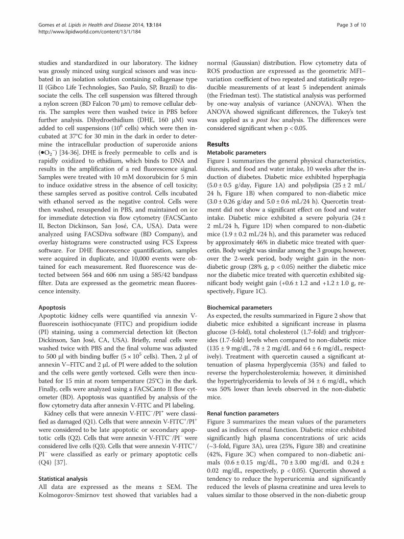
ResultsMetabolic parametersFigure 1 summarizes the general physical characteristics,diuresis, and food and water intake, 10 weeks after the in-duction of diabetes. Diabetic mice exhibited hyperphagia(5.0 ± 0.5 g/day, Figure 1A) and polydipsia (25 ± 2 mL/24 h, Figure 1B) when compared to non-diabetic mice(3.0 ± 0.26 g/day and 5.0 ± 0.6 mL/24 h). Quercetin treat-ment did not show a significant effect on food and waterintake. Diabetic mice exhibited a severe polyuria (24 ±2 mL/24 h, Figure 1D) when compared to non-diabeticmice (1.9 ± 0.2 mL/24 h), and this parameter was reducedby approximately 46% in diabetic mice treated with quer-cetin. Body weight was similar among the 3 groups; however,over the 2-week period, body weight gain in the non-diabetic group (28% g, p < 0.05) neither the diabetic micenor the diabetic mice treated with quercetin exhibited sig-nificant body weight gain (+0.6 ± 1.2 and +1.2 ± 1.0 g, re-spectively, Figure 1C).
Biochemical parametersAs expected, the results summarized in Figure 2 show thatdiabetic mice exhibited a significant increase in plasmaglucose (3-fold), total cholesterol (1.7-fold) and triglycer-ides (1.7-fold) levels when compared to non-diabetic mice(135 ± 9 mg/dL, 78 ± 2 mg/dL and 64 ± 6 mg/dL, respect-ively). Treatment with quercetin caused a significant at-tenuation of plasma hyperglycemia (35%) and failed toreverse the hypercholesterolemia; however, it diminishedthe hypertriglyceridemia to levels of 34 ± 6 mg/dL, whichwas 50% lower than levels observed in the non-diabeticmice.
Renal function parametersFigure 3 summarizes the mean values of the parametersused as indices of renal function. Diabetic mice exhibitedsignificantly high plasma concentrations of uric acids(~3-fold, Figure 3A), urea (25%, Figure 3B) and creatinine(42%, Figure 3C) when compared to non-diabetic ani-mals (0.6 ± 0.15 mg/dL, 70 ± 3.00 mg/dL and 0.24 ±0.02 mg/dL, respectively, p < 0.05). Quercetin showed atendency to reduce the hyperuricemia and significantlyreduced the levels of plasma creatinine and urea levels tovalues similar to those observed in the non-diabetic group
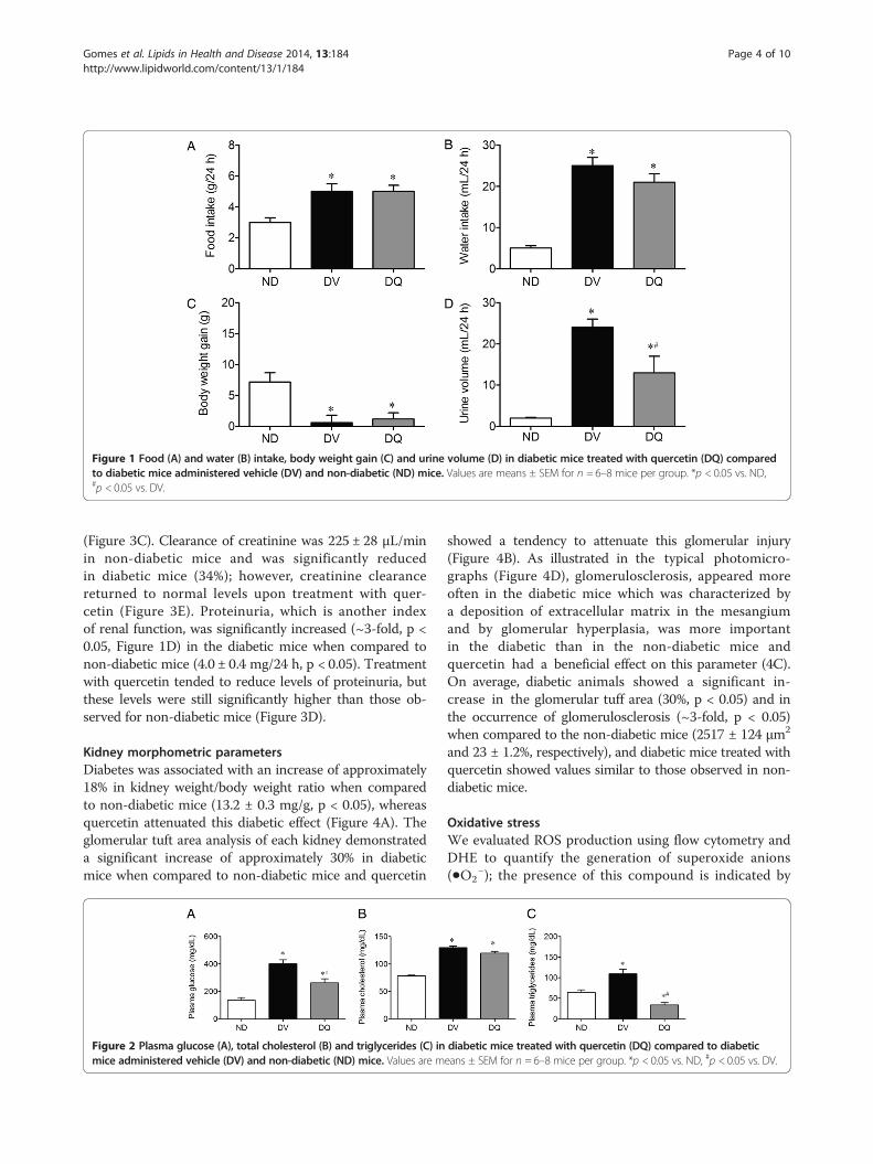
Figure 1 Food (A) and water (B) intake, body weight gain (C) and urine volume (D) in diabetic mice treated with quercetin (DQ) comparedto diabetic mice administered vehicle (DV) and non-diabetic (ND) mice. Values are means ± SEM for n = 6–8 mice per group. *p < 0.05 vs. ND,#p < 0.05 vs. DV.
Gomes et al. Lipids in Health and Disease 2014, 13:184 Page 4 of 10http://www.lipidworld.com/content/13/1/184
(Figure 3C). Clearance of creatinine was 225 ± 28 μL/minin non-diabetic mice and was significantly reducedin diabetic mice (34%); however, creatinine clearancereturned to normal levels upon treatment with quer-cetin (Figure 3E). Proteinuria, which is another indexof renal function, was significantly increased (~3-fold, p <0.05, Figure 1D) in the diabetic mice when compared tonon-diabetic mice (4.0 ± 0.4 mg/24 h, p < 0.05). Treatmentwith quercetin tended to reduce levels of proteinuria, butthese levels were still significantly higher than those ob-served for non-diabetic mice (Figure 3D).
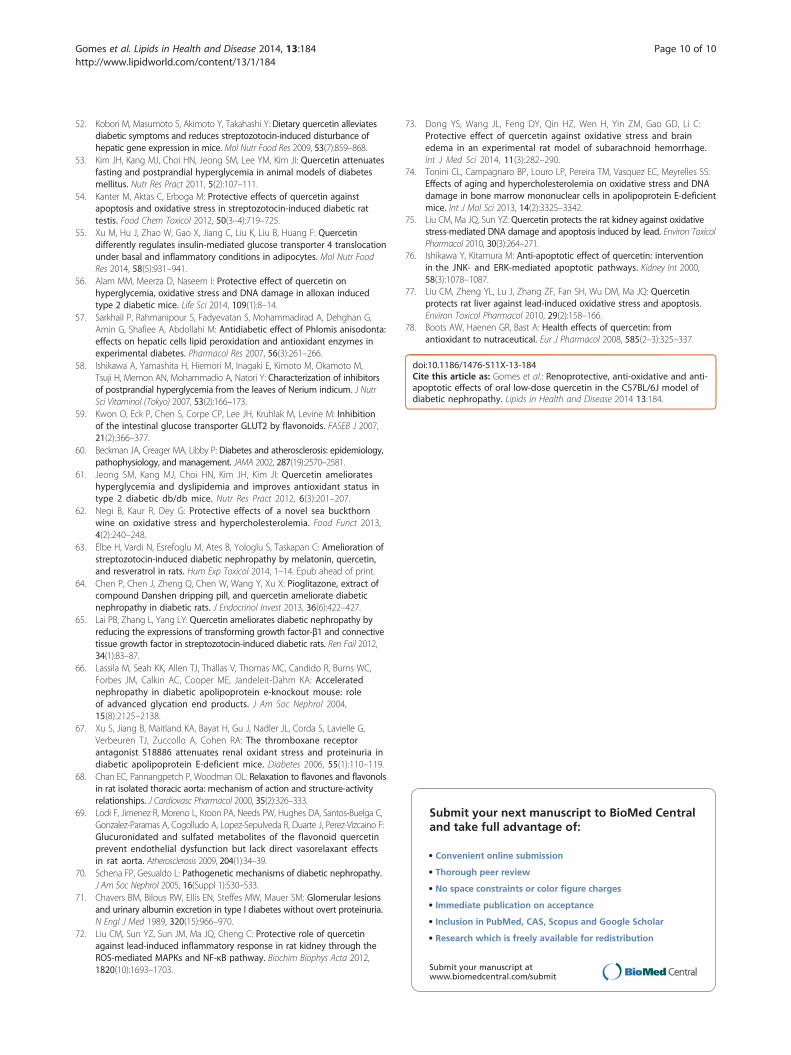
Kidney morphometric parametersDiabetes was associated with an increase of approximately18% in kidney weight/body weight ratio when comparedto non-diabetic mice (13.2 ± 0.3 mg/g, p < 0.05), whereasquercetin attenuated this diabetic effect (Figure 4A). Theglomerular tuft area analysis of each kidney demonstrateda significant increase of approximately 30% in diabeticmice when compared to non-diabetic mice and quercetin
Figure 2 Plasma glucose (A), total cholesterol (B) and triglycerides (C) inmice administered vehicle (DV) and non-diabetic (ND) mice. Values are m
showed a tendency to attenuate this glomerular injury(Figure 4B). As illustrated in the typical photomicro-graphs (Figure 4D), glomerulosclerosis, appeared moreoften in the diabetic mice which was characterized bya deposition of extracellular matrix in the mesangiumand by glomerular hyperplasia, was more importantin the diabetic than in the non-diabetic mice andquercetin had a beneficial effect on this parameter (4C).On average, diabetic animals showed a significant in-crease in the glomerular tuff area (30%, p < 0.05) and inthe occurrence of glomerulosclerosis (~3-fold, p < 0.05)when compared to the non-diabetic mice (2517 ± 124 μm2
and 23 ± 1.2%, respectively), and diabetic mice treated withquercetin showed values similar to those observed in non-diabetic mice.
Oxidative stressWe evaluated ROS production using flow cytometry andDHE to quantify the generation of superoxide anions(•O2
−); the presence of this compound is indicated by
diabetic mice treated with quercetin (DQ) compared to diabeticeans ± SEM for n = 6–8 mice per group. *p < 0.05 vs. ND, #p < 0.05 vs. DV.
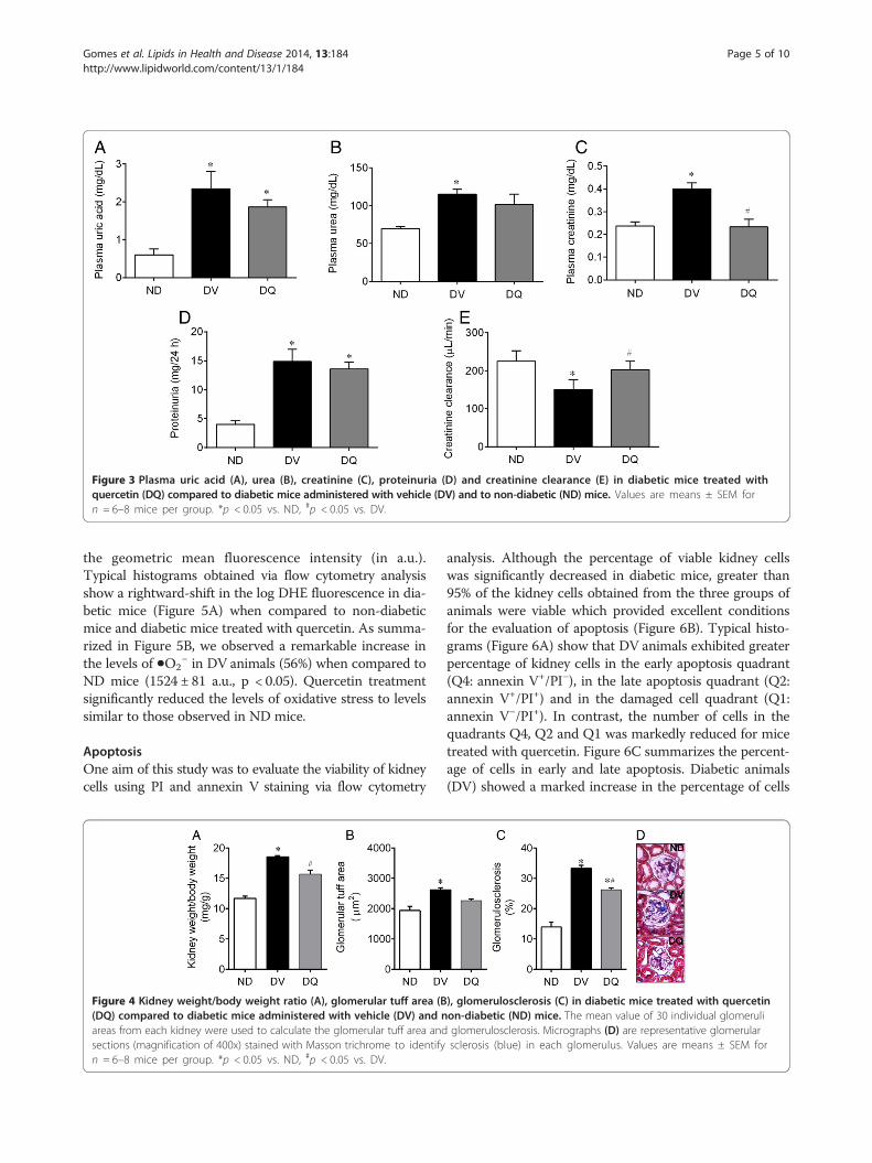
Figure 3 Plasma uric acid (A), urea (B), creatinine (C), proteinuria (D) and creatinine clearance (E) in diabetic mice treated withquercetin (DQ) compared to diabetic mice administered with vehicle (DV) and to non-diabetic (ND) mice. Values are means ± SEM forn = 6–8 mice per group. *p < 0.05 vs. ND, #p < 0.05 vs. DV.
Gomes et al. Lipids in Health and Disease 2014, 13:184 Page 5 of 10http://www.lipidworld.com/content/13/1/184
the geometric mean fluorescence intensity (in a.u.).Typical histograms obtained via flow cytometry analysisshow a rightward-shift in the log DHE fluorescence in dia-betic mice (Figure 5A) when compared to non-diabeticmice and diabetic mice treated with quercetin. As summa-rized in Figure 5B, we observed a remarkable increase inthe levels of •O2
− in DV animals (56%) when compared toND mice (1524 ± 81 a.u., p < 0.05). Quercetin treatmentsignificantly reduced the levels of oxidative stress to levelssimilar to those observed in ND mice.
ApoptosisOne aim of this study was to evaluate the viability of kidneycells using PI and annexin V staining via flow cytometry
Figure 4 Kidney weight/body weight ratio (A), glomerular tuff area (B(DQ) compared to diabetic mice administered with vehicle (DV) and nareas from each kidney were used to calculate the glomerular tuff area andsections (magnification of 400x) stained with Masson trichrome to identifyn = 6–8 mice per group. *p < 0.05 vs. ND, #p < 0.05 vs. DV.
analysis. Although the percentage of viable kidney cellswas significantly decreased in diabetic mice, greater than95% of the kidney cells obtained from the three groups ofanimals were viable which provided excellent conditionsfor the evaluation of apoptosis (Figure 6B). Typical histo-grams (Figure 6A) show that DV animals exhibited greaterpercentage of kidney cells in the early apoptosis quadrant(Q4: annexin V+/PI−), in the late apoptosis quadrant (Q2:annexin V+/PI+) and in the damaged cell quadrant (Q1:annexin V−/PI+). In contrast, the number of cells in thequadrants Q4, Q2 and Q1 was markedly reduced for micetreated with quercetin. Figure 6C summarizes the percent-age of cells in early and late apoptosis. Diabetic animals(DV) showed a marked increase in the percentage of cells
), glomerulosclerosis (C) in diabetic mice treated with quercetinon-diabetic (ND) mice. The mean value of 30 individual glomeruliglomerulosclerosis. Micrographs (D) are representative glomerularsclerosis (blue) in each glomerulus. Values are means ± SEM for

Figure 5 Production of superoxide anions. A: representativehistograms from flow cytometry analysis using dihydroethidium(DHE) in diabetic mice treated with quercetin (DQ), compared todiabetic mice that received the vehicle (DV) and non-diabetic(ND) mice; the log fluorescence (X-axis) shows the intensity offluorescence (+) for the number of kidney cells assayed. B: bargraph shows geometric mean fluorescence intensity. *p < 0.05 vs. NDgroup, #p < 0.05 vs. DV group.
Gomes et al. Lipids in Health and Disease 2014, 13:184 Page 6 of 10http://www.lipidworld.com/content/13/1/184
in early (4-fold) and late apoptosis (6-fold) when com-pared to non-diabetic (ND) mice (1.58 ± 0.35% and 0.46 ±0.15%, respectively). Treatment of diabetic (DV) mice withquercetin (DQ) had a marked effect on the progressionfrom early to late apoptosis (Figure 6C). Quercetin treat-ment tended to reduce the high levels of early apoptosis,but quercetin was able to decrease the percentage valuesto levels similar to those observed in non-diabetic animals.
DiscussionThis work showed that chronic oral treatment with low-dose quercetin exerts antidiabetic effects and attenuates thedevelopment of nephropathy in STZ-induced DN mice.These results are supported by a decrease of plasma glu-cose, creatinine, triglycerides, proteinuria and diminution ofmesangial matrix expansion accompanied by a reductionin •O2
− production and apoptosis in kidney cells.STZ-injected mice exhibit destruction of β cells and re-
duction in insulin secretory capacity [15,38]. Consequently,
these mice develop typical characteristics of diabetes suchas polydipsia, polyuria, and proteinuria accompanied byloss of body weight (even with polyphagia) as observed inthe present study. Furthermore, in accordance with ourbiochemical and morphological data, we considered thefive stages of clinical classification of DN [39,40] and sug-gest that this experimental murine model corresponds toa stage 4 clinical classification due to its declining glom-erular filtration rate and proteinuria; thus, this model issuitable for evaluating the nephroprotective effects of thebioflavonoid quercetin. It must be noted that we avoidedinterfering with the intrinsic nephrotoxic effects of STZby acquiring our data 6 weeks after STZ administrationwhen the kidneys are known to have recovered from theacute renal injury caused by STZ [38,41,42]; thus, we canassume that the damages observed in our model was dueonly to diabetic status.Interest in studies of the effects of natural antioxidants
prevent oxidative damage in mouse models of diabetes hasrecently grown [2,38,43]. The best candidates are generallymolecules that show high power antioxidant capacity, highpermeability to mitochondrion [44], long half-lives [45]and enhancement of enzyme activity [43]; quercetin ex-hibits all of these traits. This bioflavonoid may inhibit theformation of ROS in the three different ways: (a) by inter-acting directly with •O2
− during initiation, (b) by forminghydroxyl radicals via the chelation of iron ions, and (c) bydirectly reacting with lipid peroxyl radicals scavengersduring lipid peroxidation [46]. Additionally, it is possiblethat quercetin increases the activity of superoxide dis-mutase, catalase, glutathione peroxidase, glutathione reduc-tase and glutathione [43,47]. All these effects may promoteradical scavenging activity, anti-inflammatory and anti-apoptotic effects and contribute to renal protection againstSTZ-induced DN.Recent data has shown that the persistent hyperglycemia
leads to an increase in the activity of several pathways in-volved in the disease that contribute to oxidative stress:(1) auto-oxidation of glucose, (2) advanced glycationend-product (AGE) formation, (3) polyol pathway flux,(4) protein kinase C (PKC) isoforms activation and (5)mitochondrial dysfunction [5,10,48-50]. Our results dem-onstrate that quercetin attenuates hyperglycemia in ac-cordance with some authors [46,51-54], however, otherauthors disagree [1,43]. Independent of this apparentinconsistence, the impact on glycemia may be due to per-turbations of some pathways. Previous studies have shownthat this bioflavonoid can protect pancreatic β-cells fromoxidative stress and damage (either directly or indirectly)and improve insulin secretion in STZ models [51,53]. Add-itionally, quercetin can stimulate glucose uptake in per-ipheral tissues via the translocation of GLUT4 [46,55,56]and can increase hepatic glucokinase activity [53], thusaugmenting both oxidation and storage of glucose and

Figure 6 Flow cytometric analysis of apoptosis in kidney cells. Each dot plot (A) was constructed using propidium iodide (PI) and annexinV/FITC staining indicating: damaged cells (Q1), cells that are undergoing late apoptosis (Q2), viable cells (Q3) and cells in early apoptosis (Q4).Bar graph B shows the average percentage of PI positive (viable) cells, which were used to quantify apoptosis. Bar graph C shows theaverage percentage of cells in early and late apoptosis in non-diabetic mice (ND), diabetic mice administered vehicle (DV) and diabetic mice treatedwith quercetin (DQ). Values are mean ± SEM for 6 to 8 animals per group. *p < 0.05 vs. ND group and #p < 0.05 vs. DV group.
Gomes et al. Lipids in Health and Disease 2014, 13:184 Page 7 of 10http://www.lipidworld.com/content/13/1/184
reducing hepatic gluconeogenesis and glycogenolysis [57].Moreover, it has been reported that quercetin can block-ade α-glucosidase activity in vitro [57,58] and in vivo [53],thus inhibiting the digestion and absorption of carbohy-drates in the small intestine [59]. In our study, this latter hy-pothesis provide a better explanation for the hypoglycemiceffect exhibited by quercetin, considering that it exhibitslow bioavailability via oral administration due to poorsolubility and stability [25]. Therefore, we cannot dis-card the notion that indirect effects of quercetin onoxidative stress are responsible for the improvementin glycemic homeostasis, as recently observed in amouse model by Alam et al. [56].It has been shown that decreases in body weight are due
to intense dehydration and catabolism of fats and proteins,even under the polyphagia and polydipsia evident condi-tions observed in the DN group [43,60]. This hypothesis issupported by the observation that hypertriglyceridemiaand, in part, azotemia occurs in diabetic animals. Quer-cetin treatment was capable of reducing polyuria and fatand protein levels, likely as a consequence of better controlof the glycemic state which leads to lower release of fattyacids from adipose tissue and normalizes triglyceridesplasma levels without modifying the hypercholesterolemia;these results are similar to those observed by others [11].It should be noted that, since this flavonoid may decreaseLDL and increase HDL [61,62], we cannot discard thepossibility of alteration in the ratio HDL/LDL, maintainingthe hypercholesterolemia invariable. Moreover, the dosewe used in the present study (10 mg/Kg) was unable to
improve body weight, which conflicts with another report[43]. This apparent contradiction could be due to differenttypes of experimental rodent models, the use of higherdoses (from 2.5- to 10-fold) or the route of administration(e.g., intraperitoneal), which could increase the bioavail-ability of quercetin as observed by others [38,63-65].Our data corroborate the hypothesis that links renal
dysfunction to renal oxidative stress and is in agreementwith other reports [1,56]. Under pathologic conditions ofoxidative stress, the increased production of ROS couldcompromise NO bioavailability and promote the forma-tion of a variety of vasoconstrictive mediators that couldaffect renal functions, such as tissue perfusion and glom-erular filtration [1,24,42]. These changes could contrib-ute to increases in serum creatinine, urea and uric acid,accompanied by a reduction of creatinine clearance andproteinuria, as observed by us and by others [66,67]. It islikely that the antioxidative properties of quercetin de-creases the production of vasoactive autacoids, whichmay play a role in its improvement of renal dysfunctionin diabetes. This hypothesis is further supported by re-ports that quercetin produce a direct vasorelaxant effectin vascular tissues [68-70]. Furthermore, the glomerulusis considerably more sensitive to oxidative injuries thanother nephron segments [70]. This and the above factorscould explain our finding regarding the improvement increatinine clearance in the DN group treated with quer-cetin, despite the absence of the normalization of otherrenal parameters. It should be emphasized that urea anduric acid can be generated by amino acid and purine

Gomes et al. Lipids in Health and Disease 2014, 13:184 Page 8 of 10http://www.lipidworld.com/content/13/1/184
catabolism, respectively, and that both products are alsoexcreted by tubular secretion [66,67].In addition to the biochemical parameters, our mor-
phological data also demonstrate that quercetin exhibitsa nephroprotective effect. In diabetic mice, we observeda significant increase in glomerular tuft size and glomer-ulosclerosis indicating an early diabetes-induced renalinjury; these results are in agreement with other reports[5,21,32,67,71]. The novelty of the present study is thattreatment with quercetin resulted in a significant amelior-ation of the observed glomerulosclerosis and the kidneyweight/body weight ratio, most likely due to the hypoglycemiceffect discussed above in addition to its direct antioxida-tive properties. Oxidative stress plays a major role in thedisruption of cellular functions in the kidney and leadsto increased vascular permeability and tissue damage [6].This hypothesis is supported by the finding of others[11,43] showing evidence of renal oxidative stress in dia-betic rats [38,63-65].In support of this oxidative stress hypothesis, flow cy-
tometry assessment of dihydroethidium (DHE) fluores-cence showed that the increased levels of •O2
− in thekidneys of diabetic mice was reduced by quercetin treat-ment and resulted in the abolishment of ROS (Figure 5).This finding corroborates the concept that the overpro-duction of •O2
− is implicated in the pathophysiology ofdiabetic nephropathy and that this flavonoid is a potentscavenger of ROS that could potentially reduce the riskof related diseases [29,30,46,54,56,72,73]. Moreover, oxi-dative stress is recognized as a strong mediator of apop-tosis [43,54,74]. Our flow cytometry data show, for thefirst time that DN in this mouse model is accompaniedby apoptosis, indicating a marked effect of this diseaseon kidney cell function. Interestingly, quercetin treat-ment was able to greatly ameliorate the onset of bothearly and late apoptosis in diabetic mice and to restore cellviability. Our data corroborate the findings of Liu et al.[75] who observed lower ROS production and apoptosis inkidneys damaged by lead when treated with the same bio-flavonoid; these results are in agreement with others whoreported that quercetin possesses anti-apoptotic properties[54,76,77]. In contrast, high levels of quercetin increasedthe number of dead kidney cells, indicating the presence ofdose-dependent auto-oxidative activity followed by inhib-ition of mitochondrial respiration which affect ROS pro-duction [43,78]. Therefore, our data contribute to a betterunderstanding about the potential use of quercetin as anantioxidant in a dose-dependent manner.Although other studies have tested the effects of quer-
cetin on the DN rat model, it should be considered thatthe common route of administration has been via intra-peritoneal [43,46,54,63]. On the other hand when quer-cetin is administered via oral [38,64,65], the dose hasbeen much higher (25 to 100 mg/Kg) than that we used
in the genetic background C57BL/6J mouse. The import-ance of our data is that we found beneficial effects, in-cluding a decreased oxidative stress and anti-apoptoticeffect, both detected by direct measurements, even usinga lowest relative bioavailability oral dose of quercetin (10mg/Kg). Thus, these additional data highlight the im-portance of quercetin as potential nutraceutical for man-agement of DN.In conclusion, our results were obtained from the most
commonly used mouse genetic background (C57BL/6J)and suggest that oxidative stress plays a pivotal role in thepathophysiology of DN and that oral low-dose quercetinexhibits a beneficial effect by ameliorating the conse-quences of hyperglycemia-induced ROS overproductionin the kidney. Thus, quercetin is a promising therapeuticagent that could potentially be used for the preventionand/or treatment of renal dysfunction caused by diabetes.
Competing interestsThe authors declare that they have no competing interests.
Authors’ contributionsIBSG contributed to the design of the study, carried out experimentalanalysis and acquisition of data, analysis and interpretation of the data anddrafted the manuscript. MLP and BPC carried out the flow cytometry analysisfor evaluation of oxidative stress and apoptosis. MCLFSS was the responsiblefor the histological analysis. TMCP participated in the critical revision of themanuscript. SSM participated in the design of the study and supervised thecare and treatment of the groups of animals. ECV was the supervisor of thefirst author, the study’s design, carried out the experimental analysis andcritically reviewed the manuscript. All authors read and approved the finalversion of the manuscript.
AcknowledgementsECV is supported by the National Council for the Development of Scienceand Technology (CNPq, Ref. 302582/2011-8 Grant). SSM is supported by theNational Council for the Development of Science and Technology (CNPq,Ref. 305188/2012-7 Grant) and the State Agency for the Development ofScience and Technology (FAPES/CNPq/PRONEX Edital 012/2009).
Author details1Department of Pharmaceutical Sciences, Health Sciences Center, FederalUniversity of Espirito Santo (UFES), Vitoria, Brazil. 2Department ofPhysiological Sciences, Laboratory of Translational Physiology, HealthSciences Center, UFES, Vitoria, Brazil. 3Pharmaceutical Sciences GraduateProgram, Vila Velha University (UVV), Vila Velha, ES, Brazil.
Received: 7 August 2014 Accepted: 29 November 2014Published: 6 December 2014
References1. Anjaneyulu M, Chopra K: Quercetin, an anti-oxidant bioflavonoid,
attenuates diabetic nephropathy in rats. Clin Exp Pharmacol Physiol2004, 31(4):244–248.
2. Lin CY, Yin MC: Renal protective effects of extracts from guava fruit(Psidium guajava L.) in diabetic mice. Plant Foods Hum Nutr 2012,67(3):303–308.
3. Aires Neto P, Gomes HV, Campos M: Management of hyperglycemia inpatients with chronic kidney disease. J Nephrol 2013, 26(4):629–635.
4. Vallon V, Rose M, Gerasimova M, Satriano J, Platt KA, Koepsell H, Cunard R,Sharma K, Thomson SC, Rieg T: Knockout of Na-glucose transporter SGLT2attenuates hyperglycemia and glomerular hyperfiltration but notkidney growth or injury in diabetes mellitus. Am J Physiol Renal Physiol2013, 304(2):F156–F167.
5. Powell DW, Kenagy DN, Zheng S, Coventry SC, Xu J, Cai L, Carlson EC,Epstein PN: Associations between structural and functional

Gomes et al. Lipids in Health and Disease 2014, 13:184 Page 9 of 10http://www.lipidworld.com/content/13/1/184
changes to the kidney in diabetic humans and mice. Life Sci2013, 93(7):257–264.
6. Brown WV: Microvascular complications of diabetes mellitus: renalprotection accompanies cardiovascular protection. Am J Cardiol 2008,102(12A):10L–13L.
7. Brownlee M: Biochemistry and molecular cell biology of diabeticcomplications. Nature 2001, 414(6865):813–820.
8. Tan AL, Forbes JM, Cooper ME: AGE, RAGE, and ROS in diabeticnephropathy. Semin Nephrol 2007, 27(2):130–143.
9. Campbell KN, Raij L, Mundel P: Role of angiotensin II in the development ofnephropathy and podocytopathy of diabetes. Curr Diabetes Rev 2011, 7(1):3–7.
10. Singh R, Kishore L, Kaur N: Diabetic peripheral neuropathy: currentperspective and future directions. Pharmacol Res 2014, 80:21–35.
11. Ozcelik D, Tuncdemir M, Ozturk M, Uzun H: Evaluation of traceelements and oxidative stress levels in the liver and kidney ofstreptozotocin-induced experimental diabetic rat model. Gen PhysiolBiophys 2011, 30(4):356–363.
12. Stanton RC: Oxidative stress and diabetic kidney disease. Curr Diab Rep2011, 11(4):330–336.
13. Tachibana H, Ogawa D, Sogawa N, Asanuma M, Miyazaki I, Terami N,Hatanaka T, Horiguchi CS, Nakatsuka A, Eguchi J, Wada J, Yamada H, Takei K,Makino H: Metallothionein deficiency exacerbates diabetic nephropathyin streptozotocin-induced diabetic mice. Am J Physiol Renal Physiol 2014,306(1):F105–F115.
14. Marrazzo G, Barbagallo I, Galvano F, Malaguarnera M, Gazzolo D, Frigiola A,D’Orazio N, Li Volti G: Role of dietary and endogenous antioxidants indiabetes. Crit Rev Food Sci Nutr 2014, 54(12):1599–1616.
15. Like AA, Rossini AA: Streptozotocin-induced pancreatic insulitis: newmodel of diabetes mellitus. Science 1976, 193(4251):415–417.
16. Tesch GH, Allen TJ: Rodent models of streptozotocin-induced diabeticnephropathy. Nephrology (Carlton) 2007, 12(3):261–266.
17. Vasquez EC, Johnson RF, Beltz TG, Haskell RE, Davidson BL, Johnson AK:Replication-deficient adenovirus vector transfer of gfp reporter geneinto supraoptic nucleus and subfornical organ neurons. Exp Neurol 1998,154(2):353–365.
18. Vasquez EC, Peotta VA, Gava AL, Pereira TM, Meyrelles SS: Cardiac andvascular phenotypes in the apolipoprotein E-deficient mouse. J BiomedSci 2012, 19:22.
19. Nogueira BV, Peotta VA, Meyrelles SS, Vasquez EC: Evaluation of aorticremodeling in apolipoprotein E-deficient mice and renovascularhypertensive mice. Arch Med Res 2007, 38(8):816–821.
20. Pereira TM, Nogueira BV, Lima LC, Porto ML, Arruda JA, Vasquez EC,Meyrelles SS: Cardiac and vascular changes in elderly atheroscleroticmice: the influence of gender. Lipids Health Dis 2010, 19(9):87.
21. Breyer MD, Böttinger E, Brosius FC 3rd, Coffman TM, Harris RC, Heilig CW,Sharma K, AMDCC: Mouse models of diabetic nephropathy. J Am SocNephrol 2005, 16(1):27–45.
22. Brosius FC 3rd, Alpers CE: New targets for treatment of diabeticnephropathy: what we have learned from animal models. Curr OpinNephrol Hypertens 2013, 22(1):17–25.
23. Tavafi M: Diabetic nephropathy and antioxidants. J Nephropathol 2013,2(1):20–27.
24. Cooper ME: Interaction of metabolic and haemodynamic factors inmediating experimental diabetic nephropathy. Diabetologia 2001,44(11):1957–1972.
25. Cai X, Fang Z, Dou J, Yu A, Zhai G: Bioavailability of quercetin: problemsand promises. Curr Med Chem 2013, 20(20):2572–2582.
26. Pérez-Gregorio MR, Regueiro J, Simal-Gándara J, Rodrigues AS, Almeida DP:Increasing the added-value of onions as a source of antioxidantflavonoids: a critical review. Crit Rev Food Sci Nutr 2014, 54(8):1050–1062.
27. Olson ER, Melton T, Dong Z, Bowden GT: Stabilization of quercetinparadoxically reduces its proapoptotic effect on UVB-irradiated humankeratinocytes. Cancer Prev Res (Phila) 2008, 1(5):362–368.
28. Perez-Vizcaino F, Duarte J, Jimenez R, Santos-Buelga C, Osuna A:Antihypertensive effects of the flavonoid quercetin. Pharmacol Rep2009, 61(1):67–75.
29. Bischoff SC: Quercetin: potentials in the prevention and therapy ofdisease. Curr Opin Clin Nutr Metab Care 2008, 11(6):733–740.
30. Coqueiro A, Regasini LO, Skrzek SC, Queiroz MM, Silva DH, da Silva Bolzani V:Free radical scavenging activity of Kielmeyera variabilis Clusiaceae).Molecules 2013, 18(2):2376–2385.
31. Machha A, Mustafa MR: Chronic treatment with flavonoids preventsendothelial dysfunction in spontaneously hypertensive rat aorta.J Cardiovasc Pharmacol 2005, 46(1):36–40.
32. Ajay M, Achike FI, Mustafa AM, Mustafa MR: Effect of quercetin on alteredvascular reactivity in aortas isolated from streptozotocin-induceddiabetic rats. Diabetes Res Clin Pract 2006, 73(1):1–7.
33. Bradford MM: A rapid and sensitive method for the quantitation ofmicrogram quantities of protein utilizing the principle of protein-dyebinding. Anal Biochem 1976, 72:248–254.
34. Campagnaro BP, Tonini CL, Nogueira BV, Casarini DE, Vasquez EC, Meyrelles SS:DNA damage and augmented oxidative stress in bone marrow mononuclearcells from Angiotensin-dependent hypertensive mice. Int J Hypertens 2013,2013:305202.
35. Rodrigues BP, Campagnaro BP, Balarini CM, Pereira TM, Meyrelles SS,Vasquez EC: Sildenafil ameliorates biomarkers of genotoxicity in anexperimental model of spontaneous atherosclerosis. Lipids Health Dis2013, 28(12):128.
36. Dias AT, Rodrigues BP, Porto ML, Gava AL, Balarini CM, Freitas FP, Palomino Z,Casarini DE, Campagnaro BP, Pereira TM, Meyrelles SS, Vasquez EC: Sildenafilameliorates oxidative stress and DNA damage in the stenotic kidneys inmice with renovascular hypertension. J Transl Med 2014, 6(12):35.
37. Monga J, Pandit S, Chauhan RS, Chauhan CS, Chauhan SS, Sharma M:Growth inhibition and apoptosis induction by (+)-Cyanidan-3-ol inhepatocellular carcinoma. PLoS One 2013, 8(7):e68710.
38. Wang C, Pan Y, Zhang QY, Wang FM, Kong LD: Quercetin and allopurinolameliorate kidney injury in STZ-treated rats with regulation of renalNLRP3 inflammasome activation and lipid accumulation. PLoS One 2012,7(6):e38285.
39. Mogensen CE, Christensen CK, Vittinghus E: The stages in diabetic renaldisease. With emphasis on the stage of incipient diabetic nephropathy.Diabetes 1983, 32(Suppl 2):64–78.
40. Jerums G, Panagiotopoulos S, Premaratne E, MacIsaac RJ: Integratingalbuminuria and GFR in the assessment of diabetic nephropathy.Nat Rev Nephrol 2009, 5(7):397–406.
41. Kraynak AR, Storer RD, Jensen RD, Kloss MW, Soper KA, Clair JH, DeLuca JG,Nichols WW, Eydelloth R: Extent and persistence of streptozotocin-inducedDNA damage and cell proliferation in rat kidney as determined by in vivoalkaline elution and BrdUrd labeling assays. Toxicol Appl Pharmacol 1995,135(2):279–286.
42. Ortega A, Fernández A, Arenas MI, López-Luna P, Muñóz-Moreno C, Arribas I,Olea N, García-Bermejo L, Lucio-Cazana J, Bosch RJ: Outcome of acute renalinjury in diabetic mice with experimental endotoxemia: role ofhypoxia-inducible factor-1 α. J Diabetes Res 2013, 2013:254529.
43. Oršolić N, Gajski G, Garaj-Vrhovac V, Dikić D, Prskalo ZŠ, Sirovina D:DNA-protective effects of quercetin or naringenin in alloxan-induceddiabetic mice. Eur J Pharmacol 2011, 656(1–3):110–118.
44. Ortega R, García N: The flavonoid quercetin induces changes inmitochondrial permeability by inhibiting adenine nucleotidetranslocase. J Bioenerg Biomembr 2009, 41(1):41–47.
45. Sesink AL, O’Leary KA, Hollman PC: Quercetin glucuronides but notglucosides are present in human plasma after consumption ofquercetin-3-glucoside or quercetin-4′-glucoside. J Nutr 2001,131(7):1938–1941.
46. Pereira Braga C, Momentti AC, Barbosa Peixoto F, de Fátima Ferreira Baptista R,dos Santos FA, Fava FH, Fernandes AA: Influence of treatment withquercetin on lipid parameters and oxidative stress of pregnantdiabetic rats. Can J Physiol Pharmacol 2013, 91(2):171–177.
47. Molina MF, Sanchez-Reus I, Iglesias I, Benedi J: Quercetin, a flavonoidantioxidant, prevents and protects against ethanol-induced oxidativestress in mouse liver. Biol Pharm Bull 2003, 26(10):1398–1402.
48. Du XL, Edelstein D, Dimmeler S, Ju Q, Sui C, Brownlee M: Hyperglycemiainhibits endothelial nitric oxide synthase activity by posttranslationalmodification at the Akt site. J Clin Invest 2001, 108(9):1341–1348.
49. Moreira PI, Rolo AP, Sena C, Seiça R, Oliveira CR, Santos MS: Insulinattenuates diabetes-related mitochondrial alterations: a comparativestudy. Med Chem 2006, 2(3):299–308.
50. Blake R, Trounce IA: Mitochondrial dysfunction and complicationsassociated with diabetes. Biochim Biophys Acta 2014, 1840(4):1404–1412.
51. Vessal M, Hemmati M, Vasei M: Antidiabetic effects of quercetin instreptozocin-induced diabetic rats. Comp Biochem Physiol C ToxicolPharmacol 2003, 135C(3):357–364.
Gomes et al. Lipids in Health and Disease 2014, 13:184 Page 10 of 10http://www.lipidworld.com/content/13/1/184
52. Kobori M, Masumoto S, Akimoto Y, Takahashi Y: Dietary quercetin alleviatesdiabetic symptoms and reduces streptozotocin-induced disturbance ofhepatic gene expression in mice. Mol Nutr Food Res 2009, 53(7):859–868.
53. Kim JH, Kang MJ, Choi HN, Jeong SM, Lee YM, Kim JI: Quercetin attenuatesfasting and postprandial hyperglycemia in animal models of diabetesmellitus. Nutr Res Pract 2011, 5(2):107–111.
54. Kanter M, Aktas C, Erboga M: Protective effects of quercetin againstapoptosis and oxidative stress in streptozotocin-induced diabetic rattestis. Food Chem Toxicol 2012, 50(3–4):719–725.
55. Xu M, Hu J, Zhao W, Gao X, Jiang C, Liu K, Liu B, Huang F: Quercetindifferently regulates insulin-mediated glucose transporter 4 translocationunder basal and inflammatory conditions in adipocytes. Mol Nutr FoodRes 2014, 58(5):931–941.
56. Alam MM, Meerza D, Naseem I: Protective effect of quercetin onhyperglycemia, oxidative stress and DNA damage in alloxan inducedtype 2 diabetic mice. Life Sci 2014, 109(1):8–14.
57. Sarkhail P, Rahmanipour S, Fadyevatan S, Mohammadirad A, Dehghan G,Amin G, Shafiee A, Abdollahi M: Antidiabetic effect of Phlomis anisodonta:effects on hepatic cells lipid peroxidation and antioxidant enzymes inexperimental diabetes. Pharmacol Res 2007, 56(3):261–266.
58. Ishikawa A, Yamashita H, Hiemori M, Inagaki E, Kimoto M, Okamoto M,Tsuji H, Memon AN, Mohammadio A, Natori Y: Characterization of inhibitorsof postprandial hyperglycemia from the leaves of Nerium indicum. J NutrSci Vitaminol (Tokyo) 2007, 53(2):166–173.
59. Kwon O, Eck P, Chen S, Corpe CP, Lee JH, Kruhlak M, Levine M: Inhibitionof the intestinal glucose transporter GLUT2 by flavonoids. FASEB J 2007,21(2):366–377.
60. Beckman JA, Creager MA, Libby P: Diabetes and atherosclerosis: epidemiology,pathophysiology, and management. JAMA 2002, 287(19):2570–2581.
61. Jeong SM, Kang MJ, Choi HN, Kim JH, Kim JI: Quercetin ameliorateshyperglycemia and dyslipidemia and improves antioxidant status intype 2 diabetic db/db mice. Nutr Res Pract 2012, 6(3):201–207.
62. Negi B, Kaur R, Dey G: Protective effects of a novel sea buckthornwine on oxidative stress and hypercholesterolemia. Food Funct 2013,4(2):240–248.
63. Elbe H, Vardi N, Esrefoglu M, Ates B, Yologlu S, Taskapan C: Amelioration ofstreptozotocin-induced diabetic nephropathy by melatonin, quercetin,and resveratrol in rats. Hum Exp Toxicol 2014, 1–14. Epub ahead of print.
64. Chen P, Chen J, Zheng Q, Chen W, Wang Y, Xu X: Pioglitazone, extract ofcompound Danshen dripping pill, and quercetin ameliorate diabeticnephropathy in diabetic rats. J Endocrinol Invest 2013, 36(6):422–427.
65. Lai PB, Zhang L, Yang LY: Quercetin ameliorates diabetic nephropathy byreducing the expressions of transforming growth factor-β1 and connectivetissue growth factor in streptozotocin-induced diabetic rats. Ren Fail 2012,34(1):83–87.
66. Lassila M, Seah KK, Allen TJ, Thallas V, Thomas MC, Candido R, Burns WC,Forbes JM, Calkin AC, Cooper ME, Jandeleit-Dahm KA: Acceleratednephropathy in diabetic apolipoprotein e-knockout mouse: roleof advanced glycation end products. J Am Soc Nephrol 2004,15(8):2125–2138.
67. Xu S, Jiang B, Maitland KA, Bayat H, Gu J, Nadler JL, Corda S, Lavielle G,Verbeuren TJ, Zuccollo A, Cohen RA: The thromboxane receptorantagonist S18886 attenuates renal oxidant stress and proteinuria indiabetic apolipoprotein E-deficient mice. Diabetes 2006, 55(1):110–119.
68. Chan EC, Pannangpetch P, Woodman OL: Relaxation to flavones and flavonolsin rat isolated thoracic aorta: mechanism of action and structure-activityrelationships. J Cardiovasc Pharmacol 2000, 35(2):326–333.
69. Lodi F, Jimenez R, Moreno L, Kroon PA, Needs PW, Hughes DA, Santos-Buelga C,Gonzalez-Paramas A, Cogolludo A, Lopez-Sepulveda R, Duarte J, Perez-Vizcaino F:Glucuronidated and sulfated metabolites of the flavonoid quercetinprevent endothelial dysfunction but lack direct vasorelaxant effectsin rat aorta. Atherosclerosis 2009, 204(1):34–39.
70. Schena FP, Gesualdo L: Pathogenetic mechanisms of diabetic nephropathy.J Am Soc Nephrol 2005, 16(Suppl 1):S30–S33.
71. Chavers BM, Bilous RW, Ellis EN, Steffes MW, Mauer SM: Glomerular lesionsand urinary albumin excretion in type I diabetes without overt proteinuria.N Engl J Med 1989, 320(15):966–970.
72. Liu CM, Sun YZ, Sun JM, Ma JQ, Cheng C: Protective role of quercetinagainst lead-induced inflammatory response in rat kidney through theROS-mediated MAPKs and NF-κB pathway. Biochim Biophys Acta 2012,1820(10):1693–1703.
73. Dong YS, Wang JL, Feng DY, Qin HZ, Wen H, Yin ZM, Gao GD, Li C:Protective effect of quercetin against oxidative stress and brainedema in an experimental rat model of subarachnoid hemorrhage.Int J Med Sci 2014, 11(3):282–290.
74. Tonini CL, Campagnaro BP, Louro LP, Pereira TM, Vasquez EC, Meyrelles SS:Effects of aging and hypercholesterolemia on oxidative stress and DNAdamage in bone marrow mononuclear cells in apolipoprotein E-deficientmice. Int J Mol Sci 2013, 14(2):3325–3342.
75. Liu CM, Ma JQ, Sun YZ: Quercetin protects the rat kidney against oxidativestress-mediated DNA damage and apoptosis induced by lead. Environ ToxicolPharmacol 2010, 30(3):264–271.
76. Ishikawa Y, Kitamura M: Anti-apoptotic effect of quercetin: interventionin the JNK- and ERK-mediated apoptotic pathways. Kidney Int 2000,58(3):1078–1087.
77. Liu CM, Zheng YL, Lu J, Zhang ZF, Fan SH, Wu DM, Ma JQ: Quercetinprotects rat liver against lead-induced oxidative stress and apoptosis.Environ Toxicol Pharmacol 2010, 29(2):158–166.
78. Boots AW, Haenen GR, Bast A: Health effects of quercetin: fromantioxidant to nutraceutical. Eur J Pharmacol 2008, 585(2–3):325–337.
doi:10.1186/1476-511X-13-184Cite this article as: Gomes et al.: Renoprotective, anti-oxidative and anti-apoptotic effects of oral low-dose quercetin in the C57BL/6J model ofdiabetic nephropathy. Lipids in Health and Disease 2014 13:184.
Submit your next manuscript to BioMed Centraland take full advantage of:
• Convenient online submission
• Thorough peer review
• No space constraints or color figure charges
• Immediate publication on acceptance
• Inclusion in PubMed, CAS, Scopus and Google Scholar
• Research which is freely available for redistribution
Submit your manuscript at www.biomedcentral.com/submit