The “pro-apoptotic genies” get out of mitochondria: Oxidative lipidomics and redox activity of...
14
Chemico-Biological Interactions 163 (2006) 15–28 The “pro-apoptotic genies” get out of mitochondria: Oxidative lipidomics and redox activity of cytochrome c/cardiolipin complexes V.E. Kagan a,d,∗ , Y.Y. Tyurina a,d , H. Bayir a,b,c , C.T. Chu h,j , A.A. Kapralov a,d , I.I. Vlasova a,d , N.A. Belikova a,d , V.A. Tyurin a,d , A. Amoscato e , M. Epperly g , J. Greenberger g , S. DeKosky f , A.A. Shvedova i , J. Jiang a,d a Center for Free Radical & Antioxidant Health, University of Pittsburgh, Bridgeside Point, 100 Technology Drive, Suite 350, Pittsburgh, PA 15219, USA b Safar Center for Resuscitation Research, University of Pittsburgh, 3434 Fifth Ave, Pittsburgh, PA 15260, USA c Department of Critical Care Medicine, University of Pittsburgh, 3434 Fifth Ave, Pittsburgh, PA 15260, USA d Department of Environmental and Occupational Health, University of Pittsburgh, Bridgeside Point, 100 Technology Drive, Suite 350, Pittsburgh, PA 15219, USA e Department of Pathology, University of Pittsburgh, Bridgeside Point, 300 Technology Drive, Pittsburgh, PA 15219, USA f Department of Neurology, University of Pittsburgh, S-417 BST, 200 Lothrop St, Pittsburgh, PA 15261, USA g Department of Radiation Oncology, University of Pittsburgh, Presbyterian University Hospital, B-Wing, 200 Lothrop St, Pittsburgh, PA 15213, USA h Pittsburgh Institute for Neurodegenerative Diseases, University of Pittsburgh, University of Pittsburgh, S-417 BST, 200 Lothrop St, Pittsburgh, PA 15261, USA i Physiology/Pathology Research Branch, Health Effects Laboratory Division, NIOSH, Morgantown, WV, USA j Department of Pathology, University of Pittsburgh, S-417 BST, 200 Lothrop St, Pittsburgh, PA 15261, USA Available online 12 May 2006 Abstract One of the prominent consequences of the symbiogenic origin of eukaryotic cells is the unique presence of one particular class of phospholipids, cardiolipin (CL), in mitochondria. As the product originated from the evolution of symbiotic bacteria, CL is predominantly confined to the inner mitochondrial membrane in normally functioning cells. Recent findings identified CL and its oxidation products as important participants and signaling molecules in the apoptotic cell death program. Early in apoptosis, massive membrane translocations of CL take place resulting in its appearance in the outer mitochondrial membrane. Consequently, significant amounts of CL become available for the interactions with cyt c, one of the major proteins of the intermembrane space. Binding of CL with cytochrome c (cyt c) yields the cyt c/CL complex that acts as a potent CL-specific peroxidase and generates CL hydroperoxides. In this review, we discuss the catalytic mechanisms of CL oxidation by the peroxidase activity of cyt c as well as the role of oxidized CL (CLox) in the release of pro-apoptotic factors from mitochondria into the cytosol. Potential implications of cyt Abbreviations: 2D-HPTLC, two dimensional high performance thin layer chromatography; CLox, oxidized cardiolipin; DOPC, dioleoyl-l-- phosphatidylcholine; DTPA, diethylentriaminepentaacetic acid; EPR, electron paramagnetic resonance; ESI, electrospray ionization; HPLC, high performance liquid chromatography; IMM, inner mitochondria membrane; MNP, 2-methyl-2-nitrosopropane; OMM, outer mitochondria membrane; PBS, phosphate buffered saline; Syn, synuclein; TLCL, 1,1 2,2 -tertalinoleoyl cardiolipin; TOCL, 1,1 2,2 -tetraoleoyl cardiolipin ∗ Correspondence to: Center for Free Radical & Antioxidant Health, Department of EOH, University of Pittsburgh, Bridgeside Point, 100 Technology Drive, Suite 350, Pittsburgh, PA 15219, USA. Tel.: +1 412 624 9479; fax: +1 412 624 9361. E-mail address: [email protected] (V.E. Kagan). 0009-2797/$ – see front matter © 2006 Elsevier Ireland Ltd. All rights reserved. doi:10.1016/j.cbi.2006.04.019
-
Upload
independent -
Category
Documents
-
view
1 -
download
0
Transcript of The “pro-apoptotic genies” get out of mitochondria: Oxidative lipidomics and redox activity of...

Chemico-Biological Interactions 163 (2006) 15–28
The “pro-apoptotic genies” get out of mitochondria:Oxidative lipidomics and redox activity of
cytochrome c/cardiolipin complexes
V.E. Kagan a,d,∗, Y.Y. Tyurina a,d, H. Bayir a,b,c, C.T. Chu h,j, A.A. Kapralov a,d,I.I. Vlasova a,d, N.A. Belikova a,d, V.A. Tyurin a,d, A. Amoscato e, M. Epperly g,
J. Greenberger g, S. DeKosky f, A.A. Shvedova i, J. Jiang a,d
a Center for Free Radical & Antioxidant Health, University of Pittsburgh, Bridgeside Point, 100 Technology Drive,Suite 350, Pittsburgh, PA 15219, USA
b Safar Center for Resuscitation Research, University of Pittsburgh, 3434 Fifth Ave, Pittsburgh, PA 15260, USAc Department of Critical Care Medicine, University of Pittsburgh, 3434 Fifth Ave, Pittsburgh, PA 15260, USA
d Department of Environmental and Occupational Health, University of Pittsburgh, Bridgeside Point,100 Technology Drive, Suite 350, Pittsburgh, PA 15219, USA
e Department of Pathology, University of Pittsburgh, Bridgeside Point, 300 Technology Drive, Pittsburgh, PA 15219, USAf Department of Neurology, University of Pittsburgh, S-417 BST, 200 Lothrop St, Pittsburgh, PA 15261, USA
g Department of Radiation Oncology, University of Pittsburgh, Presbyterian University Hospital,B-Wing, 200 Lothrop St, Pittsburgh, PA 15213, USA
h Pittsburgh Institute for Neurodegenerative Diseases, University of Pittsburgh, University of Pittsburgh,
S-417 BST, 200 Lothrop St, Pittsburgh, PA 15261, USAi Physiology/Pathology Research Branch, Health Effects Laboratory Division, NIOSH, Morgantown, WV, USAj Department of Pathology, University of Pittsburgh, S-417 BST, 200 Lothrop St, Pittsburgh, PA 15261, USA
Available online 12 May 2006
Abstract
One of the prominent consequences of the symbiogenic origin of eukaryotic cells is the unique presence of one particular classof phospholipids, cardiolipin (CL), in mitochondria. As the product originated from the evolution of symbiotic bacteria, CL ispredominantly confined to the inner mitochondrial membrane in normally functioning cells. Recent findings identified CL andits oxidation products as important participants and signaling molecules in the apoptotic cell death program. Early in apoptosis,massive membrane translocations of CL take place resulting in its appearance in the outer mitochondrial membrane. Consequently,
significant amounts of CL become available for the interactions with cyt c, one of the major proteins of the intermembrane space.Binding of CL with cytochrome c (cyt c) yields the cyt c/CL complex that acts as a potent CL-specific peroxidase and generates CLhydroperoxides. In this review, we discuss the catalytic mechanisms of CL oxidation by the peroxidase activity of cyt c as well asthe role of oxidized CL (CLox) in the release of pro-apoptotic factors from mitochondria into the cytosol. Potential implications of cytAbbreviations: 2D-HPTLC, two dimensional high performance thin layer chromatography; CLox, oxidized cardiolipin; DOPC, dioleoyl-l-�-phosphatidylcholine; DTPA, diethylentriaminepentaacetic acid; EPR, electron paramagnetic resonance; ESI, electrospray ionization; HPLC, highperformance liquid chromatography; IMM, inner mitochondria membrane; MNP, 2-methyl-2-nitrosopropane; OMM, outer mitochondria membrane;PBS, phosphate buffered saline; Syn, synuclein; TLCL, 1,1′2,2′-tertalinoleoyl cardiolipin; TOCL, 1,1′2,2′-tetraoleoyl cardiolipin
∗ Correspondence to: Center for Free Radical & Antioxidant Health, Department of EOH, University of Pittsburgh, Bridgeside Point, 100Technology Drive, Suite 350, Pittsburgh, PA 15219, USA. Tel.: +1 412 624 9479; fax: +1 412 624 9361.
E-mail address: [email protected] (V.E. Kagan).
0009-2797/$ – see front matter © 2006 Elsevier Ireland Ltd. All rights reserved.doi:10.1016/j.cbi.2006.04.019

16 V.E. Kagan et al. / Chemico-Biological Interactions 163 (2006) 15–28
c/CL peroxidase intracellular complexes in disease conditions (cancer, neurodegeneration) are also considered. The discovery of thenew role of cyt c/CL complexes in early mitochondrial apoptosis offers interesting opportunities for new targets in drug discoveryprograms. Finally, exit of cyt c from damaged and/or dying (apoptotic) cells into extracellular compartments and its accumulationin biofluids is discussed in lieu of the formation of its peroxidase complexes with negatively charged lipids and their significance in
the development of systemic oxidative stress in circulation.© 2006 Elsevier Ireland Ltd. All rights reserved.ity
Keywords: Cytochrome c; Oxidative lipidomics; Mitochondrial toxic1. Introduction
“Living is no laughing matter: you must live withgreat seriousness . . .” Nazim Hikmet “On Living”,February, 1948.
It is hard to believe that a renowned Turkish poet,Nazim Hikmet, meant cellular aspects of life when hetalked about the seriousness of life. Yet, at the molecularlevel, life may be as serious as our personal and societalinteractions. In our body and tissues, cells send signalsand communicate with each other; some of these signalsare no laughing matter at all because they mean death,apoptotic or necrotic cell death. Among these signalingmolecules, lipids are emerging as a new and importantclass of agents.
It is a common knowledge that lipids are essentialfor the integrity of cell membranes by constituting theirhydrophobic core. For many years, this structural roleof lipids as “building blocks” of the membrane bilayer,required for the proper arrangement of membrane pro-teins, overshadowed their other not less important func-tions, particularly in signaling mechanisms [1]. A signifi-cant variety of different classes of lipids and an enormousnumber of distinct molecular species of lipids in cellscannot be easily rationalized within a simple concept oftheir necessity for the maintenance of appropriate mem-brane fluidity [2].
With the advent of mass-spectrometry-based shot-gun and functional lipidomic assessments of global lipidcomposition and individual species of lipids in normalcells and their changes in disease conditions, it becamequite apparent that there is a significant role for lipids,particularly for different modified lipid species, in cellsignaling [1,3]. Indeed, membrane lipids are known assources of precursors for second messengers synthesizedmost commonly via hydrolytic pathways from parentallipids to yield a variety of signaling molecules as differ-
ent as inositol-phosphates, diacylglycerols, phosphatidicacid, arachidonic acid and eicosanoids, ceramides, etc.The greatest contribution to the variety of molecularspecies of lipids is due to numerous combinations oftheir fatty acid residues with polar head-group structures.In mammals, most lipids contain polyunsaturated fattyacids that are readily oxidizable [4]. As a result, a hugenumber of oxidatively modified lipid molecules may beformed. Surprisingly, their role in signaling is presentlyjust beginning to emerge.
This review is focused on apoptotic signaling byoxidized phospholipids, more specifically by oxidationproducts formed early in apoptosis from a mitochondria-specific phospholipid, cardiolipin (CL). It summarizesinformation on catalytic mechanisms of CL oxidationby the peroxidase activity of cytochrome c (cyt c) – as itforms complexes with CL – as well as the role of oxidizedCL (CLox) in the release of pro-apoptotic factors frommitochondria into the cytosol. Potential implications ofcyt c/CL peroxidase intracellular complexes in diseaseconditions (cancer, neurodegeneration) are also consid-ered. Finally, exit of cyt c from damaged and/or dying(apoptotic) cells into biofluids is discussed in lieu of theformation of its peroxidase complexes with negativelycharged lipids and their significance in the developmentof systemic oxidative stress in circulation.
2. Cardiolipin in mitochondria: topographychanges early in apoptosis
CLs are anionic doubly negatively charged speciesrepresenting two regular phosphatidyl lipid moietiesfused into one molecule via the third glycerol backbone.In mitochondria, about 25 mol% of all lipids is repre-sented by its one unusual mitochondria specific class,CLs [5]. In eukaryotes CLs are confined predominantlyto the inner mitochondrial membrane (IMM), wherebyover 65 mol% of CLs are located in its inner leaflet thusconstituting the phospholipid majority [6].
Each CL molecule contains four fatty acid residues.Therefore, there is a large number of theoretically pos-sible CL molecular species. For example, 10 differ-ent fatty acids will give 104 possible combinations of
CL. Each additional modification of fatty acid residues(e.g., peroxidation of polyunsaturated fatty acids) willincrease this number by manifold. Surprisingly, only afew of many possible CL molecular species are present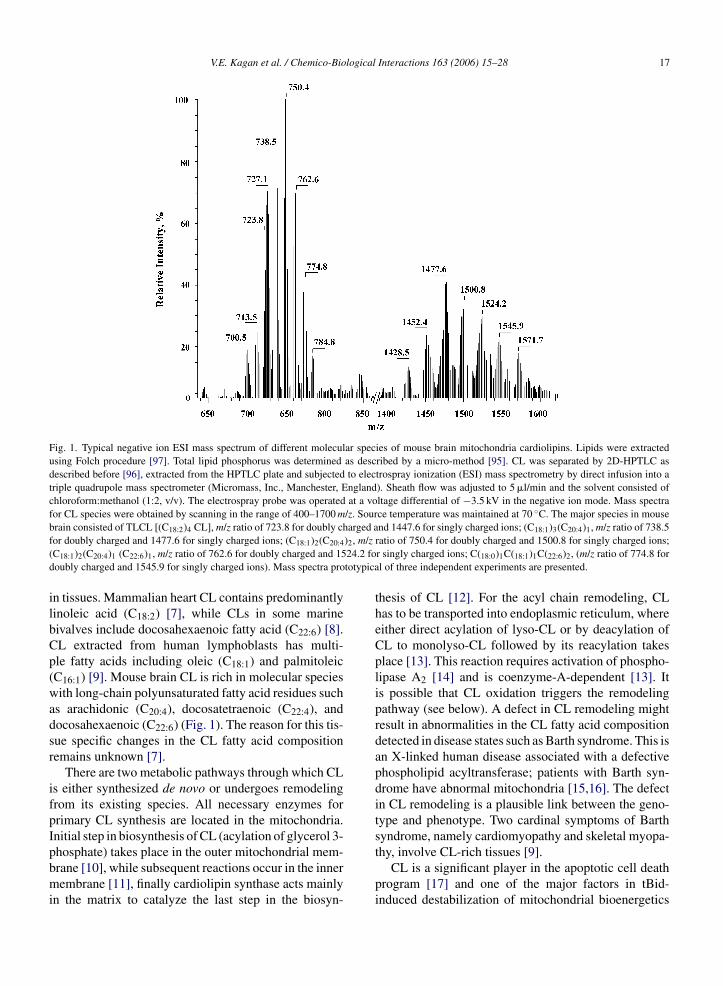
V.E. Kagan et al. / Chemico-Biological Interactions 163 (2006) 15–28 17
Fig. 1. Typical negative ion ESI mass spectrum of different molecular species of mouse brain mitochondria cardiolipins. Lipids were extractedusing Folch procedure [97]. Total lipid phosphorus was determined as described by a micro-method [95]. CL was separated by 2D-HPTLC asdescribed before [96], extracted from the HPTLC plate and subjected to electrospray ionization (ESI) mass spectrometry by direct infusion into atriple quadrupole mass spectrometer (Micromass, Inc., Manchester, England). Sheath flow was adjusted to 5 �l/min and the solvent consisted ofchloroform:methanol (1:2, v/v). The electrospray probe was operated at a voltage differential of −3.5 kV in the negative ion mode. Mass spectrafor CL species were obtained by scanning in the range of 400–1700 m/z. Source temperature was maintained at 70 ◦C. The major species in mouseb arged af )2, m/z( 24.2 fod totypica
ilbCp(wadsr
ifpIpbmi
rain consisted of TLCL [(C18:2)4 CL], m/z ratio of 723.8 for doubly chor doubly charged and 1477.6 for singly charged ions; (C18:1)2(C20:4
C18:1)2(C20:4)1 (C22:6)1, m/z ratio of 762.6 for doubly charged and 15oubly charged and 1545.9 for singly charged ions). Mass spectra pro
n tissues. Mammalian heart CL contains predominantlyinoleic acid (C18:2) [7], while CLs in some marineivalves include docosahexaenoic fatty acid (C22:6) [8].L extracted from human lymphoblasts has multi-le fatty acids including oleic (C18:1) and palmitoleicC16:1) [9]. Mouse brain CL is rich in molecular speciesith long-chain polyunsaturated fatty acid residues such
s arachidonic (C20:4), docosatetraenoic (C22:4), andocosahexaenoic (C22:6) (Fig. 1). The reason for this tis-ue specific changes in the CL fatty acid compositionemains unknown [7].
There are two metabolic pathways through which CLs either synthesized de novo or undergoes remodelingrom its existing species. All necessary enzymes forrimary CL synthesis are located in the mitochondria.nitial step in biosynthesis of CL (acylation of glycerol 3-
hosphate) takes place in the outer mitochondrial mem-rane [10], while subsequent reactions occur in the innerembrane [11], finally cardiolipin synthase acts mainlyn the matrix to catalyze the last step in the biosyn-
nd 1447.6 for singly charged ions; (C18:1)3(C20:4)1, m/z ratio of 738.5ratio of 750.4 for doubly charged and 1500.8 for singly charged ions;r singly charged ions; C(18:0)1C(18:1)1C(22:6)2, (m/z ratio of 774.8 forl of three independent experiments are presented.
thesis of CL [12]. For the acyl chain remodeling, CLhas to be transported into endoplasmic reticulum, whereeither direct acylation of lyso-CL or by deacylation ofCL to monolyso-CL followed by its reacylation takesplace [13]. This reaction requires activation of phospho-lipase A2 [14] and is coenzyme-A-dependent [13]. Itis possible that CL oxidation triggers the remodelingpathway (see below). A defect in CL remodeling mightresult in abnormalities in the CL fatty acid compositiondetected in disease states such as Barth syndrome. This isan X-linked human disease associated with a defectivephospholipid acyltransferase; patients with Barth syn-drome have abnormal mitochondria [15,16]. The defectin CL remodeling is a plausible link between the geno-type and phenotype. Two cardinal symptoms of Barthsyndrome, namely cardiomyopathy and skeletal myopa-
thy, involve CL-rich tissues [9].CL is a significant player in the apoptotic cell deathprogram [17] and one of the major factors in tBid-induced destabilization of mitochondrial bioenergetics

logical
18 V.E. Kagan et al. / Chemico-Bio[18,19]. Apoptosis-associated proteins such as Bid andtBid [20,21] have CL-binding domains [20,22,23] andreveal dynamic interactions with CL and its metabo-lites, mono- and di-lyso-CLs [24]. These interactionsare likely to be most effective at the contact sites of theinner and outer mitochondrial membranes [25] result-ing in changes in CL trans-membrane distribution (see
Scheme 1) and its reorganization in micro-domains witha hexagonal Hn configuration favorable for the releaseof cyt c and other pro-apoptotic factors [20,23,26–28].Therefore, CL is believed to significantly contribute toScheme 1. Possible involvement of Bid in trans-membrane redistribution ofactivated caspase-8 initiates the cleavage of the pro-apoptotic BCL-2 family memitochondria. At the contact sites of the inner and outer mitochondrial membin CL trans-membrane distribution that likely occur after the production of reamembrane markedly increases. The CL distribution between the two monolayof CL is localized in the outer monolayer of the membrane. The CL redistribuavailable for interactions with cyt c. These interactions result in the formation oc activating it to a peroxidase. The peroxidase activity is specific to CL and gfactors from mitochondria into the cytosol. CL oxidation can also trigger theA2 and subsequent tBid/Bid dependent transport of mono-lyso-CL into endop
Interactions 163 (2006) 15–28
the outer membrane permeabilization and cyt c releaseduring apoptosis.
In normal cells, almost 80% of CL is localized in theinner mitochondrial membrane where it is distributedbetween the matrix and intermembrane surfaces at a ratioof 60:40 [29–32]. Although there is ∼70-fold excess ofCL available for 1:1 stoichiometric binding with cyt c
[5,33], most CL is not free but rather interacts with mito-chondrial electron-transport complexes [34,35]. How-ever, in apoptotic cells, the CL content in the outermitochondrial membrane markedly increases to reachCL in mitochondria during apoptosis. Very early during apoptosis,mber, Bid, yielding a truncated fragment (tBid) that translocates to the
ranes, tBid reveals dynamic interactions with CL resulting in changesctive oxygen species. Thus the CL content in the outer mitochondrialers of the inner membrane also changes in a way that almost 70 mol%ted to the outer leaflet of the inner mitochondrial membrane becomesf cyt c/CL complex. H2O2 gets access to the heme catalytic site of cyt
enerates CL hydroperoxides that induced the release of pro-apoptoticremodeling pathway including hydrolysis of CLox by phospholipaselasmic reticulum (where its reacylation takes place).

logical
ttaifimabptChdtionaCmcrl[
3c
ciiNtitmcpto
camspciitc
V.E. Kagan et al. / Chemico-Bio
he level of approximately 40 mol%. The CL distribu-ion between the two monolayers of the inner membranelso changes such that almost 70 mol% of CL is foundn the outer monolayer while 30 mol% remains con-ned to the matrix side of the membrane [32]. Theseembrane translocations of CL occur very early during
poptosis, well before changes in mitochondrial mem-rane potential or other markers of apoptosis, such aslasma-membrane exposure of PS, but after the produc-ion of reactive oxygen species [29,32]. The amounts ofL that may become available for interactions with cyt c,ence for tight binding of cyt c in the membrane, changeramatically during apoptosis. The changes in CL dis-ribution during apoptosis are likely associated with itsnteractions with tBid (see Scheme 1). In fact, additionf tBid to mouse liver mitochondria in vitro induced sig-ificant changes in CL distribution between the innernd outer membranes and accumulation of mono-lyso-L [36]. In line with this, a marked accumulation ofono-lyso-CL was accompanied by simultaneous mito-
hondrial trans-membrane migration of tBid and cyt celease when low concentrations of exogenous mono-yso-CL and tBid were added to isolated mitochondria20].
. CL binds with cytochrome c and changes itsatalytic profile
Cyt c is a basic protein which under physiologicalonditions has a net charge of +8 [37]. As a result,t avidly binds with negatively charged membranes,ncluding anionic (phospho)lipid membranes [38–41].otably, this interactions confers peroxidase activity on
he protein [42]. This is caused by its partial unfold-ng and weakening of the coordination bond betweenhe heme-iron and Met80 [43–45]. As a result, small
olecules – like H2O2 – get access to the heme site ofyt c activating it to a peroxidase. In mitochondria, theeroxidase activity of the cyt c/CL complex is specifico CL and acts as CL oxygenase yielding CL hydroper-xides [32].
In resting-state mitochondria, most of CLs and cytare spatially separated and, therefore, the peroxidase
ctivity of cyt c is very low [32]. However, massive trans-embrane migration of CL during apoptosis (see above)
ets the stage for the elevated content of cyt c/CL com-lexes and markedly enhances the peroxidase activity ofyt c [32]. In fact, the amounts of cyt c available in the
ntermembrane space of mitochondria, rather than CLn the outer leaflet of the IMM and the inner leaflet ofhe OMM, determine the level of peroxidase activity ofyt c/CL complexes during apoptosis. Not surprisingly,Interactions 163 (2006) 15–28 19
cells with siRNA manipulated levels of cyt c exert lowerlevels of CL oxygenase activity and sensitivity to apop-tosis (see below) that are proportional to the amounts ofcyt c [32].
Thus during apoptosis, a significant peroxidase activ-ity of cyt c/CL complexes can generate CL hydroper-oxides, provided sufficient levels of H2O2 (a source ofoxidizing equivalents required for activation of the per-oxidase) are available. One of the important sources ofH2O2 during apoptosis is dysregulated electron transportcapable of generating high concentrations of superoxidewhose spontaneous or MnSOD-catalyzed dismutationproduces H2O2 [46–48]. Moreover, CL-bound cyt c hasa negative redox potential precluding its participationin electron transport as an electron acceptor from themitochondrial complex III [49]. Thus electron transportbetween complexes III and IV cannot be serviced by cytc/CL complexes. Finally, a recently discovered functionof p66 (normally, a tyrosine kinase adaptor protein [50])as a generator of H2O2 in apoptosis contributes to therequired supplementation with sufficient amounts of oxi-dizing equivalents to catalyze CL oxygenation [51]. Asa consequence, CL oxidation is universally discoveredin a number of different cell lines undergoing apoptosis[32]. Ionizing-irradiation is one of many examples of apro-apoptotic stimulation triggering significant CL oxi-dation dependent on cyt c levels [32]. As shown on Fig. 2,ionizing-irradiation of mouse heart mitochondria resultsin accumulation of CL oxidation products detectable asincreased amounts of total CL hydroperoxides as wellas individual molecular species of polyunsaturated CLhydroperoxides in mass spectra. Similarly, in tissues withsignificant levels of apoptotically dying cells resultingfrom exposure to damaging factors (e.g., in brain aftertraumatic injury), accumulation of CL hydroperoxidesoccurs as a characteristic biomarker [52].
Peroxidase activation of cyt c/CL complexes pro-ceeds through the formation of the reactive inter-mediates—compounds I and II and the production ofprotein radicals, most commonly tyrosyl radical inter-mediates [53,54]. Interaction of cyt c with CL results inpartial unfolding of the protein making possible interac-tions the heme catalytic site with H2O2. As in manyother peroxidase reactions, this results in the forma-tion of compounds I and II followed by the genera-tion of protein-derived, most commonly, tyrosyl radi-cals [55–57]. Cyt c has four tyrosine residues some ofwhich are located close to the heme moiety and some
are present on the surface of the protein. In the unfoldedprotein/CL complex, tyrosyl radical abstracts hydrogenfrom one of the CL polyunsaturated lipid acyl chainsyielding a lipid radical (see below). After the addition
20 V.E. Kagan et al. / Chemico-Biological
Fig. 2. Oxidation of mouse heart mitochondrial cardiolipin in responseto ionizing radiation. Mitochondria were isolated from the hearts ofC57BL10 mice. The isolated mitochondria were irradiated to 25 Gy.(A) Typical negative ion ESI mass spectrum of different molecularspecies of mouse heart mitochondria cardiolipins before and after ion-izing radiation. Lipids from mitochondria were extracted using Folchprocedure [97]. Total lipid phosphorus was determined as describedby a micro-method [95]. Lipids were analyzed by electrospray ion-ization tandem mass spectrometry by direct infusion into a triplequadrupole mass spectrometer (Micromass, Inc., Manchester, Eng-land). Mass spectra for the (M − H)− CL species were obtained byscanning in the range of 1200–1800 every 1.5 s and summing individ-ual spectra. CID spectra were obtained by selecting the ion of interestand performing daughter ion scanning in Q3 at 400 Da/s using Ar as thecollision gas. Mitochondria displayed several species of CL as deter-mined by mass spectrometry (top panel). Tandem mass spectrometry(MS/MS analysis) of individual mitochondrial CL species was usedto determine the fatty acyl chain composition. There was a markeddecrease in the CL cluster around m/z 1472 upon radiation treatment(bottom panel). In addition, there appeared a “new” cluster of massions at m/z values of 1566, 1568, 1570 and 1572. The new massescorrespond to mass ions of 1470 ((C18:3)2/(C18:2)1/(C20:3)1 CL), 1472((C18:3)1/(C18:2)2/(C20:3)1 CL), 1474 ((C18:2)2/(C18:1)1/(C20:4)1 CL)and 1476 ((C18:2)1/(C18:1)2/(C20:4)1 CL) each with an additional 96 Da.
Interactions 163 (2006) 15–28
of oxygen, the latter is converted into a peroxyl radi-cal which is further protonated to CL hydroperoxide. Ashighly potent oxidants, the peroxidase reactive interme-diates can attack both endogenous substrates such as CLlocated in close proximity to the catalytic site, ascor-bate, thiols, etc., as well as exogenously added reducingcompounds (e.g., different phenolic compounds). Pro-tein radical intermediates can be directly detected in theEPR spectra of cyt c complexes with different molec-ular species of phospholipids [32,56,57]. In addition,spin trapping experiments can be employed to iden-tify specific types of radicals subsequently involved notonly in intermolecular protein oxidation but also in theoxidation of other substrates such as lipids. Finally, rel-atively long-lived radicals of phenolic compounds, e.g.,phenoxyl radicals of a hindered phenolic compound,etoposide, are also EPR-detectable. We used a spin-trap,2-methyl-2-nitrosopropane (MNP), to detect and iden-tify the radicals generated by cyt c/CL. Cyt c incubatedwith non-oxidizable TOCL in the presence of H2O2generated EPR signal that was identical to previouslyreported MNP-adduct of protein-derived (tyrosyl) radi-cal of cyt c [55,57] (Fig. 3A). When oxidazable TLCLwas used in place of TOCL, cyt c/CL complexes pro-duced two types of MNP adducts: one of them wasthe same protein-derived (tyrosyl) radical–MNP adduct[55,57], while the other one could be identified as a lipidradical (pentadienyl radical) adduct [58,59] (Fig. 3B).No significant radical MNP adduct was detected in theabsence of CL, and no adducts were measured in theabsence of H2O2. Importantly, the same MNP adducts
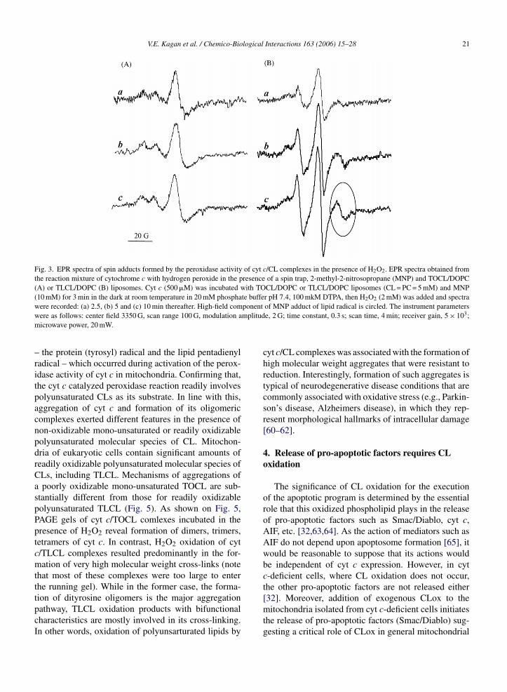
could be distinguished in mitochondria incubated inthe presence of H2O2 (Fig. 4). Detailed analysis andsubtraction of spectra (see figure legend) elicited thatsuperposition of the signals from two radical adductsSpecifically, peaks at +96 Da relative to peaks in the native lipidspectrum were attributed to tri-peroxy derivatives of CL species.(B) Accumulation of phospholipid hydroperoxides in mouse heartin response to ionizing irradiation. After irradiation of mouse heart,lipids were extracted using Folch procedure [97] and total lipid phos-phorus was determined as described by a micro-method [95]. Oxi-dized phospholipids were hydrolyzed by pancreatic phospholipase A2
(2 U/�l) in 25 mM phosphate buffer containing 1 mM CaCl2, 0.5 mMEDTA and 0.5 mM SDS (pH 8.0 at room temperature for 30 min).Fatty acid hydroperoxides formed were determined by fluorescenceHPLC of resorufin stoichiometrically formed during their microper-oxidase 11-catalyzed reduction in the presence of Amplex Red (for40 min at 4 ◦C) [32]. Fluorescence HPLC (Eclipse XDB-C18 column,5 �m, 150 mm × 4.6 mm, mobile phase was composed of 25 mM dis-odium phosphate buffer (pH 7.0)/methanol (60:40, v/v), Ex = 560 nm,Em = 590 nm), was performed on a Shimadzu LC-100AT HPLC sys-tem equipped with fluorescence detector (RF-10Axl) and autosampler(SIL-10AD).
V.E. Kagan et al. / Chemico-Biological Interactions 163 (2006) 15–28 21
Fig. 3. EPR spectra of spin adducts formed by the peroxidase activity of cyt c/CL complexes in the presence of H2O2. EPR spectra obtained fromthe reaction mixture of cytochrome c with hydrogen peroxide in the presence of a spin trap, 2-methyl-2-nitrosopropane (MNP) and TOCL/DOPC(A) or TLCL/DOPC (B) liposomes. Cyt c (500 �M) was incubated with TOCL/DOPC or TLCL/DOPC liposomes (CL = PC = 5 mM) and MNP(10 mM) for 3 min in the dark at room temperature in 20 mM phosphate buffer pH 7.4, 100 mkM DTPA, then H2O2 (2 mM) was added and spectraw ponentw mplitudm
–ritpacnpdrCaspPptcmtttpcI
ere recorded: (a) 2.5, (b) 5 and (c) 10 min thereafter. High-field comere as follows: center field 3350 G, scan range 100 G, modulation aicrowave power, 20 mW.
the protein (tyrosyl) radical and the lipid pentadienyladical – which occurred during activation of the perox-dase activity of cyt c in mitochondria. Confirming that,he cyt c catalyzed peroxidase reaction readily involvesolyunsaturated CLs as its substrate. In line with this,ggregation of cyt c and formation of its oligomericomplexes exerted different features in the presence ofon-oxidizable mono-unsaturated or readily oxidizableolyunsaturated molecular species of CL. Mitochon-ria of eukaryotic cells contain significant amounts ofeadily oxidizable polyunsaturated molecular species ofLs, including TLCL. Mechanisms of aggregations ofpoorly oxidizable mono-unsaturated TOCL are sub-
tantially different from those for readily oxidizableolyunsaturated TLCL (Fig. 5). As shown on Fig. 5,AGE gels of cyt c/TOCL comlexes incubated in theresence of H2O2 reveal formation of dimers, trimers,etramers of cyt c. In contrast, H2O2 oxidation of cyt/TLCL complexes resulted predominantly in the for-ation of very high molecular weight cross-links (note
hat most of these complexes were too large to enterhe running gel). While in the former case, the forma-
ion of dityrosine oligomers is the major aggregationathway, TLCL oxidation products with bifunctionalharacteristics are mostly involved in its cross-linking.n other words, oxidation of polyunsarturated lipids byof MNP adduct of lipid radical is circled. The instrument parameterse, 2 G; time constant, 0.3 s; scan time, 4 min; receiver gain, 5 × 10³;
cyt c/CL complexes was associated with the formation ofhigh molecular weight aggregates that were resistant toreduction. Interestingly, formation of such aggregates istypical of neurodegenerative disease conditions that arecommonly associated with oxidative stress (e.g., Parkin-son’s disease, Alzheimers disease), in which they rep-resent morphological hallmarks of intracellular damage[60–62].
4. Release of pro-apoptotic factors requires CLoxidation
The significance of CL oxidation for the executionof the apoptotic program is determined by the essentialrole that this oxidized phospholipid plays in the releaseof pro-apoptotic factors such as Smac/Diablo, cyt c,AIF, etc. [32,63,64]. As the action of mediators such asAIF do not depend upon apoptosome formation [65], itwould be reasonable to suppose that its actions wouldbe independent of cyt c expression. However, in cytc-deficient cells, where CL oxidation does not occur,the other pro-apoptotic factors are not released either
[32]. Moreover, addition of exogenous CLox to themitochondria isolated from cyt c-deficient cells initiatesthe release of pro-apoptotic factors (Smac/Diablo) sug-gesting a critical role of CLox in general mitochondrial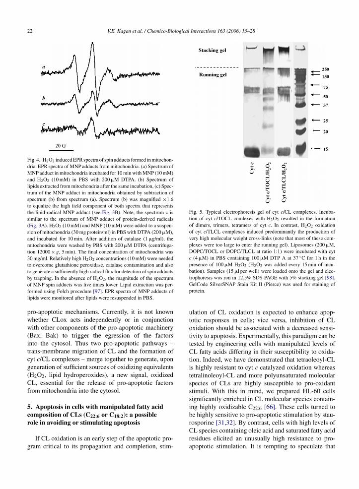
22 V.E. Kagan et al. / Chemico-Biological Interactions 163 (2006) 15–28
Fig. 4. H2O2 induced EPR spectra of spin adducts formed in mitochon-dria. EPR spectra of MNP adducts from mitochondria. (a) Spectrum ofMNP adduct in mitochondria incubated for 10 min with MNP (10 mM)and H2O2 (10 mM) in PBS with 200 �M DTPA. (b) Spectrum oflipids extracted from mitochondria after the same incubation, (c) Spec-trum of the MNP adduct in mitochondria obtained by subtraction ofspectrum (b) from spectrum (a). Spectrum (b) was magnified ×1.6to equalize the high field component of both spectra that representsthe lipid-radical MNP adduct (see Fig. 3B). Note, the spectrum c issimilar to the spectrum of MNP adduct of protein-derived radicals(Fig. 3A). H2O2 (10 mM) and MNP (10 mM) were added to a suspen-sion of mitochondria (30 mg protein/ml) in PBS with DTPA (200 �M),and incubated for 10 min. After addition of catalase (1 �g/ml), themitochondria were washed by PBS with 200 �M DTPA (centrifuga-tion 12000 × g, 5 min). The final concentration of mitochondria was30 mg/ml. Relatively high H2O2 concentrations (10 mM) were neededto overcome glutathione peroxidase, catalase contamination and alsoto generate a sufficiently high radical flux for detection of spin adducts
Fig. 5. Typical electrophoresis gel of cyt c/CL complexes. Incuba-tion of cyt c/TOCL comlexes with H2O2 resulted in the formationof dimers, trimers, tetramers of cyt c. In contrast, H2O2 oxidationof cyt c/TLCL complexes induced predominantly the production ofvery high molecular weight cross-links (note that most of these com-plexes were too large to enter the running gel). Liposomes (200 �M,DOPC/TOCL or DOPC/TLCL at ratio 1:1) were incubated with cytc (4 �M) in PBS containing 100 �M DTP A at 37 ◦C for 1 h in thepresence of 100 �M H2O2 (H2O2 was added every 15 min of incu-bation). Samples (15 �l per well) were loaded onto the gel and elec-
by trapping. In the absence of H2O2, the magnitude of the spectrumof MNP spin adducts was five times lower. Lipid extraction was per-formed using Folch procedure [97]. EPR spectra of MNP adducts oflipids were monitored after lipids were resuspended in PBS.
pro-apoptotic mechanisms. Currently, it is not knownwhether CLox acts independently or in conjunctionwith other components of the pro-apoptotic machinery(Bax, Bak) to trigger the egression of the factorsinto the cytosol. Thus two pro-apoptotic pathways –trans-membrane migration of CL and the formation ofcyt c/CL complexes – merge together to generate, upongeneration of sufficient sources of oxidizing equivalents(H2O2, lipid hydroperoxides), a new signal, oxidizedCL, essential for the release of pro-apoptotic factorsfrom mitochondria into the cytosol.
5. Apoptosis in cells with manipulated fatty acidcomposition of CLs (C or C ): a possible
22:6 18:2role in avoiding or stimulating apoptosisIf CL oxidation is an early step of the apoptotic pro-gram critical to its propagation and completion, stim-
trophoresis was run in 12.5% SDS-PAGE with 5% stacking gel [98].GelCode SilverSNAP Stain Kit II (Pierce) was used for staining ofprotein.
ulation of CL oxidation is expected to enhance apop-totic responses in cells; vice versa, inhibition of CLoxidation should be associated with a decreased sensi-tivity to apoptosis. Experimentally, this paradigm can betested by engineering cells with manipulated levels ofCL fatty acids differing in their susceptibility to oxida-tion. Indeed, we have demonstrated that tetraoleoyl-CLis highly resistant to cyt c catalyzed oxidation whereastetralinoleoyl-CL and more polyunsaturated molecularspecies of CLs are highly susceptible to pro-oxidantstimuli. With this in mind, we prepared HL-60 cellssignificantly enriched in CL molecular species contain-ing highly oxidizable C22:6 [66]. These cells turned tobe highly sensitive to pro-apoptotic stimulation by stau-
rosporine [31,32]. By contrast, cells with high levels ofCL species containing oleic acid and saturated fatty acidresidues elicited an unusually high resistance to pro-apoptotic stimulation. It is tempting to speculate that
logical
mocrtaacfop
6Ne
opftmaocomos
ctdaceleSmmtaosr[pcwrt
V.E. Kagan et al. / Chemico-Bio
anipulation of CL susceptibility to oxidation may bene of the factors that are utilized by undifferentiatedells – such as stem cells or tumor cells – to obtainesistance to pro-apoptotic signals hence avoid apop-otic cell death. Conversely, drug-discovery programsimed at eradication of tumor cells may benefit frompproaches to stimulate oxidation of CL in their mito-hondria by forcing them to integrate polyunsaturatedatty acid residues. One of the approaches can be basedn non-toxic nutritional manipulations using sources ofolyunsaturated fatty acids (e.g., fish oil).
. Regulation of CL oxidation in mitochondria—O, nitroxides, Vitamin E homologues, and
toposide
Given that CL oxidation is one of the very early stagesf the apoptotic program, taking place up-stream of theoint of no-return (caspase activation and proteolysis),ormation of cyt c/CL complexes may be an importantarget for the control of apoptosis. Several approaches
ay be envisioned to achieve this goal. As CL avail-bility is an important stage for cyt c activation to CLxygenase, regulation of CL trans-migration in mito-hondria can be targeted. A role of tBid in stimulationf CL redistribution in apoptotic mitochondria becomesore evident indicating that tools to affect interactions
f tBid with mitochondria may be constitute promisingtrategies [67,68].
Control of redox environment that is conducive ofyt c/CL peroxidase activity is another interesting wayo affect the CL oxidation stage of apoptosis. High oxi-izing potential of the cyt c/CL peroxidase intermedi-tes [32,69] offers a number of opportunities for theirhemical reduction by both endogenous substrates andxogenous agents. Ascorbate, cysteine, GSH and otherow molecular weight thiols, Vitamin E homologues arexamples of the former type while different phenolic andH-containing drugs represent the latter category anday be utilized for the reduction of peroxidase inter-ediates. These general considerations, however, do not
ake into account key structural factors that may restrictccess of the bulky reducing agents to the catalytic sitef cyt c accessible only for small molecules. One suchmall molecule is NO which can act as a reductant foreactive intermediates of cyt c/CL peroxidase complexes69]. Therefore, NO and NO-releasing compounds mayarticipate in regulation of CL oxidation by cyt c/CL
omplexes. Moreover, the role of mitochondrial NOS –hose functions are not well understood – [70] can beationalized in terms of its participation in the regula-ion of the peroxidase activity of cyt c/CL complexes as
Interactions 163 (2006) 15–28 23
an anti-apoptotic mechanism preventing inadvertent CLoxidation.
Because the peroxidase function of cyt c/CL com-plexes requires H2O2 or organic (lipid) hydroperoxides[71–73], the removal of these oxidizing equivalents isequivalent to inhibition of the CL oxygenase activity.Obviously, different GSH-dependent mechanisms (GSHperoxidases) [74,75] as well as thioredoxin [76–78],peroxiredoxin [79] may be effective in removing thehydroperoxides through respective enzymatic pathways.
Another protective pathway may be realized throughprevention of H2O2 formation by eliminating its majorsource in apoptotic mitochondria, superoxide radicals.Disrupted electron transport is said to play a majorrole in the production of superoxide radicals on one ormore respiratory complexes during apoptosis [80–82].Dismutation of superoxide radicals that occurs eitherspontaneously or through catalytic action of MnSODgenerates H2O2 that feeds the peroxidase reaction ofcyt c/CL complexes. Therefore, prevention of superoxideproduction may be one of the most potent approaches toinhibit CL oxidation. Stable nitroxide radicals seem to bealmost ideal candidate agents to quench the productionof superoxide in mitochondria. This is because nitrox-ides possess several features that are essential for theirability to control the levels of superoxide radicals andH2O2. First, they readily undergo one-electron reductionby electron transport to yield respective hydroxylamines.This prevents one-electron reduction of oxygen to formsuperoxide radicals. The hydroxylamines formed fromnitroxides act as effective radical scavengers to pro-duce nitroxides, ie undergo recycling. Second, nitroxideradicals exert an SOD mimetic activity and dismutatesuperoxide radicals. The product of this reaction is H2O2which can feed the peroxidase cycle of cyt c/CL com-plexes. However, by acting as an electron acceptor frommitochondrial complexes and by eliminating superox-ide radicals, the electron scavenging and SOD activityof nitroxides contribute to prevention of superoxide reac-tion with NO resulting in peroxynitrite, a potent oxidantknown to contribute to dysregulation of electron trans-port in mitochondria [83,84]. Thus, overall SOD mimeticactivity of nitroxides may be an important protectivemechanisms, similar to that of MnSOD in mitochondria[85,86]. Third, nitroxides display catalase activity thatdirectly eliminates H2O2. These combined propensitiesof nitroxides make them highly promising mitochon-drial anti-apoptotic agents. Unfortunately, these activ-
ities of nitroxides require their presence in mitochondriawhich, in turn, only happens when very high (milomolar)concentrations of nitroxides are utilized in vivo. Untilrecently, this precluded effective use of nitroxides as
logical
24 V.E. Kagan et al. / Chemico-Bioanti-apoptotic remedies. Targeting of nitroxides to mito-chondria, however, may overcome this problem. In fact,latest developments in the field indicate that an remark-ably enhanced anti-apoptotic activity of nitroxides canbe achieved by their effective delivery into mitochondriausing different approaches [87,88].
Since execution of apoptotic program in mitochon-dria is associated with CL peroxidation products requiredfor the release of pro-apoptotic factors, lipid antioxi-dants capable of inhibiting CL peroxidation may actas anti-apoptotic agents. Etoposide, a widely-used anti-tumor drug, is a prototypical inducer of apoptosis and,at the same time, an effective lipid radical scavenger andlipid antioxidant. We found that CL oxidation duringapoptosis is realized not via a random CL peroxidationmechanism but rather proceeds as a result of peroxidasereaction in a tight cyt c/CL complex that restrains inter-actions of etoposide with radical intermediates generatedin the course of the reaction [89]. While etoposide caninhibit cyt c catalyzed oxidation of CL competing withit as a peroxidase substrate, the inhibition is realized atfar higher concentrations than those at which it inducesapoptotic cell death. Thus, oxidation of CL by cyt c/CLperoxidase complex, which is essential for apoptosis, isnot inhibited by pro-apoptotic concentrations of the drug.
7. Peroxidase complexes of cyt c can be formedin extramitochondrial compartments: biomedicalimplications
Hitherto, we discussed double complexes of cyt c withCL, however, more complex associations of these twomolecules, with other partners are likely to occur, partic-ularly in extramitochondrial compartments. As an exam-ple, synaptic terminals contain high levels of �-synuclein(Syn) which has a phospolipid-binding domain. If Synbinds CL, and if the Syn/CL complex thus formed willbe able to additionally bind cytosolic cyt c, the triplecomplex Syn/CL/cyt c will be generated in which acti-vation of cyt c peroxidase activity will still be possible.Moreover, the peroxidase activity, in the presence ofH2O2 will be inducing oligomerization of their consti-tutive components to form mixed aggregates similar toLewy bodies. In both Lewy bodies and in our experi-mental aggregates, co-localization of Syn and cyt c willbe overlapping and detectable along with the presenceof CL. This has been indeed observed in both modelbiochemical systems as well as in apoptotic HeLa cells
(expressing significant amounts of Syn in the cytosol)upon the tert-butyl hydroperoxide induced release of cytc. In line with these observations, co-localization of cyt cand Syn was detected in the substantia nigra Lewy bod-Interactions 163 (2006) 15–28
ies in patients with Parkinson’s disease [90]. Overall,these combined results are compatible with the hypothe-sis that during pro-apoptotic damage of mitochondria insynaptic terminals, released cyt c and CL are captured byan abundant cytosolic protein, Syn. This mode of actionof Syn prevents retrograde spread of pro-apoptotic fac-tors (cyt c) to the soma region of the neuronal cells thusprotecting them from total cell apoptosis.
Cell damage may be accompanied by a significantrelease of cyt c into extracellular spaces and bioflu-ids. The presence of cyt c in plasma and cerebrospinalfluid has been reported in several disease conditions(e.g., brain trauma, sepsis, and systemic inflammatoryresponse syndrome) [91–93]. Because dyslipidemia andelevated contents of free fatty acids are also typical ofmost common pro-inflammatory diseases, it is possiblethat complexes of cyt c with these negatively chargedlipids will be formed resulting in a peroxidase activity.The consequences of their formation can be envisionedin terms of enhanced oxidative stress and antioxidantdepletion. Indeed, any H2O2 production by inflamma-tory response would unavoidably trigger its utilizationaccompanied by consumption of extracellular reduc-tants (ascorbate, thiols). Moreover, oxidation depletesNO leading to vasoconstrictive conditions. Future stud-ies are necessary to better understand the significanceof these events and their manifestations in severe pro-inflammatory conditions such as sepsis, preeclampsia,etc.
8. Concluding remarks
The biology of apoptosis in mitochondria, createsconditions for tight interactions of one of the major pro-teins of the intermembrane space, cyt c, with the majormembrane phospholipid, CL. The complex thus formeddoes not function as an electron shuttle in the respiratorychain but rather operates as a CL-specific oxygenase,a qualitatively new catalytic entity with different enzy-matic properties. Peroxidase catalytic activity of the cytc/CL complex is essential for the execution of the apop-totic program. Using an oxidative lipidomics approachwe established that this is due the production of oxidizedmolecular species of CL that participate in the release ofpro-apoptotic factors from mitochondria into the cytosol.Thus, in contrast to the previously well recognized pro-apoptotic role of cyt c in apoptosome formation, thisnew function of cyt c is dependent on its redox prop-
erties. Specific features of the redox behavior of cyt cand its interactions with different molecular species ofCL are important for CL oxidation and its regulation ofthe sensitivity of cells, including tumor cells, to apop-
logical
todmwli
rCtwa[
A
As
R
[
[
[
[
[
[
[
[
[
[
[
[
[
[
[
[
[
[
[
V.E. Kagan et al. / Chemico-Bio
osis. The discovery of the early stage of apoptosis alsoffers interesting opportunities for new targets in drugiscovery programs. One can also envision that employ-ent of new delivery vehicles (e.g., nanoparticles, singlealled carbon nanotubes) coated with different molecu-
ar species of CL and cyt c may be particularly effectiven achieving these therapeutic goals.
Reliable fulfillment and coordination of cell functionselies on prodigality of its essential mechanisms. WhileL oxidation may be an essential mechanism of apop-
otsis, it likely that alternative pathways are turned onhen CL oxidation becomes impossible or ineffective
s has been demonstrated for yeast cells deficient in CL94].
cknowledgements
Supported by grants from NIH HL70755, NIH U19IO68021, NIOSH OH008282, AHA0535365N, Penn-
ylvania Department of Health SAP 4100027294.
eferences
[1] M.R. Wenk, The emerging field of lipidomics, Nat. Rev. DrugDiscov. 4 (2005) 594–610.
[2] C.N. Serhan, Mediator lipidomics, Prostaglandins Other LipidMediat. 77 (2005) 4–14.
[3] X. Han, J. Yang, H. Cheng, K. Yang, D.R. Abendschein, R.W.Gross, Shotgun lipidomics identifies cardiolipin depletion in dia-betic myocardium linking altered substrate utilization with mito-chondrial dysfunction, Biochemistry 44 (2005) 16684–16694.
[4] V.E. Kagan, Lipid peroxidation in biomembranes, CRC Press,Boca Raton, FL, 1988, pp. 1–181.
[5] G. Daum, Lipids of mitochondria, Biochim. Biophys. Acta 822(1985) 1–42.
[6] J.J. Krebs, H. Hauser, E. Carafoli, Asymmetric distribution ofphospholipids in the inner membrane of beef heart mitochondria,J. Biol. Chem. 254 (1979) 5308–5316.
[7] M. Schlame, S. Shanske, S. Doty, T. Konig, T. Sculco, S. DiMauro,T.J. Blanck, Microanalysis of cardiolipin in small biopsies includ-ing skeletal muscle from patients with mitochondrial disease, J.Lipid Res. 40 (1999) 1585–1592.
[8] E. Kraffe, P. Soudant, Y. Marty, N. Kervarec, P. Jehan, Evidenceof a tetradocosahexaenoic cardiolipin in some marine bivalves,Lipids 37 (2002) 507–514.
[9] M. Schlame, M. Ren, Y. Xu, M.L. Greenberg, I. Haller, Molecularsymmetry in mitochondrial cardiolipins, Chem. Phys. Lipids 138(2005) 38–49.
10] C.B. Hesler, M.A. Carroll, D. Haldar, The topography of glyc-erophosphate acyltransferase in the transverse plane of the mito-chondrial outer membrane, J. Biol. Chem. 260 (1985) 7452–7456.
11] K.Y. Hostetler, H. van den Bosch, L.L. van Deenen, The mecha-
nism of cardiolipin biosynthesis in liver mitochondria, Biochim.Biophys. Acta 260 (1972) 507–513.12] M. Schlame, D. Haldar, Cardiolipin is synthesized on the matrixside of the inner membrane in rat liver mitochondria, J. Biol.Chem. 268 (1993) 74–79.
[
Interactions 163 (2006) 15–28 25
13] M. Schlame, B. Rustow, Lysocardiolipin formation and reacyla-tion in isolated rat liver mitochondria, Biochem. J. 272 (1990)589–595.
14] T. Brustovetsky, B. Antonsson, R. Jemmerson, J.M. Dubinsky, N.Brustovetsky, Activation of calcium-independent phospholipaseA (iPLA) in brain mitochondria and release of apoptogenic factorsby BAX and truncated BID, J. Neurochem. 94 (2005) 980–994.
15] P. Vreken, F. Valianpour, L.G. Nijtmans, L.A. Grivell, B. Plecko,R.J. Wanders, P.G. Barth, Defective remodeling of cardiolipinand phosphatidylglycerol in Barth syndrome, Biochem. Biophys.Res. Commun. 279 (2000) 378–382.
16] P.G. Barth, R.J. Wanders, P. Vreken, E.A. Janssen, J. Lam, F.Baas, X-linked cardioskeletal myopathy and neutropenia (Barthsyndrome) (MEVI 302060), J. Inherit. Metab. Dis. 22 (1999)555–567.
17] T. Kuwana, M.R. Mackey, G.A. Perkins, M.H. Ellisman, M. Lat-terich, R. Schneiter, D.R. Green, D.D. Newmeyer, Bid, Bad andlipids cooperate to form supramolecular openings in the outermitochondrial membrane, Cell 111 (2002) 1–12.
18] F. Gonzalvez, J.J. Bessoule, F. Rocchiccioli, S. Manon, P.X. Petit,Role of cardiolipin on tBid and tBid/Bax synergistic effects onyeast mitochondria, Cell Death Differ. 12 (2005) 659–667.
19] F. Gonzalvez, F. Pariselli, P. Dupaigne, I. Budihardjo, M. Lutter,B. Antonsson, P. Diolez, S. Manon, J.C. Martinou, M. Gou-bern, X. Wang, S. Bernard, P.X. Petit, tBid interaction with car-diolipin primarily orchestrates mitochondrial dysfunctions andsubsequently activates Bax and Bak, Cell Death Differ. 12 (2005)266–614.
20] M.D. Esposti, I.M. Cristea, S.J. Gaskell, Y. Nakao, C. Dive,Proapoptotic Bid binds to monolysocardiolipin, a new molecu-lar connection between mitochondrial membranes and cell death,Cell Death Differ. 10 (2003) 1300–1309.
21] M. Degli Esposti, Lipids, cardiolipin and apoptosis: a greasylicence to kill, Cell Death Differ. 9 (2002) 234–236.
22] M. Lutter, G.A. Perkins, X. Wang, The pro-apoptotic Bcl-2 familymember tBid localizes to mitochondrial contact sites, BMC CellBiol. 2 (2001) 22.
23] J. Liu, A. Weiss, D. Durrant, N.W. Chi, R.M. Lee, The cardiolipin-binding domain of Bid affects mitochondrial respiration andenhances cytochrome c release, Apoptosis 9 (2004) 533–541.
24] I.M. Cristea, M. Degli Esposti, Membrane lipids and cell death:an overview, Chem. Phys. Lipids 129 (2004) 133–160.
25] T.H. Kim, Y. Zhao, W.X. Ding, J.N. Shin, X. He, Y.W. Seo, J.Chen, H. Rabinowich, A.A. Amoscato, X.M. Yin, Bid-cardiolipininteraction at mitochondrial contact site contributes to mitochon-drial cristae reorganization and cytochrome C release, Mol. Biol.Cell. 15 (2004) 3061–3072.
26] R.F. Epand, J.C. Martinou, M. Fornallaz-Mulhauser, D.W.Hughes, R.M. Epand, The apoptotic protein tBid promotes leak-age by altering membrane curvature, J. Biol. Chem. 277 (2002)32632–32639.
27] J. Liu, D. Durrant, H.S. Yang, Y. He, F.G. Whitby, D.G. Myszka,R.M. Lee, The interaction between tBid and cardiolipin ormonolysocardiolipin, Biochem. Biophys. Res. Commun. 330(2005) 865–870.
28] M. Lutter, M. Fang, X. Luo, M. Nishijima, X. Xie, X. Wang, Car-diolipin provides specificity for targeting of tBid to mitochondria,
Nat. Cell Biol. 2 (2000) 754–761.29] M. Garcia Fernandez, L. Troiano, L. Moretti, M. Nasi, M. Pinti, S.Salvioli, J. Dobrucki, A. Cossarizza, Early changes in intramito-chondrial cardiolipin distribution during apoptosis, Cell GrowthDiffer. 13 (2002) 449–455.

logical
[
[
[
[
[
[
[
[
[
[
[
[
[
[
[
[
[
[
[
[
[
[
[
[
[
[
[
[
[
[
26 V.E. Kagan et al. / Chemico-Bio
30] A. Cossarizza, i.M. Pint, L. Moretti, D. Bricalli, R. Bianchi, L.Troiano, M.G. Fernandez, F. Balli, P. Brambilla, C. Mussini,A. Vigano, Mitochondrial functionality and mitochondrial DNAcontent in lymphocytes of vertically infected human immunod-eficiency virus-positive children with highly active antiretroviraltherapy-related lipodystrophy, J. Infect. Dis. 185 (2002) 299–305.
31] V.E. Kagan, G.G. Borisenko, Y.Y. Tyurina, V.A. Tyurin, J.Jiang, A.I. Potapovich, V. Kini, A.A. Amoscato, Y. Fujii, Oxida-tive lipidomics of apoptosis: redox catalytic interactions ofcytochrome c with cardiolipin and phosphatidylserine, FreeRadic. Biol. Med. 37 (2004) 1963–1985.
32] V.E. Kagan, V.A. Tyurin, J. Jiang, Y.Y. Tyurina, V.B. Ritov,A.A. Amoscato, A.N. Osipov, N.A. Belikova, A.A. Kapralov, V.Kini, I.I. Vlasova, Q. Zhao, M. Zou, P. Di, D.A. Svistunenko,I.V. Kurnikov, G.G. Borisenko, Cytochrome c acts as a cardi-olipin oxygenase required for release of proapoptotic factors, Nat.Chem. Biol. 1 (2005) 223–232.
33] Y. Shidoji, K. Hayashi, S. Komura, N. Ohishi, K. Yagi, Lossof molecular interaction between cytochrome c and cardiolipindue to lipid peroxidation, Biochem. Biophys. Res. Commun. 264(1999) 343–347.
34] K. Pfeiffer, V. Gohil, R.A. Stuart, C. Hunte, U. Brandt, M. Green-berg, H. Schagger, Cardiolipin stabilizes respiratory chain super-comlexes, J. Biol. Chem. 278 (2003) 52873–52880.
35] M.S. Sharpley, R.J. Shannon, F. Draghi, J. Hirst, Interac-tions between phospholipids and nadh:ubiquinone oxidoreduc-tase (complex i) from bovine mitochondria, Biochemistry 45(2006) 241–248.
36] V.A. Tyurin, A.N. Osipov, Y.Y. Tyurina, H. Bayir, L.V. Basova,N.A. Belikova, A.A. Kapralov, Q. Zhao, J. Jiang, P.K. Gill, D.H.Waldeck, V.E. Kagan, Lysocardiolipins in apoptosis: interactionwith cytochrome c, tBID and asymmetry of distribution in mito-chondria, in: 45th Annual Meeting, Society of Toxicology, SanDiego, CA, 2006, p. 410.
37] R.E. Dickerson, The structures of cytochrome c and the rates ofmolecular evolution, J. Mol. Evol. 1 (1971) 26–45.
38] M. Rytomaa, P. Mustonen, P.K. Kinnunen, Reversible, non-ionic, and pH-dependent association of cytochrome c withcardiolipin-phosphatidylcholine liposomes, J. Biol. Chem. 267(1992) 22243–22248.
39] S. Oellerich, H. Wackerbarth, P. Hildebrandt, Conformationalequilibria and dynamics of cytochrome c induced by binding ofsodium dodecyl sulfate monomers and micelles, Eur. Biophys. J.32 (2003) 599–613.
40] M. Rytomaa, P.K. Kinnunen, Evidence for two distinct acidicphospholipid-binding sites in cytochrome c, J. Biol. Chem. 269(1994) 1770–1774.
41] E.K. Tuominen, C.J. Wallace, P.K. Kinnunen, Phospholipid–cytochrome c interaction: evidence for the extended lipid anchor-age, J. Biol. Chem. 277 (2002) 8822–8826.
42] R. Radi, J.F. Turrens, B.A. Freeman, Cytochrome c-catalyzedmembrane lipid peroxidation by hydrogen peroxide, Arch.Biochem. Biophys. 288 (1991) 118–125.
43] A.M. Cassina, R. Hodara, J.M. Souza, L. Thomson, L. Castro, H.Ischiropoulos, B.A. Freeman, R. Radi, Cytochrome c nitration byperoxynitrite, J. Biol. Chem. 275 (2000) 21409–21415.
44] Y.R. Chen, L.J. Deterding, B.E. Sturgeon, K.B. Tomer, R.P.
Mason, Protein oxidation of cytochrome C by reactive halogenspecies enhances its peroxidase activity, J. Biol. Chem. 277 (2002)29781–29791.45] S. Prasad, N.C. Maiti, S. Mazumdar, S. Mitra, Reaction ofhydrogen peroxide and peroxidase activity in carboxymethylated
[
[
Interactions 163 (2006) 15–28
cytochrome c: spectroscopic and kinetic studies, Biochim. Bio-phys. Acta 1596 (2002) 63–75.
46] I. Fridovich, Superoxide radical and superoxide dismutases,Annu. Rev. Biochem. 64 (1995).
47] M.W. Epperly, J.E. Gretton, C.A. Sikora, M. Jefferson, M.Bernarding, S. Me, J.S. Greenberger, Mitochondrial localizationof superoxide dismutase is required for decreasing radiation-induced cellular damage, Radiat. Res. 160 (2003) 568–578.
48] P. Mantymaa, T. Siitonen, T. Guttorm, M. Saily, V. Kinnula, E.R.Savolainen, P. Koistinen, Induction of mitochondrial manganesesuperoxide dismutase confers resistance to apoptosis in acutemyeloblastic leukaemia cells exposed to etoposide, Br. J. Haema-tol. 108 (2000) 574–581.
49] H. Wackerbarth, P. Hildebrandt, Redox and conformational equi-libria and dynamics of cytochrome c at high electric fields,Chemphyschem. 4 (2003) 714–724.
50] K. Sato, T. Nagao, M. Kakumoto, M. Kimoto, T. Otsuki, T.Iwasaki, A.A. Tokmakov, K. Owada, Y. Fukami, Adaptor proteinShe is an isoform-specific direct activator of the tyrosine kinasec-Src, J. Biol. Chem. 277 (2002) 29568–29576.
51] M. Giorgio, E. Migliaccio, F. Orsini, D. Paolucci, M. Moroni, C.Contursi, G. Pelliccia, L. Luzi, S. Minucci, M. Marcaccio, P. Pin-ton, R. Rizzuto, P. Bernardi, F. Paolucci, P.G. Pelicci, Electrontransfer between cytochrome c and p66Shc generates reactiveoxygen species that trigger mitochondrial apoptosis, Cell 122(2005) 221–233.
52] H. Bayir, V.A. Tyurin, Y.Y. Tyurina, J. Jiang, V.B. Ritov, X. Zhang,Q. Zhao, P. Kochanek, R. Clark, S. DeKosky, V. Kagan, A newbiomarcker of early apoptosis in the brain: cardiolipin oxidation,in: 45th Annual Meeting, Society of Toxicology, San Diego, USA,Toxicol. Sci. 90 ((1-S) 2005) (2006) 410.
53] D.A. Svistunenko, Reaction of haem containing proteins andenzymes with hydroperoxides: the radical view, Biochim. Bio-phys. Acta 1707 (2005) 127–155.
54] A. Ivancich, C. Jakopitsch, M. Auer, S. Un, C. Obinger,Protein-based radicals in the catalase-peroxidase of synechocys-tis PCC6803: a multifrequency EPR investigation of wild-typeand variants on the environment of the heme active site, J. Am.Chem. Soc. 125 (2003) 14093–14102.
55] Y.R. Chen, C.L. Chen, W. Chen, J.L. Zweier, O. Augusto, R. Radi,R.P. Mason, Formation of protein tyrosine ortho-semiquinoneradical and nitrotyrosine from cytochrome c-derived tyrosyl rad-ical, J. Biol. Chem. 279 (2004) 18054–18062.
56] K. Nakai, R.P. Mason, Immunochemical detection of nitric oxideand nitrogen dioxide trapping of the tyrosyl radical and the result-ing nitrotyrosine in sperm whale myoglobin, Free Radic. Biol.Med. 39 (2004) 1050–1058.
57] D.P. Barr, M.R. Gunther, L.J. Deterding, K.B. Tomer, R.P. Mason,ESR spin-trapping of a protein-derived tyrosyl radical from thereaction of cytochrome c with hydrogen peroxide, J. Biol. Chem.271 (1996) 15498–15503.
58] J.B. Feix, B. Kalyanaraman, Spin trapping of lipid-derived radi-cals in liposomes, Biochim. Biophys. Acta 992 (1989) 230–235.
59] D. Pietraforte, L. Turco, E. Azzini, M. Minetti, On-line EPRstudy of free radicals induced by peroxidase/H2O2 in humanlow-density lipoproteins, Biochim. Biophys. Acta 1583 (2002)176–184.
60] J. Everse, P.W. Coates, The cytotoxic activity of lactoperoxi-dase: enhancement and inhibition by neuroactive compounds,Free Radic. Biol. Med. 37 (2004) 839–849.
61] C.T. Chu, J.L. Caruso, T.J. Cummings, J. Ervin, C. Rosenberg,C.M. Hulette, Ubiquitin immunochemistry as a diagnostic aid for

logical
[
[
[
[
[
[
[[
[
[
[
[
[
[
[
[
[
[
[
[
[
[
[
[
[
[
[
[
[
[
[
[
V.E. Kagan et al. / Chemico-Bio
community pathologists evaluating patients who have dementia,Mod. Pathol. 13 (2000) 420–426.
62] J. Everse, P.W. Coates, Role of peroxidases in Parkinson disease:a hypothesis, Free Radic. Biol. Med. 38 (2005) 1296–1310.
63] G. Petrosillo, F.M. Ruggiero, G. Paradies, Role of reactive oxy-gen species and cardiolipin in the release of cytochrome c frommitochondria, FASEB J. 17 (2003) 2202–2208.
64] G. Petrosillo, F.M. Ruggiero, M. Pistolese, G. Paradies, Reac-tive oxygen species generated from the mitochondrial electrontransport chain induce cytochrome c dissociation from beef-heartsubmitochondrial particles via cardiolipin peroxidation. Possiblerole in the apoptosis, FEBS Lett. 509 (2001) 435–438.
65] C.T. Chu, J.H. Zhu, G. Cao, A. Signore, S. Wang, J. Chen, Apop-tosis inducing factor mediates caspase-independent l-methyl-4-phenylpyridinium toxicity in dopaminergic cells, J. Neurochem.94 (2005) 1685–1695.
66] Y.Y. Tyurina, V.A. Tyurin, Y. Fujii, V.E. Kagan, HL-60 cells withmetabolically bioengineered polyunsaturated cardiolipin molecu-lar species exert increased sensitivity to apoptosis, in: 44th AnnualMeeting, Society of Toxicology, New Orleans, Toxicol. Sci. 84(2297) (2005) 471.
67] F. Sandra, M. Degli Esposti, K. Ndebele, P. Gona, D. Knight,M. Rosenquist, R. Khosravi-Far, Tumor necrosis factor-relatedapoptosis-inducing ligand alters mitochondrial membrane lipids,Cancer Res. 65 (2005).
68] M.D. Esposti, The roles of Bid, Apoptosis 7 (2002) 433–440.69] I.I. Vlasova, V.A. Tyurin, A.A. Kapralov, I.V. Kurnikov, A.N.
Osipov, M.V. Potapovich, D.A. Stoyanovsky, V.E. Kagan, Nitricoxide inhibits peroxidase activity of cytochrome c/cardiolipincomplex and blocks cardiolipin oxidation, J. Biol. Chem. (2006)(Epub ahead of print).
70] P. Ghafourifar, E. Cadenas, Mitochondrial nitric oxide synthase,Trends Pharmacol. Sci. 26 (2005) 190–195.
71] D.P. Barr, R.P. Mason, Mechanism of radical production fromthe reaction of cytochrome c with organic hydroperoxides. AnESR spin trapping investigation, J. Biol. Chem. 270 (1995)12709–12716.
72] J. Van der Zee, Formation of peroxide- and globin-derived rad-icals from the reaction of methaemoglobin and metmyoglobinwith t-butyl hydroperoxide: an ESR spin-trapping investigation,Biochem. J. 322 (1997) 633–639.
73] P.G. Furtmuller, U. Burner, W. Jantschko, G. Regelsberger, C.Obinger, Two-electron reduction and one-electron oxidation oforganic hydroperoxides by human myeloperoxidase, FEBS Lett.484 (2000) 139–143.
74] Q. Ran, H. Liang, M. Gu, W. Qi, C.A. Walter, L.J.n. Roberts,B. Herman, A. Richardson, H. Van Remmen, Transgenic miceoverexpressing glutathione peroxidase 4 are protected againstoxidative stress-induced apoptosis, J. Biol. Chem. 279 (2004)55137–55146.
75] Q. Ran, H. Van Remmen, M. Gu, W. Qi, L.J.n. Roberts, T. Pro-lla, A. Richardson, Embryonic fibroblasts from Gpx4+/− mice:a novel model for studying the role of membrane peroxida-tion in biological processes, Free Radic. Biol. Med. 35 (2003)1101–1109.
76] T. Andoh, P.B. Chock, C.C. Chiueh, The roles of thioredoxinin protection against oxidative stress-induced apoptosis in SH-
SY5Y cells, J. Biol. Chem. 277 (2002) 9655–9660.77] Y. Chen, J. Cai, T.J. Murphy, D.P. Jones, Overexpressed humanmitochondrial thioredoxin confers resistance to oxidant-inducedapoptosis in human osteosarcoma cells, J. Biol. Chem. 277 (2002)33242–33248.
[
Interactions 163 (2006) 15–28 27
78] A. Miranda-Vizuete, A.E. Damdimopoulos, G. Spyrou, The mito-chondrial thioredoxin system, Antioxid. Redox Signal 2 (2000)801–810.
79] S.G. Rhee, S.W. Kang, T.S. Chang, W. Jeong, K. Kim, Perox-iredoxin, a novel family of peroxidases, IUBMB Life 52 (2001)35–41.
80] S. Raha, B.H. Robinson, Mitochondria, oxygen free radicals, andapoptosis, A. J. Med. Genet. 106 (2001) 62–70.
81] J.E. Ricci, N. Waterhouse, D.R. Green, Mitochondrial functionsduring cell death, a complex (I–V) dilemma, Cell Death Differ.10 (2003) 488–492.
82] J. Cai, K.C. Nelson, M. Wu, P.J. Sternberg, D.P. Jones, Oxidativedamage and protection of the RPE, Prog. Retin Eye Res. 19 (2000)205–221.
83] A. Kanai, M.W. Epperly, L. Pearce, L. Birder, M. Zeidel, S.Meyers, J.S. Greenberger, W. de Groat, G. Apodaca, J. Peter-son, Differing roles of mitochondrial nitric oxide synthase incardiomyocytes and urothelial cells, Am. J. Physiol. Heart Circ.Physiol. 286 (2004) H13–H21.
84] L.L. Pearce, A.J. Kanai, M.W. Epperly, J. Peterson, Nitrosativestress results in irreversible inhibition of purified mitochondrialcomplexes I and III without modification of cofactors, NitricOxide 13 (2005) 254–263.
85] Y.R. Chen, M.R. Gunther, R.P. Mason, An electron spin reso-nance spin-trapping investigation of the free radicals formed bythe reaction of mitochondrial cytochrome c oxidase with H2O2,J. Biol. Chem. 274 (1999) 3308–3314.
86] A. Samuni, C.M. Krishna, J.B. Mitchell, C.R. Collins, A. Russo,Superoxide reaction with nitroxides, Free Radic. Res. Commun.9 (1990) 241–249.
87] P. Wipf, J. Xiao, J. Jiang, N.A. Belikova, V.A. Tyurin, M.P.Fink, V.E. Kagan, Mitochondrial targeting of selective electronscavengers: synthesis and biological analysis of hemigramicidin-TEMPO conjugates, J. Am. Chem. Soc. 127 (2005) 12460–12461.
88] A. Dhanasekaran, S. Kotamraju, C. Karunakaran, S.V. Kalivendi,S. Thomas, J. Joseph, B. Kalyanaraman, Mitochondria superoxidedismutase mimetic inhibits peroxide-induced oxidative damageand apoptosis: role of mitochondrial superoxide, Free Radic. Biol.Med. 39 (2005) 567–583.
89] V.A. Kini, Y.Y. Tyurina, V.A. Tyurin, A. Lysytsya, J. Yalowich,V.E. Kagan, Two facets of ertoposide: pro-apoptotic agent andantioxidant, in: 44th Annual Meeting, Society of Toxicology, NewOrleans, Toxicol. Sci. 84 (2296) (2005).
90] M. Hoshimoto, A. Takeda, L.J. Hsu, T. Takenouch, E. Masliah,Role of cytochrome c as a stimulator of alpha-synuclein aggre-gation in Lewy body disease, J. Biol. Chem. 274 (1999)28849–28852.
91] M.A. Satchell, Y. Lai, P.M. Kochanek, S.R. Wisniewski, E.L.Fink, N.A. Siedberg, R.P. Berger, S.T. DeKosky, P.D. Adelson,R.S. Clark, Cytochrome c, a biomarker of apoptosis, is increasedin cerebrospinal fluid from infants with inflicted brain injury fromchild abuse, J. Cereb. Blood Flow Metab. 25 (2005) 919–927.
92] Y. Kobayashi, I.M. Mor, T. Naruto, N. Kobayashi, T. Sugai, T.Imagawa, S. Yokota, Dynamic movement of cytochrome c frommitochondria into cytosol and peripheral circulation in massivehepatic cell injury, Pediatr. Int. 46 (2004) 685–692.
93] N. Adachi, M. Hirota, M. Hamaguchi, K. Okamoto, K. Watanabe,
F. Endo, Serum cytochrome c level as a prognostic indicator inpatients with systemic inflammatory response syndrome, Clin.Chim. Acta 342 (2004) 127–136.94] S.L. Iverson, M. Enoksson, V. Gogvadze, M. Ott, S. Orrenius, Car-diolipin is not required for Bax-mediated cytochrome c release

logical
[
[
[
28 V.E. Kagan et al. / Chemico-Bio
from yeast mitochondria, J. Biol. Chem. 279 (2004) 1100–1107.
95] C.J.F. Bottcher, C.M. Van Gent, C. Pries, A rapid and sensitive
sub-micro phosphorus determination, Anal. Chim. Acta 24 (1961)203–204.96] V.E. Kagan, V.B. Ritov, Y.Y. Tyurina, V.A. Tyurin, Sensitive andSpecific Fluorescent Probing of Oxidative Stress in DifferentClasses of Membrane Phospholipids in Live Cells Using Metabol-
[
Interactions 163 (2006) 15–28
ically Integrated cis-Parinaric Acid, Humana Press Inc., Totowa,NJ, 1998.
97] J. Folch, M. Lees, G.H. Sloan-Stanley, A simple method for iso-
lation and purification of total lipids from animal tissue, J. Biol.Chem. 226 (1957) 497–509.98] M. Laemmli, Cleavage of structural proteins during the assem-bly of the head of bacteriophage T4, Nature 227 (1970) 680–685.