In-vitro and in-vivo anti-cancer activity of a novel gemcitabine???cardiolipin conjugate
8
International Journal of Pharmaceutics 441 (2013) 728–735 Contents lists available at SciVerse ScienceDirect International Journal of Pharmaceutics journa l h omepa g e: www.elsevier.com/locate/ijpharm Pharmaceutical nanotechnology In vitro and in vivo anticancer activity of a novel puerarin nanosuspension against colon cancer, with high efficacy and low toxicity Yancai Wang a,∗ , Yingying Ma a , Ying Zheng b , Ju Song b , Xiao Yang b , Chao Bi b , Dianrui Zhang c , Qiang Zhang d a School of Chemistry and Pharmaceutical Engineering, Shandong Polytechnic University, Jinan 250353, China b Institute of Chinese Medical Sciences, University of Macau, Macao c College of Pharmacy, Shandong University, Jinan 250012, PR China d School of Pharmaceutical Sciences, Peking University, Beijing 100083, PR China a r t i c l e i n f o Article history: Received 1 August 2012 Received in revised form 11 September 2012 Accepted 11 October 2012 Available online 23 October 2012 Keywords: Puerarin Nanosuspensions In vivo HT-29 cells Apoptosis a b s t r a c t The present study aims to evaluate the anticancer activity of puerarin nanosuspensions in human colon cancer HT-29 cell line in vitro and in vivo. Puerarin nanosuspensions were prepared by the high-pressure homogenization (HPH) technique. The HT-29 cells were incubated with increasing concentrations of puerarin solution and nanosuspensions for indicated times. MTT evaluated cellular viability and inves- tigated the effect of puerarin on cell proliferation of HT-29. Annexin V-FITC/PI staining method was conducted to determine the influences of the puerarin nanosuspensions on cell cycle and apoptosis. The in vivo anticancer activity of the puerarin nanosuspensions was observed in HT-29 cancer bearing mice. The puerarin nanosuspensions were well re-dispersed in aqueous media a mean diameter about 400–500 nm. Cytotoxicity assay, observation of morphological changes and early apoptosis revealed that the puerarin nanosuspensions could significantly enhance the in vitro anti-proliferation against HT-29 cells compared to the puerarin free solution. The prepared puerarin nanosuspensions in vivo evaluation showed higher anticancer efficacy and lower toxicity compared to the free solution, as shown by changes in tumor volumes, body weights, and survival rates. Based on these data, the potential of the puerarin nanosuspensions to serve as a cancer chemotherapeutic agent for colon cancer could be suggested. © 2012 Elsevier B.V. All rights reserved. 1. Introduction Puerarin, a well-known isoflavone-C-glucoside (Fig. 1), has been identified as a major constituent in Pueraria radix (Tian et al., 2011). It has been shown to have beneficial effects on liver disease (cir- rhosis) (Liu et al., 2010a; Xiao et al., 2011), cardiovascular (Wu et al., 2007; Zhang et al., 2008), neurological (Gu et al., 2010; Zhu et al., 2008), anti-platelet aggregation (Hu et al., 2010b) and hyper- glycemic disorders (Meng et al., 2009). Furthermore, numerous studies demonstrated the anticancer activity of puerarin in animal models as well as proliferation inhibition and apoptosis induc- tion in a variety of cancer cell lines in vitro (Hien et al., 2010; Kim et al., 2008b; Liu et al., 2010b; Yu and Li, 2006). Previous study has indicated that puerarin suppresses the growth of Human colon carcinoma cell line HT-29 and some of the mechanisms were demonstrated (Yu and Li, 2006). Colon cancer, a serious health problem in most developed countries, is the third most common form of cancer and the ∗ Corresponding author. Tel.: +86 531 89631208; fax: +86 531 89631208. E-mail address: [email protected] (Y. Wang). second-leading cause of cancer-related death in the Western world (Go et al., 2010; Yu et al., 2004). A key regulator of tissue homeosta- sis is the apoptosis or programmed cell death. And the imbalances between cell death and proliferation may result in tumor forma- tion (Sheng et al., 2008). To induce apoptosis-related signaling in cancer cells while disrupting their proliferation were the objective of using anticancer agents (Hu et al., 2010a). Due to the low solubility of puerarin in water, the current puer- arin injection formulation contained 50% (v/v) 1, 2-propanediol which served as cosolvent. The 1, 2-propanediol and its metabolism may be one of the allergize agents (Budden et al., 1979; Morshed et al., 1988; Ruddick, 1972). Given the interesting results obtained in recent work with silybin and deacety mycoepoxydiene nanosus- pensions, in vitro and in vivo antitumor evaluation of the puerarin nanosuspensions was carried out (Wang et al., 2010, 2011). Nanosuspensions, a new approach for the formulation for the poorly soluble drugs, are sub-micron colloidal dispersions of pure particles of drugs, which are stabilized by surfactants (Rabinow, 2004). The nanosuspensions can be obtained either by top-down approach or by bottom-up technique. The top-down techniques for nanosuspensions production comprise high-pressure homog- enization (HPH) and media milling. The bottom-up technologies 0378-5173/$ – see front matter © 2012 Elsevier B.V. All rights reserved. http://dx.doi.org/10.1016/j.ijpharm.2012.10.021
-
Upload
independent -
Category
Documents
-
view
0 -
download
0
Transcript of In-vitro and in-vivo anti-cancer activity of a novel gemcitabine???cardiolipin conjugate

P
Ia
YDa
b
c
d
a
ARR1AA
KPNIHA
1
iIreegsmtKscd
c
0h
International Journal of Pharmaceutics 441 (2013) 728– 735
Contents lists available at SciVerse ScienceDirect
International Journal of Pharmaceutics
journa l h omepa g e: www.elsev ier .com/ locate / i jpharm
harmaceutical nanotechnology
n vitro and in vivo anticancer activity of a novel puerarin nanosuspensiongainst colon cancer, with high efficacy and low toxicity
ancai Wanga,∗, Yingying Maa, Ying Zhengb, Ju Songb, Xiao Yangb, Chao Bib,ianrui Zhangc, Qiang Zhangd
School of Chemistry and Pharmaceutical Engineering, Shandong Polytechnic University, Jinan 250353, ChinaInstitute of Chinese Medical Sciences, University of Macau, MacaoCollege of Pharmacy, Shandong University, Jinan 250012, PR ChinaSchool of Pharmaceutical Sciences, Peking University, Beijing 100083, PR China
r t i c l e i n f o
rticle history:eceived 1 August 2012eceived in revised form1 September 2012ccepted 11 October 2012vailable online 23 October 2012
eywords:uerarin
a b s t r a c t
The present study aims to evaluate the anticancer activity of puerarin nanosuspensions in human coloncancer HT-29 cell line in vitro and in vivo. Puerarin nanosuspensions were prepared by the high-pressurehomogenization (HPH) technique. The HT-29 cells were incubated with increasing concentrations ofpuerarin solution and nanosuspensions for indicated times. MTT evaluated cellular viability and inves-tigated the effect of puerarin on cell proliferation of HT-29. Annexin V-FITC/PI staining method wasconducted to determine the influences of the puerarin nanosuspensions on cell cycle and apoptosis.The in vivo anticancer activity of the puerarin nanosuspensions was observed in HT-29 cancer bearingmice. The puerarin nanosuspensions were well re-dispersed in aqueous media a mean diameter about
anosuspensionsn vivoT-29 cellspoptosis
400–500 nm. Cytotoxicity assay, observation of morphological changes and early apoptosis revealed thatthe puerarin nanosuspensions could significantly enhance the in vitro anti-proliferation against HT-29cells compared to the puerarin free solution. The prepared puerarin nanosuspensions in vivo evaluationshowed higher anticancer efficacy and lower toxicity compared to the free solution, as shown by changesin tumor volumes, body weights, and survival rates. Based on these data, the potential of the puerarinnanosuspensions to serve as a cancer chemotherapeutic agent for colon cancer could be suggested.
. Introduction
Puerarin, a well-known isoflavone-C-glucoside (Fig. 1), has beendentified as a major constituent in Pueraria radix (Tian et al., 2011).t has been shown to have beneficial effects on liver disease (cir-hosis) (Liu et al., 2010a; Xiao et al., 2011), cardiovascular (Wut al., 2007; Zhang et al., 2008), neurological (Gu et al., 2010; Zhut al., 2008), anti-platelet aggregation (Hu et al., 2010b) and hyper-lycemic disorders (Meng et al., 2009). Furthermore, numeroustudies demonstrated the anticancer activity of puerarin in animalodels as well as proliferation inhibition and apoptosis induc-
ion in a variety of cancer cell lines in vitro (Hien et al., 2010;im et al., 2008b; Liu et al., 2010b; Yu and Li, 2006). Previoustudy has indicated that puerarin suppresses the growth of Humanolon carcinoma cell line HT-29 and some of the mechanisms were
emonstrated (Yu and Li, 2006).Colon cancer, a serious health problem in most developedountries, is the third most common form of cancer and the
∗ Corresponding author. Tel.: +86 531 89631208; fax: +86 531 89631208.E-mail address: [email protected] (Y. Wang).
378-5173/$ – see front matter © 2012 Elsevier B.V. All rights reserved.ttp://dx.doi.org/10.1016/j.ijpharm.2012.10.021
© 2012 Elsevier B.V. All rights reserved.
second-leading cause of cancer-related death in the Western world(Go et al., 2010; Yu et al., 2004). A key regulator of tissue homeosta-sis is the apoptosis or programmed cell death. And the imbalancesbetween cell death and proliferation may result in tumor forma-tion (Sheng et al., 2008). To induce apoptosis-related signaling incancer cells while disrupting their proliferation were the objectiveof using anticancer agents (Hu et al., 2010a).
Due to the low solubility of puerarin in water, the current puer-arin injection formulation contained 50% (v/v) 1, 2-propanediolwhich served as cosolvent. The 1, 2-propanediol and its metabolismmay be one of the allergize agents (Budden et al., 1979; Morshedet al., 1988; Ruddick, 1972). Given the interesting results obtainedin recent work with silybin and deacety mycoepoxydiene nanosus-pensions, in vitro and in vivo antitumor evaluation of the puerarinnanosuspensions was carried out (Wang et al., 2010, 2011).
Nanosuspensions, a new approach for the formulation for thepoorly soluble drugs, are sub-micron colloidal dispersions of pureparticles of drugs, which are stabilized by surfactants (Rabinow,
2004). The nanosuspensions can be obtained either by top-downapproach or by bottom-up technique. The top-down techniquesfor nanosuspensions production comprise high-pressure homog-enization (HPH) and media milling. The bottom-up technologies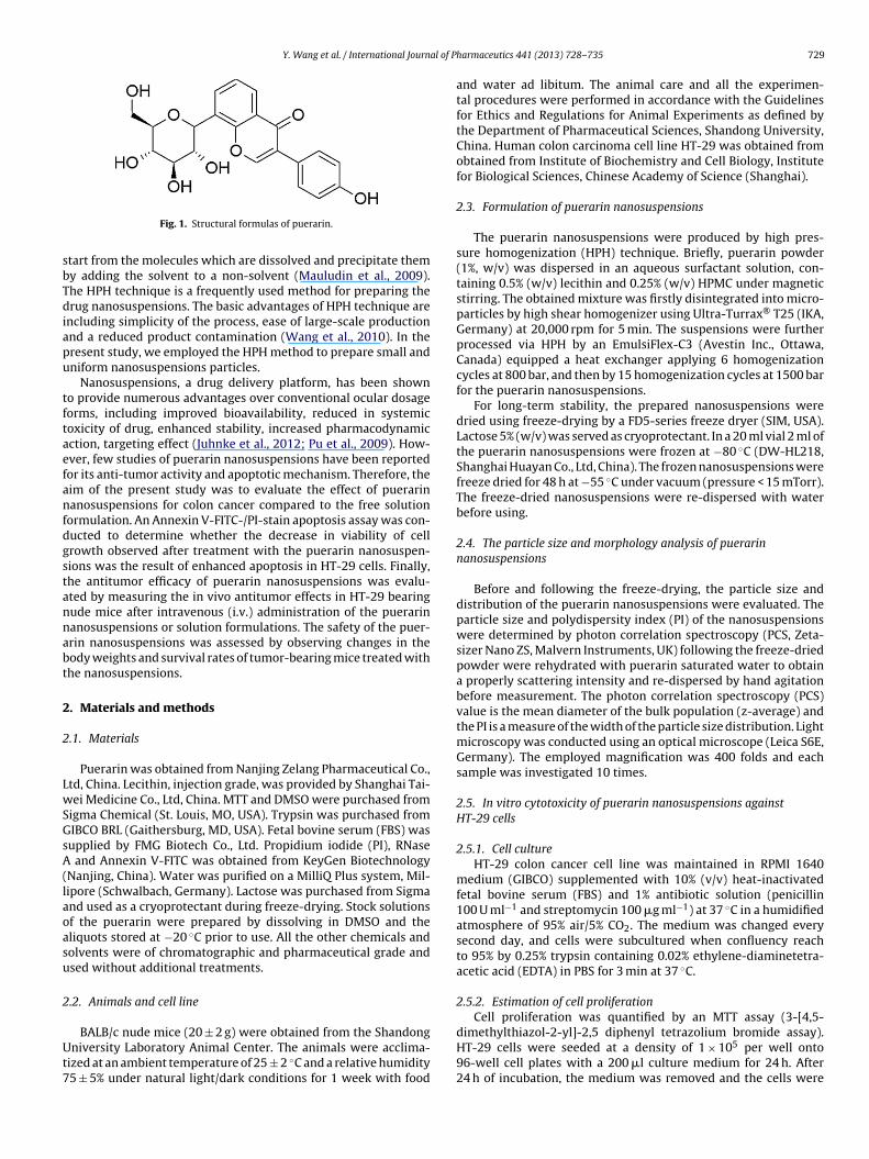
Y. Wang et al. / International Journal of P
sbTdiapu
tftaefanfdgstannabt
2
2
LwSGsA(laoasu
2
Ut7
Fig. 1. Structural formulas of puerarin.
tart from the molecules which are dissolved and precipitate themy adding the solvent to a non-solvent (Mauludin et al., 2009).he HPH technique is a frequently used method for preparing therug nanosuspensions. The basic advantages of HPH technique are
ncluding simplicity of the process, ease of large-scale productionnd a reduced product contamination (Wang et al., 2010). In theresent study, we employed the HPH method to prepare small andniform nanosuspensions particles.
Nanosuspensions, a drug delivery platform, has been showno provide numerous advantages over conventional ocular dosageorms, including improved bioavailability, reduced in systemicoxicity of drug, enhanced stability, increased pharmacodynamicction, targeting effect (Juhnke et al., 2012; Pu et al., 2009). How-ver, few studies of puerarin nanosuspensions have been reportedor its anti-tumor activity and apoptotic mechanism. Therefore, theim of the present study was to evaluate the effect of puerarinanosuspensions for colon cancer compared to the free solution
ormulation. An Annexin V-FITC-/PI-stain apoptosis assay was con-ucted to determine whether the decrease in viability of cellrowth observed after treatment with the puerarin nanosuspen-ions was the result of enhanced apoptosis in HT-29 cells. Finally,he antitumor efficacy of puerarin nanosuspensions was evalu-ted by measuring the in vivo antitumor effects in HT-29 bearingude mice after intravenous (i.v.) administration of the puerarinanosuspensions or solution formulations. The safety of the puer-rin nanosuspensions was assessed by observing changes in theody weights and survival rates of tumor-bearing mice treated withhe nanosuspensions.
. Materials and methods
.1. Materials
Puerarin was obtained from Nanjing Zelang Pharmaceutical Co.,td, China. Lecithin, injection grade, was provided by Shanghai Tai-ei Medicine Co., Ltd, China. MTT and DMSO were purchased from
igma Chemical (St. Louis, MO, USA). Trypsin was purchased fromIBCO BRL (Gaithersburg, MD, USA). Fetal bovine serum (FBS) wasupplied by FMG Biotech Co., Ltd. Propidium iodide (PI), RNase
and Annexin V-FITC was obtained from KeyGen BiotechnologyNanjing, China). Water was purified on a MilliQ Plus system, Mil-ipore (Schwalbach, Germany). Lactose was purchased from Sigmand used as a cryoprotectant during freeze-drying. Stock solutionsf the puerarin were prepared by dissolving in DMSO and theliquots stored at −20 ◦C prior to use. All the other chemicals andolvents were of chromatographic and pharmaceutical grade andsed without additional treatments.
.2. Animals and cell line
BALB/c nude mice (20 ± 2 g) were obtained from the Shandongniversity Laboratory Animal Center. The animals were acclima-
ized at an ambient temperature of 25 ± 2 ◦C and a relative humidity5 ± 5% under natural light/dark conditions for 1 week with food
harmaceutics 441 (2013) 728– 735 729
and water ad libitum. The animal care and all the experimen-tal procedures were performed in accordance with the Guidelinesfor Ethics and Regulations for Animal Experiments as defined bythe Department of Pharmaceutical Sciences, Shandong University,China. Human colon carcinoma cell line HT-29 was obtained fromobtained from Institute of Biochemistry and Cell Biology, Institutefor Biological Sciences, Chinese Academy of Science (Shanghai).
2.3. Formulation of puerarin nanosuspensions
The puerarin nanosuspensions were produced by high pres-sure homogenization (HPH) technique. Briefly, puerarin powder(1%, w/v) was dispersed in an aqueous surfactant solution, con-taining 0.5% (w/v) lecithin and 0.25% (w/v) HPMC under magneticstirring. The obtained mixture was firstly disintegrated into micro-particles by high shear homogenizer using Ultra-Turrax® T25 (IKA,Germany) at 20,000 rpm for 5 min. The suspensions were furtherprocessed via HPH by an EmulsiFlex-C3 (Avestin Inc., Ottawa,Canada) equipped a heat exchanger applying 6 homogenizationcycles at 800 bar, and then by 15 homogenization cycles at 1500 barfor the puerarin nanosuspensions.
For long-term stability, the prepared nanosuspensions weredried using freeze-drying by a FD5-series freeze dryer (SIM, USA).Lactose 5% (w/v) was served as cryoprotectant. In a 20 ml vial 2 ml ofthe puerarin nanosuspensions were frozen at −80 ◦C (DW-HL218,Shanghai Huayan Co., Ltd, China). The frozen nanosuspensions werefreeze dried for 48 h at −55 ◦C under vacuum (pressure < 15 mTorr).The freeze-dried nanosuspensions were re-dispersed with waterbefore using.
2.4. The particle size and morphology analysis of puerarinnanosuspensions
Before and following the freeze-drying, the particle size anddistribution of the puerarin nanosuspensions were evaluated. Theparticle size and polydispersity index (PI) of the nanosuspensionswere determined by photon correlation spectroscopy (PCS, Zeta-sizer Nano ZS, Malvern Instruments, UK) following the freeze-driedpowder were rehydrated with puerarin saturated water to obtaina properly scattering intensity and re-dispersed by hand agitationbefore measurement. The photon correlation spectroscopy (PCS)value is the mean diameter of the bulk population (z-average) andthe PI is a measure of the width of the particle size distribution. Lightmicroscopy was conducted using an optical microscope (Leica S6E,Germany). The employed magnification was 400 folds and eachsample was investigated 10 times.
2.5. In vitro cytotoxicity of puerarin nanosuspensions againstHT-29 cells
2.5.1. Cell cultureHT-29 colon cancer cell line was maintained in RPMI 1640
medium (GIBCO) supplemented with 10% (v/v) heat-inactivatedfetal bovine serum (FBS) and 1% antibiotic solution (penicillin100 U ml−1 and streptomycin 100 �g ml−1) at 37 ◦C in a humidifiedatmosphere of 95% air/5% CO2. The medium was changed everysecond day, and cells were subcultured when confluency reachto 95% by 0.25% trypsin containing 0.02% ethylene-diaminetetra-acetic acid (EDTA) in PBS for 3 min at 37 ◦C.
2.5.2. Estimation of cell proliferationCell proliferation was quantified by an MTT assay (3-[4,5-
dimethylthiazol-2-yl]-2,5 diphenyl tetrazolium bromide assay).HT-29 cells were seeded at a density of 1 × 10⁵ per well onto96-well cell plates with a 200 �l culture medium for 24 h. After24 h of incubation, the medium was removed and the cells were

7 al of P
t(Cc2tctowiccccli(ip
2
ci1At
Fn
30 Y. Wang et al. / International Journ
reated with 200 �l of medium containing various concentrations1.25, 2.5, 12.5 and 25 �g ml−1) of puerarin for the indicated times.ontrol cells were supplemented with 0.05% DMSO vehicle. Eachoncentration of puerarin was repeated in six wells. A total of0 �l of MTT (5 mg ml−1 in PBS) was added to each well at theime of incubation. After 4 h of incubation, the supernatant wasarefully removed and 150 �l of DMSO was added to each wello terminate the reaction. The plates were shaken for 10 min. Theptical density was measured by a microplate reader at a referenceavelength of 550 nm and a test wavelength of 490 nm. Growth
nhibition by puerarin nanosuspensions were calculated as per-entage of cell viability, taking the viability of the vehicle-treatedells as 100%. The IC50 value (in �g ml−1), which represents theoncentration of test substance that lowers MTT reduction by 50%ompared to the untreated control groups, was estimated frominear regression analysis of experimental data. The cell growthnhibit rations were calculated as follows: cell inhibitory ratio%) = (ODv − ODs)/ODv × 100% (Zhang et al., 2010). ODs and ODv
ndicated the optical density of the cell lines incubated with sam-les and vehicle control, respectively.
.5.3. Fluorescent morphological assayCharacteristic apoptotic morphological changes of the HT-29
ells were assessed by fluorescent microscopy using propidium
odide (PI) staining. Briefly, the cells were seeded at a density of× 106 cells in 6-well plates, and cultured for 24 h in RPMI-1640.fter culturing, the cells were treated with the indicated concen-
rations of puerarin solutions or the nanosuspensions formulations
ig. 2. The particle size distribution of puerarin nanosuspensions before (A1) and followianosuspensions before (A2) and following (B2) freeze-drying.
harmaceutics 441 (2013) 728– 735
for 24 h. The free puerarin solutions contained 0.1% (v/v) DMSO. Thecontrol group received drug-free medium with 0.05% (v/v) DMSO.After cells were washed with PBS 3 times, the collected cells werestained with PI for 20 min at 37 ◦C. The cells were examined with areverse fluorescence microscopy (Olympus Optical Co., Ltd, Japan).
2.5.4. Evaluation of apoptosisTo investigate the proportion of HT-29 cells undergoing apo-
ptosis, the Annexin V-FITC-/PI-stain experiments were conductedaccording to the manufacturer’s instructions (KeyGEN, Nanjing,China). HT-29 cells were exposed to different concentrations ofpuerarin free solution and puerarin nanosuspensions for 24 h andallowed to adhere. The control group received drug-free mediumwith 0.05% (v/v) DMSO. At the end of the treatment period, 3 × 10⁵
cells were collected by centrifugation, washed twice with cold PBSat 1000 rpm for 5 min and gently resuspended in 500 �l bindingbuffer. Then 5 �l Annexin V-FITC and 5 �l PI solution were addedand incubated with cells in the dark for 15 min according to manu-facturer’s instruction. At the end of incubation, cell apoptosis wasanalyzed on a FACScan flow cytometer (Becton Dickinson, USA).
2.6. In vivo anticancer activity of puerarin nanosuspensions inHT-29 bearing mice
In vivo anticancer activity was evaluated using HT-29 bear-
ing BALB/c nude mice. Fifty mice were inoculated with HT-29human colon cancer cells in the dorsal area (1 × 10⁷ cells/mouse).The mice were fed with regular diet and double-distilled water.Around 2–5 weeks after inoculation, the mice with carcinomasng (B2) the freeze-drying; the light microscopy images (magnification 400×) of the
al of Pharmaceutics 441 (2013) 728– 735 731
oillHn(wedtaaftnpbw
2
ps
3
3
bridvostn
OamhpauntmalftE
wnbspmmt
8 hours
**
0
20
40
60
80
100
1.25 2.5 12.5 25
Puerarin concentration (µg/mL)
% i
nh
ibit
ion
ra
te
.
Puerarin soluti on
Puerarin nanosuspensions
16 hours
**
*
**
0
20
40
60
80
100
1.25 2.5 12.5 25Puerarin concentration (µg/mL)
% i
nh
ibit
ion
ra
te
.
24 hours*
***
*
***
0
20
40
60
80
100
1.25 2.5 12.5 25
Puerarin concentration (µg/mL)
% i
nh
ibit
ion
ra
te.
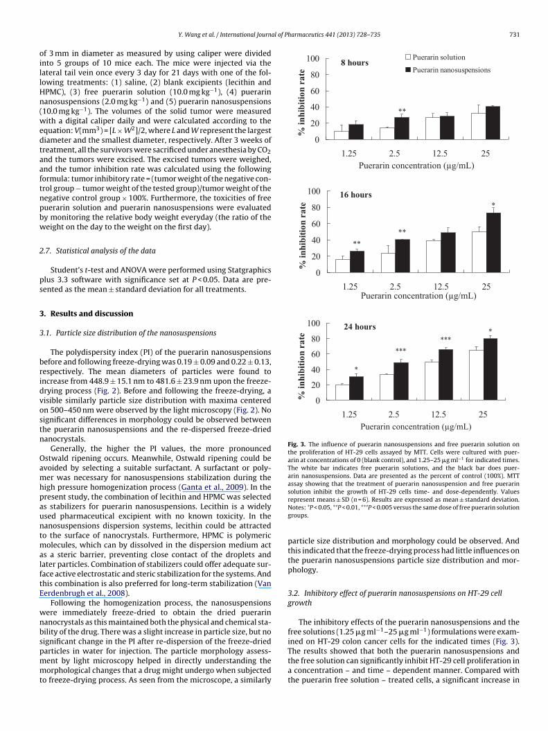
Fig. 3. The influence of puerarin nanosuspensions and free puerarin solution onthe proliferation of HT-29 cells assayed by MTT. Cells were cultured with puer-arin at concentrations of 0 (blank control), and 1.25–25 �g ml−1 for indicated times.The white bar indicates free puerarin solutions, and the black bar does puer-arin nanosuspensions. Data are presented as the percent of control (100%). MTTassay showing that the treatment of puerarin nanosuspension and free puerarinsolution inhibit the growth of HT-29 cells time- and dose-dependently. Valuesrepresent means ± SD (n = 6). Results are expressed as mean ± standard deviation.
Y. Wang et al. / International Journ
f 3 mm in diameter as measured by using caliper were dividednto 5 groups of 10 mice each. The mice were injected via theateral tail vein once every 3 day for 21 days with one of the fol-owing treatments: (1) saline, (2) blank excipients (lecithin andPMC), (3) free puerarin solution (10.0 mg kg−1), (4) puerarinanosuspensions (2.0 mg kg−1) and (5) puerarin nanosuspensions10.0 mg kg−1). The volumes of the solid tumor were measuredith a digital caliper daily and were calculated according to the
quation: V(mm3) = [L × W2]/2, where L and W represent the largestiameter and the smallest diameter, respectively. After 3 weeks ofreatment, all the survivors were sacrificed under anesthesia by CO2nd the tumors were excised. The excised tumors were weighed,nd the tumor inhibition rate was calculated using the followingormula: tumor inhibitory rate = (tumor weight of the negative con-rol group − tumor weight of the tested group)/tumor weight of theegative control group × 100%. Furthermore, the toxicities of freeuerarin solution and puerarin nanosuspensions were evaluatedy monitoring the relative body weight everyday (the ratio of theeight on the day to the weight on the first day).
.7. Statistical analysis of the data
Student’s t-test and ANOVA were performed using Statgraphicslus 3.3 software with significance set at P < 0.05. Data are pre-ented as the mean ± standard deviation for all treatments.
. Results and discussion
.1. Particle size distribution of the nanosuspensions
The polydispersity index (PI) of the puerarin nanosuspensionsefore and following freeze-drying was 0.19 ± 0.09 and 0.22 ± 0.13,espectively. The mean diameters of particles were found toncrease from 448.9 ± 15.1 nm to 481.6 ± 23.9 nm upon the freeze-rying process (Fig. 2). Before and following the freeze-drying, aisible similarly particle size distribution with maxima centeredn 500–450 nm were observed by the light microscopy (Fig. 2). Noignificant differences in morphology could be observed betweenhe puerarin nanosuspensions and the re-dispersed freeze-driedanocrystals.
Generally, the higher the PI values, the more pronouncedstwald ripening occurs. Meanwhile, Ostwald ripening could bevoided by selecting a suitable surfactant. A surfactant or poly-er was necessary for nanosuspensions stabilization during the
igh pressure homogenization process (Ganta et al., 2009). In theresent study, the combination of lecithin and HPMC was selecteds stabilizers for puerarin nanosuspensions. Lecithin is a widelysed pharmaceutical excipient with no known toxicity. In theanosuspensions dispersion systems, lecithin could be attractedo the surface of nanocrystals. Furthermore, HPMC is polymeric
olecules, which can by dissolved in the dispersion medium acts a steric barrier, preventing close contact of the droplets andater particles. Combination of stabilizers could offer adequate sur-ace active electrostatic and steric stabilization for the systems. Andhis combination is also preferred for long-term stabilization (Vanerdenbrugh et al., 2008).
Following the homogenization process, the nanosuspensionsere immediately freeze-dried to obtain the dried puerarinanocrystals as this maintained both the physical and chemical sta-ility of the drug. There was a slight increase in particle size, but noignificant change in the PI after re-dispersion of the freeze-dried
articles in water for injection. The particle morphology assess-ent by light microscopy helped in directly understanding theorphological changes that a drug might undergo when subjectedo freeze-drying process. As seen from the microscope, a similarly
Notes: *P < 0.05, **P < 0.01, ***P < 0.005 versus the same dose of free puerarin solutiongroups.
particle size distribution and morphology could be observed. Andthis indicated that the freeze-drying process had little influences onthe puerarin nanosuspensions particle size distribution and mor-phology.
3.2. Inhibitory effect of puerarin nanosuspensions on HT-29 cellgrowth
The inhibitory effects of the puerarin nanosuspensions and thefree solutions (1.25 �g ml−1–25 �g ml−1) formulations were exam-ined on HT-29 colon cancer cells for the indicated times (Fig. 3).
The results showed that both the puerarin nanosuspensions andthe free solution can significantly inhibit HT-29 cell proliferation ina concentration – and time – dependent manner. Compared withthe puerarin free solution – treated cells, a significant increase in
732 Y. Wang et al. / International Journal of Pharmaceutics 441 (2013) 728– 735
F T-29
f l−1; (D
i–dnt26mmp2goe
letitatefit
3H
tctcMtwwt
ig. 4. Puerarin nanosuspensions induced morphology changes in HT-29 cells: Hormulations for 24 h. (A) Control; (B) solution 2.50 �g ml−1; (C) solution 12.5 �g m
nhibition rates was displayed in the puerarin nanosuspensionstreated cells under the same concentrations. Furthermore, the
ose required for 50% growth inhibition (IC50 values) of puerarinanosuspensions were apparently lower than that of free solu-ion formulations after the same incubation time, accounting for9.37 �g ml−1–38.84 �g ml−1, 11.62 �g ml−1–16.56 �g ml−1, and.67 �g ml−1–10.17 �g ml−1 after 8 h, 16 h, and 24 h of treat-ent, respectively. The IC50 values of the nanosuspensions wereuch lower than that of the solutions formulations at each time
oint. When the incubation time extended to 24 h, exposure to.5 �g ml−1 and 12.5 �g ml−1 puerarin nanosuspensions inhibitedrowth by 50.5% and 65.9%, respectively. Therefore, we used dosagef 2.5 �g ml−1 and 12.5 �g ml−1 and incubation of 24 h for furtherxperiments.
Previous reports demonstrated that puerarin inhibited cell pro-iferation and induced apoptosis in several cancer cell lines (Hient al., 2010; Peng et al., 2001; Yu and Li, 2006). In the present study,he puerarin nanosuspensions showed significantly higher in vitronhibitory effects compared to the free solution at the same concen-ration. The inhibitory effects were increased with concentrationsnd incubation times. These results indicated that compared withhe free puerarin solution, the puerarin nanosuspensions had annhanced efficacy. The ability of live cells reducing the MTT intoormazan crystal which could be dissolved in DMSO and resultednto a purple solution is the mechanism of the present MTT cyto-oxicity assay (Wang et al., 2006).
.3. Puerarin nanosuspensions induced morphologic changes inT-29 cells
To test whether the decrease in cell viability observed afterreatment with the samples was due to apoptosis, the treated andontrol cells were stained with propidium iodide (PI) after exposureo the various concentrations of the formulations for 24 h. And theell morphology was examined under a fluorescent microscope.icroscopic analysis confirmed that puerarin nanosuspensions
reatment induced cell apoptosis (Fig. 4). The control group cellsere only a thimbleful of cells dyed and normal morphologiesere observed. After incubation with puerarin nanosuspensions,
he cellular morphology of HT-29 cells was severely distorted.
cells were incubated with puerarin free solution and puerarin nanosuspensions) nanosuspensions 2.5 �g ml−1; (E) nanosuspensions 12.5 �g ml−1.
The majority cells of the nanosuspensions groups displayed moreobvious changes in cell morphology including cell shrinkage, cellfragmentation, and cell irregularity, compared with the solutiongroups at the same dose. Furthermore, the irregular fragmentednuclei and irregular cytoplasmic membranes could be observedin the puerarin nanosuspensions treated cells. Results of the PIstaining demonstrated that puerarin nanosuspensions can induceapoptosis of HT-29 cells. The observed morphological changes ofthe nanosuspensions formulation treated cells suggested that thepuerarin-induced HT-29 cell proliferation inhibition was involvedin a mechanism of apoptosis (Hostanska et al., 2003; Lee et al.,2012).
3.4. Assessment of puerarin nanosuspensions-induced apoptosisin HT-29 cells
To compare the degree of apoptosis induced by the puerarinnanosuspensions and solution, Annexin V-FITC and PI double stain-ing was employed to distinguish between apoptotic and necroticcells. As exhibited in Fig. 5, the experiment results revealed thatafter treatment with puerarin free solution at different concentra-tions for 24 h, the early apoptosis rate of TH-29 cells increased withpuerarin concentration, reaching 0.0%, 7.9%, and 9.6%, at concen-trations of 0 �g ml−1, 2.5 �g ml−1, and 12.5 �g ml−1, respectively.Therefore, puerarin free solution treatment induces apoptosisof TH-29 cells in a concentration-dependent manner. To inves-tigate the effect of nanosuspensions on inducing apoptosis ofTH-29 cells, the cells were treated with puerarin nanosuspensionsat the same concentration and experimental conditions. It wasfound that puerarin nanosuspensions-treated TH-29 cells revealedthe expected increase in the early apoptosis rate, reaching 0.0%,40.4%, and 46.7%, at concentrations of 0 �g ml−1, 2.5 �g ml−1, and12.5 �g ml−1, respectively. Compared with puerarin free solution,the puerarin nanosuspensions formulation displayed a higher earlyapoptosis inducing rate. These results demonstrated that puer-arin free solution could induce early apoptosis, and the puerarin
nanosuspensions formulations possessed a more prominent apo-ptosis inducing rate on TH-29 cells in a concentration-dependentmanner. Thus, the puerarin nanosuspensions could induce apopto-sis leading to the subsequent inhibition of cellular proliferation.
Y. Wang et al. / International Journal of Pharmaceutics 441 (2013) 728– 735 733
F ent w2 suspenc in V-s
riscai
mmaCotenpftm
3
am
TT
ig. 5. Induction of nanosuspensions on early apoptosis of HT-29 cells after treatm.5 �g ml−1; (C) solution 12.5 �g ml−1; (D) nanosuspensions 2.5 �g ml−1; (E) nanoells (early-phase apoptotic cells). The top right quadrant represents PI- and Annex
Compared to the cell necrosis, apoptosis is a form of self-egulated cell death. In response to several agents includingonizing radiation or anticancer chemotherapeutic drugs, apopto-is is an important mode of cell death and is an active process ofell destruction (Sun et al., 2012). And for many anticancer agents,poptosis is also considered as an important mechanism involvedn the inhibition of cancer cell proliferation (Zou et al., 2012).
Apoptosis, characterized initially by a series of stereotypicorphological changes, such as chromatin condensation, DNA frag-entation, and cell shrinkage, is a major form of cell death and
highly complex cascade of cellular events (Mogil et al., 1994).ompared with the solution formulations, the reduced viabilityf the cells in the presence of the nanosuspensions whether dueo apoptosis induction is our mainly consideration (Winkelmannt al., 2008). Although the present date revealed that puerarinanosuspensions induced growth inhibition and loss of membranehospholipid asymmetry, with translocation of phosphatidylserinerom the inner leaflet of the phospholipid bilayer to the cell surface,he mechanisms underlying the puerarin formulations triggered
orphological characteristics of apoptosis are not clearly defined.
.5. In vivo anticancer effects in HT-29 bearing mice
The in vivo anticancer effect of the puerarin nanosuspensionsnd the puerarin free solutions were assessed in HT-29 bearingice. The tumor weight, tumor inhibition rate and tumor volume of
able 1he in vivo antitumor effects in HT-29 bearing mice (n = 10).
Group Tumor weight (g)
Saline 1.92 ± 0.14
Blank excipients 1.86 ± 0.18
Puerarin nanosuspensions at 2 mg kg−1 1.27 ± 0.22
Puerarin nanosuspensions at 10 mg kg−1 0.82 ± 0.19
Puerarin solution at 10 mg kg−1 1.40 ± 0.21
a P value in the t-test denoting statistical significance vs. saline group.
ith free puerarin and puerarin nanosuspension for 24 h. (A) Control; (B) solutionsions 12.5 �g ml−1; the right bottom quadrant represents the Annexin V-stained
tained cells (late-phase apoptotic/necrotic cells).
all the tested groups were calculated. Table 1 lists the tumor inhibi-tion rates of all the tested groups. The in vivo anticancer study onceagain indicated that compared with the free puerarin solution, thenanosuspensions formulation exhibited a notably enhanced anti-cancer efficacy. No significant difference was observed betweenmice treated with saline and blank excipients mice. Fig. 6 showedthat the relative body weight and the tumor volume in the tumor-bearing nude mice were obvious different among the variousexperiment groups. Both saline and blank excipients showed sim-ilar anticancer activities within 3 weeks. Neither saline nor blankexcipients had any measurable influences on tumor growth, in bothgroups the tumor volumes and weights increased rapidly. With amarked reduction of the tumor volume and tumor weight, eitherpuerarin solution or puerarin nanosuspensions groups exhibitedsignificantly enhanced tumor inhibitions compared with the salineor blank excipients groups. Furthermore, the tumor inhibition rateof the puerarin nanosuspensions was significantly higher thanthat of the puerarin free solution. The increases of body weightsin saline-treated mice and blank excipients-treated mice werebecause of the remarkable increases in tumor sizes, whereas thedecreases in body weights in the free puerarin solution groupresulted from the toxicity of the formulation (Kim et al., 2008a).
After 3 weeks’ puerarin free solutions treatment, one out of the tenmice had not survived. After the high- or low-dose treatments, allten mice in each nanosuspensions group survived. A lower survivalrate means that tumor-bearing mice were given a more toxic drugInhibition rate (%) Pa
N.A. N.A.2.78 N.A.33.57 P < 0.0557.04 P < 0.0126.78 P < 0.05

734 Y. Wang et al. / International Journal of P
Fig. 6. Body weight changes and in vivo tumor volumes after treated with saline,blank excipients, nanosuspensions and solution. The symbols are as follows:s1(
fanw
ncimtsssvap
nis2ttraitoT
aline (♦); blank excipients (©); nanosuspensions 2 mg kg−1 (�); nanosuspensions0 mg kg−1 (�); solutions 10 mg kg−1 (�). The results represent the means ± SDn = 10). *P < 0.05 and **P < 0.01.
ormulation. Deactivation and athrepsy were observed in the puer-rin free solution treated mice. By naked eye, mice treated with theanosuspensions remained vigorous, had a healthy appearance andere normal groomed throughout the entire experiment.
In this study, we attempted to demonstrate that puerarinanosuspensions had more effective apoptotic effects in HT-29ells. In the present study, while puerarin free solution treatmentsnhibit rapid growth, it did not stop the growth of HT-29 tumor
asses. Furthermore, the in vivo evaluation experiments indicatedhat the anticancer efficacy of the puerarin nanosuspensions wasignificantly higher than that of the puerarin free solution at theame dosage. As the present study was performed on a specifictrain of mice using a HT-29 colon cancer cell line, it should beery cautious about applying these data to human clinical studieslthough the results point to be more effective treatment by theuerarin nanosuspensions formulations (Wu et al., 2011).
The nanoparticulate drug delivery systems, including theanosuspensions, could increase anti-tumor efficacy while reduc-
ng systemic side effects had been extensively discussed in previoustudies (Kim et al., 2008a; Mattheolabakis et al., 2009; Zhang et al.,007). In the tumor treatment, the biggest clinical benefits ofhe nanosuspensions formulations were for passive targeting intoumors via a phenomenon termed as enhanced permeation andetention (EPR) effect. Due to the leaky nature of the tumor vascul-ture, circulating nanosuspensions particle diffuse preferentially
nto tumor tissues. And the extent of tumor targeting depends onhe circulation time of the nanosuspensions, which in turn dependsn the particle size and density of coating (Wong et al., 2008).he range of the tumor vascular pore size is 300 nm to 700 nm,harmaceutics 441 (2013) 728– 735
depending upon the tumor type. Therefore, there was an upperlimit placed upon the size of the particle, permitting diffusionthrough the vascular tumor pores (Fang et al., 2011; Torchilin,2011). In the present study, the main particle size of puerarinnanosuspensions was below 500 nm. Furthermore, the nanosus-pensions formulation also indicated reduced toxicity compared tothe free solution.
4. Conclusions
A newly developed nanosuspensions of puerarin was success-fully formulated and the in vitro/in vivo anticancer activities wereevaluated in the present study. The nanosuspensions formulation,effectively produced by the high pressure homogenization tech-nique, exhibited an enhanced antiproliferative effect by reducingcell viability and inducing morphological changes. The puerarinnanosuspensions exerted HT-29 human colon cancer cell growthinhibition by the induction of early apoptosis. In vivo, the puerarinnanosuspensions were better tolerated and induced significantlyhigher anticancer efficacy than the puerarin free solution. Insummary, the puerarin nanosuspensions suppressed the HT-29proliferation, increased the early apoptosis, and enhanced thein vivo anticancer effectives of human colon cancer HT-29 cells.These data indicated that the puerarin nanosuspensions could bean effective chemotherapeutic agent in colon cancer treatment.
Acknowledgments
This work was supported by Outstanding Young ScientistResearch Award Fund of Shandong Province (No. BS2012YY023)and the National Basic Research Program of China (973Program),No. 2009CB930300.
References
Budden, R., Kuhl, U.G., Bahlsen, J., 1979. Experiments on the toxic, sedative andmuscle relaxant potency of various drug solvents in mice. Pharmacol. Ther. B5, 467–474.
Fang, J., Nakamura, H., Maeda, H., 2011. The EPR effect: unique features of tumorblood vessels for drug delivery, factors involved, and limitations and augmen-tation of the effect. Adv. Drug Deliv. Rev. 63, 136–151.
Ganta, S., Paxton, J.W., Baguley, B.C., Garg, S., 2009. Formulation and pharmacokineticevaluation of an asulacrine nanocrystalline suspension for intravenous delivery.Int. J. Pharm. 367, 179–186.
Go, H., Hwang, H.J., Nam, T.J., 2010. A glycoprotein from Laminaria japonica inducesapoptosis in HT-29 colon cancer cells. Toxicol. In Vitro 24, 1546–1553.
Gu, L., Yang, Y., Sun, Y., Zheng, X., 2010. Puerarin inhibits acid-sensing ion channelsand protects against neuron death induced by acidosis. Planta Med. 76, 583–588.
Hien, T.T., Kim, H.G., Han, E.H., Kang, K.W., Jeong, H.G., 2010. Molecular mecha-nism of suppression of MDR1 by puerarin from Pueraria lobata via NF-kappaBpathway and cAMP-responsive element transcriptional activity-dependent up-regulation of AMP-activated protein kinase in breast cancer MCF-7/adr cells.Mol. Nutr. Food Res. 54, 918–928.
Hostanska, K., Reichling, J., Bommer, S., Weber, M., Saller, R., 2003. Hyperforin aconstituent of St John’s wort (Hypericum perforatum L.) extract induces apoptosisby triggering activation of caspases and with hypericin synergistically exertscytotoxicity towards human malignant cell lines. Eur. J. Pharm. Biopharm. 56,121–132.
Hu, W., Lee, S.K., Jung, M.J., Heo, S.I., Hur, J.H., Wang, M.H., 2010a. Induction of cellcycle arrest and apoptosis by the ethyl acetate fraction of Kalopanax pictus leavesin human colon cancer cells. Bioresour. Technol. 101, 9366–9372.
Hu, W., Zhang, Q., Yang, X., Wang, Y., Sun, L., 2010b. Puerarin inhibits adhesionmolecule expression in tnf-alpha-stimulated human endothelial cells via mod-ulation of the nuclear factor kappaB pathway. Pharmacology 85, 27–35.
Juhnke, M., Martin, D., John, E., 2012. Generation of wear during the productionof drug nanosuspensions by wet media milling. Eur. J. Pharm. Biopharm. 81,214–222.
Kim, J.H., Kim, Y.S., Park, K., Lee, S., Nam, H.Y., Min, K.H., Jo, H.G., Park, J.H., Choi, K.,Jeong, S.Y., Park, R.W., Kim, I.S., Kim, K., Kwon, I.C., 2008a. Antitumor efficacy of
cisplatin-loaded glycol chitosan nanoparticles in tumor-bearing mice. J. Control.Release 127, 41–49.Kim, Y.S., Kim, J.J., Cho, K.H., Jung, W.S., Moon, S.K., Park, E.K., Kim, D.H., 2008b.Biotransformation of ginsenoside Rb1, crocin, amygdalin, geniposide, puerarin,ginsenoside Re, hesperidin, poncirin, glycyrrhizin, and baicalin by human fecal
al of P
L
L
L
M
M
M
M
M
P
P
R
R
S
S
T
T
V
Y. Wang et al. / International Journ
microflora and its relation to cytotoxicity against tumor cells. J. Microbiol.Biotechnol. 18, 1109–1114.
ee, J.S., Jung, W.K., Jeong, M.H., Yoon, T.R., Kim, H.K., 2012. Sanguinarine inducesapoptosis of HT-29 human colon cancer cells via the regulation of Bax/Bcl-2 ratioand caspase-9-dependent pathway. Int. J. Toxicol. 31, 70–77.
iu, C.M., Ma, J.Q., Sun, Y.Z., 2010a. Puerarin protects the rat liver against oxida-tive stress-mediated DNA damage and apoptosis induced by lead. Exp. Toxicol.Pathol., 016, http://dx.doi.org/10.1016/j.etp.2010.11.
iu, X.J., Zhao, J., Gu, X.Y., 2010b. The effects of genistein and puerarin on the activa-tion of nuclear factor-kappaB and the production of tumor necrosis factor-alphain asthma patients. Pharmazie 65, 127–131.
attheolabakis, G., Taoufik, E., Haralambous, S., Roberts, M.L., Avgoustakis, K., 2009.In vivo investigation of tolerance and antitumor activity of cisplatin-loadedPLGA-mPEG nanoparticles. Eur. J. Pharm. Biopharm. 71, 190–195.
auludin, R., Muller, R.H., Keck, C.M., 2009. Development of an oral rutin nanocrystalformulation. Int. J. Pharm. 370, 202–209.
eng, X.H., Ni, C., Zhu, L., Shen, Y.L., Wang, L.L., Chen, Y.Y., 2009. Puerarin pro-tects against high glucose-induced acute vascular dysfunction: role of hemeoxygenase-1 in rat thoracic aorta. Vascul. Pharmacol. 50, 110–115.
ogil, R.J., Shi, Y., Bissonnette, R.P., Bromley, P., Yamaguchi, I., Green, D.R., 1994. Roleof DNA fragmentation in T cell activation-induced apoptosis in vitro and in vivo.J. Immunol. 152, 1674–1683.
orshed, K.M., Nagpaul, J.P., Majumdar, S., Amma, M.K., 1988. Kinetics of propyleneglycol elimination and metabolism in rat. Biochem. Med. Metab. Biol. 39, 90–97.
eng, X.Y., Qi, Z.H., Chen, H.P., 2001. Study on the differentiation and apoptosis ofHL-60 cell line induced by puerarin. Hunan Yi Ke Da Xue Xue Bao 26, 126–128.
u, X., Sun, J., Wang, Y., Liu, X., Zhang, P., Tang, X., Pan, W., Han, J., He, Z., 2009.Development of a chemically stable 10-hydroxycamptothecin nanosuspensions.Int. J. Pharm. 379, 167–173.
abinow, B.E., 2004. Nanosuspensions in drug delivery. Nat. Rev. Drug Discov. 3,785–796.
uddick, J.A., 1972. Toxicology, metabolism, and biochemistry of 1,2-propanediol.Toxicol. Appl. Pharmacol. 21, 102–111.
heng, X., Sun, Y., Yin, Y., Chen, T., Xu, Q., 2008. Cirsilineol inhibits proliferation ofcancer cells by inducing apoptosis via mitochondrial pathway. J. Pharm. Pharma-col. 60, 1523–1529.
un, L.Q., Xue, B., Li, X.J., Wang, X., Qu, L., Zhang, T.T., Zhao, J., Wang, B.A., Zou, X.M.,Mu, Y.M., Lu, J.M., 2012. Inhibitory effects of salvianolic acid B on apoptosis ofSchwann cells and its mechanism induced by intermittent high glucose. Life Sci.90, 99–108.
ian, F., Xu, L.H., Zhao, W., Tian, L.J., Ji, X.L., 2011. The optimal therapeutic timing andmechanism of puerarin treatment of spinal cord ischemia-reperfusion injury inrats. J. Ethnopharmacol. 134, 892–896.
orchilin, V., 2011. Tumor delivery of macromolecular drugs based on the EPR effect.Adv. Drug Deliv. Rev. 63, 131–135.
an Eerdenbrugh, B., Van den Mooter, G., Augustijns, P., 2008. Top-down produc-tion of drug nanocrystals: nanosuspension stabilization, miniaturization andtransformation into solid products. Int. J. Pharm. 364, 64–75.
harmaceutics 441 (2013) 728– 735 735
Wang, X., Ge, J., Wang, K., Qian, J., Zou, Y., 2006. Evaluation of MTT assay formeasurement of emodin-induced cytotoxicity. Assay Drug Dev. Technol. 4,203–207.
Wang, Y., Liu, Z., Zhang, D., Gao, X., Zhang, X., Duan, C., Jia, L., Feng, F.,Huang, Y., Shen, Y., Zhang, Q., 2011. Development and in vitro evaluation ofdeacety mycoepoxydiene nanosuspension. Colloids Surf. B: Biointerfaces 83,189–197.
Wang, Y., Zhang, D., Liu, Z., Liu, G., Duan, C., Jia, L., Feng, F., Zhang, X., Shi, Y., Zhang,Q., 2010. In vitro and in vivo evaluation of silybin nanosuspensions for oral andintravenous delivery. Nanotechnology 21, 155104.
Winkelmann, I., Nassl, A.M., Daniel, H., Wenzel, U., 2008. Proteome response in HT-29 human colorectal cancer cells to two apoptosis-inducing compounds withdifferent mode of action. Int. J. Cancer 122, 2223–2232.
Wong, J., Brugger, A., Khare, A., Chaubal, M., Papadopoulos, P., Rabinow, B., Kipp, J.,Ning, J., 2008. Suspensions for intravenous (IV) injection: a review of develop-ment, preclinical and clinical aspects. Adv. Drug Deliv. Rev. 60, 939–954.
Wu, L., Qiao, H., Li, Y., Li, L., 2007. Cardioprotective effects of the combined use ofpuerarin and Danshensu on acute ischemic myocardial injury in rats. Phytother.Res. 21, 751–756.
Wu, M.F., Chen, Y.L., Lee, M.H., Shih, Y.L., Hsu, Y.M., Tang, M.C., Lu, H.F., Tang,N.Y., Yang, S.T., Chueh, F.S., Chung, J.G., 2011. Effect of Agaricus blazei Murrillextract on HT-29 human colon cancer cells in SCID mice in vivo. In Vivo 25,673–677.
Xiao, C., Li, J., Dong, X., He, X., Niu, X., Liu, C., Zhong, G., Bauer, R., Yang, D., Lu, A.,2011. Anti-oxidative and TNF-alpha suppressive activities of puerarin derivative(4AC) in RAW264.7 cells and collagen-induced arthritic rats. Eur. J. Pharmacol.666, 242–250.
Yu, Z., Li, W., 2006. Induction of apoptosis by puerarin in colon cancer HT-29 cells.Cancer Lett. 238, 53–60.
Yu, Z., Li, W., Liu, F., 2004. Inhibition of proliferation and induction of apoptosis bygenistein in colon cancer HT-29 cells. Cancer Lett. 215, 159–166.
Zhang, L., Yang, M., Wang, Q., Li, Y., Guo, R., Jiang, X., Yang, C., Liu, B., 2007.10-Hydroxycamptothecin loaded nanoparticles: preparation and antitumoractivity in mice. J. Control. Release 119, 153–162.
Zhang, S.Y., Chen, G., Wei, P.F., Huang, X.S., Dai, Y., Shen, Y.J., Chen, S.L., Sun-Chi,C.A., Xu, H.X., 2008. The effect of puerarin on serum nitric oxide concentrationand myocardial eNOS expression in rats with myocardial infarction. J. Asian Nat.Prod. Res. 10, 373–381.
Zhang, Z., Zhang, X., Xue, W., Yangyang, Y., Xu, D., Zhao, Y., Lou, H., 2010. Effects oforidonin nanosuspension on cell proliferation and apoptosis of human prostaticcarcinoma PC-3 cell line. Int. J. Nanomed. 5, 735–742.
Zhu, J., Wang, X., Shang, Y., Xie, X., Zhang, F., Chen, J., Fu, G., 2008. Puerarin reducesendothelial progenitor cells senescence through augmentation of telomerase
activity. Vascul. Pharmacol. 49, 106–110.Zou, T., Percival, S.S., Cheng, Q., Li, Z., Rowe, C., Gu, L., 2012. Prepa-ration, characterization, and induction of cell apoptosis of cocoaprocyanidins-gelatin-chitosan nanoparticles. Eur. J. Pharm. Biopharm.,http://dx.doi.org/10.1016/j.ejpb.2012.05.006.