Introduction of pneumococcal conjugate vaccination in Ethiopia
Upload
khangminh22Category
view
0download
0
Citation: Hamdy, M.S.;
Elbehairi, S.E.I.; Shati, A.A.;
Abd-Rabboh, H.S.M.; Alfaifi, M.Y.;
Fawy, K.F.; Ibrahium, H.A.;
Alamri, S.; Awwad, N.S. Cytotoxic
Potential of Bio-Silica Conjugate with
Different Sizes of Silver
Nanoparticles for Cancer Cell Death.
Materials 2022, 15, 4074. https://
doi.org/10.3390/ma15124074
Academic Editor: Xiaohong Wang
Received: 7 April 2022
Accepted: 2 June 2022
Published: 8 June 2022
Publisher’s Note: MDPI stays neutral
with regard to jurisdictional claims in
published maps and institutional affil-
iations.
Copyright: © 2022 by the authors.
Licensee MDPI, Basel, Switzerland.
This article is an open access article
distributed under the terms and
conditions of the Creative Commons
Attribution (CC BY) license (https://
creativecommons.org/licenses/by/
4.0/).
materials
Article
Cytotoxic Potential of Bio-Silica Conjugate with Different Sizesof Silver Nanoparticles for Cancer Cell DeathMohamed S. Hamdy 1 , Serag Eldin I. Elbehairi 2 , Ali A. Shati 2, Hisham S. M. Abd-Rabboh 1 ,Mohammad Y. Alfaifi 2 , Khaled F. Fawy 1, Hala A. Ibrahium 2, Saad Alamri 2 and Nasser S. Awwad 1,*
1 Department of Chemistry, Faculty of Science, King Khalid University, P.O. Box 9004,Abha 61413, Saudi Arabia; [email protected] (M.S.H.); [email protected] (H.S.M.A.-R.);[email protected] (K.F.F.)
2 Department of Biology, Faculty of Science, King Khalid University, P.O. Box 9004, Abha 61413, Saudi Arabia;[email protected] (S.E.I.E.); [email protected] (A.A.S.); [email protected] (M.Y.A.);[email protected] (H.A.I.); [email protected] (S.A.)
* Correspondence: [email protected]
Abstract: Well-defined silver nanoparticles were doped into bio-based amorphous silica (Ag-b-SiO2)with different silver contents (from 2 to 20 wt%) by a solvent-free procedure. The four as-synthetizedsamples were hydrogenated at 300 ◦C to ensure the formation of zero-valent Ag nanoparticles. Theprepared samples were characterized by X-ray powder diffraction (XRD), elemental analysis, N2
sorption measurements, scanning electron microscopy (SEM), X-ray photoelectron spectroscopy(XPS), and high-resolution transmission electron microscopy (HR-TEM). The characterization dataconfirmed the formation of well-defined zero-valent silver nanoparticles in the range of 3–10 nmin the low-loading samples, while in high-loading samples, bulky particles of silver in the rangeof 200–500 nm were formed. The in vitro cytotoxic activities of the Ag-b-SiO2 samples were testedagainst the tumor cell lines of breast (MCF-7), liver (HepG2), and colon (HCT 116) over a concentrationrange of 0.01 to 1000 g. The prepared samples exhibited a wide range of cytotoxic activities againstcancer cells. An inverse relationship was observed between the silver nanoparticles’ size and thecytotoxic activity, while a direct relationship between the silver nanoparticles’ size and the apoptoticcell death was noticed.
Keywords: silver nanoparticles; bio-based silica; cytotoxicity; apoptosis; necrosis; anti-cancer
1. Introduction
Despite the significant efforts of scientists to control cancer, there is a global increasein cancer patients around the world [1]. Moreover, the World Health Organization (WHO)indicated cancer as a main reason for death around the world, almost 10 M deaths in 2020caused by different types of cancer [2]. Therefore, the creation and/or development ofunusual treatments for cancer is crucial. The pioneering work of El-Sayed [3,4] indicated thehigh potential of metallic nanoparticles, e.g., gold and silver, in fighting cancer cells. Manyarticles have reported the toxic behavior of Au [5,6] and Ag nanoparticles [7,8] againstcells of various cancer types. Silver nanoparticles were applied either as solo metallicparticles or supported zero-valent nanoparticles. Several methods have been reported toprepare the solo zero-valent nanoparticles such as chemical [9], physical [10], and evenbiological [11] synthesis.
However, there is a concern about the stability of the presence of silver nanoparticlesin a solution for a relatively long time. Silver nanoparticles can be agglomerated [12]and hence lose their activity as a toxic agent. The stability of the nanoparticles can beimproved when located on a suitable support such as silica [13], alumina [14], zeolite [15],and, recently, on graphene oxide [16].
Materials 2022, 15, 4074. https://doi.org/10.3390/ma15124074 https://www.mdpi.com/journal/materials

Materials 2022, 15, 4074 2 of 12
Recently, the current research team reported the extraction and purification of amor-phous silica from Hassawi rice husk/straw [17] through few simple treatment steps(Figure 1); silicate ions could be formed, which could be utilized to produce amorphousspheres of mesoporous silica [17]. The extracted bio-based silica was used to supportsilver nanoparticles in one-step synthesis by using a solution of silver nitrate without theuse of any organic solvents. The motivation behind the current study is to introduce acost-effective material that can be applied as anti-cancer materials. The synthesis and thecharacterization of the silver nanoparticles supported on amorphous bio-based silica arereported; moreover, the anti-cancer activity [18–23] of the prepared material against severalcancer cells is discussed.
Figure 1. The valorization of Hassawi rice husk/straw into different products [17]. In the currentwork, the research team utilizes the bio-based silica to prepare anti-cancer compounds.
2. Results2.1. The Characterization of Ag-Doped Amorphous Silica
XRD analysis was applied to study the crystallinity of the prepared samples, and theobtained patterns are plotted in Figure 2. In the entire samples, a very broad band wasobserved near 23◦ 2θ as an indication for the amorphous nature of the silica support [23].Moreover, four beaks were located at the locations of 38.4◦, 44.7◦, 64.5◦, and 78.0◦ 2θ, whichcorrespond to the FCC (the face-centered cubic) planes of (111), (200), (220), and (331),which can be attributed to the presence of zero-valent silver nanoparticles according to theJCPDS database card no# 04-0783 [24]. Moreover, the peak intensity of silver nanoparticlesincreased with the loading, which is consistent with increasing silver content in the samples.
Figure 2. XRD patterns of the prepared silver nanoparticles supported on bio-based silica.

Materials 2022, 15, 4074 3 of 12
ICP analysis was applied to investigate the exact amount of silver in each sample;on the other hand, N2 sorption analysis was used to study the texture properties of theprepared samples. The obtained results are listed in Table 1.
Table 1. The exact amount of silver as obtained from ICP-OE and the textural properties of theprepared as obtained from N2 sorption measurements.
Sample Ag IntendedContent
Ag ObtainedContent
Surface Aream2/g
Pore Volumecm3/g
Pore Sizenm
Ag-0 0 0 580.1 0.425 4.83Ag-2 2 1.45 620.2 0.414 4.31Ag-5 5 3.86 611.3 0.398 4.35
Ag-10 10 7.65 629.6 0.365 3.86Ag-20 20 13.58 637.4 0.325 3.62
The elemental analysis of the prepared samples showed that almost 70–78% of thesilver amount added to the synthesis mixture were obtained in the final solid sample.Moreover, the texture properties showed that the surface area of the prepared samples wasfound to be increased from 580 m2/g in the bare bio-based silica sample to 620–637 m2/g asan indication for the effect of silver nanoparticles on the integrity of the prepared samples.Moreover, the pore volume and pore diameter were found to be decreased due to thepresence of silver nanoparticles in the pores of the mesoporous bio-based silica.
The morphology of the prepared samples’ surface was observed by the SEM technique,and the micrographs of the prepared samples are presented in Figure 3. The micrographs ofthe small loading samples (i.e., Ag-2 and -5) showed only the clean surface of the bio-basedamorphous silica. More importantly, no other phase(s) was/were detected. However, themicrographs of the high-loading samples (Figure 3, Ag-10 and -20) showed some extraframework silver nanoparticles.
The size and distribution of silver nanoparticles were investigated through the HR-TEM technique, and the obtained micrographs are presented in Figure 4. The micrographsclearly showed the difference in the location and the size of silver nanoparticles. In the Ag-2sample, the majority of silver nanoparticles were embedded and well-distributed in thesilica support; moreover, the size of the nanoparticles ranged between 3 and 10 nm with agood distribution within the silica support. However, in the Ag-20 sample, the micrographshowed a considerable part of silver nanoparticles located inside the silica support, whilesome other agglomerated silver particles can also be observed. The size distribution inAg-20 seemed to be very broad, the average of the embedded nanoparticles seemed to be3–10, while the size of the extra framework particles ranged from 200 to 500 nm, whichagreed with the results of the XRD and SEM study.
Finally, the oxidation state of the silver nanoparticles was investigated through XPSanalysis (Figure 5). XPS analysis showed two peaks at 368.1 eV and 370 eV, which corre-sponded to Ag 3d3/2 and Ag 3d5/2, respectively, as an indication for the presence of silvernanoparticles in the zero-state oxidation state [25,26].
2.2. The Anti-Cancer Activity
The SRB assay was used to assess the in vitro cytotoxic activities of the prepared Ag-b-SiO2 samples against the tumor cell lines MCF-7, HepG2, and HCT 116 over a concentrationrange of 0.01 to 1000 g. The obtained results showed negligible cytotoxic activity for thebare bio-based silica sample against the three investigated cancer cells.

Materials 2022, 15, 4074 4 of 12
Figure 3. SEM micrographs of the prepared silver nanoparticles supported on amorphous bio-basedsilica samples.
Figure 4. The HR-TEM micrographs of Ag-2 and Ag-20 samples.

Materials 2022, 15, 4074 5 of 12
Figure 5. XPS analysis of Ag-2 sample.
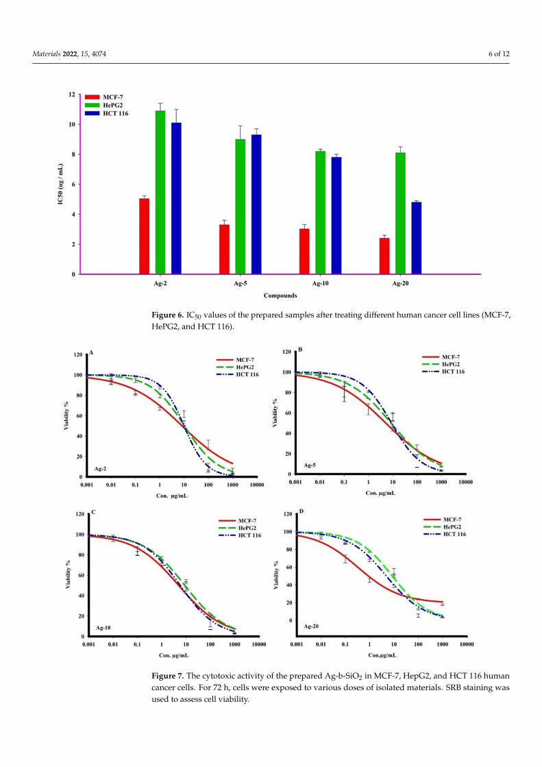
On the other hand, the obtained results showed that the prepared Ag-b-SiO2 samplesexhibited a wide range of cytotoxic activities against cancer cells. Ag-20 showed the mostpotent cytotoxic traits in human breast adenocarcinoma metastatic cells (MCF-7), withan IC50 of 2.4 ± 0.2 g/mL, and other compounds Ag-2, Ag-5, and Ag-10 exhibited themost significant killing effect against the same cancer cells (MCF-7) with IC50s of 5.03 ± 0.2,3.3 ± 0.3, and 3.02 ± 0.3 g/mL, respectively. In comparison to other cancer cells, thecompound Ag-20 showed strong cell killing with an IC50 of 4.8 ± 0.1 g/mL on humancolon adenocarcinoma cells (HCT 116). Furthermore, Ag-2, Ag-5, and Ag-10 samplesshowed cytotoxicity against HepG2 and HCT 116 cancer cells, with IC50s ranging from7.8 ± 0.2 to 10.9 ± 0.5 g/mL (Table 2, and Figures 6 and 7). The obtained results clearlyshowed that there was a great effect of the nanoparticle size on the cytotoxic activity of theprepared samples.
Table 2. The IC50 (µg) of the prepared Ag-b-SiO2 samples against different human solid tumor cells.
Sample IC50 (µg)MCF-7
IC50 (µg)HePG2
IC50 (µg)HCT 116
Ag-2 5.03 ± 0.2 10.9 ± 0.5 10.1 ± 0.9Ag-5 3.3 ± 0.3 9 ± 0.9 9.3 ± 0.4
Ag-10 3.02 ± 0.3 8.2 ± 0.14 7.8 ± 0.2Ag-20 2.4 ± 0.2 8.1 ± 0.4 4.8 ± 0.1
The cytotoxicity of the prepared Ag-b-SiO2 samples in MCF-7, HepG2, and HCT116human cancer cells was monitored in a time frame of 72 h, and cells were exposed to variousdoses of the prepared samples. Dual fluorescent staining of acridine orange/ethidiumbromide (AO/EtBr) was applied to recognize MCF-7, HepG2, and HCT 116 cancer cells. Thecancer cells were treated for 48 h before being investigated under a fluorescent microscope.The viability of the cell control was indicated by regular green staining with normal, round,intact nuclei and cytoplasm. After treating all cancer cells with Ag-5, a higher rate of celldeath was observed in the early apoptosis pathway, even though the percentage was higherthan Ag-5 after treating HCT116 cancer cells with Ag-2. In addition, Ag-10 caused a highpercentage of MCF-7, HePG2, and HCT 116 cells to necrotize. After treatment with Ag-20,all cancer cells showed the highest rate of late apoptosis (Figures 8 and 9).
Materials 2022, 15, 4074 6 of 12
Figure 6. IC50 values of the prepared samples after treating different human cancer cell lines (MCF-7,HePG2, and HCT 116).
Figure 7. The cytotoxic activity of the prepared Ag-b-SiO2 in MCF-7, HepG2, and HCT 116 humancancer cells. For 72 h, cells were exposed to various doses of isolated materials. SRB staining wasused to assess cell viability.

Materials 2022, 15, 4074 7 of 12
Figure 8. The effect of nanosilver compounds treatment on the apoptosis and necrosis of MCF-7,HepG2, and HCT 116 human tumor cells was evaluated using acridine orange (AO) and ethidiumbromide (EB) staining after 48 h of treatment, inducing various nuclear changes such as chromatinfragmentation and condensation at 200×. Yellow arrows indicate live cells, pink arrows indicateearly apoptotic cells, red arrows indicate necrotic cells, and blue arrows indicate late apoptotic cells.

Materials 2022, 15, 4074 8 of 12
Figure 9. The percentage of apoptotic/necrotic cells of MCF-7, HePG2, and HCT 116 tumor cells wasdetermined 48 h after treatment with silver nanoparticles.
Materials 2022, 15, 4074 9 of 12
3. Discussion
Generally, the obtained results showed the inhibition of the three different kinds ofcancer cells through apoptotic death for the cells. The mechanism of inhibition was reportedearlier. In the case of colon cancer cells (HCT 116), it was reported that silver nanoparticlescan activate PUMA, and caspases-3, -8, and -9 [27,28]. These genes are responsible for theapoptotic mechanism of cell death [29]. Meanwhile, in the case of HeGP2, the presence ofsilver nanoparticles induces several cytomorphological changes on the liver cancer cellsin terms of cell shrinkage and oxidative stress, resulting in apoptosis [30,31]. Moreover,the effect of silver nanoparticles on the inhibition of MCF-7 cells was also reported byVivek [32] and Jang [33], and again, the apoptosis cell death was reported as a majorcause of inhibition. Then, the obtained results in the current study are in line with thereported research in which silver nanoparticles inhibit the cancer cells through the apoptoticmechanism. It is important to mention that the MCF-7 cells are adenocarcinoma (growingin glands of organs), while HCT116 and HePG2 cells are carcinoma; as a result, MCF-7 is abreast cancer model, and its behavior and response to therapies differ from those of othercell types (HCT116 and HePG2).
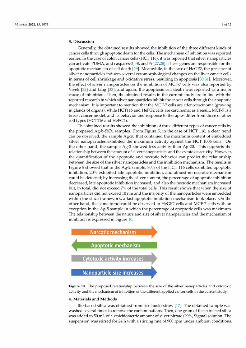
The obtained results showed the inhibition of three different types of cancer cells bythe prepared Ag-b-SiO2 samples. From Figure 5, in the case of HCT 116, a clear trendcan be observed; the sample Ag-20 that contained the maximum content of embeddedsilver nanoparticles exhibited the maximum activity against the HCT 1006 cells. Onthe other hand, the sample Ag-2 showed less activity than Ag-20. This supports therelationship between the amount of silver nanoparticles and the cytotoxic activity. However,the quantification of the apoptotic and necrotic behavior can predict the relationshipbetween the size of the silver nanoparticles and the inhibition mechanism. The results inFigure 8 showed that in the Ag-2 sample, 80% of the HCT 116 cells exhibited apoptoticinhibition, 20% exhibited late apoptotic inhibition, and almost no necrotic mechanismcould be detected; by increasing the silver content, the percentage of apoptotic inhibitiondecreased, late apoptotic inhibition increased, and also the necrotic mechanism increasedbut, in total, did not exceed 7% of the total cells. This result shows that when the size ofnanoparticles did not exceed 10 nm and the majority of the nanoparticles were embeddedwithin the silica framework, a fast apoptotic inhibition mechanism took place. On theother hand, the same trend could be observed in HeGP2 cells and MCF-7 cells with anexception in the Ag-5 sample in which the percentage of apoptotic cells was maximum.The relationship between the nature and size of silver nanoparticles and the mechanism ofinhibition is expressed in Figure 10.
Figure 10. The proposed relationship between the size of the silver nanoparticles and cytotoxicactivity and the mechanism of inhibition of the different applied cancer cells in the current study.
4. Materials and Methods
Bio-based silica was obtained from rice husk/straw [17]. The obtained sample waswashed several times to remove the contaminations. Then, one gram of the extracted silicawas added to 50 mL of a stoichiometric amount of silver nitrate (99%, Sigma) solution. Thesuspension was stirred for 24 h with a stirring rate of 900 rpm under ambient conditions.

Materials 2022, 15, 4074 10 of 12
The suspension was dried at 100 ◦C for 24 h, and then the dried solid powder was calcinedat 400 ◦C for 180 min with a heating ramp of 5 degrees/minute followed by hydrogenationin a tube furnace at 300 ◦C for another 180 min. Finally, the obtained solid powder wassealed, stored in glass vials, and coded as Ag-x (where x is the loading of Ag).
X-ray diffraction (XRD) analysis was carried out using a Schimadzu 6000 DX diffrac-tometer (Shimadzu, Japan) with the aid of a graphite monochromator with Cu-Kα
(λ = 0.1541 nm). The exact amount of silver was analyzed by using inductively coupledplasma-optical emission spectrometry (ICP-OES) through the ICAP 7000 (1 340 910, Qte-gra Software, Germany) instrument. The nitrogen adsorption–desorption isotherms wererecorded on a QuantaChrome Autosorb-6B at 77 K. Scanning electron microscopy (SEM,Jeol Model 6360 LVSEM, USA) was used to investigate the morphological structure, and themicrographs were obtained after coating the samples by gold to avoid charging effects andto enhance contrast. Moreover, SEM was equipped with energy-dispersive X-ray analysis(EDX, Jeol Model 6360 LVSEM, USA) that was used as qualitative and semi-quantitativeelemental analysis. High-Resolution Transmission Electron Microscopy (HR-TEM) wascarried out on a Philips CM30UT electron microscope with a field emission gun as thesource of electrons, operated at 300 kV. Samples were mounted on a copper-supportedcarbon polymer grid by placing a few droplets of a suspension of ground sample in ethanolon the grid, followed by drying under ambient conditions. Finally, the chemical stateof metals and surface compositions of materials were analyzed by X-ray photoelectronspectroscopy (XPS). All measurements were carried out at room temperature.
The human cancer cell lines were obtained from Dr. Mohamed Tantawy, Medical andClinical Studies Institute, National Research Center, Egypt, and provided by AmericanType Culture Collection (ATCC): breast adenocarcinoma (metastatic mammary gland)(MCF-7) (ATCC® HTB22™), hepatocellular carcinoma (HepG-2) (ATCC® HB8065™), andcolon adenocarcinoma (HCT 116) (ATCC® CCL247™). In a humidified, 5% (/v/v) CO2atmosphere at 37◦, cells were maintained in RPMI-1640 supplemented with (100 g/mL),penicillin (100 units/mL), and heat-inactivated fetal bovine serum (10% /v/v) [18,19].
A sulforhodamine B assay was used to assess the prepared samples’ cytotoxicityagainst (MCF-7, HepG-2, and HCT 116) human tumor cells (SRB). Amounts of 80% con-fluent growing cells were trypsinized and cultured for 24 h in a 96-well tissue cultureplate before being treated with nanosilver. Untreated cells compared to cells were exposedto the six different concentrations of each compound (0.01, 0.1, 1, 10, and 1000 g/mL)(control). The cells were exposed to the concentrations for 72 h before being fixed with TCA(10% w/v) for 1 h at 4 ◦C. After multiple washes, cells were stained for 10 min in the darkwith a 0.4 percent (w/v) SRB solution. Glacial acetic acid, 1% (/v/v), was used to removeany remaining stain. The SRB-stained cells dissolved in Tris-HCl after drying overnight,and the color intensity was determined in a microplate reader at 540 nm. Using SigmaPlot12.0 software, the relationship between viability percentage and compound concentrationswas analyzed to determine the IC50 (drug dose that reduces survival to 50%) [20].
The DNA binding dyes acridine orange (AO) and ethidium bromide (EtBr) were usedto detect viable, apoptotic, and necrotic cells morphologically. When AO intercalates intoDNA, it causes both nonviable and viable cells to emit green fluorescence. Nonviable cellstake up EtBr, and it emits red fluorescence by intercalating into DNA, while viable cellsdecline it.
The cells were cultured inside a six-well plate on a cover slide. The cells were treatedwith IC50 concentrations of nanosilver and incubated for 48 h in a CO2 incubator at 37 ◦Cand 5% CO2. The cells were washed twice with a cold PBS solution. The cells werestained in each well with a mixture of Acridine Orange 100 g/mL and Ethidium Bromide(AO/EB) 100 g/mL in phosphate-buffered saline (PBS) with a concentration of 1×, and thenincubated for 5 min at room temperature (RT) before being examined with a fluorescencemicroscope [21,22].

Materials 2022, 15, 4074 11 of 12
5. Conclusions
Silver nanoparticles were supported on bio-based silica with different loadings byusing silver nitrate solution. The characterization data showed that the systems efficiencyby using only water as a solvent was >70%. Moreover, in the small loading samples, onlysmall nanoparticles of silver with an average size of 3–10 nm were obtained, while athigh loadings of sliver, extra frameworks of bulky silver (average 200–500 nm) were alsoobserved. The prepared samples exhibited high cytotoxic activity against three kinds ofcancer cells. A clear trend was observed, which illustrates a positive relationship betweenthe size of silver nanoparticles and the cytotoxic activity, but a negative relationship betweenthe size and the apoptotic mechanism of cell death.
Author Contributions: Conceptualization, M.S.H. and N.S.A.; methodology, M.S.H. and H.S.M.A.-R.;characterization, M.S.H. and K.F.F.; biology study, S.E.I.E., A.A.S., H.A.I., M.Y.A. and S.A.; writing—original draft preparation, M.S.H. and S.E.I.E.; writing—review and editing, M.S.H. and N.S.A.;supervision, N.S.A.; project administration, H.S.M.A.-R.; funding acquisition, N.S.A. All authorshave read and agreed to the published version of the manuscript.
Funding: This research was funded by the Deputyship for Research and Innovation, Ministry ofEducation in Saudi Arabia, grant number IFP-KKU-2020/11.
Institutional Review Board Statement: Not applicable.
Informed Consent Statement: Not applicable.
Data Availability Statement: Not applicable.
Acknowledgments: The authors extend their appreciation to the Deputyship for Research andInnovation, Ministry of Education in Saudi Arabia for funding this research work though the projectnumber IFP-KKU-2020/11.
Conflicts of Interest: The authors declare no conflict of interest.
References1. Wild, C.P.; Weiderpass, E.; Stewart, B.W. World Cancer Report: Cancer Research for Cancer Prevention; International Agency for
Research on Cancer: Lyon, France, 2020.2. Ferlay, J.; Ervik, M.; Lam, F.; Colombet, M.; Mery, L.; Piñeros, M. Global Cancer Observatory: Cancer Today; International Agency for
Research on Cancer: Lyon, France, 2020. Available online: https://gco.iarc.fr/today (accessed on 18 February 2021).3. Dreaden, E.C.; Mackey, M.A.; Huang, X.; Kang, B.; El-Sayed, M.A. Beating cancer in multiple ways using nanogold. Chem. Soc.
Rev. 2011, 40, 3391–3404. [CrossRef] [PubMed]4. Jain, P.K.; El-Sayed, I.H.; El-Sayed, M.A. Au nanoparticles target cancer. Nano Today 2007, 2, 18–29. [CrossRef]5. Jain, S.; Hirst, D.G.; O’Sullivan, J.M. Gold nanoparticles as novel agents for cancer therapy. Br. J. Radiol. 2012, 85, 101–113.
[CrossRef] [PubMed]6. Lim, Z.-Z.J.; Li, J.-E.J.; Ng, C.-T.; Yung, L.-Y.L.; Bay, B.-H. Gold nanoparticles in cancer therapy. Acta Pharmacol. Sin. 2011, 32,
983–990. [CrossRef]7. Yesilot, S.; Acar, A. Silver nanoparticles; a new hope in cancer therapy? East. J. Med. 2019, 24, 111–116. [CrossRef]8. Vaid, P.; Raizada, P.; Saini, A.K.; Saini, R.V. Biogenic silver, gold and copper nanoparticles - A sustainable green chemistry
approach for cancer therapy. Sustain. Chem. Pharm. 2020, 16, 100247. [CrossRef]9. Abbasi, E.; Milani, M.; Aval, S.F.; Kouhi, M.; Akbarzadeh, A.; Nasrabadi, H.T.; Nikasa, P.; Joo, S.W.; Hanifehpour, Y.;
Nejati-Koshki, K.; et al. Silver nanoparticles: Synthesis methods, bio-applications and properties. Crit. Rev. Microbiol. 2016, 42,173–180. [CrossRef] [PubMed]
10. Iravani, S.; Korbekandi, H.; Mirmohammadi, S.V.; Zolfaghari, B. Synthesis of silver nanoparticles: Chemical, physical andbiological methods. Res. Pharm. Sci. 2014, 9, 385–406. [PubMed]
11. Singhal, G.; Bhavesh, R.; Kasariya, K.; Sharma, A.R.; Singh, R.P. Biosynthesis of silver nanoparticles using Ocimum sanctum(Tulsi) leaf extract and screening its antimicrobial activity. J. Nanoparticle Res. 2011, 13, 2981–2988. [CrossRef]
12. Elzey, S.; Grassian, V.H. Agglomeration, isolation and dissolution of commercially manufactured silver nanoparticles in aqueousenvironments. J. Nanoparticle Res. 2010, 12, 1945–1958. [CrossRef]
13. Jiang, Z.-J.; Liu, C.-Y.; Sun, L.-W. Catalytic Properties of Silver Nanoparticles Supported on Silica Spheres. J. Phys. Chem. B 2005,109, 1730–1735. [CrossRef] [PubMed]
14. Paul, P.; Bhanja, P.; Salam, N.; Mandi, U.; Bhaumik, A.; Islam, S.M.; Alam, S. Silver nanoparticles supported over mesoporousalumina as an efficient nanocatalyst for N-alkylation of hetero (aromatic) amines and aromatic amines using alcohols as alkylatingagent. J. Colloid Interface Sci. 2017, 493, 206–217. [CrossRef]
Materials 2022, 15, 4074 12 of 12
15. Zaarour, M.; El Roz, M.; Dong, B.; Retoux, R.; Aad, R.; Cardin, J.; Dufour, C.; Gourbilleau, F.; Gilson, J.-P.; Mintova, S.Photochemical Preparation of Silver Nanoparticles Supported on Zeolite Crystals. Langmuir 2014, 30, 6250–6256. [CrossRef][PubMed]
16. De Faria, A.F.; Martinez, D.S.T.; Meira, S.M.M.; de Moraes, A.C.M.; Brandelli, A.; Filho, A.G.S.; Alves, O.L. Anti-adhesion andantibacterial activity of silver nanoparticles supported on graphene oxide sheets. Colloids Surf. B Biointerfaces 2014, 113, 115–124.[CrossRef]
17. Nasser, S.A.; Mohamed, S.H.; Serag Eldin, I.E.; Ali, A.S.; Abd-Rabboh, H.S.M.; Mohammad, Y.A.; Khaled, F.F.; Hala, A.I.; Saad, A.Valorization of Rice Husk and Straw agriculture wastes of Hassawi Saudi Arabia: Production of Bio-based Silica, Lignocelluloses,and Activated Carbon. U.S. Patent 63/322,907, 23 March 2022.
18. Mahmoud, A.M.; Al-Abd, A.M.; Lightfoot, D.A.; El-Shemy, H.A. Anti-cancer characteristics of mevinolin against three differentsolid tumor cell lines was not solely p53-dependent. J. Enzym. Inhib. Med. Chem. 2012, 27, 673–679. [CrossRef]
19. Ibrahim, S.R.M.; Abdallah, H.M.; Mohamed, G.A.; Ross, S.A. Integracides H-J: New tetracyclic triterpenoids from the endophyticfungus Fusarium sp. Fitoterapia 2016, 112, 161–167. [CrossRef] [PubMed]
20. Alahdal, A.M.; Asfour, H.Z.; Ahmed, S.A.; Noor, A.O.; Al-Abd, A.M.; Elfaky, M.A.; Elhady, S.S. Anti-Helicobacter, Antitubercularand Cytotoxic Activities of Scalaranes from the Red Sea Sponge Hyrtios erectus. Molecules 2018, 23, 978. [CrossRef]
21. Liu, E.-H.; Qi, L.-W.; Wu, Q.; Peng, Y.-B.; Li, P. Anticancer Agents Derived from Natural Products. Mini-Reviews Med. Chem. 2009,9, 1547–1555. [CrossRef] [PubMed]
22. Albright, F.; Stephenson, R.A.; Agarwal, N.; Teerlink, C.C.; Lowrance, W.T.; Farnham, J.M.; Albright, L.A.C. Prostate cancer riskprediction based on complete prostate cancer family history. Prostate 2015, 75, 390–398. [CrossRef]
23. Mashkani, S.M.H.; Ramezani, M. Silver and silver oxide nanoparticles: Synthesis and characterization by thermal decomposition.Mater. Lett. 2014, 130, 259–262. [CrossRef]
24. Xue, B.; Chen, P.; Hong, Q.; Lin, J.; Tan, K.L. Growth of Pd, Pt, Ag and Au nanoparticles on carbon nanotubes. J. Mater. Chem.2001, 11, 2378–2381. [CrossRef]
25. Lopez-Salido, I.; Lim, D.C.; Kim, Y.D. Ag nanoparticles on highly ordered pyrolytic graphite (HOPG) surfaces studied using STMand XPS. Surf. Sci. 2005, 588, 6–18. [CrossRef]
26. Schnippering, M.; Carrara, M.; Foelske, A.; Kötz, R.; Fermín, D.J. Electronic properties of Ag nanoparticle arrays. A Kelvin probeand high resolution XPS study. Phys. Chem. Chem. Phys. 2007, 9, 725–730. [CrossRef] [PubMed]
27. Kuppusamy, P.; Ichwan, S.J.A.; Al-Zikri, P.N.H.; Suriyah, W.H.; Soundharrajan, I.; Govindan, N.; Maniam, G.P.; Yusoff, M.M. InVitro Anticancer Activity of Au, Ag Nanoparticles Synthesized Using Commelina nudiflora L. Aqueous Extract Against HCT-116Colon Cancer Cells. Biol. Trace Element Res. 2016, 173, 297–305. [CrossRef] [PubMed]
28. Mustata, R.C.; Vasile, G.; Fernandez-Vallone, V.; Strollo, S.; Lefort, A.; Libert, F.; Monteyne, D.; Pérez-Morga, D.; Vassart, G.;Garcia, M.-I. Identification of Lgr5-Independent Spheroid-Generating Progenitors of the Mouse Fetal Intestinal Epithelium. CellRep. 2013, 5, 421–432. [CrossRef] [PubMed]
29. Acharya, D.; Satapathy, S.; Somu, P.; Parida, U.K.; Mishra, G. Apoptotic Effect and Anticancer Activity of Biosynthesized SilverNanoparticles from Marine Algae Chaetomorpha linum Extract Against Human Colon Cancer Cell HCT-116. Biol. Trace ElementRes. 2020, 199, 1812–1822. [CrossRef] [PubMed]
30. Wang, X.; Li, T.; Su, X.; Li, J.; Li, W.; Gan, J.; Wu, T.; Kong, L.; Zhang, T.; Tang, M.; et al. Genotoxic effects of silver nanoparticleswith/without coating in human liver HepG2 cells and in mice. J. Appl. Toxicol. 2019, 39, 908–918. [CrossRef]
31. Priya, K.; Vijayakumar, M.; Janani, B. Chitosan-mediated synthesis of biogenic silver nanoparticles (AgNPs), nanoparticlecharacterisation and in vitro assessment of anticancer activity in human hepatocellular carcinoma HepG2 cells. Int. J. Biol.Macromol. 2020, 149, 844–852. [CrossRef]
32. Vivek, R.; Thangam, R.; Muthuchelian, K.; Gunasekaran, P.; Kaveri, K.; Kannan, S. Green biosynthesis of silver nanoparticles fromAnnona squamosa leaf extract and its in vitro cytotoxic effect on MCF-7 cells. Process Biochem. 2021, 47, 2405–2410. [CrossRef]
33. Jang, S.J.; Yang, I.J.; Tettey, C.O.; Kim, K.M.; Shin, H.M. In-vitro anticancer activity of green synthesized silver nanoparticles onMCF-7 human breast cancer cells. Mater. Sci. Eng. C 2016, 68, 430–435. [CrossRef] [PubMed]
Copyright © 2022 FDOKUMEN