Evaluation of the Cytotoxic Effects of Humid Lightweight
12
Introduction Humans are exposed to a wide variety of pollutants in their surrounding environments, and there is increasing interest in the potential adverse effects of the combined interaction of these pollutants when they are simultaneously present in mixtures. However, more research is required to improve understanding of the cytotoxicity of such mixtures, since they consist of a variety of components. In fact, possible toxicants that can affect humans, as well as all other living organisms, are often derived from landfill sites and incinerators (1). In Europe, the incineration process is one of the most common methods used in the management and treatment of an ever-increasing amount of waste, and it is a means of minimising landfill disposal, in line with EU directives (2, 3). Coal ash (a generic term for particulate matter derived from incineration processes) is a mixture of known and unknown toxic components. It is expelled from the chimneys of incinerators, and can adversely affect human health and the environment (4–7). However, whilst the properties of many single toxicants of coal ash are understood, little is known about what happens when these toxic substances are mixed together, as rou- tinely happens in coal ash. Concurrent human expo- sure to multiple contaminants may, in fact, increase their toxic effects, as it could bring about new inter- actions that then lead to more and greater adverse effects (8). Moreover, coal ash itself is disposed of either in landfills, where high-density polyethylene covers are placed over clay to prevent leaking, or it is recycled and used for engineering, manufacturing and agricultural purposes. This may dramatically increase the risk to human health and ecosystems. Evaluation of the Cytotoxic Effects of Humid Lightweight Coal Ash derived from the Disposal of Waste on Normal Human Keratinocyte and Endothelial Cell Lines in 2-D and 3-D Culture Chiara Scanarotti, 1a Stefania Vernazza, 1 Massimiliano Brignone, 2 Jenia Danailova, 1 Maria A. Pronzato 1 and Anna M. Bassi 1 1 Department of Experimental Medicine, Section of General Pathology, Laboratory of Analysis and Research in Physiopathology (LARF), University of Genoa, Italy; 2 CPG Lab Srl, Cairo Montenotte, Italy Summary — The presence of waste in the environment has frequently been indicated as a significant risk to human health. Therefore, landfill sites and the disposal of urban solid and non-hazardous waste by incin- eration are subject to much environmental monitoring, in addition to the regulations already in place. However, little action has been taken, and consequently no specific legislation exists, in relation to the assessment of the real biological risk of various substances, including chemical mixtures and ashes, derived from the incineration processes. This study assessed the cytotoxic potential of humid lightweight coal ash (LA) derived from incineration processes and waste management, on two cell lines: NCTC 2544 normal human keratinocytes and HECV endothelial cells. To reach this goal and to assess more-realistic methods for animal replacement, we employed different in vitro experimental approaches: acute and longer expo- sure to LA, by direct and indirect contact (0–2mg/ml and 16mg, respectively), both in 2-D and 3-D cultures. In 2-D HECV cultures, we observed a decrease in the viability index, but only during direct contact with LA doses higher than 0.1mg/ml. Moreover, some striking differences in cytotoxicity were observed between the 2-D and 3-D models. Taken together, these observations indicate that, for studying pollutant toxicity during longer exposure times, 3-D cultures in direct contact with the pollutant seem to offer a more suit- able approach — they mimic the in vivo behaviour of cells more realistically and under strictly controlled conditions. Thus, in readiness for possible forthcoming European regulations, we believe that the proposed study, even in its preliminary phase, can provide new advice on the assessment of the toxic and biological potential of particular chemical compounds derived from waste management processes. Key words: 3-D cultures, endothelial cells, in vitro, keratinocytes, skin toxicity, viability index. Address for correspondence: Chiara Scanarotti, Laboratory of Analysis and Research on Physiopathology (LARF), Department of Experimental Medicine, Section of General Pathology, University of Genoa, Via L.B. Alberti 2, 16132 Genova, Italy. E-mail: [email protected] ATLA 41, 491–502, 2013 491 a This author was a 2012 Lush Young Researcher Prize winner.
Transcript of Evaluation of the Cytotoxic Effects of Humid Lightweight

Introduction
Humans are exposed to a wide variety of pollutantsin their surrounding environments, and there isincreasing interest in the potential adverse effectsof the combined interaction of these pollutantswhen they are simultaneously present in mixtures.However, more research is required to improveunderstanding of the cytotoxicity of such mixtures,since they consist of a variety of components. Infact, possible toxicants that can affect humans, aswell as all other living organisms, are often derivedfrom landfill sites and incinerators (1).
In Europe, the incineration process is one of themost common methods used in the management andtreatment of an ever-increasing amount of waste, andit is a means of minimising landfill disposal, in linewith EU directives (2, 3). Coal ash (a generic term for
particulate matter derived from incinerationprocesses) is a mixture of known and unknown toxiccomponents. It is expelled from the chimneys ofincinerators, and can adversely affect human healthand the environment (4–7). However, whilst theproperties of many single toxicants of coal ash areunderstood, little is known about what happens whenthese toxic substances are mixed together, as rou-tinely happens in coal ash. Concurrent human expo-sure to multiple contaminants may, in fact, increasetheir toxic effects, as it could bring about new inter-actions that then lead to more and greater adverseeffects (8). Moreover, coal ash itself is disposed ofeither in landfills, where high-density polyethylenecovers are placed over clay to prevent leaking, or it isrecycled and used for engineering, manufacturingand agricultural purposes. This may dramaticallyincrease the risk to human health and ecosystems.
Evaluation of the Cytotoxic Effects of Humid LightweightCoal Ash derived from the Disposal of Waste on NormalHuman Keratinocyte and Endothelial Cell Lines in 2-D and3-D Culture
Chiara Scanarotti,1a Stefania Vernazza,1 Massimiliano Brignone,2 Jenia Danailova,1 Maria A.Pronzato1 and Anna M. Bassi1
1Department of Experimental Medicine, Section of General Pathology, Laboratory of Analysis and Researchin Physiopathology (LARF), University of Genoa, Italy; 2CPG Lab Srl, Cairo Montenotte, Italy
Summary — The presence of waste in the environment has frequently been indicated as a significant riskto human health. Therefore, landfill sites and the disposal of urban solid and non-hazardous waste by incin-eration are subject to much environmental monitoring, in addition to the regulations already in place.However, little action has been taken, and consequently no specific legislation exists, in relation to theassessment of the real biological risk of various substances, including chemical mixtures and ashes, derivedfrom the incineration processes. This study assessed the cytotoxic potential of humid lightweight coal ash(LA) derived from incineration processes and waste management, on two cell lines: NCTC 2544 normalhuman keratinocytes and HECV endothelial cells. To reach this goal and to assess more-realistic methodsfor animal replacement, we employed different in vitro experimental approaches: acute and longer expo-sure to LA, by direct and indirect contact (0–2mg/ml and 16mg, respectively), both in 2-D and 3-D cultures.In 2-D HECV cultures, we observed a decrease in the viability index, but only during direct contact with LAdoses higher than 0.1mg/ml. Moreover, some striking differences in cytotoxicity were observed betweenthe 2-D and 3-D models. Taken together, these observations indicate that, for studying pollutant toxicityduring longer exposure times, 3-D cultures in direct contact with the pollutant seem to offer a more suit-able approach — they mimic the in vivo behaviour of cells more realistically and under strictly controlledconditions. Thus, in readiness for possible forthcoming European regulations, we believe that the proposedstudy, even in its preliminary phase, can provide new advice on the assessment of the toxic and biologicalpotential of particular chemical compounds derived from waste management processes.
Key words: 3-D cultures, endothelial cells, in vitro, keratinocytes, skin toxicity, viability index.
Address for correspondence: Chiara Scanarotti, Laboratory of Analysis and Research onPhysiopathology (LARF), Department of Experimental Medicine, Section of General Pathology, Universityof Genoa, Via L.B. Alberti 2, 16132 Genova, Italy.E-mail: [email protected]
ATLA 41, 491–502, 2013 491
aThis author was a 2012 Lush Young Researcher Prize winner.
However, the risks associated with the limited lifes-pan of these liners must also be taken into account, aswell as the need for a greater awareness of the per-manent safe storage of coal ash. However, contrary tothese requirements, the current, insufficient, EUMember State regulations result in the uncontrolleduse and disposal of coal ash (9).
Several studies on the possible negative healtheffects of residing close to incinerators have recentlybeen published, together with a number of well-con-ducted reviews (10–15). Interestingly, such ecologicalstudies have suggested links with, not only somereproductive outcomes like infant death and congen-ital malformations, birth defects, and congenital andgestational term anomalies and stillbirths, but alsowith several cancers (16–21). However, it has to besaid that the results of these studies are often contra-dictory. Consequently, the different policies issued onwaste disposal in Europe often conflict, so constantlyrequire amendment. The potential health implica-tions of this lack of clear policies are largely unknown(18), so more information is required on their likelyhuman health impacts.
If in vivo studies were performed to test these mix-tures and gain further information on the potentialhealth effects, then a very large number of labora-tory animals would be required. This requirementwould introduce increasing ethical and economicalconcerns. In addition, there are other factors to con-sider that are related to inter-species, intra-speciesand exposure time variability, adverse effect levels indifferent species (not yet detectable with standardtests), species-specific action mechanisms, and so on.Moreover, due to the uncertainty associated withsuch data for hazard and risk assessments, and thecurrent lack of specific regulations on this topic,alternative methods for coal ash toxicity studies areurgently needed, in order to provide for betterReplacement, Reduction and Refinement (the ThreeRs) in toxicological research (22). The developmentof alternative methods is not merely ethical dogma,but is also a scientific duty. The scientific data showthat animal models are often too far removed fromhumans, and the results obtained are not appropri-ate or directly transferable. So, there is an increasingrequirement for effective in vitro models to preventthe needless suffering and death of animals, and tooptimise new alternative methods, in order toimprove the quality of the resulting data.
Coal ash, like many other dangerous substances,can be found deposited in and acting upon organ sys-tems, including the skin, lungs, bladder and brain(23). The skin, in particular, should be considered asthe first point of contact with an environmental chem-ical, which often induces skin irritation, or eveninduces skin sensitisation. However, it should bepointed out that acute exposure to these irritant sub-stances does not necessarily affect cell viability. Up tonow, irritation potential has been tested on three-dimensional (3-D) cell models, but according to
ECVAM Scientific Advisory Committee (ESAC)Statement No. 26 on the validity of in vitro tests forskin irritation (3-D skin models, such as EpiDerm™),the quantification of IL-1α cytokine levels is no longerregarded as a suitable endpoint for 3-D skin models.This is due to inter-batch variation in IL-1α releasefrom different donors, and the impossibility ofdefining a standard value for IL-1α release. However,we have already reported that the less-differentiatedNCTC 2544 human keratinocyte cell line is more sus-ceptible to stimulation by irritants than is the differ-entiated HaCaT cell line. We have also shown thatNCTC 2544 keratinocytes can synthesise and modu-late the levels of pro-inflammatory IL-1α and LIFcytokines after exposure to selected irritants (24, 25).
Furthermore, evidence from both humans and lab-oratory animals highlights the fact that exposure toair pollution is strongly associated with endothelialdysfunction, which plays a central role in triggeringcardiovascular events. In fact, Nadadur et al. (26)reported that exposure to air particulates may induceperturbations in endothelial cell permeability andmembrane integrity, via modulation of several genesassociated with endothelial function pathways.
In this preliminary study, we assessed the cytotoxiceffects of humid lightweight coal ash (LA) derivedfrom incineration processes and waste management,which is classified in the European Waste Catalogue(EWC) as EWC 10 01 02 (wastes from power stationsand other combustion plants, specifically coal fly ash),on normal human cell lines derived from the skin andvascular endothelium.
We used two cell lines: NCTC 2544 (normalundifferentiated keratinocytes) and HECV (vascu-lar endothelial cells). Changes in cell morphologyand viability indices were monitored after exposurefor 4, 16 and 24 hours to LA, in order to determinewhether the toxic effects varied according towhether the contact was acute or prolonged.Moreover, the analysis was also carried out in 3-Dcultures, which allows cells to grow and adapt totheir environment in a manner that more-closelyresembles in vivo conditions. Our reasoning wasthat, by comparing the data obtained from both 3-Dand two-dimensional (2-D) cultures, we would beable to highlight different cellular responses for thepurpose of better identifying the potential hazardsand risks to human health.
492 C. Scanarotti et al.
Replacement benefits of this approach:— the cell lines used in this in vitro testing
method are human-derived, therefore more-appropriate and directly transferable resultscan be obtained;
— in vitro models help to prevent the needlesssuffering and death of animals; and
— the potential identification of new biomark-ers will give more-realistic threshold levels ofhuman risk from environmental pollutants.
Materials and Methods
Standard culture medium, fetal bovine serum(FBS), penicillin–streptomycin solution, glutamine,and Alvetex® scaffold multi-well plates were pur-chased from EuroClone S.p.A. (Milan, Italy),Millicell® wells from Millipore S.p.A. (Milan, Italy),and TripLE™ Express from Gibco (Invitrogen LifeTechnologies, Milan, Italy). All other chemicals andreagents were of the highest purity available, andwere purchased from Sigma–Aldrich (Milan, Italy).
All aseptic and experimental techniques wereperformed in strict compliance with the standardoperating procedures of the Laboratory of Analysisand Research on Physiopathology (LARF), whichhas a quality management system certified in linewith UNI EN ISO 9001:2008.
Cell cultures
As an alternative to existing in vitro models, twonormal stabilised human cell lines were used:NCTC 2544 (undifferentiated keratinocytes) andHECV (endothelial cells from human umbilicalvein), provided by the Biological Bank, a core facil-ity at the IRCCS San Martino University Hospital,IST National Institute for Cancer Research, Genoa,Italy.
The cell lines were maintained in DMEMmedium, supplemented with 10% v/v FBS, 2mMglutamine and 1% v/v penicillin–streptomycin solu-tion, at 37°C in a humidified atmosphere containing5% v/v CO2. The medium was changed every 2 to 3days; at 80% confluence, the cells were sub-culturedby using TripLE Express, a recombinant serine pro-tease enzyme that contains no components of ani-mal origin. Cells of both cell lines were seeded in6-well and 24-well plates, at 15 × 104 and 4 × 10⁴
cells/well, respectively, 24 hours before theexperimental procedures.
To more-closely mimic the skin environment, wealso assessed the cellular responses to LA exposureby culturing NCTC 2544 and HECV cells in Alvetexscaffold 24-well plates. According to the manu -facturer’s protocol, 100μl of NCTC 2544 and HECVcell suspensions (equivalent to 5 × 10⁵ cells/well)were added in droplets over the surface of theAlvetex scaffold disc, which had previously beenactivated with 70% v/v ethanol. Then, 2ml of stan-dard medium were gently added to each well. The 3-D cultures were grown for 5 days, with a dailychange of medium, before exposure to LA.
Experimental treatments
The LA, provided by CPG Lab Srl (CairoMontenotte, Savona, Italy), was sterilised in anoven at 120°C for 120 minutes, and suspended in
complete DMEM medium. Subsequently, the sus-pension was put in a sonicator bath for 30 minutes,to obtain a better dispersion of the particles beforeexposure of the cells. A range of LA concentra -tions — 0.0001, 0.001, 0.01, 0.1, 0.25, 0.5, 1 and2mg/ml — was chosen, starting from the hazardousexposure limit for inhalable particulate matter(10mg/m3) according to the literature (26, 27).NCTC 2544 and HECV cultures were exposed toLA, by direct and indirect contact, for 4, 16 and 24hours.
For the direct contact exposure, LA (dose range0.0001–2mg/ml) was added directly to the culturemedium. To perform the indirect contact exposure,16 ± 2mg of LA, (according to Organisation forEconomic Co-operation and Development TestGuideline [OECD TG] 439) were seeded ontoremovable inserts (Millicell well, hanging cellculture insert 0.4μm PET) that were placed in eachwell of the 6-well plates in which the NCTC 2544and HECV cells were growing. At the same time,wells with control solutions were set up: standardculture medium was used as the negative control,and 5% w/v sodium dodecyl sulphate (SDS) as thepositive control. Each experimental condition wasperformed in two independent experiments, intriplicate wells.
In the indirect contact assay, the 3-D NCTC 2544and HECV cultures were exposed to LA at the samedoses as those used for the direct contact assay.Untreated 3-D cultures were used as the negativecontrol. After a 24-hour exposure, each disc wasremoved, gently dipped in PBS and placed in a new24-well plate, in order to perform the viability tests.Each experimental condition was performed inthree independent experiments, in duplicate wells.
Evaluation of toxic effects
A qualitative assessment of cell morphology wascarried out, for untreated and treated cultures, byusing a phase-contrast microscope (Leica DMIRBHC Fluo 257041; Leica Microsystems Srl S.p.A.,Milano, Italy) at 10× magnification. The level ofmorphological damage (i.e. damage degree) wasscored on a scale of 0–4, with 0 representing no tox-icity and 4 representing severe toxicity, according toguideline UNI EN ISO 10993-5:2009; values > 2are associated with cytotoxicity.
At the end of each experimental treatment, themagnitude of cell viability was assessed by the thia-zolyl blue tetrazolium dye reduction (MTT) andNeutral Red uptake (NRU) assays (28, 29). At thesame time, in concurrent experiments, the MTTassay was used as a measure of relative cell prolif-eration (30). We had previously performed checks toexclude the possibility of direct MTT reduction(according to OECD TG 439) or interference withNeutral Red dye by LA.
Cytotoxic effects of humid lightweight coal ash 493
Tetrazolium salt (MTT) was dissolved in PBS(5mg/ml) and 100μl were added per well to reach afinal concentration of 0.5mg/ml. After a 3-hourincubation at 37°C, a solution of 1N hydrogen chlo-ride–isopropanol (1:24, v/v) was added to each well(100μl/well), and mixed to dissolve the dark-blueformazan crystals formed. The NRU assay was per-formed by exposing cell cultures to culture medium(without serum and phenol red) containing 50μg/mlNeutral Red dye. After a 3-hour incubation at 37°C,the cells were washed twice in PBS to remove theexcess dye, and bleached with 1ml of a solution of1% v/v glacial acetic acid, 50% v/v ethanol, 49% v/vwater.
The optical density (OD) of the solutions contain-ing the dissolved formazan crystals and the releasedNeutral Red dye (for the MTT and NRU tests,respectively) was determined at 570nm, for bothassays, by using the Uniskan II Microplate reader(Labsystem, Helsinki, Finland). The cell viabilityindex in the treated cultures was compared to thevalues corresponding to the negative controls, andexpressed as a percentage to predict the irritationpotential. A chemical compound was consideredtoxic, if the cell viability was reduced by 15% com-pared to that of the untreated cultures, according toECVAM guidelines (31).
Statistics
The results are expressed as the mean ± SD of twoor three independent experiments performed intriplicate or duplicate, respectively. The signifi-cance was assessed by one-way analysis of variance(ANOVA), followed by Dunnett’s test. Instat andPrism software packages (Graph-Pad Software,Inc., La Jolla, CA, USA) were used.
Results
Direct exposure to LA at the lower concentrationstested — 0.0001, 0.001, 0.01 mg/ml — did not affectthe viability indices in either cell line (data notshown). Therefore, the data obtained after treat-ment with higher doses of LA, i.e. 0.1, 0.25, 0.5, 1and 2mg/ml, are reported here.
The NCTC 2544 and HECV cells were monitoredby phase-contrast microscopy (10×), after indirectand direct exposure to LA and at the end of eachexperimental period, to derive a numerical value forthe degree of cytotoxicity. The digital imagesobtained are shown in Figures 2 and 3.
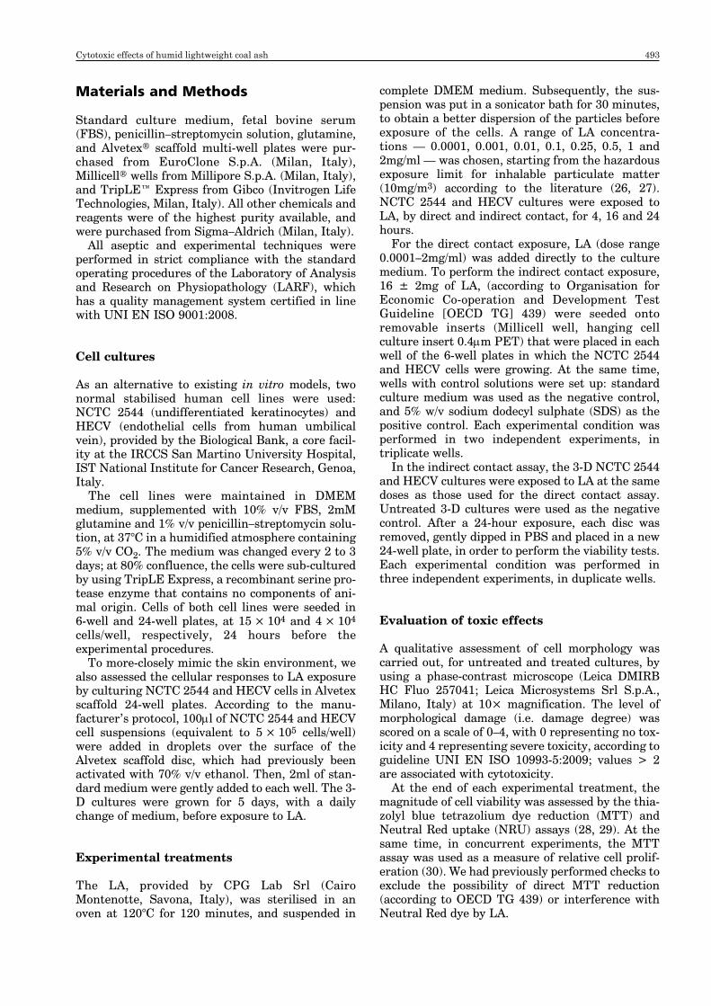
After 24 hours of indirect contact with 16mg LA,slight morphological alterations were evident inboth NCTC 2544 and HECV cells, as compared tothe untreated cultures. For the LA-treated cultures,the cellular damage score was ≤ 2, while for theSDS-treated cultures (positive control), marked tox-
icity was exhibited and the score was 4 (Table 1 andFigure 2). This trend was also confirmed by bothviability indices — after a 24-hour exposure to LA,the MTT test results showed evidence of slight tox-icity, resulting in a 13% and 15% decrease in viabil-ity, respectively, for HECV and NCTC 2544 cells(Table 2). Based on these observations, the follow-ing analyses were performed only after directexposure to LA.
In the digital images obtained of cultures afterdirect contact, LA appeared as black solid particles.
Figure 1: A typical incinerator
Table 1: Qualitative evaluation of themorphology of NCTC 2544 andHECV cells after indirect contactwith LA
Exposure TreatmentCell time line (hours) None SDS LA
NCTC 4 0 4 ± 1* 0 ± 12544 16 0 4 ± 1* 0 ± 1
24 0 4 ± 1* 1 ± 1
HECV 4 0 4 ± 1* 0 ± 116 0 4 ± 1* 0 ± 124 0 4 ± 1* 1 ± 1
A cytotoxicity score was given (between 0–4), based on aqualitative evaluation (by phase-contrast microscopy) ofNCTC 2544 and HECV cell morphology throughout theexperimental treatments. The cell cultures were exposed,through indirect contact, to 16 ± 2mg LA and 5% SDS(positive control) for 4, 16 and 24 hours. Untreated cellswere cultured and evaluated in parallel. The averagescore ± SD related to two independent experiments,performed in triplicate; *p < 0.01 versus the respectiveuntreated cultures.
494 C. Scanarotti et al.
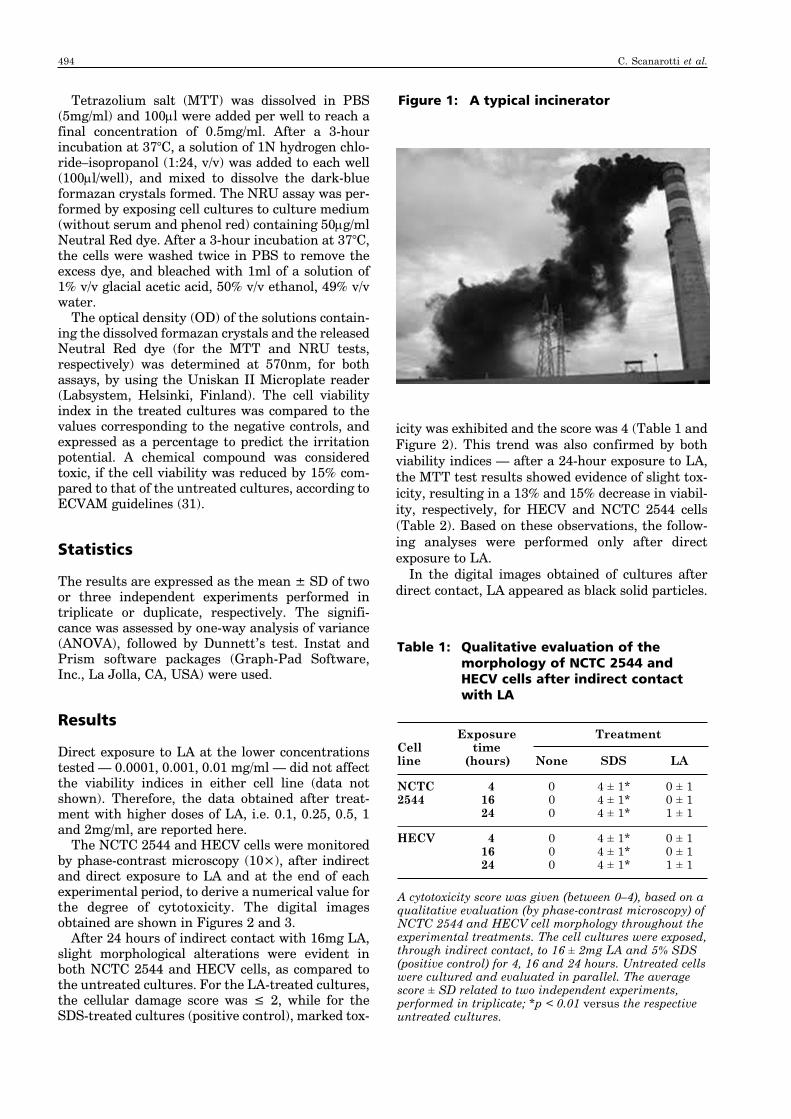
The HECV cell morphology underwent more-marked dose-dependent and time-dependentchanges than that of NCTC 2544 cells (Figure 3).Consequently, we set the damage score at 3 and 1,respectively, for HECV and NCTC 2544 cultures,after a 24-hour exposure to the highest doses of LA(1mg/ml and 2mg/ml; Table 3).
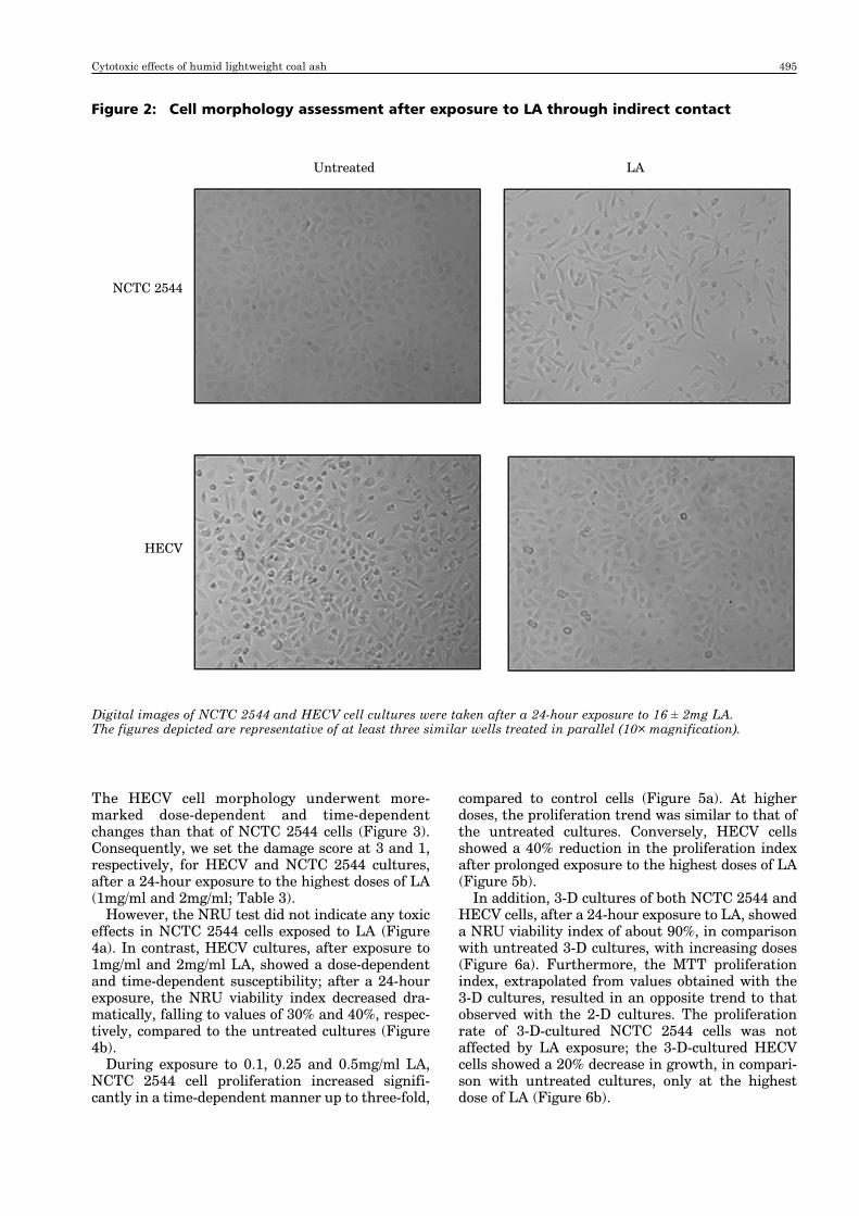
However, the NRU test did not indicate any toxiceffects in NCTC 2544 cells exposed to LA (Figure4a). In contrast, HECV cultures, after exposure to1mg/ml and 2mg/ml LA, showed a dose-dependentand time-dependent susceptibility; after a 24-hourexposure, the NRU viability index decreased dra-matically, falling to values of 30% and 40%, respec-tively, compared to the untreated cultures (Figure4b).
During exposure to 0.1, 0.25 and 0.5mg/ml LA,NCTC 2544 cell proliferation increased signifi-cantly in a time-dependent manner up to three-fold,
compared to control cells (Figure 5a). At higherdoses, the proliferation trend was similar to that ofthe untreated cultures. Conversely, HECV cellsshowed a 40% reduction in the proliferation indexafter prolonged exposure to the highest doses of LA(Figure 5b).
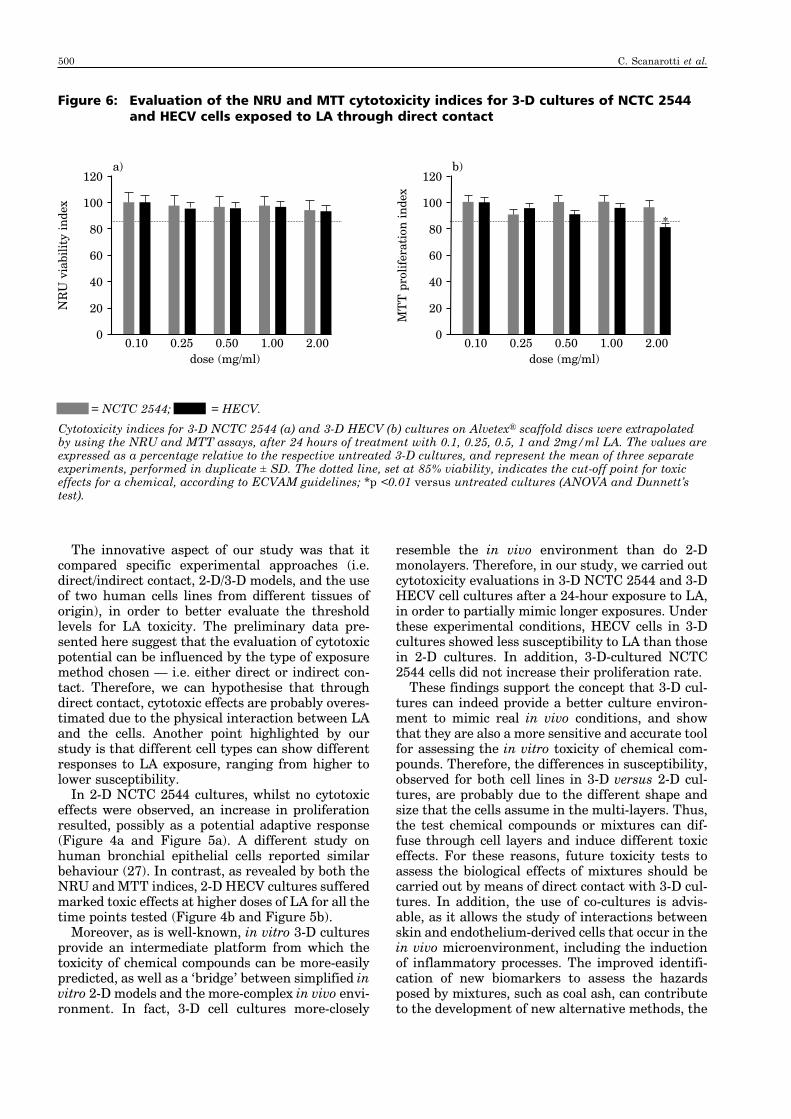
In addition, 3-D cultures of both NCTC 2544 andHECV cells, after a 24-hour exposure to LA, showeda NRU viability index of about 90%, in comparisonwith untreated 3-D cultures, with increasing doses(Figure 6a). Furthermore, the MTT proliferationindex, extrapolated from values obtained with the3-D cultures, resulted in an opposite trend to thatobserved with the 2-D cultures. The proliferationrate of 3-D-cultured NCTC 2544 cells was notaffected by LA exposure; the 3-D-cultured HECVcells showed a 20% decrease in growth, in compari-son with untreated cultures, only at the highestdose of LA (Figure 6b).
Figure 2: Cell morphology assessment after exposure to LA through indirect contact
Digital images of NCTC 2544 and HECV cell cultures were taken after a 24-hour exposure to 16 ± 2mg LA. The figures depicted are representative of at least three similar wells treated in parallel (10× magnification).
Untreated LA
NCTC 2544
HECV
Cytotoxic effects of humid lightweight coal ash 495
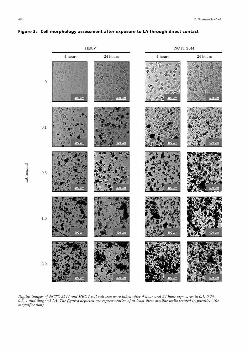
Figure 3: Cell morphology assessment after exposure to LA through direct contact
Digital images of NCTC 2544 and HECV cell cultures were taken after 4-hour and 24-hour exposures to 0.1, 0.25,0.5, 1 and 2mg/ml LA. The figures depicted are representative of at least three similar wells treated in parallel (10×magnification).
HECV NCTC 2544
4 hours 24 hours 4 hours 24 hours
LA
(m
g/m
l)
0
0.1
0.5
1.0
2.0
496 C. Scanarotti et al.

Discussion
At the moment, in EU countries, the managementof hazardous waste partly relies on incinerationprocesses, as it is environmentally preferable tolandfill disposal (32). However, industrial combus-tion processes might be an important source of var-ious hazardous environmental pollutants (33). Coalash, derived from combustion waste, is the particu-late material that remains after the combustionprocess, and consists of a mixture of toxic com-pounds that might contribute to public health risksand environmental problems (34).
People can be exposed to coal ash from both land-fill sites and environmental contamination, as wellas in public areas such as the workplace, or even intheir own homes. In fact, coal ash is used as a fillerin a wide range of materials, including soil, bricksand cement, so it is present in constructions such asdriveways, buildings and sports fields. Indeed, ithas been shown that the use of recycled coal ash islinked to environmental pollution (35–37), but, asyet, no current regulations completely safeguardthe public from these risks.
Epidemiological and animal toxicology studiesprovide coherent evidence that the health effects ofcoal ash are multiple, ranging from damage to theskin, increased risk of cancer, and damage to therespiratory and cardiovascular systems, as a resultof alterations that cause endothelial dysfunction(26, 38–42). For all the above reasons, furtherresearch is required — and to obtain the most effec-tive results, research is needed that will integratethe principles of the Three Rs, with the ultimategoal of the replacement of animal models inresearch and toxicological testing. In addition, it is
important to note that, in toxicological research,both the dosage and the timing of exposure tochemicals have important and significant effects onany potential health outcomes.
Several in vitro and in vivo studies on the poten-tial toxicity of coal ash have demonstrated cyto-toxic, genotoxic and inflammatory effects, and theresults were often related to the induction of oxida-tive stress by toxic constituents (such as transitionmetals, polycyclic aromatic hydrocarbons, and sil-ica; 43–45). Therefore, the aim of this study was todevelop a new in vitro model with two human celllines, NCTC 2544 and HECV, in order to evaluatethe toxic effects of humid lightweight coal ashderived from waste disposal.
A recent in vitro study on the inflammatoryeffects of residual combusted oil fly ash (ROFA) onprimary human umbilical vein endothelial cells (26)by using the MTT assay did not provide evidence ofcytotoxic effects, even though the analysis was car-ried out after eight hours of exposure to0.1–10μg/ml ROFA, which was lower than the expo-sure times used in our study. In addition, Diabaté etal. reported toxic effects on rat alveolarmacrophages and human bronchial epithelial cellsafter a 24-hour exposure to total incinerator fly ash(IFA), starting at 0.1mg/ml for both cell lines (27).
Table 2: The viability index of NCTC 2544and HECV cultures after exposureto LA through indirect contact
Exposure Viability index (%)Cell time line (hours) NRU MTT
NCTC 4 100 ± 5 100 ± 122544 16 97 ± 7 99 ± 4
24 93 ± 8 87 ± 8*
HECV 4 100 ± 15 98 ± 1216 98 ± 10 96 ± 824 92 ± 6 85 ± 6*
The values represent the NRU and MTT viability andproliferation indices, respectively, in NCTC 2544 andHECV cultures after 4, 16 and 24 hours of exposure to16 ± 2mg of LA, through indirect contact. The data areexpressed as a percentage relative to the respectivecontrol, and shown as the mean ± SD of twoindependent experiments, performed in triplicate; *p < 0.01 versus the respective untreated cultures.
Table 3: Qualitative evaluation ofmorphology in NCTC 2544 andHECV cells after direct contact withLA
Exposure time(hours)
Cell Doseline Treatment (mg/ml) 4 16 24
NCTC None — 0 0 02544 SDS (5%) — 4 ± 1* 4 ± 1* 4 ± 1*
LA 0.1 0 ± 1 0 ± 1 0 ± 10.5 0 ± 1 0 ± 1 0 ± 11.0 0 ± 1 1 ± 1 1 ± 12.0 0 ± 1 1 ± 1 1 ± 1
HECV None — 0 0 0SDS (5%) — 4 ± 1* 4 ± 1* 4 ± 1*
LA 0.1 0 ± 1 0 ± 1 0 ± 10.5 0 ± 1 1 ± 1 2 ± 11.0 0 ± 1 1 ± 1 3 ± 1*2.0 0 ± 1 1 ± 1 3 ± 1*
A cytotoxicity score was given (between 0–4), based on aqualitative evaluation (by phase-contrast microscopy) ofNCTC 2544 and HECV cell morphology throughout theexperimental treatments. The cell cultures were exposed,through direct contact, to 0.1, 0.5, 1 and 2mg/ml of LA,and 5% SDS (positive control) for 4, 16 and 24 hours.Untreated cells were cultured and evaluated in parallel.The average score ± SD related to two independentexperiments, performed in triplicate; *p < 0.01 versusthe respective untreated cultures.
Cytotoxic effects of humid lightweight coal ash 497
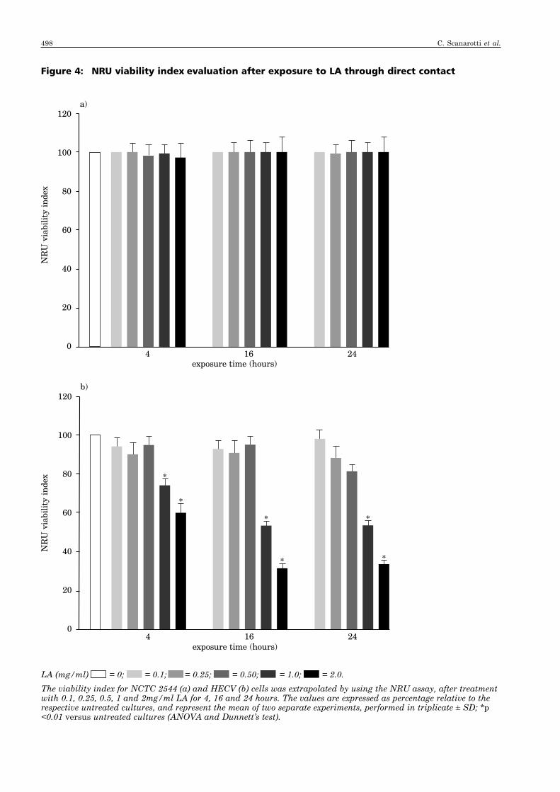
Figure 4: NRU viability index evaluation after exposure to LA through direct contact
LA (mg/ml) = 0; = 0.1; = 0.25; = 0.50; = 1.0; = 2.0.The viability index for NCTC 2544 (a) and HECV (b) cells was extrapolated by using the NRU assay, after treatmentwith 0.1, 0.25, 0.5, 1 and 2mg/ml LA for 4, 16 and 24 hours. The values are expressed as percentage relative to therespective untreated cultures, and represent the mean of two separate experiments, performed in triplicate ± SD; *p<0.01 versus untreated cultures (ANOVA and Dunnett’s test).
120
100
80
60
40
20
04 16 24
exposure time (hours)
b)
120
100
80
60
40
20
04 16 24
exposure time (hours)
NR
U v
iabi
lity
inde
xN
RU
via
bilit
y in
dex
a)
*
* *
**
*
498 C. Scanarotti et al.
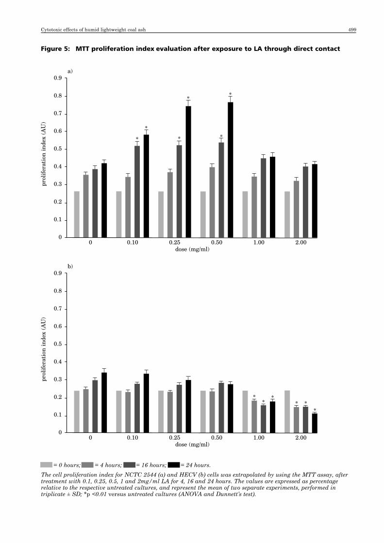
Figure 5: MTT proliferation index evaluation after exposure to LA through direct contact
= 0 hours; = 4 hours; = 16 hours; = 24 hours.The cell proliferation index for NCTC 2544 (a) and HECV (b) cells was extrapolated by using the MTT assay, aftertreatment with 0.1, 0.25, 0.5, 1 and 2mg/ml LA for 4, 16 and 24 hours. The values are expressed as percentagerelative to the respective untreated cultures, and represent the mean of two separate experiments, performed intriplicate ± SD; *p <0.01 versus untreated cultures (ANOVA and Dunnett’s test).
0.9
0.8
0.7
0.6
0.5
0.4
0.3
0.2
0.1
00 0.10 0.25 0.50 1.00 2.00
dose (mg/ml)
prol
ifer
atio
n in
dex
(AU
)
b)
0.9
0.8
0.7
0.6
0.5
0.4
0.3
0.2
0.1
00 0.10 0.25 0.50 1.00 2.00
dose (mg/ml)
prol
ifer
atio
n in
dex
(AU
)
a)
*
*
* *
**
**
** *
*
Cytotoxic effects of humid lightweight coal ash 499
The innovative aspect of our study was that itcompared specific experimental approaches (i.e.direct/indirect contact, 2-D/3-D models, and the useof two human cells lines from different tissues oforigin), in order to better evaluate the thresholdlevels for LA toxicity. The preliminary data pre-sented here suggest that the evaluation of cytotoxicpotential can be influenced by the type of exposuremethod chosen — i.e. either direct or indirect con-tact. Therefore, we can hypothesise that throughdirect contact, cytotoxic effects are probably overes-timated due to the physical interaction between LAand the cells. Another point highlighted by ourstudy is that different cell types can show differentresponses to LA exposure, ranging from higher tolower susceptibility.
In 2-D NCTC 2544 cultures, whilst no cytotoxiceffects were observed, an increase in proliferationresulted, possibly as a potential adaptive response(Figure 4a and Figure 5a). A different study onhuman bronchial epithelial cells reported similarbehaviour (27). In contrast, as revealed by both theNRU and MTT indices, 2-D HECV cultures sufferedmarked toxic effects at higher doses of LA for all thetime points tested (Figure 4b and Figure 5b).
Moreover, as is well-known, in vitro 3-D culturesprovide an intermediate platform from which thetoxicity of chemical compounds can be more-easilypredicted, as well as a ‘bridge’ between simplified invitro 2-D models and the more-complex in vivo envi-ronment. In fact, 3-D cell cultures more-closely
resemble the in vivo environment than do 2-Dmonolayers. Therefore, in our study, we carried outcytotoxicity evaluations in 3-D NCTC 2544 and 3-DHECV cell cultures after a 24-hour exposure to LA,in order to partially mimic longer exposures. Underthese experimental conditions, HECV cells in 3-Dcultures showed less susceptibility to LA than thosein 2-D cultures. In addition, 3-D-cultured NCTC2544 cells did not increase their proliferation rate.
These findings support the concept that 3-D cul-tures can indeed provide a better culture environ-ment to mimic real in vivo conditions, and showthat they are also a more sensitive and accurate toolfor assessing the in vitro toxicity of chemical com-pounds. Therefore, the differences in susceptibility,observed for both cell lines in 3-D versus 2-D cul-tures, are probably due to the different shape andsize that the cells assume in the multi-layers. Thus,the test chemical compounds or mixtures can dif-fuse through cell layers and induce different toxiceffects. For these reasons, future toxicity tests toassess the biological effects of mixtures should becarried out by means of direct contact with 3-D cul-tures. In addition, the use of co-cultures is advis-able, as it allows the study of interactions betweenskin and endothelium-derived cells that occur in thein vivo microenvironment, including the inductionof inflammatory processes. The improved identifi-cation of new biomarkers to assess the hazardsposed by mixtures, such as coal ash, can contributeto the development of new alternative methods, the
Figure 6: Evaluation of the NRU and MTT cytotoxicity indices for 3-D cultures of NCTC 2544and HECV cells exposed to LA through direct contact
= NCTC 2544; = HECV. Cytotoxicity indices for 3-D NCTC 2544 (a) and 3-D HECV (b) cultures on Alvetex® scaffold discs were extrapolatedby using the NRU and MTT assays, after 24 hours of treatment with 0.1, 0.25, 0.5, 1 and 2mg/ml LA. The values areexpressed as a percentage relative to the respective untreated 3-D cultures, and represent the mean of three separateexperiments, performed in duplicate ± SD. The dotted line, set at 85% viability, indicates the cut-off point for toxiceffects for a chemical, according to ECVAM guidelines; *p <0.01 versus untreated cultures (ANOVA and Dunnett’stest).
120
100
80
60
40
20
00.10 0.25
dose (mg/ml)0.50 1.00 2.00
MT
T p
rolif
erat
ion
inde
x
b)120
100
80
60
40
20
00.10 0.25
dose (mg/ml)0.50 1.00 2.00
NR
U v
iabi
lity
inde
x
a)
*
500 C. Scanarotti et al.

purposes of which are to predict the real potentialrisks to human health and ecosystems.
Acknowledgements
We gratefully acknowledge the 2012 Lush Prizeaward and also Professor Margherita Ferro, whocontributed to the development of this study, givingus the basis for refining these in vitro methods andpromoting their involvement in the field of patho-logical research. The authors also wish to thank MsSuzanne Patten, for her assistance in the revisionof our English.
References
1. Porta, D., Milani, S., Lazzarino, A.I., Perucci, C.A. &Forastiere, F. (2009). Systematic review of epidemi-ological studies on health effects associated withmanagement of solid waste. Environmental Health8, 60.
2. Eurostat (2010). Environmental Statistics andAccounts in Europe, 342pp. Luxembourg, Luxem -bourg: Publications Office of the European Union.
3. Saner, D., Blumer, Y.B., Lang, D.J. & Koehler, A.(2011). Scenarios for the implementation of EUwaste legislation at national level and their conse-quences for emissions from municipal waste inciner-ation. Resources Conservation & Recycling 57,67–77.
4. Allsopp, M., Costner, P. & Johnston, P. (2001).Incineration and human health: State of knowledgeof the impacts of waste incinerators on humanhealth. Environmental Science & Pollution ResearchInternational 8, 141–145.
5. Anon. (2008). The Health Effects of Waste Inciner -ators, 2nd edn, 71pp. London, UK: British Societyfor Ecological Medicine.
6. Reis, M.F. (2011). Solid waste incinerators: Healthimpacts. In Encyclopaedia of Environmental Health,pp. 162–217. Amsterdam, The Netherlands: Elsevier.
7. Klaassen, C. (2007). Casarett & Doull’s Toxicology:The Basic Science of Poisons, 7th edn, 1454pp. NewYork, NY, USA: McGraw-Hill.
8. Ranzi, A., Fano, V., Erspamer, L., Lauriola, P.,Perucci, C.A. & Forastiere, F. (2011). Mortality andmorbidity among people living close to incinerators:A cohort study based on dispersion modeling forexposure assessment. Environmental Health 10, 22.
9. Gottlieb, B., Gilbert, S.G. & Evans, G.E. (2010). CoalAsh: The Toxic Threat to Our Health and Environ -ment, 27pp. Washington, DC, USA: Physicians forSocial Responsibility & Earthjustice. Available at:http://earthjustice.org/sites/default/files/files/CoalAsh_Earthjustice.pdf (Accessed 06.12.13).
10. Franchini, M., Rial, M., Buiatti, E. & Bianchi, F.(2004). Health effects of exposure to waste incinera-tor emissions: A review of epidemiological studies.Annali dell’Istituto Superiori di Sanità 40, 101–115.
11. WHO (2007) Population Health and Waste Manage -ment: Scientific Data and Policy Options, 91pp.Copenhagen, Denmark: World Health OrganisationRegional Office for Europe. Available at: http://www.euro.who.int/__data/assets/pdf_file/0012/91101/E91021.pdf (Accessed 06.12.13).
12. Vrijheid, M. (2000). Health effects of residence nearhazardous waste landfill sites: A review of epidemio-logic literature. Environmental Health Perspectives108, Suppl. 1, 101–112.
13. Rushton, L. (2003). Health hazards and waste man-agement. British Medical Bulletin 68, 183–197.
14. Saunders, P. (2007). A systematic review of the evi-dence of an increased risk of adverse birth out-comes in populations living in the vicinity oflandfill waste disposal sites. In Population Healthand Waste Management: Scientific Data and PolicyOptions, pp. 25–27. Copenhagen, Denmark: WorldHealth Organisation Regional Office for Europe.
15. Tango, T., Fujita, T., Tanihata, T., Minowa, M., Doi,Y., Kato, N., Kunikane, S., Uchiyama, I., Tanaka, M.& Uehata, T. (2004). Risk of adverse reproductiveoutcomes associated with proximity to municipalsolid waste incinerators with high dioxin emissionlevels in Japan. Journal of Epidemiology 14, 83–93.
16. Vinceti, M., Malagoli, C., Teggi, S., Fabbi, S.,Goldoni, C., De Girolamo, G., Ferrari, P., Astolfi, G.,Rivieri, F. & Bergomi, M. (2008). Adverse pregnancyoutcomes in a population exposed to the emissions ofa municipal waste incinerator. Science of the TotalEnvironment 407, 116–121.
17. Dummer, T.J., Dickinson, H.O. & Parker, L. (2003).Adverse pregnancy outcomes around incineratorsand crematoriums in Cumbria, North West England,1956–93. Journal of Epidemiology & CommunityHealth 57, 456–461.
18. Lin, C.M., Li, C.Y. & Mao, I.F. (2006). Birth out-comes of infants born in areas with elevated ambientexposure to incinerators generated PCDD/F. Envir -onment International 32, 624–629.
19. Elliott, P., Hills, M., Beresford, J., Kleinschmidt, I.,Jolley, D., Pattenden, S., Rodrigues, L., Westlake, A.& Rose, G. (1992). Incidence of cancers of the larynxand lung near incinerators of waste solvents and oilsin Great Britain. Lancet 339, 854–858.
20. Elliott, P., Shaddick, G., Kleinschmidt, I., Jolley, D.,Walls, P., Beresford, J. & Grundy, C. (1996). Cancerincidence near municipal solid waste incinerators inGreat Britain. British Journal of Cancer 73, 702–710.
21. Forastiere, F., Badaloni, C., De Hoogh, K., von Kraus,M.K., Martuzzi, M., Mitis, F., Palkovicova, L., Porta,D., Preiss, P., Ranzi, A., Perucci, C.A. & Briggs, D.(2011). Health impact assessment of waste manage-ment facilities in three European countries. Env -ironmental Health 10, 53.
22. Louisse, J., Verwei, M., Woutersen, R.A., Blaauboer,J.B. & Rietjens, M.C.M. (2012). Toward in vitro bio-markers for developmental toxicity and their extrap-olation to the in vivo situation. Expert Opinion onDrug Metabolism & Toxicology 8, 11–27.
23. Stranks, J. (1997). The Handbook of Health andSafety Practice, 1024pp. [pp. 239–246; pp. 320–323.]London, UK: Pitman Publishing.
24. Burlando, B., Parodi, A., Volante, A. & Bassi, A.M.(2008). Comparison of the irritation potentials ofBoswellia serrata gum resin and of acetyl-11-keto-beta-boswellic acid by in vitro cytotoxicity tests onhuman skin-derived cell lines. Toxicology Letters177, 144–149.
25. Parodi, A., Sanguineti, R., Catalano, M., Penco, S.,Pronzat, M.A., Scanarotti, C. & Bassi, A.M. (2010). Acomparative study of leukaemia inhibitory factorand interleukin-1alpha intracellular content in ahuman keratinocyte cell line after exposure to cos-
Cytotoxic effects of humid lightweight coal ash 501

metic fragrances and sodium dodecyl sulphate.Toxicology Letters 192, 101–107.
26. Nadadur, S.S., Haykal-Coates, N., Mudipalli, A. &Costa, D.L. (2009). Endothelial effects of emissionsource particles: Acute toxic response gene expres-sion profiles. Toxicology in Vitro 23, 67–77.
27. Diabaté, S., Mülhopt, S., Paur, H.R., Wottrich, R. &Krug, H.F. (2002). In vitro effects of incinerator flyash on pulmonary macrophages and epithelial cells.International Journal of Hygiene & EnvironmentalHealth 204, 323–326.
28. Borenfreund, E. & Purner, J.A. (1985). Toxicitydetermined in vitro by morphological alterations andneutral red absorption. Toxicology Letters 24,119–124.
29. Riddell, R.J., Clothier, R.H. & Balls, M. (1986). Anevaluation of three in vitro cytotoxicity assays. Food& Chemical Toxicology 24, 469–471.
30. Mosmann, T. (1983). Rapid colorimetric assay forcellular growth and survival: Application to prolifer-ation and cytotoxicity assays. Journal of Immun -ological Methods 65, 55–63.
31. Anon. (2013). European Centre for the Validation ofAlternative Methods. [Homepage.] Available at: http://ihcp.jrc.ec.europa.eu/our_labs/eurl-ecvam (Accessed06.12.13).
32. Morselli, L., De Robertis, C., Luzi, J., Passarini, F. &Vassura, I. (2008). Environmental impacts of wasteincineration in a regional system (Emilia Romagna,Italy) evaluated from a life cycle perspective.Journal of Hazardous Materials 159, 505–511.
33. Domingo, J.L. (2002). Public fear of dioxins frommodern municipal waste incinerators is not justified.Environmental Health Perspectives 110, A288–A289.
34. Celik, M., Donbak, L., Unal, F., Yuzbasioglu, D.,Aksoy, H. & Yilmaz, S. (2007). Cytogenic damage inworkers from a coal-fired power plant. MutationResearch 627, 158–163.
35. Landman, A.A. (2003). Literature review of fly ash.In Aspects of Solid State Chemistry of Fly Ash andUltramarine Pigments, pp. 14–37, PhD Thesis.Pretoria, South Africa: University of Pretoria.Available at: http://upetd.up.ac.za/thesis/available/etd-06042004-062900/unrestricted/02chapter1.pdf(Accessed 05.12.13).
36. Brake, S.S., Jensen, R.R. & Mattox, J.M. (2004).Effects of coal fly ash amended soils on trace element
uptake in plants. Environmental Geology 45, 680–689.
37. Mahur, A.K., Kumar, R., Mishrsa, M., Sengupta, D.& Prasad, R. (2008). An investigation of radon exha-lation rate and estimation of radiation doses in coaland fly ash samples. Applied Radiation & Isotopes66, 401–406.
38. Chakraborty, R. & Mukherjee, A. (2009). Muta -genicity and genotoxicity of coal fly ash waterleachate. Ecotoxicology & Environmental Safety 72,838–842.
39. Mayfield, D.B. & Lewis A.S. (2013). Environmentalreview of coal ash as a resource for rare earth andstrategic elements. World of Coal Ash Conference,22–25 April 2013, Lexington, KY, USA. Available at:http://www.worldofcoalash.org/2013/ashpdf/ap051-Mayfield-2013.pdf (Accessed 20.12.13).
40. Schilling, C.J., Tams, I.P., Schilling, R.S.F., Nevitt,A., Rossiter, C.E. & Wilkinson, B. (1988). A surveyinto the respiratory effects of prolonged exposure topulverized fuel ash. British Journal of IndustrialMedicine 45, 810–817.
41. Becker, S., Soukup, J.M. & Gallagher, J.E. (2002).Differential particulate air pollution induced oxidantstress in human granulocytes, monocytes, and alve-olar macrophages. Toxicology in Vitro 16, 209–218.
42. Bai, N., Khazaei, M., van Eeden, S.F. & Laher, I.(2007). The pharmacology of particulate matter airpollution-induced cardiovascular dysfunction. Pharm -acology & Therapeutics 113, 16–29.
43. Costa, D.L. & Dreher, K.L. (1997). Bioavailable tran-sition metals in particulate matter mediate car-diopulmonary injury in healthy and compromisedanimal models. Environmental Health Perspectives105, 1053–1060.
44. van Maanen, J.M., Borm, P.J., Knaapen, A., vanHerwijnen, M., Schilderman, P.A., Smith, K.R.,Aust, A.E., Tomatis, M. & Fubini, B. (1999). In vitroeffects of coal fly ashes: Hydroxyl radical generation,iron release, and DNA damage and toxicity in ratlung epithelial cells. Inhalation Toxicology 11,1123–1141.
45. Soutar, C.A., Robertson, A., Miller, B.G., Searl, A. &Bignon, J. (2000). Epidemiological evidence on thecarcinogenicity of silica: Factors in scientific judge-ment. Annals of Occupational Hygiene 44, 3–14.
502 C. Scanarotti et al.