Stabilization of charge separation and cardiolipin confinement in antenna–reaction center...
16
Stabilization of charge separation and cardiolipin confinement in antenna–reaction center complexes purified from Rhodobacter sphaeroides Manuela Dezi a , Francesco Francia a , Antonia Mallardi b , Giuseppe Colafemmina c,d , Gerardo Palazzo c,d , Giovanni Venturoli a,d, ⁎ a Dipartimento di Biologia, Laboratorio di Biochimica e Biofisica, Università di Bologna, 40126 Bologna, Italy b Istituto per i Processi Chimico-Fisici, CNR, 70126 Bari, Italy c Dipartimento di Chimica, Università di Bari, 70126 Bari, Italy d Consorzio Nazionale Interuniversitario per le Scienze Fisiche della Materia (CNISM), Italy Received 24 March 2007; received in revised form 19 May 2007; accepted 22 May 2007 Available online 26 May 2007 Abstract The reaction center-light harvesting complex 1 (RC–LH1) purified from the photosynthetic bacterium Rhodobacter sphaeroides has been studied with respect to the kinetics of charge recombination and to the phospholipid and ubiquinone (UQ) complements tightly associated with it. In the antenna-RC complexes, at 6.5 b pH b 9.0, P + Q B − recombines with a pH independent average rate constant bkN more than three times smaller than that measured in LH1-deprived RCs. At increasing pH values, for which bkN increases, the deceleration observed in RC–LH1 complexes is reduced, vanishing at pH N 11.0. In both systems kinetics are described by a continuous rate distribution, which broadens at pH N 9.5, revealing a strong kinetic heterogeneity, more pronounced in the RC–LH1 complex. In the presence of the antenna the Q A Q B − state is stabilized by about 40 meV at 6.5 b pH b 9.0, while it is destabilized at pH N 11. The phospholipid/RC and UQ/RC ratios have been compared in chromatophore membranes, in RC–LH1 complexes and in the isolated peripheral antenna (LH2). The UQ concentration in the lipid phase of the RC–LH1 complexes is about one order of magnitude larger than the average concentration in chromatophores and in LH2 complexes. Following detergent washing RC–LH1 complexes retain 80–90 phospholipid and 10–15 ubiquinone molecules per monomer. The fractional composition of the lipid domain tightly bound to the RC–LH1 (determined by TLC and 31 P-NMR) differs markedly from that of chromatophores and of the peripheral antenna. The content of cardiolipin, close to 10% weight in chromatophores and LH2 complexes, becomes dominant in the RC–LH1 complexes. We propose that the quinone and cardiolipin confinement observed in core complexes reflects the in vivo heterogeneous distributions of these components. Stabilization of the charge separated state in the RC–LH1 complexes is tentatively ascribed to local electrostatic perturbations due to cardiolipin. © 2007 Elsevier B.V. All rights reserved. Keywords: LH1-reaction center complex; Bacterial photosynthetic apparatus; Cardiolipin; Ubiquinone pool; Electron transfer; Rhodobacter sphaeroides 1. Introduction Rhodobacter (Rb.) sphaeroides, a member of the proteo- bacteria α-subgroup, is one of the best-characterized photo- synthetic bacteria and its photosynthetic apparatus has become a reference model in studying the primary processes of photo- synthesis and, more in general, challenging aspects of bio- energetic electron transfer chains. As in other purple non- sulphur bacteria, the intra-cytoplasmic membrane system of Rb. sphaeroides is endowed with several highly organized trans- membrane pigment protein complexes which catalyze a light- induced electron transfer coupled to the pumping of protons Biochimica et Biophysica Acta 1767 (2007) 1041 – 1056 www.elsevier.com/locate/bbabio Abbreviations: BChl, bacteriochlorophyll; CAPS, 3-[Cyclohexylamino]-1- propane sulfonic acid; CHES, 2-[N-cyclohexylamino]ethane sulfonic acid; CL, cardiolipin; ICP-AES, Inductively Coupled Plasma Atomic Emission Spectro- scopy; LDAO, lauryldimethylamine-N-oxide; LH, light harvesting complex; MES, 2-[N-Morpholino]ethanesulfonic acid; NMR, nuclear magnetic reso- nance; OG, n-octyl-β-D-glucopyranoside; P, primary electron donor of the RC; PC, phosphatidylcholine; PE, phosphatidylethanolamine; PG, phosphatidylgly- cerol; PIPES, piperazine-N,N′-bis[2-ethanesulfonic acid]; Q A ,Q B , primary and secondary quinone acceptor; Rb., Rhodobacter; RC, reaction center; TLC, thin layer chromatography; TRIS, tris[hydroxymethil]aminomethane; UQ, ubiquinone ⁎ Corresponding author. Dipartimento di Biologia, Laboratorio di Biochimica e Biofisica, Università di Bologna, 40126 Bologna, Italy. Tel.: +39 051 2091288; fax: +39 051 242576. E-mail address: [email protected] (G. Venturoli). 0005-2728/$ - see front matter © 2007 Elsevier B.V. All rights reserved. doi:10.1016/j.bbabio.2007.05.006
-
Upload
independent -
Category
Documents
-
view
1 -
download
0
Transcript of Stabilization of charge separation and cardiolipin confinement in antenna–reaction center...

1767 (2007) 1041–1056www.elsevier.com/locate/bbabio
Biochimica et Biophysica Acta
Stabilization of charge separation and cardiolipin confinement inantenna–reaction center complexes purified from Rhodobacter sphaeroides
Manuela Dezi a, Francesco Francia a, Antonia Mallardi b, Giuseppe Colafemmina c,d,Gerardo Palazzo c,d, Giovanni Venturoli a,d,⁎
a Dipartimento di Biologia, Laboratorio di Biochimica e Biofisica, Università di Bologna, 40126 Bologna, Italyb Istituto per i Processi Chimico-Fisici, CNR, 70126 Bari, Italyc Dipartimento di Chimica, Università di Bari, 70126 Bari, Italy
d Consorzio Nazionale Interuniversitario per le Scienze Fisiche della Materia (CNISM), Italy
Received 24 March 2007; received in revised form 19 May 2007; accepted 22 May 2007Available online 26 May 2007
Abstract
The reaction center-light harvesting complex 1 (RC–LH1) purified from the photosynthetic bacterium Rhodobacter sphaeroides has beenstudied with respect to the kinetics of charge recombination and to the phospholipid and ubiquinone (UQ) complements tightly associated with it.In the antenna-RC complexes, at 6.5bpHb9.0, P+QB
− recombines with a pH independent average rate constant bkN more than three times smallerthan that measured in LH1-deprived RCs. At increasing pH values, for which bkN increases, the deceleration observed in RC–LH1 complexes isreduced, vanishing at pH N11.0. In both systems kinetics are described by a continuous rate distribution, which broadens at pH N9.5, revealing astrong kinetic heterogeneity, more pronounced in the RC–LH1 complex. In the presence of the antenna the QAQB
− state is stabilized by about40 meV at 6.5bpHb9.0, while it is destabilized at pH N11. The phospholipid/RC and UQ/RC ratios have been compared in chromatophoremembranes, in RC–LH1 complexes and in the isolated peripheral antenna (LH2). The UQ concentration in the lipid phase of the RC–LH1complexes is about one order of magnitude larger than the average concentration in chromatophores and in LH2 complexes. Following detergentwashing RC–LH1 complexes retain 80–90 phospholipid and 10–15 ubiquinone molecules per monomer. The fractional composition of the lipiddomain tightly bound to the RC–LH1 (determined by TLC and 31P-NMR) differs markedly from that of chromatophores and of the peripheralantenna. The content of cardiolipin, close to 10% weight in chromatophores and LH2 complexes, becomes dominant in the RC–LH1 complexes.We propose that the quinone and cardiolipin confinement observed in core complexes reflects the in vivo heterogeneous distributions of thesecomponents. Stabilization of the charge separated state in the RC–LH1 complexes is tentatively ascribed to local electrostatic perturbations due tocardiolipin.© 2007 Elsevier B.V. All rights reserved.
Keywords: LH1-reaction center complex; Bacterial photosynthetic apparatus; Cardiolipin; Ubiquinone pool; Electron transfer; Rhodobacter sphaeroides
Abbreviations: BChl, bacteriochlorophyll; CAPS, 3-[Cyclohexylamino]-1-propane sulfonic acid; CHES, 2-[N-cyclohexylamino]ethane sulfonic acid; CL,cardiolipin; ICP-AES, Inductively Coupled Plasma Atomic Emission Spectro-scopy; LDAO, lauryldimethylamine-N-oxide; LH, light harvesting complex;MES, 2-[N-Morpholino]ethanesulfonic acid; NMR, nuclear magnetic reso-nance; OG, n-octyl-β-D-glucopyranoside; P, primary electron donor of the RC;PC, phosphatidylcholine; PE, phosphatidylethanolamine; PG, phosphatidylgly-cerol; PIPES, piperazine-N,N′-bis[2-ethanesulfonic acid]; QA, QB, primary andsecondary quinone acceptor; Rb., Rhodobacter; RC, reaction center; TLC, thinlayer chromatography; TRIS, tris[hydroxymethil]aminomethane; UQ,ubiquinone⁎ Corresponding author. Dipartimento di Biologia, Laboratorio di Biochimica
e Biofisica, Università di Bologna, 40126 Bologna, Italy. Tel.: +39 0512091288; fax: +39 051 242576.
E-mail address: [email protected] (G. Venturoli).
0005-2728/$ - see front matter © 2007 Elsevier B.V. All rights reserved.doi:10.1016/j.bbabio.2007.05.006
1. Introduction
Rhodobacter (Rb.) sphaeroides, a member of the proteo-bacteria α-subgroup, is one of the best-characterized photo-synthetic bacteria and its photosynthetic apparatus has become areference model in studying the primary processes of photo-synthesis and, more in general, challenging aspects of bio-energetic electron transfer chains. As in other purple non-sulphur bacteria, the intra-cytoplasmic membrane system of Rb.sphaeroides is endowed with several highly organized trans-membrane pigment protein complexes which catalyze a light-induced electron transfer coupled to the pumping of protons
1042 M. Dezi et al. / Biochimica et Biophysica Acta 1767 (2007) 1041–1056
across the energy transducing membrane. The resultingelectrochemical potential of protons drives the synthesis ofATP via a chemiosmotic circuit, enabling the transformation ofelectromagnetic energy into chemical energy [1].
The primary light-induced electron transfer events occur in amembrane-bound pigment–protein complex called the reactioncenter (RC) (for reviews see [2–4]). Within the RC, a bacterio-chlorophyll special pair (P), facing the periplasmic side of themembrane, acts as the primary electron donor. Upon light ex-citation it delivers an electron, via a bacteriopheophytin mole-cule to the primary quinone acceptor, QA, placed close to theopposite, cytoplasmic side of the complex. The primary chargeseparated state (P+QA
−), generated in about 200 ps, is thenstabilized by electron transfer from QA
− to a second ubiquinonemolecule, bound at the QB site of the RC. In vitro, when nophysiological or artificial electron donor is available to re-reduceflash generated P+, the electron on QB
− recombines with the holeon P+, restoring the initial ground state of the RC. In vivo, thephotoxidized donor, P+, is rapidly re-reduced by a soluble c-typecytochrome, so that a second charge separation can take placeacross the RC, leading to the double reduction and protonation ofQB to ubiquinol (QH2) [4]. The ubiquinol molecule leaves theRC and is replaced at the QB site by oxidized ubiquinone (UQ)from a pool present in stoichiometric excess over the RC. UQH2
and oxidized cyt c2 generated by the RC are utilized by thecytochrome bc1 complex as reductant and oxidant, respectively,resulting in a cyclic electron transfer chain which pumps protonsfrom the cytoplasmic to the periplasmic side of the membrane[1,5].
In vivo, the RC is intimately associated with another integralpigment protein complex, called light-harvesting complex 1(LH1), whose primary function is to collect photons and funnelexcitation energy to the RC. A second, peripheral, light-har-vesting complex (LH2) is present in the intracytoplasmic mem-branes, which transfers excitation energy to the RC only throughthe LH1 complex [6]. Both antenna systems are composed oftwo small transmembrane polypeptides (α and β), which bindbacteriochlorophyll (BChl) and carotenoids and form oligo-meric ring-shaped structures [6]. The Rb. sphaeroidesLH2 com-plex is built from nineαβ heterodimers, arranged in a closed ringwith an internal diameter of about 40 Å [7]. The LH1 complex ischaracterized by an increased number of αβ heterodimers,giving rise to a ring-like structure large enough to surround a RC,forming the RC–LH1 complex [8,9]. This core complexincludes an additional small polypeptide (PufX) which is strictlyrequired for the photosynthetic growth under physiologicalconditions [10]. Electron microscopy images of the RC–LH1–PufX complex in tubular membranes [11] and in 2D crystals [8]showed an S-shaped dimer made up of two C-shaped open ringsof LH1, surrounding the RC. Biochemical data support thenotion that the presence of PufX decreases the number of αβheterodimers [12], interrupting the LH1 ring, and that this smallpolypeptide is involved in dimerization of the complex [13].
A large body of structural information is available for theindividual complexes involved in the electron transfer chain ofRb. sphaeroides or of closely related species. The crystal-lographic structure of the Rb. sphaeroides RC [14–17] and of
the bc1 complex from Rhodobacter capsulatus [18] are knownat atomic resolution; lower resolution projections maps of theantenna complexes have been determined more recently[7,8,19]. As a consequence, there is now a growing interest inthe supramolecular organization of these complexes and in itsfunctional implications. AFM studies of native membrane fromRb. sphaeroides confirm the existence in vivo of RC–LH1–PufX dimers [20]. AFM images obtained in vivo in a closelyrelated species (Rhodobacter blasticus) also suggest a highlyorganized architecture of the bacterial photosynthetic apparatus[21]. Functional studies have led to the proposal of a“supercomplex” structure of the electron transfer proteins,associating two RCs, one bc1 complex and one cytochrome c2(see [22] and references therein). A heterogeneous spatialdistribution of the quinone pool has also been postulated on thebasis of electron transfer studies performed in chromatophores[23,24]. A related question which has gained recently attentionis the role of specific lipids not only in the structural stabilityand activity of individual membrane complexes, but also in theassembly and stability of supermolecular structures [25–27].Crystallographic data have shown that the RC co-purify withtightly bound lipids which were structurally resolved [28,29].
The definition of a specific supermolecular architecture inthe whole photosynthetic apparatus, its level of static (and/ordynamic) organization and its functional relevance are far frombeing clarified. When focusing on the behaviour of the LH1–RC core complex there is clear evidence, however, that thethermodynamics and kinetics of electron transfer processeswithin the RC are markedly affected by the degree of integrityof the system. In a previous work [30] we have compared thekinetics of charge recombination of the P+QA
− and P+QB− states
induced by a single turnover photoexcitation in purified RC–LH1 complexes and in RCs deprived of the antenna. We foundthat the stability of the P+QB
− state is considerably enhanced inthe core complex as compared to purified RCs in the absence ofLH1. In the former system, the free energy decreaseaccompanying electron transfer from QA to QB approachesthat measured in the intact membrane. We also found that alarge fraction (about 40%) of the endogenous membrane UQpool is functionally retained in the purified RC–LH1. Thisobservation, confirmed by Comayras et al. [24], suggests aparticular affinity of the quinones for the core complex.However, analysis of the charge recombination, based on akinetic model which considers rapid quinone binding equili-brium at the QB site, indicates that the stabilization of thecharged separated state in the core complex cannot be explainedsolely by a quinone concentration effect. LH1 is likely tomaintain within its ring structure a lipid domain whoseinteraction with the RC could also be responsible forstabilization of the charge separated state. To shed light onthese points, in the present paper we have: (i) extended thekinetic analysis of charge recombination in the RC–LH1complexes by examining its pH dependence; (ii) determined inparallel the quinone and lipid complements associated with thecore complexes, comparing them with those of chromatophores;(iii) characterized the fractional composition of phospholipidscopurifying with core complexes and compared it with that of
1043M. Dezi et al. / Biochimica et Biophysica Acta 1767 (2007) 1041–1056
the intact membrane and of purified LH2 complexes. Theresults obtained suggest that specific lipid–protein and lipid–quinone interactions might play a role in the structuralorganization of the core complex determining an optimizedenvironment for the RC.
2. Materials and methods
2.1. Bacterial strains and growth
Rb. sphaeroides strains used for the purification of RC–LH1 complexes aredescribed in Francia et al. [13]. The wild type (wt) and the pufX-deleted mutant(PufX−) carry respectively the pufoperon either entire or deleted of the pufXgene onto a low copy number plasmid. These strains were grown in Sistrommedium [13]. Growth conditions for Rb. sphaeroides R26 are described in ref.[31]. Rb. capsulatus FJ2, a kind gift of Prof. F. Daldal, is a mutant strain deletedin both the cyt c2 and the membrane-bound cyt cy [32]. It was grown semi-aerobically in the darkness using YPS medium supplemented with spectino-mycin and kanamycin [32]. Photosynthetic strains were grown at 30 °C in Rouxbottles exposed to two 100 W tungsten lamps and cells were harvested in thelate-log phase.
2.2. Purification of RC, RC–LH1 and LH2
Chromatophore vesicles were isolated from the intra-cytoplasmic mem-branes of Rb. sphaeroides and capsulatus essentially as described in Baccarini-Melandri et al. [33], frozen in liquid nitrogen and stored at −80 °C. Dimeric andmonomeric forms of RC–LH1 and LH2 were isolated by using a differentialsolubilization protocol slightly modified from ref. 13. The membranes werewashed with a sodium bromide solution to remove the peripheral proteins andthen solubilized with 3% n-octyl-β-D-glucopyranoside (OG) and 0.5% sodiumcholate. The extracted complexes were loaded on a sucrose density gradient inpresence of 0.6% OG and 0.2% sodium cholate and separated by zonecentrifugation. The sucrose present in the protein suspensions was removed bygel-filtration through a Sephadex G-25 column (PD10, Pharmacia, Sweden)eluting with 50 mM glycilglycine, 0.6% OG, 0.2% sodium cholate, pH 7.8. Thesugar-free complexes were concentrated by ultrafiltration with a 100 kDa cutoffCentricon concentrator (Amicon, Witten, Germany).
The RC, deprived of the antenna, was purified from Rb. sphaeroides R-26using lauryldimethylamine-N-oxide (LDAO) detergent as described by Gray etal. [31]. To study the kinetics of charge recombination in RC, LDAO wasexchanged with 0.6% OG and 0.2% sodium cholate. This replacement wasperformed to allow a comparison with kinetics measured in RC–LH1 in thesame detergent solution and to improve measurements of charge recombinationat acidic pH values. In fact, at variance with OG/Na cholate suspension, at pHb7, LDAO suspensions of RC undergo a progressive emulsification, resulting insome alteration of the recombination kinetics and increase of turbidity [34]. Toexchange the detergents the RC was diluted below the LDAO critical micellarconcentration (0.025%) and dialyzed against the appropriate buffer plus 0.06%OG and 0.02% sodium cholate for 36 h. After this treatment the RC was passedthrough a PD10 column and eluted with buffers containing 0.6% OG and 0.2%sodium cholate.
2.3. Spectrophotometric measurements
The concentration of photoactive RC in the different preparations examinedwas evaluated spectrophotometrically by measuring the concentration of theprimary donor P photoxidized by a train of 6 laser pulses fired 100 ms apart. P+
concentration was calculated from flash-induced absorbance changes at 600 nmusing a differential extinction coefficient of 19.5 mM−1 cm−1 [35]. Chro-matophores were diluted at 50 μM BChl in 50 mM glycylglycine, 50 mM KCl,pH 7.0. Antimycin A (5 μM) and myxothiazol (0.5 μM) were added to inhibitthe cyt bc1 complex. The sample was also supplemented with 10 mM each ofnigericin and valinomycin to collapse the transmembrane proton gradient and toavoid spectral interferences due to electrochromic effects. Both in chromato-phores and in the purified complexes, measurements were routinely performed
under two different conditions: (a) in the presence of 0.5 mM Na ascorbate tofully reduce the primary donor P in the dark; (b) in the presence of equimolar(0.5 mM) potassium ferro/ferricyanide to redox poise the system at 450 mV. Inthe latter case the total concentration of photoxidizable primary donor wasevaluated taking into account the extent of pre-oxidation of the special pair in thedark, calculated from the Nernst equation on the basis of the midpoint potential,Em, of the P+/P couple. In chromatophores we assumed Em(P
+/P)=445 mV[36,37], while for the isolated complexes Em(P
+/P) was set equal to (495±10)mV, as measured in detergent suspensions of purified RCs [38]. A lowermidpoint potential, Em(P
+/P)= (467±5) mV, has been determined in thepresence of the LH1 antenna in membranes from a Rb. sphaeroides strainwhich lacked the peripheral LH2 antenna (RCLH1 phenotype). The differencehas been ascribed to the influence of the LH1 complexes [39]. However, we usedEm(P
+/P)=495 mV (i.e. the value measured in purified RC-only [38]) also in thecase of RC–LH1 complexes for two reasons: (i) with this assumption, themeasurements performed under oxidizing conditions (approach (b)), whencorrected for P dark preoxidation, yielded values in reasonable agreement withthe direct measurements performed under reducing conditions (approach (a));(ii) assuming that LH1 decreases the Em of P+/P, as shown in intact membranes[39], would imply a variation in the free energy change for P+QA
− recombination.According to Marcus theory and data in ref.40, this would in turn result in adeceleration of P+QA
− recombination in RC–LH1 as compared to RC-onlycomplexes, an effect which we did not observe in the purified complexes (seepH dependence of the charge recombination kinetics and Fig. 2). In any case, thealternative use of Em=(467±5) mV, as determined by Visschers et al. [39],would result in a systematic 30% increase in the P+ concentration estimatedaccording to approach (b) in the case of RC–LH1 complexes.
The concentration of the peripheral antenna in the LH2 suspensions wasdetermined on the basis of the BChl content evaluated according to Clayton[41]. A number of 27 BChl molecules per LH2 complex has been assumed [6,7].
The kinetics of charge recombination following a single laser pulse weremonitored at 422 nm using a kinetic spectrophotometer of local design. At thiswavelength the light–dark differential spectrum of the RC exhibits a peakmainly due to photoxidation of the primary donor P, the remaining contribution(about 10%) being ascribed to formation of semiquinone on the acceptorcomplex [42]. The kinetic spectrophotometer is described in [43], except for thefollowing modifications. The photomultiplier was protected from scatteredexcitation light by 0.01% blocking, 10 nm bandwidth, interference filtercentered at 420 nm. Rapid digitization and averaging of the amplifiedphotomultiplier signal was done by a Le Croy 9410 digital oscilloscopecontrolled by an Olivetti M290 personal computer. Excitation, at 90° withrespect to the measuring beam, was provided by a frequency doubled Nd:YAGlaser (Quanta System, Handy 710) delivering 200 mJ pulses of 7 ns width. Atthis pulse energy, used when recording the charge recombination kinetics,saturation of a single photoexcitation was around 85% and larger than 90% forRC-only and RC–LH1 preparations, respectively. When repetitive photoexcita-tions (trains of 6 flashes) were used to estimate the total photoxidizable primarydonor (see above), the pulse energy was increased to 310 mJ. To avoid actiniceffects due to the measuring light, the monitoring beam was gated shut untilapproximately 1 s before the laser pulse. The sample was thermostated at 294±1K. The temperature of the sample was monitored by a Pt-100 resistancethermometer (Degussa GR 2105) immersed directly into the sample.
Most of the kinetic measurements have been performed in a buffer mixturecontaining 2-[N-Morpholino]ethanesulfonic acid (MES), glycylglycine and 3-[Cyclohexylamino]-1-propane sulfonic acid (CAPS), each at 20 mM (MGCbuffer). In other measurements the following buffers were used at 10 mMconcentration: piperazine-N,N′-bis[2-ethanesulfonic acid] (PIPES) at6.5bpHb7.7, tris[hydroxymethil]aminomethane (TRIS) at 7.7bpHb9.0 and 2-[N-cyclohexylamino]ethane sulfonic acid (CHES) at pHN9.0 adjusting the ionicstrength at the desired value by additions of KCl. The value of pH was varied bysmall additions of KOH and HCl. All the chemicals were from Sigma Aldrich.To measure the recombination kinetics of the P+QA
− state, electron transfer fromQA− to QB was inhibited by adding o-phenanthroline at concentrations between
2.5 and 10 mM depending on the preparation examined.In the kinetic analysis of charge recombination fitting was performed by
non-linear least-square minimization routines based on a modified Marquardtalgorithm, and confidence intervals of fitting parameters were estimated by anexhaustive search method as described in ref. [44].

1044 M. Dezi et al. / Biochimica et Biophysica Acta 1767 (2007) 1041–1056
2.4. HPLC determination of the quinone content
Quinones were extracted from chromatophores and from the purifiedcomplexes using the procedure described in Venturoli et al. [45]. The extractswere dried under N2 flow and resuspended in 2-propanol before injection intothe HPLC apparatus (Jasco Pu-1580). A C-18 reverse phase column (WatersSpherisorb 5 μm ODS2, 4.6×250 mm) was used, connected to a Jasco UV 970detector operating at 275 nm. The mobile phase (flow rate 1 mL/min) was amixture of 99.5% ethanol, 0.5% pure water plus 1 mL/L HClO4 65%. Thecalibration curves were made injecting aliquots of UQ10 (Sigma Aldrich) inethanol at the appropriate concentrations, determined spectrophotometricallyusing ε275=14.7 mM−1 cm−1.
2.5. Phospholipid analysis
The total phospholipid complement associated with the purified complexes(RC, RC–LH1, and LH2) and the phospholipid/RC ratio in the chromatophoremembranes have been determined on the basis of the phosphorous contentmeasured by Inductively Coupled Plasma Atomic Emission Spectroscopy (ICP-AES). The relative phospholipid composition was evaluated followingphospholipid extraction by Thin Layer Chromatography (TLC) and by 31P-NMR analysis.
For ICP-AESmeasurements, the purified complexes were diluted with waterto concentrations ranging between 10 and 50 nM and chromatophores to 2 μMBChl. Diluted samples were directly pumped into the nebulizer of a SpectroCiros apparatus (Spectro A.I. Inc., Malborough, MA, USA) and analyzed forphosphorous content.
Lipids were extracted using the Bligh and Dyer method [46] slightlymodified as follow. To decrease as much as possible the detergent concentrationbefore lipid extraction, the dimeric and monomeric forms of the RC–LH1 andLH2 complexes, isolated from the sucrose density gradient, were concentratedby ultrafiltration to a volume of 1.5 mL, at concentrations ranging between 1.7and 3 μM. Each sample was passed twice through a PD10 column eluting with50 mM glycylglycine buffer, pH 7.8, without detergent. The collected fractionswere further diluted (at least 6 times) with the same buffer and centrifuged for16 h at 180,000×g. The pellet was resuspended in 2 mL of buffer and wasextracted with a mixture of chloroform and methanol to yield a H2O/CH3OH/CHCl3 ratio of 0.8:2:1 (v/v) in a final volume of 9 mL. The suspension wasshaken for 15 min and then centrifuged 10 min at 1200×g. The extraction wasrepeated following resuspension of the pellet in water. The suspensionsrecovered after each extraction were collected in a separator funnel andsupplemented with H2O and CHCl3 to obtain H2O/CH3OH/CHCl3 ratios equalto 1:0.9:1 (v/v) which enable the separation of two phases. The lower one,enriched in lipids, was collected and dried. The lipid extract was resuspended inchloroform and stored at −20 °C. The chromatophore suspensions atconcentrations ranging between 1 and 2.5 mM BChl were directly extractedin the organic solvent mixture without any previous filtration and centrifugation.
For TLC analysis lipid extracts were loaded onto silica plates(20 cm×20 cm×0.25 mm thickness, 60 Å, Sigma Aldrich) and a solventmixture of chloroform/methanol/acetic acid/water, 75:13:9:3 (v/v), was used asmobile phase. Lipids were detected by spraying a 5% sulphuric acid solution,followed by incubation at 120 °C for 30 min. Phospholipid standards (fromSigma) were L-α-phosphatidyl-choline (PC), L-α-phosphatidyl-DL-glycerol(PG), L-α-phosphatidyl-ethanolamine (PE) and cardiolipin (CL). Quantitativeanalyses of the phospholipids contents were performed by densitometry usingTotal Lab software (Nonlinear Dynamics Ltd, Newcastle upon Tyne, UK). Thelipid standard curves were linear in the range 0.5–10 μg.
For 31P-NMR measurements, lipids (extracted as described above) havebeen dried under nitrogen flow and re-dissolved in 1 mL of a solvent mixturepreviously prepared by mixing 10 mL of perdeuterated dimethyl formamide(DMF-d7), 3 mL triethylamine (ET3N) and 1 g of guanidinium hydrochloride(GH+) according to ref. 47. NMR experiments were performed with a 400 MHzVarian Inova spectrometer equipped with a multinuclear switch probe operatingat 161.844 MHz for the 31P nucleus. The experiments were carried out at roomtemperature (20 °C) in 5 mm tubes; the DMF-d7 present in the mixture ET3N-DMF-GH+ acts as lock solvent. To perform a quantitative analysis, 31P-NMRproton decoupled spectra were acquired by exploiting an inverse gated pulsesequence to suppress the nuclear Overhauser effect and by using a 70° r.f. pulse
(7 μs), a 1 s acquisition time, a relaxation delay of 20 s and a number of scanssufficient to achieve the desired signal-to-noise ratio. The relaxation times T1 ofseveral phospholipids have been previously measured in ET3N-DMF-GH+, andrange from 0.85 to 1.3 s [47]; therefore the sum of the acquisition time plus therelaxation delay was always more than five times T1. The quantitative analysiswas carried out through fitting of spectra to get the peak areas by use of thesoftware MestReC from Mestrelab Research (Santiago de Compostela, Spain).
3. Results
3.1. pH dependence of the charge recombination kinetics
The kinetics of charge recombination following a laser pulsehave been studied as a function of pH (6.5bpHb11.5) in RCsdeprived of the antenna and in the dimeric and monomericforms of the core RC–LH1 complex. Fig. 1 shows tracesrecorded at two pH values in RC-only and in dimeric RC–LH1complexes. In agreement with previous measurements [30] atpH=7.6 the recovery kinetics are drastically slowed in the coreRC–LH1 complex as compared to the RC-only (panel A). Incontrast, at alkaline pH values (panel B), kinetics do not differsubstantially, being strongly and comparably accelerated inRC–LH1 and RC-only.
The kinetics, which exhibit a clear biphasic character (seeFig. 1), have been fitted to a fast exponential decay plus aslowly decaying power law, according to
PþðtÞPþð0Þ ¼ Afe
�kf t þ 1� Afð Þd 1þ r2
bkNdt
� ��bkN2
r2
ð1Þ
where Af represents the fraction of RCs recombining from theP+QA
− state with a typical rate constant kf≈10 s−1. The slowercomponent, which dominates the decay and is attributed torecombination from the P+QB
− state, deviates systematicallyfrom an exponential behaviour. It is accurately described by acontinuous distribution of rate constants, characterized by anaverage rate constant bkN and variance σ2. Eq. (1) waspreviously shown to account satisfactorily for the kineticsmeasured at neutral pH values [30]. To avoid the effects ofparameter correlation, when fitting the kinetics to Eq. (1) thevalue of kf was fixed to 9.5 s−1, as measured in RC-only and inRC–LH1 complexes at neutral pH values in the presence of o-phenanthroline [30]. This fitting procedure was extended to thewhole pH range investigated in view of the followingobservations:
(a) In the pH range between 6.5 and 9.0, where the twokinetic components are characterized by well separatedtime scales, leaving kf as a free adjustable parameter,yields values ranging between 8 and 10 s−1 in all thepreparations examined.
(b) These values are consistent with those obtained for thekinetics of P+QA
− recombination in RC-only complexes inLDAO detergent over a large pH range (6.2bpHb11.8)[48]. We have measured the kinetics of P+QA
− recombina-tion in RC–LH1 core complexes and in RCs suspended inthe same detergent (OG and Na-cholate) as a function ofpH, by monitoring the decay of P+ after a laser pulse in the

Fig. 2. The rate constant of P+QA− recombination determined from the decay
kinetics of flash generated P+ in RC (closed circles) and in RC–LH1 dimers(open symbols) as a function of pH. Measurements in RCs were performed inthe presence of 2.5 mM o-phenanthroline using PIPES, TRIS and CHES asbuffers, depending on the pH range as described under Materials and methods,and adjusting the ionic strength to 100 mM. Kinetics in RC–LH1 suspensionswere recorded in the presence of 10 mM o-phenanthroline in the same buffers(open squares) and in MGC buffer (open circles). The confidence intervalswithin two standard deviations are shown as vertical bars. The dotted horizontalline indicates the average of all the measured rate constant values.
Fig. 1. Decay kinetics of P+ generated by a laser pulse in RC–LH1 dimers and inRC-only complexes at pH=7.6 (panel A) and at pH=11.2 (panel B). Kineticshave been measured at 422 nm and normalized to the maximal absorbancechange induced by the laser pulse fired at t=0. RC and RC–LH1 complexeswere suspended in MGC buffer (see Materials and methods) at concentrations of2.2 μM and 1.2 μM, respectively. Continuous lines represent best fits to Eq. (1)corresponding to the following parameters. At pH=7.6, bkN=0.97 s−1 (0.96,0.98), σ=0.48 s−1 (0.46, 0.49) for RC; bkN=0.22 s−1 (0.20, 0.24), σ=0.23 s−1
(0.10, 0.32) for RC–LH1. At pH=11.2, bkN=5.60 s−1 (5.42, 5.78), σ=2.38 s−1
(2.04, 2.76) for RC; bkN=5.87 s−1 (4.48, 7.04), σ=4.70 s−1 (3.02, 6.20) forRC–LH1. Values in brackets represent the extremes of confidence intervalswithin two standard deviations. Although in panel A the trace measured in theRC–LH1 complex is shown over a 4 second time interval, the slow kineticsobserved at pH b10 were routinely sampled and fitted on a 20 s time interval.
1045M. Dezi et al. / Biochimica et Biophysica Acta 1767 (2007) 1041–1056
presence of o-phenanthroline. Interestingly, in RC-onlypreparations, o-phenanthroline blocks electron transfer toQB over the whole RC population at a concentration of2.5 mM, as judged from the essentially monoexponential,fast decay of P+ observed. At this inhibitor concentration,a considerable fraction of slow P+ decay, reflecting P+QB
−
recombination, is still present in RC–LH1 complexes andcomplete inhibition (single, fast exponential decay) isobserved only at o-phenanthroline concentrations higherthan 10 mM. Fig. 2 compares the rate constants obtainedunder these conditions in dimeric core complexes and inRCs. In both cases, the rate constant is pH independent, asfound for the isolated RC in LDAO detergent [48]. Thesame values are obtained within the experimental error in
RC–LH1 and in RC-only. No dependence on the ionicstrength (between 20 and 100 mM) was observed.Essentially the same, pH independent, rate constantvalue was obtained in monomeric RC–LH1 from wtand PufX-deleted strains (not shown).
Fitting to Eq. (1) yields an accurate description of thekinetics of charge recombination over the whole pH rangeinvestigated (see continuous lines in Fig. 1). The fractionalamplitude Af of the fast phase is essentially pH independent (notshown), somewhat varying from preparation to preparation in arange between 0.1 and 0.3. In Fig. 3 the values obtained for bkN(panel A) and σ (panel B) in RCs and in different preparationsof RC–LH1 core complexes are plotted as a function of pH. Tocheck whether the kinetics recorded at alkaline pH values wereaffected by possible partial denaturation of the complexes, aftermeasurements at the highest pH values, samples were re-equilibrated at lower pHs and kinetics re-measured. Reversi-bility of the titrations was observed for all preparations.
A further prerequisite for a physically meaningful inter-pretation of these results is that the supermolecular integrity ofthe core complexes is preserved over the whole pH rangeinvestigated. In fact, we cannot exclude a priori that theassociations between the RC and the LH1 antenna and/orbetween two RC–LH1 monomers in the dimer are disrupted atalkaline pH values. To test these possibilities, we have re-loadedan already purified dimeric RC–LH1 complex on sucrosegradients equilibrated with buffers at different pH values. Asshown in Fig. 4, the band attributed to the dimeric form of RC–LH1 [13] is still present after ultracentrifugation at alkaline pHs(pH=10.0 and 11.0). As previously found at pH=7.8 [13] re-loading of the dimer produces a partial monomerization, theextent of which however is not enhanced by alkalinization.From this observation we argue that core complexes preserve

Fig. 3. The pH dependence of the average rate constant bkN (panel A) and of the rate distribution width σ (panel B) for P+QB− recombination in RC (closed symbols)
and in a series of RC–LH1 core complex preparations (open symbols). Kinetic parameters were obtained by fitting P+ decays following a laser pulse to Eq. (1) (see Fig.1 and text for further details). In RC suspensions, measurements were performed in 20 mMMGC buffer (closed squares) and using PIPES, TRIS and CHES as buffersat a final ionic strength of 100 mM (closed circles), as described under Materials and methods. Kinetics were monitored in MGC buffer in the dimeric (open squares)and monomeric (open diamonds) forms of RC–LH1 complexes purified from photosynthetically grown cells, in RC–LH1 dimers isolated from semiaerobically grownbacteria (open circles) and in monomeric core complexes from the PufX-deleted strain (open triangles). Kinetics for the dimeric RC–LH1 complex fromphotosynthetically grown cells have been measured also using PIPES, TRIS and CHES as buffers at 10 mM each (open inverse triangles) and after adjusting the ionicstrength at 100 mM with KCl additions (crosses). Titrations were carried out routinely from acidic to alkaline pH values. For each preparation data sets includeexperimental points obtained by titrating in the reverse direction after the high pH measurements. For visual clarity the confidence intervals within two standarddeviations (vertical bars) are shown only for a selected number of measurements in the neutral and alkaline range of pH values.
1046 M. Dezi et al. / Biochimica et Biophysica Acta 1767 (2007) 1041–1056
substantially their integrity during spectrophotometric measure-ments. Since RC–LH1 complexes at pH 7.8 have been shown tocopurify with a large fraction (about 40%) of the nativemembrane quinone pool, we also measured the ubiquinone
Fig. 4. Rate zonal centrifugation of previously separated monomeric (left tube)and dimeric forms of RC–LH1 complexes, reloaded on a sucrose densitygradient (10–40% w/w sucrose) containing 0.6% OG and 0.2% sodium cholatein 20 mMMGC buffer (at pH 7.8) and 20 mM CAPS (at pH 10.0 and 11.0). Thearrow indicates the position of the band corresponding to the dimeric complex.Determination of the UQ10 complement in the dimer fractions yields UQ10/RC≈15 before and after sucrose gradient centrifugation at all pH values.
content of the dimeric fraction before and after re-loading on thesucrose gradient at different pHs. Essentially the sameubiquinone complement per photoxidizable RC was obtainedat all pH values, indicating that UQ does not redistribute uponchanging the pH. This observation is consistent with the findingthat the fraction Af of the fast phase in the kinetics of chargerecombination does not depend upon the pH (see above),indicating the same occupancy of the QB site of the RC also atalkaline pH values.
As shown in Fig. 3, the kinetics of charge recombinationhave been analyzed in dimeric RC–LH1 complexes purifiedfrom wt cells grown both photosynthetically and semiaerobi-cally in the dark; also the monomeric forms isolated from wtcells and from the PufX-deleted strain have been characterized.Within the large experimental uncertainty, all the corecomplexes examined exhibit comparable values of the averagerate constant bkN, with a similar pH dependence. Over the pHrange between 6.5 and 9.5, the values of bkN in the corecomplexes are on average four times smaller than in the RC-only, in agreement with previous measurements at pH ≈8 [30].However, at higher pH values, for which the average rateconstant increases, the marked stabilization of the P+QB
− chargeseparated state observed in the core complexes decreasesprogressively and vanishes at pH N11.
At pH values lower than approximately 9.0, the kinetics ofthe slow component of charge recombination are moderatelyand similarly distributed around bkN in all the core complexes
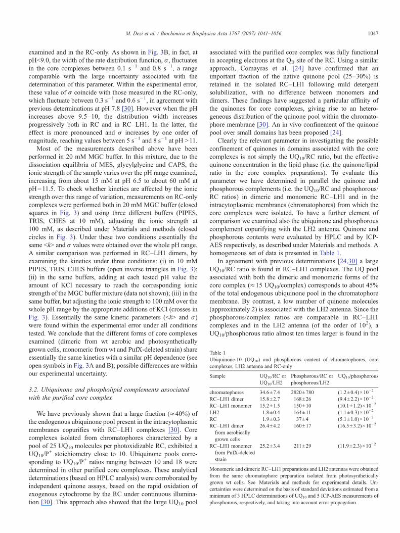
Table 1Ubiquinone-10 (UQ10) and phosphorous content of chromatophores, corecomplexes, LH2 antenna and RC-only
Sample UQ10/RC orUQ10/LH2
Phosphorous/RC orphosphorous/LH2
UQ10/phosphorous
chromatophores 34.6±7.4 2820±780 (1.2±0.4)×10−2
RC–LH1 dimer 15.8±2.7 168±26 (9.4±2.2)×10−2
RC–LH1 monomer 15.2±1.5 150±10 (10.1±1.2)×10−2
LH2 1.8±0.4 164±11 (1.1±0.3)×10−2
RC 1.9±0.3 37±4 (5.1±1.0)×10−2
RC–LH1 dimerfrom aerobicallygrown cells
26.4±4.2 160±17 (16.5±3.2)×10−2
RC–LH1 monomerfrom PufX-deletedstrain
25.2±3.4 211±29 (11.9±2.3)×10−2
Monomeric and dimeric RC–LH1 preparations and LH2 antennas were obtainedfrom the same chromatophore preparation isolated from photosyntheticallygrown wt cells. See Materials and methods for experimental details. Un-certainties were determined on the basis of standard deviations estimated from aminimum of 3 HPLC determinations of UQ10 and 5 ICP-AES measurements ofphosphorous, respectively, and taking into account error propagation.
1047M. Dezi et al. / Biochimica et Biophysica Acta 1767 (2007) 1041–1056
examined and in the RC-only. As shown in Fig. 3B, in fact, atpHb9.0, the width of the rate distribution function, σ, fluctuatesin the core complexes between 0.1 s−1 and 0.8 s−1, a rangecomparable with the large uncertainty associated with thedetermination of this parameter. Within the experimental error,these value of σ coincide with those measured in the RC-only,which fluctuate between 0.3 s−1 and 0.6 s−1, in agreement withprevious determinations at pH 7.8 [30]. However when the pHincreases above 9.5–10, the distribution width increasesprogressively both in RC and in RC–LH1. In the latter, theeffect is more pronounced and σ increases by one order ofmagnitude, reaching values between 5 s−1 and 8 s−1 at pH N11.
Most of the measurements described above have beenperformed in 20 mM MGC buffer. In this mixture, due to thedissociation equilibria of MES, glycylglycine and CAPS, theionic strength of the sample varies over the pH range examined,increasing from about 15 mM at pH 6.5 to about 60 mM atpH=11.5. To check whether kinetics are affected by the ionicstrength over this range of variation, measurements on RC-onlycomplexes were performed both in 20 mMMGC buffer (closedsquares in Fig. 3) and using three different buffers (PIPES,TRIS, CHES at 10 mM), adjusting the ionic strength at100 mM, as described under Materials and methods (closedcircles in Fig. 3). Under these two conditions essentially thesame bkN and σ values were obtained over the whole pH range.A similar comparison was performed in RC–LH1 dimers, byexamining the kinetics under three conditions: (i) in 10 mMPIPES, TRIS, CHES buffers (open inverse triangles in Fig. 3);(ii) in the same buffers, adding at each tested pH value theamount of KCl necessary to reach the corresponding ionicstrength of the MGC buffer mixture (data not shown); (iii) in thesame buffer, but adjusting the ionic strength to 100 mM over thewhole pH range by the appropriate additions of KCl (crosses inFig. 3). Essentially the same kinetic parameters (bkN and σ)were found within the experimental error under all conditionstested. We conclude that the different forms of core complexesexamined (dimeric from wt aerobic and photosyntheticallygrown cells, monomeric from wt and PufX-deleted strain) shareessentially the same kinetics with a similar pH dependence (seeopen symbols in Fig. 3A and B); possible differences are withinour experimental uncertainty.
3.2. Ubiquinone and phospholipid complements associatedwith the purified core complex
We have previously shown that a large fraction (≈40%) ofthe endogenous ubiquinone pool present in the intracytoplasmicmembranes copurifies with RC–LH1 complexes [30]. Corecomplexes isolated from chromatophores characterized by apool of 25 UQ10 molecules per photoxidizable RC, exhibited aUQ10/P
+ stoichiometry close to 10. Ubiquinone pools corre-sponding to UQ10/P
+ ratios ranging between 10 and 18 weredetermined in other purified core complexes. These analyticaldeterminations (based on HPLC analysis) were corroborated byindependent quinone assays, based on the rapid oxidation ofexogenous cytochrome by the RC under continuous illumina-tion [30]. This approach also showed that the large UQ10 pool
associated with the purified core complex was fully functionalin accepting electrons at the QB site of the RC. Using a similarapproach, Comayras et al. [24] have confirmed that animportant fraction of the native quinone pool (25–30%) isretained in the isolated RC–LH1 following mild detergentsolubilization, with no difference between monomers anddimers. These findings have suggested a particular affinity ofthe quinones for core complexes, giving rise to an hetero-geneous distribution of the quinone pool within the chromato-phore membrane [30]. An in vivo confinement of the quinonepool over small domains has been proposed [24].
Clearly the relevant parameter in investigating the possibleconfinement of quinones in domains associated with the corecomplexes is not simply the UQ10/RC ratio, but the effectivequinone concentration in the lipid phase (i.e. the quinone/lipidratio in the core complex preparations). To evaluate thisparameter we have determined in parallel the quinone andphosphorous complements (i.e. the UQ10/RC and phosphorous/RC ratios) in dimeric and monomeric RC–LH1 and in theintracytoplasmic membranes (chromatophores) from which thecore complexes were isolated. To have a further element ofcomparison we examined also the ubiquinone and phosphorouscomplement copurifying with the LH2 antenna. Quinone andphosphorous contents were evaluated by HPLC and by ICP-AES respectively, as described under Materials and methods. Ahomogeneous set of data is presented in Table 1.
In agreement with previous determinations [24,30] a largeUQ10/RC ratio is found in RC–LH1 complexes. The UQ poolassociated with both the dimeric and monomeric forms of thecore complex (≈15 UQ10/complex) corresponds to about 45%of the total endogenous ubiquinone pool in the chromatophoremembrane. By contrast, a low number of quinone molecules(approximately 2) is associated with the LH2 antenna. Since thephosphorous/complex ratios are comparable in RC–LH1complexes and in the LH2 antenna (of the order of 102), aUQ10/phosphorous ratio almost ten times larger is found in the

Table 2The effects of NaBr washing of chromatophores and detergent washing ofpurified complexes on phosphorous and UQ10 determinations
Sample UQ10/RC orUQ10/LH2
Phosphorous/RC orphosphorous/LH2
UQ10/phosphorous
chromatophores 24.8±6.4 3480±870 (0.71±0.26)×10−2
NaBr washedchromatophores
23.2±5.9 1490±330 (1.56±0.53)×10−2
RC–LH1 dimer 10.2±2.1 250±15 (4.1±0.9)×10−2
Detergent-washedRC–LH1 dimer
10.0±1.9 88±6 (11.4±2.3)×10−2
RC–LH1 monomer 9.5±1.8 285±13 (3.3±0.6)×10−2
Detergent-washedRC–LH1 monomer
9.3±1.4 81±7 (11.5±2.0)×10−2
LH2 1.6±0.3 117±8 (1.4±0.3)×10−2
Detergent-washedLH2
1.2±0.4 68±6 (1.8±0.6)×10−2
All complexes have been purified from the same chromatophore preparation ofphotosynthetically grown wt cells. Na–Br washing of chromatophores and thetreatment for partial removal of detergent in the purified complexes aredescribed in detail under Materials and methods.
1048 M. Dezi et al. / Biochimica et Biophysica Acta 1767 (2007) 1041–1056
core complexes as compared to the LH2 antenna. In purifiedcomplexes (RC–LH1, LH2) we can reasonably assume that thedetected phosphorous comes essentially from phospholipids.These data indicate therefore that the quinone concentration inthe lipid phase associated with RC–LH1 is markedly larger thanthat of the antenna. Moreover, since both RC–LH1 and LH2 areisolated using the same purification procedure, it is quiteunlikely that the quinone enrichment found in RC–LH1 resultsartefactually from detergent solubilization of the complex. Asshown in Table 1, RC–LH1 complexes (dimers) purified fromaerobically grown cells and from the PufX-deleted strain(monomers) are also characterized by high UQ10/complexratios, resulting in high ubiquinone concentrations (UQ10/phosphorous ratios larger than 10−1).
In the chromatophore membrane, the obtained UQ10/phosphorous ratio (1.2×10⁻²) is about eight times smallerthan that determined in the core complex. Assuming that also inthe chromatophore membrane the content of phosphorous canbe attributed mainly to phospholipids, this would indicate thatthe quinone concentration in the core complex is much largerthan the average quinone concentration in the chromatophoremembrane. Two aspects, however, have to be considered, whencomparing the quinone concentration in intact membranes andin purified complexes. First, a considerable fraction of thephosphorous determined in chromatophore suspensions can bedue to contamination by ribosomal particles rather than to thephospholipids [49]. To quantify this possible contribution, wedetermined phosphorous/RC and UQ10/RC ratios in chromato-phores washed with a 2-M NaBr solution. This treatment isknown to yield preparations essentially free of ribosomalproteins [50,51]. A second point, relevant when comparing thequinone concentration in isolated complexes and in membranes,concerns the lipid complement associated with the purifiedcomplexes. This large lipid complement (which varies con-siderably from preparation to preparation) is likely to include afraction of weakly bound phospholipids (possibly organized inmicellar structures involving or interacting with the detergentmolecules) and a tightly bound fraction. We measured thereforethe phosphorous/complex and UQ10/complex ratios alsofollowing detergent washing of the RC–LH1 and LH2preparations (as described under Materials and methods).When analyzing the composition of the phospholipids (seebelow), this treatment was routinely performed before lipidextraction to avoid detergent interference in TLC.
The results of the measurements described above aresummarized in Table 2. A comparison of the phosphorous/RCratio evaluated in untreated chromatophores and followingNaBr washing indicates that a large fraction (about 60%) of thephosphorous detected in untreated chromatophores comes from(presumably ribosomal) contamination. The UQ10/RC ratio isessentially unaffected by NaBr washing, resulting in a UQ10/phosphorous ratio of 1.6×10⁻² in the washed membranes. Indimeric and monomeric RC–LH1 complexes purified from thischromatophore preparation an approximately two times largerUQ10/phosphorous ratio is determined. When the phosphorouscontent measured in the untreated chromatophore preparation ofTable 1 is corrected accordingly (assuming that only 40% of the
detected phosphorous is contributed by phospholipids) a UQ10/phosphorous ratio of 3.1×10⁻² is obtained, which is about onethird of that determined in the purified core complexpreparations. This seems to indicate that the quinone concen-tration in the RC–LH1 is only moderately increased withrespect to the average concentration in the chromatophoremembrane. However, the results obtained in the purifiedcomplexes following detergent washing yield additionalinformation on this point. In fact, both in the monomeric andin the dimeric forms of the RC–LH1, this treatment causes aconsiderable reduction of the phosphorous/complex ratios, butleaves unaffected the ubiquinone complement (UQ10/complex).This is consistent with the idea that a large fraction of the lipidscopurifying with core complexes are only weakly bound,probably in interaction with detergent molecules, and that, mostimportantly, the tightly bound lipid fraction which remainsassociated with the core complex retains the entire quinonecomplement. The latter fact indicates that the large fraction ofthe membrane quinone pool associated with the core complexesis confined within a restricted lipid domain formed by less than102 phospholipid molecules (see Table 2). As a final result, theeffective quinone concentration in the detergent washed corecomplexes is almost one order of magnitude larger than theaverage quinone concentration evaluated in chromatophoresafter NaBr washing.
3.3. Lipid fractional composition in chromatophores and in thepurified complexes
The results described in the previous section bring to lightsignificant differences in the quinone/phospholipid ratiosmeasurable in RC–LH1, LH2 and chromatophores. Thisconclusion led us to search for possible differences also in thefractional composition of the lipid complements associated withthe purified core complexes, the peripheral antenna and intactmembranes. The lipid fractional composition was examined byexploiting two independent techniques, namely thin layer

Fig. 5. Proton decoupled 31P-NMR spectra of phospholipids extracted fromdifferent preparations and of mixtures of standards; the composition (wt.%) ofthe standard mixture is PC=5.3%, PE=42.1%, PG=31.6%, CL=21.0%. Fromtop to bottom: standard mixture, 20 mg overall lipids per mL; extract from
1049M. Dezi et al. / Biochimica et Biophysica Acta 1767 (2007) 1041–1056
chromatography (TLC) and 31P-NMR spectroscopy. Bothtechniques allow the determination of the relative amounts ofphospholipids but it should be noticed that TLC yields a weight-averaged composition (sulphuric acid staining is proportional tothe molecular weight), while 31P-NMR gives a compositionaveraged over the phosphorous nuclei. The two compositionswill coincide only if the ratio (molecular weight)/(phosphorousatom) is the same for all the phospholipids.
TLC analysis of lipid extracts provides clear evidence of adifferent lipid composition in the isolated core complexes ascompared to the LH2 antennas and to chromatophores. Theresults of the densitometric analysis of TLC plates aresummarized in Table 3. RC–LH1 and LH2 complexes werepurified from the same chromatophore preparation fromphotosynthetically grown cells. Four phospholipids were clearlyidentified on the basis of standards, i.e. phosphatidylcholine(PC), phosphatidylglycerol (PG), phoshatidylethanolamine (PE)and cardiolipin (CL). In the chromatophore extract, PE, PG andPC are the dominant lipids, accounting for approximately 40%,30% and 20% of the total weight respectively, in agreement withprevious determinations (e.g. [52,53]). The composition of theLH2 antenna is similar, except for a moderate increase in the CLcontent at the expenses of PE. At variance, the fractionalcomposition of the RC–LH1 complex is distinctly anddrastically different from that of chromatophores and of theLH2 antenna. In the core complex, in fact, CL becomes thedominant lipid (accounting for about 50% of the total). The CLincrease is accompanied by a considerable decrease in PE and, toa lower extent, in PC and PG.
These effects are consistent with the results of 31P-NMRanalysis of lipids extracted from an independent set ofchromatophores and purified complexes. Proton decoupled31P-NMR spectra of lipids extracted frommembranes, RC–LH1and LH2 complexes are shown in Fig. 5. The top and bottomspectra, corresponding to standard mixtures of PC, CL, PG andPE at high and low concentration, show that PC, CL and PG giverise to three distinct resonances (at 0.0, 0.77, and 1.25 ppm,respectively). As previously described in the literature [54], PEgives rise to three peaks (0.53, 0.47 and 0.18 ppm). The relativecontribution of the corresponding peaks is concentrationdependent. At high concentration of PE (see the top spectrumin Fig. 5), the peak at 0.53 ppm prevails, but dilution induces thedevelopment of the two high field NMR signals (at 0.47 ppm and0.18 ppm), attributed to the formation of adducts between PEand guanidinium ions (see the bottom spectrum in Fig. 5).
Chromatophore extracts are rich in phospholipids andovernight averaging yielded well resolved spectra. On the
Table 3Relative composition of lipids extracted from chromatophore membranes, RC–LH1 and LH2 complexes determined by TLC densitometric analysis
Sample PG (%) CL (%) PE (%) PC (%)
Chromatophores 30.2±1.3 8.8±1.8 40.1±0.6 20.9±1.1NaBr washed chromatophores 29.2±1.4 9.0±1.6 40.7±0.8 21.1±1.2RC–LH1 dimer 24.0±7.5 50.0±9.4 12.1±4.1 13.9±4.5LH2 29.4±3.5 14.1±5.8 36.6±2.7 19.9±2.4
The contribution of each lipid is expressed as percent of the total weight.
chromatophores, extract from chromatophores washed with NaBr; extract fromLH2 complexes; extract from RC–LH1 complexes; standard mixture, 1 mgoverall lipids per mL. In the case of chromatophores and LH2 complexescontinuous lines are fits to Voightian resonances; the peak positions and thecorresponding percentual composition obtained from the peak areas are listed inTable 4 (see text for details). In the case of RC–LH1 the continuous line is asimulation based on the relative phospholipid composition determined by TLC.The following resonances have been considered: PG at 1.25 ppm, CL at0.76 ppm, PC at 0.00 ppm, PE at 0.53 ppm and at 0.18 ppm. Peak widths wereset to 0.05 ppm for all lipids and the areas have been fixed according to the ratiosof Table 3 (PG=24%, CL=50%, PC=14%, PE=12%, equally distributed overthe two peaks considered).

1050 M. Dezi et al. / Biochimica et Biophysica Acta 1767 (2007) 1041–1056
contrary, when analyzing extracts from LH2 and RC–LH1preparations 1 week averaging was required to obtain areasonable statistics. In the spectra of chromatophore andLH2 extracts peaks corresponding to PG, PE, PC and CL aredetected. Each spectrum was deconvoluted in terms of aminimum number of Voightian functions and the relativephosphorous molar contribution was determined by integratingthe peaks corresponding to PG, PC, CL and PE. In the case ofPE (see above) the contribution of the peaks at 0.53 ppm,0.47 ppm and 0.18 ppm were summed up. In LH2 extracts twoadditional Voightian functions had to be included, peaking at1.8 ppm (unidentified phospholipid X1) and at 0.28 ppm(unidentified phospholipid X2). Only this last additional peakwas detectable in the case of the chromatophore membranes.The results of these deconvolution procedures are summarizedin Table 4. The molar fractions of PE, PG and PC in thechromatophore extracts are in good agreement with TLCdeterminations, when similar molecular weights are assumedfor these phospholipids. The cardiolipin content detected byNMR is systematically lower than that found by TLC if weassume that the CL molecular weight is twice that of otherlipids. However, the relative changes of CL between samples isconsistent: in both TLC and NMR determinations the CLcontent of LH2 complexes is about two times that found inmembrane extracts. The quantitative disagreement could be dueto the presence of the unidentified phospholipids probed byNMR but not by TLC. The presence of lipids other than PE, PC,PG and CL in the membranes of Rb. sphaeroides has beenreported (see e.g. [52,53]). We made no effort to identify suchlipids detectable in our NMR spectra. Due to the long averagingtime required by NMR analysis (see above) we cannot excludethat the unidentified lipids are degradation products. Theincrease in CL content of LH2 with respect to the averagechromatophore composition is consistent with results obtainedon spin-labelled chromatophores from Rb. sphaeroides [55].This study provided evidence that the antenna proteinspreferentially interact with negatively charged immobilizedlipids. A specific composition, differing from that of mem-branes, has been recently documented for the LH2 antenna ofRhodopseudomonas acidophila [56].
In the spectrum of the lipid extract from the RC–LH1complexes, due to the low signal to noise ratio, only the CLpeak at 0.77 ppm is clearly detectable. Two peaks at thefrequencies corresponding to PG and PC slightly exceed thelevel of noise. In agreement with TLC determinations, thisindicates that the lipid complement of the RC–LH1 is largelydominated by cardiolipin. To further check for consistency, the
Table 4Percentual lipid composition of chromatophores and LH2 antenna complexes determ
Sample X1 1.80 ppm PG 1.25 ppm CL 0.
Chromatophores – 31.8 1.6NaBr washed chromatophores – 34.4 2.1LH2 1.1 37.2 5.1
Data are from Fig. 5. The relative contribution of each lipid, calculated by integraphosphorous mole percent of the total. Also listed are the average positions of the peafor further details.
31P-NMR spectrum of the RC–LH1 lipid extract was simulatedassuming the lipid composition obtained from TLC (Table 3).The good agreement between the experimental spectrum andthat simulated (continuous line in Fig. 5) confirms that themassive increase in cardiolipin occurs mainly at the expenses ofPE, whose contribution to the NMR spectrum is comparable tothe noise level.
In summary, both TLC and NMR analyses concur to indicatethat the fractional lipid composition of the core RC–LH1complex differ substantially from the average composition ofthe chromatophore membrane, being strongly enriched incardiolipin.
4. Discussion
We have compared the recombination kinetics of the light-induced P+QA
− and P+QB− states in RC–LH1 core and RC-only
complexes over a wide pH range (6.5–11.8). In these twosystems essentially the same, pH independent, P+QA
− recombi-nation kinetics are observed (see Fig. 2). Noteworthy, in order toblock the electron transfer from QA
− to QB over the whole RCpopulation, a higher o-phenanthroline concentration (10 mM)was needed in RC–LH1. Since o-phenanthroline competes withUQ10 at the QB site [57], this observation is consistent with thepresence of a large, functional ubiquinone pool in the corecomplexes. P+QA
− recombination kinetics were reasonablydescribed by a single exponential decay. The average of thevalues shown in Fig. 2 yields a rate constant kAP=9.6 s−1 ingood agreement with a number of previous determinations inRCs (see e.g. [48]). By contrast, recombination of the P+QB
−
state was strongly decelerated in the presence of the LH1antenna at 6.5bpHb9.5 (see Fig. 3A). At higher pH values thiseffect is progressively reduced, vanishing around pH 11.0; at pHN11.0 the average rate constant measured in RC–LH1 exceedsslightly that observed in RC-only. Over the pH range examinedno significant difference was found between the kineticsrecorded in the dimeric and monomeric forms of RC–LH1purified from photosynthetically or semiaerobically growncells, as well as in the monomeric PufX− complex. At variancewith this latter finding, Comayras et al. [58] have observed inPufX−RC–LH1 monomers a P+QB
− recombination markedlyslower than in wild type core complexes. In PufX− monomers,at pH 8.0, they measured an half-time consistent with bkNvalues determined by us in all forms of core complexes, butobserved a faster recombination in wild type RC–LH1. We haveno explanation for this discrepancy. Both in RC-only and inRC–LH1 P+QB
− recombination kinetics are characterized by a
ined by 31P-NMR analysis of extracts
77 ppm PE 0.53, 0.47, 0.18 ppm X2 0.28 ppm PC 0.0 ppm
39.4 4.8 22.436.5 5.4 21.621.8 11.4 23.4
ting the Voightian functions fitted to the corresponding peaks, is expressed asks; X1 and X2 indicate unidentified phospholipids. See text and legend of Fig. 5

Fig. 6. The free energy difference ΔG0 between QA−QB and QAQB
− as a functionof pH. Symbols are as in Fig. 3. ΔG0 values were calculated according to Eq.(2). The values of the average rate constant bkN are taken from Fig. 3A; kAP hasbeen fixed to 9.6 s−1 on the basis of the data of Fig. 2. Continuous lines are bestfit to Eq. (3) corresponding to the following values of the fitting parameters. ForRC-only: ΔGH+
0 =−53 meV, pKA=9.6, pKB=10.7. For RC–LH1 corecomplexes: ΔGH+
0 =−90 meV, pKA=9.4, pKB=11.9.
1051M. Dezi et al. / Biochimica et Biophysica Acta 1767 (2007) 1041–1056
continuous spectrum of rate constants. The variance of the ratedistribution increases steeply above pH 10, giving rise tokinetics which are more strongly dispersed in the corecomplexes.
The pH dependence measured by us in OG suspensions ofRC-only is similar to that measured in LDAO suspensions ofRCs [48]. The values of the rate constant bkN measured in OGat 6.5bpHb9.0 are approximately two times larger than the rateconstant values determined by Kleinfeld et al. in LDAOsuspensions [48]. We confirmed that this difference issystematic and related to the detergent, by comparing thecharge recombination kinetics in the same RC-only preparation,before and after replacement of LDAO by OG and cholate (notshown). We have no clear interpretation for this detergent effect.LDAO and OG differ markedly in size, packing features [59]and H-bonding capability; these parameters could determinesubtle structural differences in the RC-detergent complex,possibly related to the kinetic effects observed.
The deceleration of the P+QB− recombination observed in
RC–LH1 as compared to the RC-only at 6.5bpHb9.5 isconsistent with previous measurements at pH≈8 [30,58]. Theinterpretation of this effect in relation to the large ubiquinonecomplement copurifying with the core complexes has beenconsidered in ref. [30] to which the reader is referred for athorough discussion. In that paper the effects of a rapidexchange of quinone at the QB site of the RC were examined onthe basis of a general kinetic scheme, developed by Shinkarevand Wraight [60], which takes also into account the possiblecompetition between the direct and the indirect (through theP+QA
− state) recombination route. We summarize in thefollowing the main conclusions reached in ref. [30], which area prerequisite for discussing the results obtained in the presentpaper over an extended pH range: (a) the dominant contributionto the average rate constant of P+QB
− recombination comes fromthe indirect route; (b) since P+QA
− recombination was notaffected by the presence of the LH1 antenna, the slowing downof charge recombination in RC–LH1 complexes implies adecrease in the energy level of the P+QAQB
− state relative to theground state; (c) an increase in the quinone concentration inrapid equilibrium with the QB site can decelerate therecombination kinetics, resulting in bkN ≈0.5 s−1 at saturatingquinone concentrations. This conclusion was reached on thebasis of studies performed on RCs in reverse micelles [43] andin proteoliposomes [61]. In both systems it was found thatincreasing the quinone concentration stabilized the P+QB
− state.The values of bkN observed in the present work at 6.5bpHb9.0in RC–LH1 fluctuate between 0.1 and 0.4 s−1 (see Fig. 3A).They cannot therefore be explained solely by a quinoneconcentration effect, although the high ubiquinone concentra-tion in the associated lipid phase (see Table 1 and 2) is likely toapproach saturation. Under this condition, assuming that chargerecombination proceeds essentially by thermal repopulation ofthe P+QA
− state, the free energy difference ΔG0 between thestates P+QA
− QB and P+QAQB− can be simply related to the rate
constant for P+QB− (bkN) and P+QA
− (kAP) recombination, i.e.
DG0 ¼ �kBT lnðkAP=bkN� 1Þ ð2Þ
where kB is the Boltzmann constant and T the absolutetemperature [48,60].
Fig. 6 shows the pH dependence of ΔG0 calculated from thedata of Fig. 3B by using Eq. (2) and setting kAP=9.6 s−1 overthe whole pH range (see Fig. 2). The pH dependence of electrontransfer from QA
− to QB has been extensively studied in RCcomplexes (for a review see [60]). At pH N6.5 this dependenceis basically explained by a simple thermodynamic model, whichconsiders the coupling between electron transfer and theprotonation equilibrium of a single aminoacid residue near theQB site [48]. Detailed kinetic analyses in mutated RCs(reviewed in [60]) support the idea that the pH dependence ofthe free energy difference ΔG0 is determined by protonation ofGluL212, as originally proposed by Paddock et al. [62].According to the model, the measured free energy differenceΔG0 is given by [48]:
DG0 ¼ DG0Hþ � kBT ln
1þ 10ðpH�pKBÞ
1þ 10ðpH�pKAÞ ð3Þ
where ΔGH+0 is the free energy difference for QA
− to QB electrontransfer when the residue is protonated and pKA and pKB arethe acid dissociation constants of the residue when the RC is inthe QA
−QB and QAQB− states, respectively. Eq. (3) describes an
S-shaped curve with turning points at pKA and pKB, beingΔGH+
0 andΔG00 =ΔGH+
0 −kBTln 10(pKA−pKB) the limiting freeenergies at low and high pH values, respectively [48]. Whenfitting Eq. (3) to the pH dependence measured in RC-only weobtain ΔGH+
0 =−53 meV, pKA=9.6, pKB=10.7. These valuesare comparable to those obtained by Kleinfeld et al. [48] inLDAO suspensions of RCs (ΔGH+
0 =−67 meV, pKA=9.8,pKB=11.3), except that the presence of OG as detergent slightlydestabilizes the QAQB
− state when the coupled residue isprotonated and the QA
−QB state in the deprotonated form. Fittingto the pH dependence measured in RC–LH1 yields ΔGH+
0 =−90 meV, pKA=9.4, pKB=11.9. As it is evident from Fig. 6,

1052 M. Dezi et al. / Biochimica et Biophysica Acta 1767 (2007) 1041–1056
over the physiological pH range (when the ionisable residuecoupled to the electron transfer is protonated) the QAQB
− state ismarkedly stabilized (by 37 meV) in the core complexes ascompared to the RC-only. Moreover pKB is apparently shiftedby about one pH unit, i.e., at extremely alkaline pH values, theQAQB
− state is more destabilized in RC–LH1 (ΔG00 =56 meV for
the core complexes, as compared toΔG00 =11 meV in RC-only).
The value of ΔG0 measured in RC–LH1 under physiologicalconditions (ΔGH+
0 ) agrees with estimates of the free energydifference in chromatophores [63], showing that the integrity ofthe core complex accounts quantitatively for the stabilization ofthe QAQB
− observed in the native membranes. The lipidcomplement of the RC–LH1 complexes exhibits a specificcomposition (Table 3 and Fig. 5) and is likely to play a role instabilizing the charge separated P+QB
− state (see below).Our analysis of P+ decay shows that the kinetics of P+QB
−
recombination, which are moderately distributed below pH 9,becomes strongly non-exponential at increasing pH values (seeFigs. 1 and 3B). This behaviour, which is more pronounced incore complexes (but is also observed in RC-only complexes),has not been reported previously for RCs purified from Rb.sphaeroides [48]. In RC-only, the width, σ, of the ratedistribution function is always considerably smaller (aboutone third) than the average rate constant, bkN (cf. Fig. 3A andB). At variance, in RC–LH1 complexes, it is systematicallycomparable to bkN, indicating a substantial kinetic hetero-geneity. In view of the pH dependence of the recombinationrate, the observed distribution of the kinetics may result from anensemble of conformations differing in the protonation state ofthe residue(s) thermodynamically coupled to the electrontransfer between QA and QB. We note that in general theexistence of an ensemble of conformations, each characterizedby a different rate constant, will give rise to an observablekinetic heterogeneity only when the interconversion betweenconformations is slower than the electron transfer processobserved (in our case the recombination of the P+QB
− state). Afaster interconversion will average the kinetic heterogeneity,resulting in the observation of monoexponential (or scarcelydistributed) kinetics. This could explain the increase of thedistribution width at alkaline pH values (Fig. 3B), whenassuming that the relevant conformations interconvert at a ratewhich is comparable to the rate of charge recombination atpHb9. At higher pH values, the rate of interconversion wouldbecome progressively smaller than the increasing rate ofrecombination of the P+QB
− state, and the conformationalheterogeneity would become progressively observable.
Interestingly at alkaline pH values the variance of the ratedistribution is considerably larger in RC–LH1 than in RC-onlycomplexes, suggesting that the conformational heterogeneityof the system increases when its structural complexity andintegrity increase. A recent AFM investigation has revealedlarge molecule-to-molecule variations for the LH1 ring, in termsof both shape and size [64]. The large kinetic heterogeneitywe observed in RC–LH1 is consistent with results obtainedin chromatophores of Rb. capsulatus FJ2 [65], a c2- and cy-minus strain, in which the kinetics of P+QB
− recombination canbe accurately studied by avoiding any interference due to
exogenous electron donors/acceptors. In this system chargerecombination kinetics were fitted to the sum of two exponentialdecays at pH≤8.5, but at pH 10 a third exponential phase had tobe added to account for the kinetics [65], indicating that also inchromatophores recombination kinetics becomes progressivelynon-exponential (distributed) at alkaline pH values. We havereanalyzed the pH dependence of this reaction in Rb. capsulatusFJ2 chromatophore at 6.5bpHb12 and found that the kinetics ofP+QB
− recombination are best described by a power law, i.e. by arate distribution characterized by a width σwhich increases by afactor of 5 from pH 9.5 to pH 11.5 (data not shown).
Our analysis of the lipid and quinone complements ofchromatophores and of the purified complexes indicates that alipid domain corresponding to about 80–90 head groupphosphatides is tightly associated with the purified RC–LH1(see Table 2) and that the purified complex retains aconsiderable fraction of the total ubiquinone pool present inthe membrane. The resulting ubiquinone concentration in thecore complex lipid phase is about 7-fold larger than the averageubiquinone concentration in the chromatophore membrane(Table 2). Such an enrichment in quinone is not found in thepurified LH2 antenna complex, which is also characterized by atightly associated lipid complement. These observationsindicate that quinones tend to associate specifically with theRC–LH1 complex and support the idea of a strong in vivoheterogeneity in the spatial distribution of quinones within thechromatophore membrane. By exploiting an independentapproach, Comayras et al. [24] have recently provided evidencefor confinement of quinones within small domains. Our datasupport the view that quinone-rich patches in the membrane arepromoted by interactions of quinones with the RC–LH1 proteinsubunits and/or with the specific lipid environment associatedwith the core complex. By studying the accumulation of QA
−
during the pool photoreduction, Comayras et al. [24] obtainedinformation on the statistical distribution of the UQ10/RCstoichiometry both in the purified RC–LH1 complexes and inthe proposed quinone domains of the chromatophore mem-branes. For both systems, data were consistent with a broaddistribution. Since the effective quinone concentration in rapidequilibrium with the QB site affects the rate of P+QB
−
recombination ([60], see also [30]), a broadly distributedUQ10/RC stoichiometry can well contribute to the broaddistribution of the rate constant observed by us (Fig. 3B).Statistical fluctuations in the fractional composition of the lipidsassociated with the core complexes are also a possible source ofkinetic heterogeneity, since the interaction of the RC withdifferent lipids has been shown to affect differently the kineticsof charge recombination (see below). The kinetic heterogeneityobserved in RC-only could arise from heterogeneity in theoccupancy of one or more lipid binding sites that becomeexposed on the surface of the RC on detergent purification ofthis part of the RC–LH1 complex.
The effects of a lipid environment on the thermodynamicsand kinetics of electron transfer processes within the RC hasbeen studied mainly by comparing the behaviour of RCs indetergent suspensions and when incorporated into liposomes ofdifferent phospholipid composition. A systematic analysis [66]
1053M. Dezi et al. / Biochimica et Biophysica Acta 1767 (2007) 1041–1056
showed that in RCs embedded in PC vesicles the rate constantfor P+QB
− recombination decreases from 0.77 s−1, measured inLDAO detergent suspension, to 0.39 s−1 at pH 7. Incorporationinto PG liposomes induces a further decrease of the rateconstant to 0.26 s−1. Correspondingly, the calculated freeenergy differences for QA
− to QB electron transfer,ΔG0, equal to−62 meV in detergent, decreased in PC and PG to −77 meVandto −89 meV, respectively [66]. The values obtained in thepresence of PG are close to our estimate in RC–LH1complexes. Changes in ΔG0 are attributed to a modificationof the QA/QA
− redox potential, measured by delayed fluores-cence [66]. Addition of PG and CL to RC in detergentsuspension also caused a significant slowing of P+QB
−
recombination [67], which was more pronounced (approxi-mately 3-fold) in the case of CL. The effect was related to a30 mV decrease of the redox potential of QA/QA
− as evaluatedfrom delayed fluorescence data. Giustini et al. [68] foundconsistently that recombination of the primary charge separatedstate was accelerated in QB-deprived RCs incorporated intocardiolipin/lecithin liposomes or suspended in detergentmicelles doped with cardiolipin, suggesting a destabilizationof the P+QA
− state induced by CL and the occurrence of severalCL binding sites on the RC. The extent of P+QB
− decelerationinduced by the addition of CL [67] is comparable to the slowingdown observed by us in RC–LH1 with respect to RC-onlycomplexes. In the case of RC–LH1, however, we have proposedthat the stabilization of the charge separated P+QB
− state is due toa change in the energy level of the QB
− state, rather than in theredox potential of the primary quinone acceptor, because wefound the same rate constant kAP of P
+QA− recombination in RC-
only and in core complexes. The kAP values we measured (Fig.2) are fully consistent with the kinetics reported by Comayras etal. [58] for wt dimeric RC–LH1 and for monomeric complexesisolated from the PufX-deleted strain. The kinetics of P+QA
−
recombination are quite sensitive to variations in the associatedfree energy change, obeying at room temperature a classicalMarcus relation [40]. According to the data of Lin et al. [40] anincrease of 30 meV in the free energy change is expected toincrease the value of kAP from 9.4 s−1 to approximately 11.5s−1, an effect which is not seen in the RC–LH1 complex (Fig. 2and ref. [58]).
The results obtained in proteoliposomes [66,68] andcardiolipin-doped detergent suspensions [67,68] indicate inany case that the PG and CL can significantly perturb theelectron transfer equilibrium between QA and QB. This suggeststhat the deceleration of charge recombination observed by us inRC–LH1 is likely due to the interaction of the RC with thenative lipid environment which is preserved in the purified corecomplexes. Remarkably, the contributions of PG and CL, whichappear more effective in perturbing the thermodynamics ofinterquinone electron transfer, account for about 75% of thetotal weight of the lipid complement associated with the RC–LH1 (Table 3). The lipid composition of RC–LH1 appears to bequite specific, differing markedly from that of the purified LH2antenna, which is more similar to the average chromatophorecomposition and is characterized by a much lower content ofcardiolipin (see Tables 3, 4 and Fig. 5). The presence of
negatively charged lipids, tightly bound to the RC–LH1complex, is likely to perturb the local electrostatics as well asthe extended hydrogen-bond network in the region of the QB
pocket of the RC as discussed in ref. 66, resulting in theincreased stability of the P+QB
− state at 6.5bpHb9.0 and in thealteration of the pH dependence of the free energy ΔG0 forinterquinone electron transfer (Fig. 6). The QAQB acceptorcomplex constitutes a finely tuneable structural and functionalunit, responding to long distance interactions [69]. It isconceivable that in systems (RC-only and RC–LH1) character-ized by specific protein–lipid and protein–protein contacts thelipid environment affects the quinone acceptor complexdifferently, altering the thermodynamic properties either of QA
or of QB.A significant fraction of the X-ray crystal structures of the
Rb. sphaeroides RC contains a molecule of CL tightly boundto the intra-membrane surface of the M subunit (for a recentreview see [27]). In a recent X-ray diffraction study two lipidbinding sites have been identified unequivocally in RCscocrystallized with added brominated phospholipids [70].Since the elucidation of the structural details of CL–RCinteractions [28], molecules of CL have been modelled in avariety of membrane protein complexes, including the cyto-chrome bc1 complex and the bovine cytochrome c oxidase (fora review see [26]). Often in these structures, CL binds atinterfaces between subunits, suggesting a role in the stabiliza-tion of supermolecular assemblies [26]. In view of this it is notsurprising that the lipid complement tightly bound to RC–LH1showed a specific composition, characterized by a markedenrichment in the CL content as compared to the LH2 antennaand to the average chromatophore composition. Noteworthy,when cardiolipin binding was destabilized by site-directedmutagenesis, the preparation of highly pure mutated RCs couldbe more easily achieved, omitting a final ion exchangepurification step [71]. This has been taken as evidence that, inthe absence of the CL molecule, the tendency of the RC toassociate with other proteins is lessened. An additionalobservation which points to a role of cardiolipin in mediatingprotein–protein interactions is that, in the lipidic cubic phasecrystal structure of Rb. sphaeroides RC, the head groupphosphatides of CL interacts electrostatically not only withthree positively charged residues of the M subunit (Lys144,His145 and Arg267) but also with Lys202 of the L subunit of asymmetry related reaction center. In this way, strong crystalcontacts are formed between two symmetry-related RCs at themembrane surface, resulting in a quasi-dimeric structure [17].
Phospholipids are expected to interact with the RC–LH1complex at the protein surfaces, forming annular shells, as wellas in cavities and clefts located more deeply and frequentlyobserved in multisubunit assemblies. Based on the lowresolution structure of the dimeric RC–LH1 obtained byScheuring et al. [8], we can roughly estimate the number oflipid molecules necessary to build a complete annular shell atthe outer membrane spanning surface of the complex. Byassuming an area of 60 Å2 for an average phospholipid head[72] approximately 50–60 molecules per RC–LH1 monomercan be accommodated to form an external annular shell. A

1054 M. Dezi et al. / Biochimica et Biophysica Acta 1767 (2007) 1041–1056
hydrophobic volume of about 7.5×10⁴ Å3 has been previouslyestimated for the interstice between the inner surface of the LH1antenna ring and the outer RC surface [30]. After subtracting avolume of approximately 1.8×10⁴ Å3 estimated for a pool of 10UQ10molecules [30], filling of the remaining space wouldrequire approximately 30–35 additional phospholipid heads, sothat a total of 80–95 phospholipid molecules per RC–LH1monomer could be placed in close interaction with the corecomplex. Although this estimate is based on extremely crudeassumptions, it compares well with a phosphorous/RC ratio∼80–90, determined after detergent washing both in dimericand monomeric RC–LH1 complexes. By examining a structuralmodel of the RC–LH1 built by Fotiadis et al. [73] it has beenpointed out that a large cavity exists between the surface of theRC (in the region of the QB site) and the inner surface of thesurrounding LH1 antenna [27]. As already suggested [27], thislarge cavity provides additional space for lipid molecules,forming a suitably flexible environment for quinone moleculeswaiting in proximity of the QB site.
Based on the increasing number of membrane proteinstructures with tightly bound lipids, some features of lipid-binding sites have been tentatively identified [26]. Basicresidues, including in the order of occurrence Arg, Lys, Tyr,His, have been observed as primary ligands of phosphodiestergroups. Aromatic residues are frequently involved in lipidstabilization. Tryptophan residues, located in the hydrophobic/hydrophilic transition zone, interact by hydrogen bondsbetween the indole nitrogen and the phosphodiester group andby a lamellar orientation of their indole rings between adjacentphospholipids chains. This pattern is thought to provide aninterlocking system for the annular lipid shell [26]. A ring ofsurface exposed arginine and histidine side chains is located atthe boundary between the transmembrane and the extrinsicportions of the RC on the cytoplasmic side [74]. Moreover ringsof lipid-exposed tryptophan residues are present both in theLH1 and in the RC complexes [75]. These regular arrays ofresidues provide in principle a structural basis for thesupermolecular organization of the antenna–RC complex bylipid-mediated protein–protein interactions. It may be ofinterest that two tryptophan residues are found close to the N-terminal end of the transmembrane α-helix of the PufX proteinof Rb. sphaeroides [76–78] and that, among all second-sitemutations that suppress the PufX− phenotype, most are non-sense mutations of the LH1 lipid-exposed tryptophans whichprevent the assembly of the LH1 subunits [79].
In view of the above considerations it is conceivable that theconfinement of cardiolipin in the RC–antenna may play a rolein the structural organization of the complex. Our data, whenconsidered in relation to studies performed on RCs reconstitutedinto quinone-enriched phospholipids vesicles [61,66] andcardiolipin-doped detergent suspensions [67], suggest that thecardiolipin and quinone confinement observed in RC–LH1complexes may concur to promote the stabilization of the P+QB
−
charge separated state of the RC. Whether the high quinoneconcentration in the complex is in turn determined or assisted bythe cardiolipin-rich lipid environment of the core complexneeds to be investigated. It seems in any case that protein–
lipid–quinone interactions interconnect closely at the level ofthe core complex and cooperate in optimizing the yield and theturnover rate of charge separation by the RC.
Acknowledgements
This workwas supported byMIUR of Italy (Grants PRIN2005prot. 2005052128 and PRIN2005 prot. 2005027011). G.P. andG. C. were partially supported by the Consorzio Interuniversitarioper lo sviluppo dei Sistemi a Grande Interfase (CSGI-Firenze).We are grateful to B.A. Melandri and P. Turina for stimulatingdiscussions and to C. Pagnucco for technical assistance in HPLCdeterminations.
References
[1] W.A. Cramer, D.B. Knaff, Energy Transduction in Biological Membranes,Springer-Verlag, New York, 1990.
[2] G. Feher, J.P. Allen, M.Y. Okamura, D.C. Rees, Structure and function ofbacterial photosynthetic reaction centres, Nature 339 (1989) 111–116.
[3] M.R. Gunner, The reaction center protein from purple bacteria: structureand function, Curr. Top. Bioenerg. 16 (1991) 319–367.
[4] M.Y. Okamura, M.L. Paddock, M.S. Graige, G. Feher, Proton and electrontransfer in bacterial reaction centers, Biochim. Biophys. Acta 1458 (2000)148–163.
[5] A.R. Crofts, C.A. Wraight, The electrochemical domain of photosynthesis,Biochim. Biophys. Acta 726 (1983) 149–185.
[6] N.W. Isaacs, R.J. Cogdell, A.A. Freer, S.M. Prince, Light-harvestingmechanisms in purple photosynthetic bacteria, Curr. Opin. Struct. Biol. 5(1995) 794–797.
[7] S. Scheuring, J. Seguin, S. Marco, D. Levy, C. Breyton, B. Robert, J.L.Rigaud, AFM characterization of tilt and intrinsic flexibility of Rhodo-bacter sphaeroides light harvesting complex 2 (LH2), J. Mol. Biol. 325(2003) 569–580.
[8] S. Scheuring, F. Francia, J. Busselez, B.A. Melandri, J.L. Rigaud, D. Levy,Structural role of PufX in the dimerization of the photosynthetic corecomplex of Rhodobacter sphaeroides, J. Biol. Chem. 279 (2004)3620–3626.
[9] E.C. Abresch, H.L.A. Axelrod, J.T. Beatty, J.A. Johnson, R. Nechushtai,M.L. Paddock, Characterization of a highly purified, fully active,crystallisable RC–LH1–PufX core complex from Rhodobacter sphaer-oides, Photosynth. Res. 86 (2005) 61–70.
[10] J.W. Farchaus, W.P. Barz, H. Grünberg, D. Oesterhelt, Studies on theexpression of the PufX polypeptide and its requirement for photoheter-otrophic growth in Rhodobacter sphaeroides, EMBO J. 11 (1992)2779–2788.
[11] C. Jungas, J.L. Ranck, J.L. Rigaud, P. Joliot, A. Vermeglio, Supramole-cular organization of the photosynthetic apparatus of Rhodobactersphaeroides, EMBO J. 18 (1999) 534–542.
[12] P. McGlynn, C.N. Hunter, M.R. Jones, The Rhodobacter sphaeroidesPufX protein is not required for photosynthetic competence in the absenceof a light harvesting system, FEBS Lett. 349 (1994) 349–353.
[13] F. Francia, J. Wang, G. Venturoli, B.A. Melandri, W.P. Barz, D. Oesterhelt,The Reaction Center–LHI antenna complex of Rhodobacter sphaeroidescontains one PufX molecule which is involved in dimerization of thiscomplex, Biochemistry 38 (1999) 6834–6845.
[14] J.P. Allen, G. Feher, T.O. Yeates, H. Komiya, D.C. Rees, Structure of thereaction center from Rhodobacter sphaeroides R-26: the cofactors, Proc.Natl. Acad. Sci. U. S. A. 84 (1987) 5730–5734.
[15] J.P. Allen, G. Feher, T.O. Yeates, H. Komiya, D.C. Rees, Structure of thereaction center from Rhodobacter sphaeroides R-26: the protein subunits,Proc. Natl. Acad. Sci. U. S. A. 84 (1987) 6162–6166.
[16] U. Ermler, G. Fritzsch, S.K. Buchanan, H. Michel, Structure of thephotosynthetic reaction centre from Rhodobacter sphaeroides at 2.65 Å

1055M. Dezi et al. / Biochimica et Biophysica Acta 1767 (2007) 1041–1056
resolution: cofactors and protein–cofactor interactions, Structure 2 (1994)925–936.
[17] G. Katona, U. Andreasson, E.M. Landau, L.E. Andreasson, R. Neutze,Lipidic cubic phase crystal structure of the photosynthetic reaction centrefrom Rhodobacter sphaeroides at 2.35 Å resolution, J. Mol. Biol. 331(2003) 681–692.
[18] E.A. Berry, L.-S. Huang, L.K. Saechao, N.G. Pon, M. Valkova-Valchanova, F. Daldal, X-ray structure of Rhodobacter capsulatus bc1:comparison with its mitochondrial and chloroplast counterparts, Photo-synth. Res. 81 (2004) 251–275.
[19] P. Qian, C.N. Hunter, P.A. Bullough, The 8.5 Å projection structure of thecore RC–LH1–PufX dimer of Rhodobacter sphaeroides, J. Mol. Biol. 349(2005) 948–960.
[20] S. Bahatyrova, R.N. Frese, C.A. Siebert, J.D. Olsen, K.O. van der Werf, R.van Grondelle, R.A. Niederman, P.A. Bullough, C. Otto, C.N. Hunter, Thenative architecture of a photosynthetic membrane, Nature 430 (2004)1058–1062.
[21] S. Scheuring, J. Busselez, D. Levy, Structure of the dimeric PufX-containing core complex of Rhodobacter blasticus by in situ atomic forcemicroscopy, J. Biol. Chem. 280 (2005) 1426–1431.
[22] A. Vermeglio, P. Joliot, Supramolecular organization of the photosyntheticchain in anoxygenic bacteria, Biochim. Biophys. Acta 1555 (2002) 60–64.
[23] L.A. Drachev, M.D. Mamedov, A.Y. Mulkidjanian, A.Y. Semenov, V.P.Shinkarev, M.I. Verkhovsky, Transfer of ubiquinol from the reaction centerto the bc1 complex in Rhodobacter sphaeroides chromatophores underoxidizing conditions, FEBS Lett. 245 (1989) 43–46.
[24] F. Comayras, C. Jungas, J. Lavergne, Functional consequences of theorganization of the photosynthetic apparatus in Rhodobacter sphaeroides.I. Quinone domains and excitation transfer in chromatophores and reactioncenter–antenna complexes, J. Biol. Chem. 280 (2005) 11203–11213.
[25] P.K. Fyfe, K.E. McAuley, A.W. Roszak, N.W. Isaacs, R.J. Cogdell, M.R.Jones, Probing the interface between membrane proteins and membranelipids by X-ray crystallography, Trends Biochem. Sci. 26 (2001) 106–112.
[26] H. Palsdottir, C. Hunte, Lipids in membrane protein structures, Biochim.Biophys. Acta 1666 (2004) 2–18.
[27] M.R. Jones, Lipids in photosynthetic reaction centres: structural roles andfunctional holes, Prog. Lipid Res. 46 (2007) 56–87.
[28] K.E. McAuley, P.K. Fyfe, J.P. Ridge, N.W. Isaacs, R.J. Cogdell, M.R.Jones, Structural details of an interaction between cardiolipin and anintegral membrane protein, Proc. Natl. Acad. Sci. U. S. A. 96 (1999)14706–14711.
[29] A. Camara-Artigas, D. Brune, J.P. Allen, Interactions between lipids andbacterial reaction centers determined by protein crystallography, Proc.Natl. Acad. Sci. U. S. A. 99 (2002) 11055–11060.
[30] F. Francia, M. Dezi, A. Rebecchi, A. Mallardi, G. Palazzo, B.A. Melandri,G. Venturoli, Light-harvesting complex I stabilizes P+QB
− charge separationin reaction centers of Rhodobacter sphaeroides, Biochemistry 43 (2004)14199–14210.
[31] K.A. Gray, J.W. Farchaus, J.W. Wachtveilt, J. Breton, D. Oesterhelt, Initialcharacterization of site-directed mutants of tyrosine M210 in the reactioncentre of Rhodobacter sphaeroides, EMBO J. 9 (1990) 2061–2070.
[32] F.E. Jenney Jr., F. Daldal, A novel membrane-associated c-typecytochrome, cyt cy, can mediate the photosynthetic growth of Rhodobac-ter capsulatus and Rhodobacter sphaeroides, EMBO J. 12 (1993)1283–1292.
[33] A. Baccarini-Melandri, B.A. Melandri, Partial resolution of the photopho-sphorylating system of Rhodopseudomonas capsulata, Methods Enzymol.23 (1971) 556–561.
[34] R. Piazza, M. Pierno, E. Vignati, G. Venturoli, F. Francia, A. Mallardi, G.Palazzo, Liquid–liquid phase separation of surfactant-solubilized mem-brane protein, Phys. Rev. Lett. 90 (2003) 208101.
[35] J.R. Bowyer, S. Meinhardt, G.V. Tierney, A.R. Crofts, Resolved differencespectra of redox centers involved in photosynthetic electron flow in Rho-dopseudomonas capsulata and Rhodopseudomonas sphaeroides, Bio-chim. Biophys. Acta 635 (1981) 167–186.
[36] P.L. Dutton, J.B. Jackson, Thermodynamic and kinetic characterization ofelectron transfer components in situ in Rhodopseudomonas sphaeroidesand Rhodospirillum rubrum, Eur. J. Biochem. 30 (1972) 495–510.
[37] J.B. Jackson, R.J. Cogdell, A.R. Crofts, Some effects of o-phenanthrolineon electron transport in chromatophores from photosynthetic bacteria,Biochim. Biophys. Acta 292 (1973) 218–225.
[38] J.C. Williams, R.G. Alden, H.A. Murchison, J.M. Peloquin, N.W.Woodbury, J.P. Allen, Effects of mutations near the bacteriochlorophyllsin reaction centers from Rhodobacter sphaeroides, Biochemistry 31(1992) 11029–11037.
[39] R.W. Visschers, S.I.E. Vulto, M.R. Jones, R. van Grondelle, R.Kraayenhof, Functional LH1 antenna complexes influence electrontransfer in bacterial photosynthetic reaction centers, Photosynth. Res. 59(1999) 95–104.
[40] X. Lin, H.A. Murchison, V. Nagarajan, W.W. Parson, J.P. Allen, J.C.Williams, Specific alteration of the oxidation potential of the electrondonor in reaction centers from Rhodobacter sphaeroides, Proc. Natl. Acad.Sci. U. S. A. 91 (1994) 10265–10269.
[41] R.K. Clayton, Spectroscopic analysis of bacteriochlorophylls in vitro andin vivo, Photochem. Photobiol. 5 (1966) 669–677.
[42] L. Slooten, Electron acceptors in reaction center preparations fromphotosynthetic bacteria, Biochim. Biophys. Acta 275 (1972) 208–218.
[43] A. Mallardi, G. Palazzo, G. Venturoli, Binding of ubiquinone tophotosynthetic reaction center: determination of enthalpy and entropychanges of the binding process in reverse micelles, J. Phys. Chem., B 101(1997) 556–561.
[44] G. Palazzo, A. Mallardi, A. Hochkoeppler, L. Cordone, G. Venturoli,Electron transfer kinetics in photosynthetic reaction centers embedded intrehalose glasses: trapping of conformational substrates at room tempera-ture, Biophys. J. 82 (2002) 558–568.
[45] G. Venturoli, J.G. Fernandez-Velasco, A.R. Crofts, B.A. Melandri,Demonstration of a collisional interaction of ubiquinol with theubiquinol–cytochrome c2 oxidoreductase complex in chromatophoresfrom Rhodobacter sphaeroides, Biochim. Biophys. Acta 851 (1986)340–352.
[46] E.G. Bligh, W.J. Dyer, A rapid method of total lipid extraction andpurification, Can. J. Biochem. Physiol. 37 (1959) , 911–917.
[47] M. Bosco, N. Culeddu, R. Toffanin, P. Pollesello, Organic solvent systemsfor 31P nuclear magnetic resonance analysis of lecithin phospholipids:applications to two-dimensional gradient-enhanced 1H-detected hetero-nuclear multiple quantum coherence experiments, Anal. Biochem. 245(1997) 38–47.
[48] D. Kleinfeld, M.Y. Okamura, G. Feher, Electron transfer in reactioncenters of Rhodopseudomonas sphaeroides. I. Determination of thecharge recombination pathway of D+QAQB
− and free energy and kineticrelations between QA
−QB and QAQB−, Biochim. Biophys. Acta 766 (1984)
126–140.[49] P.J. Fraker, S. Kaplan, Isolation and fractionation of the photosynthetic
membranous organelles from Rhodopseudomonas sphaeroides, J. Bacter-iol. 108 (1971) 465–473.
[50] D. Schlueserner, F. Fisher, J. Kruip, M. Rögner, A. Poetsch, Mapping themembrane proteome of Corynebacterium glutamicum, Proteomics 5(2005) 1317–1330.
[51] C. Klein, C. Garcia-Rizo, B. Bisle, B. Scheffer, H. Zischka, F. Pfeiffer, F.Siedler, D. Oesterhelt, The membrane protome of Halobacteriumsalinarum, Proteomics 5 (2005) 180–197.
[52] J.C. Onishi, R.A. Niederman, Rhodopseudomonas sphaeroides mem-branes: alterations in phospholipid composition in aerobically andphototrophically grown cells, J. Bacteriol. 149 (1982) 831–839.
[53] T.J. Donohue, B.D. Cain, S. Kaplan, Alterations in the phospholipidcomposition of Rhodopseudomonas sphaeroides and other bacteriainduced by Tris, J. Bacteriol. 152 (1982) 595–606.
[54] S. Murgia, S. Mele, M. Monduzzi, Quantitative characterization ofphospholipids in milk fat via 31P NMR using a monophasic solventmixture, Lipids 38 (2003) 585–591.
[55] G.B. Birrel, W.R. Sistrom, O.H. Griffith, Lipid–protein associations inchromatophores from the photosynthetic bacterium Rhodopseudomonassphaeroides, Biochemistry 17 (1978) 3763–3768.
[56] N.J. Russel, J.K. Coleman, T.D. Howard, E. Johnston, R.J. Cogdell,Rhodopseudomonas acidophila strain 10050 contains photosynthetic LH2antenna complexes that are not enriched with phosphatidylglycerol, and
1056 M. Dezi et al. / Biochimica et Biophysica Acta 1767 (2007) 1041–1056
the phospholipids have a fatty acyl composition that is unusual for purplenon-sulfur bacteria, Biochim. Biophys. Acta 1556 (2002) 247–253.
[57] C.A. Wraight, Oxidation–reduction physical chemistry of the acceptorquinone complex in bacterial photosynthetic reaction centers: evidence fora new model of herbicide activity, Israel, J. Chem. 21 (1981) 348–354.
[58] F. Comayras, C. Jungas, J. Lavergne, Functional consequences of theorganization of the photosynthetic apparatus in Rhodobacter sphaeroides.II. A study of PufX-membranes, J. Biol. Chem. 280 (2005) 11214–11223.
[59] P. Thiyagarajan, D.M. Tiede, Detergent micelle structure and micelle–micelle interactions determined by small-angle neutron scattering undersolution conditions used for membrane protein crystallization, J. Phys.Chem. 98 (1994) 10343–10351.
[60] V.P. Shinkarev, C.A. Wraight, Electron and proton transfer in the acceptorquinone complex of reaction centers of phototrophic bacteria, in: J.Deisenhofer, J.R. Norris (Eds.), The Photosynthetic Reaction Center, vol.1, Academic Press, San Diego, 1993, pp. 193–255.
[61] G. Palazzo, A. Mallardi, M. Giustini, D. Berti, G. Venturoli, Cumulantanalysis of charge recombination kinetics in bacterial reaction centersreconstituted into lipid vesicles, Biophys. J. 79 (2000) 1171–1179.
[62] M.L. Paddock, S.H. Rongey, G. Feher, M.Y. Okamura, Pathway of protontransfer in bacterial reaction centers: Replacement of glutamic acid 212 inthe L subunit by glutamine inhibits quinone (secondary acceptor) turnover,Proc. Natl. Acad. Sci. U. S. A. 86 (1989) 6602–6606.
[63] H. Arata, Free-energy change accompanying the reduction of the reactioncenter secondary quinone in Rhodopseudomonas sphaeroides chromato-phores, Biochim. Biophys. Acta 809 (1985) 284–287.
[64] S. Bahatyrova, R.N. Frese, K.O. van der Werf, C. Otto, C.N. Hunter, J.D.Olsen, Flexibility and size heterogeneity of the LH1 light harvestingcomplex revealed by atomic force microscopy, J. Biol. Chem. 279 (2004)21327–21333.
[65] J. Lavergne, C. Matthews, N. Ginet, Electron and proton transfer on theacceptor side of the reaction center in chromatophores of Rhodobactercapsulatus: evidence for direct protonation of the semiquinone state of QB,Biochemistry 38 (1999) 4542–4552.
[66] L. Nagy, F. Milano, M. Dorogi, A. Agostiano, G. Laczkó, K. Szebényi, G.Váró, M. Trotta, P. Maróti, Protein/lipid interaction in the bacterialphotosynthetic reaction center: phosphatidylcholine and phosphatidylgly-cerol modify the free energy levels of the quinones, Biochemistry 43(2004) 12913–12923.
[67] L. Rinyu, E.W. Martin, E. Takahashi, P. Maróti, C.A. Wraight, Modulationof the free energy of the primary quinone acceptor (QA) in reaction centersfrom Rhodobacter sphaeroides: contributions from the protein and
protein–lipid(cardiolipin) interactions, Biochim. Biophys. Acta 1655(2004) 93–101.
[68] M. Giustini, F. Castelli, I. Husu, M. Giovini, A. Mallardi, G. Palazzo,Influence of cardiolipin on the functionality of the QA site of the photo-synthetic bacterial reaction center, J. Phys. Chem., B 109 (2005)21187–21196.
[69] P. Sebban, P. Maróti, M. Shiffer, D. Hanson, Electrostatic dominoes: longdistance propagation of mutational effects in photosynthetic reactioncenters of Rhodobacter capsulatus, Biochemistry 34 (1995) 8390–8397.
[70] A.W. Roszak, A.T. Gardiner, N.W. Isaacs, R.J. Cogdell, Brominated lipidsidentify lipid binding sites on the surface of the reaction center fromRhodobacter sphaeroides, Biochemistry 46 (2007) 2909–2916.
[71] P.K. Fyfe, N.W. Isaacs, R. Cogdell, M. Jones, Disruption of a specificmolecular interaction with a bound lipid affects the thermal stability of thepurple bacterial reaction centre, Biochim. Biophys. Acta 1608 (2004)11–22.
[72] S. McLaughlin, Electrostatic potentials at membrane–solution interfaces,Curr. Top. Membr. Transp. 9 (1977) 71–144.
[73] D. Fotiadis, P. Qian, A. Philippsen, P.A. Bullough, A. Engel, C.N. Hunter,Structural analysis of the reaction center light-harvesting complex Iphotosynthetic core complex of Rhodospirillum rubrum using atomicforce microscopy, J. Biol. Chem. 279 (2004) 2063–2068.
[74] J. Deisenhofer, H. Michel, The photosynthetic reaction centre from thepurple bacterium Rhodopseudomona viridis, EMBO J. 8 (1989)2149–2170.
[75] X. Hu, K. Schulten, How nature harvests sunlight, Phys. Today 50 (1997)28–34.
[76] F. Francia, P. Turina, B.A. Melandri, G. Venturoli, The molecular role ofthe PufX protein in bacterial photosynthetic electron transfer, in: C.Nicolini (Ed.), Biophysics of Electron Transfer and Molecular Bioelec-tronics, Plenum Press, New York, 1998, pp. 103–116.
[77] R.B. Tunnicliffe, E.C. Ratcliffe, C.N. Hunter, M.P. Williamson, Thesolution structure of the PufX polypeptide from Rhodobacter sphaeroides,FEBS Lett. 580 (2006) 6967–6971.
[78] Z.Y. Wang, H. Suzuki, M. Kobayashi, T. Nozawa, Solution Structure of theRhodobacter sphaeroides PufX Membrane Protein. Implications for theQuinone exchange and Protein–Protein Interaction, Biochemistry 46(2007) 3635–3642.
[79] W.P. Barz, D. Oesterhelt, Photosynthetic deficiency of a pufX deletionmutant of Rhodobacter sphaeroides is suppressed by point mutations inthe light-harvesting complex genes pufB or pufA, Biochemistry 33 (1994)9741–9752.


![Structure of a [2Fe–2S] ferredoxin from Rhodobacter capsulatus likely involved in Fe–S cluster biogenesis and conformational changes observed upon reduction](https://static.fdokumen.com/doc/165x107/63363fcacd4bf2402c0b6cfb/structure-of-a-2fe2s-ferredoxin-from-rhodobacter-capsulatus-likely-involved.jpg)