Occurrence and sources of selected phenolic endocrine disruptors in Ria de Aveiro, Portugal
Upload
independentCategory
view
3download
0
104
Review
Mitochondria-targeted disruptors and inhibitors ofcytochrome c/cardiolipin peroxidase complexes:A new strategy in anti-apoptotic drug discovery
Valerian E. Kagan1, 2, Ayse Bayir1, Hulya Bayir1, 2, 3, Detcho Stoyanovsky1, 4,Grigory G. Borisenko1, 5, Yulia Y. Tyurina1, 2, Peter Wipf6, Jeffrey Atkinson7,Joel S. Greenberger1, 8, Robert S. Chapkin9 and Natalia A. Belikova1, 2
1 Center for Free Radical and Antioxidant Health, University of Pittsburgh, PA, USA2 Department of Environmental and Occupational Health, University of Pittsburgh, PA, USA3 Department of Critical Care Medicine, University of Pittsburgh, PA, USA4 Department of Surgery, University of Pittsburgh, PA, USA5 Research Institute of Physico-Chemical Medicine, Moscow, Russia6 Department of Chemistry, University of Pittsburgh, PA, USA7 Department of Chemistry and Centre for Biotechnology, Brock University, Ontario, Canada8 Department of Radiation Oncology, University of Pittsburgh, PA, USA9 Faculty of Nutrition and Center for Environmental and Rural Health, Texas A&M University, Texas, USA
Thre critical role of mitochondria in programmed cell death leads to the design of mitochondriotropicagents as a strategy in regulating apoptosis. For anticancer therapy, stimulation of proapoptotic mito-chondrial events in tumor cells and their suppression in surrounding normal cells represents a promis-ing paradigm for new therapies. Different approaches targeting regulation of components of mito-chondrial antioxidant system such as Mn-SOD demonstrated significant antitumor efficiency, particu-larly in combination therapy. This review is focused on a newly discovered early stage of mitochon-dria-dependent apoptosis – oxidative lipid signaling involving a mitochondria-specific phospholipidcardiolipin (CL). Cytochrome c (cyt c) acts as a CL-specific peroxidase very early in apoptosis. Atthis stage, the hostile events are still secluded within the mitochondria and do not reach the cytosolictargets. CL oxidation process is required for the release of pro-apoptotic factors into the cytosol.Manipulation of cyt c interactions with CL, inhibition of peroxidase activity, and prevention of CLperoxidation are prime targets for the discovery of anti-apoptotic drugs acting before the “point-of-no-return” in the fulfillment of the cell death program. Therefore, mitochondria-targeted disruptorsand inhibitors of cyt c/CL peroxidase complexes and suppression of CL peroxidation represent newstrategies in anti-apoptotic drug discovery.
Keywords: Apoptosis / Cardiolipin / Cytochrome c / Drug delivery / Mitochondria-targeting /
Received: October 3, 2007; revised: April 16, 2008; accepted: April 24, 2008
1 Introduction
If categories of emotions could be applied to relationshipsbetween mitochondria and cells, the best description maybe by the words of the well known lyrics “It's a thin linebetween love and hate.” Unavoidable transition from anae-robic to aerobic life on our planet about two billions yearago forced interactions between eukaryotic single-cellular
and multicellular organisms and aerobic bacteria with theirelectron transporting oxygen reducing capacities. Theseapparently “symbiotic” relationships evolved as an essen-tial mechanism in the fight against a hostile oxygen-richenvironment and allowed organisms to effectively utilizethe high oxidizing potential of oxygen as an acceptor ofelectrons in reactions coupled with the production of energyin specialized organelles, mitochondria.
Correspondence: Dr. Valerian E. Kagan, Department of Environmen-tal and Occupational Health, Graduate School of Public Health, Uni-versity of Pittsburgh, 100 Technology Drive, Suite 350, Pittsburgh, PA15219-3130, USAE-mail: [email protected]: +1-412-624-9361
Abbreviations: Apaf-1, apoptotic protease activating factor; CL, car-diolipin; cyt c, cytochrome c; DHA, docosahexaenoic acid; GS, gra-micidin S; MP, membrane potential; NBD-CL, 7-nitrobenz-2-oxa-1,3-diazole-CL; ROS, reactive oxygen species; TOCL, tetraoleoyl-CL;TPP, triphenylphosphonium
i 2009 WILEY-VCH Verlag GmbH & Co. KGaA, Weinheim www.mnf-journal.com
:
DOI 10.1002/mnfr.200700402 Mol. Nutr. Food Res. 2009, 53, 104 –114
Mol. Nutr. Food Res. 2009, 53, 104 –114
Separation of the bacteria-containing compartments andinsulating them with a specialized additional outer mem-brane may be perceived as a sign of “mistrust or suspicion”between the parties involved, yet contributing to the mutu-ally beneficial co-existence of the host and the guest organ-isms. This suspicion – on the borderline with animosity –was enhanced and maintained by the presence in the mito-chondrial intermembrane space of different cell death-inducing protein factors such as cytochrome c (cyt c), apop-tosis-inducing factor (AIF), Smac/Diablo [1]. Duringpeaceful co-existence, these proteins are involved in essen-tial functions, particularly the shuttling of electrons by cytc. AIF has a pronounced similarity in aminoacid sequencewith several mitochondrial oxidoreductases [2]; the role ofSmac/Diablo under normal conditions is unknown. How-ever, the fragility of these well coordinated relationshipsand strictly controlled functions are readily revealed (dur-ing famine) by unfavorable conditions which unmask thereal calamitous interplay between mitochondria and hostcells. In a process called programmed (interestingly – pro-grammed by which of the two partners?) cell death, theseproteins, pro-apoptotic factors, are released from mitochon-dria and activate the machinery of hydrolysis and disassem-bly of the major macromolecules – proteins and DNA [1].Reciprocally, host cells are permanently prepared to initiate
the program of elimination of damaged (weakened) or use-less (e.g., in starvation conditions) mitochondria using aspecifically designed program, mitophagy.
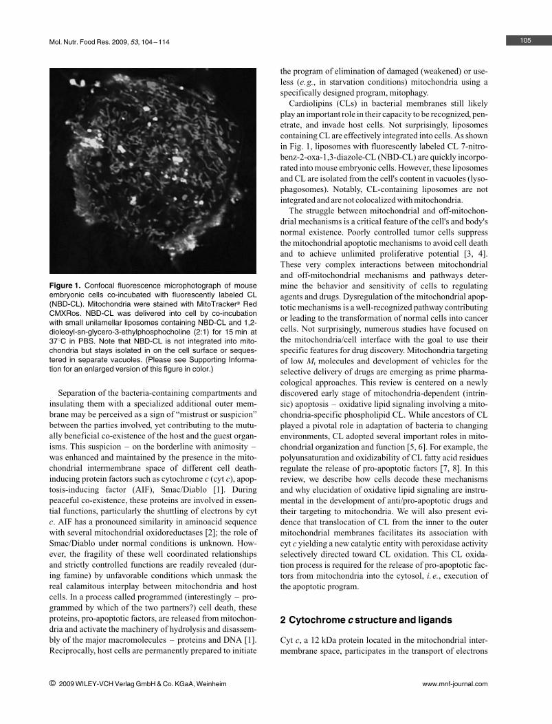
Cardiolipins (CLs) in bacterial membranes still likelyplay an important role in their capacity to be recognized, pen-etrate, and invade host cells. Not surprisingly, liposomescontaining CL are effectively integrated into cells. As shownin Fig. 1, liposomes with fluorescently labeled CL 7-nitro-benz-2-oxa-1,3-diazole-CL (NBD-CL) are quickly incorpo-rated into mouse embryonic cells. However, these liposomesand CL are isolated from the cell's content in vacuoles (lyso-phagosomes). Notably, CL-containing liposomes are notintegrated and are not colocalized with mitochondria.
The struggle between mitochondrial and off-mitochon-drial mechanisms is a critical feature of the cell's and body'snormal existence. Poorly controlled tumor cells suppressthe mitochondrial apoptotic mechanisms to avoid cell deathand to achieve unlimited proliferative potential [3, 4].These very complex interactions between mitochondrialand off-mitochondrial mechanisms and pathways deter-mine the behavior and sensitivity of cells to regulatingagents and drugs. Dysregulation of the mitochondrial apop-totic mechanisms is a well-recognized pathway contributingor leading to the transformation of normal cells into cancercells. Not surprisingly, numerous studies have focused onthe mitochondria/cell interface with the goal to use theirspecific features for drug discovery. Mitochondria targetingof low Mr molecules and development of vehicles for theselective delivery of drugs are emerging as prime pharma-cological approaches. This review is centered on a newlydiscovered early stage of mitochondria-dependent (intrin-sic) apoptosis – oxidative lipid signaling involving a mito-chondria-specific phospholipid CL. While ancestors of CLplayed a pivotal role in adaptation of bacteria to changingenvironments, CL adopted several important roles in mito-chondrial organization and function [5, 6]. For example, thepolyunsaturation and oxidizability of CL fatty acid residuesregulate the release of pro-apoptotic factors [7, 8]. In thisreview, we describe how cells decode these mechanismsand why elucidation of oxidative lipid signaling are instru-mental in the development of anti/pro-apoptotic drugs andtheir targeting to mitochondria. We will also present evi-dence that translocation of CL from the inner to the outermitochondrial membranes facilitates its association withcyt c yielding a new catalytic entity with peroxidase activityselectively directed toward CL oxidation. This CL oxida-tion process is required for the release of pro-apoptotic fac-tors from mitochondria into the cytosol, i. e., execution ofthe apoptotic program.
2 Cytochrome c structure and ligands
Cyt c, a 12 kDa protein located in the mitochondrial inter-membrane space, participates in the transport of electrons
105
i 2009 WILEY-VCH Verlag GmbH & Co. KGaA, Weinheim www.mnf-journal.com
Figure 1. Confocal fluorescence microphotograph of mouseembryonic cells co-incubated with fluorescently labeled CL(NBD-CL). Mitochondria were stained with MitoTrackerm RedCMXRos. NBD-CL was delivered into cell by co-incubationwith small unilamellar liposomes containing NBD-CL and 1,2-dioleoyl-sn-glycero-3-ethylphosphocholine (2:1) for 15 min at378C in PBS. Note that NBD-CL is not integrated into mito-chondria but stays isolated in on the cell surface or seques-tered in separate vacuoles. (Please see Supporting Informa-tion for an enlarged version of this figure in color.)
V. E. Kagan et al. Mol. Nutr. Food Res. 2009, 53, 104 –114
in the respiratory chain by shuttling electrons from complexIII (ubiquinol cyt c oxidoreductase) to complex IV (cyt coxidase) [9]. For many years, this activity was viewed as theonly function of cyt c. Recently, this notion of monofunc-tionality of cyt c has been shaken when the protein attractednew attention due to several additional roles it plays [10,11]. As an effective acceptor of electrons, cyt c has beensuggested to act as a component of the enzymatic antioxi-dant system of mitochondria [12, 13]. This is based on itsability to accept electrons from superoxide radicals anddump then on complex IV. In this capacity, cyt c has anunlimited ability to prevent superoxide accumulation inmitochondria. To what extent this remarkable antioxidantpotential of cyt c is realized during excessive spillage ofelectrons on complexes I and III under conditions of oxida-tive/nitrosative stress still remains to be quantified. It hasbeen documented that extramitochondrial cyt c is an activeparticipant of programmed cell death (apoptosis) wherebyit is released from mitochondria and interacts with apop-totic protease activating factor (Apaf-1), causing the forma-tion of apoptosomal complexes and activation of proteolyticcaspase cascades [11, 14]. Finally, chronologically thelatest function of cyt c has been associated with its ability tobe activated and fulfill the task of a peroxidase [8, 15]. As ahemo-protein, cyt c has all the prerequisites to be convertedinto a peroxidase. Paradoxically, the peroxidase activity ofcyt c under normal conditions is negligibly low [15, 16].However, numerous studies in model systems explored theability of cyt c
,
s conversion into an active peroxidase as aresult of post-translational modifications. It has been dem-onstrated that oxidative or nitrative chemical modificationsof the protein lead to the induction of significant peroxidaseactivity [17–19]. Similarly, interaction of cyt c with hydro-phobic anions – detergents or anionic phospholipids – cantrigger peroxidase activation [20–22]. However, to date thephysiological significance of these “properoxidative” mod-ifications of cyt c has not been defined. Our previous workhas identified mitochondrial apoptotic events leading tospecific interactions and high affinity binding of cyt c witha mitochondria-specific phospholipid CL. This results inthe activation of cyt c into a CL-specific peroxidase withselective catalytic competence toward peroxidation of poly-unsaturated molecular species of CL. Ultimately, this activ-ity results in the accumulation of CL oxidation products,mainly CL–OOH and their reduction products, CL–OH[8]. This is significant because oxygenated CL speciesappear to be essential for mitochondrial membrane permea-bilization and release of pro-apoptotic factors (includingcyt c itself) into the cytosol [8, 23]. Elucidation of thesedifferent modalities through which cyt c exerts its diversi-fied catalytic functions requires a better understanding ofits structural organization. In particular, knowledge ofphylogenetically conserved segments in cyt c should pro-vide insight regarding their role and evolutionary develop-ment.
3 Cyt c interactions with other proteins andligands
Modifications of cyt c amino acid residues could have func-tional effects on its ability to transport electrons and modu-late apoptosis. It was demonstrated that cyt c uses severallysines (including Lys13, Lys72, and Lys87; here and belowresidue numeration is for horse heart cyt c) to react with cytc peroxidase and cyt c oxidase [24]. These same lysine resi-dues are also likely involved in interactions with cyt b5 andc1. Notably, cyt c peroxidase is present only in relativelyprimitive organisms such as yeast and bacteria. Its function(accepting reducing equivalents from cyt c and reducingH2O2 to water) in higher species might be regulated by cytc/CL complexes.
McLendon's group has shown that a large number of cytc residues interact with Apaf-1, e.g., Lys7, Lys25, Lys39,Lys72, and also sequence 62–65 (Glu–Thr–Leu–Met)[25]. Lys72 is important to cyt c activity (Lys72Ala muta-tion eliminated its electron transport function). Mutation ofLys7, Lys25, Lys39, and domain 62–65 reduced the activ-ity of cyt c–Apaf-1–caspase-9 complex toward procas-pase-3 cleavage. Single site-directed mutations resulted in a5–10-fold decrease in procaspase-3 cleavage activity,whereas multiple mutations reduced caspase activity bymore than 1000-fold [25].
As a polyvalent cation, cyt c has multiple binding sitesfor a variety of small anions, including phosphate, ADP,ATP, and citrate [26, 27]. Experiments with ATP photoaf-finity – labeled cyt c have demonstrated that this siteinvolves the invariant residues Lys72, Lys86, Lys87, andArg91 [27–29]. ATP binds to this site at physiological con-centrations and under physiological ionic strength condi-tions, which suggests that this site is of biological signifi-cance. The occupancy of the site depends on the [ATP]/[ADP] ratios but is independent of the redox state of cyt c.ATP bound to the site diminishes the electron flow in therespiratory chain [28]. Both complex III and IV activitiesare affected. As the ATP binding site includes Lys residuesthat are involved in the interaction of cyt c with the reduc-tase and oxidase, the inhibition is likely to be the result ofdirect steric and electrostatic effects.
Lysine residues are also preferential sites for lipid hydro-peroxide-derived modification on cyt c [30]. The majoradducts are associated with Lys5, Lys7, Lys8, His33,Lys86, Lys87, Lys99, and Lys100. In addition, Isom et al.[31] have shown that 4-hydroxy-2-nonenal (4HNE), a sec-ondary product of lipid peroxidation, forms conjugates withcyt c through His33, Lys87, and Arg38.
Chemical modification of cyt c by lipid peroxidationproducts was recently deemed physiologically relevant afterit was discovered that cyt c can oxidize CL and phosphati-dylserine (PS), in vivo [8, 32]. In complementary experi-ments, we also demonstrated that the complex of cyt c/CL(i) exhibits peroxidase activity [15], (ii) produces CL hydro-
106
i 2009 WILEY-VCH Verlag GmbH & Co. KGaA, Weinheim www.mnf-journal.com

Mol. Nutr. Food Res. 2009, 53, 104 –114
peroxides required for release of proapoptotic factors [8],and (iii) potentially utilizes CL hydroperoxides as sub-strates for further peroxidase reactions [33].
4 Conservancy of cyt c amino acid residues
Overall, cyt c is highly conserved among different species(Fig. 2). The level of conservancy of cyt c
,
s segments spe-cifically involved in its different functions may provideinformation on their phylogenetic significance. Therefore,we will briefly consider participation of different domainsof cyt c in its functional endeavors.
From an evolutionary standpoint, one of the major func-tions of cyt c is to transport electrons through the respira-tory chain. Cyt c is a typical hemoprotein with the covalentattachment of heme to the polypeptide chain through thio-ether bonds to the cysteine residues of a Cys–Xxx–Xxx–
Cys–His peptide motif. The distal ligand – methionine –is also strictly invariable among all living organisms (withdiffering numeration, because the length of the polypeptidechain is different). Lysines participating in interaction withcyt c peroxidase and cyt c oxidase (Lys72 and Lys87 [24])are also invariantly present in all organisms, whereas Lys13in some cases (Saccharomyces cerevisiae and Caenorhab-ditis elegans) is substituted for the similarly basic and polararginine. The ATP binding domain formed by Lys72,Lys86, Lys87, and Arg91 [27–29] is also very conservative,i. e., there are no single substitutes for these amino acid resi-dues among living organisms.
Within the context of this review, it is important that tyro-sine residues likely involved in its peroxidase function (butalso essential for the electron transport) [34–36] are pre-served as well. Horse heart cyt c Tyr48, Tyr67, Tyr74, andTyr97 are present in all other species (with one exception ofAsn for one species, Macaca mulatta). One additional tyro-
107
i 2009 WILEY-VCH Verlag GmbH & Co. KGaA, Weinheim www.mnf-journal.com
Figure 2. (A) Alignment of cyt c protein sequences. Sequence alignment of cyt c from different species was prepared using Gene-ious Basic software (http://www.geneious.com/, New Zealand) with modifications. Aminoacid residues relevant to the peroxidasefunction of cyt c/CL complexes (binding of CL, attachment of heme, catalytic function) are shown as “framed.” Note the high conserv-ancy of these residues. (B) Phylogenic tree of cyt c demonstrating the evolutionary relationships between several sequences. (Thiswas generated using Geneious Basic software – http://www.geneious.com/, New Zealand with modifications; the length of treebranches did not reflect actual divergence). (Please see Supporting Information for an enlarged version of this figure in color.)
V. E. Kagan et al. Mol. Nutr. Food Res. 2009, 53, 104 –114
sine is present in primates near Tyr47; two additional tyro-sines are located at the N-terminus of cyt c of C. elegans.His26 and His33, which play a putative role in peroxidasereaction of partially unfolded cyt c [37, 38], are also moreor less evolutionary stable – correspondingly only substitu-tion of His26 for Thr in C. elegans and His33 for Trp, Asn,or Cys in several other species, occur.
Lysine residues responsible for Apaf-1 interaction andfurther caspases activation [25] are not as well conserved:Lys7 is present in S. cerevisiae, but in Drosophila mela-nogaster, Xenopus tropicalis, Danio rerio, and mammals,this Lys is substituted for polar acidic Glu. It is worth notingthat the neighboring Lys8 is invariant in almost all cases. Tothe contrary, Lys25 and Lys39 are invariant in all speciesexcept S. cerevisiae and C. elegans – correspondingly,there are nonpolar neutral proline and polar basic histidinein yeast and nonpolar neutral alanine and polar neutralthreonine in nematodes. It is probable that this diversityreflects an evolutionary new function of cyt c, i. e., partici-pation in apoptosis.
Overall, the segments of the protein essential for the per-oxidase function of cyt c seem to preserve conservancyfrom species to species. All prerequisites for peroxidase
activity, namely, heme moiety, its distal and proximalligands (Met, His), tyrosine residues, and amino acidsrequired for the binding with CL (Lys, Asn, His, see below)are highly conserved.
5 CL binding to cyt c
As the current review is focused on the pro-apoptotic perox-idase function of cyt c, we will further discuss structuralrequirements essential for the association of cyt c withanionic phospholipids, particularly, CL. There is no singleuniversally accepted opinion on the binding of CL to cyt c.The most commonly used model for cyt c binding to themembrane involves the A-site for electrostatic interactionand the C-site for hydrophobic interaction. Site A consistsof basic residues, such as Lys72 and Lys73. Site C is anotherlipid-binding domain with a high affinity for protonatedacidic phospholipids. The invariant Asn52 in horse heartcyt c has been assigned as the amino acid residue that bindsto protonated acidic phospholipids via hydrogen bonds. It isbelieved that a hydrophobic component of the C-site medi-ated interaction is due to an extended lipid anchorage of
108
i 2009 WILEY-VCH Verlag GmbH & Co. KGaA, Weinheim www.mnf-journal.com
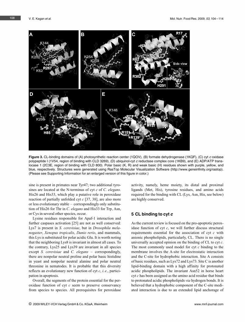
Figure 3. CL-binding domains of (A) photosynthetic reaction center (1QOV), (B) formate dehydrogenase (1KQF), (C) cyt c oxidasepolypeptide I (1V54, region of binding with CLD 3269), (D) ubiquinol-cyt c reductase complex core (1KB9), and (E) ADP/ATP trans-locase 1 (2C3E, region of binding with CLD 800). Polar basic (K, R) and weak basic (H) residues shown with purple, yellow, andblue, respectively. Structures were generated using RasTop Molecular Visualization Software (http://www.geneinfinity.org/rastop).(Please see Supporting Information for an enlarged version of this figure in color.)

Mol. Nutr. Food Res. 2009, 53, 104 –114
one of the fatty acid chains into the hydrophobic channel incyt c [39–41]. The work of Kostrzewa et al. [42] revealedone more possible site of lipid/cyt c interaction, Lys86, andLys87 along with Lys72. Using paramagnetically-labeledcyt c, these authors demonstrated that cyt c electrostaticallyinteracts with the membrane and found that cyt c does notpenetrate the membrane's interior. In 2005, Nantes's groupbased on low pH (a7.5) modification of cyt c proposed theexistence of additional sites of lipid–cyt c interaction:Lys22, Lys25, His26, Lys27, His33, and Lys87 [43]. Thiscollective site (L-site) is on the opposite side of the cyt cmolecule compared to sites A and C. It is possible that thissite is involved in the attachment of the protein to the mem-brane lipid bilayer.
One possible approach to study the structural organiza-tion of the cyt c molecule and assess the contribution of itsdomains regarding interactions with anionic phospholipids,is to utilize comparative molecular modeling and crystallo-graphic information available in existing databases. Wehave analyzed the structure of a number of putative CL-binding proteins. There are 37 known crystal structures ofmolecules with CL as a ligand in the Protein Data Bank(http://www.pdb.org). They belong to five distinct groups:photosynthetic reaction center from Rhodobacter sphaer-oides, yeast cyt bc1 complex, formate dehydrogenase Nfrom Escherichia coli, bovine mitochondrial ADP/ATP car-rier, and bovine heart cyt c oxidase.
Our strategy to further characterize the nature of CLbinding to cyt c was based on the analysis of likely partici-pants in close proximity to the CL-binding domain (withina 5 � distance) (Fig. 3). We found that Lys, Arg, and Hisresidues are located in close proximity to the polar nega-tively charged heads of CL. Additionally, hydrophobic a-helixes are neighboring the CL fatty acid chain. The sameapproach was documented in the review of Palsdottir andHunte [44] for several negatively charged phospholipids.Tight binding interactions with three residues wereobserved: Lys–Lys–Yyy, Arg–Lys–Yyy, and His–Arg–Asn. Based on these comparisons, the overall structureXxx–Xxx–Yyy, where Xxx is a positively charged residueand Yyy a polar residue, can be inferred as a CL-bindingmotif. These authors pointed out that the suggested motifsare nonlinear, i. e., ligands from different subunits may con-tribute. Also, several CL-binding domains composed of twoamino acid residues or even one (lysine or serine) may exist.After determination of CL-binding domains on known crys-tal structures, we aligned this CL-binding domain to cyt c(1HRC). We revealed two possible sites for CL binding –one near Lys72 and Lys73 and the other one contiguous toLys99 and Lys100 (Fig. 4).
Obviously, additional studies based on molecular model-ing are needed to further characterize other essential motifscontributing to specific hydrophobic interactions of CLwith nonpolar grooves in the protein structure. These stud-ies may be useful for the development of optimized inhibi-
tors capable of disrupting cyt c/CL interactions, and hencethe formation of a peroxidase competent complex. Thisapproach may lead to the development of new anti-apop-totic agents.
6 Mitochondria-targeted inhibitors ofperoxidase activity of cyt c/CL complexes
Given the importance of mitochondrial events during theinitial critical stages of the execution of the apoptotic pro-gram, the design of mitochondriotropic agents is believedto be a promising novel strategy in regulating apoptosis. Inthe context of anticancer therapy, stimulation of pro-apop-totic mitochondrial events in tumor cells, and suppressionof mitochondrial stages of apoptosis in surrounding normalcells may represent a promising paradigm for new effectivetherapies. Different approaches targeting the regulation ofcomponents of the mitochondrial antioxidant system suchas manganese superoxide dismutase (Mn-SOD) were testedand showed significant antitumor efficiency, particularly incombination therapy [45]. Another opportunity is employ-ment of nanoparticles loaded with cell death signals (suchas cyt c) and targeted to cancer cells. We have recently dem-onstrated that single walled carbon nanotubes can be coatedwith specific phospholipid recognition signals along withcyt c and directed to and taken-up by phagocytozing cellsresulting in triggering of apoptosis (Konduru et al., unpub-lished data).
109
i 2009 WILEY-VCH Verlag GmbH & Co. KGaA, Weinheim www.mnf-journal.com
Figure 4. Alignment of CL-binding domain of formate dehydro-genase (1KQF) to cyt c (1HRC). Two critical domains havebeen identified, Lys72–Lys73 and Lys99–Lys100. Proteinalignment (by lysine residues) was performed using ICM-Browser software (http://www.molsoft.com/icm_browser.html).(Please see Supporting Information for an enlarged version ofthis figure in color.)
V. E. Kagan et al. Mol. Nutr. Food Res. 2009, 53, 104 –114
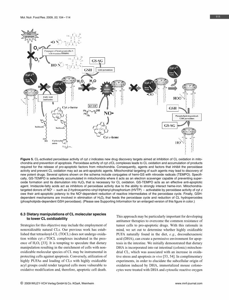
The peroxidase reaction of cyt c/CL complexes is drivenby H2O2 as a source of oxidizing equivalents. During apop-tosis, the latter is provided by disrupted electron transport.Interaction of CL with cyt c yields a complex whose normalredox potential is about minus (–) 400 mV more negativethan that of intact cyt c [46]. As a result, cyt c/CL cannotaccept electrons from mitochondrial complex III leading toenhanced production of superoxide whose dismutationyields H2O2. Thus, the formation of cyt c/CL complexes inmitochondria is, in principle, sufficient to trigger mitochon-drial generation of H2O2 to feed the peroxidase cycle andcatalyze CL oxidation. Clearly, prevention or inhibition ofH2O2 production should block the supply of oxidizingequivalents and inhibit the peroxidase-catalyzed CL oxida-tion. Our efforts are focused on several effective approachesto achieve this goal.
Nitroxide radicals are ideal candidates for this role asthey can readily accept excessive electrons from damagedmitochondrial electron carriers and hence prevent superox-ide production. Moreover, reduction of nitroxides produceshydroxylamines which can further act as radical scavengersand donors of electrons capable of quenching the peroxi-dase reaction. Provided the concentration of nitroxides issufficiently high in mitochondria, they could be ideal candi-dates as inhibitors of mitochondrially driven CL oxidation.
Recently, several strategies have been developed todeliver and concentrate low Mr cargos in mitochondria.Compounds targeting mitochondria (mitochondriotropics)act through several diverse accumulation mechanisms andchemical features [47]. One of them is based on the conju-gation of the nitroxides with a hydrophobic cation, triphe-nylphosphonium (TPP) that is readily electrophoresed intomitochondria at the expense of their membrane potential(MP) [48]. Indeed, TPP-nitroxide was able to inhibit apop-tosis in cells by accumulating in their mitochondrial frac-tions. The disadvantage of this protocol is that TPP reducesmitochondrial MP and induces damage. Additionally, effec-tive scavenging of electrons by nitroxides may be associ-ated not only with the benefit of preventing superoxide andH2O2 generation but also with a risk of energy crisis. Indeed,high concentrations of accumulated nitroxides may com-pletely block electron transport in mitochondria, henceblock the ATP production.
Another approach is based on conjugates of nitroxideswith fragments of molecules with relatively high affinity tomitochondrial membranes. For example, fragments of gra-micidin S (GS) have been successfully used for this purposeand proved to be effective in preventing superoxide produc-tion in cells, CL oxidation in mitochondria and protectingcells against different pro-apoptotic triggers such as actino-mycin D, radiation, or staurosporine [49–51]. More impor-tantly, these mitochondria-targeted nitroxides were able toprotect against apoptosis in vivo by preventing CL oxida-tion induced by intestinal hemorrhagic shock [52]. Optimi-zation of nitroxide carriers promises to give a new genera-
tion of effective anti-apoptotic agents acting at an earlymitochondrial stage.
Additional opportunity to inhibit peroxidase activity ofcyt c/CL complexes toward CL oxidation is to use alterna-tive sacrificial substrates and thus prevent CL oxidation.Among such potent reductants of peroxidase and its reac-tive intermediates is nitric oxide (NO9). We have recentlydeveloped a prodrug – alkyl-hydroxylamine – whosemetabolism by peroxidases converts it into an NO donor.Conjugation of alkyl-hydroxylamine with TPP (TPPO)selectively targeted the conjugate – (2-hydroxyaminovi-nyl)TPP – into mitochondria whereby the cargo was con-verted into an NO-donor by a cyt c-dependent metabolism.This resulted in the release of NO, and most importantly,protection against apoptosis (Stoyanovsky et al., unpub-lished data).
6.1 Imidazole fatty acids
Peroxidase activity of cyt c/CL complexes is central to CLoxidation and the effects of CL–OOH in the mitochondrialmembrane permeabilization and release of pro-apoptoticfactors. Without CL, cyt c is a very poor peroxidase due to ahexacoordinate occupancy of Fe in the heme [16]. Uponbinding of CL, the protein undergoes partial unfoldingaccompanied by loosening of its Fe coordination by Met80resulting in enhanced access of the heme catalytic site tosmall molecules like H2O2. If the availability of the hemecan be blocked by a small molecule, the peroxidase activitywill be minimized. In this respect, an interesting group ofcompounds are imidazole derivatives of fatty acids. Wehave designed and synthesized several derivatives in such away that the carboxy group interacts with one of the criticalLys residues of the protein, while the imidazole moiety atdifferent positions of the acyl chain – protruding into thehydrophobic pocket – appears to interact with the hemeiron to lock the catalytic site and form a high affinity com-plex. In this complex, H2O2 will have no access to the hemecatalytic site. Experiments are now underway to assess theeffectiveness of imidazole fatty acids as inhibitors of perox-idase activity of cyt c in model cell culture systems.
6.2 Modified oxidizable derivatives of CL
An interesting opportunity for inhibition of the peroxidaseactivity toward oxidation of CL may be explored by usingCL homologs with readily oxidizable functionalities thatwill compete with the oxidation of PUFAs of CL. In our pre-liminary experiments with NBD-CL, we found that NBDgroups are the prime target for the peroxidase-catalyzedoxidation. This suggests that via appropriate optimizationprotocols, CL derivatives may be designed that will beeffective at outcompeting natural substrates of in cyt c/CLcomplexes and preventing accumulation of pro-apoptoticCL–OOH.
110
i 2009 WILEY-VCH Verlag GmbH & Co. KGaA, Weinheim www.mnf-journal.com
Mol. Nutr. Food Res. 2009, 53, 104 –114
6.3 Dietary manipulations of CL molecular speciesto lower CL oxidizability
Strategies for this objective may include the employment ofnonoxidizable natural CLs. Our previous work has estab-lished that tetraoleoyl-CL (TOCL) does not undergo oxida-tion within cyt c/TOCL complexes incubated in the pres-ence of H2O2 [33]. It is tempting to speculate that dietarymanipulation resulting in the enrichment of cells with non-oxidizable molecular species of CL may be instrumental inprotecting cells against apoptosis. Conversely, utilization ofhighly PUFAs and loading of CLs with highly oxidizableacyl groups could render targeted cells more vulnerable tooxidative modification and, therefore, apoptotic cell death.
This approach may be particularly important for developingantitumor therapies to overcome the common resistance oftumor cells to pro-apoptotic drugs. With this rationale inmind, we set out to determine whether highly oxidizablePUFA naturally found in the diet, e.g., docosahexaenoicacid (DHA), can create a permissive environment for apop-tosis in the intestine. We initially demonstrated that dietaryDHA is incorporated into rat intestinal (colonic) mitochon-drial CL, which was associated with an increase in oxida-tive stress and apoptosis in vivo [53, 54]. In complimentaryexperiments, in order to elucidate the subcellular origin ofoxidation induced by DHA, immortalized mouse colono-cytes were treated with DHA and cytosolic reactive oxygen
111
i 2009 WILEY-VCH Verlag GmbH & Co. KGaA, Weinheim www.mnf-journal.com
Figure 5. CL-activated peroxidase activity of cyt c indicates new drug discovery targets aimed at inhibition of CL oxidation in mito-chondria and prevention of apoptosis. Peroxidase activity of cyt c/CL complexes leads to CL oxidation and accumulation of productsrequired for the release of pro-apoptotic factors from mitochondria. Consequently, agents and factors that inhibit the peroxidaseactivity and prevent CL oxidation may act as anti-apoptotic agents. Mitochondrial targeting of such agents may lead to discovery ofnew potent drugs. Several options shown on the schema include conjugates of hemi-GS with nitroxide radicals (TEMPO). Specifi-cally, GS-TEMPO is selectively accumulated in mitochondria where it acts as an electron scavenger capable of preventing super-oxide formation and its dismutation into H2O2 that is necessary for CL oxidation. GS-TEMPO acts as an effective anti-apoptoticagent. Imidazole-fatty acids act as inhibitors of peroxidase activity due to the ability to strongly interact heme-iron. Mitochondria-targeted donors of NO9 – such as 2-hydroxyamino-vinyl-triphenyl-phosphonium (HVTP) – activatable by peroxidase activity of cyt cowe their anti-apoptotic potency to the NO9-dependent reduction of reactive intermediates of the peroxidase cycle. Finally, GSH-dependent mechanisms are involved in elimination of H2O2 that feeds the peroxidase cycle and reduction of CL hydroperoxides(phospholipids-dependent GSH peroxidase). (Please see Supporting Information for an enlarged version of this figure in color.)
V. E. Kagan et al. Mol. Nutr. Food Res. 2009, 53, 104 –114
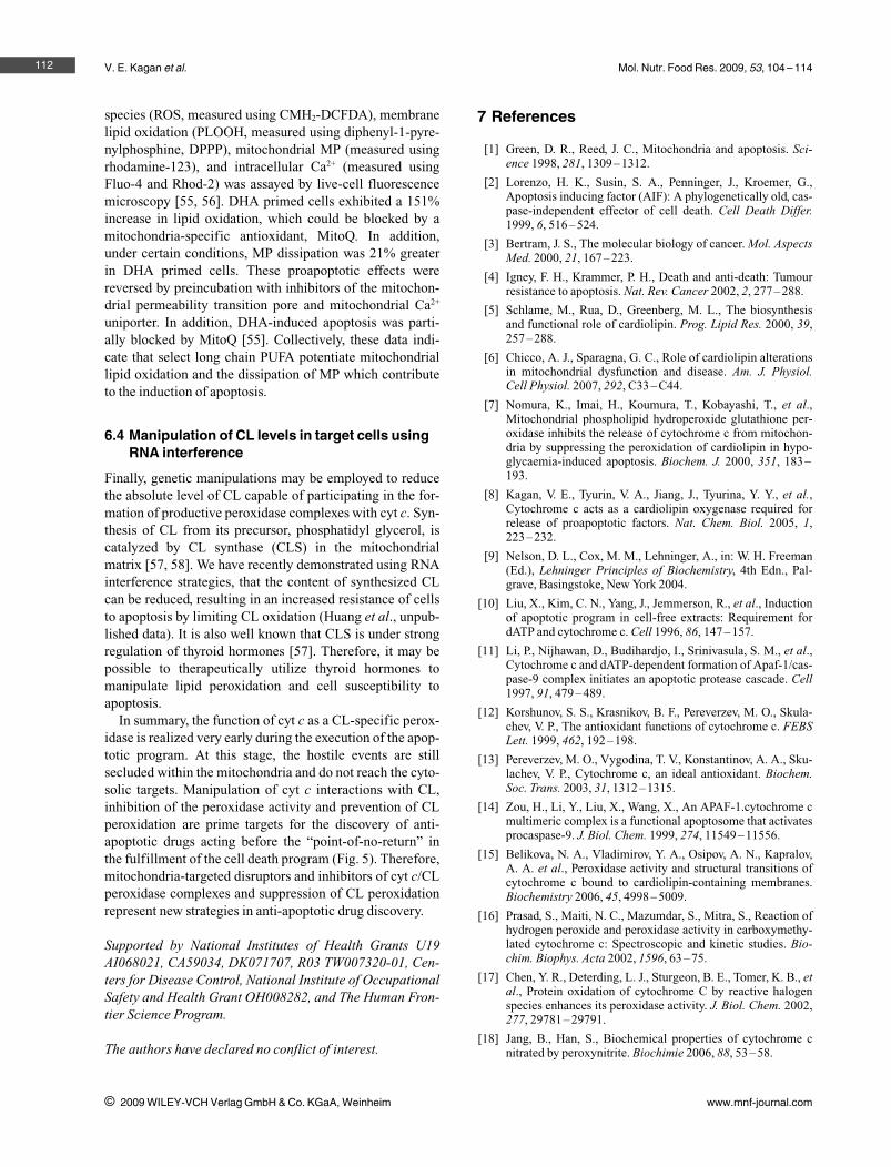
species (ROS, measured using CMH2-DCFDA), membranelipid oxidation (PLOOH, measured using diphenyl-1-pyre-nylphosphine, DPPP), mitochondrial MP (measured usingrhodamine-123), and intracellular Ca2+ (measured usingFluo-4 and Rhod-2) was assayed by live-cell fluorescencemicroscopy [55, 56]. DHA primed cells exhibited a 151%increase in lipid oxidation, which could be blocked by amitochondria-specific antioxidant, MitoQ. In addition,under certain conditions, MP dissipation was 21% greaterin DHA primed cells. These proapoptotic effects werereversed by preincubation with inhibitors of the mitochon-drial permeability transition pore and mitochondrial Ca2+
uniporter. In addition, DHA-induced apoptosis was parti-ally blocked by MitoQ [55]. Collectively, these data indi-cate that select long chain PUFA potentiate mitochondriallipid oxidation and the dissipation of MP which contributeto the induction of apoptosis.
6.4 Manipulation of CL levels in target cells usingRNA interference
Finally, genetic manipulations may be employed to reducethe absolute level of CL capable of participating in the for-mation of productive peroxidase complexes with cyt c. Syn-thesis of CL from its precursor, phosphatidyl glycerol, iscatalyzed by CL synthase (CLS) in the mitochondrialmatrix [57, 58]. We have recently demonstrated using RNAinterference strategies, that the content of synthesized CLcan be reduced, resulting in an increased resistance of cellsto apoptosis by limiting CL oxidation (Huang et al., unpub-lished data). It is also well known that CLS is under strongregulation of thyroid hormones [57]. Therefore, it may bepossible to therapeutically utilize thyroid hormones tomanipulate lipid peroxidation and cell susceptibility toapoptosis.
In summary, the function of cyt c as a CL-specific perox-idase is realized very early during the execution of the apop-totic program. At this stage, the hostile events are stillsecluded within the mitochondria and do not reach the cyto-solic targets. Manipulation of cyt c interactions with CL,inhibition of the peroxidase activity and prevention of CLperoxidation are prime targets for the discovery of anti-apoptotic drugs acting before the “point-of-no-return” inthe fulfillment of the cell death program (Fig. 5). Therefore,mitochondria-targeted disruptors and inhibitors of cyt c/CLperoxidase complexes and suppression of CL peroxidationrepresent new strategies in anti-apoptotic drug discovery.
Supported by National Institutes of Health Grants U19AI068021, CA59034, DK071707, R03 TW007320-01, Cen-ters for Disease Control, National Institute of OccupationalSafety and Health Grant OH008282, and The Human Fron-tier Science Program.
The authors have declared no conflict of interest.
7 References
[1] Green, D. R., Reed, J. C., Mitochondria and apoptosis. Sci-ence 1998, 281, 1309–1312.
[2] Lorenzo, H. K., Susin, S. A., Penninger, J., Kroemer, G.,Apoptosis inducing factor (AIF): A phylogenetically old, cas-pase-independent effector of cell death. Cell Death Differ.1999, 6, 516–524.
[3] Bertram, J. S., The molecular biology of cancer. Mol. AspectsMed. 2000, 21, 167–223.
[4] Igney, F. H., Krammer, P. H., Death and anti-death: Tumourresistance to apoptosis. Nat. Rev. Cancer 2002, 2, 277–288.
[5] Schlame, M., Rua, D., Greenberg, M. L., The biosynthesisand functional role of cardiolipin. Prog. Lipid Res. 2000, 39,257–288.
[6] Chicco, A. J., Sparagna, G. C., Role of cardiolipin alterationsin mitochondrial dysfunction and disease. Am. J. Physiol.Cell Physiol. 2007, 292, C33 –C44.
[7] Nomura, K., Imai, H., Koumura, T., Kobayashi, T., et al.,Mitochondrial phospholipid hydroperoxide glutathione per-oxidase inhibits the release of cytochrome c from mitochon-dria by suppressing the peroxidation of cardiolipin in hypo-glycaemia-induced apoptosis. Biochem. J. 2000, 351, 183 –193.
[8] Kagan, V. E., Tyurin, V. A., Jiang, J., Tyurina, Y. Y., et al.,Cytochrome c acts as a cardiolipin oxygenase required forrelease of proapoptotic factors. Nat. Chem. Biol. 2005, 1,223–232.
[9] Nelson, D. L., Cox, M. M., Lehninger, A., in: W. H. Freeman(Ed.), Lehninger Principles of Biochemistry, 4th Edn., Pal-grave, Basingstoke, New York 2004.
[10] Liu, X., Kim, C. N., Yang, J., Jemmerson, R., et al., Inductionof apoptotic program in cell-free extracts: Requirement fordATP and cytochrome c. Cell 1996, 86, 147–157.
[11] Li, P., Nijhawan, D., Budihardjo, I., Srinivasula, S. M., et al.,Cytochrome c and dATP-dependent formation of Apaf-1/cas-pase-9 complex initiates an apoptotic protease cascade. Cell1997, 91, 479–489.
[12] Korshunov, S. S., Krasnikov, B. F., Pereverzev, M. O., Skula-chev, V. P., The antioxidant functions of cytochrome c. FEBSLett. 1999, 462, 192–198.
[13] Pereverzev, M. O., Vygodina, T. V., Konstantinov, A. A., Sku-lachev, V. P., Cytochrome c, an ideal antioxidant. Biochem.Soc. Trans. 2003, 31, 1312 –1315.
[14] Zou, H., Li, Y., Liu, X., Wang, X., An APAF-1.cytochrome cmultimeric complex is a functional apoptosome that activatesprocaspase-9. J. Biol. Chem. 1999, 274, 11549–11556.
[15] Belikova, N. A., Vladimirov, Y. A., Osipov, A. N., Kapralov,A. A. et al., Peroxidase activity and structural transitions ofcytochrome c bound to cardiolipin-containing membranes.Biochemistry 2006, 45, 4998 –5009.
[16] Prasad, S., Maiti, N. C., Mazumdar, S., Mitra, S., Reaction ofhydrogen peroxide and peroxidase activity in carboxymethy-lated cytochrome c: Spectroscopic and kinetic studies. Bio-chim. Biophys. Acta 2002, 1596, 63 –75.
[17] Chen, Y. R., Deterding, L. J., Sturgeon, B. E., Tomer, K. B., etal., Protein oxidation of cytochrome C by reactive halogenspecies enhances its peroxidase activity. J. Biol. Chem. 2002,277, 29781 –29791.
[18] Jang, B., Han, S., Biochemical properties of cytochrome cnitrated by peroxynitrite. Biochimie 2006, 88, 53–58.
112
i 2009 WILEY-VCH Verlag GmbH & Co. KGaA, Weinheim www.mnf-journal.com
Mol. Nutr. Food Res. 2009, 53, 104 –114
[19] Cassina, A. M., Hodara, R., Souza, J. M., Thomson, L., et al.,Cytochrome c nitration by peroxynitrite. J. Biol. Chem. 2000,275, 21409–21415.
[20] Pinheiro, T. J., Cheng, H., Seeholzer, S. H., Roder, H., Directevidence for the cooperative unfolding of cytochrome c inlipid membranes from H-(2)H exchange kinetics. J. Mol.Biol. 2000, 303, 617–626.
[21] Muga, A., Mantsch, H. H., Surewicz, W. K., Membrane bind-ing induces destabilization of cytochrome c structure. Bio-chemistry 1991, 30, 7219 –7224.
[22] Mustonen, P., Virtanen, J. A., Somerharju, P. J., Kinnunen, P.K., Binding of cytochrome c to liposomes as revealed by thequenching of fluorescence from pyrene-labeled phospholi-pids. Biochemistry 1987, 26, 2991 –2997.
[23] Belikova, N. A., Jiang, J., Tyurina, Y. Y., Zhao, Q., et al., Car-diolipin-specific peroxidase reactions of cytochrome c inmitochondria during irradiation-induced apoptosis. Int. J.Radiat. Oncol. Biol. Phys. 2007, 69, 176–186.
[24] Poulos, T. L., Kraut, J., A hypothetical model of the cyto-chrome c peroxidase. Cytochrome c electron transfer com-plex. J. Biol. Chem. 1980, 255, 10322–10330.
[25] Yu, T., Wang, X., Purring-Koch, C., Wei, Y., et al., A muta-tional epitope for cytochrome C binding to the apoptosis pro-tease activation factor-1. J. Biol. Chem. 2001, 276, 13034 –13038.
[26] Osheroff, N., Brautigan, D. L., Margoliash, E., Mapping ofanion binding sites on cytochrome c by differential chemicalmodification of lysine residues. Proc. Natl. Acad. Sci. USA1980, 77, 4439 –4443.
[27] Corthesy, B. E., Wallace, C. J., The oxidation-state-dependentATP-binding site of cytochrome c. A possible physiologicalsignificance. Biochem. J. 1986, 236, 359–364.
[28] Craig, D. B., Wallace, C. J., Studies of 8-azido-ATP adductsreveal two mechanisms by which ATP binding to cytochromec could inhibit respiration. Biochemistry 1995, 34, 2686 –2693.
[29] McIntosh, D. B., Parrish, J. C., Wallace, C. J., Definition of anucleotide binding site on cytochrome c by photoaffinitylabeling. J. Biol. Chem. 1996, 271, 18379 –18386.
[30] Williams, M. V., Wishnok, J. S., Tannenbaum, S. R., Covalentadducts arising from the decomposition products of lipidhydroperoxides in the presence of cytochrome c. Chem. Res.Toxicol. 2007, 20, 767 –775.
[31] Isom, A. L., Barnes, S., Wilson, L., Kirk, M., et al., Modifica-tion of Cytochrome c by 4-hydroxy- 2-nonenal: Evidence forhistidine, lysine, and arginine-aldehyde adducts. J. Am. Soc.Mass Spectrom. 2004, 15, 1136 –1147.
[32] Jiang, J., Kini, V., Belikova, N., Serinkan, B. F., et al., Cyto-chrome c release is required for phosphatidylserine peroxida-tion during Fas-triggered apoptosis in lung epithelial A549cells. Lipids 2004, 39, 1133–1142.
[33] Tyurina, Y. Y., Kini, V., Tyurin, V. A., Vlasova, II, et al.,Mechanisms of cardiolipin oxidation by cytochrome c: Rele-vance to pro and antiapoptotic functions of etoposide. Mol.Pharmacol. 2006, 70, 706 –717.
[34] Harrison, J. E., A proposed hydrogen transfer function forcytochrome c. Proc. Natl. Acad. Sci. USA 1974, 71, 2332 –2334.
[35] Barr, D. P., Gunther, M. R., Deterding, L. J., Tomer, K. B., etal., ESR spin-trapping of a protein-derived tyrosyl radicalfrom the reaction of cytochrome c with hydrogen peroxide. J.Biol. Chem. 1996, 271, 15498–15503.
[36] Qian, S. Y., Chen, Y. R., Deterding, L. J., Fann, Y. C., et al.,Identification of protein-derived tyrosyl radical in the reac-tion of cytochrome c and hydrogen peroxide: Characteriza-tion by ESR spin-trapping, HPLC and MS. Biochem. J. 2002,363, 281–288.
[37] Balakrishnan, G., Hu, Y., Oyerinde, O. F., Su, J., et al., A con-formational switch to beta-sheet structure in cytochrome cleads to heme exposure. Implications for cardiolipin peroxi-dation and apoptosis. J. Am. Chem. Soc. 2007, 129, 504–505.
[38] Chevance, S., Le Rumeur, E., de Certaines, J. D., Simon-neaux, G., et al., 1H NMR structural characterization of thecytochrome c modifications in a micellar environment. Bio-chemistry 2003, 42, 15342 –15351.
[39] Tuominen, E. K., Wallace, C. J., Kinnunen, P. K., Phospholi-pid-cytochrome c interaction: Evidence for the extended lipidanchorage. J. Biol. Chem. 2002, 277, 8822–8826.
[40] Rytomaa, M., Kinnunen, P. K., Reversibility of the binding ofcytochrome c to liposomes. Implications for lipid-proteininteractions. J. Biol. Chem. 1995, 270, 3197 –3202.
[41] Rytomaa, M., Mustonen, P., Kinnunen, P. K., Reversible, non-ionic, and pH-dependent association of cytochrome c withcardiolipin-phosphatidylcholine liposomes. J. Biol. Chem.1992, 267, 22243 –22248.
[42] Kostrzewa, A., Pali, T., Froncisz, W., Marsh, D., Membranelocation of spin-labeled cytochrome c determined by para-magnetic relaxation agents. Biochemistry 2000, 39, 6066 –6074.
[43] Kawai, C., Prado, F. M., Nunes, G. L., Di Mascio, P., et al.,pH-Dependent interaction of cytochrome c with mitochon-drial mimetic membranes: The role of an array of positivelycharged amino acids. J. Biol. Chem. 2005, 280, 34709 –34717.
[44] Palsdottir, H., Hunte, C., Lipids in membrane protein struc-tures. Biochim. Biophys. Acta 2004, 1666, 2–18.
[45] Epperly, M. W., Tyurina, Y. Y., Nie, S., Niu, Y. Y., et al.,MnSOD-plasmid liposome gene therapy decreases ionizingirradiation-induced lipid peroxidation of the esophagus. InVivo 2005, 19, 997–1004.
[46] Basova, L. V., Kurnikov, I. V., Wang, L., Ritov, V. B., et al.,Cardiolipin switch in mitochondria: Shutting off the reduc-tion of cytochrome c and turning on the peroxidase activity.Biochemistry 2007, 46, 3423 –3434.
[47] Horobin, R. W., Trapp, S., Weissig, V., Mitochondriotropics:A review of their mode of action, and their applications fordrug and DNA delivery to mammalian mitochondria. J. Con-trol. Release 2007, 121, 125 –136.
[48] Adlam, V. J., Harrison, J. C., Porteous, C. M., James, A. M., etal., Targeting an antioxidant to mitochondria decreases car-diac ischemia-reperfusion injury. FASEB J. 2005, 19, 1088 –1095.
[49] Wipf, P., Xiao, J., Jiang, J., Belikova, N. A., et al., Mitochon-drial targeting of selective electron scavengers: Synthesis andbiological analysis of hemigramicidin-TEMPO conjugates. J.Am. Chem. Soc. 2005, 127, 12460 –12461.
[50] Jiang, J., Kurnikov, I., Belikova, N. A., Xiao, J., et al., Struc-tural requirements for optimized delivery, inhibition of oxida-tive stress, and antiapoptotic activity of targeted nitroxides. J.Pharmacol. Exp. Ther. 2007, 320, 1050–1060.
[51] Fink, M. P., Macias, C. A., Xiao, J., Tyurina, Y. Y., et al.,Hemigramicidin-TEMPO conjugates: Novel mitochondria-targeted anti-oxidants. Biochem. Pharmacol. 2007, 74, 801 –809.
113
i 2009 WILEY-VCH Verlag GmbH & Co. KGaA, Weinheim www.mnf-journal.com
V. E. Kagan et al. Mol. Nutr. Food Res. 2009, 53, 104 –114
[52] Macias, C. A., Chiao, J. W., Xiao, J., Arora, D. S., et al., Treat-ment with a novel hemigramicidin-TEMPO conjugate pro-longs survival in a rat model of lethal hemorrhagic shock.Ann. Surg. 2007, 245, 305–314.
[53] Hong, M. Y., Chapkin, R. S., Barhoumi, R., Burghardt, R. C.,et al., Fish oil increases mitochondrial phospholipid unsatura-tion, upregulating reactive oxygen species and apoptosis inrat colonocytes. Carcinogenesis 2002, 23, 1919 –1925.
[54] Chapkin, R. S., Hong, M. Y., Fan, Y. Y., Davidson, L. A., etal., Dietary n-3 PUFA alter colonocyte mitochondrial mem-brane composition and function. Lipids 2002, 37, 193 –199.
[55] Ng, Y., Barhoumi, R., Tjalkens, R. B., Fan, Y. Y., et al., Therole of docosahexaenoic acid in mediating mitochondrialmembrane lipid oxidation and apoptosis in colonocytes. Car-cinogenesis 2005, 26, 1914 –1921.
[56] Kolar, S. S., Barhoumi, R., Lupton, J. R., Chapkin, R. S.,Docosahexaenoic acid and butyrate synergistically inducecolonocyte apoptosis by enhancing mitochondrial Ca2+accumulation. Cancer Res. 2007, 67, 5561 –5568.
[57] Hatch, G. M., Regulation of cardiolipin biosynthesis in theheart. Mol. Cell Biochem. 1996, 159, 139 –148.
[58] Schlame, M., Hostetler, K. Y., Cardiolipin synthase frommammalian mitochondria. Biochim. Biophys. Acta 1997,1348, 207–213.
114
i 2009 WILEY-VCH Verlag GmbH & Co. KGaA, Weinheim www.mnf-journal.com
Copyright © 2022 FDOKUMEN