Chemiluminescent biosensors based on porous supports with immobilized peroxidase
11
ELSEVIER Biosensors & Bioelectronics Vol. 13. No. 1, pp. 75-85, 1998 © 1998 Published by Elsevier Science S. A. All rights reserved. Printed in Great Britain PII: S0956-5663(97)00072-9 0956-5663/98/$19.00 Chemiluminescent biosensors based on porous supports with immobilized peroxidase Maya Yu. Rubtsova,* Galina V. Kovba & Alexey M. Egorov Laboratory of Enzyme Engineering, Chemistry Faculty, M. V. Lomonosov Moscow State University, Moscow 119899, Russia (Revised version received 12 May 1997; accepted 12 June 1997) Abstract: Combining a sensitive and fast chemiluminescence detection method with novel immobilization methods for horseradish peroxidase (HRP) has enabled us to develop sensitive sensors for hydrogen peroxide and the herbicide 2,4-dichlorophenoxyacetic acid (2,4-D). HRP biospecific immobilization was shown to have well-defined advantages in terms of a higher specific activity of the enzyme on the surface and a greater sensitivity in the enhanced chemiluminescence (ECL) reaction. The lower detection limit for hydrogen peroxide was 0.025 nmol with HRP immobilized on nitrocellulose membrane through avidin-biotin linkage. An immunosensor for the detection of 2,4-D provides a lower detection limit of 0.2 txg/l with specific antibodies covalently immobilized on photoactivated nylon. The assay with chemiluminescent detec- tion was also characterized with a wider concentration range when compared with the colorimetric assay. ©1998 Published by Elsevier Science Limited Keywords: p-AFBA = p-azidotetrafluorobenzaldehyde, ABTS = 2,2'-azino-bis(3- ethylbenzthiazoline-6-sulfonic acid), BSA = bovine serum albumin, 2,4-D = 2,~;-dichlorophenoxyacetic acid, ECL = enhanced chemiluminescence, HRP = horseradish peroxidase, Ig = immunoglobulins INTRODUCTION Enhanced chemiluminescence (ECL) reactions are of particular interest for different types of assay including immunoassay (Kricka & Thorpe, 1983; Thorpe & Kricka, 1987; Vlasenko et al., 1989), flow-injection analysis (Arefyev et al., 1990; Osi- pov et al., 1996), DNA-probe assay and bio- chemical analysis (Evans et al., 1993; Hlavay & Guilbault, 1993; ttlavay et al., 1994; Navas & Limenez, 1996). ECL is the co-oxidation of lumi- *Author to whom correspondence should be addressed. nol and a substrate called an enhancer by hydro- gen peroxide in the presence of horseradish per- oxidase (HRP). ECL as a detection system has definite advantages such as a high sensitivity and a short assay time. The detection limit for HRP in solution has been reported to be about 25 amole (Kricka et al., 1996). The mechanism of the enhanced luminescence is very complicated and includes several steps involving substrates in an ionized form and the formation of charged radical intermediates. Stud- ies of the kinetics of luminol and enhancer oxi- dation suggest that the enhancer acts as the enzyme substrate and the resulting enhancer rad- 75
-
Upload
independent -
Category
Documents
-
view
5 -
download
0
Transcript of Chemiluminescent biosensors based on porous supports with immobilized peroxidase

ELSEVIER
Biosensors & Bioelectronics Vol. 13. No. 1, pp. 75-85, 1998 © 1998 Published by Elsevier Science S. A.
All rights reserved. Printed in Great Britain
PII: S0956-5663(97)00072-9 0956-5663/98/$19.00
Chemiluminescent biosensors based on porous supports with immobilized
peroxidase
Maya Yu. Rubtsova,* Galina V. Kovba & Alexey M. Egorov
Laboratory of Enzyme Engineering, Chemistry Faculty, M. V. Lomonosov Moscow State University, Moscow 119899, Russia
(Revised version received 12 May 1997; accepted 12 June 1997)
Abstract: Combining a sensitive and fast chemiluminescence detection method with novel immobilization methods for horseradish peroxidase (HRP) has enabled us to develop sensitive sensors for hydrogen peroxide and the herbicide 2,4-dichlorophenoxyacetic acid (2,4-D). HRP biospecific immobilization was shown to have well-defined advantages in terms of a higher specific activity of the enzyme on the surface and a greater sensitivity in the enhanced chemiluminescence (ECL) reaction. The lower detection limit for hydrogen peroxide was 0.025 nmol with HRP immobilized on nitrocellulose membrane through avidin-biotin linkage. An immunosensor for the detection of 2,4-D provides a lower detection limit of 0.2 txg/l with specific antibodies covalently immobilized on photoactivated nylon. The assay with chemiluminescent detec- tion was also characterized with a wider concentration range when compared with the colorimetric assay. ©1998 Published by Elsevier Science Limited
Keywords: p-AFBA = p-azidotetrafluorobenzaldehyde, ABTS = 2,2'-azino-bis(3- ethylbenzthiazoline-6-sulfonic acid), BSA = bovine serum albumin, 2,4-D = 2,~;-dichlorophenoxyacetic acid, ECL = enhanced chemiluminescence, HRP = horseradish peroxidase, Ig = immunoglobulins
INTRODUCTION
Enhanced chemiluminescence (ECL) reactions are of particular interest for different types of assay including immunoassay (Kricka & Thorpe, 1983; Thorpe & Kricka, 1987; Vlasenko et al., 1989), flow-injection analysis (Arefyev et al., 1990; Osi- pov et al., 1996), DNA-probe assay and bio- chemical analysis (Evans et al., 1993; Hlavay & Guilbault, 1993; ttlavay et al., 1994; Navas & Limenez, 1996). ECL is the co-oxidation of lumi-
*Author to whom correspondence should be addressed.
nol and a substrate called an enhancer by hydro- gen peroxide in the presence of horseradish per- oxidase (HRP). ECL as a detection system has definite advantages such as a high sensitivity and a short assay time. The detection limit for HRP in solution has been reported to be about 25 amole (Kricka et al., 1996).
The mechanism of the enhanced luminescence is very complicated and includes several steps involving substrates in an ionized form and the formation of charged radical intermediates. Stud- ies of the kinetics of luminol and enhancer oxi- dation suggest that the enhancer acts as the enzyme substrate and the resulting enhancer rad-
75

Maya Yu. Rubtsova et al. Biosensors & Bioelectronics
ical reacts with luminol in an electron-transfer reaction generating the luminol radical (Easton et al., 1996; Sun et al., 1994). The multicomponent nature of the system and the presence of a large number of steps, including that generating light, make the system sensitive to the reaction con- ditions and the microenvironment of the enzyme.
Enzymes present as components of biosensors are usually used in an immobilized form on different solid supports. The catalytic properties of immobilized enzymes as well as the life-time and stability can be improved by an appropriate choice of support and immobilization method. Different polymeric materials including synthetic porous membranes have found wide application as matrices for different assay formats (Nau & Nieman, 1979; Geckeler & Muller, 1993). Biosensors based on porous membranes are characterized by robust handling properties and the detection can be performed qualitatively by eye or quantitatively using portable inexpensive equipment (Gorovits et al., 1993; Rubtsova & Gavrilova, 1994). Optimization of the immobiliz- ation method and the detection procedure can significantly improve the analytical properties of the sensor. When the enzyme is immobilized on the surface of different types of membrane the nature of the polymer comprising the membrane can significantly affect the accumulation rate of radicals as intermediates and, consequently, the light emission.
The aim of this work was to compare different immobilization techniques for HRP and anti- bodies, and to optimize the conditions for enhanced chemiluminescence detection on the membrane surface for use in chemiluminescent bio- and immunosensors. The immobilization methods studied in our work are schematically depicted in Fig. 1. The attachment of the enzyme was achieved in accordance with three principles. The first means the nonspecific adsorption and covalent fixation of native peroxidase directly on the surface of the membrane. By means of physi- cal adsorption the protein is attached to the poly- meric support by weak interactions such as hydro- gen bonds, and ionic, polar and hydrophobic interactions. The advantage of this procedure is that it does not require any additional chemicals and, consequently, there is no danger of contami- nating the solution to be analysed by excess reagents or reaction products. However, the disad- vantage is that a change in the reaction conditions (pH, temperature, etc.) can result in a weakening
of the binding followed by loss of the biomolec- ule. For covalent attachment of HRP we used a method in which a nylon membrane was modified with p-azidotetrafluorobenzaldehyde (p-AFBA) using ultraviolet irradiation (Yan et al., 1993; Dobricov & Shishkin, 1990). Recently we have developed several types of dot-immunoassay based on light-sensitive porous supports (Gorovits et al., 1993; Rubtsova & Gavrilova, 1994). The method of membrane photoactivation with p- AFBA is a rather gentle and simple method to obtain a support with free aldehyde groups that can then interact with the amino groups of enzymes or proteins through Schiff's base forma- tion. Since organic solvents such as acetone and methanol are necessary for the activation of mem- branes, this method can be applied only to nylon and not to nitrocellulose.
In parallel with our investigations concerning the immobilization of native HRP, we also exam- ined a second approach that means the immobiliz- ation of HRP conjugated to other proteins [bovine serum albumin (BSA) or nonspecific rabbit immunoglobulins (Ig)]. We hypothesized that in the latter case the attachment reaction would occur through this other protein, and at least a part of the peroxidase molecule would be exposed to the external medium. This can improve the accessibility of the enzyme active site to the substrate. With this aim in mind we also exam- ined another approach that can be considered as biospecific immobilization. Two methods based on attaching the enzyme through specific anti- bodies or through a streptavidin-biotin linkage were studied.
We optimized the immobilization technique and the detection conditions to check the applica- bility of chemiluminescent biosensors, based on porous membranes, to the detection of hydrogen peroxide and herbicides in water samples. The assay for hydrogen peroxide is of importance not only for the detection of H202 itself, but also for the detection of many enzymes whose reactions release hydrogen peroxide as a product. As chemiluminescence has been found to be a fast and highly sensitive detect'ion system, we expected it to be very useful in a competitive immunoassay for the determination of herbicides and other pollutants that need to be detected at very low levels.
76

Biosensors & Bioelectronics Chemiluminescent biosensors based on porous supports
F F F F
F F F F
I.< HRP 4 •
B onyaedHRP
Fig. 1. Schematic representation of immobilization methods studied. (1) Adsorption. (2) Covalent immobilization on photoactivated nylon. (3) Adsorption of conjugated peroxidase. (4) Immobilization through antibodies specific for
HRP 5 Immobilization through avidin-biotin linkage.
EXPERIMENTAL
M a t e r i a l s
HRP 4B (RZ 3.3) was obtained from Biozyme (UK). Chemicals and substrates for HRP were supplied by Sigma (St. Louis, MO). 2,4-D was purchased from Riedel-de-Haen (Germany). Pyro- gen-free purified water (Milli-Q System, Mill i-
pore, UK) was used throughout all experiments. Hydrogen peroxide solution (30%) was stan- dardized by permanganate titration. Membranes had a pore size of 0.45 p= Nitrocellulose (Hybond ECL), nylon (Hybond N) and positively charged nylon (Hybond N + ) were obtained from Amer- sham Int. plc (UK). Polyclonal antibodies against HRP were supplied by Immunotek (Moscow, Russia) and mouse monoclonal antibodies against
77

Maya Yu. Rubtsova et al. Biosensors & Bioelectronics
2,4-D were kindly provided by Dr Milan Fr?mek (Veterinary Research Institute, Czech Republic).
Conjugates of HRP with BSA and immuno- globulins were synthesized according to Nak- ane & Kawaoi (1974).
For HRP biotinylation, 1 mg of N-hydroxysuc- cinimide ester of biotin or biotinamidocaproate was dissolved in 200/xl of DMSO followed by the addition of HRP solution in 0.1 M NaHCO3 pH 8.6 (2 mg/ml). After incubation for 2 h at room temperature and gentle stirring the solution was dialysed against 0.01 M phosphate buffer pH 7.4 with 0.15 M NaC1.
Immobilization procedures
All incubations were performed at room tempera- ture with agitation. Dilutions of native HRP, bioti- nylated HRP and HRP conjugated to BSA or immunoglobulins were performed in 0-1 M Tris- HCI buffer pH 8-0. For washing steps we used the same buffer with 0-1% (v/v) Tween 20 (TBST).
Different methods were used for peroxidase immobilization. In method 1, a membrane piece (10 cm x 10 cm) was cut (using a scalpel, tweezers and gloves) and incubated in a solution of the enzyme (3.0"10-7M) for 2 h and then washed. In method 2, a piece of the nylon membrane of the same size was wetted with a solution of p- azidotetrafluorobenzaldehyde in methanol (25 mg/ml), air-dried in the dark, exposed to UV-light for 10 min, washed with methanol twice, then with distilled water and 0.01 M carbonate buffer pH 9.5. For the covalent fixation, a preactivated membrane piece was incubated in a solution of HRP (1.5"10-7M) in 0.01 M carbonate buffer pH 9.5 for 30 rain and then washed. In method 3, a piece of membrane was incubated in solutions of HRP conjugated to BSA or rabbit immunoglob- ulin (3.0"10-7M), then washed. In method 4, the membranes were incubated in a solution of anti- bodies specific to HRP (4-0"10-8M) for 2 h, then washed and incubated in a solution of HRP (1.0" 10 -v M) in TBST for 1 h, and washed again. In method 5, pieces of membrane were incubated in a solution of streptavidin (10mg/ml) in 0.1 M Tris-HC1 buffer pH 8.0 for 2 h, washed, then incubated in a solution of biotinylated HRP (1.0" 10-VM) in TBST for 1 h and washed.
Speetrophotometric measurements
Adsorption measurements were performed on a Beckman DU-8B spectrophotometer (USA). A
78
round piece of membrane (Q3, 5 mm) with immo- bilized peroxidase was placed in a substrate sol- ution containing 0.05 mM ABTS and 1.0mM H202 in 0.1 M KHzPOa-citric acid buffer pH 5.0. The volume of each probe was 3 ml. The rate of coloured product accumulation was detected at 20°C by measuring the absorbance at 414 nm. The surface specific activity of the enzyme was calculated as the concentration of coloured pro- duct formed for 1 min per 1 mm 2 of membrane.
Chemiluminescent detection
Intensity of light emitted from the surface of porous membranes was detected with a portable scanning luminometer (Immunotek, Russia). The device is specially adapted for the quantitative measuring of light emission on a membrane or film surface. For this, a piece of membrane or film should be placed into a special nontrans- parent membrane holder with a round gap for the membrane piece (Q3, 5 ram). The holder can be moved by an in-built motor to a photomultiplier, and the distance from the membrane surface to the light detector at the measurement is about 15 ram. As a result of the recording procedure, the light emission curve from the membrane sur- face can be obtained.
Before the detection a piece of membrane with immobilized enzyme was placed on a sheet of cling film, then covered with a piece of Whatman paper and wetted with an excess of substrate solution, then drained and wrapped in dry cling film. The wrapped piece of membrane with its support was immediately placed in the holder of the luminometer, and the light emission was detected.
The optimized formulation of the substrate sol- ution was as follows: 1.0 mM sodium luminol, 0.4 mM para-hydroxycinnamic acid, 0.5 mM hydrogen peroxide in 100mM borate-NaOH buffer pH 9-5. Para-hydroxycinnamic acid was used as an enhancer, as it was characterized with the highest values for reaction rates with HRP Compound I and II in aqueous solutions (Easton et al., 1996).
Membrane sensor for hydrogen peroxide
Determination of hydrogen peroxide was carried out by immersing the square pieces (6 mm x 6 mm) of nitrocellulose membranes with immobil- ized HRP in the sample mixed with reagent

Biosensors & Bioelec~;ronics
solution at room temperature. The sample of 5/xl was mixed with 20/xl of reagent solution contain- ing 1.2 mM sodium luminol, 0-5 mM para-hyd-
roxycinnamic acid in 100mM borate-NaOH buffer pH 9-5. The total volume was 25/xl. Then the light emission was detected as described above. The background light intensity was checked by immer,sing the membrane in a sub- strate mixture without hydrogen peroxide. Maximum intensity was reached in 30-60 s. The light emitted was rneasured over 5 min for each sample at least three times, and the mean value was calculated. The detection limit was calculated as the concentration of hydrogen peroxide at a signal that is twice the standard deviation of six replicates of the 7ero standard plus the mean zero signal.
Membrane immunosensor for 2,4-D based on a competitive imnmnoassay
Antibodies specific to the herbicide were immobi- lized by dotting onto nylon and nitrocellulose support, or covalently on photoactivated nylon. The membranes with immobilized antibodies were incubated in a solution of 1% BSA in TBS for 1 h at room temperature to prevent the nonspecific sorption of proteins, then they were washed with TBS and dried at room temperature. Before assay the square pieces (6 mm × 6 mm) were cut out of a sheet of membranes. All incubation and washing steps were performed in 3 ml polystyrene tubes. The pieces of membranes were dipped in a solution of 800 .u~l standard or sample mixed with 200/xl of 2,4-D-HRP conjugate in TBST for 30 min. After washing three times with TBST the membranes were immersed in the substrate solution for ECL detection, and the light emission was detected as described above. The detection limit was calculated with the concentration of 2,4-D at a signal that is twice the standard devi- ation of six replicates of the zero standard plus the mean zero signal.
For the colour development the pieces of mem- branes washed after immunoreaction were immersed in a solution containing 1 mM ortho-
dianisidine, 0-01% sodium dextran sulphate (FW 500000), 1 mM H202 in 0.1 M KH2PO4-citric acid buffer pH 4-7. After 20 min for the develop- ment, the membranes were removed from the solution and washed with distilled water. The absorbance of the spots was measured in the reflection mode at 380 nm with dual-wavelength
Chemiluminescent biosensors based on porous supports
flying spot scanner (model CS-9000, Shimadzu, Japan).
RESULTS AND DISCUSSION
HRP immobilization
In order to find the most useful immobilization method it is very important to choose the appro- priate parameter for the estimation of enzyme concentration, as well as its catalytic activity on a membrane. We found that direct estimation of HRP concentration on the membrane surface by standard biochemical methods is limited by the non-sufficient sensitivity of the methods. In order to compare the efficiency of immobilization pro- cedures, we estimated the apparent specific activity of immobilized HRP using 2,2'-azino- bis(3-ethylbenzthiazoline-6-sulfonic acid) (ABTS).
The values of surface specific activity of HRP immobilized on porous supports by different methods (Fig. 1) are presented in Table 1. In the comparison of various immobilization procedures, the HRP surface specific activity was maximal for the enzyme attached through a streptavidin- biotin linkage for all studied membranes. Methods involving biospecific immobilization of HRP through a streptavidin-biotin linkage or antibodies offered products with a higher catalytic activity of approximately 15-20-fold for nylon and four- fold for nitrocellulose, compared with the immob- ilization of native enzyme directly on support. Adsorption of native HRP was poor, covalent immobilization on photoactivated nylon through amino groups of the enzyme increased the specific activity slightly. Immobilization of HRP conju- gated to BSA and non-specific immunoglobulins showed a specific activity five-10-fold higher for nylon membranes and two-fold higher for nitro- cellulose, compared with immobilization of the native peroxidase. No significant difference between surface activity of HRP conjugated with BSA or immunoglobulins was found.
The poor activity of directly attached peroxi- dase can be explained either by its lower adsorp- tion, or by the decrease of catalytic activity during the fixation reaction and the effect of polymeric material on its activity. Immobilization of the enzyme conjugated to the other protein allows the microenvironment near the enzyme to change and to reduce the negative effect of membrane
79

Maya Yu. Rubtsova et al. Biosensors & Bioelectronics
TABLE 1 Specific activity of HRP immobilized on porous membranes by different methods
Immobilization Immobilization Membrane type HRP specific Maximal light Overall light principle method activity towards intensity a emission b
ABTS pmol/min* RLU/mm 2 RLU/mm 2 mm 2
(I) Immobilization Adsorption NC 50 + 5.0 68 + 8.0 640 + 70 of native peroxidase directly on the membrane
(II) Immobilization of HRP conjugated to protein-carrier
( l id Biospecific immobilization
Covalent immobilization on
photoactivated membranes
Adsorption
Attachment of HRP to adsorbed specific antibodies
Attachment of biotinylated HRP
to adsorbed streptavidin
N 10 + 1.2 18 + 2.0 360 + 45 N + 12 + 1.3 50 + 6.0 730 + 57
N 12 + 1.5 61 + 7.0 610 + 65
N + 37 + 3.0 68 + 7.0 860 + 70 NC 100 + 9.2 90 + 12.0 1100 + 120
N 50 + 5.2 82 + 10.0 610 + 72 N + 110 + 12.0 110 + 15.0 1400 + 150 NC 180 + 15.0 250 + 30.0 3300 + 410
N 120 + 13.0 190 + 22.0 2400 + 260 N + 200 + 21.0 180 + 20.0 2100 + 220 NC 220 + 22.0 320 + 26.0 3700 + 350
N 140 + 15.0 300 + 32.0 2500 + 260 N + 230 + 25.0 240 + 25.0 2700 + 260
Abbreviations: NC, nitrocellulose; N, nylon; N + , positive charged nylon.aMaximal intensity was determined as maximal value of intensity detected per 1 mm 2 of membrane with immobilized enzyme.bOverall light emission was determined as integral of total surface light emission per 1 mm 2 of membrane.
hydrophobic i ty on the catalyt ic activity, on the other hand, the enzyme molecule undergoes a modif icat ion during the coupl ing to a protein- carrier. The at tachment through specific anti- bodies a l lows the enzyme to fix in a native, nonmodif ied form. Biot inyla t ion also does not seem to change the catalyt ic act ivi ty of the enzyme markedly . At the same time, both these methods can provide more favourable orientat ion of the protein on the solid support making it easier for it to interact with the substrate. Thus, one can conclude that biospecif ic immobi l iza t ion makes it poss ib le to overcome the l imitat ions caused by direct immobi l iza t ion and retain higher act ivi ty of the enzyme.
Effect of p H
Fig. 2 presents the pH-dependence for the ECL react ion ca ta lysed by HRP immobi l i zed on differ- ent porous supports. The pH op t imum for the ECL react ion in a solut ion with p -hydroxyc in - namic acid as enhancer has a m a x i m u m at 9.4. The pH op t imum for the ECL react ion proceeding on the membrane surface with adsorbed peroxi- dase is s l ightly shifted by approx imate ly 0.3 pH unit towards the alkal ine region for ni t rocel lulose and by 0.2 pH units to the acidic region for pos i t ive ly charged nylon compared with that f rom the ECL react ion in solut ion [Fig. 2(a)]. At the same time, the form of pH-dependence on the
80
Biosensors & Bioeh'ctronics Chemiluminescent biosensors based on porous supports
Lightintensity,%
100.
80-
60-
40
8.0 8.5 9.0 9.5 10.0
pH
Light intensity, %
100
80
60
4O b
o l / -.~.~o~:~
i i i i
8.0 8.5 9.0 9.5 10.0
pH
Fig. 2. pH-dependence for the ECL reaction detected on the surface of porous membranes with (a) adsorbed HRP, (b) HRP immobilized through specific antibodies. Optimal concentrations for both substrate and enhancer are presented in the Experimental section. (A) Nitrocellulose. (B) Nylon. (C) Postively charged nylon. (D) detection in
solution.
membrane surface is sharper for all membranes studied. The changes in the electrostatic field near the protein molecule on the membrane are likely to be the reasons for such changes in pH-depen- dence (Goldman et al., 1971; Goldstein, 1972). When HRP was immobilized through antibodies there was no marked shift in pH maximum for immobilized enzyme [Fig. 2(b)], the same pH- dependences were obtained for the enzyme immo- bilized through a streptavidin-biotin linkage. As biospecific immobilization results in further location of the enzyme from the solid support, one can propose the effect of support on the enzyme to decrease; pH optimum becomes wider and comparable with that for native peroxidase in a solution.
Effect of substrate concentration
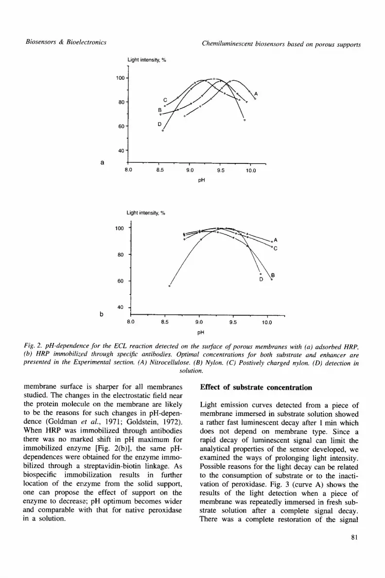
Light emission curves detected from a piece of membrane immersed in substrate solution showed a rather fast luminescent decay after 1 min which does not depend on membrane type. Since a rapid decay of luminescent signal can limit the analytical properties of the sensor developed, we examined the ways of prolonging light intensity. Possible reasons for the light decay can be related to the consumption of substrate or to the inacti- vation of peroxidase. Fig. 3 (curve A) shows the results of the light detection when a piece of membrane was repeatedly immersed in fresh sub- strate solution after a complete signal decay. There was a complete restoration of the signal
81

Maya Yu. Rubtsova et al. Biosensors & Bioelectronics
Light intensity, RLU
600
5OO S
40O
3O0
200
100
0 J I
o 50 1
1 oo i i
150 200 250
T i m e , sec
I
3 0 0
Fig. 3. Time course of light emission from HRP adsorbed onto the surface of a nitrocellulose mem- brane. (A) Detection on a membrane after successive additions of fresh substrate solution. (B) Detection on a membrane supported by a piece of thick filter paper
both soaked in substrate solution.
up to the initial value, indicating that substrate depletion is the main reason for the signal decay. At the same time we revealed bell-shaped depen- dences of maximal light intensity on luminol and enhancer concentration (data not shown), and our attempts to overcome substrate depletion by adding more substrate in the reagent solution resulted in the decrease of maximal intensity. Then we tried to vary the volume of the solution with the addition of a thick hydrophilic support (e.g. Whatman paper) under the membrane. Such support should be fixed snugly to the back side of the membrane, and as a result the duration of the signal can be increased (Fig. 3, curve B). When measuring the light emission from the membrane with its support, the signal remains constant for at least 10 min, which is important for application in biosensors.
Effect of support and immobilization method on HRP chemiluminescent detection
Maximal light intensity and overall light emission during the time course of the reaction are the main features of the ECL reaction. These para- meters were determined for the different prep- arations of immobilized HRP, including immobil- ization of native peroxidase directly on the surface of support, immobilization of conjugated peroxidase and biospecific immobilization. Table 1 contains the data on the comparison of the light emission from different supports. The maxi-
mal light intensity was recorded on nitrocellulose with HRP immobilized through a streptavidin- biotin linkage. In the comparison of immobiliz- ation techniques, the values for both parameters of the ECL reaction were higher when HRP was immobilized through a streptavidin-biotin linkage or specific antibodies.
For HRP adsorbed directly on the surface of membrane we revealed the marked effect of poly- meric material on the light emission. The light intensity was maximal on both nitrocellulose and positively charged nylon. At the same time we detected markedly different activities towards ABTS on these types of membranes, and they were lower on both types of nylon compared with that for nitrocellulose membrane. So, the light detection is more efficient on the surface of positively charged nylon support compared with nitrocellulose. The effect of positive charges can be similar to the effect of enhancement of light intensity in the presence of positively charged polymers reported recently (Gorovits et al., 1995; Kricka et al., 1996). At the same time we revealed the lower emission for HRP immobilized biospecifically on the surface of positively charged nylon compared with nylon and nitrocel- lulose. At the immobilization of HRP conjugated to immunoglobulins we revealed comparable values of maximal intensity for different supports. In this case, the molecules of the enzyme are localized further from the solid support and are surrounded with the molecules of protein-carrier. The presence of the other protein closes the enzyme molecule and it eliminates the effect of polymeric material on the enzyme activity.
The photoactivation of nylon with p-AFBA results in the increase of the maximal intensity and the overall emission when compared with that observed on nonmodified nylon. At the same time, this enhancement was not marked on the surface of photoactivated charged nylon compared with that for photoactivated nylon. Photoactiv- ation with p-AFBA increases the hydrophobicity of membrane surface, that in turn can affect the light emission on the surface of activated nylon. At the same time, the distance between enzyme and support surface lengthens due to the spacer arm. As the effect of positive charges strongly depends on the distance between them and the enzyme, this can be a reason for the elimination of the effect of positive charges on enzyme activity on the surface of activated charged nylon.
In the comparison of light detection efficiency
82
Biosensors & Bioelectronics
from different membranes we can conclude that methods of biospecific immobilization provide the maximal HRP activity in the ECL reaction. The nitrocellulose membrane shows higher light inten- sities for all types of biospecific immobilization due to a higher amount of fixed enzyme and better efficiency of light detection. Positively charged nylon has marked advantages in higher emission when HRP is adsorbed directly on the membrane.
Determination of hydrogen peroxide in water samples
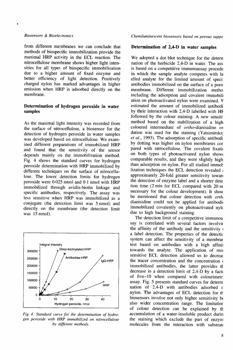
As the maximal light intensity was recorded from the surface of nitrocellulose, a biosensor for the detection of hydrogen peroxide in water samples was developed based on nitrocellulose. We exam- ined different preparations of immobilized HRP and found that the sensitivity of the sensor depends mainly on the immobilization method. Fig. 4 shows the standard curves for hydrogen peroxide determination with HRP immobilized by different techniques on the surface of nitrocellu- lose. The lower detection limits for hydrogen peroxide were 0.025 nmol and 0.1 nmol with HRP immobilized through avidin-biotin linkage and specific antibodies, respectively. The assay was less sensitive when HRP was immobilized as a conjugate (the detection limit was 5 nmol) and directly on the membrane (the detection limit was 15 nmol).
Integral intensily 300000 I 7 "C~tr7 "biOtinylated HRP
250000 J / / Antibodies-HRP ~IgG.HR P
.oooool / / / , ooooj / / .
0 10 20 30 40 Hydrogen peroxide, nmol
Fig. 4. Standard cur~e for the determination of hydro- gen peroxide with HRP immobilized on nitrocellulose
by different methods.
Chemiluminescent biosensors based on porous suppo
Determination of 2,4-D in water samples
We adopted a dot blot technique for the detern nation of the herbicide 2,4-D in water. The ass is based on a competitive immunoassay procedu in which the sample analyte competes with la elled analyte for the limited amount of. speci: antibodies immobilized on the surface of a poro membrane. Different immobilization metho including the adsorption and covalent immobili ation on photoactivated nylon were examined. V estimated the amount of immobilized antibodi by their interaction with 2,4-D labelled with HF followed by the colour staining. A new sensiti' method based on the stabilization of a high coloured intermediate of ortho-dianisidine o~ dation was used for the staining (Yatsimirska Z et al., 1993). The adsorption of specific antibodi by dotting was higher on nylon membranes cot pared with nitrocellulose. The covalent fixati~ on both types of photoactivated nylon show~ comparable results, and they were slightly high than adsorption on nylon. For all studied immol: lization techniques the ECL detection revealed approximately 20-fold greater sensitivity towarl the detection of enzyme label and a shorter dete tion time (2 min for ECL compared with 20 m necessary for the colour development). It shou be mentioned that colour detection with orth, dianisidine could not be applied for antibodb immobilized covalently on photoactivated nyk due to high background staining.
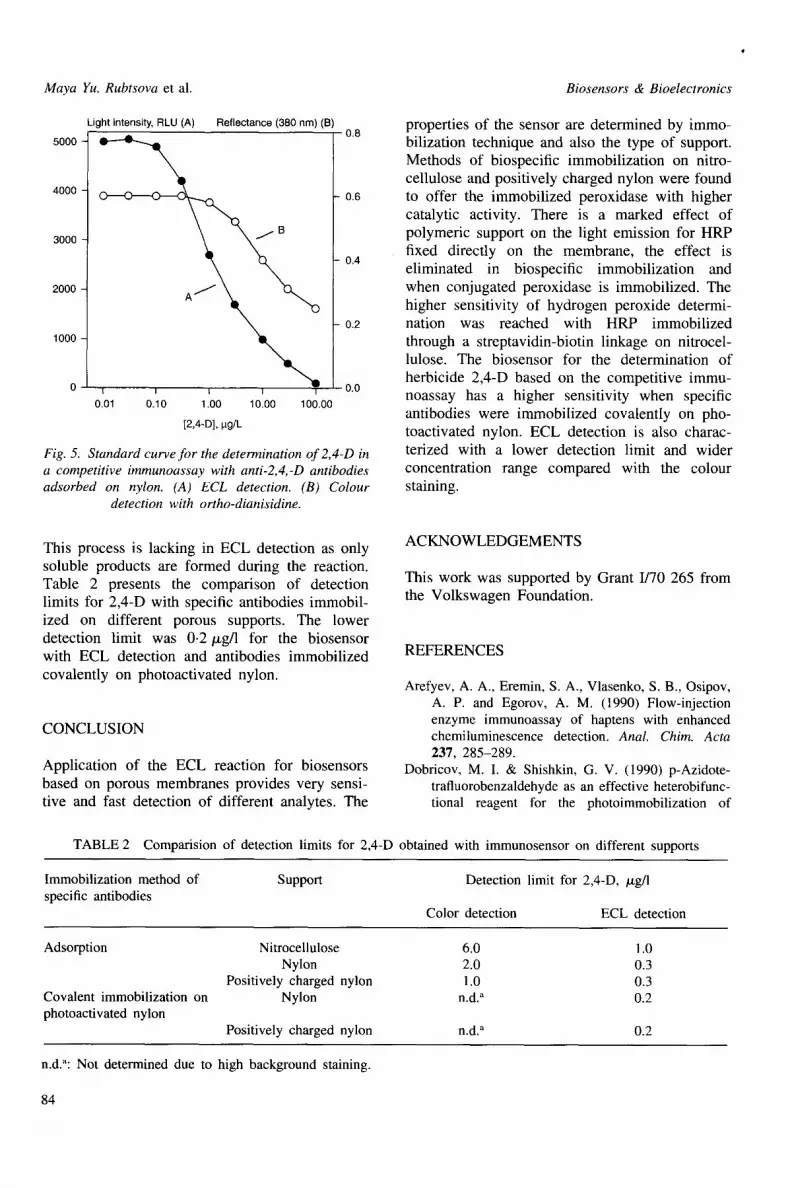
The detection limit of a competitive immunoa say is correlated with several factors involvi[ the affinity of the antibody and the sensitivity q a label detection. The properties of the detectic system can affect the sensitivity of a membrar test based on antibodies with a high affinil towards the analyte. The application of tool sensitive ECL detection allowed us to decrea, the tracer concentration and the concentration immobilized antibodies, the latter provides tt decrease in a detection limit of 2,4-D by a fact( of five-10 when compared with colourimetr! assay. Fig. 5 presents standard curves for determ nation of 2,4-D with antibodies adsorbed c nylon. The advantages of ECL detection for tl: biosensors involve not only higher sensitivity bt also wider concentration range. The limitatior of colour detection can be explained by th accumulation of a water-insoluble product durin the staining which exclude the part of enzym molecules from the interaction with substratl
Maya Yu. Rubtsova et al.
Light intensity, RLU (A) Reflectance (380 nm) (B)
5000 -
4000
3000
2000
1000
0.8
(3-------O
j B
I I I I I
0.01 0.10 1.00 10.00 100.00
0.6
0.4
0.2
0 0.0
[2,4-D], I.tg/L
Fig. 5. Standard curve for the determination of 2,4-D in a competitive immunoassay with anti-2,4,-D antibodies adsorbed on nylon. (A) ECL detection. (B) Colour
detection with ortho-dianisidine.
This process is lacking in ECL detection as only soluble products are formed during the reaction. Table 2 presents the comparison of detection limits for 2,4-D with specific antibodies immobil- ized on different porous supports. The lower detection limit was 0-2/zg/1 for the biosensor with ECL detection and antibodies immobilized covalently on photoactivated nylon.
CONCLUSION
Application of the ECL reaction for biosensors based on porous membranes provides very sensi- tive and fast detection of different analytes. The
Biosensors & Bioelectronics
properties of the sensor are determined by immo- bilization technique and also the type of support. Methods of biospecific immobilization on nitro- cellulose and positively charged nylon were found to offer the immobilized peroxidase with higher catalytic activity. There is a marked effect of polymeric support on the light emission for HRP fixed directly on the membrane, the effect is eliminated in biospecific immobilization and when conjugated peroxidase is immobilized. The higher sensitivity of hydrogen peroxide determi- nation was reached with HRP immobilized through a streptavidin-biotin linkage on nitrocel- lulose. The biosensor for the determination of herbicide 2,4-D based on the competitive immu- noassay has a higher sensitivity when specific antibodies were immobilized covalently on pho- toactivated nylon. ECL detection is also charac- terized with a lower detection limit and wider concentration range compared with the colour staining.
ACKNOWLEDGEMENTS
This work was supported by Grant 1/70 265 from the Volkswagen Foundation.
REFERENCES
Arefyev, A. A., Eremin, S. A., Vlasenko, S. B., Osipov, A. P. and Egorov, A. M. (1990) Flow-injection enzyme immunoassay of haptens with enhanced chemiluminescence detection. Anal. Chim. Acta 237, 285-289.
Dobricov, M. I. & Shishkin, G. V. (1990) p-Azidote- trafluorobenzaldehyde as an effective heterobifunc- tional reagent for the photoimmobilization of
TABLE 2 Comparision of detection limits for 2,4-D obtained with immunosensor on different supports
Immobilization method of specific antibodies
Suppo~ Detection limit for 2,4-D, /xg/1
Color detection ECL detection
Adsorption
Covalent immobilization on photoactivated nylon
Nitrocellulose 6.0 1.0 Nylon 2.0 0.3
Positively charged nylon 1.0 0.3 Nylon n.d. a 0.2
Positively charged nylon n.d." 0.2
n.d.": Not determined due to high background staining.
84

Biosensors & Bioelectronics
enzymes. Bioorganicheskaya Chimiya, 6, 50-57 (in Russian).
Easton, P. M., Simmonds, A. C., Rakishev, A., Egorov, A. M. and C~ndeias, L. P. (1996) Quantitative model of the e.nhancement of peroxidase-induced luminol luminescence. JACS 118, 6619-6624.
Evans, M. R., Simmonds, A. C., Cunningham, M. W., Durrant, I., Harvey, B. & Fowler, S. (1993) Enhanced chemiluminescence in filter-based DNA and protein detection. In Bioluminescence and Chemiluminescence. Status Report. Proceedings VII Int. Syrup. on Biolum. Chemilum., ed. A. A. Szalay, L. J. Kricka & P. Stanley J. Wiley, Banff, pp. 410-414.
Geckeler, K. E. and Muller, B. (1993) Polymer materials in biosensors. Naturwissenschalfien 80, 18-24.
Goldman, R., Kedem, O. and Katchelsky, E. (1971) Kinetic behavior of alkaline phosphatese collodion membranes. Biochemistry 10~ 165-172.
Goldstein, L. (1972) Microenvironmental effects on enzyme catalysis. A kinetic study of polyanionic and polycationic derivatives of chymotrypsin. Bio- chemistry 11, 4072-4084.
Gorovits, E. L., Izumrudov, V. A., Pisarev, V. V., Gavrilov, E. M and Egorov, A. M. (1995) Effect of synthetic polyelectrolytes on enhanced chemi- luminescence reaction in solution. Biotechnol. Appl. Biochem. 22, 249-260.
Gorovits, B. M., Osipov, A. P. and Egorov, A. M. (1993) New immunoassay technique using anti- body immobilized on a membrane and flow cuvette as reaction vessel. J. Immunol. Meth. 157, 11-17.
Hlavay, J. and Guilbault, G. (1993) A fiber-optic biosensor for determination of hydrogen peroxide. Effect of the membrane type on the immobilization of POD enzyme. Acta Chim. Hung. 130, 83-93.
Hlavay, J., Haemmerli, S. D. and Guilbault, G. G. (1994) Fibre-optic biosensor for hypoxanthine and xanthine based on a chemiluminescence reaction. Biosensors & Bioelectronics 9, 189-195.
Kricka, L. G. and Thorpe, H. G. (1983) Chemilumi- nescent and bieluminescent methods in analytical chemistry. Analyst 108, 1274-1296.
Kricka, L. J., Ji, X., Thorpe, G. H. G., Edwards, B., Voyta, J. and Bronstein, I. (1996) Comparison of 5-hydroxy-2,3-dihydrophthalazine-l-4,dione and luminol as co-substrates for detection of horserad-
Chemiluminescent biosensors based on porous suppo
ish peroxidase in enhanced chemiluminescent re~ tions. J. Immunoassay 17, 67-83.
Nakane, P. K. and Kawaoi, A. (1974) Peroxida, labelled antibody. A new method of conjugati( J. Histochem. Cytochem. 22, 1084-1091.
Nau, V. and Nieman, T. A. (1979) Application microporous membranes to chemiluminescen analysis. Anal. Chem. 51, 424-428.
Navas, M. J. and Limenez, A. M. (1996) Review chemiluminescent methods in food analysis. Fo Chemistry 55, 7-15.
Osipov, A. P., Zaitseva, N. V. and Egorov, A. 1 (1996) Chemiluminescent immunoenzyt biosensor with a thin-layer flow-through ce Application for study of a real-time biomolecu antigen-antibody interation. Biosensors & Bioelt tronics 11, 881-887.
Rubtsova, M. Yu. and Gavrilova, E. M. (1994) J assay for human myoglobin using the technique membrane immunoassay with light-sensitive nyl supports. Anal. Letters 27, 2961-2971.
Sun, W., Ji, X., Kricka, L. J. and Dunford, H. (1994) Rate constants for reactions of horseradi peroxidase compounds I and II with 4-substitut arylboronic acids. Can. J. Chem 72, 2159-2162
Thorpe, G. H. G. & Kricka, L. J. (1987) Enhanc chemiluminescent assays for horseradish pero: dase: characteristics and applications. In Biolum, escence and Chemiluminescence: New Persp~ tires, ed. J. Scholmerich, R. Andreesen, A. Ka t M. Ernst & W. G. Woods. J. Wiley, Chichest pp. 199-208.
Vlasenko, S. B., Arefyev, A. A., Klimov, A. D., Ki B. B., Gorovits, E. L., Osipov, A. P., Gavrilo~ E. M. and Egorov, A. M. (1989) An investigati on the catalytic mechanism of enhanced chen luminescence: immunochemical applications of t reaction. J. Biolum. Chemilum. 4, 164-176.
Yan, M., Cai, S. X., Wybourne, M. N. and Keana, F. W. (1993) Photochemical functionalization polymer surfaces and the production of biomok ule-carrying micrometer-scale structures by dee UV lithograph using 4-substituted perfluoropher azides. J. Am. Chem. Soc. 115, 814-816.
Yatsimirskaya, E. A., Gavrilova, E. M. and Egorov, M. (1993) Detection system for membrane imn~ noassay based on the trapping of a highly color intermediate of the peroxidase reaction. An Biochem. 211, 274-278.