Purification and Partia1 Characterization of Tomato Extensin Peroxidase
9
Plant Physiol. (1 995) 109: 11 15-1 123 Purification and Partia1 Characterization of Tomato Extensin Peroxidase' Michael Daniel Brownleader*, Naziha Ahmed, Michael Trevan, Martin F. Chaplin, and Prakash M. Dey School of Applied Science, South Bank University, 103 Borough Road, London, United Kingdom SEI OAA (M.D.B., N.A., M.T., M.F.C.); and Department of Biochemistry, Royal Holloway, University of London, Egham, Surrey, United Kingdom TW20 OEX (P.M.D.) Early plant defense response is characterized by elevation of activity of peroxidases and enhanced insolubilization of hy- droxyproline-rich glycoproteins, such as extensin, in the cell wall. The insolubilization process (cross-linking between soluble extensin precursor molecules) is catalyzed by extensin peroxidases. We have ionically eluted extensin peroxidasesfrom intact water-washed sus- pension-cultured tomato (hybrid of Lycopersicon esculenfum Mill. and Lycopersicon peruvianum L. [Mill.]) cells and purified them to homogeneity by molecular sieve and cation-exchange chromatog- raphy. Four ionic forms of peroxidase (PI, PII, EPIII, and EPIV) were resolved; only the latter two cross-linked tomato soluble extensin. The molecular weight (34,000-37,000), amino acid composition, and isoelectric point (9.0) of the extensin peroxidases were deter- mined. Substrate specificities of the enzymes were investigated: soluble extensin and potato lectin (a hydroxyproline-rich glycopro- tein with a domain that strongly resembles extensin) were cross- linked by only two forms of the enzyme, whereas bovine serum albumin, aldolase, insulin, a number of other marker proteins, and proteins eluted from tomato cells (except extensin) could not be cross-linked. We have also isolated a yeast elicitor that enhances total peroxidase activity and extensin insolubilization within 1 h of challenge in cultured cells of tomato. A highly sensitive enzyme- linked immunosorbent assay technique using polyclonal antiserum raised against soluble tomato extensin was used to demonstrate extensin insolubilization in vivo. A tomato cell-wall peroxidase that cross-links extensin has been purified and may have a role in plant defense. Plants generate a large array of inducible defense re- sponses following mechanical damage or pathogen attack. These include synthesis of antimicrobial compounds (phy- toalexins), deposition of callose, lignin, and cell-wall- bound phenolics, accumulation of HRGPs in the cell wall, and synthesis of protease inhibitors and hydrolytic en- zymes (Lamb and Dixon, 1994). Extensin (an HRGP) exists in two developmental forms: soluble extensin, which is ionically bound to the cell wall, and an insoluble form that is deposited in the cell wall by a peroxidase/H,O,-medi- ated cross-linking process (Cooper and Varner, 1984). M.D.B. is the recipient of a special research fellowship from The Leverhulme Trust. This work was supported by Biotechnol- ogy and Biological Sciences Research Council. * Corresponding author; e-mail brown1mdBsbu.ac.uk; fax 44- 1-71-815-7999. In an earlier study, extensin and an associated peroxi- dase were eluted from cell walls of suspension-cultured tomato (hybrid of Lycopersicon esculentum Mill. and Lyco- persicon peruvianum L. [Mill.]) cells (Brownleader et al., 1993). Cross-linking of extensin was then demonstrated by an FPLC gel-filtration assay in the presence of H,O, (Brownleader et al., 1993).Everdeen et al. (1988) had earlier identified an extensin-cross-linking enzyme that they re- ferred to as extensin peroxidase; their nomenclature is used in this paper. Peroxidases (EC 1.11.1.7) have been found ionically and covalently bound to the plant cell wall (Ridge and Osborne, 1971), but their function in the plant cell wall is not com- pletely understood. The precise role of individual isoforms of peroxidases remains unclear owing to the lack of infor- mation on the localization of the enzymes and the avail- ability of their specific substrates in vivo. They are required for lignin biosynthesis, but they also occur in cell walls that do not form lignin (Whitmore, 1971). Subsequent work by Whitmore (1976) demonstrated that peroxidases catalyze the linking of ferulic acid to cell walls of pine (Pinus elliot- tii). Cationic peroxidases have been found to be most ef- fective in IAA catabolism, and anionic isoforms are be- lieved to be associated with lignification (Gaspar et al., 1991; Medina et al., 1993). Ros-Barceló et al. (1987, 1989) observed that lupin acidic peroxidases are located primar- ily in the cell wall, whereas basic peroxidases are primarily vacuolar and bound to the tonoplast (Ros-Barceló et al., 1991). However, Ferrer et al. (1992) demonstrated that two strongly basic peroxidases are located in the cell wall of etiolated lupin hypocotyls. Peroxidases are also believed to play a significant role in plant disease resistance. Increased peroxidase activity and elevated HRGP deposition in the cell wall have been demonstrated after challenging the plant with pathogens (Esquerré-Tugayé and Lamport, 1979; Reimers et al., 1992; Lamb et al., 1993). This paper is concerned with the purification and char- acterization of ionically bound cell-wall peroxidase activi- ties from cultured tomato cells that cross-link the HRGP Abbreviations: ABTS, 2,2'-azino-bis(3-ethyl benzthiazoline-6- sulfonic acid); CM-cellulose, carboxymethyl-cellulose; EPIII, EPIV, "extensin peroxidase" I11 and IV; EtOH, ethanol; FPLC, fast pro- tein liquid chromatography; HRGP, hydroxyproline-rich glyco- protein; HRP, horseradish peroxidase; L-DOPA, L-dihydroxyphe- nylalanine; PI, PII, peroxidase I and 11; MeOH, methanol; RZ, A405 / A,,, . 1115
Transcript of Purification and Partia1 Characterization of Tomato Extensin Peroxidase
Plant Physiol. (1 995) 109: 11 15-1 123
Purification and Partia1 Characterization of Tomato Extensin Peroxidase'
Michael Daniel Brownleader*, Naziha Ahmed, Michael Trevan, Martin F. Chaplin, and Prakash M. Dey
School of Applied Science, South Bank University, 103 Borough Road, London, United Kingdom SEI OAA (M.D.B., N.A., M.T., M.F.C.); and Department of Biochemistry, Royal Holloway, University of London,
Egham, Surrey, United Kingdom TW20 OEX (P.M.D.)
Early plant defense response i s characterized by elevation of activity of peroxidases and enhanced insolubilization of hy- droxyproline-rich glycoproteins, such as extensin, in the cell wall. The insolubilization process (cross-linking between soluble extensin precursor molecules) is catalyzed by extensin peroxidases. We have ionically eluted extensin peroxidases from intact water-washed sus- pension-cultured tomato (hybrid of Lycopersicon esculenfum Mill. and Lycopersicon peruvianum L. [Mill.]) cells and purified them to homogeneity by molecular sieve and cation-exchange chromatog- raphy. Four ionic forms of peroxidase (PI, PII, EPIII, and EPIV) were resolved; only the latter two cross-linked tomato soluble extensin. The molecular weight (34,000-37,000), amino acid composition, and isoelectric point (9.0) of the extensin peroxidases were deter- mined. Substrate specificities of the enzymes were investigated: soluble extensin and potato lectin (a hydroxyproline-rich glycopro- tein with a domain that strongly resembles extensin) were cross- linked by only two forms of the enzyme, whereas bovine serum albumin, aldolase, insulin, a number of other marker proteins, and proteins eluted from tomato cells (except extensin) could not be cross-linked. We have also isolated a yeast elicitor that enhances total peroxidase activity and extensin insolubilization within 1 h of challenge in cultured cells of tomato. A highly sensitive enzyme- linked immunosorbent assay technique using polyclonal antiserum raised against soluble tomato extensin was used to demonstrate extensin insolubilization in vivo. A tomato cell-wall peroxidase that cross-links extensin has been purified and may have a role in plant defense.
Plants generate a large array of inducible defense re- sponses following mechanical damage or pathogen attack. These include synthesis of antimicrobial compounds (phy- toalexins), deposition of callose, lignin, and cell-wall- bound phenolics, accumulation of HRGPs in the cell wall, and synthesis of protease inhibitors and hydrolytic en- zymes (Lamb and Dixon, 1994). Extensin (an HRGP) exists in two developmental forms: soluble extensin, which is ionically bound to the cell wall, and an insoluble form that is deposited in the cell wall by a peroxidase/H,O,-medi- ated cross-linking process (Cooper and Varner, 1984).
M.D.B. is the recipient of a special research fellowship from The Leverhulme Trust. This work was supported by Biotechnol- ogy and Biological Sciences Research Council.
* Corresponding author; e-mail brown1mdBsbu.ac.uk; fax 44- 1-71-815-7999.
In an earlier study, extensin and an associated peroxi- dase were eluted from cell walls of suspension-cultured tomato (hybrid of Lycopersicon esculentum Mill. and Lyco- persicon peruvianum L. [Mill.]) cells (Brownleader et al., 1993). Cross-linking of extensin was then demonstrated by an FPLC gel-filtration assay in the presence of H,O, (Brownleader et al., 1993). Everdeen et al. (1988) had earlier identified an extensin-cross-linking enzyme that they re- ferred to as extensin peroxidase; their nomenclature is used in this paper.
Peroxidases (EC 1.11.1.7) have been found ionically and covalently bound to the plant cell wall (Ridge and Osborne, 1971), but their function in the plant cell wall is not com- pletely understood. The precise role of individual isoforms of peroxidases remains unclear owing to the lack of infor- mation on the localization of the enzymes and the avail- ability of their specific substrates in vivo. They are required for lignin biosynthesis, but they also occur in cell walls that do not form lignin (Whitmore, 1971). Subsequent work by Whitmore (1976) demonstrated that peroxidases catalyze the linking of ferulic acid to cell walls of pine (Pinus elliot- tii). Cationic peroxidases have been found to be most ef- fective in IAA catabolism, and anionic isoforms are be- lieved to be associated with lignification (Gaspar et al., 1991; Medina et al., 1993). Ros-Barceló et al. (1987, 1989) observed that lupin acidic peroxidases are located primar- ily in the cell wall, whereas basic peroxidases are primarily vacuolar and bound to the tonoplast (Ros-Barceló et al., 1991). However, Ferrer et al. (1992) demonstrated that two strongly basic peroxidases are located in the cell wall of etiolated lupin hypocotyls. Peroxidases are also believed to play a significant role in plant disease resistance. Increased peroxidase activity and elevated HRGP deposition in the cell wall have been demonstrated after challenging the plant with pathogens (Esquerré-Tugayé and Lamport, 1979; Reimers et al., 1992; Lamb et al., 1993).
This paper is concerned with the purification and char- acterization of ionically bound cell-wall peroxidase activi- ties from cultured tomato cells that cross-link the HRGP
Abbreviations: ABTS, 2,2'-azino-bis(3-ethyl benzthiazoline-6- sulfonic acid); CM-cellulose, carboxymethyl-cellulose; EPIII, EPIV, "extensin peroxidase" I11 and IV; EtOH, ethanol; FPLC, fast pro- tein liquid chromatography; HRGP, hydroxyproline-rich glyco- protein; HRP, horseradish peroxidase; L-DOPA, L-dihydroxyphe- nylalanine; PI, PII, peroxidase I and 11; MeOH, methanol; RZ, A405 / A,,, .
1115
1116 Brownleader et al. Plant Physiol. Vol. 109, 1995
extensin and that may play a role in plant disease resistance.
MATERIALS AND METHODS
Materiais
Callus of tomato cells (hybrid of Lycopersicon esculentum Mill. and Lycopersicon peruvianum L. [Mill.]) was a gift from Dr. Stephen Fry (Department of Botany, University of Ed- inburgh, UK). CM-cellulose cation-exchange resin was ob- tained from Whatman and Superose-12 and Sephacryl S-200 gel-filtration resin were purchased from Pharmacia. Protogel for SDS-PAGE was obtained from National Diag- nostics (Manville, NJ). Calibration marker kits for IEF and SDS-PAGE were obtained from Pharmacia. Polyclonal an- tiserum against extensin was prepared at Royal Holloway (University of London). Alkaline phosphatase-labeled goat anti-rabbit secondary antibody was purchased from Sigma. Phenolphthalein phosphate substrate and stop solution for ELISA were purchased from Serono Diagnostics (Geneva, Switzerland).
Growth Conditions of Cell Suspension Culture
Tomato suspension cells were grown in Murashige and Skoog tissue culture medium (basal salt mixture) (Mura- shige and Skoog, 1962) comprising 20 g/L Glc and a sup- plement solution of 100 mg/L myo-inositol, 0.5 mg/L nic- otinic acid, 0.5 mg/L pyridoxine HC1, 0.1 mg/L thiamine HCI, 2 mg/L Gly, 10 mg/L IAA, and 0.5 mg/L kinetin. Aliquots of sterilized medium (200 mL) in 500-mL conical flasks were inoculated with 7-d-old suspension cells and incubated on a gyratory shaker at 110 rpm under continu- ous fluorescent lighting for 7 d at 25°C.
Elution of Soluble Extensin Monomer and Extensin Peroxidase
Ionic desorption of soluble extensin and extensin perox- idase from cell walls of intact water-washed tomato cells in suspension cultures was accomplished with 50 mM CaCI,, adjusted to pH 3.0 with 6 M HC1, by Büchner filtration. The filtrate was subsequently dialyzed in 5 L of 30 mM hydro- chloric acid solution (pH 3.0) overnight. The low pH was chosen to prevent cross-linking of extensin by peroxidase if H,O, or organic peroxides were present in the filtrate, and this was followed by dialysis against distilled water for an additional 3 h (Brownleader and Dey, 1993).
Purification of Extensin and Extensin Peroxidase
After dialysis, the salt-eluted protein was concentrated by CM-cellulose (CM-52; strong cation-exchange resin) chromatography. The column (5 X 3.3 cm) was equili- brated with degassed 20 mM McIlvaine’s buffer (Na,HPO,- citric acid buffer), pH 5.5 (McIlvaine, 1921). Protein was eluted by addition of 20 mM McIlvaine’s buffer, pH 5.5, containing 0.5 M NaC1. Elution was monitored spectropho- tometrically at a wavelength of 280 nm to detect protein.
The CM-cellulose resin had a large protein-binding capac- ity and could also be used for batch processing.
The eluted protein was concentrated by Filtron ultrafil- tration (10-kD Omega membrane, Filtro11 Technology Corp., Northborough, MA) to a final volume of 5 mL ,and the sample was applied to a 5-200 Sephacryl gel-filtraiion column (105 X 2.5 cm) equilibrated with degassed Q.1 M
sodium acetate buffer, pH 5.0, containing 0.1 M NaCl and 0.02% (w/v) sodium azide. Fractions containing either ex- tensin or extensin peroxidase were monitored spectropho- tometrically ai a wavelength of 280 nm to detect proiein and pooled separately. Four recognized criteria were used to identify exlensin, and this has been discussed in our previous paper (Brownleader and Dey, 1993). Extensin eluted earlier than extensin peroxidase and was recognized by the fact that it was cross-linked by extensin peroxidase in the presence of H,O,. No other proteins tested, e.g BSA or aldolase, could be cross-linked. Hyp and Ara contents, amino acid analysis, and the presence of the putative se- quence motif -Ser(Hyp),- in the N-terminal end of the peak fractions reported to be extensin were also used as indica- tors of the identity of this HRGP. The pooled fractions containing extensin were dialyzed against distilled water and concentrated to a final volume of 3 mL by Filtron ultrafiltration (10-kD Omega membrane). The extensin sample was then freeze-dried. AI1 peaks were assayed for their ability to oxidize guaiacol. Only one peak oxidized guaiacol, and i he corresponding fractions containing per- oxidase that eluted after extensin were pooled and then dialyzed against water. In addition, this peak absclrbed strongly at 405 nm, indicating that the protein component contained a heme group.
The dialyzed fractions corresponding to high A,,, and containing high guaiacol-oxidizing (2-methoxyphenol) ac- tivity were loaded onto a preparative CM-52 ion-exchange column (5 X 3.3 cm) pre-equilibrated with degassed 20 mM McIlvaine’s buffer, pH 6.0. The proteins were eluted with a linear O to 0.4 M NaCl gradient at a flow rate of 3 mL,”in and monitored spectrophometrically at 280 and 405 nm to detect protein and heme, respectively. At this stage, the peak that cross-linked extensin was pooled and dialyzed against water. Two milliliters of this sample were loaded onto an analytical Mono S HR 5/5 FPLC column (0.5 X 5 cm; 1-mL bed volume) that was equilibrated with 20 mM McIlvaine’s buffer, pH 5.5. The protein was eluted with a linear O to 0.4 M NaCl gradient at a flow rate of 0.4 mL/ min. This analytical column produced a better resolution of proteins than the preparative CM-52 cellulose resin.
Routine Assay of Peroxidase Activity
Activity of peroxidase fractions during purification was routinely monitored by oxidation of guaiacol. The assay contained 20 mM potassium phosphate buffer, pH 7.0, 13.3 mM guaiacol, and 33 p~ H,O,. The reaction was initiated by addition of peroxidase (2 pL) and the color develop- ment was monitored over a 2-min time period at 470 nm.
A wide range of low-molecular-weight, nonprotein sub- strates were tested for the tomato peroxidase preparations, and the rates of reaction were compared with those of HRP

Tomato Extensin Peroxidase 1117
I 1 RZ>3.2
O 20 40 60 80 100 120 140 160 Fraction number
Figure 1. CM-cellulose cation-exchange chromatography of Seph- acryl S-200-purified peroxidase showing resolution of peroxidase into foi" I and II. O, A,,,, indicating protein content. O, A,,,, indicating heme content. O, Guaiacol oxidation.
(see "Results and Discussion"). These substrates include a number of naturally occurring low-molecular-weight com- pounds of plants. There were two objectives: first, to shed light on the possible in vivo functions of the enzymes, and second, to find a more convenient substrate for spectropho- tometric assay in order to distinguish between peroxidases that do and do not cross-link extensin. Substrate specificity of peroxidase was monitored with the following low-mo- lecular-weight substrates: 2 mM ABTS (A,,,), 0.7 mM ascor- bic acid (A,,,), 20 mM catechol (A,,,), 3.6 mM 4-methyl catechol (100% EtOH, A,,,), 0.12 mM coniferyl alcohol(1 M
NaOH, A,,,), 0.09 mM ferulic acid (1 M NH,, A,,,), 0.5 mM NADH (A340), 13.3 mM guaiacol (A,,,), 3 mM p-phe- nylenediamine (100% EtOH, A,,,), 0.1 mM syringaldazine (A,,,), 0.09 mM syringaldehyde (DMSO, A55,), 0.02 mM L-DOPA (0.5 M HC1, A,,,), 0.2 mM L-Tyr (0.1 M NaOH, A2,,), 0.2 mM D-Tyr (A,,,), 0.12 mM 4-aminoantipyrene (A,,,), 0.06 mM vanillin azine (100% MeOH, A,,,), 0.16 mM 4-hydroxy-3-methoxybenzyl alcohol(95% EtOH, A,,,), 0.27 mM 3-hydroxy-4-methoxy benzyl alcohol (100% MeOH, AZ9,), 0.22 mM 4-hydroxy-3-methoxy benzylamine HC1 (1 M NaOH, AZ9,), and 0.1 mM L-Tyr-ethyl ester HC1 (100% EtOH, A,,,). Distilled water was the solvent unless stated otherwise in parentheses. The solvent concentration never exceeded 0.2% (v/v) in the assay. The incubation medium contained 0.2 M potassium phosphate buffer, pH 7.0, and 0.6 mM H,O,. Reactions were monitored over a 2-min time course at the appropriate wavelength.
Assay of Extensin Cross-Linking by Extensin Peroxidase
Approximately 35 pg of tomato extensin or 53 pg of potato lectin was incubated with 0.2 M sodium phosphate
buffer, pH 7.0, in the presence of 75 p~ H,O,. The reaction was initiated with extensin peroxidase (5 pL, 13 pg) in a total volume of 100 pL. After incubation for 30 min in the dark at room temperature, the incubation mixture was injected onto a Superose-12 gel-filtration FPLC column (Pharmacia, 30 X 1 cm) using a Rheodyne (Cotati, CA) volume injector. The reaction was performed in the dark to prevent decomposition of H,O, by light. The column was equilibrated with 0.2 M sodium phosphate buffer, pH 7.0, containing 0.005% (w/v) sodium azide at a flow rate of 0.2 mL/min, and the eluate was monitored at 280 nm. The lower-molecular-weight extensin monomer eluted slower than extensin cross-linked after incubation with peroxidase and H,O,.
SDS-PAG E
Purity and molecular weight of extensin peroxidase were determined by 12% acrylamide SDS-PAGE as described by Laemmli (1970). The calibration markers used for molecu- lar weight determination were a-lactalbumin ( M , 14,000), trypsin inhibitor (M, 20,000), carbonic anhydrase (M, 30,000), ovalbumin ( M , 43,000), BSA ( M , 67,0001, and phos- phorylase b ( M , 94,000).
IEF
The pI of extensin peroxidase was established using a Pharmacia Phast gel system (pH 3-9). EPIII and EPIV
I A Extensin
A
I , I I I f 1
10 20 30 40 50 60 70 80 90 Time (MIN)
1 fl Crosslinked extensin I
1 (Peak I) - ~ 1 Ui 10 20 3 b 40 50 60 70 80 90
Time (MIN)
Figure 2. Extensin cross-linking assay in vitro by Superose-1 2 gel- filtration FPLC using 20 p g of peak I I enzyme (A) (see Fig. 1) or 1 pg of peak I enzyme (B), demonstrating that peak I and not peak II constitutes extensin peroxidases. The lower-molecular-weight exten- sin monomer eluted at a larger retention time than the cross-linked extensin.
Brownleader et al. Plant Physiol. Vol. 109, 1995 1118
(0.15-0.3 pg) were loaded onto a prefocused gel that was developed for 12 min at 2000 V (2.5 mA). The gel was subsequently fixed in 20% TCA for 5 min at 2OoC, washed in Me0H:acetic acid (30:10%, v/v) for an additional 2 min at 20"C, stained in 0.02% (w/v) Coomassie brilliant blue R350 (Phast gel Blue R) dissolved in Me0H:acetic acid (30:10%, v/v), and allowed to incorporate 0.1% copper sulfate for 10 min at 50°C. The gel was then destained with Me0H:acetic acid (30:10%, v/v) for an additional 10 min at 50°C. Calibration markers were purchased from Pharmacia and designed for use with pH 3.0 to 9.0 IEF gels.
Protein Determination
Protein concentration was estimated by the dye-binding method of Bradford (1976). BSA (fraction V, essentially fatty acid free) was used as the protein standard.
N-Terminal Amino Acid Sequencing
N-terminal sequencing of the extensin peroxidases was determined by a pulsed-liquid protein sequencer (477A; Applied Biosystems).
Amino Acid Analysis
Samples of the tomato peroxidases were acid hydrolyzed in 6 M HCI at 110°C for 16 h. The acid hydrolysates were analyzed on an LBK Alpha-Plus Analyzer (Pharmacia LKB Biotechnology, St. Albans, UK) using sodium buffers and ninhydrin determination.
Partia1 Purification of Elicitor
Brewer's yeast extract (100 g), an autolysate of Saccharo- myces cerevisiae, was dissolved in 200 to 300 mL of distilled water. EtOH was added to a final concentration of 80% (v/v), and the precipitate was allowed to settle for 4 d at 4°C. The supernatant was decanted, and the resultant pre- cipitate was dissolved in 100 mL of distilled water and freeze-dried. This preparation is regarded as the partially purified elicitor.
Cell Elicitation and Extensin lnsolubilization
Freeze-dried elicitor was dissolved in distilled water to 20 mg/mL, autoclaved at 121°C for 15 min, and allowed to cool. Hahn and Albersheim (1976) have demonstrated that the crude yeast elicitor is stable at 121°C for 30 min. Elicitor was added at a final concentration of 0.5 mg/mL to tomato cells (estimated 2.5 mg elicitor/g fresh weight cells) and incubated for 1 h on an orbital shaker under light at 100 rpm. Water replaced the elicitor in the control experiment. The tomato cells were washed by Büchner filtration with an excess of distilled water. Water-washed cells (4 g) were eluted with 50 mM CaCl,, pH 3.0 (1 mL), and the eluate recovered by centrifugation. This gentle elution was re- peated two more times. The eluate (100 pL) was then coated onto a microtiter plate, diluted 1:l in coating buffer (1.59 g of Na,CO,, 2.93 g of NaHCO,, 0.2 g of NaN, in 1 L of distilled water, pH 9.6), and incubated overnight at 4°C.
Soluble extensin was measured by ELISA. Cell eluate (100 wL) was assayed for peroxidase activity as before. Siirplus filtrate was stored at -20°C.
ELISA
The eluate was coated onto ELISA plates, and after over- night incubation, the microtiter plates were washed twice with PBS, pH 7.4. PBS was prepared as a concentrated stock solution (80 g of NaCl, 2 g of KC1,11.5 g of Na,HPO,, 2 g of KH,PO,) and diluted 10 times prior to use. The plates were dried by shaking them on filter paper, and then 100 pL of 1% BS.4 blocking reagent in PBS was added and incubated at room temperature for 1 h. The plate wa:? then washed with PBS and incubated with primary extensin antiserum (1:20,000 dilution with 1% BSA in PBS) for 1 h. The plate was then washed in PBS to remove the primary antibody. Fifty microliters of secondary antibody (alkaline- phosphatase-labeled goat anti-rabbit IgG) diluted 1:lOOO in 1% BSA in PBS, pH 7.4, was pipetted into the wells and incubated for 2 h. The plate was then washed in PBS. Phenolphthalein phosphate substrate (50 pL) was added and incubated for 45 min. Stop solution (185 pL) was then added to stop any further color development, and the plate was read at 570 nm.
Production of Antibodies against Tomato Extensin
Polyclonal antiserum was raised in rabbit to the purified native extensin (Brownleader and Dey, 1993). Extensin (100 pg) was dissolved in 250 pL of PBS (0.15 M NaC1, 20 mM sodium phosphate, pH 7.0), mixed with 250 pL of Freunds complete adjuvant, and injected subcutaneously into a rab- bit. Two other injections of the protein were given as above after 4-week intervals. Blood serum was then collected and stored at -70°C. The antiserum had a titer of 1:5600 with 50 ng of extensin.
RESULTS AND DISCUSSION
Purification of Extensin Peroxidase
The activity of extensin peroxidase was assessed b y de- termining the ability of the enzyme to produce the cross- linked high-molecular-weight form of the soluble extensin as the end product when both were incubated in vitro in the presence of H,O,. The mild conditions of intact cell elution with CaC1, (0.05 M) accounted for only 2% of the total protein of the whole-cell homogenate (Everdeen et al., 1988). Therefore, a significant purification of both extensin and extensin peroxidase was achieved by this procedure. The S-200 enzyine preparation was resolved into two active peaks (I and 11) by CM-cellulose chromatography (Fig. 1). Although both peaks oxidized guaiacol, only peak I was able to cross-link soluble extensin in vitro and was there- fore used as a source of extensin peroxidase for further purification. It is important to note that the RZ value of peak I1 (3.2) is 2.3-fold higher than the RZ value of peak I. Figure 2 demonstrates that 1 pg of peak I protein was able to cross-link extensin in vitro, whereas 20 pg of peak I1 had no effect on extensin oligomerization. Peak I was further

Tomato Extensin Peroxidase 1119
0.4
•0.3
•0.1
I 0 4 8 12 16 20 24 28 32 36 401 Elution volume (ml)
Figure 3. Mono S FPLC of peak I from CM-cellulose column (Fig. 1).
purified by Mono S FPLC. Four main protein peaks wereeluted with a NaCl gradient (Fig. 3). All peaks possessedperoxidase activity when assayed with guaiacol. The ex-tensin cross-linking assay demonstrated that PI and PII (upto 20 jug) did not cross-link, whereas EPIII (RZ 1.5) andEPIV (RZ 1.8) (1-9 /xg) cross-linked extensin. It is difficultto relate the RZ values of the enzymes to their puritybecause this estimation depends on the amount and theionic form of the heme as well as the proportion of thearomatic amino acids in the protein.
All four peaks displayed single protein bands by SDS-PAGE (Fig. 4A). The molecular masses estimated for EPIIIand EPIV were 37 and 34 kD, respectively (Fig. 4B). Thesevalues coincide well with the molecular masses determinedby gel filtration (results not shown). Therefore, EPIII andEPIV are not multisubunit proteins.
Partial Characterization of Extensin Peroxidase Isoforms
The pi values of both EPIII and EPIV were approxi-mately 9.0 (results not shown), which confirms that theseare basic peroxidases and is consistent with the ionic bind-ing of the enzymes to cationic column media (Figs. 1 and 3).The N termini of the isozymes were found to be blockedwhen subjected to amino acid sequencing. This is typical ofmany peroxidases. The amino acid compositions of EPIIIand EPIV were similar (Table I); they lacked Cys (no de-tectable cysteic acid after acid hydrolysis in 6 N HC1 at110°C for 24 h), which is unusual when compared with thepublished results of a number of cationic peroxidases (Ta-ble II). Another notable feature of the two enzyme forms isthe lower levels of Asp/Asn and higher levels of Glu/Gln.These amino acid residues together with Arg and His areconsidered to be important in catalysis within the plantperoxidase superfamily (Welinder and Gajhede, 1993). Theconserved residues in this family are Gly, Pro, Asp, andArg, and these contribute to the required protein backboneconformation. Extensin peroxidases also appear to be richin Gly (12.7-14.6 mol%) in contrast to other cationic per-oxidases, except barley peroxidase P8.5 (Kerby and Som-merville, 1992), which had an average Gly content of 3 to 4mol%. The overall amino acid composition of the twoenzymes (EPIII and EPIV), except for Cys, resembled moreclosely that of the barley peroxidase P8.5 than those of
other cationic peroxidases from horseradish or peanut(Table II).
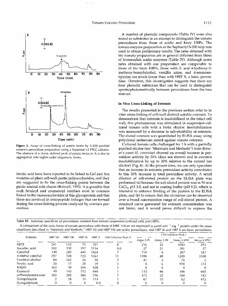
Substrate specificity of EPIII and EPIV demonstrated thatonly soluble tomato extensin can be cross-linked. Otherproteins eluted from tomato cells and fractionated bySephacryl S-200 gel filtration could not be cross-linked (thenegative results are not presented in this paper). Somemarker proteins such as BSA (a glycoprotein), aldolase, andinsulin were also not cross-linked. However, potato lectin,an HRGP (Alien and Neuberger, 1973; Alien et al., 1978;Matsumoto et al., 1983) that is structurally related to ex-tensin, was cross-linked by both extensin peroxidases (Fig.5). The lectin protein has two domains: a Ser/Hyp glyco-protein domain, which has a strong resemblance to exten-sin, and a Cys-rich, nonglycosylated domain. Extensins aredevoid of Cys (Lamport, 1980) and because the Ser/Hypglycoprotein domain of the lectin resembles extensin, it islikely to contain the cross-linking site. Exhaustive pronasetreatment of potato lectin following reductive alkylinationwas shown to release a Mr 33,000 glycopeptide that is richin Ser and Hyp but not in Gly or Cys (Alien et al., 1978). Itis interesting that 33 kD is the same size as the HRGPreputedly insolubilized in soybean cell wall after fungalchallenge (Bradley et al., 1992). The nature of the linkageinvolved in the cross-linking process has not yet beenidentified. Although the existence of intrapeptide isodity-
kD
946743
3O
2O
14
B
4.8-
4.7-
4.6-
4J-
4J-
EP IV (34kDa)
0 0.1 0.2 0.3 0.4 0.5 0.6 0.7 0.8 0.9
Figure 4. SDS-PAGE of Mono S FPLC-resolved extensin peroxidases.A, Lane 1 , 3.7 ;u,g of PI; lane 2, 1 .5 /ig of PII; lane 3, 0.6 ng of EPIII;lane 4, 3.7 fig of EPIV; lane 5, molecular mass markers. B, Thestandard curve derived from the marker proteins.

1120 Brownleader et al. Plant Physiol. Vol. 109, 1995
Table 1. Amino acid analysis o f Mono S-purified peroxidases from suspension-cultured tomato cells
Amino Acid M o n o S PI M o n o S PII M o n o S EPlll M o n o S EPIV
AspIAsn Thr Ser GIu/G In Pro GlY Ala CY s Val Met I le Leu TY r Phe His L Y S Arg TrP
moi %
15.1 11.9 10.6 7.6 7.0 5.8 7.8 7.4 5.9 7.7 11.1 12.8 4.1 5.3 4.7 8.4 11.4 12.7
12.9 6.5 12.0 1.7 1.1 N D 5.2 7.0 7.8
N Da N D 1 .o 5.3 5.7 5.7 8.7 8.9 8.8 N D 0.25 2.7 4.3 3.7 4.2 1.3 1.7 1.9 2.2 4.8 7.3 7.9 4.9 3.4 NA‘ NA NA
10.1 5.2 8.7
10.0 8.7
14.6 8.5 N D 6.5 N
3.8 6.4 1.8 3.8 1.7 6.2 3.9 NA
a ND, Not detected. NA, Not assayed.
rosine linkages have been known to exist in cell-wall insol- uble extensin (Fry, 1983), such linkages have not been reported between extensin molecules. Comparison of the nature of in vitro cross-linking of extensin with the in vivo process that occurs in the cell wall must await further structural analyses, which are in progress in our laborato- ries. Proteolytic digestion of extensin and further analysis of the proteolytic fragments will allow u s to determine the in vitro cross-linking site of extensin before establishing whether the same type of cross-link exists in insoluble
cell-wall extensin. However, with respect to the in vivo role of the extensin peroxidase, it is noteworthy that boih sol- uble extensin and the enzyme are bound to the cell wall, which is also the site of extensin insolubilization.
Table 111 clearly demonstrates that the four forms of the tomato peroxidase can be distinguished from HRPs by their total inability to act upon NADH. EPIII and EPIV can be distinguished from the non-cross-linking forms, 1’1 and PII, by the high rates obtained with ascorbic acid, ferulic acid, and guaiacol. EPIV shows higher rates than EPIII with 4-methyl catechol, guaiacol, and syringaladazine. It is not clear, for example, why EPIII and EPIV display a prefer- ence for 4-methyl catechol rather than catechol. The methyl group does not appear to alter the chemical properties of the catechol significantly except to increase slightly the hydrophobicity. The addition of the methyl group may enhance a possible hydrophobic interaction with the active site. Other Mono S-resolved isoforms also show a consid- erable degree of preference for 4-methyl catechol compared with HRPs. Sterjiades et al. (1993) reported the specific activity of HRP (Sigma P-8250, containing at least five isoforms) using catechol and 4-methyl catechol as 122 and 1248 units/mg, respectively; the values for Acer acidic per- oxidase with the two substrates were 0.18 and 89.6, respec- tively. Subtle alterations in the structure of phenolic sub- strates appear to have a marked effect on peroxidase catalytic activity. Guaiacol appears to be a better substrate than p-phenylenediamine. Experiments identifying the ex- act structural requirements of substrates for EPIII and EPIV would be interesting. EPIII can also be distinguished from EPIV because the former reacts with considerably higher rates with ABTS, ascorbic acid, coniferyl alcohol, and feru- lic acid. Zimmerlin et al. (1994) have purified a cationic peroxidase from French bean cell wall that can yield dehy- drogenation oligomers of ferulic acid. Coumaric acid and
Table 11. Published amino acid analysis of cationic peroxidases Amino Acid P8.5a Cationicb HRP HRP BC HRP Cc Peanut Peroxidased
Asp/Asn Thr Ser G lu/Gln Pro GlY Ala CY s Va I Met Ile Leu TY r Phe His
LYS Arg TrP
N D 9.2
11.3 N D 5.1
10.0 9.2 2.7 7.3 1.1 3.4 7.7 1 .o 3.3 1.4 4.2 4.0 N D
15.4 7.7 8.4 6.4 5.3 5.3 7.2 2.5 6.5 1.9 4.0
11.5 1.6 6.5 1.2 1.9 6.5 0.3
15.5 6.6 5.0 6.6 4.2 2.5 4.4 0.9 4.4 1 .o 3.7
10.9 1.8 8.8 1.1 1.8 8.5 N D
14.5 5.8 4.7 6.2 4.0 2.2 4.0 0.9 4.1 0.9 3.6
10.1 2.0 8.3 1 .o 1.8 8.2 N D
15.9 7.5 6.5 6.9 4.7 4.7 7.4
5.4 0.8 3.9 9.5 2.1 8.9 2.5 3.3 6.2 ND
2.8
a Cationic extracellular barley peroxidase (Kerby and Sommerville, 1991). Cationic horseradish peroxidase B and C (Shannon et al., 1966).
’ Cationic horseradish peroxidase C1 (Mazza and Welinder, Cationic peanut peroxidase (van Huystee and Maldonado, 1980).
1982).
Tomato Extensin Peroxidase 1121
I A
O W N
4
T 1 0.002 AU
Potat! lectin I c
I I
o i o 20 30 40 50 60 70 80 90 Time (min)
Peroxidase I
T 0.002AU
O 10 20 30 40 50 60 70 80 90 Time (min)
Figure 5. Assay of cross-linking of potato lectin by S-200-purified extensin peroxidase preparation using a Superose-1 2 FPLC column. The absence of a sharp, defined peak of potato lectin in A is due to aggregation into higher-order oligomeric forms.
ferulic acid have been reported to be linked to Gal and Ara moieties of plant cell-wall pectic polysaccharides, and they are suggested to be the cross-linking points between the pectic neutra1 side chains (Bolwell, 1993). It is possible that such feruloyl and coumaroyl residues exist in extensin linked to the monosaccharides of this glycoprotein and that these are involved in interpeptide linkages that are formed during the cross-linking process catalyzed by extensin per- oxidase.
A number of phenolic compounds (Table 1V) were also tested as substrates in an attempt to distinguish the tomato peroxidases from those of acidic and basic HRPs. The tomato enzyme preparation at the Sephacryl S-200 step was used to obtain preliminary results. The rates obtained with the tomato preparation are in general different from those of horseradish acidic enzymes (Table IV). Although some rates obtained with our preparation are comparable to those of the basic HRPs, those with 2- and 4-hydroxy-3- methoxy-benzylalcohol, vanillin azine, and 4-aminoan- tipyrine are much lower than with HRP X, a basic peroxi- dase. Therefore, this investigation suggests that there are four phenolic substrates that can be used to distinguish spectrophotometrically between peroxidases from the two sources.
In Vivo Cross-Linking of Extensin
The results presented in the previous section refer to in vitro cross-linking of cell-wall-derived soluble extensin. To demonstrate that extensin is insolubilized in the intact cell wall, this phenomenon was stimulated in suspension-cul- tured tomato cells with a biotic elicitor. Insolubilization was measured by a decrease in salt-elutibility of extensin. The eluted extensin was quantitated by ELISA assay using polyclonal antiserum raised against tomato extensin.
Cultured tomato cells challenged for 1 h with a partially purified elicitor (see "Materials and Methods") from Brew- er's yeast (S. cerevisiae) showed an overall increase in per- oxidase activity by 20% (data not shown) and in extensin insolubilization by up to 30% relative to the control (no elicitor) (Fig. 6). At the present time, we can only speculate that an increase in extensin peroxidase activity contributes to this 20% increase in total peroxidase activity. A seria1 dilution of salt-eluted protein on the ELISA plate was performed (a) because the salt-eluted protein was in 50 mM CaCl,, pH 3.0, and not in coating buffer (pH 9.2), which is intended to enhance binding of the protein to the ELISA plate, and (b) to ensure that the elicitation can be observed over a broad concentration range of cell-eluted protein. A standard curve generated for extensin concentration was not linear, and it would prove difficult to express the
Table III. Substrate specificity of peroxidases isolated from tomato suspension-cultured cells and HRPs.
A comparison of the ionic forms of tomato peroxidase with those of HRP. Values are expressed as ymol min-' mg-' protein under the assay conditions described in "Materials and Methods." HRP VI1 and HRP Vl l l are acidic peroxidases, and HRP IX and HRP X are basic peroxidases.
CM-Cellulose Peak I
M o n o S PI Mono S PII M o n o S EPlll M o n o S EPlV Substrate HRP VI1 HRP V l l l HRP IX HRP X CM-Cellulose Peak I I
ABTS 241 132 74 251 8 21 6 33 1043 295
Catechol 140 285 445 1424 7 71 8 6 267 342 4-Methyl catechol 297 108 723 1265 11 1396 48 1209 31 08 Coniferyl alcohol 84 182 26 56 2 41 2 76 2 Ferulic acid 50 17 28 59 5 4 6 978 513 NADH 363 30 204 628 1 O O O O C ua i acol 49 143 222 649 1 132 66 486 665 p-Phenylenediamine 263 305 384 796 7 472 32 184 143 Syringaldazine 2 18 15 113 1 67 33 62 472
Ascorbic acid 502 330 297 1214 0.0 27 21 281 57
Syringaldehyde O O O 0.0 O O O O O

1122
1.4 - 13 -
1.2 -
1.1 -
1-
0.9 - O
VI t- 0.8 -
d 0.7 -
0.6 -
0.5 -
0.4 -
Brownleader et al. Plant Physiol. Vol. 109, 1995
___ Table IV. Substrate specificity of peroxidase isolated from tomato suspension-cultured cells and HRPs.
assay conditions descrihed in “Materials and Methods.” A comparison of the unresolved forms of tomato peroxidase with those of HRP. Values are expressed as AA min-’ mg-’ protein under the
HRP VI1 and HRP Vlll are acidic peroxidases, and HRP I X and HRP X are hasic peroxidases. ___ Substrate HRP VI1 HRP Vlll HRP IX HRP X S-200 Peroxidase --
4-Hydroxy-3- 11 38 47 198 117
3-Hydroxy-4- 8 3 4 269 27
4-Hydroxy-3- 47 44 72 229 85
methoxy-henzylamine
methoxy-henzyl alcohol
methoxy-henzyl alcohol L-Tyr ethyl ester 20 3 8 8 7 L-Tyr 5 2 1 4 2 o-Tyr 1 1 1 1 1 L-DOPA” 15 93 9 9 13 Vanillin azine 381 696 453 563 165 4-Aminoantipyrene 9 76 11 318 3
a Under identical conditions, phenolase and tyrosinase oxidized L-DOPA with rates of 1 and 87 AA min-’ mg-’, respectively.
change in A of salt-eluted protein in microgram amounts of extensin. This is particularly true if one considers that the antiserum is polyclonal and may cross-react with other HRGPs present in the salt-eluate. The response to the elicitor over the short period of 1 h of incubation suggests that the process is not likely to involve de novo protein synthesis.
Extensin insolubilization is a recognized plant defense response (Esquerré-Tugayé et al., 1979; Lamb et al., 1993). Severa1 researchers have stimulated plant defense events such as phytoalexin accumulation, ethylene biosynthesis,
Control
Elicitor
Cell eluate dilution
Figure 6. Effect of yeast elicitor on extensin insoluhilization. Control represents nonelicited cells. Soluhle extensin in the cell eluates was assayed by ELISA.
and Phe-ammonia lyase activity with elicitor derived from yeast (Hahn and Albersheim, 1978; Basse and Boller, l992). In this context, it is important to note the distinction be- tween the peroxidases of the pathogon and those of the host plant and that the pathogen peroxidases may contrib- ute to the increased activites of total peroxidases in disease resistance. This prompted us to use a yeast elicitor, devoid of peroxidases, to investigate peroxidase-mediated cell- wall extensin deposition. Our results showing a rapid in vivo insolubilization of extensin may indicate a rapid gen- eration of signals at the infection site that may act as a primary defense barrier to the invading pathogen. Mlodu- lation of extensin peroxidase activity could occur by (alter- ing the local levels of H,O, or extensin, modulatin;; the amount of a specific inhibitor of extensin cross-lirtking (Brownleader et al., 1993), or creating a more favorable pH for extensin cross-linking.
ACKNOWLEDCMENTS
We thank Mr. Ian Moss (Charing Cross and Westminster Med- ical School) for performing the amino acid analysis and Mrs. Margaret Pickering (Royal Holloway, University of London) for performing the peptide sequencing.
Received June 7, 1995; accepted August 23, 1995. Copyright Clearance Center: 0032-O889/95/ 109/ 11 15/09.
LITERATURE ClTED
Allen AK, Desai NN, Neuberger A, Creeth MJ (1978) Properties of potato lectin and the nature of its glycoprotein linkages. Biochem J 171: 665-674
Allen AK, Neuberger A (1973) Purification and properties clf the lectin from potato tubers, a hydroxyproline-containing glyco- protein. Biochem J 135 307-314
Basse CW, Boller T (1992) Glycopeptide elicitors of stress re- sponses in tomato cells. Plant Physiol 98: 1239-1247
Bolwell GP (1993) Dynamic aspects of plant extracellular matrix. Int Rev Cytol 146: 261-324

Tomato Extensin Peroxidase 1123
Bradford MM (1976) A rapid and sensitive method for the quan- titation of microgram quantities of protein utilizing the principle of protein-dye binding. Ana1 Biochem 72: 248-254
Bradley DJ, Kjellbom PP, Lamb CJ (1992) Elicitor- and wound- induced oxidative crosslinking of a proline-rich plant cell wall protein: a novel, rapid defence response. Cell 7 0 21-30
Brownleader MD, Dey PM (1993) Purification of extensin from cell walls of tomato (hybrid of Lycopevsicon esculentum and L . peruuianum) cells in suspension culture. Planta 191: 457-469
Brownleader MD, Golden K, Dey PM (1993) An inhibitor of extensin peroxidase in cultured tomato cells. Phytochemistry 33:
Cooper JB, Varner JE (1984) Crosslinking of soluble extensin in isolated cell walls. Plant Physiol 76 414417
Esquerré-Tugayé M-T, Lafitte C, Mazau D, Toppan A, Touze A (1979) Cell surfaces in plant-microorganism interactions. 11. Ev- idence for the accumulation of hydroxyproline-rich glycopro- teins in the cell wall of diseased plants as a defense mechanism. Plant Physiol 64: 320-326
Esquerré-Tugayé M-T, Lamport DTA (1979) Cell surfaces in plant-microorganism interactions. I. A structural investigation of cell wall hydroxyproline-rich glycoproteins which accumu- late in fungus-infected plants. Plant Physiol 6 4 314-319
Everdeen DS, Kiefer S , Willard JJ, Muldoon EP, Dey PM, Li X-B, Lamport DTA (1988) Enzymic crosslinkage of monomeric ex- tensin precursors in uitvo. Plant Physiol 87: 616-621
Ferrer MA, Pedreíío MA, Ros Barceló A, Muiíoz R (1992) The cell wall localization of two strongly basic isoperoxidases in etio- lated Lupinus albus hypocotyls and its significance in coniferyl alcohol oxidation and indole-3-acidic acid catabolism. J Plant Physiol 139: 611-616
Fry SC (1983) Oxidative phenolic coupling reactions crosslink hydroxyproline-rich glycoprotein molecules in plant cell wall. Curr Top Plant Biochem Physiol 2: 59-72
Gaspar T, Penel C, Hagege D, Greppin H (1991) Peroxidases in plant growth, differentiation, and development processes. In J Lobarwewzki, H Greppin, C Penel, T Gaspar, eds, Biochemical, Molecular and Physiological Aspects of Plant Peroxidases. Uni- versity M. Curie-Sklodowski, Lublin, Poland, and University of Geneva, Switzerland, pp 249-280
Hahn MG, Albersheim P (1978) Host-pathogen interactions. XIV. Isolation and partia1 characterization of an elicitor from yeast extract. Plant Physiol 6 2 107-111
Kerby K, Sommerville SC (1992) Purification of an infection- related, extracellular peroxidase from barley. Plant Physiol 1 0 0 397-402
Laemmli UK (1970) Cleavage of structural proteins during the assembly of the head of bacteriophage T4. Nature 227: 680-685
Lamb CJ, Brisson L, Bradley DJ, Kjellbom P (1993) Stimulus- dependent oxidative cross-linking of a proline-rich cell wall protein: a novel, rapid defense response and control point in cellular maturation. In B Fritig, M Legrand, eds, Mechanisms of Plant Defense Responses. Kluwer Academic Publishers, Dor- drecht, The Netherlands, pp 250-256
755-758
Lamb CJ, Dixon RA (1994) Molecular mechanisms underlying induction of plant defence gene transcription. In DJ Bowles, PM Gilmartin, PJ Knox, GG Lunt eds, Molecular Botany: Signals and the Environment. The Biochemical Society, London, pp 241-248
Lamport DTA (1980) Structure and function of plant-glycopro- teins. In J Preiss, ed, The Biochemistry of Plants, Vol3. Academic Press, New York, pp 501-541
Matsumoto I, Jimbo A, Mizano Y, Seme N, Jeauloz RW (1983) Purification and characterization of potato lectin. J Biol Chem
McIlvaine TC (1921) A buffer solution for colorimetric compari- son. J Biol Chem 49: 183-186
Medina MI, De Forchetti SM, Tigier HA (1993) Kinetic properties of alfalfa root isoperoxidases with IAA oxidase and syringald- azine oxidase activities. In KG Welinder, SK Rasmussen, C Penel, H Greppin, eds, Plant Peroxidases, Biochemistry and Physiology. University of Geneva, Switzerland, pp 175-180
Murashige T, Skoog F (1962) A revised medium for rapid growth and bioassay with tobacco tissue and culture. Physiol Plant 15: 473497
Reimers PJ, Guo A, Leach JE (1992) Increased activity of a cationic peroxidase associated with an incompatible interaction between Xanthmonas oryzae pv oryzae and rice (Oryza sativa). Plant Physiol
Ridge I, Osborne DJ (1971) Role of peroxidase when hydroxypro- line-rich glycoprotein in plant cell wall is increased by ethylene. Nature New Biol 229 205-208
Ros-Barceló A, Ferrer MA, García-Forenciano E, Muiíoz R (1991) The tonoplast localization of two basic isoperoxidases of high pI in Lupinus. Bot Acta 104: 272-278
Ros Barceló A, Muiíoz R, Sabater F (1987) Lupin peroxidase. I. Isolation and characterization of cell wall-bound isoperoxidase activity. Physiol Plant 71: 448454
Ros Barceló A, Muiíoz R, Sabater F (1989) Subcellular location of basic and acidic soluble isoperoxidases in Lupinus. Plant Sci 63:
Sterjiades R, Dean JFD, Gamble G, Himmelsbach DS, Eriksson KEL (1993) Extracellular laccase and peroxidases from sycamore maple (Acer pseudoplatanus) cell suspension cultures: reactions with monolignols and lignin model compounds. Planta 190:
Welinder KG, Gajhede M (1993) Structure and evolution of per- oxidases. In KG Welinder, SK Rasmussen, C Penel, H Greppin, eds, Plant Peroxidases, Biochemistry and Physiology. University of Geneva, Switzerland, pp 35-42
Whitmore FW (1971) Effect of indoleacetic acid and hydroxypro- line on isozymes of peroxidase in wheat coleoptiles. Plant Physiol47: 169-171
Whitmore FW (1976) Binding of ferulic acid to cell walls by peroxidases of Pinus elliottii. Phytochemistry 15: 375-378
Zimmerlin A, Wojstaszek P, Bolwell GP (1994) Synthesis of de- hydrogenation polymers of ferulic acid with high specificity by a purified cell wall peroxidase from French bean (Phaseolus uulgaris L.). Biochem J 299: 747-753
258: 2886-2891
99: 1044-1050
31-38
75-87