Facile biosensors for rapid detection of COVID-19
33
1 Facile biosensors for rapid detection of COVID-19 Lizhou Xu 1# *, Danyang Li 2# , Sami Ramadan 1 , Yanbin Li 3 , Norbert Klein 1 1 Department of Materials, Imperial College London, London, SW7 2AZ, UK 2 School of Cancer and Pharmaceutical Sciences, King's College London, 150 Stamford Street, London, SE1 9NH, UK 3 Department of Biological and Agricultural Engineering, University of Arkansas, Fayetteville, AR, 72701, USA *Corresponding author: Dr. Lizhou Xu Email address: [email protected] # Dr Lizhou Xu and Dr Danyang Li contribute equally to this work.
-
Upload
khangminh22 -
Category
Documents
-
view
1 -
download
0
Transcript of Facile biosensors for rapid detection of COVID-19

1
Facile biosensors for rapid detection of COVID-19
Lizhou Xu1# *, Danyang Li2#, Sami Ramadan1, Yanbin Li3, Norbert Klein1
1 Department of Materials, Imperial College London, London, SW7 2AZ, UK
2 School of Cancer and Pharmaceutical Sciences, King's College London, 150 Stamford
Street, London, SE1 9NH, UK
3 Department of Biological and Agricultural Engineering, University of Arkansas,
Fayetteville, AR, 72701, USA
*Corresponding author:
Dr. Lizhou Xu
Email address: [email protected]
# Dr Lizhou Xu and Dr Danyang Li contribute equally to this work.

2
Abstract: Currently the world is being challenged by a public health emergency caused by the
coronavirus pandemic (COVID-19). Extensive efforts in testing for coronavirus infection,
combined with isolating infected cases and quarantining those in contact, have proven successful
in bringing the epidemic under control. Rapid and facile screening of this disease is in high
demand. This review summarises recent advances in strategies reported by international
researchers and engineers concerning how to tackle COVID-19 via rapid testing, mainly through
nucleic acid- and antibody testing. The roles of biosensors as powerful analytical tools are
emphasized for the detection of viral RNAs, surface antigens, whole viral particles, antibodies and
other potential biomarkers in human specimen. We critically review in depth newly developed
biosensing methods especially for in-field and point-of-care detection of SARS-CoV-2.
Additionally, this review describes possible future strategies for virus rapid detection. It helps
researchers working on novel sensor technologies to tailor their technologies in a way to address
the challenge for effective detection of COVID-19.
Key words: SARS-CoV-2, COVID-19, rapid detection, point-of-care testing, coronavirus,
biosensor

3
1. Introduction
Since the outbreak of severe acute respiratory syndrome coronavirus 2 (SARS-CoV-2), with the
disease referred to as novel coronavirus disease (COVID-19) first reported in early January 2020
in Wuhan, China (N. Zhu et al., 2020),(Wang et al., 2020), the growing trend of infected cases is
not yet under control (Chan et al., 2020),(C. Huang et al., 2020). COVID-19 was officially
announced as a pandemic by the World Health Organisation (WHO) on 11th March 2020. So far
there have been more than 7,823,289 cases confirmed globally, with 431,541 deaths from more
than 210 countries and territories as of 15th June, 2020 (World Health Organization, 2020a).
SARS-CoV-2 is the virus strain that causes the respiratory illness COVID-19. SARS-CoV-2 is
believed to have zoonotic origins and has close genetic similarity to bat coronaviruses (N. Chen
et al., 2020). It is a positive-sense single-stranded RNA virus with approximately 50-200 nm in
diameter (Figure 1A) (Xu et al., 2020). Similar to other coronaviruses, SARS-CoV-2 mainly has
four structural proteins, namely, the spike (S), membrane (M), envelop (E), and nucleocapsid (N)
proteins, respectively (Wrapp et al., 2020). The virus makes full use of S protein to bind to
angiotensin-converting enzyme 2 (ACE2) on the human cell surface, to gain entry into a host cell.
The N protein holds the viral genome but also involves in the host cellular response to viral
infection. S, E, and M proteins together create the viral outer protecting membrane (Figure 1B)
(Wrapp et al., 2020). Both proteins (e.g. S protein) and viral RNA can be used as targets for
COVID-19 detection. Alternatively, antibodies such as IgM and IgG from patient samples could
also be detected for understanding the infection history. SARS-CoV-2 RNA is detectable 2–3 days
before onset of symptoms and can remain up to 25–50 days afterwards, depending on disease
severity (He et al., 2020). Many studies show IgM antibodies start to be detectable around 5–10
days after onset of symptoms and rise rapidly, followed by IgG antibody response closely (Peeling
et al., 2020). These seroconversions are typically within the first 3 weeks with the mean time of
9-11 days after onset of symptoms for total antibodies (10-12 days for IgM and 12-14 days for
IgG). RNA level can remain high despite high concentrations of IgM and IgG antibodies in patient
blood (Zhao et al., 2020). These viral infection and immune response studies highlight the
detection window for SARS-CoV-2 diagnosis and more importantly, guide the strategic
implementation of appropriate types of testing at different infection stages. For example, immune
testing can play a big role in tracing symptomatic cases at the middle/late stage of the infection
(e.g. 5-10 days after symptom onset). IgM positive result in symptomatic patients fulfilling the
COVID-19 case definition is strongly suggestive of SARS-CoV-2 infection. However, RNA testing
is still recommended for confirming the case.

4
Figure 1. Schematic diagram of (A) 3D model of the SARS-CoV-2 virion. Reprint from CDC Public Health Image Library (ID 23312: Alissa Eckert and Dan Higgins). (B) Related targeting sites (biomolecules) for COVID-19 detection. Not to scale. Partially reprinted from (Morales-Narváez and Dincer, 2020).
A key message from the WHO in early March is: ‘test, test, test’ [(World Health Organization,
2020b)]. Testing especially rapid detection is extremely critical and a powerful way to monitor and
manage the pandemic before vaccines or effective drugs become available. Effective detection
helps to confirm infected cases with symptoms shown or even when still within the virus incubation
period (diagnostic testing), thus allowing treatment on-time. Millions of RNA-based tests have
been carried out around the world looking for the presence of viral genes in a nose or throat swab
as a sign of active infection. Additionally, blood tests for antibodies to SARS-CoV-2 can indirectly
indicate an active infection or post-infection immunity/infection history (surveillance testing).
Prompt testing also helps efficient allocation of medical resources in hospitals and saves time for
frontline health workers. Particularly for low-income countries, fast, affordable, in-field and point-
of-care testing can have substantial effects in controlling the spread of the disease where health
systems may be weak and access to medical treatment limited. Furthermore, valuable information
about the local distribution of infections enhances the accuracy of epidemiological prediction and
facilitates corresponding policymaking. Therefore, rapid, facile, cost-effective and accessible
detections for large-scale screening, in-field testing and point-of-care diagnosis of the disease are
of great importance and urgency for quickly controlling the highly contagious and rapid spread of
COVID-19. In this work, we first review the recent advance in rapid testing for COVID-19. We then
highlight the roles of biosensors for rapid and facile detection of SARS-CoV-2 covering viral RNAs,
surface antigens/whole viruses, antibodies and potential biomarkers detection. Finally, future
developments of novel virus biosensors are discussed.

5
2. State-of-the-art of rapid testing approaches for SARS-CoV-2
Currently, CT imaging, haematology tests and molecular methods based on viral genetic
measurements are the primary tools used for clinical diagnosis of COVID-19, together with the
identification of clinical symptoms to confirm infection (Y. H. Jin et al., 2020). These laboratory
tests are essential to control the burgeoning of the disease. An RNA-based metagenomic next-
generation sequencing (mNGS) approach was used to identify the sequence of SARS-CoV-2
immediately after the initial outbreak (L. Chen et al., 2020). mNGS is a sensitive technique, but it
is restricted by throughput, turnaround time, high cost and a requirement for high technical
expertise. Rapid detection approaches could usher in an era of point-of-care testing (POCT) or
in-field screening of viruses. Figure 2 shows the two main testing approaches that are currently
being used for COVID-19 globally: nucleic acid testing and antibody testing.
Figure 2. Current main testing approaches for COVID-19: nucleic acid testing and antibody testing.

6
2.1 Nucleic acid (RNA) testing
Many laboratory-based molecular diagnostic kits have been developed by disease control
organisations, research institutes and private companies and used for testing patients’ specimens
since the beginning of COVID-19 pandemic (Chinese Center for Disease Control and Prevention,
2020), (CDC, 2020),(Corman et al., 2020), (Roche Ltd, 2020a). Polymerase chain reaction (PCR)-
based nucleic acid testing looks for viral RNAs in upper respiratory specimens (throat and/or nasal
swabs) from an individual. Table 1 summarises recent viral nucleic acid-based detection methods
for SARS-CoV-2 testing. The quantitative reverse transcription PCR (qRT-PCR) has gradually
become the current gold standard for the diagnosis of SARS-CoV-2 infection. SARS-CoV-2 genes
such as ORF1ab (open reading frame), RdRp (RNA-dependent RNA polymerase gene), E
(envelope protein gene), and N (nucleocapsid protein gene) can be targeted for diagnosis. The
general protocol of qRT-PCR is based on the extraction of RNA from respiratory swabs dissolved
in viral transport media (VTM), and subsequent one-step reverse transcription and real-time qRT-
PCR targeting one or several gene sequences from SARS-CoV-2 (Zou et al., 2020). Researchers
have tried to simplify this current protocol by avoiding the RNA extraction step based on direct
nasopharyngeal swab VTM heating before the qRT-PCR, which may provide viable options to
overcome any supply chain issues and help to increase the testing throughput (Alcoba-Florez et
al., 2020). Other new RNA-based methods for SARS-CoV-2 detection (Table 1) have also been
developed to tackle this crisis, such as Reverse Transcription Loop-Mediated Isothermal
Amplification (RT-LAMP) technique (Lamb et al., 2020). Yan et al. evaluated a RT-LAMP assay
for the SARS-COV-2 within 30 min using primers targeting ORF1ab and S (spike) genes, with a
LOD of 2 × 101 copies and 2 × 10² copies of RNA per reaction, respectively (Yan et al., 2020). No
cross-reactivity was found with another 60 respiratory pathogens. The sensitivity (true positive
rate) for clinical specimen diagnosis (n=130) was close to 100% (95% CI 92.3%-100%), as was
its specificity (95% CI 93.7%-100%). Compared to qRT-PCR, RT-LAMP is faster and does not
require prior RNA isolation from the samples. The reagents for RT-LAMP are relatively cheap and
stable at room temperature, and therefore this technique holds promise for use outside of a central
laboratory (in-field detection) by staff without special training and without need of advanced
equipment (Park et al., 2020). A colorimetric-LAMP method was reported using pH-sensitive dyes
to visualise the LAMP amplification via the change in pH resulting from proton accumulation due
to the incorporation of deoxynucleoside triphosphates (dNTPs) (Y. Zhang et al., 2020). A
sensitivity/LOD as low as 4.8 copies/μL was achieved in testing RNA samples purified from patient
respiratory swabs, and the results were in 100% agreement with those from the qRT-PCR method.
This effort expands the toolbox of molecular tests beyond sophisticated diagnostic laboratories in

7
aiming to combat and monitor the growing public health threat. Additionally, digital PCR (dPCR)
has been reported to improve the LOD to at least 10-fold lower than that of RT-PCR, and overall
accuracy in the clinical detection of 109 samples was reported to be 96.3%, suggesting the
potential of dPCR for the detection of asymptomatic and suspect patients (Lu et al., 2020).

8
Table 1. Representative commercial test kits with POCT potential and other nucleic acid-based tests for screening of COVID-19.
Sample volume
1
Detection target
Detection method
Sensitivity2 Specificity3
(True negative rate)
Assay detection
time
Turnaround time
Commercial products/registration
status
Ref.
Limit of detection
True positive
rate
5 μL RNA (RdRp, E, N genes)
Real-time qRT-PCR
3.9 copy/reaction (E gene); 3.6 copy/reaction (RdRp gene)
100% (n=297)
100% (n=297)
~ 2 h > 4 h Developed by academic and public laboratories
in national and European research
networks
(Corman et al., 2020)
5 μL RNA Real-time qRT-PCR
3.2 copy/µL / / ~ 2 h > 4 h The CDC Flu SC2 Multiplex Assay;
FDA-EUA
(CDC, 2020)
/ RNA (ORF-1a, E gene regions)
Real-time qRT-PCR
/ / / ~ 3-8 h ~ 1 day
Roche Cobas® SARS-CoV-2 Test (cobas® 6800/8800 Systems);
FDA-EUA + CE-IVD mark
(Roche Ltd, 2020a)
/ RNA LAMP / / / 5 min (positive);
13 min (negative)
< 30 min
Abbott ID NOW platform;
FDA-EUA
(Abbott, 2020)
/ RNA PCR with lateral flow
assay
/ / / < 30 min < 1 h Mesa Biotech Accula SARS-CoV-2 Test;
FDA-EUA
(Mesa Biotech, 2020)
/ RNA Real-time qRT-PCR
/ / / < 45 min < 1 h Cepheid Xpert® Xpress SARS-CoV-2;
FDA-EUA
(Cepheid, 2020)
/ RNA (N gene) Isothermal DNA
amplification
/ 95.0% (n=20)
100% (n=30)
< 30 min < 1 h Cue Health, Cue COVID-19 Test;
FDA-EUA
(Cue Health, 2020)

9
/ RNA Molecular method
/ 98.7% (n=102)
100% (n=102)
< 90 min < 90 min
DRW SAMBA II machines
(University of Cambridge, 2020)
20 μL RNA LAMP with colorimetric
readout
4.8 copy/μL / / ~ 30 min < 1 h Tested swab samples; in clinical validation
stages
(Y. Zhang et al., 2020)
< 10 μL RNA (E, N genes)
CRISPR-based LAMP with lateral flow assay
10 copy/µL 95% (n=40)
100% (n=42)
< 45 min < 1 h Tested swab samples; in clinical validation
stages
(Broughton et al., 2020)
14 μL RNA Digital PCR > 1 copy/μL / / < 45 min < 1 h Tested swab samples; in clinical validation
stages
(Lu et al., 2020)
25 μL RNA (ORF1ab, S
genes)
Reverse transcription-
LAMP
20 copy/reaction
100% (n=58)
100% (n=72)
< 30 min < 1 h Tested swab samples; in clinical validation
stages
(Yan et al., 2020)
25 μL RNA Reverse transcription-
LAMP
1.02 fg
/ < 30 min < 1 h Only detects simulated patient samples
(Lamb et al., 2020)
15 μL RNA Reverse transcription-
LAMP
100 copy/reaction
/ / < 30 min < 1 h No clinical samples tested
(Park et al., 2020)
/ Synthetic complementary DNA (RdRp)
RCA with magnetic
nanoparticles
sub-femtomolar
/ / ~ 100 min < 2 h No clinical samples tested
(Tian et al., 2020)
1 The sample volume is part of the viral transport medium (VTM) for transport of specimens collected by respiratory swabs (e.g. nasopharyngeal or oropharyngeal).
2 The sensitivity of an analytical method usually means the change of measured signal corresponding to the change of the concentration of analyte, and/or refers to a method’s limit of detection (detection limit), which is the smallest amount of analyte that we can determine with confidence [(Harvey, 2010)]; the sensitivity of a
clinical test refers to the ability to correctly identify those patients with the disease (also called the true positive rate) [(Lalkhen and McCluskey, 2008)].
3 The specificity of a clinical test refers to the ability to correctly identify those patients without the disease (also called true negative rate) [(Lalkhen and McCluskey, 2008)].
Note: qRT-PCR (quantitative reverse transcription-polymerase chain reaction); LAMP (loop-mediated isothermal amplification); CRISPR (clustered regularly interspaced short palindromic repeats); FDA-EUA (Food and Drug Administration-Emergency Use Authorization); CE-IVD (CE marking-In Vitro Diagnostic); RCA (rolling circle amplification)

10
Although RT-PCR tests are sensitive and widely used as current diagnostic tool for SARS-CoV-
2, they can only be pursued in certified laboratories with expensive equipment and trained
technicians but not in places such as airports, borders, big shopping centres, etc, where testing
facilities are not easily accessible. Considering the delivery time of samples to centralised labs,
RT-PCR testing could take over 24 h or longer from sampling to results (turnaround time), despite
the assay itself taking only a few hours. Additionally, high false negative rates might be an issue
for the RT-PCR testing of COVID-19 due to errors in sampling and testing (Corman et al.,
2020),(Xie et al., 2020),(Tang Xiao et al., 2020). Sometimes the identification of target viral gene
sequences cannot confirm the presence of active virus particles, because a positive detection
may be due to residues of viral RNA. As summarised in Table 1, many attempts have indeed
been made to realise reliable and faster molecular diagnosis of COVID-19, such as LAMP as
mentioned above. Many have been available on the market while some still need clinical
validation using patient samples. Their commercialisation statuses are highlighted in Table 1 as
FDA-EUA (The U.S. Food and Drug Administration-Emergency Use Authorization) or CE-IVD
(European CE marking-In Vitro Diagnostic). As of Aug. 21, 2020, the FDA had issued more than
176 molecular tests to diagnose infections with the SARS-CoV-2 virus, including 17 “Diagnostics-
Molecular-Home Collection” kits for collecting specimens at home and then sending them to the
authorised lab for testing (US FDA, 2020). Some molecular diagnostic tests require a highly
trained operator to manually perform the test (e.g., an RNA extraction step using specific
extraction platforms and kits). For example, the Influenza SARS-CoV-2 (Flu SC2) Multiplex Assay
developed by the Centres for Disease Control and Prevention (CDC) based on qRT-PCR can
only be run in high complexity labs, as it can detect and differentiate SARS-CoV-2, influenza A,
and/or influenza B in upper and lower respiratory specimens and rely on specific instruments
(CDC, 2020). While others are automated and require only limited training to perform. Among
those, the isothermal nucleic acid amplification approach with shorter sampling-to-result time and
a simpler protocol shows promise in being able to overcome the drawbacks associated with
conventional RT-PCR. As a successful example, the ‘ID NOW Rapid Isothermal System’ was
launched for the qualitative detection of COVID-19 with isothermal nucleic acid amplification
technology on 28th March 2020 by one of the leading biomedical companies (Abbott) (Abbott,
2020). This device is able to give positive results in 5 min and negative results in 13 min. Although
it is only available for professional use at the moment, this portable coronavirus testing kit takes
molecular testing to the frontlines and has substantially enhanced testing capacity in the USA
from early April 2020 after approval by the U.S. FDA under EUA. However, clinical test
performance results for this product have not been reported yet. Many other products for nucleic

11
acid detection have also emerged for the point-of-care testing of COVID-19 which were recently
approved by authorities for emergency use, such as the Accula SARS-CoV-2 Test from Mesa
Biotech (Mesa Biotech, 2020), Xpert® Xpress SARS-CoV-2 from Cepheid Xpert (Cepheid, 2020),
and SAMBA II machines (University of Cambridge, 2020) among others.
2.2 Antibody testing
Testing for antibodies in the patient’s blood is another modality for COVID-19 detection. Antibody
test kits are usually designed for the qualitative detection of IgM and/or IgG antibodies to SARS-
CoV-2 in a given serum, plasma (EDTA, citrate) or venipuncture whole blood specimen from a
patient. The lateral flow test strip (LFTS) or lateral flow immunoassay (LFIA) is widely used for
this purpose. This is a simple cellulose-based device employing chromatographic lateral flow
which is intended to detect the presence of a target analyte (antibody to SARS-CoV-19) in a liquid
sample (blood/serum/plasma, etc.) without the need for specialized and costly equipment –
although lab-based equipment can be used to achieve higher sensitivity (Posthuma-Trumpie et
al., 2009). It usually contains a sample pad, a conjugate pad, a nitrocellulose membrane and an
absorbent pad. The sample pad is exposed to the sample (a mixture of blood cells, vesicles, cell
debris, antibodies, small molecules, etc.) and acts as a filter to promote lateral aid flow. The
sample rehydrates the pre-immobilised gold-conjugated recombinant antigen (i.e. spike protein
or its receptor binding domain (RBD)) on the conjugate pad and the antibodies bind with their
matching antigens. Due to capillary force, the sample continues to flow along the nitrocellulose
membrane to reach the test line and the control line. The absorbent pad will absorb excess sample
fluid. Colloidal gold nanoparticles are often used for colorimetric visualisation, but coloured latex
nanoparticles, fluorophores, etc., can also be used [(Sajid et al., 2015)]. Table 2 summarises
representative SARS-CoV-2 antibody-based test strips which are fast and at relatively low cost.
For example, a rapid and point-of-care lateral flow immunoassay has been developed for the
simultaneous detection of IgM and IgG antibodies against SARS-CoV-2 virus in blood within 15
min (Z. Li et al., 2020). A chemiluminescence-immunoassay has also been reported for the
detection of SARS-CoV-2 infections and surveillance of changing antibody patterns based on the
recombinant nucleocapsid antigen and magnetic beads (Lin et al., 2020). Clinical IgG testing
identified 65 SARS-CoV-2 infections from 79 confirmed patients and only two false-positive cases
from the control group (n = 80) with sensitivity and specificity values reaching 82.3% and 97.5%
respectively. In addition, a colloidal gold-based immunochromatographic (ICG) strip test detecting
viral IgM or IgG was carried out with 134 samples from 105 patients, and a sensitivity of 11.1%

12
was achieved at the early stage (1-7 days after onset), 92.9% at intermediate stage (8-14 days
after onset) and 96.8% at late stage (more than 15 days) [(Pan et al., 2020)]. However, the
specificity was not evaluated for this ICG assay. Nonetheless, according to a multi-centre cross-
sectional study, the positive rate for IgG from antibody testing could reach 100% at around 20
days after symptom onset (Long et al., 2020), confirming the strong ability of antibody testing kits
for use in late-stage infections. The positive rate of serum IgG single testing has been reported to
be higher than that of IgM alone in COVID-19 detection, but the detection of both IgG and IgM
was shown to be more accurate (Z. Li et al., 2020),(Y. Jin et al., 2020).
Antibody test kits are not yet available for home testing but do allow testing in laboratories or by
healthcare workers at a point-of-care. Antibody testing cannot confirm the presence of the virus.
Positive results mean acquired immunity against COVID-19 infection, which might be ascribed to
past or present infections with non-SARS-CoV-2 strains such as coronavirus HKU1. In contrast,
negative results do not rule out SARS-CoV-2 infection, particularly for those who have been in
contact with virus carriers. IgM was found to be detectable in patient’s blood after 3-6 days post-
infection, with IgG detectable after 8 days (Xie et al., 2020),(Long et al., 2020). Hence antibody
testing is useful at the intermediate or late stages rather than the early stage of infection (Pan et
al., 2020). In a word, this rapid screening tool is more suitable as a complementary method to
nucleic acid testing (especially for negative results) by providing important immunological
evidence for physicians to make diagnostic and pre-treatment decisions, but not as a sole basis
for the diagnosis or exclusion of COVID-19 infection (W. Zhang et al., 2020). Notably, once a
vaccine for COVID-19 is available and people become immunised by vaccination, antibody testing
may not be able to differentiate those who acquire immunity from those infected ones. Industries
have been active in developing antibody test kits (mostly immunoassays) (Table 2), such as Novel
Coronavirus (2019-nCoV) IgM/IgG Antibody Rapid Test Kits from National Bio Green Sciences
LLC (FDA-EUA approved) (National Bio Green Sciences LLC, 2020), qSARS-CoV-2 IgG/IgM
Rapid Test from Cellex (FDA-EUA approved) (Cellex, 2020), COVID-19 Coronavirus Rapid Test
Cassette from SureScreen Diagnostics (SureScreen Diagnostics, 2020), etc. As of Aug. 21, 2020,
the FDA had issued more than 39 serological (antibody) tests for SARS-CoV-2, as well as 3
antigen tests (US FDA, 2020). A technical report from the European Centre for Disease
Prevention and Control released that there are over 60 CE-marked rapid SARS-CoV-2 antibody
tests as of 1st April, 2020 on the market (European Centre for Disease Prevention and Control,
2020). These tests for the qualitative detection of antibodies to SARS-CoV-2 in blood, serum,
and/or plasma are intended for use as an aid in identifying individuals with an adaptive immune
response to SARS-CoV-2, indicating recent or prior infection. Clinical validation for COVID-19
13
should be carried out by comparison with a gold standard test for a sufficiently large number of
target samples before authorising them as stand-alone diagnostic tests (European Centre for
Disease Prevention and Control, 2020). For instance, six commercial POCT lateral flow tests
were evaluated for COVID-19 antibodies (Lassaunière et al., 2020) and their overall performance
was ranked based on the detection sensitivity and specificity. These findings facilitate the
selection of serological assays for the detection of SARS-CoV-2 specific antibodies for diagnostic
purposes as well as sero-epidemiological and vaccine development studies. The pandemic is
spreading around the world and many countries are facing a second wave of COVID-19.
Therefore, more sensitive, low-cost, specific and fast antibody analytical methods are still in great
demand to screen for immunity among populations and to help track the progress of the epidemic.

14
Table 2. Representative commercial POCT kits and reported antibody tests for screening of COVID-19
Sample volume1
Detection target
Detection method
Sensitivity (True positive rate)2
Specificity (True
negative rate)3
Assay detection
time
Turn-around
time
Commercial products/
registration status
Ref.
/ IgM and IgG
LFIA / / < 15 min < 30 min National Bio Green Sciences, NBGS’
Novel Coronavirus (2019-nCoV) IgM / IgG Antibody Rapid
Test Kits;
FDA-EUA
(National Bio
Green Sciences
LLC, 2020)
/ IgM and IgG
LFIA (colloidal gold)
/ / < 15 min < 30 min Cellex, qSARS-CoV-2 IgG/IgM Rapid
Test;
FDA-EUA
(Cellex, 2020)
/ IgG and IgM
LFIA 99.0% (n=128) 99.0% (n=312)
< 15 min < 30 min Autobio Diagnostics, Anti-SARS-CoV-2
Rapid Test;
FDA-EUA
(Autobio, 2020)
/ Total antibody against N
protein
Electrochemi- luminescence immunoassay
100% (n=29) 99.8% (n=5272)
~ 18 min < 30 min Roche Diagnostics, Elecsys Anti-SARS-
CoV-2;
FDA-EUA
(Roche Ltd,
2020b)
/ Total antibody against
RBD of S1 protein
Chemi-luminescent microparticle immunoassay
100% (n=42) 99.8% (n=1091)
~ 10 min < 20 min Siemens Healthcare, Atellica IM SARS-
CoV-2 Total (COV2T);
FDA-EUA
(Siemens, 2020)
10-15 μL IgM and IgG
LFIA 88.7% (n=397) 90.6% (n=128)
< 15 min < 30 min Medomics Medical Technologies
(Z. Li et al., 2020)

15
/ IgM and IgG
LFIA 97.8% (IgM) and 99.6% (IgG)
/ < 10 min < 25 min SureScreen Diagnosis, COVID-19
Coronavirus Rapid Test Cassette
(SureScreen
Diagnostics, 2020)
50 μL IgM and IgG
(recombinant
nucleocapsid)
Chemi-luminescence immunoassay
82.3% (n=79) 97.5% (n=80) < 30 min < 45 min Tianshen Tech, A chemical immuno-
luminescence analyzer ACCRE6
(Lin et al., 2020)
10 μL (serum/
plasma),
20 μL (whole blood)
IgM or IgG Colloidal gold-based immune-chromatographic
(ICG) strip
11.1% (early stage, 1-7 days after onset), 92.9% (inter-mediate
stage, 8-14 days after onset) and
96.8% (late stage, >15 days)
(n=134)
/ < 15 min < 30 min Tested blood sample; in clinical validation
stages
(Pan et al., 2020)
/ Antibodies Graphene field effect transistor
(Gr-FET)
/ / ~ 2 min / Only tested recombinant spike
protein
(X. Zhang et al., 2020)
<1 μl (serum)
Antibody Immune-precipitation and
parallel DNA sequencing
90-97% ~ 97% At least hours
~ 1-2 week
/ (Xu et al., 2015)
1 The blood sample is usually in small amount collected from fingertip by “finger-prick”.
2 The sensitivity of a clinical test refers to the ability to correctly identify those patient samples (also called the true positive rate) [(Lalkhen and McCluskey, 2008)].
3 The specificity of a clinical test refers to the ability to correctly identify those non-patient samples (also called true negative rate) [(Lalkhen and McCluskey, 2008)].
Note: IgG (Immunoglobulin G); IgM (Immunoglobulin M); LFIA (lateral flow immunoassay); FDA-EUA (Food and Drug Administration-Emergency Use Authorization)

16
3. Biosensors for rapid and facile detection of SARS-CoV-2
3.1 Detection of nucleic acids
Biosensors as smart analytical devices combing specific recognition of target and sensitive
readout of signals can facilitate rapid, facile and cost-effective detection of COVID-19 in field and
at a point-of-care. The lateral flow technology can serve as a facile biosensing platform to couple
with nucleic acid testing approaches for viral RNA detection. For example, a promising multiplex
RT-LAMP coupled with a nanoparticle-based lateral flow biosensor (mRT-LAMP-LFB) was
developed for diagnosing COVID-19 with LOD of 12 copies per reaction in 1 h (X. Zhu et al.,
2020). The sensitivity of SARS-CoV-2 test was 100% (33/33 oropharynx swab patient samples),
and the specificity was also 100% (96/96 oropharynx swab non-patient samples). A CRISPR
(clustered regularly interspaced short palindromic repeats)/Cas12-based biosensor combined
with a lateral flow assay was recently developed for SARS-CoV-2 enabling a test result in around
30 min (Figure 3A) (Broughton et al., 2020). After extraction of RNAs from patients samples, the
so-called DETECTR performs simultaneous reverse transcription and isothermal amplification
using loop-mediated amplification (RT-LAMP) at 62°C for 20 min, followed by the Cas12 detection
of predefined coronavirus sequences at 37°C for 10 min, after which the cleavage of a reporter
molecule confirms detection of the virus as visualised on a lateral flow strip. The compatibility with
the lateral strip also enables this CRISPR-based technology to be used for tests at the point-of-
care away from the clinical diagnostics laboratory. Similarly, Huang and colleagues reported a
rapid CRISPR-Cas12a fluorescent reporter assay couple with one-step isothermal recombinase
polymerase amplification (RPA) methods for amplifying target regions from extracted viral RNAs,
with a sample-to-answer time of ~50 min, and a LOD of 2 copies per sample (Z. Huang et al.,
2020). This assay can be readily performed in 96-well microtiter plates and is currently under
investigation of the potential to integrate onto a microfluidic chip and smart phone reading system
for point-of-care settings. Other automatic integrated gene detection systems are in clinical
validation stage for COVID-19, for example, CoVIDNudge (Gibani et al., 2020), AIGS (Y. Li et al.,
2020). In another example, a dual-functional plasmonic biosensor combining the plasmonic
photothermal (PPT) effect and localized surface plasmon resonance (LSPR) sensing transduction
has been reported to provide an alternative for clinical COVID-19 detection (Figure 3B) (Qiu et
al., 2020). This dual-functional LSPR biosensor exhibits a high sensitivity toward viral sequences
including RdRp, ORF1ab, and E genes from SARS-CoV-2 with a lower LOD down to the
concentration of 0.22 pM and allows precise detection of the specific target in a multigene mixture.
This biosensor offers a reliable and easy-to-implement diagnosis platform to improve the
17
diagnostic accuracy in clinical tests and relieve the pressure on PCR-based tests. Besides, Jiao
et al. developed a fluorescence biosensor based on a DNA nanoscaffold hybrid chain reaction
(DNHCR) for rapid detection of SARS-CoV-2 RNA (Figure 3C) (Jiao et al., 2020). In this
biosensor, the DNA nanoscaffolds constructed by the self-assembly of long DNA strands and self-
quenching probes (H1) act as the sensing element. The target RNAs initiate the hybridization of
H1 and free H2 DNA probes along the nanoscaffold to illuminate the DNA nanostring, which
reflects the virus concentration. This DNHCR biosensor can detect SARS-CoV-2 within 10 min
and under mild condition (15-35°C), showing great potential in routine clinical diagnosis. In
addition, researchers have conceived new concepts to largely improve the detection capacity of
qRT-PCR testing, by taking a pooling approach to enable simultaneous detection of dozens of
samples. A study shows that the group testing can identify a positive sample among 64 different
samples with enough sensitivity (Yelin et al., 2020). Therefore, if scaled up appropriately, such
pooling methods could facilitate mass and large-scale testing with less use of resources and
quicker time.

18
Figure 3. Biosensors reported for viral RNA detection of SARS-CoV-2. (A) Schematic of SARS-CoV-2 DETECTR workflow. Conventional RNA extraction can be used as an input to DETECTR (LAMP preamplification and Cas12-based detection for E gene, N gene and RNase P), which is visualized by a fluorescent reader or lateral flow strip. Reprint from (Broughton et al., 2020). (B) A dual-functional plasmonic biosensor combining the plasmonic photothermal (PPT) effect and localized surface plasmon resonance (LSPR) sensing transduction for the clinical COVID-19 diagnosis. Reprint from (Qiu et al., 2020). (C) A DNA nano scaffold hybrid chain reaction (DNHCR)-based biosensor for the detection of SARS-CoV-2 RNA. Reprinted from (Jiao et al., 2020).

19
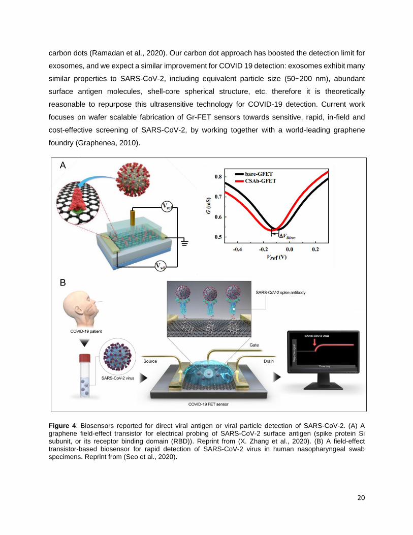
3.2 Direct detection of surface antigens and/or whole viruses
In addition to rapid detection of viral genetic components in swabs using biosensors, it would be
attractive to implement the direct and facile sensing of the whole virus particle or its corresponding
surface antigen epitope. This strategy could be employed for the development of a diagnostic or
screening tool for COVID-19. There have been some relevant research studies reported so far.
For example, an ultrasensitive graphene field-effect transistor (Gr-FET) immunosensor was
reported recently as an effort towards simple and rapid screening for COVID-19 (Figure 4A) (X.
Zhang et al., 2020). The graphene surface was functionalised with SARS-COV-2 spike S1 subunit
protein antibody (CSAb) or ACE2 receptor. The hybridization of the slightly positively charged S1
protein (which contains a receptor binding domain, RBD) with the immobilised CSAb/ACE2
receptors alters its conductance/resistance via field effect, which can be electrically read out in a
sensitive way. This Gr-FET immunosensor can rapidly identify (in about 2 min) and accurately
capture the COVID-19 spike protein S1 at a LOD down to 0.2 pM, in a real-time and label-free
manner. Although whole coronavirus particles instead of pure antigen proteins need to be tested
for assay validation, and clinical trials are then required, this work represents an early proof-of-
concept study and demonstrated the potential of Gr-FET technology for sensitive and rapid
detection of coronaviruses. Interestingly, another field-effect transistor (FET)-based graphene
biosensing device coated with a specific antibody against SARS-CoV-2 spike protein has been
reported for direct detection of SARS-CoV-2 (Figure 4B) (Seo et al., 2020). The introduced virus
particles onto the antibody coated graphene surface generated readable electric changes. This
COVID-19 FET sensor not only detects SARS-CoV-2 antigen protein transport medium for swab
samples, but also detects cultured viruses and viruses in clinical samples. The LOD for clinical
sample detection (n=19 patients and normal subjects) reached 2.42 × 10² copies/mL. A larger
clinical sample size would be needed to further validate its clinical potential for virus detection.
Nevertheless, given the complexity of clinical samples, the development of novel materials for
FET sensors which could overcome problems of non-specific interactions and screening effects
associated with clinical samples would be necessary to provide more accurate detection. One of
the key issues here is high assay sensitivity with a minimum of false alarms, which needs to be
achieved in order to facilitate practical applications. Our team has been developing graphene
sensors and FET technology for the detection of cells, exosomes, and biomolecules in biofluids
over the past few years (Kwong Hong Tsang et al., 2019), (Delle et al., 2018), (Hanham et al.,
2015). One of our recent work on Gr-FET biosensors for exosome detection can achieve LOD
down to single particle level within 10 min, enhanced by surface nano decoration of specific
20
carbon dots (Ramadan et al., 2020). Our carbon dot approach has boosted the detection limit for
exosomes, and we expect a similar improvement for COVID 19 detection: exosomes exhibit many
similar properties to SARS-CoV-2, including equivalent particle size (50~200 nm), abundant
surface antigen molecules, shell-core spherical structure, etc. therefore it is theoretically
reasonable to repurpose this ultrasensitive technology for COVID-19 detection. Current work
focuses on wafer scalable fabrication of Gr-FET sensors towards sensitive, rapid, in-field and
cost-effective screening of SARS-CoV-2, by working together with a world-leading graphene
foundry (Graphenea, 2010).
Figure 4. Biosensors reported for direct viral antigen or viral particle detection of SARS-CoV-2. (A) A graphene field-effect transistor for electrical probing of SARS-CoV-2 surface antigen (spike protein Si subunit, or its receptor binding domain (RBD)). Reprint from (X. Zhang et al., 2020). (B) A field-effect transistor-based biosensor for rapid detection of SARS-CoV-2 virus in human nasopharyngeal swab specimens. Reprint from (Seo et al., 2020).
21
It is important to understand the clinical infectious dose of SARS-CoV-2 in order to develop
suitable biosensors for screening or diagnosis. The majority of viral RNA concentrations in upper
respiratory tract samples are between 102 - 108 RNA copies (or virus particles) per swab, with the
highest to be 7.11 × 10⁸ copy/swab at day 4, first week of symptom onset, and in most cases viral
doses in sputum and stool samples are above 102 copies per swab, as suggested by a recent
virological analysis of 9 patients (Wölfel et al., 2020). Similar conclusions on viral load were drawn
from another clinical study of 17 patients with COVID-19 (Zou et al., 2020). These data indicate
that a biosensor capable of accurately detecting 100 virus particles or more (e.g. LOD ≤ 102
RNA/swab) would be potential as a screening tool, and even as a diagnostic tool. It might also be
useful for hospital discharge management, since patients beyond day 10 of symptoms with less
than 100 viral RNA copies per μL of sputum could be considered for early discharge and ensuing
home isolation (Wölfel et al., 2020). Obviously it is easier to detect severe COVID-19 cases than
mild ones, as the former tend to express higher viral load and longer virus-shedding periods (Liu
et al., 2020). Hence, desired antigen testing methods are those sensitive enough to detect the
low end of clinically relevant viral loads, typically around 103 viral nucleic acids per mL (one virion
per μL), which is as low as a single particle within one sample, in order to compete with the current
RT-PCR diagnostic method whose LOD is as low as 3 copy/μL of input sample (equal to 3 viral
particles) (CDC, 2020),(Zou et al., 2020). Another important issue is the specificity, especially for
surface antigen/whole virus detection. Positive results from antigen tests can be highly accurate,
but there is a higher chance of false negatives, so negative results do not rule out infection, which
may cause severe contagion. Other coronaviruses such as Middle East respiratory syndrome
coronavirus (MERS-CoV) and Severe Acute Respiratory Syndrome coronavirus (SARS-CoV) are
likely to cause interference in the detection of SARS-CoV-2 due to their similar size, viral structure,
and infective properties. Influenza virus A or B may also affect the selectivity of detection.
Therefore, the biorecognition element, usually antibodies or aptamers, should be carefully
selected in the design of biosensors and their specific binding with target virus should be well
assessed against those potential interferable viruses.
With advance in functional materials, novel sensing mechanisms and nanotechnology, single-
virus analysis would be possible, though still challenging in clinical settings (Schmidt and Hawkins,
2016). Meanwhile, emerging technical progress would help to overcome other challenging issues
such as assay stability and reproducibility for biosensors in antigen/whole virus detection, thus
facilitating accurate screening or diagnosis. SARS-CoV-2 is a spherical particle around 100 nm
in diameter, with many antigens expressed on the surface (Bar-On et al., 2020). Such properties
of the SARS-CoV-2 virus are similar to other viruses such as influenza viruses which researchers

22
are more familiar with and more detection approaches are available. Therefore ideas to improve
COVID-19 detection toward rapid, facile and reliable applications can be inspired by previous
research (Schmidt and Hawkins, 2016),(Wang and Li, 2016). For example, by working closely
with industry partners and clinicians, our team is currently developing a portable graphene sensor
for both viral antigen and whole virus detection, adapting from an established point-of-care
platform for exosome analysis (Kwong Hong Tsang et al., 2019), (Ramadan et al., 2020). Many
other biosensing platforms developed for rapid, in-field, and portable detection of various viruses
by our team over the past decades are promising to be repurposed for SARS-CoV-2. For instance,
a target-responsive hydrogel aptasensor embedded with quantum dot fluorescent reporters could
be used for rapid, one-step and in-field detection of virus in 30 min (Xu et al., 2016). A smart bio-
nanogate controlled enzymatic biosensor could enhance the sensitivity of virus detection to
almost single particle level (Wang et al., 2015). Other rapid and in-field technologies including an
impedance immunosensor based on low-cost microelectrodes (Lin et al., 2015) (Wang et al.,
2011), an impedance biosensor with gold nanoparticles for signal amplification (Karash et al.,
2016), a facile quartz crystal microbalance biosensor (Wang and Li, 2013), etc. have
demonstrated outstanding capability to sensitively and specifically detect viruses in swab samples
and these approaches are highly promising to be developed as portable COVID-19 detection.
Aptamers are single-stranded RNA or DNA oligonucleotides capable of selective and sensitive
binding to target antigens via hydrogen bonding, electrostatic, and hydrophobic interactions
(Wang et al., 2013). They represent an alternative to antibodies as virus recognition agents in the
design of such novel biosensors. Therefore, testing based on antigen/whole virus holds promise
for rapid and facile screening in-field or at point-of-care.
3.3 Detection of antibodies
Apart from the reported and commercially available lateral flow test kits for detecting generated
antibodies in blood, researchers around the world are intensifying the investigation of new and
emerging biosensing technologies for antibody detection. For example, the Gr-FET sensing
platform is possible to be developed for screening potential antibody candidates to SARS-CoV-2,
if surface antigens like spike proteins are initially coated onto graphene for the sensor design.
Zhang and the co-workers have demonstrated the principle of spike protein S1 antigen
functionalized Gr-FETs for fast analysis and screening of neutralizing antibodies, which can block
coronaviruses from attaching and infecting the health cell (X. Zhang et al., 2020). Nevertheless,
apart from the current lateral flow-based immunoassays which are based on mature industrial

23
platforms, we believe more rapid detection approaches will be developed with the help of
nanomaterials, microfluidics, and 3D printing, etc. for effective screening of COVID-19.
3.4 Detection of other biomarkers
Apart from the detection of viral RNA, surface antigen, whole virus particle, and the corresponding
antibodies, to detect other novel biomarkers may present an interesting testing strategy. In a
recent exploratory study to investigate a diagnostic mechanism based on early traces of
mitochondrial reactive oxygen species (ROS) overproduction as lung cells’ dysfunctions induced
by SARS-CoV-2, Miripour and coworkers developed a rapid, portable and simple electrochemical
sensor for ROS measurement in the sputum sample (with a volume of <500 μL) (Miripour et al.,
2020). This ROS detector system consists of a disposable sensor as the main diagnostic part of
the system, an integrated portable automatic electrochemical readout board and a sample holding
unit (Figure 5A). The sensor was fabricated by multi-wall carbon nanotubes (MWCNTs) on the
tip of steel needles in the conformation of three electrodes (Working (WE), Counter (CE), and
Reference (RE)) with a triangular distance of 3 mm from each other and can sensitively measure
the current signal of the sample (Figure 5B) under sweeping potential ranging from -0.8 to 0.8 V
with a scan rate of 100 mV/s. Comparing to clinical diagnostics (n>140 patient samples), more
than 97% of true positive patients were detected while the ROS sensor declares the diagnosis in
less than 30 s. The clinical analysis specificity would need to be verified. Nevertheless, this type
of compact and portable sensing systems is attractive as a powerful assistant in the fast screening
of the patients who need further medical examination during the pandemic. The analysis of
recently published studies highlights the role of systemic vasculitis and cytokine mediated
coagulation disorders as the principal actors of multi organ failure in severe COVID-19 patients
and many potential biomarkers have been identified with homocysteine and angiotensin II, in
particular, could play a significant role (Ponti et al., 2020). In addition, recent studies show
individuals with severe COVID-19 may be at risk for cytokine storm syndrome (Mehta et al., 2020),
therefore, point-of-care methods and biosensors that are capable for monitoring cytokine (e.g. IL-
6) levels would be needed and beneficial for patients suffering from the severe viral inflammation
(Russell et al., 2020).

24
Figure 5. The COVID-19 ROS diagnosis system consists of three needle electrodes coated by functionalized multi-wall carbon nanotubes (A) and is capable of current measurement for differentiating patient samples (B). G1: hospitalized in ICU (n=25); G2: hospitalized without need to ICU care (n=36); G3: PCR positive non-hospitalized (n=45); G4: PCR negative healthy controls (n=36). Reprinted from (Miripour et al., 2020).
3.5 “REASSURED” biosensors for virus detection
To mitigate the global spread of the COVID-19 pandemic, low-cost, fast, reliable, and sensitive
detection methods are still in great demand to screen for the disease in field and at point-of-care
and for the immunity among large populations. It is of primary importance to diagnose those
already showing symptoms (suspects). It is highly desirable to enable the diagnosis of those
without any symptoms (asymptomatic carriers), as in those cases the infected viral dose is usually
low and they probably represent a sizeable percentage (more than one-third) of total infections
(Qiu, 2020),(Nishiura et al., 2020). Beyond the screening technologies discussed in this review,
a promising and futuristic (albeit not unrealistic) type of fast detection kit for the SARS-CoV-2 and
other viral infections can be visualized to be, for example, a small cartridge or chip with a handheld
or portable device that would directly detect viral particles in a swab, or even a breath or saliva
sample, within a short period of time without pre-treatment or enrichment. Alternatively, target
molecules such as the RNA of the virus could be detected directly, followed by the simple
breakage of the outer viral membrane to release the RNA. As one realistic possibility, single
immobilized viral particles could be detected through light scattering, given that their relatively
high refractive index resembles that of DNA, and commercial CMOS imaging sensors could be

25
employed to detect single virus particles. To summarize, the criteria now known by the acronym
ASSURED (Affordable, Sensitive, Specific, User-friendly, Rapid, Equipment-free, Delivered), as
coined by the WHO in 2004, should represent the guidelines to be followed in building a strong
health care system (Kettler et al., 2004). With rapid advances in digital and mobile health
technology, so-called REASSURED (Real-time connectivity, Ease of specimen collection,
Affordable, Sensitive, Specific, User-friendly, Rapid and Robust, Equipment-free or simple and
Environmentally friendly, Deliverable to end-users) diagnostic systems could be established to
strengthen health care systems and improve patient outcomes (Figure 6) (Land et al., 2019).
Particularly in low-income and middle-income countries (LMICs) and resource-limited areas
without sufficient access to clinical laboratories, the use of point-of-care molecular assays and
rapid immunodiagnostic tests (serology testing) should be recommended without any hesitation
for SARS-CoV-2 detection (Peeling et al., 2020). Both are now commercially available, scalable,
and affordable to enable rapid community-based testing for COVID-19 in these LMICs and areas.
Global cooperation and international solidarity could empower their capacity and enhance the
goods/reagents supply to combat this pandemic. The “REASSURED” biosensors for virus
detection as discussed in this review can help triage symptomatic individuals in community
settings, test contacts of confirmed cases, and assist in situational analysis and surveillance,
providing the methods highly specific for the disease.
Figure 6. Schematic diagram of “RESSURED” biosensors for the detection of SARS-CoV-2 including approaches based on targeting viral RNAs, surface antigens, whole viruses, antibodies and other biomarkers in human specimens.

26
4. Conclusions and future perspectives
We do not doubt that the COVID-19 pandemic will be fought effectively in the near future. Lessons
to be learned from the shocking number of deaths and huge economic crisis involved should alert
us to the need to be well-prepared for any viral or other pathogenic microbial outbreaks in the
future. In this context, rapid detection strategies are key to the prevention and management of
potential future epidemics. Thus it is even more important and essential to develop lab-
independent, hospital-decentralised, personalised, and point-of-care diagnostic approaches with
cheap, fast, high-throughput and portable screening. Novel sensors based on functional materials,
nanotechnologies and creative sensing mechanisms hold promise in terms of unprecedented
sensitivity, minimal size and low cost. Progress in molecular and synthetic biology, the discovery
of novel binding agents such as nanobodies and aptamers, and bioengineering may facilitate
greater specificity in detection. New smart sensing approaches that combine the ultrahigh
sensitivity of biosensors with advances in artificial intelligence and the Internet of Things can help
to provide better control of any potential spread of diseases (Jeong et al., 2020). The present
pandemic will clearly contribute to the definition of goals for agendas in interdisciplinary science
in the near future.
Acknowledgements
L. Xu, S. Ramadan and N. Klein have received funding from the UK Engineering and Physical
Science Research Council (EPSRC) through the active grants EP/P02985X/1 and EP/M020398/1.
Conflict of interest
None of the authors has a financial or personal relationship with other people or organizations
that could inappropriately influence or bias the content of this review.
References
Abbott, 2020. Detect COVID-19 in as Little as 5 Minutes | Abbott Newsroom [WWW Document]. Abbott.Com. URL https://www.abbott.com/corpnewsroom/product-and-innovation/detect-covid-19-in-as-little-as-5-minutes.html
Alcoba-Florez, J., Gonzalez-Montelongo, R., Inigo-Campos, A., Artola, D.G.-M. de, Gil-Campesino, H., Team, T.M.T.S., Ciuffreda, L., Valenzuela-Fernandez, A., Flores, C., 2020. Fast SARS-CoV-2 detection by RT-qPCR in preheated nasopharyngeal swab samples. Int.

27
J. Infect. Dis. 2020.04.08.20058495. https://doi.org/https://doi.org/10.1016/j.ijid.2020.05.099
Autobio, 2020. Autobio’s two antibody detection kits for SARS-CoV-2 [WWW Document]. URL http://www.autobio.com.cn/en/index.php?m=content&c=index&a=show&catid=29&id=54 (accessed 8.23.20).
Bar-On, Y.M., Flamholz, A., Phillips, R., Milo, R., 2020. SARS-CoV-2 (COVID-19) by the numbers. Elife 9. https://doi.org/10.7554/eLife.57309
Broughton, J.P., Deng, X., Yu, G., Fasching, C.L., Servellita, V., Singh, J., Miao, X., Streithorst, J.A., Granados, A., Sotomayor-Gonzalez, A., Zorn, K., Gopez, A., Hsu, E., Gu, W., Miller, S., Pan, C.-Y., Guevara, H., Wadford, D.A., Chen, J.S., Chiu, C.Y., 2020. CRISPR–Cas12-based detection of SARS-CoV-2. Nat. Biotechnol. 1–5. https://doi.org/10.1038/s41587-020-0513-4
CDC, 2020. CDC’s Diagnostic Multiplex Assay for Flu and COVID-19 and Supplies | CDC [WWW Document]. URL https://www.cdc.gov/coronavirus/2019-ncov/lab/multiplex.html (accessed 8.22.20).
Cellex, 2020. COVID-19, lgG/lgM rapid test, antibody detection, blood test, serological test, coronavirus disease, lateral flow immunoassay [WWW Document]. URL https://cellexcovid.com/ (accessed 4.14.20).
Cepheid, 2020. Xpert® Xpress SARS-CoV-2 has received FDA Emergency Use Authorization [WWW Document]. URL https://www.cepheid.com/coronavirus (accessed 4.13.20).
Chan, J.F.W., Yuan, S., Kok, K.H., To, K.K.W., Chu, H., Yang, J., Xing, F., Liu, J., Yip, C.C.Y., Poon, R.W.S., Tsoi, H.W., Lo, S.K.F., Chan, K.H., Poon, V.K.M., Chan, W.M., Ip, J.D., Cai, J.P., Cheng, V.C.C., Chen, H., Hui, C.K.M., Yuen, K.Y., 2020. A familial cluster of pneumonia associated with the 2019 novel coronavirus indicating person-to-person transmission: a study of a family cluster. Lancet 395, 514–523. https://doi.org/10.1016/S0140-6736(20)30154-9
Chen, L., Liu, W., Zhang, Q., Xu, K., Ye, G., Wu, W., Sun, Z., Liu, F., Wu, K., Zhong, B., Mei, Y., Zhang, W., Chen, Y., Li, Y., Shi, M., Lan, K., Liu, Y., 2020. RNA based mNGS approach identifies a novel human coronavirus from two individual pneumonia cases in 2019 Wuhan outbreak. Emerg. Microbes Infect. 9, 313–319. https://doi.org/10.1080/22221751.2020.1725399
Chen, N., Zhou, M., Dong, X., Qu, J., Gong, F., Han, Y., Qiu, Y., Wang, J., Liu, Y., Wei, Y., Xia, J., Yu, T., Zhang, X., Zhang, L., 2020. Epidemiological and clinical characteristics of 99 cases of 2019 novel coronavirus pneumonia in Wuhan, China: a descriptive study. Lancet 395, 507–513. https://doi.org/10.1016/S0140-6736(20)30211-7
Chinese Center for Disease Control and Prevention, 2020. Laboratory testing for COVID-19 [WWW Document]. URL http://www.chinacdc.cn/en/COVID19/202003/P020200323390321297894.pdf
Corman, V.M., Landt, O., Kaiser, M., Molenkamp, R., Meijer, A., Chu, D.K., Bleicker, T., Brünink, S., Schneider, J., Schmidt, M.L., Mulders, D.G., Haagmans, B.L., van der Veer, B., van den Brink, S., Wijsman, L., Goderski, G., Romette, J.L., Ellis, J., Zambon, M., Peiris, M., Goossens, H., Reusken, C., Koopmans, M.P., Drosten, C., 2020. Detection of 2019 novel coronavirus (2019-nCoV) by real-time RT-PCR. Euro Surveill. 25, 2000045. https://doi.org/10.2807/1560-7917.ES.2020.25.3.2000045
Cue Health, 2020. Cue — COVID-19 [WWW Document]. URL

28
https://www.cuehealth.com/covid-19 (accessed 8.23.20).
Delle, L.E., Pachauri, V., Sharma, S., Shaforost, O., Ma, H., Adabi, M., Lilischkis, R., Wagner, P., Thoelen, R., Klein, N., O’Kennedy, R., Ingebrandt, S., 2018. ScFv-modified graphene-coated IDE-arrays for ‘label-free’ screening of cardiovascular disease biomarkers in physiological saline. Biosens. Bioelectron. 102, 574–581. https://doi.org/10.1016/j.bios.2017.12.005
European Centre for Disease Prevention and Control, 2020. An overview of the rapid test situation for COVID-19 diagnosis in the EU / EEA. Stockholm.
Gibani, M.M., Toumazou, C., Sohbati, M., Sahoo, R., Karvela, M., Hon, T.-K., De Mateo, S., Burdett, A., Felice Leung, K.Y., Barnett, J., Orbeladze, A., Luan, S., Pournias, S., Sun, J., Flower, B., Bedzo-Nutakor, J., Amran, M., Quinlan, R., Skolimowska, K., Klaber, R., Davies, G., Muir, D., Randell, P., Crook, D., Taylor, G.P., Barclay, W., Mughal, N., P Moore, L.S., Jeffery, K., Cooke, G.S., Graham Cooke, P., 2020. CovidNudge: diagnostic accuracy of a novel lab-free point-of-care diagnostic for SARS-CoV-2 1. AUTHORS. medRxiv. https://doi.org/10.1101/2020.08.13.20174193
Graphenea, 2010. High quality graphene producer – Graphenea [WWW Document]. URL https://www.graphenea.com/ (accessed 4.16.20).
Hanham, S.M., Watts, C., Otter, W.J., Lucyszyn, S., Klein, N., 2015. Dielectric measurements of nanoliter liquids with a photonic crystal resonator at terahertz frequencies. Appl. Phys. Lett. 107, 032903. https://doi.org/10.1063/1.4927242
Harvey, D., 2010. Chapter3: The Vocabulary of Analytical Chemistry, in: Analytical Chemistry 2.0. https://doi.org/10.1515/9783110289039.619
He, X., Lau, E.H.Y., Wu, P., Deng, X., Wang, J., Hao, X., Lau, Y.C., Wong, J.Y., Guan, Y., Tan, X., Mo, X., Chen, Y., Liao, B., Chen, W., Hu, F., Zhang, Q., Zhong, M., Wu, Y., Zhao, L., Zhang, F., Cowling, B.J., Li, F., Leung, G.M., 2020. Temporal dynamics in viral shedding and transmissibility of COVID-19. Nat. Med. 26, 672–675. https://doi.org/10.1038/s41591-020-0869-5
Huang, C., Wang, Y., Li, X., Ren, L., Zhao, J., Hu, Y., Zhang, L., Fan, G., Xu, J., Gu, X., Cheng, Z., Yu, T., Xia, J., Wei, Y., Wu, W., Xie, X., Yin, W., Li, H., Liu, M., Xiao, Y., Gao, H., Guo, L., Xie, J., Wang, G., Jiang, R., Gao, Z., Jin, Q., Wang, J., Cao, B., 2020. Clinical features of patients infected with 2019 novel coronavirus in Wuhan, China. Lancet 395, 497–506. https://doi.org/10.1016/S0140-6736(20)30183-5
Huang, Z., Tian, D., Liu, Y., Lin, Z., Lyon, C.J., Lai, W., Fusco, D., Drouin, A., Yin, X., Hu, T., Ning, B., 2020. Ultra-sensitive and high-throughput CRISPR-p owered COVID-19 diagnosis. Biosens. Bioelectron. 164, 112316. https://doi.org/10.1016/j.bios.2020.112316
Jeong, H, Adv, S., Jeong, Hyoyoung, Rogers, J.A., Xu, S., 2020. Continuous on-body sensing for the COVID-19 pandemic : Gaps and opportunities. Sci. Adv. 4794, eabd4794. https://doi.org/10.1126/sciadv.abd4794
Jiao, J., Duan, C., Xue, L., Liu, Y., Sun, W., Xiang, Y., 2020. DNA nanoscaffold-based SARS-CoV-2 detection for COVID-19 diagnosis. Biosens. Bioelectron. 167, 112479. https://doi.org/10.1016/j.bios.2020.112479
Jin, Y., Wang, M., Zuo, Z., Fan, C., Ye, F., Cai, Z., Wang, Y., Cui, H., Pan, K., Xu, A., 2020. Diagnostic value and dynamic variance of serum antibody in coronavirus disease 2019. Int. J. Infect. Dis. https://doi.org/10.1016/j.ijid.2020.03.065
Jin, Y.H., Cai, L., Cheng, Z.S., Cheng, H., Deng, T., Fan, Y.P., Fang, C., Huang, D., Huang,

29
L.Q., Huang, Q., Han, Y., Hu, B., Hu, F., Li, B.H., Li, Y.R., Liang, K., Lin, L.K., Luo, L.S., Ma, J., Ma, L.L., Peng, Z.Y., Pan, Y.B., Pan, Z.Y., Ren, X.Q., Sun, H.M., Wang, Y., Wang, Yun Yun, Weng, H., Wei, C.J., Wu, D.F., Xia, J., Xiong, Y., Xu, H.B., Yao, X.M., Yuan, Y.F., Ye, T.S., Zhang, X.C., Zhang, Y.W., Zhang, Y.G., Zhang, H.M., Zhao, Y., Zhao, M.J., Zi, H., Zeng, X.T., Wang, Yong Yan, Wang, X.H., 2020. A rapid advice guideline for the diagnosis and treatment of 2019 novel coronavirus (2019-nCoV) infected pneumonia (standard version). Mil. Med. Res. https://doi.org/10.1186/s40779-020-0233-6
Karash, S., Wang, R., Kelso, L., Lu, H., Huang, T.J., Li, Y., 2016. Rapid detection of avian influenza virus H5N1 in chicken tracheal samples using an impedance aptasensor with gold nanoparticles for signal amplification. J. Virol. Methods 236, 147–156. https://doi.org/10.1016/j.jviromet.2016.07.018
Kettler, H., White, K., Hawkes, S., 2004. Mapping the landscape of diagnostics for sexually transmitted infections: Key findings and recommandations, UNICEF/UNDP/World Bank/WHO.
Kwong Hong Tsang, D., Lieberthal, T.J., Watts, C., Dunlop, I.E., Ramadan, S., del Rio Hernandez, A.E., Klein, N., 2019. Chemically Functionalised Graphene FET Biosensor for the Label-free Sensing of Exosomes. Sci. Rep. 9. https://doi.org/10.1038/s41598-019-50412-9
Lalkhen, A.G., McCluskey, A., 2008. Clinical tests: Sensitivity and specificity. Contin. Educ. Anaesthesia, Crit. Care Pain 8, 221–223. https://doi.org/10.1093/bjaceaccp/mkn041
Lamb, L.E., Bartolone, S.N., Ward, E., Chancellor, M.B., 2020. Rapid detection of novel Coronavirus (COVID-19) by reverse transcription-loop-mediated isothermal amplification. medRxiv 2020.02.19.20025155. https://doi.org/10.1101/2020.02.19.20025155
Land, K.J., Boeras, D.I., Chen, X.S., Ramsay, A.R., Peeling, R.W., 2019. REASSURED diagnostics to inform disease control strategies, strengthen health systems and improve patient outcomes. Nat. Microbiol. 4, 46–54. https://doi.org/10.1038/s41564-018-0295-3
Lassaunière, R., Frische, A., Harboe, Z.B., Nielsen, A.C., Fomsgaard, A., Krogfelt, K.A., Jørgensen, C.S., 2020. Evaluation of nine commercial SARS-CoV-2 immunoassays. medRxiv 2020.04.09.20056325. https://doi.org/10.1101/2020.04.09.20056325
Li, Y., Li, J., Zhang, Y., Dai, L., Li, L., Liu, J., Zhang, S., Wu, X., Hu, Y., Qin, C., Jiang, T., Kang, X., 2020. Development of an automatic integrated gene detection system for novel severe acute respiratory syndrome-related coronavirus (SARS-CoV2). Emerg. Microbes Infect. 9, 1489–1496. https://doi.org/10.1080/22221751.2020.1782774
Li, Z., Yi, Y., Luo, X., Xiong, N., Liu, Y., Li, S., Sun, R., Wang, Y., Hu, B., Chen, W., Zhang, Y., Wang, J., Huang, B., Lin, Y., Yang, J., Cai, W., Wang, X., Cheng, J., Chen, Z., Sun, K., Pan, W., Zhan, Z., Chen, L., Ye, F., 2020. Development and clinical application of a rapid IgM-IgG combined antibody test for SARS-CoV-2 infection diagnosis. J. Med. Virol. jmv.25727. https://doi.org/10.1002/jmv.25727
Lin, D., Liu, L., Zhang, M., Hu, Y., Yang, Q., Guo, J., Dai, Y., Xu, Y., Cai, Y., Chen, X., Huang, K., Zhang, Z., 2020. Evaluations of serological test in the diagnosis of 2019 novel coronavirus (SARS-CoV-2) infections during the COVID-19 outbreak. medRxiv 2020.03.27.20045153. https://doi.org/10.1101/2020.03.27.20045153
Lin, J., Wang, R., Jiao, P., Li, Yuntao, Li, Yanbin, Liao, M., Yu, Y., Wang, M., 2015. An impedance immunosensor based on low-cost microelectrodes and specific monoclonal antibodies for rapid detection of avian influenza virus H5N1 in chicken swabs. Biosens. Bioelectron. 67, 546–552. https://doi.org/10.1016/j.bios.2014.09.037

30
Liu, Y., Yan, L.M., Wan, L., Xiang, T.X., Le, A., Liu, J.M., Peiris, M., Poon, L.L.M., Zhang, W., 2020. Viral dynamics in mild and severe cases of COVID-19. Lancet Infect. Dis. https://doi.org/10.1016/S1473-3099(20)30232-2
Long, Q., Deng, H., Chen, J., Hu, J., Liu, B., Liao, P., Lin, Y., Yu, L., Mo, Z., Xu, Y., Gong, F., Wu, G., Zhang, Xian-xiang, Chen, Y., Li, Z., Wang, K., Zhang, Xiao-li, Tian, W., Niu, C., Yang, Q., Xiang, J., Du, H., Liu, H., Lang, C., Luo, X., Wu, S., Cui, X., Zhou, Z., Wang, J., Xue, C., Li, X., Wang, L., Tang, X., Zhang, Y., Qiu, J., Liu, X., Li, J., Zhang, D., Zhang, F., Cai, X., Wang, D., Hu, Y., Ren, J., Tang, N., Liu, P., Li, Q., Huang, A., 2020. Antibody responses to SARS-CoV-2 in COVID-19 patients: the perspective application of serological tests in clinical practice. medRxiv 2020.03.18.20038018. https://doi.org/10.1101/2020.03.18.20038018
Lu, R., Wang, J., Li, M., Wang, Y., Dong, J., Cai, W., 2020. SARS-CoV-2 detection using digital PCR for COVID-19 diagnosis, treatment monitoring and criteria for discharge. medRxiv 2020.03.24.20042689. https://doi.org/10.1101/2020.03.24.20042689
Mehta, P., McAuley, D.F., Brown, M., Sanchez, E., Tattersall, R.S., Manson, J.J., 2020. COVID-19: consider cytokine storm syndromes and immunosuppression. Lancet. https://doi.org/10.1016/S0140-6736(20)30628-0
Mesa Biotech, 2020. Accula SARS-CoV-2 diagnostic test for COVID-19 — Mesa Biotech [WWW Document]. URL https://www.mesabiotech.com/coronavirus (accessed 4.13.20).
Miripour, Z.S., Sarrami-Forooshani, R., Sanati, H., Makarem, J., Taheri, M.S., Shojaeian, F., Eskafi, A.H., Abbasvandi, F., Namdar, N., Ghafari, H., Aghaee, P., Zandi, A., Faramarzpour, M., Hoseinyazdi, M., Tayebi, M., Abdolahad, M., 2020. Real-time diagnosis of reactive oxygen species (ROS) in fresh sputum by electrochemical tracing; correlation between COVID-19 and viral-induced ROS in lung/respiratory epithelium during this pandemic. Biosens. Bioelectron. 165, 112435. https://doi.org/10.1016/j.bios.2020.112435
Morales-Narváez, E., Dincer, C., 2020. The impact of biosensing in a pandemic outbreak: COVID-19. Biosens. Bioelectron. 163, 112274. https://doi.org/10.1016/j.bios.2020.112274
National Bio Green Sciences LLC, 2020. 15 minute COVID-19 rapid test kit announced - JEMS [WWW Document]. URL https://www.jems.com/2020/04/01/15-minute-covid-19-rapid-test-kit-announced/ (accessed 4.14.20).
Nishiura, H., Kobayashi, T., Suzuki, A., Jung, S.-M., Hayashi, K., Kinoshita, R., Yang, Y., Yuan, B., Akhmetzhanov, A.R., Linton, N.M., Miyama, T., 2020. Estimation of the asymptomatic ratio of novel coronavirus infections (COVID-19). Int. J. Infect. Dis. https://doi.org/10.1016/j.ijid.2020.03.020
Pan, Y., Li, X., Yang, G., Fan, J., Tang, Y., Zhao, J., Long, X., Guo, S., Zhao, Z., Liu, Y., Hu, H., Xue, H., Li, Y., 2020. Serological immunochromatographic approach in diagnosis with SARS-CoV-2 infected COVID-19 patients. J. Infect. 81, 28–32. https://doi.org/10.1101/2020.03.13.20035428
Park, G.-S., Ku, K., Beak, S.-H., Kim, S.J., Kim, S. Il, Kim, B.-T., Maeng, J.-S., 2020. Development of reverse transcription loop-mediated isothermal amplification (RT-LAMP) assays targeting SARS-CoV-2. J. Mol. Diagnostics 2020.03.09.983064. https://doi.org/10.1101/2020.03.09.983064
Peeling, R.W., Wedderburn, C.J., Garcia, P.J., Boeras, D., Fongwen, N., Nkengasong, J., Sall, A., Tanuri, A., Heymann, D.L., 2020. Serology testing in the COVID-19 pandemic response. Lancet Infect. Dis. 0. https://doi.org/10.1016/s1473-3099(20)30517-x
31
Ponti, G., Maccaferri, M., Ruini, C., Tomasi, A., Ozben, T., 2020. Biomarkers associated with COVID-19 disease progression. Crit. Rev. Clin. Lab. Sci. https://doi.org/10.1080/10408363.2020.1770685
Posthuma-Trumpie, G.A., Korf, J., Van Amerongen, A., 2009. Lateral flow (immuno)assay: Its strengths, weaknesses, opportunities and threats. A literature survey. Anal. Bioanal. Chem. 393, 569–582. https://doi.org/10.1007/s00216-008-2287-2
Qiu, G., Gai, Z., Tao, Y., Schmitt, J., Kullak-Ublick, G.A., Wang, J., 2020. Dual-Functional Plasmonic Photothermal Biosensors for Highly Accurate Severe Acute Respiratory Syndrome Coronavirus 2 Detection. ACS Nano 14, 5268–5277. https://doi.org/10.1021/acsnano.0c02439
Qiu, J., 2020. Covert coronavirus infections could be seeding new outbreaks. Nature. https://doi.org/10.1038/d41586-020-00822-x
Ramadan, S., Lobo, R., Zhang, Y., Xu, L., Shaforost, O., Tsang, D.K.H., Yin, T., Qiao, M., Rajeshirke, A., Jiao, L.R., Petrov, P.K., Dunlop, I.E., Titirici, M.-M., Klein, N., 2020. Carbon dot (CDs) enhanced graphene field effect transistors for ultrasensitive detection of exosomes. ACS Sensors (under rev.
Roche Ltd, 2020a. cobas® SARS-CoV-2 Test [WWW Document]. URL https://diagnostics.roche.com/global/en/products/params/cobas-sars-cov-2-test.html (accessed 4.15.20).
Roche Ltd, 2020b. Elecsys® Anti-SARS-CoV-2 [WWW Document]. URL https://diagnostics.roche.com/us/en/products/params/elecsys-anti-sars-cov-2.html (accessed 8.23.20).
Russell, S.M., Alba-Patiño, A., Baron, E., Borges, M., González-Freire, M., de la Rica, R., 2020. Biosensors for Managing the COVID-19 Cytokine Storm: Challenges Ahead. ACS sensors 2020. https://doi.org/10.1021/acssensors.0c00979
Sajid, M., Kawde, A.N., Daud, M., 2015. Designs, formats and applications of lateral flow assay: A literature review. J. Saudi Chem. Soc. 19, 689–705. https://doi.org/10.1016/j.jscs.2014.09.001
Schmidt, H., Hawkins, A.R., 2016. Single-virus analysis through chip-based optical detection. Bioanalysis. https://doi.org/10.4155/bio-2016-0004
Seo, G., Lee, G., Kim, M.J., Baek, S.-H., Choi, M., Ku, K.B., Lee, C.-S., Jun, S., Park, D., Kim, H.G., Kim, S.-J., Lee, J.-O., Kim, B.T., Park, E.C., Kim, S. Il, 2020. Rapid detection of COVID-19 causative virus (SARS-CoV-2) in human nasopharyngeal swab specimens using field-effect transistor-based biosensor. ACS Nano 14, 5135–5142. https://doi.org/10.1021/acsnano.0c02823
Siemens, 2020. SARS-CoV-2 Total Assay - Siemens Healthineers USA [WWW Document]. URL https://www.siemens-healthineers.com/en-us/laboratory-diagnostics/assays-by-diseases-conditions/infectious-disease-assays/cov2t-assay (accessed 8.23.20).
SureScreen Diagnostics, 2020. COVID-19 coronavirus rapid test cassette | SureScreen Diagnostics [WWW Document]. URL https://www.surescreen.com/products/covid-19-coronavirus-rapid-test-cassette (accessed 4.14.20).
Tang Xiao, A., Xin Tong, Y., Gao, C., Zhu, L., Jie Zhang, Y., Zhang, S., 2020. Dynamic profile of RT-PCR findings from 301 COVID-19 patients in Wuhan, China: a descriptive study. J. Clin. Virol. 104346. https://doi.org/10.2139/ssrn.3548769

32
Tian, B., Gao, F., Fock, J., Dufva, M., Hansen, M.F., 2020. Homogeneous circle-to-circle amplification for real-time optomagnetic detection of SARS-CoV-2 RdRp coding sequence. Biosens. Bioelectron. 165, 112356. https://doi.org/10.1016/j.bios.2020.112356
University of Cambridge, 2020. Rapid COVID-19 diagnostic test developed by Cambridge team to be deployed in hospitals [WWW Document]. Online. URL https://www.cam.ac.uk/research/news/rapid-covid-19-diagnostic-test-developed-by-cambridge-team-to-be-deployed-in-hospitals (accessed 4.13.20).
US FDA, 2020. FDA Combating COVID-19 with Medical Devices. https://doi.org/https://www.hsdl.org/?view&did=838993
Wang, C., Horby, P.W., Hayden, F.G., Gao, G.F., 2020. A novel coronavirus outbreak of global health concern. Lancet 395, 470–473. https://doi.org/10.1016/S0140-6736(20)30185-9
Wang, R., Li, Y., 2016. Biosensors for rapid detection of avian influenza, in: Steps Forwards in Diagnosing and Controlling Influenza. InTech. https://doi.org/10.5772/64353
Wang, R., Li, Y., 2013. Hydrogel based QCM aptasensor for detection of avian influenza virus. Biosens. Bioelectron. 42, 148–155. https://doi.org/10.1016/j.bios.2012.10.038
Wang, R., Lin, J., Lassiter, K., Srinivasan, B., Lin, L., Lu, H., Tung, S., Hargis, B., Bottje, W., Berghman, L., Li, Y., 2011. Evaluation study of a portable impedance biosensor for detection of avian influenza virus. J. Virol. Methods 178, 52–58. https://doi.org/10.1016/j.jviromet.2011.08.011
Wang, R., Xu, L., Li, Y., 2015. Bio-nanogate controlled enzymatic reaction for virus sensing. Biosens. Bioelectron. 67, 400–407. https://doi.org/10.1016/j.bios.2014.08.071
Wang, R., Zhao, J., Jiang, T., Kwon, Y.M., Lu, H., Jiao, P., Liao, M., Li, Y., 2013. Selection and characterization of DNA aptamers for use in detection of avian influenza virus H5N1. J. Virol. Methods 189, 362–369. https://doi.org/10.1016/j.jviromet.2013.03.006
Wölfel, R., Corman, V.M., Guggemos, W., Seilmaier, M., Zange, S., Müller, M.A., Niemeyer, D., Jones, T.C., Vollmar, P., Rothe, C., Hoelscher, M., Bleicker, T., Brünink, S., Schneider, J., Ehmann, R., Drosten, C., Wendtner, C., 2020. Virological assessment of hospitalized patients with COVID-2019. Nature. https://doi.org/10.1038/s41586-020
World Health Organization, 2020a. Coronavirus disease (COVID-2019) situation reports - 147.
World Health Organization, 2020b. WHO Director-General’s opening remarks at the media briefing on COVID-19 - 16 March 2020 [WWW Document]. URL https://www.who.int/dg/speeches/detail/who-director-general-s-opening-remarks-at-the-media-briefing-on-covid-19---16-march-2020
Wrapp, D., Wang, N., Corbett, K.S., Goldsmith, J.A., Hsieh, C.L., Abiona, O., Graham, B.S., McLellan, J.S., 2020. Cryo-EM structure of the 2019-nCoV spike in the prefusion conformation. Science (80-. ). 367, 1260–1263. https://doi.org/10.1126/science.aax0902
Xie, X., Zhong, Z., Zhao, W., Zheng, C., Wang, F., Liu, J., 2020. Chest CT for Typical 2019-nCoV Pneumonia: Relationship to Negative RT-PCR Testing. Radiology 200343. https://doi.org/10.1148/radiol.2020200343
Xu, G.J., Kula, T., Xu, Q., Li, M.Z., Vernon, S.D., Ndung’u, T., Ruxrungtham, K., Sanchez, J., Brander, C., Chung, R.T., O’Connor, K.C., Walker, B., Larman, H.B., Elledge, S.J., 2015. Comprehensive serological profiling of human populations using a synthetic human virome. Science (80-. ). 348, aaa0698–aaa0698. https://doi.org/10.1126/science.aaa0698
Xu, L., Wang, R., Kelso, L.C., Ying, Y., Li, Y., 2016. A target-responsive and size-dependent

33
hydrogel aptasensor embedded with QD fluorescent reporters for rapid detection of avian influenza virus H5N1. Sensors Actuators, B Chem. 234, 98–108. https://doi.org/10.1016/j.snb.2016.04.156
Xu, X., Chen, P., Wang, J., Feng, J., Zhou, H., Li, X., Zhong, W., Hao, P., 2020. Evolution of the novel coronavirus from the ongoing Wuhan outbreak and modeling of its spike protein for risk of human transmission. Sci. China Life Sci. https://doi.org/10.1007/s11427-020-1637-5
Yan, C., Cui, J., Huang, L., Du, B., Chen, L., Xue, G., Li, S., Zhang, W., Zhao, L., Sun, Y., Yao, H., Li, N., Zhao, H., Feng, Y., Liu, S., Zhang, Q., Liu, D., Yuan, J., 2020. Rapid and visual detection of 2019 novel coronavirus (SARS-CoV-2) by a reverse transcription loop-mediated isothermal amplification assay. Clin. Microbiol. Infect. https://doi.org/https://doi.org/10.1016/j.cmi.2020.04.001
Yelin, I., Aharony, N., Tamar, E.S., Argoetti, A., Messer, E., Berenbaum, D., Shafran, E., Kuzli, A., Gandali, N., Shkedi, O., Hashimshony, T., Mandel-Gutfreund, Y., Halberthal, M., Geffen, Y., Szwarcwort-Cohen, M., Kishony, R., 2020. Evaluation of COVID-19 RT-qPCR Test in Multi-Sample Pools. Clin. Infect Dis. https://doi.org/10.1093/cid/ciaa531
Zhang, W., Du, R.H., Li, B., Zheng, X.S., Yang, X. Lou, Hu, B., Wang, Y.Y., Xiao, G.F., Yan, B., Shi, Z.L., Zhou, P., 2020. Molecular and serological investigation of 2019-nCoV infected patients: implication of multiple shedding routes. Emerg. Microbes Infect. 9, 386–389. https://doi.org/10.1080/22221751.2020.1729071
Zhang, X., Qi, Q., Jing, Q., Ao, S., Zhang, Z., Ding, M., 2020. Electrical probing of COVID-19 spike protein receptor binding domain via a graphene field-effect transistor. arXiv 1–20.
Zhang, Y., Odiwuor, N., Xiong, J., Sun, L., Nyaruaba, R.O., Wei, H., Tanner, N.A., 2020. Rapid molecular detection of SARS-CoV-2 (COVID-19) virus RNA using colorimetric LAMP. medRxiv 2, 2020.02.26.20028373. https://doi.org/10.1101/2020.02.26.20028373
Zhao, J., Yuan, Q., Wang, H., Liu, W., Liao, X., Su, Y., Wang, X., Yuan, J., Li, T., Li, J., Qian, S., Hong, C., Wang, F., Liu, Y., Wang, Z., He, Q., Li, Z., He, B., Zhang, T., Fu, Y., Ge, S., Liu, L., Zhang, J., Xia, N., Zhang, Z., 2020. Antibody responses to SARS-CoV-2 in patients of novel coronavirus disease 2019. Clin. Infect. Dis. https://doi.org/10.1093/cid/ciaa344
Zhu, N., Zhang, D., Wang, W., Li, X., Yang, B., Song, J., Zhao, X., Huang, B., Shi, W., Lu, R., Niu, P., Zhan, F., Ma, X., Wang, D., Xu, W., Wu, G., Gao, G.F., Tan, W., 2020. A novel coronavirus from patients with pneumonia in China, 2019. N. Engl. J. Med. 382, 727–733. https://doi.org/10.1056/NEJMoa2001017
Zhu, X., Wang, X., Han, L., Chen, T., Wang, L., Li, H., Li, S., He, L., Fu, X., Chen, S., Xing, M., Chen, H., Wang, Y., 2020. Multiplex reverse transcription loop-mediated isothermal amplification combined with nanoparticle-based lateral flow biosensor for the diagnosis of COVID-19. Biosens. Bioelectron. 166, 112437. https://doi.org/10.1016/j.bios.2020.112437
Zou, L., Ruan, F., Huang, M., Liang, L., Huang, H., Hong, Z., Yu, J., Kang, M., Song, Y., Xia, J., Guo, Q., Song, T., He, J., Yen, H.L., Peiris, M., Wu, J., 2020. SARS-CoV-2 viral load in upper respiratory specimens of infected patients. N. Engl. J. Med. https://doi.org/10.1056/NEJMc2001737