Energy Harvesting and Remote Powering for Implantable Biosensors
Upload
khangminh22Category
view
0download
0
www.biosensing.net/iaeac
The 6th Workshop on
Rome, October 8th-12th, 2004
International Association of Environmental Analytical Chemistry
University of Rome “La Sapienza”
Italian National Agency for New Technology,Energy and the Environment
Italian National Agency for New Technology,Energy and the Environment
th BiosensorWorkshopWorkshop
Biosensors andBioAnalytical
µ-Techniques inEnvironmental and
Clinical Analysis
Th
e 6th
Wor
ksh
op o
n B
iose
nso
rs a
nd
Bio
An
alyt
ical
µ-T
ech
niq
ues
in
En
viro
nm
enta
l an
d C
lin
ical
An
alys
is
Edito da ENEA -lungotevere Thaon di Revel, 76 - 00196 Roma - Finito di stampare nel mese di settembre 2004
International Journal of Environmental Analytical ChemistryEditor: Juan Albaiges,
Department of Environmental Chemistry,CID-CSIC, Jordi Girona Salgado, 18-26, 8034 Spain http://www.tandf.co.uk/journals/titles/03067319.asp
A B S T R A C T B O O K
www.ictp.trieste.it
www.dow.com
www.metrohm.it
www.pall.com
www.biosensing.net
www.palmsens.com
COP_ATTI_426_297 14-07-2004 15:48 Pagina 1

Chairs:
Roberto Pilloton, ENEA, Rome, Italy Ursula Spichiger, CCS, ETH Technopark, Zurich, Switzerland
Scientific Committee:
Juan Albaiges, CID-CSIC, Jordi Girona Salgado, Spain Antje J. Bæumner, Cornell University, Ithaca, USA Luigi Campanella, University of Rome, La Sapienza, Italy Carlo Cremisini, ENEA, Rome, Italy Elena Dominguez, University of Alcala, Spain Richard A. Durst, Cornell University, Geneva, USA Lo Gorton, Lund University, Sweden John Hart, University of The West England, UK Bertold Hock, Technical University of Munchen, Germany Thierry Livache, CEA Grenoble, France Marco Mascini, Florence University, Italy Franco Mazzei, University of Rome, La Sapienza, Italy Giuseppe Palleschi, University of Rome, Tor Vergata, Italy Aldo Roda, University of Bologna, Italy Pankaj Vadgama, Queen Mary, University of London, UK Michael Wilson, Central Science Laboratory, Sand Hutton, UK
Local Scientific Committee:
Walter Vastarella, ENEA, Rome, Italy Dario Compagnone, University of Teramo, Italy Maria Rita Montereali, ENEA, Rome, Italy Jan Maly Univ. JE .Purkine, Usti nad Laben, Czech Rep Mihaela Ilie ENEA,, Rome, Italy/ University Politehnica Bucuresti, Ronania Suna Timur, Ege University, Izmir, Turkey Livia Della Seta, ENEA, Rome, Italy Tiziana Farneti, ENEA, Rome, Italy Simona Montilla, University of Rome, La Sapienza, Italy Katri Punakivi, University of Rome, La Sapienza, Italy
Secretary
Marianne Frei, IAEAC Valeria De Benedictis, ENEA, Rome, Italy Simona Montilla, University of Rome, La Sapienza, Italy Katri Punakivi, University of Rome, La Sapienza, Italy
Staff
Marco D'Andrea, ENEA, Rome, Italy Anna Maria Fagioli, ENEA, Rome, Italy Paolo Manganini, University of Rome, La Sapienza, Italy Wilma Melchiori, ENEA, Rome, Italy Natale Miracolo, ENEA, Rome, Italy Orsola Pandolfo, University of Rome, La Sapienza, Italy

IAEAC: The 6th Workshop on Biosensors and BioAnalytical µ-Techniques in Environmental and Clinical Analysis ENEA – University of Rome “La Sapienza”: October 8-12, 2004 – Rome, Italy
2
TOPICS: Recently the word "ENVIRONMENT" has been acquiring a broader meaning. Prevention, monitoring, and depuration are now focused not only on the chemical detection of air, water and soil pollutants but also on the health of the ecosystem, quality of life, including not only man but all living beings clinical diagnostics and food safety, industrial activities and products, effects from chemical, biological and physical agents. In this sense the need for controls of such a complex ENVIRONMENT reflects the request for an increased measurement ability, mainly in terms of number of analyses and costs, but also in terms of knowledge of the relationship between causes and effects. For instance, effects of radiations from electromagnetic fields on animals, cell metabolism, genes and proteins represent a relevant topic which is still to be understood and studied in its many aspects. Genetically modified organisms (GMO) and microorganisms (GMMO), for new processes and products in the field of agriculture, food and therapy (new drugs and vaccines), represent new challenges for the sustainable progress of mankind. However, they have to be well known and controlled, to rule out their possible negative effect on health and biodiversity. For these reasons, the use of sensor-based analytical methods, originally focused on chemical and biochemical tests, is gaining increasing interest in the fields of environmental toxicity testing, for ecosystem monitoring as well as testing of crops and foods of animal origin, clinical diagnosis and therapy.
The increased interest in sensor–based techniques is proven by the significant number of both scientific papers and registered patents on this subject. Multidisciplinarity between chemistry, material sciences, biochemistry, molecular biology, physics, µ-electronic technologies, and engineering has created important new ideas in several research fieldsT,T including biosensing, and remarkable results for improving quality of life on our planet can be expected. For these reasons, the workshop chairs, the scientific and the local organizing committees are certain that the workshop will be a successful occasion for researchers to meet and generate new ideas and relevant results. Young researchers are encouraged to attend in order to contribute their enthusiasm and new ideas to the biosensing field. For this purpose, travel awards are being made available from sponsors.
Key Words
Optrode, electrode, acoutrode based biosensors, Oriented and/or Reversible Immobilization of Genetically Modified Molecules, Molecular recognition, Biomimetic, Aptamers, Receptors, Immunoassay, Immunosensors, Electronic/Bioelectronic Noses, Novel affinity-based biosensors, Enzyme-biosensors, Photosynthesis, Photosynthetic biosensors, New amplification strategies, Flow Injection-based systems, Biochips, µ-arrays, µ-fluidics, Lab on a chip, µ-Analytical Biosensors, µ-Total Analysis Systems, Environmental bioanalysis, Clinical Analysis, Metabolic Biosensors, DNA chips, GM(M)O, Protein chips.
TTHE WORKSHOPT
This will be the 6th Workshop on Biosensors and BioAnalytical µ-Techniques in Environmental and Clinical Analysis, organized by TIAEACT and TENEAT. Previous Biosensor Workshops were held in Paris, France in 1994, in Lund, Sweden in 1996, in Las Vegas, Nevada (USA) in 1998, in Mao, Menorca (Spain) in 1999 and in TIthaca, NY (USA)T in 2002. Topics of the next edition will be extended to TµT-systems and nano-technologies in environmental and clinical analysis to take into account the scientific and technical developments occuring in the field of biosensors and their applications. The meeting will be an opportunity for scientists in all areas of environment, intended in a broader sense, including health (clinical sciences), life quality (work environment,

IAEAC: The 6th Workshop on Biosensors and BioAnalytical µ-Techniques in Environmental and Clinical Analysis ENEA – University of Rome “La Sapienza”: October 8-12, 2004 – Rome, Italy
3
indoor environment, home environment, food quality) to discuss special analytical techniques of common interest.
The 6th edition will be a workshop of young researchers with new ideas and cross-branch approaches. As a matter of fact partial support has been given to selected young scientists. Nucleotide, enzyme and receptor based biosensors, µ-systems and µ-arrays, silicon based technologies, new materials and nanotechnologies, µ-fluidics and lab on a chip will cross the problems and the solutions for the environment, food and medicine.
TINVITED LECTURERST
Invited Lectures will be given during the scientific sessions thanks to the friendly help of several deans: Bert Hock, Lo Gorton, Luigi Campanella, Elena Dominguez, Marco Mascini, Pankaj Vadgama, Michael Wilson, Giuseppe Palleschi, Thierry Livache. The organization of the workshop likes also to aknowledge them for their kind participation.
TPUBLISHING YOUR MANUSCRIPTST
Papers will be referred and published on a special issue of International Journal of Environmental Analytical Chemistry (TIJEACT):
Thttp://www.tandf.co.uk/journals/titles/03067319.htmlT
TSUPPORTING YOUNG SCIENTISTST
Selected young scientists have been partially supported: 3 PhD, 13 Phd students, 15 young researchers.
TPOSTER AWARDST
Additional awards (250-700 eur) will be assigned on Tuesday 12th afternoon for the papers presented in each poster session of Saturday 9th, Monday 11th and Tuesday 12.

IAEAC: The 6th Workshop on Biosensors and BioAnalytical µ-Techniques in Environmental and Clinical Analysis ENEA – University of Rome “La Sapienza”: October 8-12, 2004 – Rome, Italy
4
PROGRAM
Friday, October the 8th at University of Rome, La Sapienza,
P.le A.Moro 5, Vecchio Edificio di Chimica (B)
15.00-19.00 Registration
15.30 Visit to the Chemistry Museum (Prof. Errico Zeuli)
17.00 Welcome Session (Aula Magna)
Dr. Roberto Pilloton (ENEA, Workshop Chairperson)
Prof. Dieter Klockow (IAEAC President),
Prof. Ursula Spichiger (co-chair, CSS, Zurigo),
Prof. Luigi Campanella (University of Roma),
Prof. Marco Mascini (University of Firenze).
18.00-19.00 Welcome Party
Saturday, October the 9th at University of Rome, La Sapienza
P.le A.Moro 5, Edificio di Chimica Farmaceutica (C)
09.00 IL01 - STABLE USE OF BIOSENSORS AT THE SAMPLE INTERFACE TIMES - M. Khurana, G. Kyriacou, J. Gargiuli, D. Ateha, UP. VadgamaU pag. 14
ENZYME BASED BIOSENSORS I – Chair Prof. M. Mascini
09.30 O01 - SENSORS AND BIOSENSORS BASED ON HETEROGENEOUS CARBON ELECTRODES – UK. KalcherU, F. Svegl, K. Vytras, I. Svancara, P. Kotzian, E. Turkusic, N. Beyene pag. 15
09.50 O02 - VOLTAMMETRIC ANALYSIS OF GLUCOSE USING POLY – 4 VINYLPYRIDINE MODIFIED CARBON FIBER ELECTRODE – UF. Ahmad U, S.Ab Ghani pag. 16
10.10 O03 - CHARACTERISATION OF ENZYME BIOSENSORS ON CARBON FILM ELECTRODE SUPPORTS - UM. FlorescuU, C.M.A. Brett pag. 17
ENZYME BASED BIOSENSORS II – Chair Prof. L. Campanella
10.30 O04 - POTENTIAL USE OF BIOSENSORS FOR THE DETECTION OF DOPING SUBSTANCES AND METHODS – UF. BotrèU, M. Mazzarino, S. Montilla, F. Rossi, F. Mazzei pag. 18
10.50 O05 - HIGHLY SENSITIVE PROTEASE ASSAY USING SELF-QUENCHING PEPTIDE PROBES - UN. MarméU, J.P. Knemeyer, M. Sauer, J. Wolfrum pag. 19
11.10 O06 - AMPLIFIED BIOSENSOR BASED ON GLUTAMATE RECEPTOR INCORPORATED IN A MHBLM (MIXED HYBRID BILAYER LIPID MEMBRANES) ARRAY - L. Campanella, S. Cavallo, A. D'Annibale, UG. FaveroU, T. Ferri, E. Mattei pag. 20
11.30 Coffee break

IAEAC: The 6th Workshop on Biosensors and BioAnalytical µ-Techniques in Environmental and Clinical Analysis ENEA – University of Rome “La Sapienza”: October 8-12, 2004 – Rome, Italy
5
12.00 IL02 - AMPEROMETRIC DETECTION OF AFFINITY EVENTS BASED ON POLYELECTROLYTE MULTILAYERS: APPLICATIONS TO IMMUNOSENSORS AND DNA SENSORS – UE. Dominguez pag. 21
RECEPTOR BASED SENSORS – Chair Prof. B. Hock
12.30 O07 - MEMBRANE IMMUNOANALYTICAL TECHNIQUES FOR ENVIRONMENTAL MONITORING - UB.B. Dzantiev U pag. 22
12.50 O08 - USE OF A NEW WHOLE CELL BIOLUMINESCENT BIOSENSOR BASED ON RECOMBINANT YEAST STRAIN FOR ENVIRONMENTAL MONITORING OF ANDROGEN-LIKE COMPOUNDS - UE. MicheliniU, P. Leskinen, M. Virta, M. Karp, A. Roda pag. 23
13.10 O09 - ELECTROCHEMICAL DETECTION OF ENDOCRINE DISRUPTING CHEMICALS BY IMPEDANCE TECHNIQUES AND BY A YEAST TWO HYBRID MICROBIAL SYSTEM - V. Sacks-Granek, A. Schwartz-Mittelman, A. Baruch, T. Neufeld, UJ. Rishpon pag. 24
13.30 Lunch
15.00 POSTER SESSIONS
Sunday, October the 10th at University of Rome, La Sapienza, P.le A.Moro 5
09.00 Tour to Orvieto and "Orvieto con Gusto" event
20.00 Social Dinner

IAEAC: The 6th Workshop on Biosensors and BioAnalytical µ-Techniques in Environmental and Clinical Analysis ENEA – University of Rome “La Sapienza”: October 8-12, 2004 – Rome, Italy
6
Monday, October the 11th at ENEA
07.45 University of Rome, La Sapienza, P.le A.Moro 5: Bus for ENEA Research Center
09.00 IL03 - GENE EXPRESSION PATTERNS AS A TOOL FOR BIOANALYSIS - UB.HockU, M.Alberti, U.Kausch, R.Leibiger pag. 25
DNA BASED SENSORS I - Chair Dr. M. Minunni
09.30 O10 - RAPID RESISTANCE GENOTYPING OF TEM BETA-LACTAMASES USING DNA-MICROARRAYS - UV. GrimmU, S. Ezaki, M. Susa, C. Knabbe, R. D. Schmid, T. T. Bachmann pag. 26
09.50 O11 - ELECTROCHEMICAL GENOSENSOR BASED ON COLLOIDAL GOLD AND TITANIUM DIOXIDE NANOPARTICLES FOR THE DNA HYBRIDIZATION DETECTION - UM. OzsozU, A. Erdem, D. Ozkan, T. J. Pinnavaia pag. 27
10.10 O12 - FLUORIMETRIC BASED WIDE RANGE DETECTION OF COMPOUNDS WITH AFFINITY FOR NUCLEIC ACIDS – UY. LiuU, B. Danielsson pag. 28
10.30 Coffee break
11.00 IL04 - SENSORS FOR THE DETERMINATION OF PESTICIDE RESIDUES; A TOOL FOR THE JOB OR A JOB FOR THE TOOL? – UM.F. Wilson U pag. 29
ENVIRONMENTAL MONITORING I – Chair Dr. F. Mazzei
11.30 O13 - MOLECULAR IMPRINTED POLYMERS AS SENSING MEMBRANE FOR DIRECT E LECTROCHEMICAL DETECTION OF POLLUTANTS - M. Pesavento, UG. D’AgostinoU, G. Alberti pag. 30
11.50 O14 - IMMUNOCHEMICAL DETECTION METHODS FOR BIOACTIVE ENVIRONMENTAL POLLUTANTS - UE.P. MeulenbergU, G. Peelen, E. Lukkien, K. Koopal pag. 31
12.10 O15 - IMMOBILIZATION OF LUMINESCENT BACTERIA FOR AN ENVIRONMENTAL BIOSENSOR - UM. PernettiU, M.C. Annesini, C.Merli, G. Thouand, D. Poncelet pag. 32
12.30 O16- USE OF MEDIATED ELECTROCHEMICAL DETECTION OF CATABOLISM IN YEAST FOR ENVIRONMENTAL BIOSENSORS - UK.H.R. BaronianU, A.J. Downard, G. Kunze, K. Tag, S. Gurazada, D. Robson pag. 33
12.50 Lunch
BIOANALYTICAL APPLICATIONS IN FOOD ANALYSIS I - Chair: Prof. P. Vadgama
14.00 O17 - BIOMIMETIC RECEPTORS FOR ACETYLCHOLINESTERASE INHIBITORS - Marcello Mascini, M. Del Carlo, UD. CompagnoneU pag. 34
14.20 O18 - SULFITE DETERMINATION USING SULFITE OXIDASE BIOSENSOR BASED GLASSY CARBON ELECTRODE COATED WITH THIN MERCURY FILM - UM.K. SezgintürkU, E Akyilmaz, N. Ertas, E. Dinçkaya pag. 35
14.40 O19 - DEVELOPMENT AND VALIDATION OF SPR BIOSENSOR ASSAYS FOR THE DETECTION OF ANTIBIOTICS IN FOODS OF ANIMAL ORIGIN AND ENVIRONMENTAL SAMPLES - US.L. SteadU, H. Ashwin, M. Dickinson, S. Richmond, M. Sharman pag. 36

IAEAC: The 6th Workshop on Biosensors and BioAnalytical µ-Techniques in Environmental and Clinical Analysis ENEA – University of Rome “La Sapienza”: October 8-12, 2004 – Rome, Italy
7
BIOANALYTICAL APPLICATIONS IN FOOD ANALYSIS II - Chair: Dr. W. Vastarella
15.00 O20 - XANTHINE OXIDASE MODIFIED GLASSY CARBON PASTE ELECTRODES – Ü.A. Kirgöz, US. TimurU, J. Wang, A. Telefoncu pag. 38
15.20 O21 - BIOANALYTICAL STRATEGIES FOR DURUM WHEAT PRODUCTS CONTROL - M. Del Carlo, UM. Mascini U, A. Pepe, M. De Gregorio, A. Visconti, D. Compagnone pag. 39
15.40 Coffee break
16.10 IL05 - ENVIRONMENTAL APPLICATIONS WITH DNA ELECTROCHEMICAL BIOSENSORS – UM. Mascini pag. 40U
ENVIRONMENTAL MONITORING II– Chair Dr. M. Wilson
16.30 O22 - AUTOMATED WATER ANALYSER COMPUTER SUPPORTED SYSTEM (AWACSS) FOR UNATTENDED CONTINUOUS MONITORING OF ENVIRONMENTAL POLLUTION- UJ. Tschmelak U, G. Proll, J. Kaiser, J. Wilkinson, R. Nudd, R. Abuknesha, D. Barceló, F. Sacher, J. Slobodnik, L. Tothova, G. Gauglitz pag. 42
16.50 O23 - AN AUTOMATED INSTRUMENT FOR THE IMMUNOCHEMICAL DETERMINATION OF ESTROGENIC HORMONES IN SURFACE AND WASTE WATER - R. UJ. SchneiderU, T. Hintemann, C. Schneider, S. Uhlig pag. 43
17.10 O24 - LACCASE BIOSENSORS BASED ON VARIOUS THIN FILM ELECTRODES –Ü.A. Kýrgöz, US. Timur U, N. Pazarlýoðlu, H. Tural, A. Telefoncu, R. Pilloton pag. 44
ENVIRONMENTAL MONITORING II – Chair Dr. F. Botrè
17.30 O25 - AN AUTOMATED IMMUNOSENSOR FOR AUTONOMOUS IN-LINE DETECTION OF HEAVY METALS: VALIDATION FOR HEXAVALENT URANIUM- H. Yu, X. Li, R.C. Blake II, R.M. Jones, UD.A. Blake pag. 45
17.50 O26 - PRECONCENTRATION AND VOLTAMMETRIC DETERMINATION OF TRACE MERCURY AT SONOGEL ELECTRODE MODIFIED WITH POLY3METHYLTHIOPHENE – UH. ZejliU, P. Sharrock, J.L. Hidalgo, K. Temsamani pag. 46
18.10 Bus to Rome
IAEAC: The 6th Workshop on Biosensors and BioAnalytical µ-Techniques in Environmental and Clinical Analysis ENEA – University of Rome “La Sapienza”: October 8-12, 2004 – Rome, Italy
8
Tuesday, October the 12th at ENEA
07.45 University of Rome, La Sapienza, P.le A.Moro 5: Bus for ENEA Research Center
09.00 IL06 - SELF-ASSEMBLED BIOCONJUGATES FOR BIOCHIP TECHNOLOGIES - UC.M. Niemeyer pag. 47
MICROSYSTEMS AND NANOTECHNOLOGIES I – Chair Prof. L. Gorton
09.30 O27 - ADVANCED PLATFORMS FOR HYPERSELECTIVE AND TARGETED SEPARATION OF ANALYTES IN COMPLEX MIXTURES - Y. Markushin, UR. Jankowiak pag. 48
09.50 O28 - A NEW, AUTOMATED, PORTABLE IMMUNOCHEMICAL SYSTEM FOR FIELD SCREENING - UP.M. Krämer U, I.M. Ciumasu, C.-M. Weber, G. Kolb, D. Tiemann, I. Frese, H. Löwe, A.A. Kettrup pag. 49
10.10 Coffee break
MICROSYSTEMS AND NANOTECHNOLOGIES II Chair: Prof. C.M. Niemeyer
10.30 O29 - ELECTROCHEMICALLY DRIVEN IMMOBILIZATION OF BIOMOLECULES IN A CONTINUOUS FLOW µ-CELL - UJ. MalyU, J. Krejci, M. Ilie, L. Nardi, E. Cianci, A. Masci, V. Foglietti, W. Vastarella, R. Pilloton pag. 50
10.50 O30 - µ-TECHNIQUES FOR MEASURING BOD AND DTA - UN. PascoU, J. Hay, J. Webber, A. Scott pag. 51
11.10 O31 - SIGNAL ENHANCEMENT OF PROTEIN CHIPS - UC.PreiningerU, U.Sauer, S. Obersriebnig, M. Trombitas pag. 52
APPLICATION IN CLINICAL ANALYSIS – Chair Prof. C. Botré
11.30 O32 - SCREEN-PRINTED ENZYME-FREE ELECTROCHEMICAL SENSORS FOR CLINICAL AND FOOD ANALYSIS - UKh. Brainina pag. 53
11.50 IL07 - ELECTRICAL WIRING OF VIABLE GLUCONOBACTER OXYDANS CELLS WITH A FLEXIBLE OSMIUM-REDOX POLYELECTROLYTE - I. Vostiar, E. E. Ferapontova, UL. Gorton pag. 54
ENZYME BASED BIOSENSORS III – Chair Dr. Suna Timur
12.20 O33 - ELECTROCHEMICAL MONITORING OF 2,4-DICHLOROPHENOL BASED ON CLAY MODIFIED ELECTRODES - A. Erdem, UD. OzkanU, T.J. Pinnavaia, M. Ozsoz pag. 55
12.40 O34 - DETECTION OF THE HERBICIDE ISOPROTURON IN SOIL BY ELECTROCHEMICAL PHOTOSYNTHETIC BIOSENSOR - UJ. MasojídekU, J. Malý, K. Klem, R. Nedoma pag. 56
13.00 Lunch
14.30 IL08 - POLYPYRROLE BASED BIOLOGICAL CHIPS :FROM DNA TO OLIGOSACCHARIDE INTERACTION MEASUREMENT - UT. LivacheU, E. Descamps, E Mercey, R Calemczuk, H. Lortat-Jacobb, A. Roget pag. 57

IAEAC: The 6th Workshop on Biosensors and BioAnalytical µ-Techniques in Environmental and Clinical Analysis ENEA – University of Rome “La Sapienza”: October 8-12, 2004 – Rome, Italy
9
DNA BASED SENSORS I - Chair Prof. E. Dominguez
15.00 O35 - BIOSENSORS FOR GENETICALLY MODIFIED ORGANISMS DETECTION - UM. Minunni,U S. Tombelli, I. Mannelli , M. Spiriti, M. Mascini pag. 58
15.20 O36 - PNA-DNA DOUBLE HELICES FOR DETECTION OF SINGLE POINT MUTATIONS AT ROOM TEMPERATURE - UG. HablU, J.P. Knemeyer, N. Marmé, J. Wolfrum pag. 59
15.40 O37 - IDENTIFICATION OF ANTIBIOTIC RESISTENT TUBERCULOSIS BACTERIA USING SELF-QUENCHING DNA-PROBES - UJ.P. Knemeyer U, G. Habl, N. Marmé, M. Sauer, O. Nolte, J. Wolfrum pag. 60
16.00 O38 - DNA CHIP TO WHOLE CELL-BASED BIOSENSORS - B.C. Kim, J.M. Ahn, UMan Bock Gu pag. 61U
16.20 Coffee break
POSTER AWARDS (Chairs Prof.Dieter Klockow, Prof.Ursula Spichiger, Dr.Roberto Pilloton, Dr.Franco Mazzei)
16.50 O39W - Presentation of the winner of the ROLAND W.FREI AWARD
17.10 O40W - Presentation of the winner of the COSMIC AWARD
17.30 O41W - Presentation of the winner of the PalmSens AWARD
17.50 O42W - Presentation of the winner of the Metrohm award
18.10 End of the workshop
18.30 Bus to Rome

IAEAC: The 6th Workshop on Biosensors and BioAnalytical µ-Techniques in Environmental and Clinical Analysis ENEA – University of Rome “La Sapienza”: October 8-12, 2004 – Rome, Italy
10
POSTER SESSION (on Saturday afternoon)
DNA BASED SENSORS (DNA)
DNA01. ELECTROANALYTICAL STUDIES OF SYNTHETIC OLIGONUCLEOTIDES HYBRIDIZATION - UV.C. DiculescuU, T.S. Oretskaya, A.M. Oliveira Brett pag. 64
DNA02. DETECTION OF DNA/DNA HYBRIDIZATION BY ELECTROGENERATED CHEMILUMINESCENCE - UG.FirraoU pag. 65
DNA03. DEVELOPMENT OF A DNA CHIP UTILIZING RANDOM GENOMIC DNA SEQUENCE FOR SPECIES IDENTIFICATION - B.C. Kim, J.H. Park, UMan Bock GuU pag. 66
DNA04. ENZYME-AMPLIFIED GENOSENSOR FOR GMOs DETECTION USING FARADIC IMPEDANCE SPECTROSCOPY - F. Lucarelli, UG. MarrazzaU, M. Mascini pag. 67
DNA05. ELECTROCHEMICAL GENOSENSOR FOR THE DISCRIMINATION OF HERPES SYMPLEX TYPE IAND TYPE II VIRUSES FROM REAL SAMPLES - P. Kara, B. Meric, A. Zeytinoglu, UM. Ozsoz pag. 68U
DNA06. VOLTAMMETRIC DETECTION OF THE EFFECT OF g-RADIATION ON DNA - UJ.A.P. PiedadeU, P.S.C. Oliveira, A.M. Oliveira-Brett, M. do Carmo Lopes pag. 69
DNA07. DNA-BASED SENSORS WITH APPLICATION TO FOOD ANALYSIS - M. Minunni, US. TombelliU, I. Mannelli, M. Spiriti, M. Mascini pag. 70
DNA08. A NOVEL PIEZOELECTRIC BIOSENSOR FORMAT FOR THE DETECTION OF CLINICALLY RELEVANT TP53 MUTATIONS - UM. MinunniU, M. Adami, S. Tombelli, D. Dell’Atti, M.M. Spiriti, M. Mascini pag. 71
DNA09. POROUS SILICON BASED DNA MICROSENSORS - I. Rendina, UL. De StefanoU, L. Moretti, P. Arcari, A. Lamberti, A. Rossi pag. 72
ENVIRONMENTAL MONITORING (ENV)
ENV01. DESIGN OF A WHOLE CELL BIOSENSOR FOR HEAVY METAL DETECTION BASED ON THE CILIATE TETRAHYMENA TERMOPHILA - US. BarchettaU, A. La Terza, C. Miceli pag. 73
ENV02. BI-ENZYMATIC WHOLE-CELL ALGAL BIOSENSORS DESIGN FOR ENVIRONMENTAL MONITORING - UC.DurrieuU, C. Chouteau, J. Henrya, J. Thomazi, J.M. Chovelon and C. Tran-Minh pag. 74
ENV03. QUARTZ CRYSTAL ASSAY FOR RAPID DETECTION OF NITRIFYING BACTERIAL CELLS - UH. EndoU, T. Hayashi, H. Ren, H. Muramatsu pag. 75
ENV04. COMPARISON OF TWO PESTICIDE MEASUREMENT METHODS: ACHE INHIBITION ON BIOSENSOR AND CLASSICAL ANALYTICAL METHOD – GC – UZ.GrosmanovaU, J.Krejci J. , J. Týnek pag. 76
ENV05. MEASUREMENT OF BENZENE AND TOLUENE IN A MOSTLY AGRICULTURAL USED AREA NEAR THE RIVER PO WITH ON-LINE THERMODESORPTION/GC/MS - UA.GrundmannU, S.Besche, R.Bandur, Th.Hoffmann pag. 77
ENV06. CHARACTERIZATION OF GRAPHITE ELECTRODE MODIFIED WITH IMMOBILIZED LACCASE FROM CORIOLUS HIRSUTUS AND ITS USE FOR BIOELECTROCHEMICAL MONITORING OF SOME PHENOLIC COMPOUNDS IN FLOW INJECTION ANALYSIS - UB. HaghighiU, L. Gorton, T. Ruzgas pag. 78
ENV07. GENETICALLY ENGINEERED TETRAHYMENA THERMOPHILA CELLS: A PROMISING NOVEL EUKARYOTIC SENSING ELEMENT FOR TOXICITY ASSESSMENT - UA. La TerzaU, S. Barchetta, P. Ballarini, F. Buonanno, C. Miceli pag. 79
ENV08. A WHOLE CELL BIOLUMINESCENT BIOSENSOR ON A CHIP FOR ON LINE DETECTION OF CADMIUM AND OTHER HEAVY METALS IN FRESHWATER - T. Charrier, M.J. Durand, M. Dion, H. Horry, M. Pernetti, D. Poncelet, C. Merli, Ph Daniel, P. Picart, UG. ThouandU pag. 80

IAEAC: The 6th Workshop on Biosensors and BioAnalytical µ-Techniques in Environmental and Clinical Analysis ENEA – University of Rome “La Sapienza”: October 8-12, 2004 – Rome, Italy
11
ENV09. ELECTROCHEMICAL DETERMINATION OF AMMONIA IN DRINKING WATER - UC. Lete U, A. Amine, M. C. Cheregi, M. Burgio, G. Palleschi pag. 81
ENV10. CARBONIC ANHYADRASE BASED ENVIRONMENTAL BIOANALYSIS - UM.G. Lionetto U, R. Caricato, E. Erroi, M.E. Giordano, T. Schettino pag. 82
ENV11. TRANSTHYRETIN BIOSENSOR ASSAY FOR THYROID ENDOCRINE DISRUPTORS – UG.MarchesiniU, W.Haasnoot, E.Meulenberg, A. Brouver, H.Irth pag. 83
ENV12. IMMUNOASSAYS FOR POLLUTANTS WITH ENDOCRINE DISRUPTING ACTIVITY - UE.P. MeulenbergU, K. Koopal, R. Rhemrev pag. 84
ENV13. AMPEROMETRIC ENZYME-BASED SENSOR FOR CHROMATE BIOAVAILABILITY DETERMINATION – UC. MichelU, F. Battaglia-Brunet, C.Tran Minh, M. Bruschi, I. Ignatiadis pag. 85
ENV14. ELECTROCHEMICAL SENSORS FOR HEAVY METALS DETECTION IN LIQUID MEDIA - UM. MiuU, I. Kleps, A. Angelescu, M. Simion pag. 86
ENV15. A NEW CHEMILUMINESCENCE SENSOR - UE. Omanović U, K. Kalcher pag. 87
ENV16. EVALUATION OF ANTIBODY IMMOBILIZATION STRATEGIES FOR A PIEZOELECTRIC IMMUNOSENSOR DEVELOPMENT - UA. Papadopoulou-BouraouiU, J. Barrero-Moreno, M. Manso, M. Lejeune, D. Gilliland, G. Ceccone, F. Rossi pag. 88
ENV17. DETERMINING THE TOXICITY RESPONSE TO CONTAMINANTS USING THE MICREDOX® BIOTOXICITY ASSAY – UA. ScottU, N. Pasco, J. Hay pag. 89
ENV18. CONDUCTOMETRIC Hg SENSOR BASED ON POLYANILINE AS TRANSDUCER - UP.R. SinghU, A.Q. Contractor pag. 90
ENV19. A SENSITIVE ENZYMATIC BIOSENSOR FOR THE DETECTION OF ENVIRONMENTAL POLLUTION IN FISH BILE SAMPLES - UE. BulukinU, G. Jonsson, T. Baussant, M. Mascini pag. 91
ENV20. ASSAY OF ENZYMES OF CLINICAL AND BIOLOGICAL SIGNIFICANCE BY AN INTERFERENCE FREE CHOLINE BIOSENSOR - UR. Ciriello U, A. Guerrieri pag. 92
ENV21. ELECTROPHYSICAL ANALYSIS OF MICROBIAL CELLS AND BIOSENSOR TECHNOLOGY - UO.V. IgnatovU, O.I. Guliy, V.D. Bunin, V.V. Ignatov pag. 93
ENV22. RAPID AND HIGHLY SENSITIVE ELECTROCHEMICAL BIOSENSOR FOR ALKALINE PHOSPHATASE DETERMINATION - B. Serra, M.D. Morales, UM.L. MenaU, A.J. Reviejo, J.M. Pingarrón pag. 94
ENV23. BIOELECTRONIC SNIFFER DEVICES FOR FORMALDEHYDE IN THE GAS PHASE - UK.MitsubayashiU, G.Nishio, Y.Nakayama, H.Amagai, H.Watanabe, N. Jaffrezic-Renault, T.Noguer, J.L. Marty pag. 95
ENV24. AMPEROMETRIC TYROSINASE BIOSENSOR BASED ON SONOGEL CARBON MATERIALS - M. El Kaoutit, L.M. Cubillana-Aguilera, K. Tensamani, R. Seeberc, J.L.Hidalgo-Hidalgo de Cisneros, UI. Naranjo-RodríguezU pag. 96
ENV25. IMPROVING BIOAVAILABILITY OF HYDROPHOBIC TOXICANTS TO IMMOBILISED WHOLE-BACTERIAL CELL BIOSENSORS - UF.M. StainsbyU, D.S. Carson, J. Philp pag. 97
ENV26. KLUYVEROMYCES. LACTIS CELLS DISPLAYING A GIP-ANCHORED FORM OF MOUSE ACETYLCOLINESTERASE AS BIOSENSORS FOR DETECTING PESTICIDES - A.De Jaco, F. Farina, C. Grillo, F. Panzavolta, UC. PalocciU, D. Uccelletti pag. 98
ENV27. DETERMINATION OF PHENOLIC COMPOUNDS WITH AN AMPEROMETRIC LACCASE BIOSENSOR BASED ON A GOLD ELECTRODE MODIFIED WITH A SELF-ASSEMBLED MONOLAYER - M.L. Mena, V. Carralero, A. González-Cortés, UP. Yáñez-SedeñoU, J.M. Pingarrón pag. 99
ENV28. A HIGHLY SENSITIVE IMMUNOASSAY FOR THE DETERMINATION OF THE ENDOCRINE DISRUPTOR 17b-ESTRADIOL IN THE AQUATIC ENVIRONMENT - UT. Hintemann U, A. Heck, R.J. Schneider pag. 100

IAEAC: The 6th Workshop on Biosensors and BioAnalytical µ-Techniques in Environmental and Clinical Analysis ENEA – University of Rome “La Sapienza”: October 8-12, 2004 – Rome, Italy
12
ENZYME BASED BIOSENSORS (ENZ)
ENZ01. CROSS-LINKED CRYSTALLINE GLUCOSE OXIDASE (CLEC-GOD) PREPARATIONS: ADVANTAGEOUS FOR AMPEROMETRIC GLUCOSE BIOSENSING DEVICES - UY. Yigzaw,U L. Gorton pag. 101
ENZ02. ELECTROCHEMICAL BIOSENSOR FOR DETECTION OF PHOTOSYNTHETIC HERBICIDES - UJ. FrolíkU, R. Nedoma, J. Malý, J. Krejčí, J. Masojidek pag. 102
ENZ03. KINETIC CHARACTERIZATION OF AN AMINE OXIDASE BASED AMPEROMETRIC ELECTRODE - K. Punakivi, F. Mazzei, US. Montilla U, E. Agostinelli, C. Botre pag. 103
ENZ04. COMPOSITE POLYPYRROLE-BASED POTENTIOMETRIC BIOSENSOR FOR PHOSPHATE DETERMINATION IN NATURAL WATERS - US.B. AdelojuU, A. Lawal pag. 104
FOOD APPLICATIONS (FOOD)
FOOD01. AMPEROMETRIC SENSOR FOR DETERMINATION OF OXYGEN PERMEABILITY OF POLYMER MEMBRANES: APPLICATIONS IN THE FIELD OF CONTACT LENSES AND FOOD WRAPPING FILMS - UR. AntiochiaU, L. Campanella, R. Dragone, F. Magno pag. 105
FOOD02. COMPARISON OF THE GAS CHROMATOGRAPHY-MASS SPECTROMETRY AND ELECTRONIC TONGUE ANALYSIS FOR THE CLASSIFICATION OF ONIONS AND SHALLOTS - UJ. AugerU, I. Arnault, A. Legin, A. Rudnitskaya, B. Seleznev , G. Sparfel, C. Doré pag. 106
FOOD03. AUTOMATED FLOW IMMUNOASSAY SYSTEM FOR AFLATOXIN M1 DETERMINATION IN RAW MILK. FROM CONCEPT TO PROTOTYPE - UM. BadeaU, M. Velasco-Garcia, T.Mottram, A.F. Danet, G.Palleschi pag. 107
FOOD04. MEASUREMENT OF ANTIOXIDANT CAPACITY USING SENSORS AND BIOSENSORS: CASE STUDY - UL.CampanellaU, A. Bonanni, T.Gatta, M.Tomassetti pag. 108
FOOD05. FLOW CELL SENSOR ARRAY FOR THE RECOGNITION OF BEER - UP. CiosekU, A. Kasprzyk, Z. Brzózka, W. Wróblewski pag. 109
FOOD06. RAPID AND INNOVATIVE ANALYTICAL METHOD FOR STAPHYLOCOCCUS AUREUS DETERMINATION – D. Moscone, M. Bancone, E. Delibato, UG. VolpeU, G. Palleschi pag. 110
FOOD07. GENOSENSORS FOR FUNGAL CONTAMINANTS OF CEREALS - UM. MasciniU, M. Del Carlo, D. Compagnone, A. Visconti pag. 111
FOOD08. DEVELOPMENT OF BIOSENSORS FOR DETECTION OF FOOD-BORNE PATHOGENS BY ANTIBODY PHAGE DISPLAY - UG.C. PaoliU, J.D. Brewster pag. 112
FOOD09. POSSIBILITIES AND LIMITATIONS OF A NOVEL HYBRID BIOSENSOR FOR DETECTING TOXIC COMPOUNDS IN FOOD - UG.E. PellegriniU, G. Carpico, E. Coni pag. 113
FOOD10. AMPEROMETRIC ENZYME IMMUNOSENSORS FOR THE DETERMINATION OF VARIOUS PATHOGENIC MICROORGANISMS - UG.R. SafinaU, E.P. Medyantseva, O. G. Fomina, O.N. Vanyagina, N.I. Glushko, H.C. Budnikov pag. 114
FOOD11. DETERMINATION OF POLYPHENOL "POOL" IN OLIVE OIL MILL WASTE WATER USING A BIOSENSOR OPERATING IN AQUEOUS SOLUTION OR IN ORGANIC SOLVENT - L. Campanella, E. Martini, N. Todini, UM. TomassettiU pag. 115
FOOD12. TYROSINASE BIOSENSOR BASED ON MODIFIED SCREEN PRINTED ELECTRODES - UW. VastarellaU, M.R. Montereali, L. Della Seta, R.Pilloton pag. 116
FOOD13. SUITABLY THIO-OLIGONUCLETIDE FUNCTIONALISED GOLD NANOPARTICLES INCREASING THE SENSITIVITY OF SPR IMAGING EXPERIMENTS. APPLICATION IN FOOD ANALYSIS - J. Spadavecchia, M.G. Manera, M. Epifani, F. Quaranta, A. Taurino, R. Rella pag. 117
FOOD14. DEVELOPMENT OF AN IMMUNOSENSOR FOR AFLATOXIN B1 IN BARLEY - UN.H.S.AmmidaU, L.Micheli, G.Palleschi pag. 118

IAEAC: The 6th Workshop on Biosensors and BioAnalytical µ-Techniques in Environmental and Clinical Analysis ENEA – University of Rome “La Sapienza”: October 8-12, 2004 – Rome, Italy
13
APPLICATIONS IN MEDICINE (MED)
MED01. FLUORESCENT BACTERIA SENSING IN IRON POLLUTED MEDIA - UD.E. CreangaU, Al. Vlahovici, A. Poiata, P. Tupu pag. 119
MED02. EFFECT OF AMPICILLIN ON THE ELECTROPHYSICAL CHARACTERISTICS OF ESCHERICHIA COLI - UO. I. GuliyU, O.V. Ignatov, L.N. Markina, V.D. Bunin, V.V. Ignatov pag. 120
MED03. DETERMINATION OF BIOMARKERS IN HUMAN BREATH BY TDS/GC/MS - UA. GrundmannU, R. Bandur, Th. Hoffmann pag. 121
MED04. ELECTROCHEMICAL ENZYME-FREE UREA SENSOR FOR CLINICAL ANALYSIS - UA.N. KozitsinaU, M.S. Oslina pag. 122
MED05. MODELING AND OPTIMIZATION OF PIEZOELECTRIC MICROPUMPS FOR BIOMEDICAL APPLICATIONS - I. Fuduli, A. Montefusco, UE. MorgantiU, M. Petasecca, G.U. Pignatel pag. 123
MED06. LASER SENSORS OF TRACE GASES IN HUMAN BREATH - UA. PuiuU, G. Giubileo pag. 124
MED07. A THIN HG FILM SENSOR BASED ON GLASSY CARBON ELECTRODE FOR CYSTEINE DETERMINATION - UM.K. SezgintürkU, E. Dinçkaya pag. 125
MED08. rLCRV FROM YERSINIA PESTIS INHIBITS THE LPS MEDIATED ACTIVATION OF MURINE PERITONEAL MACROPHAGES - UR.K. Sharma U, A. Sodhi, H.V. Batra pag. 126
µ-SYSTEMS AND NANOTECHNOLOGY (NANO)
NANO01. A SILICON MICROMACHINED GAS CHROMATOGRAPH FOR VOC MONITORING - A. Benvenuto, A. Adami, V. Guarnieri, UL. LorenzelliU, M. Zen pag. 127
NANO02. NANOSTRUCTURED POLYMERIC THIN FILMS: PREPARATION, ESR AND ATR-FTIR INVESTIGATIONS - US. DreveU, G. Damian, O. Cozar, Margareta Bako pag. 128
NANO03. CARBON NANOTUBES-MODIFIED SCREEN-PRINTED ELECTRODES FOR CHEMICAL SENSORS AND BIOSENSORS - UM. TrojanowiczU, A. Mulchandani, M. Mascini pag. 129
NANO04. CHARACTERISATION OF TETHERED BILAYERS ON GOLD ELECTRODES AS MODELS OF BIOMIMETIC MEMBRANES - UC. GuidottiU, R. Guidelli, M. Mascini, M.R. Moncelli pag. 130
NANO05. STUDY OF LANGMUIR MONOLAYERS AND LB FILM CONTAINING DRUG MOLECULES FOR BIOSENSOR APPLICATIONS - US. MorandiU, M. Puggelli, G. Caminati pag. 131
NANO06. MEASUREMENTS OF pH, POTASSIUM, CALCIUM AND AMMONIUM ON TERRESTRIAL ROCKY SUBSTRATA AND BIOFILMS - S. Piermarini, M. Volpini, J. Calvo Quintana, P. Albertano, G. Palleschi, UD. MosconeU pag. 132
NANO07. GLASS MICROCHANNEL TECHNOLOGY FOR CAPILLARY ELECTROPHORESIS - UL. M. StrambiniU, M. Piotto, A. Nannini pag. 133
NANO08. CHEMILUMINESCENCE DETECTION OF HERBICIDE MEDIATED INHIBITION OF THYLAKOIDS IN A µ-FLUIDIC SYSTEM - UD.G. VarsamisU, D.C. Cullen pag. 134
RECEPTOR BASED BIOSENSORS (REC)
REC01. SITE-DIRECTED ANTIBODY IMMOBILIZATION ON GOLD SUBSTRATE FOR IMMUNOSENSOR APPLICATIONS – A.Schmidt, R.Thampi, M.S. Thakur, UC.R. Suri pag. 135U
REC02. FLUORESCENCE QUENCHING STUDIES OF CROWN ETHER COMPLEXES - HOST-GUEST INCLUSION SENSORS WITH BOTH HIGH SENSITIVITY AND SELECTIVITY - UK.-H. Feller U, A. Barann, K. Vogel, K. Schönefeld, E. Weber, P. Müller pag. 136

IAEAC: The 6th Workshop on Biosensors and BioAnalytical µ-Techniques in Environmental and Clinical Analysis ENEA – University of Rome “La Sapienza”: October 8-12, 2004 – Rome, Italy
14
Author index pag. 141

IAEAC: The 6th Workshop on Biosensors and BioAnalytical µ-Techniques in Environmental and Clinical Analysis ENEA – University of Rome “La Sapienza”: October 8-12, 2004 – Rome, Italy
15
IL01 - STABLE USE OF BIOSENSORS AT THE SAMPLE INTERFACE TIMES
TM. KhuranaTP
a,PG. KyriacouP
aP, J. GargiuliP
aP, D. Ateh P
aP and UP. VadgamaUP
aP
P
aP IRC in Biomedical Materials, Queen Mary, University of London, UK
[email protected] Thttp://www.irc-biomed-materials.qmul.ac.uk/T
keywords: Membranes, Biocompatibility, Conducting Polymers, Microfluidics
A major requirement for enzyme electrodes is the need for continuous, stable solute flux to the enzyme layer. However, surface fouling progressively alters the response. This problem has been addressed by using a range of polymeric membranesP
[1]P deposited either by solvent casting (polyurethane, PVC) or by
electropolymerisation (polypyrrole). Enzymes (lactate glucose oxidase) crosslinked to produce biosensors have, moreover, allowed in vitro and in vivo monitoring.
Critical practical use has been the design of polymeric membranes to tune the selectivity and stability of biosensors. These features can be further modified with the addition of plasticisers to membranes. Such plasticisers increase mechanical flexibility of polymers, but more importantly modify the surface properties (e.g. hydrophilicity). As a consequence, they determine biosensor survival and also the transfer of the analytes through the membranes towards the transducer surface.
Surface-directed fluid flows can be used to create mobile protective films over sensing interfacesP
[2]P to
help reduce fouling both in vivo and in vitro. in vitro, laminar flow (Reynolds number <100) has provided protective-membrane-like flow in parallel
with analyte flow. Low molecular weight solutes (glucose, HB2BO B2B, ascorbate) for example diffuse across the parallel flow interface whereas protein transport is restricted.
In the case of low molecular weights dyes, mathematical modeling has provided diffusion coefficient values consistent with those found in the literature. Also electrochemical response has followed the Levich equation for mass transport. A study of flow velocity exhibited differential effects depending on whether enzyme/substrate were located in the same flow stream or in separate ones and for when these were revised. It was also observed that adsorption of solutes at the electrodes was dependent on the level of flow shear applied.
in vivo studies have demonstrated that microflow over an electrode can reduce fouling. Generally fluid microflow has greatly stabilised electrode response in biological samples. However, some drift in response is still observed. Suitability for cell-loaded samples also needs to be assessed. In the context of tissue it was observed that hydration improved analyte access to the implanted sensors. In conclusion, microflow helped improve biosensor response stability while reliable tissue monitoring was favoured by tissue hydration and there is possibility of adopting this approach to environmental and other samples.
Finally, microflow can also be used to fabricate ultrathin polymeric membranes in situ. These membranes are designed to be formed by polymer precipitation, polymer synthesis or crosslinking reactions at the interface between two laminar fluids. Different combinations of solvents (miscible or immiscible) are being investigated as well as different flow-cell designs in order to achieve solid, free-standing films. In related work, the behaviour of complex combinations of laminar fluids and step-by-step modeling of membrane formation is being investigated. The value of such microfabricated barrier membranes in fluidic structures is the ease with which they may permit sample separation and analyte extraction for monitoring.
[1] Rigby GP, Ahmed S, Horsman G, Vadgama P. (1999) ‘In vivo glucose monitoring with open microflow – influences of fluid composition and preliminary evaluation in man’. Analytica Chimica Acta, 385, 23-32. [2] Gregory CM, Hatfield JV, Higgins S, Iacovides H, Vadgama P. (2000) ‘A novel open flow microflow sensor for reduced fouling of chemical sensors in physiological sampling environments’ Sensors and Actuators, B: Chemical 65 (1-3), 305-309. The authors express their gratitude to JDFI, EU, RSC, the European Commission, the “ROSEPROMILK” project and the EPSRC for their generous support.

IAEAC: The 6th Workshop on Biosensors and BioAnalytical µ-Techniques in Environmental and Clinical Analysis ENEA – University of Rome “La Sapienza”: October 8-12, 2004 – Rome, Italy
16
O01 - SENSORS AND BIOSENSORS BASED ON HETEROGENEOUS CARBON ELECTRODES
TUK.KalcherUTP
aP, F.SveglP
aP, K.VytrasP
bP, I.SvancaraP
aP, P.KotzianP
bP, E.TurkusicP
cP and N.BeyeneP
dP
P
aPKarl-Franzens-University, Institute of Chemistry – Analytical Chemistry, Universitaetsplatz 1,
8010 Graz (Austria) P
bPUniversity of Pardubice, Department of Analytical Chemistry, nam.Cs.legii 565,
53210 Pardubice (Czech Republic) P
cPUniversity of Sarajevo, Department of Chemistry, Zmaja od Bosne 33-35,
7000 Sarajevo (Bosnia and Herzegovina) P
dPUniversity of Pretoria, Department of Chemistry, 002 Pretoria (South Africa)
[email protected] http://www.kfunigraz.ac.at/achwww/kalcher.html
keywords: electrode, enzyme-biosensor, flow-injection based systems, environmental bioanalysis
Heterogeneous carbon electrodes are characterized by embedment of electrically conductive carbon particles (graphite, glassy carbon) in a liquid or solid matrix. The most prominent representatives are carbon pastes (CPEs) and screen printed carbon electrodes (SPCEs). The heterogeneous nature of such materials facilitates their modification, in the simplest form just by direct addition of modifying agents (“direct mixing”) [1,2].
Electrodes from such materials can be preferably used for designing sensors with good analytical performance. They may
• act as supporting material for film electrodes (by generating films in situ from the electrode bulk without adding film-forming ions to the test solution) [e.g., 3,4],
• exhibit accumulative properties due to chemical functional groups (chemical preconcentration); • exert electrocatalytic effects (mediating properties). Basic design guidelines and characteristics of such sensors will be discussed. Operation modes and models will be presented. Particular emphasis will be paid to the development of biosensors based on electrocatalytic activity, where a biological entity (e.g., enzyme) is involved in the recognition process of the sensor. Heterogeneous carbon materials (e.g., CPEs) are best suited for basic investigations of such systems due to their ease of modification under very moderate conditions. But they also allow mass production of sensors in case that they are mechanically stable (e.g., SPCEs). Examples will be given for glucose [5,6], glutamate, β-N-oxalyl-α,β-diaminopropionic acid (β-ODAP) [7], and sarcosine [8] based on corresponding oxidases producing hydrogen peroxide as an intermediate, which can be detected via electrocatalytic mediators (oxidizing or reducing) to lower its overpotential. Catalysts under consideration are oxides (manganese dioxide, ruthenium dioxide, iron oxides) and Prussian blue analogs (e.g., ruthenium purple). Practical applications will be presented. [1] K.Kalcher; Electroanalysis 2 (1990) 419. [2] K.Kalcher, J.Wang, J.-M.Kauffmann, I.Svancara, K.Vytras, C.Neuhold, Y.Zhongping,
Electroanalysis 7 (1995) 5. [3] R.Pauliukaite, R.Metelka, I.Svancara, A.Krolicka, A.Bobrowski, K.Vytras, E.Norkus, K.Kalcher,
Analytical and Bioanalytical Chemistry 374 (2002), 1155. [4] A.Królicka, R.Pauliukaitė, I.Švancara, R.Metelka, A.Bobrowski,P
PE.Norkus,P
PK.Kalcher, K.Vytřas,
Electrochemistry Communications 4 (2002), 193. [5] K. Schachl, E. Turkusic, A. Komersova, M. Bartos, H. Moderegger, I. Svancara, H. Alemu, K.
Vytras, M. Jimenez-Castro, K. Kalcher, Collection Czech Chemical Communications, 67 (2002), 302.
[6] Emir Turkusic, Kurt Kalcher, Klemens Schachl, Alena Komersova, Martin Bartos, Helmut Moderegger, Ivan Svancara, Karel Vytras, Analytical Letters 34 (2001) 2633.
[7] N.W. Beyene, H. Moderegger, K. Kalcher, Electroanalysis, in press (2003). [8] P.Kotzian, K.Schachl, K.Kalcher, K.Vytras, in: K.Vytras, J.Kellner, J.Fischer (eds), “Monitorovani
Cizorodych Latek v Zivotnim Postredi” - IV, Univerzita Pardubice, 2002, ISBN 80-7194-496-3

IAEAC: The 6th Workshop on Biosensors and BioAnalytical µ-Techniques in Environmental and Clinical Analysis ENEA – University of Rome “La Sapienza”: October 8-12, 2004 – Rome, Italy
17
O02 - VOLTAMMETRIC ANALYSIS OF GLUCOSE USING POLY – 4 VINYLPYRIDINE MODIFIED CARBON FIBER ELECTRODE
Farook Ahmad and Sulaiman Ab GhaniP
*P
Pusat Pengajian Sains Kimia, Universiti Sains Malaysia, 11800 USM, Pulau Pinang, Malaysia [email protected] keywords: enzyme modified microelectrode; carbon fiber; glucose determination; in-vitro
An enzyme-modified microelectrode has been constructed and evaluated. The 4 – vinlypyridine was polymerized anodically at its optimum onto a carbon fiber (8 micron diameter) when its concentration was 0.003 M, pH 3.0, scan rate 50 mVs P
-1P and at a constant potential of +0.40 V vs. SCE. Initial investigation
indicated that the type of supporting electrolyte and solvent used affected this polymerization. Glucose oxidase was, then dip-coated onto the polymer modified electrode for glucose determination. The dip-coating enzyme immobilization technique has produced the best current response. The glucose analysis was only done in-vitro for a whole blood sample. The effects of common interferents in glucose determination are also reported. The operational life of the electrode spanned over two weeks of constant use under normal laboratory condition. The in-vivo glucose analysis has yet to be done but preliminary work on this looked very promising. This topic would be discussed in the future papers.

IAEAC: The 6th Workshop on Biosensors and BioAnalytical µ-Techniques in Environmental and Clinical Analysis ENEA – University of Rome “La Sapienza”: October 8-12, 2004 – Rome, Italy
18
O03 - CHARACTERISATION OF ENZYME BIOSENSORS ON CARBON FILM ELECTRODE SUPPORTS
UM. FlorescuUP
aP and C.M.A. BrettP
b
P
aP Physics Department, Transilvania University of Brasov, 500036 Brasov, Romania
P
bP Departamento de Química, Universidade de Coimbra, 3004-535 Coimbra, Portugal
[email protected] http://weby.unitbv.ro/it/Fizica/
keywords: Enzyme Based Biosensors, Carbon film electrode, Glucose, Mediator
The development and the performance of biosensors depends to a large degree on the materials employed for their construction - the physicochemical characteristics of the materials used for construction of the transducer, the matrices utilized for enzyme immobilization, and the existence of redox mediators and stabilizers.
Carbon, in many aspects, is an ideal electrode substrate, being a versatile and inexpensive material with access to a wide positive potential range, low residual currents and a reproducible surface structure.
Electrochemical enzyme biosensors have been prepared on carbon film electrode supports, with a view to use as short-term or disposable sensors. The charge transfer processes and the structure of the interfacial region of modified and unmodified electrodes, in the absence or presence of glucose have been studied using electrochemical impedance spectroscopy. Characterisation in phosphate buffer saline solution has been carried out before and after conditioning by cycling in perchloric acid or at fixed applied potential. Both pretreatments led to a reduction in the carbon surface oxidation peak and enabled improved detection of hydrogen peroxide in the pH range 5 to 7. The influence of applied potential on the charge-transfer process has been studied, with emphasis on elimination of interferences.
Glucose oxidase enzyme was immobilised on the carbon surface by mixing with glutaraldehyde, bovine serum albumin and with and without Nafion [1]. The performance of the two types of pretreated electrode was similar, with Nafion being more robust, and pretreatment of the carbon film electrode at +0.9V vs SCE for 5 min was found to be the most beneficial. Linear ranges were up to ~1.5 mM, with detection limits 60 µM. Michaelis-Menten constants between 5 to 10 mM under different conditions compare favourably with those at other carbon-based sensors.
Carbon film electrodes were also modified with films of cobalt hexacyanoferrate mediator by potential cycling from solutions containing cobalt and hexacyanoferrate ions. The voltammetric characteristics of the films were investigated in different electrolyte solutions and the properties related to insertion reactions within the crystal structure. The application of these modified electrodes as redox mediators in enzyme biosensors was investigated using the mediated detection of hydrogen peroxide, demonstrated by the determination of glucose using glucose oxidase. Excellent detection limits in the micromolar region have been attained [2].
[1] M. Florescu, C.M.A. Brett, Talanta, in press. [2] M. Florescu, C.M.A. Brett, Anal. Lett., 37(5), pag. 871 - 886, 2004.T

IAEAC: The 6th Workshop on Biosensors and BioAnalytical µ-Techniques in Environmental and Clinical Analysis ENEA – University of Rome “La Sapienza”: October 8-12, 2004 – Rome, Italy
19
O04 - POTENTIAL USE OF BIOSENSORS FOR THE DETECTION OF DOPING SUBSTANCES AND METHODS
UF. BotrèU, F. Mazzei, F. Rossi, S. Montilla and M. Mazzarino
P
1PLaboratorio Antidoping, Federazione Medico Sportiva Italiana
Largo Giulio Onesti, 1 - 00197 Roma PT
2PDepartment of Chemistry and Technology of Biologically Active Substances,
P
3PDepartment of Pharmacology and General Physiology,
University of Rome “La Sapienza”, Piazzale Aldo Moro 5, 00185 Rome, Italy keywords: Enzyme electrode, doping analysis, plasma volume expanders, modified polysaccharides
The detection of doping agents and of their metabolites in the athletes urines is generally performed by chromatographic-spectrometric techniques. These methods, although extremely powerful, require an extensive pretreatment of the urine, including a solid-liquid or liquid-liquid extraction step, enzymatic or chemical hydrolysis (when needed), preconcentration, and derivatization. While for the confirmation analysis chromatographic techniques with mass spectrometry detection still represent the unique analytical option (also from a merely normative point of view, on the basis of the guidelines for the accreditated laboratories of the World Antidoping Agency), electrochemical sensors and biosensors could represent a faster, simpler and more economical alternative for the preliminary screening analysis of doping substances and methods. Analytical methods involving the use of electrodes and bioelectrodes for the detection of pharmaceuticals and their metabolites in biological fluids can be divided into three main classes: 1. combined chromatographic-electrochemical techniques, in which the electrochemical sensor or
biosensor, assembled into a flow-through cell, constitutes the sensing element of the chromatographic detection unit;
2. stand-alone electrochemical or bioelectrochemical cells, where the detection unit is employed for batch measurements on a pre-purified fraction of the biological fluid (urine) to be assayed;
3. electrochemical immunosensors, where the immunological interaction between the sensor and the sample gives rise to a detectable change of a defined electrochemical parameter.
While the amounts of studies carried out on biosensors belonging to class 3 is still too limited to draw an even preliminary picture of the real potentiality of the relevant methods, sensors included in classes 1 and 2 have already been evaluated on real samples. An outline of presently studied methods is presented, focusing on those classes of doping substances missing a reliable screening procedure in doping control analysis: this includes the class of plasma volume expanders (PVEs) which could be rapidly screened for by amperometric and/or voltammetric sensors. We have evaluated the analytical performance of newly designed stand-alone amperometric and voltammetric electrodes; more specifically, we have developed and tested a newly designed amperometric bienzymatic electrode for the screening analysis of hydroxyethyl starch (HES). The method is based on the amperometric detection of maltose (4-O-α-D-glucopyranosyl-D-glucose) produced after hydrolysis of HES by means of α-amylase: maltose is selectively detected by the combined catalytic activity of the enzymes α-glucosidase and glucose oxidase. Preliminary results have shown a good reproducibility and linearity of the sensor, with a response time of 5 min and detection limit lower than 100 ng/ml.
IAEAC: The 6th Workshop on Biosensors and BioAnalytical µ-Techniques in Environmental and Clinical Analysis ENEA – University of Rome “La Sapienza”: October 8-12, 2004 – Rome, Italy
20
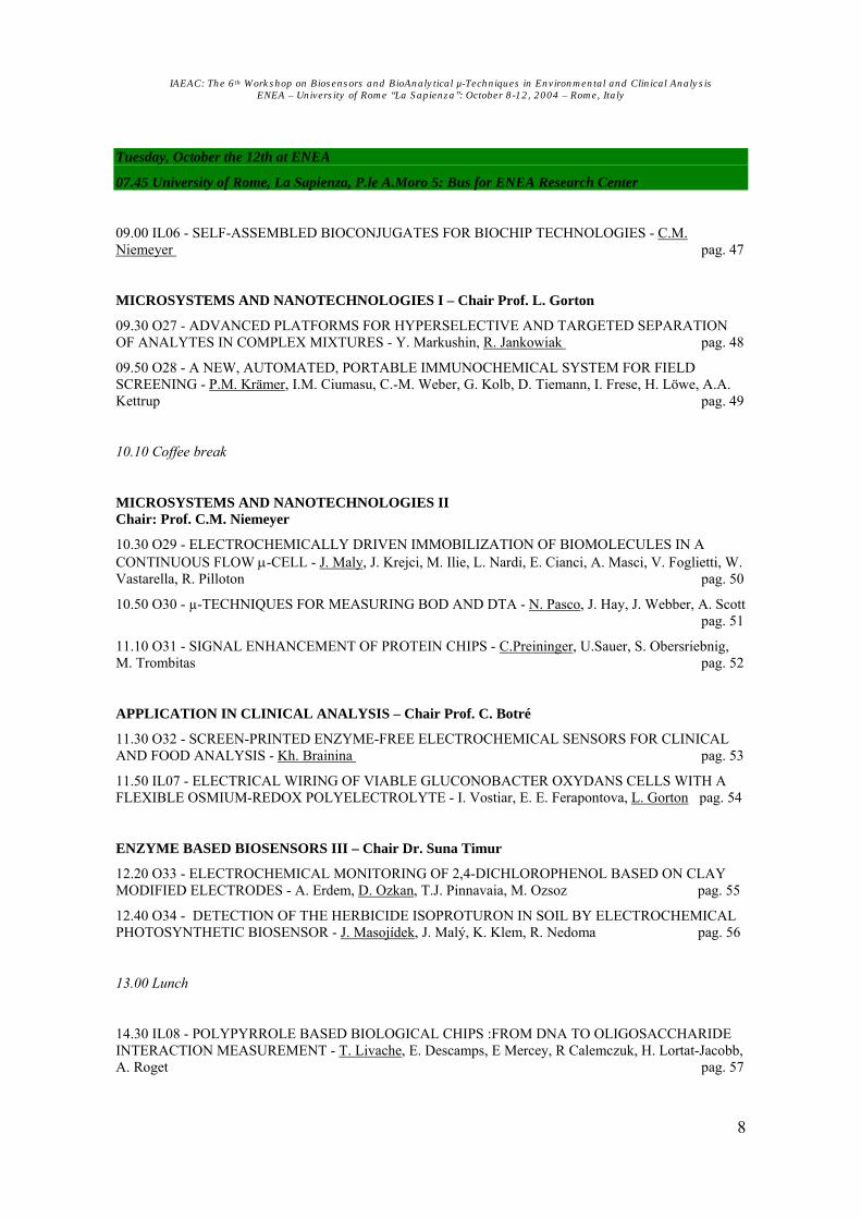
O05 - HIGHLY SENSITIVE PROTEASE ASSAY USING SELF-QUENCHING PEPTIDE PROBES
UN.MarméUP
aP, TU J.P. KnemeyerUTP
aP, M. SauerP
bP and J. WolfrumP
aP
P
aPPhysikalisch-Chemisches Institut, Universität Heidelberg, Im Neuenheimer Feld 229, 69120
Heidelberg (Germany) P
bPAngewandte Laserphysik und Laserspektroskopie, Universität Bielefeld, Universitätstr. 25,
33615 Bielefeld (Germany) [email protected] www.single-molecule-spectroscopy.de
keywords: Enzyme Based Biosensors, Protease assay, chip, single-molecule spectroscopy The interest in fast and sensitive assays for proteolytic enzymes, i.e. enzymes that specifically cleave peptide bonds, has increased considerably in the last few years, because of their involvement in many diseases like HIV or cancer. Recently, we investigated the fluorescence quenching of different organic dyes by the amino acid tryptophan.P
[1]P Especially oxazine derivatives
(e.g. MR121) are efficiently quenched by tryptophan via an electron transfer mechanism. Whereas the influence of all other amino acids is neglectable. The new protease assay is based on peptide substrates labeled with a oxazine derivative MR121. In addition to the dye the peptide contains a tryptophan residue and the respective recognition sequence for the target enzyme. Due to contact formations between the fluorescent dye and tryptophan the quantum yield of the substrate is reduced to 0.1 to 0.3, depending on the number of amino acids and their sequence between dye and tryptophan. In the presence of the target enzyme a peptide bond of the substrate is cleaved and the tryptophan residue is removed. Therefore, tryptophan is no longer quenching the dye leading to an up to ten fold increase of the fluorescence intensity. The new method is suitable as well for homogeneous as heterogeneous assay formats. We used the mono-labeled, self-quenching peptide substrates in aqueous solution in glass cuvettes and measured the fluorescence increase by a standard fluorescence spectrometer. Depending on the target enzyme the detection limit was below the picomolar range (up to 10P
-13P M). Thereby a significantly fluorescence increase was reached within minutes. For a
heterogeneous assay we coupled the substrate (Cys-Gly-Gly-Lys(dye)-Trp) covalently to a PEG modified surface using the SH-group of cystein. Due to the interaction between the dye and the tryptophan the fluorescence intensity is quenched and only a few spots could be observed (Fig. b; left). After adding the protease, (Carboxypeptidase A) the tryptophan was removed and an 8-fold increase in the number of spots (Fig b; right) was detected.
[1] N. Marmé, J.P. Knemeyer, M. Sauer, and J. Wolfrum: "Inter- and Intramolecular Fluorescence Quenching of Organic Dyes by Tryptophan" J. Bioconjugate Chem. 2003, 14, 1133-1139
Figure: a) General principle of mono-labeled self-quenching substrates. The fluorophore (gray) is coupled to one end of the peptide in close proximity to a tryptophan residue. Contact formations lead to efficient fluorescence quenching via photoinduced electron transfer. The target enzyme cleaves the substrate resulting an increase of the fluorescence intensity. b) Fluorescence scanning images (excitation power = 5 µW) of Cys-Gly-Gly-Lys(dye)-Trp covalently linked to a PEG-modified glass-surface. Left: in absence of a proteolytic enzyme, right: 10 minutes after adding Carboxypeptidase A.

IAEAC: The 6th Workshop on Biosensors and BioAnalytical µ-Techniques in Environmental and Clinical Analysis ENEA – University of Rome “La Sapienza”: October 8-12, 2004 – Rome, Italy
21
O06 - AMPLIFIED BIOSENSOR BASED ON GLUTAMATE RECEPTOR INCORPORATED IN A MHBLM
(MIXED HYBRID BILAYER LIPID MEMBRANES) ARRAY
TL. CampanellaP
aP, S. CavalloP
bP, A. D'AnnibaleP
aP, G. FaveroP
aP, T. FerriP
aP and E. MatteiP
cTP
P
aP Dipartimento di Chimica - Università di Roma “La Sapienza”, P.le Aldo Moro, 5 - 00185 Roma ITALIA
P
bP Dipartimento di Scienze Biochimiche ‘A. Rossi Fanelli’, Università di Roma “La Sapienza”, P.le Aldo
Moro, 5 - 00185 Roma ITALIA P
cP Istituto di Neurobiologia e Medicina Molecolare, Consiglio Nazionale delle Ricerche, V.le Marx, 15 -
00137 Roma ITALIA
keywords: Receptor Based Biosensors, Electrochemical sensors, Membranes, Molecular recognition, Nanostructures, Neurotransmitters, Self Assembled Monolayers
In the biological systems the cellular membranes play a key role in signal transduction: hence it is not surprising that increasing interest of several research groups working on biosensors, is focused on biological membranes to substitute traditional biological components such as enzymes, antibodies, whole cell, tissues and so on. Actually, biomembranes are very promising for biosensors development: they can interact selectively with the analyte producing membrane disruptions suitable for an electrochemical transduction. Moreover, they not only can provide versatility, sensitivity and selectivity to the biosensor component carrying out the molecular recognition, but are also the ideal environment which the natural receptors can operate in. The main interests in integrating receptors as the biological components of innovative biosensors are their extreme selectivity and the possibility to amplify the answer towards the analytes. A more recent development of our research is represented by the incorporation of glutamate receptor (GluR) in a reconstituted biological membrane (MHBLM, see below). GluR was extracted and isolated from a natural source (rat brains) by means of a procedure described in literature and suitably adapted that essentially involves homogenisation, several ultra-centrifugation steps and finally an affinity batch chromatography; finally, the protein was detected by SDS-PAGE. The MHBLM incorporating GluR exploits the signal amplification property, typical of receptors: actually, the glutamate is sensed down to few nmol lP
-1P and the system is sensitive to the modulating effect due to receptor agonists and antagonists.
Anyway, the main hindrance to the development of receptor biosensors so far is the need to incorporate the chosen receptor inside a stable, long-lasting and biomimetic biological membrane. To this end, our research group developed an experimental apparatus suitable for the preparation and characterisation of stable reconstructed biological membranes. Our approach takes to reconstituted biomembranes coupling the biomimetic characteristics of BLMs (bilayer lipid membranes) to the high stability of HBMs (hybrid bilayer membranes), thus giving place to an innovative assembly named MHBLM (mixed hybrid bilayer lipid membrane). The obtained system is constituted by an array of true biomembranes inserted in a hybrid biomembrane matrix. Over the past few years, experimental parameters for MHBLM preparation have been optimised and the biomimetic behaviour was demonstrated through the incorporation of transport peptides: particularly, Gramicidin D and Valinomycin. The MHBLM incorporating such peptides displayed a response towards different cations (NaP
+P, LiP
+P, KP
+P, NHB4PB
+P) characterized by the same selectivity order reported in literature; in
addition MHBLMs are so stable that can be used not only in a static media but also under flowing conditions. This has allowed to succeed in the incorporation of GluR obtaining a sufficiently stable and reliable system. Results obtained so far and foreseen applications in different fields such as analytical (glutamate determination), medical (molecules of pharmaceutical interest evaluation) and heuristic (investigation on neurological receptors) are extremely encouraging for the future.

IAEAC: The 6th Workshop on Biosensors and BioAnalytical µ-Techniques in Environmental and Clinical Analysis ENEA – University of Rome “La Sapienza”: October 8-12, 2004 – Rome, Italy
22
IL02 - AMPEROMETRIC DETECTION OF AFFINITY EVENTS BASED ON POLYELECTROLYTE MULTILAYERS: APPLICATIONS TO
IMMUNOSENSORS AND DNA SENSORS
TUE. DomínguezUT
Department of Analytical Chemistry, Faculty of Pharmacy, University of Alcalá – 28871 Alcalá de Heanres, Madrid (Spain)
[email protected] keywords: immunosensors, DNA sensors, polyelectrolyte multilayers, amperometry
In 1995, the general utility of polyelectrolyte multilayers (PEMs) based on electrostatic interactions for the assembly of proteins was first demonstrated by Lvov et al. Since then, this technology is being exploited for the construction of sensing devices. Amperometric transduction of affinity events challenges this technology due to the lack of faradic currents requiring then the introduction of convenient redox labelling. Frequently, catalytic labelling is used since multiple molecules account per affinity event. Enhanced sensitivity can also be envisaged if the transduction scheme permits the introduction of several catalytic molecules per affinity event. This work describes the use of polyelectrolyte multilayers as a generic technology for the design of amperometric affinity sensors. The utility of this approach will be demonstrated through two different applications including immunosensors and DNA sensors. In both cases glucose oxidase has been chosen as reporter enzyme resulting in the production of hydrogen peroxide that readily diffuses through the sensing and highly permeable transducing polyelectrolyte multilayersP
[1]P. Consequently, the efficiency of this
configuration depends on the electrocatalytic conversion of a diffusional product not being limited by the distance to the electrode surface and thus opening the route for customised sensitivity. As it will be demonstrated trace concentrations of environmental pollutants can be easily and directly detected with immunosensors. Furthermore, the use of multiple oligonucleotide sequences linked to an enzyme for the detection of specific hybridisation results in DNA sensors where signal amplification can be also controlled by successive hybridisation stepsP
[2]P. Electrochemical, microgravimetric and topographical characterisation
of these nanostructured layers will also be presented. [1] A. Narváez, G. Suárez, I. C. Popescu, I. Katakis, E. Domínguez. Reagentless biosensors based on self-deposited redox polyeletrolyte-oxidoreductases architectures. Biosen. Bioelectron., 15 (2000) 43 [2] E. Domínguez, O. Rincón, A. Narváez. Electochemical DNA sensors based on enzyme dendritic architectures: an approach for enhanced sensitivity. Anal. Chem., 76 (2004) 3132. Financial support from the Ministry of Science and Technology is deeply acknowledged (AGL-2002-04635).

IAEAC: The 6th Workshop on Biosensors and BioAnalytical µ-Techniques in Environmental and Clinical Analysis ENEA – University of Rome “La Sapienza”: October 8-12, 2004 – Rome, Italy
23
O07 - MEMBRANE IMMUNOANALYTICAL TECHNIQUES FOR ENVIRONMENTAL MONITORING
TUB.B.Dzantiev UT
Institute of Biochemistry Russian Acad. Sci., Leninsky prospect 33, 119071 Moscow, Russia [email protected]
www.webcenter.ru/~bdzan/Index.htm keywords: Immunoassay, Immunosensor
The presented investigations are directed to the creation of simple rapid analytical systems for pesticides detection in environmental monitoring. The proposed approaches are based on the recognition of pesticides by specific antibodies, separation of the formed immune complexes by means of membrane carries and followed registration of the bound complexes by electrochemical or optical techniques. The given approaches were applied to reveal pesticides belonging to triazine, sulfonylurea and chloroacetanilide classes (atrazine, simazine, chlorsulfuron, butachlor, etc.). Electrochemical immunosensors were realized with the use of field-effect transistor as a sensitive element. In the course of the assay specific immune complexes were formed at disposable porous membrane attached to gate region of the transistor. The complexes contained peroxidase label, and its quantity was stipulated for the competition between free (determined) and peroxidase-labeled pesticide molecules for their binding with specific antibodies. The detected signals were generated by the action of peroxidase under a substrate solution containing o-phenylenediamine, ascorbic acid and HB2BO B2B. The substrate composition and regime of measurements were optimised to reach maximal responses. Both total shift of pH and maximal rate of its changes were used for analyte quantification; the duration of the registration step did not exceed 3 min. An alternate developed approach in electrochemical immunoassay was based on the application of screen-printed electrode with impregnated peroxidase (Newcastle University, UK). Membrane with immobilized antibodies was attached to the electrode surface, and free pesticide competed with pesticide – glucose oxidase conjugate for binding with the antibodies. The following addition of glucose induced generation of HB2BO B2B by the bound glucose oxidase and its transformation by the peroxidase that caused the current changes. Non-bound glucose oxidase did not influence the current due to the presence of catalase in solution. Portable amperometric device was applied for the registration of results of the electrochemical immunoassays. It was combined with PC software allowing calculation of target analyte content and formation of database for measurements results. Total duration of the proposed assays was 20 min, lower limit of reliable detection varied from 0.01 to 2 ng/ml (depending on concrete antigen and used immunoreactants). Express immunoassays with visual or optical detection were realized in the format of dot-blot immunofiltration. We applied interpolyelectrolyte reaction to accelerate separation of immunoreactants during the filtration process. The extremely high rate and affinity of the electrostatic interraction between protein A-coupled polyanion polymethacrylate and immobilized polycation poly-N-ethyl-4-vinylpyridium made them a universal separation system for detection of different compounds. Protein A bound immunoreactants in solution, were competitive immunodetection of a target pesticide with the use of specific antibodies and antigen-peroxidase conjugate took place. Products of the catalytic action of bound peroxidase-labelled immune complexes may be detected visually for qualitative decision about exceeding some controlled level of pesticide content. The immunofiltration can be used also for quantitative assays based on the measurements of brightness of the formed coloured spots. A portable photometer-reflectometer with autonomous or PC-dependent data treatment was used for immunofiltration assaying of different pesticides. Algorithms for the quantification of the digital images were chosen. The assay regime was optimised to reach maximal sensitivities. Under the determined conditions the proposed immunofiltration technique was characterized by total duration of 15 min, detection limits for optical registering – from 0.02 to 0.5 ng/ml, and cut-off levels for visual assaying – from 0.5 to 10 ng/ml. To verify the developed systems they were applied for analyses of polluted samples of environmental and drinking water, milk, meat, juices and vegetables. Revealing of pesticides in these samples was no less than 85%, C.V. for repeated measurements – 5–12%. In that way the developed techniques may be recommended for environmental and agricultural control. The investigations were supported by grants of INCO-Copernicus Foundation (ICA2-CT-2001-10007), INTAS (Innovation Grant 150) and Russian Foundation for Basic Research (03-04-48773a).

IAEAC: The 6th Workshop on Biosensors and BioAnalytical µ-Techniques in Environmental and Clinical Analysis ENEA – University of Rome “La Sapienza”: October 8-12, 2004 – Rome, Italy
24
O08 - USE OF A NEW WHOLE CELL BIOLUMINESCENT BIOSENSOR BASED ON RECOMBINANT YEAST STRAIN FOR ENVIRONMENTAL MONITORING
OF ANDROGEN-LIKE COMPOUNDS
UE.MicheliniUP
aP, P.LeskinenP
bP, M.VirtaP
bP, M.KarpP
bP and A.RodaP
a
P
a PDepartment of Pharmaceutical Sciences, University of Bologna, Bologna, Italy
P
b PDepartment of Biotechnology, University of Turku, Turku, Finland.
[email protected] http://www.anchem.unibo.it
keywords: Receptor Based Biosensors, Bioluminescence, Endocrine disruptors, Pollutants
During the last few years a significant concern has arisen about the presence of natural and man-made compounds which affect human health by interfering with normal endocrine functions. These substances, defined as endocrine disrupting chemicals (EDC) represent an heterogeneous class of molecules either steroidal or not, sharing the ability of interfering with the endocrine system via nuclear receptor signaling pathwaysP
[1]P. We previously developed a new sensitive and rapid assay based on recombinant S.cerevisiae
cells for environmental monitoring of compounds able to interact with the human androgen receptor by acting as androgens or antiandrogens. The biosensor is based on yeast cells that express the human androgen receptor (hAR) and contain the sequence Androgen Response Element (ARE) regulating the expression of Photinus pyralis luciferase. In the presence of androgenic compounds, hAR, which is a ligand activated transcription factor, moves into the nucleus, and binds ARE sequences, resulting in luciferase expression and light emission measured by simple addition of D-luciferine. The androgen assay is performed in a 96-well microtiter plate format and a recombinant yeast strain constitutively expressing luciferase is used as control to correct the light signal accordingly to cell vitality and matrix aspecific effects. The biosensor responds to testosterone as reference androgen in a concentration-dependent manner from 0.05 to 1000 nM and the assay is accurate and precise, allowing the detection in aqueous environmental samples. Androgenicity was tested in municipal effluents before and after treatment in sewage treatment plants in the cities of Rome, Florence, Parma and Bologna. The samples, collected in the same period of the year were analysed with different methods, including HPLC-ES-MS and immunoassays for the detection of estrogen and androgen-like compounds of either natural or synthetic origin. Furthermore, application of the biosensor for monitoring the presence of androgens in clinical samples will be explored. The presence of illegally used synthetic androgens for human and animal doping is a huge issue and a rapid and cheap first level screening to detect the presence of those substances in blood and urines would be a valuable tool. In addition, cells' immobilization in a suitable matrix will be explored to develop a system easier to handle and with a higher reproducibility. [1] Daston GP, Cook JC, Kavlock RJ. Uncertainties for endocrine disrupters: our view on progress. Toxicol Sci. 2003, 74: 245-52

IAEAC: The 6th Workshop on Biosensors and BioAnalytical µ-Techniques in Environmental and Clinical Analysis ENEA – University of Rome “La Sapienza”: October 8-12, 2004 – Rome, Italy
25
O09 - ELECTROCHEMICAL DETECTION OF ENDOCRINE DISRUPTING CHEMICALS BY IMPEDANCE TECHNIQUES AND BY A YEAST
TWO HYBRID MICROBIAL SYSTEM
V.Sacks-Granek, A.Schwartz-Mittelman, A.Baruch, T.Neufeld and UJ.RishponU
Tel-Aviv University, Ramat-Aviv, Tel-Aviv, Israel
[email protected] http://www.tau.ac.il/lifesci/biotechnology/rishpon/rishpon.html
keywords: Receptor Based Biosensors, endocrine disruptors, impedance, yeast two hybrid
Endocrine disrupting agents are exogenous substances that alter the function(s) of the endocrine system, thereby causing adverse health effects in animals and humans. Various chemicals, such as pesticides, plasticizers, and persistent pollutants reportedly display certain endocrine-disrupting effects. Recently, several compounds, once considered harmless, were discovered to be endocrine-disrupting chemicals. Xenoestrogens, synthetic or environmental chemicals that interact with the estrogen receptor, are suspected to induce feminization in wildlife. The work presented here describes two bioelectrochemical approaches for monitoring the binding between a hormone receptor and its corresponding hormone or xenohormone. Both approaches detect hormone binding to the specific receptor. One is direct and based on impedance changes that originate from the conformational changes that follow hormone-receptor binding. The other is indirect and monitors hormone-induced receptor dimerization in vivo, employing the yeast two hybrid system. We developed a rapid impedimetric biosensor by embedding the hormone receptor in a lipid bilayer on the surface of a gold electrode. The synthetic bilayer membrane mimics the receptor’s natural environment and also functions as an electrical circuit of capacitors and resistors. The binding of estrogen and testosterone to their respective receptors on the modified electrode induces conformational changes in the lipid layer, leading to detectable alterations of the electrical circuit components. The components of the equivalent circuit before and after hormone binding were analysed. This electrochemical system enables the characterization of small differences in the bilayer structure and the quantification of hormone-receptor binding at the picomolar level. The conformational changes in the lipid structure were also followed by optical techniques (AFM, SPR). In parallel we developed a modified yeast two-hybrid bioassay for the highly sensitive detection of protein–protein interactions, based on the amperometric monitoring of β-D-galactosidase reporter gene activity, using p-aminophenyl- β-D-galactopyranoside as a substrate. Sensitive detection of 17-β-estradiol was achieved at concentrations as low as 10P
−11P M (approx 2 pg/ml) by monitoring 17-β-estradiol receptor
dimerization induced by exposure to the hormone. The sensitivity of this system was higher than that of standard optical methods by three orders of magnitude. To assess the effectiveness of both systems for detecting other estrogenic chemicals, we monitored the environmental xenoestrogens bisphenol A and nonylphenol and the flavonoid phytoestrogens genistein and naringenin for their ability to bind the estrogen receptor. The change in membrane impedance and in β-galactosidase expression in the yeast two-hybrid assay, induced by very small concentrations of estrogen, testosterone, or their xenohormones, concurred with the in vivo biological activity of these chemicals. We also examined the ability of therapeutic drugs to inhibit estrogen-receptor dimerization in the presence of physiological concentrations of 17-β-estradiol. Dimerization was considerably reduced by adding the anti-cancer drug tamoxifen or the common analgesic drug paracetamol. Working concomitantly with the two systems developed here provides the advantage of operating at different molecular levels, thereby enabling the sensitive and detection of hormones and xenohormones. Another benefit is that the results of the binding event and the results of the successive receptor dimerization reciprocally validate each other.

IAEAC: The 6th Workshop on Biosensors and BioAnalytical µ-Techniques in Environmental and Clinical Analysis ENEA – University of Rome “La Sapienza”: October 8-12, 2004 – Rome, Italy
26
IL03 - GENE EXPRESSION PATTERNS AS A TOOL FOR BIOANALYSIS
TUB.HockUT, M.Alberti, U.Kausch and R.Leibiger
Technische Universitaet Muenchen, Center of Life and Food Sciences Weihenstephan Chair of Cell Biology, Alte Akademie 12, 85354 Freising (Germany)
[email protected] Twww.wzw.tum.de/btT
keywords: Endocrine disruptors, Gene expression, Zebrafish
The number of chemicals known to act as endocrine disruptors is continuously increasing. Therefore investigations of their distribution in the environment are important. Suitable tools for testing their potential endocrine disrupting activity are required. The egg yolk protein vitellogenin plays an important role as marker for the exposure of fish to estrogenic substancesP
[1]P. Vitellogenin is usually expressed in female liver
tissue and its synthesis is controlled by estrogensP
[2]P. After exposure of male fish to endocrine acting
substances an induction of vitellogenin can be observed. In order to study estrogenic effects at the gene expression level, quantitative PCR was used. Zebrafish (Danio rerio) were exposed in flow-through tanks with 17ß-estradiol in a broad range of concentrations, which were monitored by the ELRA, a non-radioactive receptor assayP
[3]P. Quantitative PCR was performed for the examination of the expression level of
the marker gene vitellogenin (vtg1). A significant increase in the expression of the vitellogenin gene was observed in exposed male fish starting at a concentration of 200 ng/L 17ß-estradiol (see Fig.1).
Gene expression patterns can be used to obtain more detailed information on the exposure of organisms to estrogenic compounds. DNA microarrays offer the possibility to detect multiple effects without DNA amplification A large number of possible target genes can be tested on a glass slide. The comparison of tissues from non-exposed and exposed animals provides information on the effects of pollutants and their interactions on individual organisms or tissue types. Gene expression experiments of zebrafish exposed to 500 ng/L 17ß-estradiol were statistically analysed with Significance Analysis of Microarrays (SAMP
[4]P). A
strong up-regulation of vtg1 was observed. In addition, further genes were identified to be up-regulated by estrogens, for example genes coding for vitellogenin 3 (vtg3), nothepsin (nots) and dead box protein 5 (dbp5). A number of genes was found to be down-regulated, such as coding for pleiotropin 1 (plei1) and the cryptochrome 3 (cry3). [1] Van den Belt, K., Verheyen, R., Witters, H.: Comparison of vitellogenin responses in zebrafish and
rainbow trout following exposure to environmental estrogens. Ecotoxicology and Environmental Safety 56, 271-281 (2003)
[2] Wahli, W.: Evolution and expression of vitellogenin genes. Trends Genet. 4, 227-232 (1988) [3] Seifert, M., Haindl, S., Hock, B.: In vitro analysis of xenoestrogens by enzyme linked receptor assays
(ELRA). Adv Exp Med Biol. 444, 113-117 (1998) [4] Goss Tusher, V., Tibshirani, R., Chu, G.: Significance analysis of microarrays applied to the ionizing
radiation response. PNAS 98, 5116-5121 (2001)
Figure 1: Gene expression of vitellogenin 1 in male liver tissue after exposure to different concentrations of
17ß-estradiol (0-500 ng/L)

IAEAC: The 6th Workshop on Biosensors and BioAnalytical µ-Techniques in Environmental and Clinical Analysis ENEA – University of Rome “La Sapienza”: October 8-12, 2004 – Rome, Italy
27
O10 - RAPID RESISTANCE GENOTYPING OF TEM BETA-LACTAMASES USING DNA-MICROARRAYS
UV.GrimmUPU
1UP, S.EzakiP
1P, M.SusaP
2P, C.KnabbeP
2P, R.D.Schmid P
1P and T.T.BachmannP
1*P
P
1P Institute of Technical Biochemistry, University of Stuttgart, Germany
P
2P Department of Clinical Chemistry and Laboratory Medicine, Robert Bosch Hospital, Stuttgart, Germany
Tel: +49 (0) 711 685 3197, Fax: +49 (0) 711 685 3196, [email protected] www.itb.uni-stuttgart.de
keywords: DNA, microarray, bacterial resistance, beta-lactamases
Clinical standard procedures of pathogen resistance identification are laborious and require usually two days cultivation time before the resistance can be determined unequivocally. In contrast, clinicians have to face an increasing threat by antibiotic resistant pathogenic bacteria in terms of frequency and level of resistance. A major class of microbial resistance stems from the occurrence of beta-lactamases, which if mutated can cause the severe Extended Spectrum Beta-Lactamase (ESBL) or Inhibitor Resistant TEM (IRT) phenotype implying resistance to third generation cephalosporins, monobactams, and beta lactamase inhibitors. Here we describe an oligonucleotide microarray for the identification of single nucleotide polymorphisms (SNPs) of 96 % of the TEM beta-lactamase variants described to date which are related to the ESBL and/or IRT phenotype. The target DNA, originating from Escherichia coli, Enterobacter cloacae, and Klebsiella pneumoniae cells isolated from clinical samples, was amplified and fluorescently labeled by PCR using consensus primers in presence of Cy5 labeled nucleotides. The total assay including PCR, hybridization, and image analysis could be performed in 3.5 hours. The microarray results were validated using clinical standard procedures. Here, the microarray outperformed in terms of assay time and information depth. In conclusion, this array offers an attractive option for the identification and epidemiologic monitoring of TEM beta lactamases within the clinical routine diagnostics.

IAEAC: The 6th Workshop on Biosensors and BioAnalytical µ-Techniques in Environmental and Clinical Analysis ENEA – University of Rome “La Sapienza”: October 8-12, 2004 – Rome, Italy
28
O11 - ELECTROCHEMICAL GENOSENSOR BASED ON COLLOIDAL GOLD
AND TITANIUM DIOXIDE NANOPARTICLES FOR THE DNA
HYBRIDIZATION DETECTION
M. Ozsoz, A. Erdem, D. Ozkan, H. Karadeniz and K. Kerman
Department.of Analytical Chemistry, Faculty of Pharmacy, Ege University, 35100 Bornova-Izmir, TURKEY [email protected]
www.pharm.ege.edu.tr~ozsozs keywords: Genosensor, Gold/titanium nanoparticles, Self-assembled monolayer,
Carbon graphite electrodes.
Electrochemical genosensors are attractive devices for converting the hybridization event into an analytical signal for obtaining sequence-specific information in connection with clinical, environmental or forensic investigations [1]. Electrochemical genosensors rely on the immobilization of a single-stranded (ss) DNA sequence (the “probe”) for hybridizing with its complementary (“target”) strand to give rise to voltammetric signals (in connection to a suitable electrochemical label). There is a increasing interest in the electrochemical investigations of DNA detections and interactions between drug and DNA by using genosensor systems[2-5]. Recently, DNA modified Au nanoparticles and titanium dioxide nanoparticles have begun to hold a very important aspect in the design of genosensors [6,7]. Optical genosensors based on Au nanoparticles have already been studied and optimized in detail[8], but electrochemical ones are still waiting for suprising new advances. Here we describe an electrochemical DNA detection procedure based on oxidation signals of Au nanoparticles to detect an inherited disease; Factor V Leiden Mutation using polymerase chain reaction (PCR) amplicons and synthetic oligonucleotides. A pencil graphite electrodes (PGE) were modified with target DNA or PCR amplicon and hybridization was occurred with complementary probes conjugated to Au nanoparticles. After the hybridization reaction, Au oxidation signal was obtained at about +1.20 V. Monitoring the changes in the Au oxidation signal shortened the assay time and simplified the detection of the Factor V Leiden mutation from PCR amplified real samples [9].
In other study, titanium dioxide nanoparticles modified electrodes(carbon paste and pencil graphite electrodes) were used for the DNA hybridization detection. It was observed DNA molecule showed an affinity to titanium dioxide nanoparticles modified electrodes. These results represent rapid and applicable DNA detection for clinical genetic analysis.
[1] S. R. Mikkelsen, Electroanalysis, 1996, 8, 15. [2] Wang, J.; Nucleic Acids Res. 2000, 28, 3011 - 3016. [3] D.Ozkan, A. Erdem, P. Kara, K. Kerman, B. Meric, J. Hassmann, M. Ozsoz, Anal. Chem., 2002, 74, 5931-5936. [4] Palecek, E.; Fojta, M.; Anal. Chem. 2001, 73, 74A-83A. [5]F. Lucarelli, I. Palchetti, G. Marrazza, M. Mascini, Talanta, 56 (2002) 949-957. [6]A. N. Shipway, E. Katz, I. Willner, ChemPhysChem., 2000, 1, 18-52. [7] T.Paunescu, T.Rajh, G. Wiederrecht, J. Master, S. Vogt, N. Stojicevic, M. Protic, B. Lai, J. Oryhon, M. Thurnauer, G. Woloschak, Nature Materials,2003,10.10380/nmat875,1-4. [8] R. Jin, G. Wu, Z. Li, C. A. Mirkin, G. C. Schatz, J. Am. Chem. Soc., 2003, 125, 1643-1654. [9] M. OZSOZ, A. ERDEM, K. KERMAN, D. OZKAN, B. TUGRUL, N. TOPCUOGLU, H. EKREN, M. TAYLAN, ANAL. CHEM., 2003, 75, 2181-2187.

IAEAC: The 6th Workshop on Biosensors and BioAnalytical µ-Techniques in Environmental and Clinical Analysis ENEA – University of Rome “La Sapienza”: October 8-12, 2004 – Rome, Italy
29
O12 - FLUORIMETRIC BASED WIDE RANGE DETECTION OF COMPOUNDS WITH AFFINITY FOR NUCLEIC ACIDS
TUYang LiuUT and Bengt Danielsson
Pure and Applied Biochemistry Dept, LUND University Getingevagen 60, LUND, Sweden
[email protected] keywords: DNA, Fluorescence, Fiber Optic, Environment
The potential of a nucleic acid based optical bioprobe for environmental test and drug monitoring is reported on. The sensor employs the long wavelength intercalating fluorophore TO-PRO-3. Compounds, which interact with the TO-PRO-3-DNA complex, are indirectly detected by the decrease of the fluorescence intensity. We discovered that the configuration and length of the DNA dramatically affect the fluorescence intensity emitted from the TO-PRO-3-DNA complex. The plasmid DNA pBR322 was chosen for the studies described here. The proposed concept attempts to combine broad range detection with speedy and simple operation. Different types of detection methods were applied, including a fiber optic capillary fluorescence system and an epi-fluorescent CCD camera system. Several kinds of toxic aromatic amines, antibiotics and antitumor drugs were tested. Using down to 20µl of sample, a sensitivity comparable to that of current electrochemical was achieved. The fiber optic capillary fluorescence system facilitates field tests, while the epi-fluorescence CCD camera system utilizes a sample platform, such as microtiter plates or glass chips for rapid high throughput tests. The detection limit can be down to a few ppb or sub-micromolar. Both these approaches could serve as alternative to routine test in environmental or therapeutic (cytostatic) drug monitoring.

IAEAC: The 6th Workshop on Biosensors and BioAnalytical µ-Techniques in Environmental and Clinical Analysis ENEA – University of Rome “La Sapienza”: October 8-12, 2004 – Rome, Italy
30
IL04 - SENSORS FOR THE DETERMINATION OF PESTICIDE RESIDUES; A TOOL FOR THE JOB OR A JOB FOR THE TOOL?
TUMichael F. WilsonUT
Central Science Laboratory, Sand Hutton, York, YO41 1LZ (United Kingdom) [email protected]
www.csl.gov.uk keywords: environment, foods, pesticide residues, organophosphorus,
organochlorine, fungicides, insecticides
The use of immunoassays as the basis for rapid testing for pesticide residues in the environment or foods offers potential benefits in terms of speed, portability and low cost analyses. Set against this is the relatively long lead-in time for method development in comparison with adaptable chromatographic or other instrumental techniques and the prerequisite for a known target, or targets, for a method to be well defined. Thus, it is not generally possible for a laboratory to develop a novel immunoassay-based test immediately in response to, for example, a specific food scare. Overall, the advantages of immunoassay technology can be summarised as the possibilitiesP
1P for:-
Low detection limits High analyte selectivity and targeting High throughput of samples and cost effectiveness Reduced sample preparation and handling allowing possible use in the field
and taking into account the limitations:-
Matrix interferences Cross reactivity to structural analogues and other chemicals Current unsuitability for multi-analyte methods Long method development lead in time necessitating a known ‘problem’ and a (relatively)
large number of samples Sensors are ideally suited to the target analysis of specific compounds and small ranges of compounds. For example, as simple tools available to farmers, agricultural consultants or environmental inspection services to monitor the fate of residues in either a foodstuff or in the environment. An example of this is the development and use of a lateral flow device based sensor for the determination of the fungicide tebuconazole following its use on cereals. However, many problems require the determination of a range of residues from ‘all’ pesticides to large, selected groups such as organophosphorus or synthetic pyrethroids insecticides. In such cases so-called generic antibody-based sensors have been highly prone to matrix interferences, cross-reactivity and have not been widely adopted over more conventional, chemical analyses. A way forward may be the adoption of ‘arrays’ of single or limited range assays combined in a convenient format; either using micro-arrays and image analysis or some of the newer methods of micro-analysis based on such systems as the CD-ROM.
[1] Shan G, Lipton C, Gee SJ and HammockBD; in Handbook of Residue Analytical Methods for Agrochemicals, ed. Lee PW (2003) Wiley & Sons
Figure: Agricultural production often relies on a combination of close inspection of the crop in the field and sophisticated chemical analysis carried out in expensive laboratory facilities. Sensors offer the possibility of carrying out some tests ‘in the field’.

IAEAC: The 6th Workshop on Biosensors and BioAnalytical µ-Techniques in Environmental and Clinical Analysis ENEA – University of Rome “La Sapienza”: October 8-12, 2004 – Rome, Italy
31
O13 - MOLECULAR IMPRINTED POLYMERS AS SENSING MEMBRANE FOR DIRECT ELECTROCHEMICAL DETECTION OF POLLUTANTS
M.Pesavento, UG.D’AgostinoU, G.Alberti
Dipartimento di Chimica Generale, Università di Pavia, Viale Taramelli 12, 27100 Pavia, Italy [email protected]
keywords: Environment, Electrochemical sensors, Membranes, Molecular recognition
A molecular imprinted polymer membrane for atrazine has been developed and caracterized. Two electrochemical sensors based on this membrane were built. The molecular imprinted polymer has been prepared by an well already known procedure P
[1-3]P but avoiding the use of the porogen agent in order to
obtain a low porosity and a stress resistant polymeric thin membrane. The membrane was prepared directly in a special Teflon support that provides a constant shape to the membrane during the polymerization process. Moreover this home made Teflon device allows to assemble the potentiometric sensor without any further manipulation of the membrane. The first sensor we present was prepared to perform potentiometric measures. They were carried out at 20°C U+U 0,1°C in the following classical cell assembly for ion selective electrodes:
Outer ref.|| test solution | MIP membrane| internal solution | Internal ref. The internal reference solution has the following composition: HCl 3,2 10P
-2PM and atrazine 3 10P
-4P
M. The cell has a good response for atrazine over a wide concentration range (3 10P
-5P – 10P
-3PM at pH 1.5) and
the slope of the Nernstian line is 29 mV/decade. The suggested working pH is the optimal one as experimentally determinated in KCl 2 10P
-2PM solution.
The electrode responds also to another very similar triazine (simazine) with KBijB=1,4. The triazine molecules, at the working pH, are protonated P
[4]P so they carry the
charge required to create a potential difference at the interface depending on the activity of the specific ion. Very favorable characteristics of the electrode is the response time of about 10s, the possibility of using it for more than 2 months without any variation and the potential witch remains constant for more than 3 hours
within the resolution of the potentiometer (U+U 0,1 mV). The detection limit of the sensor is 2,8 10P
-5PM.The second
electrochemical sensor, that use the same membrane of the potentiometric electrode as recognition device, allows to perform potentiostatic measurements. This sensor is built positioning the membrane on a glassy carbon electrode and the measurements are made connecting the electrode to a 3-electrode system (aux.el. Pt, ref.el. Ag/AgCl and the modified glassy carbon) using a potentiostat that maintains a constant “adsorption” potential and recording the courrent during the experiment. In the presence of the analyte, a peak is detected in the first moments of the analysis. The height of this peak is directly proportional of the analyte concentration. An interesting point is that the sensor
doesn’t respond to a similar analytes (es. simazine).All of the measures are performed in water solutions in Britton Robinson Buffer pH 1.8.
[1] Matsui, J.; Miyoshi, Y.; Doblhoff-Dier, O.; Takeuchi, T. Anal.Chem. 1995, 67, 4404. [2] Matsui, J.; Okada M.; Tsuruoka, M.; Takeuchi, T. Anal. Commun. 1997, 34, 85. [3] Muldoon, M.T.; Stanker, L.H. Anal.Chem. 1997, 69, 803. [4] Skopalová, J.; Kotouček, M. Fresenius J.Anal.Chem. 1995, 351 , 650.
Figure 1: calibration curve obtained with the potentiometric sensor
E Vs Log [ ]
99
104
109
114
119
-6.75 -6.50 -6.25 -6.00 -5.75 -5.50 -5.25 -5.00 -4.75 -4.50 -4.25 -4.00Log[M]
E (mV)
Figure 2: Dynamic response for atrazine obtained with potentiometric sensor.
a: 1,7 10P
-7PM ; b: 3,8 10P
-6PM ; c: 1,8 10P
-5PM; d:
5,1 10P
-5PM ; e: 1,0 10P
-4PM
95
100
105
110
115
120
0 50 100 150Time (min)
E (mV)
a b c
d
e

IAEAC: The 6th Workshop on Biosensors and BioAnalytical µ-Techniques in Environmental and Clinical Analysis ENEA – University of Rome “La Sapienza”: October 8-12, 2004 – Rome, Italy
32
O14 - IMMUNOCHEMICAL DETECTION METHODS FOR BIOACTIVE POLLUTANTS
TUE.P.MeulenbergUTP
aP, G.PeelenP
aP, E.LukkienP
aP and K.KoopalP
bP
P
aPELTI Support, Drieskensacker 12-10, 6546 MH Nijmegen (Netherlands)
P
bPAnalytic Devices, Brugakker 3637, 3704 LM Zeist (Netherlands)
[email protected] Twww.eltisupport.nTl
keywords:Environment, ELISA, Immunosensor, Pharmaceuticals
Recently, environmental contaminants were found to include pharmaceuticals. Regarding the bioactivity of such substances several surveys have been performed 1,2]. For monitoring purposes, especially in water samples, the ELISA is a very convenient methods. Accordingly, we have raised antibodies against two target compounds that have been found at rather high levels in effluents, groundwater and surface water, i.e. ibuprofen and naproxen. Both belong to the class of analgetic, antirheumatic, antiphlogistic agents. Several polyclonal antibody preparations were obtained from rabbits and used to design ELISAs. In addition, antibody against ibuprofen was also used in an immunosensor. Ibuprofen has been found in the highest concentrations in waste water and influents of up to 3.3 µg/l, whereas in surface water it can reach levels of 1.6 µg/l. In our indirect competitive ELISA a detection limit of 0.02 µg/l was achieved, with a working range of 0.05 – 30 µg/l. The cross-reactivity of the antibodies was < 1 % for all compounds (related and unrelated), with the exception of naproxen (2 %). Matrix effects were negligible for various spiked (low and high levels) real water samples. The assay showed a linearity of RP
2P =
0.9580. Although the standard curve was very reproducible both within and between plates (precision: intra- and inter-assay variation of < 15 % and < 10 % resp.), with real water samples it sometimes was quite high (4 –30 %). Recoveries ranged from 105 – 162 % depending on the matrix and the concentration added. Naproxen has been detected in concentrations up to 0.126 µmol/l in effluents and waste water. Our ELISA displayed a detection limit of 15 µM and a working range of 15 – 150 µM, what is not yet suitable to monitor this compound. One of the antibodies appeared rather specific for naproxen, showing less than 10 % cross-reactivity with various related and unrelated substances. The linearity of the assay was RP
2P = 0.9850
for different samples. Matrix effects with respect to PBS were found for demineralised water, river water and effluent. The precision expressed in the intra-assay variation ranged from 11 – 24 %, and in the inter-assay variation using spiked samples varied from 9 – 19 % at levels of 62.5 to 250 µM. In addition, using the polyclonal antibodies against ibuprofen measurements have been performed in the Biacore in various spiked real water samples containing 200, 1000 and 8000 nM of ibuprofen. The results were compared with those determined in the ELISA; the correlation between both methods was highly significant showing an RP
2P = 0.9998. Attempts are being made to transfer the Biacore immunoassay on a
Spreeta format for field use. Until now a standard curve with a detection limit of 3.5 µM was achieved. In conclusion, the pharmaceutical contaminant ibuprofen may be measured specifically both by ELISA and an immunosensor assay (Biacore). An ELISA for naproxen, a compound with comparable bioactivity, has been developed, but it should be optimized with respect to the required sensitivity. This assay, however, is very robust and seems promising for further research. [1] S.M. Schrap et al.; Human and veterinary pharmaceuticals in Dutch surface and waste water; RIZA
Report 2003.023; Lelystad, The Netherlands, September 2003. [2] P. Mersmann; Transport- und Sorptionverhalten der Arzneimitterwirkstoffe Carbamazepin,
Clofibrinsäure, Diclofenac, Ibuprofen und Prophenazon in der wassergesättigten und –ungesättigten Zone; PhD Thesis; Technical University of Berlin, Germany, 2003.

IAEAC: The 6th Workshop on Biosensors and BioAnalytical µ-Techniques in Environmental and Clinical Analysis ENEA – University of Rome “La Sapienza”: October 8-12, 2004 – Rome, Italy
33
O15 - IMMOBILIZATION OF LUMINESCENT BACTERIA FOR AN ENVIRONMENTAL BIOSENSOR T
UM.Pernetti UP
a,bP, M.C.AnnesiniP
aP, C.MerliP
aP, G.ThouandP
bP, D.PonceletP
c
P
a PDipartimento di Ingegneria Chimica, Università di Roma “La Sapienza”,
via Eudossiana 18 - 00184 Rome (Italy) [email protected]
http://ingchim.ing.uniroma1.it P
b PUMR GEPEA, ERT CBAC, Campus de la Courtaisière, IUT, 18 Bd G. Defferre, 85035 –
La Roche-sur-Yon (France) P
c PUMR GEPEA, Department of process engineering, ENITIAA, Rue de la Géraudière BP 82225 - 44332
Nantes (France) keywords: Environment, Immobilization, Microbial sensor, Bioluminescence,
For a biosensor application, immobilization of bacteria is a key feature in order to enhance the handling, the miniaturization, the storage of the biosensor and the possibility to re-use it for repetitive measurements. Moreover immobilization limits bacteria inhibition and washing out, which generally occur with suspension bacteria. As many immobilization methods exist, a careful study must be carried out in order to select the best one for the present application. The “quality” and the “effectiveness” of the immobilization system are usually evaluated indirectly, from the biosensor output P
[1]P. A comprehensive strategy is herein proposed in order to
select the best material for immobilization of bacteria. Two main steps should be followed: • evaluation of the intrinsic characteristics of the matrix, by physical and chemical tests; • immobilization of the specific bacteria and investigation of viability, activity and stability. This strategy was applied to set up an immobilization method for a luminescent biosensor detecting pollutant agents in water. Different polymers such as agarose, alginate, PVA, silicone, were tested as immobilization matrices. In the first step, rheological, mechanical and optical properties of the four materials were investigated, being fundamental for polymer handling, biosensor stability and signal transmission. Storage and biodeterioration in marine/fluvial water and wastewater were also monitored. In order to verify whether the analytes would diffuse easily through the membrane, absorption and diffusion were studied and mass-transfer parameters were calculated. In the second step, the chosen materials were applied to the immobilization of a recombinant bioluminescent Escherichia coli::luxAB bacteria. This strain, which detects tributyltin chloride (TBT)P
[2]P, was
employed for tests in batch mode. Bacteria were induced with tributyltin chloride solutions at different concentrations and luminescence was measured by microtiter plate luminometer. Light signal was analysed employing the induction ratio: IR = RLUBsample B/RLU Breference B, where the reference is due to bacteria not induced by TBT. Immobilization procedure was optimised in order to maximise microbial activity. A re-acclimation step with glucose medium proved to be fundamental for this purpose. Results from immobilized bacteria were constantly compared with results due to free bacteria: both showed the same detection limit (0.015µM) and a similar profile, though immobilized bacteria gave higher IR values. Membranes containing bacteria could be washed with MgSOB4B solution and induced again with TBT, demonstrating the possibility of re-using immobilized bacteria for repetitive measurements, which was impossible with suspended bacteria. For storage tests membranes were sealed and stored in refrigerator at 4°C, then re-acclimated in order to monitor luminescence activity. After three weeks storage, less than 5% of activity loss was observed and the immobilized bacteria could be induced again, keeping the same detection limit. In conclusion, the systematic characterization of different immobilization matrices proved to be fundamental for the development of a biosensor. Experiments in batch mode resulted in high light signals, possibility of repetitive measurements and storage, thus encouraging further development and commercialisation. The adopted strategy could be versatile and useful to the development of every microbial biosensor. [1] Semenchuck, I.N., et al. (2000) Effect of various methods of immobilization on the stability of a microbial biosensor for surfactants based on Pseudomonas rathonis T. Applied Biochelistry and Microbiology, 36, 69-72. [2] Thouand, G., et al. (2003) Development of a biosensor for on line detection of tributyltin with a recombinant bioluminescent Escherichia coli strain. Appl. Microbiol., 62, 218-225.

IAEAC: The 6th Workshop on Biosensors and BioAnalytical µ-Techniques in Environmental and Clinical Analysis ENEA – University of Rome “La Sapienza”: October 8-12, 2004 – Rome, Italy
34
O16 - USE OF MEDIATED ELECTROCHEMICAL DETECTION OF
CATABOLISM IN YEAST FOR ENVIRONMENTAL BIOSENSORS
UK.H.R.BaronianUP
1P, A.J.DownardP
2P, G.KunzeP
3P, K.TagP
3P, S.GurazadaP
1P and D.RobsonP
1P
P
1PSchool of Applied Science, Christchurch Polytechnic Institute of Technology, Christchurch, NZ.
P
2 PDepartment of Chemistry, University of Canterbury, Christchurch, New Zealand.
P
3P Yeast Genetics Group, Institute of Plant Genetics and Crop Plant Research, Gatersleben, FRG.
[email protected] keywords: environment, yeast biosensor, electrochemical detection, environmental contamination
Mediated electrochemical detection of catabolism in prokaryote cells is well documented and forms the basis of many bacterial biosensors and microbial fuel cells. We have detected two catabolic substrate-dependent mediated electrochemical signals in Saccharomyces cerevisiae P
[1]P and other yeast species, and
used agents that block specific redox molecules in the catabolic pathways to provide evidence for the origins of the single and double mediator responses. The responses show that in the double mediator system the lipophilic mediator probably scavenges electrons from multiple sites in the catabolic pathway. There is however, significant transfer directly from reduced NAD to menadione. In the single mediator system reduction of the hydrophilic mediator occurs across the cell membrane. The yeast Arxula adeninivorans is able to catabolise a wide range of substrates. It has been used as the sensing element in the construction of multi-analyte biosensors such as rapid BOD sensors. A comparison of detection of catabolism by oxygen consumption and by a mediator method shows that the two systems are very similar in their performance although the mediator system having an advantage by operating independently of oxygen. We have also investigated the use of this detection technique to specifically detect single compounds that are significant environmental contaminants. Cultivation of A. adeninivorans with either butyl phthalate or naphthalene in minimal media with the contaminant as the sole carbon source gives the organism a catabolism targeted at the contaminant. The catabolic response of the organism to the contaminant is detected using the double mediator system. Although this technique does work it is not useful because the minimum detection level achievable is above the detection range required for environmental analysis. The method has been applied to engineered S. cerevisieae expressing the human oestrogen receptor and the Escherichia coli lac Z gene. Catabolic responses to oestradiol concentrations as low as 0.0375nM have been detected after 7 hours incubation. This is a significant reduction from the incubation period of about 72 hours required in currently used optical detection system P
[1]P. P
PEngineering cells with a receptor system
coupled to binding ‘amplifier’ has permitted the detection of a molecule at lower levels than direct catabolism of the molecule would permit. [1] K.H.R.Baronian, A.J.Downard, R.K.Lowen and N.Pasco. 2002. Detection of two distinct substrate-dependant catabolic responses in yeast using a mediated electrochemical method. Applied Microbiology and Biotechnology 60:108-113 [2] Tyler, C. R. et. al. 2000. Metabolism and Environmental Degradation of Pyrethroid Insecticides Produce Compounds with Endocrine Activities. Environmental Toxicology and Chemistry 19(4), 801-809

IAEAC: The 6th Workshop on Biosensors and BioAnalytical µ-Techniques in Environmental and Clinical Analysis ENEA – University of Rome “La Sapienza”: October 8-12, 2004 – Rome, Italy
35
O17 - BIOMIMETIC RECEPTORS FOR ACETYLCHOLINESTERASE INHIBITORS
M.Mascini, M.Del Carlo and D.Compagnone
University of Teramo, Department of Food Science, 64023 Teramo, Italy [email protected]
keywords: Molecular modelling, biomimetic traps, food contaminants
Pesticides and insecticides are applied worldwide to a broad variety of agricultural activities. Particularly, organophosphate and carbamate pesticides are extensively used due to their high insecticidal activity and relatively low persistence. Because their target enzyme, acetylcholinesterase (AChE), is common to neural transmission both in insects and in mammals, including humans, they represent a potential hazard for human health. Many biosensors and bioassays for the detection of AChE insecticides have been extensively studied and demonstrated based on the inhibition of the AChE activity. In the present approach we investigate the feasability to obtain synthetic receptors as affinity ligand for AChE inhibitors. These will be possibly used in affinity sensors development as well as in the realisation of extraction/purification material. In the study, a series of receptors based on aminoacids motif has been designed and their affinity toward different acetylcholinesterase inhibitors has been tested by a computational approach using Leapfrog® algorithm, a module from Sybyl Sofware [1,2]. The biomimetic receptor design strategy was based on the study of the biological structure, in order to produce a series of possible ligands to be used as biomimetic receptors. The estimated computational binding energy of the selected oligopeptides toward a virtual array of pesticides has been computationally obtained. Different sequences of tetrapeptides virtually able to bind organophosphates and carbamates were then obtained starting from the structure of electric eel acetylcholinesterase binding site. Affinity toward 17 different carbamate and organophosphate pesticides has been tested and compared with the affinity obtained using the acetylcholinesterase binding sites. Both classes of pesticides are, in fact, known to be inhibitors of the enzyme. These data, together with the preliminary data obtained to detect the binding affinity of the selected tetrapeptides in solution will be presented and discussed. Acknowledgements: Work supported by MIUR (DL 297/27 July 1999), SINSIAF Project [1] Payne, A. W. R., Glen, R. C. (1993). Molecular Recognition Using a Binary Genetic Search Algorithm.
J. Mol. Graph., 11, 74. [2] Goodford, P. J. (1985). A Computational Procedure for Determining Energetically Favorable Binding
Sites on Biologically Important Macromolecules. J. Med. Chem., 28, 849.

IAEAC: The 6th Workshop on Biosensors and BioAnalytical µ-Techniques in Environmental and Clinical Analysis ENEA – University of Rome “La Sapienza”: October 8-12, 2004 – Rome, Italy
36
O18 - SULFITE DETERMINATION USING SULFITE OXIDASE BIOSENSOR BASED GLASSY CARBON ELECTRODE COATED WITH
THIN MERCURY FILM
M.K.Sezgintürk P
1P, E.AkyılmazP
1P, N.ErtaşP
2P and E.DinçkayaP
1P
P
1PEge University, Faculty of Science, Biochemistry Department,
P
2PEge University, Faculty of Science, Chemistry Department,
35100, Bornova-Izmir, Turkey [email protected]
[email protected] keywords: Enzyme Based Biosensors, sulfite oxidase, sulfite, food additives
Sulfites are commonly used in the food and pharmaceutical industries are preservatives and antioxidants and brewing industry as an antimicrobial agent. In the large quantities, sulfite and its oxidation products are pollutants. Nowadays, due to the reported harmful effects towards hypersensitive people, in many countries, the sulfite content in food and beverages has been strictly limited. In the work here described it was developed a biosensor for the determination of sulfite in food. Sulfite oxidase enzyme was cross linked with gelatin using glutaraldehyde and fixed on a glassy carbon electrode coated with thin mercury film. Sulfite was enzymatically converted to sulfate in the presence of the dissolved oxygen, which was monitored by the help of the changing of oxygen reduction current in the thin Hg film layer. Sulfite determination was carried out by standard curves which were obtained by the measurement of consumed oxygen level related to sulfite concentration. The amounts of enzyme activity and gelatin, percentage of glutaraldehyde, optimum pH and temperature were investigated. Some characterization studies such as repeatability, storage stability and substrate specificity were also performed. The proposed biosensor was used for the determination of sulfite in some food samples. Results indicated a good precision and good agreement between the mean values obtained by the reference method (spectrophotometric-enzymatic method)

IAEAC: The 6th Workshop on Biosensors and BioAnalytical µ-Techniques in Environmental and Clinical Analysis ENEA – University of Rome “La Sapienza”: October 8-12, 2004 – Rome, Italy
37
O19 - DEVELOPMENT AND VALIDATION OF SPR BIOSENSOR ASSAYS FOR THE DETECTION OF ANTIBIOTICS IN FOODS OF ANIMAL ORIGIN AND
ENVIRONMENTAL SAMPLES
S.L.Stead, H.Ashwin, M.Dickinson, S.Richmond and M.Sharman
Central Science Laboratory, Sand Hutton, York Y041 1LZ, UK E-mail: [email protected] WEBSITE: http://csl.gov.uk
keywords: SPR biosensor, Food, Chloramphenicol, Streptomycin
Recently imported food products of animal origin destined for human consumption have been found to be contaminated with µg / kg levels of antibiotics, including chloramphenicol (CAP) and streptomycin. There is also a concern about the occurrence, fate and toxicity of metabolised pharmaceuticals entering into the environment as a result of intensive farming practises. As a result of animal husbandry administration of antibiotics (therapeutic or prophalytic use), low levels of antibiotic metabolites have been detected in surface and ground water.P
[1,2]P
Chloramphenicol is used in human medicine and there are concerns about the dose unrelated toxicity of the compound and also the transfer of bacterial resistance between animals and humans. P
[3]
Under EU legislation these antibiotics have assigned Maximum Residue Limits (MRL) or Minimum Required Performance Limits (MRPLs) in foods of animal origin. P
[4] PChloramphenicol has Annex IV
classification and has recently been assigned a Minimum Required Performance Limit (MRPL) of 0.3 µg / kg in meat. P
[5]P Streptomycin has been assigned an MRL of 500 or 1000 µg / kg, in certain animal species and
tissue types. In order to meet the EU performance requirements for these antibiotics, development work was conducted to develop and validate new rapid screening assays based on Biacore Q (Surface Plasmon Resonance) SPR biosensor. The assay format is competitive inhibition, with the low molecular weight hapten (analyte) immobilised directly on to the sensor surface. Polyclonal or monoclonal antibodies being employed as the recognition protein. Extraction techniques such as solid phase extraction (SPE), tissue dilution and solvent de-fatting have been developed to enable rapid sample clean up prior to biosensor analysis. The SPE approach is suitable for automation using a Gilson ASPEC system. Both assays have been validated using a variety of sample types, including porcine kidney and muscle, shellfish and honey. Tissue from both animals treated with the antibiotics (incurred tissue) and untreated animals has been included within the validation process. The cross-reactivity of both the assays to related compounds and metabolites has been determined. The CAP assay shows 100% and 65% cross-reactivity to chloramphenciol and the major mammalian metabolite, CAP glucuronide respectively. The streptomycin assay is shown to have 100% cross-reactivity to both streptomycin and dihydrostreptomycin (DHS). Method validation was conducted using tissue fortified with the target analyte. The assays are seen to give reproducible results, %CV less than 15 % over a multi-day testing period, Tables 1 and 2. The CCβ for both qualitative screening assays has been calculated as 0.09 µg / kg (CAP) and 5.7 µg / kg (streptomycin) The CCβ of both methods is significantly below half the MRPL and MRL concentrations. Using incurred tissue, the repeatability of the biosensor assays have been compared with calculated concentrations obtained by chemical analysis (HPLC or LC-MS/MS), Figure 1 shows results for the CAP bioassay. The validation data shows agreement within 5%, between SPR biosensor and confirmatory methodology. The biosensor assay is seen to detect slightly higher residue levels in the incurred tissue compared to the LC-MS/MS, it is hypothesised that the antibody captures the metabolised and degradation products as well as the parent marker molecule.

IAEAC: The 6th Workshop on Biosensors and BioAnalytical µ-Techniques in Environmental and Clinical Analysis ENEA – University of Rome “La Sapienza”: October 8-12, 2004 – Rome, Italy
38
Table 1, Multi-level repeatability for the chloramphenicol assay using fortified porcine kidney
Calculated concentration Expected concentration µg/kg µg/kg 0.15 0.3 0.45 0.6 0.7
Mean 0.16 0.33 0.45 0.59 0.74 SD 0.02 0.05 0.06 0.08 0.05
%CV 14.3 15.9 14.2 13.3 7.2 Accuracy 0.10 3.70 2.23 1.64 0.74
n 23 23 24 23 23 Table 2, Multi-level repeatability for the streptomycin assay using fortified honey Calculated concentration Expected concentration ug/kg
ug/kg 7.5 15 30 45 60
Mean 8.13 17.13 31.74 47.49 60.91 SD 0.87 2.04 5.22 4.70 3.29
%CV 10.7 11.9 16.4 9.9 5.4 Accuracy 0.08 0.14 0.06 0.06 0.02
n 21 21 21 21 18 Figure 1, Comparison of CAP biosensor assay versus LC-MS/MS using incurred porcine kidney
Comparison of Biosensor assay Vs LC-MS/MS using CAP Incurred Porcine Kidney
0
0.1
0.2
0.3
0.4
0.5
0.6
0 2 4 6 8
Replicate
Cal
cula
ted
Con
cent
ratio
n ug
/kg
LC-MS/MS
Biosensor
Mean LCMSMS Value
Mean Biosensor Value
[1] Jones, O.A.H, Voulvoulis, N, Lester, J.N, Environmental Technology, 2001, 22 (12): 1383-1394 [2] Kummerer, K. Chemosphere, 2001, 45 (6-7): 957-969 [3] H.P.Rang, M.M.Dale, Pharmacology, Second Edition, 1994 Churchhill Livingstone, UK [4] European Commission, Official Journal of the European Communities, 1999, Regulation (EEC) 2377/90
(as amended) [5] European Commission, Official Journal of the European Communities,2003, Commission Decision
2003/181/EC

IAEAC: The 6th Workshop on Biosensors and BioAnalytical µ-Techniques in Environmental and Clinical Analysis ENEA – University of Rome “La Sapienza”: October 8-12, 2004 – Rome, Italy
39
O20 - XANTHINE OXIDASE MODIFIED GLASSY CARBON PASTE ELECTRODES
Ülkü Anık KirgözP
1P, Suna Timur P
2P, Joseph Wang P
3P and Azmi TelefoncuP
2P
P
1PEge University, Faculty of Science, Chemistry Department 35100, Bornova-İzmir/TURKEY
P
2PEge University, Faculty of Science, Biochemistry Department 35100, Bornova-İzmir/TURKEY
P
3PDepartment of Chemistry and Biochemistry, New Mexico State Chemistry, Las Cruces, NM 88003 USA
[email protected] keywords: Glassy carbon paste electrodes (GCPE), biosensor, xanthine oxidase
Carbon, in many respects, is an ideal electrode substrate due to its wide anodic potential range, low residual current, chemical inertness, and low cost. Besides, carbon electrodes exhibit fast response time and can easily be fabricated in different configuration and sizes. Glassy carbon (GC) and carbon paste (CP) are the most widely used carbon electrodes. GC electrodes possess attractive electrochemical reactivity, good mechanical rigidity and negligible porosity while CP has the advantages of very low background current and composite nature including ease of modification and renewal [1]. These attractive features of carbon electrodes are the reason for the considerable attention at the production of carbon-based electroanalytical sensors. Recently, new electrode material called glassy carbon paste electrode (GCPE) was introduced. Glassy carbon paste electrodes combines the attractive properties of composite electrodes and glassy carbon, since the preparation of this electrode includes the mixing of GC micro particles with organic pasting liquid. It was observed that GCPE has better electrochemical reactivity towards the oxidation of hydrogen peroxide compared to conventional CPE. Also the GCP biosensors chronoamperometric response gave 4-fold higher current than CPE biosensor. Although the peroxide oxidation was started at +0.72 V which is more positive than GC and more negative than CPE, the simplicity of the preparation of GCP biosensor compared to the surface modification of GC is another attractive property of the new electrode material as a biosensor [2]. In the present work, GCPE was modified with xanthine oxidase that is very important for the purine metabolism in humans. The development of a sensor for xanthine and hypoxanthine is of medical and biological importance [3]. Proposed system is based on the chronoamperometric monitoring of the passing current due to the oxidation of the hydrogen peroxide that liberates during the enzymatic reaction. The characterization of the system was performed and then the biosensor was applied for xanthine and hypoxanthine detection in the plasma samples. [1] J.Wang, Electroanalytical Chemistry, second edition., Wiley, New York, 2000. [2] J.Wang, Ü. .Kirgoz, J.W. Mo, J. Lu, A.N.Kawde, A. Muck, Electrochemistry Communications 3 (2001)
3600. [3] Lucilene Dornelles Mello, Lauro Tatsuo Kubota, Food Chemistry 77 (2002) 237.

IAEAC: The 6th Workshop on Biosensors and BioAnalytical µ-Techniques in Environmental and Clinical Analysis ENEA – University of Rome “La Sapienza”: October 8-12, 2004 – Rome, Italy
40
O21 - BIOANALYTICAL STRATEGIES FOR DURUM WHEAT PRODUCTS CONTROL
M.Del CarloP
1P, M.MasciniP
1P, A.PepeP
1P, M.De GregorioP
1P, A.ViscontiP
2P and D.Compagnone*P
1P
P
1PUniversity of Teramo, Department of Food Science, 64023 Teramo, Italy.
P
2PInstitute of Sciences of Food Production, CNR, Bari, Italy
[email protected] keywords: Durum wheat, inhibition biosensor, food safety
Organophosphate and carbamate pesticides are extensively used, due to their high insecticide activity and relatively low persistence, during durum wheat storage. Currently, dichlorvos and pirymiphos methyl are among the most widely used pesticides worldwide for this application. Since their target enzyme, acetylcholinesterase (AChE), is common to neural transmission both in insects and in mammalian, including humans, they represent a potential hazard for human health. In this report we discuss the application of an analytical protocol, based on electrochemical biosensors, for the detection of these pesticides in different products, namely durum wheat, flour, and pasta. The analytical approach was based on the inhibition of the AChE by the pesticides: in the protocol, a chemically modified screen printed electrode was obtained by deposition of the electrochemical mediator Prussian Blue on the graphite surface of a screen printed electrode, this device was then used to immobilize choline oxidase. The biosensor was used to detect the product of AChE in an inhibition assay. The devices were calibrated using standard solutions and then used to evaluate the dichlorvos and pirymiphos methyl in extracts of different durum wheat product. The experimental work was also aimed to address and overcome the hurdles related to the application of the method to real samples. Namely, problems related to pesticide extraction with respect to the different sample matrix were faced in order to develop an extraction protocol suitable as rapid and in-field extraction method.
Acknowledgements: Work supported by MIUR (DL 297/27 July 1999), SINSIAF Project

IAEAC: The 6th Workshop on Biosensors and BioAnalytical µ-Techniques in Environmental and Clinical Analysis ENEA – University of Rome “La Sapienza”: October 8-12, 2004 – Rome, Italy
41
IL05 - ENVIRONMENTAL APPLICATIONS WITH DNA ELECTROCHEMICAL BIOSENSORS
Marco Mascini
Dipartimento di Chimica Università di Firenze
Via Gino Capponi 9, 50121 Firenze Italy Email: [email protected]
A Biosensor is a device where a coupling has been realized between a biological element responsible for the molecular recognition and the element responsible for the transduction of the signal. As DNA Biosensor we refer as a coupling between an oligonucleotide formed with known sequence of bases or a complex structure of DNA (like a DNA from calf thymus) and a transduction device, which could be electrochemical, optical or piezoelectric sensor. In the last years great interest has raised for such coupling and in our laboratory several procedures and applications have been worked out to exploit for analytical purposes such coupling. The most important points are related to the possibility to obtain a device rapid, cheap, simple and reliable. This is necessary to solve the increasing analytical problems linked to pollution, to the detection of genetic disorders, to the detection of polymorphism, to the detection of GMO (genetic modified organisms) and again to the detection of pathogen microorganisms by the evaluation of the hybridization reaction. Results on DNA-based biosensors will be presented ; they have been used mainly for two kind of applications 1) for the determination of low-molecular weight compounds with affinity for nucleic acids and 2) for the detection of hybridization reaction. The first application is related to the molecular interaction between surface-linked DNA and pollutants or drugs, in order to develop a simple device for rapid screening of toxic compounds or better to try to quantify the genotoxicity of a specific sample. The determination of such compounds was measured by their effect on the oxidation signal of the guanine peak of calf thymus DNA immobilised on the electrode surface and investigated by chronopotentiometric or voltammetric analysis. Applicability to river and wastewater samples is demonstrated. Moreover, disposable electrochemical sensors for the detection of a specific sequence of DNA were realised by immobilising synthetic single-stranded oligonucleotides onto a graphite or a gold screen-printed electrode. The probes became hybridised with different concentrations of complementary sequences present in the sample. The hybrids formed on the electrode surface were evaluated by the help of an enzymatic reaction to amplify the signal or by the use of some compound able to work as indicator of the hybridization detector like daunomycin. The base technique consists, in the most of the proposed applications, in the immobilization procedure on the surface of the sensor of a base sequence called as "probe". Such probe looks for the "target" in the sample solution. This is formed by the complementary sequence of the immobilized oligonucleotide. This is called hybridization. The evaluation of the extent of the hybridization allows concluding if in the sample solution the complementary sequence of the probe was present or not. Such kind of measurement can be exploited by optical, piezoelectrical or electrochemical sensors. An important parameter of such research is the immobilization procedure of oligonucleotide on the surface of the sensor. This defines for the most cases the sensitivity, i.e. the minimum concentration of the target we can measure, and moreover the selectivity, i.e. the difference between the signals obtained with a complementary sequence and a non-complementary one. Very important also is the choice of the principle of the transduction; optical methods are very sensitive, but the apparatus reported up to now are very complex (like SPR) and very expensive; electrochemical sensors seem very elegant and quite cheap and very selective. The electrochemical biosensors can be also produced as screen-printed device disposable and this give an additional interest because they eliminate completely the contamination effect from one sample to another. This is an important problem in the genetic laboratory where the amplification stage is to be performed. Recently we demonstrate as a simple disposable device electrochemical (screen printed) can allow the identification of the polymorphism due to the possible presence of the different isophorms of the Apolipoprotein E. This is due to two puntiforms mutations in the gene codifying the protein. The substitution in the two positions of the gene of a thymin instead of a cytosine determines 6 different types of proteins and this can be completely solved by the electrochemical biosensors with specific oligonucleotides immobilized on the surface. In such paper for the first time we evidenced as the PCR

IAEAC: The 6th Workshop on Biosensors and BioAnalytical µ-Techniques in Environmental and Clinical Analysis ENEA – University of Rome “La Sapienza”: October 8-12, 2004 – Rome, Italy
42
(Polymerase Chain Reaction) can be completely compatible with the subsequent use of electrochemical DNA biosensors realized by immobilizing a sequence known of bases on a graphite based sensor. In other applications we exploit a piezoelectric device by immobilizing a specific sequence of bases of the pathogen microorganism Aeromonas belonging to the gene codifying for the formation of the toxin Aerolysin. Aeromonas can be present in water, milk, cheese, in ice cream, in sausages etc. Therefore it seems quite important establish its presence and its eventual pathogenicity. In such occasion the oligonucleotide has been immobilized with a procedure more complex exploiting the affinity avidin-biotin and by reacting on the gold surface, (the reactive and sensitive surface of the piezoelectric crystal), a thiol to which avidin can be immobilized and then the oligonucleotide biotinilated can be easily fixed. This allows obtaining the final probe, which is able to detect in various real samples, the presence of the pathogen microorganism. Recently a new analytical tool for GMO detection has been reported. The biosensor approach is based on affinity sensors (DNA-based), indicating the applicability of these devices to the GMO detection.

IAEAC: The 6th Workshop on Biosensors and BioAnalytical µ-Techniques in Environmental and Clinical Analysis ENEA – University of Rome “La Sapienza”: October 8-12, 2004 – Rome, Italy
43
O22 - AUTOMATED WATER ANALYSER COMPUTER SUPPORTED SYSTEM (AWACSS) FOR UNATTENDED CONTINUOUS MONITORING OF
ENVIRONMENTAL POLLUTION
J.Tschmelak P
aP, G.ProllP
aP, J.KaiserP
bP, J.WilkinsonP
cP, R.NuddP
dP, R.AbukneshaP
eP, D.Barceló P
fP, F.SacherP
gP,
J.SlobodnikP
hP, L.TothovaP
iP and G.GauglitzP
a
P
a PInstitute of Physical and Theoretical Chemistry, Eberhard-Karls-University of Tuebingen, Auf der Morgenstelle 8,
72076 Tuebingen, Germany P
b PSiemens AG, CT PS 6, Paul-Gossen-Str. 100, 91050 Erlangen, Germany
P
c POptoelectronics Research Centre, Southampton University, Highfield, Southampton, SO17 1BJ, UK
P
d PCentral Research Laboratories Limited, Dawley Road, Hayes, Middlesex, UB3 1HH, UK
P
e PDivision of Life Sciences, King’s College London, 150 Stamford St, London, SE1 9NN, UK
P
f PIIQAB, CID-CSIC, c/Jordi Girona 18, 08034 Barcelona, Spain
P
g PDVGW - Technologiezentrum Wasser Karlsruhe, Karlsruher Str. 84, 76139 Karlsruhe, Germany
P
h PEnvironmental Institute Kos, Okružná 784/42, 97241 Kos, Slovak Republic
P
i PWater Research Institute, Nábr. arm. gen. L. Svobodu 5, 81249 Bratislava, Slovak Republic
[email protected] www.barolo.ipc.uni-tuebingen.de
keywords: Automated Water Analyser Computer Supported System (AWACSS), Immunoassays, Biosensors, Environmental Monitoring, Internet-based Network System
Pollution of water sources, aquifers and wetland systems caused by industry, agriculture, and municipally treated waste water is a pan-European problem with extreme conditions existing in newly associated European states. The European Community Water Directives have been implemented to review and implement strategies and measures to control pollution from diverse sources and to establish practical rules. However complications arise in implementing the directives. For example, Water Directives, such as Directive 2000/60/EC, demand measurement for and ever-expanding list of pollutants. In addition, true enforcement demands more frequent monitoring of water catchment areas. At the same time, little technology currently exists that can monitor water sources quickly and at a reasonable cost. The AWACSS (No. EVK1-CT-2000-00045) project intends to help meet the needs of both today’s and tomorrow’s water managers. The goal of this EU funded consortium is to develop a cost-effective, on-line, water-monitoring device that will measure a variety of pollutants quickly with remote control and surveillance. This project gathers expertise from nine groups in four different European nations. Only with this critical mass of technical expertise in instrument design along with the environmental monitoring experience of the water monitoring groups can we produce a state-of-the-art immunosensor that caters to the needs of water managers on a European-wide scale. The instrument is based on immunochemistry technology with detection via total internal reflection fluorescence (TIRF). The immunochemistry utilized in this project takes advantage of a binding inhibition test that requires antibodies directed against specific analytes and analyte derivatives that can be covalently bound to a transducer surface. Antibodies directed against a variety of small organic water contaminants have been produced along with the corresponding analyte derivatives. Once they are purified and labeled, they are developed into immunoassays and useful to the project. The water monitoring groups have catalogued contaminant levels at various European water sites. In so doing, they have developed new testing procedures for a variety of interesting small organic contaminants. In addition, they are addressing to what extent and to what effect water matrices, such as variable pH changes, temperature, and turbidity, have on the immunochemistry. These partners have had extended training on the RIANA system, so as to become familiar with immunosensors and their strengths and weaknesses. This training has been aided by the development of a web-based multimedia tutorial developed within the project. For further details please visit the homepage of the AWACSS project: www.barolo.ipc.uni-tuebingen.de/awacss.
A market survey has been conducted within the project, accessing the most relevant organic compounds, their measurement frequency, measurement costs, and the overall market volume.
TFigure: TAWACSS Biosensor with Measurement Control & Data Acquisition (PC) and PAL - Auto samplerT

IAEAC: The 6th Workshop on Biosensors and BioAnalytical µ-Techniques in Environmental and Clinical Analysis ENEA – University of Rome “La Sapienza”: October 8-12, 2004 – Rome, Italy
44
O23 - AN AUTOMATED INSTRUMENT FOR THE IMMUNOCHEMICAL DETERMINATION OF ESTROGENIC HORMONES
IN SURFACE AND WASTE WATER
R.J.SchneiderP
a,*P, T.HintemannP
aP, C.SchneiderP
bP and S.UhligP
cP
P
aPUniversity of Bonn, Institute of Plant Nutrition, Karlrobert-Kreiten-Str. 13 – 53115 Bonn (Germany)
P
bPUniversity of Heidelberg, Institute of Environmental Geochemistry, Im Neuenheimer Feld 236 – 69120
Heidelberg (Germany) P
cPquo data GmbH, Siedlerweg 20 – 01465 Dresden (Germany)
*[email protected] www.estraliser.com; www.fate.uni-bonn.de; www.quodata.com keywords: Environment, Estradiol, Ethynylestradiol, Validation
The determination of endocrine disrupters in the environment remains a highly important topic since the proof of hormonal effects on wildlife. More than 50 compounds exhibiting endocrine effects have been identified with the majority of these compounds being of estrogenic nature. The natural hormone 17β-estradiol (E2) as well as the synthetic compound 17α-ethynylestradiol (EE2), that acts as a contraceptive, have been detected in rivers and sewage treatment plant (STP) effluents. Fortunately concentrations range in the lower ng/L-level in waste water and are often below 1 ppt in surface waters. Yet there remains the fact that concentrations as low as 0.1 ng/L proved to induce onset of vitellogenin production in male fish. Even though there exists a set of methods on GC-MS(/MS) and LC-MS(/MS) instrumentation, immunoas-says are an interesting alternative for the detection of environmental hormones due to their inherent low detection limits and their cost-efficiency. We developed polyclonal antibodies selective for EE2 and E2 in rabbits starting from BSA (bovine serum albumin) conjugates of the respective hormones as immunogensP
[1]P. The antisera obtained showed sufficient specificity for the target analytes in comparison to
structurally related compounds that might be present in the samples as are conjugates and metabolites. The detection limits reached by manual performance of the direct ELISA procedure were around 10 ng/l for both compounds and thus too high for direct quantitation of the analytes in the samples. Additionally, matrix effects with humic acids have been observed that would interfere in surface water analysisP
[1]P. Matrix effects
with waste water ingredients led to false-positive results in STP effluent samples. Two strategies have been adopted to overcome these problems: a) A clean-up step for waste water samples has been designed that is based on solid-phase extraction
(SPE) on STRATA-X material (Phenomenex Inc.), silica gel clean-up and subsequent dilution. b) Automation of the assays. In former work a location-dependent bias of MTP-based ELISA tests has
been detected leading to systematic errors. Lowered incubation temperatures obtained by thermostatisation and random pipetting enables a uniform error distribution. A 5-parameter statistical calibration model helps to compensate remaining bias. In a top-down validation approach better within- and between-plates reproducibility is achieved in the automated procedure leading to lower detection limits (ca. 2 ng/l for both assays).
Funding by the European Union within the instrument Life III-Environment is acknowledged. C. Schneider thanks the German Federal Environmental Foundation (DBU) for a Ph.D. grant. [1] C. Schneider, H. F. Schöler, R. J. Schneider (2004): A novel enzyme-linked immunosorbent assay for
ethynylestradiol using a long-chain biotinylated EE2 derivative. Steroids 69: 245-253
Figure: Thermostated ELISA workstation. Random pipetting and washing is performed by a dual-lumen pipetting needle, shaking of the microtiter plate (MTP) is achieved by the shaking function of the MTP photometer.

IAEAC: The 6th Workshop on Biosensors and BioAnalytical µ-Techniques in Environmental and Clinical Analysis ENEA – University of Rome “La Sapienza”: October 8-12, 2004 – Rome, Italy
45
O24 - LACCASE BIOSENSORS BASED ON VARIOUS THIN FILM ELECTRODES
U.Anık KırgözP
aP, S.TimurP
bP, N.PazarlıoğluP
bP, H.TuralP
aP, A.TelefoncuP
bP and R.PillotonP
c
P
aPEge University, Faculty of Science, Chemistry Department, 35100, Bornova, Izmir (Turkey)
P
bPEge University, Faculty of Science, Biochemistry Department, 35100, Bornova, Izmir (Turkey)
P
cPENEA, Room 112, Via Anguillarese 301, Santa Maria di Galeria – 00060 Rome (Italy)
[email protected] keywords: Biosensor, laccase, thin film electrodes
Biosensors are analytical devices combining a biological component with a physical or chemical transducer. Electrochemical biosensor can be explained as a molecular sensing device that combines a biological recognition element to an electrode transducer. For this reason, the success of an electrochemical sensing process relies mainly on a proper choice of the working electrode [1]. Solid electrodes like gold, platinum and carbon based electrodes were utilized as electrode transducers. Apart from these, mercury thin film electrode (MTFE) based biosensor was developed and used for HB2BOB2B detection. [2]. Present study includes the use of thin film electrodes as the novel biosensor compound. Mercury film electrodes have been extensively used in electroanalytical measuring systems because of their wide cathodic range. However because of toxicity of mercury, considerable efforts have been devoted to the investigation of alternative electrode materials. Bi-film electrodes (BIFE) have recently been introduced as an alternative electrode material to MTFE. The stripping performance of this new electrode material is demonstrated to be comparable to that of mercury ones [3]. Laccases (benzenediol:oxygen oxidoreductase, E.C 1.10.3.2) are copper containing oxidoreductases, which catalyze the oxidation of a variety organic compounds coupled to the reduction of molecular oxygen to water. It displays a broad specificity for the reducing substrates, catalyzing the oxidation of different phenolic compounds. Certain phenols and related aromatic compounds are highly toxic, carcinogenic and allergic and due to their toxic effects, their determination and removal in the environment are of great importance [4]. In this study, the Trametes versicolor laccase was immobilized on MTFE and BIFE surfaces by means of gelatine which is then cross linked with glutaraldehyde and the performance of these two systems were examined. The systems were calibrated for different phenolic substances and phenol detection was performed in the presence of synthetic waste water samples. [1] Wang, J., Kırgöz, U.A., Lu, J Electrochem. Communications (2001), 3(12), 703. [2] Ertaş, N., Timur, S., Akyılmaz, E., Dinçkaya, Turk. J.Chem (2001), 24, 95. [3] Wang, J., Lu, J., Kırgöz, U.A., Hocevar, S.B., Ogorevc, B., Anal.Chim.Acta (2001), 434, 29. [4] Timur, S., Pazarlıoğlu, N., Pilloton, R., Telefoncu, A. Sensors&Actuators B: Chemical (2004), 132.

IAEAC: The 6th Workshop on Biosensors and BioAnalytical µ-Techniques in Environmental and Clinical Analysis ENEA – University of Rome “La Sapienza”: October 8-12, 2004 – Rome, Italy
46
O25 - AN AUTOMATED IMMUNOSENSOR FOR AUTONOMOUS IN-LINE DETECTION OF HEAVY METALS:
VALIDATION FOR HEXAVALENT URANIUM
H.YuP
aP, X.LiP
aP, R.C.Blake IIP
bP, R.M.JonesP
cP and D.A.BlakeP
aP
P
aPTulane University Health Sciences Center, 1430 Tulane Avenue, New Orleans, LA 70112 (USA) P
bPXavier
University of Louisiana, 1 Drexel Drive, New Orleans, LA 70125 (USA) P
cPSapidyne Instruments Inc., 967 E. ParkCenter Blvd., Boise, ID 83706 (USA)
[email protected] http://www.som.tulane.edu/labs/blake/
keywords: Environment, Heavy metals, Immunosensor, Uranium
Current chemical and engineering methods to address the worldwide environmental problems created by the Cold War era processing of uranium are woefully inadequate. In the USA alone, more than 200 million metric tons of mine tailings and other waste have been identified for clean-up and disposal. The toxicity of uranium varies according to its chemical form; the more soluble, hexavalent form, UOB2 PB
2+P, has been shown to
be the most potent systemic toxicant. Chronic exposure to uranium in drinking water is weakly associated with altered proximal tubulus function without a clear threshold, which suggests that even low uranium concentrations in drinking water can cause nephrotoxic effects P
[1]P. Macromolecules that bind specifically to
ionic uranium will be at the heart of any new biotechnology developed to monitor and control uranium contamination. Our laboratory has generated three monoclonal antibodies (8A11, 12F6, and 10A3) that recognize a specific chelated form of UOB2PB
2+ [2]P. The affinity, metal-ion specificity, and rate of complex
formation exhibited by each of these antibodies were determinedP
[3]P. These functional characteristics are
crucial in determining the performance characteristics in an immunosensor.P
PTwo of these antibodies (8A11
and 12F6) had properties suitable for incorporation into an immunosensor designed to measure UOB2PB
2+P
levels in water samples during a bioremediation process. Alpha and beta prototypes of this sensor have been developed. The instrument is an automated flow-fluorimeter containing a capillary flow/observation cell fitted with a microporous screen. For the measurement of UOB2PB
2+P, the antigen was UOB2PB
2+P complexed with the chelator 2,9-dicarboxyl-1,10-
phenanthroline (UOB2PB
2+P-DCP); the antibody in initial tests was 8A11. An immobilized form of UOB2PB
2+P-DCP
was adsorbed onto uniform particles that were subsequently deposited above the screen in a packed bed. A mixture that contained fluorescently-labeled 8A11, DCP and soluble UOB2 PB
2+P was rapidly passed through the
bead pack. Only those antibody molecules with unoccupied antigen binding sites were available to bind to the immobilized UOB2PB
2+P. The fluorescent signal generated by capture of the labeled 8A11 was inversely
proportional to the amount of soluble UOB2PB
2+P in the original sample of antibody and soluble UOB2 PB
2+P. A fresh
bed of beads was used for each determination. Stability tests of the reagents used in the assay (capture ligands, chelators, diluent buffers) have shown that these assay components are stable for at least 6 weeks at 4P
oP C. Frozen antibody stocks are stable for >2 years
at -80P
oP C and diluted stocks for ~ two weeks at 4 P
oP C. The beta prototype of this sensor had the ability to
autonomously run a standard curve for UOB2PB
2+ Pfrom stock reagents and to prepare samples from a process
line for analysis. The assay measured UOB2PB
2+P at concentrations from 0 100 nM (0 - 24 ppb). The coefficients
of variation (CV) in the assay ranged from 3.5-5.9%, with an average of 4.6%. The immunoassay results are comparable to those obtained using the Kinetic Phosphorescence Assay (KPA). Spike-and-recovery experiments using process water yielded a mean % recovery of 99.17 + 7.05. Spike and recovery experiments in natural groundwater or an artificial groundwater designed to stimulate natural samples indicated that concentrations of calcium >3 mM stimulated the binding of the 8A11 antibody to the “capture ligand”. Such stimulation may lead to false negatives in the analysis of untreated groundwater samples. A second monoclonal antibody available in the laboratory, 12F6, is neither stimulated nor inhibited by calcium concentrations up to 31 mM. Assay development for untreated surface and groundwater samples has utilized the 12F6 antibody. Supported by the Office of Science (BER) U.S. Department of Energy, Grant Nos. DE-FG02-98ER62704 and DE-FG02-02ER63459. [1] Kurttio, P. et al. (2002) Environmental Health Perspectives 110:337-42. [2] Blake, D.A. et al. (2001) Anaytic. Chimica Acta 444:3-11. [3] Blake II, R.C. et al. (2004) Bioconugate Chemistry, submitted.

IAEAC: The 6th Workshop on Biosensors and BioAnalytical µ-Techniques in Environmental and Clinical Analysis ENEA – University of Rome “La Sapienza”: October 8-12, 2004 – Rome, Italy
47
O26 - PRECONCENTRATION AND VOLTAMMETRIC DETERMINATION OF TRACE MERCURY AT SONOGEL ELECTRODE MODIFIED WITH
POLY3METHYLTHIOPHENE
H.ZejliP
aP, P.SharrockP
bP, J.L.HidalgoP
cP and K.R.TemsamaniP
aP
a: Bioelectrochemistry Reshearsh Unite , University of Abdlemalek Essaadi. Faculté des sciences de Tetouan. Morocco
b: Medical bioinorganic chemistry laboratory, University of paul sabatier, France c: department of analytical chemistry.Faculty of science. University of cadix . BP40.11510 Puerto
Real.Cadix spain [email protected]
keywords: Environment, modified electrode, poly3methylthiophene, mercury.
Several compounds have been used as modifier on past electrode (CPE) for accumulating selected heavy metal (1) they are well know as useful materials for fabrication of various electrometric sensors for analytical purpose (2,3) In a recent study .the synthesis have been developped new strategy for modification of solgel electrode with conducting polymer based on the electropolymerisation of the monomer pyrrol or 3 methylthiophene , we was been carried out by cycling the potential toward positive value between 0 and 1.6 V. the sensitivity and stability offered by this simple electrode configuration is high enough to allow accurate determination of low levels of mercury even in the presence of the most interfering ions. In conclusion the developped poly3methylthiophene modified electrode described in this work offers a reliable sensitive and selective tool for quantitive determination of mercury at ppb level. SEM-EDAX measurement performed on P3MT modified electrodes confirmed the porous structure of the film [1] Cordero-Rando M, Rodriguez I N, Hidalgo J.L. Analytical chemical Acta 370 (1998) 231-238 [2] Temsamani KR, Ceylon O,Yates BJ, Oztemiz SP, Gbatu TP, Stalcup AM, Mark HB, Kutner W. J
solid State Electrochem 6 (2002) 494 [3] Zejli H et al. Analytical Letter in press (2004)

IAEAC: The 6th Workshop on Biosensors and BioAnalytical µ-Techniques in Environmental and Clinical Analysis ENEA – University of Rome “La Sapienza”: October 8-12, 2004 – Rome, Italy
48
IL06 - SELF-ASSEMBLED BIOCONJUGATES FOR BIOCHIP TECHNOLOGIES
C.M.Niemeyer P
aP
P
aPUniversity of Dortmund, Biologisch-Chemische Mikrostrukturtechnik, Otto-Hahn-Str. 6, 44227
Dortmund (Germany) [email protected]
keywords:bioconjugates; DNA;Pproteins; Microarrays
Current developments in microarray technologies, concerning miniaturized, high throughput analyses in genome- and proteom research, biomedical diagnostics, drug screening and other applications, essentially depend on efficient chemical means for the conjugation of bioactive molecular components. [1] For instance, immobilization methods are required which allow for the functionalization of microstructured surfaces with nucleic acids, proteins, as well as small-molecule analytes. This type of bioconjugation can be achieved using chemically activated substrates, prepared, for instance, by amino silylation of glass or metal oxides, and subsequent transformation with homo- and heterobifuctional crosslinking reagents. We have recently employed PAMAM dendrimers, containing a large number of primary amino groups in their outer sphere, as an intermedate layer between the bioactive component and the solid substrate.[2] This approach not only leads to an increase in signal intensity, and thus sensitivity in analytical assays, but also yields highly homogeneous biochips with an outstanding physico-chemical stability, thus enabling efficient preparation of protein and small-molecule biochips.[3] A second field of our research concerns semisynthetic bioconjugates of proteins and nucleic acids, which can be used as reagents in immunological diagnostics and microarray technologies.[4,5] As an example, a large number of different proteins can be simultaneously immobilized on a DNA microarray, using DNA-directed immobilization. This process, which extensively utilizes both covalent bioconjugation as well as non-covalent supramolecular self-assembly, allows one to prepare highly active protein arrays, useful for immunological diagnostics and proteome research.[6-11] The DNA-directed organization of proteins also enables the fabrication of nanostructured bioconjugates by self-assembly of semisynthetic DNA-protein conjugates and gold nanoparticles.[12-14] For instance, antibody-containing biofunctionalized hybrid nanoparticles are extraordinary stable and reveal the undisturbed bioactivity of the IgG molecules. Based on the gold particle-promoted silver development, such IgG-Au conjugates can be employed as signal-generating complexes in protein microarray analyses, enabling the site-specific detection of sub-femtomol amounts of antigens. [1] Niemeyer, Chem. Eur. J. 2001, 6 3189; [2] Benters et al., Nucleic Acids Res. 2002, 30, E10; [3] Köhn et al., Angew. Chem. Int. Ed. 2003, 42, 5830; [4] Niemeyer, Trends Biotechnol. 2002, 20, 395; [5] Niemeyer et al. Nucleic Acids Res. 1999, 27, 4553; [6] Niemeyer et al. Nucleic Acids Res. 1994, 22, 5530; [7] Lovrinovic et al., 2003 Chem. Commun. 822; [8] Niemeyer et al. Nucleic Acids Res. 2003, 31, e30; [9] Niemeyer et al. ChemBioChem 2004, 5, 453; [10] Feldkamp et al. ChemPhysChem 2004, 5, 367; [11] Niemeyer et al. ChemBioChem 2002, 3, 242; [12] Niemeyer et al., Angew. Chem. Int. Ed. 1998, 37, 2265; [13] Niemeyer et al. Angew. Chem. Int. Ed. 2001, 40, 3798; [14] Niemeyer et al. Angew. Chem. Int. Ed. 2003, 42, 5766.

IAEAC: The 6th Workshop on Biosensors and BioAnalytical µ-Techniques in Environmental and Clinical Analysis ENEA – University of Rome “La Sapienza”: October 8-12, 2004 – Rome, Italy
49
O27 - ADVANCED PLATFORMS FOR HYPERSELECTIVE AND TARGETED SEPARATION OF ANALYTES IN COMPLEX MIXTURES
Y.MarkushinP
bP and R.JankowiakP
a,bP
P
aPAmes Laboratory – USDOE and P
bPDepartment of Chemistry, Iowa State University,
Ames, Iowa 50011, USA [email protected]
keywords: µ-Systems, capillary electrophoresis, µ-sensors
Although a number of powerful biomarker (i.e. protein) separation identification techniques exists (i.e. chromatography, electrophoresis, and simple equilibrium gradient methods), there is a growing need for compact and reliable advanced microfluidic devices and/or adapters for conventional capillary electrophoresis (CE) that can provide even greater resolving power of analytes. It is demonstrated that separation of analytes can be improved by introducing a new dynamic multiple equilibrium gradients (DMEG) approach for concentrating and separating analytes in complex mixtures with significantly enhanced peak capacity and resolution. It is shown that low-voltages applied as the running waveforms to specifically designed grids of electrodes microfabricated along the separation channel can significantly enhance peak capacity and separation of molecules in complex mixtures. The running waveforms (applied to the electrodes) create moving electric field gradients, which results in very efficient separation and transport of analytes to the detection area. It is anticipated that this novel methodology will provide an unprecedented level of focusing and improved separation of analytes with similar electrophoretic mobilities in complex biological fluidsCC. Resolving power, focusing phenomenon, and possible future applications will be discussed.

IAEAC: The 6th Workshop on Biosensors and BioAnalytical µ-Techniques in Environmental and Clinical Analysis ENEA – University of Rome “La Sapienza”: October 8-12, 2004 – Rome, Italy
50
O28 - A NEW, AUTOMATED, PORTABLE IMMUNOCHEMICAL SYSTEM FOR FIELD SCREENING
P.M.Krämer P
a,bP, I.M.Ciumasu P
aP, C.M.WeberP
bP, G.KolbP
cP, D.TiemannP
cP, P
PI.FreseP
cP, H.LöweP
cP and A.A.KettrupP
aP
P
aPTU MunichP
, PWZW, Dept. of Ecological Chemistry and Environmental Analysis, Weihenstephaner Steig 23,
85350 Freising-Weihenestephan (Germany) P
bPGSF-National Research Center for Environment and Health, Inst. of Ecological Chemistry, Ingolstädter
Landstr. 1, 85764 Neuherberg (Germany) P
cPIMM GmbH, Carl-Zeiss–Str. 18-20, 55129 Mainz (Germany)
[email protected] keywords: µ-fluidics, environment, immunosensor, monoclonal antibodies
The term ‘field screening’ refers to an integrated study of environmental contamination in the field. This strategy has the advantage to avoid the time-consuming transport of samples to the laboratory and it also allows greater sampling numbers. One of the (quick) methods for on-site analyses is the immunoassay, which is usually performed in the format of ELISA (enzyme-linked immunosorbent assay). ELISAs are conventionally carried out in microtiterplates or tubes. In these formats, several test-kits for different environmental contaminants are commercially availableP
[1,2]P. Usually these set-ups need a lot of pipetting and
manual handling, which are subject of mistakes by the untrained users. Therefore it was the idea to develop a new system, which consists of an instrument (µ-fluidic part, groundplate) with automated control and a one-way chip which hosts all immunreagents (antibody, enzyme-tracer). The user has only to add the sample, which can be measured either directly or with – in comparison to conventional analyses - a reduced sample preparation. During the last four years, this system has been designed and realizedP
[3]P (FIGURES 1 and 2).
The system uses horse radish peroxidase (HRP) in the enzyme-tracer. Chemiluminescence of the enzymatic product is measured. As in conventional competitive ELISA for small mole-cules, the result is inversely proportional to the analyte concentration. The key elements within this analytical system are the immunoreagents, the fluidics and the surfaces. Due to the large surface area within the measuring cell (FIGURE 2), a lot of unspecific binding can take place, and this has to be avoided. Measurements of different analytes, e.g. trinitrotoluene (TNT), atrazine, and diuron were carried outP
[4]P, usually in the lower ppb-range.
This presentation gives an overview of problems, achievements, and results. It will also discuss the limitations and potential of this automated system. In addition, the development of new monoclonal antibodies developed for this system, will be presented. [1] Ebert, K., Krämer, P.M., Kettrup, A.A., A field screening method for contaminated sites. Validation of immunoassays for PAH. In Field Screening Europe, J. Gottlieb, H. Hötzl, K. Huck, R. Niessner (eds), Kluwer Academic Publishers, Dordrecht, Boston, London, pp. 189-192 (1997) [2] Eikenberg, O., Krämer, P.M., Kettrup, A.A., Immunochemical screening for TNT in soil. Validation of commercially available Test-Kits. In Field Screening Europe, J. Gottlieb, H. Hötzl, K. Huck, R. Niessner (eds), Kluwer Academic Publishers, Dordrecht, Boston, London, pp. 193-196 (1997) [3] Kolb, G., Tiemann, D., Jiang, F., Krämer, P., Ciumasu, I., Hessel, V., Löwe, H., Development of a Minaturised & Portable Immunochemical Analysis Instrument Useful as a Detector for Chemical Warfare Agent. Oral presentation at the 2nd Defence Nanotechnology Conference, 6-7 November, 2003, London, UK [4] Ciumasu, I.M., Krämer, P.M., Weber, C.M., Kolb, G., Tiemann, D., Windisch, S., Frese, I., Kettrup, A.A., Sinlge-use immunosensor for environmental pollutants. Proof of principle for nitroaromatics and pesticides. Poster P2.4.8. Abstract in Abstract Book, Biosensors 2004, 24-26 May, 2004, Granada, Spain This research was funded by the BMBF (Ministry for Science and Education, Germany; FKZ 02WU0102)
Figure 1: Field instrument (open). Right: µ-fluidics part (open) with valves, reagent reservoirs and one-way chip (temperature controlled). Left: Touch screen with software for instrument control. Figure 2: Detail of the one-way chip. Pyramid structures, on which the immunochemistry takes place.
1
2

IAEAC: The 6th Workshop on Biosensors and BioAnalytical µ-Techniques in Environmental and Clinical Analysis ENEA – University of Rome “La Sapienza”: October 8-12, 2004 – Rome, Italy
51
O29 - ELECTRIC FIELD DIRECTED IMMOBILIZATION OF BIOMOLECULES IN CONTINUOUS FLOW µ-CELL.
J.MalyP
aP; M.Ilie P
bP; L.NardiP
cP; E.CianciP
bP; J.KrejciP
dP; A.MasciP
cP; V.FogliettiP
bP; W.VastarellaP
cP and R.PillotonP
cP
P
aPDep. of Biology, University of J.E.Purkyne, 40001 Usti nad Labem, Czech Rep.
P
bPCNR / Istituto di Fotonica e Nanotecnologie, Via Cineto Romano 42, Rome, Italy
P
cP ENEA / SP061, Via Anguillarese,301, 00060 Santa Maria di Galeria, Roma
P
d PBVT Technologies, Brno, Czech rep.
keywords: µ-Systems, Biochip, Immobilization, Electrochemical sensors
The recent technology of biochips is mainly based on non-specific immobilization of biomolecules (e.g. nanolitre spotting on preactivated surfaces) and optical detection methods (e.g. fluorescence). Such systems - although they are already available on market - face serious limitations concerning their further miniaturization down to the submicron scale and brings serious technology complications during the production of the enclosed microchamber system since biomolecules has to be deposited prior to enclosement of the chip. This can result in the lost of activity, orientation and local specificity of the immobilized species. The solution could be found in hybrid technology based on well-introduced nanolithography, self-assembling procesess and various modes of electrochemical methods of detection.
The concept of electrochemical continuous flow biochip system brings many advantages and potentials, since the electrochemical methods are: (i) highly sensitive; (ii) they can be used for detection of enzymatic redox reactions as well as for hybridization of DNA; (iii) the electronic circuits for signal and data processing can be miniaturized and can create the part of lab-on-chip instrument itself; (iv) there is a plenty of electrochemical methods which can be used on the same electrode; (v) electrochemical methods are useful also for locally directed immobilization of biomolecules on individual electrodes in array.
In order to demonstrate the concept of such technology, we have recently developed the micro-chamber [1] consisting of a planar array of gold WEs covered by a borofloatP
®P glass reservoir.
Each electrode is individually addressable and allows us to provide a huge range of electrochemical measurements under continuous flow conditions. We have shown the possibility to use chronoamperometry for site-selective deposition of so-called EDM [2-3] layers of cysteamine on gold electrodes. These layers were used as the building base for selective immobilization of natural as well as recombinant proteins on the chip [4]. Further on, the same electrodes were used for detection of the immobilized enzyme activity under flow conditions. [1] J.Maly, M.Ilie, V.Foglietti, E.Cianci, A. Minotti, B.Lanza, A.Masci, W.Vastarella, R.Pilloton; in
press; Proccedings of the 8th AISEM Conference (2004) [2] J.Maly, C.Di Meo, M.De Francesco, A.Masci, J.Masojidek, M.Sugiura, A.Volpe, R.Pilloton,
Bioelectrochemistry , Vol/Issue 63/1-2 pp. 271-275, 2004 [3] J.Maly, A.Masci, J.Masojidek, M.Sugiura and R.Pilloton, Analytical Letters, Vol. 37, No. 8., pp.
1-12, 2004. [4] J.Maly, M.Ilie , V.FogliettiP
P, E. CianciP
P, A. MinottiP
P, B.Lanza, A.Masci, W. Vastarella, R.Pilloton,
Continuous flow micro-cell for electrochemical addressing of engineered bio-molecules, (2004) submitted to Sensors and Actuators.
Figure: (A) simultaneous detection of hydrogen peroxide on 10 electrodes in array (B) detailed view of the electrode array and activity detection of selectively immobilized alkaline phosphatase at el. No. 4.
0
5
10
15
20
25
30
0200
400600
8001000
1200
01234567891011
I [nA
]
Time [
s]
Electrode No.
(A)
(B)

IAEAC: The 6th Workshop on Biosensors and BioAnalytical µ-Techniques in Environmental and Clinical Analysis ENEA – University of Rome “La Sapienza”: October 8-12, 2004 – Rome, Italy
52
O30 - µ-TECHNIQUES FOR MEASURING BOD AND DTA. MICREDOXP
®P – A MICRO-PLATFORM FOR ENVIRONMENTAL
MONITORING.
N.Pasco, J.Hay, J.Webber and A.Scott
Lincoln Ventures Ltd, PO Box 133 Lincoln, Christchurch, New Zealand [email protected]
www.lincolntechnology.co.nz keywords: µ-Systems, microbial sensor, mediator, BOD, DTA
Increasingly biological assays are being used to measure the effect of chemicals on the environment. Microbial cells offer a number of advantages as bio-catalytic agents. They are low-cost, occur naturally in the environment, respond to a wide range of target chemicals and can report biological effects such as genotoxicity, immunotoxicity and endocrine responses. Microorganisms are able to metabolise a wide range of chemical compounds and the ability to monitor changes in the cellular metabolic status at selected intervals, following exposure to an environmental or chemical challenge, provides the basis of an analytical signal. MICREDOXP
®P incubates organic substrates with a high concentration of bacterial cells and redox mediator,
in place of oxygen. During a MICREDOXP
®P assay the redox mediator is reduced, accumulating as a register
of the accompanying substrate oxidation. The use of limiting-current microelectrode amperometry enables the detection of the reduced mediator to be performed at microlitre levels. In turn this permits considerable miniaturization of the incubation volume, facilitating simultaneous incubations to be performed in 96-well (200 µl) microtitre plates. In this presentation we report on the ability of the MICREDOX P
®P assay to rapidly and reliably determine the
measurement of biochemical oxygen demand (BOD) of wastewater and to rapidly perform direct toxicity assessment (DTA) of toxic compounds. The advantages of the MICREDOXP
®P technology will be compared
with other rapid bioassay technologies currently available in the environmental sensor market.

IAEAC: The 6th Workshop on Biosensors and BioAnalytical µ-Techniques in Environmental and Clinical Analysis ENEA – University of Rome “La Sapienza”: October 8-12, 2004 – Rome, Italy
53
O31 - SIGNAL ENHANCEMENT OF PROTEIN CHIPS
C.PreiningerP
aP, U.Sauer, S.Obersriebnig and M.Trombitas
P
aPARC Seibersdorf research GmbH, Dept. Biotechnology – 2444 Seibersdorf (Austria)
[email protected] www.arcs.ac.at
keywords:nanotechnology, protein chip, particles, immunoassay
Biochips are a widely used bioanalytical tool in diagnostics, drug discovery and life science research. However, most biochips suffer from insufficient sensitivity at low probe and target concentration and from a sometimes poor signal-to-noise ratio. To overcome this problem we aim at a) the optimisation of chip processing parametersP
[1]P, b) the development of 3D hydrogel surfaces based on poly(vinylalcohol), chitosan
and poly(urethane), and c) the use of microparticles as immobilization matrix or alternative label. Chip parameters optimised were probe concentration, print buffer, arraying humidity, and crosslinker chemistry. The optimum probe concentration is set between 0.01 mg/ml and 0.5 mg/ml IgG and strongly depends on the chip surface and the choice of print puffer. The correct arraying environment for proteins is 40% humidity. Optimizing the incubation time and concentration of crosslinker as well as the print buffer results in up to 10fold signal enhancement. Three-dimensional hydrogel surfaces of high loading capacity providing a quasi-liquid environment for protein immobilization have been developed. In order to achieve mechanically stable hydrogels poly(vinylalcohol) and poly(urethane) were either crosslinked with cyanuric chloride or monochlorotriazinyl-β-cyclodextrin or incorporated in sol-gel combining the mechanical stability of silanes with the hydrophilicity and biocompatibility of the respective hydrogel. As for the particle arrays Melamin particles were coated with IgG and arrayed in print buffer (e.g. PBS, betaine) or hydrogel (e.g. poly(vinylalcohol), PLURONIC polymers) onto various chip surfaces providing multiple assay replicates. 5, 10, and 40 nm Au-particles were used as labels in antibody arrays. When processing the array with Dy633- and Au-labelled target in different ratios, surface enhanced fluorescence was observed. The effect was dependent on the particles size, the ratio and distance of the fluorophor and Au-particle and the density of reactive groups and thus the density of bound IgG. As expected, both particle approaches revealed better results than tested planar arrays. [1] C.Preininger, U.Sauer, J.Dayteg, R.Pichler, Optimizing processsing parameters for signal enhancement of oligonucleotide and protein arrays, Bioelectrochemistry, 2004, accepted.
Figure: microscope image of a well-confined, anti-rabbit IgG spot processed with Dy633/Au-labelled anit-rabbit IgG.

IAEAC: The 6th Workshop on Biosensors and BioAnalytical µ-Techniques in Environmental and Clinical Analysis ENEA – University of Rome “La Sapienza”: October 8-12, 2004 – Rome, Italy
54
O32 - SCREEN-PRINTED ENZYME-FREE ELECTROCHEMICAL SENSORS FOR CLINICAL AND FOOD ANALYSIS.
Kh.Brainina
Urals State University of Economics. 8P
th Pof March St., Bld.62, 620219 Ekaterinburg (Russia)
[email protected] Twww.iva.usue.ruT
keywords: Medicine, Food, Screen- printed sensors, Antioxidants
Electrochemical screen-printed sensors, based on carbon (metal) containing transducer and organic/inorganic receptor, immobilized on/in transducer material are proposed for clinical, water and food analysis. Toxic metals are recognized with the use of Stripping Voltammetry, based on electrochemical-electrochemical, chemical-adsorptive- electrochemical, adsorptive - electrochemical and electrochemical-chemical- electrochemical reactions. Sensors are used in ASV and in AdSV for determination of Cu, Pb, Cd, Zn, Hg, Ni, Cr, Sn, As in waters; Cu, Pb, Cd, Zn in blood and food. Sensors and analysers developed for trace metals determination can be applied in other fields too. The test methods, which are used in the portable, laboratory, and automatic on-line analysers, are useful to determine factors affecting the human health. Both disposable and long-lived sensors can serve a base to develop new sensors for medical use, in urea, antioxidants and other substances monitoring in biological fluids (plasma, serum, blood and dialysis liquid, for example). New non-enzymatic urea sensor based on solid phase catalytic system is proposed. Electrochemical (cyclic voltammetry or chronoamperometry) response is measured. Sensor has long shelf life. It can be done in different formats compatible with different analysers. Correlation between the results, obtained using urease containing sensor and proposed non enzymatic one is very good. Graphite containing screen printed sensor, modified with mercury insoluble compound is used for evaluation of Thiol-Disulphide ratio in biological fluids. Decreasing of cadmium response in the presence of thiols serves as the signal. Disulphides are determined after chemical conversion into thiols. An electrochemical immunosensor is proposed. It comprises a screen-printed graphite transducer and a layer of the forest-spring encephalitis antigen immobilized on the transducer and functioning as a biorecognition substance. The procedure includes the formation of an antigen-antibody immune complex, localization of the protein A, which is labeled with colloid gold or silver, on the complex, and recording of metal oxidation voltammograms providing information about the presence and the concentration of antibodies in the blood serum. The analytical signal (the response) is proportional to the concentration of antibodies within an interval from 10P
−2P to 10P
−7P mg mlP
-1P. The detection limit is 10P
−7P mg mlP
-1P.
A method has been proposed for measurement of the total antioxidant activity of solutions. The method is based on the variation of the mediator system potential upon addition of samples containing the oxidants. Screen printed Pt electrode serves for potential measurements. The antioxidant activity of food (juices, tea, bear) and real biological fluids, which were taken from patients receiving the permanent hemodialysis, has been evaluated. On the strength of the study performed, it may be assumed that the hemodialysis process is accompanied, in addition to the known oxidation stress, which is related to the respiratory burst of leucocytes, by the loss of antioxidants in the blood plasma of patients. These processes are compensated by the system of synthesis of antioxidants by the erythrocyte mass. Application of sensors and biosensors mentioned provides the possibility to put into practice complex approach to evaluate microelemental, antioxidant/oxidant status, Thiol-Disulphide ratio, Urea clearing using a set of electrochemical sensors.

IAEAC: The 6th Workshop on Biosensors and BioAnalytical µ-Techniques in Environmental and Clinical Analysis ENEA – University of Rome “La Sapienza”: October 8-12, 2004 – Rome, Italy
55
IL07 ELECTRICAL WIRING OF VIABLE GLUCONOBACTER OXYDANS CELLS WITH A FLEXIBLE OSMIUM-REDOX POLYELECTROLYTE
I.VostiarP
a,bP, E.E.FerapontovaP
a,c Pand L.GortonP
aP
P
aPDepartment of Analytical Chemistry, Lund University, P. O. Box 124, SE-22100 Lund (Sweden)
P
bPDepartment of Biochemical Technology, Slovak University of Technology, Radlinskeho 9, Bratislava,
(Slovak Republic) P
cPGroup of Bioinformatics, Novosibirsk IT Center, Voskhod 26a, 630102 Novosirbirsk (Russia)
[email protected] Twww.analykem.lu.seT
keywords: Whole cell biosensor, Polymeric mediator, PQQ-dependent redox enzymes
Whole cell biosensors are suitable for determination of environmental pollutants. It is therefore of interest to investigate how to connect whole viable cells to electrochemical transducers. Gluconobacter and related bacteria have previously been connected to electrodes only using monomeric mediators P
[1]P. In this
presentation we report on an application of a flexible polyvinylimidazole osmium functionalised polymer (Fig. 1) for electrical wiring of whole Gluconobacter oxydans cells P
[2]P. The performance of the redox
polymer as a mediator for intact bacterial cells was tested with glucose, ethanol and glycerol, typical substrates for the periplasmically localised PQQ-dependent membrane bound dehydrogenases. The osmium redox polymer was shown to be an efficient electron transfer mediator for the periplasmically localised dehydrogenases of intact Gluconobater oxydans cells (Fig. 2). The efficiency of the electron transfer with the osmium redox polymer was compared with a soluble mediator (ferricyanide). The basic bioelectrochemical characteristics, including IBmaxB, KBmBBPB
appP, inactivation
constants and sensitivities were estimated for the mentioned substrates.
N
CH3
CH3
N
NN
N
N
N
N
11
OsCl
CH3
CH3
II
Cl
-0.1 0.0 0.1 0.2 0.3 0.4-2.0
-1.5
-1.0
-0.5
0.0
0.5
1.0
1.5
2.0
2.5
I [µA
]
E [V] vs. SCE
Figure 1: Structure of the osmium redox polymer Figure 2: Cyclic voltammograms of intact G. oxydans cells mediated with the osmium redox polymer on a cysteamine modified gold electrode in the absence (lower) and in the presence (upper) of 10 mM ethanol at a scan rate of 5 mV sP
-1P.
The sensitivity profile for the tested substrates (glucose = 125 ± 9 nA.mMP
-1P, ethanol = 569 ± 25 nA.mMP
-1P,
glycerol = 1.92 ± 0.03 nA.mMP
-1P) as well as the pH dependence correspond well with the sensitivity profile
using ferricyanide as soluble mediator. Significant differences in the stability of the response for the substrates were found. Satisfactory stability was observed for glycerol (tB50 B = 51 h) and alcohol (tB50B = 68 h) dehydrogenase but a rapid activity decrease was found in case of glucose dehydrogenase (tB50 B = 3 h). [1] I. Ikeda and K. Kano, Biochim. Biophys. Acta, 1647 (2003) 121-126. [2] I. Vostiar, E. E. Ferapontova and L. Gorton, Electrochem. Commun., 6 (2004) 621-626.

IAEAC: The 6th Workshop on Biosensors and BioAnalytical µ-Techniques in Environmental and Clinical Analysis ENEA – University of Rome “La Sapienza”: October 8-12, 2004 – Rome, Italy
56
O33 - ELECTROCHEMICAL MONITORING OF 2,4-DICHLOROPHENOL BASED ON CLAY MODIFIED ELECTRODES
A.ErdemP
1P, D.OzkanP
1P, T.J.PinnavaiaP
2P and M.OzsozP
1P
Department.of Analytical Chemistry, Faculty of Pharmacy, Ege University, 35100 Bornova-Izmir, TURKEY
Department of Chemisty and Center for Fundamental Materials Research,
Michigan State University, East Lansing, MI 48824, USA
[email protected] www.pharm.ege.edu.tr~ozsozs
keywords : Clay, sol-gel, 2,4-dichlorophenol, carbon paste electrode, pencil graphite electrode.
The monitoring of environmental pollutants is of vital importance for the protection of public health. Biosensors are playing an important role in environmental monitoring systems, because they are sensitive devices with a broad linear dynamic range. They also have the advantage of being adaptable to microfabrication technology [1]. The fact that functionalized aromatic compounds are oxidizable at solid electrodes has made possible the electrochemical detection of carcinogenic aromatic amines [2]. The unique properties of carbon paste electrodes (CPE), which include the ability to easily renew the electrode surface, the low background current, the favorable anodic potential range, and cost-effective preparation [3], have made them especially attractive for the electrochemical detection of environmental pollutants. Herbicides and their hydrolysis by – products are the most abundant pollutants found in the environment and in agricultural products [4]. 2,4-Dichlorophenol (2,4-DCP) and 2,4-dichlorophenoxyacetic acid (2,4-D) are two such pollutants that have been associated with the occurance of cancer in humans. Methods for the determination of 2,4-D have included the use of reversed-phase high-performance liquid chromatography with fluorescence detection [5, 6] and the immobilization of monoclonal antibodies on piezoelectric [4] and gold electrode [7] sensors. A mixed ion, amphiphilic, fluorohectorite heterostructure, wherein the galleries are regularly interstratified by hydrophilic inorganic ions (NaP
+P) and lipophilic onium ions, is shown to be an efficient electrochemical
sensor for the specific determination of the herbicide 2,4-dichlorophenoxyacetic acid (2,4-D) and the pollutant 2,4-dicholorophenol (2,4-DCP) when incorporated at the 5 wt % level into a carbon paste electrode (CPE)[8]. A glassy carbon electrode (GCE) and a disposable pencil graphite electrode (PGE) modified with a mixed ion, amphiphilic, fluorohectorite heterostructure, wherein the galleries are regularly interstratified by hydrophilic inorganic ions (NaP
+P) and lipophilic onium ions, is shown to be an efficient electrochemical
sensor for the specific determination of the pollutant 2,4-dicholorophenol (2,4-DCP) when incorporated at the 2.5 (v/v)% level into a sol-gel (Triethoxysilane, TEOS) matrix on the electrode surface. The voltammetric detection of 2,4-DCP resulted in low detection limits of 26.66 nM and 19.32 nM at 2.5% (v/v) clay / sol-gel modified GCE and PGE, respectively, based on a signal-to-noise ratio of 3. The reproducible data obtained from the renewable clay / sol-gel modified PGE give promise to microfabrication of a remote sensor specific for 2,4-DCP detection. [1]Barek, J.; Cvacka, J.; Muck, A.; Quaiserova, J; Zima, J; Freneen, J. Anal. Chem. 2001, 369, 556. [2]Chey, W.M.; Adams, R.N.; Yllo, M.S. J. Electroanal. Chem. 1997, 75, 731. [3]Stulik, K., Electroanal. 1997, 4, 829. [4]Halamek, J.; Hepel, M.; Skladal, P., Biosens. Bioelectron. 2001, 16, 253. [5]Dickow, L.M.; Gerken, D.F.; Sams, R.A.; Ashcraft, S.E.,J. Anal. Toxic. 2001, 25, 35. [6]Tucker, S.P.; Reynolds, J.M.; Wiekman, D.C.; Hines, C.J.; Perkins, J.B. Appl. Occup. Environ. Hygiene, 2001, 16, 698. [7]Hianik, T.; Snejdarkova, M.; Sokolikova, L.; Meszar, E.; Krivanek, R.; Tvarozek, V.; Novotny, I.; Wang, J. Sens. 1999, B 57, 201. [8] Ozkan D., Kerman K., Meric B., Kara P, Demirkan H., Polverejan M., Pinnavaia T.J., Ozsoz M., Chem Mater,2002, 14(4),1755-1761.

IAEAC: The 6th Workshop on Biosensors and BioAnalytical µ-Techniques in Environmental and Clinical Analysis ENEA – University of Rome “La Sapienza”: October 8-12, 2004 – Rome, Italy
57
O34 - DETECTION OF THE HERBICIDE ISOPROTURON IN SOIL BY ELECTROCHEMICAL PHOTOSYNTHETIC BIOSENSOR
J.MasojídekP
a,dP, J.MalýP
bP, K.KlemP
cP and R.NedomaP
aP
P
a PInstitute of Microbiology, Academy of Sciences, CZ-379 81 Třeboň (Czech Republic)
P
bP Department of Biology, University of J.E. Purkyně, 40096 Usti nad Labem (Czech Republic)
P
cP Agricultural Research Institute Kroměříž, Ltd, CZ-767 01 Kroměříž, (Czech Republic)
P
dP Institute of Physical Biology, University of South Bohemia, CZ-373 33 Nové Hrady, Czech Republic
masojidek@ alga.cz Twww.alga.czT
keywords: Enzyme Based Amperometric Biosensor, Photosystem II, Herbicide Persistence, Screen Printed
Electrodes
The study was focused on detection of a urea-type herbicide isoproturon applied to soil in field conditions to demonstrate its degradation and movement in the soil. Liquid chromatography (HPLC) and growth tests combined with chlorophyll fluorescence measurements were used for assays. These two methods were compared with herbicide detection using a portable electrochemical biosensor based on the Photosystem II complex immobilised on printed electrodes P
1P. The PSII
complex was isolated from the thermophilic cyanobacterium Synechococcus bigranulatus.
The newly introduced biosensor assay procedure can give preliminary information about the biological activity of isoproturon in hours, which is comparable with analytical methods but much faster than growth tests. Results of these experiments illustrated kinetics of movement, degradation and persistence of the herbicide isoproturon (urea type) in various depths of soil. The biosensor assay was effective to determine the isoproturon concentration in soil extracts as low as 10P
-8 PM. Data showed that all three methods used in this study correlated as concerns the
determination of the herbicide content in soil profiles P
2P.
The use of the electrochemical biosensor demonstrated a possibility for rapid and cheap field phytotoxicity tests. It can be employed for pre-screening of environmental samples indicating the positive hits followed by further analysis using standard chromatographic methods.
Biological assays as biosensor and chlorophyll fluorescence induction could represent a suitable alternative since more complex information could be obtained as concerns real bioavailability of herbicides. Despite such an advantage growth tests last several days or weeks. Bioassay based on the electrochemical photosynthetic biosensor developed in our laboratory has combined advantages of growth tests and analytical methods. The system displays good sensitivity to urea- and triazines-type herbicides (diuron, atrazine and simazine) as well as some phenolic herbicides (ioxynil, bromoxynil and dinoseb). Due to the electrochemical detection, the process of measurement of one sample is quite fast (about half an hour) and there is a potential to carry out in situ analysis.
The sensor can be used for multiple assays since herbicides can be washed out. These results also documented usefulness of the biosensor not only for determination of persistence and movement of herbicides in agriculture and environmental studies , but also in basic research. This work was supported by the projects 522/00/1274 and 522/03/0659 of Grant Agency of the Czech Republic and by EU FP5 programme, project QLRT-2000-01629. [1] Koblížek M., Malý J., Masojídek J., Komenda J., Kučera T., Giardi, M.T. Mattoo A.K. & Pilloton R.
(2001) A biosensor for the detection of triazine and phenylurea herbicides designed using Photosystem II coupled to a screen-printed electrode. Biotechnology and Bioengineering 78, 110-116
[2] Klem K., Špundová M., Hrabalová H., Nauš J., Váňová M., Masojídek J. & Tomek P. (2002) Comparison of chlorophyll fluorescence and whole-plant bioassays of isoproturon. Weed Research 42, 335-341

IAEAC: The 6th Workshop on Biosensors and BioAnalytical µ-Techniques in Environmental and Clinical Analysis ENEA – University of Rome “La Sapienza”: October 8-12, 2004 – Rome, Italy
58
IL08 - POLYPYRROLE BASED BIOLOGICAL CHIPS : FROM DNA TO OLIGOSACCHARIDE INTERACTION MEASUREMENT.
T.Livache, E.Descamps, E.Mercey, R.Calemczuk, H.Lortat-Jacob P
bP and A.Roget
P
aPDépartement de Recherche Fondamentale sur la matière condensée
UMR 5819 (CEA-CNRS-UJF) , 38O54 Grenoble, France, P
bPInstitut de Biologie StructuraleGrenoble France
* [email protected] , www.cea.fr keywords: biological interaction, SPR imaging, biochips,
Traditional methods in clinical chemistry or molecular biology generally work on a "one analyte (target), one experiment basis". Microtechnologies have recently prompted the development of highly parallel devices allowing a high biological analysis throughput. Among them the DNA microarrays which are a collection of systematically arranged probes grafted on a solid surface, can be used to interrogate a fluorescent labeled sample applied on the biochip (end point detection process). In order to apply this parallel approach to the field of biological interaction studies, a method using multiparametric, real time, label free interaction detection compatible with biological arrays has been developed. This approach is based on a surface plasmon resonance imaging (SPRi) of the biological interactions occurring on the oligonucleotide probes arrayed on a polypyrrole chip constructed by an electrochemical processP
1P. The
arraying process is currently carried out with a new electrochemical pin allowing the use of a very small volume of sample to be deposited. The extension of this electrochemical based approach to the grafting of different biomolecules allows the construction of more versatile chips; by using SPRi detection, different kinds of applications have been developed including DNA/DNAP
2P , DNA/protein (p53)P
3P, or more recently
protein/protein interaction measurement. Moreover, an original application dealing with carbohydrate chips has been developed. Oligosaccharides such as GAG (Glycosaminoglycans) or GAG fragments involved in many fundamental processes were immobilized on the chip ; then, parallel-binding events on top of the surface can be simultaneously monitored and quantified in real time. An example of such surface (25 mmP
2P),
containing at least 25 spots made with GAGs fragments and probes with a chemokine used as a model system will be presented. 1- Guedon et al. (2000) Anal Chem. 72 (24): 6003 2- Livache et al (2003) Journal of Pharmaceutical and Biomedical Analysis. 32: 687-696 (2003) 3- Maillart et al (2004) Oncogene, in press
IAEAC: The 6th Workshop on Biosensors and BioAnalytical µ-Techniques in Environmental and Clinical Analysis ENEA – University of Rome “La Sapienza”: October 8-12, 2004 – Rome, Italy
59
O35 - BIOSENSORS FOR GENETICALLY MODIFIED ORGANISMS DETECTION
M.Minunni, S.Tombelli, I.Mannelli, M.Spiriti and M.Mascini
Dip. di Chimica, Università degli Studi di Firenze -Polo Scientifico- Via della Lastruccia n° 3, 50019 Sesto F.no (FI), Italy
e-mail: [email protected]
DNA sensing is an emerging technology based on hybridisation reaction between an immobilised DNA probe and a molecular target, consisting of a probe complementary sequence in solution. Surface plasmon resonance (SPR) and piezoelectric (QCM) sensing are reported as transduction principles for DNA-based devices. These techniques are able to monitor in real-time and without the use of any label the hybridisation reaction between nucleic acids. DNA hybridisation techniques coupled with QCM or SPR devices can be powerful tools also in food analysis because they offer high sensitivity and specificity. In addition, they may offer improved technology for the detection of organisms that are difficult to isolate from food, can be applied to more rapid real-time analysis of food samples. It is presented the development of optical and piezoelectric sensors for the detection of Genetically Modified Organism (GMOs) in different matrices. These systems have been both tested with polymerase chain reaction (PCR) amplified DNA or non amplified extracted DNA.

IAEAC: The 6th Workshop on Biosensors and BioAnalytical µ-Techniques in Environmental and Clinical Analysis ENEA – University of Rome “La Sapienza”: October 8-12, 2004 – Rome, Italy
60
O36 - PNA-DNA DOUBLE HELICES FOR DETECTION OF SINGLE POINT MUTATIONS AT ROOM TEMPERATURE
G.Habl, J.P.Knemeyer, N.Marmé and J.Wolfrum
Physikalisch-Chemisches Institut, Universität Heidelberg, Im Neuenheimer Feld 229, 69120 Heidelberg (Germany)
[email protected] www.single-molecule-spectroscopy.de
keywords: DNA, PNA, single point mutation, single-molecule spectroscopy
In this paper we present the first method to discriminate single point mutations at room temperature. Recently we found displacement of the DNA strand in a PNA-DNA double helix. Extensive investigation of this reaction shows that especially bivalent salts (e.g. MgClB2B) but also univalent salts (e.g. NaCl) accelerate the DNA displacement. Furthermore, adding glycerin or raising the temperature increases the exchange rate. We improved this reaction to develop a highly specific DNA assay and found 50 mM NaCl, 5 mM MgClB2B and 25 °C as suitable reaction conditions. The assay system consists of a TMR (Tetramethylrhodamine) labeled PNA complementary to the target DNA and a DNA strand labeled with Cy5. Both strands form a double helix and build an efficient Fluorescence Resonance Energy Transfer (FRET) system, whereby TMR is acting as donor an Cy5 as acceptor dye. According to a FRET efficiency of about 90%, the fluorescence intensity of TMR labeled PNA (590 nm) drops to a tenth by adding Cy5 labeled DNA. When a probe containing a ten fold excess of perfect matching target DNA is added, the DNA strand of the PNA-DNA double helix is displaced and FRET system is decomposed resulting in a nine fold increase of TMR fluorescence intensity at 590 nm. If the target DNA contains only a single mismatch, no significant DNA exchange is observed. With this new method we can identify DNA sequences at room temperature very specifically in a concentration range of about 10P
-8P M using standard fluorescence
spectrometers.
Figure: a) Working mechanism of the DNA assay. The underlined nucleic bases represent PNA (Peptide Nucleic Acid). TMR labeled PNA (λBabs B: 560 nm; λBemB: 590 nm) and Cy5 labeled DNA (λBabs B: 650 nm; λBemB: 670 nm) form a FRET system, that is decomposed by displacement of the DNA strand. b) The fluorescence signal at 590 nm obtained from the PNA-DNA double helix; excitation wavelength is 540 nm. After 6.5 min a ten fold excess of matching (black line), and one mismatch containing target sequence (gray line) were added.

IAEAC: The 6th Workshop on Biosensors and BioAnalytical µ-Techniques in Environmental and Clinical Analysis ENEA – University of Rome “La Sapienza”: October 8-12, 2004 – Rome, Italy
61
O37 - IDENTIFICATION OF ANTIBIOTIC RESISTENT TUBERCULOSIS BACTERIA USING SELF-QUENCHING DNA-PROBES
J.P.Knemeyer P
aP, G.HablP
aP, N.MarméP
aP, M.SauerP
bP, O.NolteP
cP and J.WolfrumP
aP
P
aPPhysikalisch-Chemisches Institut, Universität Heidelberg, Im Neuenheimer Feld 229,
69120 Heidelberg (Germany) P
bPAngewandte Laserphysik und Laserspektroskopie, Universität Bielefeld, Universitätstr. 25,
33615 Bielefeld (Germany) P
cPHygiene-Institut, Universität Heidelberg, Im Neuenheimer Feld 324, 69120 Heidelberg (Germany)
[email protected] www.single-molecule-spectroscopy.de
keywords: DNA, Tuberculosis, single-molecule spectroscopy
Many fluorescent dyes, especially rhodamine derivatives are known to form non-fluorescent H-type dimers. P
PRecently, we
have shown aggregation of the oxazine dye MR 121 and calculated the KBD B value to 3300 MP
-1P. We improved the formation of non-
fluorescent dimers for developing a DNA-probe for sensitive detection of specific DNA sequences of tuberculosis bacteria. The probes consist of an oligonucleotide forming a hairpin structure. Both ends of the oligonucleotide are labeled with the oxazine dye MR 121, whose fluorescence is strongly reduced due to dimerization while the hairpin is closed. In presence of the complementary specific target sequence of the respective tuberculosis bacterium, the probe hybridizes to the target and forms a rigid double helix separating the dyes from one another. The resulting increase of fluorescence intensity can be measured with standard fluorescence spectrometers. A more sensitive detection can be achieved by single-molecule spectroscopy using a confocal microscope setup with a diode laser (635 nm) as excitation source and a photo avalange diode as detector. Due to the small detection volume (1 picoliter) and dilution of the probe (10P
-9P M) usually no more than
one molecule is observed at the same time.P
[3]P
Fig. b shows the fluorescence signal versus time in absence and presence of 10P
-10P M the target DNA. Each
peak represents an individual molecule passing through the laser focus. DNA-probes bound to their specific target sequences effect higher and broader peaks. However, nearly no peaks above 60 kHz were measured with pure DNA-probes and after addition of DNA of antibiotic sensitive bacteria, respectively (Fig. b; top), whereas more than 200 peaks per minute could be identified in the presence of 10P
-10P M of PCR product
obtained from antibiotic resistant tuberculosis bacteria (Fig. b; bottom).
Figure: a) Working mechanism of the double labelled self-quenching DNA-Probe. By hybridisation to the specific target DNA the dyes are separated and the fluorescence increases. b) Fluorescence signal obtained from the DNA probe in the presence of 10P
-10P M PCR product obtained from antibiotic
sensitive (top), and resistant tuberculosis bacteria (bottom).

IAEAC: The 6th Workshop on Biosensors and BioAnalytical µ-Techniques in Environmental and Clinical Analysis ENEA – University of Rome “La Sapienza”: October 8-12, 2004 – Rome, Italy
62
O38 - DNA CHIP TO WHOLE CELL-BASED BIOSENSORS
B.C.Kim, J.M.Ahn and Man Bock Gu*
ADEMRC and Department of Environmental Science and Engineering, Gwangju Institute of Science and Technology (GIST), 1, Oryoung-dong, Buk-gu, Gwangju 500-712, Republic of Korea
* [email protected], www.gist.ac.kr keywords: DNA chip, whole cell-based biosensors, stress promoters, oxidative stress
Recent advances in DNA chip technology offer high-throughput screening abilities for specific species detection or transcriptome analysis. DNA- or oligonucleotide-based chip technology is a powerful genomic tool that allows researcher to view the physiological changes and dynamic molecular identifications. In this study, the processes for designing and constructing whole cell-based biosensors from DNA chip information were described. Escherichia coli and its oligo DNA chip were used to study differential gene expression under the impact of toxic chemicals. Paraquat was used as a stress inducer to E. coli. In 35 ppm paraquat induction, the most highly expressed genes (soxS, fumC, inaA, hmp) were selected, which means that these genes are highly related with sensing of those kinds of stresses or chemicals. Vibrio ficheri luciferease was used as reporter protein to easily detect the promoter activity. The promoters of the specifically up-regulated genes mentioned above, which were inserted into the vector containing promoterless luxCDABE and E. coli strains, EBsoxS (soxS’:: luxCDABE), EBsoxR (soxR’::luxCDABE), EBfumC (fumC’::luxCDABE), and EBinaA (inaA::luxCDABE) have been finally constructed successfully. The biosensing cells, constructed based upon gene expression analysis using These strains were found to be very responsive to paraquat as expected. Furthermore, 5-methylphenazinium methosulfate and difenzoquat, which have similar structure to paraquat and expect to produce superoxide radicals, were sensed using biosensing cells with different sensitivities. Also, hydrogen peroxide, menadione, and plumbagin, which were expected to produce oxygen radicals, were tested. From these results, discrimination can be done to classify the sample chemicals based on biosensing cells constructed from DNA chip informatics. Finally, it will be also presented various platforms utilizing whole cell-based biosensing cells, including continuous monitoring systems and cell array chips. The design and constructing of biosensing cells can be boosted up from the DNA chip informatics based upon these results. This selection step for appropriate promoters should be simplified and shortened to construct the biosensing cells. These approaches could be a huge step-stone to the development of a variety of different biosensing cells.

IAEAC: The 6th Workshop on Biosensors and BioAnalytical µ-Techniques in Environmental and Clinical Analysis ENEA – University of Rome “La Sapienza”: October 8-12, 2004 – Rome, Italy
63

IAEAC: The 6th Workshop on Biosensors and BioAnalytical µ-Techniques in Environmental and Clinical Analysis ENEA – University of Rome “La Sapienza”: October 8-12, 2004 – Rome, Italy
64
POSTER COMMUNICATIONS

IAEAC: The 6th Workshop on Biosensors and BioAnalytical µ-Techniques in Environmental and Clinical Analysis ENEA – University of Rome “La Sapienza”: October 8-12, 2004 – Rome, Italy
65
DNA01 - ELECTROANALYTICAL STUDIES OF SYNTHETIC
OLIGONUCLEOTIDES HYBRIDIZATION
V.C.DiculescuP
1P, T.S.OretskayaP
2P and A.M.Oliveira BrettP
1P
P
1PDepartamento de Química, Faculdade de Ciências e Tecnologia, Universidade de Coimbra, 3004-535
Coimbra, Portugal, P
2PChemistry Department, Moscow State University, Vorobjovy Gory, 119899, Moscow,
Russia keywords: DNA biosensor, ODN, hybridization, non-specific adsorption.
The need for the analysis of gene sequences and monitoring microorganisms in medical, environmental or food control, the determination of the oxidative damage to DNA and the understanding of DNA interactions with molecules or ions led to the development of DNA-based biosensors. The DNA sequences are unique to each organism, so therefore any self-replicating biological organism can be discriminated using DNA hybridization. However, synthetic oligonucleotides (ODNs) are progressively replacing genomic and cloned DNA and are ideal chemical recognition elements, because the hybridization is highly sequence-selective. The hybridization strategy at a DNA biosensor consists essentially in three steps: the immobilization of the ODN probe at the transducer surface, hybridization with the complementary strand (target) present in the solution and transduction. The first and most important step in the development and manufacture of a sensitive DNA-biosensor for hybridization detection is the immobilization procedure of the nucleic acid probe on the transducer surface, maintaining its mobility and conformational flexibility. The immobilization of an ODN probe onto a clean glassy carbon electrode (GCE) surface was followed by hybridization with complementary sequences and control experiments using non-complementary ODNs and was studied using differential pulse voltammetry and electrochemical impedance spectroscopy. Different parameters such as the immobilization and hybridization conditions, the length and the base sequence of the probe and target ODNs were evaluated. The data obtained showed that non-specific adsorption influenced the results and both selective and non-selective binding to the ODN layer strongly depended on the local environment of the immobilized ODNs. In order to reduce the contribution of non-specific adsorbed ODNs during hybridization experiments, an ODN-modification of the GCE surface was undertaken. The ODN that covered the modified GCE surface acted as a biomaterial matrix for subsequent attachment of the ODN probe and prevented further non-specific adsorption of the target ODN on the electrode surface.

IAEAC: The 6th Workshop on Biosensors and BioAnalytical µ-Techniques in Environmental and Clinical Analysis ENEA – University of Rome “La Sapienza”: October 8-12, 2004 – Rome, Italy
66
DNA02 - DETECTION OF DNA/DNA HYBRIDIZATION BY ELECTROGENERATED CHEMILUMINESCENCE
G.Firrao
Dipartimento di Biologia Applicata alla Difesa delle PIante, Università di Udine, via Scienze 208 - 33100 Udine (Italy)
[email protected] keywords: DNA, ECL, electrochemiluminescence, Ru(bpy)B3B
Organometallic ruthenium(II) complexes such as Ru(bpy)B3B [=Tris(2,2’-bipyridyl)ruthenium(II)] have been the extensively studied for the ability of generating electrochemiluminescence (ECL) at room temperature in aqueous buffered solutions, and in the presence of dissolved oxygen and other impurities. The anodic oxidation of Ru(bpy)B3PB
2+P produces ECL in the presence of tri-n-propylamine, a reaction whose mechanism is
still debated (1). ECL detectors have been reported to be used successfully in small volumes, as the their light output, unlike that of an electrochemical detector, is not affected by the presence of background electrical noise. In this work the potential use of Ru(bpy)B3 Bgenerated ECL for the sensitive detection of DNA/DNA hybridization was investigated. The intensity of the emitted light during Ru(bpy)B3 Boxidation is greatly influenced by the electrode material. Therefore, gold, silver, platinum, and various carbon based material (glassy carbon, graphite, graphite inks, glassy carbon powder ink, carbon fibre, and composite plastic/carbon materials) were tested. The light generated at glassy carbon electrodes resulted at least two order of magnitude higher than at any other material tested. Glassy carbon electrodes were derivatized with amino-modified DNA by anodic oxidation. Complementary oligonucleotides were then labelled at 5’ with Ru(bpy)B3B and hybridized to the glassy carbon immobilized DNA in 2X SSC. After washing, the hybrids were revealed by bringing the electrode to a positive potential with a pulse of 10 msec, and the generated light was detected by a photodiode. Detectable signals were recorded with this method when hybridization solutions contained at least 10 pmol of complementary Ru-labelled oligonucleotide. However, the kinetics of the hybridization was remarkably slow, possibly because the immobilization of the first helix was not limited to the 5’ end but involved DNA secondary amines, thus constraining the DNA. Pre accumulation of the Ru(bpy)B3B label at the electrode (as occurs following hybridization to the immobilized oligonucleotide) was found to greatly enhance light emission as compared to the label in free solution. The ECL generated by Ru(bpy)B3B in solution varied depending on the shape of the cell and electrode area, and did not depend linearly on the concentration. Conversely, as shown in figure, the light generated by Ru-labelled DNA preaccumulated at the electrode is proportional to the label amount. Thus, this system has potential as real time hybridization detector, because the gradual accumulation due to hybridization of labelled DNA at the electrode may result in signal increase if the electrode area and buffer volume are chosen properly. However, to achieve detectable dynamics smaller cell and higher sensitivity would be required. Since the Ru(bpy)B3B is not consumed in the reaction, to enhance sensitivity a detection strategy including prolonged excitation of the label was attempted. The light signal was detected by a cooled CCD camera. Interrogation times as long as 10 min were used with minimal reduction of the light signal strength in time, and increases of the overall sensitivity of the detector. A DNA array platform which uses this method is presently being developed. [1] Miao W., Choi J.P., and Bard, A.J. (2002) J. Am. Chem. Soc. 124:14478-85.
1
10
100
1000
0 10fmol 100fmol 1pmol 10pmol 100pmol 1nmol
In solution Accumulated at electrode
Figure: Light generated in a 300 µl cell at a 10mmP
2P
glassy carbon electrode by a Ru-labeled oligonucleotide either in solution or preaccumulated at the electrode.

IAEAC: The 6th Workshop on Biosensors and BioAnalytical µ-Techniques in Environmental and Clinical Analysis ENEA – University of Rome “La Sapienza”: October 8-12, 2004 – Rome, Italy
67
DNA03 - DEVELOPMENT OF A DNA CHIP UTILIZING RANDOM GENOMIC DNA SEQUENCE FOR SPECIES IDENTIFICATION
B.C.Kim, J.H.Park and Man Bock Gu*
ADEMRC and Department of Environmental Science and Engineering, Gwangju Institute of Science and Technology(GIST), 1, Oryoung-dong, Buk-gu, Gwangju 500-712, Republic of Korea
* [email protected], www.gist.ac.kr keywords: DNA chip, random geneomic DNA, speices detection, activated sludge
DNA chip platform technology is potentially well suited for identifying population of microorganisms in the natural complex environments and discriminating potential toxicity with metabolically changes to target organisms. It has been a central issue to understand and elucidate the microbial population changes and their dynamics in the performance of the activated sludge containing mixed dynamic populations composed of various microorganisms. Many different molecular techniques have been developed for the identification of specific species with goals of rapid, inexpensive, and high accuracy. Especially, DNA chip technology may enable us to use multiple DNA probes for the sensing of bacterial DNA and may allow us do high throughput analysis of complex microbial communities. In this study, genomic DNA of each pure cultured bacterium was digested with restriction enzymes and these fragments were ligated with library vectors. Using T3/T7 primers, random probes for each bacterium were produced. Total 13 bacteria including Gordonia amarae, Zooglea ramigera etc, which are commonly existed in the activated sludge, were used. Some of them induce sludge bulking, forming, or scumming problems frequently, and the others are related with nutrient cycles such as nitrogen, sulphur, and phosphorus. Fifty-one probe sets of each bacterium including 16s rDNA was immobilized on the amine coating slides. For reference signals, all genomic DNAs are primed with cy5-dCTP and each genomic DNA samples to be detected are primed with cy3-dCTP. For hybridization of primed fragments, a common chip hybridization method has been implemented. Scanning results and statistic analysis shows that this chip can detect presence of specific strains. In addition, mixed genomic samples were also clearly discriminated with good resolution. Therefore, this tool could be useful to monitor the existence of specific bacteria in the complex bacterial communities such as activated sludge and may offer primary population dynamics. This method offers fast and reliable sensing schemes for searching or identifying mixed bacterial communities using random genomic DNA unsequenced.

IAEAC: The 6th Workshop on Biosensors and BioAnalytical µ-Techniques in Environmental and Clinical Analysis ENEA – University of Rome “La Sapienza”: October 8-12, 2004 – Rome, Italy
68
DNA04 - ENZYME-AMPLIFIED GENOSENSOR FOR GMOs DETECTION USING FARADIC IMPEDANCE SPECTROSCOPY
F.Lucarelli, G.Marrazza and M.Mascini
Dipartimento di Chimica, Università degli Studi di Firenze, Via della Lastruccia 3, 50019 Sesto Fiorentino (Fi) Italy
[email protected] Twww.chim.unifi.it/ana T
keywords: DNA, GMO, Impedance, Screen-printed electrode
An electrochemical genosensor for the detection of genetically-modified organisms (GMOs) was developed using disposable screen-printed gold electrodes. Screen-printed gold electrodes were, firstly, modified with a mixed monolayer of a 25-mer thiol-tethered DNA probe and a spacer thiol, 6-mercapto-1-hexanol (MCH). The DNA probe sequence was internal to the sequence of the 35S promoter, which sequence is inserted in the genome of GMOs regulating the transgene expression. Immobilised probes were hybridised with biotinylated target sequences and then coupled with a streptavidin-alkaline phosphatase conjugate. The enzyme catalysed the oxidative hydrolysis of a suitable substrate (5-Bromo-4-Chloro-3-Indolyl phosphate/Nitro Blue Tetrazolium mixture) to an insoluble derivative. Product precipitation generated an insulating layer which inhibited the interfacial electron transfer of [Fe(CN)B6B]P
3-/4- Predox probe; the increased electron transfer resistance was then detected by means
of faradic impedance spectroscopy. The electroanalytical genosensor was firstly characterised using synthetic oligonucleotides. The immobilisation of a thiol-tethered probe was qualitatively investigated by means of faradic impedance spectroscopy. Impedance spectra confirmed that the thiol moiety unambiguously drives the immobilisation of the oligonucleotide probe. Furthermore, both probe surface densities and hybridisation efficiencies were quantified through chronocoulometric measurements. Calculated probe surface densities were found to be surprisingly similar to those reported for analogous immobilisation, accomplished onto evaporated gold films. Chronocoulometric results also allowed to discuss the magnitude of hybridisation signals in terms of probe surface densities and their corresponding hybridisation efficiency. The genosensor response varied linearly (RP
2P = 0.9998) with the oligonucleotide target concentration over three orders of magnitude, between
12 pmol/L and 12 nmol/L. Moreover, a detection limit of 1.2 pmol/L was estimated from the reproducibility of the determinations. The analytical usefulness of the impedimetric genosensor was finally demonstrated analysing amplified samples obtained from the pBI121 plasmid and soy and maize powders containing 1 and 5% of genetically modified product. Sensing of such unmodified amplicons was achieved via sandwich hybridisation with a biotinylated signaling probe. The electrochemical enzyme-amplified assay allowed unambiguous identification of all genetically modified samples, while no significant non-specific signal was detected in the case of all negative controls.

IAEAC: The 6th Workshop on Biosensors and BioAnalytical µ-Techniques in Environmental and Clinical Analysis ENEA – University of Rome “La Sapienza”: October 8-12, 2004 – Rome, Italy
69
DNA05 - ELECTROCHEMICAL GENOSENSOR FOR THE DISCRIMINATION OF HERPES SYMPLEX TYPE I AND TYPE II VIRUSES
FROM REAL SAMPLES
P.KaraP
aP, B.MericP
aP, A.ZeytinogluP
b Pand M.OzsozP
aP
P
aPEge University Faculty of Pharmacy Dept. of Analytical Chemistry–35100 Izmir (Turkey)
P
bPEge University Faculty of Medicine Dept. of Microbiology and Clinical
Biology – 35100 Izmir (Turkey) [email protected]
keywords: Herpes Simplex Type I and Type II, DNA Biosensor, Meldola Blue, Polymerase chain reaction
The detection of specific DNA sequences in human, viral and bacterial nucleic acids provides the basis for the diagnosis of infectious and inherited diseases [1-3]. Detection of infectious disease species and genetic mutation at the molecular level opens up the possibility of performing reliable diagnosis even before any symptoms of a disease appear. Molecular diagnostics based on the analysis of genomic sequences have offered a highly sensitive and quantitative method for the detection of infectious disease pathogens and genetic variations [4]. There has been a considerable interest in developing a DNA electrochemical biosensor for rapid applications in genetic analysis as an alternative to gel electrophoresis [5,6]. The development of DNA biosensors holds great promise for detecting inherited and infectious diseases in connection with clinical, environmental and forensic investigations. Electrochemical transducers have received considerable interest in connection to the detection of DNA hybridization [7,8]. An electrochemical biosensor for the voltammetric detection of DNA sequences related to Herpes Simplex viruses (HSV) and discrimination of HSV Type I and Type II viruses from polymerase chain reaction (PCR) amplified real samples were described in this study. The biosensor relies on the covalent immobilization of the 22-mer single stranded oligonucleotides (probe) related to both HSV Type I and Type II sequences and hybridization of these oligonucleotides with their complementary and 4 bases mismatch containing (4 bases MM) sequences at pencil graphite electrodes (PGE). The extent of hybridization between probe and target sequences was determined by using differential pulse voltammetry (DPV) and Meldola Blue (MDB) was used as the hybridization indicator. As a result of the interaction between MDB and DNA at PEGE surface, the MDB signal observed from probe sequence before hybridization and after hybridization with 4 bases MM sequence is lower than that, observed after hybridization with complementary sequence. The difference between the MDB signals obtained from probe modified, hybrid modified and 4 bases MM modified PGE were used to detect and discriminate two types of HSV from PCR amplified real samples. Numerous factors affecting the target hybridization and indicator binding reactions are optimized to maximize the sensitivity. [1] G. H. Keller, M. M. Manak, DNA probes, Stocton Press, New York, 1989, p.18. [2] Z. G. Loewy, R. Pottathilin, P. Singh, B. P. Sharma, P.Tyle (Eds.), Diagnostics in the Year 2000:
Antibody, Biosensor and Nucleic Acid Technologies, Van Nostrand Reinhold, New York, 1993, p.389.
[3] M. J. McPherson, P. Quirke, G. R. Taylor (Eds.), PCR: A Practical Approach, IRL Press (Oxford University Press), New York, 1991.
[4] F. Lucarelli, G. Marazza, A. P. F. Turner, M. Mascini, Biosensors & Bioelectronics, 19 (2004) 515. [5] B. Meric, K. Kerman, D. Ozkan, P. Kara, S. Erensoy, U.S. Akarca, M. Mascini, M. Ozsoz, Talanta,
56 (2002) 837. [6] G. Marazza, I. Chianella, M. Mascini, M. Anichini, Clin. Chim., 46 ( 2000) 31. [7] S. R. Mikkelsen, Electroanalysis, 8 (1996) 15. [8] J. Wang, Chem. Eur. J. 5 (1999) 1681.

IAEAC: The 6th Workshop on Biosensors and BioAnalytical µ-Techniques in Environmental and Clinical Analysis ENEA – University of Rome “La Sapienza”: October 8-12, 2004 – Rome, Italy
70
DNA06 - VOLTAMMETRIC DETECTION OF THE EFFECT OF γ-RADIATION ON DNA
J.A.P.PiedadeP
1P, P.S.C.Oliveira P
1P, A.M.Oliveira-BrettP
1P and Maria do Carmo LopesP
2P
P
1PDepartamento de Química, Universidade de Coimbra, 3004-535 Coimbra, Portugal
P
2PIPOFG-CROC, S.A., Coimbra, Portugal
P
keywords: DNA, oxidative damage, γ-radiation, voltammetry
In radiotherapy treatment of cancerous solid tumours, γ-radiation is used. In the establishment of any radiotherapy schedule program it is crucial to establish the radiation dose that must be delivered to a particular tumour (target volume) in order to obtain a significant level of cancerous tissue damage and simultaneously minimise injuries to the surrounding healthy tissue. It has been found that γ-radiation, as well as other ionising radiation, causes much damage to DNA. In particular, it has been shown that γ-irradiated DNA presents abnormal levels of 8-oxo-7,8-dihydroguanine and 5-formyluracil, two important biomarkers of DNA oxidative damage, as well as single and double strand DNA breaks, these DNA damages being lethal to cells. Electrochemical techniques have been employed successfully in the detection of DNA oxidative damage caused by drugs, heavy metals, pesticides, and others environmental biohazard agents, allowing the unravelling of detailed mechanistic interactions. In this context the aim of the present work was to extend these electrochemical studies to detecting the effect of γ-radiation on DNA. Aqueous and buffered solutions of calf thymus DNA (1–100 µg/mL) previously subjected to γ-irradiation, with dose levels varying between 2 to 80 Gy, have been characterised by differential pulse voltammetry using a 1.5 mm diameter glassy carbon electrode. It was possible to identify the generation of 8-oxoguanine, 5-formyluracil, single strand DNA breaks and DNA depurination in the γ-irradiated DNA samples depending on the time (dose) of irradiation. These results lead to the establishment of a sensitive and inexpensive voltammetric sensor alarm procedure for monitoring the environmental level of γ-radiation based on DNA damage electroanalytical detection.

IAEAC: The 6th Workshop on Biosensors and BioAnalytical µ-Techniques in Environmental and Clinical Analysis ENEA – University of Rome “La Sapienza”: October 8-12, 2004 – Rome, Italy
71
DNA07 - DNA-BASED SENSORS WITH APPLICATION TO FOOD ANALYSIS
M.Minunni, S.Tombelli, I.Mannelli, M.Spiriti and M.Mascini
Dip. di Chimica, Università degli Studi di Firenze -Polo Scientifico- Via della Lastruccia n° 3, 50019 Sesto F.no (FI), Italy
e-mail: [email protected]
DNA sensing is an emerging technology based on hybridisation reaction between an immobilised DNA probe and a molecular target, consisting of a probe complementary sequence in solution. Surface plasmon resonance (SPR) and piezoelectric (QCM) sensing are reported as transduction principles for DNA-based devices. These techniques are able to monitor in real-time and without the use of any label the hybridisation reaction between nucleic acids. DNA hybridisation techniques coupled with QCM or SPR devices can be powerful tools also in food analysis because they offer high sensitivity and specificity. In addition, they may offer improved technology for the detection of organisms that are difficult to isolate from food, can be applied to more rapid real-time analysis of food samples. It is presented the development of optical and piezoelectric sensors for the detection of Genetically Modified Organism (GMOs) in different matrices and the analysis of DNA satellite sequences useful for food identification of meat samples. These systems have been both tested with polymerase chain reaction (PCR) amplified DNA or non amplified extracted DNA in the case of GMO detction . Satellite DNA consists in highly repeated DNA, up to 30% of the total DNA. Since they are quite abundant in the genome, they can be detected directly in non amplified DNA. We report some results obtained obtained using only non amplified DNA, with a DNA probe specific for bovine satellite DNA (Bos taurus). The analysis of these sequences allows the direct detection of bovine DNA.

IAEAC: The 6th Workshop on Biosensors and BioAnalytical µ-Techniques in Environmental and Clinical Analysis ENEA – University of Rome “La Sapienza”: October 8-12, 2004 – Rome, Italy
72
DNA08 - A NOVEL PIEZOELECTRIC BIOSENSOR FORMAT FOR THE DETECTION OF CLINICALLY RELEVANT TP53 MUTATIONS
M.MinunniP
aP M.AdamiP
bP, S.TombelliP
aP, D.Dell’Atti P
aP, M.M.SpiritiP
aP and M.MasciniP
aP
P
1PDipartimento di Chimica, Polo Scientifico Università degli Studi di Firenze, Italy
P
2PElba Tech Srl, Via Roma 1, 57030 Marciana, Isola d’Elba (LI) -Italy
[email protected] [email protected]
keywords: DNA, piezoelectric biosensor, nanogravimetric instrument, TP53 mutations
The TP53 gene has been subject of intense research since the realisation that inactivation of this gene is common to most cancer types. Numerous publications have linked TP53 mutations in general or at specific locations to patient prognosis and therapy response. The findings of many studies using general approaches such as immunohistochemistry or sequencing are contradictory. However the detection of specific mutations, especially those occurring in the structurally important L2 and L3 zinc binding domains, which are the most common sites of TP53 mutations, have been linked to patient prognosis and more strongly to radiotherapy and chemotherapy resistance in several major cancers. In this study, a novel instrument was used to develop a DNA biosensor based coupling to the chip surface probes complementary to these domains. The system allows label-free and real time measurements of the hybridisation reaction between the immobilised probe and the complementary target in solution. The system can be assembled in a modular set-up, to allow multi-channel measurements, in parallel and in an easy and user-friendly way. The results obtained with the probe specific for the mutation occurring at the TP53 gene, corresponding to the codon 248, are reported.

IAEAC: The 6th Workshop on Biosensors and BioAnalytical µ-Techniques in Environmental and Clinical Analysis ENEA – University of Rome “La Sapienza”: October 8-12, 2004 – Rome, Italy
73
DNA09 - POROUS SILICON BASED DNA MICROSENSORS
I.RendinaP
aP, L.De StefanoP
aP, L.MorettiP
aP, P.ArcariP
bP, A.LambertiP
bP and A. RossiP
cP
P
aPIMM-CNR, Sez. di Napoli, Via P. Castellino 111 – 80131 Napoli (Italy)
P
bPDBBM – Università degli Studi di Napoli “Federico II”, Via S. Pansini 5 – 80131 Napoli (Italy)
P
cPIstituto Elettrotecnico Nazionale “G. Ferraris”, Strada delle Cacce 91, Torino, Italy
[email protected] keywords: DNA, biosensors, silicon, biochip
Porous silicon (PSi) can be used as smart transducer material in sensing application, and in particular in the detection of vapors, liquids and biochemical molecules. In fact, on exposure at chemical substances, several physical quantities, such as refractive index, photoluminescence, and electrical conductivity, change drastically. A key feature of a physical transducer, being sensitive to organic and biological molecules, either in vapor and liquid state, is a large surface area: PSi has a porous sponge-like structure with a specific area of the order of 200 ÷ 500 mP
2PcmP
-3P, so that it can assure a very effective interaction with several
adsorbates. Moreover, PSi is an available, low cost material, compatible with standard IC processes, so that it could usefully be employed in the realization of smart sensors and microsystems. In particular, PSi monolayers have been used as trial substrates in order to attach synthetic oligonucleotides for biosensing purposes. The critical issue in DNA recognition is the chemistry of the probe-oligonucleotides immobilization on the PSi surface, from which seems to depend on the stable and affordable sensor operation. Many of the results carried out to day, derived from the standard DNA immobilization chemistry exploited on silicon-oxide rich substrates (such as glass), involve the silanization of the oxidized PSi surface. A promising recently proposed alternative is that exploiting the reaction of acids molecules with the hydrogen-terminated porous silicon surface in order to obtain a more stable organic layer covalently attached to the PSi surface through Si-C bonds. In this work, we present some results about sensing of bio-chemical substances based on the functionalization of the hydrogen-terminated porous silicon surface by means of Si-C bonds. Reflectivity spectra of the same sample were registered at any step of the sensing process: in particular, while the shift due to chip interaction with non-complementary DNA is less than 1 nm, a well defined blue-shift of 8 nm is detected after exposure of the PSi to complementary DNA. The 8 nm blue-shift observed corresponds to a change in effective optical thickness δn L = 110 nm, where δn is the change in the average refractive index of the porous silicon layer due to interaction with the biological molecules, and L is its thickness. Even if is very difficult to hypothesize a quantitative model accounting for the size of the blue shift recorded, due to the complexity of the multi-step sensing process, this result suggests that the molecular complexation generates in our system a new stack made of organic and inorganic matter with an average refractive index lower than the original porous layer one. At the moment, different mechanisms are under study to explain the nature and the size of the blue shift: free charge carrier transfer between organic and inorganic layers or mechanical stress induced by intermolecular interaction could cause variation in the refractive index value or in the thickness of the layer, thus explain the change in the effective optical thickness. [1] S. Chan, P. M. Fauchet, Y. Li, L. J. Rothberg, B. L. Miller, “Porous silicon microcavities for biosensing
applications”, Phys. Stat. Sol. (a) 182, 541 (2000). [2] L. De Stefano, I. Rendina, L. Moretti, A.M. Rossi, A. Lamberti, P. Arcari, “Porous silicon microcavities
in biochemical sensing”, Sensor for Environmental Control, Edited by P. Siciliano, Word Scientific, 46-50 (2003).
0.0
0.2
0.4
0.6
0.8
1.0
Probe DNA-C DNA-NC
Ref
lect
ivity
(arb
. uni
t)
1100 1150 1200 1250 1300-0.75
-0.50
-0.25
0.00
0.25
0.50
0.75 Probe-DNA_NC Probe-DNA_C
Wavelength (nm)
Diff
eren
ce
a)
b)
Figure: Reflectometric interference spectra of PSi monolayer for DNA sensing.

IAEAC: The 6th Workshop on Biosensors and BioAnalytical µ-Techniques in Environmental and Clinical Analysis ENEA – University of Rome “La Sapienza”: October 8-12, 2004 – Rome, Italy
74
ENV01 - DESIGN OF A WHOLE CELL BIOSENSOR FOR HEAVY METAL DETECTION BASED ON THE CILIATE TETRAHYMENA TERMOPHILA
S.Barchetta, A.La Terza and C.Miceli
Dipartimento di Biologia Molecolare,Cellulare e Animale, Università di Camerino, Italy [email protected]
www.unicam.it keywords: Environment, whole-cell based biosensor, Tetrahymena thermophila, toxicity bioassay.
The analysis of environmental pollution is progressively becoming a very hard task to realize due to the great numbers of pollutants for which synergistic/antagonist and toxic effects are in general difficult to unveil. Several recent reports show that bioreporter assays based on genetically modified cells represent suitable tools for the detection of potentially toxic drug interactions and compound validationP
[1]P.
Ciliated protozoa are unicellular eukaryotic organisms which offer unique opportunities for the application of DNA technologies to the environmental biomonitoring. They are ubiquitous of humid habitats and are exposed to environmental injuries during the entire life cycle. Moreover, the availability of powerful molecular and genetic tools allow ciliates to be easily engineered with chimerical DNAP
[2]P.
In our laboratory we developed a genetically modified strain of the ciliate Tetrahymena thermophila carrying the green fluorescent protein reporter gene under the control of the homologous hsp-70 inducible promoter. This reporter system was specifically generated to detect general environmental toxicity. In order to produce new Tetrahymena bioreporter strains able to identify specific classes of toxicant, we focused our attention on other stress genes carrying inducible promoters and known to be involved in different metabolic pathways of detoxification. We characterized the gene for the ascorbate peroxidase, which is a heme-containing enzyme that uses the hydrogen peroxide as electron acceptor to catalyse a large variety of oxidative reactions. Since classical quantitative expression analysis showed that the transcription level of the ascorbate peroxidase is highly increased after cell exposure to heavy metals such as cadmium and copper, we think that this gene represents a promising source for the construction of a cell-based biosensor suitable for an efficient detection of heavy metals in the environment. Preliminary results obtained from the exposure of this new Tetrahymena thermophila bioreporter strain to heavy metals will e presented. [1] K. Wah Chu, King L. Chow. (2002). Synergistic toxicity of multiple heavy metals is revealed by a
biological assay using a nematode and its transgenic derivative. Aquatic Toxicology 61, 53-64. [2] Gaertig J., Gu L., Hai B. & Gorovsky M.A. (1994). High frequency vector-mediated transformation
and gene replacement in Tetrahymena. Nucleic. Acids Res. 22, 5391-5398.

IAEAC: The 6th Workshop on Biosensors and BioAnalytical µ-Techniques in Environmental and Clinical Analysis ENEA – University of Rome “La Sapienza”: October 8-12, 2004 – Rome, Italy
75
ENV02 - BI-ENZYMATIC WHOLE-CELL ALGAL BIOSENSORS DESIGN FOR ENVIRONMENTAL MONITORING
C.DurrieuP
aP, C.ChouteauP
a,bP, J.HenryP
aP, J.ThomaziP
cP, J.M.ChovelonP
bP and C.Tran-MinhP
cP
P
aPENTPE, Laboratoire des Sciences de l’Environnement, Rue Maurice Audin,
69518 Vaulx-en-Velin Cedex, France P
bPUniversité Claude Bernard Lyon 1, LACE, CNRS UMR 5634,
Bd du 11 novembre 1918, 69100 Villeurbanne, France, P
cPEcole Nationale Supérieure des Mines de saint Etienne, SPIN, PC2M, Génie enzymatique,
158 Cours Fauriel, 42023 saint Etienne Cedex, France [email protected]
keywords : environment, algae biosensors, alkaline phosphatase, esterase
The ever-growing discharge of chemicals in the environment leads to the requirement of early-warning systems (EWS) to detect toxic compounds in order to react quickly in case of accidental pollution. The principle of algal biosensor was adopted here to provide inexpensive biosensors for environmental control. The aim of this study is to design bi-enzymatic optical and conductimetric biosensors, based on phosphatase alkaline and esterase activities inhibition directly on algal whole cells Chlorella vulgaris, for direct determination of chemicals in freshwater. Enzyme purification step is avoided and longer lifetime obtained with enzymes kept in their natural environment. Alkaline phosphatase and esterase activities are known to be inhibited by heavy metals such as CdP
2+P, ZnP
2+P and PbP
2+P and organophosphorous pesticides respectively [1].
In this study responses and sensitivity of these biosensors will be compared, they depend largely upon the immobilization techniques of algal cells on the transducers which was carried out through physical entrapment on a quartz microfiber filter or by cross linking reactions between cells and bovine serum albumin as a load protein, glutaraldehyde was used as a cross linking agent. Chlorella cells immobilized were placed in front of an optical fiber bundle for an optical biosensor or deposited on the sensitive area of an electrode for a conductimetric biosensor. Results obtained showed a good detection of alkaline phosphatase and esterase activities with the both biosensors, which could be stored at 4°C for 20 days without significant loss of activity[2]. Inhibition tests were performed for exposure times ranging from 30 to 60 minutes to heavy metals or pesticides. Considering that inhibition rates higher than 15% were significant, it appeared that Chlorella vulgaris biosensors enabled to detect CdP
2+P and ZnP
2+P down to 1ppb while first experiments with methyl
paraoxon compounds showed that these toxic compounds could also inhibit esterase activity. However, the detection of alkaline phosphatase activity and inhibition rate in presence of heavy metals were lower when algal cells were croos-linked with albumin than in the case of algal cells were just physical entrapped. Interferences of heavy metals and substrate with albumin have to be considered. Then, physical entrapment is perhaps more adapted as cell immobilization technique than cross-linking reaction for these biosensors design. The investigation of those biological structures on the micro or nanoscale would allow an insight to their catalytic properties and optimize the biosensor responses. [1] Durrieu C., Chouteau C., Barthet L., Chovelon J.M. and Tran-Minh C. A bi-enzymatic whole-cell algal
biosensor for monitoring pollution of freshwater ecosystems, Anal. Lett., 2004, (in press). [2] Chouteau C., Dzyadevych S., Chovelon J.M., Durrieu C. Development of novel conductimetric
biosensors based on immobilized whole cell Chlorella vulgaris micro algae, Biosensors and bioelectronics 2004, Vol 19, pp1089-1096.

IAEAC: The 6th Workshop on Biosensors and BioAnalytical µ-Techniques in Environmental and Clinical Analysis ENEA – University of Rome “La Sapienza”: October 8-12, 2004 – Rome, Italy
76
ENV03 - QUARTZ CRYSTAL ASSAY FOR RAPID DETECTION OF NITRIFYING BACTERIAL CELLS
H.EndoP
aP, T.HayashiP
aP, H.RenP
aP and H.Muramatsu P
bP
P
aPDepartment of Ocean Science, Tokyo University of Marine Science & Technology,
Konan, Minato, Tokyo 108-8477 (Japan) P
bPSchool of Bionics, Tokyo University of Technology, 1404-1 Katakura, Hachioji, Tokyo 192-0982 (Japan)
[email protected] http://www.kaiyodai.ac.jp/
keywords: Environment, Bacterial detection, QCM, Immunoassay
Nitrifying bacteria like Nitrosomonas sp. and Nitrobacter sp. play an important role in aqua-environment such as sewage disposal facilities or fish tanks equipped with biological filtration. The nitrogen cycle decomposes nitrogenous organic residues (fish wastes, discarded foods, and dead plant materials) into ammonium form. Ammonia is converted into nitrite and then into nitrate by nitrifying bacterial cells. High concentrations of nitrogenous compounds such as ammonia and nitrite are extremely harmful to fish. In general, when the number of nitrifying bacterial cells achieves above 10P
7P-10P
8 Pcells/ml, the desired
decomposing effect is obtained. Therefore, it is necessary to know the presence of their enough mass for keeping good water quality. In recent years, the study on piezoelectric quartz crystal has progressed remarkably. A quartz crystal has been used as a mass detector and has been applied to determine gases, ions, and bacterial cells. In this study, we developed new system for rapid and simple detection of nitrifying bacteria using quartz crystal analysis (QCA). The method is based on the fact that nitrifying bacterial cells reacted with anti-nitrifying bacteria antibody can be analysed by QCA. The QCA system was constructed from the quartz crystal fixed to a tube cell, a quartz crystal, analyser (Seiko EG&G, Japan) and computer. AT-cut quartz crystal possessing a basic resonant frequency of 9 MHz was used. It was fixed to the tube cell with silicon gum in which only one side of the quartz crystal electrode was allowed to contact with water. Latex beads (size: ca. 1 µm) immobilized with anti-Nitrosomonas europaea rabbit antibody was used for the selective detection of nitrifying bacterial cells. The output signals of the system such as resonant frequency and resistance of the quartz crystal were monitored continuously on the computer. The suspension of the latex beads (100 µl) was added in the tube cell of the QCM and incubated for 5 min at 37 ˚C. When the suspension of N. europaea cells (50 µl) was immediately injected in the tube cell, the resonant resistance increased with increasing cell concentration within 1 min. In the case of the reference bacterial cells such as Paracoccus denitrificans and Escherichia coli, the resonant resistance was not changed. The viscosity of the cell suspension was assumed to increase due to the selective formation of aggregation between nitrifying bacteria and the antibody. Since the resonant resistance of the quartz crystal depends on the viscosity of solution, the resistance was consequently changed. In the case of the reference bacterial cells, the resonant resistance showed no apparent change since they did not react with the antibody. Thus, it is possible to discriminate between nitrifying bacteria and other species of bacteria using the QCA system. As shown in the figure, a good correlation was observed between the resistance (∆R) and the cell concentration of N. europaea. One measurement could be completed in 10 min. Our proposed method using the QCA system could be used for the rapid detection of nitrifying bacterial cells in the environmental control for water.
Figure: Correlation of the resistance (∆R) and the cell concentration of N. europaea.
0
50
100
150
200
250
300
350
104 105 106
Bacterial concentration (cells/ml)
∆R
(Ω)
0
50
100
150
200
250
300
350
104 105 106
Bacterial concentration (cells/ml)
∆R
(Ω)

IAEAC: The 6th Workshop on Biosensors and BioAnalytical µ-Techniques in Environmental and Clinical Analysis ENEA – University of Rome “La Sapienza”: October 8-12, 2004 – Rome, Italy
77
ENV04 - COMPARISON OF TWO PESTICIDE MEASUREMENT METHODS: ACHE INHIBITION USING BIOSENSOR AND CHROMATOGRAPHIC
ANALYSIS
Z.GrosmanováP
aP, J.KrejčíP
bP and J.TýnekP
aP
P
aPBrno University of Technology, FEEC, Dept. of Microelectronics, Udolni 53, 602 00 Brno,
Czech Republic, [email protected] P
bPBVT Technologies, Ltd., Hudcova 78, 612 00 Brno, Czech Republic, [email protected]
keywords: Organophosphorous Pesticides, Carbamates, Enzyme Based Biosensors, Food control
More methods are available for pesticides detection in food. This work compares standard gas and high performance liquid chromatography analysis to biosensoric method that is based on interaction of acetylcholinesterase (AChE) with organophosphorous and carbamate pesticides. Electrochemical biosensors with immobilised AChE were used. The immobilisation mixture consisted of Glutardialdehyde, Bovine Serum Albumine and AChE. The output signal of biosensors is current. The current was used for calculation of relative inhibition (RI) which was a measured quantity. Relative inhibition is defined by equation (1)
ssIdtdIRI = (1)
where IBss B is a steady state current after substrate addition, dI/dt is the rate of current decrease observed after addition of sample containing pesticide. RI is proportional to the inhibiting (toxic) effect of organophosphates and carbamates. AChE is a very sensitive biochemical system. Many effects influence its stability. They are ageing, humidity, temperature, etc. Therefore measurements with AChE biosensor are not very well reproducible. This problem was solved by a choice of toxicity standard – Syntostigmin. Measurements were evaluated as a relative inhibition of sample to relative inhibition of Syntostigmin ratio. Both sample and Syntostigmin were added within one measurement. Limit 0,1 = RIsample/RItoxicity standard for positive toxic effect was chosen as the first approach. (Toxicity standard was 1,25 µM Syntostigmin). For this choice biosensoric results match to chromatogrphic for 19 cases out of 38 measurements made in total and for 9 out of 19 positive samples. Found approval was 50 % what is not satisfactory. Following work must check the limit of 0,1 and there must be performed independent control of inhibiting pesticides content after biosensoric measurements. In current experiments it was impossible to prove whether the disapproval was caused by pesticides dissociation in sample or by inaccuracy of the method with AChE biosensor. [1] Skládal P.: Detection of organophosphate and carbamate pesticides using disposable biosensors based
on chemically modified electrodes and immobilized cholinesterase, Analytica Chimica Acta, 269 (1992) 281-287
Described measurement exemplary graph

IAEAC: The 6th Workshop on Biosensors and BioAnalytical µ-Techniques in Environmental and Clinical Analysis ENEA – University of Rome “La Sapienza”: October 8-12, 2004 – Rome, Italy
78
ENV05 - MEASUREMENT OF BENZENE AND TOLUENE IN A MOSTLY AGRICULTURAL USED AREA NEAR THE RIVER PO WITH ON-LINE
THERMODESORPTION/GC/MS
A.GrundmannP
1P, S.BescheP
1P, R.BandurP
1P and Th.HoffmannP
2P
P
1PISAS - Institute for Analytical Sciences, Bunsen-Kirchhoff-Str. 11, D-44139 Dortmund (Germany)
P
2PJohannes Gutenberg-Universität, Institut für Anorganische Chemie und Analytische Chemie,
Duesbergweg 10-14, D-55128 Mainz (Germany) [email protected]
Twww.ansci.deT keywords: Environment, TDS/GC/MS, VOCs
The emission of biogenic and anthropogenic volatile organic compounds (VOCs) is of great importance for the environment, especially for atmospherical processes. Biogenic compounds such as monoterpenes dominate over anthropogenic analytes in the worldwide balance. The estimated amount of biogenic carbon is about 1000 Mt (without methane carbon) and about 100-150 Mt for anthropogenic carbon. But regional anthropogenic emissions can be dominant. VOCs contribute to the formation of tropospheric ozone and secondary organic aerosols [1]. That may have a direct impact on the development of the climate because they play an important role in the formation of clouds by acting as condensation nuclei [2] and they contribute directly to the radiation intensity of the cloudless atmosphere by dispersing, reflecting and absorbing the sunlight. Within the EU–project Quest air samples have been taken for about 4 weeks all around the clock at a field in S. Pietro Capofiume, a village 40 km north-east of Bologna (Italy). The samples were trapped in glass-tubes filled with the adsorbents Tenax and Carbotrap and stored in the refrigerator until measurement. The samples were on-line desorped by thermodesorption (TDS) and analysed by high resolution gaschromatography/mass spectrometry (GC/MS). Quantification for some selected analytes such as benzene and toluene was performed by the help of a special developed test gas source. Besides the anthropogenic VOCs especially the appearance of some selected biogenic VOCs such as α-pinene and limonene and the oxidationproduct nopinone were registrated. [1] O’Dowd, C.D., Aalto, P., Hämeri, K., Kulmala, M. and Hoffmann, S. (2002): Atmospheric particles
from organic vapours, Nature, Vol. 416, 497-498. [2] Yu, J., Crocker III, D.R., Griffin, R.J., Flagan, R.C. and Seinfeld, J.H. (1999): Gas-Phase Ozone
Oxidation of Monoterpenes: Gaseous and Particulate Products, Journal of Atmospheric Chemistry, 34, 207-258.
Figure: Tower on the sampling site in S. Pietro Capofiume (Bologna/Italy)

IAEAC: The 6th Workshop on Biosensors and BioAnalytical µ-Techniques in Environmental and Clinical Analysis ENEA – University of Rome “La Sapienza”: October 8-12, 2004 – Rome, Italy
79
ENV06 - CHARACTERIZATION OF GRAPHITE ELECTRODE MODIFIED WITH IMMOBILIZED LACCASE FROM CORIOLUS HIRSUTUS AND ITS USE
FOR BIOELECTROCHEMICAL MONITORING OF SOME PHENOLIC COMPOUNDS IN FLOW INJECTION ANALYSIS
B.HaghighiP
aP, L.GortonP
bP and T.RuzgasP
bP
P
aP Department of Chemistry, Institute for Advanced Studies in Basic Sciences, P.O. Box 45195-159,
Gava Zang, Zanjan (Iran) [email protected]
www.iasbs.ac.ir P
bP Department of Analytical Chemistry, University of Lund, P.O. Box 124, S-221 00 Lund (Sweden)
[email protected] www.analykem.lu.se
keywords: Biosensors; Flow injection; Laccase; Phenols.
Fungal laccase P
[1] Pfrom Coriolus hirsitus was immobilized on the top of a spectroscopic graphite electrode
using physical absorption. The laccase modified graphite electrode was mounted as a working electrode into a wall jet flow through electrochemical cell P
[2] Pand then was used as a biosensing element in a single line
flow injection system for the amperometric determination of different phenolic compounds of relevance both for environmental and clinical analysis. The experimental conditions were studied and optimized for catechol as a model compound. At the optimum conditions for catechol, the output signal due to the injection of 50 µl of substrate (phenolic compound) into carrier citrate buffer solution (0.1 M at pH 4.5) with a flow rate of 0.43 ml minP
-1 Pand working potential of –50 mV vs. Ag⎮AgCl was recorded. Statistical aspects
were applied and the sensor characteristics and Michaelis-Menten constants of some phenolic compounds were calculated. The relative standard deviation of within and between experiments for 11 repetitive injections of 10 µM catechol on 10 laccase modified graphite electrodes was better than 1.2 and 9.6 %, respectively. [1] A. I. Yaropolov, O. V. Skorobogat'ko, S. S. Vartanov, and S. D. Varfolomeyev, in Appl. Biochem.
Biotechnol., Vol. 49, 1994, p. 257-80. [2] R. Appelqvist, G. Marko-Varga, L. Gorton, A. Torstensson, and G. Johansson, Anal. Chim. Acta, 169
(1985) 237-247.

IAEAC: The 6th Workshop on Biosensors and BioAnalytical µ-Techniques in Environmental and Clinical Analysis ENEA – University of Rome “La Sapienza”: October 8-12, 2004 – Rome, Italy
80
ENV07 - GENETICALLY ENGINEERED TETRAHYMENA THERMOPHILA CELLS:
A PROMISING NOVEL EUKARYOTIC SENSING ELEMENT FOR TOXICITY ASSESSMENT
A. La Terza, S.Barchetta, P.Ballarini, F.Buonanno and C.Miceli
Dept. Molecular, Cellular and Animal Biology, University of Camerino, 62032 Camerino (MC) Italy [email protected]
Twww.unicam.it T keywords: environment, eukaryotic microbial biosensor,
Tetrahymena thermophila, stress gene activation
Whole-cell-based biosensors represent an innovative and efficient class of tools useful for environmental monitoring. The majority of these biosensors that have been developed up to now are mainly based on bacteria [1]. However, bacteria sense toxicity in a different way from eukaryotes, thus limiting the extrapolation of results to higher eukaryotes and humans [2]. So far, the most used eukaryotic sensing element is represented by yeast, which is generally viewed as the benchmark eukaryotic microrganism. We propose, as a valid alternative to the yeast based sensors, the use of genetically engineered cell lines of the ubiquitous freshwater ciliated protozoa Tetrahymena thermophila. Ciliates, and in particular Tetrahymena species, offer a number of suitable characteristics to be used as biosensing element of environmental sensors: a) they occupy the first trophic levels and consequently are early warning indicators of cellular suffering; b) they are avaible for most of the newly developed molecular genetic techniques; c) they can be easily cultured and maintained in small volumes; d) their cultures can be frozen and maintained in liquid nitrogen. Moreover, the analysis of the recently sequenced macronuclear genome of Tetrahymena thermophila has revealed that this ciliate shares a degree of sequence conservation with human genes higher than that showed by other single-celled eukaryotic model organisms, yeast included. These considerations make Tetrahymena an appealing bio-system for toxicity assessment since it can provide information of direct relevance to human health [3]. In this study, Tetrahymena thermophila cells were transfected with the circular vector pD5H8 containing the coding sequence of the reporter gene “Green Fluorescent Protein” (GFP) under the control of the homologous hsp70 stress promoter, to generate a fluorescent inducible bioreporter strain able to detect general toxicity. Results obtained assaying several classes of chemicals (inorganic and organic), physical stressors and also real environmental samples will be presented. [1] Belkin S. (2003). Microbial whole-cell sensing system of environmental pollutants. Curr.Opin.
Microbiol. 6, 206-212. [2] Hollis et al. (2000). Design and application of a biosensor for monitoring toxicity of compound to
eukaryotes. Appl. Environ. Microbiol. 66(4), 1676-1679. [3] Turkewitz A et al. (2002) Functional genomics: the coming of age for Tetrahymena thermophila. Trends
Genet. 18,35-40.

IAEAC: The 6th Workshop on Biosensors and BioAnalytical µ-Techniques in Environmental and Clinical Analysis ENEA – University of Rome “La Sapienza”: October 8-12, 2004 – Rome, Italy
81
ENV08 - A WHOLE CELL BIOLUMINESCENT BIOSENSOR ON A CHIP FOR ON LINE DETECTION OF CADMIUM AND OTHER HEAVY METALS IN
FRESHWATER T.CharrierP
(a)P, M.J.DurandP
(a)P, M.DionP
(b)P, H.HorryP
(a)P, M.PernettiP
(c,d)P, D.PonceletP
(a,d)P, C.MerliP
(c)P, Ph.DanielP
(e)P,
P.PicartP
(f)P and G.ThouandP
(a)P
(a) UMR CNRS 6144 /ERT CBAC, IUT, 18 Bd G Defferre, 85035 La RocheYon- France (b) UMR CNRS 6204, Faculté des Sciences, 2, rue de la Houssinière, 44322 Nantes Cedex 3, France
(c) Chemical Engineering Dept, University "La Sapienza", via Eudossiana 18, 00184 Roma, Italy. (d) BRG, ENITIAA, route de la Géraudière, BP 82225, 44322, Nantes Cedex 3, France
(e) LPEC, UMR CNRS 6087, Avenue Olivier Messiaen, 72085 Le Mans Cedex 9, France
(f) LAUM, UMR CNRS 6613, Avenue Olivier Messiaen, 72085 Le Mans Cedex 9, [email protected]
keywords: Environment, cadmium, optical biosensor, bioluminescent bacteria
Cadmium, a heavy toxic metal known as carcinogen, is among the 33 pollutants recognized as prioritary by the European Community in the field of water policy (N°2455/2001/EC). In drinking water, the authorized concentration of cadmium is 5 µg.lP
-1P (45 nM).
In order to monitor cadmium as well as other toxic metals, we developped a recombinant bioluminescent bacteria immobilized on a disposable chip. The latter was introduced into a portable device namely LUMISENS 2, a new versatile biosensor able to use every bioluminescent strains. Three main steps were scheduled and will be presented in the workshop. Step 1 : In the biosensor, the bioluminescent bacteria is specific to the metals. Bioluminescence is the physical signal measured by the transducer when the bacteria is in contact with metals. In order to develop such bacteria, a detection system and a reporter systems must be cloned together. The former was chosen among the lux operon from Vibrio harveyi (VH), Vibrio fischeri (VF), Photorhabdus luminescens (PLu) and Photobacterium leiognathi (PLe). The luxAB genes from each of the above bioluminescent strains were cloned in the same plasmid (pBtac2) under the control of the same promoter (ptac). After introduction into the Escherichia coli XL1 bacteria, the level of bioluminescence was comparable since the constructions were strictly the same. Hence, the lux operons of PLu and VF were selected since they both allowed a bright bioluminescent signal and they were easier to manipulate with common restriction enzymes. Step 2 : The luxCDABE operon of VF was cloned as the reporter under the control of the pzntA promoter and finally introduced in the Escherichia coli XL1 bacteria. This promoter is involved in the resistance of several heavy metals like Cd, Zn,...in Escherichia coli. Thanks to this construction, the bacteria was able to produce a significant bioluminescence after 1 hour of incubation with a cadmium solution at 50 nM. We attempted to reduce this detection limit by the way of the carbon source added in the growth medium of the bacteria. A medium with acetate was preferable than glucose and allowed a drop of the detection limit from 50 nM to 5 nM. Step 3 : During this step, the above strain was used in the LUMISENS 2 biosensor (Figure). LUMISENS 2 features three main parts: (I) A central unit containing a highly sensitive photomultiplier, a multivalves pump, a heater and a small device in which chips are inserted. (II) The disposable chips where bacteria are immobilized on the optical slide inserted at the bottom of each chip. (III) An acquisition unit to control the pump and the temperature, as well as the measurement of the bioluminescence with a dedicated LabView interface.
S1 S2 S3W
PM B
MPV
PP
OF
LUMISENS 2
H
Figure : schematic representation of LUMISENS 2.
B biochip ; H heater and temperature regulation; MPV multiposition valve; PM photomultiplier and amplifier; PP peristaltic pump; S1, S2, S3 sample or washing solutions; OF optical fibers; W waste

IAEAC: The 6th Workshop on Biosensors and BioAnalytical µ-Techniques in Environmental and Clinical Analysis ENEA – University of Rome “La Sapienza”: October 8-12, 2004 – Rome, Italy
82
ENV09 - ELECTROCHEMICAL DETERMINATION OF AMMONIA IN DRINKING WATER
C.Lete P
aP, A.AmineP
bP, M.C.CheregiP
cP, M.BurgioP
aP and G.PalleschiP
aP
P
a P “Tor Vergata” University of Rome, Department of Science and Chemical Technology, (Italy)
P
bP Université HassanII-Mohammedia Faculté des Sciences et Techniques Laboratoire Analyses
Chimiques et Biocapteurs, 20650 Mohammedia, Morocco P
cP University of Bucharest, Faculty of Chemistry, (Romania)
[email protected] keywords: environment, electrochemical sensor, chronoamperometry, ammonia
Ammonia is part of the natural nitrogen cycle, it arises from the biological decomposition of nitrogen compounds and from the extensive use of fertilisers and pesticides in the agriculture area P
[T
1T
]P. The
determination of ammonia is very important for applications that include the monitoring of leakage from refrigeration systems, industrial hygiene, clinical diagnosis and environmental protection P
[T
2T
,T
3T
]P. For the
determination of ammonia both chemical and electrochemical methods are used, ammonia being determined mostly by wet chemical methods with high demands on working time and chemical reagents. Therefore, there is a need for an alternative measuring method for the rapid, easy and efficiently determination of ammonia, which could be fulfilled by an amperometric method. In this study we describe the development of a novel amperometric system based on the complexation reaction of ammonia with copper for determination of ammonia in water. The response of ammonia complex is influenced by pH, buffer solution, concentration of copper and the applied potential. We tested different buffer solutions at different pH for different applied potentials and the best behaviour of our amperometric system was obtained at –200 mV in 50 mM carbonate buffer, pH = 10 using 200 µM Cu.
0 20 40 60 80 100 120
0.0
0.1
0.2
0.3
0.4
0.5
0.6
0.7
200 µΜ C
50 µΜ Cu
20 µΜ Cu
I (µA
)
concentration of ammonia (µM)
Eappl.=-200 mV50 mM borax solution
-20 0 20 40 60 80 100 120-0.2
0.0
0.2
0.4
0.6
0.8
1.0 50 mM borax buffer pH = 10y = 0.077 + 0.00812 XR = 0.990
50 mM carbonate bufferpH = 10y = 0.0081 + 0.00724 XR = 0.9996
I (µΑ
)
concentration of ammonia (µΜ)
Fig.1 Calibration curves for different concentrations of Cu at – 200 mV vs Ag/AgCl , W E = carbon paste
Fig.2 Calibration curves for borax and carbonate buffer for 200 µM Cu at – 200 mV, vs Ag/AgCl , W E = carbon paste
The relative standard deviation obtained for 8 different electrodes in carbonate buffer was 7.84 % ( 20 µM ammonia). Further studies will be performed to optimise our amperometric system to obtain a better reproducibility and a higher difference between the response of copper and that one of ammonia complex.
We acknowledge the financial support from the project FIRB 2001 and NovTech HRPN-CT-2002-00186.
TP
1PT B. Strehlitz, B. Grundig, H. Kopinke, Analytica Acta 403 (2000) 11-23
TP
2PT D. Giovanelli, M. C. Buzzeo, N. S. Lawrence, C. Hardacre, K. R. Seddon, R. G. Compton, Talanta
62, (2004), 904-911 TP
3PT K.N. Andrew, P. J. Worsfold, M. Comber, Anal. Lett. 14 (1981) 415

IAEAC: The 6th Workshop on Biosensors and BioAnalytical µ-Techniques in Environmental and Clinical Analysis ENEA – University of Rome “La Sapienza”: October 8-12, 2004 – Rome, Italy
83
ENV10 - CARBONIC ANHYDRASE BASED ENVIRONMENTAL BIOANALYSIS
M.G.Lionetto, R.Caricato, E.Erroi, M.E.Giordano and T. Schettino
Dip.to di Scienze e Tecnologie Biologiche e Ambientali – Università di Lecce Via prov.le Lecce-Monteroni – I73100 Lecce (Italy), [email protected]
www.unile.it keywords: Environment, carbonic anhydrase, enzyme inhibition, bioanalysis
Carbonic anhydrase (CA) is a metalloenzyme catalizing the reversible idratation of COB2B in HP
+P and HCOB3PB
-P. It
is an ubiquitous enzyme in bacteria, plant and animal kingdoms, playing a fundamental role in a number of physiolgical processes. Previous studies demonstrated the sensitivity of carbonic anhydrase activity to dichlorodiphenyl-dichloro-ethane (DDT) exposure in birds [1] and to cadmium exposure in teleost [2]. The aim of the present work was to investigate the in vitro sensitivity of carbonic anhydrase activity to several organic and inorganic chemical compounds, in order to standardize a carbonic anhydrase based bioanalytical method available for monitoring environmental samples. Commercial available CA isozyme II from bovine erythrocytes was utilized for the in vitro bioassay. CA activity was determined by a modification of electrometric method previously described by Wilbur and Anderson (J. Biol. Chem., 257: 12056-12059, 1948): briefly CA activity units were calculated from the rate of HP
+P production in the reaction mixture (where COB2B as substrate was present) against a blank containing the
specific CA inhibitor acetazolamide. [HP
+P] variation in the reaction mixture was followed at 0°C using a
Mettler Delta 350 pH-meter. In our experimental set up bovin CA activity was significantly inhibited by nanomolar concentration of heavy metals (Cd, Cu and Hg), organochlorate compounds and carbammate pesticides, showing a dose-response behaviour. Carbonic anhydrase in vitro bioassay can represent a novel tool for rapid and low cost understanding of the toxicity of environmental samples, of bioavailability of pollutants in evironmental matrices, and of their additive or synergistic biological effects when present in mixtures.
[1] Pocker et al., Science, 174: 1336-1339, 1971 [2] Lionetto et al., Aq. Toxicol., 48: 561-571, 2000

IAEAC: The 6th Workshop on Biosensors and BioAnalytical µ-Techniques in Environmental and Clinical Analysis ENEA – University of Rome “La Sapienza”: October 8-12, 2004 – Rome, Italy
84
ENV11 - TRANSTHYRETIN BIOSENSOR ASSAY FOR THYROID ENDOCRINE DISRUPTORS
G.R.MarchesiniP
abcP, E.MeulenbergP
aP, W.HaasnootP
bP, A.BrouwerP
cPand H.IrthP
cP
P
aPELTI Support, Drieskensacker 12-10, 6546 MH Nijmegen The Netherlands
P
bPRIKILT-Institute of Food Safety, P.O. Box 230, 6700 AE Wageningen, The Netherlands
P
cP Free University of Amsterdam, de Boelelaan 1083, 1081 HV, Amsterdam, The NetherlandsP
[email protected] Twww.eltisupport.nTl
www.rikilt.wageningen-ur.nl keywords: Receptor Based Biosensors, Environment, SPR, Thyroid disruptors
In the present work we describe the development of a TTR inhibition assay using a commercially available Surface Plasmon Resonance (SPR) based biosensor BIACORE 3000) as a novel and fast method for the screening of environmental contaminants (endocrine disruptor chemicals, EDCs) affecting thyroid hormone mediated pathways. Although not fully understood, one of the thyroid hormone disruption mechanisms of some organohalogenated compounds or it’s metabolites is through the strong interaction of these compounds with transthyretin, one of the thyroid hormone transport proteins in vertebrates. In theory, the binding of unnatural ligands to TTR in vivo, displaces the natural ligand (L-thyroxine, T4) leading to an increased clearance of T4 and a decrease of plasma T4 concentration [1,2]. Consequently we investigated the use of a recombinant human TTR as the biorecognition molecule with a wide selectivity to produce an inhibition assay capable of screening the potential thyroid toxicity of a complex sample. The assay was designed in the BIACORE 3000 which allows measuring ligand-protein binding in real time, without labeling the reagents and the procedure is fully automated. Some organohalogenated compounds were tested for inhibition in this assay, the highest sensitivity was obtained with tetrabromo bisphenol A (TBrBPA), a brominated flame retardantant of widespread use and tetrachlorobisphenol A (TClBPA), a product of bisphenol A chlorination. The limit of detection (LOD) achieved with our assay for both chemicals was in the low ppb range. [1] Brower et al., Interactions of persistent environmental organohalogens with the thyroid hormone
system: mechanisms and and possible consequences for animal and human health; Toxicol. Ind. Health 14, 1998.
[2] Meerts et al., Potent interaction of some brominated flame retardants and related compounds with human tranthyretin in vitro; Toxicol. Sci. 56,2000.
Acknowledgements: This study is part of a PhD project supported by a Marie Curie Host Industry Fellowship from the EU-funded Marie Curie foundation. The authors thank Dr. M. Mizuguchi, Dr. K. Kawano and Mr. K. Matsubara who kindly provided the recombinant transthyretin.

IAEAC: The 6th Workshop on Biosensors and BioAnalytical µ-Techniques in Environmental and Clinical Analysis ENEA – University of Rome “La Sapienza”: October 8-12, 2004 – Rome, Italy
85
ENV12 - IMMUNOASSAYS FOR POLLUTANTS WITH ENDOCRINE DISRUPTING ACTIVITY
E.P.MeulenbergP
aP, K.KoopalP
bP and R.RhemrevP
cP
P
aPELTI Support, Drieskensacker 12-10, 6546 MH Nijmegen (Netherlands)
P
bPAnalytic Devices, Brugakker 3637, 3704 LM Zeist (Netherlands)
P
cPResQ Lab, Nijverheidsweg 14, 7948 NE, Nijeveen (Netherlands)
[email protected] Twww.eltisupport.nTl
keywords:Environment, ELISA, Immunosensor, IAC
Among the numerous pollutants with endocrine/estrogenic activity, several detergents and plasticizers are found both in the environment and in food [1]. Particularly, phenolic compounds such as alkylphenols and bisphenol A (BPA) may enter the food chain as a result of the application as emulgators in pesticides and plasticizer in plastic materials, respectively. In order to be able to measure and/or monitor these residues we designed several ELISAs. Antibodies were raised in rabbits and used in direct and indirect competitive ELISA, in IAC columns as well as in an immunosensor. Alkylphenolic compounds included octylphenol and nonylphenol. Validation of the ELISA revealed a detection limit of 1 nM (working range 5 – 1000 nM) and 2 nM (3 – 600 nM) for these compounds respectievely. Two of the antibodies showed a relatively broad cross-reactivity spectrum for 4-n-octylphenol (100 %), nonylphenol (90 %) and BPA (10 %), whereas one other antibody was highly selective for 4-n-octylphenol. Linearity of the assays was highly significant (RP
2P ≥ 0.9998); the recovery appeared dependent
on the coating conjugate used and ranged from 110 – 130 %. Matrix effects of various surface water in these assays were negligible. The same antibodies were coupled onto aldehyde-activated silica and off-line columns were evaluated with HPLC. The capacity of the columns was 1.3 nMol of nonylphenol and 1.0 nMol of octylphenol, with a reproducibility of 9 % and 3 % RSD, respectively, a significant linearity and a re-usability of more than 10 times without loss of capacity. Polyclonal anti-BPA antibodies performed very well in the indirect as well as the direct competitive ELISAs. A detection limit of 0.04 nM could be achieved; working range 10 – 10.000 nM. The linearity in the assay was highly significant (RP
2P ≥ 0.9996); intra-assay and inter-assay variation were determined at
about 14 % and 8 %, respectively. The antibodies showed a cross-reactivity with various other phenolic compounds of up tot 10 % and ≤ 0.1 % with unrelated substances. Matrix effects were moderate and the recovery of BPA in various matrices was around 100 %. IAC columns containing antibodies coupled onto aldehyde-activated silica were designed in both off-line and on-line (Guard) format coupled to HPLC. The capacity of the columns was 0.35 nMol (6 % RSD) and 0.44 nMol (2 % RSD) absolute, respectively; the detection limit was determined at 0.03 nM for a sample volume of 100 ml and the columns could be used many times without detoriation. Spiked effluent samples showed a recovery of 95 – 100 %. An immunosensor assay for BPA was developed on the Biacore 3000 wherein either antibody or BPA-dervative was directly coupled onto the chip surface [2]. A detection limit of 2 nM could be achieved and the assay appeared specific for BPA showing no cross-reactivity for other related and unrelated compounds. Real water samples (surface water and effluent) were spiked with varying concentrations of BPA with or without potentially interfering substances. A recovery of around 100 % was found comparable to the ELISAs. Intra- and inter-assay variations in these matrices were ≤ 5 % and ≤ 21 %, respectively. In conclusion, both ELISA and immunosensor assay may be used to monitor phenolic compounds, wherein alkylphenols may be preferably measured as octylphenol or octylphenol and nonylphenol simultaneously, and BPA can be determined specifically. In case of water samples with low concentrations, a IAC step may be included. The determination of these compounds with endocrine disrupting activity in relevant food has to be elaborated. [1] B. Hileman; Bisphenol A harms mouse eggs; Chemical & Engineering News, April, 2003. [2] G.R. Marchesini, E. Meulenberg, W. Haasnoot, H. Irth; Biosensor Immunoassays for the detection of Bisphenol A; Analytica Chimica Acta (submitted). Acknowledgments: This project could be achieved due to the analyses performed by G. Peelen and E. Lukkien (ELTI Support).

IAEAC: The 6th Workshop on Biosensors and BioAnalytical µ-Techniques in Environmental and Clinical Analysis ENEA – University of Rome “La Sapienza”: October 8-12, 2004 – Rome, Italy
86
ENV13 - AMPEROMETRIC ENZYME-BASED SENSOR FOR CHROMATE BIOAVAILABILITY DETERMINATION
C.MichelP
aP, F.Battaglia-BrunetP
aP, C.Tran MinhP
bP, M.BruschiP
cPand I.IgnatiadisP
aP
P
aPBRGM, EPI / Biotechnology 3 av. C. Guillemin, BP 6009 45060 – Orléans (France)
P
bPENS des Mines de St-Etienne, Centre SPIN, 158 Cours Fauriel – 42023 St Etienne (France)
P
bPCNRS, BIP, 31 ch. J. Aiguier – 13402 Marseille (France)
[email protected] www.brgm.fr
keywords:Enzyme based biosensor, Environmental bioanalysis, Electrode, Amperometry
Most heavy metals and metalloids (HMM) are toxic for plants, animals, and humans. Chromium, which can contaminate soils, ground- and surface waters, is one of the major environmental pollutants. The toxicity of chromium, like other HMM, depends on its oxidation state and bioavailability. For example, Cr(VI) is a highly toxic and soluble oxyanion, whereas Cr(III) is cationic, less toxic and less mobile because it tends to form insoluble hydroxides. The assessment of HMM bioavailability with analytical methods such as sequential extraction and mathematical modeling is difficult, expensive and time-consuming. The redox speciation of chromium can be accomplished by separate preconcentration of Cr(III) and Cr(VI) fractions using chelating resins, coprecipitation, ion chromatography, and solvent extraction, but such procedures are obviously complicated. The colorimetric analytical method at 543 nm for chromium using diphenylcarbazide as reagent is known to be selective for Cr(VI) but not Cr(III). However, 72 foreign ions are known to interfere by clouding or discoloring the sample. The measuring range of this method is 0.05-2.00 mg/L Cr or 0.11-4.46 mg/L CrOB4PB
2-P (Kit MERCK spectroquant P
®P 1.14552.0001). Cr(III) can be
indirectly determined by analyzing the total chromium (in the form of Cr(VI)) after high acidification and oxidation of the solution that leads to Cr(III) oxidation in Cr(VI). Biosensors, however, in addition to their specificity, enable rapid, quantitative and in situ measurement of the metallic derivatives that interact with the biomaterial. The chromate reductase activity of cytochrome cB3 B (Cyt cB3 B, MBr B 13000) [1, 2, 3 & 4], isolated from the sulfate-reducing bacterium Desulfomicrobium norvegicum, was used to develop an amperometric biosensor to measure chromate (CrOB4PB
2-P) bioavailability [5]. The performance of various biosensor configurations for
qualitative and quantitative determination of Cr(VI) was studied. Biosensor properties depend on the technique used to immobilize the enzyme on the electrode (GCE). Immobilization of Cyt cB3B by entrapment in poly 3,4-ethylenedioxythiophene (PEDT) films denatured the enzyme, while application of an adsorption technique did not affect enzyme activity but the detection range was limited. The best results were obtained with dialysis membranes, which allowed the determination of Cr(VI) from 0.20 to 6.84 mg.LP
-1P (3.85-132
µM) with a sensitivity of 35 nA.mgP
-1P.L (1.82 nA.µMP
-1P). No interference was observed with As(V), As(III)
and Fe(III). Only a small amount of Cyt cB3B (372 ng of protein) was needed for this biosensor. [1] M. Bruschi C. Hatchikian, L. Gololeva, J LeGall (1977), J. Bacteriol. 129: 30-38. [2] C. Michel, M. Brugna, C. Aubert, A. Bernadac, M. Bruschi (2001) Appl. Microbiol. Biotechnol. 55: 95-
100. [3] L. Florens, P. Bianco, J. Haladjian, M. Bruschi, I. Protasevich, A. Makarov (1995), FEBS Lett. 373:
280-284. [4] E. Lojou, P. Bianco, M. Bruschi (1998), J. Electroanal. Chem. 452: 167-177. [5] C. Michel, F. Battaglia-Brunet, C. Tran Minh, M. Bruschi, I. Ignatiadis. (2003), Biosensors &
Bioelectronics 19: 345-352.

IAEAC: The 6th Workshop on Biosensors and BioAnalytical µ-Techniques in Environmental and Clinical Analysis ENEA – University of Rome “La Sapienza”: October 8-12, 2004 – Rome, Italy
87
ENV14 - ELECTROCHEMICAL SENSORS FOR HEAVY METALS DETECTION IN LIQUID MEDIA
M.Miu P
aP, I.KlepsP
aP, A.AngelescuP
aP and M.SimionP
aP
P
aP Institute for Research and Development in Microtechnology, Str. Erou Iancu Nicolae 32B, 72996,
Bucharest, Romania, www.imt.ro, Phone: +40 – 1 – 490.84.12 extn 33; E-mail: [email protected]
keywords: environment, nanoelectrode arrays, cyclic voltammetry, heavy metals
The detection of heavy metal traces with very small concentrations in liquid food is important for qualitity of life control. Even infinitesimal amounts of heavy metal ions can threaten the ecosystem or human life. In the last years, the sensor elements fabrication is considered the most significant development in chemical sensor technology. The design and the experimental technology for an electrochemical sensor utilised for the detection of heavy metals in liquid media are presented in this paper. Its particularity consists in integration of all three electrodes on the same chip (figure 1): the reference electrode (RE), contraelectrode (CE) and the working electrode (WE). The last electrode, WE, is the main part of the system and it is based on an array of metal/dielectric silicon nanostructures – nanoelectrodes realized by standard processes used in silicon device manufacturing systems. Electrochemical analysis offers a good sensitivity and can be used for in situ analysis [1]. Two voltammetric methods are used to determine the concentrations of different metallic ions in support electrolit: Cyclic Voltammetry (CV) and Square-Wave Voltammetry (SWV). CV is a potential-controlled reversible electrochemical experiment; in the CV experiments an oxidation and reduction of a solution chemical species at the electrode, take place. In SWV experiments the electrode potential is gradually changed and the resulting current measured; the stripping current is proportional to the amount of species deposited. The voltammograms were accomplished with two types of voltammetric system: (i) an electrochemical system Voltamaster 1, for qualitative determinations; (ii) a measurement system that includes Trace Master 5; POL 150 Polarographic Analyser, for quantitative determinations. All measurements were carried out at 25 P
0PC in
0.1M, HCl or KNOB3B as supporting electrolytes. The detection limit was lower in Square-Wave Voltammetry (10P
-9PM) than in Cyclic Voltammetry (10P
-6PM) measurements.
The nanoelectrode arrays as working electrodes were calibrated for the CdP
2+P, Pb P
2+P, FeP
2+P, Cu P
2+P detection; the
calibration curves allow the interpolation of the results for determination of metallic ions concentrations in different solutions. The main advantages of nanoelectrodes for this type of electrochemical sensor can be briefly stated as follows: the small electrode size requires a small sample volume; a semnificative enhancement of the measured current density due to the greater number of electrodes on the chip, all of them connected at the same potential; the quick response allows monitoring of the low-frequency fluctuation of signals and rapid recording of steady-state polarization curves [2]. The experimental results reveal an important improvement of detection limit of trace metals, the nanoelectrode arrays having a superior sensitivity compared to macro-electrodes. This technology could be particularly adapted to allow the fabrication of nanoelectrodes for biological purposes, with the aim to realise a biochip for investigation the electrochemical activity of living cells; an important advantage is that it offers the possibility to imobilise on each nanoelectrode tip one cell. [1] K-S Yun, H-J Kim, S. Joo, J. Kwak, E. Yoon, Jpn. J.Appl. Phys. Vol.39 (2000) pp. 7159-7163; [2] I. Kleps, A. Angelescu, M. Miu, Materials Science and Engineering C19 (2002) pp. 219-223;
Figure 1: Lay-out of the integrated cell (i) SEM imagine of a nanoelectrode array - WE
RE
CE WE
(i)

IAEAC: The 6th Workshop on Biosensors and BioAnalytical µ-Techniques in Environmental and Clinical Analysis ENEA – University of Rome “La Sapienza”: October 8-12, 2004 – Rome, Italy
88
ENV15 - A NEW CHEMILUMINESCENCE SENSOR
E.Omanović P
aP and K.KalcherP
b
P
aPFaculty of Agriculture, University of Sarajevo, Zmaja od Bosne 8, 71 000 Sarajevo (Bosnia and
Herzegovina) P
bPInstitute for Chemistry, Department for Analytical Chemistry, Karl-Franzens University Universitätsplatz
1, A-8010 Graz, (Austria) [email protected]
keywords: Environment, Peroxyoxalate Chemiluminescence, Single shot sensor, solid phase
To employ the ample advantages (sensitivity, wide linear range, simplicity, easy instrumentation, fast response, etc.) of chemiluminescent technique for on-situ quantitation of environmental and biological samples, solid phase single shot sensor was designed. It involves preparation of transparent to opaque membranes from cellulose acetate onto which the chemiluminescent reagents are immobilized. Among different types of chemiluminescent reaction, peroxyoxalate chemiluminescence was chosen as most sensitive one. The two different chemiluminescent reagents are optimized, the reagent with 2,4-dinitrophenyloxalate used as an ester and reagent with 2,4,6-trichlorophenyl oxalate used as an ester. The optimized systems have detection limits in ppb range. The results from membrane preparation, optimization of different chemiluminescent reagents are discussed in detail.

IAEAC: The 6th Workshop on Biosensors and BioAnalytical µ-Techniques in Environmental and Clinical Analysis ENEA – University of Rome “La Sapienza”: October 8-12, 2004 – Rome, Italy
89
ENV16 - EVALUATION OF ANTIBODY IMMOBILIZATION STRATEGIES FOR A PIEZOELECTRIC IMMUNOSENSOR DEVELOPMENT
A.Papadopoulou-Bouraoui, J.Barrero-Moreno, M.Manso, M.Lejeune, D.Gilliland, G.Ceccone and F.Rossi.
European Commission, Joint Research Centre, Institute for Health and Consumer Protection, Via Enrico Fermi - 21020 Ispra (Va) (Italy).
[email protected] www.jrc.cec.eu.int
keywords: Environment, Quartz Crystal Microbalance (QCM), immunosensors, antibody immobilization
(Bio)-chemical sensors are analytical devices that use biological interactions to provide qualitative or quantitative results. More precisely, immunosensors utilize highly specific antibody-antigen interactions to achieve optimal selectivity and sensitivity. For their development, antibody immobilization is a key process that can be generally achieved through surface functionalization and covalent bonding. Among the various possible immunosensing formats, we have chosen the direct assay involving immobilization of the antibody on the solid surface and monitoring of the antibody-antigen reaction using the QCM-D (Quartz Crystal Microbalance). The gold covered quartz crystal can be made to oscillate at the crystal’s resonant frequency. The resonant frequency depends on the total oscillating mass and in this way the QCM operates as a very sensitive weighing device. The QCM-D has been successfully used as a piezoelectric transducer in immunosensor development for many applications including detection of pesticides [1], genetically modified organisms [2], and for allergy testing [3]. Most commonly surface functionalization for further use in immunosensor development can be achieved through amino, carboxylic and hydrazide groups. For this work we have focused on the use of amino groups provided to the gold surface through the following methodologies: a. SAM (self-assembled monolayer) formation using 4-aminothiophenol, b. spin-coating of sol-gels such as aminopropyl-triethoxysilane, and c. plasma deposition of allylamine. The parameters affecting surface functionalization (e.g. incubation times, concentrations of compounds, plasma deposition parameters) were investigated in order to obtain maximal stability and reactivity of the resulting layers. Furthermore, antibody immobilization procedures were studied as to their ability to achieve optimal antibody loading and orientation, which are important elements in achieving maximum assay sensitivity. Glutaraldehyde, protein A [1], nanometer-sized gold particles [4], or protein labeled gold colloids were used for this purpose. A systematic evaluation of all intermediate steps was performed using the QCM-D. The optimal antibody immobilization process will be applied towards the development of a direct piezoelectric immunosensor for benzo[a]pyrene, a priority pollutant that it is used as marker of the heavy PAHs (polycyclic aromatic hydrocarbons). [1] J. Halamek, M. Hepel, P. Skladal, Biosensors and Bioelectronics, 2001, 16, 253-260. [2] M. Minunni, S. Tombelli., E. Mariotti, and Mascini, M., 2001, Analytical and Bioanalytical Chemistry,
369, 589-593. [3] X. Su, F.T. Chew, Li S. F. Biosensors and Bioelectronics 15, 2000, 2, 629-639. [4] M. Wang, L. Wang, G. Wang, X. Ji, Y. Bai, T. Li, S. Gong, J. Li. Biosensors and Bioelectronics, 2003,
19, 575-582.

IAEAC: The 6th Workshop on Biosensors and BioAnalytical µ-Techniques in Environmental and Clinical Analysis ENEA – University of Rome “La Sapienza”: October 8-12, 2004 – Rome, Italy
90
ENV17 - DETERMINING THE TOXICITY RESPONSE TO CONTAMINANTS USING THE MICREDOXP
®P BIOTOXICITY ASSAY.
A.ScottP
aP, J.HayP
bP and N.PascoP
bP
P
aPLincoln University, PO BOX 94 Lincoln, Christchurch, New Zealand
P
bPLincoln Ventures Ltd, PO BOX 133 Lincoln, Christchurch, New Zealand
[email protected] www.lincolntechnology.co.nz
keywords: µ-Systems, microbial sensor, DTA, chlorophenols
Considerable amounts and variety of toxins are produced each year, either as a specific reagent or as a by-product from manufacturing and processing industries such as pulp and paper or wool scouring. Unfortunately many of these toxins ultimately end up in the environment causing ill effects to humans and the biota. It is recognised that biosensors can perform a dual role in environmental monitoring – the detection of target toxins and in assessing the environmental impact of these toxins. MICREDOXP
®P, a microbial-based assay, was originally designed as a rapid biological oxygen demand
(BOD) test that has since been shown to have capability for determining the direct toxicity assessment (DTA) of toxicants. Recently this assay has been miniaturised, from 17 ml to a final volume of 200 µl, converting the assay to one that is user friendly, inexpensive and facilitating a high level of replication. In order to verify that this new format would allow for an accurate impact assessment of priority contaminants a group of archetypal toxicants, the organochlorines, were tested. This poster reports the results obtained using the miniaturised MICREDOXP
®P format to determine the
toxicity of mono-, di- and penta- chlorophenols by their perturbation to the mediated respiration of Escherichia coli K12 and Bacillus subtilis B8. The data obtained confirms the capability of the miniaturised MICREDOXP
®P biotoxicity assay to provide reproducible DTAs of toxicants and that the results compare
well with other DTA assays.

IAEAC: The 6th Workshop on Biosensors and BioAnalytical µ-Techniques in Environmental and Clinical Analysis ENEA – University of Rome “La Sapienza”: October 8-12, 2004 – Rome, Italy
91
ENV18 - CONDUCTOMETRIC Hg SENSOR BASED ON POLYANILINE AS TRANSDUCER
TPankaj R.Singh and A.Q.ContractorT
Indian Institute of Technology, Bombay, Powai, Mumbai-400076. India. [email protected]
www.iitb.ac.in keywords :Environment, Conducting polymers, Electrochemical sensors, Heavy metals.
Mercury is one of the most common pollutants in natural water. Several chelating agents have been used to treat mercury poisoningP
[1,2]P. Of these agents British-anti-Lewisite (‘dimercaprol’, 2,3-dimercapto-1-
propanol, BAL) is particularly successful antidote to inorganic mercury poisoningP
[1,2]P. Albeit BAL has only
two donor sites, it forms a more stable complex with Hg P
2+P ions than EDTAP
[3P]. Although many studies have
been done looking into stability constants and binding properties in Hg-BAL complexesP
[3]P, very little work
has been done using BAL as receptor for sensor applications in determination of heavy metal ions. In present study a conductometric electrochemical sensor for HgP
2+P ion was developed using polyaniline as
transducer as well as immobilisation matrix and BAL as receptor for the heavy metal ion determination. There is release of HP
+P ions in solution when Hg-BAL complex is formed P
[4] Pcausing increase in the
conductance of the polyaniline film. The study was performed using HgP
2+P solutions of varying
concentrations prepared in 10P
-2P M HCl solution as supporting electrolyte. The polyaniline conductance was
measured in the presence of different concentrations of HgP
2+P ions. The study showed an increase in the
conductance of the polyaniline film in the presence of increasing concentrations of HgP
2+P ions at -0.2 V gate
potential vs SCE. This conductometric sensor showed a good sensitivity in the concentrations as low as 10P
-12P
M of HgP
2+P ions.
[1] T.W. Clarkson and V. DiStefano in “Drill’s Pharmacology in Medicine”, J.R. DiPalma and
R. Joseph, Eds., McGraw-Hill, 4P
thP Edn. (1971), Chapter 53.
[2] A. Swensson and U. Ulfvarson, Int. Arch. Gewerbepath. Gewerbehyg., 24, 12(1967). [3] Ronald L. Coates and Mark M. Jones, J. Inorg. Nucl. Chem., 39, 677(1977). [4] A.J. Canty and R. Kishimoto, Nature, 253, 123(1975).

IAEAC: The 6th Workshop on Biosensors and BioAnalytical µ-Techniques in Environmental and Clinical Analysis ENEA – University of Rome “La Sapienza”: October 8-12, 2004 – Rome, Italy
92
ENV19 - A SENSITIVE ENZYMATIC BIOSENSOR FOR THE DETECTION OF ENVIRONMENTAL POLLUTION IN FISH BILE SAMPLES
E.BulukinP
aP, G.JonssonP
bP, T.BaussantP
bP and M.MasciniP
aP
P
aPUniversità degli Studi di Firenze, Dipartimento di Chimica, Polo Scientifico, via della Lastruccia 3, 50019,
Sesto Fiorentino, Firenze, Italy [email protected]
P
bPRF-Akvamiljø, Mekjarvik 12, N-4070 Randaberg, Norway
keywords: Enzyme based biosensors, screen-printed carbon electrodes; alkylphenols; PAH
Due to their potential endocrine disrupting effects, the presence of xenobiotics such as alkylphenols (AP) and polycyclic aromatic hydrocarbons (PAH) from oil and gas related discharges is an environmental threat to marine ecosystems [1]. Many aquatic organisms can metabolise organic xenobiotics accumulated during exposure. In fish, a notable amount of the degradation products (metabolites) is eliminated via the bile. Hence, fish bile metabolites can be used as a biomarker of crude oil exposure in marine biota [2]. Several studies have reported the quantification of AP and PAH in water and biological tissues, using fluorescence-based or gas chromatography mass spectrometry (GC-MS) techniques. These methods are relatively slow and do not offer the possibility of in-situ use. As a result, there is a need for developing faster screening methods. For this purpose enzyme-based biosensors, due to characteristics such as low cost and small size, have proven to be an important complement to already existing techniques.
This paper presents the development and novel application of an amperometric biosensor utilising the enzyme horseradish peroxidase (EC 1.11.1.7). The sensor is aimed at the rapid determination of crude oil exposure in fish bile. Peroxidase catalysed oxidation of organic substrates (AHB2B) involves two enzyme intermediates (compound I and compound II) as illustrated by the following scheme [3].
Native HRP (FeP
3+P) + HB2 BO B2 B ∏ HRP- comp. I + HB2BO
HRP- comp. I + AHB2B ∏ HRP- comp. II + AHP
*P
HRP-comp. II + AHB2B ∏ Native HRP (FeP
3+P) + AHP
*P+ H B2BO
Compounds-I and II can be reduced back electrochemically through direct or mediated electron transfer (ET). By mediated ET organic compounds such as phenols act as electron donors. The oxidised substrates are electrochemically regenerated and the reduction current can be related to the concentration of the donor. To build up the biosensor, horseradish peroxidase was immobilised onto the working electrode of disposable three electrode screen-printed strips using glutaraldehyde as a cross-linker. Different immobilisation procedures, substrate selectivity and matrix effects were investigated in order to optimise the amperometric biosensor. This system was hence applied to study crude oil exposed and non-exposed (reference) fish bile samples. The samples were treated with the enzyme β-glucuronidase to deconjugate the PAH and alkylphenol metabolites, existing in the bile as glucuronides. Preliminary results obtained from analysis of alkylphenol and PAH reference and exposed bile samples are described. [1] M. Servos; Water Quality Research Journal of Canada, 34 (1999), pp 123-177. [2] G. Jonsson, I. Taban, K. Jørgensen, R. Sundt; Chemosphere, 54 (2004), pp 1085-1097. [3] T. Ruzgas, J. Emnéus, L. Gorton, G. Marko-Varga; Analytica Chimica Acta, 311 (1995), pp 245-253.

IAEAC: The 6th Workshop on Biosensors and BioAnalytical µ-Techniques in Environmental and Clinical Analysis ENEA – University of Rome “La Sapienza”: October 8-12, 2004 – Rome, Italy
93
ENV20 - ASSAY OF ENZYMES OF CLINICAL AND BIOLOGICAL SIGNIFICANCE BY AN INTERFERENCE FREE CHOLINE BIOSENSOR
R.Ciriello and A.Guerrieri
Dipartimento di Chimica, Università degli Studi della Basilicata, Via N. Sauro 85, 85100 Potenza (Italy) [email protected]
keywords: Enzyme Based Biosensor, Cholinesterase, Phospholipase D, Polypyrrole film
Choline (Ch) is widely distributed in nature since it is an important source of methyl groups, an essential component of certain lipids and the precursor of the well-known neurotransmitter acetylcholine. Ch can be selectively analysed by detection of HB2BOB2B enzymatically produced by choline oxidase (ChO). Based on this approach, several methods have been developed to detect Ch or species that involve Ch release such as cholinesterase (ChE), acetylcholinesterase (AChE), phospholipase D (PLD) and Ch containing phospholipids. Measurement of serum ChE is important to assess liver function, excessive exposure to organophosphorus pesticidesP
[1]P as well as in predicting susceptibility to prolonged apnea due to
succinylcholine administrationP
[2]P. The reference procedure for the measurement of serum ChE activity is the
Ellman colorimetric methodP
[3]P but unfortunately this approach suffers from haemoglobin and glutathione
interference. PLD is a widespread enzyme which have multiple effects and significance on cellular functions, such as receptor signal transductionP
[4]P and an important role in postharvest metabolism of plant
tissue. The main methodsP
[5]P for its assay mainly involve the determination of Ch by bromothymol blu or
synthetic PLD substrates. Titrimetric or pH-stat techniques have been also described as well as radioassay procedures which are the fastest and most sensitive methods until now described: however, these latter’s require the use of expensive and potentially health hazardous radiolabeled phospholipids. The fundamental role covered by ChE and PLD in clinical and biological fields justifies the increasing interest in developing assay methodologies able to assure sensitivity, accuracy without requiring expensive instrumentation or long procedure times. Amperometric sensors based on immobilised ChO can play a surely innovative and quite promising role in this field. Indeed, ChO immobilization is not an easy task. Covalent immobilization on nylon netP
[6]P, often coupled to multimembranes assemblyP
[7]P to preserve the electrode from interference and
fouling, besides being complex and time consuming, can cause a significant slowing down of the sensor response. On the contrary, a fast response time is an essential requirement for the on line monitoring of analytes in real samples. The aim of the present study was to develop a simple and sensitive enzyme assay based on a Ch amperometric biosensor easier to realize and assuring high enzyme stability, fast response time as well as anti-interferent and anti-fouling properties. This goal has been reached in our laboratory immobilizing ChO by co-crosslinking on a platinum electrode previously modified by an overoxidized polypyrrole film. Such an immobilization procedure, already reported in the case of a bienzymic biosensorP
[8,9]P, proved simple and
fast and capable to produce an immobilized enzyme-layer with high biocomponent stability and good mechanical properties. Moreover the approach using a bilayer made of co-crosslinked ChO and overoxidised polypyrrole assured notable permselectivityP
[10]P allowing the rejection of interferents usually
present in real matrices. Such a Ch biosensor, being interference free, has been employed to assay ChE and PLD in real matrices. In order to optimize the sensor response towards the enzyme to be assayed, the influence of experimental variables such as pH of buffer solution, rotation rate of the electrode and the substrate concentration has been studied. The present method, upon optimization, allowed wide linear range up to 0.6 UI/ml in the case of serum ChE (referred to acetylcholine as enzyme substrate) and to 0.3 UI/ml in the case of PLD. Moreover it revealed suitable for the assay of ChE in serum samples and PLD in plant crude extracts at activities value down to 5x10P
-4P UI/ml and to 8x10P
-5P UI/ml respectively.
[1] E. Silk, J. King, M. Whittaker, Ann. Clin. Biochem. 16 (1979) 57 [2] A. Dietz, HM Rubinstein, T. Lubrano, Clin. Chem. 19 (1973) 1309 [3] G. L. Ellman et al. Biochem.Pharmacol. 7 (1961) 88-95 [4] J. H. Exton J. Biol. Chem. 272 (1997) 15579 [5] A. J. Morris, A. M. Frohman, J. Engebrecht, Analytical Biochemistry 252 (1997) 1 [6] E. Vrbovà, I. Kroupovà, O. Valentovà, Z. Novotnnà, J. Kas, Analytica Chimica Acta 280 (1993) 43 [7] G. Palleschi, M. Lavagnini, D. Moscone, R. Pilloton, D. D’Ottavio, M. E. Evangelisti Biosensors &
Bioelectronics 5 (1990) 27 [8] A. Guerrieri, G.E. De Benedetto, F.Palmisano, P.G. Zambonin, Analyst 120 (1995) 2731 [9] A. Guerrieri, F. Palmisano, Anal. Chem. 73 (2001) 2875 [10] A. Guerrieri, G. E. De Benedetto, F. Palmisano, P. G. Zambonin, Biosensors & Bioelectronics 13 (1)
(1998) 103

IAEAC: The 6th Workshop on Biosensors and BioAnalytical µ-Techniques in Environmental and Clinical Analysis ENEA – University of Rome “La Sapienza”: October 8-12, 2004 – Rome, Italy
94
ENV21 - ELECTROPHYSICAL ANALYSIS OF MICROBIAL CELLS AND BIOSENSOR TECHNOLOGY
O.V.Ignatov P
aP, O.I.Guliy P
aP, V.D.BuninP
bP and V.V.IgnatovP
aP
P
1P Institute of Biochemistry and Physiology of Plants and Microorganisms, Russian Academy of
Sciences, 13 Pr. Entuziastov, 410049 Saratov Russia P
2P Institute of Applied Microbiology, Obolensk, Russia
[email protected] Twww.ibppm.saratov.ru/T
keywords: Electroorientation, Orientational spectrum,Toxic compounds
Measurement of electrophysical properties of microbial cells may be used for creation of new kind of biosensor systems. There were investigated the microbial cells of some strains with preliminary metabolic enzyme system of toxic compounds. These processes conduce to the redistribution of the charges in the microbial cells and may be register by electro-optical (EO) methods. The electro-optical analyser (ELUS EO), which has been developed at the State Research Center for Applied Microbiology, Obolensk, Russia, was used as the basic instrument for electro-optical measurements EO analysis is based on the recording of changes in optical characteristics of cell suspensions under the orienting effect of an electric field. Redistribution of the charges in the microbial cells may be used for determination of substrates of enzyme reaction and finally for determination of substrate concentration[1-4]. Same approach may be used for studies of microbial cells and some biological agents (antibodies and phages) binding. Since the AC electrokinetic effects depend on dielectric properties of bioparticles, their composition, morphology, phenotype, the medium, and the frequency of applied electrical field, the EO properties of cell suspensions were used for discrimination of bacteria before and after selective binding with antibodies [5]. There were shown the determination of the presence of particular bacteria within a mixed sample may be achieved by selection and matching of antibodies specific to individual bacterium types and by comparing spectra of bacterium in the presence and in the absence of specific antibody. Same principles were used for investigations of bacteria – phage interaction. Integration of the electro-optical approach with a bioselective binding agents has the following advantages: 1) bacteria from biological samples need not be purified and 2) exogenous substrates and mediators are not required for detection [6]. So electro-optical analysis of cell suspensions provide new opportunities for creation of new biosensor methods in biotechnology, environmental control and medicine. This work was supported ISTC grant 615. [1]. Ignatov O.V., Shchyogolev S .Yu., Bunin V.D., Ignatov V.V. Electro-physical properties microbial cells during aerobic metabolism toxic compounds. In: Biotransformations: Bioremediation Technology for Health and Environment Protection Vol. 36, 2002; Edited by: Ved Pal Singh & Raymond D. Stapleton, Elsevier Science B.V., The Netherlands" p.403-425. [2]. Guliy O.I., Ignatov O.V., Shchyogolev S.Yu., Bunin V.D., Ignatov V.V. Quantitative determination of organophosphorus aromatic nitro insecticides by using electric-field cell orientation in microbial suspensions // Anal.ChemActa 2002. V.462. Is.2.P.165-177. [3]. Ignatov O.V., Guliy O.I., Shchyogolev S.Yu., Bunin V.D., Ignatov V.V. Effect of p-nitrophenol metabolites on microbial-cell electrophysical properties // FEMS Microbiol. Lett. 2002. V.214. Is.1. P.81-86. [4]. Guliy O.I., Ignatov O.V., Makarov O.E., Ignatov V.V. Determination of organophosphorus aromatic nitro insecticides and p-nitrophenol by microbial-cell respiratory activity // BiosensBioelectron., 2003. Vol.18, Is.8, P. 1005-1013. [5]. Bunin V.D., Ignatov O.V., Guliy O.I., Voloshin A.G., Dykman L.A., O’Neil D., Ivnitski D. Studies of Listeria monocytogenes-antibody binding using electro-orientation. Biosens.Bioelectron. In press [6]. Bunin V.D., Ignatov O.V., Guliy O.I. Zaitseva I.S. Dykman L.A., O’Neil D., Ivnitski D. Electro-optical analysis of the Escherichia coli–phage interaction. Anal.Biochem. In press

IAEAC: The 6th Workshop on Biosensors and BioAnalytical µ-Techniques in Environmental and Clinical Analysis ENEA – University of Rome “La Sapienza”: October 8-12, 2004 – Rome, Italy
95
ENV22 - RAPID AND HIGHLY SENSITIVE ELECTROCHEMICAL
BIOSENSOR FOR ALKALINE PHOSPHATASE DETERMINATION
B.Serra, M.D.Morales, M.L.Mena, A.J.Reviejo and J.M.Pingarrón
Department of Analytical Chemistry; Faculty of Chemistry; Complutense University of Madrid; Avenida de la Complutense s/n, 28040 Madrid
[email protected] T keywords: composite amperometric biosensors, milk assays
The determination of alkaline phosphatase (ALP) activity is an important issue in milk industry, as it is used as a parameter for pasteurisation control, and in diagnostic technology, where new tests based on ALP activity measurements are continuously being proposed in the literature P
[1,2]P. Moreover, its use as a
tracer in immunoassays and molecular biology has become popular P
[3]P. In general, enzyme immunoassays
combine the specificity of the antigen-antibody reaction with the sensitivity and signal amplification of enzyme-catalysed reaction, and, among many enzymes, ALP is preferred, since its reactions are basically free of interferences, it is highly stable, and has a high turnover number, low cost and broad substrate specificity. This enzyme is able to hydrolyse orthophosphoric monoesters into alcohols, and its activity is determined by measuring the amount of alcohol generated. Very sensitive methods for ALP detection have already been developed, although most of them require very long incubation times in order to achieve this sensitivity. Thus, a critical step in further development of routine analysis in industry and diagnosis and for such enzyme immunosensors is to reduce time of analysis and simplify the whole method. In this communication, an amperometric tyrosinase composite biosensor for monitoring ALP reaction with no need of incubation step is presented, using phenyl phosphate as the substrate. Composite electrode matrices have shown to be an efficient strategy to design robust electrochemical biosensors P
[4]P. They are
highly versatile, as different species improving sensitivity (biomolecules, cofactors, mediators) can be incorporated into the bulk of the electrode matrix by simple physical inclusion, with no need of covalent attachment. In this way, three-dimensional biocomponents reservoirs can be fabricated whose surface can be easily regenerated by polishing. In addition, fast responses to the involved substrates can be expected because of the absence of supporting membranes on the electrode surface and the closeness of the biomolecules (and other components) to the electrode material. Under the optimum conditions a calibration curve for ALP was obtained [y=(7.8±0.3)x10P
10Px-(0.01±0.01);
r=0.998] with a detection limit of 6.7x10P
-14P M. This sensitivity is extremely good in comparison to the
methods proposed in the literature taking into account that no incubation for the enzymatic reaction has been carried out. In order to demonstrate the how quickly the method is, determination of ALP activity has been carried out in different types of milk: raw, fresh and UHT. It is expected to detect ALP activity only in raw milk. The standard method gives an ALP activity of 500 U/l in this type of milk P
[5]P. The estimation made by the
graphite-Teflon-tyrosinase composite electrode was 540±30 U/l. No response at all was observed for the two other types of milk. [1] W.L. Claeys, A.M. Van Loey, M.E. Hendrickx; Trends in Food Sci. & Technol. 13 (2002) 293-311 [2] V.F. Ximenes, A. Campa, W.J. Baader, L.H. Catalani; Anal. Chim. Acta 402 (1999) 99-104 [3] Y.H. Che, Z.P. Yang, Y.B. Li, D. Paul, M. Slavik; J. Rapid Methods and Automation in Microbiology 7
(1999) 47-59 [4] N. Peña, G. Ruiz, A.J. Reviejo, J.M. Pingarrón; Anal. Chem. 73 (2001) 1190-1195 [5] E. de Jong, G. Mahulette, G. Ellen; Voedingsmidde-lentechnologie 23 (1993) 11-16

IAEAC: The 6th Workshop on Biosensors and BioAnalytical µ-Techniques in Environmental and Clinical Analysis ENEA – University of Rome “La Sapienza”: October 8-12, 2004 – Rome, Italy
96
ENV23 - BIOELECTRONIC SNIFFER DEVICES FOR FORMALDEHYDE IN THE GAS PHASE
K.MitsubayashiP
aP, G.NishioP
bP, Y.NakayamaP
bP, H.AmagaiP
bP and H.WatanabeP
bP,
N. Jaffrezic-RenaultP
cP, T.NoguerP
dP and J.L. MartyP
dP
P
aPInstitute of Biomaterials and Bioengineering, Tokyo Medical and Dental University,
2-3-10 Kanda-Surugadai, Chiyoda-ku, Tokyo 101-0062, (Japan) P
bPTokai University, 1117 Kitakaname, Hiratsuka, Kanagawa 259-1292, (Japan)
P
cPEcole Centrale de Lyon, Bat. D4-D5, 69134 ECULLY Cedex (France)
P
dPCentre de Phytopharmacie, University de Perpignan, URA461-52,
Av de Villenueve, 66860, Perpignan Cedex (France) [email protected]
http://www.tmd.ac.jp/i-mde/www/index-E.html keywords: bioelectronic sniffer, formaldehyde vapor, aldehyde dehydrogenase, gas selectivity
Formaldehyde has been reported to be induced “Sick-house Syndrome. Both the maximum permitted concentrations of formaldehyde vapor as defined by WHO and the Ministry of Health and Welfare in Japan are 80 ppb that is lower than the detection limit (410 ppb) of human smell sense. For the measurement of formaldehyde in the liquid phase, biosensors have been extensively researched. A formaldehyde biosensor for the liquid phase has also been investigated and applied widely for the measurement of formaldehyde concentration P
[1]P. Aldehyde dehydrogenase
(ALDH) and formaldehyde dehydrogenase (FALDH) are commonly used in the construction of formaldehyde biosensors with diaphorase and electrochemical mediatorP
[1]P.
In this study, the ALDH immobilized gas-sensor was used to monitor formaldehyde vapor. In order to improve the gas-selectivity, formaldehyde dehydrogenase (FALDH) was also applied to the bioelectronic sniffer. Two kinds of enzyme electrodes were constructed by immobilizing ALDH or FALDH into a Pt-electrode deposited onto hydrophilic-polytetrafluoroethylene membrane. The bioelectronic sniffer device for formaldehyde vapor was fabricated by incorporating the enzyme electrode into the reaction unit with both gas- and liquid-compartments separated by a diaphragm membrane P
[2]P. The sensitive area of the electrode
was placed onto the diaphragm membrane in the liquid compartment. Gaseous substances in the gas-compartment could diffuse to the electrode through the diaphragm. An amperometric current of the sniffer-device with ALDH and FALDH increased by applying formaldehyde vapor, thus obtaining the calibration range of 10 to 2000 ppb, including the maximum permitted concentrations (80 ppb) and the human detection limit (410 ppb). As the results by applying other gaseous substances, the bio-sniffer with biocatalysts, especially FALDH, indicated highly gas-selectivity for formaldehyde vapor. [1] T. Noguer, J.L Marty, Anal. Chim. Acta, 347, pp.63-70, 1997. [2] K. Mitsubayashi, K. Yokoyama, T. Takeuchi, I. Karube, Anal. Chem., 66(20), pp.3297, 1994.
Figure: Principle reactions for detecting formaldehyde using aldehyde (or formaldehyde) dehydrogenase and diaphorase.

IAEAC: The 6th Workshop on Biosensors and BioAnalytical µ-Techniques in Environmental and Clinical Analysis ENEA – University of Rome “La Sapienza”: October 8-12, 2004 – Rome, Italy
97
ENV24 - AMPEROMETRIC TYROSINASE BIOSENSOR BASED ON SONOGEL CARBON MATERIALS
M.El KaoutitP
bP, L.M.Cubillana-AguileraP
aP, J.M. Palacios Santandera, K. Tensamani P
bP, R. SeeberP
cP,
J.L.Hidalgo-Hidalgo de CisnerosP
aP, I.Naranjo-RodríguezP
aP
P
aPDepartamento de Química Analítica, Facultad de Ciencias, Universidad de Cádiz, Apdo. 40, 11510
Puerto Real, Cádiz, (Spain) P
bPDepartement de Chimie, Faculté des Sciences de Tétouan, Université Abdelmalek Essaàdi, B.P.2121,
M’Hannech II-93002 Tétouan (Morocco) P
cPDipartimentoi di Chimica, Universitá, Via Campi 183, 42100 Reggio Emilia (Italy)
[email protected] Twww.uca.esT
keywords: Enzyme Based Biosensors, Sol-gel, Amperometry, Phenols
A tyrosinase Sonogel-carbon biosensor has been developed for the analysis of phenols; cathecol was used as the proof analyte. By means of the direct application of high-energy ultrasound (sonocatalisis) to the sol-gel precursors in acidic media, hydrolysis is carried out for a short time and without the presence of alcoholic solvents. These electrodes have been applied to the electrochemical study of several analytesP
[1]P.
To prepare the sonosol a volume of 500 µL of methyltrimethoxysilane was mixed with 100 µL of 0.2 M HCl, and the mixture insonated for 5 s; next 1g of graphite powder was added and homogeneously dispersed in the sonosol obtained. The complete procedure has been described previouslyP
[2]P. Before modification, the
electrodes were polished to remove extra composite material, wiped gently with weighing paper, thoroughly washed with deionised water, and allowed to dry at room temperature. Tyrosinase powder was dissolved in 0.1 M phosphate buffer of pH 7.4 giving an enzyme concentration of 57000 U·mlP
-1P. A fixed volume of this solution was placed onto the surface of an unmodified Sonogel-carbon
electrode and let it to adsorb and dry at room temperature. When biosensors with polymeric coatings were prepared, 1.5 µl of Nafion or PEG solution were dropped on the enzyme film. So three kinds of biosensors were used: Sonogel-carbon biosensors coated with Nafion, Sonogel-carbon biosensors coated with PEG, and uncoated Sonogel-carbon biosensors. The resulting biosensors were stored a minimum of 8 h to dry in the refrigerator at 4 °C. Moreover, it is worth to mention that all biosensors were washed carefully with deionised water before and after the manipulation, and were stored immersed in phosphate buffer solution of pH 7 at 4°C. Nafion working solutions were prepared by diluting it in adequate volume of methanol (80%, w/v). Polyethylene glycol (PEG, MW 550) working solutions were prepared by diluting with water. The cyclic voltammograms for the Sonogel-carbon tyrosinase biosensors show a background current without detectable signal. After addition of catechol, a reduction current with the shape of catalytic wave is obtained, with a maximum current value at a potential of about –20 mV. The reduction current is due to the reduction of quinone species liberated from the enzymatic reaction catalysed by the tyrosinase on the enzyme electrode. The appearance of reduction current indicates that tyrosinase has been successfully immobilized on the electrode surface, and retains its biological activity in the film. [1] M.M. Cordero-Rando, I. Naranjo-Rodríguez, J.M. Palacios-Santander, L.M. Cubillana-Aguilera, J.L.
Hidalgo-Hidalgo-de-Cisneros, Electroanalysis, submitted [2] M.M. Cordero-Rando, J.L. Hidalgo-Hidalgo de Cisneros, E. Blanco, I. Naranjo-Rodríguez, Anal. Chem.
2002, 74, 2423

IAEAC: The 6th Workshop on Biosensors and BioAnalytical µ-Techniques in Environmental and Clinical Analysis ENEA – University of Rome “La Sapienza”: October 8-12, 2004 – Rome, Italy
98
ENV25 - IMPROVING BIOAVAILABILITY OF HYDROPHOBIC TOXICANTS TO IMMOBILISED WHOLE-BACTERIAL CELL BIOSENSORS
F.M.Stainsby, D.S.Carson and J.Philp
School of Life Sciences, Napier University, Merchiston Campus, 10 Colinton Road, Edinburgh, EH10 5DT (Scotland, U.K.)
keywords: Environment, bioluminescent bacteria, immobilisation, bioavailability,
Currently, whole cell biosensors used for toxicity assessment employ naturally bioluminescent marine bacteria such as Vibrio fischeri. However, their ecological relevance for terrestrial and freshwater environments is limited and they are often extremely sensitive to industrial effluents. In addition, their high salt dependence and therefore the need for osmotic adjustment during testing can interfere with the measurement and availability of certain toxicants. Consequently, the use of ecologically relevant bacteria, genetically modified to contain the lux operon from Photorhabdus luminescens, has gained much interest over the last decade. However, it is not possible to employ such biosensors in field-based instruments due to the risk of releasing genetically modified microorganisms to the environment. To address this containment issue we have developed a method of immobilising whole cell biosensors by encapsulation in a polyvinyl alcohol (PVA) hydrogelP
[1]P. However, PVA is hydrophilic being largely made up of water and
hence hydrophobic analytes have difficulty diffusing into the PVA matrix. The aim of this study was investigate the ability of hydroxypropyl-β-cyclodextrin (HPβCD) to enhance the transfer and bioavailability of hydrophobic analytes to whole-cell bacterial biosensors immobilised in PVA hydrogels. β-Cyclodextrins can increase the apparent solubilities of hydrophobic organic compounds Since they have toroidal hydrophobic central cavities (relatively apolar cavity or low polarity cavity) with a hydrophilic shell or outer surface they are water-soluble and form inclusion complexes with hydrophobic molecules of a shape and size compatible with their hydrophobic core. Six compounds namely, 2-nitrophenol, 4-nitrophenol, and 2,4-dinitrophenol, and the more water insoluble 2-nitrotoluene, 4-nitrotoluene and 2,4-dinitrotoluene were selected as model analytes. Phenolic compounds are widely produced in industry and are abundant in soils, watercourses and wastewaters, and the presence of substituted groups (e.g. nitro-) increases their toxic effects and persistence in the environment. Soil contamination with nitrotoluenes is a legacy from activities in the munitions and defense industries and these contaminants are poorly degraded in soil. Vibrio fischeri and two genetically engineered bioluminescent pseudomonads coded 566 and SBW25r were selected as model biosensor strains. The solubilities of 2,4-dinitrophenol and the nitrotoluenes in various organic solvents (acetone, ethanol, methanol and DMSO) and HPβCD, and the toxicity of these solvent compounds to the biosensor strains were investigated. All the compounds were solubilised beyond their normal limits using various concentrations of the organic solvents and HPβCD in either water or 3% NaCl solution. HPβCD (32%w/v and below) was found to be non-toxic to all the biosensor strains while the organic solvents varied in toxicity with DMSO and methanol being the least toxic. Toxicity tests were performed for all the test analytes against all the biosensor strains as liquid cultures and the effective concentration of analyte at which 50% reduction in luminescence was achieved (ECB50 B values) calculated using PriProbit v1.62 software. The tests were repeated using biosensor strains immobilised in PVA hydrogels and the calculated ECB50B values compared with the corresponding values in liquid culture.Invariably, the EC B50B values were higher in the PVA tests or not calculable, as light output did not drop below 50% of the control. [1] Philp, J.C. et al. (2003). Analytica Chimica Acta 487, 61-74.
[1] Wiles. S. et al. (2003). Journal of Microbiological Methods 55, 667-677.

IAEAC: The 6th Workshop on Biosensors and BioAnalytical µ-Techniques in Environmental and Clinical Analysis ENEA – University of Rome “La Sapienza”: October 8-12, 2004 – Rome, Italy
99
ENV26 - KLUYVEROMYCES. LACTIS CELLS DISPAYING A GIP-ANCHORED FORM OF MOUSE ACETYLCOLINESTERASE AS
BIOSENSORS FOR DETECTING PESTICIDES
A. De JacoP
1P, F.FarinaP
1P, C.Grillo P
2P, F.PanzavoltaP
2P, C.PalocciP
2P and D.UccellettiP
1P
P
1PDipartimento di Biologia Cellulare e dello Sviluppo,
Università di Roma “La Sapienza” Piazzale A.Moro, 5 00185 Roma(Italy) P
2PDipartimento di Chimica, Università di Roma, “La Sapienza” Piazzale A.Moro, 5 00185 Roma(Italy)
[email protected] key words: acetylcholinesterase, biosensor, pesticides, yeast cells
As is well known the persistance in the environment of organophosphorous pesticides for long period of time and their adverse effects on human health led to the development in the past of methods for the analysis of these agents generally based on GC (or GC coupled with mass spectrometry) or HPLC determinations. More sensitive and reproducible methods, relatively inexpensive, with a reduced number of sample handling and preparation steps and based on enzymatic assays were developed. Among them, the more interesting from a practical point of view, are those based on the acetylcholinesterase (AChE) inhibition from organophosphorous pesticides. Recent development in this field include systems for the determination of pesticides using AChE immobilized on different supports [1-2] or displayed on the surface of eucariotic or procariotic cellular membranes [3-8]. Our group recently reported [9] the possibility of using the mammalian GPI-anchor signaling to obtain display of mouse acetylcholinesterase on the cell surface of the yeast Kluyveromyces lactis. In this work yeast cells have been tested as biosensing unit systems for detecting AchE inhibitors, like organophosphorous pesticides, using a titrimetric assay. This assay is fast and sensitive and the percentage inhibition of enzyme is correlated to the pesticide concentration. For optimization of the experimental conditions for inhibition, several parameters of the syst em are studied and discussed. For an incubation time of 5 min the calibration graph is linear from 0.25 to 1.5 ug/l. The method has also been applied to the screening of contaminants in different samples of waters derived from areas in which pesticides have been used. [1] Shi, R., Stein, K. (1996)Anal. Chim. Acta 324, 21-27 [2] Kumaran, S. Tran-Minh, C. Anal. Biochem. (1992) 200, 187-194 [3] Schreuder M.P., Mooren A.T., Toschka H.Y., Verrips T. C., and Klis F.M., (1996)Tibtech 41 115-120 [4] Little, M., Fuchs, P., Breitling, F., and Dubel, S. (1993) Trends Biotechnol. 11, 3-5 [5] Georgiou, G., Poetschke, H., Stathopoulos, C. and Francisco, J.A., (1993) Trends Biotechnol. 11, 6-[6] Scott, J.K., and Smith, G.P. (1990) Science 249, 386-390 [7] Barbas, C.F., Kang, A.S., Lerner, R. A., and Benkovic. S.J., (1991)Proc. Natl. Acad. Sci. USA 88, 7978-7982 [8] Criswell, D.J. and McCafferty, J. (1992) Trends Biotechnol. 10, 80-84 [9] Schreuder, M.P.,Mooren, A.T.A., Toschka, H.Y., Verrips, T., Klis, F.M., (1996)Trends Biotechnol. 14, 115-120 [10] Uccelletti, D., De Jaco, A., Farina, F., Mancini, P., Augusti-Tocco, G., Biagioni, S. and Palleschi C., (2002) Biochem. Biophys. Res. Comm. 298, 559-565

IAEAC: The 6th Workshop on Biosensors and BioAnalytical µ-Techniques in Environmental and Clinical Analysis ENEA – University of Rome “La Sapienza”: October 8-12, 2004 – Rome, Italy
100
ENV27 - DETERMINATION OF PHENOLIC COMPOUNDS WITH AN AMPEROMETRIC LACCASE BIOSENSOR BASED ON A GOLD ELECTRODE
MODIFIED WITH A SELF-ASSEMBLED MONOLAYER
M.L.Mena, V.Carralero, A.González-Cortés, P.Yáñez-Sedeño and J.M.Pingarrón
Department of Analytical Chemistry, Faculty of Chemistry, Complutense University of Madrid, Avenida de la Complutense s/n, 28040 Madrid (Spain)
[email protected] keywords: Enzyme Based Biosensors, Phenols, Self Assembled Monolayers, Water.
Phenols, some of which are highly toxic, are a widespread class of pollutants in the environment. Many of them are derived from the processing of vegetal oils, e.g. olive mill wastewater from the olive oil industry may contain up to 10 g/l of phenols. The maximum amount of phenols in wastewater allowed by the European Community is lower than 1 ppmP
[1]P, and therefore the detection and monitoring of phenols in
wastewater is not an easy task. In this work a new biosensor, based on laccase from Trametes versicolor immobilised on a dithiobis N-succinimidyl propionate (DTSP) self-assembled monolayer on a gold electrode, has been developed for the determination of some phenolic compounds of environmental interest (e.g. 1,4-hydroquinone, caffeic acid, ...). Exposure of gold surfaces to solutions of DTSP gives rise to the modification of the surface with N-succinimidyl-3-thiopropionate (NTSP) which can react with amino groups allowing for the covalent immobilisation of enzymes such as laccaseP
[2]P. The use of a DTSP monolayer presents several advantages in
terms of time of formation of the monolayer (1 h) and pretreatment of the gold electrode. The results obtained with this covalent immobilisation were compared with: (i) a covalent binding using carbodiimide chemistry, on a self-assembled monolayer of 3-mercaptopropionic acid deposited on the gold surface (which is the traditional method used in our laboratory), and (ii) a cross-linking with glutaraldehyde. The enzyme loading, the applied potential and the pH were optimised, and the enzymatic constants for each compound were also evaluated. The proposed biosensor is used for the detection of phenols in aqueous wastewater from an olive oil mill. [1] European Community “Urban Directive” 91/271/EC. [2] M.Darder, K.Takada, F.Pariente, E.Lorenzo and H.D.Abruña; Anal. Chem. [1999] 71, 5530-5537.

IAEAC: The 6th Workshop on Biosensors and BioAnalytical µ-Techniques in Environmental and Clinical Analysis ENEA – University of Rome “La Sapienza”: October 8-12, 2004 – Rome, Italy
101
ENV28 - A HIGHLY SENSITIVE IMMUNOASSAY FOR THE DETERMINATION OF THE ENDOCRINE DISRUPTOR 17β-ESTRADIOL IN
THE AQUATIC ENVIRONMENT
T.Hintemann*, A.Heck and R.J. Schneider
P
aPUniversity of Bonn, Institute of Plant Nutrition, Karlrobert-Kreiten-Str. 13 – 53115 Bonn (Germany)
*[email protected] www.fate.uni-bonn.de; www.estraliser.com;
keywords: Environment, Estradiol, Immunoassay, Monitoring
Both natural and synthetic steroid hormones can enter the water cycle via effluents and organic fertilizers like animal manure. The problem of hormonally contaminated water is increasing, particularly due to the rising need of re-purified water. Among the endocrine disrupting compounds (EDC), the natural hormone 17β-estradiol (E2) belongs to the most effective and most common substances. Desbrow et al. and Ternes et al. reported amounts of E2 between 1 and 48 ng LP
-1P and 64 ng LP
-1P, respectively, in different studies of
sewage treatment plant (STP) effluents.P
[1,2]P Studies in Germany and The Netherlands document up to 5.5 ng
LP
-1P and 6 ng LP
-1P E2 respectively in surface waters.P
[3,4]P
Even though the environmentally relevant concentrations of E2 are very low, they are sufficient to account for distinctly higher plasma levels of vitellogenin in male rainbow trout (Oncorhynchus mykiss).P
[5]P
For quantifications in the low ng LP
-1P-range, analysts rely on highly sophisticated methods such as GC-MS,
HPLC-MS or even tandem MS techniques. Alternatively, sensor techniques and immunochemical assays such as ELISA can be used. The advantages of ELISAs are their very low detection limit, as well as cost- and time-efficiency and their commonly easy handling. With these advantages ELISAs have a high applicability for routine investigation such as the monitoring of both surface water as well as effluent and influent of sewage treatment plants. We developed a competitive direct immunoassay for which polyclonal antibodies in rabbits were produced. The BSA (bovine serum albumin) conjugate of the 17β-estradiol-6-(O-carboxymethyl)oxim was the applied immunogen. The tracer is a homologous horseradish peroxidase conjugate. For the characterisation of the antiserum structurally related compounds, that might be present in the samples as conjugates and metabolites, were investigated for their cross-reactivity with E2. Fortunately, a cross-reactivity higher than 1% was obtained only for the less common conjugates 17β-estradiol-3-sulfate and 17β-estradiol 3-glucoronide. The matrix effects which mostly cause problems, especially in sewage water samples, were examined by analysing the effects of different humic acids concentrations. The detection limit obtained by this assay is below 10 ng LP
-1P and is therefore sufficient for the determination
of E2 in the samples. Waste water samples from a local STP and a constructed wetland were analysed. Because of matrix effects it was necessary to carry out a clean-up step which was based on a solid-phase extraction (SPE) on STRATA-X material (Phenomenex Inc.), a silica gel clean up and a subsequent dilution. This assay can be used as a general tool enabling the economical and simple monitoring of one the most important endocrine disrupters in the aquatic environment. Funding by the European Union within the instrument Life III-Environment is acknowledged.
[1] Desbrow C, Routledge EJ, Brighty GC, Sumpter JP, Waldock M, Environ. Sci. Technol. 1998, 32, 1549. [2] Ternes TA, Stumpf M, Müller J, Haberer K, Wilken RD, Servos M, Sci. Total Environ. 1999, 225, 81. [3] Adler P, Kalbfus W, Acta hydrochim. hydrobiol. 2001, 29, 227. [4] Belfroid AC, Van der Host A, Vethaak AD, Schäfer AJ, Rijs GBJ, Wegener J, Cofino WP, Sci. Total Environ. 1999, 225, 101. [5] Routledge EJ, Sheahan D, Desbrow C, Brighty GC, Waldock M, Sumpter JP, Environ. Sci. Technol.
1998, 32, 1559.

IAEAC: The 6th Workshop on Biosensors and BioAnalytical µ-Techniques in Environmental and Clinical Analysis ENEA – University of Rome “La Sapienza”: October 8-12, 2004 – Rome, Italy
102
ENZ01 - CROSS-LINKED CRYSTALLINE GLUCOSE OXIDASE (CLEC-
GOD) PREPARATIONS: ADVANTAGEOUS FOR AMPEROMETRIC GLUCOSE BIOSENSING DEVICES
Y.Yigzaw and L.Gorton
P
aPDepartment of Analytical Chemistry, University of Lund P. O. Box 124, S-221 00 Lund, Sweden
[email protected] Twww.analykem.lu.seT
keywords:Enzyme based biosensor, CLEC-GOD, Amperometric glucose biosensor
A comparative study was made for the evaluation of the performance of glucose sensors made from four glucose oxidases (GODs), three crystalline preparations (Cross linked Enzyme Crysstals, CLEC) and, one conventional commercial glucose oxidase (byzyme, UK). The sensitivities and stabilities of sensors made from CLECs were superior to the conventional GOD. However, the contrast between the CLECs and the conventional GODs, especially the operational stability is dependent with the immobilization procedure employed. The performance of CLECs is by far better when crosslinking is reinforced with entrapping the enzyme on graphite electrode with neutralized nafion that ensures long term anchoring of the enzyme on the electrode surface. Even though sensitivity diminishes by additional nafion layer, working concentration range and interference removal ability of the sensor were improved.

IAEAC: The 6th Workshop on Biosensors and BioAnalytical µ-Techniques in Environmental and Clinical Analysis ENEA – University of Rome “La Sapienza”: October 8-12, 2004 – Rome, Italy
103
ENZ02 - ELECTROCHEMICAL BIOSENSOR FOR DETECTION
OF PHOTOSYNTHETIC HERBICIDES
J.FrolíkP
aP, R.NedomaP
aP, J.MalýP
bP, J.KrejčíP
cP and J.MasojidekP
a, dP
P
a PInstitute of Microbiology, Academy of Sciences, CZ-379 81 Třeboň (Czech Republic)
P
bP Dept. of Biology, University of J.E. Purkyně, CZ-40096 Usti n. Labem (Czech Republic)
P
cP BVT Technologies Ltd., CZ-61200 Brno (Czech Republic)
P
dP Institute of Physical Biology, University of South Bohemia, CZ-373 33 Nové Hrady, Czech Republic
masojidek@ alga.cz Twww.alga.czT
keywords: Enzyme Based Amperometric Biosensor, Photosystem II, Screen Printed Electrodes
Despite newly emerging environment-friendly products available on the market, a classical photosynthetic herbicides still represent risks for the nature and health. Recently, we have developed a prototype of the electrochemical biosensor useful for pre-screening of herbicides presence in soil based on immobilized Photosystem II (further PSII). This complex was isolated from the thermophilic cyanobacterium Synechococcus bigranulatus. Here, we present an up-to-date state of our biosensor set-up consisting of a microflow system vessel, a semiautomatic control unit specially designed for amperometric measurement of PS II activity, and optionally a portable computer (see figure). The PSII complex is immobilized on the surface of the platinum working electrode centred in the middle of radially oriented three-electrode screen-printed electrode system Pt:Ag/AgCl (BVT Technologies Ltd. Brno, Czech Republic). A simple immobilisation procedure using crosslinking of PSII preparation in glutaraldehyde-BSA-glycerol matrix was used. Low price of screen-printed electrodes enables their replacement after restricted number of experiments. Such an electrode is easily mounted into the electrode slot of the Microflow System (MFS, BVT Technologies Ltd, Brno) specially designed for monitoring PSII activity P
1P. The experimental set-up of
the new Microflow System consists of the driving pump rotor submerged into a 10-mL vessel, the electrode slot and a red LED placed in front of it. The liquid is pumped through a minichannel, passing by the working electrode and another, passageway channel provides sufficient mixing of the solution inside the vessel. The control unit enables to provide a semiautomatic measurement process as well as the data acquisition and processing (BVT Technologies Ltd, Brno). Typical measuring protocol consists of short light pulses (5 sec) when the signal (measured as electrical current in tens to hundreds of nA) on the electrode reflects reoxidation of an artificial electron acceptor reduced by electrons from the PSII complex. When the light is switched off, the signal decreases close to the initial value. The light pulses are followed by 180-sec dark periods. A peak to baseline difference value is acquired. When a herbicide is added to the medium, a decrease of signal peaks during the illumination period is observed due to the blocked electron transport between the PSII complex and artificial electron acceptor. The signal height during illumination is herbicide concentration dependent. In this way, calibration curve can be constructed to estimate concentration of herbicides in natural samples. This work was supported by the project EU FP5 programme, project QLRT-2000-01629 and project 522/03/0659 of Grant Agency of the Czech Republic. [1] Koblížek M., Malý J., Masojídek J., Komenda J., Kučera T., Giardi, M.T. Mattoo A.K. & Pilloton R. (2001) A biosensor for the detection of triazine and phenylurea herbicides designed using Photosystem II coupled to a screen-printed electrode. Biotechnology and Bioengineering 78, 110-116

IAEAC: The 6th Workshop on Biosensors and BioAnalytical µ-Techniques in Environmental and Clinical Analysis ENEA – University of Rome “La Sapienza”: October 8-12, 2004 – Rome, Italy
104
ENZ03 - KINETIC CHARACTERIZATION OF AN AMINE OXIDASE BASED AMPEROMETRIC ELECTRODE
K.PunakiviP
1P, F.MazzeiP
2P, S.MontillaP
3P, E.AgostinelliP
1P and C.BotreP
3P
P
1PDepartment of Biochemical Sciences “A. Rossi Fanelli”,
P
2PDepartment of Chemistry and Technology of Biologically Active Substances,
P
3PDepartment of Pharmacology and General Physiology,
University of Rome “La Sapienza”, Piazzale Aldo Moro 5, 00185 Rome, Italy keywords: Enzyme electrode, Bovine serum amine oxidase, Kinetic characterization
Amine oxidases (AO) are copper enzymes which catalyze the oxidative deamination of aliphatic and aromatic amines to the corresponding aldehydes, hydrogen peroxide and ammonia. It has been observed that the aldehyde(s) and hydrogen peroxide induce cytotoxicity in human cancer cell cultures. In this work we evaluate the potential use of the amperometric electrodes for the studies of the enzymatic kinetics. Particularly in this work we present a new method for the characterization of the kinetic catalysis of the bovine serum amine oxidase (BSAO) by means of an electrochemical sensor. The experimental studies were performed with the BSAO free in solution as well as with BSAO immobilized on an adequate membrane and coupled with an amperometric hydrogen peroxide electrode. The characterization of the main electrochemical features of the two sensing devices have been evaluated in the presence of the following natural BSAO substrates: spermine, spermidine and benzylamine. The experimental data have been elaborated by means of a specific software in order to obtain the main kinetic parameters of the BSAO for the different substrates. The same experiments have been realized for the characterization of the inhibition activity (Ki) of two of the most important inhibitors of BSAO: phenyl hydrazine and benzyl hydrazine. The electrochemical methods are suitable either for the evaluation of the catalytic kinetic of BSAO, or for the determination of its inhibition parameters. A comparison between the main characteristics of these methods and those previously employed for the same purposes is given.

IAEAC: The 6th Workshop on Biosensors and BioAnalytical µ-Techniques in Environmental and Clinical Analysis ENEA – University of Rome “La Sapienza”: October 8-12, 2004 – Rome, Italy
105
ENZ04 - COMPOSITE POLYPYRROLE-BASED POTENTIOMETRIC BIOSENSOR FOR PHOSPHATE DETERMINATION IN NATURAL WATERS
S.B.Adeloju and A.Lawal
Water and Sensor Research Group, School of Applied Sciences and Engineering, Monash University, Churchill, Victoria 3842, Australia
[email protected] Twww.gippsland.monash.edu.au/science/T
keywords: Biosensor, Natural Waters, Phosphate, Polypyrrole
Phosphate is a well known contaminant of ground and surface water, and it is one of the two substances, the other being nitrate, that have been implicated in the frequent eutrophication of lakes and coastal waterways [1-3]. However, despite the various attempts that have been made in recent years to encourage the use of phosphate free detergent and minimise the use of phosphate fertilisers, very high concentrations of phosphate is still often found in natural waters and sediments [3]. For this reason, there is still a demand for an on-going development of sensitive analytical methods for rapid and reliable determination of phosphate in various environmental materials. Currently available analytical methods for the determination of phosphate in natural waters include colorimetry, ion chromatography, flow injection analysis (FIA) and potentiometry. More recently, the use of biosensors for the determination of phosphate has gained some prominence. The development of these phosphate biosensors has involved the use of various enzyme immobilisation methods, such as adsorption, covalent bonding, entrapment and cross-linking. Of these, the use of cross-linking is favoured by many researchers due to the simplicity it offers for direct immobilisation of relevant phosphate enzymes onto different electrodes. Some specific examples of phosphate biosensors fabricated by cross-linking of enzymes include the use of gultaraldehyde (GLA) with or without bovine serum albumin (BSA) to immobilise xanthine oxidase (XOD) and purine nucleoside phosphorylase (PNP) on nylon, teflon membrane and cellulose acetate membrane [4,5]. However, to our knowledge there is no reported study on the use of this chemical cross-linking method with polypyrrole (PPy) films for development of a phosphate biosensor. Also, to date, most of the reported phosphate biosensors are based on the amperometric measurement of the HB2 BOB2 B liberated by the enzymatic reaction. Yet, if feasible, potentiometric mode of detection will simplify the design of such biosensor and the measurement process considerably, requiring only the use of two electrodes and only potential measurement. This paper will report on the use of both a single layer (as PPy-PNP-XOD) and a composite bi-layer (as PPy-NOB3 B/BSA-GLA-PNP-XOD) configurations, which consists of an inner electropolymerised PPy-NOB3 B layer and an outer layer consisting of PNP and XOD cross-linked with a mixture of BSA and GLA, for the fabrication of potentiometric biosensors for phosphate. The latter device (PPy-NOB3 B/BSA-GLA-PNP-XOD) combines the advantages of cross-link immobilisation, such as high enzyme loading and long term stability of the enzymes, with the excellent interferant rejection of electrosynthesised polypyrrole film. Important considerations in the development of the PPy-NOB3B/BSA-GLA-PNP-XOD biosensor include influence of drying time, PNP: XOD ratio, GLA and BSA concentrations. In addition, the influence of ascorbic acid and uric acid on the sensitivity of the PPy-NOB3B/BSA-GLA-PNP-XOD biosensor will be discussed The successful application of the biosensor to the determination of phosphate in a range of natural water samples will also be reported. Figure 1 shows typical potentiometric responses obtained with one of the phosphate biosensors. [1] Wehrli, B. and Gacher, R., Environ. Sci. Technol., (1998), 32, 3659. [2] Richardson, C.J. and Qian, S.S., Environ. Sci. Technol., (1999), 33, 1545. [3] Yao, W., Millero, F.J. and Millero, S. J., Environ. Sci. Technol., (1996), 30, 536. [4] Watanabe, E. and Endo, H.K.T., Biosensor, (1988), 3, 297. [5] Konisita, H., Yoshida, D.M., Usui, K. and lkeda, T., Anal. Chim. Acta, (1995), 303, 301.
Figure 1: Dependence of the PPy-PNP-XODP
Pbiosensor
response on phosphate concentrations: (a) 10, (b) 20, (c) 30, (d) 40 and (e) 50 mM.

IAEAC: The 6th Workshop on Biosensors and BioAnalytical µ-Techniques in Environmental and Clinical Analysis ENEA – University of Rome “La Sapienza”: October 8-12, 2004 – Rome, Italy
106
FOOD01 - AMPEROMETRIC SENSOR FOR DETERMINATION OF OXYGEN PERMEABILITY OF POLYMER MEMBRANES: APPLICATIONS IN THE
FIELD OF CONTACT LENSES AND FOOD WRAPPING FILMS
R.AntiochiaP
1P, L.CampanellaP
2P, R.DragoneP
2P and F.MagnoP
1P
P
1PDepartment of Chemical Sciences, University of Padua, Via Marzolo 1- 35131 Padua, Italy
P
2PDepartment of Chemistry, University of Rome "La Sapienza",
Piazzale Aldo Moro 5 – 00185 Rome, Italy [email protected]
keywords: Medicine, Oxygen permeability, Contact lenses, Food wrapping films.
A new method for the measurement of oxygen permeability of common polymer membranes based on an amperometric sensor was developed. The system was successfully applied in different fields like contact lenses and food wrapping and storing films. In recent years, considerable attention has been paid to improve the characteristics of both commercial contact lenses and food wrapping and storing films. Oxygen permeability is one of the most important parameters to be optimized. It must to be quite high for contact lenses as they must transmit sufficient oxygen toward the cornea while it must be sufficiently low in films for food in order to prevent food oxidative degradation caused by the reactions with oxygen. The method was based on the measurement of the oxygen concentration variation with time using an amperometric oxygen sensor (Clark electrode) opportunely covered with the polymeric film and secured with a proper cap. The measure was realized under a constant flow of oxygen through the cell in order to become independent on initial oxygen concentration. This method gave more reproducible results compared to the “ISO” measurement systems [1,2] but always allowed only relative measurements. This problem was overcome by using a reference material with different thickness, Hidrofilcon-43, with a known permeability (Dk) value. By using this material and utilizing the first Fick’s law of diffusion the Dk values of different commercially available polymer films were determined. The influence of time, use and hydration degree on oxygen permeability was also tested by exposing the tested films for 36 hours to UV radiation and keeping them for 50 days in an essiccator under vacuum. The results obtained showed that oxygen permeability is proportional to the hydration degree of the film. [1] ISO 9913-1, 1996 Ophthalmic Optics-Contact Lenses-Part 1: Determination of oxygen permeability and transmissibility by the FATT (polarographic) method (German version prEN ISO 9913-1:1997) [2] ISO/DIS 9913-2, Ophthalmic Optics-Contact Lenses-Part 2: Determination of oxygen permeability and transmissibility by the coulometric method /German version prEN ISO 9913-2:1997).

IAEAC: The 6th Workshop on Biosensors and BioAnalytical µ-Techniques in Environmental and Clinical Analysis ENEA – University of Rome “La Sapienza”: October 8-12, 2004 – Rome, Italy
107
FOOD02 - COMPARISON OF THE GAS CHROMATOGRAPHY-MASS SPECTROMETRY AND ELECTRONIC TONGUE ANALYSIS FOR THE
CLASSIFICATION OF ONIONS AND SHALLOTS
J. AugerP
1*P, I.ArnaultP
1P, A.LeginP
2P, A.RudnitskayaP
2P, B.SeleznevP
2P, G.SparfelP
3P and C.DoréP
4P
P
1PUniversité François Rabelais, IRBI CNRS UMR 6035, Parc de Grandmont, 37200 Tours (France).
P
2PLaboratory of Chemical Sensors, St-Petersburg University, St-Petersburg (Russia)
P
3PLaboratoire d’Amélioration des Plantes Maraîchères, INRA, Domaine de Kerdevez, 29250 Plougoulm
(France) P
4PStation de Génétique et d’Amélioration des Plantes, INRA, Route de Saint-Cyr, 78000 Versailles (France)
[email protected] keywords: onion, shallot, sensor array, electronic tongue, sulphur compounds, PCA, GC-MS.
Onion and Jersey shallot belong to the same species (Allium cepa L.), but are from two different groups: cepa and aggregatum. Grey shallot belongs to Allium oschaninii O. Fedtsch. Onions and shallots differ in taste however both contain same sulphur volatile compounds making sensory evaluation difficult. There is a practical need to reliably discriminate onion from shallot. The aim of this study was to evaluate and compare the classification of several cultivars of onions and shallots by the “electronic tongue” and by the chemical analysis of their fresh aroma. The “e-tongue” is an analytical instrument, comprising an array of cross-sensitive chemical sensors. The fresh aroma of onion and shallot due to sulphur compounds was analysed by GC-MS. Data processing was performed by PCA. The “e-tongue” and GC-MS chemical analysis were able to separating onions from shallots. Grey shallot was separated from all samples, a result which concurs with its botanical nature.

IAEAC: The 6th Workshop on Biosensors and BioAnalytical µ-Techniques in Environmental and Clinical Analysis ENEA – University of Rome “La Sapienza”: October 8-12, 2004 – Rome, Italy
108
FOOD03 - AUTOMATED FLOW IMMUNOASSAY SYSTEM FOR AFLATOXIN M1 DETERMINATION IN RAW MILK. FROM CONCEPT TO PROTOTYPE.
M.BadeaP
a,cP, M.Velasco-GarciaP
bP, T.MottramP
bP, A.F.DanetP
cP and G.PalleschiP
aP
P
a PDipartimento di Scienze e Tecnologie Chimiche, Università di Roma “Tor Vergata”, Via della Ricerca
Scientifica - 00133 Rome (Italy) P
b PSilsoe Research Institute, Wrest Park, Silsoe - MK45 4HS Bedford (England)
P
cP Research Center for Automated Methods of Analysis, Faculty of Chemistry, University of Bucharest, Sos.
Panduri, 90-92 – Bucharest (Romania) [email protected]
keywords: food, aflatoxin M1, flow injection system, immunoassay
Aflatoxins are highly toxic mycotoxins produced by Aspergillus species growing in a wide range of food and animal feedstuffs. When aflatoxin B1 (AFB1), the most toxic aflatoxin, is ingested by cows, it is transformed into its hydroxylated product, Aflatoxin M1 (AFM1), which is then secreted in the milk. Although its toxicity is lower than that of its parent compound, AFM1 is known for its hepatotoxic and carcinogenic effect P
[1]P. Unfortunately, AFM1 is relatively stable during milk pasteurisation and storage as
well as during the preparation of various dairy products. To date aflatoxins are regulated in many countries world-wideP
[2]P. The current maximum level set by the
European Union is 0.05 µg /kg for Aflatoxin M1 in milkP
[3]P. To minimize the occurrence of AFM1, it is
essential to trace the sources of contamination using rapid, selective, sensitive and cost effective assays. Methods based on thin-layer chromatography (TLC), high-performance liquid chromatography (HPLC), fluorimetric techniques or enzyme-linked immunosorbent assay (ELISA) are commonly used in routine analysis P
[4]P. These techniques require extensive preparation steps and well-trained personnel. Moreover, the
reagents and instrumentation used are expensive.
Responding to the need for more suitable methods, some rapid methods based on the use of biosensors or immunosensors have been proposed in the last years.
One of the objectives of the EU project QLK1-CT-2001-01617 entitled ROBUST CHEMICAL SENSORS AND BIOSENSORS FOR RAPIDLY ON-LINE IDENTIFICATION OF THE FRESHLY COLLECTED MILK with partners from Italy, United Kingdom, Sweden, Spain and Romania was to develop a method for AFM1 determination that can combine the rapidity and reproducibility of the flow injection technique with the high selectivity and sensitivity characteristics of immunochemical reactions. To achieve this aim we adapted a generic flow injection immunoassay (FI-IA) system initially developed for atrazine as a model compound P
[5] P.
The developed FI-IA system is based on a protein G column inserted in the flow injection system. The first step consists in an incubation of the sample containing AFM1 (Ag) with fixed amounts of anti-AFM1 antibody (Ab) and of the tracer (Ag*, AFM1 covalently coupled to HRP) until equilibrium is reached. In this mixture a competition occurs between Ag and Ag* for the Ab. The mixture is then injected into a flow system where the separation of the free tracer (Ag*) and the antibody-bound tracer (AbAg*) is performed in a column with immobilized Protein G. The antigen – antibody complexes are retained in the column due to the high affinity of the Protein G for the antibody. The activity of the eluted enzyme label is then amperometrically detected. The immunoassay was optimised relative to conditions for antibody-antigen incubation (pH, incubation time, ionic strength, temperature) and enzymatic label detection. This method showed a dynamic concentration range between 10 - 250 ppt AFM1, a low detection limit (5 ppt), good reproducibility (RSD < 8%) and a high throughput (6 samples · h P
-1P in triplicate).
A prototype of an automated analyser for AfM1 determination in raw milk was designed and constructed on the base of the optimised lab FI-IA system. [1] International Agency for Research on Cancer, IARC Monographs on the Evaluation of Carcinogenic Risk to Humans (Vol. 56), World Health Organisation, Lyon, 1993 [2] E.E. Creppy, Toxicology Lett. 127 (2002) p.19 [3] European Commission, Commission Regulation 466/2001, Off. J of EC L77, (2001) p.7 [4] J. Stroka, E. Anklam, Trends in Anal. Chem. 21(2) (2002) p. 90 [5] E. Burestedt, C. Nistor, U. Schagerlöf, J. Emnéus, Anal. Chem. 72 (2000) p. 4171

IAEAC: The 6th Workshop on Biosensors and BioAnalytical µ-Techniques in Environmental and Clinical Analysis ENEA – University of Rome “La Sapienza”: October 8-12, 2004 – Rome, Italy
109
FOOD04 - MEASUREMENT OF ANTIOXIDANT CAPACITY USING SENSORS AND BIOSENSORS: CASE STUDY
L.Campanella, A.Bonanni, T.Gatta and M.Tomassetti
Dipartimento di Chimica, Università di Roma “La Sapienza”, P.le Aldo Moro 5, 00185 Roma Italia
[email protected] keyword: Enzyme Based Biosensors, Antioxidant Capacity, biosensor, Voltammetry,Fluorimetry.
La ricerca intrapresa alcuni anni orsono dal nostro gruppo, tendente allo sviluppo di nuovi sensori e/o biosensori per la determinazione dei radicali liberi, ci ha condotto in tempi più recenti, a mettere a punto metodi elettrochimici o biosensoristici per la determinazione della capacità antiossidante; d’altra parte la misura di quest’ultima, la misura del contenuto dei radicali liberi, in matrici reali, rappresentano null’altro che le due facce di un medesimo problema. La migliore realizzazione da noi effettuata al riguardo consiste in un biosensore a superossido dismutasi (SOD), che abbiamo utilizzato, in un primo tempo, per la determinazione del radicale superossido [1] e, in un secondo tempo, per la misura della capacità antiossidante di un gran numero di matrici reali, soprattutto di interesse alimentare, o farmaceutico [2,3]. Sono state ad esempio considerate alcune comuni bevande (vino, tè), numerose matrici vegetali, fresche o essiccate, vari tipi di frutta, spezie, integratori alimentari e prodotti erboristici. Più recentemente sono stati analizzati anche alcuni principi farmacologicamente attivi e vere e proprie specialità farmaceutiche. Negli ultimi tempi, alla ricerca condotta con questo metodo biosensoristico, abbiamo affiancato la sperimentazione di due diversi metodi voltammetrici, il primo dei quali prevede l’impiego della voltammetria ciclica [4] e di un elettrodo di lavoro glassy carbon, il secondo l’impiego della pulse voltammetry e di un elettrodo a goccia pendente di mercurio [5]. Nelle ricerche condotte fino a questo momento, questi metodi sembrano fornire risultati validi, in alcuni casi, meno in altri. La validità di questi metodi voltammetrici sembra comunque meno generale del metodo biosensoristico. Naturalmente confronti sono stati effettuati anche con metodi spettrofotometrici, o fluorimetrici classici; i risultati ottenuti hanno mostrato in genere di essere in ottimo accordo con quelli ottenuti con il metodo biosensoristico, mentre, l’accordo con i dati ottenuti con i due diversi metodi voltammetrici, non sempre è del tutto soddisfacente. [1] L. Campanella, G. Favero, M. Tomassetti, Anal. Letters 32(13) (1999) 2559-2581. [2] L. Campanella, G. Favero, L. Persi, M. Tomassetti, J. Pharm. Biomed. Anal. 23 (2000) 69-76. [3] L. Campanella, G. Favero, L. Persi, M. Tomassetti, J. Pharm. Biomed. Anal. 24 (2001) 1055-1064. [4] T. Hatano, H. Kagawa, T. Yasuhara, T. Okuda, Chem. Pharm. Bull. 36 (1998) 2090-2097 [5] E. I. Korotkova, Y. A. Karbainov, A. V. Shevchuk, J. Electroanal. Chem. 518 (2002) 56-60

IAEAC: The 6th Workshop on Biosensors and BioAnalytical µ-Techniques in Environmental and Clinical Analysis ENEA – University of Rome “La Sapienza”: October 8-12, 2004 – Rome, Italy
110
FOOD05 - FLOW CELL SENSOR ARRAY FOR THE RECOGNITION OF BEER
P.Ciosek, A.Kasprzyk, Z.Brzózka and W.Wróblewski
Warsaw University of Technology, Department of Analytical Chemistry, Noakowskiego 3, 00-664 Warsaw, Poland
[email protected] http://csrg.ch.pw.edu.pl
keywords: food, electronic tongue, sensor array, flow-cell
Food quality, safety, uniformity of products are emerging tasks in food industry. Characterization of complex composition samples in food industry is made with the application of various analytical methods, most of them basing on chromatography and spectroscopy. They are time-consuming, expensive, demanding specialized equipment and pretreatment of a sample. On the other hand, systems based on sensor arrays are powerful tools for discrimination of characteristic properties of food samples, distinguishing among various types of them, and recognition of their taste P
[1-3]P. They demand sophisticated chemometric
methods to analyse sensor array responses. Application of various pattern recognition tools, such as Artificial Neural Networks, Partial Least Squares, SIMCA, K-Nearest Neighbours enables the analysis of measured data and the classification of multidimensional pattern spaces P
[4]P.
The ability of the system embracing ion-selective electrodes to recognize various brands of mineral waters, fruit juices, and other foodstuffs in static condition was reported previously P
[1,3,4]P. In this work, the
application of solid state electrode (SSE) sensor array for the recognition of brewery from which beer sample originated is presented. A sensor array was built up of electrodes with 5 types of membranes: selective (NHB4PB
+P, ClP
-P), partially selective (NaP
+P/ K P
+P, F P
-P / H B2 BPOB4PB
-P, “cation-selective”), and pH-electrode.
Membranes compositions and conditioning were described elsewhere P
[3]P.
The flow set-up consisted of flow-through versatile head P
[5]P, peristaltic pump, a multiplexer, and data
aquisition system (Fig. 1a). Back-side contact planar Ag/AgCl electrodes based on a typical printed circuit board technology were used as transducer. Membrane solutions (approximately 7µl) were deposited on electrode surfaces and left 24h for solvent evaporation. Then the sensors were mounted in implants placed on the rim of the cell body (Fig. 1b). Beer originating from 5 Polish breweries were measured with the flow electronic tongue system. Results of the measurements were divided into 2 parts: the first one was used to create the patterns of the array responses (learning set), the second one to validate the correctness of the system work (testing set). These sets differed in time of the production and they originated from various manufacture lots. In this way the reliability of the classification results were provided and real working parameters of the system were evaluated. Data analysis performed with the application of Principal Components Analysis and Artificial Neural Networks resulted in 80% of correct classifications. Acknowledgements: This paper is financed by The Foundation for Polish Science within frame of professor fellowship. [1] P. Ciosek, Z. Brzózka, W. Wróblewski, Sens. Actuators B, in press [2] K. Toko, Meas. Sci. Technol., 9 (1998), pp 1919-1936 [3] P. Ciosek, E. Augustyniak, W. Wróblewski, Analyst, in press [4] P. Ciosek, W. Wroblewski, Proc. of Analytical Forum 2004, in press [5] M. Chudy, W. Wroblewski, A. Dybko, Z. Brzozka, Sens. Actuators B, 78 (2001), pp 320-325
SAMPLE
PUMP
inlet
outlet
referenceelectrode
PATTERN RECOGNITION
TOOLS
OUTLET
INLET
REFERENCE ELECTRODE
IMPLANT
Figure 1:Electronic tongue for flow analysis a) flow set-up b) flow-through sensor head
a)
b)

IAEAC: The 6th Workshop on Biosensors and BioAnalytical µ-Techniques in Environmental and Clinical Analysis ENEA – University of Rome “La Sapienza”: October 8-12, 2004 – Rome, Italy
111
FOOD06 - RAPID AND INNOVATIVE ANALYYTICAL METHODS FOR STAPHYLOCOCCUS AUREUS DETERMINATION.
D.MosconeP
aP, M.BanconeP
aP, E.DelibatoP
bP, G.VolpeP
aP and G.PalleschiP
aP
P
a PUniversità “Tor Vergata”, Via dellan Ricerca Scientifica, 00133 Rome (Italy)
P
b PIstituto Superiore di Sanità, Laboratorio Alimenti, V.le Regina Elena 299, 00161 Rome (Italy)
[email protected] keywords: S. aureus, innovative methods.
The analysis of foods for the presence of pathogenic bacteria is a standard practice for ensuring food safety and quality. The traditional cultural methods are very sensitive and inexpensive, but require several days to generate results because they rely on the ability of bacteria to multiply to produce visible colonies. Since the control of pathogenic microorganisms requires actions at all levels of the transmission chain according to the application of the Hazard Analysis Critical Control Points system, there is a strong need for rapid and efficient methods of detection of pathogens. The activity of our research is related to development rapid and innovative methods for detection of Staphylococcus aureus, one of the most common causes of toxinfection in industrialised countries. Different ELISA tests to detect and quantify levels of S. aureus in broth cultures were developed and compared. In all cases the assays were a modification of a “sandwich” format based on the use of common IgG and specific antibodies for the protein A, an antigen localised in the cellular wall of the S. aureus, and partially extract after boiling. Firstly, a conventional ELISA was carried out using human IgG immobilised on the surface of a microtitre plate wells in order to bind, by means of the Fc part, standard solutions of protein A or broth cultures of S. aureus (10P
4P-10P
8P cells/mL) after a boiling step. The sandwich format was
completed using monoclonal (MAb) specific for protein A. After washing off the excess materials, the amount of bound antibody was evaluated using antiglobulins linked with Alkaline Phosphatase (AbB2B-AP) specific for the former. The activity of the biocatalyst was measured at 405 nm employing p-nitrophenilphosphate as substrate. The detection limit (LOD) of the assay resulted to be 0.6 ng/mL for protein A and 2 x 10P
6P cells/mL for S. aureus. In order to improve “the performance” of the assay, two
different approaches were pursued. A first approach, to increase the colorimetric signal generated from the Alkaline Phosphatase, was the optimisation of an immunoassay named ELIMC (enzyme-linked immunomagnetic colorimetric), which employs immunomagnetic beads (IMBs) tosylactivated and a microtitre plate; a second approach was the development of an immunoassay termed enzyme-linked immunomagnetic electrochemistry (ELIME) in which the beads were localised onto the surface of a magnetised screen printed electrode (SPE). Using these systems the detection limit decreased of about 25 and 2000-fold for ELIMC, ELIME respectively. A PCR method, using nuc gene primers, was performed in parallel. Before the amplification step, it was necessary to optimise the DNA extraction from S. aureus. Other parameters such as MgClB2 B and primer concentrations, time and temperature of annealing were evaluated. In order to evidence possible inhibitions of the PCR reaction it was necessary to use an internal control, because for the future applications on real samples it is necessary to avoid false-negative results. [1] Chang Y. H., Chang T. C., Kao E. F., Chou C., Biosci. Biotech. Biochem., 1996, 60 (10), 1571-1574. [2] Brewster J. D., Mazenko R. S., J. Immunol. Methods., 1998, 211, 1-8. [3] Mirhabibollahi B., Brooks J. L., Kroll R. G., Lett. Appl. Microb., 1990, 11, 119-122. [4]Yazdankhah S. P., Hellemann A-L., Ronningen K., Olsen E., Veterinary Microbiol., 1998, 62, 17-26.

IAEAC: The 6th Workshop on Biosensors and BioAnalytical µ-Techniques in Environmental and Clinical Analysis ENEA – University of Rome “La Sapienza”: October 8-12, 2004 – Rome, Italy
112
FOOD07 - GENOSENSORS FOR FUNGAL CONTAMINANTS OF CEREALS
M.MasciniP
1P, M.Del CarloP
1P, D.CompagnoneP
1 Pand A.ViscontiP
2
P
1PUniversity of Teramo, Department of Food Science, 64023 Teramo, Italy.
P
2PInstitute of Sciences of Food Production, CNR, Bari, Italy
[email protected] keywords: Screen printed electrodes, Fusarium species, Indicator-free electrochemical system
Fusarium species, including Fusarium culmorum, are common fungal contaminants of maize, wheat, barley and other small cereals. F. culmorum is a cereal pathogen causing “foot rot” and “head blight” diseases and can produce mycotoxins such as zearalenone, deoxynivalenol and other trichothecenes that can enter the food chain. The early identification of this fungal pathogen is therefore recommended in order to avoid crop losses and protect consumer health. Polymerase chain reaction (PCR) primers have been recently designed to allow the identification of different Fusarium species, including F. culmorum. Our approach is focussed on the detection of DNA molecule due to its stability and its intrinsic redox activity. The sensing principle is based on the affinity interaction between nucleic acids: the probe is immobilised on the sensor surface and the target analyte is free in solution. The hybridisation is followed using an electrochemical procedure. Four different oligonucleotides for identification of Fusarium were designed and immobilised on screen printed electrodes (Figure 1). The immobilised probes are specific for the PCR amplified DNA fragment of a F. culmorum strain (519 pair bases). The sensors were first applied to synthetic complementary oligonucleotides, then to PCR amplified samples of F. culmorum. The specificity of the genosensors were tested in the presence of different amounts of non-complementary PCR sample (results in Table 1).
The objectives of this work were: (i) to study the genetic relationship between probes and the denatured DNA fragments obtained from the amplification; (ii) to compare the efficiency of the four different genosensors; (iii) to design a fast method to identify and quantify the PCR products.
Acknowledgements: Work supported by MIUR (DL 297/27 July 1999), SINSIAF Project
Concetration (ug/mL)
Fusarium/NCU1
(nA*V) No Comp.PCR/NCU1
(nA*V) 30.0 18.4±4.6 8.6±2.1 15.0 18.8±4.4 8.1±2.2 7.0 7.6±1.6 3.3±0.9 0.7 4.9±1.9 0.4±0.3
Fusarium/NCU2
(nA*V)
No CompPCR/NCU2(nA*V)
30.0 12.0±3.6 6.8±2.5 15.0 9.8±2.5 4.6±1.3 7.0 9.1±2.8 2.2±1.1 0.7 0.9±0.6 0.8±0.5
Fusarium/NCU3
(nA*V)
No Comp.PCR/NCU3(nA*V)
30.0 14.0±2.1 0.3±0.2 15.0 20.2±3.0 nd 7.0 32.4±4.5 nd 0.7 10.1±2.5 nd
Fusarium/NCU4
(nA*V)
No Comp.PCR/NCU4(nA*V)
30.0 19.5±3.9 5.6±1.5 15.0 27.7±5.3 6.0±1.8 7.0 15.7±4.1 3.8±0.9 0.7 17.7±4.5 0.9±0.5
5’ CGG-TCC-CGG-GTC-TCT-GAT-ATG-G 3’NCU4
5’ CTT-TAC-CCC-TCT-GTT-ACT-AAA-CTA-T 3’NCU3
5’ CTC-TGT-TAA-GTT-TCC-AGA-GTA-A 3’NCU2
5’ TGA-CTG-TTG-TGG-ACT-CAA-CC 3’NCU1
SequenceProbe
5’ CGG-TCC-CGG-GTC-TCT-GAT-ATG-G 3’NCU4
5’ CTT-TAC-CCC-TCT-GTT-ACT-AAA-CTA-T 3’NCU3
5’ CTC-TGT-TAA-GTT-TCC-AGA-GTA-A 3’NCU2
5’ TGA-CTG-TTG-TGG-ACT-CAA-CC 3’NCU1
SequenceProbe
GACATGAGTCTCTATGGAAAGAAGATTTATATTGAGACTCCTGTGCAGAGATGGGTTGAAGTGACTGTTGTGGACTCAACCGATGGTGGTATTGATGAGAACTATTTAGACTTGTCTGTGGCTGCTTTCAAGGTTCTTGCTAGGGTTGAGGATGGCGTTTCGCAGATTACTTGGAACTACATGTAGGCTTGTCGTGTCTGAAGCTCTGTTAAGTTTCCAGAGTAAAGCTGATAGCAGTATTAAATCATACATATGAAAAAAATGGGATGCAAGAAGCTCAAAGTCAAGGTCCATTCTTCAATGGACGCCATTTGCTTTACCCCTCTGTTACTAAACTATCACCCAAGACGGGAATGAATGAACGCTTGGATATCTTTCCAAGCTTGAGGGCAGCGCTCCCCGTGTTGGCGAAAAAAGGAATCAACCCCGTCCAGAAGCGTCTCAACCTCCCGTGAGTCAAGGTTCTCGTTTCTGAGTAGAACTGAATTGATCGCAAGCGGTCCCGGGTCTCTGATATGG
Figure 1. Specific primers have been used to amplify a DNA fragment (519 bp) from Fusarium culmorum strains. PCR products have been sequenced and aligned and four oligonucleotidic probes (20-25 mer) within the sequence have been selected to design the probes .(NCU1, 2, 3 and 4).
Table 1. Guanine peak area obtained from Fusarium and non Fusarium PCR products using SPE modified with 40 µg/mL of oligopeptides( nd<0.1 nA*V).

IAEAC: The 6th Workshop on Biosensors and BioAnalytical µ-Techniques in Environmental and Clinical Analysis ENEA – University of Rome “La Sapienza”: October 8-12, 2004 – Rome, Italy
113
FOOD08 - DEVELOPMENT OF BIOSENSORS FOR DETECTION OF FOOD-BORNE PATHOGENS BY ANTIBODY PHAGE
G.C.Paoli and J.D.Brewster
United States Department Agriculture, Agricultural Research Service, Eastern Regional Research Center
600 East Mermaid Lane, Wyndmoor, PA 19038 (USA) [email protected]
Twww.arserrc.govT keywords: Food; Bacterial detection; Microbial sensor; Listeria monocytogenes
Traditional microbiological methods for the detection of microbial food-borne pathogens often require 3-7 days to obtain a result. The introduction of Hazard Analysis and Critical Control Point plans and the requirement that raw meat products be held in inventory until test results are confirmed have increased the need for more rapid detection methods. Recently, a variety of rapid methods (8-48 hours) have been developed. Many of these rapid methods utilize antibody molecules to capture and detect food-borne pathogens. The development of these immunosensor methods depends on the availability of antibodies with sufficient specificity. Antibodies have been produced for several food pathogens, and researchers have made good progress in developing rapid tests for those bacteria. However, conventional antibody production methods have failed to produce effective antibodies to several important pathogens, such as Listeria monocytogenes, preventing development of rapid tests There are seven species of Listeria, of which only L. monocytogenes is a human pathogen. Thus, tests for L. monocytogenes must be very specific, since other non-pathogenic species of Listeria are widely distributed in the environment. Outbreaks of Listeriosis due to contaminated foods underscore the need for rapid and specific detection of L. monocytogenes. Phage display has proven a useful tool for the isolation of antibody fragments with desired specificities. The technique involves the display of a library of single-chain antibody (scFv) fragments on the surface of filamentous phage followed by selection of the desired recombinant phage by means of specific binding to an antigen of interest. Although phage display has advantages over conventional polyclonal and monoclonal antibody production, it has not been widely used for the selection of immunoreagents for the detection of food-borne pathogens. We have selected and screened phage displayed scFv fragments to isolate a phage displayed antibody that detects several strains of L. monocytogenes, and does not cross-react with any of the other five species of Listeria. Thus, this antibody fragment shows the high degree of specificity required for accurate detection of L. monocytogenes. As determined by western blot analysis, the antibody binds to polypeptides with apparent molecular masses of 65 kD and 100 kD that are both on the surface of L. monocytogenes cells and exported out of the cells. The efficacy of this antibody for the detection of L. monocytogenes was examined by ELISA using L. monocytogenes grown under a variety of conditions, including growth media commonly used for the isolation of L. monocytogenes from food. The antigen was present on the surface of the cells grown between 20P
oPC and 42P
oPC but was not present on the cell surface
when cells were grown at or below15P
oPC. The identity of the polypeptide antigen is being determined in
order to take a more rational approach to the development of an antibody-based method for the specific detection of L. monocytogenes. Another advantage of phage display for the development of immunoreagents is that, in addition to the selection of a highly specific antibody fragment, the gene encoding the antibody fragment is isolated. We are making a series of genetic constructs to fuse the antibody fragment to appropriate protein tags and reporters in order to develop immuno-biosensors for the detection of Listeria monocytogenes from food.

IAEAC: The 6th Workshop on Biosensors and BioAnalytical µ-Techniques in Environmental and Clinical Analysis ENEA – University of Rome “La Sapienza”: October 8-12, 2004 – Rome, Italy
114
FOOD09 - POSSIBILITIES AND LIMITATIONS OF A NOVEL HYBRID BIOSENSOR FOR DETECTING TOXIC COMPOUNDS IN FOOD
G.E.Pellegrini, G.Carpico and E.Coni
National Centre of Food Quality and Risk Assessment Istituto Superiore di Sanità, Viale Regina Elena 299, 00060 Rome (Italy)
[email protected] Twww.iss.itT
keywords: electrochemical sensor, toxic compounds, antimicrobials drug residues, food analysis For an accurate risk assessment involved in food it is necessary to have suitable methods for contaminant monitoring programs. The achievement of low-cost, simple and reliable analytical method for analysis of contaminants in foods allows obtaining information and knowledge able to solve the specific problems regarding food safety. In this framework, sensors could play an important role in the analytical determination of some drug residues and toxic compounds that are of concerns for food safetyP
[1]P.
A hybrid biosensor for rapid detection of several toxic compounds is hereby described. This device employ a non invasive electrochemical sensor based on potentiometric gas diffusion carbon dioxide electrode coupled with specific microbial culturesP
[2]P. The electrochemical device was a galvanic cell composed of
reference electrode dipped in electrolytic solution and connected to a stainless steel indicator electrode by means a silk filament that acts as sensor membraneP
[3]P. Escherichia coli and Bacillus stearothermophilus
strains, that were susceptible to multiple antibiotics, and Saccharomyces cerevisiae that were susceptible to multiple toxic compounds were employed as sensitive cellsP
[4-6]P. Determinations are based on the
perturbation of the respiration activity of an appropriate microorganism in presence of different toxic compounds. In suitable conditions, correlations between the inhibition of respiratory activity (IRA) and xenobiotic concentrations have been obtained by means of calibration curves. In this study the analyte considered for indirectly measuring antimicrobial inhibition by toxic agents has been the carbon dioxide (COB2 B). The signal detected (∆Ε, mV) is related to amount (q, mM) of the CO B2 Bdeveloped during bacterial respirationP
[7 ]P.
The sensor was tested for detection of different toxic compound. In particular, study has taken into account several antimicrobials drug residues and three heavy metalP
[8]P. Moreover it is in progress the application of
the same system to detect Polycyclic Aromatic Hydrocarbons (PAHs). The experimental approach purpose compared to older methods, inhibition based too, offers the advantages of (i) cheap; (ii) shorter analysis time; (iii) smaller sample amount (approximately 0.5 g); (iv) no sample treatment (v) good precision; and (vi) the possibility of following, in a continuous manner, the inhibition process. The low limit detection suggest that the method could be useful for drug residues and toxic compounds determination in foodP
[3;9]P, also if the sensitivity to a wide range of substances makes it not much
specific. For this reason we are evaluating the possibility of modifying on several parameters (strains, medium, pH, activator and inhibitor substances) for to improve the specificity. [1]P.D: Patel; Trend Anal. Chem., 21, 2002. [2]E. Scarano, G.E. Pellegrini, in C. Di Natale, A. D’Amico, G. Sberveglieri (Eds); Sensor and Microsistems, World Scientific, Singapore, 1998, p. 192. [3]G.E. Pellegrini, G. Carpico and E. Coni; Anal. Chim. Acta, in press. [4]I. Stock, B. Wiedemann; Diagn. Microbiol. Infect. Dis., 33, 1999 [5]K.H.R. Baronian; Biosensor and Bioelectronics, 19, 2004. [6]J. Nouvs, H. van Egmond, I. Smulders, G. Loeffen, J. Schouten, H. Stegeman; International Dairy Journal, 9, 1999. [7]R. Govind, C. Gao, L. Lai, H.H. Tabak;Watwr Enviroment Research, 69,1, 1997. [8]Z.Filipovic-Kovacevic, M. Miksaj, N.Bercuk and M.Jukic; Food Tecnol. Biotecnol., 40, 2, 2002. [9]L. Campanella, G. Favero, D. Mastrofini, M. Tomassetti; Sensors and Actuators B, 44, 279-285, 1997

IAEAC: The 6th Workshop on Biosensors and BioAnalytical µ-Techniques in Environmental and Clinical Analysis ENEA – University of Rome “La Sapienza”: October 8-12, 2004 – Rome, Italy
115
FOOD10 - AMPEROMETRIC ENZYME IMMUNOSENSORS FOR THE DETERMINATION OF VARIOUS PATHOGENIC MICROORGANISMS
G.R.Safina, E.P.Medyantseva, O.G.Fomina, O.N.Vanyagina, N.I.Glushko* and H.C.Budnikov
Kazan State University, Kazan, Russia, e-mail: [email protected]
* - Kazan Research Scientific Institute of Epidemiology and Microbiology, Kazan, Russia
Warning and prevention of infectious diseases is one of the most important medicine problems. Indeed presence even of the few amounts of pathogen can be dangerous and wrong and timeless diagnostics can provoke transition of sickness in chronic form. The developing of rapid, sensitive and selective methods of detecting bacterial pathogens is actual and perspective task for the food industry, environmental monitoring and clinical medicine. The enzyme immunosensors come up to all above-mentioned requirements because of these analytical devices combine the sensitivity of enzyme with the specific effect of antibodies. The amperometric enzyme immunosensors (EIS) based on co-immobilized cholinesterase (ChE) and antibodies (Ab) has been developed for the determination of Streptococcus pyogenes, Staphylococcus aureus and Klebsiella pneumoniae antigens (Ag). These microorganisms are the reasons of various inflammatory diseases; therefore the control of these bacteria is the important task. Screen-printed electrode served as transducer in immunosensors for detecting streptococcus and staphylococcus; whereas the sensor for determination of Klebsiella pneuminiae was based on stationary mercury film electrode. The conditions for functioning of these sensors (the matrix component, pH of buffer solution, dilutions of antibodies) and its analytical characteristics (the ranges of working concentrations and detection limits) were found. Bovine serum albumin served as matrix component for obtaining of sensors for determination of Streptococcus pyogenes and staphylococcus aureus. To prepare a biosensing part of EIS for detecting Klebsiella ChE and Ab were immobilized into cellulose nitrate membrane. Dilutions of Ab 1:20 were the best for all developed immunosensors. The ranges of working concentrations were 1×10P
-8P - 1×10P
-2P mg/ml,
1×10P
-4P - 1×10P
-8P mg/ml and 1×10P
-3P - 1×10P
-9P mg/ml for detecting Streptococcus pyogenes, staphylococcus
aureus and Klebsiella pneuminiae Ag respectively. The detection limit obtained with immunosensor for streptococcus was 5×10P
-9P mg/ml; analytical device for determination of Staphylococcus shown the detection
limit 5×10P
-9P mg/ml; the detection limit using the EIS for detecting Klebsiella was 7×10P
-10P mg/ml.
The immunosensors were used for the quantitative determinations of these antigens in blood sera including their co-operative presence. Therefore usage of the developed analytical devices permits to carry out the differential diagnostics of various infectious diseases.

IAEAC: The 6th Workshop on Biosensors and BioAnalytical µ-Techniques in Environmental and Clinical Analysis ENEA – University of Rome “La Sapienza”: October 8-12, 2004 – Rome, Italy
116
FOOD11 - DETERMINATION OF POLYPHENOL “POOL” IN OLIVE OIL MILL WASTE WATER USING A BIOSENSOR OPERATING IN AQUEOUS
SOLUTION OR IN ORGANIC SOLVENT
L.Campanella, E.Martini, N.Todini and M.TomassettiP
a
Dipartimento di Chimica, Università di Roma “La Sapienza”, P.le Aldo Moro 5, 00185 Rome Italy P
keywords: Enzyme based biosensors, polyphenols, tyrosinase biosensor, olive oil mill wastewater.
Biosensors are very versatile devices that can be used to solve problems of various kinds and that are beginning to be used in an increasing number cases in chemistry, in particular in the food and environmental fields. The last few years have seen the constant growth of a new biosensor sector, that one of OPEEs (Organic Phase Enzyme Electrodes), that is, biosensors able to act also in organic solvents. One enzymatic biosensor that has been found to be particularly versatile is the tyrosinase biosensor developed by us in two versions, one operating in aqueous solutions and the other suitable for organic solvents (OPEE) [1, 2]. This biosensor consists of an amperometric gaseous diffusion oxygen electrode coupled to the tyrosinase enzyme and has proved to be particularly useful in food analysis applications and recently also for analyses in the environmental field. In the present work it has been used to determine the phenolic component of typical environmental matrixes such as olive oil mill waste water. These waste waters have been, and continue to be, disposed on to farm lands, thus causing the inhibition of numerous microorganisms, a reduction in seed germination and the alteration of several soil characteristics such as porosity and humus concentration. Some preliminary reports have already been published on the use of biosensors in this field [3]. This research has already pointed to the possible use of the tyrosinase biosensor in organic solvents, although the validity of this approach to the problem is still extremely uncertain as many gaps remain in this research which is still to be confirmed due to the absolutely preliminary character of the performed applications. The aim of the present work was thus to experimentally verify the true behaviour of tyrosinase biosensors operating in both aqueous medium and in organic solvent, or in water/acetonitrile mixtures of different compositions by volume in order to determine the polyphenol content of olive oil mill vegetation. To this end systematic research was carried out to assess the true validity of the biosensor method for this purpose, also comparing the results obtained with those ones obtained using Folin-Ciocalteu’s well-known spectrophotometric method. [1] L. Campanella, M. P. Sammartino, M. Tomassetti. Sensors and Actuators B 7 (1992), p 383. [2] L. Campanella, G. Favero, M. P. Sammartino, M. Tomassetti. Talanta 41 (1994), pp 1015-1023, [3] L. Campanella, G. Favero, L. Persi, M. P. Sammartino, M. Tomassetti. Sensors and Microsystems,
Proceedings of AISEM 2001 Congress, Pisa 5-7 February, pp 40-49,

IAEAC: The 6th Workshop on Biosensors and BioAnalytical µ-Techniques in Environmental and Clinical Analysis ENEA – University of Rome “La Sapienza”: October 8-12, 2004 – Rome, Italy
117
FOOD12 - TYROSINASE BIOSENSOR BASED ON MODIFIED SCREEN PRINTED ELECTRODES
W.Vastarella, M.R.Montereali, L.Della Seta and R.Pilloton
ENEA -CR Casaccia, Via Anguillarese 301-SP061 I 00060 S. Maria di Galeria- Rome (ITALY)
[email protected] www.biosensing.net
keywords: Tyrosinase Based Biosensors, Screen printing technology, Amperometry, resveratrol detection
Oxidation of low density lipoproteins (LDLs) is a crucial step in the pathogenesis of atherosclerosis, because of their fundamental role in human health. Many studies have reported inhibition of oxidation by phenolic compounds in red wine in vitro, demonstrating also that the consumption of polyphenolic-rich beverages was associated to the increase in plasma antioxidant potential and therefore to the prevention of coronary heart disease [1,2]. Resveratrol attracted specific interest due to its presence in wine, as one of main natural flavonoids. Although present in a small amount in grapes and wines, it has well defined properties to scavenge free radicals and inhibit the catalysed oxidation processes metal, prooxidant- or azo-compounds induced [3-5]. Furthermore, quercetine and epicatechin and proantocianidine are strong inhibitors of the lypoprotein oxidation, thank to the capability to entrap the oxygen reactive species in plasma and interstitial fluids of arteries, which are responsible of the atherosclerosis activity [6]. The global combination of phenolic compounds in wine may protect against atherogenesis by their antioxidant effect over a long period of time. Their determination should be focused with respect to resveratrol. A new disposable tyrosinase biosensor has been developed for the analysis of phenols and polyphenols, by means of screen printed electrodes (SPEs) modification [7] with suitable mediators of redox processes. The sensor uses ferrocene modified carbon electrodes coupled with immobilised tyrosinase in a BSA glutaraldehyde (GA) matrix and was applied to the electrochemical study of several analytes. To prepare modified SPEs, ferrocene powder was directly mixed with carbon graphite pastes in a ratio which was optimised to obtain the best electrochemical sensitivity under standard flow conditions. Tyrosinase powder was dissolved in 0.1 M phosphate buffer at different concentrations (U·mlP
-1P) and
immobilised on SPEs according well known pathways, using APTES (amino propyl trietoxy silane) derivative and GA, or using mixed bovine serum albumin (BSA) and GA as crossilinking agents. The different kinds of techniques were compared in the same experimental conditions: the most sensitive experimented method was the crosslinking of tyrosinase with BSA and GA on SPEs, with detection limits in the micromolar range for the most important phenolic compounds. Optimisation of the main sensor parameters has been made with regards to the pH buffer composition, operating potential and continuous operational stability. The resulting biosensors were stored to dry in the refrigerator at 4°C for a minimum of 8 h, and then immersed in phosphate buffer solution of pH=7.00.
The procedure has been applied to real samples, firstly on a synthetic wine matrix and secondly on a no-pretreated commercially produced wine, to determine the “pool” of polyphenols and phenolic composition in the sample [8]. Even though other biosensing devices exhibit a better sensitivity and stability [9] we propose the use of such an approach for its simplicity in working electrode preparation and in enzyme immobilisation.
[1] Frankel E.N., Kanner J., German B., Parks E., Kinsella J.E.; Lancet 1993; 341; 454-457 [2] Kondo K., Matsmoto A., Kurata H., Tanahashi H., Koda H., Amachi T., Itakura H.; Lancet 1994; 344; 1152 [3] Basly J.P., Marre-Fournier F., Le Bail J.C., Habrioux G., Chulia A.J.; Life Sci 2000; 66(9); 769-777 [4] Zou J.G., Huang Y.Z., Chen Q., Wei E.H., Hsieh T.C., Wu J.M.; Biochem, mol Bio Int 1999; 47 (6); 1089-1096 [5] Rifici V.A., Stephan E.M., Schneider S.H., Khachadurian A.K J. American Coll. Nutr. 1999; 18 (2); 137-143 [6[ Yamakoshi J., Kataoka S., Koga T., Ariga T.; Atherosclerosis 1999; 142 [7] P. Skladal, N.O. Morozova, A. N. Reshetilov; Biosensors and Bioelectronics; 17 (2002) 867-873 [8] L.Campanella, A. Bonanni, E. Finotti, M. Tomasetti; Biosensors and Bioelectronics; 19 (2004) 641-651 [9] S.C. Chang, K. Rawson, C. J. McNeil; Biosensors and Bioelectronics; 17 (2002) 1015-1023
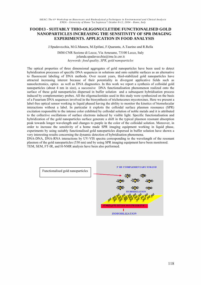
IAEAC: The 6th Workshop on Biosensors and BioAnalytical µ-Techniques in Environmental and Clinical Analysis ENEA – University of Rome “La Sapienza”: October 8-12, 2004 – Rome, Italy
118
FOOD13 - SUITABLY THIO-OLIGONUCLETIDE FUNCTIONALISED GOLD NANOPARTICLES INCREASING THE SENSITIVITY OF SPR IMAGING
EXPERIMENTS. APPLICATION IN FOOD ANALYSIS
J.Spadavecchia, M.G.Manera, M.Epifani, F.Quaranta, A.Taurino and R.Rella
IMM-CNR Sezione di Lecce, Via Arnesano, 73100 Lecce, Italy [email protected]
keywords: food quality, SPR, gold nanoparticles
The optical properties of three dimensional aggregates of gold nanoparticles have been used to detect hybridization processes of specific DNA sequences in solutions and onto suitable surfaces as an alternative to fluorescent labeling of DNA methods. Over recent years, thiol-stabilized gold nanoparticles have attracted increasing interest because of their potentiality in divergent applicative fields such as nanoelectronics, optics as well as DNA diagnostics. In this work we report a synthesis of colloidal gold nanoparticles (about 4 nm in size), a successive DNA functionalisation phenomenon realized onto the surface of these gold nanoparticles dispersed in buffer solution and a subsequent hybridisation process induced by complementary probes. All the oligonucleotides used in this study were synthesized on the basis of a Fusarium DNA sequences involved in the biosynthesis of trichotecenes mycotoxines. Here we present a label-free optical sensor working in liquid phased having the ability to monitor the kinetics of biomolecular interactions without a label. In particular it exploits the colloidal surface plasmon resonance (SPR) excitation responsible to the intense color exhibited by colloidal solution of noble metals and it is attributed to the collective oscillations of surface electrons induced by visible light. Specific functionalisation and hybridization of the gold nanoparticles surface generate a shift in the typical plasmon resonant absorption peak towards longer wavelength and changes to purple in the color of the colloidal solution. Moreover, in order to increase the sensitivity of a home made SPR imaging equipment working in liquid phase, experiments by using suitably functionalised gold nanoparticles dispersed in buffer solution have shown a very interesting results concerning the dynamic detection of hybridisation phenomena. DNA-DNA, DNA-RNA interactions by UV-VIS spectra corresponding to the wavelength of the resonant plasmon of the gold nanoparticles (530 nm) and by using SPR imaging equipment have been monitored. TEM, SEM, FT-IR, and H-NMR analysis have been also performed.
Functionalised gold nanoparticles

IAEAC: The 6th Workshop on Biosensors and BioAnalytical µ-Techniques in Environmental and Clinical Analysis ENEA – University of Rome “La Sapienza”: October 8-12, 2004 – Rome, Italy
119
FOOD14 - DEVELOPMENT OF AN IMMUNOSENSOR FOR AFLATOXIN B B1 B IN BARLEY
Nagwa H. S. AmmidaP
*P, Laura Micheli and Giuseppe Palleschi
P
* PDepartment of Chemistry, Garyounis University, Benghazi-Libya
Dipartimento di Scienze e Tecnologie Chimiche, Università di Roma “Tor Vergata”, Via della Ricerca Scientifica, 00133 Rome, Italy.
e-mail: <[email protected]> keywords: Aflatoxin B1, ELISA, screen printed electrodes, food
Aflatoxin is a metabolite of Aspergillus Flavus and Aspergillus parasiticus. The limit prescribed by the World Health Organization (WHO) for aflatoxin BB1B in various foodstuffs is 5 ng/mL. A strong legislation in Europe has been put in place in order to limit the intake of aflatoxins (e.g. Directive 98/1525/EEC setting limits in food products and Directive 98/53/EEC sampling and analysis). Maximum residue limits of 2 ppb aflatoxin BB1B in food products P
[1]P.
Cereal and their products are susceptible to mould and aflatoxin contamination during pre- as well post- harvest stages of production, therefore, many methods have development for the determination of aflatoxin BB1B. The AOAC official analytical method for determination of aflatoxins in grains is lengthy and requires the use of harmful solvents. Our goal is the assembly of a disposable electrochemical immunosensor based on the indirect competitive enzyme linked immunosorbent assay (ELISA), for simple and fast measurement of aflatoxin BB1 B (AFBB1B) in barley using differential pulse voltammetry (DPV). The immunosensor strip was assembled immobilising the biological component (i.e. the AFBB1B conjugated to bovine serum albumin, incubation the sample (or standard) with the monoclonal antibody anti-AFBB1B (MAb). A spectrophotometric ELISA was used in a preliminary phase of development, prior to transferring the assay to the SPEs. Results showed a detection limit of 20 pg/mL and 30 pg/mL for the spectrophotometric ELISA and the electrochemical immunosensor, respectively. The extraction efficiency and the matrix effect have been evaluated by spiking blank barley with AFBB1 B before and after the sample treatment. After treatment, samples were analysed using a 1:10 v/v dilution in PBS (phosphate saline buffer pH 7.4) in order to minimise the matrix effect. Good recoveries were obtained, which demonstrated the suitability of the proposed method for routine screening of AFBB1 B in barleyP
[3]P.
[1] http://Twww.aflatoxin.info/aflatoxin.aspT: properties of aflatoxin and it producing fungi. [2] Nagwa H. S. Ammida, Laura Micheli and Giuseppe Palleschi,P
“PElectrochemical Immunosensor for
Determination of Aflatoxin BB1B in Barley”, Anal. Chim. Acta. 520(2004)159-164.

IAEAC: The 6th Workshop on Biosensors and BioAnalytical µ-Techniques in Environmental and Clinical Analysis ENEA – University of Rome “La Sapienza”: October 8-12, 2004 – Rome, Italy
120
MED01 - FLUORESCENT BACTERIA SENSING IN IRON POLLUTED MEDIA
A.POIATAP
aP, A.VLAHOVICI P
bP, D.E.CREANGAP
bP and P.TUPUP
bP
P
aPUniv. of Medicine and Pharmacy "Gr. T. Popa", Fac. of
Pharmacy, 17 University Street, Iasi, Romania P
bPUniv. Al. I. Cuza, Fac. of Physics, 11 A Bd. Carol I, Iasi – 6600 (Romania)
[email protected] keywords: Bacterial siderophore, Medicine, Fluorescence sensing,
WaterfFerrofluid, Antimicrobial activity
Pseudomonas species are able to colonize various ecological niches being present in the living tissues of plants, animals and humans [1]. Pseudomonas aeruginosa, known as a human pathogen, is a fluorescent bacteria due to the pyoverdine pigment, synthesized as a siderophore with antimicrobial activity, significant subject for the microbiological research in medicine [2]. We have arranged a sensing system in order to detect the influence of iron oxides on the pyoverdine siderophore (synthesized by Pseudomonas aeruginosa withdrawn from biological specimens) and to assess its antimicrobial activity. Iron oxides have delivered in the bacterial culture media by means of small aliquots of water-based ferrofluid simulating iron pollution that can affect environment as well as human body. The fluorescence emission (excited by using a Xenon lamp) presented a large band with a maximum at 450 nm. Ferrofluid concentrations of 0.0015 ml/l up to 0.12 ml/l had a stimulatory effect on the bacterial metabolism as revealed by enhanced fluorescence intensity while concentrations of 0.5 ml/l-1.0 ml/l exhibited toxic effect diminishing pyoverdine fluorescence toward the control sample level. Antimicrobial activity of pyoverdine (extracted after thermal treatment, centrifugation and dilution) was recorded against Sarcina lutea and Staphylococcus aureus using agar diffusion method. The correlation between the ferrofluid concentration and the diameter of the growth inhibition area was studied in each case. The sensing system adaptation for continuous flow assay was discussed. References [1] Corneli, P., Matthijs, S., Environmental Microbiology, 4 (12): 787, 2002 [2] Handfield, M., Lehoux, D.E., Sanschagrin, F., Mahan, M.J., Woods, D.E., Levesque, R.C., Infect. Immun 68: 2359-2362, 2000

IAEAC: The 6th Workshop on Biosensors and BioAnalytical µ-Techniques in Environmental and Clinical Analysis ENEA – University of Rome “La Sapienza”: October 8-12, 2004 – Rome, Italy
121
0 25 50 75 100 150 2505
30
150
0
500
1000
1500
2000
2500
3000
δO
D, r
elat
ive
units
Сoncentration, µг/мл
time,
min
TFigure:T Dependence of the change in the magnitude δODBcontrol B-δODBexperiment B at an orienting field frequency of 52 kHz in viable K-12 cells suspended in distilled water (conductivity, 1.8 S mP
-1P),
obtained after incubation with various concentrations of ampicillin for various times.
MED02 - EFFECT OF AMPICILLIN ON THE ELECTROPHYSICAL CHARACTERISTICS OF ESCHERICHIA COLI
O.I.Guliy P
aP, O.V.IgnatovP
aP, L.N.MarkinaP
aP, V.D.BuninP
bP and V.V.IgnatovP
aP
P
1P Institute of Biochemistry and Physiology of Plants and Microorganisms, Russian Academy of
Sciences, 13 Pr. Entuziastov, 410049 Saratov Russia P
2P Institute of Applied Microbiology, Obolensk, Russia
[email protected] Twww.ibppm.saratov.ru/T
keywords: Electroorientation, Orientational spectrum, Ampicillin; Plasmid
We examined the effect of amoxicillin on the electrophysical characteristics of ampicillin-sensitive and ampicillin-resistant Escherichia coli cells. Substantial changes in the orientational spectra (OS) of suspensions of cells incubated with various ampicillin concentration took place only at the first five frequencies of the orienting electric field (10—1000 kHz). The maximal change in the magnitude of the electro-optical si nal occurred at 50 g/mlP
-1P of
ampicillin. The suspension-OS changes did not depend on the antibiotic-action period. Under the action of ampicillin, sensitive and resistant E. coli strains gave different electro-optical (EO) effects. After ampicillin incubation of the sensitive strains, K-12 and XL-1, there occurred a considerable change in the EO-signal magnitude. Suspensions of the resistant strains, K-12 (pUC-18) and XL-1 (pHEN1), showed no changes in this EO parameters after ampicillin incubation. Thus, the suspension-OS changes occurring under the effect of ampicillin may be used as a test for resistance to this antibiotic in the cells studied. The possibility is suggested of using electrophysical methods to study the mechanism of antibiotic action on bacterial cells, with a view to check antibiotic action of microorganisms.
This work was supported in part by Russian Science Support Foundation, and CRDF grant N REC-006.

IAEAC: The 6th Workshop on Biosensors and BioAnalytical µ-Techniques in Environmental and Clinical Analysis ENEA – University of Rome “La Sapienza”: October 8-12, 2004 – Rome, Italy
122
MED03 - DETERMINATION OF BIOMARKERS IN HUMAN BREATH BY
TDS/GC/MS
A.GrundmannP
1P, R.BandurP
1P, and Th.HoffmannP
2P
P
1PISAS - Institute for Analytical Sciences ISAS, Bunsen-Kirchhoff-Str. 11, D-44139 Dortmund
(Germany) P
2PJohannes Gutenberg-Universität, Institut für Anorganische Chemie und Analytische Chemie,
Duesbergweg 10-14, D-55128 Mainz (Germany) [email protected]
Twww.ansci.deT keywords: Medicine, Biomarkers, TDS/GC/MS, VOCs
Analytical tools such as gaschromatography / mass spectrometry (GC/MS) are increasingly established for new applications in environmental analysis, life sciences and also in medicine. The purpose of this work is to improve established methods or to create new methods which will be easier to handle, faster in their analysis, more sensitive and well-priced. Analysts as well as physicians hope to get information for an early diagnosis of lung diseases and lung cancer by the analysis of volatile organic compounds (VOCs) in human breath [1]. One of the main interests for the development of a new, suitable analytical method is an effective drying step for the breath sample in order to avoid too much water in the GC-system. This aim can be reached by condensation effects, by the use of diffusion and adsorption techniques and by the changing of physical parameters. In this drying step it must be ensured that hydrophilic analytes such as alcohols, aldehydes and ketones neither get lost nor be destroyed. Up to now it is not possible to find concrete and unanimous biomarkers for special diseases. Literature offers possibilities in between hundreds of compounds (alkanes, alcohols, aromatic compounds, terpenes, etc.) [2], statistical information for special groups of compounds (alkanes, aromatic compounds) [3] and more detailed lists for small analytes (ethane, isoprene, pentane, etc.) [4]. Therefore samples have to be checked very carefully and one has to take into consideration which analytes may be possible biomarkers because of special frequency or proportion among each other. [1] Kharitonov, S.A. and Barnes, P.J. (2002): Review, Biomarkers of some pulmonary diseases in exhaled
breath, Biomarkers, Vol. 7, NO. 1, 1-32. [2] Phillips, M.; Herrera, J.; Krishnan, S.; Zain, M.; Greenberg, J. and Cataneo, R.N. (1999): Variation in
volantile organic compounds in the breath of normal humans, J. Chromatogr. B, 729, 75-88. [3] Natale, C.D.; Magagnano, A.; Martinelli, E.; Paolesse, R.; D’Arcangelo, G.; Roscioni, C.; Finazzi-Agrò,
A.and D’Amico, A. (2003): Lung cancer identification by the analysis of breath by means of an array of non-selective gas sensors, Biosensors and Bioelectronics, 18, 1209-1218.
[4] Lärstad, M.; Loh, C.; Ljungkvist, G.; Olin, A.-C. and Torén, K. (2002): Determination of ethane, pentane and isoprene in exhaled air using a multi-bed adsorbent and end-cut gas-solid chromatography, Analyst, 127, 1440-1445.
Figure: Breath sample of a female non-smoker measured with thermodesorption/ gaschromatography / mass spectrometry

IAEAC: The 6th Workshop on Biosensors and BioAnalytical µ-Techniques in Environmental and Clinical Analysis ENEA – University of Rome “La Sapienza”: October 8-12, 2004 – Rome, Italy
123
MED04 - ELECTROCHEMICAL ENZYME-FREE UREA SENSOR FOR CLINICAL ANALYSIS
A.N.KozitsinaP
aP and M.S.OslinaP
aP
P
aPUrals State University of Economics. 8P
th Pof March St., Bld.62, 620219 Ekaterinburg (Russia)
[email protected] [email protected]
keywords: Medicine, Electrochemical sensors, Screen-printed electrodes
Determination of the urea concentration has acquired great significance in the clinic of internal diseases for diagnosis of renal abnormalities. The problem of evaluating the hemodialysis efficiency (which is judged by the urea concentration of the dialysis liquid) is very topical. Important parameters are the variation dynamics of the urea concentration during the procedure and the level of urea in blood serum before and after dialysis. The urea concentration is most frequently determined using a catalytic reaction with urease and subsequent measurement of the concentration of ammonium ions. An advantage of such sensors is their high selectivity. However, time instability of the enzyme and the need of a substrate invoke difficulties in the use and storage of the sensors. We proposed a new electrochemical sensor for determination of urea in biological materials. This sensor is free of an enzyme (urease) and is based on a solid-state catalytic reaction. Electrodes made by the thick-film technology served as transducers. Graphite-containing ink (TUE) comprised the electroconductive layer. The graphite-containing layer was modified by adding an organic solution of the transducer into the volume of the graphite-containing compound. The organic solution of the transducer included an insoluble modifier and was free of urease. It was prepared by extraction or dissolution in an organic solvent. To eliminate the effect of interfering compounds present in biological materials, blood serum or dialysate (the object) was passed through an anion-exchange column and then was placed in the cell. The detection limit was 0.008 mM, while the correlation coefficient was 0.9998. Samples of a dialysis liquid, which were taken 10 minutes, 1 hour, 2 and 4 hours after the beginning of the dialysis procedure, were analysed. By way of example, Table 1 gives measured concentrations of urea in the dialysis liquid of a patient. Table 1. Comparative results of analysis of the dialysis liquid of a patient, which were obtained by an independent urease method and the proposed technique
Urea, mM Time passed after the
beginning of dialysis
Urease-containing sensor* Urease-free sensor
10 minutes 11.8±0.2 12.3±0.5 1 hour 4.9±0.1 4.7±0.4 2 hours 3.2±0.1 3.2±0.2 4 hours 2.7±0.1 2.6±0.2
*(Vitros BUN/UREA Slide, Johnson&Johnson Clinical Diagnostics, Inc)
Table 2 gives measured concentrations of urea in blood serum of different patients.
Table 2. Comparative results of analysis of blood serum, which were obtained using an independent urease method and the proposed technique
Urea, mM No. Urease-containing sensor* Urease-free sensor
1 13.3±0.2 12.6±0.6 2 5.8±0.1 6.3±0.5 3 4.8±0.1 5.3±0.6 4 3.3±0.1 2.8±0.5 5 4.0±0.1 4.0±0.2
*(Vitros BUN/UREA Slide, Johnson&Johnson Clinical Diagnostics, Inc.) From Tables 1 and 2 it is seen that the results, which were obtained using the proposed method with the sensor described above and a standard urease method, are in good agreement.

IAEAC: The 6th Workshop on Biosensors and BioAnalytical µ-Techniques in Environmental and Clinical Analysis ENEA – University of Rome “La Sapienza”: October 8-12, 2004 – Rome, Italy
124
MED05 - MODELING AND OPTIMIZATION OF PIEZOELECTRIC MICROPUMPS FOR BIOMEDICAL APPLICATIONS
I.Fuduli, A.Montefusco, E.Morganti, M.Petasecca and G.U.PignatelTTP
a
P
a PUniversity of Perugia, D.I.E.I., via Duranti 95 – 06100 Perugia (Italy)
[email protected] www.diei.unipg.it
keywords: µ-Systems, MEMS, µ-fluidics This work aims at realizing one micropump for biomedical applications whit the following requirements: a flow-rate of 10 ml/s and maximum dimensions of 2-3 cm. This paper describe a method for optimising the performances of piezoelectric micropumps with a circuit simulator as SPICE. The equivalent network of the micropumps is extracted from their geometric characteristics. The idea to construct the equivalent network arises from the analogy between the microfluidics and electrical systems [1]. This method has been applied to two micropumps taken from literature: the first one uses diffuser valves as proposed by Ullmann [2], the other one uses membrane valves as proposed by S. Bohm [3]. We have modelled the different parts of each pump with lumped elements. The electrical equivalent network of the micropump whit diffuser valves is shown in the figure. SPICE's simulation of both models gives results which are in good agreement with data reported in literature and so, we have assumed it as good starting-point for the project. For optimising the micropump's structure, we have acted on valve’s dimensions as well as on the parameters of the membrane and the pump chamber. Micropumps’s sizes can be varied as long as the structural strength is not compromised, in other words, moving parts that sustain a certain mechanical stress such as membranes must not break if their size is changed. For this purpose the mechanical stress in the membrane is maintained below the maximum value of fracture stress limit σ Bf B Valve’s features we can modify are length, section and aperture’s angle. However a limit in performance is given by the efficiency of valve-tube system εBseriesB=(RB+ B-RB- B)/(RB+ B+RB- B+2Btube B), where RB+ B and RB- B are forward and backward valve’s resistance respectively and RBtube B is tube’s resistance. Diffuser valves have very low values of efficiency (ε=0.3 for RB- B=RBtube B). Instead, the efficiency of membrane can be close to unit (ε=0.96 for RB- B=0.13RBtube B). Other parameters critical for the optimisation are thickness and radius of membrane and pump chamber’s volume. Working on these factors, we have shown that, for diffuser micropump, a flow-rate of 450 µl/s could be reached, instead of 35 µl/s reported in literature. The max flow-rate for membrane’s micropump could rise up to 5100 µl/s, compared to the 28 µl/s reported in the literature. In conclusion, we can assert that the circuit simulation is a valid method, even if simplified, to predict the behaviour of micropumps in terms of flow rate. Further accurate analyses aimed to verify these results, according to structural and technologic considerations, are foreseen. [1]. T. Bourouina and J. P. Grandchamp, Modeling micropumps with electrical equivalent networks, J.
Micromech. Microeng., 6 (1996), pp 398-404 [2]. Ullmann and I. Fono, The piezoelectric valve-less pump-improved dynamic model, J.
Microelectromech. Syst., 11 (2002), no. 6 [3]. S. Bohm, W. Olthuis, P. Bergveld, A plastic micropump constructed with conventional techniques
and materials, Sensor and Actuator, 77 (1999), pp 223-228 [4]. R. E. Oosterbroek, “Modeling, design and realization of microfluidics components” Thesis,
MESA+ Research Institute, University of Twente, Enschede, Netherlands 1999
Figure: Electrical equivalent network of micropump

IAEAC: The 6th Workshop on Biosensors and BioAnalytical µ-Techniques in Environmental and Clinical Analysis ENEA – University of Rome “La Sapienza”: October 8-12, 2004 – Rome, Italy
125
MED06 - LASER SENSORS OF TRACE GASES IN HUMAN BREATH
A. PuiuP
aP and G. GiubileoP
bP
P
aPNILPRP, 409 Atomistilor St., PO Box MG-36 Magurele – 077125 Bucharest (Romania)
P
bPENEA, Via E. Fermi 45, Frascati – 00044 Rome (Italy)
[email protected] www.inflpr.ro
keywords: medicine, laser spectroscopy, breath analysis, lipid peroxidation
Ethylene and ethane as biomarkers for lipid peroxidation in humans can be detected in the exhaled breath by means of high sensitivity gas sensors based on high resolution molecular spectroscopy. A laser photoacoustic spectroscopy system (PAS) was used for online ethylene monitoring in the exhaled breath and a TDL absorption spectroscopy (TDLAS) system for ethane detection. The main advantage of the two spectroscopic techniques is the absolutely noninvasive character. The gas sensors built-up at NILPRP Bucharest-Magurele and at ENEA Frascati Molecular Spectroscopy Laboratory give the possibbility to real time monitoring the gas concentrations. The real time acquisition is made by a computer through a National Instruments GPIB interface. The PAS system is based on a frequency stabilised COB2B laser emitting IR radiation at 10.532 µm wavelength. Due to the high sensitivity of the system (under 1 ppbv) we are able to monitor very low concentration modifications of trace ethylene content in the exhaled air following X rays exposure in patiens. The TDLAS system realised in Frascati ENEA Molecular Spectroscopy Laboratory for monitoring ethane traces is based on a tuneable diode laser operating slightly above the liquid nitrogen temperature. The single mode 3.3 µm wavelength laser radiation is revealed by a HgCdTe detector. The electric signal is acquired by a 9354C digital LeCroy oscilloscope and transferred to a PC through a GPIB innterface. The system has been calibrated by a certified mixture. The experimental data are reported and discussed.

IAEAC: The 6th Workshop on Biosensors and BioAnalytical µ-Techniques in Environmental and Clinical Analysis ENEA – University of Rome “La Sapienza”: October 8-12, 2004 – Rome, Italy
126
MED07 - A THIN HG FILM SENSOR BASED ON GLASSY CARBON ELECTRODE FOR CYSTEINE DETERMINATION
M.K.Sezgintürkand E.Dinçkaya
Ege University, Faculty of Science, Biochemistry Department, 35100, Bornova-Izmir, Turkey
[email protected] [email protected]
keywords: Cysteine, Medicine, Electrochemical sensors, Mercury Film Electrodes
Cysteine is a nonessential aminoacid, meaning that cysteine can be made in the human body. Cysteine is one of the few amino acids that contain TsulfurT. This allows cysteine to bond in a special way and maintain the structure of proteins in the body. Cysteine strengthens the protective lining of the stomach and intestines, which may help prevent damage caused by TaspirinT and similar drugs. In addition, cysteine may play an important role in the communication between Timmune systemT cells. The determination of sulfur-containing aminoacids is very important in the clinical and the pharmaceutical fields. The abnormal concentrations of cysteine (usually > 1 mmol/L levels) in urine are good indicators for certain diseases (e.g. hepatic cystinuria) in medical diagnoses. Cysteine is also suitable in the treatment of alopecia and shows complement for the formation, growth and maintenance of hair and nails. Cysteine has an important role in the proper function of the immune system, so a deficiency of this aminoacid may either contribute to, or result from, immune suppression associated from HIV. In this study, glassy carbon electrodes modified with Hg were used as working electrode. Thin mercury films on glassy carbon electrodes coated were deposited by holding the electrode potential at different values and measurement period for coating process was investigated. pH and temperature effects on the electrode response were carried out by working at different pHs and temperatures. The calibration graph for cysteine was drawn. Repeatability and interferences studies were investigated. Finally, the sensor was applied to real samples for cysteine determination and the method was validated by Ellman’s reagent.

IAEAC: The 6th Workshop on Biosensors and BioAnalytical µ-Techniques in Environmental and Clinical Analysis ENEA – University of Rome “La Sapienza”: October 8-12, 2004 – Rome, Italy
127
MED08 - rLCRV FROM YERSINIA PESTIS INHIBITS THE LPS MEDIATED ACTIVATION OF MURINE PERITONEAL MACROPHAGES
R.K.Sharma P
1P, A.SodhiP
1 Pand H.V.BatraP
2P
P
1PSchool of Biotechnology, Banaras Hindu Univ.Varanasi-221 005, U.P, India
P
2PDivision of Microbiology, DRDE, Jhansi Road, Gwalior, India
keywords: macrophages, rLcrV, Yersinia pestis, TNF-α, IFN-γ
LcrV is an enigmatic protein of Yersinia having multiple functions like translocator of other Yops and act as a regulator too. Recent studies have been shown that LcrV is also capable of having direct effect on macrophages functions, which were previously known for effector function. Antibodies against LcrV confer resistance to plague probably due to neutralization of its immunosuppressive effect and/or due to abolished type III translocation. We studied the effect of LcrV (10 µg/ml) using peritoneal macrophages as model system because macrophages are keystone of immune system that provide active immune response by secreting various effector molecules or by phagocytosis in clearing most of the pathogens. We have observed that LcrV inhibits TNF-α, IFN-γ and its related chemokines like KC, IP-10, MCP-1, MIP-1α, MIP-1β and RANTES production in LPS (10µg/ml) stimulated murine peritoneal macrophages whereas transcription of GM-CSF was found to be unaffected in LPS treated macrophages. Moreover LcrV affected the redistribution of actin filaments and lipid rafts on the membrane of macrophages. Further studies are required to unveil the signaling network of the immunomodulation induced by LcrV.

IAEAC: The 6th Workshop on Biosensors and BioAnalytical µ-Techniques in Environmental and Clinical Analysis ENEA – University of Rome “La Sapienza”: October 8-12, 2004 – Rome, Italy
128
NANO01 - A SILICON MICROMACHINED GAS CHROMATOGRAPH FOR VOC MONITORING
A.Benvenuto, A.Adami, V.Guarnieri, L.Lorenzelli and M.Zen P
ITC-irst Microsystems Division, Via Sommarive, 18 – 38050 Trento Italy [email protected]
www.itc.it keywords: Microsystems, Gas Chromatography, Environmental analysis
Gas chromatography systems are important analytical tools for a variety of application fields, including environmental analysis and pollution management. In the last years the demand for using miniaturized portable gas chromatographic systems in hostile locations has grown and much effort has been dedicated to the development of reliable, cost-effective and portable analysis instrumentation [1-3]. Silicon-based technologies are the enabling techniques to realize such miniaturized devices with precision at the micron scale, mass production and integration of detector and other modules. In this work a Silicon micromachined gas chromatograph is presented. The miniaturized device mainly consists of two modules: a.) a silicon micromachined separation column, including inlets/outlet for gas injection system and on-chip housing for the detector; b.) a semiconductor metal oxide detector. A spiral-like column configuration has been designed. Room temperature Deep Reactive Ion Etching (DRIE) and anodic bonding to a Pyrex cover plate have been applied to obtain the Silicon microchannels. The inlets/outlet and the microchamber for the gas sensor have been fabricated on the opposite side of the microcolumn by means of Tetramethyl Ammonium Hydroxide (TMAH) wet etching. A standard gas chromatography procedure has been applied for the stationary phase deposition. Then, a detector consisting of a thick film gas sensor with a polysilicon microheather has been bonded on the top of the outlet by means of a flip-chip technique. Finally, the device has been packaged to provide electrical and fluidic connections and preliminary measurements to test the capability of the microcolumn to separate volatile organic compounds have been performed. The proposed microsystem, even if it is at early stage of development, represents a first step for the realization of a highly-integrated, portable and potentially low-cost miniaturized device for gas chromatography applications. [1] R. L. Grob (ed.), Modern practice of gas chromatography, John Wiley and Sons, Inc. (1995). [2] M. Vedder, Choosing Environmental Lab Instrumentation, Pollution Energy, 39, Nov. 1994. [3] R. R. Reston, E. S. Kolesar, Silicon-micromachined gas chromatography system used to separate and
detect ammonia and nitrogen dioxide – Part I: Design, fabrication, and integration of the gas chromatography system, Journal of Electromechanical Systems, 3, 4, 134-146, Dec. 1994.
Figure 1: A.) Front side view of the separation microcolumn prototype (2 cm x 3cm) with the detail of the microchannels (length: 1.3 m, width: 200 µm, depth: 20 µm; spacing: 100 µm); B.) Back side view of the microcolumn with the gas sensor assembled.

IAEAC: The 6th Workshop on Biosensors and BioAnalytical µ-Techniques in Environmental and Clinical Analysis ENEA – University of Rome “La Sapienza”: October 8-12, 2004 – Rome, Italy
129
NANO02 - NANOSTRUCTURED POLYMERIC THIN FILMS:
PREPARATION, ESR AND ATR-FTIR INVESTIGATIONS
Simina DreveP
1 P, G.Damian P
2 P, O.CozarP
2P and Margareta BakoP
2P
P
1 PNational R&D Institute of Isotopic and Molecular Technologies, P.O. Box 700 - R-3400
Cluj-Napoca 5 (Romania) P
2P'Babes-Bolyai' University, Faculty of Physics, 1 Kogalniceanu st., 3400 Cluj-Napoca (Roumania)
[email protected] keywords: Nanotechnology,Polyvinyl-formaldehyde, Protoporphyrin, Thin films
Understanding of a complex correlation between polymer structure and its properties is the key to the polymer-specific morphology design ready to meet new technological requirements such as “smart” polymers, electro-optical materials, artificial organs, antireflection surface coatings, permselective membranes and drug delivery devices. One application which is frequently mentioned for nanometer-sized semiconductor crystals is the conversion of the optical into electric signals. At the basis of such systems are the nanostructured thin layers of polymers, co-polymers or polymeric composites, following the application purpose. Nitroxyl radicals, which are dispersed (spin probe) in polymer matrix or covalently bonded to polymer chains (spin label), are sensitive to the environment P
[1]P
Therefore, molecular motion and microstructure of polymer systems can be identified from the ESR spectra. The paper describes the results of Electron Spin Resonance (ESR) and Attenuated total reflection Fourier transform infrared spectroscopy (ATR-FTIR) investigations of the following polymeric films: polyvinyl-formaldehyde, polyvinyl-formaldehyde + protoporphyrin + tempo (thin and bulk), and polyvinyl-formaldehyde+tempo (some polymeric films made by combining in equal proportions polyvinil-formaldehyde resin with protoporphyrin and tempo as nitroxidic compound for investigating the microstructure of the prepared copolymeric films). Such thin films area routinely prepared in laboratories P
[2]P by controlled allowance of the polymeric solution from a separation funnel, by adjusting the valve. A
clean piece of thin glass is mounted on the reservoir, and the evaporation of the solvent leave a thin polymeric layer of high homogeneity wich sticks to the surface. Polyvinyl-formaldehyde was used as skeleton resin in surface and bulk nanostructured materials because of the good physico-chemical properties for multifunctional nanostructured systems P
[3]P.
Simulation of the composite ESR spectra with a set of particular components was used to determine a fraction of slow/fast components in spectra of nitroxyl radicals. The combination of the spectra of known proportions is also used to simulate the two-component spectra of immiscible blends, although the experimental spectra with the predominantly fast tumbling probes cannot be reproduced by a simple combination.
[1] Damian,G., Miclaus,V., Radicali nitroxidici, Ed. EFES, Cluj-Napoca,2001 [2] Krist, Th., Mertens, P., Biersack, J.P., Nucl. Instr. Meth., B2 (1984) 177-181. [3] Simina Dreve, E. Indrea, I. Bratu, Margareta Bako, Gh. Mihailescu, Liliana Olenic, Stela, Pruneanu, V. Znamirovsky, L. Barbu-Tudoran, Studia Universitatis “Babes-Bolyai”, Special Issue, 1 (2003) 381-384.

IAEAC: The 6th Workshop on Biosensors and BioAnalytical µ-Techniques in Environmental and Clinical Analysis ENEA – University of Rome “La Sapienza”: October 8-12, 2004 – Rome, Italy
130
NANO03 - CARBON NANOTUBES-MODIFIED SCREEN-PRINTED ELECTRODES FOR CHEMICAL SENSORS AND BIOSENSORS
Marek TrojanowiczP
1P, Ashok MulchandaniP
2P and Marco MasciniP
3P
1. Department of Chemistry, Warsaw University, Pasteura 1, 02-093 Warsaw,
[email protected] 2. Department of Chemical and Environmental Engineering, University of California, Riverside, CA
92521, USA 3. Department of Chemistry, University of Florence, Via Lastruccia 3, I-50019 Sesto Fiorentino,
Florence, Italy
keywords: Nanotechnology, Electrochemical sensors, Screen-printed electrodes The screen-printed technology is widely employed in design of chemical andbiochemical sensors, as technology inexpensive in mass production and easy in building of multiplayer structures of sensing devices. In the most common configuration of sensing strips, the working electrode is produced of carbon (graphite) ink, screen-printed on separate strips with various polymeric or ceramic support or together on the same strip with other electrodes needed for complete electrochemical measuring cell. The way of preparation of such a working electrode do not allow to obtain a working electrode of well defined surface, hence different pretreatment procedures are recommended to improve their properties as thermal treatment, pre-anodization or cyclization in various media. These procedures most often, however, are not sufficient to provide working electrodes of satisfactory electron transfer properties.
Since their discovery by Iijima in 1991 carbon nanotubes are widely examined not only for nanotechnology use, but numerous analytical and electroanalytical applications were also already reported. They include modification of working electrodes for voltammetry, preconcentration of trace analytes, immobilization of biomolecules, preparation of gas sensors and as nanoprobes in atomic force microscopy. In these studies we have found that covering the surface of the carbon ink screen-printed electrode with a layer of a multi-walled carbon nanotubes (MWNT), significantly improves reversibility of electron processes. The carbon nanotubes have been prepared by chemical vapor deposition method, and modification of screen-screen printed graphite electrodes has been made by evaporation on the graphite surface a suspension of MWNT in dimethylformamide. The effect of the modification on reversibility of electrode process of the system hexacyanoferrate(II)/(III) was examined.
The MWNT-modification of working electrode has also a significant, positive effect on functioning of amperometric biosensors for organophosphorus pesticides with immobilized organophosphorus hydrolase by adsorption on the working electrode surface. Depending on amount of nanotubes deposited on the electrode surface, an increase of detection sensitivity of paraoxon has been found.
As another example of chemical sensing in solution, the electrocatalytic amperometric sensing of methanol in millimolar concentration range, has been also developed with the use of screen-printed graphite ink sensor, modified with MWNT for measurements in the presence of Co(II) salt in measuring solution.

IAEAC: The 6th Workshop on Biosensors and BioAnalytical µ-Techniques in Environmental and Clinical Analysis ENEA – University of Rome “La Sapienza”: October 8-12, 2004 – Rome, Italy
131
NANO04 - CHARACTERISATION OF TETHERED BILAYERS ON GOLD ELECTRODES AS MODELS OF BIOMIMETIC MEMBRANES
C.Guidotti, R.Guidelli, M. Mascini and M.R.Moncelli
Dipartimento di Chimica, Università di Firenze, via della Lastruccia 3, 50019 Sesto F.No (Fi), Italy [email protected]
keywords: nanotechnology, self-assembled layers, membranes, electrochemical sensors
Tethered lipid bilayers (tBLMs) on gold electrodes represent an important step toward the biomimesis of biological membranes. A thiolipid consisting of a lipid tail and a hydrophilic part (terminating with a sulfhydryl or disulfide group) was attached to a solid substrate through covalent bonding by a self-assembly process [1,2]. Such a thiolipid creates an amphiphilic supramolecular array of a lipid film with a hydrophilic submembrane space, mimicking the cytosol. Self-assembled monolayers (SAMs) tethered to gold electrodes were prepared with different thiolipids while tBLMs were prepared by adding a phospholipid molecule to the SAMs. The greater robustness of these tethered molecules is related to the covalent attachment of the linker on the surface. Metal surfaces allow for the investigation of the lipid bilayer under electric defined conditions. Preliminary results of various tBLMs on gold electrodes are reported. The stability and electrical properties of these tBLMs were tested using AC-voltammetry, electrochemical impedance spectroscopy (EIS) and other electrochemical techniques. After reconstitution of tBLMs with valinomycin the transport of K P
+P ions
by the carrier across the membrane was studied in an aqueous solution of KCl. Electrochemical measurements show that the thiolipid structure plays a very important role in determining the stability as well as the characteristics of the tBLMs. Furthermore the morphology of the gold substrate has an essential role in the formation of the SAMs. Different preparations techniques of tBLMs on gold electrode are reported.
[1] R. Guidelli, L. Becucci, Q. Liu, R. J. Bushby and S. D. Evans; J. Phys. Chem. B, 106 (2002) 10410-10416
[2] S. M. Schiller, R. Naumann, K. Lovejoy, H. Kunz and W. Knoll, Angew. Chem. Int. Ed., 42 (2003), 208-211

IAEAC: The 6th Workshop on Biosensors and BioAnalytical µ-Techniques in Environmental and Clinical Analysis ENEA – University of Rome “La Sapienza”: October 8-12, 2004 – Rome, Italy
132
NANO05 - STUDY OF LANGMUIR MONOLAYERS AND LB FILM CONTAINING DRUG MOLECULES FOR BIOSENSOR APPLICATIONS
S. Morandi, M. Suggelli and G. Caminati
Department of Chemistry, Via della Lastruccia,3 Sesto Fiorentino-50019 Firenze (Italy) [email protected]
keywords : nanotechnology, druf residues in food, LB Film, cyclic voltammetry. Rifamycin SV, Rifaximin (Rfx) and Rifampicin (Rfp) are macrocyclic antibiotics extensively used in veterinary medicine. The presence of significant amounts of these drugs in animal-derived food is harmful for the human health [1] and current legislation imposes extremely low limits, i.e. 10P
-7P M, for these drugs in
milk [2]. A sensitive and fast method is needed to determine such small concentrations of the antibiotics. The long-term goal of this work is the realization of a specific sensor for the rifamycins family by means of Langmuir-Blodgett (LB) nano-films of phospholipids. In fact, LB technology has been considered as a convenient tool for designing artificial system with biomembrane architecture and function. Since rifamycins are known to penetrate the bacterial membrane interacting with its lipid constituents, we chose phospholipid molecules as LB forming compounds. The investigation of Surface Pressure-Area (π-A) and Surface Potential-Area (∆V-A) isotherms of Dipalmitoylphosphatidylglycerol Sodium Salt (DPPG-Na) in the presence of the antibiotics in the subphase allowed to determinate the concentration of Rifamycins in the monolayer at the air/water interface. Moreover, the orientation of the molecules in the DPPG-Na monolayer was determined by the comparison between semi-empirical calculations and surface potential measurements. The data were correlated to the results obtained from the Equation of State for adsorption films of rifamycins. The characterisation of Rfs in water solutions and at the air/water interface with DPPG Na monolayers has been performed also by Cyclic Voltammetry measurements and Brewster Angle Mycroscopy images. The interaction between LB films of DPPG-Na and the antibiotic molecules was characterized by UV-Vis spectroscopy (fig.1). [1] N.Craven, J.C.Amderson, Res. Vet. Sci., 31 (1981) 295. [2] K.K.Manuilov, E.V. Gagaeva, Antib. Kemoter., 34 (1989) 682.
Fig 1. UV-Visible Spectra of 1LB of DPPG Na trasferred at 35 mN/m (dotted line) and at 25 mN/m (dashed line) incubated in 1x10P
-5P M Rfp solution compared with UV-
Visible spectra of 1x10P
-5P M Rfp solution

IAEAC: The 6th Workshop on Biosensors and BioAnalytical µ-Techniques in Environmental and Clinical Analysis ENEA – University of Rome “La Sapienza”: October 8-12, 2004 – Rome, Italy
133
NANO06 - MEASUREMENTS OF pH, POTASSIUM, CALCIUM AND AMMONIUM ON TERRESTRIAL ROCKY SUBSTRATA AND BIOFILMS
S.PiermariniP
aP, M.VolpiniP
bP, J.Calvo QuintanaP
a, PP.AlbertanoP
bP, G.PalleschiP
a Pand D.MosconeP
aP
P
aPDepartment of Chemical Sciences and Technologies and P
bPDepartment of Biology,
via della Ricerca Scientifica– 00133 Rome (Italy) [email protected]
keywords: Potentiometric microsensors, Roman catacombs, Cyanobacterial biofilms, Monochromatic lamps.
Microorganisms inhabit exposed surfaces of Roman catacombs where they play a role in deterioration processes of rocky substrata and influence the biogeochemical cycles [1]. To achieve a better understanding of bio-transformation and bio-decay processes of lithic faces caused by the growth of biofilm-forming cyanobacteria in hypogean monuments, and to evaluate the applicability of new non-invasive methods for monitoring the growth of these microorganisms, amperometric and potentiometric microsensors were recently developed and applied to stone substrata and microbial communities [2-5]. The ionic concentration of calcium, potassium and ammonium and pH values were measured within the soluble fraction of biofilms freshly collected from Roman catacombs using potentiometric microsensors [3-4]. Measurements of calcium, potassium and ammonium ions were also carried out on biofilms collected at the same sites and grown for nine months on calcareous slides in laboratory conditions under different light wavelenghts (white, orange, green, and blue). To evaluate the effect of monochromatic lights on ions mobilisation within the biofilms, the measurements were carried out in the dark for one hour. Uncolonised (and sterilised) rocky substrata were used as control. The measured pH values were lower in the uncolonised substrata than in the colonised ones. This fact could probably be due to the effect of photosynthetic activity that produces high amounts of hydroxil ions, thus resulting in a pH increase due to the consumption of COB2 B and HCOB3PB
-P.
Concerning laboratory grown biofilms, the calcium ions in uncolonised and colonised substrata were quite similar when the biofilms were grown under white and orange lights, while higher values were found in the control under blue light. The same situation was detected for potassium and ammonium concentrations, albeit control values were higher then in biofilms also under green light. In general, a high dispersion of results was observed in these experiments, probably caused by the heterogenity of the biofilms. In fact, by measuring the same analyte in different spots of the same biofilm relevant differences of the signal were often observed. Nevertheless, these data provided a first indication on the concentration of specific elements that are mobilised within phototrophic biofilms through microbial metabolism and reference values for the uncolonised, thus undamaged, substrata. Information on the extent to which different lighting conditions can influence chemical species variation was gained also for the first time. This allowed to develop a new low-invasive method for the monitoring of cyanobacterial biofilms through the construction of a “Portable Sensor Monitor” holding together the different microsensors for the measurement of threshold values of chemical species on endangered stones. This work was supported by the EU Programme, Energy, Environment and Sustainable Development in the frame of CATS Project, contract n° EVK4-CT2000-00028. [1] Albertano P., MosconeP
PD., Palleschi G., Hermosín B., Saiz-Jimenez C., Sánchez-MoralP
P S., Hernández-
Mariné M., Urzì C., Groth I., Schroeckh V., Saarela M., Mattila-Sandholm T., Gallon J.R., Graziottin F., Bisconti F., Giuliani R., 2003. In C. Saiz-Jimenez (ed.), Molecular Biology and Cultural Heritage, pp. 151-162, Swets & Zeitlinger B.V., Lisse (NL).
[2] Albertano P., Bruno L., D’Ottavi D., Moscone D., Palleschi G., 2000. J. Appl. Phycol., 12: 379-384. [3] Piermarini S., Quintana Calvo J., Palleschi G., Albertano P., Moscone D., 2001. In: Alpuente et al
(eds.), Science and Tecnology for the safeguard of Cultural Heritage in the Mediterranean Basin, p.276, Universidad de Alcalà Publ.
[4] Calvo Quintana J., Piermarini S., Albertano P., Palleschi G., Moscone D., 2002. In: C. di Natale, A. D’amico, L. Dori, G.C. Cardinali e S. Nicoletti (eds), Sensors and Microsystems, pp. 270-274, World Scientific, New Jersey.
[5] De Beer D., Schramm A., Santegoeds C.M., Kühl M., 1997. Appl. Env. Microb., 63 (3): 973-977.

IAEAC: The 6th Workshop on Biosensors and BioAnalytical µ-Techniques in Environmental and Clinical Analysis ENEA – University of Rome “La Sapienza”: October 8-12, 2004 – Rome, Italy
134
NANO07 - GLASS MICROCHANNEL TECHNOLOGY FOR CAPILLARY ELECTROPHORESIS
L.M.StrambiniP
aP, M.PiottoP
bP and A.NanniniP
aP
P
aPDipartimento di Ingegneria dell’Informazione, Università di Pisa, Via G. Caruso – 56122 Pisa (Italy)
P
bPIEIIT, Sezione di Pisa, CNR, Via G. Caruso – 56122 Pisa (Italy)
[email protected] Twww.iet.unipi.itT
keywords: µ-Systems, capillary electrophoresis, µ-fluidics, glass micromachining
The crossover of microfabrication technology from the integrated circuit industry into analytical chemistry has led to a considerable increase in the number of integrated microfluidic devices, known as micro total analysis systems (µTAS) or lab on a chip (LOC), which are able to perform analytical functions required by an analysis. Among them, chips for chemical analysis have been considered one of the most promising due to their short analysis times, high separation efficiency, high sample throughput, minute samples and reagents consumption and easy automationP
[1,2]P. In these applications, glass is the preferred material due to its
good optical properties, well-known surface characteristics and high breakdown voltageP
[3]P. Differently from
silicon, the micromachining of glass is not already standardized especially for what concerns the etching mask and bonding. In this work, we present the fabrication process of a device for capillary electrophoresis made up of two 26 x 40mmP
2P glass substrates each of them 800 µm thick. Five microchannels, four for samples injection and
one for the separation, have been fabricated on a substrate using a selective wet etching. A double layer (800 nm Cr and 1500 nm photoresist) was used as mask while the glass etchant was an aqueous solution of hydrofluoric acid (HF) and nitric acid (HNOB3B). The chromium layer was deposited by means of e-beam evaporation and etched with a ceric ammonium nitrate ((NH)B4BCe(NOB3B)B2B), perchloric acid (HClOB4B) and deionized water mixture. Access points to the channel reservoirs have been provided by 1 mm holes, made in the second substrate by means of diamond drills. Both substrates were then carefully cleaned in a sulfuric peroxide solution at 120 °C for 20 minutes and by successive washings with acetone, ethanol and deionized water in ultrasonic bath. Finally the two glass substrates were joined by means of the fusion bonding technique at 620 °C in a muffle oven. In figure, a SEM photo of the intersecting point between the separation channel and an injection channel is shown. The good quality of the glass etching and lithographic definition allows to obtain a geometrically well-defined sample injection volume. This property is mandatory to perform high resolution electrophoresis analysis using electro-osmotic pumping in a valveless configurationP
[4]P. Also the results of the
bonding process are quite good with no visible separation surface between the two glasses. [1] L. Zhang, F. Dang, Y. Baba, Journal of Pharmaceutical and Biomedical Analysis, 30 (2003), 1645 [2] A. Manz, N. Graber, H. M. Widmer, Sensors and Actuators B, 1 (1990) 244; [3] V. Dolnik, S. Liu, S. Jovanovich, Electrophoresis 2000, 21 (2000) 41; [4] D. Jed Harrison, P. G. Glavina, A. Manz, Sensors and Actuators B, 10 (1993) 107-116
SEM photo of micro-channels intersection after glass etching.
IAEAC: The 6th Workshop on Biosensors and BioAnalytical µ-Techniques in Environmental and Clinical Analysis ENEA – University of Rome “La Sapienza”: October 8-12, 2004 – Rome, Italy
135
NANO08 - CHEMILUMINESCENCE DETECTION OF HERBICIDE MEDIATED INHIBITION OF THYLAKOIDS IN A µ-FLUIDIC SYSTEM
D.G.Varsamis TP
a TP and D.C.Cullen
IBST, Cranfield University, Silsoe, Bedfordshire – MK45 4DT (UK) P
http://www.cranfield.ac.uk/biotech keywords: µ-Systems, Chemiluminescence, Magnetic beads, Pollutant screening
The work presented is part of a European project (Biosensors for Effective Environmental Protection; BEEP) which is aimed at the assembly and application of Photosystem II (PS II)-based biosensors for large scale environmental screening of specific herbicides and heavy metals. Taking into account the large amount of water samples of industrial and urban effluents, ground water and irrigation water which have to be measured, the development of fast and inexpensive tests is very important. A method of detecting herbicides is proposed; the photosynthetic herbicides act by binding to Photosystem II (PS II), a multiunit chlorophyll–protein complex which plays a vital role in photosynthesis. The inhibition of PS II causes a reduced photoinduced production of hydrogen peroxide, which can be measured by a chemiluminescence reaction with luminol and the enzyme horseradish peroxidase (HRP). The sensing device proposed combines the production and detection of hydrogen peroxide in a single flow assay by combining all the individual steps in a compact, portable device that utilises micro-fluidic components. The production of hydrogen peroxide by chloroplasts was investigated, and hydrogen peroxide was detected under illumination with concentrations increasing in a time- and light intensity-dependent manner. The presence of herbicides in samples reduces the hydrogen peroxide measured in a concentration-dependent manner. The integration of the above reactions, that have initially been performed in cuvette assays, has been achieved by designing and constructing a microfluidic device, which consists of a flow-channel constructed of machined Perspex sandwiching a laser cut elastomer spacer/flow-channel in which regions of appropriate reagents are immobilised reversibly. The reversible immobilisation addresses issues of reagent stability and re-generatability. The end device will be responding to a range of pollutants and be used for rapid, low cost pre-screening of large numbers of samples to determine samples that will then undergo more detailed analysis.

IAEAC: The 6th Workshop on Biosensors and BioAnalytical µ-Techniques in Environmental and Clinical Analysis ENEA – University of Rome “La Sapienza”: October 8-12, 2004 – Rome, Italy
136
REC01 - SITE-DIRECTED ANTIBODY IMMOBILIZATION ON GOLD SUBSTRATE FOR IMMUNOSENSOR APPLICATIONS
A.SchmidtP
3P, R. ThampiP
3P, M.S.ThakurP
2P and C.Raman SuriP
1P
P
1PInstitute of Microbial Technology, Sector 39-A, Chandigarh 160 036 (India)
P
2PCentral Food Technology Research Institute, Mysore (India)
P
3PDepartment of Chemistry, EPFL, CH-1015, Lausanne, Switzerland
[email protected] www.imtech.res.in
keywords: Immunosensors, Immobilization, Orientation, Antibodies
A simple method for site-directed antibody immobilization on gold substrate was investigated for immunosensor applications. Acid treated gold surface was refined with protein A via homobifunctional cross linker dithiobissuccinimide propionate (DSP) to achieve uniform, stable, and sterically accessible antibody coating. The density of immobilized antibodies covalently bound to protein A was examined as a function of protein concentration, content of binding groups available, and the reaction time. Modified surface showed high stability in terms of protein retention and no significant loss of activity during their repeated use. Their applications in QCM and SPR based immunosensor systems showed higher sensitivity and stability.

IAEAC: The 6th Workshop on Biosensors and BioAnalytical µ-Techniques in Environmental and Clinical Analysis ENEA – University of Rome “La Sapienza”: October 8-12, 2004 – Rome, Italy
137
REC02 - FLUORESCENCE QUENCHING STUDIES OF CROWN ETHER COMPLEXES -HOST-GUEST INCLUSION SENSORS WITH BOTH
HIGH SENSITIVITY AND SELECTIVITY
K.-H.FellerP
1P, A.BarannP
1P, K.VogelP
1P, K.SchönefeldP
1P, E.WeberP
2P, P.MüllerP
2P
P
1P University of Applied Sciences Jena, Faculty of Medical Engineering, Carl-Zeiss-Promenade 2, D-07745
Jena, GERMANY P
2P Freiberg University of Mining and Technology, Institute of Organic Chemistry, Leipziger Strasse 29, D-
09596 Freiberg, GERMANY [email protected]
keywords: guest-host interaction, fluorescence quenching, crown ether complexes, flavour molecules
Host-guest supramolecular chemistry offers a new very effective tool for the separation of uncharged species, including organic molecules. There are two mainstreams for the application of host-guest supramolecular interactions as sensor materials. On the one hand the inclusion of organic molecules or even small ions changes the weight of such inclusion compounds and can be measured in mass sensitive devices (by means of ultrasonic resonance frequency detuning). On the other hand the fluorescence behavior of marker molecules attached to the backbone of the inclusion molecule (the host) is dramatically changed by sterical conformational changes. We investigate the fluorescence quenching of anthracene and fluorene markers attached to the backbone of host molecules when terpene molecules are used as inclusion (guest) molecule. Both, intensity as well as spectral changes will be shown as being characteristic for the host-guest supramolecular interaction properties. [1] T. H. Brehmer, P. P. Korkas, E. Weber, Sensors & Actuators B 44 (1997) 595 [2] E. Weber, T. Hens, Qi Li, T. W. Mak, Eur. J. Org. Chem. (1999) 1115 [3] E. Weber, T. Hens, T. Brehmer, I. Csöregh, J. Chem. Soc. Perkin Trans. 2 (2000) 235 [4] B. T. Ibragimov, K. M. Beketov, E. Weber, J. Seidel, O. Sumarna, K. K. Mahkamov, K. Köhnke, J. Phys. Org. Chem. 14 (2001) 697 [5] J. Weber, M. Beeg, C. Bartzsch, K.-H. Feller, D. De la Calle Garcia, M. Reichenbächer, K. Danzer, J. High Resol. Chromatogr. 22, 322 - 326 (1999)

IAEAC: The 6th Workshop on Biosensors and BioAnalytical µ-Techniques in Environmental and Clinical Analysis ENEA – University of Rome “La Sapienza”: October 8-12, 2004 – Rome, Italy
138

IAEAC: The 6th Workshop on Biosensors and BioAnalytical µ-Techniques in Environmental and Clinical Analysis ENEA – University of Rome “La Sapienza”: October 8-12, 2004 – Rome, Italy
139
ONE DAY TOUR IN ORVIETO (Sunday 10th, 8 a.m.)
In the fertile valley of the Tiber, amid gentle hills,there is Orvieto, an isolated city, almost inaccessible, with its bell towers, the towers and the maze ofroofs and travertine houses. Orvieto offers to visitorsthe charm of a city founded by the Etrurians, whichwas shaped as it is now in the Middle Ages andduring the Renaissance.
A cultural itinerary to discover the secrets of itschurches, convents, majestic papal residences andto see the numerous parades and recollections ofhistorical events which have been held in the streetsof this Italian treasure for almost one thousandyears. It is also a tasty occasion to try, in typical
inns, the delicious cuisine of the Umbria tradition.
The city of Orvieto exists in symbiosis with the tuff cliff on which it stands, an extraordinary example of the integration of nature by the work of man.
An example of the fact that our ancestors were aware of this relationship between architecture and nature is the inscription on the famous well of San Patrizio which reads: quod natura munimento inviderat industria adiecit (What nature denied for
defence - in this case water - was added by the work of man).
Visiting this town means journeying through history, for traces ofeach and every period of the almost three thousand years of itsexistence crop up everywhere in this preconstituted physical entity.The two statues of Boniface Vlll set in the city gates at the oppositeends of the town suggest an ideal itinerary for the tourist of today,for the Pope entered the town through the Porta Soliana, knownlater, after the "Rocca" or Fortezza dell'Albornoz was built, as PortaRocca, and he left through the Porta Maggiore. Boniface Vlll wasanything but a tourist and had even been Capitano del Popolo inOrvieto, yet somehow these two statues, which earned him no less
than a charge of idolatry, can serve as symbols both of the attention the city of Orvietoso truly merits, and the traditional hospitality of its inhabitants.
Nowadays one no longer has to reach the top of the cliff on a mule, for a modernsystem of "alternative mobility" provides an access to the town that is both easy to useand fascinating with the funicular (run by water in the nineteenth century and nowcompletely modernised) at one end and a lift (to which escalators are to be added) atthe other, signs that the old historical centre, built on a human scale, has beenreturned to citizens and tourists alike and is once more the realm of the pedestrian.
In the early Middle Ages the rock of Orvieto was once more seen as an ideal naturalbulwark and the new urban centre took shape around the year thousand. It reachedits zenith in the thirteenth and fourteenth centuries and its unique urban layout is stillto all extents what it was then.

IAEAC: The 6th Workshop on Biosensors and BioAnalytical µ-Techniques in Environmental and Clinical Analysis ENEA – University of Rome “La Sapienza”: October 8-12, 2004 – Rome, Italy
140
The most representative public buildings - the Town Hall or Palazzo Comunale, thePalazzo del Popolo, the Duomo or Cathedral, and the Palazzo dei Sette rose side by sidewith the older churches such as San Giovenale, Sant'Andrea, - and the convents of SanDomenico, San Francesco, Sant'Agostino and Santa Maria dei Servi, the complex of thePapal Palace - and the private palaces and tower houses of the aristocracy.
The medieval city-state, with Orvieto at the centre of an extensive territory, achieved itshighest civic and political expression in the free commune: the Guilds and Tradesdeveloped, providing the population with a wealth of finely made objects, while life inthe city continued on its busy way, through periods of peace and turmoil, with thepassing of time marked by the strokes of the Clock of Maurizio, the first automaton ofits kind to regulate the working hours.
This year there is the 8th edition of Orvieto con Gusto.
The overall aim of the initiative is to promote the 'piacere del gusto' or joy of tasting realfood' through the support of typical products and wines which are in danger ofdisappearing due to the industrialisation of the food manufacturing process and globalpressures.
Such items which may be saved in time include 'violino di capra al pomodorino alpiennolo' and 'caciocavallo podolico alla mela Rosa dei Monti Sibillini'. Over 400products are in immediate danger of extinction.
At various points there are tables where it is possible to sample flavours that aredisappearing fast from our daily diet and need rediscovering and protecting:
Serate Golose - at the Enoteca Regionale try bread and medieval soups with fine winefrom the 'Strada dei Vini Etrusco-Romana'.
Olive oil tasting - experts take you through extra virgin.
Mercatino dei Sapori - a market in Piazza San Giovanni to discover the history of food and other products from the Orvieto area.
Umbria is one of Italy's most delicious regions and as well as the famous olive oil and wine includes such fine products as Norcia ham and Castelluccio lentils. The typical dishes can be described as bothrefined and rustic and the 9 day Orvieto con Gusto in the first 2 weeks of October isworth a visit. The event revolves around tastings, itineraries, special meals in therestaurants of the city, markets, exhibitions etc.
Thttp://www.discoveritalia.com/cgwe/cittaFaiDaTe.asp?lingua=en&IDcitta=29T

IAEAC: The 6th Workshop on Biosensors and BioAnalytical µ-Techniques in Environmental and Clinical Analysis ENEA – University of Rome “La Sapienza”: October 8-12, 2004 – Rome, Italy
141

IAEAC: The 6th Workshop on Biosensors and BioAnalytical µ-Techniques in Environmental and Clinical Analysis ENEA – University of Rome “La Sapienza”: October 8-12, 2004 – Rome, Italy
142
Author Index
AB GHANI S: O02 ADAMI A. NANO01 ADAMI M. DNA08 TADELOJUT S.B. ENZ04 AGOSTINELLI E. ENZ03 AHN J.M. O38 TAHMADT F. O02 ALKYILMAZ E. O18 ALBERTANO P. NANO06 ALBERTI O. O13 ALBERTI M. IL03 AMAGAI H. ENV23 AMINE A. ENV09 TAMMIDAT N.H.S. FOOD14 ANGELESCU A. ENV14 ANNESINI M.C. O15 TANTIOCHIAT R. FOOD01 ARCARI P. DNA09 ARNAULT I. FOOD02 TASHWINT H. O19 ATEHA D. IL01 TAUGERT J. FOOD02 BACHMANN T.T. O10 TBADEAT M. FOOD03 BAKO M NANO02 BALLARINI P. ENV07 BANCONE M. FOOD07 BANDUR R. ENV05, MED03 BARCELÓ D. O22 TBARCHETTAT S. ENV01, ENV07 TBARONIANT K.H.R. O16 BARRERO-MORENO
J. ENV16
BARUCH A. O09 BATRA H.V. MED08 BATTAGLIA-BRUNET
F. ENV13
TBAUSSANT T T. ENV19 BENVENUTO A. NANO01 BESCHE S. ENV05 BEYENE N. O01 TBLAKE T D.A. O25 TBLAKE T II R.C. O25 BONANNI A. FOOD04 BOTRÉ C. ENZ03 BOTRÉ F. O04 TBRAININAT KH. O32 BRETT C.M.A. O03 BREWSTER J.D. FOOD08 BROUWER A. ENV11

IAEAC: The 6th Workshop on Biosensors and BioAnalytical µ-Techniques in Environmental and Clinical Analysis ENEA – University of Rome “La Sapienza”: October 8-12, 2004 – Rome, Italy
143
BRZÓZKA Z. FOOD05 BRUSCHI M. ENV13 TBULUKINT E. ENV19 BUNIN V.D. ENV21, MED02 BUONANNO F. ENV07 BURGIO M. ENV09 CALEMCZUK R. IL08 CALVO QUINTANA
J. NANO06
CAMINATI G. NANO05 CAMPANELLAT L. O06, FOOD01, FOOD04, FOOD11 CARICATO R. ENV10 CARPICO G. FOOD09 CARRALERO V. ENV27 CARSON D.S. ENV25 CAVALLO S. O06 CECCONE G. ENV16 CHARRIER T. ENV08 CHEREGI M.C. ENV09 CHOUTEAU C. ENV02 CHOVELON J.M. ENV02 CIANCI E. O29 TCIOSEKT TP. T FOOD05 CIRIELLO R. ENV20 CIUMASU I.M. O28 COMPAGNONE T D. O17, O21, FOOD07 CONI E. FOOD09 CONTRACTOR A.Q. ENV18 COZAR O. NANO02 CUBILLANA-AGIULERA
L.M. ENV24
CULLEN D.C. NANO08 DAMIAN G. NANO02 TD'AGOSTINO T G. O13 D’ANNIBALE A. O06 DANET A.F. FOOD03 DANIEL P.H. ENV08 DANIELSSON B. O12 DE GREGORIO M. O21 DE JACO A. ENV26 TDE STEFANO T L. DNA09 DELL’ATTI D. DNA08 DEL CARLO M. O17, O21, FOOD07 DELLA SETA L. FOOD12 TDELIBATOT E. FOOD06 DESCAMPS E. IL08 DICKINSON M. O19 TDICULESCUT V. DNA01 DINÇKAYA E. O18, MED07 DION M. ENV08 DO CARMO LOPES M. DNA06 TDOMINGUEZ T E. IL02 DORÉ C. FOOD02 DOWNARD A.J. O16

IAEAC: The 6th Workshop on Biosensors and BioAnalytical µ-Techniques in Environmental and Clinical Analysis ENEA – University of Rome “La Sapienza”: October 8-12, 2004 – Rome, Italy
144
DRAGONE R. FOOD01 TDREVE T S. NANO02 DURAND M.J. ENV08 TDURRIEUT C. ENV02 TDZANTIEVT B.B. 007 EL KAOUTIT M. ENV24 TENDOT H. ENV03 EPIFANI M. FOOD13 ERDEM A. O11, O33 ERROI E. ENV10 ERTAS N. O18 EZAKI S. O10 FARINA F. ENV26 TFAVEROT G. O06 FERAPONTOVA E.E. IL07 TFERRI T T. O06 TFIRRAOT G. DNA02 TFLORESCUT M. O03 FOGLIETTI V. O29 FOMINA O.G. FOOD10 FRESE I. O28 TFROLIKT J. ENZ02 FUDULI I. MED05 GARGIULI J. IL01 GATTA T. FOOD04 GAUGLITZ G. O22 GILLILAND D. ENV16 GIORDANO M.E. ENV10 TGIUBILEOT G. MED06 GLUSHKO N.I. FOOD10 GONZÁLEZ-CORTÉS
A. ENV27
T GORTONT L. IL07, ENV06, ENZ01 GRILLO C. ENV26 GRIMM V. O10 TGROSMANOVAT Z. ENV04 GRUNDMANN A. ENV05, MED03 GUT M.B. O38, DNA03 GUARNIERI V. NANO01 GUERRIERI A. ENV20 GIUDELLI R. NANO04 GUIDOTTI C. NANO04 GULIY O.I. ENV21, MED02 GURAZADA S. O16 HAASNOOT W. ENV11 THABL T G. O36, O37 THAGHIGHI T B. ENV06 HAY J. O30 , ENV17 HAYASHI T. ENV03 HECK A. ENV28 HENRYA J. ENV02 HIDALGO-HIDALGO DE CISNEROS
J.L. O26, ENV24

IAEAC: The 6th Workshop on Biosensors and BioAnalytical µ-Techniques in Environmental and Clinical Analysis ENEA – University of Rome “La Sapienza”: October 8-12, 2004 – Rome, Italy
145
THINTEMAN T T. O23, ENV28 THOCKT B. IL03 HOFFMANN T.H. ENV05, MED03 HORRY H. ENV08 IGNATIADIS I. ENV13 TIGNATOVT O.V. ENV21, MED02 IGNATOV V.V. ENV21, MED02 ILIE M. O29 IRTH H. ENV11 JAFFREZIC-RENAULT
N. ENV23
TJANKOWIAKT R. O27 JONES R.M. O25 JONSSON G. ENV19 KAISER J. O22 TKALCHERT K. O01, ENV15 KARA J.H. DNA05 KARP M. O08 KASPRZYK A. FOOD05 KAUSCH U. IL03 KETTRUP A.A. O28 KIM B.C. O38, DNA03 KIRGÖZ Ü.A. O20, O24 KLEM K. O34 KLEPS I. ENV14 KNABBE C. O10 TKNEMEYERT J.P. O05, O36, O37 KOLB G. O28 KOOPAL K. O14, ENV12 KOTZIAN P. O01 TKOZITSINA T A.N. MED04 KREJCI J. ENV04, ENZ02, O29 TKRÄMERT P.M. O28 KHURANA M. IL01 KUNZE G. O16 KYRIACOU G. IL01 LAMBERTI A. DNA09 TLATERZAT A. ENV01, ENV07 LAWAL A. ENZ04 LEGIN A. FOOD02 LEIBIGER R. IL03 LEJEUNE M. ENV16 TLETET C. ENV09 LESKINEN P. O08 LI X. O25 TLIONETTOT M.G. ENV10 TLIUT Y. O12 TLIVACHE T T. IL08 LUCARELLI F. DNA04 TLORENZELLIT L. NANO01 LORTAT-JACOBB H. IL08 LUKKIEN E. O14 LÖWE H. O28

IAEAC: The 6th Workshop on Biosensors and BioAnalytical µ-Techniques in Environmental and Clinical Analysis ENEA – University of Rome “La Sapienza”: October 8-12, 2004 – Rome, Italy
146
MAGNO F. FOOD01 TMALÝT J. O29, O34, ENZ02 TMANERAT M.G. FOOD13 MANNELLI I. O35, DNA07 MANSO M. ENV16 TMARCHESINI T G. ENV11 TMARMÉ T N. O05, O36, O37 MARKINA L.N. MED02 MARKUSHIN Y. O27 TMARRAZZAT G. DNA04 MARTINI E. FOOD11 MARTY J.L. ENV23 MASCI A. O29 TMASCINI T MARCO IL05, O35, DNA04, DNA07, DNA08,
ENV19, NANO03, NANO04 MASCINI MARCELLO O17, O21, FOOD07 TMASOJIDEKT J. O34, ENZ02 MATTEI E. O06 MAZZARINO M. O04 MAZZEI F. O04, ENZ03 MEDYANTSEVA E.P. FOOD10 TMENAT M.L. ENV22, ENV27 MERCEY E. IL08 MERIC B. DNA05 MERLI C. O15, ENV08 TMEULENBERGT E.P. O14, ENV11, ENV12 MICELI C. ENV01, ENV07 MICHEL C. ENV13 MICHELI L. FOOD14 TMICHELINI T E. O08 TMINUNNI T M. O35, DNA07, DNA08 TMITSUBAYASHI T K. ENV23 TMIUT M. ENV14 TMONCELLIT M.R. NANO04 MONTEFUSCO A. MED05 MONTEREALI M.R. FOOD12 MONTILLA S. O04, ENZ03 MORALES M.D. ENV22 TMORANDI T S. NANO05 MORETTI L. DNA09 TMORGANTIT E. MED05 TMOSCONE T D. FOOD06, NANO06 MOTTRAM T. FOOD03 MULCHANDANI A. NANO03 MURAMATSU H. ENV03 MÜLLER P. REC02 NAKAYAMA Y. ENV23 NANNINI A. NANO07 TNARANJOT-RODRIGUEZ
I. ENV24
NARDI L. O29 NEDOMA R. O34, ENZ02 NEUFELD T. O09 TNIEMEYERT C.M. IL06
IAEAC: The 6th Workshop on Biosensors and BioAnalytical µ-Techniques in Environmental and Clinical Analysis ENEA – University of Rome “La Sapienza”: October 8-12, 2004 – Rome, Italy
147
NISHIO G. ENV23 NOGUER T. ENV23 NOLTE O. O37 NUDD R. O22 OBERSRIEBNIG S. O31 OLIVEIRA P.S.C. DNA06 OLIVEIRA-BRETT A.M. DNA01, DNA06 OMANOVIC E. ENV15 ORETSKAYA T.S. DNA01 OSLINA M.S. MED04 OZKAN D. O11, O33 OZSOZ M. O11, O33, DNA05 PALLESCHI G. ENV09, FOOD03, FOOD06, FOOD14,
NANO06 TPALOCCI T C. ENV26 PANZAVOLTA F. ENV26 TPAOLI T G.C. FOOD08 TPAPADOPOULOUT-BOURAOUI
A. ENV16
PARK J.H. DNA03 TPASCOT N. O30, ENV17 PAZARLÝOÐLU N. O24 PEELEN G. O14 TPELLEGRINIT G.E. FOOD09 PEPE A. O21 TPERNETTIT M. O15, ENV08 PESAVENTO M. O13 PETASECCA M. MED05 PHILP J. ENV25 PIERMARINI S. NANO06 PIGNATEL G.U. MED05 PINGARRÓN J.M. ENV22, ENV27 PICART P. ENV08 TPIEDADE T TJ.A.P. T DNA06 PILLOTON R. O24, O29, FOOD12 PINNAVAIA T. J. O33, O11 PIOTTO M. NANO07 POIATA A. MED01 PONCELET D. O15, ENV08 TPREININGER T C. O31 PROLL G. O22 PUGGELLI M. NANO05 TPUIUT A. MED06 PUNAKIVI K. ENZ03 QUARANTE F. FOOD13 RELLA R. FOOD13 REN H. ENV03 RENDINA I. DNA09 REVIEJO A.J. ENV22 RHEMREV R. ENV12 RICHMOND S. O19 TRISHPONT J. O09 ROBSON D. O16 RODA A. O08

IAEAC: The 6th Workshop on Biosensors and BioAnalytical µ-Techniques in Environmental and Clinical Analysis ENEA – University of Rome “La Sapienza”: October 8-12, 2004 – Rome, Italy
148
ROGET A. IL08 ROSSI A. DNA09 ROSSI F. O04, ENV16 RUDNITSKAYA A. FOOD02 RUZGAS T. ENV06 SACHER F. O22 SACKS-GRANEK V. O09 TSAFINAT TG. T FOOD10 SAUER M. O05 SAUER U. O31, O37 SCHAWRTZ-MITTELMAN
A. O09
SCHETTIMO T. ENV10 SCHMID R.D. O10 SCHMIDT A. REC01 TSCHNEIDERT C. O23 TSCHNEIDERT R.J. O23, ENV28 TSCOTT T A. O30, ENV17 SEEBERC R. ENV24 SELEZNEV B. FOOD02 SERRA B. ENV22 TSEZGINTURKT M.K. O18, MED07 TSHARMAT TR.K.T MED08 SHARMAN M. O19 SHARROCK P. O26 SIMION M. ENV14 TSINGHT TP. TR. ENV18 SLOBODNIK J. O22 SODHI A. MED08 SPADAVECCHIA J. FOOD13 SPARFEL G. FOOD02 SPIRITI M. O35, DNA07, DNA08 STAINSBY F.M. ENV25 TSTEAD S.L. O19 TSTRAMBINI T L.M. NANO07 TSURIT C.R. REC01 SUSA M. O10 SVANCARA I. O01 SVEGL F. O01 TAG K. O16 TAURINO A. FOOD13 TIEMANN D. O28 TELEFONCU A. O20, O24 TEMSAMANI K.R. O26 TENSAMANI K. ENV24 THAMPI R. REC01 THAKUR M.S. REC01 THOMAZI J. ENV02 TTHOUAND G. O15, ENV08 TIMUR S. O20, O24 TODINI N. FOOD11 TOMASSETTI M. FOOD04, FOOD11 TOMBELLI S. O35, DNA07, DNA08

IAEAC: The 6th Workshop on Biosensors and BioAnalytical µ-Techniques in Environmental and Clinical Analysis ENEA – University of Rome “La Sapienza”: October 8-12, 2004 – Rome, Italy
149
TOTHOVA L. O22 TRAN-MINH C. ENV02, ENV13 TROJANOWICZ M. NANO03 TROMBITAS M. O31 TSCHMELAK J. O22 TUPU P MED01 TURAL H. O24 TURKUSIC E. O01 TÝNEK J. ENV04 UCCELLETTI D. ENV26 UHLIG S. O23 VADGAMA P. IL01 VANYAGINA O.N. FOOD10 VARSAMIST D.G. NANO08 VASTARELLA W. O29, FOOD12 VELASCO-GARCIA
M. FOOD03
VLAHOVICI A. MED01 VISCONTI A. O21, FOOD07 VIRTA M. O08 VOLPE G. FOOD06 VOLPINI M. NANO06 VOSTIAR I. IL07 VYTRAS K. O01 WANG J. O20 WATANABE H. ENV23 WEBBER J. O30 WEBER C.-M. O28 WEBER E. REC02 WILKINSON J. O22 TWILSONT M.F. IL04 WOLFRUM J. O05, O36, O37 WRÓBLEWSKI W. FOOD05 YÁÑEZ-SEDEÑO P. ENV27 YIGZAW Y. ENZ01 YU H. O25 TZEJLIT H. O26 ZEN M. NANO01 ZEYTINOGLU A. DNA05

IAEAC: The 6th Workshop on Biosensors and BioAnalytical µ-Techniques in Environmental and Clinical Analysis ENEA – University of Rome “La Sapienza”: October 8-12, 2004 – Rome, Italy
150
NOTES ________________________________________________________________________
________________________________________________________________________
________________________________________________________________________
________________________________________________________________________
________________________________________________________________________
________________________________________________________________________
________________________________________________________________________
________________________________________________________________________
________________________________________________________________________
________________________________________________________________________
________________________________________________________________________
________________________________________________________________________
________________________________________________________________________
________________________________________________________________________
________________________________________________________________________
________________________________________________________________________
________________________________________________________________________
________________________________________________________________________
________________________________________________________________________
________________________________________________________________________
________________________________________________________________________
________________________________________________________________________
________________________________________________________________________
________________________________________________________________________
________________________________________________________________________
________________________________________________________________________
________________________________________________________________________
________________________________________________________________________
________________________________________________________________________
________________________________________________________________________
________________________________________________________________________
________
Copyright © 2022 FDOKUMEN


![5V]YZ 8`ge ^VR_d µ=8¶ dRjd 3Z]] - Daily Pioneer](https://static.fdokumen.com/doc/165x107/6328c60d051fac18490ee3d5/5vyz-8ge-vrd-8-drjd-3z-daily-pioneer.jpg)