Recent advances in SALDI-MS techniques and their chemical and bioanalytical applications
28
1 23 Analytical and Bioanalytical Chemistry ISSN 1618-2642 Volume 399 Number 8 Anal Bioanal Chem (2010) 399:2597-2622 DOI 10.1007/ s00216-010-4063-3 Recent advances in SALDI-MS techniques and their chemical and bioanalytical applications
-
Upload
independent -
Category
Documents
-
view
1 -
download
0
Transcript of Recent advances in SALDI-MS techniques and their chemical and bioanalytical applications

1 23
Analytical and BioanalyticalChemistry ISSN 1618-2642Volume 399Number 8 Anal Bioanal Chem (2010)399:2597-2622DOI 10.1007/s00216-010-4063-3
Recent advances in SALDI-MS techniquesand their chemical and bioanalyticalapplications

1 23
Your article is protected by copyright and
all rights are held exclusively by Springer-
Verlag. This e-offprint is for personal use only
and shall not be self-archived in electronic
repositories. If you wish to self-archive your
work, please use the accepted author’s
version for posting to your own website or
your institution’s repository. You may further
deposit the accepted author’s version on a
funder’s repository at a funder’s request,
provided it is not made publicly available until
12 months after publication.

REVIEW
Recent advances in SALDI-MS techniquesand their chemical and bioanalytical applications
K. P. Law & James R. Larkin
Received: 16 May 2010 /Revised: 18 July 2010 /Accepted: 23 July 2010 /Published online: 21 August 2010# Springer-Verlag 2010
Abstract Although laser desorption mass spectrometrywas introduced in the 1960s, the potential of laser massspectrometry was not realised until the introduction ofmatrix-assisted laser desorption/ionisation (MALDI) in the1980s. The technique relies on light-absorbing compoundscalled matrices that are co-crystallised with the analyte toachieve high ionisation and desorption efficiencies. MALDIoffers a lot of advantages and is an indispensable tool inmacromolecule analysis. However, the presence of thematrix also produces a high chemical background in theregion below m/z 700 in the mass spectrum. Surface-assisted laser desorption/ionisation (SALDI) substitutes thechemical matrix of MALDI for an active surface, whichmeans that matrix interference can be eliminated. SALDImass spectrometry has evolved in recent years into atechnique with great potential to provide insight into manyof the challenges faced in modern research, including thegrowing interest in “omics” and the demands of pharma-ceutical science. A great variety of materials have beenreported to work in SALDI. Examples include a number ofnanomaterials and surfaces. The unique properties of
nanomaterials greatly facilitate analyte desorption andionisation. This article reviews recent advances made inrelation to carbon- and semiconductor-based SALDI strate-gies. Examples of their environmental, chemical andbiomedical applications are discussed with the aim ofhighlighting progression in the field and the robustness ofthe technique, as well as to evaluate the strengths andweaknesses of individual approaches. In addition, this articledescribes the physical and chemical processes involved inSALDI and explains how the unique physical and electronicproperties of nanostructured surfaces allow them to substi-tute for the matrix in energy transfer processes.
Keywords SALDI . DIOS . NALDI . Nanomaterial .
Laser mass spectrometry
Introduction
Surface-assisted laser desorption/ionisation mass spectrom-etry (SALDI-MS) was originally proposed by Sunner andChen as early as 1995 [1]. Graphite particles between 2–150 μm in size were used as ion emitters in a techniquereferred to here as graphite SALDI. Graphite was thoughtto be a suitable substrate because it is chemically inert,electroconducting and can serve as an energy receptacle forlaser radiation. Subsequent development adopted a thinlayer of activated carbon particles immobilised on analuminium support [2]. Sunner referred to this approachas SALDI-MS in order to emphasise that surfaces andsurface structures are critical not only to sample preparationbut also to the desorption/ionisation process [2]. Althoughthe original SALDI investigation was inspired by the earlywork of Tanaka et al., in which fine cobalt particles wereused to couple the laser energy into a glycerol solution [3],
Published in the special issue on Advances in Analytical MassSpectrometry with Guest Editor Maria Careri.
K. P. Law (*)Centre for Analytical Bioscience and Laboratoryof Biophysics and Surface Analysis, School of Pharmacy,University of Nottingham,Nottingham NG7 2RD, UKe-mail: [email protected]
K. P. Law : J. R. LarkinClinical Sciences Research Institute, Warwick Medical School,University of Warwick, University Hospital,Clifford Bridge Road,Coventry CV2 2DX, UK
Anal Bioanal Chem (2011) 399:2597–2622DOI 10.1007/s00216-010-4063-3
Author's personal copy
in practice the upper mass limit and the sensitivity of thetwo approaches differed greatly. Since then, a multitude ofdifferent surfaces have been reported to work as SALDIsubstrates with varying degrees of success. Based on theelemental composition, the majority of the SALDI sub-strates reported in the literature can be classified into threemain types: carbon-based, semiconductor-based andmetallic-based. However, this classification is insufficient.For example, depending on the molecular structure oratomic arrangement, pure carbon can range from being apure insulator (diamond) to semiconducting (semiconduct-ing nanotubes) and metallic (metallic nanotubes andgraphite). While it is not a popular SALDI substrate ofchoice, silica sol-gel has also been reported as a SALDIsubstrate [4, 5]. Since the first report of the SALDItechnique, a number of notations have been used bydifferent authors. Some authors have termed the SALDItechnique “matrix-free” or “matrix-less” laser desorption/ionisation (LDI), while others have referred to it as matrix-assisted laser desorption/ionisation (MALDI) using aninorganic matrix [6–8]. These terms have also been usedinterchangeably with desorption/ionisation on silicon(DIOS) [9, 10] when a porous silicon (PSi) surface is used,and nanowire-assisted laser desorption/ionisation (NALDI)[11, 12] when a surface of nanowires is used. IUPAC’sprovisional recommendation for the Standard Definitions ofTerms Relating to Mass Spectrometry (2006), and subse-quent revisions of this, defined “SALDI” as MALDI usinga liquid plus particulate matrix [13]. Neither the definitionof “DIOS” nor that of “NALDI” were included in the finalsubmitted document. Opinion on the nomenclature of thetechnique remains divided. There have also been somewhatdivided opinions and controversy regarding the optimisa-tion of the method and the ionisation mechanism. Thephysical processes proposed in the literature were oftenspeculations that were not supported by experimentalevidence.
As discussed in a review prepared by He et al. [14], theinitial development of graphite SALDI failed to capture theattention or imagination of the wider scientific community.This position changed after the introduction of nano-materials as SALDI substrates. The technique has sincebeen rediscovered as a potentially viable method to addressthe challenges of systems biology, particularly metabolo-mics, and other areas of analytical chemistry. This isbecause SALDI has the advantages of being high through-put, free of matrix interferences in the low-mass region ofthe mass spectrum, and capable of the global profiling ofcomplex biological matrices and tissue imaging. TheSALDI technique is defined here as a collection oftechnologies that exploit novel materials and advancedsurface science for the application of laser mass spectrom-etry. To qualify as a SALDI technique, the authors also
propose that a technique should have the followingcharacteristics:
1. The LDI performance should be much higher than thatof direct laser desorption. A sanded metal or siliconsurface should only serve as an experimental control.
2. Laser fluence required to achieve LDI should be nomore than the normal operation of MALDI usingconventional organic matrices.
3. Being a soft ionisation technique, molecular ions, orquasi-molecular ions of the analyte should dominate themass spectra.
4. If fragmentation occurs, the fragmentation patternshould be both orderly and predictable.
5. A wide range classes of compounds could be analysedby the technique.
This critical review addresses recent advances or strategiesin relation to carbon-based and semiconductor-basedSALDI techniques whilst weighing up the pros and consof each technique with regards to both chemical andbiomedical applications.
Carbon nanotubes and carbon-based SALDI
Carbon nanotubes (CNTs) were discovered in 1991 [15],and, like fullerenes, are macromolecules of elementalcarbon. They can be thought of as a graphene sheet rolledinto a cylinder. These intriguing structures have sparkedmuch excitement in recent years, and a large amount ofresearch has been dedicated to their understanding. CNTsare categorised as either single-walled nanotubes (SWNTs)or multi-walled nanotubes (MWNTs). Vertically alignedSWNTs reportedly exhibit optical properties that are veryclose to those of the ideal black body in the spectral rangefrom UV to far infrared; even closer than those of superblack (a chemically etched nickel–phosphorus alloy) [16].This makes CNTs an ideal material to couple laser radiationfor the application of laser mass spectrometry [17]. The useof CNTs as a carbon-based SALDI substrate has been thesubject of several reviews [7, 18, 19]. Most noticeably,CNTs gave a far superior LDI performance than activatedcarbon. Chemically treated CNTs also exhibited an extend-ed mass range to 12 kDa (cytochrome c). Several short-comings encountered in the initial works were resolved byimmobilisation and oxidation treatments. CNTs could alsoact as an SPE sorbent to extract drug molecules from urine,which were then analysed directly by SALDI-MS [20–25].
Tang et al. undertook a detailed study comparing thedesorption efficiencies of a variety of carbon surfaces whenused as SALDI substrates [26], including multi-walled carbonnanotubes (MWNTs), buckminsterfullerene (C60), nanopo-rous graphitic carbon [Hypercarb™] (PGC), nonporous
2598 K.P. Law, J.R. Larkin
Author's personal copy
graphite particles (G), highly oriented pyrolytic graphite(HOPG) and nanodiamonds (ND) (Fig. 1). Although theorder of desorption efficiency was found to be MWNT ~ C60
> PGC > G > HOPG > ND, intriguingly, this is the oppositetrend to the extent of internal energy transfer: MWNT < C60
~ PGC < G ~ HOPG < ND. It was concluded that increasingthe extent of internal energy transfer in the SALDI processmight not enhance the desorption efficiency of benzylpyr-idinium ions (or may lead to extensive fragmentation ordegradation). The type and size of the carbon substrates wasalso found to be important. The authors noted that theirresults contrasted with those reported previously by Alim-piev et al. [27]. Specifically, HOPG and ND were found tobe poor SALDI substrates, despite these surfaces beingrough, and the HOPG surface required a high laser fluence toachieve laser desorption. These differences may arisebecause the actual substrate examined by Alimpiev was
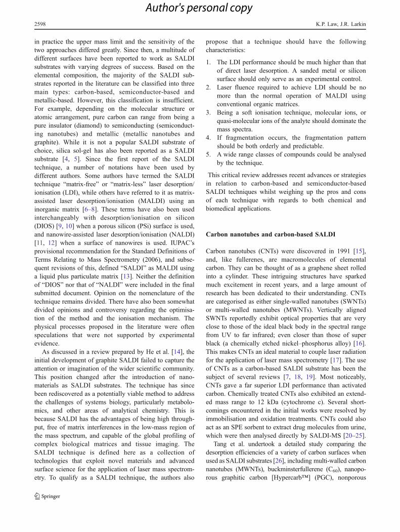
pretreated with a hyperthermal atomic beam. Furtherinvestigation revealed that parts of the carbon surfaces werelaser annealed under high laser fluence and clusters of sub-μm sized spherical particles were formed (Fig. 2a). A seriesof carbon cluster ions (differing by m/z 12.0) was alsoobserved in the negative ion mass spectra. Destruction of thesurfaces of the CNTs after laser irradiation was observed, butcarbon cluster ions were not seen in the mass spectrum(Fig. 2b).
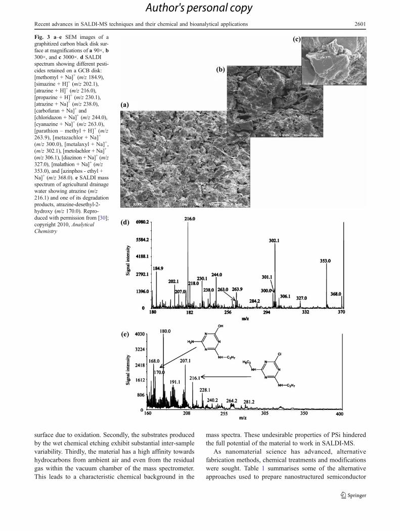
Amini and Shariatgorji have also advanced the graphiteSALDI technique [28–30]. Graphitised carbon black (GCB)particles embedded in a network of polytetrafluoroethylene(PTFE) were shown to be an active carbon-based SALDIsubstrate (Fig. 3a–c). A number of pesticides with varyingchemical properties were detected (Fig. 3d), though most ofthe pesticides were detected as Na/K adducts or fragmentedions. The method was successfully used to determine the
Fig. 1 a–f Scanning electronmicrographs of a multi-walledcarbon nanotubes, b buckmin-sterfullerene, c nanoporous gra-phitic carbon, d nonporousgraphite particles, e highly ori-ented pyrolytic graphite, and fnanodiamonds. Reproducedwith permission from [26];copyright 2009, AnalyticalChemistry
Recent advances in SALDI-MS techniques and their chemical and bioanalytical applications 2599
Author's personal copy
presence of atrazine and one of its degradation products,atrazine-desethyl-2-hydroxy, in agricultural drainage waterfrom rice fields [30] (Fig. 3e). Enhancement was furtherachieved using oxidised GCB nanoparticles, with the resultbeing that propranolol extracted from Baltic Sea bluemussels was successfully quantified by SALDI-MS usingdeuterated propranolol as the internal standard [29].
The production of carbon cluster ions in carbon-basedSALDI obscures the mass spectra and/or suppresses thesignals of the analyte due to detector saturation. However,with the development of ion-mobility (IM) mass spectrom-etry, in an approach demonstrated by Ugarov et al. [31], thecarbon cluster ions can be filtered out because the migrationtimes of these ions are distinctly different from those of thebiomolecules investigated. In that study, derivatised fuller-ene particles were chosen as the SALDI matrix and
deposited on a 14 μm thick rat brain tissue slice. Thetissue was then analysed directly. Figure 4 shows arepresentative SALDI-IM-TOF spectrum obtained beforeand after carbon cluster signal filtering. Although peptideand lipid ions were detected below m/z 2000, the presenceof fullerene ion signals greatly suppresses their signals(Fig. 4a). However, the peptides and lipids show adistinctive trend in their IM spectroscopy compared to theC60 and C120 clusters. The fullerene ions were readilysubtracted from the data (Fig. 4b), and, by combing theinformation generated by SALDI-MS and IM, detailedassignments of the lipid and peptide components could bemade. Furthermore, IM analysis of dynorphin 1–7 wasdemonstrated in the same study using derivatised SWNTsas a SALDI substrate.
Nanostructured semiconductor-based SALDI
The development of SALDI-MS entered a new chapterafter DIOS-MS was proposed by Siuzdak et al. in 1999[32–34]. The innovation of DIOS-MS is its migrationtowards a monolithic target. By using lithographical etching(illuminating n+-type silicon through an optical mask), orby covalent derivatisation before etching, photopatterningof the surface can be achieved. From the operator’sperspective, the fundamentally different approach of theDIOS-MS lies in stark contrast to the early carbon-basedSALDI approaches that are used in a manner similar to theconventional MALDI matrix. Even during the early phaseof the development of DIOS-MS, it had already been notedthat a wide range of biologically related small moleculescould be analysed. It was thought that the porous structureof PSi provided a scaffold for trapping the analyte, and theunique optical activity of these nanostructured siliconsubstrates afforded an effective means of transferring thelaser energy to the adsorbate [35]. PSi has been a subject ofintense research effort since the early 1990s, and in-depthknowledge has been accumulated in this area [36, 37].Much of the interest in PSi and its morphology derivesfrom its room-temperature photoluminescent properties[38]: visible light emission from high-porosity structuresarises from the quantum confinement effect. Bulk silicon,however, emits light only weakly (<0.001%) in the IRregion owning to its indirect band gap [37]. Recently, on thebasis that explosive vaporisation was proposed as a possibledesorption mechanism, a refined version of DIOS-MSemerged, namely nanostructure-initiator mass spectrometry(NIMS) [39].
Despite the reported success of DIOS-MS, three majorproblems were encountered during its early phase ofdevelopment. Firstly, freshly prepared hydrogen-terminatedPSi is chemically unstable and a dielectric layer forms on the
Fig. 2 a, b Scanning electron micrographs of a clusters of sphericalparticles formed on the surface of HOPG after N2 laser irradiation(laser fluence, 109 mJ cm−2) and b the destruction of multiwalledcarbon nanotubes (CNTs) after N2 laser irradiation (laser fluence,43 mJ cm−2). The carbon substrates were irradiated with the N2 laserat a frequency of 10 Hz for 1000 laser pulses. Reproduced withpermission from [26]; copyright 2009, Analytical Chemistry
2600 K.P. Law, J.R. Larkin
Author's personal copy
surface due to oxidation. Secondly, the substrates producedby the wet chemical etching exhibit substantial inter-samplevariability. Thirdly, the material has a high affinity towardshydrocarbons from ambient air and even from the residualgas within the vacuum chamber of the mass spectrometer.This leads to a characteristic chemical background in the
mass spectra. These undesirable properties of PSi hinderedthe full potential of the material to work in SALDI-MS.
As nanomaterial science has advanced, alternativefabrication methods, chemical treatments and modificationswere sought. Table 1 summarises some of the alternativeapproaches used to prepare nanostructured semiconductor
Fig. 3 a–e SEM images of agraphitized carbon black disk sur-face at magnifications of a 90×, b300×, and c 3000×. d SALDIspectrum showing different pesti-cides retained on a GCB disk:[methomyl + Na]+ (m/z 184.9),[simazine + H]+ (m/z 202.1),[atrazine + H]+ (m/z 216.0),[propazine + H]+ (m/z 230.1),[atrazine + Na]+ (m/z 238.0),[carbofuran + Na]+ and[chloridazon + Na]+ (m/z 244.0),[cyanazine + Na]+ (m/z 263.0),[parathion – methyl + H]+ (m/z263.9), [metazachlor + Na]+
(m/z 300.0), [metalaxyl + Na]+,(m/z 302.1), [metolachlor + Na]+
(m/z 306.1), [diazinon + Na]+ (m/z327.0), [malathion + Na]+ (m/z353.0), and [azinphos - ethyl +Na]+ (m/z 368.0). e SALDI massspectrum of agricultural drainagewater showing atrazine (m/z216.1) and one of its degradationproducts, atrazine-desethyl-2-hydroxy (m/z 170.0). Repro-duced with permission from [30];copyright 2010, AnalyticalChemistry
Recent advances in SALDI-MS techniques and their chemical and bioanalytical applications 2601
Author's personal copy

SALDI substrates reported to date. Methods include thin-film deposition approaches, such as a nonporous column/void silicon surface produced by plasma-enhanced chemi-cal vapour deposition (PECVD) [40, 41], germaniumnanodots (GeNDs) deposited on a silicon single-crystalsurface by molecular beam epitaxy (MBE) [42, 43], orderedsilicon nanocavity arrays produced by reactive ion etching(RIE) [44, 45], and silicon nanowires (SiNWs) produced bychemical vapour deposition (CVD) [46–48]. These physicalmethods have advantages over wet chemical etchingapproaches, as they are carried out in computer-controlledsystems ensuring the substrates produced have a hightarget-to-target reproducibility.
The SiNWs substrate has recently been made commer-cially available under the trade name of NALDI™ fromNanosys, Inc. and Bruker Daltonics, Inc. The substrates areprepared using colloidal gold nanoclusters deposited on asilicon wafer as catalysts. Nanowires are then grown usingsilane vapour (SiH4) as a reactant in a CVD furnace. TheSiNWs surface is further oxidised and derivatised with(pentafluorophenyl)-propyldimethylchlorosilane [46, 47]. Ithas been reported that the SiNWs surface exhibits signif-icantly lower ionisation fluence thresholds than the DIOSsubstrate and organic MALDI matrices. SEM images alsorevealed that the SiNWs layer is easily melted andevaporates upon laser irradiation [47]. Important surfaceparameters governing the LDI performance have beenfurther optimised and the surface morphology of theoptimised substrate is different from the earlier develop-ment version reported [12] (Fig. 5). In addition, indepen-dent study has shown that the commercial NALDI™ targetoutperforms the commercial DIOS™ target in severalaspects, particularly in low molecular weight peptideanalysis [64]. However, the positive ion NALDI mass
spectra have characteristic background ions located at m/z197, 235 and 243, etc. [65] due to the formation of Au+,[AuF2]
+, [AuSi(H2O)]+ and other related cluster ions, and
these obscure the analysis of small molecules.Alternative chemical etching methods have also been
sought. The relatively simply preparation procedure in-volved in chemical etching make these approaches viable inmost laboratories. For example, a metal-assisted oxidativeetching process termed HOME-HF etching, first proposedby Li and Bohn [66], has been explored in several studies[67–70]. This method involves an uncomplicated fabrica-tion set up and only requires a deposition of a metal layer,such as palladium, gold or platinum, on a silicon wafer,although only the substrates prepared by Au-coating priorto HOME-HF etching were reported to be of use in SALDI.A shadow mask can be used to pattern the metal depositionand to generate an array of sample spots. Early worksemployed sputter coating in metal deposition, which oftenresulted in surface heterogeneity and the formation of“pitches” (islands of less-processed surface area) [71].These were because of uneven Au deposition on the surfaceand the fluctuation of the glow discharge current duringsputter coating. Subsequently, work carried out by Tsao etal. employed an e-beam evaporation approach, avoiding theproblems encountered in sputter coating [69, 72]. In thatstudy, a range of surface morphologies were produced byHOME-HF etching, but apparently a surface morphologywith vertically aligned densely packed nanofilaments showsa superior performance (Fig 6a–c). Moreover, it was shownthat this substrate outperformed the commercial DIOS™target in peptide analysis.
Another similar approach that employs silver-assistedchemical etching has been reported by Piret et al. [63, 73].In this process, crystalline silicon is etched by a solution of
Fig. 4 a, b 2D SALDI IM-TOF MS spectra obtained from Sprague–Dawley rat brain tissue using C60((CH2)2COOH)n matrix: a originalspectrum; b the same 2D spectrum after the carbon cluster signals had
been filtered out. Reproduced with permission from [31]; copyright2004, Analytical Chemistry
2602 K.P. Law, J.R. Larkin
Author's personal copy

Tab
le1
Com
parisonof
thefabricationprocessesof
variou
snano
structured
andnano
particle
semicon
ductor
SALDIsubstrates
repo
rted
intheliterature
Substrate
Sum
maryof
fabricationprocesses(and
pre-
andpost-fabricatio
ntreatm
ent,ifavailable)
Reportedyear
References
Amorphousgerm
anium
oxide
thin
film
(QuickMass™
)Anam
orph
ousGethin
film
isprepared
byph
ysical
vapo
urdepositio
n(PVD).Deposition
/evapo
ratio
niscarriedou
tin
anelectron
beam
evaporator.The
precursormaterialis99
.999
%Gecubes,evaporated
from
avitreous
carbon
crucible.Corning
1737
glassandpolishedstainlesssteelcanbe
used
asthebasalsubstrate.
2004
[10,
49]
Siliconsubm
icrometer
groove
arrays
ZEP52
0resistsarefirstcoated
uniformly
onto
aSisurface.
The
resistson
theSisubstrates
areexposedto
anelectron
beam
andtheexposedsamples
aresubsequently
developedin
ZED-N
50.The
resistpatternsaretransferredto
Niby
aconventio
nallift-offmethod.
The
Sisubstrates
with
theNimold(m
ask)
areetched
byRIE
usingCF4–A
rgas.The
Nimaskisthen
remov
edby
soakingin
dilutedHNO3.The
fabricated
Sisurfaces
arerefreshedby
soakingin
5%HFsolutio
nanddriedin
vacuo.
2005
[50]
Ordered
silicon
nano
cavity
arrays
ASiwafer
isfirstspin-coatedwith
apositiv
ee-beam
resist,NANO
poly(m
ethylmethacrylate)
(PMMA),andisthen
placed
inan
electron
beam
litho
graphy
system
forhigh
-resolutionnanopatterning.
Adriedsurfaceisthen
etched
byreactiv
eionetching(RIE)in
agasmixture
containing
C4F8,SF6andAr.The
substrateisthen
treatedwith
O2
plasmaandstored
inair.Im
mediately
priorto
MSmeasurement,thesilicon
substrates
arerinsed
with
5%HF/EtOH
toremov
etheresidu
alSiO
2layer.Thismethodhasreplaced
that
describedin
[44].
2005/200
9[44,
45]
Zincoxidenanowires
ZnO
nanowires
areeither
grow
non
aSisubstrateby
CVD
oralternativelyproduced
onan
Au/Ti/S
istructured
substrateby
avapour
transportprocess.
2005/201
0[11,
51,52]
Siliconmicrocolumnarrays
AcleanedSiwafer
isexposedto
repeated
355nm
frequency-tripledNd:YAG
laserirradiations
inthepresence
ofam
bientair(leastactiv
e),SF6gas,or
deionisedwater
(mostactiv
e).
2006
[53,
54]
Siliconnano
pillars
plus
amorphou
sSicoating
Silica
nanoparticle
agglom
erates
aredepositedas
amaskprod
uced
byliq
uidflam
espray(LFS)on
toaSisurface.
The
surfaceisetched
byindu
ctivelycoupledSF6/O
2plasma,
buffered
inheliu
mgasplaced
onaliq
uidnitrogen
cooled
botto
melectrod
e—ametho
dkn
ownas
cryogenicdeep
reactiv
eionetching(D
RIE).Subsequ
entdevelopm
entadds
anα-Sicoatingby
PECVD,either
onaplanar
silicon
surfaceor
asurfaceof
silicon
nano
pillars.The
substratecan
befurthertreatedwith
RIE
oxygen
plasmatreatm
ent.Beforefabrication,
theSiwafer
isfirstcleanedwith
asolutio
nof
NH4OH/H
2O2/H
2O.Silicondiox
idesubstrateisfabricated
bythermal
oxidationof
asilicon
wafer.The
sample
plates
arestored
inairinside
thecleanroom
andarecleanedby
rinsingwith
methano
lbefore
MSmeasurements.
2007/200
9[55,
56]
Siliconnanoparticles
Com
mercially
available30
nmSinanoparticlespretreated
with
10%
HNO3in
aheated
sonicatio
nwater
bath
are
derivatised
with
(heptadecafluoro-1,1,2,2-tetrahydrodecyl)dim
ethylchlorosilane
(PFP).Nanoparticlesarecarefully
cleanedthroug
hout
andappliedas
aconventio
nalparticulatematrix.
2007/200
9[57,
58]
Germanium
nanodotchip
ASisurfaceisfirstcleanedby
thestandard
Shiraki
process,follo
wed
bypre-depositio
nof
asilicon
buffer
layerto
make
anatom
ically
flat
andcleansilicon
surface.
Geatom
saredepo
sitedon
thesilicon
surfaceusingmolecular
beam
epitaxy
(MBE)to
form
aself-assem
bled
nanoscaleGeisland
s.Further
cleaning
may
berequ
ired,as
describedin
[43].
2007
[42,
43]
Zincoxidenanoparticles
Com
mercially
availableZnO
nanoparticles(Zn-350)
aresuspendedin
methanolandaretreatedby
ultrasonic
agitatio
n,which
leadsto
fragmentatio
nandirregularlyshaped
nanoparticles.
2008
[59]
Ordered
nano
compo
site
silicathin
film
sA
freshlyoxidized
single
crystalSiwafer
issubm
itted
toadilutedsuspension
oftetraethylorthosilicate
(TEOS)in
ethanol/w
ater/HClandsurfactant
(Brij56).The
surfaceisthen
with
draw
nfrom
thesolutio
nandallowed
todryunderam
bient
conditions.The
condensatio
nof
silicaprecursormolecules
around
anorganisedsurfactant
phaseleadsto
anevaporation-induced
self-assem
blyprocessto
form
anorderednanostructured
silicafilm
.The
substrateisthen
exposedto
3hof
deep-U
Vlig
htto
yieldananopo
rous
silicathin
film
.Pho
topatterningisachieved
byusingan
alum
inium
mask.
2008
[60]
Siliconnitridenano
particles
Aslurry
ofsilicon
nitrideisprepared
bymixing10
mgof
nano
particleswith
100μLof
1%TFA
.The
slurry
isthen
spotteddirectly
onto
astainlesssteelMALDIplateanddried.
2009
[61]
Laser-assistedchem
ically
etched
porous
silicon
Com
binesconventio
nalelectrochemical
etchingin
HF/ethanol
solutio
nandfurthermodified355nm
frequency-tripled
Nd:YAG
laserin
etchingsolutio
n.The
surfaceiscarefully
cleanedthroug
hout.
2009
[62]
Chemically
etched
silicon
nanowirearrays
Asilicon
surfacecleanedwith
piranh
asolutio
nischem
ically
etched
inHF/AgN
O3aqueou
ssolutio
n(silv
er-assistedchem
ical
etching)
andthesubstrateissubsequently
derivatised
with
octadecyltrichlorosilane
(OTS),perfluorodecyltrichlorosilane
(FDTS),or
octyldim
ethylchlorosilane
(ODCS)in
adryN2pu
rged
glovebox
afterstoringin
anaqueoussolutio
nof
HCl/H
NO3/H
2O
overnight.The
resulting
surfaceisrinsed
with
CH2Cl 2andisop
ropylalcoho
landdriedun
derastream
ofN2.
2010
[63]
Recent advances in SALDI-MS techniques and their chemical and bioanalytical applications 2603
Author's personal copy

Dev
elo
pm
ent
vers
ion
(20
05/2
006)
Density: 10-50 wires/µm2
Diameter: 40 nmLength: a few µm
Co
mm
erci
ally
ava
ilab
le v
ersi
on
(200
7/20
08) Density: > 100
wires/µm2
Diameter: 20 nm Length: 100-500 nm
Fig. 5 Comparison of thesurface morphology of the earlydevelopment version and thecommercially available versionof the NALDI™ target. Repro-duced with permission from[47]; copyright 2006, Journal ofPhysical Chemistry, from [12];copyright 2008, Journal of theAssociation for LaboratoryAutomation
Fig. 6 a–f Comparison of the surface morphologies of a–c ananofilament silicon substrate prepared by HOME etching and d–fsilicon nanowire arrays substrate prepared by silver nitrate assistedetching. Despite being named differently, the surface morphologies of
the substrates appear to be identical. Reproduced with permission from[69]; copyright 2008, Analytical Chemistry, and from [63]; copyright2010, Langmuir
2604 K.P. Law, J.R. Larkin
Author's personal copy

HF/AgNO3. Because the surface is anodised, the silverions are reduced to silver metal and silver nanoparticlesare deposited on the surface. Therefore, the priordeposition of a metal layer (as in HOME-HF etching) isnot required. However, the method requires a follow-upstep to remove the silver particles and dendrites produced.The substrates reported to work in SALDI have anidentical surface morphology to that reported by Taso etal., despite all of these authors naming their substratesdifferently (Fig. 6d–f).
In a collaboration with Vladimir Karavanskii andSergey Nikiforov of the Prokhorov General PhysicsInstitute of Russian Academy of Sciences, the authorsexplored the PSi substrate prepared by iodine-assistedetching [9]. Figure 7 shows the microscopic structures of aPSi substrate prepared by iodine-assisted vapour-phaseetching. Using this method, the density of the surfacenanostructures can be vastly increased compared to theconventional anodisation approach. In addition, a relative-ly large and uniform target can be produced. The authors’
investigations of argon plasma etching [9] and anamorphous germanium thin film target (QuickMass™)[10] have also become the basis for developments relatingto amorphous-Si (α-Si) thin film, as reported in [74]. Theinvestigation of α-Si thin film was driven by thehypothesis that dangling bonds which are present in adisordered network of α-Si thin film play a fundamentalrole in the energy transfer mechanism.
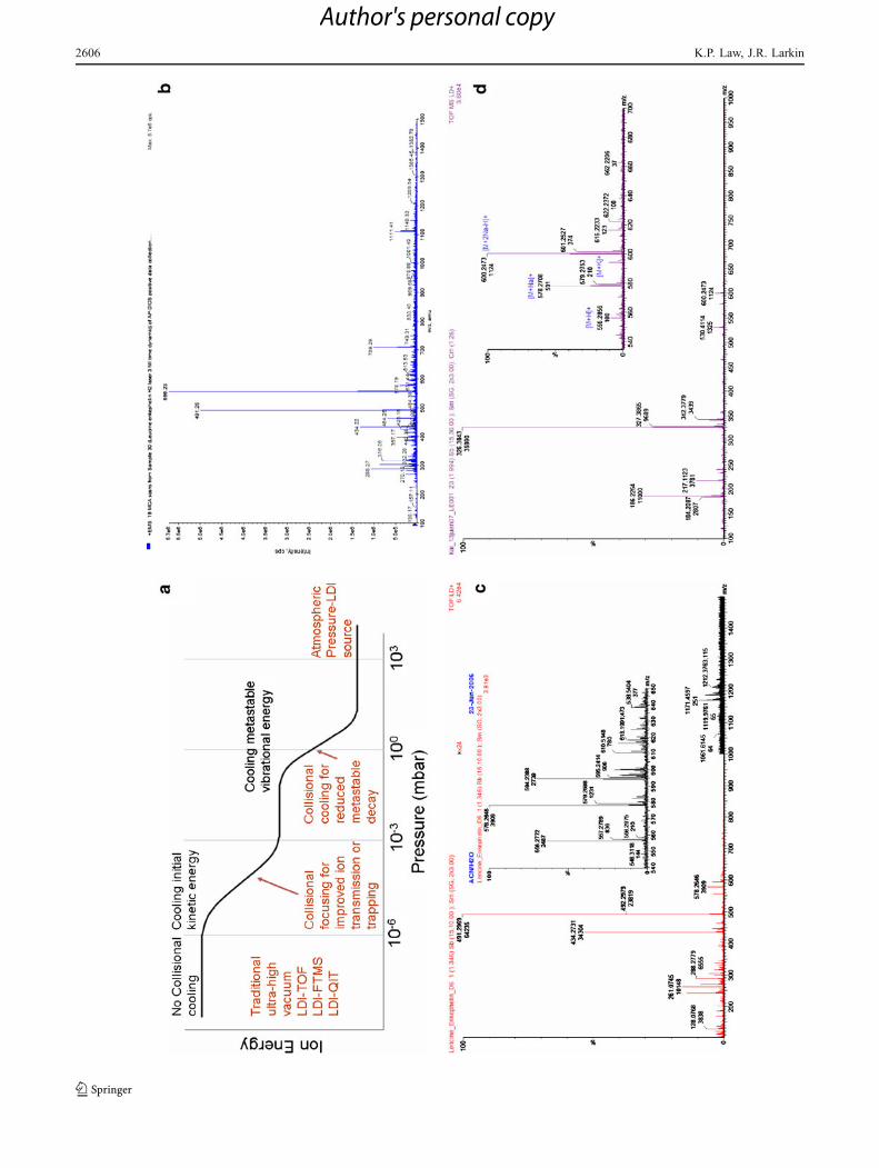
In addition to optimising the substrate physicochemicalparameters, the authors also investigated the effects ofvarying the ion source pressure on the DIOS performance.Increasing the ion source pressure from 10−6 to 10−3 and1 bar produces a damping effect on the desorbed ion. Theinternal energy of the desorbed ions is reduced andconsequently extensive metastable fragmentation or degra-dation can be minimised. Furthermore, because a surface oran interface is a highly dynamic system that is greatlyinfluenced by its surrounding environment, the ion source(vapour) pressure also affects the availability of wateradsorbed on the surface. The configuration of the massspectrometer and the experimental conditions are thereforecrucial factors, and may explain the inconsistent resultsreported in the literature. These effects are demonstrated bythe DIOS mass spectra of the neuropeptides leucine–enkephalin (Fig. 8). Different mass spectral responses wereobserved from ultrahigh vacuum, high vacuum and atmo-spheric pressure (AP) systems. The protonated molecule ofleucine–enkephalin and the fragment ion at m/z 491dominate the AP-DIOS mass spectrum. Still, in-sourcedecay could not be avoided. On the other hand, in all of thevacuum systems, the peak intensity of the protonatedmolecule was lower than its fragment and its salt adducts.In the DIOS-ToF spectrum, fragment ions at m/z 434 and491 dominated. In DIOS-Q-ToF, the spectrum was domi-nated by the fragment ion at m/z 326.
SALDI ionisation mechanism on nanostructuredsemiconductors
The elucidation of the SALDI ionisation mechanism ispossibly the most controversial and elusive area ofSALDI research. The SALDI mechanism can be sub-divided into adsorption, retention, radiation coupling andtransfer, desorption and ionisation reactions. The physicalprocesses that are thought to be involved in the radiationcoupling reaction are described below with an emphasison silicon substrates. Depending on the thickness of thenanostructure layer of the substrate, at least 3–4 levels ofthese physical processes are involved in transferring thelaser energy to the adsorbate and thereby promoting themto an excited state before the desorption and ionisationreactions.
Fig. 7 a SEM image and b AFM topography of a typical PSisubstrate prepared by vapour-phase etching. Etching was carried outby suspending a silicon substrate in a sealed chamber containingsaturated vapours of a HF-water (1:1) solution and I2 powder for 1 h atroom temperature. No light illumination or electrical bias was required
Recent advances in SALDI-MS techniques and their chemical and bioanalytical applications 2605
Author's personal copy
2606 K.P. Law, J.R. Larkin
Author's personal copy

1. Fundamental energy transfer processes
Okuno et al. noted that increasing the specific surfacearea of a surface, for example by sanding a silicon wafer,enabled the substrate to acquire some SALDI activity [50].They used such substrates to obtain mass spectra of ahydrophobic polymer, reserpine and angiotensin. However,a scratched metal surface did not result in the same activity.Undoubtedly, enhancing the specific surface area will alsoaffect the adsorption and retention of the adsorbate on thesurface and the refraction of laser light. However, thisobservation also suggests that efficient energy transfer isachieved because of the unique electronic properties of thesemiconductor surface.
In direct laser desorption, most species that are desorbedfrom the surface are neutral at the laser fluences required topreserve molecular integrity. Observations of how neutralmolecules are desorbed from a surface have concluded thatthere are two main processes that contribute to themolecular desorption: (1) laser-induced thermal desorption(LITD) [75], and (2) nonthermal laser desorption inducedby electronic transitions (DIET) (or more correctly, desorp-tion induced by multiple electronic transitions: DIMET)[76]. LITD occurs through the excitation of the substrate bythe creation of electron–hole pairs that relax and causephonon excitation. On the other hand, DIET is a nonther-mal process that occurs by substrate excitation, leading tolow-energy electron transfer to the surface species. How-ever, LITD and DIET alone do not lead to ion formation.The ionisation potential of a typical molecule requires 8–9 eV. The photon energy from the 337 nm N2 laser is only3.68 eV. Therefore, ion formation on direct laser desorptionrequires a substantially high laser fluence, which escalates
Fig. 9 a Schematic illustrationof band bending and chargetransfer on n-type semiconduc-tor surfaces. EC and EV are theconduction and valence-bandedges, while EF and ED are theFermi energy and the energy ofthe bulk donor levels. Photoex-citation leads to the formation ofelectron–hole pairs (e– and h+)or excitons. Nonradiative re-combination of excitons takesplace at the electronic surfacestate (ESS), which leads to thepromotion of the adsorbates toan excited state. Energythereby being concentrated topromote ionisation. Adaptedfrom [76]. b Schematic illustra-tion of photocarrier relaxationpathways available in siliconnanocrystallites. Reproducedwith permission from [90];copyright 1997, Journal ofApplied Physics
Fig. 8 a The effect of ion source pressure on the relative internalenergies of the desorbed ions. Also shown are positive-ion DIOS massspectra of leucine–enkephalin acquired on b an atmospheric pressure-LDI-QqLIT system, c an ultrahigh vacuum LDI-ToF system, and d ahigh-vacuum LDI-Q-ToF system
R
Recent advances in SALDI-MS techniques and their chemical and bioanalytical applications 2607
Author's personal copy

the probability of multiple photons arriving at the same sitesimultaneously, and thereby inducing fragmentation anddegradation of the adsorbate. An important concept inMALDI ionisation is photoexcitation and energy pooling.Energy pooling is one of several energy concentrationpossibilities imagined in early MALDI work. This is aphenomenon in which the electronic excitation energy oftwo nearby molecules is redistributed and higher energyprocesses become possible [77, 78]. It is thought that theunique electronic structure of a semiconductor surfaceallows the possibility of energy pooling.
Whilst charge-transfer excitation on metal and semicon-ductor surfaces involves initial photon adsorption by thesubstrate, the detailed mechanisms differ significantly forthe two surfaces. The charge-transfer dynamics on asemiconductor surface are governed mainly by the bandstructure; principally the presence of a space-charge layer.Figure 9A illustrates a simplified schematic of a charge-transfer photoexcitation process on an n-type semiconductorsurface. A space-charge layer occurs because the electronicconfiguration of the surface atoms is different from the bulk.All atoms in the bulk of a pure metal or elementalsemiconductor are equivalent. However, the atoms at thesurface do not possess their full complement of bondingpartners [79]. Consequently, a semiconductor surface usuallypossesses electronic surface states and such states areenergetically located in the band gap [76]. These relativelylocalised states can result from dangling bonds on thesurface, or from defects or impurities. Depending on thetype of surface state (donors or acceptors) and the positionof the Fermi level at the surface, the surface states may
carry charge, which is screened by an opposite chargeinside the semiconductor material. Due to the low free-carrier concentration in semiconductors, the screeninglength is long and a space-charge layer is formed, givingrise to band bending [76].
When photons with energies that are larger than the bandgap are absorbed by the surface, electrons from the lower bandcan be promoted to the upper band, and this creates energeticelectron–hole pairs or excitons. Within the space-charge layer,electron–hole pairs are driven apart by the space-charge field.For n-type semiconductors, electrons are driven into the bulk,while holes are transported toward the surface. This is becausethe charged carriers (electrons and holes) are separated by thesurface band bending, thereby producing UV radiationmediated surface oxidation [80]. The oxidation of siliconinduces a reduction potential of –0.91 eV, which is sufficientto reduce, for example, Cu(II) and Fe(III) to Cu(I) and Fe(II),respectively [81–84] (Fig. 10). When such charge separationor exciton formation creates an electric field, and when it isrelaxed via nonradiative recombination of electrons and holesor carrier–phonon scattering, this causes an electronic pertur-bation and vibrational excitation of the surface and adsorbates.
Without the band bending and the electronic surfacestate, the photoexcited electrons would quickly settle intothe energy minimum of the conduction band. This isbecause of the momentum difference between the energyminimum of the conduction band and the energy maximumof the valance band in indirect band-gap semiconductors.Consequently, nonradiative recombination of electrons andholes is prohibited, and efficient excitation of the adsor-bates does not occur. Hence, for a surface composed of pure
[Cu(I)Cl]Cl-
[Cu(I)Cl]2Cl-
[Cu(I)Cl]3Cl-
[Fe(II)Cl2]Cl-
[Fe(III)Cl3]Cl-
[Cu(II)Cl2]2Cl-
Fig. 10 Negative ion DIOS spectrum of a copper(II) acetate/iron(III) chloride mixture. Copper(II) was reduced to copper(I), and part of the iron(III) was reduced to iron(II)
2608 K.P. Law, J.R. Larkin
Author's personal copy

silicon, which does not have a thick layer of nanostructuresor nanocrystallites, and has its surface dangling bondshydrogenated and its defect density significantly reducedby hydrogen plasma (e.g. α-Si–H) [85], the SALDI activitywould be quenched [74]. Then again, substrates that consistof different materials may not share an identical excitationpathway. Graphite, for example, has a negative indirectbandgap—the energy maximum of the valence band ishigher than the energy minimum of the conduction band—and so it may not have an identical excitation pathway tothat of silicon or germanium.
2. Optical absorption (photonic and electronic band gaps)
Although surface dangling bonds or defects play animportant role in the SALDI mechanism, the fundamentalprocesses described above are initiated by the absorption ofphotons. Nonetheless, most of the UV photons are reflectedfrom a rough surface or penetrate through a thin film surface.This contrasts with high-porosity surfaces, which have highoptical absorbance over UV wavelengths and appear darkvisually. Nayak and Knapp have correctly pointed out that thisdistinct optical absorption is due to the photonic band gapbehaviour of nanoporous surfaces, which behave as photoniccrystals [86]. Since the photonic band gap behaviour is afunction of the pore lattice spacing, varying the width anddepth (or generally the overall porosity) of a porous surfacemay change the photonic band gap behaviour, resulting in ashift in the absorption maxima, which affects the SALDIactivity. This proposition is coherent with the observationthat was previously reported by Okuno et al. [50] thatSALDI from grooved surfaces is a function of the irradiationangle, i.e. laser irradiation normal to the grooves yields agood LDI signal, whereas parallel irradiation gives a poorLDI signal, along with other experimental evidence [87–89].This proposition is also consistent with the experimentalresults reported in [45]. Still, the effects of the porosity of asubstrate are not just limited to radiation absorption; it alsoaffects the adsorption, retention and desorption of theadsorbate and residual solvent, and the thermal conductivityof the substrate, thereby attenuating the overall SALDIresponses of a nanoporous substrate.
Although nanophotonic behaviour provides importantinsight into how optical absorption events may occur onnanoporous surfaces, an electronic event must follow inorder to transfer the laser energy into internal energy. It haslong been proposed that a common optical absorption eventis necessary for the DIOS activity and the photolumines-cence of the PSi, although both processes can occurindependently [67]. This contributes to the unique elec-tronic properties of the silicon nanocrystallites on the PSisurface [90]. The electronic structure of silicon is alteredon the nanoscale and the calculated band gap is reduced to0.65–0.25 eV, depending on the lattice symmetry [91]. In
agreement with the calculated values, experimental meas-urements using a time-of-flight electron spectrometer havealso shown that the band gap energies of nanosized Siclusters (Si11, Si14, Si17, Si18, Si30, Si33) are on the orderof 0.4–0.6 eV [92]. However, the role of the nanocrystallitesis not limited to acting as an absorption body for laserradiation; they also amplify all of the fundamental processesdescribed above (Fig. 9B).
3. Thermoelectric effect
The reduced thermal conductivity and field effect ofnanostructured substrates have both long been proposed tobe important elements in the SALDI mechanism. The fieldeffect was once suggested to be a sharp-tip electrostatic [27]or an (inverted) lightning-rod phenomenon [44], despite thelightning-rod effect occurring when the particle is off-resonance, providing only a very small amplitude enhance-ment [93]. Conversely, surface melting has been thought toplay a critical role in the SALDI mechanism [9, 10, 94, 95].The underlying physical process is probably the thermo-electric effect. This process is the direct conversion of atemperature gradient to electric voltage and vice versa, andit is pronounced on substrates that have a thick porous ornanowire layer and have critical dimensions or spacingsbelow 300 nm. It is believed that laser irradiation inducesrapid heating on the top of the nanowire layer. An extremetemperature results, which melts the surface, but the actualmelting temperature is hundreds of degrees lower than themelting point of the bulk materials (crystalline silicon:1410 °C; platinum: 1768 °C) due to melting-pointdepression. However, owing to the large difference in meanfree-path lengths between electrons and phonons, electronsare driven toward the cooler bottom layer—the bulk of thesubstrate and the basal support—leaving the positive holeson the top layer. However, recombination of the chargecarriers only occurs at thermal equilibrium, which is onlyslowly attained. A very high electric field is generated.Energy is transferred to the adsorbates via processes similarto that of field ionisation, in conjunction with the thermalprocesses already in place. As shown by the authorspreviously [10], this process was disrupted as the laserenergy was increased beyond an optimal point. This waspartly because the surface and the surface nanocrystalliteswere ablated and destroyed, and partly because the laserradiation was sufficiently intense to penetrate deep into thesubstrate, disturbing the thermal gradient. As a result, thethermoelectric effect was quenched.
4. Desorption
A thermal process known as explosive vaporisation hasalso been proposed as a possible desorption mechanism onporous substrates [96–98]. Explosive vaporisation occurswhen a liquid is heated so rapidly that density fluctuations
Recent advances in SALDI-MS techniques and their chemical and bioanalytical applications 2609
Author's personal copy
in the liquid become the dominant vaporisation mechanism,rather than heterogeneous nucleation. Heterogeneous nu-cleation takes place when the temperature of the bulk liquidapproaches saturation temperature and thus reaches equi-librium with its vapour. In contrast, explosive vaporisationoccurs when the liquid approaches a region of intrinsicinstability because its temperature gets too close to thesuperheat limit [99]. Indeed, when a fluid is confined to avery small cavity that is close to being molecular sized, itsphase behaviour is altered. Long-range fluctuation andthermal–vibrational effects could provide another energy-transfer pathway. Nevertheless, molecular dynamics andinterfacial interactions in a confined space remain lesswell understood and can only be studied by neutronscattering [100–103].
5. Ionisation
Depending on the chemical properties of the analytemolecules, several possible ionisation reactions follow.These include excited-state proton transfer, disproportion-ation reactions, fragmentation reactions, ion-molecularreactions, cation attachment or transfer, etc.
In excited-state proton transfer, the surface Si-OHmoieties play a critical role in the proton transfer reaction,as evidenced in [9, 74]. Surface melting, which alsoincreases the bond distances of the lattice system, is alsoimplied to facilitate the protonation reaction, especially forcompounds such as amines that form complexes with thesurface Si-OH moieties [10, 74]. The theoretical basis thatinterfacial water and Si-OH terminals are the major protonsources, but not the Si-H groups (as some have proposed),has also been described in [104].
A close inspection of the SALDI mass spectra of aminesacquired from a reflector ToF mass analyser often reveal atailing region of the molecular ion peak. This phenomenonis due to the metastable states of desorbed ions. Metastablepeaks are result from fragmentations that occur in the field-free region of the spectrometer (i.e. while passing down theflight tube). Fragment ions are lost by the reflector due totheir change in kinetic energy. Only fragments that stillhave kinetic energies close to that of the precursor, such as[M+H-NH3]
+, are transmitted, due to the energy toleranceof the reflector, and this gives rise to a “tailing” of thesignal. However, in linear ToF systems, the intensity of amolecular ion signal does not reduce, because the linearvelocity of the fragmenting ion is conserved and thefragments formed are therefore detected at the same flighttime as their intact precursor. The internal energy of theexcited molecules is redistributed via intramolecular vibra-tional energy redistribution (IVR) [105]. The energy israndomly distributed over the molecule and becomesconcentrated in particular ways which in turn give rise tounimolecular reconfiguration and dissociation [106],
obeying the principles of quasi-equilibrium theory (QET)[107]. This may explain the ordered fragmentation oftenobserved. Indeed, the relatively controlled and specificfragmentation pattern and its application for post-sourcedecay (PSD) in DIOS, similar to that of MALDI, is anunusual capability of the technique that is worth payingattention to [32, 35, 108, 109].
Gas-phase secondary ion-molecular reactions can alsooccur and give rise to products of cation attachment andalkylation. As an additional note, ionisation reactions inSALDI or MALDI are chemical processes and, like anychemical reactions, are governed by the principles ofthermodynamics. Accordingly, it is the entropy of thereaction which ultimately determines the ionic speciesdetected by the mass spectrometer, while the rate ofreaction determines the selectivity.
Chemical and biomedical applications
Over the years, a large number of works demonstratingthe versatility of the SALDI technique have beenreported, and these were reviewed previously by Peter-son [6]. The applications reported range from novelpharmaceutical and biomedical applications to environ-mental and polymer sciences. Of all these SALDItechniques, DIOS has been the most widely used. Forexample, DIOS has been shown to be a powerful tool forthe forensic investigation of contraceptive polymers in analleged sexual assault case, and it produced evidence thatwould not have been otherwise made available to theprosecution by another analytical technique [110, 111]. Ithas also been shown that the technique is highly desirablefor the forensic analysis of illicit drugs [112]. In thatreport, despite the presence of “matrix constituents” in the“authentic” sample (e.g. ecstasy tablets seized by thepolice), the authors reported that interference was notobserved. However, another report described that 11impurities were identified in ecstasy by DIOS, dependingon the synthetic route employed [113]. It was suggestedthat profiling the impurities might provide information ontheir origin. These authors also reported the identificationof catecholamines (dopamine and noradrenaline) in ahuman peripheral blood lymphocyte extract [114], recon-firming the result published by the same group eight yearsearlier [115]. Okuno and Wada have also demonstrated thequantitation of salicylate in human serum using DIOS-MSin negative ion mode [116]. An ordered nanocavity siliconSALDI substrate was also used to monitor osmoticallyinduced fluctuations in plant metabolites in both wild-typeand transgenic Arabidopsis thaliana (the transgenic wasmodified with a human inositol polyphosphate-5-phosphatase enzyme) under the environmental stress of
2610 K.P. Law, J.R. Larkin
Author's personal copy

drought. Root, shoot and leaf extracts were studied [44,117]. On the other hand, there has been a great deal ofinterest in using DIOS-MS in bottom-up and middle-down proteomics, for example to enhance the confidencein protein identification by extending the additionalinformation window in the low-mass region of the mass
spectrum, to detect peptide peaks that cannot be detectedby MALDI, and to identify post-translation modificationsthat can only be detected by DIOS [46, 70, 97, 118–122].Further selected applications highlighting the strength anduniqueness of the SALDI technique are discussed in thissection.
Fig. 11 a, e Schematic of an on-chip NIMS enzymatic assay. a Thefluorinated tagged metabolite is first immobilised in the fluorousinitiator liquid phase coated on the surface. b The surface with thesample to screen for enzymatic activity is then incubated. c After anon-target sample clean-up, the target is then submitted to laser massspectrometry analysis. d The chemical structure of the fluorinated
tagged substrate (S, [M+H]+ m/z 1,074.30) and the products of β-1,4-galactosidase (P1, [M+H]+ m/z 911.24) and α-2,3-sialyltransferase(P2, [M+H]+ m/z 1,365.40). e Mass spectra of the substrate (left) andthe resulting products (right). Reproduced with permission from [127];copyright 2009, National Academy of Sciences (USA)
Recent advances in SALDI-MS techniques and their chemical and bioanalytical applications 2611
Author's personal copy

Monitoring enzymatic activity and enzyme inhibitor assay
On-target enzyme-catalysed reaction monitoring and directanalysis by DIOS-MS was first reported by Thomas et al.[119]. A number of enzyme systems were investigated. Forinstance, acetylcholine esterase (AChE) was allowed toreact with its naturally occurring substrate acetylcholine toproduce choline. MALDI and ESI were also employed forcomparison, but neither was found to produce a strong,reproducible signal for choline. The lack of a strongchromophore on choline (and possibly other unadornednatural substrates) also made absorption or emissiontechniques difficult to employ. However, no technicalproblem was encountered when using DIOS-MS, and theyreported that the presence of the enzyme on the surface (inthe concentration range investigated) did not interfere withthe detection of the small molecules of interest. Thestrength of this approach was further demonstrated by itsability to directly determine the selectivities of enzymeinhibitors. Three different inhibitors of AChE (huperzine A,tacrine and 2,6-dimethoxyphenyl-N-butylcarbamate) werestudied, and the inhibition potentials of the inhibitors werefound to correlate with their relative Ki values. Furthercomments can be found in [123].
Despite the fact that on-target enzyme activity andinhibitor assay offers a wide range of advantages, the on-target approach was abandoned in favour of an off-lineapproach in subsequent studies [124–126], until furtherwork was reported by Northen et al. [127]. This wasachieved after substantial effort had been invested in thedevelopment of NIMS [39] and the development of aselective on-target sample enrichment and clean-up method:fluorous affinity tagging [108]. This new approach tookadvantage of the fact that perfluorinated tagged componentshave a strong affinity toward the perfluorinated liquidinitiator coated on the surface and are noncovalentlyimmobilised. This factor is apparently a key considerationfor their subsequent mass analysis because of the difficul-ties encountered in using covalent linker approachespreviously. A biological matrix, such as a cell lysate, whichcontains the enzyme being investigated is then added to thesurface and allowed to react with its correspondingsubstrate. Because the fluorous-tagged reactant and prod-ucts are preferentially retained, this allows the use ofsurface washing steps to remove all other unlabelledcomponents and cellular materials. This approach not onlyeliminates the prior purification of the enzyme from acomplex biological sample as well as the subsequentquenching step required in the previous studies, but it alsoallows the resulting mass spectra to be free of interferencesand/or ion suppression (Fig. 11). However, since thereactant is partly immobilised on the surface, which mayhinder the binding of the substrate to the enzyme, careful
experimental design must be implemented to permit thismethod to work effectively. In their study, a five-carbonlinker was introduced between the tagging and substrate toreduce steric hindrance for enzyme binding, and argininewas incorporated to facilitate ionisation (Fig. 11d). Thissystem was then applied to study the β-1,4-galactosidaseactivity of bacteria that were found to colonise in the nearvicinity of a hot spring in the Yellowstone National Park.The results showed that the bacterial galactosidase is most
Fig. 12 a Cross-section of a PSi kinetic analysis microfluidic device. Adroplet travels down the channel, depositing reaction product on thechannel walls. b The deposited reaction product is then desorbed andionised according to the principles of DIOS-MS. c An overview of theregions of the device and a DIOS-IMS image of arginine (m/z 173.1±1)over the device after an enzymatic reaction has taken place within it.Intensity correlates with relative concentration. Reproduced withpermission from [128]; copyright 2008, Analytical Chemistry
2612 K.P. Law, J.R. Larkin
Author's personal copy
active at a temperature of 65 °C but retains its activity ateven 100 °C. In addition, despite the bacteria being collectedfrom a slightly alkaline environment, the bacterial galacto-sidase activity was optimal at pH 5.5, suggesting that theintracellular pH was different from that of the surroundingsof the bacteria. Deoxygalactonojirimycin was also found tohave a stronger inhibitory effect on galactosidase thanphenylethyl-β-D-thiogalactopyranoside does.
A limitation of the approaches described above is thatthey can only be applied to enzyme systems which have arelatively low or moderate reaction rate. Improvements toallow time-resolved kinetic analysis for moderately fastreaction systems were reported by Nichols et al. [128].They used an integrated microfluidic system with a PSimicrochannel to perform a chemical reaction in a contin-uous flow regime and an in-line investigation of productformation by DIOS-MS in order to study an arginasesystem. The basic idea of the microfluidic system is that thedevice first mixes an enzyme droplet and a substratedroplet. This combined enzyme/substrate droplet thentravels down a PSi microfluidic channel, depositing a traceamount of reaction substrate and product into the siliconpore walls. Because the pore size is tuned to be larger thanthe substrate and product, but smaller than the meandiameter of the enzyme, the enzyme is therefore stericallyhindered from entering the pores. This not only allowscontinuous substrate diffusion into the pores and thereaction to continue down the line, but it also eliminatesthe need for quenching. The flow rate of the liquid droplets,or in this case the distance they travelled through themicrochannel network, represents the time domain of thereaction. The whole device is then submitted to imagingmass spectrometry, and the relative concentrations of thereactant and product are monitored with respect to theposition (Fig. 12). The most obvious advantage of such adevice is being able to rapidly obtain enzyme initialvelocity measurements, and because the entire surface ofthe microchannel is analysed using DIOS-MS, there isnearly zero dead time.
Pharmaceutical development
One of the most challenging areas of drug development isthe search for novel receptor–ligand pairs and enzymeinhibitors, as many novel proteins have no known bindingpartners. Zou et al. have demonstrated an innovativeapproach using DIOS to address this challenge [129]. Theyimmobilised a targeted protein (BSA) on a PSi surface thatacted as a probe. A mixture of drug molecules that containedpossible binding partners to the protein was then incubatedwith the probe. The drug molecules that have a high affinityfor the targeted protein were captured by the immobilisedprotein, and those with a weak affinity for the targeted
protein were washed off. The captured drug molecules werethen identified by means of on-probe SALDI analysis. Thismethod offers high-throughput screening of lead-drugcandidates, but—unlike the conventional methods—it doesnot require fluorescence or chemiluminescence tags (note:labelling molecules can sometimes alter the binding affinity),radioactive markers, or even prior purification. It is notlimited to immobilising protein on the probe; the methodcould potentially be extended to other biological macro-molecules, such as DNA and RNA. The method wasdeveloped further in a subsequent study after a silicon–carbon surface attachment method was realised in which PSisubstrates were chemically modified to yield carboxylic acidterminated surfaces, allowing proteins to be covalentlyattached to the surface through an amide bond [130]. Ahemoglobin-modified surface was applied to identify thenoncovalent binding between hemoglobin and 13 environ-mentally relevant chemicals, including antimicrobials,insecticides, fungicides and herbicides. In all of thecompounds investigated, only triphenyltin chloride wasshown to bind haemoglobin strongly, indicating that it mayhave a higher toxicity than the other compounds tested.Then again, the immobilisation of typsin on a DIOSsubstrate for enzymatic activity and inhibitor assay, al-though possible, was not as promising [131]. Immobilisa-tion of the enzyme resulted in a slight loss of its catalyticactivity and the value of νmax was found to be lower thanthat of free trypsin. Furthermore, because of the surfacechemical modification, the substrate exhibited a lowerSALDI activity, and the addition of organic matrix wasrequired to successfully desorb the peptide fragmentsgenerated.
Biochemical evolution
Ribose molecules form the backbone structure of DNA andRNA. The borate ion is thought to play an important role inthe synthesis of ribose and other pentoses from simpleorganic precursors such as formaldehyde and glycolalde-hyde which have been identified in star-forming regions ofinterstellar medium [132, 133]. It is thought that glyco-laldehyde may have a role to play in the origins of life inour universe, and the reaction is thought to occur on orunder the surfaces of tiny dust grains, initiated by a shockwave of energy. One important question in the evolution ofribonucleic acids is why ribose was favoured over otherpentose isomers. Using the DIOS technique, an attempt wasmade by Li et al. to answer this question [68]. The actualstudy was performed to quantify the relative tendencies ofribose, lyxose, arabinose and xylose to form a borate-centred pentose dimer on the surface or in the gas phase,in a combination of conditions similar to that of theinterstellar medium. The results showed that ribose
Recent advances in SALDI-MS techniques and their chemical and bioanalytical applications 2613
Author's personal copy
exhibits a higher affinity to boron than other isomericpentoses do. The resulting enrichment could provideinsight into why ribose was favoured as a building blockin RNA evolution.
Metabolic profiling
With the increasing interest in metabolomics over the lastdecade, SALDI-MS and other novel ionisation techniques—collectively known as direct analysis mass spectrometry(DAMS)—are becoming attractive alternatives or comple-mentary approaches in metabolomics. Goodacre et al. werethe first to demonstrate that metabolic footprinting (metab-
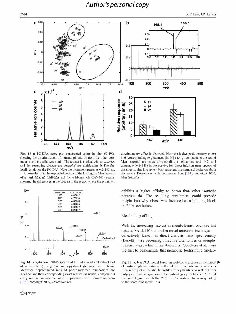
Fig. 13 a PC-DFA score plot constructed using the first 60 PCs,showing the discrimination of mutants g1 and s4 from the other yeastmutants and the wild-type strain. The test set is marked with an asterisk,and the separating clusters are encircled for clarification. b The firstloadings plot of the PC-DFA. Note the prominent peaks at m/z 145 and146, seen clearly in the expanded portion of the loadings. cMean spectraof g1 (gln3Δ), g5 (dal80Δ) and the wild-type wh (BY4741) strains,showing the differences in the spectra in the region where the prominent
discriminatory effect is observed. Note the higher peak intensity at m/z146 (corresponding to glutamate, [M-H]–) for g1 compared to the rest. dMean spectral responses corresponding to glutamine (m/z 147) andglutamate (m/z 148) in the positive-ion direct infusion mass spectra ofthe three strains in c (error bars represent one standard deviation aboutthe mean). Reproduced with permission from [134]; copyright 2005,Metabolomics
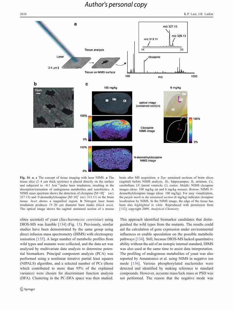
Fig. 14 Negative-ion NIMS spectra of 1 μl of a yeast cell extract andof water (blank) using 3-aminopropyldimethylethoxysilane initiator.Identified deprotonated ions of phosphorylated nucleotides arelabelled, and their corresponding exact masses (as neutral compounds)are given in the inserted table. Reproduced with permission from[136]; copyright 2009, Metabolomics
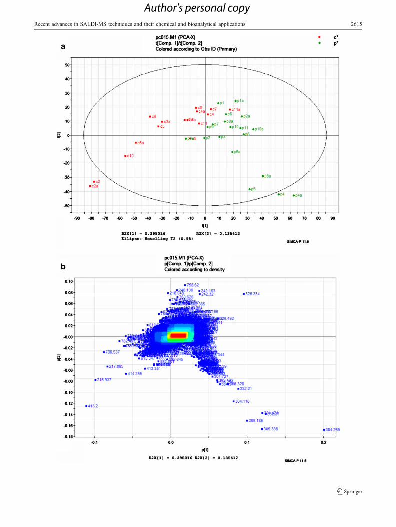
Fig. 15 a, b A PCA model based on metabolite profiles of methanol/chloroform plasma extracts collected from patients and controls. aPCA score plot of metabolite profiles from patients who suffered frompolycystic ovarian syndrome. The patient group is labelled “P” andthe control group is labelled “C”. b PCA loading plot correspondingto the score plot shown in a
b
2614 K.P. Law, J.R. Larkin
Author's personal copy
Recent advances in SALDI-MS techniques and their chemical and bioanalytical applications 2615
Author's personal copy
olites secreted) of yeast (Saccharomyces cerevisiae) usingDIOS-MS was feasible [134] (Fig. 13). Previously, similarstudies have been demonstrated by the same group usingdirect infusion mass spectrometry (DIMS) with electrosprayionisation [135]. A large number of metabolic profiles fromwild types and mutants were collected, and the data set wasanalysed by multivariate data analysis to determine poten-tial biomarkers. Principal component analysis (PCA) wasperformed using a nonlinear iterative partial least squares(NIPALS) algorithm, and a selected number of PCs (thosewhich contributed to more than 95% of the explainedvariance) were chosen for discriminant function analysis(DFA). Clustering in the PC-DFA space was then studied.
This approach identified biomarker candidates that distin-guished the wild types from the mutants. The results couldaid the calculation of gene expression under environmentalinfluences or enable speculation on the possible metabolicpathways [134]. Still, because DIOS-MS lacked quantitativeability without the aid of an isotopic internal standard, DIMSwas also used at the same time to assist data interpretation.The profiling of endogenous metabolites of yeast was alsoreported by Amantonico et al. using NIMS in negative ionmode [136]. Various phosphorylated nucleotides weredetected and identified by making reference to standardcompounds. However, accurate mass/lock mass or PSD wasnot performed. The reason that the negative mode was
Fig. 16 a, c The concept of tissue imaging with laser NIMS. a Thetissue slice (2–4 μm thick sections) is placed directly on the surfaceand subjected to ~0.1 Jcm−2/pulse laser irradiation, resulting in thedesorption/ionisation of endogenous metabolites and xenobiotics. ANIMS mass spectrum shows the detection of clozapine [M+H]+ (m/z327.13) and N-desmethylclozapine [M+H]+ (m/z 313.11) in the braintissue. Inset shows a magnified region. b Nitrogen laser beamirradiation produces 15–20 μm diameter burn marks (black area).The optical image shows the sagittal unstained section of a mouse
brain after MS acquisition. c Top: unstained sections of brain slices(sagittal) before NIMS analysis. Hc, hippocampus; St, striatum; Ce,cerebellum; LV, lateral ventricle; Ct, cortex. Middle: NIMS clozapineimages (dose: 100 mg/kg rat and 6 mg/kg mouse). Bottom: NIMS N-desmethylclozapine image (dose: 100 mg/kg). For easy visualization,the purple mark in the unstained section (6 mg/kg) indicates clozapinelocalization by NIMS. In the NIMS image, the edge of the tissue hasbeen also highlighted in white. Reproduced with permission from[142]; copyright 2009, Analytical Chemistry
2616 K.P. Law, J.R. Larkin
Author's personal copy
preferred over the positive mode was that only adenosinenucleotides could be detected in the positive mode [137]. Itwas also noted that the selection of the initiator was criticalto the successful analysis of the nucleotides (Fig. 14).
Clinical diagnosis
A metabolic profiling approach can also be effectivelyapplied to clinical applications [138]. In a preliminary studycarried out by the authors, the plasma extracts obtainedfrom patients who suffered from polycystic ovariansyndrome (POS) were studied by DIOS-MS. The DIOSmetabolic profiles generated were compared with those ofhealthy subjects. Multivariate statistical analysis was thenapplied to evaluate the data generated (Fig. 15). Theprocedures used for data conversion, filtering, peak align-ment, normalisation and statistical analysis were the sameas those described in [138]. It can be seen from the PCAloading plot that the patient and the control groups areseparated with an overlap of <10%. The informationobtained can be used to estimate disease progression andthe effect of medical treatment. The control c2 was identifiedas an outlier and was diagnosed with a different medicalcondition. The ions corresponding to the differences between
the patient and the control groups can easily be identified inthe PCA score plot, and (as discussed above) can provideinformation to determine biomarker candidates.
Imaging mass spectrometry (IMS)
Normally, biological compounds are extracted into asolution and deposited onto the substrate prior to massspectrometric study. Given that SALDI is a surface massspectrometric technique similar to MALDI and secondaryion mass spectrometry (SIMS), small-molecule IMS ispossible. He et al. were the first to demonstrate that SALDI-IMS of biological tissue was achievable [139]. Two-dimensional ion images of mouse liver tissue and humancervical cancer cells were obtained. The density or thethickness of the biological tissue was important, since laserradiation must penetrate through the tissue sample and beabsorbed by the PSi layer underneath. To overcome thislimitation, organic matrix was sublimated onto the tissuesection in subsequent studies [140, 141]. This approach wastermed matrix-enhanced surface-assisted laser desorption/ionisation mass spectrometry (ME-SALDI-MS) to indicatethe hybrid nature of the technique and the enhancementachieved. 2D ion images of mouse heart and brain tissue
Resolution FOV Image Quality
DESI 0.25-1 mm
NIMS 100 µm
SIMS 100 nm
Incr
easi
ng
Sp
atia
l Det
ail
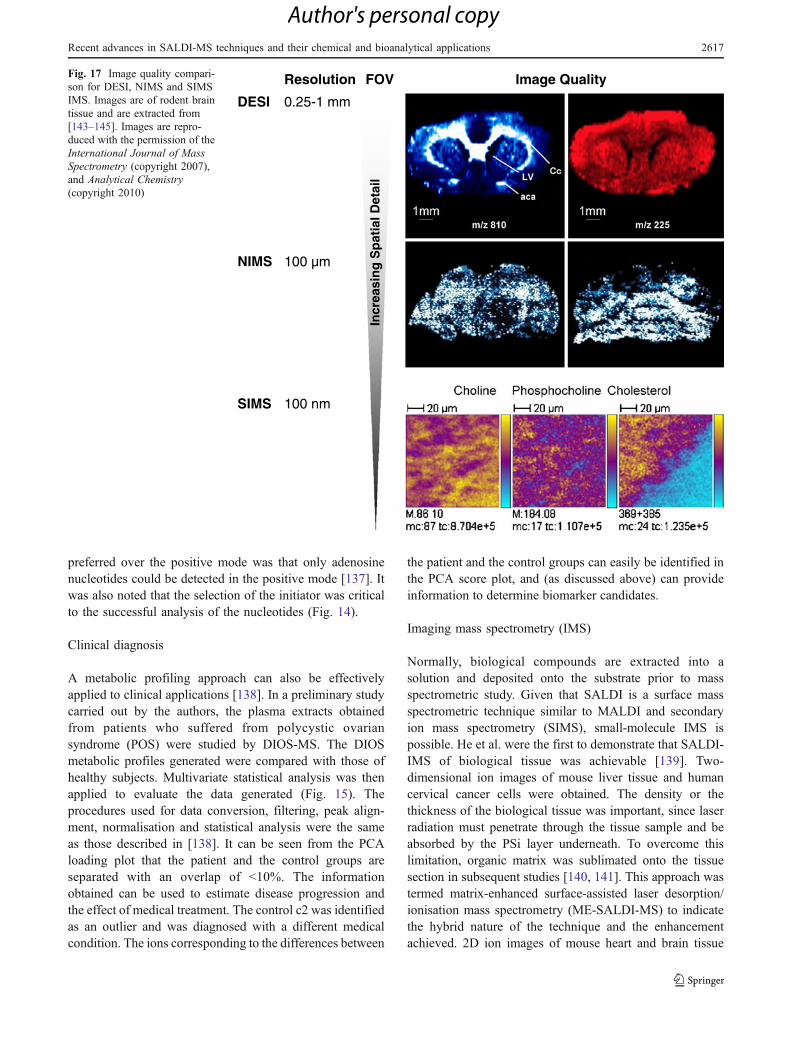
Fig. 17 Image quality compari-son for DESI, NIMS and SIMSIMS. Images are of rodent braintissue and are extracted from[143–145]. Images are repro-duced with the permission of theInternational Journal of MassSpectrometry (copyright 2007),and Analytical Chemistry(copyright 2010)
Recent advances in SALDI-MS techniques and their chemical and bioanalytical applications 2617
Author's personal copy
1000 nm
10 µm x 10 µm
10 µm x 10 µm
a
b
c
C H 3
C H 3
O HOH
Cl
O H
Cl
O H
Cl
Cl
Cl
Cl
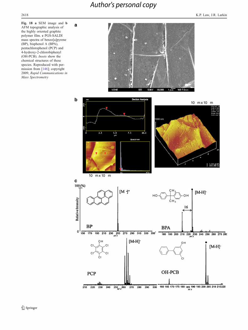
Fig. 18 a SEM image and bAFM topographic analysis ofthe highly oriented graphitepolymer film. c PGS-SALDImass spectra of benzo[a]pyrene(BP), bisphenol A (BPA),pentachlorophenol (PCP) and4-hydroxy-2-chlorobiphenyl(OH-PCB). Insets show thechemical structures of thesespecies. Reproduced with per-mission from [146]; copyright2009, Rapid Communications inMass Spectrometry
2618 K.P. Law, J.R. Larkin
Author's personal copy

were also successfully obtained. Siuzdak et al. alsoexploited NIMS for IMS. Ion images of the drug moleculedistribution in brain tissue (Fig. 16), the cholesteroldistribution in brain tissue and the sucrose distribution ina Gerbera jamesonii flower stem were demonstrated [142,143]. SALDI has advantages over MALDI, SIMS anddesorption electrospray ionisation (DESI) in small-moleculeIMS because of the matrix background interference inMALDI, the extensive fragmentation in SIMS and therelatively low spatial resolution in DESI [144, 145] (Fig. 17).Direct analysis of drug molecules and their metabolites frombiofluids was also demonstrated in [142].
Analysis of environmental pollutants
Using pyrolytic highly oriented graphite polymer film(PGS), various environmental pollutants, including per-fluorooctanoic sulfonic acid (PFOS), perfluorooctanoic acid(PFOA), benzo[a]pyrene (BP), bisphenol A (BPA), penta-chlorophenol (PCP), 4-hydroxy-2-chlorobiphenyl (OH-PCB), 4-chloroaniline (4-CA) and 2,4-dichloroaniline(2,4-CA) were investigated by Kawasaki et al. (Fig. 18)[146]. The investigation of PFOS and PFOA was previous-ly carried out using DIOS-MS in negative ion mode [147],and a detection limit of 1 ppt was achieved for PFOS. Thissensitivity could not be achieved by MALDI or LC-ESI-MS, and this highlights a unique ability of DIOS. PFOS inthe tap water (water service at Osaka in Japan) wasquantified (8.7±2.2 ppt) using sodium dodecyl-d25 sulfate(SDS-d25) as an internal standard. Although DIOS showeda high performance in the detection of PFOS, the sensitivityof PFOA was relatively poor. A new method that usedoxidised PGS modified with the cationic polymer poly-ethyleneimine (PEI) was found to be more suitable for theanalysis of PFOA. The PFOA content in river watercollected from the Kanzaki River in Osaka, Japan wasquantified (50±20 ppb) using PFOA-13C8 as an internalstandard. All of these results were in agreement with thoseobtained by LC-MS/MS.
Other environmental pollutants (PFOS, BP, BPA, PCPand OH-PCB) were also detected efficiently by PGS-SALDI as the protonated or deprotonated molecular ion.The authors stressed that these compounds would havebeen difficult to detect simultaneously by GC-MS or LC-MS. Detailed examination of the PGS-SALDI mass spectrarevealed that BP was photoionised and detected as a radicalcation in positive ion mode. BPA, PCP and OH-PCB weredetected as deprotonated molecules in negative ion mode.Interestingly, the spectrum of BPA shows a peak locatedm/z 16 lower than the [M–H]– peak of BPA, which (in ourview) could be a CH4 neutral loss fragment peak of BPA.Nevertheless, for reasons unclear to the authors, 4-CA and2,4-CA were not detected. As mentioned earlier, photoex-
citation plays an important role in SALDI. A possiblereason (in our view) is that UV radiation excites theelectrons of the halo-aromatic system, and the electrons arepromoted to the π* state. The interaction of the non‐bonding pair of electrons on a substitute with the π electronhas the effect of stabilising the π* state, thereby loweringits energy. This induces an opposite effect to oxygen andnitrogen. For oxygen, delocalisation of its lone pairs ofelectrons with the excited halo-aromatic system favoursdeprotonation. However, delocalisation of the lone pair ofelectrons located at the nitrogen of the amine group reducesits electron density, which does not favour protonation. Theformation of anilinium cation results in loss of a lone pairinteracting with the π* state and is energetically unfav-ourable. Deprotonation of aniline in the gas phase requiresa relatively large input of energy. The calculated deproto-nation energy of aniline at the amine nitrogen is 1534 ±8 kJ mol−1. The calculated values are 121, 145 and 151 kJmol−1 higher at C2, C3 and C4 positions, respectively[148]. The relative position of the chorine in the aromaticsystem also has an effect. For both 4-CA and 2,4-CA, thechlorine atoms are located at the ortho and/or para positions,and this induces a stronger electron-withdrawing effect thana chlorine located at the meta position.
Conclusions
For the past decade, progress in systems biology andpharmaceutical development has required innovation in theanalytical sciences. Mass spectrometry, one of the mostpowerful and sensitive analytical platforms available, is atthe forefront of attempts to answer the challenges presentedby systems biology and other areas of analytical andbioanalytical chemistry. Many new mass spectrometry tech-nologies and applications have emerged and been reported.Under these circumstances, SALDI-MS has been reinvented.The technique has now developed into a number of varietiesthat are vastly different from that originally proposed. Thus,standardisation of terminology is needed. Themost significantdevelopment of the technique has involved the use ofnanomaterials, such as CNTs, metallic nanoparticles (mostlysilver, platinum and gold) and nanostructured surfaces. Thedevelopment of DIOS-MS in particular, and subsequentlyNIMS and NALDI, has attracted the attention of analyticalscientists. These methods have since become a benchmark forsemiconductor-based SALDI research. SALDI-MS has manyadvantages, and the potential applications of the technique areextremely wide. In the authors’ view, the selected chemicaland biomedical applications are of particular interest becausethese applications make use of the advantages of the techniqueand are relatively difficult or are less suitable to perform usingother mass spectrometry approaches. On the other hand, the
Recent advances in SALDI-MS techniques and their chemical and bioanalytical applications 2619
Author's personal copy

elucidation of the SALDI ionisation mechanism was contro-versial. Available data were scattered, inconsistent andcomplicated by the different experimental conditionsemployed by different researchers. Data interpretation wasoften subjective, and much was speculated or unsupported.However, some knowledge has accumulated, and this hasinfluenced the direction of SALDI development. SALDIshares many features with other laser mass spectrometrytechniques and, as with MALDI, multiple interrelatedexcitation and ionisation reactions occur simultaneously. Thedifference is that the functions of the matrix are substituted byan active substrate. It is foreseeable that substrate develop-ment and the exploration of novel biomedical, clinical,chemical and environmental applications will continue.
References
1. Sunner J, Dratz E, Chen Y-C (1995) Anal Chem 67:4335–43422. Han M, Sunner J (2000) J Am Soc Mass Spectrom 11:644–6493. Tanaka K, Waki H, Ido Y, Akita S, Yoshida Y, Yoshida T (1988)
Rapid Commun Mass Spectrom 2:151–1534. Zhang Q, Zou H, Guo Z, Zhang Q, Chen X, Ni J (2001) Rapid
Commun Mass Spectrom 15:217–2235. Hoang TT, Chen Y, May SW, Browner RF (2004) Anal Chem
76:2062–20706. Peterson DS (2007) Mass Spectrom Rev 26:19–347. Pan C, Xu S, Zhou H, Fu Y, Ye M, Zou H (2007) Anal Bioanal
Chem 387:193–2048. Cohen L, Go EP, Siuzdak G (2007) Small-molecule desorption/
ionization mass analysis. In: Hillenkamp F, Peter-Katalinić J(eds) MALDI MS: a practical guide to instrumentation, methodsand applications, 1st edn. Wiley-VCH, Weinheim
9. Law KP (2010) Int J Mass Spectrom 290:47–5910. Law KP (2010) Int J Mass Spectrom 290:72–8411. Kang M-J, Pyun J-C, Lee J-C, Choi Y-J, Park J-H, Park J-G, Lee J-
G, Choi H-J (2005) Rapid CommunMass Spectrom 19:3166–317012. Daniels RH, Dikler S, Li E, Stacey C (2008) J Assn Lab
Automation 13:314–32113. IUPAC (2004) Project: Standard definitions of terms relating to
mass spectrometry. http://www.iupac.org/web/ins/2003-056-2-500.Accessed 23 April 2010
14. Guo Z, Ganawi A, Liu Q, He L (2006) Anal Bioanal Chem384:584–592
15. Iijima S (1991) Nature 354:56–5816. Mizuno K, Ishii J, Kishida H, Hayamizu Y, Yasuda S, Futaba DN,
YumuraM,Hata K (2009) ProcNatl Acad Sci USA106:6044–604717. Shin SJ, Choi D-W, Kwak H-S, Lim GI, Choi YS (2006) Bull
Korean Chem Soc 27:581–58318. Dattelbaum AM, Iyer S (2006) Expert Rev Proteomics 3:153–16119. Najam-ul-Haq M, Rainer M, Szabó Z, Vallant R, Huck CW,
Bonn GK (2007) J Biochem Biophys Methods 70:319–32820. Xu S, Li Y, Zou H, Qiu J, Guo Z, Guo B (2003) Anal Chem
75:6191–619521. Pan C, Xu S, Zou H, Guo Z, Zhang Y, Guo B (2004) J Am Soc
Mass Spectrom 16:263–27022. S-f Ren L, Zhang Z-h Cheng, Guo Y-l (2005) J Am Soc Mass
Spectrom 16:333–33923. Ren S-f, Guo Y-l (2005) Rapid Commun Mass Spectrom
19:255–26024. Pan C, Xu S, Hu L, Su X, Ou J, Zou H, Guo Z, Zhang Y, Guo B
(2005) J Am Soc Mass Spectrom 16:883–892
25. Chen W-Y, Wang L-S, Chiu H-T, Chen Y-C, Lee C-Y (2004) JAm Soc Mass Spectrom 15:1629–1635
26. Tang H-W, Ng K-M, Lu W, Che C-M (2009) Anal Chem81:4720–4729
27. Alimpiev S, Nikiforov S, Karavanskii V, Minton T, Sunner J(2001) J Chem Phys 115:1891–1901
28. Shariatgorji M, Amini N, Thorsen G, Crescenzi C, Ilag LL(2008) Anal Chem 80:5515–5523
29. Amini N, Shariatgorji M, Thorsén G (2009) J Am Soc MassSpectrom 20:1207–1213
30. Amini N, Shariatgorji M, Crescenzi C, Thorsén G (2010) AnalChem 82:290–296
31. Ugarov MV, Egan T, Khabashesku DV, Schultz JA, Peng H,Khabashesku VN, Furutani H, Prather KS, Wang HWJ, JacksonSN, Woods AS (2004) Anal Chem 76:6734–6742
32. Wei J, Buriak J, Siuzdak G (1999) Nature 399:243–24633. Lewis W, Shen Z, Finn MG, Siuzdak G (2003) Int J Mass
Spectrom 226:107–11634. Nordstrom A, He L, Siuzdak G (2007) Desorption/ionization on
silicon (DIOS). In: Gross ML, Caprioli RM (eds) The encyclo-pedia of mass spectrometry volume 6: molecular ionizationmethods, 1st edn. Elsevier, Amsterdam
35. Shen Z, Thomas JJ, Averbuj C, Broo KM, Engelhard M, CrowellJE, Finn MG, Siuzdak G (2001) Anal Chem 73:612–619
36. Stewart MP, Buriak JM (2000) Adv Mater 12:859–86937. Schmeltzer JM, Buriak JM (2004) Recent developments in the
chemistry and chemical applications of porous silicon. In: Rao CNR,Müller A, Cheetham AK (eds) The chemistry of nanomaterials:synthesis, properties and applications, vol 2. Wiley-VCH,Weinheim
38. Canham LT (1990) Appl Phys Lett 57:1046–104839. Northen TR, Yanes O, Northen MT, Marrinucci D, Uritboonthai
W, Apon J, Golledge SL, Nordstrom A, Siuzdak G (2007)Nature 449:1033–1036
40. Kalkan AK, Bae S, Li H, Hayes DJ, Fonash SJ (2000) J ApplPhys 88:555–561
41. Kalkan AK, Henry MR, Li H, Cuiffi JD, Hayes DJ, Palmer C,Fonash SJ (2005) Nanotechnology 16:1383–1391
42. Seino T, Sato H, Yamamoto A, Nemoto A, Torimura M, Tao H(2007) Anal Chem 79:4827–4832
43. Sato H, Nemoto A, Yamamoto A, Tao H (2009) Rapid CommunMass Spectrom 23:603–610
44. Finkel NH, Prevo BG, Velev OD, He L (2005) Anal Chem77:1088–1095
45. Xiao Y, Retterer ST, Thomas DK, Tao J-Y, He L (2009) J PhysChem C 113:3076–3083
46. Go EP, Apon JV, Luo G, Saghatelian A, Daniels RH, Sahi V, DubrowR, Cravatt BF, Vertes A, SiuzdakG (2005) Anal Chem 77:1641–1646
47. Luo G, Chen Y, Daniels H, Dubrow R, Vertes A (2006) J PhysChem B 110:13381–13386
48. Coffinier Y, Janel S, Addad A, Blossey R, Gengembre L, PayenE, Boukherroub R (2007) Langmuir 23:1608–1611
49. Hayes DJ (2004) Micrototal analysis system for enzymatic drugmetabolism and analysis (Ph.D. thesis). Department of Engi-neering Science and Mechanics, Pennsylvania State University,University Park
50. Okuno S, Arakawa R, Okamoto K, Matsui Y, Seki S, Kozawa T,Tagawa S, Wada Y (2005) Anal Chem 77:5364–5369
51. Shin JH, Song JY, Park HM (2009) Mater Lett 63:145–14752. Shin WJ, Shin JH, Song JY, Han SY (2010) J Am Soc Mass
Spectrom 21:989–9253. Chen Y, Vertes A (2006) Anal Chem 78:5835–584454. Chen Y, Luo G, Diao J, Chornoguz O, Reeves M, Vertes A
(2007) J Phys Conf Ser 59:548–55455. Sainiemi L, Keskinen H, Aromaa M, Luosujärvi L, Grigoras K,
Kotiaho T, Mäkelä JM, Franssila S (2007) Nanotechnology18:505303–505310
2620 K.P. Law, J.R. Larkin
Author's personal copy

56. Jokinen V, Aura S, Luosujärvi L, Sainiemi L, Kotiaho T, FranssilaS, Baumann M (2009) J Am Soc Mass Spectrom 20:1723–1730
57. Wen X, Dagan S, Wysocki VH (2007) Anal Chem 79:434–44458. Dagan S, Hua Y, Boday DJ, Somogyi A, Wysocki RJ, Wysocki
VH (2009) Int J Mass Spectrom 283:200–20559. Watanabe T, Kawasaki H, Yonezawa T, Arakawa R (2008) J
Mass Spectrom 43:1063–107160. Dattelbaum AM, Hicks RK, Shelley J, Koppisch AT, Iyer S
(2008) Micropor Mesopor Mat 114:193–20061. Shariatgorji M, Amini N, Ilag L (2009) J Nanopart Res
11:1509–151262. Li J, Lu C, Hu XK, Yang X, Loboda AV, Lipson RH (2009) Int J
Mass Spectrom 285:137–14263. Gl Piret, Drobecq H, Coffinier Y, Melnyk O, Boukherroub R
(2010) Langmuir 26:1354–136164. Shenar N, Cantel S, Martinez J, Enjalbal C (2009) Rapid
Commun Mass Spectrom 23:2371–237965. Guénin E, Lecouvey M, Hardouin J (2009) Rapid Commun
Mass Spectrom 23:1395–140066. Li X, Bohn PW (2000) Appl Phys Lett 77:2572–257467. Kruse RA, Li X, Bohn PW, Sweedler JV (2001) Anal Chem
73:3639–364568. Li Q, Ricardo A, Benner SA, Winefordner JD, Powell DH
(2005) Anal Chem 77:4503–450869. Tsao C-W, Kumar P, Liu J, DeVoe DL (2008) Anal Chem
80:2973–298170. Skipp P, Farooqui M, Pickard K, Li Y, Evans AGR, O’Connor
CD (2004) Expanding the information window to increaseproteomic sensitivity and selectivity. In: Valdes JJ, SekowskiJW (eds) Proceedings of NATO Advanced Workshop onProteomics and Toxicogenomics. IOS Press, Amsterdam
71. Li Q (2005) Exploring desorption/ionization on porous siliconmass spectrometry and its applications (Ph.D. thesis). Depart-ment of Chemistry, University of Florida, Gainesville
72. Tsao C-W (2008) Interfacing microfluidic bioanalysis with highsensitivity mass spectrometry (Ph.D. dissertation). Department ofMechanical Engineering, University of Maryland, College Park
73. Piret G, Coffinier Y, Roux C, Melnyk O, Boukherroub R (2008)Langmuir 24:1670–1672
74. Alimpiev S, Grechnikov A, Sunner J, Karavanskii V, SimanovskyY, Zhabin S, Nikiforov S (2008) J Chem Phys 128:014711–014719
75. Hanley L, Kornienko O, Ada ET, Fuoco E, Trevor JL (1999) JMass Spectrom 34:705–723
76. Zhu X (1994) Annu Rev Phys Chem 45:113–14477. Knochenmuss R (2002) J Mass Spectrom 37:867–87778. Knochenmuss R (2006) Analyst 131:966–98679. Kolasinski K (2002) Surface science: foundations of catalysis
and nanoscience. Wiley, Chichester80. Stewart MP, Buriak JM (2001) J Am Chem Soc 123:7821–783081. Budimir N, Fournier F, Blais J-C, Wind F, Tabet J-C (2003)
Study of fatty acids and sulfonic acids by desorption/ionizationon silicon mass spectrometry. In: 51st Annu Conf ASMS,Montreal, Canada, 8–12 June 2003
82. Okuno S, Arakawa R, Wada Y (2004) J Mass Spectrom Soc Jpn52:13–20
83. Okuno S, Nakano M, G-e Matsubayashi, Arakawa R, Wada Y(2004) Rapid Commun Mass Spectrom 18:2811–2817
84. Budimir N, Blais J-C, Fournier F, Tabet J-C (2007) J MassSpectrom 42:42–48
85. Umezu I, Kohno K, Aoki K, Kohama Y, Sugimura A, Inada M(2002) Vacuum 66:453–456
86. Nayak R, Knapp DR (2007) Anal Chem 79:4950–495687. Xu D, Guo G, Gui L, Tang Y, Zhang B, Qin G (1998)
Electrochem Solid-State Lett 1:227–22988. Xu D, Guo G, Gui L, Tang Y, Zhang BR, Qin GG (1999) J Phys
Chem B 103:5468–5471
89. Xu D, Guo G, Gui L, Tang Y, Qin GG (2000) Pure Appl Chem72:237–243
90. Cullis AG, CanhamLT, Calcott PDJ (1997) J Appl Phys 82:909–96591. Palaria A, Klimeck G, Strachan A (2008) Electronic structure
and transport in silicon nano-structures with non-ideal bondingenvironments. TECHCON, Austin
92. MausM, Ganteför G, EberhardtW (2000) Appl Phys A 70:535–53993. Kottmann JP, Martin OJF, Smith DR, Schultz S (2000) Opt
Express 6:213–21994. Northen TR, Woo HK, Northen MT, Nordström A, Uritboonthail
W, Turner KL, Siuzdak G (2007) J Am Soc Mass Spectrom18:1945–1949
95. WadaY, Yanagishita T,Masuda H (2007) Anal Chem 79:9122–912796. Luo G, Chen Y, Siuzdak G, Vertes A (2005) J Phys Chem B
109:2445097. Nordstrom A, Apon JV, Uritboonthai W, Go EP, Siuzdak G
(2006) Anal Chem 78:272–27898. Vertes A (2007) Soft laser desorption ionization—MALDI,
DIOS and nanostructures. In: Phipps CR (ed) Laser ablationand its applications. Springer, New York
99. Gloria R, Lichtenberg J, Hierlemann A, Poulikakos D (2005)Microplatform for investigation of explosive vaporization in microenclosures. In: 9th Int Conf on Miniaturized Systems for Chemistryand Life Sciences (μTAS), Boston, MA, USA, 9–13 Oct 2005
100. Huwe A, Kremer F, Behrens P, Schwieger W (1999) Phys RevLett 82:2338–2341
101. Bellissent-Funel M-C, Lal J, Bosio L (1993) J Chem Phys98:4246–4252
102. Bellissent-Funel M-C, Chen SH, Zanotti J-M (1995) Phys Rev E51:4558–4569
103. Guégan R, Morineau D, Loverdo C, Béziel W (2006) Phys RevE 73:011707
104. Chen Y, Chen H, Aleksandrov A, Orlando TM (2008) J PhysChem C 112:6953–6960
105. King AK, Bellm SM, Hammond CJ, Reid KL, Towrie M,Matousek P (2005) Mol Phys 103:1821–1827
106. Budimir N, Lesage D, Naban-Maillet J, Fournier F, Blais J-C,Wind F, Vékey K, Tabet J-C (2004) Internal energy of ionsproduced by desorption/ionisation on porous silicon (DIOS).52nd Annu Conf ASMS, Nashville, TN, USA, 24–27 May 2004
107. Rosenstock HM, Wallenstein MB, Wahrhaftig AL, Eyring H(1952) Proc Natl Acad Sci USA 38:667–678
108. Go EP, Uritboonthai W, Apon JV, Trauger SA, Nordstrom A,O’Maille G, Brittain SM, Peters EC, Siuzdak G (2007) JProteome Res 6:1492–1499
109. Trauger SA, Go EP, Shen Z, Apon JV, Compton BJ, BouvierESP, Finn MG, Siuzdak G (2004) Anal Chem 76:4484–4489
110. Thomas JJ, Blackledge RD, Siuzdak G (2001) Anal Chim Acta442:183–190
111. Shen ZX, Thomas JJ, Siuzdak G, Blackledge RD (2004) JForensic Sci 49:1028–1035
112. Pihlainen K, Grigoras K, Franssila S, Ketola R, Kotiaho T,Kostiainen R (2005) J Mass Spectrom 40:539–545
113. Kraj A, Świst M, Strugala A, Parczewski A, Silberringa J (2006)Eur J Mass Spectrom 12:253–259
114. Kraj A, Jarzebinska J, Gorecka-Drzazga A, Dziuban J, SilberringJ (2006) Rapid Commun Mass Spectrom 20:1969–1972
115. Bergquist J, Silberring J (1998) Rapid Commun Mass Spectrom12:683–688
116. Okuno S, Wada Y (2005) J Mass Spectrom 40:1000–1004117. Finkel NH (2005) Surface-assisted laser desorption/ionization-
mass spectrometry (SALDI-MS) of controlled nanofeatures andthe associated thermal properties (MSci thesis). Department ofChemistry, North Carolina State University, Raleigh
118. LaikoVV, TaranenkoNI, Berkout VD,Musselman BD,DoroshenkoVM (2002) Rapid Commun Mass Spectrom 16:1737–1742
Recent advances in SALDI-MS techniques and their chemical and bioanalytical applications 2621
Author's personal copy
119. Thomas JJ, Shen Z, Crowell JE, Finn MG, Siuzdak G (2001)Proc Natl Acad Sci USA 98:4932–4937
120. Go EP, Prenni JE, Wei J, Jones A, Hall SC, Witkowska HE, ShenZ, Siuzdak G (2003) Anal Chem 75:2504–2506
121. Kinumi T, Shimomae Y, Arakawa R, Tatsu Y, Shigeri Y, YumotoN, Niki E (2006) J Mass Spectrom 41:103–112
122. Go EP, Wikoff WR, Shen Z, O'Maille G, Morita H, Conrads TP,Nordstrom A, Trauger SA, Uritboonthai W, Lucas DA, ChanKC, Veenstra TD, Lewicki H, Oldstone MB, Schneemann A,Siuzdak G (2006) J Proteome Res 5:2405–2416
123. Liesener A, Karst U (2005) Anal Bioanal Chem 382:1451–1464
124. Shen Z, Go EP, Gamez A, Apon JV, Fokin V, Greig M, VenturaM, Crowell JE, Blixt O, Paulson JC, Stevens R, Finn MG,Siuzdak G (2004) ChemBioChem 5:921–927
125. Wall DB, Finch JW, Cohen SA (2004) Rapid Commun MassSpectrom 18:1482–1486
126. Steenwyk RC, Hutzler JM, Sams J, Shen Z, Siuzdak G (2006)Rapid Commun Mass Spectrom 20:3717–3722
127. Northen TR, Lee J-C, Hoang L, Raymond J, Hwang D-R,Yannone SM, Wong C-H, Siuzdak G (2008) Proc Natl Acad SciUSA 105:3678–3683
128. Nichols KP, Azoz S, Gardeniers HJGE (2008) Anal Chem80:8314–8319
129. Zou H, Zhang Q, Guo Z, Guo B, Zhang Q, Chen X (2002)Angew Chem Int Ed 41:646–648
130. Hu L, Xu S, Pan C, Zou H, Jiang G (2007) Rapid Commun MassSpectrom 21:1277–1281
131. Xu S, Pan C, Hu L, Zhang Y, Guo Z, Li X, Zou H (2004)Electrophoresis 25:3669–3676
132. Hollis JM, Lovas FJ, Jewell PR (2000)Astrophys J 540:L107–L110133. BurkeDJ, BrownWA (2010) Phys ChemChemPhys 12:5947–5969
134. Vaidyanathan S, Jones DG, Ellis J, Jenkins TE, Dunn W, Hayes A,BurtonN, Oliver S, Kell DB, Goodacre R (2005)Metabolomics 1:1–8
135. Allen J, Davey H, Broadhurst D, Heald J, Rowland J, Oliver S,Kell D (2003) Nat Biotechnol 21:692–696
136. Amantonico A, Flamigni L, Glaus R, Zenobi R (2009)Metabolomics 5:346–353
137. Gómez D, Fernández JA, Astigarraga E, Marcaide A, Azcárate S(2007) Phys State Solidi (c) 4:2185–2189
138. Miura D, Fujimura Y, Tachibana H, Wariishi H (2010) AnalChem 82:498–504
139. Liu Q, Guo Z, He L (2007) Anal Chem 79:3535–3541140. Liu Q, Xiao Y, Pagan-Miranda C, Chiu YM, He L (2009) J Am
Soc Mass Spectrom 20:80–88141. Liu Q, He L (2009) J Am Soc Mass Spectrom 20:2229–2237142. Yanes O, Woo H-K, Northen TR, Oppenheimer SR, Shriver L,
Apon J, Estrada MN, Potchoiba MJ, Steenwyk R, ManchesterM, Siuzdak G (2009) Anal Chem 81:2969–2975
143. Patti GJ, Woo H-K, Yanes O, Shriver L, Thomas D, UritboonthaiW, Apon JV, Steenwyk R, Manchester M, Siuzdak G (2010)Anal Chem 82:121–128
144. Ifa DR, Wiseman JM, Song Q, Cooks RG (2007) Int J MassSpectrom 259:8–15
145. Debois D, Brunelle A, Laprévote O (2007) Int J Mass Spectrom260:115–120
146. Kawasaki H, Takahashi N, Fujimori H, Okumura K, Watanabe T,Matsumura C, Takemine S, Nakano T, Arakawa R (2009) RapidCommun Mass Spectrom 23:3323–3332
147. Kawasaki H, Shimomae Y, Watanabe T, Arakawa R (2009)Colloid Surf A 347:220–224
148. Nguyen MT (2007) General and theoretical aspects of anilines.In: Rappoport Z (ed) The Chemistry of Anilines, Part 1 JohnWiley & Sons, Chichester, West Sussex, UK
2622 K.P. Law, J.R. Larkin
Author's personal copy