
Optimization of Xenon Biosensors for Detection of Protein Interactions
S
I
IGa
Cb
c
d
a
ARRAA
KSIDTDG
1
itaifttt1fcid
eo
0d
Biosensors and Bioelectronics 25 (2010) 1229–1234
Contents lists available at ScienceDirect
Biosensors and Bioelectronics
journa l homepage: www.e lsev ier .com/ locate /b ios
hort communication
nkjet printed and “doctor blade” TiO2 photodetectors for DNA biosensors
wona Bernacka-Wojcika, Rohan Senadeeraa,b, Pawel Jerzy Wojcika, Leonardo Bione Silvaa,oncalo Doriac,d, Pedro Baptistac, Hugo Aguasa, Elvira Fortunatoa,∗, Rodrigo Martinsa
CENIMAT/I3N, Departamento de Ciência dos Materiais, Faculdade de Ciências e Tecnologia, Universidade Nova de Lisboa andEMOP-UNINOVA, Campus de Caparica, 2829-516 Caparica, PortugalInstitute of Fundamental Studies, Hanthana Road, Kandy 20000, Sri LankaCIGMH, Departamento de Ciências da Vida, Faculdade de Ciências e Tecnologia, Universidade Nova de Lisboa, Campus de Caparica, 2829-516 Caparica, PortugalREQUIMTE, Departamento de Química, Faculdade de Ciências e Tecnologia, Universidade Nova de Lisboa, Campus de Caparica, 2829-516 Caparica, Portugal
r t i c l e i n f o
rticle history:eceived 13 July 2009eceived in revised form 7 September 2009ccepted 21 September 2009vailable online 1 October 2009
eywords:ensors/biosensors
a b s t r a c t
A dye sensitized TiO2 photodetector has been integrated with a DNA detection method based onnon-cross-linking hybridization of DNA-functionalized gold nanoparticles, resulting in a disposable col-orimetric biosensor. We present a new approach for the fabrication of dye sensitized TiO2 photodetectorsby an inkjet printing technique—a non-contact digital, additive, no mask and no vacuum patterningmethod, ideal for cost efficient mass production. The developed biosensor was compared against a dyesensitized photodetector fabricated by the traditional “doctor blade” method. Detection of gold nanopar-ticle aggregation was possible for concentrations as low as 1.0 nM for the “doctor blade” system, and
nkjet printingye sensitized photodetectoritanium dioxideNA detectionold nanoparticles
1.5 nM for the inkjet printed photodetector. The demonstrated sensitivity limits of developed biosensorsare comparable to those of spectrophotometric techniques (1.0 nM). Our results show that a differencehigher than 17% by traditional photodetector and 6% by inkjet printed in the photoresponses for thecomplementary and non-complementary gold nanoprobe assays could be attained for a specific DNAsequence from Mycobacterium tuberculosis, the etiologic agent of human tuberculosis. The decrease ofcosts associated with molecular diagnostic provided by a platform such as the one presented here may
rtanc
prove of paramount impo. Introduction
There is an increasing need to develop simple, rapid, andnexpensive detection platforms for diagnostic applications, moni-oring food and water contamination by pathogens, environmentalnalysis, among others (Pejcic et al., 2006). Currently, the major-ty of available assays is performed under laboratory conditions,requently with the assistance of expensive instruments andrained personnel. To circumvent these issues, we have combinedhree emerging technologies—dye sensitized TiO2 photodetec-ion (O’Regan and Grätzel, 1991), inkjet printing technology (Le,998) and colorimetric DNA detection method based on DNA-unctionalized gold nanoparticles (Storhoff et al., 1998). Thisombination is of utmost relevance for the fabrication of a biosens-ng platform that leads to significant cost and time reduction in
etection assays.The increasing control over the unique size-dependent prop-rties of nanoscale materials has paved the way for integrationf such materials into many and/or improved technologies. One
∗ Corresponding author. Tel.: +351212948562; fax: +351 212948558.E-mail address: [email protected] (E. Fortunato).
956-5663/$ – see front matter © 2009 Elsevier B.V. All rights reserved.oi:10.1016/j.bios.2009.09.027
e in developing countries.© 2009 Elsevier B.V. All rights reserved.
such technology has been a range of colorimetric assays for DNAdetection based on the distance-dependant optical properties ofoligonucleotide functionalized gold nanoparticles. These nanopar-ticle based systems have been extensively used for the labelingof DNA/protein such that the specific nanoparticle tag allows forthe detection of target molecules based on the properties of thematerial (Baptista et al., 2008). The surface plasmon resonance(SPR) of gold nanoparticles is responsible for their intense colors. Insolution, monodisperse gold nanoparticles (AuNPs) appear red andexhibit a relatively narrow SPR band centered around 520 nm inthe UV/vis spectrum. In contrast, a solution containing aggregatedAuNPs appears blue in color, corresponding to a characteristic redshift of the SPR. DNA or protein can be used as a linking moleculeto aggregate the AuNPs and thus taking advantage of the opticalproperties of disperse versus aggregated gold particles for biode-tection assays (Mirkin et al., 1996). Recently, a non-cross-linkinghybridization method has been presented, where aggregation ofthe DNA-functionalized gold nanoparticles (Au-nanoprobes) is
induced by an increasing ionic strength of the solution—the pres-ence of a complementary target to the Au-nanoprobe preventsaggregation and the solution remains red; whereas the presence ofnon-complementary/mismatched targets does not prevent aggre-gation, resulting in a visible color change from red to blue (Baptista
1 s and B
etlcce
scTtTctts
tsoptuiccabtTwsci
lticadmbmanpirfsd
2
2
td(CaGt
230 I. Bernacka-Wojcik et al. / Biosensor
t al., 2006; Doria et al., 2007). We have previously integratedhis system with an optoelectronic platform, using a high intensityight source and a color sensitive amorphous/nanocrystalline sili-on photodetector, so as to create a high efficiency biosensor thatan be used for the detection of specific DNA sequences (Fortunatot al., 2006; Martins et al., 2007; Silva et al., 2008).
Here, we report a new approach for the fabrication of dye sen-itized nanocrystalline TiO2 photodetectors (known also as Grätzelell (O’Regan and Grätzel, 1991)) by an inkjet printing technique.his inkjet printed photodetector was optimized for use in conjunc-ion with the Au-nanoprobe assay towards utilization as biosensor.his low cost and user-friendly biosensor may lead to significantost reduction in more than one order of magnitude by minimizinghe costs related to the high technology associated with fabrica-ion of silicon-based photodetectors without losing the requiredensitivity.
The photocurrent generation of these dye sensitized TiO2 pho-odetectors depends on the incident photon energy and the highestpectral response occurs around the 530 nm region (the SPR peakf monodisperse Au-nanoprobes), thus enhancing the already highotential of this biosensing platform. Moreover, the sensitivity ofhis device can easily be tuned and adjusted by changing the dyessed in the sensitization (O’Regan and Grätzel, 1991). The main lim-
tation of dye sensitized photodetectors over their silicon-basedounterparts is the fact that the liquid electrolyte causes signifi-ant problems associated with the device sealing and stability thatffect the commercialization of these devices. This problem cane overcome by employing solid state dye sensitized photodetec-ors that contain no volatile electrolytes (Senadeera et al., 2002).he other disadvantage of these photodetectors, when comparingith their Si counterparts, is the lower light to electricity conver-
ion efficiency. Nevertheless, for application of photodetectors in aolorimetric biosensor, conversion efficiency does not play a verymportant role.
The main development is the introduction of adequate andow cost processing technique for the fabrication of dye sensi-ized photodetectors—the inkjet printing technology. The use ofnkjet printing technique, not only simplifies a patterning pro-ess, but also reduces the consumption of materials and energy,llowing the scaling-up from lab prototype device to industrial pro-uction. It is a non-contact, no mask and no vacuum patterningethod, ideal for cost efficient mass production (Calvert, 2001),
eing a very attractive and powerful technology for the develop-ent of biosensors (Hart et al., 1996) and DNA arrays (Schena et
l., 1998). The decrease of costs associated with molecular diag-ostic provided by a platform such as the one presented here mayrove of paramount importance in developing countries. The use of
nkjet printing in mass production of dye sensitized photodetectorseduces strongly their prices and opens up the possibility of easyabrication of disposable photodetectors to be used in these biosen-ors, thus effective for broad application without compromising theevice’s reliability.
. Experimental
.1. Fabrication of photodetectors
In the fabrication of the photodetectors by inkjet printingechnique, viscosity and surface tension controlled aqueous TiO2ispersion (ink) were prepared using appropriate amounts of TiO2
Solaronix SA, Aubonne, Switzerland) and Triton X-100 (JT Bakerhemical, Phillipsburg, USA). The dispersion was filtered through1.2 �m pore size filter (Chromofil PET-120/25; Macherey-NagelmbH & Co. KG, Düren, Germany) and ultrasonically mixed withhe appropriate amount of ethanol. A modified commercially
ioelectronics 25 (2010) 1229–1234
available Canon PIXMA IP4500 desktop printer (Canon Virginia,Inc., Newport News, USA) with a resolution of 9600 × 2400 dpiwas used as the “device printer”. With this printing unit wecan achieve the deposition of the material as a digital print-ing process, where the ink/material is ejected directly onto aglass substrate from 1536 nozzles driven by an electronic sig-nal. The fabrication of the active layer of TiO2 with a thickness of∼4–5 �m by inkjet printing was performed at room temperatureand atmosphere pressure on pre-cleaned, indium doped tin oxideconducting glass substrates (ITO, 10–12 � sq−1; Nippon SheetGlass, Tokyo, Japan) and then sintered at 450 ◦C for 30 min in oven(Nabertherm, Lilienthal/Bremen, Germany). Improved electricalcontacts from the ITO substrates were then printed with a com-mercial solvent-based silver nanoparticle formulation (SunTronicJettable Silver-U5741; Sun Chemical Corporation, Parsippany, USA)using the same printer and afterwards sintered at 120 ◦C for 30 min.Substrates were then stained in an absolute ethanol solutionof Ruthenium dye (N3, cisdi(thiocyanato)bis(2,2-bipyridyl-4,4-dicarboxylate) ruthenium(ll); Solaronix SA, Aubonne, Switzerland)for 24 h. Photodetectors were then fabricated by sandwiching thesephotoelectrodes with a Pt-coated fluorine doped conducting glass(FTO; Nippon Sheet Glass, Tokyo, Japan). The redox electrolytecontaining lithium iodide (0.5 M; Sigma–Aldrich, Steinheim, Ger-many) and iodine (0.05 M; Sigma–Aldrich, Steinheim, Germany)in a 6:4 (v/v) mixture of acetonitrile (Sigma–Aldrich, Steinheim,Germany) and propylene carbonate (Sigma–Aldrich, Steinheim,Germany) was introduced between the dye coated TiO2 electrodeand the Pt-counter electrode. Photodetectors with a TiO2 thickness∼7 �m were fabricated using the same TiO2 paste by “doctor blade”method, as described by Senadeera et al. (2005).
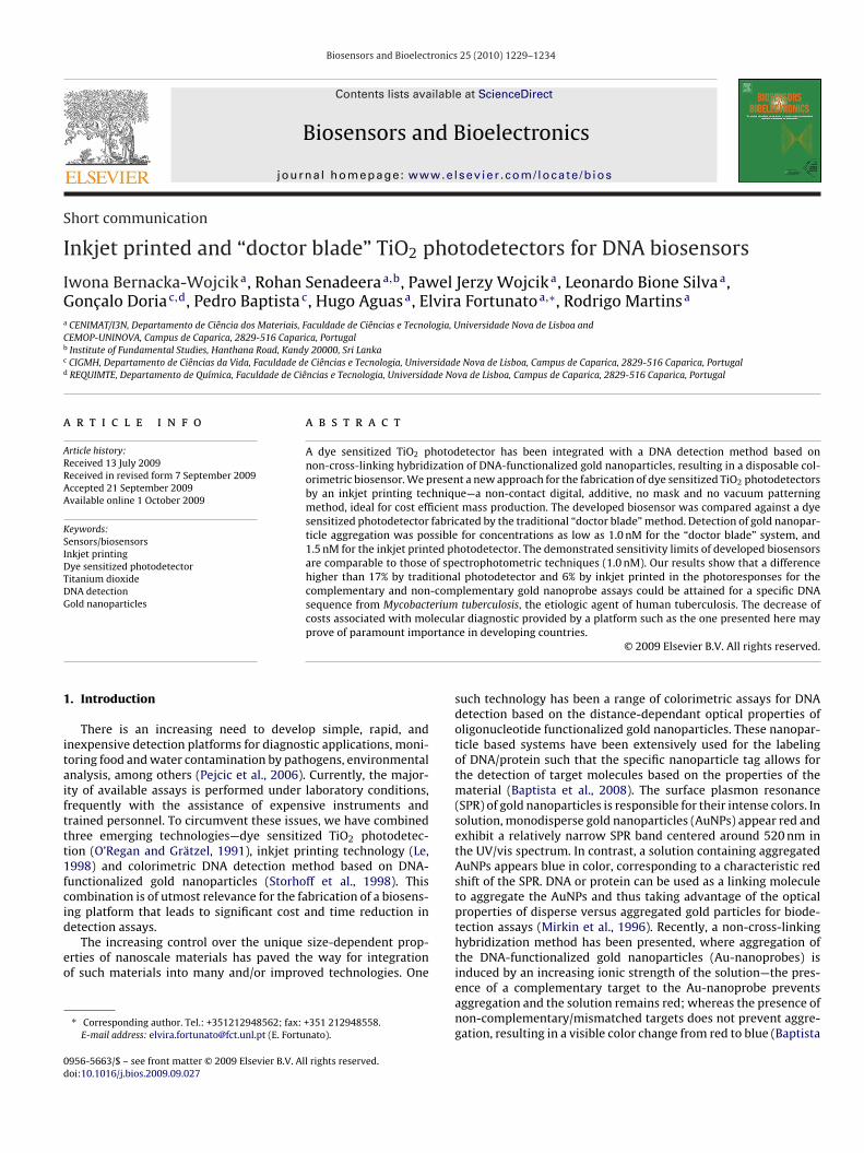
Fig. 1 shows a schematic of the production of both photodetec-tors. The inset shows a scanning electron micrograph of the surfacemorphology of the TiO2 film prepared by inkjet printing technol-ogy after sintering at 450 ◦C. No cracks on the surface are observedwhich indicates a very high inter-particle connectivity. The averagediameter of TiO2 particles is 25 nm.
2.2. Characterization of photodetectors
The effectiveness of the photosensor to convert light of variouswavelengths into electrical current was measured as the incidentphoton to current conversion efficiency (IPCE). IPCE (%) is definedas the number of electrons generated by light per number of pho-tons incident on the photodetector, formulated by the simplifiedequation:
IPCE = 1240Jsc
�Wi(1)
where Jsc is the short-circuit current density (�A cm−2), � the exci-tation wavelength (nm) and Wi is the photon flux (W m−2). TheIPCE was measured using a homemade setup coupled to a KeithleyMultimeter 238 (Keithley Instruments, Inc., Cleveland, USA) via acomputer.
Thickness of TiO2 layers was determined by a profilometerAmbios Technology XP-200 (Ambios Technology, Inc., Santa Cruz,USA). Scanning electron micrograph of inkjet printed TiO2 layerwas done using Hitachi S4100 (Hitachi, Tokyo, Japan). Absorptionspectrum of ethanolic solution of the Ruthenium dye was mea-sured using a UV/vis/NIR scanning spectrophotometer UV-3101PC(Shimadzu, Kyoto, Japan).
2.3. Preparation of DNA-functionalized gold nanoparticles
All oligonucleotides were purchased from STAB Vida, Oeiras,Portugal. The Au-nanoprobes were synthesized by derivatizingan aqueous solution of AuNPs with a thiolated oligonu-

I. Bernacka-Wojcik et al. / Biosensors and Bioelectronics 25 (2010) 1229–1234 1231
F printio e filmh
cslboccA
2
PAhK
ig. 1. Schematic of the TiO2 photodetector fabrication by “doctor blade” and inkjetf the TiO2 film prepared by inkjet printing technology after sintering at 450 ◦C. Thigh inter-particle connectivity. The average diameter of TiO2 particles is 25 nm.
leotide (5′-thiol-GGACGTGGAGGCGATC-3′) recognizing a specificequence within the rpo-beta gene of Mycobacterium tubercu-osis (GenBank accession no. BX842574) and used as describedy Baptista et al. (2006), with either a complementary DNAligonucleotide harboring part of the rpo-beta gene of M. tuber-ulosis (5′-GATCGCCTCCACGTCCGTTGGTATCAAGGTT-3′) or a non-omplementary DNA oligonucleotide (5′-GGTCGTCAGACTGTCG-TGAAGCC-3′), both at a final concentration of 1.33 �M.
.4. Detection procedure
In the prototype biosensor, plastic cuvettes (10 mm path length;lastibrand, Wertheim, Germany), filled with 75 �l of AuNPs oru-nanoprobes solution, were placed between the light source (aigh power LED, 530 nm light, 35 nm spectral half-width, Luxeon®
2 Emitter; Quadica Developments Inc., Brantford, Canada) and
ng technique. The scanning electron micrograph presented the surface morphologyis about 4 �m thick, no cracks on the surface are observed which indicates a very
the photodetector. The entire platform is placed within a blackenvironment, in order to eliminate ambient light interference.Photocurrents generated by photodetectors were measured bya digital electrometer (Keithley 238; Keithley Instruments, Inc.,Cleveland, USA) connected to a computer via USB/GPIB interfacecard (Agilent® 82357A; Agilent Technologies, Santa Clara, USA)with developed software (MatLab®).
Measurements were performed after 15 min of salt addition toAuNPs or Au-nanoprobes solution (0.02 M NaCl or MgCl2, respec-tively). The measurements were repeated 6 times for each samplewith a time interval of 150 s. Ultra pure water (Millipore, Billerica,
USA) and 10 mM phosphate buffer (pH 8), 0.1 M NaCl were usedas reference solutions for AuNPs and Au-nanoprobes, respectively.For validation of the prototype, UV-3101PC and UVmini-1240UV/vis/NIR scanning spectrophotometers (Shimadzu, Kyoto, Japan)were used for AuNPs and Au-nanoprobes measurements, respec-
1 s and Bioelectronics 25 (2010) 1229–1234
ta2r2
ucuO
3
smDawobiipwctamswmdcianms
ctt
Fig. 2. Incident photon to current conversion efficiency (IPCE) of the photodetectorobtained by “doctor blade” method (DBP) and by inkjet printing (IPP). The TiO2 layerof DBP is about 1.75 times thicker than in the case of the IPP, which may explain the
Farmm
232 I. Bernacka-Wojcik et al. / Biosensor
ively. DNA detection reactions were carried out with theppropriate target and Au-nanoprobes at a final concentration of.5 nM, in 10 mM phosphate buffer (pH 8). After 10 min of denatu-ation at 95 ◦C, the mixtures were allowed to slowly cool down to0 ◦C until salt addition.
The reported expanded uncertainties are based on a standardncertainty multiplied by a coverage factor k = 2.65, providing aoverage probability of approximately 95%. The uncertainty eval-ation has been carried out in accordance with Internationalrganization for Standardization requirements.
. Results and discussion
We developed a disposable biosensor integrating a dye sen-itized TiO2 photodetector obtained by standard “doctor blade”ethod (from now on referred as “doctor blade” photodetector,BP; Smestad, 1998) and by inkjet printing technology (describeds inkjet printed photodetector, IPP) to be used in conjunctionith the Au-nanoprobe based method. In the case of fabrication
f the IPP, through an improved formulation of specific water-ased inks (Section 2), we attained a TiO2 photodetector by thermal
nkjet printing technology. The inks must meet strict physicochem-cal properties (viscosity, surface tension, etc.) to achieve optimalerformance of printed device. The optimum composition of TiO2ater-based ink was 20 wt% of ethanol and 0.12 wt% of TiO2. Such
omposition resulted in a viscosity that was acceptable for conven-ional office printers (1–10 cP). Additionally, Triton X-100 (5%) wasdded to the ink, which resulted in more effective jetting whileaintaining a well defined and uniform film layer. The disper-
ion of TiO2 does not undergo a sedimentation process (particlesith diameter smaller than 1.2 �m). Long term viscosity measure-ents showed that this dispersion is also free of aggregation and
emonstrated a greater chemical stability of crystal over solution inomparison with sol–gel ink. Highly conductive contact tracks fromnkjet printed silver nanoparticle further helped in the stabilitynd performance of this photodetector. This inkjet printing tech-ique may circumvent the existing time-consuming and expensiveicrofabrication of conductive tracks by lithography or electroly-
is.Fig. 2 depicts the incident photon to current conversion effi-
iencies (IPCE) of the traditional DBP and novel IPP. The inset ofhe figure shows the absorption spectrum of the ethanolic solu-ion of Ruthenium (N3) dye. While the absorption spectrum of
ig. 3. Responses of the sensor with (A) “doctor blade” photodetector and (B) inkjet printedggregated by salt addition (blue circles). The detection response (Rdet)—difference in theeference solution and through the sample solution. Measurements using a high power LEDeasurements: absorption peak area integral from 520 to 540 nm as a function of AuNeasurements. (For interpretation of the references to color in this figure legend, the rea
observed difference at the IPCE. Inset: Absorption spectrum of the ethanolic solutionof Ruthenium N3 dye.
the N3 in ethanol showed an intense absorption at ∼539 nm, theIPCE peaks of DBP and IPP were at 537 and 525 nm, respectively.As it is generally observed with the dye sensitized photodevices,the close relationship and shifts of the IPCE peaks with the opticalabsorption spectra indicate that the photoelectrochemical con-version is achieved through photosensitization, namely that N3molecules absorbed photons and generated excited electrons, andthe excited electrons were subsequently transferred to the con-duction band of the TiO2 electrode. These shifts mainly depend onthe strength of the chelation of the dye molecules to the semicon-ducting surface and the surface morphology of the semiconductor.For both photodetectors, the maximum IPCE was observed around530 nm, which is in the vicinity of the SPR peak of the monodisperseAu-nanoprobes, confirming the compatibility of the detector andAu-nanoprobes integration in a biosensor. The IPP showed lowerIPCE than the DBP, probably due to the higher film thickness of the
active layer of the DBP, which absorbs more dye molecules, hencecreates higher photocurrent density and the IPCE (Junghanel andTributsch, 2005; Kang et al., 2004).photodetector for different AuNP concentrations. Non-aggregated (red squares) andphotocurrent densities generated by photodetector when illuminating through the
with 530 and 35 nm spectral half-width. Inset: Corresponding spectrophotometricPs concentration. Standard deviation bars were determined from 6 independentder is referred to the web version of this article.)

s and Bioelectronics 25 (2010) 1229–1234 1233
aPtrcr
tsstsrtwtsopswssn(tTatasigost(lgslmasAduattldo
jsenPwtamAc
Fig. 4. (A) Schematic representation of DNA detection of the developed biosen-sor (Wi—the photon flux and Rdet—detection responses). (B) The Au-nanoprobebased assays for M. tuberculosis DNA with the novel sensors with DBP “andIPP: POS—complementary DNA target; NEG—non-complementary DNA target;Rdet—difference in the photocurrent densities generated by photodetector when illu-minating through the reference solution and through the sample solution. Inset:Absorption spectra of DNA solutions (in arbitrary units). The absorption spec-trum of NEG was shifted for the same absorbance value at � = 400 nm as POS(shift = −0.0067 a.u.) so as to allow better color change visualization. DNA detection
I. Bernacka-Wojcik et al. / Biosensor
The fabricated photodetectors were individually mounted onplatform inside a black box to cut-off incident background light.lastic cuvettes filled with each solution were then placed betweenhe light source (green LED) and the photodetector. The photocur-ent generated by the photodetector due to the non-absorbed lightoming out from the cuvette was measured by an electrometer andelayed to the computer.
Several non-aggregated AuNPs solutions (red) of known concen-rations were used to assess the response and the sensitivity of theensors, and compared with data attained by conventional UV/vispectrophotometry. Fig. 3A and B shows the results obtained usinghe novel platforms with either DBP or IPP, respectively. The insethows the corresponding spectrophotometry results. The detectionesponses (Rdet) were calculated by measuring the differences inhe photocurrent densities obtained for the reference (ultra pureater and phosphate buffer for AuNPs and Au-nanoprobes, respec-
ively) and the sample solution. Again, the sensor with the IPPhowed lower photoresponses probably due to the lower thicknessf the active layer compared to the device with DBP. To com-are the responses of the developed sensors with those of thepectrophotometer, the integral of the absorption peak over theavelength range between 520 and 540 nm (spanning the emis-
ion spectra of the LED) was used. The output signals of both theensors and the spectrophotometer show a linear variation for theon-aggregated particles (red color solution) with high accuracyR2 > 0.98). In the case of the aggregated particles (blue color solu-ion), a non-linear variation is observed for both methodologies.his can be explained by the several different events occurring (e.g.ggregation, flocculation, precipitation and possible adsorption tohe plastic surface of the cuvette), which lead to light scatteringnd decrease the amount of light being transmitted through theolution. Nevertheless, in terms of application as biosensor whats relevant is the capability of discrimination between the aggre-ated and non-aggregated forms of the AuNPs, and not the linearityf these responses. Very clear differences in the responses of theensors were observed for the AuNPs solution concentrations rou-inely used in the Au-nanoprobe based DNA detection methodfrom 1.5 to 2.5 nM; Baptista et al., 2006; Doria et al., 2007). Forow concentrations, below 1.0 nM for DBP and 1.5 nM for IPP, theenerated photocurrent densities were nearly the same for botholutions indicating the sensitivity limit of the sensor. The detectionimit of the spectrophotometer is superior to 0.5 nM. The measure-
ents were consistent and reproducible, and comparable to thosettained for spectrophotometry, showing that the developed sen-ors may be a low cost and effective alternative for use with theuNPs based assay. We observe larger fluctuations of the acquiredata in the comparison with spectrophotometry, because we aresing a LED, which does not have a single wavelength emission butn emission profile of 30 nm half-width peak. This together withhe considerable noise introduced by the system, e.g. photodetec-or, cables, power supply and electrometer is responsible for thearger uncertainty bars. All these factors causing fluctuations duringata acquisition need to be dealt upon at a later stage of prototypeptimization.
Finally, as a proof-of-concept, the sensors were applied in con-unction with the Au-nanoprobe assay to detect a specific DNAequence (Fig. 4) from M. tuberculosis (Baptista et al., 2006), thetiologic agent of human tuberculosis. Mixtures containing the Au-anoprobe and complementary oligonucleotide target (positive,OS—red) or non-complementary target (negative, NEG—blue),ere screened by both the developed sensors and the spectropho-
ometer. The Au-nanoprobe aggregation (non-complementaryssay) induced by salt addition resulted in the shift of the SPR maxi-um of 26 nm compared to the complementary assay (Fig. 4, inset).clear difference in the detection response of the sensors between
omplementary (POS) and non-complementary (NEG) assays is
was carried out with the appropriate oligonucleotide target at a final concentrationof 1.33 �M and Au-nanoprobes at a final concentration of 2.5 nM. Standard deviationbars were determined from 6 independent measurements.
observable in the �A range. The percentage difference betweendetection response for complementary and non-complementaryassays was 17.5% for the DBP and 6.7% for the IPP (Fig. 4B). Eventhough the inkjet printed photodetectors gave lower differenceof detection responses than photodetectors fabricated by the tra-
ditional method, it should be noted that the inkjet printing isemployed for the first time in the dye sensitized TiO2 technology,and the properties of IPPs still need to be fully optimized. Neverthe-less, the IPP based platform was able to clearly detect the presenceor the absence of a specific DNA target.
1 s and B
4
dtplsmasctdnopAgirdnptdtwopbprm
A
c
Commun. 17, 2259–2261.Silva, L.B., Baptista, P., Raniero, L., Doria, G., Martins, R., Fortunato, E., 2008. Sens.
Actuator B: Chem. 132, 508–511.Smestad, G.P., 1998. Sol. Energy Mater. Sol. Cells 55, 157–178.Storhoff, J.J., Elghanian, R., Mucic, R.C., Mirkin, C.A., Letsinger, R.L., 1998. J. Am. Chem.
Soc. 120, 1959–1964.
234 I. Bernacka-Wojcik et al. / Biosensor
. Conclusions
To our knowledge the use of dye sensitized TiO2 sensors for DNAetection by means of Au-nanoprobes has been demonstrated forhe first time. Moreover, we describe here pioneer results of inkjetrinting technology application to the fabrication of the active
ayers of photodetectors. The developed biosensor including dyeensitized photodetector fabricated by traditional “doctor blade”ethod detected the aggregation of AuNPs concentrations as low
s 1.0 nM, while for the inkjet printed photodetector the limit waset at 1.5 nM. The sensitivity limits of the developed biosensors areomparable to the sensitivity limit of the spectrophotometry basedechniques (1.0 nM). Both photodetectors gave clear and distinctifferences in the detection responses for the complementary andon-complementary DNA assays. Our results show that a differencef 17.5% by traditional photodetector and 6.7% by inkjet printed inhotoresponses for the complementary and non-complementaryu-nanoprobe assays could be attained. The DBP gives nearly asood results as spectrophotometry, while usability and reliabil-ty of the established IPP used in DNA sequence identification stillequire further experimental development, mainly concerning theetermination of the most adequate thickness and role of TiO2anoparticles size on the overall device performance. Moreover, thehotodetectors’ sensitivity can be further improved by enhancinghe spectral responses of the detector through the use of differentyes. Once the photodetectors have been optimized, we will applyhe same methodology to other relevant DNA target sequences andith increased complexity. Further research is being also carried
ut to evaluate the possible association within a fully integrated,ortable biosensor by means of microfluidic technology. The com-ination of three emerging technologies: (i) dye sensitized TiO2hotodetection, (ii) inkjet printing technology and (iii) colorimet-ic DNA detection based on DNA-functionalized gold nanoparticlesay lead to significant cost and time reduction in DNA detection.
cknowledgements
The authors acknowledge Bruno Veigas, DCV–FCT/UNL forontrol DNA and Rede Nacional de Microscopia Electrónica
ioelectronics 25 (2010) 1229–1234
and the Pólo de Aveiro (FCT Project: REDE/1509/RME/2005)for performing SEM measurements. The work partially sup-ported by FCT-MCTES through program contract of CENIMAT/I3Nand CIGMH; and project PTDC/FIS/74274/2006, PTDC/SAU-BEB/66511/2006, PTDC/FIS/742/2006, SFRH/BD/44258/2008 forIB-W, PTDC/EEA-ELC/64975/2006 and SFRH/BD/45224/2008 forPJW, SFRH/BD/24665/2005 for LBS, and SFRH/BDE/15544/2005and STAB Vida, Lda for GD. RS acknowledges the postdoctoralposition awarded under Ciência 2007 and the leave granted fromthe Institute of Fundamental Studies, Sri Lanka.
References
Baptista, P., Koziol-Montewka, M., Paluch-Oles, J., Doria, G., Franco, R., 2006. Clin.Chem. 52, 1433–1434.
Baptista, P., Pereira, E., Eaton, P., Doria, G., Miranda, A., Gomes, I., Quaresma, P.,Franco, R., 2008. Anal. Bioanal. Chem. 391, 943–950.
Calvert, P., 2001. Chem. Mater. 13, 3299–3305.Doria, G., Franco, R., Baptista, P., 2007. IET Nanobiotechnol. 1, 53–57.Fortunato, E., Martins, R., Baptista, P., FCT-UNL, 2006. Patent: 103561-PT.Hart, A.L., Turner, A.P.F., Hopcroft, D., 1996. Biosens. Bioelectron. 11, 263–270.Junghanel, M., Tributsch, H., 2005. J. Phys. Chem. B 109, 22876–22883.Kang, M.G., Ryu, K.S., Chang, S.H., Park, N.G., Hong, J.P., Kim, K.J., 2004. Bull. Korean
Chem. Soc. 25, 742–744.Le, H.P., 1998. J. Imaging Sci. Technol. 42, 49–62.Martins, R., Baptista, P., Raniero, L., Doria, G., Silva, L., Franco, R., Fortunato, E., 2007.
Appl. Phys. Lett., 90, 023903(1–3).Mirkin, C.A., Letsinger, R.L., Mucic, R.C., Storhoff, J.J., 1996. Nature 382, 607–609.O’Regan, B., Grätzel, M., 1991. Nature 353, 737–740.Pejcic, B., De Marco, R., Parkinson, G., 2006. Analyst 131, 1079–1090.Schena, M., Heller, R.A., Theriault, T.P., Konrad, K., Lachenmeier, E., Davis, R.W., 1998.
Trend Biotechnol. 16, 301–306.Senadeera, G.K.R., Jayaweera, P.V.V., Perera, V.P.S., Tennakone, K., 2002. Sol. Energy
Mater. Sol. Cells 73, 103–108.Senadeera, R., Fukuri, N., Saito, Y., Kitamura, T., Wada, Y., Yanagida, S., 2005. Chem.
Copyright © 2022 FDOKUMEN