Integrated electrochemical DNA biosensors for lab-on-a-chip devices
12
Review Integrated electrochemical DNA biosensors for lab-on-a-chip devices Analytical devices able to perform accurate and fast automatic DNA detection or sequen- cing procedures have many potential benefits in the biomedical and environmental fields. The conversion of biological or biochemical responses into quantifiable optical, mechanical or electronic signals is achieved by means of biosensors. Most of these transducing elements can be miniaturized and incorporated into lab-on-a-chip devices, also known as Micro Total Analysis Systems. The use of multiple DNA biosensors integrated in these miniaturized laboratories, which perform several analytical operations at the microscale, has many cost and efficiency advantages. Tiny amounts of reagents and samples are needed and highly sensitive, fast and parallel assays can be done at low cost. A particular type of DNA biosensors are the ones used based on electrochemical principles. These sensors offer several advantages over the popular fluorescence-based detection schemes. The resulting signal is electrical and can be processed by conventional electronics in a very cheap and fast manner. Furthermore, the integration and miniaturization of electro- chemical transducers in a microsystem makes easier its fabrication in front of the most common currently used detection method. In this review, different electrochemical DNA biosensors integrated in analytical microfluidic devices are discussed and some early stage commercial products based on this strategy are presented. Keywords: DNA / Electrochemical DNA biosensors / Electrochemistry / Lab-on-a-chip / Micro Total Analysis systems DOI 10.1002/elps.200900319 1 Introduction One of the analytical applications that have caught more attention during the last 25 years is DNA detection and sequencing. This is because it has many potential benefits in the biomedical and environmental fields, and also in other less popularly known areas such as forensics or archeology. Integrating DNA sensors with lab-on-a-chip (LOC) devices opens the possibilities of developing portable equipments that would be very useful for point-of-care medical screenings or any on-the-field analysis. By definition, LOC devices are microfluidic devices able to perform complete laboratory assays that can be alternatively referred as Micro Total Analysis systems when the application is of an analytical nature. The automation and integration of one or several laboratory operations in small devices goes back to the early 1990s. It was soon regarded as a means to obtain higher efficiency, faster analysis time, and lower reagent consumption, than other existing analysis techniques [1]. In 1998, a more advanced device was presented [2]. A single closed system with dimensions of a few squared centimeters and a height of 1 mm was developed in order to prepare and analyze nanoliter-sized DNA samples by gel electrophoresis, containing microfabricated fluidic channels, heaters, temperature sensors, and fluorescence detectors. The main aim of developing miniaturized and inte- grated devices is to reduce the assay cost and also the interaction of the sample with external factors, reducing the sample contamination and making the analysis cleaner and less hazardous. However, they also present some other advantages derived from scaling down chemical processes to the microscale such as diffusion mixing efficiency enhancement, improvement of heat transport, decreased reaction time, and formation of by-products, bringing as a result higher reaction yields [3]. DNA sample preparation steps, including complex processes such as the PCR have been demonstrated to be achievable on the microscale making such LOC devices very suitable for complete DNA analysis when integrating detection elements. The PCR is a very desirable step when only tiny amounts of the DNA sample are available since it is Mo ` nica Mir 1,2 Antoni Homs 1,2,3 Josep Samitier 1,2,3 1 Nanobioengineering group, Institute for Bioengineering of Catalonia, Barcelona, Spain 2 Centro de Investigacio ´n Biome ´dica en Red en Bioingenierı ´a, Biomateriales y Nanomedicina, Spain 3 Department of Electronics, University of Barcelona, Barcelona, Spain Received May 15, 2009 Revised July 10, 2009 Accepted July 11, 2009 Abbreviations: ALP, alkaline phosphatase; CG, colloidal gold; CMOS, complementary metal-oxide-semiconductor; CNT, carbon nanotubes; FET, field-effect transistors; HRP, horseradish peroxidase; a-HL, a-hemolysin; IDE, interdigitated electrode; LOC, lab-on-a-chip; NTFET, nanotube field-effect transistor; NW, nanowires; SiNW, silicon nanowires; SWNT, single wall nanotube Correspondence: Dr. Mo ` nica Mir, Intitut de BioEnginyeria de Catalunya Baldiri Reixac, 13, Barcelona 08028, Spain E-mail: [email protected] Fax: 134-9340319702 & 2009 WILEY-VCH Verlag GmbH & Co. KGaA, Weinheim www.electrophoresis-journal.com Electrophoresis 2009, 30, 3386–3397 3386
Transcript of Integrated electrochemical DNA biosensors for lab-on-a-chip devices

Review
Integrated electrochemical DNA biosensorsfor lab-on-a-chip devices
Analytical devices able to perform accurate and fast automatic DNA detection or sequen-
cing procedures have many potential benefits in the biomedical and environmental fields.
The conversion of biological or biochemical responses into quantifiable optical, mechanical
or electronic signals is achieved by means of biosensors. Most of these transducing
elements can be miniaturized and incorporated into lab-on-a-chip devices, also known as
Micro Total Analysis Systems. The use of multiple DNA biosensors integrated in these
miniaturized laboratories, which perform several analytical operations at the microscale,
has many cost and efficiency advantages. Tiny amounts of reagents and samples are
needed and highly sensitive, fast and parallel assays can be done at low cost. A particular
type of DNA biosensors are the ones used based on electrochemical principles. These
sensors offer several advantages over the popular fluorescence-based detection schemes.
The resulting signal is electrical and can be processed by conventional electronics in a very
cheap and fast manner. Furthermore, the integration and miniaturization of electro-
chemical transducers in a microsystem makes easier its fabrication in front of the most
common currently used detection method. In this review, different electrochemical DNA
biosensors integrated in analytical microfluidic devices are discussed and some early stage
commercial products based on this strategy are presented.
Keywords:
DNA / Electrochemical DNA biosensors / Electrochemistry / Lab-on-a-chip /Micro Total Analysis systems DOI 10.1002/elps.200900319
1 Introduction
One of the analytical applications that have caught more
attention during the last 25 years is DNA detection and
sequencing. This is because it has many potential benefits in
the biomedical and environmental fields, and also in other less
popularly known areas such as forensics or archeology.
Integrating DNA sensors with lab-on-a-chip (LOC) devices
opens the possibilities of developing portable equipments that
would be very useful for point-of-care medical screenings or
any on-the-field analysis. By definition, LOC devices are
microfluidic devices able to perform complete laboratory
assays that can be alternatively referred as Micro Total Analysis
systems when the application is of an analytical nature.
The automation and integration of one or several
laboratory operations in small devices goes back to the early
1990s. It was soon regarded as a means to obtain higher
efficiency, faster analysis time, and lower reagent
consumption, than other existing analysis techniques [1]. In
1998, a more advanced device was presented [2]. A single
closed system with dimensions of a few squared centimeters
and a height of 1 mm was developed in order to prepare and
analyze nanoliter-sized DNA samples by gel electrophoresis,
containing microfabricated fluidic channels, heaters,
temperature sensors, and fluorescence detectors.
The main aim of developing miniaturized and inte-
grated devices is to reduce the assay cost and also the
interaction of the sample with external factors, reducing the
sample contamination and making the analysis cleaner and
less hazardous. However, they also present some other
advantages derived from scaling down chemical processes to
the microscale such as diffusion mixing efficiency
enhancement, improvement of heat transport, decreased
reaction time, and formation of by-products, bringing as a
result higher reaction yields [3].
DNA sample preparation steps, including complex
processes such as the PCR have been demonstrated to be
achievable on the microscale making such LOC devices very
suitable for complete DNA analysis when integrating
detection elements. The PCR is a very desirable step when
only tiny amounts of the DNA sample are available since it is
Monica Mir1,2
Antoni Homs1,2,3
Josep Samitier1,2,3
1Nanobioengineering group,Institute for Bioengineering ofCatalonia, Barcelona, Spain
2Centro de InvestigacionBiomedica en Red enBioingenierıa, Biomateriales yNanomedicina, Spain
3Department of Electronics,University of Barcelona,Barcelona, Spain
Received May 15, 2009Revised July 10, 2009Accepted July 11, 2009
Abbreviations: ALP, alkaline phosphatase; CG, colloidalgold; CMOS, complementary metal-oxide-semiconductor;CNT, carbon nanotubes; FET, field-effect transistors; HRP,
horseradish peroxidase; a-HL, a-hemolysin; IDE,
interdigitated electrode; LOC, lab-on-a-chip; NTFET,
nanotube field-effect transistor; NW, nanowires; SiNW,
silicon nanowires; SWNT, single wall nanotube
Correspondence: Dr. Monica Mir, Intitut de BioEnginyeria deCatalunya Baldiri Reixac, 13, Barcelona 08028, SpainE-mail: [email protected]: 134-9340319702
& 2009 WILEY-VCH Verlag GmbH & Co. KGaA, Weinheim www.electrophoresis-journal.com
Electrophoresis 2009, 30, 3386–33973386

able to amplify a single or few copies up to several orders of
magnitude. This technique is based on the application of
repeated thermal cycles to replicate a target DNA on a solution
containing the mediating DNA polymerase enzyme and
primers (short DNA fragments) complementary to the target.
The increasing demand of cheap, highly accurate, fast
and easy to use analytical assays in the fields of molecular
diagnostics, drug discovery, and pharmaceutical screening
boosted the research in this field at the end of the last
century. The LOC concept has achieved a wide popularity
since its first proposal had proven an effective way to achieve
highly sensitive and complex laboratory assays with mini-
mal effort from the analyst, even it can be used by non-
skilled persons. It has also opened the possibilities of
portable analysis for on-the-field applications due to the
highly reduced dimensions of the devices. Furthermore,
their reduced cost and effectivity makes them a suitable
candidate to be used for global public health applications
even in the third world [4]. Whereas optical detection
methods with fluorescent dyes have dominated the DNA
sensor industry and surface plasmon resonance and piezo-
electric techniques can achieve low limits of detection, the
application of electrochemical read out can provide signifi-
cant advantages in LOC devices. Electrochemistry detection
provides high sensitivity, small dimensions, low cost, fast
response, easy signal integration, and compatibility with
microfabrication technology, making this transducer the
best candidate for the integration in microchip devices.
Accordingly to their detection working principle elec-
trochemical DNA biosensors can be classified as ampero-
metric, voltamperometric, potentiometric, or impedimetric.
Generally, biomolecular recognition takes place close to the
surface of an electrode. For this reason, the electrode
shape, dimension, construction material, and surface prop-
erties play a very important role on the performance of
these biosensors. In most of the cases, the complete
electrochemical detection is achieved by a combination
of a working electrode where the sensing takes place and
a counter reference electrode that should maintain a
stable potential not dependent on the particular detection
reaction but only on the media. These factors have to be
taken into deep consideration when miniaturizing this type
of sensors.
Integration of electrochemical DNA biosensors on
analytical microfluidic devices is a promising approach that
has been already developed in research, as well as in some
commercial biomedical products that are discussed through
the article. However, it still presents several challenges that
should be addressed in the fore coming years in order to
achieve their massive use.
2 Amperometric DNA biosensors
These types of sensors are generally based on the measure-
ment of resulting current in oxidation or reduction
processes of electroactive species in a biochemical reaction
[5]. Amperometric detection in biosensors generally involves
the use of enzymes as biomarker molecules. The read-out of
the enzyme needs the interaction with a specific substrate,
which is converted catalytically in a different molecule. The
product obtained is a redox molecule that can be detected
with this technique and with the advantage that the enzyme
catalyses the reaction, generating signal amplification.
Although the majority of amperometric sensors immo-
bilize the bioreceptor on the electrode surface, in the case of
enzymatic labels it is not necessary, while the redox product
generated with the substrate can diffuse to the electrode in
order to be detected. In this case, the DNA probe is attached
on the PDMS channels [6]. The PDMS channels were
oxidized with plasma, in order to activate the surface for
silane coupling. The silane molecules bear thiol groups
where the maleimide labeled capture probes are attached,
where the target labeled with alkaline phosphatase (ALP)
hybridize. Once it is immobilized on the channel, the
substrate of the enzyme is injected and a redox active
molecule is produced which flows until reaching the indium
tin oxide interdigitated electrode (IDE). In this work a limit
of detection of 1 nM was reported. However, reproducibility
was not tested and the plotted results indicate that the 1 nM
signal had minimum differences with the blank, a more
reliable detection limit for their experiments being 10 nM.
This detection configuration provides a large bioreceptor
surface and since these molecules are not immobilized on
the electrode, as commonly in electrochemical biosensors, it
does not present a barrier that hinders access and detection
of the redox molecules. However, this system is difficult to
use in an array format, since each array spot would need a
specific microchannel.
The combination of amperometric sensors with inte-
grated electronic circuitry and components have recently
brought to market biochips with huge electrochemical
genetic arrays [7]. Combimatrix has commercialized a LOC
device having an integrated multielectrode array able to
perform parallel immunoassays to detect viruses and
bacteria in complex biological samples [8]. The chip has
sample processing capabilities and the sensing part is
composed of individually addressable electrodes controlled
by active complementary metal-oxide-semiconductor
(CMOS) circuitry. This company presented and oligonu-
cleotide microarray platform that provides 12 544 platinum
electrodes, having a diameter of 44 mm/cm2 and coated with
a porous reaction layer. A platinum grid surrounding the
electrodes works as a counter electrode. Different oligo-
deoxynucleotide probes were synthesized at each sensing
microelectrode.
The sensing approach is based on the detection of redox
active chemistries proximal to specific microarray electro-
des. Microarray probes are hybridized to biotin-labeled
targets and then the horseradish peroxidase (HRP)–strep-
tavidin conjugate binds to the biotin molecule, causing an
enzymatic oxidation of the electron donor substrate. The
hybridization is detected by the current generated in the
electro-reduction of the HRP. It is important to notice that
Electrophoresis 2009, 30, 3386–3397 Microfluidics and Miniaturization 3387
& 2009 WILEY-VCH Verlag GmbH & Co. KGaA, Weinheim www.electrophoresis-journal.com
this complete platform obtained lower detection limits
(0.75 pM) than its fluorescent detection counter part
(1.5 pM), characterizing the gene expression assay [8]. This
platform has been used to develop nucleic acid assays for
highly accurate genotyping of a variety of common and bio-
thread pathogens.
2.1 Amperometric sandwich
One of most common electrochemical biosensor platform
that has been adapted to be integrated in microfluidic
systems is the sandwich configuration that permits label
free target detection. In the sandwich system two capture
probes are involved, one immobilized on the electrode
surface, where the target hybridize, and a second tagged
probe affine to the non-hybridized segment of the target
(Fig. 1).
In the work reported by Liao et al. [9, 10] seven different
thiolated capture probes from different bacterial species
were self-assembled on screen-printed gold electrodes. A
DNA sequence of 16S rRNA was amplified with PCR. This
technique permits the in vitro cloning of a specific DNA
sequence as well as the labeling of the amplified sequence.
The amplified sequence was hybridized on the capture
probes of the differently functionalized screen-printed
electrochemical cells. The signaling HRP marked serves to
label the target upon hybridization. The electroreduction
current is generated by hydrogen peroxide mediated by
tetramethylbenzidine), obtaining a low detection limit of
12 pM. The microchip design was very simple and it
was composed of 16 individual electrochemical cells
connected to the same microchannel. This channel is inte-
grated to an external macro system for separation and
concentration of pathogens in urine specimens. Conse-
quently, reported pre-treatment is still far to be an integrated
micro-laboratory.
In other research works, site addressing by magnets has
been used in microfluidic devices in order to concentrate
and attach on the sensing electrodes surface oligonucleotide
targets. The research group of Prof. Mascini reported an
enzyme-linked sandwich on magnetic beads for PCR-
amplified gene sequence of Cor a 1.04 allergen detection
[11]. The sensor platform developed starts with a streptavi-
din-coated magnetic beads. Owing to the strong streptavi-
din–biotin interaction, this coated particles allow an easily
and stable biotinylated capture probe immobilization. The
PCR-amplified oligonucleotide target was incubated with
the functionalized particles and this hybridization event was
enlightened by means of a second biotinylated probe that
interact with the non-hybridized part of the target sequence.
This third oligonucleotide interacts with streptavidin-ALP,
which can be easily detected by amperometry. An incon-
venience of this system, with two different biotinylated
oligonucleotides in the sandwich system, is the requirement
of a blocking step with biotin in between, in order to avoid
the adsorption of the second biotinylated strand on the
streptavidin surface. The microfluidic cartridge is commer-
cialized by DiagnoSwiss SA under the ImmuChipTM name.
The chip consists of eight microchannels etched in poly-
amide. Each microchannel contains eight carbon screen-
printed working electrodes and in the upper part of the
channel screen-printed counter and reference electrodes.
The amperometric detection limit obtained from the ALP
label was 0.2 nM, obtaining a negligible response from the
non-complementary sequences.
A similar sensoric platform was used in the chip
developed by Gabig-Ciminska et al. [12], who also use a
sandwich format labeled with ALP on streptavidin magnetic
beads. This device was developed for the analysis of 16S
rDNA in Escherichia coli extracts. The chip consists of four
pairs of gold IDE patterned with lift-off on silicon wafer and
encapsulated with silicon oxynitride. The target was ampli-
fied by PCR and detected amperometrically on the chip. The
selectivity of the capture probe and the non-specific
adsorption of the label were tested, obtaining a good anti-
fouling with BSA. The detection limit reported with this
chip was 1 nM.
3 Voltammetric DNA biosensors
Voltammetric technique determines the amount of matter
transformed during a redox reaction when the potential
varies with time in a pre-determined manner and the
current is measured as a function of potential. Redox
molecules are detected with this technique [13].Figure 1. Schematic of the amperometric detection mechanismbased on a sandwich conformation.
Electrophoresis 2009, 30, 3386–33973388 M. Mir et al.
& 2009 WILEY-VCH Verlag GmbH & Co. KGaA, Weinheim www.electrophoresis-journal.com
3.1 Voltammetric sandwich
The e-SENSORTM reported from Motorola in 2001 [14] is a
voltammetric sandwich-based sensor that six years later was
incorporated in a fully integrated biochip [15]. This is a full-
integrated cartridge that comprises different units for pre-
treatment and detection of the sample. This LOC integrates
a mixing unit for rare cell capture using immuno-magnetic
separation, which is a useful tool in order to enrich rare
target cells for a large volume sample, a single chamber unit
for cell pre-concentration, purification, lyses and PCR, in
order to simplify the chip, and a DNA microarray detection
chamber. The chip was fabricated in plastic, with a
patterned printed circuit board and a 4� 4 gold microarray
chip. It was tested with real whole blood sample for the
detection of hematochromatosis-associated SNP. All the
sequence of processes, from the blood injection to the signal
readout, takes about 3 h (90 min for the PCR and 60 min for
the detection). The hybridization was detected though a
sandwich assay, using a second probe ferrocene labeled,
detected by alternating current voltammetry.
Magnetic streptavidin-coated beads are used to concen-
trate and detect DNA in a sandwich format also in other
studies [16]. In the work of Baeumner et al. a redox molecule
entrapped in a liposome capsule serves as a label to the
second probe, which recognizes specifically the synthetic
DNA sequence of Dengue virus serotype 3. The hybridized
target flows through soft-lithographed PDMS microchannel
attracted by a magnet to an interdigitated array patterned by
lift-off on glass substrate. Once the magnet immobilizes the
target, the lysis of the liposome is induced with b-octylglu-
copyranoside, realizing the redox molecule entrapped that is
voltammperometrically detected. The encapsulation capacity
of the liposomes brings the advantage of having a high
amount of redox molecules that can be detected in each
hybridized duplex. Oppositely only one redox molecule perhybridized target can be detected in other systems. Never-
theless, this sandwich with liposome label reports a similar
limit of detection (10 nM) as the system with a single label
per duplex. Moreover, the structure of the liposome
capsule could be unstable in certain environments, so in a
complex real sample matrix this kind of labels could not be
efficient.
3.2 Redox intercalators
In order to remove the needs of a label for the hybridization
detection, which is introduced mainly though a sandwich
format or by PCR, redox DNA intercalator molecules can be
used. These kinds of redox molecules have the appropriate
size and chemical nature (polycyclic, aromatic and planar)
which makes them fit in between base pairs of DNA. Thus,
once the double strand is formed, after the target
hybridization with the capture probe, the intercalators are
entrapped in the double helix. These redox molecules are
detected by voltammetric techniques.
Simultaneous DNA amplification by PCR and subse-
quent electrochemical detection was performed in a micro-
well [17]. Platinum temperature sensors, heaters, and gold
electrodes were patterned on top of the silicon substrate that
was covered with a bottom glass substrate for sealing the
chamber. In a 8 mL silicon chamber an asymmetric ampli-
fication was carried out that brings thousands of single
stranded target sequence, which are incubated for 1 h in the
same chamber in order to facilitate its hybridization with
complementary capture probes immobilized on gold elec-
trodes patterned on the chamber top. In order to get rid of
any unhybridized molecule, the chamber was rinsed and
washed. The hybridization was detected with differential
pulse voltammetry by means of redox molecules intercalated
into the double helix. The double function of the chamber
facilitates and reduces cost of the device fabrication and
minimizes the size of the chip.
The hybridization of the target with the capture
probe can also be detected in solution, without previous
immobilization of these molecules [18]. The capture
probe and the synthetic DNA target are introduced
separately into a microchannel, where both flow and
hybridize. The created double strand of DNA was inter-
calated with a redox molecule and it was detected by cyclic
voltamperometry when the double strand flows on the area
where the electrodes were patterned. The hybridization
selectivity was tested with a mismatched sequence obtaining
20% of the signal. Although this chip removes the problem
associated with the immobilization of the DNA receptor,
this configuration does not allow a washing step after
hybridization, which is very important in real sample
with complex matrix that complicates the hybridization
detection.
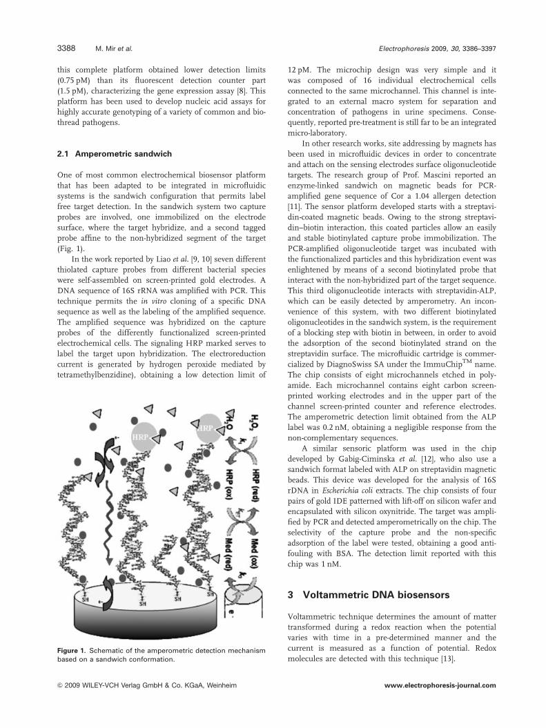
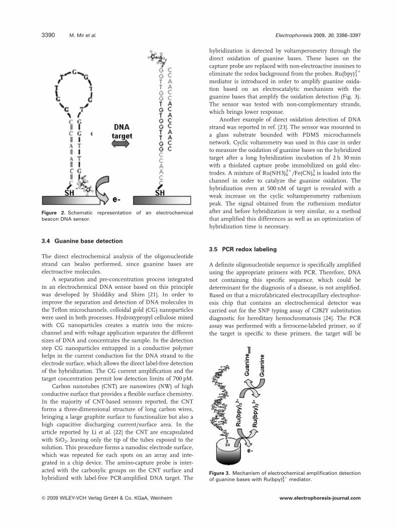
3.3 Electronic beacon
The e-DNA sensor reported by Fan et al., in 2003 [19] was
applied in microfluidic devices five years later [20]. The
ferrocene-labeled DNA stem-loop structure used in the
e-DNA sensor changes its conformation due to target
hybridization, which alters the electron transfer tunneling
distance between the electrode and the ferrocene label that
can be detected by differential pulse voltammetry (Fig. 2). In
this chip three different ferrocene-thiolated beacon probes
were attached on the gold array by means of electronic site-
selective deposition. After use, the electrodes were almost
totally (97%) regenerated by positive potential sweep
desorption. The gold array was patterned on glass and the
microfluidic channels were incorporated in a PDMS slide.
The electrochemical beacon configuration permits a
reagentless and label-free system. Furthermore, the non-
complementary neighboring capture probes showed little
effect on the signal (3%), demonstrating that it is a specific
sensor. However, a background signal of about 60% from
the ferrocene signal remains after hybridization of synthetic
oligonucleotide target.
Electrophoresis 2009, 30, 3386–3397 Microfluidics and Miniaturization 3389
& 2009 WILEY-VCH Verlag GmbH & Co. KGaA, Weinheim www.electrophoresis-journal.com
3.4 Guanine base detection
The direct electrochemical analysis of the oligonucleotide
strand can bealso performed, since guanine bases are
electroactive molecules.
A separation and pre-concentration process integrated
in an electrochemical DNA sensor based on this principle
was developed by Shiddiky and Shim [21]. In order to
improve the separation and detection of DNA molecules in
the Teflon microchannels, colloidal gold (CG) nanoparticles
were used in both processes. Hydroxypropyl cellulose mixed
with CG nanoparticles creates a matrix into the micro-
channel and with voltage application separates the different
sizes of DNA and concentrates the sample. In the detection
step CG nanoparticles entrapped in a conductive polymer
helps in the current conduction for the DNA strand to the
electrode surface, which allows the direct label-free detection
of the hybridization. The CG current amplification and the
target concentration permit low detection limits of 700 pM.
Carbon nanotubes (CNT) are nanowires (NW) of high
conductive surface that provides a flexible surface chemistry.
In the majority of CNT-based sensors reported, the CNT
forms a three-dimensional structure of long carbon wires,
bringing a large graphite surface to functionalize but also a
high capacitive discharging current/surface area. In the
article reported by Li et al. [22] the CNT are encapsulated
with SiO2, leaving only the tip of the tubes exposed to the
solution. This procedure forms a nanodisc electrode surface,
which was repeated for each spots on an array and inte-
grated in a chip device. The amino-capture probe is inter-
acted with the carboxylic groups on the CNT surface and
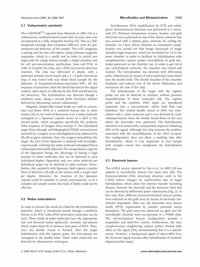
hybridized with label-free PCR-amplified DNA target. The
hybridization is detected by voltamperometry through the
direct oxidation of guanine bases. These bases on the
capture probe are replaced with non-electroactive inosines to
eliminate the redox background from the probes. Ru(bpy)321
mediator is introduced in order to amplify guanine oxida-
tion based on an electrocatalytic mechanism with the
guanine bases that amplify the oxidation detection (Fig. 3).
The sensor was tested with non-complementary strands,
which brings lower response.
Another example of direct oxidation detection of DNA
strand was reported in ref. [23]. The sensor was mounted in
a glass substrate bounded with PDMS microchannels
network. Cyclic voltammetry was used in this case in order
to measure the oxidation of guanine bases on the hybridized
target after a long hybridization incubation of 2 h 30 min
with a thiolated capture probe immobilized on gold elec-
trodes. A mixture of Ru(NH3)631/Fe(CN)6
3 is loaded into the
channel in order to catalyze the guanine oxidation. The
hybridization even at 500 nM of target is revealed with a
weak increase on the cyclic voltamperometry ruthenium
peak. The signal obtained from the ruthenium mediator
after and before hybridization is very similar, so a method
that amplified this differences as well as an optimization of
hybridization time is necessary.
3.5 PCR redox labeling
A definite oligonucleotide sequence is specifically amplified
using the appropriate primers with PCR. Therefore, DNA
not containing this specific sequence, which could be
determinant for the diagnosis of a disease, is not amplified.
Based on that a microfabricated electrocapillary electrophor-
esis chip that contains an electrochemical detector was
carried out for the SNP typing assay of C282Y substitution
diagnostic for hereditary hemochromatosis [24]. The PCR
assay was performed with a ferrocene-labeled primer, so if
the target is specific to these primers, the target will be
Figure 2. Schematic representation of an electrochemicalbeacon DNA sensor.
Figure 3. Mechanism of electrochemical amplification detectionof guanine bases with Ru(bpy)3
21 mediator.
Electrophoresis 2009, 30, 3386–33973390 M. Mir et al.
& 2009 WILEY-VCH Verlag GmbH & Co. KGaA, Weinheim www.electrophoresis-journal.com

amplified and labeled with ferrocene. The remainder of
reagents from the PCR are separated by CE on polyacryla-
mide-coated channels using a hydroxyethylcellulose sieving
matrix at a potential of 400 V/cm. In order to minimize the
effect of the electrophoresis electric field on electrochemical
detection, the working electrodes were placed between
200 mm from the separation channel exit, where the
ferrocene label was detected with voltamperometric techni-
ques. The absence of response obtained with a mutated
sequence corroborates the selectivity of the sensor.
Based on the same electrophoretic separation and
detection principle, a PDMS microchip was developed for
159 bp PCR amplicon detection [25]. In this case the ferro-
cene label was incorporated after the PCR amplification. The
label was attached to a single base that is hybridized to the
amplified double helix. This system is more time consum-
ing than the previous one because this needs an extra step
for the hybridization of a single base labeled that is not
100% efficient. This step could reduce the number of targets
labeled, compared with the labeling through the PCR
primer, with a subsequent decrease in sensitivity.
4 Potentiometric DNA biosensors
Potentiometric biosensors are based on charge potential
changes at the sensing electrode with respect to the
reference one that is usually translated in a voltage change.
On these measurements current should be kept nearly zero.
DNA potentiometric detectors based on current
measurements that result in a charge accumulation effect at
the gate electrode of transistor devices by specific binding of
DNA molecules are also referred as field-effect DNA
biosensors. These DNA biosensors are particularly attractive
since they can be fabricated in very small sizes and
massively integrated in LOC devices in silicon manufactur-
ing technologies that could integrate many other electronic
elements.
Pure electrochemical field-effect biosensors are mainly
metal-oxide-semiconductor structures and can be classified
into electrolyte–insulator–semiconductor and ion-selective
field-effect transistors (FET) devices Fig. 4-A. A particular
type of those are NW detectors including CNTs. NW are
long tubes with diameters of a few nanometers and due to
their small dimensions their conductivity behavior is
strongly dependent on external charges and biocompounds.
Surface charge of the NW will strongly influence the current
circulating through it.
4.1 Field-effect DNA biosensors
Sakata et al. developed a genetic FET chip based on the
potentiometric detection of hybridization and intercalation
on its gate [26]. Four FET and a temperature sensor were
integrated in a chip area of 5 mm by 5 mm. The detection of
charge density change as a result of hybridization using
genetic FET was demonstrated. Oligonucleotide probes were
immobilized in a Si3N4 gate surface and the electrical output
signal was obtained directly without any labeling. The
threshold voltage of the transistor shifted along the X-axis by
78 mV in the positive direction. This positive shift was due
to negative charges of the oligonucleotide probes and to
various intrinsic charges induced on the gate surface during
Figure 4. Representation of several field-effect DNA biosensors. (A) The SiO2, alternatively Si3N4, layer passivating the surface betweenthe electrodes on the gate of the ion-selective FET sensor can be functionalized with complementary target oligonucleotides in order toachieve specific recognition. (B) A single CNT acts as a conduction channel between two electrodes in a DNA NTFET sensor. The CNTcan be functionalized with immobilized oligonucleotides. (C) Alternatively a CNT network with immobilized synthetic oligonucleotidescould be used instead of a single NT.
Electrophoresis 2009, 30, 3386–3397 Microfluidics and Miniaturization 3391
& 2009 WILEY-VCH Verlag GmbH & Co. KGaA, Weinheim www.electrophoresis-journal.com
immobilization process steps. When the complementary
target oligonucleotides were introduced on the gate surface,
the threshold voltage shifted in the positive direction 11 mV
further. This was due to the negative charges of comple-
mentary oligonucleotide as a result of hybridization at the
gate surface. Nearly all the oligonucleotide probes were
considered to be hybridized with the target DNA since a
comparatively high concentration of target DNA (100 mM)
was used and hybridization time was sufficient [26].
In 2006, Barbaro et al. fabricated tested an integrated
field-effect device for fully electronic DNA detection [27].
The sensors part of the chip was realized in a standard
0.8 mm CMOS process by Austria Microsystems and
consisted of 16 sensors divided in two clusters to test
detection capabilities. The sensing surfaces of both clusters
were activated with two different oligonucleotide probes.
The microfluidics structure was build on top of the sensors
with PDMS and the sensors were previously directly bonded
to a printed circuit board having overall area dimensions of a
microscope cover slip. Hybridization tests were performed
using two different solutions containing the targets
dissolved in a low ionic strength buffer in order to minimize
electrical noise. The two solutions were introduced using
syringes in two small reaction chambers of the microfluidic
structure that contained the clusters of sensors. The
temperature of the chip was maintained at 501C and the
electrical signals coming from the biosensors were analyzed
by a computer. A differential threshold voltage was calcu-
lated using one sensor in each cluster. The computed
threshold voltage of each sensor in the cluster used as a
reference was subtracted from the one obtained in the
corresponding sensor in the active cluster. This methodol-
ogy allowed the elimination of changes due to a global
cause, such as rinsing procedure, that were canceled by
differentiation since they affected all sensors at the same
time. Specific single sensor changes, such as successful
hybridization, were detected as a robust differential signal.
Completion of hybridization reaction was detected on eight
pairs of sensors for the first target and for five out-of eight
for the second. It was believed that biological reaction
statistical fluctuations affected the result.
The device sensors were designed to be fully compatible
with the standard commercial CMOS processes demon-
strating the feasibility of realizing low-cost, large-scale
integrated, and fast, DNA electronic detectors.
4.2 CNT and nanowire field-effect DNA biosensors
In the nanotube field-effect transistor (NTFET) a single
wall nanotube (SWNT), Fig. 4(B), or a network of those,
Fig. 4(C), acts as a conduction channel between two
electrodes. The current that flows through the channel is
modulated by an electric field applied at the gate electrode.
The NTFET working principles are still under debate [28].
Variations on the electrode to CNT contact resistance
rather than on the channel conductance are reported as the
response mechanism by several works. The CNT transistors
would work as unconventional Schottky barrier transistors
[29] and their performance will depend on the source–drain
electrodes geometry and conductive material properties as
well as the nanotube diameter. CNT with diameters over
1.4 nm and electrodes with the lowest metal work function
have been reported as having the best NTFET performances
[30]. A shorter tunneling barrier will produce a device with
higher maximum ON-state current and in a sensing system
an electron donating molecule could be able to further lower
the electrode work function [28]. However, in liquid-gated
NTFETs the nanotubes are very sensitive to the electrostatic
environment and small disturbances caused by biomole-
cules can lead to significant changes of the device conduc-
tance [31]. Furthermore, the device capacitance changes will
also slightly influence the NTFET characteristics. In this
case, local permittivity decreases in relation to the electrolyte
solution due to molecular adsorption. Most of the results
reported in literature when using these devices as biomo-
lecular sensors in liquid environments seem to be related to
the tunneling barrier and the electrostatic gating. The
passivation of the metal–NT contact would allow more
controllable biosensors since the Schottky barrier effect
seems to be less reproducible and consistent.
However, NTFET presents enhanced electrical proper-
ties in respect to other field-effect classical devices and it has
several structural characteristics that make them very
suitable as sensing devices, such as having a large surface
packed of atoms and being easy to functionalize [28–34].
NTFET devices sensitivity and surface-selective chemistry
surface show them to be very good candidates for DNA
electrochemical sensing. Furthermore, recent studies have
shown that SWNT adsorb DNA molecules that probably
produce a change in its conformation and its electronic
structure [35, 36].
A CNT network with immobilized synthetic oligonu-
cleotides NTFET chip working as a selective detector of DNA
immobilization and hybridization was reported by Star et al.[37]. Interdigitated NTFET devices were fabricated by using
chemical vapor deposition at 9001C to grow SWNT on top of
doped Si wafers with SiO2 at their surface. Dispersed iron
nanoparticles were used as growth promoter. Evaporated
Ti–Au films were patterned on top of the nanotubes by
standard photolithography to create the electrical leads.
H63D SNP discrimination, in a gene responsible for
hereditary hemochromatosis, was accomplished by moni-
toring the NTFET electronic transfer characteristics chan-
ges. SNP discrimination was done in the presence of 5 mg/
mL non-homologous DNA, similar to the concentration of
DNA in 1 mL of blood. The use of covalently attached DNA
capture probes, surfactants to lower background noise, and
microfluidic on-chip structures to improve the sample-
delivery method were proposed as further improvements.
Tang et al. [38] also reported the fabrication and test of
NTFET DNA sensors and studied their sensing mechan-
isms. In this case, the patterned electrodes in each sensing
chip were made of Au–Ti and covered with a self-assembled
Electrophoresis 2009, 30, 3386–33973392 M. Mir et al.
& 2009 WILEY-VCH Verlag GmbH & Co. KGaA, Weinheim www.electrophoresis-journal.com
monolayer of mercaptohexanol. They found that DNA
hybridization on the electrodes was mainly responsible for
the conductance changes due to modulation of the electrical
alignment between the SWNT and the gold contacts. It
induced a change in the metal work function of the device
electrodes. The SWNT translated the hybridization on top of
Au electrodes into an electrical signal. Recent results [39]
also suggested that the device electrode may play a signifi-
cant role in the observed NTFET response.
The proposed fabrication method was readily scalable to
high-density sensor arrays to be integrated in LOC devices
incorporating microfuidics to prepare and deliver the samples.
Alternatively and similarly to the nanotubes approach
silicon nanowires (SiNW) can be also used in FET DNA
biochemical sensing devices. These nanostructures can be
tuned in a quite reproducible manner by controlling their
diameter, dopand concentrations, and surface modification,
due to the insulating native oxide around the NW [40]. A
recent article [41] has shown a delicate trade-off between the
sensitivity of the SiNW sensor and the stability of the recep-
tor–target molecule as a function of ion concentration. The
sensitivity of these nanosensors will increase when doping,
diameter and length of the NW are reduced. The amount of
induced charge in these NW is set by the dielectric properties
of the analyzing media due to electrostatics effect.
SiNW FET sensors functionalized with peptide nucleic
acid receptors were shown to distinguish between mutated and
wild type cystic fibrosis transmembrane receptor gene by NW
conductance changes [42]. An array of SiNW FET sensors for
multiplexed detection of cancer markers was presented in 2005
showing very high sensitivity and selectivity [43]. It successfully
detected low concentrations (0.9 pg/mL) in undiluted serum
samples, having the cancer marker surpassed by proteins
100 billion times. Furthermore, in the same work, a similar
device was able to measure telomerase binding and activity
that have been related to many cancer processes, without
amplification in samples having only ten cells. Recently,
detection of DNA methylation without PCR amplification and
bisulfite treatment using NW has been demonstrated [44]. The
linear wires, with diameters ranging from 28 to 100 nm, were
grown at precise contact points using existing semiconductor
manufacturing techniques. Methylated promoter was recog-
nized by monoclonal anti-5-methylcytosine antibodies immo-
bilized on the NW surface. The best results were obtained with
the 28 nm NWs achieving significant detection sensitivity at
2.5� 10�19 mol DNA. No false positives were detected in non-
methylated DNA samples up to 2.5� 10�15 mol.
It is expectable that many applications of these novel
sensors to the LOC technology will appear in the fore
coming years.
5 Impedimetric DNA biosensors
Impedimetric transduction is based on sensing the electrical
resistance and reactance of the evaluated particular medium
delimitated by the measuring electrodes.
Among the different methods used in this type of
sensors, electrochemical impedance spectroscopy is a very
well-known technique that dates back to the mid seventies.
On the other hand, the advent of nanotechnology and its
application to sensor and LOC technology have brought
more recently a particular, relatively new and very promis-
ing, type of impedance DNA biosensors: the nanopores.
5.1 Impedance spectroscopy detection
Electrochemical impedance spectroscopy is measured by
applying a small excitation signal of alternating current
potential to an electrochemical cell and measuring the
circuit ability to resist the flow of electrical current through
the cell. This method is highly sensitive and is also able to
detect changes on the electrode surface upon biomolecule
interactions [45].
A microfluidic device that incorporates CE for biomo-
lecule separation used this technique to detect 8-hydroxy-
deoxyguanosine DNA adduct, which is a frequently used
biomarker reported on the oxidative stress [46]. Both elec-
trodes in the electrophoresis and in the detection chamber
were electroplated with palladium in order to improve the
separation efficiency and the detection limit. However, the
concentration limit achieved was about 100 nM, which is
high in comparison with most of the electrochemical chip
devices summarized in this review.
It is important to notice that changes on the buffer or on
the distance between the working and the reference elec-
trode will bring changes on the measurement as a result of
the high sensitivity of the impedance technique. A glass
microchip with integrated microelectrodes was fabricated
for the purpose of studying these effects in a work done by
Park et al. [47]. The chip was tested with synthetic lambda
DNA as a sample biomolecule.
The majority of impedimetric sensors, as these
previously reported, detect Faradaic impedance of the bior-
eceptor immobilized on the electrode surface. Examples of
non-Faradaic impedance detection of biomolecule interac-
tions are less reported in literature and mainly consist in
attaching the capture probe in between the two electrodes.
In this kind of sensors, IDE configurations are commonly
used in order to have a narrow gap between the electrodes,
since the distance between electrodes is directly related to
the sensitivity of the sensor. The electric field generated in
one of the IDEs is distributed in the area where the target
interacts with the bioreceptor, creating a change of the
generated electric field, which is detected on the second
IDE. The ionic media is a drawback in this kind of sensors,
because of the pre-dominant electrical spreading resistance
of the solution. The group of Gris [48] overcomes this
problem optimizing the washing step after the target
hybridization in order to remove all the ions from the
solution. Their chip was composed of four IDE fabricated
with lift-off photolithography integrated in a PDMS
patterned with microfluidic channels and mechanically
Electrophoresis 2009, 30, 3386–3397 Microfluidics and Miniaturization 3393
& 2009 WILEY-VCH Verlag GmbH & Co. KGaA, Weinheim www.electrophoresis-journal.com
clamped in between two slides of PMMA. The four sensing
areas were functionalized externally through silane chem-
istry on glass with different capture probes in order to check
the non-specific adsorption of the sample in the absence of
capture probe and with mismatched sequences. The chip
was first tested with synthetic DNA target obtaining a
detection limit of 10 nM. Afterwards the capability of the
chip with real DNA samples was demonstrated with the
detection of PCR-amplified DNA from S. choleraesuis.
5.2 Nanopore DNA detection
Nanopores, or holes of dimensions of a few nanometers,
have shown a huge potential to become the next generation
of DNA detectors and sequencers. A recent review about the
potential and the challenges of this DNA impedimetric
detection technique has been recently published [49]. These
novel DNA detectors are based in a very well known and
highly used technique to count and characterize particles.
This technique is named the Coulter counter (Beckman
Coulter, Fullerton, CA, USA) and it is based in electrically
driving a particle through a fluidic restriction (Fig. 5), the
pore, keeping track of the current changes at given applied
constant voltage [50]. This and other similar techniques are
broadly known as resistive-pulse sensing, a detailed general
review has also been published [51]. Since the inner
diameter of nanopores is on the same scale as many single
molecules it can be used as a restriction to monitor them.
The ionic current through the nanopore of a suitable
diameter is modulated by the nucleotides of RNA, or DNA,
strands as they are electrophoretically driven through it at a
constant determined voltage [52].
Construction of nanopore structures was originally
achieved by using naturally occurring protein pores in a
lipid bilayer [53, 54]. Interest raised and research done
during the last ten years have allowed to achieve them in
more stable solid-state materials [55, 56], graphene [57], or
polymers, such as polycarbonate [58] or Kapton [59].
However, the most studied and commonly used nano-
pore for sensing purposes is still a-hemolysin (aHL) protein
that spontaneously inserts itself into lipid membranes,
Fig. 5(A). This toxin is still functional at high temperatures
and spontaneously creates transmembrane channels of
1.4 nm diameter at its narrowest point. Owing to its
dimension, it allows the passage of ssDNA that unravels
locally forming an ordered nucleotide cue while crossing the
pore [54].
As a drawback of the technique, it was soon realized that
ion–current blockades at the aHL nanopore was a conse-
quence of the effect of several nucleotides [60] since the
RNA bases translocation at the narrowest space occurs too
fast [61]. Since then, different alternatives have been used to
modify this protein to use it as a nanopore DNA sensor. By
chemically engineering these proteins it is possible to probe
complimentary DNA hybridization. Nucleotide oligomer
covalent attachment to the lumen of aHL binds to the
matching sequence delaying the pore translocation and
achieving a sequence-specific detection sensor of single-base
resolution [62]. Mutated aHL pore having a higher positive
charge have been created in order to improve the frequency
of ssDNA capture from solution and enhance the sensor
sensitivity [63]. A recent article showed that the nanopore
with a covalently attached adapter is able to identify
nucleoside 50-monophosphate molecules with accuracies
averaging 99.8% [64]. Furthermore, methylated cytosine can
also be distinguished from the standard DNA bases. The
Oxford Nanopore Technologies is currently integrating this
sensing method in a complete system to analyze epigenic
modifications.
Moreover, studies have shown that unmodified aHL
interacts with DNA hairpin molecules and the computa-
tional analysis of the complex signature allows the discri-
mination among DNA hairpins at single nucleotide
resolution [65].
The use of protein-based nanopores has several limita-
tions in front of its man-made counterparts. They have
limited chemical and thermal stability, relatively fixed size
and shape, they are not easy to integrate on microdevices
and to modify to change their DNA translocation and affi-
nity properties. Furthermore, 5.4 kb lengths of ssDNA have
been threaded through solid-state nanopores in front of the
25 kb achieved by the protein engineered ones [49].
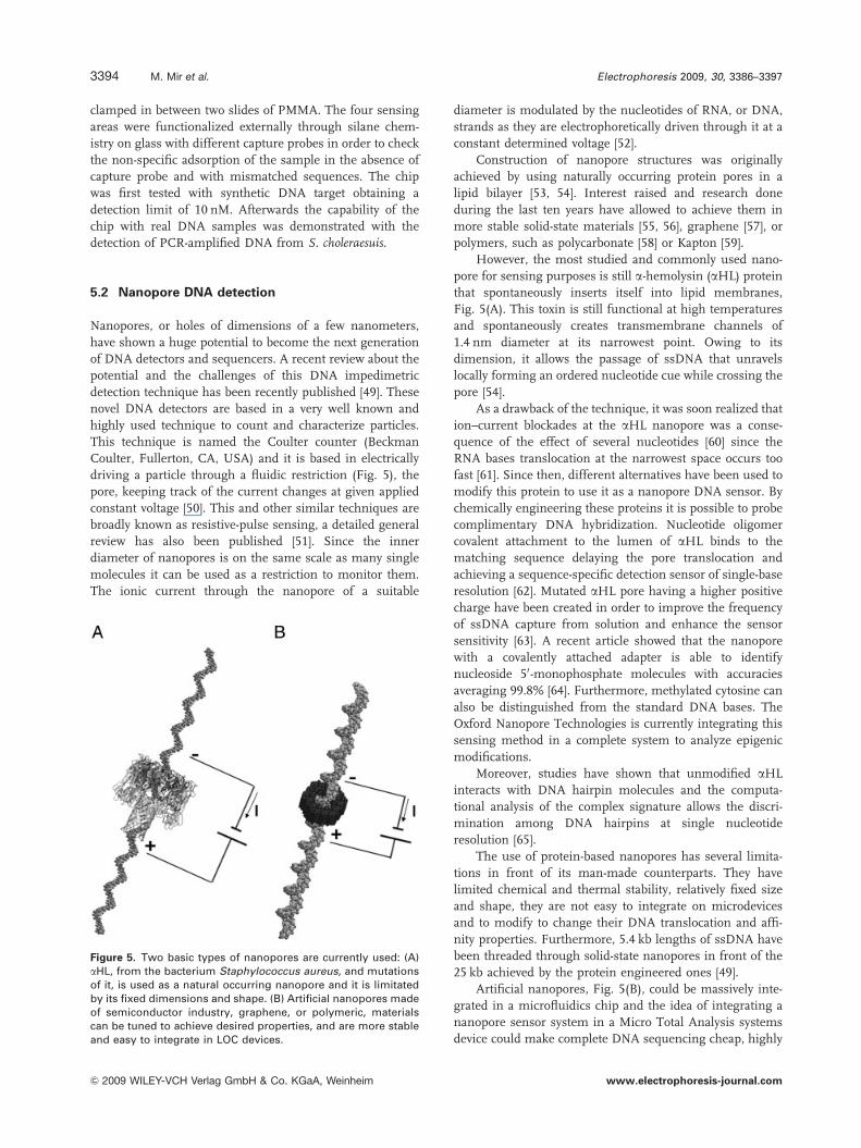
Artificial nanopores, Fig. 5(B), could be massively inte-
grated in a microfluidics chip and the idea of integrating a
nanopore sensor system in a Micro Total Analysis systems
device could make complete DNA sequencing cheap, highly
Figure 5. Two basic types of nanopores are currently used: (A)aHL, from the bacterium Staphylococcus aureus, and mutationsof it, is used as a natural occurring nanopore and it is limitatedby its fixed dimensions and shape. (B) Artificial nanopores madeof semiconductor industry, graphene, or polymeric, materialscan be tuned to achieve desired properties, and are more stableand easy to integrate in LOC devices.
Electrophoresis 2009, 30, 3386–33973394 M. Mir et al.
& 2009 WILEY-VCH Verlag GmbH & Co. KGaA, Weinheim www.electrophoresis-journal.com
parallelizable, portable and fast. Regular microfabrication
materials, as silicon oxide and silicon nitride, have been
proven suitable to build useful DNA sequence nanopores
using clean-room techniques as ion beam sculpting or
e-beam drilling [55, 56].
6 Concluding remarks and futureprospects
This review summarizes the strategies carried out by
different research groups, in order to integrate electroche-
mical DNA biosensors in microfluidic cartridges and some
of these include various sample pre-treatments, with the
purpose of miniaturize all the steps necessary in a DNA
analysis laboratory. Table 1 summarizes the main articles
described in this review, in order to have an easier overview
on these. Although, some techniques already have shown a
great deal of maturity, some other very promising ones are
still on their youth and many things have to be further
improved and explored in order to exploit the best of their
capabilities. This is especially true for the nanotechnology
advances related techniques such as field-effect NW, and
particularly CNT, and impedimetric nanopore-based
approaches.
NW and CNT DNA biosensors working principles are
still not fully understood and this brings many doubts about
results repeatability and stability. A great effort to under-
stand the basic principles behind them and to develop a
theory strongly individuating and isolating the different
affecting physical phenomena in their performance is
needed before being able to use them on commercial devi-
ces.
On the other hand, sequencing of single bases by means
of nanopore molecular sensors has proven to be out of reach
for the moment since the oligonucleotide strands tend to
cross the nanopore at very high velocities. Combination of
these techniques with other oligonucleotide manipulating
methods and tuning of the nanopore properties by applying
biochemical engineering and functionalization of the pore,
or by using other fabrication materials and techniques,
could bring a solution to this problem in the next years.
Furthermore, fabrication of these structures integrated on
LOC devices is still not straightforward bringing an addi-
tional challenge to be overcome by technology in order to
massively use this type of sensors.
Finally, the great advantage of integrating DNA biosen-
sors into an LOC device should be the reduction in sample
and reagents volumes. However, to fully achieve this and
bring these devices into profitable commercialization it is
needed to integrate the sample preparation protocol into the
LOC device. This introduces many other challenges such as
the a priori smart design, simplification, and combination of
the different protocol steps, or the interconnection of differ-
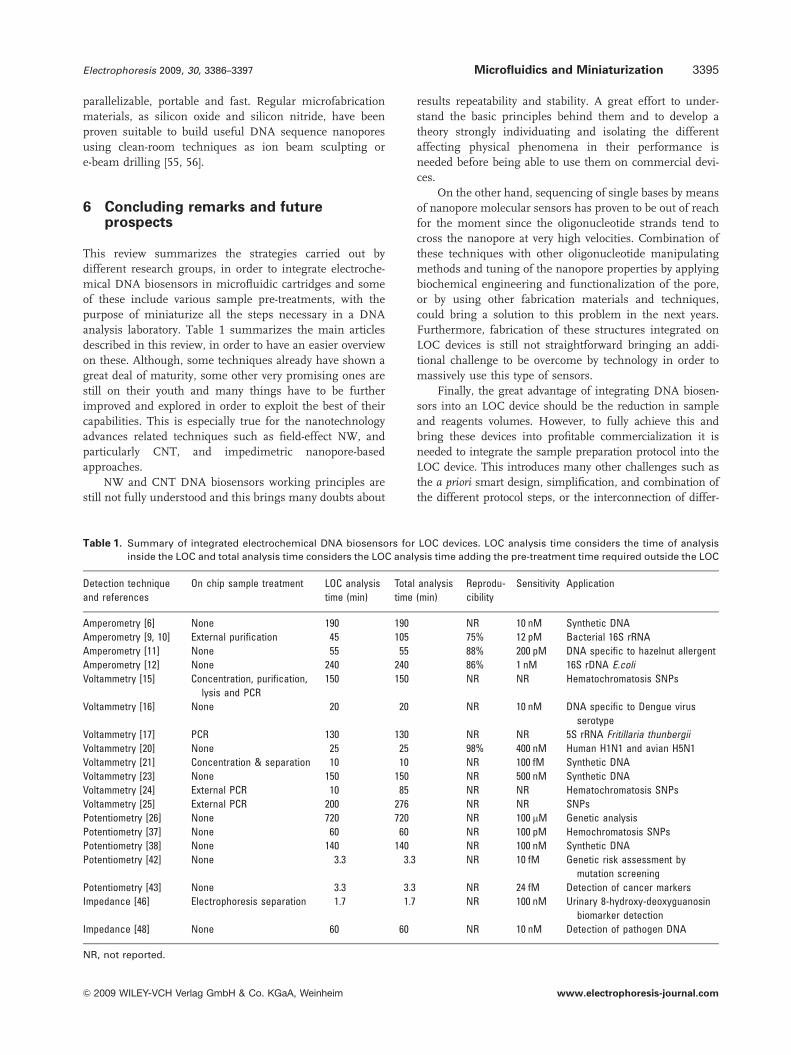
Table 1. Summary of integrated electrochemical DNA biosensors for LOC devices. LOC analysis time considers the time of analysis
inside the LOC and total analysis time considers the LOC analysis time adding the pre-treatment time required outside the LOC
Detection technique
and references
On chip sample treatment LOC analysis
time (min)
Total analysis
time (min)
Reprodu-
cibility
Sensitivity Application
Amperometry [6] None 190 190 NR 10 nM Synthetic DNA
Amperometry [9, 10] External purification 45 105 75% 12 pM Bacterial 16S rRNA
Amperometry [11] None 55 55 88% 200 pM DNA specific to hazelnut allergent
Amperometry [12] None 240 240 86% 1 nM 16S rDNA E.coli
Voltammetry [15] Concentration, purification,
lysis and PCR
150 150 NR NR Hematochromatosis SNPs
Voltammetry [16] None 20 20 NR 10 nM DNA specific to Dengue virus
serotype
Voltammetry [17] PCR 130 130 NR NR 5S rRNA Fritillaria thunbergii
Voltammetry [20] None 25 25 98% 400 nM Human H1N1 and avian H5N1
Voltammetry [21] Concentration & separation 10 10 NR 100 fM Synthetic DNA
Voltammetry [23] None 150 150 NR 500 nM Synthetic DNA
Voltammetry [24] External PCR 10 85 NR NR Hematochromatosis SNPs
Voltammetry [25] External PCR 200 276 NR NR SNPs
Potentiometry [26] None 720 720 NR 100 mM Genetic analysis
Potentiometry [37] None 60 60 NR 100 pM Hemochromatosis SNPs
Potentiometry [38] None 140 140 NR 100 nM Synthetic DNA
Potentiometry [42] None 3.3 3.3 NR 10 fM Genetic risk assessment by
mutation screening
Potentiometry [43] None 3.3 3.3 NR 24 fM Detection of cancer markers
Impedance [46] Electrophoresis separation 1.7 1.7 NR 100 nM Urinary 8-hydroxy-deoxyguanosin
biomarker detection
Impedance [48] None 60 60 NR 10 nM Detection of pathogen DNA
NR, not reported.
Electrophoresis 2009, 30, 3386–3397 Microfluidics and Miniaturization 3395
& 2009 WILEY-VCH Verlag GmbH & Co. KGaA, Weinheim www.electrophoresis-journal.com
ent blocks and its dead fluid volumes reduction. Further-
more, the choice of the right LOC material for the application
as well as the reduction in the final production costs are also
issues that sometimes escape the mind of scientists and that
can be great impediments to the onset of industrial and social
interest. However, these limitations are the same that are
found in other transduction methods, such as the optical,
once they are evaluated to be integrated on an LOC device and
they are being resolved in most of the cases for each parti-
cular application. Moreover, electrochemical genetic sensors
present the advantage of a complete electronic read-out that
reduces the cost of external interfaces and facilitates the
choice of the LOC device material that would not have the
need of highly demanding optical properties.
It is clear by the end of this review that electrochemical
oligonucleotide detection presents several advantages over
other methods suitable to be integrated on LOC devices.
Several commercial applications are already on the market
and there is a nice prospective for the appearance of new
products based on these technologies in the fore coming
years despite the actual limitations.
The authors have declared no conflict of interest.
7 References
[1] Manz, A., Fettinger, J. C., Verpoorte, E., Ludi, H.,Widmer, H. M., Harrison, D. J., Trends Anal. Chem.1991, 10, 144–149.
[2] Burns, M., Johnson, B., Brahmasandra, S., Handique,K., Webster, J., Krishnan, M., Sammarco, T. et al.,Science 1998, 282, 484–487.
[3] Janasek, D., Franzke, J., Manz, A., Nature 2006, 442,374–380.
[4] Yager, P., Edwards, T., Fu, E., Helton, K., Nelson, K.,Tam, M. R., Weigl, B. H., Nature 2006, 442, 412–418.
[5] Mir, M., Dondapati, S., Vreeke, M., Katakis, I., Electro-chem. Commun. 2007, 9, 1715–1718.
[6] Liu, D. J., Perdue, R. K., Sun, L., Crooks, R. M., Langmuir2004, 20, 5905–5910.
[7] Sadik, O. A, Aluoch, A. O., Zhou, A., Biosens. Bioelec-tron. 2009, 24, 2749–2765.
[8] Ghindilis, A. L., Smith, M. W., Schwarzkopf, K. R., Roth,K. M., Peyvan, K., Munro, S. B., Lodes, M. J. et al.,Biosens. Bioelectron. 2007, 22, 1853–1860.
[9] Liao, J. C., Ma, Y. B., Gau, V., Mastali, M., Sun, C. P., Li,Y., McCabe, E. R. B. et al., 1st IEEE InternationalConference on Nano/Micro Engineered and MolecularSystems, Vol. 1–3. 2006,, pp. 1109–1112.
[10] Liao, J. C., Mastali, M., Gau, V., Suchard, M. A., Moller,A. K., Bruckner, D. A., Babbitt, J. T. et al., J. Clin.Microbiol. 2006, 44, 561–570.
[11] Berti, F., Laschi, S., Palchetti, I., Rossier, J. S., Reymond,F., Mascini, M., Marrazza, G., Talanta 2009, 77, 971–978.
[12] Gabig-Ciminska, M., Holmgren, A., Andresen, H.,Barken, K. B., Wumpelmann, M., Albers, J., Hintsche, R.et al., Biosens. Bioelectron. 2004, 19, 537–546.
[13] Mir, M., Katakis, I., Talanta, 2008, 75, 432–441.
[14] Farkas, D. H., Clin. Chem. 2001, 47, 1871–1872.
[15] Liu, R. H., Grodzinski, P., Yang, J., Lenigk, R., IntegratedBiochips DNA Anal. 2007, 46–67.
[16] Baeumner, A. J., Kwakye, S., Goral, V. N., Biosens.Bioelectron. 2006, 21, 2217–2223.
[17] Lee, T. M. H., Carles, M. C., Hsing, I. M., Lab Chip 2003,3, 100–105.
[18] Sawada, K., Oda, C., Takao, H., Ishida, M., IEEE Source:Transducers ’05, Digest of Technical Papers, Vol. 1–2,2005, pp. 279–282.
[19] Fan, C., Plaxco, K. W., Heeger, A. J., Proc. Natl. Acad.Sci. USA 2003, 100, 9134–9137.
[20] Pavlovic, E., Lai, R. Y., Wu, T. T., Ferguson, B. S., Sun, R.,Plaxco, K. W., Soh, H. T., Langmuir 2008, 24, 1102–1107.
[21] Shiddiky, M. J. A., Shim, Y. B., Anal. Chem. 2007, 79,3724–3733.
[22] Li, J., Koehne, J. E., Cassell, A. M., Chen, H., Ng, H. T.,Ye, Q., Fan, W. et al., Electroanalysis 2005, 17, 15–27.
[23] Triroj, N., Lapierre-Devlin, M. A., Kelley, S. O., Beres-ford, R., IEEE Sensors J. 2005, 6, 1395–1402.
[24] Ertl, P., Emrich, C. A., Singhal, P., Mathies, R. A., Anal.Chem. 2004, 76, 3749–3755.
[25] Hebert, N. E., Brazill, S. A., Lab Chip 2003, 3, 241–247.
[26] Sakata, T., Kamahori, M., Miyahara, Y., JapaneseJ. Appl. Phys. 2005, 44, 2854–2859.
[27] Barbaro, M., Bonfiglio, A., Raffo, L., Alessandrini, A.,Facci, P., Barak, I., IEEE Electron. Device Lett. 2006, 27,595–597.
[28] Kauffman, D. R., Star, A., Chem. Soc. Rev. 2008, 37,1197–1206.
[29] Heinze, S., Tersoff, J., Martel, R., Deryke, V., Appenzel-ler, J., Avouris, P., Phys. Rev. Lett. 2002, 89, 106801/1–106801/4.
[30] Chen, Z., Appenzeller, J., Knoch, J., Lin, Y., Avouris, P.,Nano Lett. 2005, 7, 1497–1502.
[31] Heller, I., Janssens, A. M., Mannik, J., Minot, E. D.,Lemay, S. G., Dekker, C., Nano Lett. 2008, 8, 591–595.
[32] Bradley, K., Briman, M., Star, A., Gruner, G., Nano Lett.2004, 4, 253–256.
[33] Zheng, M., Jagota, A., Semke, B. A., Diner, R. S.,Mclean, S., Lustig, R., Richardson, R. E., Tassi, N. G.,Nat. Mater. 2003, 2, 338–342.
[34] Niyogi, N., Hamon, M. A., Hu, H., Zhao, B., Bhowmik, P.,Sen, R., Itkis, M. E., Haddon, R. C., Acc. Chem. Res. 2002,35, 1105–1113.
[35] Zhao, X., Johnson, J. K., J. Am. Chem. Soc. 2007, 129,10438–10445.
[36] Jeng, E. S., Barone, P. W., Nelson, J. D., Strano, M. S.,Small 2007, 3, 1602–1609.
[37] Star, A., Tu, E., Niemann, J., Gabriel, J.-C. P., Joiner,C. S., Valcke, C., Proc. Natl. Acad. Sci. USA 2006, 103,921–926.
[38] Tang, X., Bansaruntip, S., Nakayama, N., Yenilmez, E.,Chang, Y.-I., Wang, Q., Nano Lett. 2006, 6, 1632–1636.
[39] Gui, E. L., Li, L. J., Zhang, K., Xu, Y., Dong, X., Ho, X., Lee, P.S. et al., J. Am. Chem. Soc. 2007, 129, 14427–14432.
Electrophoresis 2009, 30, 3386–33973396 M. Mir et al.
& 2009 WILEY-VCH Verlag GmbH & Co. KGaA, Weinheim www.electrophoresis-journal.com

[40] Patolsky, F., Zheng, G., Lieber, C. M., Anal. Chem. 2006,78, 4260–4269.
[41] Nair, P. R., Alam, M. A., IEEE Trans. Electron Dev. 2007,54, 3400–3408.
[42] Hahm, J., Lieber, C. M., Nano Lett. 2004, 4, 51–54.
[43] Zheng, G. F., Patolsky, F., Cui, Y., Wang, W. U., Lieber,C. M., Nat. Biotechnol. 2005, 23, 1294–1301.
[44] Maki, W. C., Mishra, N. N., Cameron, E. G., Filanoski, B.,Rastogi, S. K., Maki, G. K., Biosens. Bioelectron. 2008,23, 780–787.
[45] Mir, M., Jenkins, A. T. A., Katakis, I., Electrochem.Commun. 2008, 10, 1533–1536.
[46] Dawoud, A. A., Kawaguchi, T., Jankowiak, R., Anal.Bioanal. Chem. 2007, 388, 245–252.
[47] Park, M. I., Hong, J., Yoon, D. S., Park, C. O., Im, G.,Material Research Society Symposium Proceedings,Vol. 773. 2003, 773, pp. 149–154.
[48] Berdat, D., Rodriguez, A. C. M., Herrera, F., Gijs,M. A. M., Lab Chip 2008, 8, 302–308.
[49] Branton, D., Deamer, D. W., Marziali, A., Bayley, H.,Benner, S. A., Butler, T., Di Ventra, M. et al., Nat.Biotechnol. 2008, 10, 1146–1153.
[50] Allen, T., Particle Size Measurement. Chapman & Hall,London, UK 1990.
[51] Bayley, H., Martin, C. R., Chem. Rev. 2000, 100, 2575–2594.
[52] Deamer, D. W., Akeson, M., Trends Biotechnol. 2000, 18,131–180.
[53] Tobkes, N., Wallace, B. A., Bayley, H., Biochemistry1985, 24, 1915–1920.
[54] Kasianowicz, J. J., Brandin, E., Branton, D., Deamer,D. W., Proc. Natl. Acad. Sci. USA 1996, 93, 13770–13773.
[55] Li, J., Stein, D., McMullan, C., Branton, D., Aziz, M. J.,Golovchenko, J. A., Nature 2001, 412, 166–169.
[56] Storm, A. J., Chen, J. H., Ling, X. S., Zandbergen, H. W.,Dekker, C., Nat.Mater. 2003, 2, 537–540.
[57] Tabib-Azar, M., Moetakef, P., Sharghi-Moshtaghin, R.,IEEE Sens. 2008, 2008, 566–568.
[58] Harrell, C. C., Choi, Y., Horne, L. P., Baker, L. A., Siwy,Z. S., Martin, C. R., Langmuir 2006, 22, 10837–10843.
[59] Mara, A., Siwy, Z., Trautmann, C., Wan, J., Kamme, F.,Nano Lett. 2004, 4, 497–501.
[60] Meller, A., Nivon, L., Branton, D., Phys. Rev. Lett. 2001,86, 3435–3438.
[61] Dekker, C., Nat. Nanotechnol. 2007, 2, 209–215.
[62] Howorka, S., Cheley, S., Bayley, H., Nat. Biotechnol.2001, 19, 636–639.
[63] Maglia, G., Restrepo, M. R., Mikhailova, E., Bayley, H.,Proc. Natl. Acad. Sci. USA 2008, 105, 19720–19725.
[64] Clarke, J., Wu, H. C., Jayasinghe, L., Patel, A., Reid, S.,Bayley, H., Nat. Nanotechnol. 2009, 4, 265–270.
[65] Vercoutere, W., Winters-Hilt, S., Olsen, H., Deamer, D.,Haussler, D., Akeson, M., Nat. Biotechnol. 2001, 19,248–252.
Electrophoresis 2009, 30, 3386–3397 Microfluidics and Miniaturization 3397
& 2009 WILEY-VCH Verlag GmbH & Co. KGaA, Weinheim www.electrophoresis-journal.com