Molecularly Imprinted Polymer-Hybrid Electrochemical Sensor for the Detection of β-Estradiol
Upload
independentCategory
view
1download
0
A
afeeebt©
K
1
eaneHclhsd(ee
h
0d
Biosensors and Bioelectronics 22 (2007) 2525–2531
Electrochemical detection of 17�-estradiol using DNA aptamerimmobilized gold electrode chip
Yeon Seok Kim a, Ho Sup Jung b, Toshihiko Matsuura b, Hea Yeon Lee b,∗,Tomoji Kawai b, Man Bock Gu a,∗
a College of Life Sciences and Biotechnology, Korea University, Anam-dong, Seongbuk-gu, Seoul 136-701, Republic of Koreab Institute for Scientific and Industrial Research, Osaka University, 8-1 Mihogaoka, Ibaraki, Osaka 567-0047, Japan
Received 5 June 2006; received in revised form 27 September 2006; accepted 3 October 2006Available online 21 November 2006
bstract
An electrochemical detection method for chemical sensing has been developed using a DNA aptamer immobilized gold electrode chip. DNAptamers specifically binding to 17�-estradiol were selected by the SELEX (Systematic Evolution of Ligands by EXponential enrichment) processrom a random ssDNA library, composed of approximately 7.2 × 10¹⁴ DNA molecules. Gold electrode chips were employed to evaluate thelectrochemical signals generated from interactions between the aptamers and the target molecules. The DNA aptamer immobilization on the goldlectrode was based on the avidin–biotin interaction. The cyclic voltametry (CV) and square wave voltametry (SWV) values were measured to
valuate the chemical binding to aptamer. When 17�-estradiol interacted with the DNA aptamer, the current decreased due to the interference ofound 17�-estradiol with the electron flow produced by a redox reaction between ferrocyanide and ferricyanide. In the negative control experiments,he current decreased only mildly due to the presence of other chemicals.2006 Elsevier B.V. All rights reserved.
stawTeaarasuM
eywords: Electrochemical detection; Aptamer; 17�-Estradiol
. Introduction
Endocrine disrupting chemicals (EDCs), including 17�-stradiol, pose a serious threat due to an estrogenic potentialctivity that they mimic. The detection of these chemicals withinatural systems, therefore, is necessary for protecting public andnvironmental health. Instrumental analysis methods, such asPLC or GC/MS, are very sensitive at detecting these toxic
hemicals but are also very complicated to perform and require aong time for analysis to be completed. Hence, many researchersave been trying to develop a simpler and more sensitive biosen-or system for chemical sensing. In biosensor systems for theetermination of 17�-estradiol, the human estrogen-receptor
hER) has been commonly used (Seifert et al., 1999; Butalat al., 2003; Wozei et al., 2006). These biosensors can detect thestrogenic chemicals including 17�-estradiol sensitively, but the∗ Corresponding authors. Tel.: +82 2 3290 3417; fax: +82 2 928 6050.E-mail addresses: [email protected] (M.B. Gu),
[email protected] (H.Y. Lee).
Wrs
opag
956-5663/$ – see front matter © 2006 Elsevier B.V. All rights reserved.oi:10.1016/j.bios.2006.10.004
pecificity was not good due to the affinity of estrogen-receptoro the other xenoendocrines. The cell-based biosensors werelso developed with recombinant cells harboring hER promoterith a reporter lacZ (Hahn et al., 2006; Wozei et al., 2006).hese assays can give the information about the concentration ofndocrine disrupting chemicals (EDCs) including 17�-estradiolnd their activities inside the cell, but they normally requirelong detection time with a low specificity due to a wide
ange of hEr affinities to EDCs. Recently, the immunoassaysnd nanobiosensors using nanoparticles have been developed asensitive and rapid methods for the detection of 17�-estradiol bytilizing antibodies or functional polymers (Glass et al., 2004;iyashita et al., 2005; Pemberton et al., 2005; Lin and Li, 2006;ei et al., 2006). The specificities of these biosensors were
eported to be better than hER-based biosensors due to theirtructure-specific biorecognition.
In recent years, numerous studies for in vitro selection of
ligonucleotides (aptamers) that bind to specific target com-ounds have been reported (Ellington and Szostak, 1990; Tuerknd Gold, 1990). Its biomolecular recognition ability for tar-et molecules has opened a new era in affinity biosensing since
2 Bioel
taIaacbcFaHlb(brairmuadas
i2tsaaf2amoeratdbopawaSKmbiiecb
ce
cblIitTwFd
2
2
bcrccfSeasowli
GC
omTiw
2
tsa1tio
526 Y.S. Kim et al. / Biosensors and
he signal of the biosensor depends upon the specificity and theffinity of the molecular recognition element (Luzi et al., 2003).n most diagnostics and biomolecular sensing studies, the highffinity and specific molecular recognition are achieved usingntibodies, but there are some limitations, including animals orell line requirement with complicated purification steps for anti-ody production. Unlike antibodies, in vitro selected aptamersould be reproducibly synthesized and obtained economically.urthermore, antibodies produced against target molecules thatre inherently less immunogenic are difficult to be generated.owever, aptamers are not restricted for the various targets,
ike proteins, peptides, amino acids, nucleotides, drugs, car-ohydrates and other small organic and inorganic compoundsJayasena, 1999; Frauendorf and Jaschke, 2001). As such, anti-odies may not be suitable for developing biosensors for envi-onmental toxic chemicals, compared with aptamers. Moreover,ptamers can be modified with certain functional groups ands immobilized on many surfaces, both directly and indirectly,esulting in well ordered receptor layers. Finally, aptamers areore stable than antibodies and thus are more resistant to denat-
ration and degradation. Consequently, aptamers offer a usefullternative to antibodies as sensing molecules, especially in theevelopment of biosensor for chemicals (Jayasena, 1999; Liss etl., 2002). In addition, it was known that DNA aptamers are moretable than RNA aptamers and easier to identify and handle.
Many papers about aptamer-based affinity biosensors formportant proteins have already been reported (Jiang et al.,004; Lee et al., 2004; Savran et al., 2004). The key point inhe development of a biosensor system is to generate a mea-urable signal from the target–probe interaction (McCauley etl., 2003). Optical methods such as a sandwich method, anptamer beacon method, and other methods to generate lightrom bio-recognition results have been reported (Fang et al.,001; Hamaguchi et al., 2001; Nutiu and Li, 2003; Pavlov etl., 2004; Rupcich et al., 2005). More recently, an analyticalethod based on atomic force microscopy (AFM) was devel-
ped and used to evaluate aptamer–protein interactions (Jiangt al., 2004). However, optical methods with fluorescence oradioactive tags have some disadvantages in terms of their costnd experimental complexity, since they need labeling dyes orags and expensive optical detection devices. Mass-dependantevices, in addition, are also very useful systems in aptasensorsecause they offer a label-free system. However, in detectionf small organic chemicals, these methods including surfacelasmon resonance (SPR), quartz crystal microbalance (QCM)nd cantilever methods, are not efficient because the moleculareight of the target chemicals is generally too small to gener-
te an efficient signal (Liss et al., 2002; Minunni et al., 2004;chlensog et al., 2004; Gronewold et al., 2005; Misono andumar, 2005). Herein, we have developed an electrochemicalethod for 17�-estradiol detection using a DNA aptamer immo-
ilized on a gold electrode chip. The electrochemical approachs cost-effective because it does not require expensive dyes and
nstruments (Dijksma et al., 2001; Drummond et al., 2003; Wangt al., 2003; Cai et al., 2004). An electrochemical signal, i.e., ahange in the current, is produced directly from the interactionetween the target compound and the aptamer and this current(1Aw
ectronics 22 (2007) 2525–2531
hange is not related to the mass of target compound (Ikebukurot al., 2005; Kawde et al., 2005).
In this research, to develop an aptamers-based electrochemi-al biosensor, we isolated single-stranded DNA aptamer that canind to 17�-estradiol. The selected DNA aptamer was immobi-ized on a gold electrode based on an avidin–biotin interaction.mmobilization of the ssDNA aptamer on the streptavidin mod-fied gold electrode was confirmed and optimized using a SPRechnique based on aptamer immobilized BIAcore sensor chips.he cyclic voltametry (CV) and square wave voltametry (SWV)ere measured and used to confirm electrochemical detection.inally, the electrochemical detection of 17�-estradiol was con-ucted with a decrease of electric current.
. Materials and methods
.1. DNA aptamer
A single-stranded DNA aptamer against 17�-estradiol haseen isolated by SELEX process from a random ssDNA library,omposed of approximately 7.2 × 10¹⁴ DNA molecules. All ofandom DNA library, primers and DNA aptamers were pur-hased from GenoTech Co. (Deajeon, Korea). A total of sevenycles of repetitions and enzymatic enrichment were carried outor this selection. Ten ssDNA aptamers obtained finally fromELEX process were analyzed in terms of their affinity to 17�-stradiol based on the equilibrium-filtration method (Jenison etl., 1994; Kato et al., 2000). The dissociation constants for 10sDNA aptamers were in the range of 0.1–3 �M. In this study,ne of the 10 ssDNA aptamers showing a constant of 0.13 �Mas selected as a final probe DNA aptamer molecule. The fol-
owing DNA strands were the selected DNA aptamer probe usedn this study:
5′-Biotin–GCTTCCAGCTTATTGAATTACACGCAGAGG-TA GCGGCTCTGCGCATTCAATTGCTGCGCGCTGAAG-GCGGAAGC-3′.
It was 76-mer and 23 kDa. The secondary structure analysisf DNA aptamers was performed by using the web-based toolfold, the free-energy minimization algorithm (Zuker, 2003).he suggested secondary structure of the DNA aptamer selected
s shown in Fig. 1. The DNA aptamer was dissolved in distilledater (18 m�, Millipore) for further experiments.
.2. Immobilization of the DNA aptamer on a gold surface
To construct an electrochemical-based aptamer biosensor,he biotinylated ssDNA aptamer selected was immobilized on atreptavidin-modified gold electrode. The detailed procedure iss follows: at first, the bare gold electrode was cleaned with0 mM H2SO4 under electric potential within a range of 1o −0.2 V. Etching with 5 mM K3Fe(CN)6 solution contain-ng 100 mM KCl was carried out to enhance the uniformityf the bare gold surface. Next, the self assembled monolayer
SAM) was spontaneously formed through an incubation ofmM 3,3′-dithiodipropionic acid (Sigma Co., USA) for 30 min.fter washing with Milli Q water, the chip was treated for 30 minith the same volumes of 1 mM N-(3-dimethylaminopropyl)-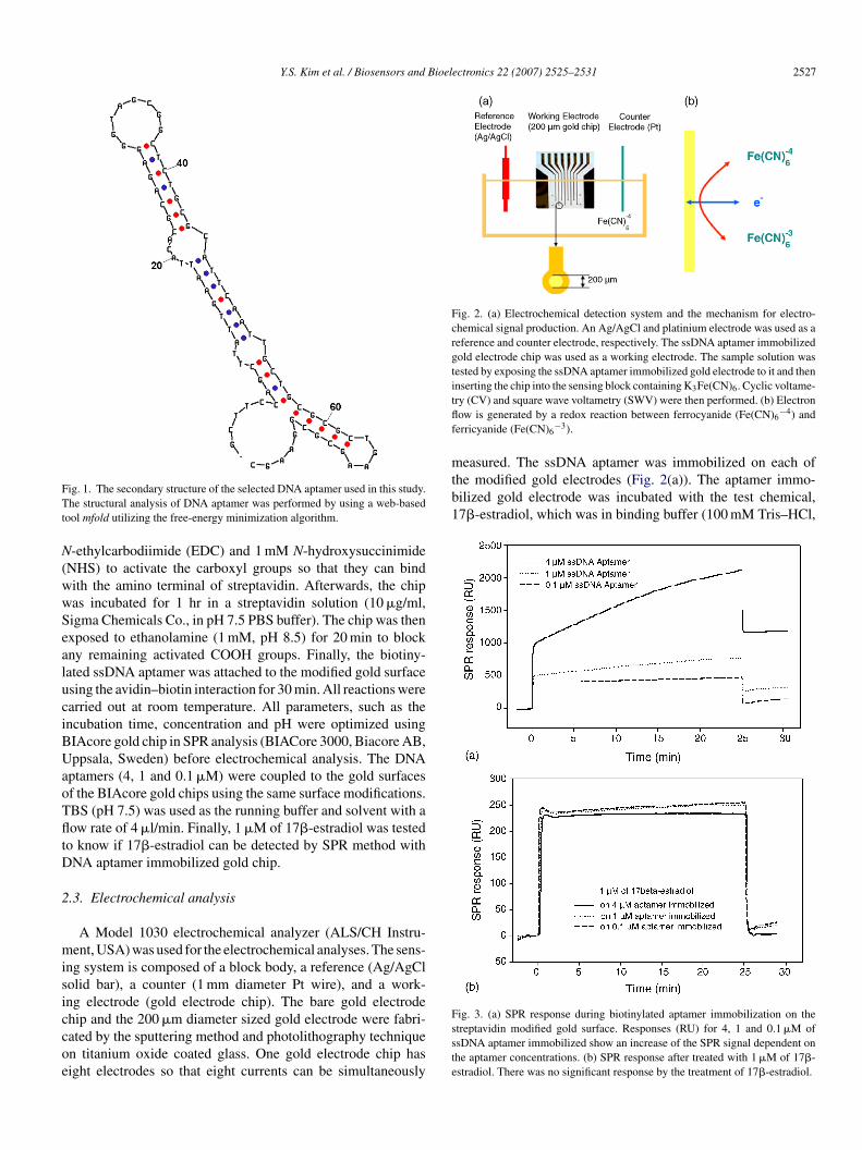
Y.S. Kim et al. / Biosensors and Bioelectronics 22 (2007) 2525–2531 2527
FTt
N(wwSealuciBUaoTfltD
2
misiccoe
Fig. 2. (a) Electrochemical detection system and the mechanism for electro-chemical signal production. An Ag/AgCl and platinium electrode was used as areference and counter electrode, respectively. The ssDNA aptamer immobilizedgold electrode chip was used as a working electrode. The sample solution wastested by exposing the ssDNA aptamer immobilized gold electrode to it and theninserting the chip into the sensing block containing K Fe(CN) . Cyclic voltame-tflf
measured. The ssDNA aptamer was immobilized on each ofthe modified gold electrodes (Fig. 2(a)). The aptamer immo-bilized gold electrode was incubated with the test chemical,17�-estradiol, which was in binding buffer (100 mM Tris–HCl,
ig. 1. The secondary structure of the selected DNA aptamer used in this study.he structural analysis of DNA aptamer was performed by using a web-based
ool mfold utilizing the free-energy minimization algorithm.
-ethylcarbodiimide (EDC) and 1 mM N-hydroxysuccinimideNHS) to activate the carboxyl groups so that they can bindith the amino terminal of streptavidin. Afterwards, the chipas incubated for 1 hr in a streptavidin solution (10 �g/ml,igma Chemicals Co., in pH 7.5 PBS buffer). The chip was thenxposed to ethanolamine (1 mM, pH 8.5) for 20 min to blockny remaining activated COOH groups. Finally, the biotiny-ated ssDNA aptamer was attached to the modified gold surfacesing the avidin–biotin interaction for 30 min. All reactions werearried out at room temperature. All parameters, such as thencubation time, concentration and pH were optimized usingIAcore gold chip in SPR analysis (BIACore 3000, Biacore AB,ppsala, Sweden) before electrochemical analysis. The DNA
ptamers (4, 1 and 0.1 �M) were coupled to the gold surfacesf the BIAcore gold chips using the same surface modifications.BS (pH 7.5) was used as the running buffer and solvent with aow rate of 4 �l/min. Finally, 1 �M of 17�-estradiol was tested
o know if 17�-estradiol can be detected by SPR method withNA aptamer immobilized gold chip.
.3. Electrochemical analysis
A Model 1030 electrochemical analyzer (ALS/CH Instru-ent, USA) was used for the electrochemical analyses. The sens-
ng system is composed of a block body, a reference (Ag/AgClolid bar), a counter (1 mm diameter Pt wire), and a work-ng electrode (gold electrode chip). The bare gold electrode
hip and the 200 �m diameter sized gold electrode were fabri-ated by the sputtering method and photolithography techniquen titanium oxide coated glass. One gold electrode chip hasight electrodes so that eight currents can be simultaneouslyFsste
3 6
ry (CV) and square wave voltametry (SWV) were then performed. (b) Electronow is generated by a redox reaction between ferrocyanide (Fe(CN)6
−4) anderricyanide (Fe(CN)6
−3).
ig. 3. (a) SPR response during biotinylated aptamer immobilization on thetreptavidin modified gold surface. Responses (RU) for 4, 1 and 0.1 �M ofsDNA aptamer immobilized show an increase of the SPR signal dependent onhe aptamer concentrations. (b) SPR response after treated with 1 �M of 17�-stradiol. There was no significant response by the treatment of 17�-estradiol.
2528 Y.S. Kim et al. / Biosensors and Bioelectronics 22 (2007) 2525–2531
F on 2a ortiont re leg
2fsaKsostsprcwls
3
3
teq
cfgvfsgttba
cbtau(awc
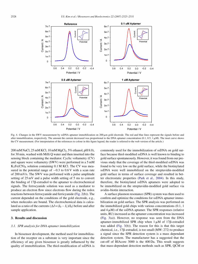
ig. 4. Changes in the SWV measurement by ssDNA aptamer immobilizationfter immobilization, respectively. The amount the current decreased was prophe CV measurement. (For interpretation of the references to colour in this figu
00 mM NaCl, 25 mM KCl, 10 mM MgCl2, 5% ethanol, pH 8.0),or 30 min, washed with Milli Q water and then inserted into theensing block containing the mediator. Cyclic voltametry (CV)nd square wave voltametry (SWV) were performed in a 5 mM3Fe(CN)6 solution containing 0.1 M KCl. The CV was mea-
ured in the potential range of −0.3 to 0.6 V with a scan ratef 200 mV/s. The SWV was performed with a pulse amplitudeetting of 25 mV and a pulse width setting of 5 ms to converthe binding of 17�-estradiol to the aptamer to electrochemicalignals. The ferrocyanide solution was used as a mediator toroduce an electron flow since electrons flow during the redoxeactions between ferrocyanide and ferricyanide (Fig. 2(b)). Theurrent depended on the conditions of the gold electrode, e.g.,hen molecules are bound. The electrochemical data is calcu-
ated as a ratio of the currents (�I = (I0 − I1)/I0) before and afterample application.
. Results and discussion
.1. SPR analysis for DNA aptamer immobilization
In biosensor development, the method used for immobiliza-ion of the receptor on a substrate is very important since thefficiency of any given biosensor is greatly influenced by theuality of immobilization. The thiol-modification of ssDNA is
adct
00 �m gold electrode. The red and blue lines represent the signals before andal to the DNA aptamer concentration (0.1, 0.5, 1 �M). The inset curve shows
end, the reader is referred to the web version of the article.)
ommonly used for the immobilization of ssDNA on gold sur-ace because thiol-modified ssDNA is well known to binding toold surface spontaneously. However, it was found from our pre-ious study that the coverage of the thiol-modified ssDNA wasound to be very low on the gold surface, while the biotinylatedsDNA were well immobilized on the streptavidin-modifiedold surface in terms of surface coverage and resulted in bet-er electrostatic properties (Park et al., 2004). In this study,herefore, the biotinylated ssDNA aptamers were adopted toe immobilized on the streptavidin-modified gold surface viavidin–biotin interaction.
A surface plasmon resonance (SPR) system was then used toonfirm and optimize the conditions for ssDNA aptamer immo-ilization on gold surface. The SPR analysis was performed onhe immobilized gold chips with various concentrations (0.1, 1nd 4 �M) of the ssDNA aptamer. The SPR responses (relativenits, RU) increased as the aptamer concentration was increasedFig. 3(a)). However, no response was seen from the DNAptamer-immobilized SPR chip when 1 �M of 17�-estradiolas added (Fig. 3(b)). The reason for this is that this target
hemical, i.e., 17�-estradiol, is too small (MW: 272) to produce
signal since the SPR detection system is a mass dependantetection system. The manufacturer has a suggested that theut-off of BIAcore 3000 is the 400 Da. This result suggestshat mass-dependant detection methods such as SPR, QCM or
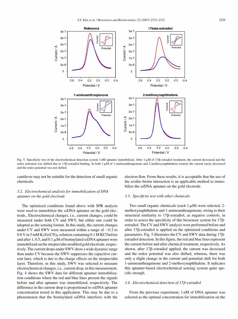
Y.S. Kim et al. / Biosensors and Bioelectronics 22 (2007) 2525–2531 2529
F immor inoaa
cc
3a
wtmau0aittrleFtbdcp
etb
3
msoeapetsao1tc
ig. 5. Specificity test of the electrochemical detection system 1 nM aptameredox potential was shifted due to 17�-estradiol binding. In both 1 �M of 1-amnd the redox potential was not shifted.
antilever may not be suitable for the detection of small organichemicals.
.2. Electrochemical analysis for immobilization of DNAptamer on the gold electrode
The optimized conditions found above with SPR analysisere used to immobilize the ssDNA aptamer on the gold elec-
rode,. Electrochemical changes, i.e., current changes, could beeasured under both CV and SWV, but either one could be
dopted as the sensing format. In this study, the current changesnder CV and SWV were measured within a range of −0.3 to.6 V in 5 mM K3Fe(CN)6 solution containing 0.1 M KCl beforend after 1, 0.5, and 0.1 �M of biotinylated ssDNA aptamer weremmobilized on the streptavidin modified gold electrode, respec-ively. The current drops under SWV show a wide dynamic rangehan under CV because the SWV suppresses the capacitive cur-ent later, which is due to the charge effects on the streptavidinayer. Therefore, in this study, SWV was selected to measurelectrochemical changes, i.e., current drop, in this measurement.ig. 4 shows the SWV data for different aptamer immobiliza-
ion conditions where the red and blue lines present the signals
efore and after aptamer was immobilized, respectively. Theifference in the current drop is proportional to ssDNA aptameroncentration tested in this application. This may be due to ahenomenon that the biotinylated ssDNA interferes with the3
s
bilized. After 1 �M of 17�-estradiol treatment, the current decreased and thenthraqionone and 2-methoxynaphthalene treated, the current rarely decreased
lectron flow. From these results, it is acceptable that the use ofhe avidin–biotin interaction is an applicable method to immo-ilize the ssDNA aptamer on the gold electrode.
.3. Specificity test with other chemicals
Two small organic chemicals (each 1 �M) were selected, 2-ethoxynaphthalene and 1-aminoanthraquinone, owing to their
tructural similarity to 17�-estradiol, as negative controls, inrder to assess the specificity of this biosensor system for 17�-stradiol. The CV and SWV analysis were performed before andfter 17�-estradiol is applied on the optimized conditions andarameters. Fig. 5 illustrates the CV and SWV data during 17�-stradiol detection. In this figure, the red and blue lines representhe current before and after chemical treatment, respectively. Ashown, after 17�-estradiol applied, the current was decreasednd the redox potential was also shifted, whereas, there wasnly a slight change in the current and potential shift for both-aminoanthraquinone and 2-methoxynaphthalene. It indicateshis aptamer-based electrochemical sensing system quite spe-ific enough.
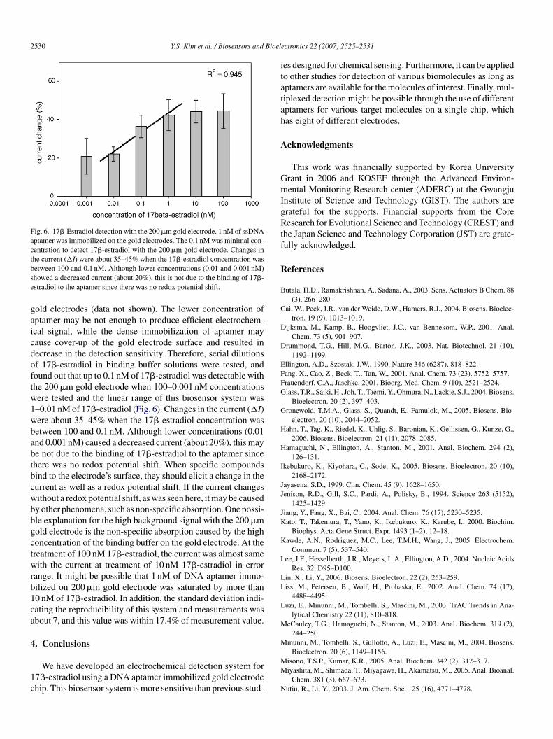
.4. Electrochemical detection of 17β-estradiol
From the previous experiment, 1 nM of DNA aptamer waselected as the optimal concentration for immobilization on the
2530 Y.S. Kim et al. / Biosensors and Bioel
Fig. 6. 17�-Estradiol detection with the 200 �m gold electrode. 1 nM of ssDNAaptamer was immobilized on the gold electrodes. The 0.1 nM was minimal con-centration to detect 17�-estradiol with the 200 �m gold electrode. Changes inthe current (�I) were about 35–45% when the 17�-estradiol concentration wasbse
gaicdoftw1wbabtbcwbbgctwrb1ca
4
1c
itatah
A
GmIgRtf
R
B
C
D
D
EFFG
G
H
H
I
JJ
JK
K
L
LL
L
M
M
etween 100 and 0.1 nM. Although lower concentrations (0.01 and 0.001 nM)howed a decreased current (about 20%), this is not due to the binding of 17�-stradiol to the aptamer since there was no redox potential shift.
old electrodes (data not shown). The lower concentration ofptamer may be not enough to produce efficient electrochem-cal signal, while the dense immobilization of aptamer mayause cover-up of the gold electrode surface and resulted inecrease in the detection sensitivity. Therefore, serial dilutionsf 17�-estradiol in binding buffer solutions were tested, andound out that up to 0.1 nM of 17�-estradiol was detectable withhe 200 �m gold electrode when 100–0.001 nM concentrationsere tested and the linear range of this biosensor system was–0.01 nM of 17�-estradiol (Fig. 6). Changes in the current (�I)ere about 35–45% when the 17�-estradiol concentration wasetween 100 and 0.1 nM. Although lower concentrations (0.01nd 0.001 nM) caused a decreased current (about 20%), this maye not due to the binding of 17�-estradiol to the aptamer sincehere was no redox potential shift. When specific compoundsind to the electrode’s surface, they should elicit a change in theurrent as well as a redox potential shift. If the current changesithout a redox potential shift, as was seen here, it may be causedy other phenomena, such as non-specific absorption. One possi-le explanation for the high background signal with the 200 �mold electrode is the non-specific absorption caused by the highoncentration of the binding buffer on the gold electrode. At thereatment of 100 nM 17�-estradiol, the current was almost sameith the current at treatment of 10 nM 17�-estradiol in error
ange. It might be possible that 1 nM of DNA aptamer immo-ilized on 200 �m gold electrode was saturated by more than0 nM of 17�-estradiol. In addition, the standard deviation indi-ating the reproducibility of this system and measurements wasbout 7, and this value was within 17.4% of measurement value.
. Conclusions
We have developed an electrochemical detection system for7�-estradiol using a DNA aptamer immobilized gold electrodehip. This biosensor system is more sensitive than previous stud-
MM
N
ectronics 22 (2007) 2525–2531
es designed for chemical sensing. Furthermore, it can be appliedo other studies for detection of various biomolecules as long asptamers are available for the molecules of interest. Finally, mul-iplexed detection might be possible through the use of differentptamers for various target molecules on a single chip, whichas eight of different electrodes.
cknowledgments
This work was financially supported by Korea Universityrant in 2006 and KOSEF through the Advanced Environ-ental Monitoring Research center (ADERC) at the Gwangju
nstitute of Science and Technology (GIST). The authors arerateful for the supports. Financial supports from the Coreesearch for Evolutional Science and Technology (CREST) and
he Japan Science and Technology Corporation (JST) are grate-ully acknowledged.
eferences
utala, H.D., Ramakrishnan, A., Sadana, A., 2003. Sens. Actuators B Chem. 88(3), 266–280.
ai, W., Peck, J.R., van der Weide, D.W., Hamers, R.J., 2004. Biosens. Bioelec-tron. 19 (9), 1013–1019.
ijksma, M., Kamp, B., Hoogvliet, J.C., van Bennekom, W.P., 2001. Anal.Chem. 73 (5), 901–907.
rummond, T.G., Hill, M.G., Barton, J.K., 2003. Nat. Biotechnol. 21 (10),1192–1199.
llington, A.D., Szostak, J.W., 1990. Nature 346 (6287), 818–822.ang, X., Cao, Z., Beck, T., Tan, W., 2001. Anal. Chem. 73 (23), 5752–5757.rauendorf, C.A., Jaschke, 2001. Bioorg. Med. Chem. 9 (10), 2521–2524.lass, T.R., Saiki, H., Joh, T., Taemi, Y., Ohmura, N., Lackie, S.J., 2004. Biosens.
Bioelectron. 20 (2), 397–403.ronewold, T.M.A., Glass, S., Quandt, E., Famulok, M., 2005. Biosens. Bio-
electron. 20 (10), 2044–2052.ahn, T., Tag, K., Riedel, K., Uhlig, S., Baronian, K., Gellissen, G., Kunze, G.,
2006. Biosens. Bioelectron. 21 (11), 2078–2085.amaguchi, N., Ellington, A., Stanton, M., 2001. Anal. Biochem. 294 (2),
126–131.kebukuro, K., Kiyohara, C., Sode, K., 2005. Biosens. Bioelectron. 20 (10),
2168–2172.ayasena, S.D., 1999. Clin. Chem. 45 (9), 1628–1650.enison, R.D., Gill, S.C., Pardi, A., Polisky, B., 1994. Science 263 (5152),
1425–1429.iang, Y., Fang, X., Bai, C., 2004. Anal. Chem. 76 (17), 5230–5235.ato, T., Takemura, T., Yano, K., Ikebukuro, K., Karube, I., 2000. Biochim.
Biophys. Acta Gene Struct. Expr. 1493 (1–2), 12–18.awde, A.N., Rodriguez, M.C., Lee, T.M.H., Wang, J., 2005. Electrochem.
Commun. 7 (5), 537–540.ee, J.F., Hesselberth, J.R., Meyers, L.A., Ellington, A.D., 2004. Nucleic Acids
Res. 32, D95–D100.in, X., Li, Y., 2006. Biosens. Bioelectron. 22 (2), 253–259.iss, M., Petersen, B., Wolf, H., Prohaska, E., 2002. Anal. Chem. 74 (17),
4488–4495.uzi, E., Minunni, M., Tombelli, S., Mascini, M., 2003. TrAC Trends in Ana-
lytical Chemistry 22 (11), 810–818.cCauley, T.G., Hamaguchi, N., Stanton, M., 2003. Anal. Biochem. 319 (2),
244–250.inunni, M., Tombelli, S., Gullotto, A., Luzi, E., Mascini, M., 2004. Biosens.
Bioelectron. 20 (6), 1149–1156.isono, T.S.P., Kumar, K.R., 2005. Anal. Biochem. 342 (2), 312–317.iyashita, M., Shimada, T., Miyagawa, H., Akamatsu, M., 2005. Anal. Bioanal.
Chem. 381 (3), 667–673.utiu, R., Li, Y., 2003. J. Am. Chem. Soc. 125 (16), 4771–4778.

Bioel
P
P
P
R
S
S
STW
Y.S. Kim et al. / Biosensors and
ark, J.W., Lee, H.Y., Kim, J.M., Yamasaki, R., Kanno, T., Tanaka, K., Tanaka,H., Kawai, T., 2004. J. Biosci. Bioeng. 97 (1), 29–32.
avlov, V., Xiao, Y., Shlyahovsky, B., Willner, I., 2004. J. Am. Chem. Soc. 126(38), 11768–11769.
emberton, R.M., Mottram, T.T., Hart, J.P., 2005. J. Biochem. Biophys. Methods
63 (3), 201–212.upcich, N., Nutiu, R., Li, Y., Brennan, J.D., 2005. Anal. Chem. 77 (14),4300–4307.
avran, C.A., Knudsen, S.M., Ellington, A.D., Manalis, S.R., 2004. Anal. Chem.76 (11), 3194–3198.
W
W
Z
ectronics 22 (2007) 2525–2531 2531
chlensog, M.D., Gronewold, T.M.A., Tewes, M., Famulok, M., Quandt, E.,2004. Sens. Actuators B Chem. 101 (3), 308–315.
eifert, M., Haindl, S., Hock, B., 1999. Anal. Chim. Acta 386 (3), 191–199.uerk, C., Gold, L., 1990. Science 249 (4968), 505–510.ang, J., Liu, G., Merkoci, A., 2003. J. Am. Chem. Soc. 125 (11), 3214–3215.
ei, S., Molinelli, A., Mizaikoff, B., 2006. Biosens. Bioelectron. 21 (10),1943–1951.ozei, E., Hermanowicz, S.W., Holman, H.Y.N., 2006. Biosens. Bioelectron.
21 (8), 1654–1658.uker, M., 2003. Nucleic Acids Res. 31 (13), 3406–3415.
Copyright © 2022 FDOKUMEN