Electrochemical aptasensor for endocrine disrupting 17β-estradiol based on a poly (3,...
19
Sensors 2010, 10, 9872-9890; doi:10.3390/s101109872 sensors ISSN 1424-8220 www.mdpi.com/journal/sensors Article Electrochemical Aptasensor for Endocrine Disrupting 17β-Estradiol Based on a Poly(3,4-ethylenedioxylthiopene)-Gold Nanocomposite Platform Rasaq A. Olowu, Omotayo Arotiba, Stephen N. Mailu, Tesfaye T. Waryo, Priscilla Baker * and Emmanuel Iwuoha Sensor Lab, Department of Chemistry, University of the Western Cape, Bellville, 7535, South Africa; E-Mails: [email protected] (R.A.O.); [email protected] (O.A.); [email protected] (S.N.M.); [email protected] (T.T.W.); [email protected] (E.I.) * Author to whom correspondence should be addressed; E-Mail: [email protected]; Tel.: +27-21-959-3051; Fax: +27-21-959-3055. Received: 3 August 2010; in revised form: 17 September 2010 / Accepted: 11 October 2010 / Published: 3 November 2010 Abstract: A simple and highly sensitive electrochemical DNA aptasensor with high affinity for endocrine disrupting 17β-estradiol, was developed. Poly(3,4-ethylenedioxylthiophene) (PEDOT) doped with gold nanoparticles (AuNPs) was electrochemically synthesized and employed for the immobilization of biotinylated aptamer towards the detection of the target. The diffusion coefficient of the nanocomposite was 6.50 × 10 −7 cm 2 s −1 , which showed that the nanocomposite was highly conducting. Electrochemical impedance investigation also revealed the catalytic properties of the nanocomposite with an exchange current value of 2.16 × 10 −4 A, compared to 2.14 × 10 −5 A obtained for the bare electrode. Streptavidin was covalently attached to the platform using carbodiimide chemistry and the aptamer immobilized via streptavidin—biotin interaction. The electrochemical signal generated from the aptamer–target molecule interaction was monitored electrochemically using cyclic voltammetry and square wave voltammetry in the presence of [Fe(CN) 6 ] −3/−4 as a redox probe. The signal observed shows a current decrease due to interference of the bound 17β-estradiol. The current drop was proportional to the concentration of 17β-estradiol. The PEDOT/AuNP platform exhibited high electroactivity, with increased peak current. The platform was found suitable for the immobilization of the DNAaptamer. The aptasensor was able to distinguish 17β-estradiol from structurally similar endocrine OPEN ACCESS
-
Upload
johannesburg -
Category
Documents
-
view
2 -
download
0
Transcript of Electrochemical aptasensor for endocrine disrupting 17β-estradiol based on a poly (3,...
Sensors 2010, 10, 9872-9890; doi:10.3390/s101109872
sensors ISSN 1424-8220
www.mdpi.com/journal/sensors
Article
Electrochemical Aptasensor for Endocrine Disrupting
17β-Estradiol Based on a Poly(3,4-ethylenedioxylthiopene)-Gold
Nanocomposite Platform
Rasaq A. Olowu, Omotayo Arotiba, Stephen N. Mailu, Tesfaye T. Waryo, Priscilla Baker * and
Emmanuel Iwuoha
Sensor Lab, Department of Chemistry, University of the Western Cape, Bellville, 7535, South Africa;
E-Mails: [email protected] (R.A.O.); [email protected] (O.A.); [email protected] (S.N.M.);
[email protected] (T.T.W.); [email protected] (E.I.)
* Author to whom correspondence should be addressed; E-Mail: [email protected];
Tel.: +27-21-959-3051; Fax: +27-21-959-3055.
Received: 3 August 2010; in revised form: 17 September 2010 / Accepted: 11 October 2010 /
Published: 3 November 2010
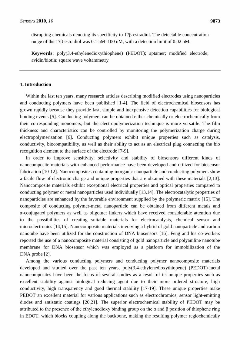
Abstract: A simple and highly sensitive electrochemical DNA aptasensor with high affinity
for endocrine disrupting 17β-estradiol, was developed. Poly(3,4-ethylenedioxylthiophene)
(PEDOT) doped with gold nanoparticles (AuNPs) was electrochemically synthesized and
employed for the immobilization of biotinylated aptamer towards the detection of the
target. The diffusion coefficient of the nanocomposite was 6.50 × 10⁻⁷
cm2 s
−1, which
showed that the nanocomposite was highly conducting. Electrochemical impedance
investigation also revealed the catalytic properties of the nanocomposite with an exchange
current value of 2.16 × 10⁻⁴
A, compared to 2.14 × 10⁻⁵
A obtained for the bare electrode.
Streptavidin was covalently attached to the platform using carbodiimide chemistry and the
aptamer immobilized via streptavidin—biotin interaction. The electrochemical signal
generated from the aptamer–target molecule interaction was monitored electrochemically
using cyclic voltammetry and square wave voltammetry in the presence of [Fe(CN)6]−3/−4
as a redox probe. The signal observed shows a current decrease due to interference of the
bound 17β-estradiol. The current drop was proportional to the concentration of 17β-estradiol.
The PEDOT/AuNP platform exhibited high electroactivity, with increased peak current.
The platform was found suitable for the immobilization of the DNAaptamer. The
aptasensor was able to distinguish 17β-estradiol from structurally similar endocrine
OPEN ACCESS
Sensors 2010, 10
9873
disrupting chemicals denoting its specificity to 17β-estradiol. The detectable concentration
range of the 17β-estradiol was 0.1 nM–100 nM, with a detection limit of 0.02 nM.
Keywords: poly(3,4-ethylenedioxythiophene) (PEDOT); aptamer; modified electrode;
avidin/biotin; square wave voltammetry
1. Introduction
Within the last ten years, many research articles describing modified electrodes using nanoparticles
and conducting polymers have been published [1-4]. The field of electrochemical biosensors has
grown rapidly because they provide fast, simple and inexpensive detection capabilities for biological
binding events [5]. Conducting polymers can be obtained either chemically or electrochemically from
their corresponding monomers, but the electropolymerization technique is more versatile. The film
thickness and characteristics can be controlled by monitoring the polymerization charge during
electropolymerization [6]. Conducting polymers exhibit unique properties such as catalysis,
conductivity, biocompatibility, as well as their ability to act as an electrical plug connecting the bio
recognition element to the surface of the electrode [7-9].
In order to improve sensitivity, selectivity and stability of biosensors different kinds of
nanocomposite materials with enhanced performance have been developed and utilized for biosensor
fabrication [10-12]. Nanocomposites containing inorganic nanoparticle and conducting polymers show
a facile flow of electronic charge and unique properties that are obtained with these materials [2,13].
Nanocomposite materials exhibit exceptional electrical properties and optical properties compared to
conducting polymer or metal nanoparticles used individually [13,14]. The electrocatalytic properties of
nanoparticles are enhanced by the favorable environment supplied by the polymeric matrix [15]. The
composite of conducting polymer-metal nanoparticle can be obtained from different metals and
π-conjugated polymers as well as oligomer linkers which have received considerable attention due
to the possibilities of creating suitable materials for electrocatalysis, chemical sensor and
microelectronics [14,15]. Nanocomposite materials involving a hybrid of gold nanoparticle and carbon
nanotube have been utilized for the construction of DNA biosensors [16]. Feng and his co-workers
reported the use of a nanocomposite material consisting of gold nanoparticle and polyaniline nanotube
membrane for DNA biosensor which was employed as a platform for immobilization of the
DNA probe [2].
Among the various conducting polymers and conducting polymer nanocomposite materials
developed and studied over the past ten years, poly(3,4-ethylenedioxythiopene) (PEDOT)-metal
nanocomposites have been the focus of several studies as a result of its unique properties such as
excellent stability against biological reducing agent due to their more ordered structure, high
conductivity, high transparency and good thermal stability [17-19]. These unique properties make
PEDOT an excellent material for various applications such as electrochromics, sensor light-emitting
diodes and antistatic coatings [20,21]. The superior electrochemical stability of PEDOT may be
attributed to the presence of the ethylenedioxy binding group on the α and β position of thiophene ring
in EDOT, which blocks coupling along the backbone, making the resulting polymer regiochemically
Sensors 2010, 10
9874
defined [21]. PEDOT has been reported to be excellent for synthesis of nanostructured materials and
devices as a result of their electrical, electronic, magnetic, and optical properties which is similar to
metals or semiconductors [22,23]. PEDOT coatings can be prepared by electrochemical polymerization
in aqueous solution which allows the direct incorporation of water soluble anions [7,24]. Xiao and
co-workers have developed an adenosine 5’-triphosphate (ATP) doped PEDOT for neural
recording [25]. A silver nanograin incorporating a poly(3,4-ethylenedioxythiophene) (PEDOT)
modified electrode for electrocatalytic sensing of hydrogen peroxide by simple electrochemical
method was reported recently by Balamurugan and his co-workers [26].
One of the major classes of environmental contaminants are the endocrine disrupting chemicals
(EDCs), which interfere with the function of the endocrine system. In recent years, these endocrine
disrupting chemicals have become one of the main topics of research in the field of environmental
sciences [27]. EDCs are defined as exogenous substances that cause adverse effect in organisms or its
progeny, consequent to changes in endocrine function. They are ubiquitous in the environment because
of their large number of uses in the residential, industrial and agricultural application [28]. The
detection of these chemicals within the natural system is therefore necessary for protecting public and
environmental health. Instrumental analysis methods such as high performance liquid chromatography
(HPLC) and gas chromatography coupled with mass spectrophotometer (GC/MS) are very sensitive at
detecting these toxic endocrine disrupting chemicals but are also very complicated to perform
and require long analysis times to be accomplished. Using GC/MS an estimated concentration
of 0.18–19.1 µg/kg was obtained in different foodstuffs in Germany compared to a calculated daily
intake of 7.5 µg/day for nonylphenol, an endocrine disrupting chemical [29]. Concentrations of
between 7.7–11,300 ng/L for some phenolic endocrine disrupting chemicals were obtained from river
water in China using GC/MS coupled with the negative chemical ionization (NCI) technique [30]. The
estimated daily human intake of estrogenic and anti-estrogenic counterparts, based on in vitro
potencies relative to 17β-estradiol, has indicated that a woman taking birth control pills ingests
about 6,675 µg/equivalent per day, postmenopausal estrogen treatment amount to 3,350 µg and
ingestion of estrogen flavonoids in food represent 102 µg, while the daily ingestion of environmental
organochlorine estrogen was estimated to be 2.5 × 10⁻⁶
µg [31].
Since the mid 1990s, a variety of adverse effects of endocrine disrupting chemicals on the endocrine
systems of man and animals have been observed, which are of particular environmental concern [32].
Most EDCs are synthetic organic chemical that are introduced to the environment by anthropogenic
sources but they can also be naturally generated by the estrogenic hormones 17β-estradiol and
estrone [28]. It has been stated that the increasing incidence of breast and testicular cancers in humans
may be caused by exposure to EDCs especially via drinking water whose source are often from surface
waters [33,34]. The fate and behavior of EDCs are influenced by the structure and physicochemical
parameters [35]. Most of these chemicals are present at low concentrations but many of them
raised considerable toxicological concern. Children born to women exposed to high levels of
polychlorobiphenyl (PCB) via consumption of polluted fish oil or rice oil have been reported to show
delayed mental growth with lower Intelligence Quotient (IQ) scores, cognitive dysfunction, poorer
visual identification memory and behavioral difficulties [36]. The incident of testicular cancer in men
has increased significantly during the last decade. The incident of cancer in men under 50 years of age
has increased approximately to 2–4% per annum since the 1960s in Great Britain, while in Denmark
Sensors 2010, 10
9875
the most common malignancy among men from age 25–34 years is testicular cancer. Breast cancer is
the most common tumor in women in the World. The relative rate of recurrence varies five-fold
between countries with the highest incidence in Western Europe and in North America. There has been
a steady increase of breast cancer incidence rates over the last decades everywhere in Europe. The
increased risk in of cancer has been linked to exposure to estrogenic chemicals and it has been reported
that woman exposed to organochlorine chemicals such as DDT and certain PCB congeners may have
higher incidence of breast cancer than non-exposed woman [36]. Prostate cancer is the second leading
form of cancer in males in USA. Deaths due to prostate cancer have increased by 17% over the
past three decades, despite improved diagnosis [36]. Considering the serious adverse effect of EDCs
on human health and environment even at low concentration, it is urgent to identify an efficient
method of estimating the concentration level of these chemicals in the environment.
To assess the impact of these chemical in the environment requires improved analytical methods
and tools [37]. Recently more attention has been focused toward electrochemical aptasensor based on
modified electrode for the determination of environmental pollutants. This is due to their excellent
analytical performance such as specificity, selectivity, simplicity, wide linear range response,
reproducibility and low cost [38,39].
In this work we report the electrochemical synthesis of poly(3,4-ethylenedioxylthiopene)-PEDOT
doped with gold nanoparticles (AuNPs) for the immobilization of biotinylated aptamer for the
detection of 17β-estradiol—an endocrine disruptor. The single strand DNA which is the aptamer was
chosen from that which has been reported in literature. This means that the sequence has been selected
and patented and thus available for use. The sequence was designed according to literature [40]. The
aptamer was biotinylated so as to effect a better immobilization chemistry using streptavidin—biotin
interaction. The nanocomposite platform was characterized electrochemically by cyclic voltammetry in
the presence of a [Fe (CN)6]−3/−4
redox probe.
2. Experimental Section
2.1. Chemicals and Reagents
3,4-Ethylenedioxythiophene (EDOT), 3,3-dithiodipropionic acid (DPA), N-(3-dimethylaminopropyl)-
N-ethylcarbodiimide hydrochloride (EDAC), N-hydroxysuccinimide (NHS), streptavidin,
HAuCl4·3H2O, 17β-estradiol, sodium monohydrogen phosphate, potassium dihydrogen phosphate,
lithium perchlorate, potassium ferricyanide [K3Fe(CN)6] and potassium ferricyanide [K4Fe(CN)6] and
sodium dodecylsulphate were procured from Sigma-Aldrich (South Africa). All chemicals were of
analytical grade and were used as received. Deionized water (18.2 MΩ) purified by a milli-QTM
system
(Millipore) was used throughout the experiment for aqueous solution preparation. A 76-mers
biotinylated ssDNA aptamer synthesized by Inqaba Biotechnical Industries (Pty) Ltd., Hatfield, South
Africa was used as aptamer probe. The sequence of the 76-mers sized biotinylated aptamer is
given below:
5’-BiotinGCTTCCAGCTTATTGAATTACACGCAGAGGGTAGCGGCTCTGCGC
ATTCAATTGCTGCGCGCTGAAGCGCGGAAGC-3’
Sensors 2010, 10
9876
2.2. Solutions
Phosphate buffer saline (PBS) of pH 7.5 containing 10 mM Na2HPO4, KH2PO4 and 0.1 M KCl was
prepared, 5 mM (1:1) solution of K3Fe(CN)6 and K4Fe(CN)6 was prepared in 100 mL of PBS
at pH 7.5. One hundred µM biotinylated DNA aptamer stock was prepared in tris EDTA (TE) buffer
(pH 8.0) and stored at −20 °C. Working DNA aptamer solution were prepared by diluting to the
desired concentration in phosphate buffer storage at 4 °C and discarded after four weeks. Ten mL
solution of 0.1 M lithium perchlorate containing 0.1 M EDOT monomer and 0.1 M of sodium dodecyl
sulphate were prepared for the electropolymerization. A stock solution of 50 mL of binding buffer
(pH 8.0) containing 100 mM tris HCl, 200 mM NaCl, 25 mM KCl ,10 mM MgCl2 and 5% ethanol were
prepared. Stock solution of 1 mM N-[3-(dimethylaminopropyl)-N-3-ethylcarbodiimide hydrochloride]
(EDAC), 1 mM N-hydroxysuccinimide (NHS), 1 mM 3,3-dithiodipropionic acid (DPA), as well as stock
solution of 50 µg/mL septravidin in PBS (pH 7.5) were prepared. One hundred µM stock solution
of 17β-estradiol (target) solution was prepared from which working target solution was prepared and
diluted to desired concentrations with binding buffer. All stock solutions prepared were stored at 4 °C
before and after use.
2.3. Electrochemical Measurement
A three electrode system was used to perform all electrochemical experiments. A gold electrode
with a diameter of 1.6 mm was used as the working electrode, platinum wire as the counter electrode,
and Ag/AgCl (3M Cl−) as the reference electrode. All electrochemical (voltammetric) experiments
were recorded with Zahnner IM6 electrochemical workstation (MeBtechnik).
Square wave voltammetry (SWV) measurements were recorded at amplitude 25 mV and frequency
of 15 Hz with this electrochemical work station. Cyclic voltammetry measurements were recorded
with the same electrochemical workstation. All solutions were de-aerated by purging with argon
through it for 5 mins. The experiments were carried out under room temperature. UV/Vis spectra
measurements were recorded with the Nicolette Evolution 100 Spectrometer (Thermo Electron
Corporation, UK). Transmission electron microscopy (TEM) was performed in a Tecnai G2 F2O
X-Twin MAT. TEM characterizations were performed by placing a drop of the solution on a carbon
coated copper grid and dried under electric UV lamp for 30 minutes.
2.4. Preparation of Gold Nanoparticles
AuNP was prepared by citrate reduction of HAuCl4 solution according to literature [13,41]. The
AuNP prepared was stored in a brown bottle at 4 °C before use.
2.5. Preparation of a Poly(3,4-ethylenedioxythiopene) Modified Gold Electrode
Prior to surface modification, the gold working electrode was polished to a mirror-like surface with
alumina powder of sizes 1.0, 0.3 and 0.05 µm respectively. The electrode was dipped in piranha
solution for 10 minutes for effective cleaning and the electrode was then sonicated in ethanol and
water consecutively for 5 mins. The electrode was further cleaned electrochemically in sulphuric acid
by cycling between the potential of −200 mV to 1,500 mV until a reproducible cyclic voltammogram
Sensors 2010, 10
9877
was obtained. Subsequently the gold electrode was rinsed with copious amount of water and absolute
ethanol respectively. PEDOT film was deposited on the bare gold electrode from an aqueous solution
of 0.1 M lithium perchlorate containing 0.1 M 3,4-ethylenedioxylthiopene and 0.1 M sodium dodecyl
sulphate by cycling the potential between −1,000 mV to 1,000 mV at a scan rate of 50 mV/s for
ten cycles, as shown in Figure 2.
2.6. Fabrication of PEDOT—Gold Nanoparticle Modified Electrode
The PEDOT modified gold electrode prepared above was immersed in a colloidal gold
nanoparticles solution for 24 h for self assembly of the gold nanoparticles on the PEDOT film to obtain
a nanocomposite modified electrode.
2.7. Fabrication of the Aptasensor
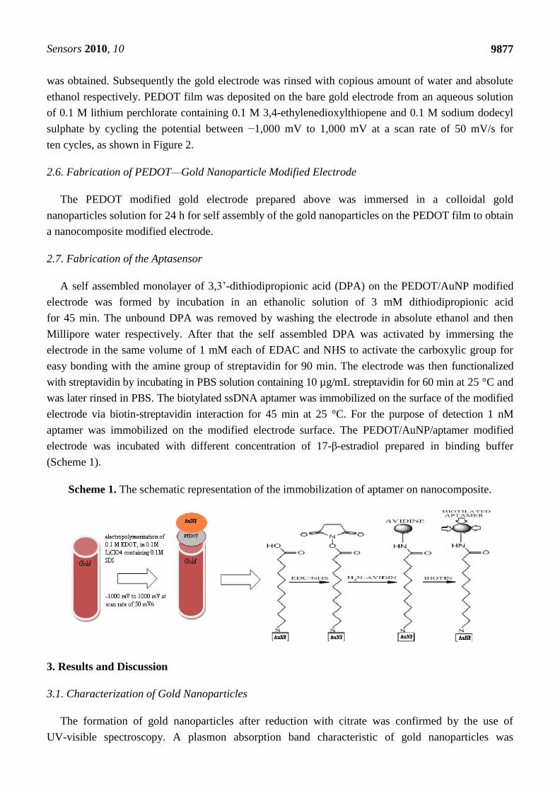
A self assembled monolayer of 3,3’-dithiodipropionic acid (DPA) on the PEDOT/AuNP modified
electrode was formed by incubation in an ethanolic solution of 3 mM dithiodipropionic acid
for 45 min. The unbound DPA was removed by washing the electrode in absolute ethanol and then
Millipore water respectively. After that the self assembled DPA was activated by immersing the
electrode in the same volume of 1 mM each of EDAC and NHS to activate the carboxylic group for
easy bonding with the amine group of streptavidin for 90 min. The electrode was then functionalized
with streptavidin by incubating in PBS solution containing 10 µg/mL streptavidin for 60 min at 25 °C and
was later rinsed in PBS. The biotylated ssDNA aptamer was immobilized on the surface of the modified
electrode via biotin-streptavidin interaction for 45 min at 25 °C. For the purpose of detection 1 nM
aptamer was immobilized on the modified electrode surface. The PEDOT/AuNP/aptamer modified
electrode was incubated with different concentration of 17-β-estradiol prepared in binding buffer
(Scheme 1).
Scheme 1. The schematic representation of the immobilization of aptamer on nanocomposite.
3. Results and Discussion
3.1. Characterization of Gold Nanoparticles
The formation of gold nanoparticles after reduction with citrate was confirmed by the use of
UV-visible spectroscopy. A plasmon absorption band characteristic of gold nanoparticles was
Sensors 2010, 10
9878
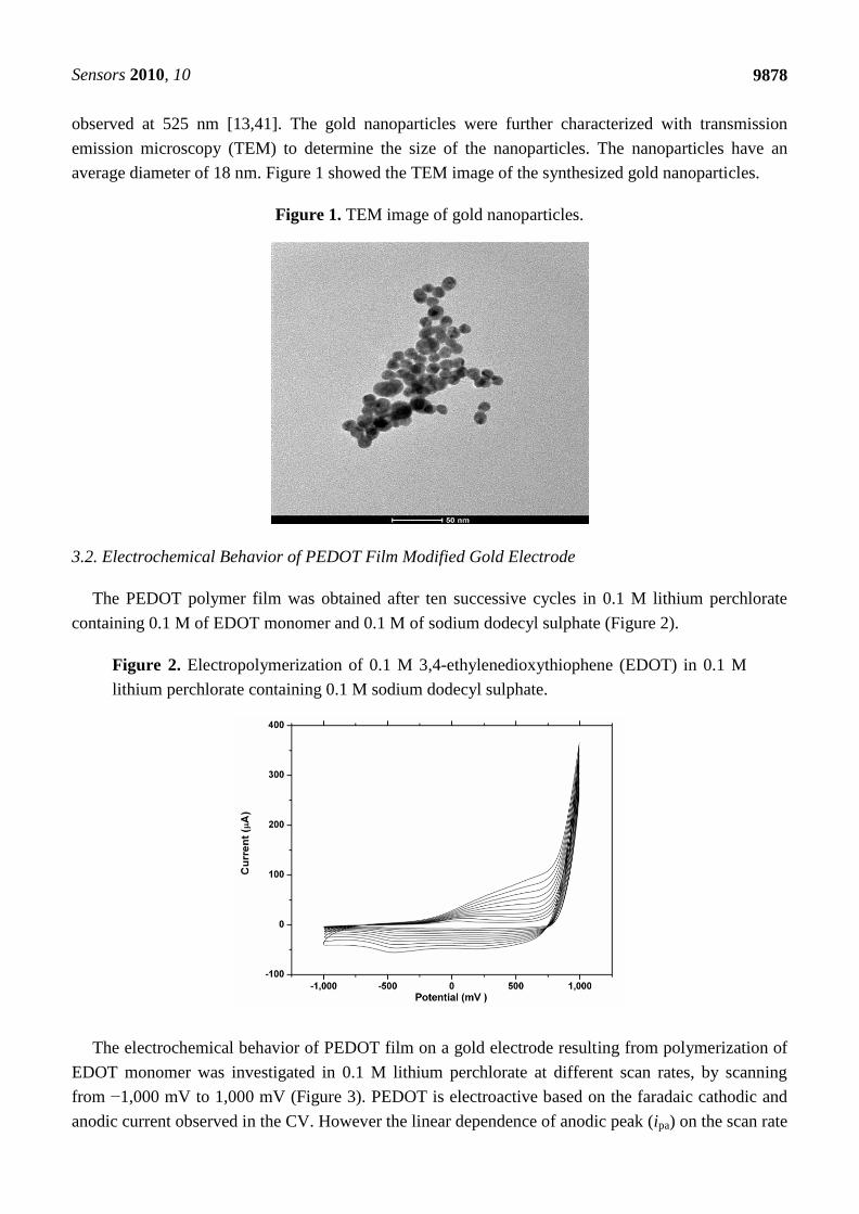
observed at 525 nm [13,41]. The gold nanoparticles were further characterized with transmission
emission microscopy (TEM) to determine the size of the nanoparticles. The nanoparticles have an
average diameter of 18 nm. Figure 1 showed the TEM image of the synthesized gold nanoparticles.
Figure 1. TEM image of gold nanoparticles.
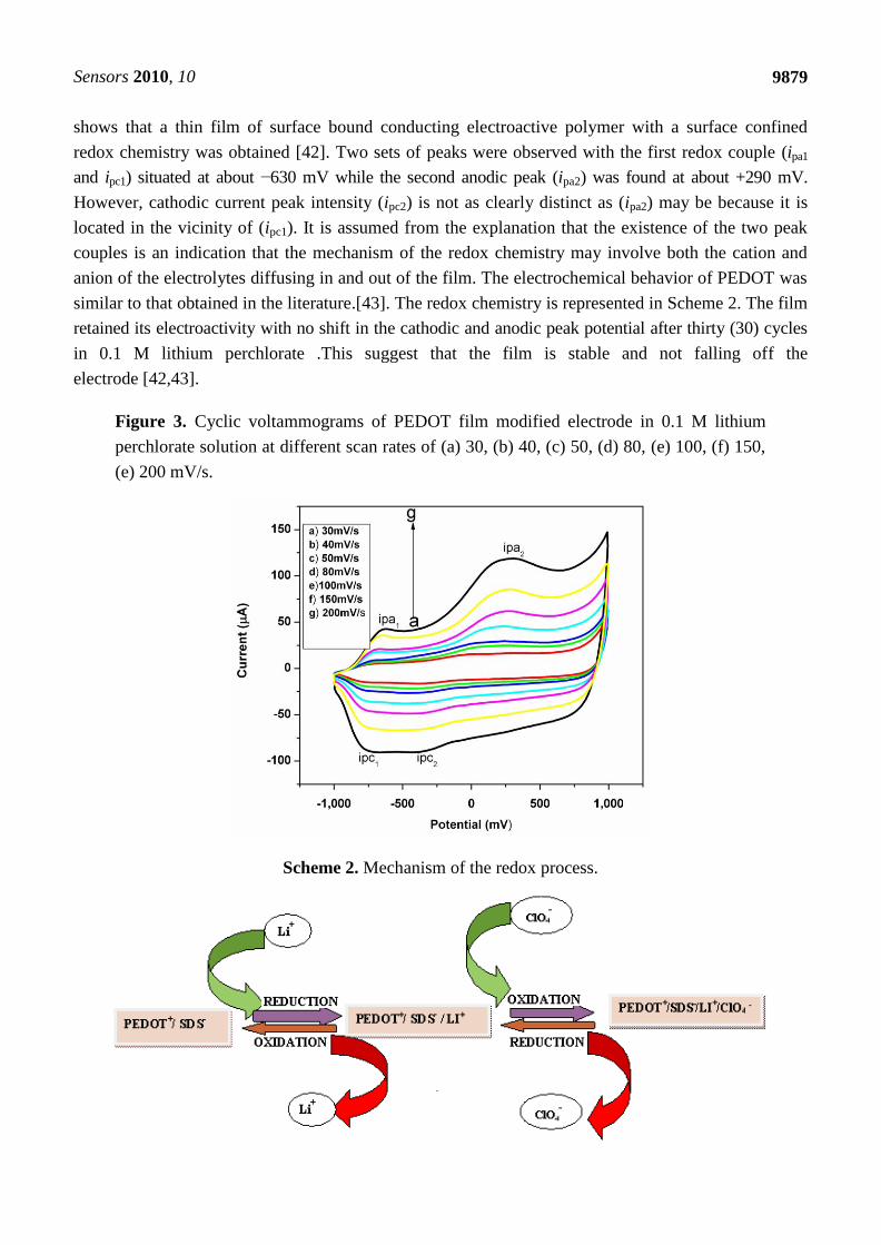
3.2. Electrochemical Behavior of PEDOT Film Modified Gold Electrode
The PEDOT polymer film was obtained after ten successive cycles in 0.1 M lithium perchlorate
containing 0.1 M of EDOT monomer and 0.1 M of sodium dodecyl sulphate (Figure 2).
Figure 2. Electropolymerization of 0.1 M 3,4-ethylenedioxythiophene (EDOT) in 0.1 M
lithium perchlorate containing 0.1 M sodium dodecyl sulphate.
The electrochemical behavior of PEDOT film on a gold electrode resulting from polymerization of
EDOT monomer was investigated in 0.1 M lithium perchlorate at different scan rates, by scanning
from −1,000 mV to 1,000 mV (Figure 3). PEDOT is electroactive based on the faradaic cathodic and
anodic current observed in the CV. However the linear dependence of anodic peak (ipa) on the scan rate
Sensors 2010, 10
9879
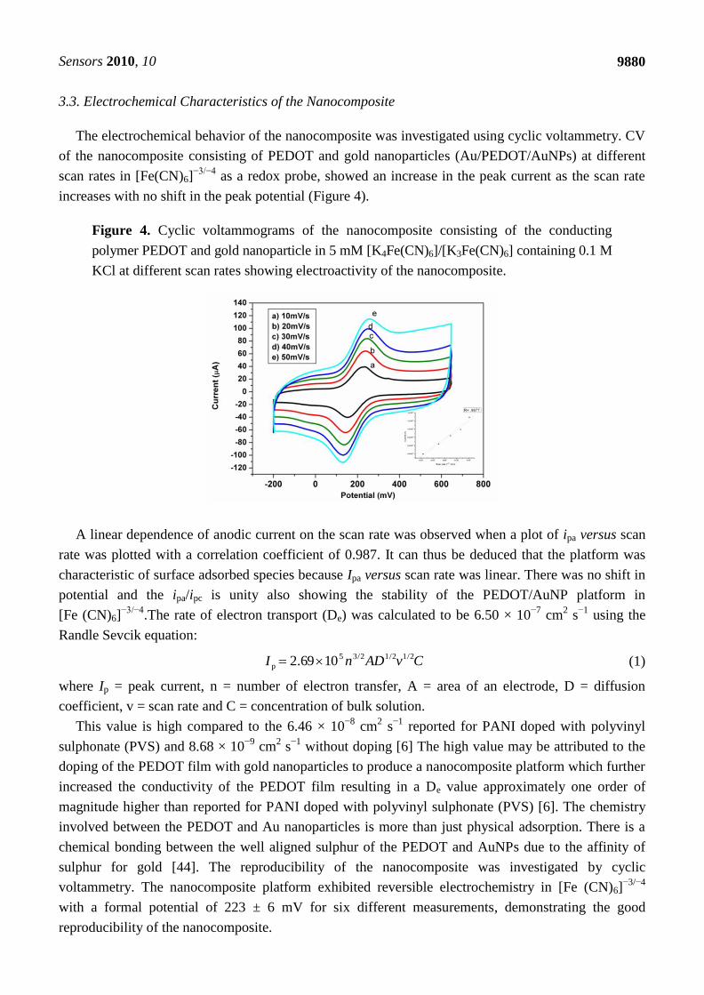
shows that a thin film of surface bound conducting electroactive polymer with a surface confined
redox chemistry was obtained [42]. Two sets of peaks were observed with the first redox couple (ipa1
and ipc1) situated at about −630 mV while the second anodic peak (ipa2) was found at about +290 mV.
However, cathodic current peak intensity (ipc2) is not as clearly distinct as (ipa2) may be because it is
located in the vicinity of (ipc1). It is assumed from the explanation that the existence of the two peak
couples is an indication that the mechanism of the redox chemistry may involve both the cation and
anion of the electrolytes diffusing in and out of the film. The electrochemical behavior of PEDOT was
similar to that obtained in the literature.[43]. The redox chemistry is represented in Scheme 2. The film
retained its electroactivity with no shift in the cathodic and anodic peak potential after thirty (30) cycles
in 0.1 M lithium perchlorate .This suggest that the film is stable and not falling off the
electrode [42,43].
Figure 3. Cyclic voltammograms of PEDOT film modified electrode in 0.1 M lithium
perchlorate solution at different scan rates of (a) 30, (b) 40, (c) 50, (d) 80, (e) 100, (f) 150,
(e) 200 mV/s.
Scheme 2. Mechanism of the redox process.
Sensors 2010, 10
9880
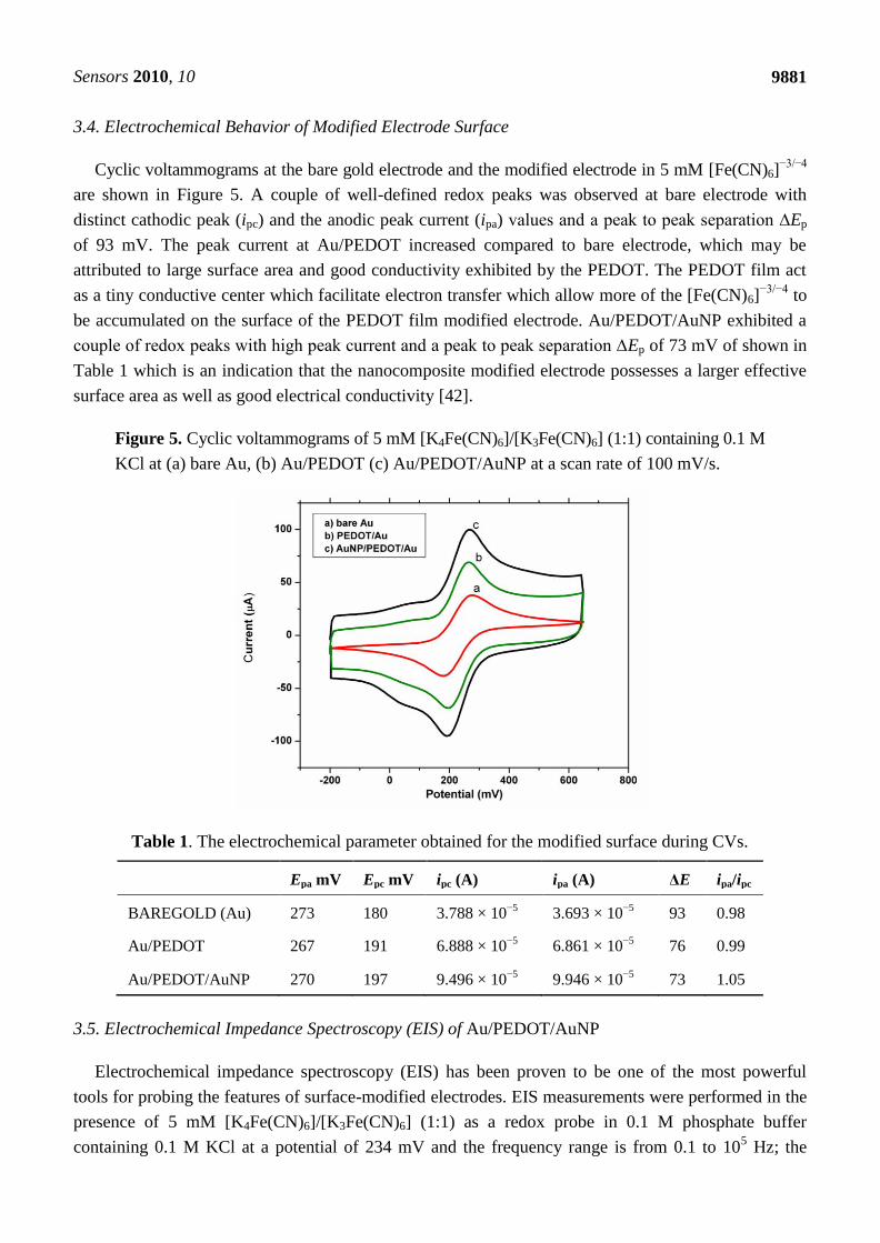
3.3. Electrochemical Characteristics of the Nanocomposite
The electrochemical behavior of the nanocomposite was investigated using cyclic voltammetry. CV
of the nanocomposite consisting of PEDOT and gold nanoparticles (Au/PEDOT/AuNPs) at different
scan rates in [Fe(CN)6]−3/−4
as a redox probe, showed an increase in the peak current as the scan rate
increases with no shift in the peak potential (Figure 4).
Figure 4. Cyclic voltammograms of the nanocomposite consisting of the conducting
polymer PEDOT and gold nanoparticle in 5 mM [K4Fe(CN)6]/[K3Fe(CN)6] containing 0.1 M
KCl at different scan rates showing electroactivity of the nanocomposite.
A linear dependence of anodic current on the scan rate was observed when a plot of ipa versus scan
rate was plotted with a correlation coefficient of 0.987. It can thus be deduced that the platform was
characteristic of surface adsorbed species because Ipa versus scan rate was linear. There was no shift in
potential and the ipa/ipc is unity also showing the stability of the PEDOT/AuNP platform in
[Fe (CN)6]−3/−4
.The rate of electron transport (De) was calculated to be 6.50 × 10⁻⁷
cm2 s
−1 using the
Randle Sevcik equation:
CvADnI 1/21/23/25
p 1069.2 (1)
where Ip = peak current, n = number of electron transfer, A = area of an electrode, D = diffusion
coefficient, v = scan rate and C = concentration of bulk solution.
This value is high compared to the 6.46 × 10⁻⁸
cm2 s
−1 reported for PANI doped with polyvinyl
sulphonate (PVS) and 8.68 × 10⁻⁹
cm2 s
−1 without doping [6] The high value may be attributed to the
doping of the PEDOT film with gold nanoparticles to produce a nanocomposite platform which further
increased the conductivity of the PEDOT film resulting in a De value approximately one order of
magnitude higher than reported for PANI doped with polyvinyl sulphonate (PVS) [6]. The chemistry
involved between the PEDOT and Au nanoparticles is more than just physical adsorption. There is a
chemical bonding between the well aligned sulphur of the PEDOT and AuNPs due to the affinity of
sulphur for gold [44]. The reproducibility of the nanocomposite was investigated by cyclic
voltammetry. The nanocomposite platform exhibited reversible electrochemistry in [Fe (CN)6]−3/−4
with a formal potential of 223 ± 6 mV for six different measurements, demonstrating the good
reproducibility of the nanocomposite.
Sensors 2010, 10
9881
3.4. Electrochemical Behavior of Modified Electrode Surface
Cyclic voltammograms at the bare gold electrode and the modified electrode in 5 mM [Fe(CN)6]−3/−4
are shown in Figure 5. A couple of well-defined redox peaks was observed at bare electrode with
distinct cathodic peak (ipc) and the anodic peak current (ipa) values and a peak to peak separation ∆Ep
of 93 mV. The peak current at Au/PEDOT increased compared to bare electrode, which may be
attributed to large surface area and good conductivity exhibited by the PEDOT. The PEDOT film act
as a tiny conductive center which facilitate electron transfer which allow more of the [Fe(CN)6]−3/−4
to
be accumulated on the surface of the PEDOT film modified electrode. Au/PEDOT/AuNP exhibited a
couple of redox peaks with high peak current and a peak to peak separation ∆Ep of 73 mV of shown in
Table 1 which is an indication that the nanocomposite modified electrode possesses a larger effective
surface area as well as good electrical conductivity [42].
Figure 5. Cyclic voltammograms of 5 mM [K4Fe(CN)6]/[K3Fe(CN)6] (1:1) containing 0.1 M
KCl at (a) bare Au, (b) Au/PEDOT (c) Au/PEDOT/AuNP at a scan rate of 100 mV/s.
Table 1. The electrochemical parameter obtained for the modified surface during CVs.
Epa mV Epc mV ipc (A) ipa (A) ΔE ipa/ipc
BAREGOLD (Au) 273 180 3.788 × 10⁻⁵
3.693 × 10⁻⁵
93 0.98
Au/PEDOT 267 191 6.888 × 10⁻⁵
6.861 × 10⁻⁵
76 0.99
Au/PEDOT/AuNP 270 197 9.496 × 10⁻⁵
9.946 × 10⁻⁵
73 1.05
3.5. Electrochemical Impedance Spectroscopy (EIS) of Au/PEDOT/AuNP
Electrochemical impedance spectroscopy (EIS) has been proven to be one of the most powerful
tools for probing the features of surface-modified electrodes. EIS measurements were performed in the
presence of 5 mM [K4Fe(CN)6]/[K3Fe(CN)6] (1:1) as a redox probe in 0.1 M phosphate buffer
containing 0.1 M KCl at a potential of 234 mV and the frequency range is from 0.1 to 105 Hz; the
Sensors 2010, 10
9882
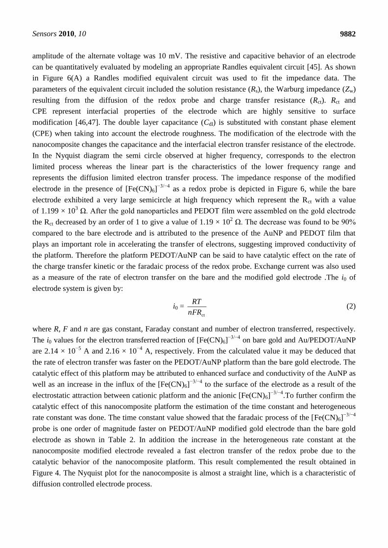
amplitude of the alternate voltage was 10 mV. The resistive and capacitive behavior of an electrode
can be quantitatively evaluated by modeling an appropriate Randles equivalent circuit [45]. As shown
in Figure 6(A) a Randles modified equivalent circuit was used to fit the impedance data. The
parameters of the equivalent circuit included the solution resistance (Rs), the Warburg impedance (Zw)
resulting from the diffusion of the redox probe and charge transfer resistance (Rct). Rct and
CPE represent interfacial properties of the electrode which are highly sensitive to surface
modification [46,47]. The double layer capacitance (Cdl) is substituted with constant phase element
(CPE) when taking into account the electrode roughness. The modification of the electrode with the
nanocomposite changes the capacitance and the interfacial electron transfer resistance of the electrode.
In the Nyquist diagram the semi circle observed at higher frequency, corresponds to the electron
limited process whereas the linear part is the characteristics of the lower frequency range and
represents the diffusion limited electron transfer process. The impedance response of the modified
electrode in the presence of [Fe(CN)6]−3/−4
as a redox probe is depicted in Figure 6, while the bare
electrode exhibited a very large semicircle at high frequency which represent the Rct with a value
of 1.199 × 10³ Ω. After the gold nanoparticles and PEDOT film were assembled on the gold electrode
the Rct decreased by an order of 1 to give a value of 1.19 × 10² Ω. The decrease was found to be 90%
compared to the bare electrode and is attributed to the presence of the AuNP and PEDOT film that
plays an important role in accelerating the transfer of electrons, suggesting improved conductivity of
the platform. Therefore the platform PEDOT/AuNP can be said to have catalytic effect on the rate of
the charge transfer kinetic or the faradaic process of the redox probe. Exchange current was also used
as a measure of the rate of electron transfer on the bare and the modified gold electrode .The i0 of
electrode system is given by:
i0 = ctnFR
RT (2)
where R, F and n are gas constant, Faraday constant and number of electron transferred, respectively.
The i0 values for the electron transferred reaction of [Fe(CN)6]−3/−4
on bare gold and Au/PEDOT/AuNP
are 2.14 × 10⁻⁵
A and 2.16 × 10⁻⁴
A, respectively. From the calculated value it may be deduced that
the rate of electron transfer was faster on the PEDOT/AuNP platform than the bare gold electrode. The
catalytic effect of this platform may be attributed to enhanced surface and conductivity of the AuNP as
well as an increase in the influx of the [Fe(CN)6]−3/−4
to the surface of the electrode as a result of the
electrostatic attraction between cationic platform and the anionic [Fe(CN)6]−3/−4
.To further confirm the
catalytic effect of this nanocomposite platform the estimation of the time constant and heterogeneous
rate constant was done. The time constant value showed that the faradaic process of the [Fe(CN)6]−3/−4
probe is one order of magnitude faster on PEDOT/AuNP modified gold electrode than the bare gold
electrode as shown in Table 2. In addition the increase in the heterogeneous rate constant at the
nanocomposite modified electrode revealed a fast electron transfer of the redox probe due to the
catalytic behavior of the nanocomposite platform. This result complemented the result obtained in
Figure 4. The Nyquist plot for the nanocomposite is almost a straight line, which is a characteristic of
diffusion controlled electrode process.
Sensors 2010, 10
9883
Figure 6. (A) Randles equivalent circuit and (B) Nyquist plots obtained for (a) bare Au,
(b) Au/PEDOT/AuNP modified electrode. EIS measurements were carried out in 5 mM
[Fe(CN)6]−3/−4
containing 0.1 M KCl.
(A)
(B)
Table 2. Electrochemical impedance kinetic parameters of the bare electrode and the
nanocomposite platform.
Kinetic parameters
Exchange current Time constant Heterogeneous rate constant
Bare electrode 2.14 × 10⁻⁵
A 1.49 × 10⁻³
s/rad 2.21 × 10⁻³
cm/s
PEDOT/AuNP platform 2.16 × 10⁻⁴
A 9.60 × 10⁻⁵
s/rad 2.23 × 10⁻²
cm/s
The electrode coverage is a key factor which can be used to estimate the surface state of the
electrode, and the charge resistance is related to it. The surface coverage (θ) of the nanocomposite on
bare gold electrode can be estimated from EIS according to Equation (3) [48]:
electrodeBare
ct
electrodemodified
ct1R
R (3)
where Rct denotes the charge transfer resistance. The surface coverage of (θ) of the electrode was
estimated to be 90.1% which will enable more of the bioreceptor (aptamer) to be adsorbed on the
surface of the electrode for better sensitivity of the aptasensor to its target.
Rs Rct Wo
CPE1
Element Freedom Value Error Error %
Rs Fixed(X) 0 N/A N/A
Rct Fixed(X) 0 N/A N/A
Wo-R Fixed(X) 0 N/A N/A
Wo-T Fixed(X) 0 N/A N/A
Wo-P Fixed(X) 0.5 N/A N/A
CPE1-T Fixed(X) 0 N/A N/A
CPE1-P Fixed(X) 1 N/A N/A
Data File:
Circuit Model File:
Mode: Run Simulation / Freq. Range (0.001 - 1000000)
Maximum Iterations: 100
Optimization Iterations: 0
Type of Fitting: Complex
Type of Weighting: Calc-Modulus
Sensors 2010, 10
9884
3.6. Quantitative Analysis of 17β-Estradiol Using Aptamer Immobilized PEDOT/AuNP Modified Electrode
Electrochemical analysis was performed to confirm and optimize the condition for ssDNA
aptamer immobilized on the surface of the modified electrode. Various concentrations of ssDNA
aptamer (0.5 µM–1.0 µM) were initially mobilized on the modified electrode. The current changes in
CV were measured before and after immobilization of ssDNA aptamer on the modified and current
drop in CV was observed. The difference in current drop varies with the concentration of the ssDNA
aptamer on the modified electrode which may be attributed to biotinylated ssDNA aptamer
interference with the electron flow [49]. The decrease in current response with increase in
concentration may be due to the electrostatic repulsion that exist between the negatively charge
phosphate backbone of the DNA aptamer and the negatively charged hydroxyl ion substituent of the
target in aqueous solution, which is one of the subtle substituents for aptamer discrimination within
molecule of similar structures in the presence of [Fe(CN)6]−3/−4
as redox probe. Therefore binding of
aptamer to charged small molecule in aqueous solution such as 17β-estradiol to aptamer can affect
electron flow combined with diffusion rate in the presence of [Fe(CN)6]−3/−4
[48]. This also showed
that PEDOT/AuNP could be a fine platform for immobilization of ssDNA aptamer.
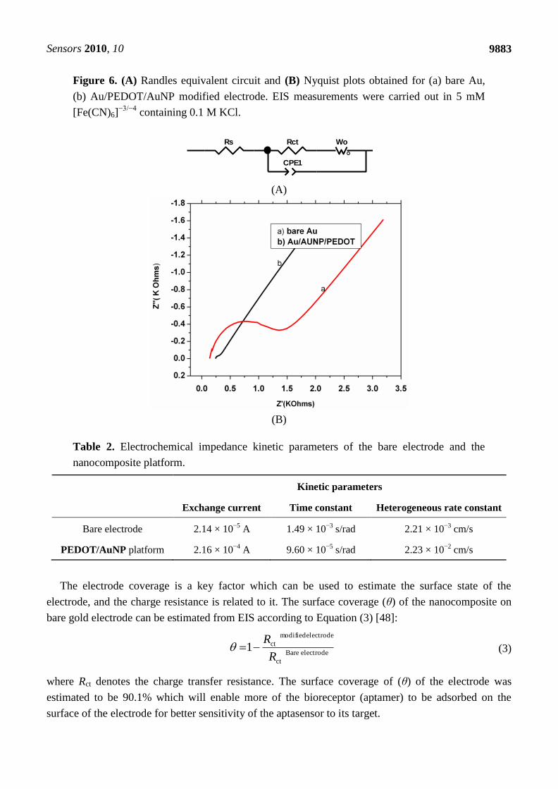
The CV and SWV analysis was carried out against a series of 17β-estradiol concentrations
(0.1 nM–100 nM) to probe the binding of the 17β-estradiol using 1 nM ssDNA aptamer immobilized
on the surface of PEDOT/AuNP modified electrode. The interaction of the target-aptamer complex
was monitored by reduction in the electron flux produced from redox reaction between ferrocyanide
and ferricyanide. The current drop with increase in concentration was observed in SWV (Figure 7).
The decrease in current after 17β-estradiol treatment was based on the specific interaction of aptamer
and the target due to electrostatic repulsion that occur between the negatively charged phosphate
backbone of the aptamer and negatively charge ion (OH−) of the target in solution. The formation of
this aptamer-target complex changes the permeability of the layer toward charged ferricyanide ion and
consequently the rate of their diffusion [49]. Although a wide range of 17β-estradiol was used, but the
minimum concentration with which a current drop occurred was at 0.1 nM 17β-estradiol with slight
change in potential and the increasing concentration dependence changes in current was seen till 100 nM
in SWV.
The difference in peak potential (ΔE = Epa − Epc) from the cyclic voltammograms was increased
which was dependent on the concentration of the target molecule. After the target molecule was
adsorbed onto the surface of the biosensor, it exhibited a reduced peak current depending on the
concentration. The decrease in peak current was observed which could be attributed to slow kinetics of
the charge transfer, which is caused by the binding of 17β-estradiol to aptamer [40,49]. However with
the series of 17β-estradiol dilutions tested it was found out that 0.1 nM 17β-estradiol was detectable
with a lower concentration of 1 nM ssDNA aptamer immobilized on the nanocomposite platform. The
high sensitivity of the aptasensor may be attributed to high surface coverage of 90.1% exhibited by the
nanocomposite which significantly enhanced the loading of the DNAaptamer probe and hence
markedly improve the sensitivity for the target as shown in Table 3 [16].
Sensors 2010, 10
9885
Figure 7. Square wave voltammograms of the electrochemical analysis of 17β-estradiol
using 1 nM DNA aptamer immobilized on the nanocomposite platform showing current
drop to concentration of target in range of 0.1–100 nM.
Table 3. Comparism of the aptasensor with other sensors.
Methods Linear range (M) LOD(M) Reference
Electrochemistry
Au/PEDOT/AuNP
0.1 × 10⁻⁹
–100 × 10−9
0.02 × 10⁻⁹
This work
Electrochemistry
Bare GCE
4 × 10⁻⁵
–1 × 10⁻³
1 × 10⁻⁵
[50]
Electrochemistry
Poly-serine/GCE
1 × 10⁻⁷
–3 × 10⁻⁵
2 × 10⁻⁸
[51]
Electrochemistry
GCE/nanoPt-MWNT
5 × 10⁻⁷
–1.5 × 10⁻⁵
1.8 × 10⁻⁷
[52]
3.7. Specificity and Selectivity Test of the Aptamer
Beside sensitivity, selectivity is also a remarkable feature for an aptamer sensor. Three different
compounds that have similar structures as the target 17β-estradiol were taken as negative control and
tested with the aptamer. A different signal must be differentiated from these substances compared to
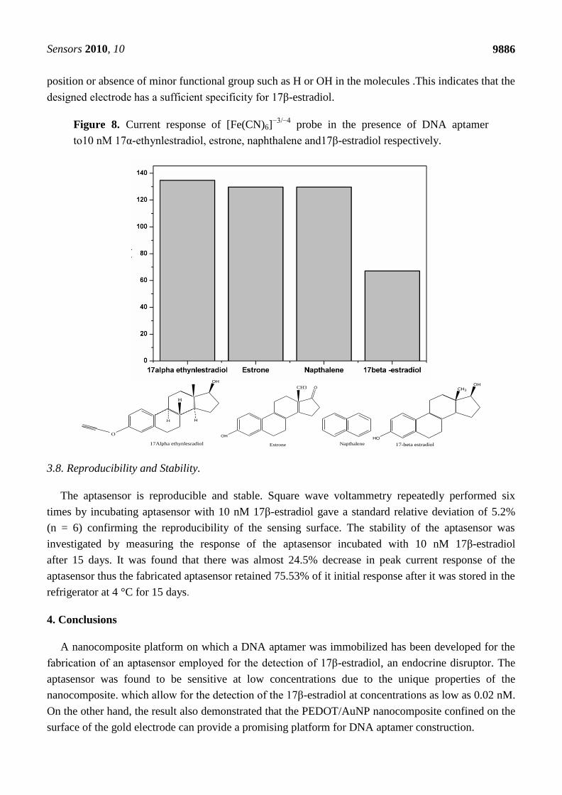
a 17β-estradiol aptamer [49]. Figure 8 compares the response of the fabricated aptasensor
to 17β-estradiol and similarly structured substances in the presence of [Fe(CN)6]−3/−4
as probe. In
Figure 8, 10 nM 17α-ethynestradiol, estrone and naphthalene exhibited high current responses when
incubated with aptasensor, which may be attributed to low binding action between the aptamer and the
analogue chemicals. However, low current response was obtained when 17β-estradiol was incubated
with aptasensor due to high binding action that existed between aptamer and 17β-estradiol resulting in
the formation of an aptamer-target complex which hindered the electron flow as well as the diffusion
rate in the presence of [Fe(CN)6]−3/−4
. The specificity of the target was mostly dependent on change in
Sensors 2010, 10
9886
position or absence of minor functional group such as H or OH in the molecules .This indicates that the
designed electrode has a sufficient specificity for 17β-estradiol.
Figure 8. Current response of [Fe(CN)6]−3/−4
probe in the presence of DNA aptamer
to10 nM 17α-ethynlestradiol, estrone, naphthalene and17β-estradiol respectively.
OH
HH
O
H
17Alpha ethynlesradiol
OH
OCH3
Estrone Napthalene
CH3
HO
OH
17-beta estradiol
3.8. Reproducibility and Stability.
The aptasensor is reproducible and stable. Square wave voltammetry repeatedly performed six
times by incubating aptasensor with 10 nM 17β-estradiol gave a standard relative deviation of 5.2%
(n = 6) confirming the reproducibility of the sensing surface. The stability of the aptasensor was
investigated by measuring the response of the aptasensor incubated with 10 nM 17β-estradiol
after 15 days. It was found that there was almost 24.5% decrease in peak current response of the
aptasensor thus the fabricated aptasensor retained 75.53% of it initial response after it was stored in the
refrigerator at 4 °C for 15 days.
4. Conclusions
A nanocomposite platform on which a DNA aptamer was immobilized has been developed for the
fabrication of an aptasensor employed for the detection of 17β-estradiol, an endocrine disruptor. The
aptasensor was found to be sensitive at low concentrations due to the unique properties of the
nanocomposite. which allow for the detection of the 17β-estradiol at concentrations as low as 0.02 nM.
On the other hand, the result also demonstrated that the PEDOT/AuNP nanocomposite confined on the
surface of the gold electrode can provide a promising platform for DNA aptamer construction.
Sensors 2010, 10
9887
Acknowledgements
The research work was funded by the National Research Fund South Africa. We wish to thank the
University of the Western Cape for the opportunity given to embark upon this research work.
References
1. Stelian, L.; Ion, I.; Alina, C.I. Voltammetric Determination of Phenol at Platinum Electrodes
Modified with Polypyrole Doped with Ferricyanide. Rev. Roum. Chim. 2009, 54, 351-357.
2. Feng, Y.; Yang, T.; Zhang, W.; Jiang, C.; Jiao, K. Enhanced Sensitivity for Deoxyribonucleic
Acid Electrochemical Impedance Sensor: Gold Nanoparticle/Polyaniline Nanotube Membranes.
Anal. Chim. Acta 2008, 616, 144-151.
3. Xiaoxia, L.; Honglan, Q.; Lihua, S.; Qiang, G.; Chengxiao, Z. Electrochemical Aptasensor for the
Dteremination of Cocaine Incorporating Gold Nanoparticles Modification. Electroanalysis 2008,
20, 1475-1482.
4. Aixue, L.; Yang, F.; Ma, Y.; Yang, X.R. Electrochemical Impedance Detection of DNA
Hybridization Based on Dendrimer Modified Electrode. Biosens. Bioelectron. 2007, 22, 1716-1722.
5. Zhao, G.-C.; Yang, X. A Label-Free Electrochemical RNA Aptamer for Selective Detection of
Theophylline. Electrochem. Commun. 2010, 12, 300-302.
6. Mathebe, N.G.R.; Morrin, A.; Iwuoha, E.I. Electrochemistry and Scanning Electron Microscopy
of Polyaniline/Peroxidase-Based Biosensor. Talanta 2004, 64, 115-120.
7. Sakmeche, N.; Bazzaoui, E.A.; Fall, M.; Aeiyach, S.; Jouini, M.; Lacroix, J.C.; Aaron, J.J.;
Lacaze, P.C. Application of Sodium Dodecyl Sulphate (SDS) Micellar Solution as an Organised
Medium for Electropolymerization of Thiopene Derivatives in Water. Synth. Meth. 1997, 84,
191-192.
8. Ogura, K.; Nakaoka, K.; Nakayama, M. Studies on Ion Transport During Potential Cycling of a
Prussian Blue (Inner) Polyaniline (Outer) Bilayer Electrode by Quartz Crystal Microbalance and
Fourier Transform Infrared Reflection Spectroscopy. J. Electroanal. Chem. 2000, 486, 119-125.
9. Lupu, S.; Mihailiciuc, C.; Pigani, L.; Renato, S.; Nicolae, T.; Chiara, Z. Electrochemical
Preparation and Characterization of Bilayer Film Composed by Prussian Blue and Conducting
Polymer. Electrochem. Commun. 2002, 4, 750-758.
10. Xueliang, W.; Tao, Y.; Yuayua, F.; Kui, J.; Guiaun, L. A Novel Hydrogen Peroxide Biosensor
Based on the Synergic Effect of Gold-Platinum Alloy Nanoparticle/Polyaniline Nanotube/Chistosan
Nanocomposite Membrane. Electroanalysis 2009, 21, 819-825.
11. Yang, T.; Zhang, W.; Du, M.; Jiao, K. A PDDA/Poly(2,6-pyridinedicarboxylic acid)-CNTs
Composite Film DNA Electrochemical Sensor and its Application for the Detection of Specific
Sequences Related to PAT Gene and NOS Gene. Talanta 2008, 75, 987-994.
12. Jiang, C.; Yang, T.; Jiao, K.; Gao, H. A DNA Electrochemical Sensor with Poly-L-Lysine/
Single-Walled Carbon Nanotubes Films and its Application for the Highly Sensitive EIS
Detection of PAT Gene Fragment and PCR Amplification of NOS Gene. Electrochim. Acta 2008,
53, 2917-2924.
Sensors 2010, 10
9888
13. Hussain, I.; Brust, M.; Papworth, A.J.; Cooper, A.I. Preparation of Acrylate-Stabilized Gold and
Silver Hydrosols and Gold-Polymer Composite Films. Langmuir 2003, 19, 4831-4835.
14. Liu, F.-J.; Huang, L.-M.; Wen, T.-C.; Li, C.-F.; Huang, S.-L.; Gopalan, A. Platinum Particles
Dispersed Polyaniline-Modified Electrodes Containing Sulfonated Polyelectrolyte for Methanol
Oxidation. Synth. Meth. 2008, 158, 767-774.
15. O’Mullane, A.P.; Dale, S.E.; Macpherson, J.V.; Unwin, P.R. Fabrication and Electrocatalytic
Properties of Polyaniline/Pt Nanoparticle Composite. Chem.Commun. 2004, 14, 1606-1607.
16. Zhou, N.; Yang, T.; Jiang, C.; Du, M.; Jiao, K. Highly Sensitive Electrochemical Impedance
Spectroscopic Detection of DNA Hybridization Based on Aunano-CNT/Pannano Films. Talanta
2009, 77, 1021-1026.
17. Alemán, C.; Teixeira-Dias, B.; Zanuy, D.; Estrany, F.; Armelin, E.; del Valle, L.J. A
Comprehensive Study of the Interactions between DNA and Poly(3,4-ethylenedioxythiophene).
Polymer 2009, 50, 1965-1974.
18. Argun, A.A.; Cirpan, A.; Reynolds, J.R. Using Poly(3,4-ethylenedioxylthiophene) Polystyrene
Sulfonate (PEDO/PSS). Adv. Mater. 2003, 15, 1338-1341.
19. Drillet, J.F.; Dittmeyer, R.; Juttner, K. Study of the Activity and Longterm Stability of PEDOT at
Platinum Catalyst Support for the DMFC Anode. J. Appl. Electrochem. 2007, 37, 1219-1226.
20. Dong-Hun, H.; Jae-Woo, K.; Su-Moon, P. Electrochemistry of Conducting Polymer 38.
Electrodeposited Poly(3,4-ethylenedioxylthiophene) Studied by Current Sensing Atomic Force
Micrscopy. J. Phys.Chem. B 2006, 110, 14874-14880.
21. Jui, H.C.; Chi-An, D.; Wen-Yen, C. Synthensis of Highly Conductive EDOT Copolymer Film via
Oxidative Chemical in situ Polymerization. J. Polym. Sci. A 2008, 46, 1662-1673.
22. Zotti, G; Vercelli, B.; Berlin, A. Gold Nanoparticle Linking to Polypyrole and Polythiophene
Monolayers and Multilayers. Chem. Mater. 2008, 20, 6509-6516.
23. Prakash, A.; Ouyang, J.; Lin, J.L.; Yan, Y. Polymer Memory Device Based on Conjugated
Polymer and Gold Nanoparticles. J. Appl. Phys. 2006, 100, 054309:1-054309:5.
24. Pigani, L.; Heras, A.; Colina, Á.; Seeber, R.; López-Palacios, J. Electropolymerisation of
3,4-ethylenedioxythiophene in Aqueous Solutions. Electrochem. Commun. 2004, 6, 1192-1198.
25. Xiao. Y.H.; Li, C.M.; Toh, M.L.; Xue, R. Adenosine 5’ Triophosphate Incorporated
Poly(3,4-ethylenedioxythiophene) Modified Electrode a Bioactive Platform with Electroactivity,
Stability and Biocompartibility. Chem. Biol. Interact. 2005, 157-158, 423-426.
26. Balamurugan, A.; Chen, S. Silver Nanograin Incorporated PEDOT Modified Electrode for
Electrocatalytic Sensing of Hydrogen Peroxide. Electroanalysis 2009, 12, 1419-1423.
27. Zhang, Y.; Zhou, J.L. Occurrence and Removal of Endocrine Disrupting Chemicals in
Wastewater. Chemosphere 2008, 73, 848-853.
28. Schilirò, T.; Pignata, C.; Rovere, R.; Fea, E.; Gilli, G. The Endocrine Disrupting Activity of
Surface Waters and of Wastewater Treatment Plant Effluents in Relation to Chlorination.
Chemosphere 2009, 75, 335-340.
29. Klaus, G.; Volkmar, H.; Bjoern, T.; Einhard, K.; Hartmut, P.; Torsen, R. Endocrine Disrupting
Nonylphenols Are Ubiquitous in Food. Envir. Sci. Technol. 2002, 36, 1676-1680.
Sensors 2010, 10
9889
30. Zhao, J.L.; Ying, G.G.; Wang, L.; Yang, J.F.; Yang, X.B.; Yang, L.H.; Li, X. Determination of
Phenolic Endocrine Disrupting Chemicals and Acidic Pharmaceuticals in Surface Water of the
Pearl Rivers in South China by Gas Chromatography-Negative Chemical Ionization-Mass
Spectrometry. Sci. Total Envir. 2009, 407, 962-974.
31. Safe, S. Endocrine Disruptors and Human Health: Is There a Problem. Toxicology 2004, 205, 3-10.
32. Pothitou, P.; Voutsa, D. Endocrine Disrupting Compounds in Municipal and Industrial
Wastewater Treatment Plants in Northern Greece. Chemosphere 2008, 73, 1716-1723.
33. Safe, S. Clinical Correlate of Environmental Endocrine Disruptors. Trend Endocrine Mat. 2005,
16, 139-144.
34. Synder, S.A.; Westerhoff, P.; Yoon, Y. Phamaceutical, Personal Care Product and Endocrine
Disrupting Chemical in Water Implication for Water Industry. Environ. Eng. Sci. 2003, 20,
449-469.
35. Jiang, J.Q.; Yin, Q.; Zhou, J.L.; Pearce, P. Occurrence and Treatment Trials of Endocrine
Disrupting Chemicals (Edcs) in Wastewaters. Chemosphere 2005, 61, 544-550.
36. Amaral Mendes, J.J. Endocrine Disrupters a Major Medical Challenge. Food Chem. Toxicol.
2002, 40, 781-788.
37. Koester, C.J.; Simoni, S.C.; Esser, B.K. Environmental Analysis. Anal. Chem. 2003, 75, 2813-2821.
38. Cheng, A.K.H.; Sen, D.; Yu, H.Z. Design and Testing of Aptamer-Based Electrochemical
Biosensors for Proteins and Small Molecules. Bioelectrochemistry 2009, 77, 1-12.
39. Li, X.; Shen, L.; Zhang, D.; Qi, H.; Gao, Q.; Ma, F.; Zhang, C. Electrochemical Impedance
Spectroscopy for Study of Aptamer-Thrombin Interfacial Interactions. Biosen. Bioelectron. 2008,
23, 1624-1630.
40. Kim, Y.S.; Jung, H.S.; Matsuura, T.; Lee, H.Y.; Kawai, T.; Gu, M.B. Electrochemical Detection
of 17β-estradiol Using DNA Aptamer Immobilized Gold Electrode Chip. Biosens. Bioelectron.
2007, 22, 2525-2531.
41. Robertson, D.; Tiersch, B.; Kosmella, S.; Koetz, J. Preparation of Crystalline Gold Nanoparticles
at the Surface of Mixed Phosphatidylcholine-Ionic Surfactant Vesicles. J. Colloid Interface Sci.
2007, 305, 345-351.
42. Vasantha, V.S.; Thangamuthu, R.; Mingchen, S. Electrochemical Polymerization of
Poly(3,4-ethylenedioxylthiophene) from Aqueous Solution Containing Hydroxyl
Propyl-β-cyclodextrine and the Electrocatalytic Behavior of Modified Electrode towards
Oxidation of Sulphur Oxoanion and Nitrite. Electroanalysis 2008, 20, 1754-1759.
43. Damlin, P.; Kvarnström, C.; Ivaska, A. Electrochemical Synthesis and in situ
Spectroelectrochemical Characterization of Poly(3,4-ethylenedioxythiophene) (PEDOT) in Room
Temperature Ionic Liquids. J. Electroanal. Chem. 2004, 570, 113-122.
44. Shin, H.C.; Su-Moon, P. Electrochemistry of Conductive Polymers 39. Contacts between
Conducting Polymers and Noble Metal Nanoparticles Studied by Current-Sensing Atomic Force
Microscopy. J. Phys. Chem. B 2006, 110, 25656-25664.
45. Abd-Elgawad, R.; Josep, S.; Liuis, A.; ;Baldrich, E.; Sullivian, C. Reusable Impedimetric
Aptasensor. Anal. Chem. 2005, 77, 6320-6323.
Sensors 2010, 10
9890
46. Arotiba, O.A.; Joseph, O.; Everlyne, S.; Nicolette, H.; Tesfaye, W.; Nazeem, J.; Baker, P.G.L.;
Iwuoha, E.I. An Electrochemical DNA Biosensor Developed on a Nanocomposite Platform of
Gold and Poly(propyleneimine) Dendrimer. Sensors 2008, 8, 6791-6809.
47. Arotiba, O.A.; Owino, J.H.; Baker, P.G.; Iwuoha, E.I. Electrochemical Impedimetry of
Electrodeposited Poly(propyleneimine) Dendrimer Monolayer. J. Electroanal. Chem. 2010, 638,
287-292.
48. Zhang, R.; Di-Jin, G.; Chen, D.; Hu, X. Simultaneous Electrochemical Determination of
Dopamine, Ascorbic Acid, and Uric Acid Using (Acid Chrome Blue K) Modified Glassy Carbon
Electrode. Sens. Acuat. B 2009, 138, 174-181.
49. Kim, Y.S.; Niazi, J.H.; Gu, M.B. Specific Detection of Oxytetracycline Using DNA
Aptamer-Immobilized Interdigitated Array Electrode Chip. Anal. Chim. Acta 2009, 634, 250-254.
50. Salci, B.; Biryol, I. Voltammetric Investigation of β-estradiol. Pharmaceut. Biomed. Anal. 2002,
28, 753-758.
51. Song, J.C.; Yang, J.; Hu, X.M. Electrochemical Determination of Estradiol Using a
Poly(L-Serine) Film-Modified Electrode. J. Appl. Electrochem. 2008, 38, 833-836.
52. Lin, X.Q.; Li, Y.X. A Sensitive Determination of Estrogens with a Pt Nano-Clusters/
Multi-Walled Carbon Nanotubes Modified Glassy Carbon Electrode. Biosens. Bioelectron. 2006,
22, 253-256.
© 2010 by the authors; licensee MDPI, Basel, Switzerland. This article is an open access article
distributed under the terms and conditions of the Creative Commons Attribution license
(http://creativecommons.org/licenses/by/3.0/).