Estradiol inhibits vascular endothelial cells pro-inflammatory activation induced by C-reactive...
11
Estradiol inhibits vascular endothelial cells pro-inflammatory activation induced by C-reactive protein E ´ milie Cossette • Isabelle Cloutier • Kim Tardif • Genevie `ve DonPierre • Jean-Franc ¸ois Tanguay Received: 17 May 2012 / Accepted: 17 October 2012 / Published online: 31 October 2012 Ó The Author(s) 2012. This article is published with open access at Springerlink.com Abstract In addition of being an important inflammatory biomarker and a risk factor for cardiovascular disease, much evidence indicates that the C-reactive protein (CRP) contributes to the atherosclerosis development process. This plasmatic protein synthesized by hepatocytes in response to inflammation and tissue injury induces pro- inflammatory molecules’ expression by endothelial cells (ECs). Previous studies showed that the 17b-estradiol (E2) has beneficial effects on vascular cells by reducing in vitro pro-inflammatory molecules expressions in EC. Therefore, we hypothesize that E2 blocks or reduces CRP-mediated inflammatory responses by modulating endogenous pro- duction of CRP in EC and/or activation mechanisms. Using human aortic ECs (HAECs), we first evaluated CRP pro- duction by vascular EC and second demonstrated its self- induction. Indeed, recombinant human CRP stimulation induces a fivefold increase of CRP expression. A 1-h pre- treatment of E2 at a physiologic dose (10 -9 M) leads to an important decrease of CRP production suggesting a partial blockage of its amplification loop mechanism. Further- more, in HAEC, E2 reduces the secretion of the most potent agonist of CRP induction, the IL-6, by 21 %. E2 pre-treatment also decreased the expression of pro- inflammatory molecules IL-8, VCAM-1, and ICAM-1 induced by CRP and involved in leukocytes recruitment. In addition, we demonstrated that E2 could restore vascular endothelial growth factor-mediated EC migration response impaired by CRP suggesting another pro-angiogenic property of this hormone. These findings suggest that E2 can interfere with CRP pro-inflammatory effects via acti- vation signals using its rapid, non-genomic pathway that may provide a new mechanism to improve vascular repair. Keywords C-reactive protein Á Estradiol Á Atherosclerosis Á Inflammation Á Endothelial cells Introduction Cardiovascular disease (CVD) is currently the most important cause of death in developed countries. Athero- sclerosis, the underlying cause of most CVDs, is a dynamic and progressive inflammatory disease characterized by lipid plaque formation within the arterial wall and luminal reduction. In fact, accumulating data suggest that the inflammatory process plays a central role in the initiation, progression, and the final steps of this pathology as vul- nerable plaque rupture [1, 2]. Many risk factors have been associated with the development of atherosclerosis such as age, hypercholesterolemia, male sex, diabetes, obesity, and hypertension [3–6]. As atherosclerosis represents a process of chronic vascular inflammation, investigations have confirmed inflammatory biomarkers as new risk factors. Various plasmatic inflammatory markers are now consid- ered to identify patients with higher risk of future CVD [7]. However, the C-reactive protein (CRP) has emerged as the most powerful predictor and is an extensively studied systemic marker of inflammation [8]. In fact, among other systemic inflammatory mediators, CRP has been widely accepted as a strong and independent risk factor predicting E ´ . Cossette Á I. Cloutier Á K. Tardif Á G. DonPierre Á J.-F. Tanguay (&) Research Center, Montreal Heart Institute, 5000 Be ´langer Street, Montreal, QC H1T 1C8, Canada e-mail: [email protected] E ´ . Cossette Á K. Tardif Á G. DonPierre Á J.-F. Tanguay De ´partement de Sciences Biome ´dicales, Faculte ´ de Me ´decine, Universite ´ de Montre ´al, 2900 Blvd E ´ douard-Montpetit, Montreal, QC H3T 1J4, Canada 123 Mol Cell Biochem (2013) 373:137–147 DOI 10.1007/s11010-012-1482-9
-
Upload
independent -
Category
Documents
-
view
1 -
download
0
Transcript of Estradiol inhibits vascular endothelial cells pro-inflammatory activation induced by C-reactive...

Estradiol inhibits vascular endothelial cells pro-inflammatoryactivation induced by C-reactive protein
Emilie Cossette • Isabelle Cloutier • Kim Tardif •
Genevieve DonPierre • Jean-Francois Tanguay
Received: 17 May 2012 / Accepted: 17 October 2012 / Published online: 31 October 2012
� The Author(s) 2012. This article is published with open access at Springerlink.com
Abstract In addition of being an important inflammatory
biomarker and a risk factor for cardiovascular disease,
much evidence indicates that the C-reactive protein (CRP)
contributes to the atherosclerosis development process.
This plasmatic protein synthesized by hepatocytes in
response to inflammation and tissue injury induces pro-
inflammatory molecules’ expression by endothelial cells
(ECs). Previous studies showed that the 17b-estradiol (E2)
has beneficial effects on vascular cells by reducing in vitro
pro-inflammatory molecules expressions in EC. Therefore,
we hypothesize that E2 blocks or reduces CRP-mediated
inflammatory responses by modulating endogenous pro-
duction of CRP in EC and/or activation mechanisms. Using
human aortic ECs (HAECs), we first evaluated CRP pro-
duction by vascular EC and second demonstrated its self-
induction. Indeed, recombinant human CRP stimulation
induces a fivefold increase of CRP expression. A 1-h pre-
treatment of E2 at a physiologic dose (10-9 M) leads to an
important decrease of CRP production suggesting a partial
blockage of its amplification loop mechanism. Further-
more, in HAEC, E2 reduces the secretion of the most
potent agonist of CRP induction, the IL-6, by 21 %. E2
pre-treatment also decreased the expression of pro-
inflammatory molecules IL-8, VCAM-1, and ICAM-1
induced by CRP and involved in leukocytes recruitment. In
addition, we demonstrated that E2 could restore vascular
endothelial growth factor-mediated EC migration response
impaired by CRP suggesting another pro-angiogenic
property of this hormone. These findings suggest that E2
can interfere with CRP pro-inflammatory effects via acti-
vation signals using its rapid, non-genomic pathway that
may provide a new mechanism to improve vascular repair.
Keywords C-reactive protein � Estradiol �Atherosclerosis � Inflammation � Endothelial cells
Introduction
Cardiovascular disease (CVD) is currently the most
important cause of death in developed countries. Athero-
sclerosis, the underlying cause of most CVDs, is a dynamic
and progressive inflammatory disease characterized by
lipid plaque formation within the arterial wall and luminal
reduction. In fact, accumulating data suggest that the
inflammatory process plays a central role in the initiation,
progression, and the final steps of this pathology as vul-
nerable plaque rupture [1, 2]. Many risk factors have been
associated with the development of atherosclerosis such as
age, hypercholesterolemia, male sex, diabetes, obesity, and
hypertension [3–6]. As atherosclerosis represents a process
of chronic vascular inflammation, investigations have
confirmed inflammatory biomarkers as new risk factors.
Various plasmatic inflammatory markers are now consid-
ered to identify patients with higher risk of future CVD [7].
However, the C-reactive protein (CRP) has emerged as the
most powerful predictor and is an extensively studied
systemic marker of inflammation [8]. In fact, among other
systemic inflammatory mediators, CRP has been widely
accepted as a strong and independent risk factor predicting
E. Cossette � I. Cloutier � K. Tardif � G. DonPierre �J.-F. Tanguay (&)
Research Center, Montreal Heart Institute, 5000 Belanger Street,
Montreal, QC H1T 1C8, Canada
e-mail: [email protected]
E. Cossette � K. Tardif � G. DonPierre � J.-F. Tanguay
Departement de Sciences Biomedicales, Faculte de Medecine,
Universite de Montreal, 2900 Blvd Edouard-Montpetit,
Montreal, QC H3T 1J4, Canada
123
Mol Cell Biochem (2013) 373:137–147
DOI 10.1007/s11010-012-1482-9
CVD [9]. As so, elevated baseline concentration of high
sensitive CRP (hsCRP) correlates with the risk of future
atherosclerotic events [10, 11].
CRP, composed of five identical associated and non-
glycosylated 23-kDa subunits, is an acute phase reactant
induced during inflammation reaching up to 100- to 1,000-
fold its baseline plasma concentration in 24–72 h [12].
Synthesized mainly in the liver by hepatocytes in response
to inflammation and tissue injury, it was detected in ath-
erosclerotic lesions and coronary artery walls [13]. More
recently, it was demonstrated to be secreted by other cell
types such as smooth muscle cells (SMCs), macrophages,
and endothelial cells (ECs) [14–16]. Initially considered as
an inflammatory biomarker of CVD, evidence now sug-
gests that CRP may also participate in all processes of
atherogenesis from endothelial dysfunction to plaque rup-
ture [17]. Indeed, CRP has been implicated in reducing
mediators of vasodilatation such as nitric oxide (NO) [18],
inducing expression of pro-inflammatory molecules by EC
[19, 20] and promoting recruitment of leukocytes to vas-
cular lesions [21]. CRP may contribute to lipid content in
forming plaques by aggregating low density-lipoprotein
(LDL) molecules which upon excessive uptake by macro-
phages will favor foam cell development [22]. CRP pro-
motes vascular SMC proliferation and migration [23] while
slowing down the reendothelialization process by reducing
the vascular endothelial growth factor (VEGF)-mediated
migratory response of EC after vascular injury [24].
Therefore, CRP may be an important therapeutic target for
the prevention and treatment of atherosclerosis.
Women develop coronary heart diseases on an average of
10 years later than men. This has been attributed, at least in
part, to the protective effects of female sex hormones, par-
ticularly estrogens [25, 26]. In fact, this effect is lost after
menopause when the concentration of 17b-estradiol (E2) is
reduced drastically [27]. Several studies have shown that E2
has vasoprotective effects and can modulate inflammatory
responses [26, 28]. One of its mechanisms of action in car-
diovascular protection consists in improving lipid profile by
increasing high density-lipoprotein–cholesterol, while low-
ering LDL–cholesterol [29]. E2 also promotes arterial
vasorelaxation and inhibits platelet aggregation by regulat-
ing NO bioavailability [30]. Another important role in va-
soprotection is to accelerate reendothelialization and repair
after vascular injury. We have shown that intravascular
delivery of E2 before stent implantation improves vascular
healing with accelerated reendothelialization and inhibition
of the inflammatory response, reducing in-stent restenosis
[31, 32]. E2 also regulates a variety of anti-inflammatory
properties such as reducing vascular expression of chemo-
kines, cytokines, and adhesion molecules, therefore
decreasing leukocyte recruitment and accumulation into the
vascular wall [33, 34].
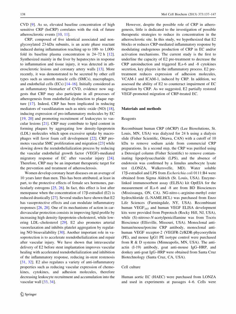
However, despite the possible role of CRP in athero-
genesis, little is dedicated to the investigation of possible
therapeutic strategies to reduce its concentration in the
atherosclerotic site. In this study, we hypothesized that E2
blocks or reduces CRP-mediated inflammatory response by
modulating endogenous production of CRP in EC and/or
activation mechanisms. The current study is the first to
underline the capacity of E2 pre-treatment to decrease the
CRP autoinduction and triggered ILs-6 and -8 cytokines
secretion, key players in the inflammatory process. E2 pre-
treatment reduces expression of adhesion molecules,
VCAM-1 and ICAM-1, induced by CRP. In addition, we
assessed the ability of E2 to counteract impairment of EC
migration by CRP. As we suggested, E2 partially restored
VEGF-promoted migration of CRP-treated EC.
Materials and methods
Reagents
Recombinant human CRP (rhCRP) (Lee Biosolutions, St.
Louis, MN, USA) was dialyzed for 24 h using a dialysis
slide (Fisher Scientific, Ottawa, CAN) with a cutoff of 10
kDa to remove sodium azide from commercial CRP
preparations. In a second step, the CRP was purified using
a Detoxigel column (Fisher Scientific) to remove contam-
inating lipopolysaccharide (LPS), and the absence of
endotoxin was confirmed by a limulus amebocyte lysate
test (LONZA, Walkersvelle, MD). Water soluble
17b-estradiol and LPS from Escherichia coli 0111:B4 were
obtained from Sigma Aldrich (St. Louis, USA). Enzyme-
linked immunosorbent assay (ELISA) kit OptEIA for the
measurement of ILs-6 and -8 are from BD Biosciences
(Mississauga, ON, CA). NG-nitro-L-arginine-methyl ester
hydrochloride (L-NAME.HCL) was purchased from Enzo
Life Sciences (Farmingdale, NY, USA). Recombinant
human VEGF165 and human VEGF ELISA development
kits were provided from Peprotech (Rocky Hill, NJ, USA),
while (S)-nitroso-N-acetylpenicillamine was from Tocris
Bioscience (Ellisville, Missouri, USA). Monoclonal anti-
human/mouse/porcine CRP antibody, monoclonal anti-
human VEGF receptor-2 (VEGFR-2)/KDR-phycoerythrin
(PE), and mouse IgG1 PE isotype control were purchased
from R & D systems (Minneapolis, MN, USA). The anti-
actin (I-19) antibody, goat anti-mouse IgG–HRP, and
donkey anti-goat IgG–HRP were obtained from Santa Cruz
Biotechnology (Santa Cruz, CA, USA).
Cell culture
Human aortic EC (HAEC) were purchased from LONZA
and used in experiments at passages 4–6. Cells were
138 Mol Cell Biochem (2013) 373:137–147
123

cultured with EGM-2MV BulletKit (LONZA) supple-
mented with 5 % FBS, 0.6 % HEPES, and maintained at
37 �C in a 5 % CO2 humidified incubator. All FBS used
were pre-treated with 1 % charcoal to eliminate endoge-
nous estrogens. For this, charcoal was added to FBS, gently
mixed for 1 h at room temperature before filtration on a
0.22-lm filter and stored at 4 �C. HAEC were plated in six-
well plates (Costar, Corning, NY, USA) at 1.5 9 104 cells/
cm2 and cultured to 80–90 % confluence before being
starved in EGM-2MV, 0.1 % FBS for 18 h prior the dif-
ferent treatments.
Analysis of protein expression by western blot
To evaluate the self-induction of CRP and adhesion molecules
(VCAM-1 and ICAM-1) protein expression, HAECs were
first treated with rhCRP at different doses (1, 2.5, 5, 10 and
25 lg/ml) for 24 h. To evaluate the impact of estrogen on
these inductions, cells were pre-treated with E2 (10-8 or
10-9 M) for 1 h before being exposed to rhCRP at 25 lg/ml
for 24 h. Single treatments were used as reference controls.
After the incubation period, cells were lysed using lysis buffer
(20 mM Tris–HCl, 150 mM NaCl, 1.2 % Triton X-100,
1 mM EGTA, 1 mM EDTA, 1 mM PMSF, 15.1 ll/ml ap-
rotinine, 10 lg/ml leupeptin, and 1 mM NaVO3). Total pro-
tein extract was quantified by Bradford technique (Bio-Rad).
Equivalent amount of protein (20 lg) was migrated on a 15 %
sodium dodecyl sulfate gel (SDS-PAGE). rhCRP (5 ng) was
added to SDS-PAGE as positive control. Proteins were
transblotted to a polyvinylidene difluoride membrane that was
soaked in 5 % non-fat dry milk prepared in TBS-T [Tris-
buffered saline (63 mM Tris–HCl, 7.3 mM NaCl) containing
0.1 % Tween 20] for 1 h at room temperature to block non-
specific binding. Membranes were then incubated overnight
with one of the following primary antibodies: anti-CRP (1/
500), anti-actin I-19 (1/1,000), anti-VCAM-1(1/1,000), and
anti-ICAM-1 (1/2,000) antibodies. After three washes in TBS-
T at room temperature, membranes were incubated with a
horseradish peroxidase-conjugated goat anti-mouse IgG (1/10
000) or donkey anti-goat IgG (1/20 000) or donkey anti-rabbit
IgG (1/10 000) for 1 h at room temperature. The blots were
washed three times with TBS-T and antigen detection was
performed using Immun-Star Western C kit (Bio-Rad). The
band intensity was analysed by Quantity One program.
Results are expressed in the ratio over actin.
ELISA assay for ILs-6, -8 and VEGF
Cytokines secretion by HAEC was evaluated after stimu-
lation with rhCRP at 25 lg/ml for 24 h with or without a
pre-treatment with E2 (10-8 or 10-9 M) for 1 h. After the
incubation period, supernatants were harvested and
centrifuged to remove cells. ILs-6, -8, and VEGF concen-
trations in culture media were measured using commer-
cially available ELISA kits. All procedures were performed
according to the manufacturer’s instructions. All samples
were assessed in triplicate.
Cellular migration assay
HAEC migration mediated by VEGF was assessed in
Transwell cell-culture 96 well plates (Corning) equipped
with a gelatine-coated polycarbonate membrane with 5-lM
pores. Before the assay, cells were pre-treated in six-well
plates with E2 (10-8 or 10-9 M) for 1 h, stimulated with
rhCRP at 25 lg/ml for 24 h or each treatment alone.
L-NAME (10-4 M) was added for 30 min before the treat-
ment of E2 and stimulation of rhCRP or the combination of
both. Cells were harvested with trypsin–0.05 % EDTA for
2 min, resuspended in EGM-2MV, 1 % FBS and 5 9 104
cells were added in the upper chamber of the Transwell plate
and were migrated for 4 h. EGM-2MV and 1 % FBS alone or
with VEGF (20 ng/ml) was added to the lower chamber as
chemoattractant. For inhibitors’ study, L-NAME (10-4 M)
was added to the upper and lower compartment and was
present throughout the experiment. After 4 h incubation at
37 �C in the presence of 5 % CO2, the cells were fixed in
methanol, stained with hematoxylin–eosin dye, and the top
side of the insert membrane was scrubbed free of cells with a
cotton swab. Membranes were removed using a scalpel and
mounted on microscope slides with migrated cell face up.
Three evenly spaced fields on each membrane were chosen
and pictures were taken using an inverse light microscope
(CKX41 of Olympus) equipped with a camera (QIMAG-
ING, QICAM, Olympus) to obtain a computer-digitized
image. Counts of migrated cells were performed by a person
blinded to treatment by means of ImagePro 6.2 software.
Each condition was tested at least in triplicate.
VEGFR-2 expression using flow cytometer
HAEC were pre-treated with E2 (10-8 or 10-9 M) for 1 h,
stimulated with rhCRP at 25 lg/ml for 24 h or each
treatment alone to evaluate VEGFR-2 expression at the cell
surface. Harvested cells were resuspended in PBS–0.5 %
BSA and non-specific binding sites were blocked with 5 %
of normal mouse serum before 20-min incubation with
monoclonal anti-human VEGFR-2/KDR-phycoerythrin
(PE, 5 lg/ml) or mouse IgG1 PE isotype control
(2.5 lg/ml) for 20 min. Cells were then washed with PBS–
0.5 % BSA and resuspended in cytometer buffer. System II
software for XL/XLMCL flow cytometer was used for
acquisition on an Epics XL coulter cytometer. For each
sample, 10,000 cells were analyzed by the program Wea-
sel. Data are expressed as mean fluorescence intensities
Mol Cell Biochem (2013) 373:137–147 139
123

(MFIs) after background subtraction from the correspond-
ing IgG control.
Statistical analysis
All data were presented as mean ± SEM. Statistical
analyses were performed by one-way analysis of variance
for multiple testing followed by Dunnett for ELISA, western
blot, adhesion molecules analysis (VCAM-1 and ICAM-1)
and EC migration assays, and by Tukey–Kramer multiple
comparisons post-test for all other experiments. All statistics
were performed by the GraphPad InStat software. Proba-
bility values were considered significant at p \ 0.05.
Results
Auto-induction of CRP production by HAECs
Self-induced CRP protein expression in vascular EC was
investigated. There was no CRP protein detectable under
basal (non-stimulated cells) condition. Incubation of
HAECs with rhCRP enhanced the CRP protein level in a
dose-dependent manner (Fig. 1). A fivefold increase in
CRP self-induction was obtained with 25 lg/ml of rhCRP
when compared to 10 lg/ml. This increase was significant
compared to all other rhCRP concentrations. The dose of
25 lg/ml was selected to conduct further investigation.
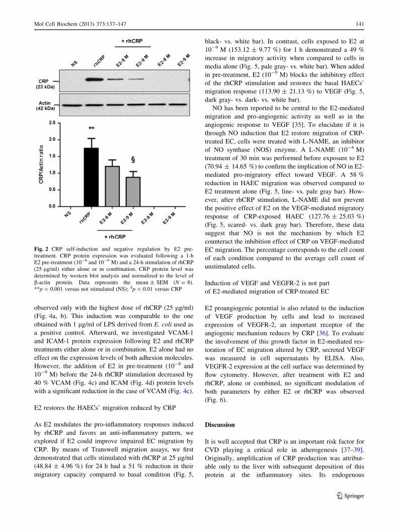
E2 inhibits the auto-induction of CRP by HAECs
To evaluate the capacity of E2 to reduce or block the CRP
self-induction, cells were pre-treated with a supraphysio-
logic and physiologic doses (10-8 and 10-9 M) of E2
before the rhCRP stimulation. HAEC treated with E2 alone
were still negative for CRP protein expression (Fig. 2).
When added in pre-treatment before the CRP stimulation,
E2 significantly inhibits the CRP self-induction with a
50 % reduction at the physiologic dose of E2 in a dose-
dependent manner (10-9 M) (p \ 0.01).
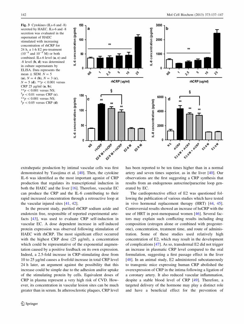
E2 reduces CRP-induced pro-inflammatory cytokine
response
As E2 pre-treatment reduces the CRP expression, we have
investigated the impact of E2 on IL-6 secretion, one of the
most potent agonists of CRP production [12]. In a dose–
response study, we first observed a significant increase in
IL-6 secretion only with the 25 lg/ml of rhCRP (Fig. 3a).
A twofold rise in IL-6 release was observed between the 10
lg/ml CRP dose and the highest dose. An E2 pre-treatment
for 1 h at 10-9 M reduced by 21 % the IL-6 secretion
triggered by the rhCRP stimulation (Fig. 3c).
We have extended our analysis to another cytokine—the
IL-8, an important player of the inflammatory process
promoting leukocyte recruitment and adhesion to the
endothelium. A response profile similar to the one of IL-6
was observed with a significant increase in IL-8 secretion
only with the 25 lg/ml rhCRP stimulation (Fig. 3b). E2
pre-treatment at 10-8 and 10-9 M for 1 h significantly
reduced by up to 21 % the impact of CRP on IL-8 pro-
duction (Fig. 3d). E2 by itself, at the tested doses, had no
effect on the basal level of ILs-6 and -8 produced by the
HAEC.
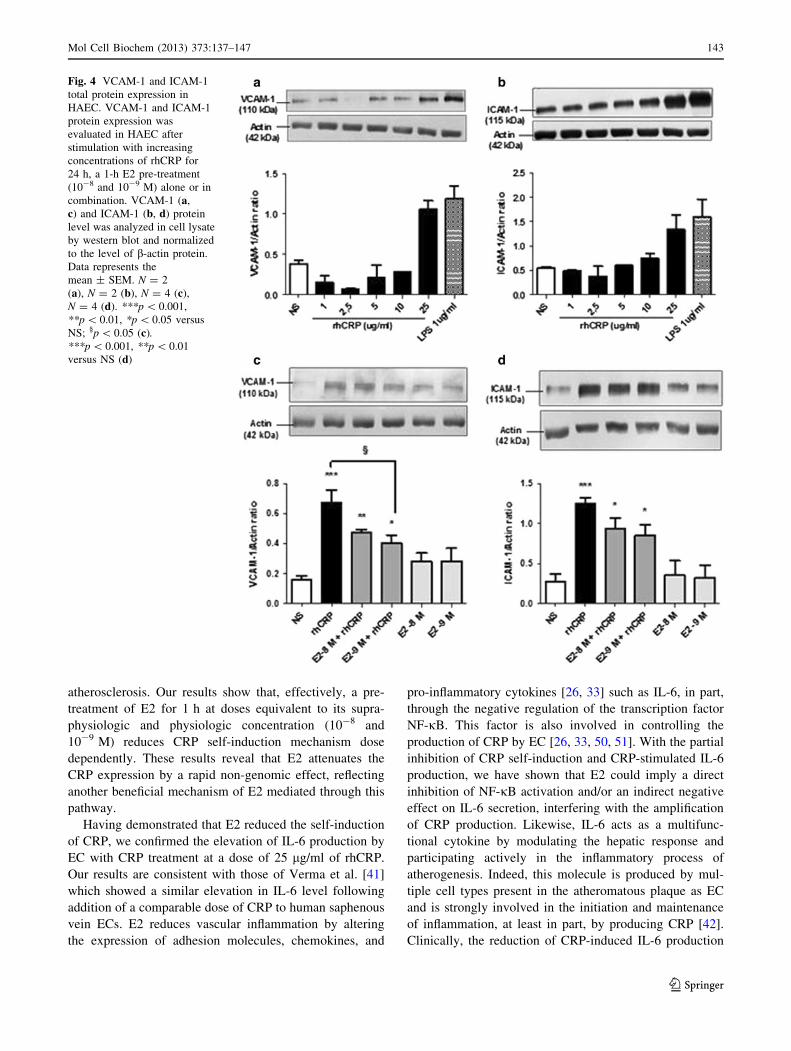
E2 reduces CRP-induced adhesion molecule
upregulation
CRP is known to trigger adhesion molecule expression,
such as VCAM-1 and ICAM-1 [19], involved in inflam-
matory cells’ recruitment to endothelial lesion sites. After
having observed the capacity of E2 to reduce CRP-induced
IL-8 secretion, we evaluated if it could also reduce EC
adhesion molecule expression induced by rhCRP. We first
evaluated VCAM-1 and ICAM-1 total protein expression
in HAECs after stimulation with increasing doses of
detoxified rhCRP. Enhanced levels of these proteins were
Fig. 1 CRP autoinduction in a dose-dependent manner. CRP protein
expression was evaluated following HAEC treatment with increasing
concentrations of rhCRP for 24 h. CRP protein level was determined
by western blot analysis and normalized to the level of b-actin
protein. Data represent the mean ± SEM (N = 4). **p \ 0.001
versus CRP 25 lg/ml; *p \ 0.01 versus CRP 25 lg/ml
140 Mol Cell Biochem (2013) 373:137–147
123
observed only with the highest dose of rhCRP (25 lg/ml)
(Fig. 4a, b). This induction was comparable to the one
obtained with 1 lg/ml of LPS derived from E. coli used as
a positive control. Afterward, we investigated VCAM-1
and ICAM-1 protein expression following E2 and rhCRP
treatments either alone or in combination. E2 alone had no
effect on the expression levels of both adhesion molecules.
However, the addition of E2 in pre-treatment (10-8 and
10-9 M) before the 24-h rhCRP stimulation decreased by
40 % VCAM (Fig. 4c) and ICAM (Fig. 4d) protein levels
with a significant reduction in the case of VCAM (Fig. 4c).
E2 restores the HAECs’ migration reduced by CRP
As E2 modulates the pro-inflammatory responses induced
by rhCRP and favors an anti-inflammatory pattern, we
explored if E2 could improve impaired EC migration by
CRP. By means of Transwell migration assays, we first
demonstrated that cells stimulated with rhCRP at 25 lg/ml
(48.84 ± 4.96 %) for 24 h had a 51 % reduction in their
migratory capacity compared to basal condition (Fig. 5,
black- vs. white bar). In contrast, cells exposed to E2 at
10-9 M (153.12 ± 9.77 %) for 1 h demonstrated a 49 %
increase in migratory activity when compared to cells in
media alone (Fig. 5, pale gray- vs. white bar). When added
in pre-treatment, E2 (10-9 M) blocks the inhibitory effect
of the rhCRP stimulation and restores the basal HAECs’
migration response (113.90 ± 21.13 %) to VEGF (Fig. 5,
dark gray- vs. dark- vs. white bar).
NO has been reported to be central to the E2-mediated
migration and pro-angiogenic activity as well as in the
angiogenic response to VEGF [35]. To elucidate if it is
through NO induction that E2 restore migration of CRP-
treated EC, cells were treated with L-NAME, an inhibitor
of NO synthase (NOS) enzyme. A L-NAME (10-4 M)
treatment of 30 min was performed before exposure to E2
(70.94 ± 14.65 %) to confirm the implication of NO in E2-
mediated pro-migratory effect toward VEGF. A 58 %
reduction in HAEC migration was observed compared to
E2 treatment alone (Fig. 5, line- vs. pale gray bar). How-
ever, after rhCRP stimulation, L-NAME did not prevent
the positive effect of E2 on the VEGF-mediated migratory
response of CRP-exposed HAEC (127.76 ± 25.03 %)
(Fig. 5, scared- vs. dark gray bar). Therefore, these data
suggest that NO is not the mechanism by which E2
counteract the inhibition effect of CRP on VEGF-mediated
EC migration. The percentage corresponds to the cell count
of each condition compared to the average cell count of
unstimulated cells.
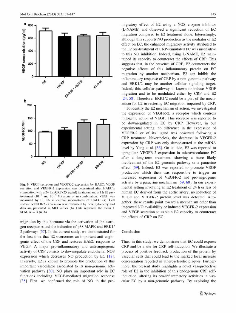
Induction of VEGF and VEGFR-2 is not part
of E2-mediated migration of CRP-treated EC
E2 proangiogenic potential is also related to the induction
of VEGF production by cells and lead to increased
expression of VEGFR-2, an important receptor of the
angiogenic mechanism reduces by CRP [36]. To evaluate
the involvement of this growth factor in E2-mediated res-
toration of EC migration altered by CRP, secreted VEGF
was measured in cell supernatants by ELISA. Also,
VEGFR-2 expression at the cell surface was determined by
flow cytometry. However, after treatment with E2 and
rhCRP, alone or combined, no significant modulation of
both parameters by either E2 or rhCRP was observed
(Fig. 6).
Discussion
It is well accepted that CRP is an important risk factor for
CVD playing a critical role in atherogenesis [37–39].
Originally, amplification of CRP production was attribut-
able only to the liver with subsequent deposition of this
protein at the inflammatory sites. Its endogenous
Fig. 2 CRP self-induction and negative regulation by E2 pre-
treatment. CRP protein expression was evaluated following a 1-h
E2 pre-treatment (10-8 and 10-9 M) and a 24-h stimulation of rhCRP
(25 lg/ml) either alone or in combination. CRP protein level was
determined by western blot analysis and normalized to the level of
b-actin protein. Data represents the mean ± SEM (N = 8).
**p \ 0.001 versus not stimulated (NS); §p \ 0.01 versus CRP
Mol Cell Biochem (2013) 373:137–147 141
123
extrahepatic production by intimal vascular cells was first
demonstrated by Yasojima et al. [40]. Then, the cytokine
IL-6 was identified as the most important agonist of CRP
production that regulates its transcriptional induction in
both the HAEC and the liver [16]. Therefore, vascular EC
can produce the CRP and the IL-6 contributing to their
rapid increased concentration through a retroactive loop at
the vascular injured sites [41, 42].
In the present study, purified rhCRP sodium azide and
endotoxin free, responsible of reported experimental arte-
facts [43], was used to evaluate CRP self-induction in
vascular EC. A dose dependent increase in self-induced
protein expression was observed following stimulation of
HAEC with rhCRP. The most significant effect occurred
with the highest CRP dose (25 lg/ml), a concentration
which could be representative of the exponential augmen-
tation caused by a positive feedback on its own expression.
Indeed, a 2.5-fold increase in CRP-stimulating dose from
10 to 25 lg/ml causes a fivefold increase in total CRP level
24 h later, an argument against the possibility that this
increase could be simple due to the adhesion and/or uptake
of the stimulating protein by cells. Equivalent doses of
CRP in plasma represent a very high risk of CVD. How-
ever, its concentration in vascular lesion sites can be much
greater than in serum. In atherosclerotic plaques, CRP level
has been reported to be ten times higher than in a normal
artery and seven times superior, as in the liver [40]. Our
observations are the first suggesting a CRP synthesis that
results from an endogenous autocrine/paracrine loop gen-
erated by EC.
The cardioprotective effect of E2 was questioned fol-
lowing the publication of various studies which have tested
in vivo hormonal replacement therapy (HRT) [44, 45].
Controversial results showed an increase of hsCRP with the
use of HRT in post-menopausal women [46]. Several fac-
tors may explain such conflicting results including drug
composition (estrogen alone or combined with progester-
one), concentration, treatment time, and route of adminis-
tration. Some of these studies used relatively high
concentration of E2, which may result in the development
of complications [47]. As so, transdermal E2 did not trigger
an increase in plasmatic CRP level compared to the oral
formulation, suggesting a first passage effect in the liver
[48]. In an animal study, E2 administered subcutaneously
to transgenic mice expressing human CRP abolished the
overexpression of CRP in the intima following a ligation of
a coronary artery. It also reduced vascular inflammation,
despite a stable blood level of CRP [49]. Therefore, a
targeted delivery of the hormone may play a distinct role
and have a beneficial effect for the prevention of
Fig. 3 Cytokines (ILs-6 and -8)
secreted by HAEC. ILs-6 and -8
secretion was evaluated in the
supernatant of HAEC
stimulated with increasing
concentration of rhCRP for
24 h, a 1-h E2 pre-treatment
(10-8 and 10-9 M) or both
combined. ILs-6 level (a, c) and
-8 level (b, d) was determined
in culture supernatants by
ELISA. Data represents the
mean ± SEM. N = 5
(a), N = 4 (b), N = 3 (c),
N = 5 (d). **p \ 0.001 versus
CRP 25 lg/ml (a, b);
**p \ 0.001 versus NS;§p \ 0.01 versus CRP (c).
**p \ 0.001 versus NS;§p \ 0.05 versus CRP (d)
142 Mol Cell Biochem (2013) 373:137–147
123
atherosclerosis. Our results show that, effectively, a pre-
treatment of E2 for 1 h at doses equivalent to its supra-
physiologic and physiologic concentration (10-8 and
10-9 M) reduces CRP self-induction mechanism dose
dependently. These results reveal that E2 attenuates the
CRP expression by a rapid non-genomic effect, reflecting
another beneficial mechanism of E2 mediated through this
pathway.
Having demonstrated that E2 reduced the self-induction
of CRP, we confirmed the elevation of IL-6 production by
EC with CRP treatment at a dose of 25 lg/ml of rhCRP.
Our results are consistent with those of Verma et al. [41]
which showed a similar elevation in IL-6 level following
addition of a comparable dose of CRP to human saphenous
vein ECs. E2 reduces vascular inflammation by altering
the expression of adhesion molecules, chemokines, and
pro-inflammatory cytokines [26, 33] such as IL-6, in part,
through the negative regulation of the transcription factor
NF-jB. This factor is also involved in controlling the
production of CRP by EC [26, 33, 50, 51]. With the partial
inhibition of CRP self-induction and CRP-stimulated IL-6
production, we have shown that E2 could imply a direct
inhibition of NF-jB activation and/or an indirect negative
effect on IL-6 secretion, interfering with the amplification
of CRP production. Likewise, IL-6 acts as a multifunc-
tional cytokine by modulating the hepatic response and
participating actively in the inflammatory process of
atherogenesis. Indeed, this molecule is produced by mul-
tiple cell types present in the atheromatous plaque as EC
and is strongly involved in the initiation and maintenance
of inflammation, at least in part, by producing CRP [42].
Clinically, the reduction of CRP-induced IL-6 production
Fig. 4 VCAM-1 and ICAM-1
total protein expression in
HAEC. VCAM-1 and ICAM-1
protein expression was
evaluated in HAEC after
stimulation with increasing
concentrations of rhCRP for
24 h, a 1-h E2 pre-treatment
(10-8 and 10-9 M) alone or in
combination. VCAM-1 (a,
c) and ICAM-1 (b, d) protein
level was analyzed in cell lysate
by western blot and normalized
to the level of b-actin protein.
Data represents the
mean ± SEM. N = 2
(a), N = 2 (b), N = 4 (c),
N = 4 (d). ***p \ 0.001,
**p \ 0.01, *p \ 0.05 versus
NS; §p \ 0.05 (c).***p \ 0.001, **p \ 0.01
versus NS (d)
Mol Cell Biochem (2013) 373:137–147 143
123

with E2 pre-treatment shown by our result could indicate a
key role of this hormone in the prevention or reduction of
cytokine inflammatory response implicated in the devel-
opment and maintenance of atherosclerosis. An important
step in atherosclerosis development is the recruitment of
leukocytes in the vascular wall where they become foam
cells. This inflammatory stage is favored by an augmen-
tation of chemokine secretion, including IL-8 and MCP-1,
and in adhesion molecules expression (ICAM-1, VCAM-1,
and E-selectin) at the endothelium [52]. Besides, a sub-
stantial enhancement of CRP production in sites of ather-
omatous lesions further increases expression of pro-
inflammatory molecules by EC [19–21]. The important
upregulation of ILs-6, -8, ICAM-1, and VCAM-1 expres-
sion by HAEC was observed with the 25 lg/ml dose of
rhCRP but not the lower dose of 10 lg/ml. This corrobo-
rates with the effective dose reported by Devaraj et al. [19,
20]. Another team has demonstrated an ICAM and VCAM
induction in human EC with 10 lg/ml CRP and even a
lower dose [21], a level of activity that we do not observe
with our highly purified rhCRP.
The well-recognized anti-inflammatory activity of E2
could counteract some of the pro-atherogenic activity of
the CRP. Indeed, this hormone at 10-8 and 10-9 M was
shown to inhibit leukocyte migration and adhesion by
blocking IL-8 secretion in human umbilical vein EC
(HUVEC) [53]. E2 was also shown to attenuate monocytes
recruitment to HAEC in response to TNF-a [34]. For the
first time, we report that an E2 pre-treatment prevents
partial HAEC inflammatory response to rhCRP. In fact, it
results in a decreased IL-8 secretion and a reduced VCAM-
1 and ICAM-1 protein expression, events that could reduce
leukocytes recruitment induced by the CRP.
Vascular repair is as important as the reduction of
inflammation to prevent progression of atherogenesis pro-
cess. To allow arterial injury healing, regeneration of a
healthy endothelium is essential to restore control of vas-
cular tone, homeostasis as well as anticoagulant, anti-
aggregating, and anti-inflammatory properties [54].
Reendothelialization and angiogenesis necessary for
endothelium reconstitution involves EC proliferation and
migration. However, some evidence suggests that CRP
may inhibit these cellular functions [55]. In accordance to
these data, a recent article demonstrated that long-term
exposure to this plasmatic protein significantly inhibited
VEGF-induced migration of HUVEC [24]. In our experi-
mental setting, we observed that a 24-h exposition of
HAEC to rhCRP was sufficient to reduce their migration
response to VEGF by more than 50 %. On the other hand,
E2 has the ability to facilitate vascular repair. Effectively,
we previously have demonstrated an improved reend-
othelialization and vascular healing process after local
delivery of E2 at the site of vascular injury [31, 56]. We
have also shown the induction of EC proliferation and
Fig. 5 E2 restores the
migratory response of CRP-
stimulated HAEC to VEGF.
HAEC migration toward VEGF
(20 ng/ml) was determined in
Transwell chamber following
E2 1-h pre-treatment (10-9 M)
and a 24-h stimulation with
rhCRP (25 lg/ml) alone or in
combination. L-NAME
(10-4 M) was added 30 min
before E2 treatment. L-NAME
was also added to both the upper
and lower compartment in
control wells. HAEC migration
is shown as a percentage of
VEGF-induced increased
compared to the average of
unstimulated cells in basal
condition. Data represents the
mean ± SEM (N = 3).
*p \ 0.05 versus NS; §p \ 0.05
versus CRP; §§p \ 0.01 versus
CRP; �p \ 0.01 versus E2
144 Mol Cell Biochem (2013) 373:137–147
123
migration by this hormone via the activation of the estro-
gen receptor-a and the induction of p38 MAPK and ERK1/
2 pathways [57]. In the current study, we demonstrated for
the first time that E2 overcomes an important anti-angio-
genic effect of the CRP and restores HAEC response to
VEGF. A major pro-inflammatory and anti-angiogenic
activity of CRP consists to downregulate endothelial NOS
expression which decreases NO production by EC [18].
Inversely, E2 is known to promote the production of this
important vasodilator associated to its non-genomic acti-
vation pathway [30]. NO plays an important role in EC
functions including VEGF-mediated migration response
[35]. First, we confirmed the role of NO in the pro-
migratory effect of E2 using a NOS enzyme inhibitor
(L-NAME) and observed a significant reduction of EC
migration compared to E2 treatment alone. Interestingly,
although this supports NO production as the mediator of E2
effect on EC, the enhanced migratory activity attributed to
the E2 pre-treatment of CRP-stimulated EC was insensitive
to this NO inhibition. Indeed, using L-NAME, E2 main-
tained its capacity to counteract the effects of CRP. This
suggests that, in the presence of CRP, E2 counteracts the
negative effects of this inflammatory protein on EC
migration by another mechanism. E2 can inhibit the
inflammatory response of CRP by a non-genomic pathway
and ERK1/2 may be another cellular signaling target.
Indeed, this cellular pathway is known to induce VEGF
migration and to be modulated either by CRP and E2
[24, 58]. Therefore, ERK1/2 could be a part of the mech-
anism for E2 in restoring EC migration impaired by CRP.
To identify the E2 mechanism of action, we investigated
the expression of VEGFR-2, a receptor which controls
mitogenic action of VEGF. This receptor was reported to
be downregulated in EC by CRP. However, in our
experimental setting, no difference in the expression of
VEGFR-2 or of its ligand was observed following a
CRP treatment. Nevertheless, the decrease in VEGFR-2
expression by CRP was only demonstrated at the mRNA
level by Yang et al. [36]. On its side, E2 was reported to
upregulate VEGFR-2 expression in microvasculature EC
after a long-term treatment, showing a more likely
involvement of the E2 genomic pathway or a paracrine
effect [59]. Indeed, E2 was reported to promote VEGF
production which then was responsible to trigger an
increased expression of VEGFR-2 and pro-angiogenic
activity by a paracrine mechanism [59, 60]. In our experi-
mental setting involving an E2 treatment of 24 h or less of
human EC derived from the aortic artery, no induction of
VEGF and VEGFR-2 protein level was detected. Alto-
gether, these results point toward a mechanism other than
improved NO availability or induced VEGFR-2 expression
and VEGF secretion to explain E2 capacity to counteract
the effects of CRP on EC.
Conclusion
Thus, in this study, we demonstrate that EC could express
CRP and be a site for CRP self-induction. We illustrate a
process of positive feedback production of the protein by
vascular cells that could lead to the marked local increase
concentration reported in atherosclerotic plaques. Further-
more, the present study highlights a novel vasoprotective
role of E2 in the inhibition of this endogenous CRP self-
induction, altering its pro-inflammatory activities in vas-
cular EC by a non-genomic pathway. By exploring the
Fig. 6 VEGF secretion and VEGFR-2 expression by HAEC. VEGF
secretion and VEGFR-2 expression was determined after HAEC’s
stimulation with a 24 h rhCRP (25 lg/ml) treatment and a 1 h E2 pre-
treatment (10-8 and 10-9 M) alone or in combination. VEGF was
measured by ELISA in culture supernatants of HAEC (a). Cell
surface VEGFR-2 expression was evaluated by flow cytometry and
data are presented as MFI values (b). Data represent the mean ±
SEM. N = 3 (a, b)
Mol Cell Biochem (2013) 373:137–147 145
123
angiogenic potential of E2, our study demonstrates for the
first time that this hormone restores EC migration altered
by CRP. Further investigation will be needed to clarify
mechanisms of E2 vascular protection by negative regu-
lation of important proatherogenic inflammatory pathways
controlled by CRP.
Acknowledgments This project was supported financially by the
Grants from the Heart and Stroke Foundation of Canada (HSFC). We
gratefully thank the Higher Education and Postdoctoral Faculty of the
University of Montreal for the Excellence Award and Redaction
Grant for 2010–2011. We also thank the Faculty of Medicine of the
University of Montreal for the master degree scholarship of
2009–2010. Dr. Jean-Francois Tanguay is also supported by Fonda-
tion de l’Institut de Cardiologie de Montreal and reseau TheCell
(FRSQ).
Conflict of interest None of the authors has competing financial
interest to declare.
Open Access This article is distributed under the terms of the
Creative Commons Attribution License which permits any use, dis-
tribution, and reproduction in any medium, provided the original
author(s) and the source are credited.
References
1. Lusis AJ (2000) Atherosclerosis. Nature 407:233–241
2. Mullenix PS, Andersen CA, Starnes BW (2005) Atherosclerosis
as inflammation. Ann Vasc Surg 19:130–138
3. Lavie CJ, Milani RV, Ventura HO (2009) Obesity and cardio-
vascular disease: risk factor, paradox, and impact of weight loss.
J Am Coll Cardiol 53:1925–1932
4. Karavidas A, Lazaros G, Tsiachris D, Pyrgakis V (2010) Aging
and the cardiovascular system. Hell J Cardiol 51:421–427
5. Beckman JA, Creager MA, Libby P (2002) Diabetes and ath-
erosclerosis: epidemiology, pathophysiology, and management.
JAMA 287:2570–2581
6. Reriani MK, Lerman LO, Lerman A (2010) Endothelial function
as a functional expression of cardiovascular risk factors. Biomark
Med 4:351–360
7. Koenig W (2007) Cardiovascular biomarkers: added value with
an integrated approach? Circulation 116:3–5
8. Ridker PM (2001) High-sensitivity C-reactive protein: potential
adjunct for global risk assessment in the primary prevention of
cardiovascular disease. Circulation 103:1813–1818
9. Pepys MB, Hirschfield GM (2003) C-reactive protein: a critical
update. J Clin Investig 111:1805–1812
10. Berk BC, Weintraub WS, Alexander RW (1990) Elevation of
C-reactive protein in ‘‘active’’ coronary artery disease. Am J
Cardiol 65:168–172
11. Liuzzo G, Biasucci LM, Gallimore JR, Grillo RL, Rebuzzi AG,
Pepys MB, Maseri A (1994) The prognostic value of C-reactive
protein and serum amyloid a protein in severe unstable angina.
N Engl J Med 331:417–424
12. Devaraj S, Singh U, Jialal I (2009) The evolving role of
C-reactive protein in atherothrombosis. Clin Chem 55:229–238
13. Zhang YX, Cliff WJ, Schoefl GI, Higgins G (1999) Coronary
C-reactive protein distribution: its relation to development of
atherosclerosis. Atherosclerosis 145:375–379
14. Calabro P, Willerson JT, Yeh ET (2003) Inflammatory cytokines
stimulated C-reactive protein production by human coronary
artery smooth muscle cells. Circulation 108:1930–1932
15. Ciubotaru I, Potempa LA, Wander RC (2005) Production of
modified C-reactive protein in U937-derived macrophages. Exp
Biol Med (Maywood) 230:762–770
16. Venugopal SK, Devaraj S, Jialal I (2005) Macrophage condi-
tioned medium induces the expression of C-reactive protein in
human aortic endothelial cells: potential for paracrine/autocrine
effects. Am J Pathol 166:1265–1271
17. Verma S, Szmitko PE, Ridker PM (2005) C-reactive protein
comes of age. Nat Clin Pract Cardiovasc Med 2:29–36 (quiz 58)
18. Venugopal SK, Devaraj S, Yuhanna I, Shaul P, Jialal I (2002)
Demonstration that C-reactive protein decreases eNOS expres-
sion and bioactivity in human aortic endothelial cells. Circulation
106:1439–1441
19. Pasceri V, Willerson JT, Yeh ET (2000) Direct proinflammatory
effect of C-reactive protein on human endothelial cells. Circu-
lation 102:2165–2168
20. Devaraj S, Kumaresan PR, Jialal I (2004) Effect of C-reactive
protein on chemokine expression in human aortic endothelial
cells. J Mol Cell Cardiol 36:405–410
21. Devaraj S, Davis B, Simon SI, Jialal I (2006) CRP promotes
monocyte-endothelial cell adhesion via Fcgamma receptors in
human aortic endothelial cells under static and shear flow
conditions. Am J Physiol Heart Circ Physiol 291:H1170–
H1176
22. Fu T, Borensztajn J (2002) Macrophage uptake of low-density
lipoprotein bound to aggregated C-reactive protein: possible
mechanism of foam-cell formation in atherosclerotic lesions.
Biochem J 366:195–201
23. Cirillo P, Golino P, Calabro P, Cali G, Ragni M, De Rosa S,
Cimmino G, Pacileo M, De Palma R, Forte L, Gargiulo A, Co-
rigliano FG, Angri V, Spagnuolo R, Nitsch L, Chiariello M
(2005) C-reactive protein induces tissue factor expression and
promotes smooth muscle and endothelial cell proliferation. Car-
diovasc Res 68:47–55
24. Schneeweis C, Grafe M, Bungenstock A, Spencer-Hansch C,
Fleck E, Goetze S (2010) Chronic CRP-exposure inhibits VEGF-
induced endothelial cell migration. J Atheroscler Thromb
17:203–212
25. Barrett-Connor E (1997) Sex differences in coronary heart dis-
ease. Why are women so superior? The 1995 Ancel Keys Lecture.
Circulation 95:252–264
26. Xing D, Nozell S, Chen YF, Hage F, Oparil S (2009) Estrogen
and mechanisms of vascular protection. Arterioscler Thromb
Vasc Biol 29:289–295
27. Czubryt MP, Espira L, Lamoureux L, Abrenica B (2006) The role
of sex in cardiac function and disease. Can J Physiol Pharmacol
84:93–109
28. Baker L, Meldrum KK, Wang M, Sankula R, Vanam R, Raies-
dana A, Tsai B, Hile K, Brown JW, Meldrum DR (2003) The role
of estrogen in cardiovascular disease. J Surg Res 115:325–344
29. Mendelsohn ME, Karas RH (1999) The protective effects of
estrogen on the cardiovascular system. N Engl J Med
340:1801–1811
30. Chen Z, Yuhanna IS, Galcheva-Gargova Z, Karas RH, Mendel-
sohn ME, Shaul PW (1999) Estrogen receptor alpha mediates the
nongenomic activation of endothelial nitric oxide synthase by
estrogen. J Clin Investig 103:401–406
31. Chandrasekar B, Sirois MG, Geoffroy P, Lauzier D, Nattel S,
Tanguay JF (2005) Local delivery of 17beta-estradiol improves
reendothelialization and decreases inflammation after coronary
stenting in a porcine model. Thromb Haemost 94:1042–1047
32. Tanguay JF (2005) Vascular healing after stenting: the role of
17-beta-estradiol in improving re-endothelialization and reducing
restenosis. Can J Cardiol 21:1025–1030
33. Straub RH (2007) The complex role of estrogens in inflammation.
Endocr Rev 28:521–574
146 Mol Cell Biochem (2013) 373:137–147
123

34. Mikkola TS, St Clair RW (2002) Estradiol reduces basal and
cytokine induced monocyte adhesion to endothelial cells. Matu-
ritas 41:313–319
35. Papapetropoulos A, Garcia-Cardena G, Madri JA, Sessa WC
(1997) Nitric oxide production contributes to the angiogenic
properties of vascular endothelial growth factor in human endo-
thelial cells. J Clin Investig 100:3131–3139
36. Yang H, Nan B, Yan S, Li M, Yao Q, Chen C (2005) C-reactive
protein decreases expression of VEGF receptors and neuropilins
and inhibits VEGF165-induced cell proliferation in human
endothelial cells. Biochem Biophys Res Commun 333:1003–1010
37. Ridker PM, Rifai N, Rose L, Buring JE, Cook NR (2002)
Comparison of C-reactive protein and low-density lipoprotein
cholesterol levels in the prediction of first cardiovascular events.
N Engl J Med 347:1557–1565
38. Koenig W, Sund M, Frohlich M, Fischer HG, Lowel H, Doring
A, Hutchinson WL, Pepys MB (1999) C-reactive protein, a
sensitive marker of inflammation, predicts future risk of coronary
heart disease in initially healthy middle-aged men: results from
the MONICA (Monitoring Trends and Determinants in Cardio-
vascular Disease) Augsburg Cohort Study, 1984 to 1992. Circu-
lation 99:237–242
39. Kuller LH, Tracy RP, Shaten J, Meilahn EN (1996) Relation of
C-reactive protein and coronary heart disease in the MRFIT
nested case-control study. Multiple Risk Factor Intervention
Trial. Am J Epidemiol 144:537–547
40. Yasojima K, Schwab C, McGeer EG, McGeer PL (2001) Gen-
eration of C-reactive protein and complement components in
atherosclerotic plaques. Am J Pathol 158:1039–1051
41. Verma S, Li SH, Badiwala MV, Weisel RD, Fedak PW, Li RK,
Dhillon B, Mickle DA (2002) Endothelin antagonism and inter-
leukin-6 inhibition attenuate the proatherogenic effects of
C-reactive protein. Circulation 105:1890–1896
42. Ferri C, Croce G, Cofini V, De Berardinis G, Grassi D, Casale R,
Properzi G, Desideri G (2007) C-reactive protein: interaction
with the vascular endothelium and possible role in human ath-
erosclerosis. Curr Pharm Des 13:1631–1645
43. Taylor KE, Giddings JC, van den Berg CW (2005) C-reactive
protein-induced in vitro endothelial cell activation is an artefact
caused by azide and lipopolysaccharide. Arterioscler Thromb
Vasc Biol 25:1225–1230
44. Grady D, Wenger NK, Herrington D, Khan S, Furberg C, Hun-
ninghake D, Vittinghoff E, Hulley S (2000) Postmenopausal
hormone therapy increases risk for venous thromboembolic dis-
ease. The heart and estrogen/progestin replacement study. Ann
Intern Med 132:689–696
45. Stampfer MJ, Colditz GA, Willett WC, Manson JE, Rosner B,
Speizer FE, Hennekens CH (1991) Postmenopausal estrogen
therapy and cardiovascular disease. Ten-year follow-up from the
nurses’ health study. N Engl J Med 325:756–762
46. Duvernoy C (2003) Estrogen and C-reactive protein: does an
alternate route lead to a more attractive destination? Thromb
Haemost 90:1–2
47. Hsia J, Langer RD, Manson JE, Kuller L, Johnson KC, Hendrix
SL, Pettinger M, Heckbert SR, Greep N, Crawford S, Eaton CB,
Kostis JB, Caralis P, Prentice R (2006) Conjugated equine
estrogens and coronary heart disease: the Women’s Health Ini-
tiative. Arch Intern Med 166:357–365
48. Lacut K, Oger E, Le Gal G, Blouch MT, Abgrall JF, Kerlan V,
Scarabin PY, Mottier D (2003) Differential effects of oral and
transdermal postmenopausal estrogen replacement therapies on
C-reactive protein. Thromb Haemost 90:124–131
49. Wang D, Oparil S, Chen YF, McCrory MA, Skibinski GA, Feng
W, Szalai AJ (2005) Estrogen treatment abrogates neointima
formation in human C-reactive protein transgenic mice. Arte-
rioscler Thromb Vasc Biol 25:2094–2099
50. Calabro P, Golia E, Yeh ET (2009) CRP and the risk of athero-
sclerotic events. Semin Immunopathol 31:79–94
51. Verma S, Badiwala MV, Weisel RD, Li SH, Wang CH, Fedak
PW, Li RK, Mickle DA (2003) C-reactive protein activates the
nuclear factor-kappaB signal transduction pathway in saphenous
vein endothelial cells: implications for atherosclerosis and
restenosis. J Thorac Cardiovasc Surg 126:1886–1891
52. Sullivan GW, Sarembock IJ, Linden J (2000) The role of
inflammation in vascular diseases. J Leukoc Biol 67:591–602
53. Rodriguez E, Lopez R, Paez A, Masso F, Montano LF (2002)
17Beta-estradiol inhibits the adhesion of leukocytes in TNF-alpha
stimulated human endothelial cells by blocking IL-8 and MCP-1
secretion, but not its transcription. Life Sci 71:2181–2193
54. Aird WC (2007) Phenotypic heterogeneity of the endothelium: I.
Structure, function, and mechanisms. Circ Res 100:158–173
55. Schwartz R, Osborne-Lawrence S, Hahner L, Gibson LL,
Gormley AK, Vongpatanasin W, Zhu W, Word RA, Seetharam
D, Black S, Samols D, Mineo C, Shaul PW (2007) C-reactive
protein downregulates endothelial NO synthase and attenuates
reendothelialization in vivo in mice. Circ Res 100:1452–1459
56. Chandrasekar B, Nattel S, Tanguay JF (2001) Coronary artery
endothelial protection after local delivery of 17beta-estradiol
during balloon angioplasty in a porcine model: a potential new
pharmacologic approach to improve endothelial function. J Am
Coll Cardiol 38:1570–1576
57. Geraldes P, Sirois MG, Bernatchez PN, Tanguay JF (2002)
Estrogen regulation of endothelial and smooth muscle cell
migration and proliferation: role of p38 and p42/44 mitogen-
activated protein kinase. Arterioscler Thromb Vasc Biol
22:1585–1590
58. Kibayashi E, Urakaze M, Kobashi C, Kishida M, Takata M, Sato
A, Yamazaki K, Kobayashi M (2005) Inhibitory effect of pita-
vastatin (NK-104) on the C-reactive-protein-induced interleukin-
8 production in human aortic endothelial cells. Clin Sci (Lond)
108:515–521
59. Suzuma I, Mandai M, Takagi H, Suzuma K, Otani A, Oh H,
Kobayashi K, Honda Y (1999) 17 Beta-estradiol increases VEGF
receptor-2 and promotes DNA synthesis in retinal microvascular
endothelial cells. Investig Ophthalmol Vis Sci 40:2122–2129
60. Herve MA, Meduri G, Petit FG, Domet TS, Lazennec G, Mourah
S, Perrot-Applanat M (2006) Regulation of the vascular endo-
thelial growth factor (VEGF) receptor Flk-1/KDR by estradiol
through VEGF in uterus. J Endocrinol 188:91–99
Mol Cell Biochem (2013) 373:137–147 147
123