U94 inhibits PCa gene expression
13
BioMed Central TIONAL INTERNA CANCER CELL Page 1 of 13 (page number not for citation purposes) Cancer Cell International Open Access Primary research U94 alters FN1 and ANGPTL4 gene expression and inhibits tumorigenesis of prostate cancer cell line PC3 Ekwere T Ifon 1 , Alan LY Pang 2 , Warren Johnson 2 , Kathleen Cashman 1 , Sharon Zimmerman 1 , Sumitra Muralidhar 1 , Wai-Yee Chan 2,3,4,5 , John Casey 1 and Leonard Jason Rosenthal* 1 Address: 1 Department of Microbiology and Immunology, Georgetown University Medical Center, 3900 Reservoir Road, NW, Washington, D.C. 20057, USA, 2 Laboratory of Clinical Genomics, NICHD, National Institutes of Health, 9000 Rockville Pike, Bethesda, MD 20892, USA, 3 Department of Pediatrics, Georgetown University Medical Center, 3800 Reservoir Road, NW, Washington, D.C. 20057, USA, 4 Department of Cell Biology, Georgetown University Medical Center, 3800 Reservoir Road, NW, Washington, D.C. 20057, and 5 Department of Biochemistry & Molecular Biology, Georgetown University Medical Center, 3800 Reservoir Road, NW, Washington, D.C. 20057, USA; Email: Ekwere T Ifon - [email protected]; Alan LY Pang - [email protected]; Warren Johnson - [email protected]; Kathleen Cashman - [email protected]; Sharon Zimmerman - [email protected]; Sumitra Muralidhar - [email protected]; Wai-Yee Chan - [email protected]; John Casey - [email protected]; Leonard Jason Rosenthal* - [email protected] * Corresponding author Abstract Background: Insensitivity of advanced-stage prostate cancer to androgen ablation therapy is a serious problem in clinical practice because it is associated with aggressive progression and poor prognosis. Targeted therapeutic drug discovery efforts are thwarted by lack of adequate knowledge of gene(s) associated with prostate tumorigenesis. Therefore there is the need for studies to provide leads to targeted intervention measures. Here we propose that stable expression of U94, a tumor suppressor gene encoded by human herpesvirus 6A (HHV-6A), could alter gene expression and thereby inhibit the tumorigenicity of PC3 cell line. Microarray gene expression profiling on U94 recombinant PC3 cell line could reveal genes that would elucidate prostate cancer biology, and hopefully identify potential therapeutic targets. Results: We have shown that stable expression of U94 gene in PC3 cell line inhibited its focus formation in culture, and tumorigenesis in nude mice. Moreover gene expression profiling revealed dramatic upregulation of FN 1 (fibronectin, 91 ± 16- fold), and profound downregulation of ANGPTL 4 (angiopoietin-like-4, 20 ± 4-fold) in U94 recombinant PC3 cell line. Quantitative real-time polymerase chain reaction (QRT-PCR) analysis showed that the pattern of expression of FN 1 and ANGPTL 4 mRNA were consistent with the microarray data. Based on previous reports, the findings in this study implicate upregulation of FN 1 and downregulation of ANGPTL 4 in the anti tumor activity of U94. Genes with cancer inhibitory activities that were also upregulated include SERPINE 2 (serine/cysteine protease inhibitor 2, 7 ± 1-fold increase) and ADAMTS 1 (a disintegrin-like and metalloprotease with thrombospondin type 1 motif, 7 ± 2-fold increase). Additionally, SPUVE 23 (serine protease 23) that is pro- tumorigenic was significantly downregulated (10 ± 1-fold). Conclusion: The dramatic upregulation of FN 1 and downregulation of ANGPTL 4 genes in PC3 cell line stably expressing U94 implicate up-regulation of FN 1 and downregulation of ANGPTL 4 in anti tumor activity of U94. Further studies are necessary to determine functional roles of differentially expressed genes in U94 recombinant PC3 cell line, and hopefully provide leads to potential therapeutic targets in prostate cancer. Published: 22 June 2005 Cancer Cell International 2005, 5:19 doi:10.1186/1475-2867-5-19 Received: 13 May 2004 Accepted: 22 June 2005 This article is available from: http://www.cancerci.com/content/5/1/19 © 2005 Ifon et al; licensee BioMed Central Ltd. This is an Open Access article distributed under the terms of the Creative Commons Attribution License (http://creativecommons.org/licenses/by/2.0 ), which permits unrestricted use, distribution, and reproduction in any medium, provided the original work is properly cited.
-
Upload
independent -
Category
Documents
-
view
0 -
download
0
Transcript of U94 inhibits PCa gene expression
BioMed CentralC
TIONALINTERNACANCER CELLCancer Cell International
ss
Open AccePrimary researchU94 alters FN1 and ANGPTL4 gene expression and inhibits tumorigenesis of prostate cancer cell line PC3Ekwere T Ifon1, Alan LY Pang2, Warren Johnson2, Kathleen Cashman1, Sharon Zimmerman1, Sumitra Muralidhar1, Wai-Yee Chan2,3,4,5, John Casey1 and Leonard Jason Rosenthal*1Address: 1Department of Microbiology and Immunology, Georgetown University Medical Center, 3900 Reservoir Road, NW, Washington, D.C. 20057, USA, 2Laboratory of Clinical Genomics, NICHD, National Institutes of Health, 9000 Rockville Pike, Bethesda, MD 20892, USA, 3Department of Pediatrics, Georgetown University Medical Center, 3800 Reservoir Road, NW, Washington, D.C. 20057, USA, 4Department of Cell Biology, Georgetown University Medical Center, 3800 Reservoir Road, NW, Washington, D.C. 20057, and 5Department of Biochemistry & Molecular Biology, Georgetown University Medical Center, 3800 Reservoir Road, NW, Washington, D.C. 20057, USA;
Email: Ekwere T Ifon - [email protected]; Alan LY Pang - [email protected]; Warren Johnson - [email protected]; Kathleen Cashman - [email protected]; Sharon Zimmerman - [email protected]; Sumitra Muralidhar - [email protected]; Wai-Yee Chan - [email protected]; John Casey - [email protected]; Leonard Jason Rosenthal* - [email protected]
* Corresponding author
AbstractBackground: Insensitivity of advanced-stage prostate cancer to androgen ablation therapy is a serious problem in clinicalpractice because it is associated with aggressive progression and poor prognosis. Targeted therapeutic drug discovery effortsare thwarted by lack of adequate knowledge of gene(s) associated with prostate tumorigenesis. Therefore there is the need forstudies to provide leads to targeted intervention measures. Here we propose that stable expression of U94, a tumor suppressorgene encoded by human herpesvirus 6A (HHV-6A), could alter gene expression and thereby inhibit the tumorigenicity of PC3cell line. Microarray gene expression profiling on U94 recombinant PC3 cell line could reveal genes that would elucidate prostatecancer biology, and hopefully identify potential therapeutic targets.
Results: We have shown that stable expression of U94 gene in PC3 cell line inhibited its focus formation in culture, andtumorigenesis in nude mice. Moreover gene expression profiling revealed dramatic upregulation of FN 1 (fibronectin, 91 ± 16-fold), and profound downregulation of ANGPTL 4 (angiopoietin-like-4, 20 ± 4-fold) in U94 recombinant PC3 cell line. Quantitativereal-time polymerase chain reaction (QRT-PCR) analysis showed that the pattern of expression of FN 1 and ANGPTL 4 mRNAwere consistent with the microarray data. Based on previous reports, the findings in this study implicate upregulation of FN 1and downregulation of ANGPTL 4 in the anti tumor activity of U94. Genes with cancer inhibitory activities that were alsoupregulated include SERPINE 2 (serine/cysteine protease inhibitor 2, 7 ± 1-fold increase) and ADAMTS 1 (a disintegrin-like andmetalloprotease with thrombospondin type 1 motif, 7 ± 2-fold increase). Additionally, SPUVE 23 (serine protease 23) that is pro-tumorigenic was significantly downregulated (10 ± 1-fold).
Conclusion: The dramatic upregulation of FN 1 and downregulation of ANGPTL 4 genes in PC3 cell line stably expressing U94implicate up-regulation of FN 1 and downregulation of ANGPTL 4 in anti tumor activity of U94. Further studies are necessary todetermine functional roles of differentially expressed genes in U94 recombinant PC3 cell line, and hopefully provide leads topotential therapeutic targets in prostate cancer.
Published: 22 June 2005
Cancer Cell International 2005, 5:19 doi:10.1186/1475-2867-5-19
Received: 13 May 2004Accepted: 22 June 2005
This article is available from: http://www.cancerci.com/content/5/1/19
© 2005 Ifon et al; licensee BioMed Central Ltd. This is an Open Access article distributed under the terms of the Creative Commons Attribution License (http://creativecommons.org/licenses/by/2.0), which permits unrestricted use, distribution, and reproduction in any medium, provided the original work is properly cited.
Page 1 of 13(page number not for citation purposes)
Cancer Cell International 2005, 5:19 http://www.cancerci.com/content/5/1/19
BackgroundProstate cancer is the most common form of malignancyin US males. An estimated 29,900 fatalities out of 230,110new cases are expected in the year 2004 [1]. Androgenablation is currently the mainstay in prostate cancer ther-apy, but its efficiency is marred by the relapse of someadvanced-stage prostate cancer cells into an androgenrefractory state [2,3]. Advanced-stage prostate cancer pro-gression is usually aggressive and correlates with poorprognosis [2,4,5]. Hence, insensitivity to androgen abla-tion by advanced-stage prostate cancer invariably consti-tutes a major problem in clinical therapy. Therefore thereis an urgent need for the development of targeted thera-peutic strategies in advanced-stage prostate cancer.
Knowledge of the genes that are associated with prostatecancer is important for designing an effective therapeuticstrategy. However, present knowledge of the molecularbiology of prostate cancer is inadequate to define etiologicgenes [5-7]. Consequently, current therapeutic strategiesin prostate cancer are inefficient [8-20], and an effectivetargeted therapy remains elusive. This situation promptedour laboratory to embark on studies to provide alternativeleads for the development of efficacious and targeted antiprostate cancer agent(s). In our approach, we investigatedthe anti tumor activity of U94 protein (U94) in prostatecancer cell line, PC3.
U94 is a 1473 bp gene located in the HD12 fragment ofhuman herpesvirus 6A (HHV-6A), strain U1102 [21]. U94encodes a 490 amino acid protein that is not found inother herpesviruses [21,22], and U94 is expressed at verylow levels [23,24]. Recent reports suggest that U94 is alatency gene, and modulates viral DNA replication [23-26]. Moreover, structural homology of U94 to Rep 78/68from adeno-associated virus type 2 (AAV-2) [21,27] sug-gests that there might be functional similarities betweenthese proteins. Strong evidence in support of functionalsimilarities between U94 and Rep 78/68 is the observa-tion that U94 complemented the replication of an AAV-2mutant that was deficient in Rep 78/68 [28]. Additionally,recent reports show that U94 also inhibits gene transcrip-tion [29], which is a biological function of its homologueRep78/68. However, U94 may affect gene transcriptiondifferently than Rep 78/68, because U94 activates humanimmunodeficiency virus 1 (HIV-1) long terminal repeat(LTR) promoter in fibroblast cell lines [28] and inhibitsHIV-1 LTR in T-cell lines [29], whereas Rep 78/68 inhibitsHIV-1 LTR promoter in both fibroblast cell lines and T-celllines [28].
Previous studies demonstrated that U94 suppressed trans-formation by oncogenes [22,29]. Data from these studiesshowed that an NIH 3T3 cell line stably expressing U94gene suppressed transformation by the oncogene H-ras,
when compared to the parental NIH 3T3 cell line treatedunder similar conditions [29]. We were motivated by thefindings in previous studies to determine the anti tumorpotential of U94 in the human prostate tumor cell linePC3.
In this paper we report that the expression of U94 proteinin PC3 cells inhibited foci formation (Figure 2; Table 1),and the tumorigenicity of recombinant PC3 cell line inathymic nude mice (Figure 3). Moreover, gene expressionanalyses (Figures 4 and 5), and QRT-PCR (Table 2)revealed dramatic upregulation of FN 1 (~91-fold) andprofound downregulation of ANGPTL 4 (~20-fold) in 2separate recombinant PC3 cell lines stably expressingU94. Our study also demonstrated the differential expres-sion pattern of several other genes in the presence of U94.This is the first study to report the inhibitory potential ofU94 on the tumorigenicity of advanced-stage prostatecancer cell line PC3.
ResultsPreviously, we have demonstrated that U94 inhibitedgene transcription and also transformation by oncogenes[22,29]. Several reports have implicated the malfunctionof transcription regulatory factors [30-38] as well as theactivities of oncogenes [39-48] as etiologic factors in pros-tate tumorigenesis. Hence we wanted to determinewhether U94 could exert inhibitory activity on the tumor-igenesis of PC3 cell line.
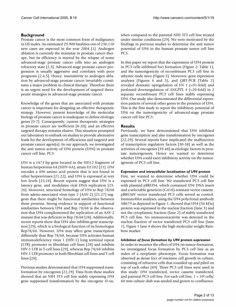
Expression and intracellular localization of U94 proteinFirst, we wanted to determine whether U94 could beexpressed in PC3 cell line. We transfected PC3 cell linewith plasmid pBKU94, which contained U94 DNA insertand a selectable geneticin (G418)-resistant vector cassette.pBKCMV vector transfected PC3 cells served as control.Immunoblot analyses, using the U94 polyclonal antibodyAB679 as depicted in Figure 1, showed that U94 (56 kDa)protein was expressed in the nuclear fraction (lane 3) andnot the cytoplasmic fraction (lane 2) of stably transfectedPC3 cell line. No immunoreactivity was detected in thenuclear fraction of vector transfected PC3 cell line (lane1). Figure 1 lane 4 shows the high molecular weight Rain-bow marker.
Inhibition of focus formation by U94 protein expressionIn order to monitor the effect of U94 on tumor formation,we investigated focus formation by PC3 cell line as anindex of a neoplastic phenotype. Focus formation wasobserved as dense foci of intensive cell growth in culture,consisting of refractive cells that rounded up and piled ontop of each other [49]. Three PC3 cell lines were used inthis study: U94 transfected, vector cassette transfected,and parental PC3 cell line. For each cell line, 1 × 10⁶ cells/60 mm culture dish was seeded and grown to confluence.
Page 2 of 13(page number not for citation purposes)
Cancer Cell International 2005, 5:19 http://www.cancerci.com/content/5/1/19
Focus formation was examined 10 days post confluence.The result of this study (Table 1) showed a drastic reduc-tion in focus formation by PC3 cells expressing U94: thenumber of foci were reduced ~35-fold and 40-fold incomparison with the control vector transfected and paren-tal PC3 cell lines, respectively. Figure 2 shows large andwidespread foci in the culture of control vector transfectedand parental PC3 cell lines. The culture of recombinantPC3 cell line expressing U94 protein showed only fewfoci, grossly reduced in size. Our findings suggest that U94may exhibit anti tumor activity in vitro.
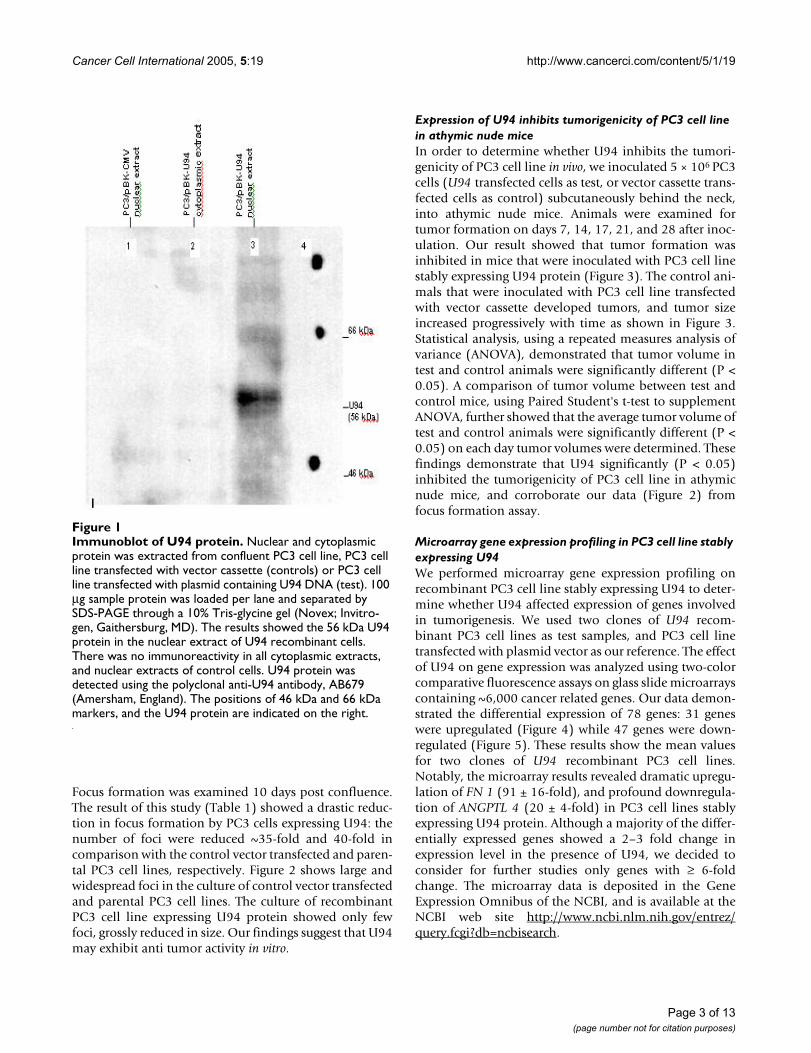
Expression of U94 inhibits tumorigenicity of PC3 cell line in athymic nude miceIn order to determine whether U94 inhibits the tumori-genicity of PC3 cell line in vivo, we inoculated 5 × 10⁶ PC3cells (U94 transfected cells as test, or vector cassette trans-fected cells as control) subcutaneously behind the neck,into athymic nude mice. Animals were examined fortumor formation on days 7, 14, 17, 21, and 28 after inoc-ulation. Our result showed that tumor formation wasinhibited in mice that were inoculated with PC3 cell linestably expressing U94 protein (Figure 3). The control ani-mals that were inoculated with PC3 cell line transfectedwith vector cassette developed tumors, and tumor sizeincreased progressively with time as shown in Figure 3.Statistical analysis, using a repeated measures analysis ofvariance (ANOVA), demonstrated that tumor volume intest and control animals were significantly different (P <0.05). A comparison of tumor volume between test andcontrol mice, using Paired Student's t-test to supplementANOVA, further showed that the average tumor volume oftest and control animals were significantly different (P <0.05) on each day tumor volumes were determined. Thesefindings demonstrate that U94 significantly (P < 0.05)inhibited the tumorigenicity of PC3 cell line in athymicnude mice, and corroborate our data (Figure 2) fromfocus formation assay.
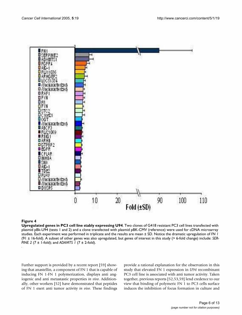
Microarray gene expression profiling in PC3 cell line stably expressing U94We performed microarray gene expression profiling onrecombinant PC3 cell line stably expressing U94 to deter-mine whether U94 affected expression of genes involvedin tumorigenesis. We used two clones of U94 recom-binant PC3 cell lines as test samples, and PC3 cell linetransfected with plasmid vector as our reference. The effectof U94 on gene expression was analyzed using two-colorcomparative fluorescence assays on glass slide microarrayscontaining ~6,000 cancer related genes. Our data demon-strated the differential expression of 78 genes: 31 geneswere upregulated (Figure 4) while 47 genes were down-regulated (Figure 5). These results show the mean valuesfor two clones of U94 recombinant PC3 cell lines.Notably, the microarray results revealed dramatic upregu-lation of FN 1 (91 ± 16-fold), and profound downregula-tion of ANGPTL 4 (20 ± 4-fold) in PC3 cell lines stablyexpressing U94 protein. Although a majority of the differ-entially expressed genes showed a 2–3 fold change inexpression level in the presence of U94, we decided toconsider for further studies only genes with ≥ 6-foldchange. The microarray data is deposited in the GeneExpression Omnibus of the NCBI, and is available at theNCBI web site http://www.ncbi.nlm.nih.gov/entrez/query.fcgi?db=ncbisearch.
Immunoblot of U94 proteinFigure 1Immunoblot of U94 protein. Nuclear and cytoplasmic protein was extracted from confluent PC3 cell line, PC3 cell line transfected with vector cassette (controls) or PC3 cell line transfected with plasmid containing U94 DNA (test). 100 µg sample protein was loaded per lane and separated by SDS-PAGE through a 10% Tris-glycine gel (Novex; Invitro-gen, Gaithersburg, MD). The results showed the 56 kDa U94 protein in the nuclear extract of U94 recombinant cells. There was no immunoreactivity in all cytoplasmic extracts, and nuclear extracts of control cells. U94 protein was detected using the polyclonal anti-U94 antibody, AB679 (Amersham, England). The positions of 46 kDa and 66 kDa markers, and the U94 protein are indicated on the right.
Page 3 of 13(page number not for citation purposes)

Cancer Cell International 2005, 5:19 http://www.cancerci.com/content/5/1/19
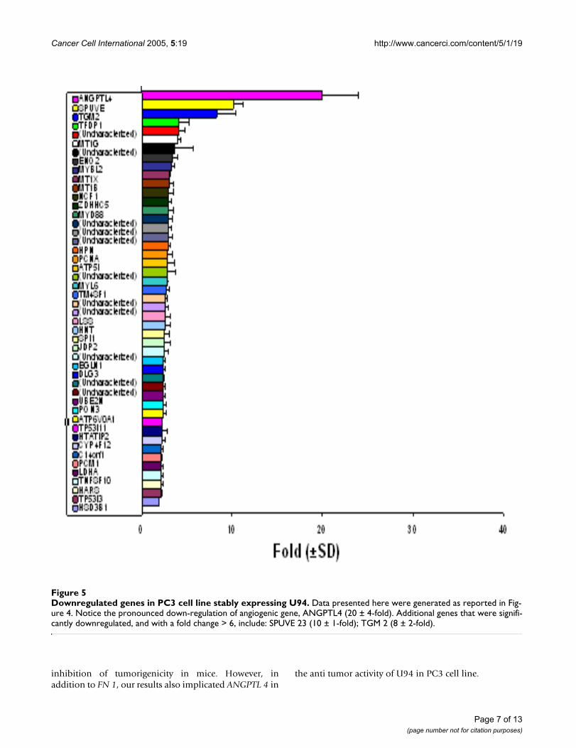
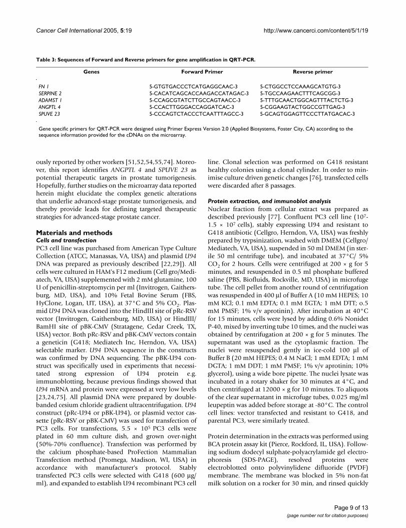
Quantitative real-time PCRWe performed QRT-PCR to confirm the microarray data.In one clone of U94 recombinant PC3 cell line, the QRT-PCR data (Table 2) showed that the changes in expressionlevels of FN 1 and ANGPTL 4 mRNA were 183 ± 27-foldincrease and 76 ± 13-fold decrease, respectively. In the sec-ond clone of U94 recombinant PC3 cell line, the corre-sponding changes in expression levels were 467 ± 32-foldincrease and 67 ± 5-fold decrease, respectively. Theobserved differences in the fold changes between themicroarray and QRT-PCR data may be due, at least in part,to differences in detection sensitivity of the two tech-niques, as well as the subtle differences in experimentalconditions and physiological conditions in the microen-vironment of the cells in culture. Nevertheless, the trendobserved from QRT-PCR data was consistent with thetrend from microarray data. Additionally, the results fromboth techniques showed elevation of SERPINE 2 andADAMTS 1 expressions (Table 2 and Figure 4), and down-regulation of SPUVE 23 (Table 2 and Figure 5). TGM-2(transglutaminase 2) showed 8 ± 2-fold decrease bymicroarray, but we did not perform QRT-PCR.
DiscussionIn the present study, we have demonstrated for the firsttime that U94 protein inhibited focus formation and tum-origenicity of the prostate cancer cell line PC3. This studyis particularly interesting because PC3 cell line is a deriva-tive of advanced-stage prostate cancer metastasis to boneand is insensitive to androgen ablation therapy. Insensi-
tivity to androgen ablation therapy is associated withaggressive progression of the cancer, and ultimately fatalin less than 24 months [2,6]. Therefore the anti tumoractivity of U94 in PC3 cell line is novel and interesting,and may have a translational application.
The impetus for our study on anti tumor activity of U94 inPC3 cell line was given by previous findings [22,29] thatU94 suppressed transformation by oncogenes. Appar-ently, U94 shares this functional activity with its homo-logue Rep 78/68 of AAV-2 [24]. However, themechanism(s) of transformation suppressor activity is notunderstood. A previous report showed that U94 lost itsactivity when translation termination linkers wereinserted at codons 25, 125 and 245 of its nucleotidesequence [29]. This finding implicates U94 proteinexpression in anti tumor activity in recombinant PC3 cellline. Therefore we performed immunoblot analysis anddemonstrated that U94 protein (56 kDa) was expressed,and localized to the nucleus (Figure 1, lane 3) in PC3 cellline. Nuclear localization of U94 protein suggests thatU94 might exert activity, probably on gene expression, inthe nucleus of PC3 cell line. This view is in consonancewith previous findings [22,29] that U94 inhibited geneexpression. A previous study [29] showed that U94 sup-pressed the P97 promoter, which controls the expressionof the E6 and E7 transforming genes of human papilloma-virus 16 (HPV-6). Therefore we suspect that the tumorsuppressor activity of U94 in our study was exerted byinhibition of gene expression. It is interesting to note that
Inhibition of focus formation by PC3 cell line stably expressing U94 proteinFigure 2Inhibition of focus formation by PC3 cell line stably expressing U94 protein. PC3 cell line transfected with plasmid containing U94 DNA was used for studies. The controls were PC3 cell line transfected with vector cassette or parental PC3 cell line. 1 × 10⁶ cells/ 75 cm2 culture flask was grown to confluence, and focus formation was examined 10 days after. Panel A: PC3 cells (negative control); Panel B: PC3 cells transfected with vector cassette (vector control); Panel C: PC3 cells stably expressing U94 protein (test). Control cells formed foci (Panel A and Panel B) consisting of rounded refractive cells piling on top of each other. Notice that the expression of U94 protein (Panel C) inhibited focus formation. (Magnification: 20X on Olympus CK2 microscope, Olympus Optical Co. Ltd. Japan).
foci
A B C
Page 4 of 13(page number not for citation purposes)
Cancer Cell International 2005, 5:19 http://www.cancerci.com/content/5/1/19
the expression of U94 protein does not affect the growthpattern of NIH 3T3 cell line [29]. This observation is sup-ported by another report [25] that lymphoid cells stablyexpressing U94 had the same morphology and growthcharacteristics as parental cell line. Thus previous findingssuggest that U94 is not toxic to cells, and we speculate thatthe same would be true for the PC3 cell line.
In the current investigation, we examined the effect of sta-ble expression of U94 protein on focus formation, amalignant phenotype, by recombinant PC3 cell line.Focus formation by PC3 cell line stably expressing U94was inhibited drastically (~30- to 40-fold) in comparisonto that of control parental and vector transfected cell lines(Table 1). As shown in Figure 2, widespread and large fociwere formed by control PC3 cell lines in contrast to back-ground foci formed by U94 recombinants. It is possiblethat the few foci observed in the background originated
from spontaneous transformation and/ or leakage duringclonal selection. Furthermore, our studies demonstratedthat the anti cancer activity of U94 was sustainable in vivoas tumor development was significantly (P < 0.05) inhib-ited in mice that were treated with U94 recombinant PC3cell line (Figure 3). Previous studies linked prostate malig-nancy to the activities of oncogenes [39-48]. Therefore theinhibition of focus formation and tumorigenicity in ourstudy supports the hypothesis that U94 probably inhib-ited oncogenic activities in prostate cancer cell line PC3,and thereby exhibited anti cancer activity. Our microarraydata identified FN 1 that was dramatically elevated (~91-fold) (Figure 4) and ANGPTL 4 that was profoundlyreduced (~20-fold) (Figure 5) as genes of interest in thisstudy. Up-regulation of FN 1 in the current study was actu-ally unexpected because previous reports [22,29]suggested that U94 inhibited gene expression. In contrast,our findings suggested that U94 actually altered geneexpression in PC3 cell line positively or negatively.Although it is not known how U94 mediated gene expres-sion, our data is interesting because ANGPTL 4 is pro-ang-iogenic [50], and reduced expression could negativelyimpact tumorigenesis. Additionally, previous reports [51-54] implicated elevated FN 1 and/ or its derivatives in par-adigms of tumor inhibition.
Fibronectin (FN) is a major component of extracellularmatrix (ECM), where it is assembled as insoluble poly-mers, and is present in the blood as a soluble dimer [55].Fibronectin 1 (FN 1) is a homologue of FN, and containsa self-assembly domain, which induces FN 1-FN 1 polym-erization [56-58]. Therefore the terms FN 1 and FN areused interchangeably in regard to polymerization in thisreport.
FN 1-FN 1 interaction is reported [59,60] to induce con-formational changes that increase the binding ability ofFN 1 to receptor(s). We speculate that the tremendousupregulation of FN 1 transcription in the presence of U94led to elevated translation and secretion of protein prod-uct in PC3 cells. The increased level of FN 1 protein in turnaccelerated FN 1-FN 1 polymerization [56-58]. We sus-pect that polymeric FN 1 binding to PC3 cell surface mit-igated malignant signaling [61]. The potential anti-malignancy activity of FN 1 is evident from a recent report[62] that exogenous FN 1 could reverse transformedphenotype. Hence, FN 1 interaction with PC3 cell surfacemight have contributed, at least in part, to inhibition offocus formation in vitro (Figure 2) and tumorigenesis invivo (Figure 3).
Strong support for in vivo anti tumor activity of polymericFN 1 is given by a previous report [53] demonstrating thatsystemic administration of polymeric FN 1 exhibited antitumor activity in mice bearing various types of tumors.
Tumorigenicity of U94 recombinant PC3 cell line in nude miceFigure 3Tumorigenicity of U94 recombinant PC3 cell line in nude mice. Stable G418 resistant cell lines were generated by transfection of PC3 cells with either pRc-RSV (vector con-trol) or pRc-U94 (test). Confluent cells (5 × 10⁶ cells/100 µl), in culture medium without antibiotics or serum were inoculated behind the neck into athymic nude mice (Ncr nu/nu), and monitored for tumor production. Tumor size was measured on days 7, 14, 17, 21 and 28 post inoculation. Data from two animal experiments (experiment 1: n = 3 per group; and experiment 2, n = 4 per group) were pooled. The average tumor volume in cubic centimeters was plotted against time in days. The error bars represent standard devi-ation. Notice the significant reduction in tumor size in ani-mals inoculated with U94 recombinant PC3 cell line. A repeated measures analysis of variance demonstrated a signif-icant difference (P < 0.05) in tumor volume between test and control animals. Additionally, paired Student's t-test showed a significant difference in average tumor size of test animals in comparison to control animals.
Page 5 of 13(page number not for citation purposes)
Cancer Cell International 2005, 5:19 http://www.cancerci.com/content/5/1/19
Further support is provided by a recent report [59] show-ing that anastellin, a component of FN 1 that is capable ofinducing FN 1-FN 1 polymerization, displays anti ang-iogenic and anti metastastic properties in vivo. Addition-ally, other workers [52] have demonstrated that peptidesof FN 1 exert anti tumor activity in vivo. These findings
provide a rational explanation for the observation in thisstudy that elevated FN 1 expression in U94 recombinantPC3 cell line is associated with anti tumor activity. Takentogether, previous reports [52,53,59] lend credence to ourview that binding of polymeric FN 1 to PC3 cells surfaceinduces the inhibition of focus formation in culture and
Upregulated genes in PC3 cell line stably expressing U94Figure 4Upregulated genes in PC3 cell line stably expressing U94. Two clones of G418 resistant PC3 cell lines transfected with plasmid pBk-U94 (tests 1 and 2) and a clone transfected with plasmid pBK-CMV (reference) were used for cDNA microarray studies. Each experiment was performed in triplicate and the results are mean ± SD. Notice the dramatic upregulation of FN 1 (91 ± 16-fold). A subset of other genes was also upregulated, but genes of interest in this study (> 6-fold change) include: SER-PINE 2 (7 ± 1-fold); and ADAMTS 1 (7 ± 2-fold).
Page 6 of 13(page number not for citation purposes)
Cancer Cell International 2005, 5:19 http://www.cancerci.com/content/5/1/19
inhibition of tumorigenicity in mice. However, inaddition to FN 1, our results also implicated ANGPTL 4 in
the anti tumor activity of U94 in PC3 cell line.
Downregulated genes in PC3 cell line stably expressing U94Figure 5Downregulated genes in PC3 cell line stably expressing U94. Data presented here were generated as reported in Fig-ure 4. Notice the pronounced down-regulation of angiogenic gene, ANGPTL4 (20 ± 4-fold). Additional genes that were signifi-cantly downregulated, and with a fold change > 6, include: SPUVE 23 (10 ± 1-fold); TGM 2 (8 ± 2-fold).
Page 7 of 13(page number not for citation purposes)
Cancer Cell International 2005, 5:19 http://www.cancerci.com/content/5/1/19
ANGPTL 4 was shown in a chicken chorioallantoic mem-brane assay to induce a strong pro-angiogenic response,independent of VEGF gene [50]. Since angiogenesis isimplicated in vascular development, and neovasculariza-tion is the hallmark of tumor progression [63,64], aninhibitor of angiogenesis could greatly impact tumor ther-apy. In the current study we have demonstrated thatANGPTL 4 was profoundly inhibited (downregulatedabout 20-fold) in U94 recombinant PC3 cell line. It istherefore expected that downregulation of ANGPTL 4would exert a negative effect on vascular development,and thereby inhibit PC3 cell line tumorigenicity in vivo.Although it is not clear how U94 mediates the expressionof ANGPTL 4, recent reports [65-67] show that angiogen-esis is regulated by ECM signals. Interestingly, otherreports [53,59] suggest that the anti-angiogenic propertyof polymeric FN 1 is mediated by induction of ECM sig-nals. Therefore, it appears that there may be a casual orcausal relationship between anti tumor activity of poly-meric FN 1 and the inhibition of ANGPTL 4 in U94recombinant PC3 cell line. Since ANGPTL 4 supports vas-cular development [50], we speculate that ANGPTL 4 didnot mediate the inhibition of focus formation by PC3 cellline in this study.
In addition to FN 1 and ANGPTL 4, we also chose for fur-ther studies a subset of other genes that expressed differ-entially > 6-fold. Genes in this category included SERPINE2 (elevated ~7-fold), ADAMTS 1 (upregulated ~7-fold)and SPUVE 23 (downregulated ~10-fold). SERPINE 2encodes a serine proteinase inhibitor, and was recentlyimplicated in anti cancer activity [68]. ADAMTS 1 is anactive metalloproteinase associated with ECM [69]. It isessential for normal growth [70], but also displays anti-angiogenic activity [71]. In consonance with previousreports [68,71], data from the current study suggest thatSERPINE 2 and ADAMST 1 probably exerted anti tumoractivity. Previous studies [72,73] showed that the expres-sion of SPUVE 23, a serine protease, is associated withincreased malignant potential. Therefore we propose thatdownregulation of SPUVE 23 in U94 recombinant PC3cell line is tantamount to anti tumor activity.
In conclusion, the findings in this study have suggestedthat U94 exhibits anti tumor potential in PC3 cell line.The dramatic elevation of FN 1 expression and reductionof ANGPTL 4 expression in U94 recombinant PC3 cellline can be interpreted as evidence of the mechanism ofU94 anti tumor activity. Therefore data from our studyseem to support the anti tumor hypothesis of FN 1 previ-
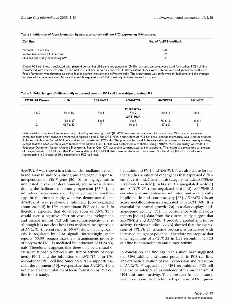
Table 1: Inhibition of focus formation by prostate cancer cell line PC3 expressing U94 protein.
Cell line No. of foci/75 cm2flask
Parental PC3 cell line 80Vector transfected PC3 cell line 69PC3 cell line stably expressing U94 2
Clonal PC3 cell lines, transfected with plasmid containing U94 gene and geneticin (G418) resistant cassette, were used for studies. PC3 cell line transfected with vector cassette or parental PC3 cell line served as controls. G418 resistant clones were sub-cultured and grown to confluence. Focus formation was detected as dense foci of actively growing and refractive cells. The experiment was performed in duplicate, and the average number of foci was reported. Notice that stable expression of U94 drastically inhibited focus formation.
Table 2: Fold changes of differentially expressed genes in PC3 cell line stablyexpressing U94.
PC3/U94 Clones FNI SERPINE2 ADAMTS1 ANGPTL4 SPUVE23
Microarray1 & 2 91 ± 16 7 ± 1 7 ± 2 -20 ± 4 -10 ± 1
QRT-PCR1 183 ± 27 3 ± 1 4 ± 1 -76 ± 13 -6 ± 12 467 ± 33 7 10 ± 1 -67 ± 5 -9
Differential expression of genes was determined by microarray, and QRT-PCR was used to confirm microarray data. Microarray data were computed from array analyses presented in Figures 4 and 5. For QRT-PCR, a subculture of PC3 cell lines used for microarray was used for studies: 2 clones of U94 transfected PC3 cells and vector transfected PC3 cells. The protocol for total RNA extraction was same as for microarray studies, except that the RNA extracts were treated with DNase 1. QRT-PCR was performed in triplicate, using SYBR® Green I chemistry, on 7900 HTS Sequence Detection System (Applied Biosystems, Foster City, CA) according to manufacturer's instructions. The results are presented as average of 3 experiments ± SD. Notice that Microarray data and QRT-PCR data show similar trends; moreover the trend of QRT-PCR results was reproducible in 2 clones of U94 recombinant PC3 cell lines.
Page 8 of 13(page number not for citation purposes)
Cancer Cell International 2005, 5:19 http://www.cancerci.com/content/5/1/19
ously reported by other workers [51,52,54,55,74]. Moreo-ver, this report identifies ANGPTL 4 and SPUVE 23 aspotential therapeutic targets in prostate tumorigenesis.Hopefully, further studies on the microarray data reportedherein might elucidate the complex genetic alterationsthat underlie advanced-stage prostate tumorigenesis, andthereby provide leads for defining targeted therapeuticstrategies for advanced-stage prostate cancer.
Materials and methodsCells and transfectionPC3 cell line was purchased from American Type CultureCollection (ATCC, Manassas, VA, USA) and plasmid U94DNA was prepared as previously described [22,29]). Allcells were cultured in HAM's F12 medium (Cell gro/Medi-atech, VA, USA) supplemented with 2 mM glutamine, 100U of penicillin-streptomycin per ml (Invitrogen, Gaithers-burg, MD, USA), and 10% Fetal Bovine Serum (FBS,HyClone, Logan, UT, USA), at 37°C and 5% CO2. Plas-mid U94 DNA was cloned into the HindIII site of pRc-RSVvector (Invitrogen, Gaithersburg, MD, USA) or HindIII/BamHI site of pBK-CMV (Stratagene, Cedar Creek, TX,USA) vector. Both pRc-RSV and pBK-CMV vectors containa geneticin (G418; Mediatech Inc, Herndon, VA, USA)selectable marker. U94 DNA sequence in the constructswas confirmed by DNA sequencing. The pBK-U94 con-struct was specifically used in experiments that necessi-tated strong expression of U94 protein e.g.immunoblotting, because previous findings showed thatU94 mRNA and protein were expressed at very low levels[23,24,75]. All plasmid DNA were prepared by double-banded cesium chloride gradient ultracentrifugation. U94construct (pRc-U94 or pBK-U94), or plasmid vector cas-sette (pRc-RSV or pBK-CMV) was used for transfection ofPC3 cells. For transfections, 5.5 × 10⁵ PC3 cells wereplated in 60 mm culture dish, and grown over-night(50%-70% confluence). Transfection was performed bythe calcium phosphate-based ProFection MammalianTransfection method (Promega, Madison, WI, USA) inaccordance with manufacturer's protocol. Stablytransfected PC3 cells were selected with G418 (600 µg/ml), and expanded to establish U94 recombinant PC3 cell
line. Clonal selection was performed on G418 resistanthealthy colonies using a clonal cylinder. In order to min-imise culture driven genetic changes [76], transfected cellswere discarded after 8 passages.
Protein extraction, and immunoblot analysisNuclear fraction from cellular extract was prepared asdescribed previously [77]. Confluent PC3 cell line (107-1.5 × 10⁷ cells), stably expressing U94 and resistant toG418 antibiotic (Cellgro, Herndon, VA, USA) was freshlyprepared by trypsinization, washed with DMEM (Cellgro/Mediatech, VA, USA), suspended in 50 ml DMEM (in ster-ile 50 ml centrifuge tube), and incubated at 37°C/ 5%CO2 for 2 hours. Cells were centrifuged at 200 × g for 5minutes, and resuspended in 0.5 ml phosphate bufferedsaline (PBS, Biofluids, Rockville, MD, USA) in microfugetube. The cell pellet from another round of centrifugationwas resuspended in 400 µl of Buffer A (10 mM HEPES; 10mM KCl; 0.1 mM EDTA; 0.1 mM EGTA; 1 mM DTT; o.5mM PMSF; 1% v/v aprotinin). After incubation at 40°Cfor 15 minutes, cells were lysed by adding 0.6% NonidetP-40, mixed by inverting tube 10 times, and the nuclei wasobtained by centrifugation at 200 × g for 5 minutes. Thesupernatant was used as the cytoplasmic fraction. Thenuclei were resuspended gently in ice-cold 100 µl ofBuffer B (20 mM HEPES; 0.4 M NaCl; 1 mM EDTA; 1 mMDGTA; 1 mM DDT; 1 mM PMSF; 1% v/v aprotinin; 10%glycerol), using a wide bore pipette. The nuclei lysate wasincubated in a rotary shaker for 30 minutes at 4°C, andthen centrifuged at 12000 × g for 10 minutes. To aliquotsof the clear supernatant in microfuge tubes, 0.025 mg/mlleupeptin was added before storage at -80°C. The controlcell lines: vector transfected and resistant to G418, andparental PC3, were similarly treated.
Protein determination in the extracts was performed usingBCA protein assay kit (Pierce, Rockford, IL, USA). Follow-ing sodium dodecyl sulphate-polyacrylamide gel electro-phoresis (SDS-PAGE), resolved proteins wereelectroblotted onto polyvinylidene difluoride (PVDF)membrane. The membrane was blocked in 5% non-fatmilk solution on a rocker for 30 min, and rinsed quickly
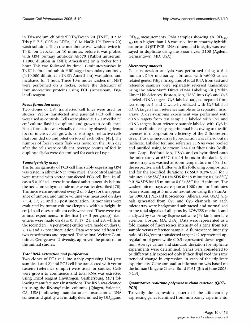
Table 3: Sequences of Forward and Reverse primers for gene amplification in QRT-PCR.
Genes Forward Primer Reverse primer
FN 1 5-GTGTGACCCTCATGAGGCAAC-3 5-CTGGCCTCCAAAGCATGTG-3SERPINE 2 5-CACATCAGCACCAAGACCATAGAC-3 5-TGCCAAGAACTTTCAGCGG-3ADAMST 1 5-CCAGCGTATCTTGCCAGTAACC-3 5-TTTGCAACTGGCAGTTTACTCTG-3ANGPTL 4 5-CCACTTGGGACCAGGATCAC-3 5-CGGAAGTACTGGCCGTTGAG-3SPUVE 23 5-CCCAGTCTACCCTCAATTTAGCC-3 5-GCAGTGGAGTTCCCTTATGACAC-3
Gene specific primers for QRT-PCR were designed using Primer Express Version 2.0 (Applied Biosystems, Foster City, CA) according to the sequence information provided for the cDNAs on the microarray.
Page 9 of 13(page number not for citation purposes)
Cancer Cell International 2005, 5:19 http://www.cancerci.com/content/5/1/19
in Tris/sodium chloride/EDTA/Tween 20 (TNET, 0.2 MTris pH 7.5; 0.05 M EDTA; 1.0 M NaCl; 1% Tween 20)wash solution. Then the membrane was washed twice inTNET on a rocker for 10 minutes, before it was probedwith U94 primary antibody AB679 (Rabbit antiserum,1:1000 dilution in TNET; Amersham) on a rocker for 1hour. This was followed by three 10-minutes washes inTNET before anti rabbit-HRP-tagged secondary antibody(1:10,000 dilution in TNET; Amersham) was added andincubated for 1 hour. Three 10-minutes washes in TNETwere performed on a rocker, before the detection ofimmunoreactive proteins using ECL (Amersham, Eng-land) reagent.
Focus formation assayTwo clones of U94 transfected cell lines were used forstudies. Vector transfected and parental PC3 cell lineswere used as controls. Cells were plated at 1 × 10⁶ cells/ 75cm2 culture flask in duplicate and grown to confluence.Focus formation was visually detected by observing densefoci of intensive cell growth, consisting of refractive cellsthat rounded up and piled on top of each other [49]. Thenumber of foci in each flask was noted on the 10th dayafter the cells were confluent. Average counts of foci induplicate flasks were determined for each cell type.
Tumorigenicity assayThe tumorigenicity of PC3 cell line stably expressing U94was tested in athymic Ncr nu/nu mice. The control animalswere treated with vector transfected PC3 cell line. In allcases 5 × 10⁶ cells were inoculated subcutaneously behindthe neck, into athymic nude mice as earlier described [78].The mice were monitored every 2 or 3 days for the appear-ance of tumors, and tumor volume was measured on days7, 14, 17, 21 and 28 post inoculation. Tumor sizes wereevaluated by tumor volume (length × width × height, incm). In all cases confluent cells were used. There were twoanimal experiments. In the first (n = 3 per group), dataentries were made on days 0, 7, 17, 21, and 28, while inthe second (n = 4 per group) entries were made on days 0,7, 14, and 17 post inoculation. Data were pooled from thetwo experiments and reported. The Animal Welfare Com-mittee, Georgetown University, approved the protocol forthe animal studies.
Total RNA extraction and purificationTwo clones of PC3 cell line stably expressing U94 (testsamples 1 and 2) and PC3 cell line transfected with vectorcassette (reference sample) were used for studies. Cellswere grown to confluence and total RNA was extractedusing Trizol reagent (Invitrogen, Gaithersburg, MD) fol-lowing manufacturer's instructions. The RNA was cleanedup using the RNeasy® mini columns (Qiagen, Valencia,CA, USA) following manufacturers' instructions. RNAcontent and quality was initially determined by OD260and
OD280 measurements. RNA samples showing an OD260/
280 ratio higher than 1.8 was used for microarray hybridi-zation and QRT-PCR. RNA content and integrity was reas-sayed in duplicate using the Bioanalyzer 2100 (Agilent,Germantown, MD, USA).
Microarray analysisGene expression analysis was performed using a 6 khuman cDNA microarray fabricated with ~6000 cancerrelated genes. Fifty micrograms of total RNA from test andreference samples were separately reversed transcribedusing the MicroMax™ Direct cDNA Labeling Kit (PerkinElmer Life Sciences, Boston, MA, USA) into Cy3 and Cy5labeled cDNA targets. Cy3-labeled targets prepared fromtest samples 1 and 2 were hybridized with Cy5-labeledcDNA targets from reference sample onto separate micro-arrays. A dye-swapping experiment was performed withcDNA targets from test sample 1 labeled with Cy5 andcDNA targets from reference sample labeled with Cy3 inorder to eliminate any experimental bias owing to the dif-ferences in incorporation efficiency of the 2 fluorescentdyes. Thus the microarray hybridization was carried out intriplicate. Labeled test and reference cDNAs were pooledand purified using Microcon YM-100 filter units (Milli-pore Corp., Bedford, MA, USA), and co-hybridized ontothe microarray at 65°C for 14 hours in the dark. Eachmicroarray was washed at room temperature in 45 ml ofthe respective wash buffer with the following compositionand for the specified duration: 1x SSC/ 0.2% SDS for 5minutes; 0.5x SSC/ 0.01% SDS for 15 minutes; 0.06x SSC/0.01% SDS for 15 minutes; 0.06x SSC for 15 minutes. Thewashed microarrays were spun at 1000 rpm for 4 minutesbefore scanning at 5 micron resolution using the ScanAr-ray 5000XL (Packard Biosciences, Billerica, MA, USA). Sig-nals generated from Cy3 and Cy5 channels on eachmicroarray were background subtracted and normalizedto the total signals of all spots by LOWESS method, andanalyzed by ScanArray Express software (Perkin Elmer LifeSciences, Boston, MA, USA). Data were represented as afold change of fluorescence intensity of a gene from testsample versus reference sample. A fluorescence intensityratio of U94/vector transfected targets ≥ 2 represented up-regulation of gene; while ≤ 0.5 represented down-regula-tion. Average values and standard deviation for triplicateexperiments were determined. Genes were considered tobe differentially expressed only if they displayed the sametrend of change in expression in each of the triplicateexperiments. Gene annotation information was based onthe human Unigene Cluster Build #161 (5th of June 2003;NCBI)
Quantitative real-time polymerase chain reaction (QRT-PCR)To verify the expression pattern of the differentiallyexpressing genes identified from microarray experiments,
Page 10 of 13(page number not for citation purposes)
Cancer Cell International 2005, 5:19 http://www.cancerci.com/content/5/1/19
QRT-PCR was performed as described previously (79).Equal amounts of total RNA from test and reference celllines were treated with DNase 1 (Invitrogen, Gaithers-burg, MD, USA), and reverse transcribed using randomhexamers and SuperScript II (Invitrogen, Gaithersburg,MD, USA) to prepare the first strand cDNA samples forQRT-PCR analyses. The RT product was diluted 5-fold,and 1 µl is equivalent to 1x concentration. Gene specificprimers (Table 3) were designed by Primer Express Ver-sion 2.0 (Applied Biosystems, Foster City, CA, USA)according to the sequence information provided for thecDNAs on the microarray. The primers were BLASTedagainst the non-redundant and EST mouse sets fromNCBI to confirm specificity. QRT-PCR was performed intriplicate using SYBR® Green I chemistry on 7900 HTSSequence Detection System (Applied Biosystems, FosterCity, CA, USA) according to manufacturer's instructions.The temperature cycle for QRT-PCR was set up asfollowing: 50°C for 2 minutes; 95°C for 10 minutes;95°C 15 seconds and 60°C for 1 minute for 40 cycles. Afinal dissociation cycle running at 95°C for 15 seconds,60°C for 15 seconds and 95°C for 15 seconds was set upfor monitoring the specificity of amplification. The rela-tive standard curve method was used for quantifying geneexpression level, in which the CT values of a series of fixedamounts of test sample (or reference sample) cDNAs(0.01x , 0.1x and 1x as described above) were plottedagainst these amounts of cDNAs. The CT value for a geneat 0.1x concentration in the reference sample (or test sam-ple) was fitted onto the standard curve to obtain therespective expression level. A smaller CT value indicates ahigher expression level, and vice versa. Genes showing CTvalues ≥ 40 were considered to be non-expressing. Thefinal gene expression data were reported after normalisingto that of 18S RNA.
Statistical analysisA repeated measures analysis of variance (ANOVA), sup-plemented by Paired Student's t-test, was used to evaluatethe differences in tumor volume between U94 treated andcontrol vector treated mice. A value of p < 0.05 was con-sidered statistically significant. SAS software (v8.2, SASInstitute, Cary, NC, USA) was used for ANOVA. Experi-mental data, where applicable, are represented as mean ±SD.
Authors' contributionsETI performed Cell culture, molecular biology studies,immunoassays, participated in statistical analysis, anddrafted the manuscript; ALYP carried out the microarrayhybridization and data analysis; WJ performed the QRT-PCR analysis; SM carried out the tumorigenicity studies inmice; WYC performed RNA extraction, data analysis andparticipated in coordination of the studies; KC and SZassisted in some of the molecular biology studies, and JC
participated in coordination of the studies; LJR conceivedof the study, and coordinated the studies. All authors readand approved the final manuscript.
Competing interestsThe author(s) declare that they have no competinginterests.
AcknowledgementsETI was supported by a Minority Supplement (CMBB/NCI/NIH) to Public Health Service (PHS) grant CA 78120 from the National Institutes of Health (NIH; Bethesda, MD, USA). This work was supported in part by PHS/NIH grant CA 78120, and a Contract from the National Foundation for Cancer Research (Bethesda, MD, USA) awarded to LJR. Assistance with the tum-origenicity assays was provided by the Lombardi Cancer Research Center Animal Care Facility. Special thanks to: Dr. Yan A. Su, Department of Pathology, Loyola University Medical Center, Maywood, IL, USA for fabri-cating glass slide microarrays for NICHD/NIH; and Michael Sheridan, Sc. D., Consulting Epidemiologist, Inova Health System & Director, Epidemiology & Biostatistics, Department of Medicine, Inova Fairfax Hospital, VA, USA, for performing the ANOVA.
References1. American Cancer Society: Cancer Statistics Presentation.
[http://www.cancer.org/docroot/STT/stt_0_2004.asp?sitearea=STT&level=1].
2. Ficazzola MA, Taneja SS: Prospects for gene therapy in humanprostate cancer. Molecular Medicine Today 1998, 4:494-504.
3. Buttyan R, Shabsigh A, Perlman H, Colombel M: Regulation ofApoptosis in the Prostate Gland by Androgenic Steroids.Trends Endocrinol Metab 1999, 10:47-54.
4. Chakravarti A, Zhai GG: Molecular and genetic prognostic fac-tors of prostate cancer. World J Urol 2003, 21:265-274.
5. Karan D, Lin M, Johansson SL, Batra SK: Current Status of theMolecular Genetics of Human Prostatic Adenocarcinomas.Int J Cancer 2003, 103:285-293.
6. Abate-Shen C, Shen MM: Molecular genetics of prostate cancer.Genes and Dev 2000, 14:2410-2434.
7. Kumazawa T, Tsuchiya N, Wang L, Sato K, Kamoto T, Ogawa O,Nakamura A, Kato T, Habuchi T: Microsatellite polymorphism ofsteroid hormone synthesis gene CYP11A1 is associated withadvanced prostate cancer. Int J Cancer 2004, 110:140-144.
8. Flaherty KT, Malkowicz SB, Vaughn DJ: Phase I study of weeklyliposome-encapsulated doxorubicin in patients withadvanced, androgen-independent prostate cancer. Am J ClinOncol 2004, 27:136-139.
9. Joshi B, Li L, Taffe BG, Zhu Z, Wahl S, Tian H, Ben-Josef E, Taylor JD,Porter AT, Tang DG: Apoptosis Induction by a Novel Anti-Prostate Cancer Compound, BMD188 (a Fatty Acid-contain-ing Hydroxamic Acid), Requires the Mitochondrial Respira-tory Chain. Cancer Res 1999, 59:4343-4355.
10. Goodin S, Rao KV, DiPaola RS: State-of-the-Art Treatment ofMetastatic Hormone-Refractory Prostate Cancer. Oncologist2002, 7:360-370.
11. Gulley J, Dahut W: Novel clinical trials in androgen-independ-ent prostate cancer. Clin Prostate Cancer 2002, 1:51-57.
12. Nimmanapalli R, Perkins CL, Orlando M, O'Bryan E, Nguyen D, BhallaKN: Pretreatment with Paclitaxel Enhances Apo-2 Ligand/Tumor Necrosis Factor- related Apoptosis-inducing Ligand-induced Apoptosis of Prostate Cancer Cells by InducingDeath Receptors 4 and 5 Protein Levels. Cancer Res 2001,61:759-763.
13. Chay CH, Cooper CC, Hellerstedt BA, Pienta KJ: Antimetastaticdrugs in prostate cancer. Clin Prostate Cancer 2002, 1:14-19.
14. Allay JA, Steiner MS, Zhang Y, Reed CP, Cockroft J, Lu Y: Adenovi-rus p16 gene therapy for prostate cancer. World J Urol 2000,18:111-120.
15. Arlen PM, Figg WD, Gulley J, Cox MC, Linehan WM, Dahut W:National Cancer Institute intramural approach to advancedprostate cancer. Clin Prostate Cancer 2002, 1:153-162.
Page 11 of 13(page number not for citation purposes)
http://www.ncbi.nlm.nih.gov/entrez/query.fcgi?cmd=Retrieve&db=PubMed&dopt=Abstract&list_uids=9857369
Cancer Cell International 2005, 5:19 http://www.cancerci.com/content/5/1/19
16. Raghow S, Hooshdaran MZ, Katiyar S, Steiner MS: Toremifene pre-vents prostate cancer in the transgenic adenocarcinoma ofmouse prostate model. Cancer Res 2002, 62:1370-1376.
17. Steiner MS, Wang Y, Zhang Y, Zhang X, Lu Y: p16/MTS1/INK4Asuppresses prostate cancer by both pRb dependent and inde-pendent pathways. Oncogene 2000, 19:1297-1306.
18. Sternberg C: Overview of international collaborative groupprostate cancer trials. Crit Rev Oncol Hematol 2002, 43:153-158.
19. Collis SJ, Khater K, DeWeese TL: Novel therapeutic strategies inprostate cancer management using gene therapy in combi-nation with radiation therapy. World J Urol 2003, 21:275-289.
20. Timme TL, Satoh T, Tahir SA, Wang H, Teh BS, Butler EB, Miles BJ,Amato RJ, Kadmon D, Thompson TC: Therapeutic targets formetastatic prostate cancer. Curr Drug Targets 2003, 4:251-261.
21. Thompson BJ, Efstathiou S, Honess RW: Acquisition of the humanadeno-associated virus type-2 rep gene by human herpesvi-rus type-6. Nature 1991, 351:78-80.
22. Araujo JC, Doniger J, Kashanchi F, Hermonat PL, Thompson J,Rosenthal LJ: Human Herpesvirus 6A suppresses both trans-formations by H-ras and Human Immunodeficiency Virustype 1 promoters. J Virol 1995, 69:4933-4940.
23. Mori Y, Dhepakson P, Shimamoto T, Ueda K, Gomi Y, Tani H, Mats-uura Y, Yamanishi K: Expression of human herpesvirus 6B repwithin infected cells and binding of its gene product to theTATA-Binding protein in vitro and in vivo. J Virol 2000,74:6096-6104.
24. Dhepakson P, Mori Y, Jiang YB, Huang HL, Akkapaiboon P, Okuno T,Yamanishi K: Human herpesvirus-6 rep/U94 gene product hassingle-stranded DNA-binding activity. J Gen Virol 2002,83:847-854.
25. Rotola A, Ravaioli T, Gonelli A, Dewhurst S, Cassai E, Di Luca D: U94of human herpesvirus 6 is expressed in latently infectedperipheral blood mononuclear cells and blocks viral geneexpression in transformed lymphocytes in culture. Proc NatlAcad Sci USA 1998, 95:13911-13916.
26. Turner S, DiLuca D, Gompels UA: Characterization of a humanherpesvirus 6 variant A 'amplicon' and replication modula-tion by U94-Rep 'latency gene'. Journal of Virological Methods2002, 105:331-341.
27. Gompels UA, Nicholas J, Lawrence G, Jones M, Thomson BJ, MartinME, Efstathiou S, Craxton M, Macaulay HA: The DNA sequence ofhuman herpesvirus-6: structure, coding content, andgenome evolution. Virology 1995, 209:29-51.
28. Thomson BJ, Weindler FW, Gray D, Schwaab V, Heilbronn R:Human herpesvirus 6 (HHV-6) is a helper virus for adeno-associated virus type 2 (AAV-2) and the AAV-2 rep genehomologue in HHV-6 can mediate AAV-2 DNA replicationand regulate gene expression. Virology 1994, 204:304-311.
29. Araujo JC, Doniger J, Stöppler H, Sadaie MR, Rosenthal LJ: Cell linescontaining and expressing the human herpesvirus 6A ts geneare protected from both H-ras and BPV-1 transformation.Oncogene 1997, 14:937-943.
30. Kibel AS, Faith DA, Bova GS, Isaacs WB: Loss of heterozygosity at12P12-13 in primary and metastatic prostateadenocarcinoma. J Urol 2000, 164:192-196.
31. Guo Y, Sklar GN, Borkowski A, Kyprianou N: Loss of the cyclin-dependent kinase inhibitor p27(Kip1) protein in human pros-tate cancer correlates with tumor grade. Clin Cancer Res 1997,3:2269-2274.
32. Tsihlias J, Kapusta LR, DeBoer G, Morava-Protzner I, Zbieranowski I,Bhattacharya N, Catzavelos GC, Klotz LH, Slingerland JM: Loss ofcyclin-dependent kinase inhibitor p27Kip1 is a novel prognos-tic factor in localized human prostate adenocarcinoma. Can-cer Res 1998, 58:542-548.
33. Yang RM, Naitoh J, Murphy M, Wang HJ, Phillipson J, deKernion JB,Loda M, Reiter RE: Low p27 expression predicts poor disease-free survival in patients with prostate cancer. J Urol 1998,159:941-945.
34. Park MS, Rosai J, Nguyen HT, Capodieci P, Cordon-Cardo C, Koff A:p27 and Rb are on overlapping pathways suppressing tumor-igenesis in mice. Proc Natl Acad Sci USA 1999, 96:6382-6387.
35. Chi SG, deVere White RW, Muenzer JT, Gumerlock PH: Frequentalteration of CDKN2 (p16(INK4A)/MTS1) expression inhuman primary prostate carcinomas. Clin Cancer Res 1997,3:1889-1897.
36. Jarrard D, Modder J, Fadden P, Fu V, Sebree L, Heisey D, Schwarze S,Friedl A: Alterations in the p16/pRb cell cycle checkpointoccur commonly in primary and metastatic human prostatecancer. Cancer Lett 2002, 185:191.
37. Jarrard DF, Sarkar S, Shi Y, Yeager TR, Magrane G, Kinoshita H, NassifN, Meisner L, Newton MA, Waldman FM, Reznikoff CA: p16/pRbpathway alterations are required for bypassing senescence inhuman prostate epithelial cells. Cancer Res 1999, 59:2957-2964.
38. Alexander K, Hinds PW: Requirement for p27(KIP1) in retino-blastoma protein-mediated senescence. Mol Cell Biol 2001,21:3616-3631.
39. Colombel M, Symmans F, Gil S, O'Toole KM, Chopin D, Benson M,Olsson CA, Korsmeyer S, Buttyan R: Detection of the apoptosis-suppressing oncoprotein Bcl-2 in hormone-refractoryhuman prostate cancers. Am J Pathol 1993, 143:390-400.
40. Apakama I, Robinson MC, Walter NM, Charlton RG, Royds JA, FullerCE, Neal DE, Hamdy FC: Bcl-2 overexpression combined withp53 protein accumulation correlates with hormone-refrac-tory prostate cancer. Brit J Cancer 1996, 74:1258-1262.
41. Furuya Y, Krajewski S, Epstein JI, Reed JC, Isaacs JT: Expression ofBcl-2 and the progression of human and rodent prostaticcancers. Clin Cancer Res 1996, 2:389-398.
42. McDonnell TJ, Navone NM, Troncoso P, Pisters LL, Conti C, vonEschenbach AC, Brisbay S, Logothetis CJ: Expression of bcl-2oncoprotein and p53 protein accumulaion in bone marrowmetastases of androgen independent prostate cancer. J Urol1997, 157:569-574.
43. Chaudhary KS, Abel PD, Lalani EN: Role of the Bcl-2 gene familyin prostate cancer progression and its implications for ther-apeutic intervention. Environ Health Perspect 1999, 107(Suppl1):49-57.
44. Buttyan R, Sawczuk IS, Benson MC, Siegal JD, Olsson CA: Enhancedexpression of the c-myc protooncogene in high-grade humanprostate cancers. Prostate 1987, 11:327-337.
45. Phillips MEA, Ferro MA, Smith PJB, Davies P: Intranuclear andro-gen receptor deployment and protooncogene expression inhuman diseased prostate. Urol Int 1987, 42:115-119.
46. Nag A, Smith RG: Amplification, rearrangement, and elevatedexpression of c-myc in the human prostatic carcinoma cellline LNCaP. Prostate 1989, 15:115-122.
47. Peehl DM: Oncogenes in Prostate Cancer. Cancer 1993,71:1159-1164.
48. Dhanasekaran SM, Barrette TR, Ghosh D, Shah R, Varambally S, Kura-chi K, Pienta KJ, Rubin MA, Chinnaiyan AM: Delineation of prog-nostic biomarkers in prostate cancer. Nature 2001,412:822-826.
49. Muralidhar S, Doniger J, Mendelson E, Araujo JC, Kashanchi F, AzumiN, Brady JN, Rosenthal LJ: Human cytomegalovirus mtrII onco-protein binds to p53 and down-regulates p53-activatedtranscription. J Virol 1996, 70:8691-8700.
50. Le-Jan S, Amy C, Cazes A, Monnot C, Lamande N, Favier J, Philippe J,Siboney M, Gasc JM, Corvol P, Germain S: Angiopoietin-like 4 is aproangiogenic factor produced during ischemia and in con-ventional renal cell carcinoma. Am J Pathol 2003, 162:1521-1528.
51. Yi M, Ruoslahti E: A fibronectin fragment inhibits tumorgrowth, angiogenesis, and metastasis. Proc Natl Acad Sci USA2001, 98:620-624.
52. Zhang GM, Yang Y, Huang B, Xiao H, Li D, Feng ZH: Experimentalstudy on therapeutic effect of in vivo expression of Cell I-HepII recombinant polypeptide of fibronectin on murine H22hepatocellular carcinoma. World J Gastroenterol 2003,9:1940-1945.
53. Yi M, Sakai T, Fassler R, Ruoslahti E: Antiangiogenic proteinsrequire plasma fibronectin or vitronectin for in vivo activity.Proc Natl Acad Sci USA 2003, 100:11435-11438.
54. Pasqualini R, Bourdoulous S, Koivunen E, Woods VL Jr, Ruoslahti EA:Polymeric form of fibronectin has antimetastatic effectsagainst multiple tumor types. Nat Med 1996, 2:1197-1203.
55. Hynes RO: The dynamic dialogue between cells and matrices:implications of fibronectin's elasticity. Proc Natl Acad Sci USA1999, 96:2588-2590.
56. Schwarzbauer JE: Identification of the fibronectin sequencesrequired for assembly of a fibrillar matrix. J Cell Biol 1991,113:1463-1473.
57. Hocking DC, Sottile J, McKeown-Longo PJ: Fibronectin's III-1module contains a conformation-dependent binding site for
Page 12 of 13(page number not for citation purposes)
http://www.ncbi.nlm.nih.gov/entrez/query.fcgi?cmd=Retrieve&db=PubMed&dopt=Abstract&list_uids=1851252
http://www.ncbi.nlm.nih.gov/entrez/query.fcgi?cmd=Retrieve&db=PubMed&dopt=Abstract&list_uids=1851252
http://www.ncbi.nlm.nih.gov/entrez/query.fcgi?cmd=Retrieve&db=PubMed&dopt=Abstract&list_uids=7609062
http://www.ncbi.nlm.nih.gov/entrez/query.fcgi?cmd=Retrieve&db=PubMed&dopt=Abstract&list_uids=7609062
http://www.ncbi.nlm.nih.gov/entrez/query.fcgi?cmd=Retrieve&db=PubMed&dopt=Abstract&list_uids=9811900
http://www.ncbi.nlm.nih.gov/entrez/query.fcgi?cmd=Retrieve&db=PubMed&dopt=Abstract&list_uids=9811900
http://www.ncbi.nlm.nih.gov/entrez/query.fcgi?cmd=Retrieve&db=PubMed&dopt=Abstract&list_uids=9811900
http://www.ncbi.nlm.nih.gov/entrez/query.fcgi?cmd=Retrieve&db=PubMed&dopt=Abstract&list_uids=7747482
http://www.ncbi.nlm.nih.gov/entrez/query.fcgi?cmd=Retrieve&db=PubMed&dopt=Abstract&list_uids=7747482
http://www.ncbi.nlm.nih.gov/entrez/query.fcgi?cmd=Retrieve&db=PubMed&dopt=Abstract&list_uids=7747482
http://www.ncbi.nlm.nih.gov/entrez/query.fcgi?cmd=Retrieve&db=PubMed&dopt=Abstract&list_uids=8091661
http://www.ncbi.nlm.nih.gov/entrez/query.fcgi?cmd=Retrieve&db=PubMed&dopt=Abstract&list_uids=8091661
http://www.ncbi.nlm.nih.gov/entrez/query.fcgi?cmd=Retrieve&db=PubMed&dopt=Abstract&list_uids=8091661
http://www.ncbi.nlm.nih.gov/entrez/query.fcgi?cmd=Retrieve&db=PubMed&dopt=Abstract&list_uids=9050993
http://www.ncbi.nlm.nih.gov/entrez/query.fcgi?cmd=Retrieve&db=PubMed&dopt=Abstract&list_uids=9815624
http://www.ncbi.nlm.nih.gov/entrez/query.fcgi?cmd=Retrieve&db=PubMed&dopt=Abstract&list_uids=9815624
http://www.ncbi.nlm.nih.gov/entrez/query.fcgi?cmd=Retrieve&db=PubMed&dopt=Abstract&list_uids=9458103
http://www.ncbi.nlm.nih.gov/entrez/query.fcgi?cmd=Retrieve&db=PubMed&dopt=Abstract&list_uids=9458103
http://www.ncbi.nlm.nih.gov/entrez/query.fcgi?cmd=Retrieve&db=PubMed&dopt=Abstract&list_uids=9474188
http://www.ncbi.nlm.nih.gov/entrez/query.fcgi?cmd=Retrieve&db=PubMed&dopt=Abstract&list_uids=9474188
http://www.ncbi.nlm.nih.gov/entrez/query.fcgi?cmd=Retrieve&db=PubMed&dopt=Abstract&list_uids=9815578
http://www.ncbi.nlm.nih.gov/entrez/query.fcgi?cmd=Retrieve&db=PubMed&dopt=Abstract&list_uids=9815578
http://www.ncbi.nlm.nih.gov/entrez/query.fcgi?cmd=Retrieve&db=PubMed&dopt=Abstract&list_uids=7688182
http://www.ncbi.nlm.nih.gov/entrez/query.fcgi?cmd=Retrieve&db=PubMed&dopt=Abstract&list_uids=7688182
http://www.ncbi.nlm.nih.gov/entrez/query.fcgi?cmd=Retrieve&db=PubMed&dopt=Abstract&list_uids=7688182
http://www.ncbi.nlm.nih.gov/entrez/query.fcgi?cmd=Retrieve&db=PubMed&dopt=Abstract&list_uids=8883414
http://www.ncbi.nlm.nih.gov/entrez/query.fcgi?cmd=Retrieve&db=PubMed&dopt=Abstract&list_uids=8883414
http://www.ncbi.nlm.nih.gov/entrez/query.fcgi?cmd=Retrieve&db=PubMed&dopt=Abstract&list_uids=8883414
http://www.ncbi.nlm.nih.gov/entrez/query.fcgi?cmd=Retrieve&db=PubMed&dopt=Abstract&list_uids=9816182
http://www.ncbi.nlm.nih.gov/entrez/query.fcgi?cmd=Retrieve&db=PubMed&dopt=Abstract&list_uids=9816182
http://www.ncbi.nlm.nih.gov/entrez/query.fcgi?cmd=Retrieve&db=PubMed&dopt=Abstract&list_uids=9816182
http://www.ncbi.nlm.nih.gov/entrez/query.fcgi?cmd=Retrieve&db=PubMed&dopt=Abstract&list_uids=8996359
http://www.ncbi.nlm.nih.gov/entrez/query.fcgi?cmd=Retrieve&db=PubMed&dopt=Abstract&list_uids=8996359
http://www.ncbi.nlm.nih.gov/entrez/query.fcgi?cmd=Retrieve&db=PubMed&dopt=Abstract&list_uids=8996359
http://www.ncbi.nlm.nih.gov/entrez/query.fcgi?cmd=Retrieve&db=PubMed&dopt=Abstract&list_uids=2446300
http://www.ncbi.nlm.nih.gov/entrez/query.fcgi?cmd=Retrieve&db=PubMed&dopt=Abstract&list_uids=2446300
http://www.ncbi.nlm.nih.gov/entrez/query.fcgi?cmd=Retrieve&db=PubMed&dopt=Abstract&list_uids=2441504
http://www.ncbi.nlm.nih.gov/entrez/query.fcgi?cmd=Retrieve&db=PubMed&dopt=Abstract&list_uids=2441504
http://www.ncbi.nlm.nih.gov/entrez/query.fcgi?cmd=Retrieve&db=PubMed&dopt=Abstract&list_uids=2441504
http://www.ncbi.nlm.nih.gov/entrez/query.fcgi?cmd=Retrieve&db=PubMed&dopt=Abstract&list_uids=2678039
http://www.ncbi.nlm.nih.gov/entrez/query.fcgi?cmd=Retrieve&db=PubMed&dopt=Abstract&list_uids=2678039
http://www.ncbi.nlm.nih.gov/entrez/query.fcgi?cmd=Retrieve&db=PubMed&dopt=Abstract&list_uids=8428339
http://www.ncbi.nlm.nih.gov/entrez/query.fcgi?cmd=Retrieve&db=PubMed&dopt=Abstract&list_uids=8970996
http://www.ncbi.nlm.nih.gov/entrez/query.fcgi?cmd=Retrieve&db=PubMed&dopt=Abstract&list_uids=8970996
http://www.ncbi.nlm.nih.gov/entrez/query.fcgi?cmd=Retrieve&db=PubMed&dopt=Abstract&list_uids=8970996
http://www.ncbi.nlm.nih.gov/entrez/query.fcgi?cmd=Retrieve&db=PubMed&dopt=Abstract&list_uids=8898745
http://www.ncbi.nlm.nih.gov/entrez/query.fcgi?cmd=Retrieve&db=PubMed&dopt=Abstract&list_uids=8898745
http://www.ncbi.nlm.nih.gov/entrez/query.fcgi?cmd=Retrieve&db=PubMed&dopt=Abstract&list_uids=8898745
http://www.ncbi.nlm.nih.gov/entrez/query.fcgi?cmd=Retrieve&db=PubMed&dopt=Abstract&list_uids=2045422
http://www.ncbi.nlm.nih.gov/entrez/query.fcgi?cmd=Retrieve&db=PubMed&dopt=Abstract&list_uids=2045422
Cancer Cell International 2005, 5:19 http://www.cancerci.com/content/5/1/19
Publish with BioMed Central and every scientist can read your work free of charge
"BioMed Central will be the most significant development for disseminating the results of biomedical research in our lifetime."
Sir Paul Nurse, Cancer Research UK
Your research papers will be:
available free of charge to the entire biomedical community
peer reviewed and published immediately upon acceptance
cited in PubMed and archived on PubMed Central
yours — you keep the copyright
Submit your manuscript here:http://www.biomedcentral.com/info/publishing_adv.asp
BioMedcentral
the amino-terminal region of fibronectin. J Biol Chem 1994,269:19183-19191.
58. Aguirre KM, McCormick RJ, Schqarzbauer JE: Fibronectin self-association is mediated by complementary sites within theamino-terminal one-third of the molecule. J Biol Chem269:27863-27868.
59. Briknarovà K, Åkerman ME, Hoyt DW, Ruoslahti E, Ely KR: Anastel-lin, an FN 3 Fragment with Fibronectin Polymerization Aci-tivity, Resembles Amyloid Fibril Precursors. J Mol Biol 2003,332:205-215.
60. Pickford AR, Smith SP, Staunton D, Boyd J, Campbell ID: The hairpinstructure of the 6F11F22F2 fragment from human fibronectinenhances gelatin binding. The EMBO Journal 2001, 20:1519-1529.
61. Clark EA, Brugge JS: Integrins and signal transduction path-ways: the road taken. Science 1995, 268:233-239.
62. Brenner KA, Corbett SA, Schwarzbauer JE: Regulation of fibronec-tin matrix assembly by activated Ras in transformed cells.Oncogene 2000, 19:3156-3163.
63. Folkman J: Angiogenesis in cancer, vascular, rheumatoid andother disease. Nat Med 1995, 1:27-31.
64. Risau W: Mechanism of angiogenesis. Nature 1997, 386:671-674.65. Varner JA: The role of vascular cell integrins alpha v beta 3 and
alpha v beta 5 in angiogenesis. EXS 1997, 79:361-90.66. Ruoslahti e: The RGD story: a personal account. Matrix Biology
2003, 22:459-465.67. Hynes RO: A reevaluation of integrins as regulators of
angiogenesis. Nat Med 2002, 8:918-921.68. Vitale M, Matola TD, Rossi G, Laezza C, Fenzi G, Bifulco M: Prenyl-
transferase inhibitors induce apoptosis in proliferating thy-roid cells through a p53-Independent, CrmA-Sensitive, andcaspase-3-like protease-dependent mechanism. Endocrinology1999, 140:698-704.
69. Kuno K, Terashima Y, Matsushima K: ADAMTS-1 is an activemetalloproteinase associated with the extracellular matrix.J Biol Chem 1999, 274:18821-18826.
70. Shindo T, Kurihara H, Kuno K, Yokoyama H, Wada T, Kurihara Y,Imai T, Wang Y, Ogata M, Nishimatsu H, Moriyama N, Oh-hashi Y,Morita H, Ishikawa T, Nagai R, Yazaki Y, Matsushima K: ADAMTS-1: a metalloproteinase-disintegrin essential for normalgrowth, fertility, and organ morphology and function. J ClinInvest 2000, 105:1345-1352.
71. Vazquez F, Hastings G, Ortega MA, Lane TF, Oikemus S, LombardoM, Iruela-Arispe ML: METH-1, a human ortholog of ADAMTS-1, and METH-2 are members of a new family of proteins withangio-inhibitory activity. J Biol Chem 1999, 274:23349-23357.
72. Pineiro-Sanchez ML, Goldstein LA, Dodt J, Howard L, Yeh Y, ChenW: Identification of the 170-kDa Melanoma membrane-bound gelatinase (seprase) as a serine integral membraneprotease. J Biol Chem 1997, 272:7595-7601.
73. Vacca A, Ria R, Presta M, Ribatti D, Iurlaro M, Merchionne F, Tang-hetti E, Dammacco F: alpha (v) beta (3) integrin engagementmodulates cell adhesion, proliferation, and protease secre-tion in human lymphoid tumor cells. Exp Hematol 2001,29:993-1003.
74. Abeysinghe HR, Cao Q, Xu J, Pollock S, Veyberman Y, Guckert NL,Keng P, Wang N: THY1 expression is associated with tumorsuppression of human ovarian cancer. Cancer Genetics andCytogenetics 2002, 143:125-132.
75. Rapp JC, Krug LT, Inoue N, Dambaugh TR, Pellett PE: U94 thehuman herpesvirus 6 homolog of the Parvovirus nonstruc-tural gene, is highly conserved among isolates and isexpressed at low mRNA levels as a spliced transcript. Virology2000, 268:504-516.
76. Beheshti B, Park PC, Sweet JM, Trachtenberg J, Jewett MA, Squire JA:Evidence of chromosomal instability in prostate cancerdetermined by spectral karyotyping (SKY) and interphasefish analysis. Neoplasia 2001, 3:62-69.
77. Olnes MI, Kurl RN: Isolation of nuclear extracts from fragilecells: a simplified procedure applied to thymocytes. Biotech-niques 1994, 17:828-829.
78. Muralidhar S, Pumfery AM, Hassani M, Sadaie MR, Kishishita M, BradyJN, Doniger J, Medveczky P, Rosenthal LJ: Identification of kaposin(open reading frame K12) as a human herpesvirus 8 (Kaposi'ssarcoma-associated herpesvirus) transforming gene. J Virol1998, 72:4980-4988.
79. Pang AL, Taylor HC, Johnson W, Alexander S, Chen Y, Su YA, Li X,Ravindranath N, Dvm M, Rennert OM, Chan WY: Identification ofdifferentially expressed genes in mouse spermatogenesis. JAndrol 2003, 24:899-911.
Page 13 of 13(page number not for citation purposes)
http://www.ncbi.nlm.nih.gov/entrez/query.fcgi?cmd=Retrieve&db=PubMed&dopt=Abstract&list_uids=8034677
http://www.ncbi.nlm.nih.gov/entrez/query.fcgi?cmd=Retrieve&db=PubMed&dopt=Abstract&list_uids=7716514
http://www.ncbi.nlm.nih.gov/entrez/query.fcgi?cmd=Retrieve&db=PubMed&dopt=Abstract&list_uids=7716514
http://www.ncbi.nlm.nih.gov/entrez/query.fcgi?cmd=Retrieve&db=PubMed&dopt=Abstract&list_uids=7584949
http://www.ncbi.nlm.nih.gov/entrez/query.fcgi?cmd=Retrieve&db=PubMed&dopt=Abstract&list_uids=7584949
http://www.ncbi.nlm.nih.gov/entrez/query.fcgi?cmd=Retrieve&db=PubMed&dopt=Abstract&list_uids=9109485
http://www.ncbi.nlm.nih.gov/entrez/query.fcgi?cmd=Retrieve&db=PubMed&dopt=Abstract&list_uids=9002227
http://www.ncbi.nlm.nih.gov/entrez/query.fcgi?cmd=Retrieve&db=PubMed&dopt=Abstract&list_uids=9002227
http://www.ncbi.nlm.nih.gov/entrez/query.fcgi?cmd=Retrieve&db=PubMed&dopt=Abstract&list_uids=9927296
http://www.ncbi.nlm.nih.gov/entrez/query.fcgi?cmd=Retrieve&db=PubMed&dopt=Abstract&list_uids=9927296
http://www.ncbi.nlm.nih.gov/entrez/query.fcgi?cmd=Retrieve&db=PubMed&dopt=Abstract&list_uids=9927296
http://www.ncbi.nlm.nih.gov/entrez/query.fcgi?cmd=Retrieve&db=PubMed&dopt=Abstract&list_uids=9065413
http://www.ncbi.nlm.nih.gov/entrez/query.fcgi?cmd=Retrieve&db=PubMed&dopt=Abstract&list_uids=9065413
http://www.ncbi.nlm.nih.gov/entrez/query.fcgi?cmd=Retrieve&db=PubMed&dopt=Abstract&list_uids=9065413
http://www.ncbi.nlm.nih.gov/entrez/query.fcgi?cmd=Retrieve&db=PubMed&dopt=Abstract&list_uids=7840956
http://www.ncbi.nlm.nih.gov/entrez/query.fcgi?cmd=Retrieve&db=PubMed&dopt=Abstract&list_uids=7840956
http://www.ncbi.nlm.nih.gov/entrez/query.fcgi?cmd=Retrieve&db=PubMed&dopt=Abstract&list_uids=9573267