Coronary Reactive Hyperemia Olivecrona, Göran - Lucris
75
Coronary Reactive Hyperemia Olivecrona, Göran 2007 Link to publication Citation for published version (APA): Olivecrona, G. (2007). Coronary Reactive Hyperemia. Lund University, Faculty of Medicine. https://www.avhandlingar.se/avhandling/14c65a3bb6/ Total number of authors: 1 General rights Unless other specific re-use rights are stated the following general rights apply: Copyright and moral rights for the publications made accessible in the public portal are retained by the authors and/or other copyright owners and it is a condition of accessing publications that users recognise and abide by the legal requirements associated with these rights. • Users may download and print one copy of any publication from the public portal for the purpose of private study or research. • You may not further distribute the material or use it for any profit-making activity or commercial gain • You may freely distribute the URL identifying the publication in the public portal Read more about Creative commons licenses: https://creativecommons.org/licenses/ Take down policy If you believe that this document breaches copyright please contact us providing details, and we will remove access to the work immediately and investigate your claim.
-
Upload
khangminh22 -
Category
Documents
-
view
0 -
download
0
Transcript of Coronary Reactive Hyperemia Olivecrona, Göran - Lucris

LUND UNIVERSITY
PO Box 117221 00 Lund+46 46-222 00 00
Coronary Reactive Hyperemia
Olivecrona, Göran
2007
Link to publication
Citation for published version (APA):Olivecrona, G. (2007). Coronary Reactive Hyperemia. Lund University, Faculty of Medicine.https://www.avhandlingar.se/avhandling/14c65a3bb6/
Total number of authors:1
General rightsUnless other specific re-use rights are stated the following general rights apply:Copyright and moral rights for the publications made accessible in the public portal are retained by the authorsand/or other copyright owners and it is a condition of accessing publications that users recognise and abide by thelegal requirements associated with these rights. • Users may download and print one copy of any publication from the public portal for the purpose of private studyor research. • You may not further distribute the material or use it for any profit-making activity or commercial gain • You may freely distribute the URL identifying the publication in the public portal
Read more about Creative commons licenses: https://creativecommons.org/licenses/Take down policyIf you believe that this document breaches copyright please contact us providing details, and we will removeaccess to the work immediately and investigate your claim.

Coronary Reactive Hyperemia
Göran Olivecrona
Department of Cardiology Clinical Sciences, Lund Lund University, 2007

und University Faculty of Medicine Doctoral Dissertation Series 2007:75
7
SSN: 1652-822059-53-4
L
opyright 2007 Göran OlivecronaCPrinted at KFS AB, Lund, Sweden 200
IISBN: 978-91-855
2

Coronary Reactive Hyperemia
his thesis is dedicated to my dear wife Cecilia and our wonderful children Axel, Jacob and
also dedicate the thesis in loving memory of my younger brother Lars.
T
Emma.
I
3

Medicus curat, natura sanat
4
1 List of Publications
wing papers, referred to in the text by their Roman
Wang LW, Olivecrona GK, Gotberg M, Olsson ML, Sörhede
I. Olivecrona GK, Gotberg M, Harnek J, Wang LW, Jacobson
III. Olivecrona GK, Gotberg M, Harnek J, Jacobson KA, Jern S,
V. Olivecrona GK, Gotberg M, Van der Pals J, Harnek J, Erlinge
. Gotberg M, Olivecrona GK, Engblom H, Ugander M, van der
his thesis is based on the folloTnumeral:
I.Winzell M, Erlinge D. ADP acting on P2Y13 receptors is a negative feedback pathway for ATP release from human red blood cells. Circulation research 2005; Feb 4;96(2):189-196
IKA, Erlinge D. Coronary Artery reperfusion: The ADPreceptor P2Y1 mediates early reactive hyperemia in vivo inpigs. Purinergic Signalling 2005; 1: 1-7.
Erlinge D. The ADP receptor P2Y1 mediates t-PA release in pigs during cardiac ischemia. J Thromb Thrombolysis Epubahead of print Feb 10, 2007
ID. Mild hypothermia reduces coronary blood flow during post-ischemic reactive hyperemia in a pig model. BMC
Cardiovascular Disorders Epub ahead of print Feb 26, 2007
VPals J, Heiberg E, Arheden H, David Erlinge D. Rapid and short duration hypothermia before reperfusion reducesmyocardial infarct size in pigs Submitted, (Abstract Presented as poster at TCT, Washington DC, Oct 2006)
5

6

2 Contents
List of publications……………………………………………….. 512 Contents………………………………………………………….... 7 3 Abbreviations……………………………………………………... 9 4 Abstract…………………………………………………………....115 Introduction……………………………………………………….136 Hypotheses of the thesis………………………………………......21 7 Materials and methods…………………………………………... 238 Reagents, Ethics and Statistics………………………………….. 319 Results…………………………………………………………….. 3310 Main findings…………………………………………………….. 4511 Discussion……………………………………………………….... 4712 Populärvetenskaplig sammanfattning (Summary in Swedish).. 5713 Acknowledgements………………………………………………. 5914 Sources of Funding………………………………………………. 6115 References………………………………………………………... 63 16 Appendix: Original Papers I-V…………………………………. 75
7

8

3 Abbreviations
AR Area At Riskhosphate
larizing Factor
omputed Tomography
AADP Adenosine DipAMI Acute Myocardial Infarction AMP Adenosine MonophosphateAPV Average Peak Velocity (cm/s) ATP Adenosine TriphosphateBE Base ExcessDNA Deoxyribonucleic AcidEDHF Endothelium Derived HyperpoIS Infarct Size
ing ArteryLAD Left Anterior DescendLV Left VentricleMI Myocardial InfarctionMRI Magnetic Resonance ImagingNO Nitrous OxideRBC Red Blood CellRNA Ribonucleic AcidSMC Smooth Muscle Cell
T on CSPEC Single Photon Emissit-PA Tissue Plaminogen Activator UDP Uridine DiphosphateUTP Uridine Triphosphate
9

10

4 Abstractmechanism of post ischemic reactive hyperemia is still unknown but now Introduction: The
thought to be multifactorial and perhaps involving purinergic signalling. Purines such as ATP and ADP have recently been discovered to play a vital role in the regulation of vascular tone.We postulated that ADP could play a vital role in the mechanism of coronary post ischemicreactive hyperemia and that increased concentrations of plasma ADP could also stimulate a negative feed back loop governing release of ATP from red blood cells as well as an increased release of t-PA. It was also hypothesized that hypothermia may have effects on coronary reactive hyperemia and that rapidly induced hypothermia prior to reperfusion in an infarction model could reduce myocardial infarct size. Material and Methods: All studies were performed in a closed chest porcine model in which coronary reactive hyperemia was studied after a ten minute occlusion of the LAD distal to the first diagonal branch, and blood flow measured in the distal LAD with a Doppler flow wire. Blood samples were collected in a peripheral artery, in the Coronary Sinus and in a central vein. Hypothermia was induced with an endovascular catheter and cold saline solution. In the infarction model in conjunction with hypothermia, the occlusion of the LAD was maintainedfor 40 minutes before reperfusion. Risk area was analyzed by SPECT and final infarct size by MRI. Results: (1) The P2Y13 receptor on red blood cells is stimulated by ADP and when activated attenuates ATP release from red blood cells to plasma. (2) Activation of thecoronary endothelial P2Y1 receptors causes hyperemia in coronary arteries. (3) Inhibition of coronary P2Y1 receptors reduces peak blood flow during reactive hyperemia by 46 %. (4)Coronary t-PA release during ischemia and reperfusion is mediated through P2Y1 receptors.(5) Mild systemic hypothermia reduces peak flow in coronary arteries by a 43 % reduction in peak reactive hyperemia. (6) Rapidly induced hypothermia during anterior myocardialinfarction before reperfusion reduces final infarct size compared with rapidly induced hypothermia in conjunction with or after reperfusion. (7) Rapidly induced hypothermia before reperfusion in the setting of anterior myocardial infarction in pigs abolishes microvascularobstruction. Conclusions: ADP is important in both regulation of microvascular circulation as well as stimulating a large part of the increased blood flow seen during reactive hyperemiaand the subsequent release of t-PA. Additional research on reactive hyperemia, now with the use of hypothermia, led to the conclusion that mild hypothermia also attenuates the blood flow seen in coronary reactive hyperemia. Further research in the infarction model, using hypothermia as adjunctive treatment resulted in a nearly halved final infarct size and an abolishment of microvascular obstruction in the animals treated with hypothermia comparedto controls. This may be due to a reduction of the reperfusion injury and our research indicates that a major part of the myocardial reperfusion injury may occur during the short period of reactive hyperemia which is within the same timeframe in which the initiation of myocardial tissue swelling can be seen, an increased release of t-PA, ATP and ADP can be measured, and microvascular obstruction develops as visualized by MRI.
11

12
5 Introductionnumber one cause of death in the western world. This is despite the
s the increased blood flow that occurs in response to an organs or a tissues
uring
Heart disease is still theenormous progress that has been made in treating cardiovascular disease during the last 20 years. In 1987 the first major thrombolysis trial, ISIS 2, was published which heralded a newactive strategy of treating patients with acute myocardial infarcts1. It is also during this timewe have seen an exponential increase in the number of angioplasties performed while thenumber of coronary artery bypass grafting surgery (CABG) procedures has reached a plateau. Treatment of myocardial infarction has in our generation gone from a passive symptomatictreatment with prolonged bed-rest (several weeks!)2 to an extremely active treatment, firstwith thrombolytic drugs in combination with aspirin1 and later with balloon angioplasty to dislodge the blood clot causing the infarct3-5. The median hospital stay in Lund is now only 4 days following a myocardial infarct. The mortality rate of myocardial infarction todaycompared to 20 years ago has been reduced by approx 75%1, 6.Yet there are still areas to be explored within cardiovascular research and in particular within coronary artery disease beginning with the very basics of physiology of the coronarycirculation. There is also still a need for continuation of research to salvage myocardial tissue lost during a myocardial infarct.
yperemiaHHyperemia idemand of oxygen. It can be thought of as an active hyperemia when caused by an increased demand of oxygen such as in a muscle performing work. Reactive hyperemia (or post-ischemic reactive hyperemia) is the increased blood flow to an organ or a tissue following a temporary blockage of an artery. This increased blood flow seen during reactive hyperemiaand active hyperemia is well known to exist but has been difficult to explain7.It is intuitively easy to understand why the blood flow to a leg performing a strenuous exercise would increase during work. The increase in metabolism in the skeletal muscles ofthe leg causes a sharply increased cellular use of nutrients and oxygen. Obviously cells would then release large quantities of vasodilatory substances which then in turn would relax blood vessel tonus on a capillary level and thus increase blood flow. Active hyperemia in skeletal muscle has been measured to increase blood flow by 20- to 25-fold to that seen at rest7.
eactive hyperemia is similar but may not be exactly the same as active hyperemia. DRthe ischemic period, when the artery is blocked, local metabolic activities are thought to set in motion a vasodilatory response once the blockage is removed. Cardiac blood flow during reactive hyperemia has been measured to increase over 700% compared to that seen at baseline8.If we make a closed fist and then choke off the blood supply by applying pressure to the twoarteries in the wrist before we open the hand it will be white, but will turn red when we release pressure over the arteries. There is as yet no firm clear mechanism for how a local tissue can rapidly increase blood flow in response to a stimulus. Such mechanism must abide by several limiting criteria. First the mechanism must be able to occur very rapidly upon stimulus, as well as to be able to be inhibited quickly. The mechanism must be able to exert its effect on blood flow only in the local tissue where the hyperemia is to occur. In Arthur C Guyton’s latest physiology textbook (2006), it is proposed that the affected tissue can cause hyperemia in two manners. First, through a response with release of vasodilatory substances such as adenosine, carbon dioxide, adenosine phosphate compounds, histamine, potassiumions and hydrogen ions. Secondly hyperemia could be caused by a lack of oxygen which could affect opening of more precapillary sphincters in the affected tissue, thereby increasingblood flow7. It has been unclear how many of these pathways actually do play a part in
13

hyperemia and if such, to what extent. Each hypothesis has its proponents and opponents in the research community and no consensus has thus been reached on the cause of hyperemia. It would then seem that the mechanism of hyperemia still remains the enigma it has been since the start of vascular physiology.
The role of ATP and red blood cells in arterial vascular controlecent research by Bengt Saltins group in Copenhagen and by Randy Sprague and Mary
use of hyperemia9-12. The
messenger substance although Björn
embers of that class include Adenosine Diphosphate (ADP), Uridine
d ion channel receptors) and P2Y (metabotropic G-protein coupled receptors). P2
r instance, ATP acting on P2X1 receptors on the adventitial cells of
REllsworth in St. Louis has proposed an entirely new theory of the catheory consists of Adenosine Triphosphate (ATP) and its release from red blood cells in response to increased demand of oxygen from a tissue.ATP has long been thought of as an energy-substrate in cells as well as a component in DNA/RNA. Little thought had been given to ATP asFolkow had as early as 1949 found out that ATP was a potent vasodilator13. Folkow also noticed that the content of haemolysed red blood cells could also cause increased blood-flow.13 Today we know that red blood cells contain massive amounts of ATP (mmolcompared to µmol) in comparison to extracellular plasma and also contain the membrane-bound glycolytic enzymes needed for the production of ATP12, 14, 15. Actually one can think ofthe red blood cells as big bags containing mainly oxygen and ATP. We all know why redblood cells contain oxygen, but it has not been clear what function ATP has had in red blood cells.The class of substances which ATP belongs to is called nucleotides (consisting of purines and pyrimidines). Other mTriphosphate (UTP), and Uridine Diphosphate (UDP). The first proposal for cell surface receptors for nucleotides was made by Drury in 192916. However renewed interest in the field of nucleotides as extra-cellular signal substances and their targets on membrane boundreceptors did not occur until the publication of Burnstock's classic review article in 197217
where he coined the name P2 receptors as the target of extracellular nucleotides. Nucleotideshave since slowly come to be accepted as important extracellular signal substances and P2 receptors have become the focus of an increasing amount of research. Initially the theory ofnucleotides acting as extracellular signal substances and the existence of P2 receptors met a great deal of scepticism in the research community. It was then not until 1978 that the existence of plasma membrane receptors for extracellular nucleotides was formallyrecognized18 even though this was based only on the pharmacological and functionalevidence.There are now 15 known P2 receptors (Table 1) consisting of two subtypes, P2X (ionotropic ligand-gatereceptors are found in almost all mammalian cells where they regulate important activities ofthe cells. It is therefore a new research-field in development with effects of many P2 receptorsremaining to be elucidated. Only a limited number of specific stable agonists and antagonists have hitherto been developed to some of the P2-receptors and this has thus far limitedresearch to P2 receptors.What is known is that the same nucleotide may have very different effects on different cells within the same organ. Foan artery causes smooth muscle cells of the artery to constrict19, 20 while ATP acting on P2Y2
receptors (and ATP’s subsequent degradation product ADP acting on P2Y1 receptors) on the endothelial cells of an artery causes smooth muscle cells to relax with ensuing vasodilatation as a result21. Thus the dualistic role of purines was established22, 23.
14
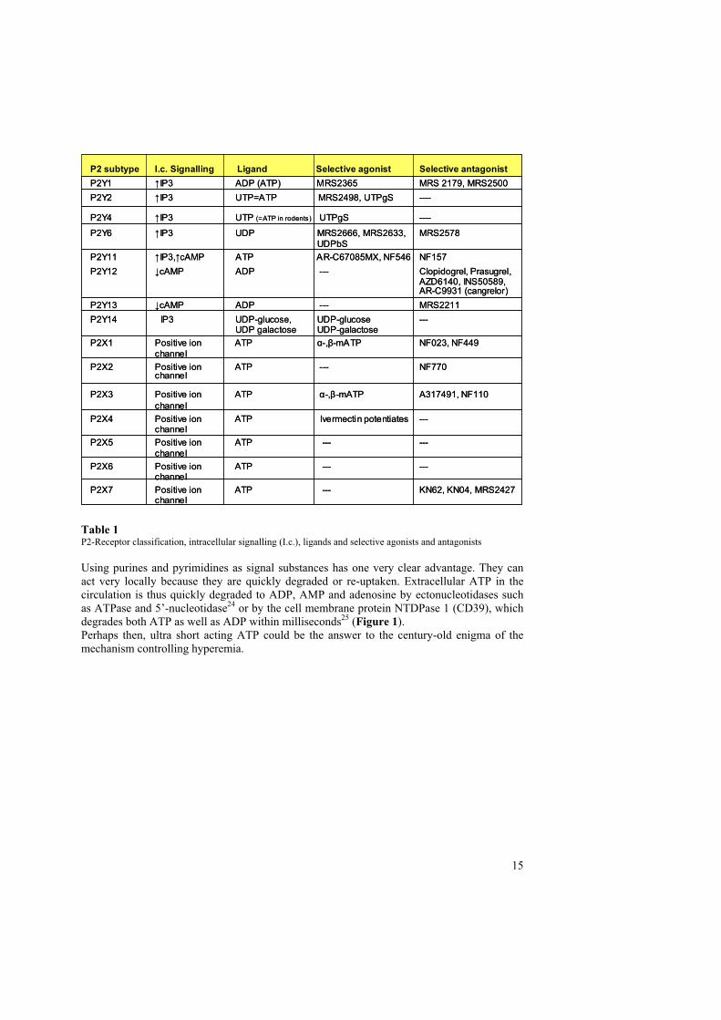
P2 subtype I.c. Signalling Ligand Selective agonist Selective antagonist
P2Y1 IP3 ADP (ATP) MRS2365 MRS 2179, MRS2500
P2Y2 IP3 UTP=ATP MRS2498, UTPgS ----
P2Y4 IP3 UTP (=ATP in rodents) UTPgS ----
P2Y6 IP3 UDP MRS2666, MRS2633, MRS2578
UDPbS
P2Y11 IP3, cAMP ATP AR-C67085MX, NF546 NF157
P2Y12 cAMP ADP --- Clopidogrel, Prasugrel,AZD6140, INS50589,AR-C9931 (cangrelor)
P2Y13 cAMP ADP --- MRS2211
P2Y14 IP3 UDP-glucose, UDP-glucose ---UDP galactose UDP-galactose
P2X1 Positive ion ATP -, -mATP NF023, NF449
channel
P2X2 Positive ion ATP --- NF770channel
P2X3 Positive ion ATP -, -mATP A317491, NF110
channel
P2X4 Positive ion ATP Ivermectin potentiates ---channel
P2X5 Positive ion ATP --- ---
channel
P2X6 Positive ion ATP --- ---channel
P2X7 Positive ion ATP --- KN62, KN04, MRS2427channel
able 1assification, intracellular signalling (I.c.), ligands and selective agonists and antagonists
sing purines and pyrimidines as signal substances has one very clear advantage. They can
tury-old enigma of the
TP2-Receptor cl
Uact very locally because they are quickly degraded or re-uptaken. Extracellular ATP in thecirculation is thus quickly degraded to ADP, AMP and adenosine by ectonucleotidases such as ATPase and 5’-nucleotidase24 or by the cell membrane protein NTDPase 1 (CD39), which degrades both ATP as well as ADP within milliseconds25 (Figure 1).Perhaps then, ultra short acting ATP could be the answer to the cenmechanism controlling hyperemia.
15
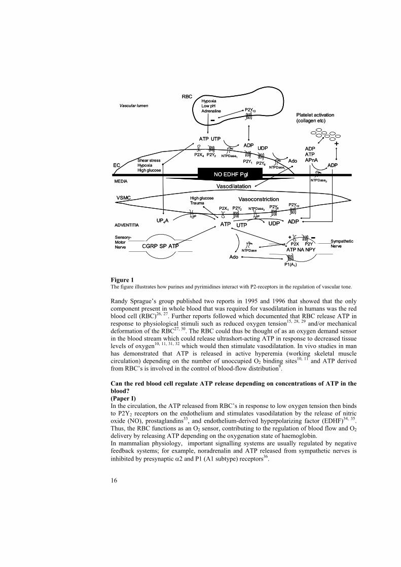
Figure 1 The figure illustrates how purines and pyrimidines interact with P2-receptors in the regulation of vascular tone.
Randy Sprague’s group published two reports in 1995 and 1996 that showed that the only component present in whole blood that was required for vasodilatation in humans was the red blood cell (RBC)26, 27. Further reports followed which documented that RBC release ATP in response to physiological stimuli such as reduced oxygen tension15, 28, 29 and/or mechanical deformation of the RBC27, 30. The RBC could thus be thought of as an oxygen demand sensor in the blood stream which could release ultrashort-acting ATP in response to decreased tissue levels of oxygen10, 11, 31, 32 which would then stimulate vasodilatation. In vivo studies in manhas demonstrated that ATP is released in active hyperemia (working skeletal musclecirculation) depending on the number of unoccupied O2 binding sites10, 11 and ATP derived from RBC’s is involved in the control of blood-flow distribution9.
Can the red blood cell regulate ATP release depending on concentrations of ATP in the blood?(Paper I) In the circulation, the ATP released from RBC’s in response to low oxygen tension then binds to P2Y2 receptors on the endothelium and stimulates vasodilatation by the release of nitric oxide (NO), prostaglandins33, and endothelium-derived hyperpolarizing factor (EDHF)34, 35.Thus, the RBC functions as an O2 sensor, contributing to the regulation of blood flow and O2
delivery by releasing ATP depending on the oxygenation state of haemoglobin.In mammalian physiology, important signalling systems are usually regulated by negative feedback systems; for example, noradrenalin and ATP released from sympathetic nerves is inhibited by presynaptic 2 and P1 (A1 subtype) receptors36.
16
EC
VSMC
Vascular lumen
MEDIA
Platelet activation(collagen etc)
Sensory-
Motor
Nerve
ATP
ATP
ADPATPAPnA
Hypoxia
Low pH
Adrenaline
High glucose
Trauma
Shear stress
Hypoxia
High glucose
Sympathetic
Nerve
P2Y2P2X4
P2Y6P2Y1
UP4AUTP
UTP
UDPADP
NTPDase1
NTPDase1
Ado
NTPDase2
ADP
P2Y13
-
P2Y2P2X1 NTPDase2
UDP ADP
P2Y12P2Y6
+
NTPDase
Ado
Vasoconstriction
Vasodilatation
-
P1(A1)
RBC
ADVENTITIA
C RP SP P2X P2Y
ATP NA NPY
+
EC
VSMC
Vascular lumen
MEDIA
Platelet activation(collagen etc)
Sensory-
Motor
Nerve
ATP
ATP
ADPATPAPnA
Hypoxia
Low pH
Adrenaline
High glucose
Trauma
Shear stress
Hypoxia
High glucose
Sympathetic
Nerve
P2Y2P2X4
P2Y6P2Y1
UP4AUTP
UTP
UDPADP
NTPDase1
NTPDase1
Ado
NTPDase2
ADP
P2Y13
-
P2Y2P2X1 NTPDase2
UDP ADP
P2Y12P2Y6
+
NTPDase
Ado
Vasoconstriction
Vasodilatation
-
P1(A1)
RBC
ADVENTITIA
RP SP P2X P2Y
ATP NA NPY
+
PPATATGCG
NO EDHF PglNO EDHF Pgl

We hypothesized that ATP release from RBC’s could perhaps be regulated by a P2 receptor–mediated negative feedback pathway which is the topic of the first manuscript (I) of this dissertation.
What is the role for the selective ADP/ATP-dependent P2Y1 receptors in hyperemia(Paper II) Although active hyperemia and reactive hyperemia share many similarities it was not clear if reactive hyperemia is also dependent of ATP or its degradation-product, ADP, for the increase in blood flow. During a coronary artery occlusion, the area of the heart supplied by the artery is deprived of its blood-supply. Upon reperfusion there is a dramatic rise in coronary blood flow, far above the baseline flow prior to the occlusion 37. The mechanism forthis enormous increase in flow during reactive hyperemia is somewhat of an enigma because the incurred oxygen debt does not in itself justify the flow increase 38. Several factors have been implicated, and the mechanism is now thought to be multifactorial in origin. Several substances (adrenalin, ADP/ATP, substance P, bradykinin and to some extent also adenosine) activate receptors on the endothelium of the coronary artery, stimulating release of nitrous oxide (NO), prostaglandins and endothelium-derived hyperpolarizing factor (EDHF), that in turn cause relaxation of the underlying smooth muscle cells (SMC) 33-35, 38, 39. Adenosine stimulates SMC relaxation directly by acting on specific receptors (mainly A2A) on the SMC33. K+
ATP channels are regulated by the cellular metabolic state and when they are activated the SMC is hyperpolarized, which causes relaxation. Thus, the K+
ATP channels could provide a link between intracellular ischemia and vaso-relaxation. Indeed, the K+
ATP channel inhibitor glibenclamide has been shown to inhibit a part of the reactive hyperemia 40, 41. The effect of adenosine, prostaglandins, and NO during reactive hyperemia in coronary vessels has been studied with use of specific receptor blockers (NOS-inhibitors, adenosine receptorantagonists, and cyclo-oxygenase inhibitors), and it has been found that there still exists an unaccountable rise in blood flow during reperfusion 41-45.Rongen et al. have proposed a role for P2 receptors in reactive hyperemia, but noexperimental evidence has yet been published probably because of the previous lack of specific antagonists 46. In previous studies, it has been demonstrated that adenosine and evenmore potently ATP administered through intracoronary injection and infusion can cause a pharmacologic hyperemia nearly as prominent as reactive hyperemia 46-48.The extracellular purine nucleotides ATP and ADP, which regulate vascular tone and blood pressure by stimulating P2 receptors, are released in the heart during ischemia from cardiac myocytes, endothelial cells, red blood cells, platelets and sympathetic nerves 33, 49, and concentrations of ATP and ADP have also been shown to increase during coronary reactive hyperemia in the venous outflow in the Coronary Sinus50. P2Y1 receptors are found in abundance on the endothelial cells of the coronary vessel wall and can promote hyperemia in response to selective P2Y1 agonists 51. Research has shown that P2Y1 receptors promotesmooth muscle relaxation through both NO and EDHF 33-35, 39. Recently, a selective P2Y1
receptor inhibitor, MRS2179, has become available 35, 39, 52, facilitating further exploration ofthese effects. We therefore hypothesized that the selective P2Y1 antagonist MRS2179 could reduced postischemic reactive hyperemia in the coronary circulation. The results are covered in the second manuscript (II) of this dissertation.
t-PA-release during ischemia(Paper III) Tissue-type plasminogen activator (t-PA) is an enzyme of great importance in maintaining the endothelial wall of blood vessels free of thrombi formation. t-PA itself is synthesized and stored both in endothelial cells as well as vascular neurons53, 54. Release of t-PA is caused by
17

several substances ( 2 receptor activators, Platelet activating factor, Isoproterenol, Metacholine, Bradykinin, and extracellular nucleotides (such as ATP, ADP and UTP) as well as by cardiac sympathetic nerve stimulation and local ischemia55-62. The actual mechanism by which the above mentioned factors cause the release of t-PA is still unclear but it has been postulated that the release may be an EDHF mediated phenomenon63.ADP activates P2Y1 receptors on the endothelium39 and on platelets64 which causes smoothmuscle cell relaxation and activation of platelets in arteries. Endothelial P2Y1-receptors, whenactivated by ADP, mediate SMC relaxation through both NO and EDHF34, 35. We thereforepostulated that by blocking the P2Y1 receptor, and thus EDHF, we would find a decreased release of t-PA during and immediately following cardiac ischemia, if t-PA release is indeed mediated through EDHF and thus dependent upon the P2Y1 receptor.
Myocardial infarctionThe acute coronary syndrome and its pathophysiologyThe vast majority of acute coronary syndromes involve the rupture of a vulnerable plaque in a coronary artery65, 66 and the subsequent formation of a thrombus at the site which maypartially or fully occlude the artery67, 68. If the rupture is small it may heal by itself with fewor no symptoms69. A larger rupture and / or a more aggressive formation of thrombus mayhowever cause intermittent symptoms giving rise to a condition known as unstable angina. In such scenario it is believed that the thrombus-formation is in a dynamic state and that several factors including interactions between the endothelium, platelets, coagulation factors and the fibrinolytic system may increase or decrease the size of the thrombus thus causingintermittent symptom in the patient. Symptoms may also be caused by rheological and hemodynamic factors such as seen in patients with anaemia or those performing exercises. The condition of unstable angina may progress to a very small injury of the heart which is then commonly called Non-ST Elevation Myocardial Infarct (NSTEMI).Unstable angina may also progress to the complete occlusion of the coronary artery and then give rise to an acute myocardial infarction which is also called ST Elevation Myocardial Infarct (STEMI). A STEMI may also be the first symptom of a ruptured plaque in a coronary artery without any precursor symptoms.
Acute myocardial infarction and evolution of treatment In acute myocardial infarction when there is a complete occlusion of the coronary artery(STEMI) supplying a region of the heart muscle, irreversible cellular damage starts to occurafter approximately 20 minutes through a number of well known mechanisms 70-72. It was not until after the results of the pivotal study ISIS 2 that reperfusion became a therapeutic optionin the treatment of acute myocardial infarction, albeit by pharmacological thrombolysis in combination with aspirin1. Mortality was reduced by 40% and reperfusion by thrombolysisbecame the treatment choice for acute myocardial infarction within 12 hour of symptoms1, 73-
78.During the 1990’s interventional cardiologists became proponents of treating patients with acute myocardial infarction by means of mechanical reperfusion with coronary angioplasty balloons (Primary PCI). Several trials were conducted comparing thrombolysis vs. PCI in the setting of acute myocardial infarction which indicated an advantage in using Primary PCI, at least in patients with more than 2 hours of symptoms6, 79, 80. Although meta-analysis of data from 23 randomized trials (n=7739) indicated a significant 22% reduction in short term mortality among PCI-treated patients80, the one-year mortality data from 26 205 patients in the Swedish RIKS-HIA registry indicated a 50% reduction in mortality for patients treatedwith PCI compared with patients treated with thrombolysis 6.
18

Reperfusion injury The idea of salvaging myocardial tissue in myocardial infarction is a fairly recent concept as stated by Eugene Braunwald in his 1974 editorial in The New England Journal of Medicine- “Reduction of myocardial infarct size”81.Reperfusion injury is hypothesized to be the additional myocardial injury sustained by theheart during the process of reperfusion of an infarct-related artery. Whether such an entityexists or not is still a matter of debate82-86. What is certain is however that in patients withongoing STEMI, the earlier reperfusion takes place the more myocardial tissue is saved87 and mechanical reperfusion seems to save more myocardial tissue than reperfusion through thrombolysis88.Hearse and Bolli characterized four mechanism of reperfusion injury; arrhythmias, stunning,lethal reperfusion injury (cell death) and accelerated necrosis89. Other proposed mechanismsof reperfusion injury include calcium overload, complement activation, mitochondrial injury, effects of oxygen-derived free radical generation, increased osmotic stress, endothelial dysfunction with reduced flow and neutrophil activation85, 89-92. Of importance may also be the swelling of myocytes and tissue oedema which may occur as early as after 30 sec following reperfusion93. Histological studies have shown reperfusion injury to be associatedwith tissue oedema, microvascular / endothelial injury, myocytes damage and cell necrosis94.Research on reducing reperfusion injury has focused on preconditioning (repeated short periods of occlusions prior to the myocardial infarct)95-98, postconditioning (repeated shortcycles of reocclusion following the opening of an infarct-related vessel)99-102, administrationof pharmacological agents103 and on hypothermia104-115. Although animal research have indicated a positive effect of all above mentioned treatment strategies, none of the strategiestested in humans (pharmacological treatment, postconditioning or hypothermia) have proved to clearly reduce infarct size in a large randomized trial.
Hypothermia during myocardial ischemia(Paper IV and V) Therapeutic mild hypothermia has experimentally in large animals been shown to limitmyocardial infarct size if applied prior to reperfusion104, 107, 113, but not following reperfusion113. It has also been shown that the earlier hypothermia is applied to an ischemicmyocardium, the more tissue stands to be salvaged114. Animal research also indicate that attaining a temperature less than 35°C prior to reperfusion is needed to see a positive effect of hypothermia107. With the invention of endovascular systems to rapidly cool a large body masswithout need for topical cooling Dae et al. successfully performed the first safety trial in a pig model using the SetPoint System (Radiant Medical, Inc)116. Dae et al. then developed the first large animal infarct model employing endovascular cooling prior to reperfusion in which an absolute reduction of 36% in final infarct size was seen104. The importance of the latterexperiment by Dae et al. is that it was performed animals with a weight similar to humans and hypothermia was initiated well after onset of ischemia simulating a real life situation.In light of the positive results from prior animal experiments, several human trials employing hypothermia through endovascular cooling as adjunct treatment to primary PCI in the setting of myocardial infarction were performed. Unfortunately the results of these trials have not been able to substantiate an effect of hypothermic treatment. However, post hoc analysis ofthe patients in the two only large randomized trials (COOL-MI and ICE-IT) who reached atemperature of <35°C before reperfusion showed a reduction in infarct size suggesting a benefit of induction of hypothermia before reperfusion. It is also important to note that only a minority of the patients randomized to hypothermia in COOL-MI and ICE-IT actually reached the target temperature of < 35°C prior to reperfusion of the infarct-related artery. It is unclear why so few patients randomized to cooling did not actually reach target temperature
19

prior to reperfusion, but it is our belief that the angiography in these patients was generally completed far before the patients had reached their target temperature due to the slow coolingprocess, and that a delay to the start of the PCI and reperfusion in order to reach targettemperature was deemed unethical. Due to the large body mass of humans in the clinicalsetting it is difficult to achieve adequate hypothermia without delaying reperfusion therapy. With external cooling or endovascular cooling alone it takes 30 min to 1 h for the patients to reach target temperature104, 106, 111, 112. The mechanism(s) by which mild hypothermia exerts its effect is still unknown, although it is a common belief that the decrease in metabolicdemand in the hypothermic myocardium is one explanation for the reduced infarction size. Thus, there is a need to better understand the protective mechanisms of hypothermia, in order to design future human trials in a better way.
Hypothermia in reactive hyperemia
Since reactive hyperemia is an occurrence which immediately follows reperfusion, and thus potentially could be involved in the development of a reperfusion injury, we wanted to examine if reactive hyperemia could be attenuated by mild hypothermia. Myocardial tissueoedema has been documented to occur as rapidly as after 30 s following reperfusion in a 15minute ischemic model93 and is thus in the same time frame when reactive hyperemiareaches its peak flow within 5 minutes of reperfusion97. There is also the appearance of an excess of O2-derived free radicals during the first minute of reperfusion with a peak at between 4 and 7 minute post reperfusion117, and during this time frame a generalizedmitochondrial and cell swelling can also be visualized on electronic microscopy93.We hypothesized that the large blood flow seen during coronary reactive hyperemia could be attenuated by mild systemic hypothermia and that this may be a mechanism by which hypothermia could reduce reperfusion injury.
Hypothermia during acute myocardial infarct
Although a number of animal experiments and several human trials have been performed inwhich hypothermia has been used to attenuate myocardial infarct size, there is still no evidence on when a crucial cell saving effect of therapeutic hypothermia occurs, or for how long hypothermic treatment needs to be maintained. Animal studies indicate that a positiveeffect of hypothermia is only seen if cooling is initiated before reperfusion104, 107. The effect of this may be due to two things. First of all initiation of hypothermia may inhibit further infarct evolution in itself. Secondly, induction of hypothermia prior to reperfusion may reduce the postulated reperfusion injury thought to occur in conjunction with the opening of an infarct related vessel which may also be the reason why neither of the two large randomizedhuman cooling trials showed a positive effect of hypothermia in the setting of myocardialinfarction. We therefore wanted to test if there was a difference between rapidly induced hypothermia shortly before reperfusion and rapidly induced hypothermia in conjunction with reperfusion. Data from such an animal experiment could help establish if treatment withhypothermia has its main benefit in the short period of reactive hyperemia during reperfusion,and could also give insights to why the trials COOL-MI and ICE-IT failed to meet their endpoints.
20

6 Hypotheses of the thesis
I. The P2Y13 receptor on the red blood cell functions as a negativefeedback-loop for ATP release from the red blood cell.
II. The endothelial P2Y1 receptor mediates coronary reactive hyperemia.
III. t-PA release can be mediated by P2Y1 receptors.
IV. Hypothermia can reduce coronary reactive hyperemia.
V. Rapidly induced hypothermia shortly before reperfusion reduces myocardial infarct size compared to rapidly induced hypothermia shortly after reperfusion.
21

22

7 Materials and methods The closed chest pig model In our research we needed to develop an animal model of sufficient size to accommodatecatheters and angioplasty balloons for the catheterization of the heart, and also to allow for frequent blood samples. In the last manuscript we also needed to perform Magnetic Resonance Imaging (MRI) and gated single photon emission computed tomography (SPECT). We also wanted to use a “closed chest model” in order to physiologically simulate “real” conditions. Cardiovascular research involving coronary arteries in large animal modelsfrequently use an “open chest model”. An open chest model means that a sternotomy or a thoracotomy is performed in order to have access to the heart from the outside. One can thenuse the heart for various experiments such as permanently or temporarily ligating coronary arteries. The advantage of using an open chest approach is that most materials used are reusable and the basic surgical technique is fairly straight forward. The closed chest modeloffers most of the same possibilities when research is focused on coronary arteries but in thismodel access to the arteries is gained through catheters commonly engaging the left coronaryartery. This requires use of a great deal of expensive single use (disposable) materials whichincreases costs as well as the need to have access to a catheterization laboratory and skills in interventional cardiology. When using large animals in research involving coronary arteries,the pig or the dog is commonly selected. We choose the pig model for several reasons, first ofall the anatomy and the distribution of the coronary vascular system in pigs is much moresimilar to humans than to dogs118-122. The pig also has a, to humans, comparativelycorresponding heart-to-body ratio of about 0.005 for both119, 123. Compared to humans, the pig heart has one major difference. This difference is in regards to the venous system. In humansthe great cardiac vein (the continuation of the anterior ventricular vein) with returning blood from the heart-muscle confluences with the middle and small cardiac veins to form the Coronary Sinus which returns the venous blood to the right atrium with the ostium located at the base of the right atrium. The pigs have a completely different anatomy in that the leftAzygos Vein enters the right atrium at the base and the Coronary Sinus confluences with the Azygos Vein a few centimetres before the ostium to the right atrium119. This makescatheterization of the Coronary Sinus in pigs much more challenging than in humans.The pig was also chosen as model because it is readily available in large quantities and isdeemed ethically to be more suitable for research than primates or dogs.
Animal preparations(papers I-V)
AnaesthesiaHealthy, domestic male and female pigs were fasted overnight with free access to water and were premedicated with azaperone (Stresnil Vet., Leo; Helsingborg, Sweden), 2 mg/kgintramuscularly 30 min before the procedure. After induction of anesthesia with thiopental 5-25 mg/kg (Pentothal, Abbott, Stockholm, Sweden), the animals were orally intubated with cuffed endotracheal tubes. A slow infusion of 1.25 l/ml Fentanyl (Fentanyl, Pharmalink AB,Stockholm, Sweden) in Ringer´s acetate solution was started at a rate of 1.5 ml/min and adjusted as needed. Mechanical ventilation was then established with a Siemens-Elema 300B ventilator in the volume-controlled mode. Initial settings were: respiratory rate of 15/min,tidal volume of 10 ml/kg, and positive end-expiratory pressure of 5 cm H2O. Min volume was subsequently adjusted in order to obtain normocapnia (35-40 mm Hg). The animals were ventilated with a mixture of dinitrous oxide (70%) and oxygen (30%). Anesthesia was complemented with small intermittent doses of 5 mg meprobamat (Mebumal, DAK, Copenhagen, Denmark) and thiopental (Pentothal, Abbott, Stockholm, Sweden), if needed.
23

Catheterization of the Coronary Sinus
(Papers I and III)
A 14 F or 12F introducer sheath (Boston Scientific Scimed, Maple Grove, MN, USA) was inserted into the surgically exposed right jugular vein through which a short 10 F Coronary Sinus catheter of our own design was introduced into the ostium of the Azygos vein in the right atrium. Then a 6F coronary MPA catheter (Onset, Cordis Co. Miami, FL. USA) waspassed through the 10 F catheter and the Azygos Vein and into the Coronary Sinus. An angiogram was obtained using 5 -10 ml of the contrast medium Omnipaque™ 300 mg I-/ml(Nycomed, Oslo, Norway) to ensure correct positioning of the catheter.
General Catheterizations
(Paper I – III)
A 6 F introducer sheath (Boston Scientific Scimed, Maple Grove, MN, USA) was inserted into the surgically exposed left femoral artery. The side port of the introducer was connected to a pressure transducer and balanced to atmospheric pressure with zero reference at the mid-axillary level for continuously monitoring of the arterial pressure. A three-lead ECG wasdisplayed on the same monitor as the pressure curve (78342 A, Hewlett and Packard GMBH. Boeblingen, Germany).
A 6 F introducer sheath (Boston Scientific Scimed, Maple Grove, MN, USA) was inserted into the surgically exposed left carotid artery and a 6F JL 3.5 or 4.0 Wiseguide™ (Boston Scientific Scimed, Maple Grove, MN, USA) was inserted into the left main coronary arteryand 10,000 IU of Heparin was administered. An angiogram was obtained using 8 -10 ml of the contrast medium Omnipaque™ 300 mg I-/ml (Nycomed, Oslo, Norway) to ensure correct positioning of the catheter. The catheter was used to place a 0.014-inch, 12 MHz pulsedDoppler flow velocity transducer (Jometrics Flowire, Jomed NV) into the mid-portion of the left anterior descending artery (LAD), (not in paper I) and a 0.014-inch PT choice™ guidewire (Boston Scientific Scimed, Maple Grove, MN, USA) into the distal portion of the LAD. A 3.0 x 20 mm over the wire Maverick™ angioplasty balloon (Boston Scientific Scimed, Maple Grove, MN, USA) was then positioned in the mid portion of the LAD but proximal to the flow velocity transducer followed by the withdrawal of the PT choice guidewire (Figure 2). Continuous coronary velocity flow profiles were displayed and recorded using the Doppler flow wire connected to a FloMap monitor (Cardiometrics,Mountain View, CA). Flow was measured in units of average peak velocity (APV) incentimeters per second.
Left Pulmonary Artery
LAD
Circumflex Artery
Balloon catheter
Doppler flow wire
Right Coronary
Artery
Superior Vena
Cava
Coronary catheter
Left Pulmonary Artery
LAD
Circumflex Artery
Balloon catheter
Doppler flow wire
Right Coronary
Artery
Superior Vena
Cava
Coronary catheter Figure 2
Schematic illustration of the model usedin the closed chest pig model. The leftcoronary artery is engaged with a coronarycatheter, through which an over-the-wireangioplasty balloon is placed in the LAD,distal to the first diagonal branch. ADoppler flow wire runs adjacent to theangioplasty balloon with the tip placedin the distal LAD.
Left Pulmonary Artery
LAD
Circumflex Artery
Balloon catheter
Doppler flow wire
Right Coronary
Artery
Superior Vena
Cava
Coronary catheter
Left Pulmonary Artery
LAD
Circumflex Artery
Balloon catheter
Doppler flow wire
Right Coronary
Artery
Superior Vena
Cava
Coronary catheter Figure 2
Schematic illustration of the model usedin the closed chest pig model. The leftcoronary artery is engaged with a coronarycatheter, through which an over-the-wireangioplasty balloon is placed in the LAD,distal to the first diagonal branch. ADoppler flow wire runs adjacent to theangioplasty balloon with the tip placedin the distal LAD.
24

General Catheterizations
(paper IV)
A 6 F introducer sheath (Boston Scientific Scimed, Maple Grove, MN, USA) was inserted into the surgically exposed left femoral artery as described previously.
A 12 F introducer sheath (Boston Scientific Scimed, Maple Grove, MN, USA) was inserted into the surgically exposed left Femoral Vein. A 10.7 F cooling (temperature control) catheter (Celsius Control™, Innercool Therapeutic, San Diego, CA, USA) was inserted through the sheath and positioned in the Inferior Vena Cava (Figure 3). Body temperature was measuredwith a temperature probe (TYCO Healthcare Norden AB, Solna, Sweden) placed in the distal part of the esophagus. The catheter and the temperature probe were then connected to the console and the system was set to maintain a temperature of 37.0° C. The normal temperature of a pig is located somewhere between 37.0° and 38.5° C according to our laboratoryveterinarian and the temperature of the pigs was thus set at the lower end of normal.
Figure 3
Illustration of the Celsius Control™ cooling catheter used
A 6 F introducer sheath (Boston Scientific Scimed, Maple Grove, MN, USA) was inserted into the surgically exposed left carotid artery and the left coronary artery was catheterized as described previously, and an angiogram obtained.
The catheter was used to place a Doppler flow velocity transducer as earlier described into the distal portion of the LAD together with a PT choice™ guidewire into the distal portion ofthe LAD. A 3.0 or 3.5 x 20 mm over the wire Maverick™ angioplasty balloon was then positioned in the mid portion of the LAD, proximal to the flow velocity transducer but distalto the first diagonal branch, followed by the withdrawal of the PT choice guidewire. Continuous coronary velocity flow profiles were displayed and recorded as described earlier.
General Catheterizations(paper V) The endovascular cooling catheter was inserted, as earlier explained above, into the Vena Cava. Body temperature was measured in the distal part of the esophagus as previously described. The catheter and the temperature probe were then connected to the Celsius Control
25

and the system was set to maintain a normal pig body temperature of 38.0° C. A 6 Fintroducer sheath (Boston Scientific Scimed, Maple Grove, MN, USA) was then inserted into the surgically exposed left carotid artery upon which a 6 F JL4 Wiseguide™ was inserted into the left main coronary artery. The catheter was used to place a 0.014-inch PT Choice™ guide wire into the distal portion of the LAD and a 3.0-3.5 x 12 mm Maverick monorail™angioplasty balloon was then positioned in the mid portion of the LAD, immediately distal to the first diagonal branch.
Protocol
Infusion of saline solution in the LAD
The lumen of the angioplasty balloon was connected to an infusion pump, Asena CC (Alavis Medical, Bristol, England). The infusion pump was initially used to infuse Ringer´s acetatesolution and NaCl (0.9%) at rates of 0.5, 1, 2, 4 and 6 ml/min in the LAD through the inner lumen of the angioplasty balloon catheter. At an infusion rate of 2 ml/min or less, there were no effects on blood flow in the LAD.
In vivo pig model with microdialysis for determining ATP levels in blood (Paper I) Through a catheter, the microdialysis probe (CMA70) was inserted and the microdialysis tip was placed in the Coronary Sinus. A 6F introducer sheath was inserted into the surgicallyexposed left carotid artery. Through the left carotid artery, a catheter was placed in the left anterior descending artery (LAD). Finally, 2 ml of 2-MeSADP (10 _mol/L) was infused in theLAD for 1 minute. Samples were collected by microdialysis (5 µL/min) from the Coronary Sinus every 5 minutes, and ATP measured. ATP was measured by ATP Bioluminescent assaykit (Sigma) according to the supplier’s instructions.
Infusion of 2-MeSADP and MRS2179 in the LAD
(Paper II)
In five pigs, 2-MeSADP (10-5 M) at 1 ml/min was infused and FloMap measurements were performed. 2-MeSADP (10-5 M) was then infused with MRS2179 (10-3 M) at a rate of 1 ml/min. To test the effect of MRS2179 on ATP, 5 ml of ATP (10-4 M) was delivered into theLAD (n = 9). Following the 30-min washout period, 5 ml of ATP (10-4 M) together with 5 mlof MRS 2179 (10-3 M) was delivered into the LAD. The order of the ATP and the combination of ATP + MRS2179 infusions was altered randomly. To test the effect ofMRS2179 on P2Y2/4 receptors, 5 ml of UTP (10-4 M) was delivered into the LAD (n = 3). Following the 30-min washout period, 5 ml of UTP (10-4 M) together with 5 ml of MRS2179(10-3 M) was delivered into the LAD. The 2-MeSADP infusions were delivered for 2 minwhile the ATP and UTP infusions were delivered for only 1 min.
Inhibition of P2Y1 receptors with MRS2179 during ischemia and reperfusion of the LAD
(Paper II)
In five pigs 2-MeSADP (10-5M ) at 1 ml/min was infused and FloMap measurements were performed. 2-MeSADP (10-5M) was then infused with MRS2179 (10-3M) at a rate of 1ml/min. An occlusion of the LAD, distal to the first diagonal branch, was achieved with inflation of the angioplasty balloon for a period of ten min. During the first and tenth min of
26

coronary ischemia, 2.5 ml of MRS2179 (10-3M) was delivered distal to the occlusion in theLAD in 8 pigs. 10 pigs were used as controls, in which the same volumes of NaCl 0.9% were infused. Reactive hyperemia measured in Average Peak Velocity (APV) in cm/s, was performed only once in each pig. Blood gas analysis was performed at baseline and at one andten min following reperfusion in the 18 pigs treated with balloon-inflation.
Measurement of t-PA during simultaneous inhibition of P2Y1 receptors with MRS2179 during ischemia and reperfusion of the LAD
(Paper III)
Infusions of MRS2179 and 2-MeSADP were performed in an identical fashion as earlier described in measurement of reactive hyperemia, see above.
To test the effect of MRS2179 on t-PA release following reactive hyperemia, an occlusion ofthe LAD, distal to the first diagonal branch, was achieved with inflation of the angioplastyballoon for a period of ten min. During the first and tenth min of coronary ischemia, 2.5 ml of MRS2179 (10-3M) was delivered distal to the occlusion in the LAD in 8 pigs. 10 pigs were used as controls, in which the same volumes of NaCl 0.9% were infused. t-PA samples were collected in the Coronary Sinus and in a peripheral artery at baseline, during ischemia and at 1 and 5 minutes following reperfusion (balloon deflation). Reactive hyperemia t-PA measurements were only measured once in each pig.
t-PA measurements
Plasma concentrations of t-PA were determined by commercial ELISA kits (TintElize®t-PA,Biopool AB, Umeå, Sweden and COALIZA®PAI, Chromogenix, Haemochrom Diagnostica AB, Mölndal, Sweden). All samples from one experiment were assayed in duplicate on the same microtest plate. Intra-assay variation coefficients were 2.7 and 3.1% for respective assay. Plasma glucose, cholesterol, and triglycerides were analyzed by standard methods atthe Department of Clinical Chemistry at Sahlgrenska University Hospital.
Measurement of coronary reactive hyperemia during mild hypothermia
(Paper IV)
In all 16 pigs baseline registrations were performed. Regardless of initial temperature, all pigswere cooled or warmed (as needed) to a baseline temperature of 37 C, which was thenmaintained for 30 minutes. The pigs were then randomized to the hypothermia group or to the control group using a simple randomization by drawing folded notes out of a box which read “cool” or “warm”. The pigs randomized to hypothermia were then cooled with the endovascular cooling catheter to a temperature of 34.0 C, prior to balloon inflation, and temperature was then maintained until sacrifice. The pigs randomized to the control group were actively maintained at 37 C using the endovascular cooling catheter until sacrifice.
In all pigs, the LAD was then occluded distal to the first diagonal branch by inflation of the angioplasty balloon for a period of ten min. The coronary blood flow in the LAD was measured before, during and after occlusion of the LAD. During the reperfusion phase, coronary blood flow was measured at every 10 sec. Flow was measured in APV, in cm/sec.Occlusion of the LAD was performed only once in each pig.
Blood gases were collected through the MPA catheter in the Coronary Sinus and the FemoralArtery sheath during baseline, early ischemia (one minute after balloon inflation in the LAD), early reperfusion (one minute following balloon deflation in the LAD) and late reperfusion (ten minutes following balloon deflation in the LAD).
27

At baseline, measurements of blood pressure, pulse and APV were performed. Blood pressureand pulse were measured continuously.
Rapid induction of hypothermia in pigs with myocardial infarction
(Paper V) Ischemia protocol
After a stable core body temperature of 38.0° C was achieved, ischemia was induced by inflation of the angioplasty balloon for 40 min. An angiogram was performed after inflation of the balloon and before deflation of the balloon in order to verify total occlusion of thecoronary vessel and correct balloon positioning. After deflation of the balloon a subsequentangiogram was performed to verify restoration of blood flow in the previously occluded artery.
Hypothermia protocol
The pigs were randomized to rapid hypothermia before reperfusion, (pre-reperfusion hypothermia, n=8) or immediately after reperfusion (post-reperfusion hypothermia, n=8). Anormothermic group (n=5) was also studied in order to provide comparison between different hypothermia protocols and normothermia. Hypothermia was induced by a rapid intravenous infusion of 1000 ml of 4° C cold saline into a central vein together with the Celsius ControlTM
endovascular cooling system after 25 min of ischemia or immediately after reperfusion when coronary blood flow was restored (Figure 4). Target temperature was 33° C and successfulcooling was defined as a temperature of 35o C. Hypothermia was then actively maintainedfor 30 min followed by passive rewarming with blankets.
Post-reperfusion hypothermia
Induction of
hypothermia
Pre-reperfusion hypothermia
Ischemia (40 min)
Normothermia (38OC)
Normothermia
Ischemia (40 min)
Normothermia (38OC) Hypothermia (33OC)
Ischemia (40 min)
Normothermia (38OC) Hypothermia (33OC)
Normothermia (38°C)
Induction of
hypothermia
Post-reperfusion hypothermia
Induction of
hypothermia
Pre-reperfusion hypothermia
Ischemia (40 min)
Normothermia (38OC)
Normothermia
Ischemia (40 min)
Normothermia (38OC) Hypothermia (33OC)
Ischemia (40 min)
Normothermia (38OC) Hypothermia (33OC)
Normothermia (38°C)
Induction of
hypothermia
Figure 4 Protocol for induction of hypothermia. In the pre-reperfusion group, hypothermia was started after 25 min ofischemia (15 min before reperfusion) and in the post-reperfusion group, hypothermia was started immediatelyafter reperfusion. The normothermic group was maintained at 38.0° C.
28

In vivo assessment of area at risk by SPECT
SPECT was used to assess the area at risk (AAR) as percent of left ventricular myocardium.Five hundred MBq of 99mTc-tetrofosmin was administered intravenously ten minutes before deflation of the angioplasty balloon. The anesthetized pigs were then imaged in a supine position with a dual head camera (ADAC Vertex, Milpitas, CA, USA) at 32 projections (40 s per projection) with a 64 X 64 matrix yielding a digital resolution of 5 X 5 X 5 mm. Iterative reconstruction using maximum likelihood-expectation maximization (MLEM) was performedwith a low-resolution Butterworth filter with a cut-off frequency set to 0.6 of Nyquist and order 5.0. No attenuation or scatter correction was applied. Finally short and long-axis imageswere reconstructed. Quantification of the size of AAR was performed automatically as theextent of the perfusion defect as determined by commercially available software (AutoQUANTTM 4.3.1 and a standard database; ADAC, Milpitas, CA, USA)124.
Infarct size and microvascular obstruction assessed by ex vivo MRI
Ex vivo imaging of the heart was undertaken using a 1.5 T Philips Intera CV MR scanner (Philips, Best, the Netherlands) according to a previous described protocol125. In brief, acommercially available gadolinium-based contrast agent (Magnevist, gadopentetate
dimeglumine, Gd-DTPA, Schering Nordisker AB, Järfälla, Sweden) was administeredintravenously (0.2 mmol/kg) both 60 and 15 minutes prior to removal of the heart. The heart was removed 4h 22 min ± 47 min after initiation of reperfusion. After removal, the heart was immediately rinsed in cold saline and the ventricles were filled with balloons containingdeuterated water. T1-weighted images (TR = 20ms, TE = 3.2ms, flip angle = 70° and 2 averages) with an isometric resolution of 0.5 mm covering the entire heart were then acquired using a head coil.The MR images were analyzed using freely available software (Segment 1.457, http://segment.heiberg.se)126. The endocardial and epicardial borders of the left ventricularmyocardium were manually delineated in short-axis ex vivo images. This defined the volumeof the left ventricular myocardium. The infarct size (IS) was first determined as the volume of infarcted myocardium (cm3). The infarct volume was calculated as the product of the slice thickness (cm) and the area of hyperenhanced pixels (cm2) with a signal intensity above the infarction threshold defined as > 8 SD above the mean intensity of non-affected remotemyocardium. Microvascular obstruction was defined as hypointense regions in the core of the infarction which had signal intensity less than the threshold for infarction. These regions weremanually included in the infarct volume. The volume of microvascular obstruction (cm3) was calculated as the difference between the infarct volume before and after manual inclusion of regions of microvascular obstruction. Furthermore, the size of microvascular obstruction was expressed as percent of the total infarct volume. Penultimately, the infarct size was expressedas percent of left ventricular myocardium.One animal in the normothermic group suffered from severe bradycardia which required manual open chest cardiac compression in order to secure the adequate circulation of the second injection of contrast media. The remote myocardium was somewhat increased in signal intensity and the threshold for infarction in this animal was therefore defined as pixels which were 3SD above the remote myocardium as determined by visual assessment. Finally,infarct size was expressed as a percentage of the area at risk (IS/AAR) in order to adjust for anydifference in area at risk between the groups.
Patchiness index
Infarct homogeneity was assessed by a patchiness index based on infarct surface area. The highresolution ex vivo MR images allowed quantification of the surface area of the infarct. Infarctsurface area (cm2) was automatically determined as the product of the slice thickness (cm) and
29

the distance along the pixel border between infarcted and non-infarcted pixels (cm) in each slice.For equally homogeneous infarcts, a larger infarct volume will yield a larger surface area. A patchiness index (cm-1) was therefore calculated as the infarct surface area (cm2) divided by the infarct volume (cm3). Thus, the patchiness index provided a method for estimating the homogeneity of the myocardial infarction adjusted for infarct size.
30

8 Reagents, Ethics and Statistics
ReagentsUnless otherwise stated, drugs were purchased from Sigma (USA).
EthicsThe Ethics Committee of Lund University approved the project.
StatisticsPaper I Data are expressed as mean ± SEM. n indicates the number of subjects tested. The Student ttest was used. Differences were considered significant at *P<0.05, **P<0.01, and ***P<0.001 (two-tailed test), compared with control if not indicated.
Paper II Calculations and statistics were performed using the GraphPad Prism 3.02 software. Values are presented as mean ± SEM. Statistical significance was accepted when P < 0.05 (two-tailed test). One-way analysis of variance (ANOVA) test followed by Dunnett’s multiplecomparison test was used.
Paper III Calculations and statistics were performed using the GraphPad Prism 3.02 software. Valuesare presented as mean ± SEM. Statistical significance was accepted when P < 0.05 (two-tailedtest). One-way analysis of variance (ANOVA) test followed by the Dunnett multiple comparison test was used.
Paper IV Calculations and statistics were performed using the GraphPad Prism, version 4.0 software. Values are presented as mean ± SEM. Statistical significance was accepted when P < 0.05 (two-tailed test). Two-way analysis of variance (ANOVA) test followed by Bonferroni post test was used.
Paper V
Calculations and statistics were performed using the GraphPad Prism 4.0 software. Values arepresented as mean ± SEM. Statistical significance was accepted when P < 0.05 (Mann-Whitney’s test).
31

32

9 Results The P2Y1 receptor mediates early coronary reactive hyperemia During infusion with isotonic crystalloid (Ringer’s acetate solution) and NaCl (9%) in theLAD there was a slight flow increase with infusion rates at or above 3 ml/min, but not at flow < 2 ml/min. In Figure 5, Ringer’s acetate solution was infused at 1 ml/min. When the ADP analogue 2-MeSADP (10-5 M) was infused at a rate of 1 ml/min, flow in the LAD increasedsignificantly (P< 0.05; Figure 5). However, the effects of 2-MeSADP (10-5 M) on blood flow in the LAD was fully inhibited when infused together with the P2Y1 receptor antagonist MRS2179 (10-3 M) at a rate of 1 ml/min, (n=5, P< 0.05) (Figure 5). Following a 30-minwashout-period, the dilatations to 2-MeSADP without MRS2179 could be repeated with similar results as the initial dilatation (data not shown). MRS2179 alone did not have any effect on basal coronary flow.
0 25 50 75 1000
100
200
300
2-MeSADP
2-MeSADP+MRS2179
*
*****
*
Ringer
Time (sec)
Co
ron
ary
flo
w (
% i
ncre
ase)
Figure 5
Ringer´s acetate solution infused intocoronary vessels of pigs, open circles,did not alter coronary flow from baseline.Infusion of 2-MeSADP increased flowby 300%, closed circles.The 2-MeSADP-mediated flow increasewas essentially aborted by simultaneousinfusion of MRS 2179, closed squares.Data are expressed as percentage ofbaseline flow (100%) and shown asmeans ± s.e.m, * p < 0.05, ** p < 0.01. (n=5
ATP delivered selectively in the LAD caused an increase of flow in the LAD by a factor of 5. When ATP was delivered together with MRS2179 there was significant reduction of flow in the LAD by approximately 50% (n = 9), demonstrating that a major portion of the ATP induced flow is mediated through its degradation-product ADP acting on P2Y1 receptors(Figure 6).
0 25 50 75 1000
100
200
300
400
500
600
ATP
ATP + MRS2179*
***
*
*
*
***
Time (sec)
Co
ron
ary
Flo
w (
% i
ncre
ase
)
Figure 6
Intracoronary infusion of ATP increased flow inpig coronary arteries by a factor of 5, closed circles.The ATP-mediated flow increase was then reducedby 50% during simultaneous intracoronaryadministration of MRS 2179, open circles.Data are expressed as percentage of baselineflow (100%) and shown as means ± s.e.m,* p < 0.05, ** p < 0.01. (n=9)
33

UTP delivered selectively in the LAD caused an increase of flow in the LAD by a factor of3.5. When UTP was delivered together with MRS2179 there was no difference in increased flow in the LAD, demonstrating the selectivity of MRS2179 (n = 3, Figure 7).
0 10 20 30 40 50 60 700
50
100
150
200
250
300
350
400
450
UTP -4
UTP + MRS2179
Time (sec)
Co
ron
ary
Flo
w I
ncre
ase (
%)
Figure 7 Intracoronary infusion of UTP increased flow, in pig coronary arteries, approximately by a factor of four, closedcircles. The UTP-mediated flow increase was not blocked by MRS2179, open circles. Data are expressed aspercentage of baseline flow (100%) and shown as means ± s.e.m, p=NS, (n=3).
Post-ischemic flow in the LAD increased nearly sevenfold in the 10 pigs treated with balloon inflation alone in the LAD (n = 10). In contrast, a significant 46% reduction of flow in the early phase (1–2.5 min following balloon deflation corresponding to the period of the fastest flow during post-ischemic hyperemia) was observed in the eight pigs receiving bolus doses of MRS 2179 (P < 0.05, Figure 8).
0.0 2.5 5.0 7.5 10.0 12.5 15.0 17.5 20.0
0
100
200
300
400
500
600
700
800
Control
MRS2179
* *
*
*
Ischemia
Time (min)
Co
ron
ary
Flo
w (
% i
ncre
ase)
Figure 8 The ensuing reactive hyperemia following a ten-minute coronary occlusion was measured as a nearly seven-foldincrease of flow (closed circles). Infusion of MRS2179 reduced the early post-ischemic flow increase by 46%.Data are expressed as percentage of baseline (100%) and shown as means ± s.e.m, * p < 0.05, (n=8-10 pigs).
34

During infusions of NaCl, 2-MeSADP and MRS2179, there were no significant differences in blood pressure or pulse rate between the groups at the analyzed time intervals as listed in paper II. There was no difference in basal coronary flow rates between the control and MRS 2179 groups (10.2 ± 4.9 and 11.3 ± 3.7 cm/s, mean ± S.D., P =NS). The flow rates returned to initial values at the end of the experiments. The analyzed blood-gas samples of the 18 pigs in the occlusion/reperfusion group showed no statistical difference between the pigs receiving MRS2179 and the group treated as controls (Table 2).
Baseline 1 min reperfusion 5 min reperfusion
Control MRS2179Control MRS2179Control MRS2179pH 7.47 0.057.49 0.037.45 0.077.48 0.037.46 0.067.47 0.05pCO2 5.4 0.8 5.3 0.5 5.6 1.0 5.3 0.4 5.3 0.7 5.3 0.6pO2 46.8 19.043.7 16.340.6 14.646.1 20.235.4 3.9 36.3 4.4Na+ 132 2 135 1 133 2 134 1 132 2 135 1K+ 3.5 0.2 3.6 0.3 3.4 0.2 3.7 0.3 3.4 0.3 3.6 0.1Hb 89 8 92 3 91 8 93 4 88 5 92 3O2 97.1 0.2 97.2 0.2 97.9 1.5 97.2 0.2 97.1 0.1 97.2 0.2HCO3 29.0 1.9 30.0 0.3 28.6 1.6 29.4 0.3 28.2 1.3 29.2 0.7BaseExcess5.6 0.8 6.1 0.2 4.9 0.8 5.6 0.3 5.1 0.8 5.3 0.4
Table 2 Arterial blood-gas analysis of pigs with occlusion and reperfusion of a coronary vessel treated with MRS 2179 (n=8)or used as controls (n=10). Samples were collected at baseline and at one and five minutes following reperfusion.There were no statistical differences between pigs treated with MRS 2179 or pigs used as controls. Data are expressed as mean ± s.e.m, p =NS.
The P2Y13 receptor on erythrocytes regulates ATP release Regulation of Plasma ATP Concentrations in an In Vivo Pig Model
After intracoronary injection of 2-MeSADP, samples were obtained from the venous effluent of the heart in the Coronary Sinus with a microdialysis probe. Analysis showed decreased concentrations of plasma ATP after intracoronary injection of 2-MeSADP (Figure 9).
CONTR
OL
mol/L
)
2-M
eSADP (
0
50
100
*
Pla
sm
a A
TP
(%
of
co
ntr
ol)
Figure 9
Effects of 2-MeSADP on plasma ATP concentrationsin an in vivo pig model. Bar graphs show plasma ATPconcentrations sampled from the venous outflow ofthe heart with a microdialysis probe in the coronarysinus 5-10 min after intracoronary injection of 2-MeSADP (10 mol/L, 2 ml). n=8.
35

t-PA release is mediated through P2Y1 receptorsInfusion of ADP analogue and P2Y1 antagonist in the non-ischemic heart
Intracoronary infusion of the stable ADP agonist 2-MeSADP (10-5M) at a rate of 1ml /mincaused a significant increase of the t-PA level in the Coronary Sinus compared to infusion of Ringer’s acetate (data not shown). The effects of 2-MeSADP (10-5M, n=9) on t-PA release was significantly inhibited when infused together with the P2Y1 receptor antagonist MRS2179 (10-3M, n=6) at a rate of 1 ml/min. t-PA increased to a maximum (at 1 min) of 1.81 0.32 ng/ml compared to 0.78 0.28 ng/ml in the presence of MRS2179, p<0.05, (Figure10a). t-PA was sampled selectively in coronary sinus. Following a 30 min washout period, the effects of 2-MeSADP on t-PA release could be repeated with similar results as during theinitial infusion of 2-MeSADP 10-5M (data not shown). MRS2179 alone did not have any effecton t-PA release (data not shown).
a b
t-PA release
0 1 2 3 4
0.0
0.5
1.0
1.5
2.0
2.52-MesADP+MRS2179
2-MeSADP§
*n.s.
§
§ = p<0.05 compared to baseline t-PA
* = p<0.05 compared to 2-MeSADP alone
Time (min)
t-P
A (
ng
/ml)
t-PA release
0 1 2 3 4
0.0
0.5
1.0
1.5
2.0
2.52-MesADP+MRS2179
2-MeSADP§
*n.s.
§
§ = p<0.05 compared to baseline t-PA
* = p<0.05 compared to 2-MeSADP alone
Time (min)
t-P
A (
ng
/ml)
t-PA*flow
0 1 2 3
0
1
2
3
4
5
6
72-MeSADP
2-MeSADP + MRS2179
**
§
§
§ = p<0.05 compared to baseline t-PA
* = p<0.05 compared to 2-MeSADP alone
Time (min)
t-P
A x
flo
w
t-PA*flow
0 1 2 3
0
1
2
3
4
5
6
72-MeSADP
2-MeSADP + MRS2179
**
§
§
§ = p<0.05 compared to baseline t-PA
* = p<0.05 compared to 2-MeSADP alone
Time (min)
t-P
A x
flo
w
t-PA release
0 1 2 3 4
0.0
0.5
1.0
1.5
2.0
2.52-MesADP+MRS2179
2-MeSADP§
*n.s.
§
§ = p<0.05 compared to baseline t-PA
* = p<0.05 compared to 2-MeSADP alone
Time (min)
t-P
A (
ng
/ml)
t-PA release
0 1 2 3 4
0.0
0.5
1.0
1.5
2.0
2.52-MesADP+MRS2179
2-MeSADP§
*n.s.
§
§ = p<0.05 compared to baseline t-PA
* = p<0.05 compared to 2-MeSADP alone
Time (min)
t-P
A (
ng
/ml)
t-PA*flow
0 1 2 3
0
1
2
3
4
5
6
72-MeSADP
2-MeSADP + MRS2179
**
§
§
§ = p<0.05 compared to baseline t-PA
* = p<0.05 compared to 2-MeSADP alone
Time (min)
t-P
A x
flo
w
t-PA*flow
0 1 2 3
0
1
2
3
4
5
6
72-MeSADP
2-MeSADP + MRS2179
**
§
§
§ = p<0.05 compared to baseline t-PA
* = p<0.05 compared to 2-MeSADP alone
Time (min)
t-P
A x
flo
w
Figure 10 (A) Illustrates that an ADP analogue release t-PA in the pig coronary circulation and that the effect is mediatedby the P2Y1 receptor: Release of t-PA due to continuous intracoronary infusion of 2-MeSADP (10-5M)(1ml/min) alone (filled circles, n=9), or 2-MeSADP (10-5M) jointly infused with the P2Y1 antagonist MRS2179(10-3M) at a rate of 1 ml/min (open circles, n=6). t-PA was sampled selectively in the Coronary Sinus. * = p<0.05 compared to baseline. § = p< 0.05 compared to 2-MeSADP alone.
(B) Illustrates an estimate of differences in total t-PA release: Continuous infusion of 2-MeSADP (10-5M) at 1ml/min increased flow in the LAD significantly as measured with the FloMap Doppler wire (data not shown). 2-MeSADP (10-5M) jointly infused with MRS2179 (10-3M) at a rate of 1 ml/min did not increase flow. Whenrelease of t-PA was measured in the coronary sinus and factored with the measured flow in the LAD, there was a significant increase of t-PA release due to infusion of 2-MeSADP. The estimate of differences in total t-PA release was calculated by multiplying with the relative increase in flow compare to baseline
The effect was even more prominent when an estimation of total t-PA release was performed by factoring the measured t-PA level in the coronary sinus with measured blood flow in the LAD with a FloMap Doppler wire, (Figure 10b). Continuous infusion of 2-MeSADP (10-5M) at 1ml/min increased flow in the LAD significantly as measured with the FloMap Doppler wire, as previously shown. When release of t-PA was measured in the Coronary Sinus and factored with the measured flow in the LAD, there was a significant increase of t-PA release due to infusion of 2-MeSADP. The estimate of differences in total t-PA release was calculated by multiplying with the relative increase in flow compared to baseline. Unfortunately this closedchest model does not allow determination of coronary blood flow in ml, only in cm/s, APV. However, our control experiments with angiographic measurements of vessel diameter during
36

flow increase, demonstrated a minor increase in LAD vessel diameter. Thus, any error due to lack of correction for vessel diameter (i.e. quantification in ml), would underestimate a flowincrease and would thus if anything underestimate the findings of the experiment.Cardiac ischemia experiments
There was no observed difference in t-PA levels in peripheral arterial samples from the Femoral Artery of t-PA during the baseline, ischemic or reperfusion periods, (Figure 11). t-PA was measured in a peripheral artery (Femoral Artery) before, during and after coronary ischemia. Levels of t-PA in the peripheral arterial circulation were at these points unaffectedby coronary infusion of MRS2179 during the first and tenth minute of coronary ischemia, 2.5 ml of MRS2179 (10-3M).
Arterial t-PA during ischemia reperfusion
selin
e
mis
ch.
mis
ch.
mis
ch.
mre
p.
mre
p.2
3
4
Control
MRS2179
Art
eri
al
t-P
A (
ng
/ml)
Base- 1 min 2 min 9 min 1 min 5 min
line
Ischemia Reperfusion
Arterial t-PA during ischemia reperfusion
selin
e
mis
ch.
mis
ch.
mis
ch.
mre
p.
mre
p.2
3
4
Control
MRS2179
Art
eri
al
t-P
A (
ng
/ml)
Base- 1 min 2 min 9 min 1 min 5 min
line
Ischemia Reperfusion
Arterial t-PA during ischemia reperfusion
selin
e
mis
ch.
mis
ch.
mis
ch.
mre
p.
mre
p.2
3
4
Control
MRS2179
Art
eri
al
t-P
A (
ng
/ml)
Base- 1 min 2 min 9 min 1 min 5 min
line
Ischemia Reperfusion
Figure 11
No systemic changes in t-PA concentrationsduring ischemia were observed: t-PA wasmeasured in a peripheral artery(Femoral artery) before, during and aftercoronary ischemia. Levels of t-PA in theperipheral arterial circulation were at thesepoints unaffected by coronary infusion of2.5ml of MRS2179 (10-3M) during the firstand tenth minute of coronary ischemia.
Arterial t-PA during ischemia reperfusion
selin
e
mis
ch.
mis
ch.
mis
ch.
mre
p.
mre
p.2
3
4
Control
MRS2179
Art
eri
al
t-P
A (
ng
/ml)
Base- 1 min 2 min 9 min 1 min 5 min
line
Ischemia Reperfusion
Arterial t-PA during ischemia reperfusion
selin
e
mis
ch.
mis
ch.
mis
ch.
mre
p.
mre
p.2
3
4
Control
MRS2179
Art
eri
al
t-P
A (
ng
/ml)
Base- 1 min 2 min 9 min 1 min 5 min
line
Ischemia Reperfusion
Arterial t-PA during ischemia reperfusion
selin
e
mis
ch.
mis
ch.
mis
ch.
mre
p.
mre
p.2
3
4
Control
MRS2179
Art
eri
al
t-P
A (
ng
/ml)
Base- 1 min 2 min 9 min 1 min 5 min
line
Ischemia Reperfusion
Figure 11
No systemic changes in t-PA concentrationsduring ischemia were observed: t-PA wasmeasured in a peripheral artery(Femoral artery) before, during and aftercoronary ischemia. Levels of t-PA in theperipheral arterial circulation were at thesepoints unaffected by coronary infusion of2.5ml of MRS2179 (10-3M) during the firstand tenth minute of coronary ischemia.
t-PA release as measured in the Coronary Sinus increased during ischemia and reperfusion, an effect that was significantly inhibited by the ADP blocker MRS2179, (Figure 12). t-PA was measured in the Coronary Sinus (venous) before, during and after 10 minutes of coronary ischemia. Measured levels of t-PA were significantly decreased during the later phase ofischemia and the early phase of reperfusion by coronary infusion of MRS2179 during the first and tenth minute of coronary ischemia, 2.5 ml of MRS2179 (10-3M). * = p<0.05.
Vein t-PA during ischemia reperfusion
selin
e
mis
ch.
mis
ch.
mis
ch.
mre
p.
mre
p.
3
4
5
6
7Control
MRS2179*
*
Ven
ou
s t
-PA
(n
g/m
l)
Base- 1 min 2 min 9 min 1 min 5 min
line
Ischemia Reperfusion
Vein t-PA during ischemia reperfusion
selin
e
mis
ch.
mis
ch.
mis
ch.
mre
p.
mre
p.
3
4
5
6
7Control
MRS2179*
*
Ven
ou
s t
-PA
(n
g/m
l)
Base- 1 min 2 min 9 min 1 min 5 min
line
Ischemia Reperfusion
Vein t-PA during ischemia reperfusion
selin
e
mis
ch.
mis
ch.
mis
ch.
mre
p.
mre
p.
3
4
5
6
7Control
MRS2179*
*
Ven
ou
s t
-PA
(n
g/m
l)
Base- 1 min 2 min 9 min 1 min 5 min
line
Ischemia Reperfusion
Figure 12
The t-PA released during ischemia and reperfusion ispartly inhibited by the ADP P2Y1 receptor blockerMRS2179: t-PA was measured in the Coronary Sinus(Venous) before, during and after 10 minutes ofcoronary ischemia and increased significantly(n=8, p<0.05). Measured levels of t-PAwere significantly decreased during the later phaseof ischemia and the early phase of reperfusion bycoronary infusion of MRS2179 during the first andtenth minute of coronary ischemia, n=7, * = p<0.05.
Vein t-PA during ischemia reperfusion
selin
e
mis
ch.
mis
ch.
mis
ch.
mre
p.
mre
p.
3
4
5
6
7Control
MRS2179*
*
Ven
ou
s t
-PA
(n
g/m
l)
Base- 1 min 2 min 9 min 1 min 5 min
line
Ischemia Reperfusion
Vein t-PA during ischemia reperfusion
selin
e
mis
ch.
mis
ch.
mis
ch.
mre
p.
mre
p.
3
4
5
6
7Control
MRS2179*
*
Ven
ou
s t
-PA
(n
g/m
l)
Base- 1 min 2 min 9 min 1 min 5 min
line
Ischemia Reperfusion
Vein t-PA during ischemia reperfusion
selin
e
mis
ch.
mis
ch.
mis
ch.
mre
p.
mre
p.
3
4
5
6
7Control
MRS2179*
*
Ven
ou
s t
-PA
(n
g/m
l)
Base- 1 min 2 min 9 min 1 min 5 min
line
Ischemia Reperfusion
Figure 12
The t-PA released during ischemia and reperfusion ispartly inhibited by the ADP P2Y1 receptor blockerMRS2179: t-PA was measured in the Coronary Sinus(Venous) before, during and after 10 minutes ofcoronary ischemia and increased significantly(n=8, p<0.05). Measured levels of t-PAwere significantly decreased during the later phaseof ischemia and the early phase of reperfusion bycoronary infusion of MRS2179 during the first andtenth minute of coronary ischemia, n=7, * = p<0.05.
Net t-PA release over the coronary bed was inhibited by the ADP P2Y1 receptor blocker MRS2179, (Figure 13). t-PA was simultaneously measured in the Coronary Sinus (venous) and the Femoral Artery (arterial) before, during and after 10 minutes of coronary ischemia.The difference of measured levels of arterial and venous t-PA yielded a significantly decreased release of t-PA during the greater part of the ischemic period and the early phase of
37

reperfusion by coronary infusion of MRS2179 during the first and tenth minute of coronary ischemia, 2.5 ml of MRS2179 (10-3M).
Net t-PA during ischemia reperfusion
selin
e
mis
ch.
mis
ch.
mis
ch.
mre
p.
mre
p.0
1
2
3 *
*
Control
MRS2179
*
Vein
-art
eri
al
t-P
A (
ng
/ml)
Base- 1 min 2 min 9 min 1 min 5 min
line
Ischemia Reperfusion
Net t-PA during ischemia reperfusion
selin
e
mis
ch.
mis
ch.
mis
ch.
mre
p.
mre
p.0
1
2
3 *
*
Control
MRS2179
*
Vein
-art
eri
al
t-P
A (
ng
/ml)
Base- 1 min 2 min 9 min 1 min 5 min
line
Ischemia Reperfusion
Net t-PA during ischemia reperfusion
selin
e
mis
ch.
mis
ch.
mis
ch.
mre
p.
mre
p.0
1
2
3 *
*
Control
MRS2179
*
Vein
-art
eri
al
t-P
A (
ng
/ml)
Base- 1 min 2 min 9 min 1 min 5 min
line
Ischemia Reperfusion
Figure 13
Net t-PA release over the coronary bed was inhibitedby the ADP P2Y1 receptor blocker MRS2179: t-PAwas simultaneously measured in the Coronary Sinus(Venous) and the Femoral Artery (Arterial) before,during and after 10 minutes of coronary ischemia.The difference of measured levels of arterial andvenous t-PA yielded a significantly decreased releaseof t-PA during the greater part of the ischemic periodand the early phase of reperfusion by coronaryinfusion of MRS2179 during the first and tenthminute of coronary ischemia,(2.5 ml of MRS2179, 10-3M). n=7-8, *p<0.05.
Net t-PA during ischemia reperfusion
selin
e
mis
ch.
mis
ch.
mis
ch.
mre
p.
mre
p.0
1
2
3 *
*
Control
MRS2179
*
Vein
-art
eri
al
t-P
A (
ng
/ml)
Base- 1 min 2 min 9 min 1 min 5 min
line
Ischemia Reperfusion
Net t-PA during ischemia reperfusion
selin
e
mis
ch.
mis
ch.
mis
ch.
mre
p.
mre
p.0
1
2
3 *
*
Control
MRS2179
*
Vein
-art
eri
al
t-P
A (
ng
/ml)
Base- 1 min 2 min 9 min 1 min 5 min
line
Ischemia Reperfusion
Net t-PA during ischemia reperfusion
selin
e
mis
ch.
mis
ch.
mis
ch.
mre
p.
mre
p.0
1
2
3 *
*
Control
MRS2179
*
Vein
-art
eri
al
t-P
A (
ng
/ml)
Base- 1 min 2 min 9 min 1 min 5 min
line
Ischemia Reperfusion
Figure 13
Net t-PA release over the coronary bed was inhibitedby the ADP P2Y1 receptor blocker MRS2179: t-PAwas simultaneously measured in the Coronary Sinus(Venous) and the Femoral Artery (Arterial) before,during and after 10 minutes of coronary ischemia.The difference of measured levels of arterial andvenous t-PA yielded a significantly decreased releaseof t-PA during the greater part of the ischemic periodand the early phase of reperfusion by coronaryinfusion of MRS2179 during the first and tenthminute of coronary ischemia,(2.5 ml of MRS2179, 10-3M). n=7-8, *p<0.05.
Post-ischemic flow in the LAD increased nearly seven-fold in the ten pigs treated with balloon inflation alone in the LAD, and net total t-PA release (factorial correction for blood flow, see above) in the Coronary Sinus was significantly decreased when MRS2179 was administered, Figure 14.
t-PA corrected for flow changes
1 50
5
10
15
Control
MRS2179
*
ns
Min after reperfusion
To
tal
t-P
A r
ele
ased
(*f
low
)
t-PA corrected for flow changes
1 50
5
10
15
Control
MRS2179
*
ns
Min after reperfusion
To
tal
t-P
A r
ele
ased
(*f
low
)
Figure 14
An estimate of relative changes in total t-PA release:The greatest effect of MRS2179 to decrease measuredlevels of t-PA during the ischemia- reperfusion phasesoccurred at one minute after reperfusion. The samewas observed regarding coronary flow in the LAD aspreviously reported. This figure illustrates the arterialvenous difference factored with the measured flow inthe LAD at one minute and 5 minutes followingreperfusion. A significant decrease of total t-PArelease corrected for flow changes was measured atone minute, but not at 5 minutes following reperfusion.Coronary LAD blood flow was abolished duringischemia, making it impossible to estimate total t-PArelease during this period. n=7-8, *p<0.05.
t-PA corrected for flow changes
1 50
5
10
15
Control
MRS2179
*
ns
Min after reperfusion
To
tal
t-P
A r
ele
ased
(*f
low
)
t-PA corrected for flow changes
1 50
5
10
15
Control
MRS2179
*
ns
Min after reperfusion
To
tal
t-P
A r
ele
ased
(*f
low
)
Figure 14
An estimate of relative changes in total t-PA release:The greatest effect of MRS2179 to decrease measuredlevels of t-PA during the ischemia- reperfusion phasesoccurred at one minute after reperfusion. The samewas observed regarding coronary flow in the LAD aspreviously reported. This figure illustrates the arterialvenous difference factored with the measured flow inthe LAD at one minute and 5 minutes followingreperfusion. A significant decrease of total t-PArelease corrected for flow changes was measured atone minute, but not at 5 minutes following reperfusion.Coronary LAD blood flow was abolished duringischemia, making it impossible to estimate total t-PArelease during this period. n=7-8, *p<0.05.
The greatest effect of MRS2179 (to decrease measured levels of t-PA) during the ischemia-reperfusion phases occurred at one minute after reperfusion. The same was observed regarding peak coronary blood flow in the LAD Figure 8. A significant decrease of estimatedrelative total t-PA release corrected for flow changes was observed at one minute, but not at 5 minutes following reperfusion. Coronary LAD blood flow was abolished during ischemia dueto the inflated balloon, making it impossible to estimate total t-PA release during this period. During infusions of NaCl, 2-MeSADP and MRS2179 there were no significant differences in blood pressure or pulse rate between the groups at the analysed time intervals, (see paper III). There was no difference in basal t-PA levels or coronary flow rates between the group treated with MRS2179 and controls. The t-PA levels and the flow rates returned to initial values at theend of the experiments in all animals. The blood-gas samples analysed of the 18 pigs in the occlusion/reperfusion group showed no statistical difference between the pigs receiving MRS2179 and the group treated as controls.
38

Hypothermia can reduce coronary reactive hyperemia
Coronary blood flow in the LAD increased dramatically during the early reperfusion phase. Peak flow was observed in both groups within 3 minutes following start of reperfusion (deflation of the balloon). The peak flow observed during post ischemic reactive hyperemiawas significantly reduced by 43% in the 8 pigs randomized to hypothermia compared to the 8 pigs in the control group (p<0.01, Figure 15). There was no observed difference in coronary flow between the groups during baseline or after 7 minutes from reperfusion.
Coronary reactive hyperemia
10 15 20 25 30
0
25
50
75
100
Control
Hypothermia
Time (min)
Co
ron
ary
flo
w (
ap
v)
A B C
43%
0 10 20
**
Coronary reactive hyperemia
10 15 20 25 30
0
25
50
75
100
Control
Hypothermia
Time (min)
Co
ron
ary
flo
w (
ap
v)
Coronary reactive hyperemiaCoronary reactive hyperemia
10 15 20 25 30
0
25
50
75
100
Control
Hypothermia
Time (min)
Co
ron
ary
flo
w (
ap
v)
A B C
43%
0 10 20
**
Figure 15 The figure illustrates coronary blood flow expressed as average peak velocity (APV), in cm/s, in the LAD atbaseline samples (A), at samples during the LAD occlusion (ischemia) (B), and at 10 sec intervals during thetime of the reperfusion period (C), i.e., the phase of reactive hyperemia. The hypothermic group (n=8) had a significant 43% lower peak flow than the control group (n=8) during the period of reactive hyperemia. There was no observed difference in coronary flow between the groups during baseline or after 7 minutes fromreperfusion.
As expected, there was a reduction in heart rate observed among the pigs randomized to the hypothermia group during the entire period of hypothermia (Figure 16a). The difference inheart rate was maintained at the same level during baseline, ischemia and reperfusion, and unaffected by the increased coronary flow measured during reactive hyperemia. Systolic blood pressure was non-significantly reduced in the hypothermia group, but the mean arterial blood pressure was similar or even slightly increased in the reperfusion phase compared to the control group (Figure 16b).
39

A B
Baseline Ischemia Reperfusion
Heart rate during baseline, ischemia and reperfusion
50
75
100
125Control
Hypothermia
Be
ats
per
min
ute
Baseline Ischemia Reperfusion
Heart rate during baseline, ischemia and reperfusion
50
75
100
125Control
Hypothermia
Be
ats
per
min
ute
50
75
100
125Control
Hypothermia
Be
ats
per
min
ute
Baseline Ischemia Reperfusion
Mean arterial pressure during baseline, ischemia and reperfusionB
60
70
80
90
100
110Control
Hypothermia
Mea
nart
eri
al p
ress
ure
(m
mH
g)
Baseline Ischemia ReperfusionBaseline Ischemia Reperfusion
Mean arterial pressure during baseline, ischemia and reperfusionB
60
70
80
90
100
110Control
Hypothermia
Mea
nart
eri
al p
ress
ure
(m
mH
g)
60
70
80
90
100
110Control
Hypothermia
Mea
nart
eri
al p
ress
ure
(m
mH
g) Mean arterial pressure during baseline, ischemia and reperfusion
Baseline Ischemia Reperfusion
Heart rate during baseline, ischemia and reperfusion
50
75
100
125Control
Hypothermia
Be
ats
per
min
ute
Baseline Ischemia Reperfusion
Heart rate during baseline, ischemia and reperfusion
50
75
100
125Control
Hypothermia
Be
ats
per
min
ute
50
75
100
125Control
Hypothermia
Be
ats
per
min
ute
Baseline Ischemia Reperfusion
Mean arterial pressure during baseline, ischemia and reperfusionB
60
70
80
90
100
110Control
Hypothermia
Mea
nart
eri
al p
ress
ure
(m
mH
g)
Baseline Ischemia ReperfusionBaseline Ischemia Reperfusion
Mean arterial pressure during baseline, ischemia and reperfusionB
60
70
80
90
100
110Control
Hypothermia
Mea
nart
eri
al p
ress
ure
(m
mH
g)
60
70
80
90
100
110Control
Hypothermia
Mea
nart
eri
al p
ress
ure
(m
mH
g) Mean arterial pressure during baseline, ischemia and reperfusion
Figure 16 Heart rate (HR) was measured during baseline, ischemia, and reperfusion in both the control group (n=8) and thehypothermic group (n=8). During the entire period of hypothermia HR was lower in the hypothermic group (A).Mean arterial pressure, expressed as mm/Hg, was similar and reduced in both groups during ischemia andreperfusion compared to baseline (B).
Blood gas analysis showed a significant decrease in peripheral arterial base excess (BE) at one and ten minutes following reperfusion in the control group compared to the hypothermicgroup, See paper IV. Samples collected in the Coronary Sinus showed a prominent reduction in BE in both groups at one minute after reperfusion, but BE did not differ significantlybetween the groups, See Paper IV. Other measurements of blood gas analysis did not show any significant differences in regard to O2 saturation or pH between the groups (not shown). In separate pigs (n=4) the diameter of the coronary vessels was measured at the baseline temperature of 37 C (n=4) and at the hypothermic target temperature of 34 C (n=2). Thevessel diameter was again measured during contrast injection in the LAD in both hypothermicand control pigs during early and late reperfusion. The diameter of the LAD never varied more than 10% from baseline, for each pig, and in our experiments compensation for diameterchanges did not markedly influence the results.
Rapidly induced hypothermia before reperfusion reduces myocardial infarct size.
One pig in the pre-reperfusion hypothermia group died of intractable ventricular fibrillation shortly after initiation of ischemia. One pig in the post-reperfusion hypothermia group died ofpulse-less electrical activity 30 min after initiation of ischemia. One pig in the normothermicgroup died of pulse-less electrical activity 15 min after initiation of ischemia. Thus, seven pigs in the pre-reperfusion hypothermia group, seven pigs in the post-reperfusion hypothermiagroup, and five pigs in the normothermia group were available for analysis.
Temperature measurements
Measurements of central temperature during the experiment are shown in figure 17. At the time of balloon inflation there was no difference in temperature between the groups. In less than 10 minutes after initiation of hypothermia, the temperature had been lowered to below35.0° C in all animals. There was a significant difference in temperature at the time of reperfusion between the hypothermia groups (pre-reperfusion hypothermia: 34.2 ± 0.4° C;post-reperfusion hypothermia: 37.8 ± 0.2° C; p <0.001).
40

Temperature
0 10 20 30 40 50 60 70
33
34
35
36
37
38
Early hypothermia
Late hypothermia
Normothermia
Time (min)
p(
)
Occlusion Reperfusion
Pre-reperfusion hypothermia
Post-reperfusion hypothermia
Normothermia
Te
mp
(C
)
Time (min)
Central body temperature
Temperature
0 10 20 30 40 50 60 70
33
34
35
36
37
38
Early hypothermia
Late hypothermia
Normothermia
Time (min)
p(
)
Occlusion Reperfusion
Pre-reperfusion hypothermia
Post-reperfusion hypothermia
Normothermia
Te
mp
(C
)
Time (min)
Central body temperature
Figure 17Core body temperature (oesophageal) measurements in the different groups. The combination of infusion of coldsaline with endovascular cooling caused a rapid reduction in core body temperature. Data are expressed as mean± SEM.
Assessment of area at risk and infarct size
As shown in figure 18 there was no difference in AAR between the groups (pre-reperfusion hypothermia: 38 ± 2 %, post-reperfusion hypothermia: 38 ± 3 %, normothermia: 40 ± 4 %, n.s. for all comparisons).
Early cooling Late Cooling Normothermia0
5
10
15
20
25
30
35
40
45
Are
a a
t ri
sk
(%
)
Post-reperfusion
hypothermia
Pre-reperfusion
hypothermiaNormothermia
n=7n=7 n=5
Area at risk
Early cooling Late Cooling Normothermia0
5
10
15
20
25
30
35
40
45
Are
a a
t ri
sk
(%
)
Post-reperfusion
hypothermia
Pre-reperfusion
hypothermiaNormothermia
n=7n=7 n=5
Area at riskFigure 18
Size of area at risk (AAR) by SPECT. There was no
difference in AAR between the different groups.
Early cooling Late Cooling Normothermia0
5
10
15
20
25
30
35
40
45
Are
a a
t ri
sk
(%
)
Post-reperfusion
hypothermia
Pre-reperfusion
hypothermiaNormothermia
n=7n=7 n=5
Area at risk
Early cooling Late Cooling Normothermia0
5
10
15
20
25
30
35
40
45
Are
a a
t ri
sk
(%
)
Post-reperfusion
hypothermia
Pre-reperfusion
hypothermiaNormothermia
n=7n=7 n=5
Area at riskFigure 18
Size of area at risk (AAR) by SPECT. There was no
difference in AAR between the different groups.
Pre-reperfusion hypothermia caused a 40 % relative reduction in relative infarct size (IS/AAR) compared to post-reperfusion hypothermia (44 ± 5 % vs. 74 ± 3 %; p<0.001, Figure 19) and 44 % compared to normothermia (79 ± 4, p=0.003). There was no significant difference in IS/AAR between post-reperfusion hypothermia and normothermia (p=0.15).
41

Early Cooling Late Cooling Normothermia0
10
20
30
40
50
60
70
80
90
Infarct size / Area at risk
Infa
rct
siz
e /
Are
a a
t ri
sk
(%
)
Post-reperfusion
hypothermia
Pre-reperfusion
hypothermiaNormothermia
n=7 n=7 n=5
Early Cooling Late Cooling Normothermia0
10
20
30
40
50
60
70
80
90
Infarct size / Area at risk
Infa
rct
siz
e /
Are
a a
t ri
sk
(%
)
Post-reperfusion
hypothermia
Pre-reperfusion
hypothermiaNormothermia
n=7 n=7 n=5
Figure 19
Infarct size (IS) measured by ex-vivo MRI as apercentage of area at risk (AAR) by SPECT inthe two groups. Pre-reperfusion hypothermiacauses a 40% relative reduction in infarct sizecompared to post-reperfusion hypothermia andby 44% compared to normothermia
Early Cooling Late Cooling Normothermia0
10
20
30
40
50
60
70
80
90
Infarct size / Area at risk
Infa
rct
siz
e /
Are
a a
t ri
sk
(%
)
Post-reperfusion
hypothermia
Pre-reperfusion
hypothermiaNormothermia
n=7 n=7 n=5
Early Cooling Late Cooling Normothermia0
10
20
30
40
50
60
70
80
90
Infarct size / Area at risk
Infa
rct
siz
e /
Are
a a
t ri
sk
(%
)
Post-reperfusion
hypothermia
Pre-reperfusion
hypothermiaNormothermia
n=7 n=7 n=5
Figure 19
Infarct size (IS) measured by ex-vivo MRI as apercentage of area at risk (AAR) by SPECT inthe two groups. Pre-reperfusion hypothermiacauses a 40% relative reduction in infarct sizecompared to post-reperfusion hypothermia andby 44% compared to normothermia
The infarct volume as percent of left ventricular volume (uncorrected for AAR) also differedmarkedly: pre-reperfusion hypothermia: 17 ± 2 % (% of left ventricle) compared to post-reperfusion hypothermia 28 ± 3 % (p<0.001), and normothermia 31 ± 4% (p=0.002), (Figure20).
Early Cooling Late Cooling Normothermia0
10
20
30
40
Infarct size
Infa
rct
siz
e(%
)
Post-reperfusion
hypothermia
Pre-reperfusion
hypothermiaNormothermia
n=7 n=7 n=5
Early Cooling Late Cooling Normothermia0
10
20
30
40
Infarct size
Infa
rct
siz
e(%
)
Post-reperfusion
hypothermia
Pre-reperfusion
hypothermiaNormothermia
n=7 n=7 n=5
Figure 20
Infarct size (IS) measured by ex vivo MRI,expressed as a percentage of the left ventricularmass.
Early Cooling Late Cooling Normothermia0
10
20
30
40
Infarct size
Infa
rct
siz
e(%
)
Post-reperfusion
hypothermia
Pre-reperfusion
hypothermiaNormothermia
n=7 n=7 n=5
Early Cooling Late Cooling Normothermia0
10
20
30
40
Infarct size
Infa
rct
siz
e(%
)
Post-reperfusion
hypothermia
Pre-reperfusion
hypothermiaNormothermia
n=7 n=7 n=5
Figure 20
Infarct size (IS) measured by ex vivo MRI,expressed as a percentage of the left ventricularmass.
Furthermore, 6 out of 7 pigs in the post-reperfusion hypothermia group and all 5 pigs in the normothermia group had hypointense zones in the infarction, typical for microvascularobstruction. None of the 7 pigs in the pre-reperfusion hypothermia group displayed microvascular obstruction (Figure 21). The difference in size of the regions of microvascularobstruction was also significant: Pre-reperfusion hypothermia, 0 % compared to post-reperfusion hypothermia (10 ± 5 %; p<0.001), pre-reperfusion hypothermia compared to normothermia: (30 ± 5 %; p=0.003), but also between post-reperfusion hypothermia and normothermia (p=0.02).
42

Early cooling Late coolingNormothermia0
10
20
30
40
Post-reperfusion
hypothermia
Pre-reperfusion
hypothermiaNormothermia
n=7 n=7
Mic
rova
sc
ula
r o
bs
tru
cti
on
(%
)
n=5
Microvascular obstruction
Early cooling Late coolingNormothermia0
10
20
30
40
Post-reperfusion
hypothermia
Pre-reperfusion
hypothermiaNormothermia
n=7 n=7
Mic
rova
sc
ula
r o
bs
tru
cti
on
(%
)
n=5
Microvascular obstructionFigure 21
Microvascular obstruction measured by ex vivo MRI,expressed as a percentage of the left ventricular mass.Pre-reperfusion hypothermia totally abolishedmicrovascular obstruction. Post-reperfusionhypothermia significantly decreased the extent ofmicrovascular obstruction compared to normothermia.(* = p < 0.05, ** = p < 0.01, *** = p < 0.001).Data are expressed as mean ± SEM.
Early cooling Late coolingNormothermia0
10
20
30
40
Post-reperfusion
hypothermia
Pre-reperfusion
hypothermiaNormothermia
n=7 n=7
Mic
rova
sc
ula
r o
bs
tru
cti
on
(%
)
n=5
Microvascular obstruction
Early cooling Late coolingNormothermia0
10
20
30
40
Post-reperfusion
hypothermia
Pre-reperfusion
hypothermiaNormothermia
n=7 n=7
Mic
rova
sc
ula
r o
bs
tru
cti
on
(%
)
n=5
Microvascular obstructionFigure 21
Microvascular obstruction measured by ex vivo MRI,expressed as a percentage of the left ventricular mass.Pre-reperfusion hypothermia totally abolishedmicrovascular obstruction. Post-reperfusionhypothermia significantly decreased the extent ofmicrovascular obstruction compared to normothermia.(* = p < 0.05, ** = p < 0.01, *** = p < 0.001).Data are expressed as mean ± SEM.
Patchiness
In the pre-reperfusion hypothermia group a patchy appearance of the myocardial infarctions was observed (Figure 22). In contrast, in the post-reperfusion hypothermia and normothermicgroups the infarctions were more homogeneous in appearance. There was a significant difference in the previously described patchiness index between the groups pre-reperfusion hypothermia (15.7 ± 2.7 cm-1) compared to post-reperfusion hypothermia (7.4 ± 0.3 cm-1;p=0.02), and compared to normothermia (5.8 ± 0.5 cm-1; p=0.005). There was also a significant difference in patchiness index between the post-reperfusion hypothermia and normothermia groups (p=0.048).
43
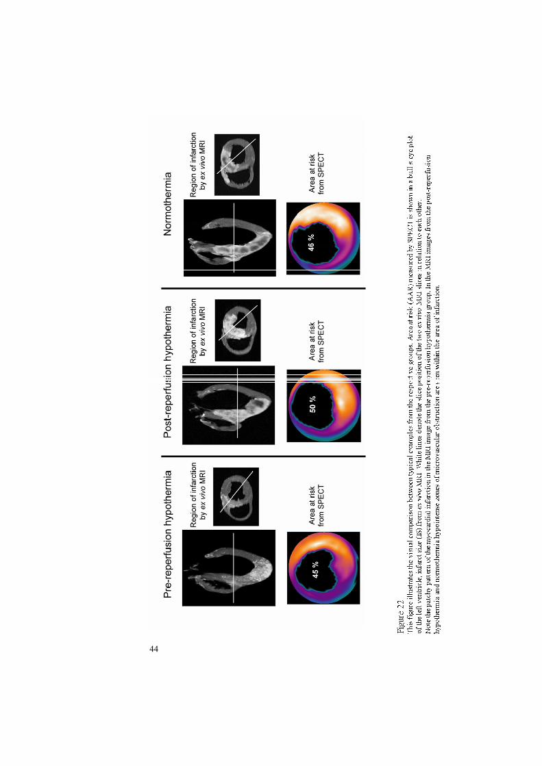
44

10 Main findings
1. The P2Y13 receptor on the red blood cell is stimulated by ADP, and whenactivated attenuates ATP release from red blood cells to the extracellular plasma.
2. Activation of the coronary endothelial P2Y1 receptors causes hyperemia in coronary arteries.
3. Inhibition of coronary P2Y1 receptors reduces peak coronary blood flow during reactive hyperemia by 46 %.
4. Coronary t-PA release during ischemia and reperfusion is mediated throughP2Y1 receptors.
5. Mild systemic hypothermia reduces peak coronary blood flow by 43 % duringreactive hyperemia.
6. Rapidly induced hypothermia during anterior myocardial infarction before reperfusion significantly reduces final infarct size compared with rapidly induced hypothermia in conjunction with or after reperfusion.
7. Rapidly induced hypothermia before reperfusion in the setting of anterior myocardial infarction in pigs abolishes microvascular obstruction.
45

46

11 Discussion
The pig model For the experiments in regard to the P2 receptors (Papers I-III) it was essential to work with a live animal model instead of using a Langendorf model because the experiments depended on the circulation of RBC’s that could release ATP. The pig model was selected also because large animals could more easily facilitate the closed chest approach with insertion of largelumen catheters. The closed chest pig model allowed us to study coronary ischemia, infarction and reperfusion in a physiologically appropriate setting while still retaining control of the coronary circulation. The commonly used method of ligation of a coronary artery in an open chest model creates unnecessary trauma to the animal. Due to the similar coronary anatomy in pigs, compared to man, it is a common large animal model to use in cardiovascular research. However, earlier cardiovascular research in the 1950’s through the 1980’s commonly used the dog as the animal model. In regards to coronary research this may have serious disadvantages. The coronary anatomy in man and pigs is more similar to each other than either is to the dogand differs also to the presence of an extensive collateral network seen in dogs127 which is notseen in pigs128 and in healthy humans129.A recent trial by Hedstrom et al. showed that patients with acute myocardial infarcts shows a similar infarct expansion over time compared to that of dogs in contrast to that seen in pigs87.How is this possible?Of considerable importance is that the experimentally induced infarcts in pigs and in dogs is generally performed in young animals. In contrast, patients treated for acute myocardial infarctions are generally well above middle age and are in various stages of an atherosclerotic coronary disease. Many of these patients have already developed visible collaterals at the timeof catheterization and in those with no visible collaterals there may still be collaterals, but to small to be recognized. A patient may also have had several episodes of ischemia prior to the occluding infarct which may have been silent episodes or actual episodes of chest-painswhich could have resulted in preconditioning.
The P2Y13 receptor acts as a negative feedback loop on the surface of red blood cell’s toattenuate ATP release Our finding of a negative feedback system for ADP release fits well with previous evidenceof ATP as an extracellular transmitter, as first proposed by Burnstock17, and it is possible that this negative feedback system plays an important role in the control of peripheral and coronary circulation. In underperfused organs, hypoxia stimulates an increase in intracellular cAMP, which leads to the release of ATP to the blood plasma. This increase of cAMP could be enhanced by adrenaline, that is in itself elevated during hypoxia130. Extracellular ATP stimulates endothelial cells to release dilatory factors such as NO, mediating vasodilatation and improved oxygen delivery to the organ. To shut down the ATP release on the venous side (where the blood is still deoxygenated), and to avoid unnecessary ATP release, the degradation product ADP acting on P2Y13 receptors could inhibit cAMP and decrease ATP-release from RBC’s. ATP and ADP are then degraded by ectonucleotidases to adenosine, which is quickly taken up in the RBC’s where it can be recycled to ATP by the glycolytic pathway.The described negative feedback pathway may be important to avoid high extracellularplasma concentrations of ATP. At levels above 100 µmol/L, ATP concentrations may exceed the catalytic capacity of ectonucleotidases and, could in fact, stimulate ATP release by increasing permeability of the RBC131. At high concentrations of ATP, a self sustaining process may thus be instigated, which may contribute to the irreversible stage of circulatoryshock that can develop rapidly in severely ill patients. It is possible that in extreme conditions
47

such as circulatory or septic shock with acidosis and hypoxia, high ATP levels could be deleterious, leading to a drop in blood pressure. However, in most situations increased ATP levels ought to have positive effects by increasing tissue perfusion.
The P2Y1 receptor mediates coronary reactive hyperemia The selective P2Y1 blocker MRS2179 was found to significantly reduce the early peak flow in coronary reactive hyperemia in pigs. This is supported by the flow increase caused by the selective P2Y1 agonist 2-MeSADP, which could be completely blocked by MRS2179. The mechanism of reactive hyperemia is still not completely understood but appears to bemultifactorial in origin. Investigations of the effects of adenosine43, 132, 133, prostaglandins 134-
137, K+ATP channels40, 41, 45, 138-140 and the role of NO42, 45, 139, 141-144, acting alone or in combinations with each other, have been performed. Earlier research on reactive hyperemia in the heart has been performed both in vivo in large animals, as well as in Langendorf modelsin rodents. However, using rodents in a Langendorf model has its inherent drawbacks due to the lack of ATP release from RBC’s in response to ischemia. In large animals, in vivo modelshave shown that both ATP and ADP are released during reactive hyperemia and adenosine by itself contributes to about 1/3 of the reactive hyperemia with NO contributing slightly less43, 44
and is mainly seen in the later phase of reactive hyperemia45, 145. During reactive hyperemiaincreased levels of ATP and ADP have been measured50. ATP is then rapidly degraded to ADP, which in turn binds to the vascular endothelium at the site of the P2Y1 receptor.Selective blockers of the P2Y1 receptors have recently become available 87, 146, allowing us to test the role of P2Y1 receptors during reactive hyperemia. The porcine in vivo model in our experiment was chosen because the presence of whole blood and a live model was essential.The use of angioplasty over-the-wire balloons allowed for precision in attaining both accurate and localised induction of ischemia, and delivery of infusions. The physiological alterations induced by open chest experiments could thus be avoided. The infusion of Ringer’s acetate solution at the same rate as later infusions of 2-MeSADP and MRS2179 did not alter measurements of flow from baseline. The selective P2Y1 receptor agonist 2-MeSADPinduced a predicted increase in flow, which could be completely abolished by co-infusion of 2-MeSADP and the selective P2Y1 receptor blocker MRS2179. In contrast, UTP, which activates P2Y2/4 receptors, stimulated a flow increase that was unaffected by MRS2179, demonstrating the selectivity for P2Y1 receptors of MRS2179. To test the contribution ofP2Y1 receptors to reactive hyperemia, the LAD was occluded, and MRS2179 was infused intothe ischemic portion of the heart supplied by the LAD. The 46% reduction of peak flow achieved during reactive hyperemia indicates that P2Y1 receptors are of major importance as an activator of endothelium-derived smooth muscle cell relaxing factors such as NO and EDHF. NO and EDHF have been shown to mediate a major part of early reactive hyperemia40, 42, 44, 45, and both are released by ADP acting on P2Y1 receptors33-35, 39. The K+ ATPchannel inhibitor glibenclamide blocks the remaining early reactive hyperemia45.Interestingly, glibenclamide is also an inhibitor of P2Y1-mediated vasodilatation34. It is therefore possible that a part of the hyperemia blocked by glibenclamide is stimulated by ADP via endothelial P2Y1 receptors and that glibenclamide in part acts downstream of the P2Y1 activation and not only after intracellular metabolic regulation of K+ ATP channels. The time profile of the mediators of reactive hyperemia is highly interesting. Previous studies using adenosinedeaminase, theophylline43, selective A2A antagonists45, 145, or A2A knockout mice145, have shown that adenosine mediates the late phase of reactive hyperemia. This adenosine is probably derived from degradation by ecto-nucleotidases of the ADP that we now demonstrate mediates a major part of the early peak phase.In conclusion, our experiments suggest that ADP stimulating P2Y1 receptors mediates a majorpart of peak reactive hyperemia in the heart, and inhibition of the endothelial coronary P2Y1
48

receptors could potentially attenuate the reperfusion injury incurred during primaryangioplasty in the setting of acute myocardial infarction94.
The purinergic hypothesis of coronary reactive hyperemia We would like to propose that ATP is released during coronary ischemia from red blood cells; cardiomyocytes, endothelial cells and platelets, and that ATP could mediate an even earlier part of the reactive hyperemia (Figure 23). This has not been tested yet due to the lack ofselective antagonists. (The presence of ATP- and UTP-responsive endothelial P2Y2/4 receptorshas been demonstrated before33 and was confirmed here by the hyperaemic effect of UTP.)ATP is then degraded to ADP, which mediates peak hyperemia via endothelial P2Y1
receptors, followed by degradation of ADP to adenosine resulting in late-phase hyperemiamediated via A2A receptors on SMC (Figure 23).
Figure 23 This figure illustrates a hypothesis of smooth muscle cells relaxation in response to accumulation of purinergicsubstrates in coronary vessels following post-ischemic reperfusion. Red blood cells, heart myocytes, endothelialcells, platelets and sympathetic nerves release ATP during hypoxia. ATP may contribute to the very early reactivehyperemia, although this has not been proven due to lack of specific antagonists. ATP is quickly degraded to ADP which stimulates P2Y1 receptors on the endothelium, thus initiating the peak flow during the early phase of reactivehyperaemia, as demonstrated in the present study. ADP is then degraded to adenosine that stimulates A2A –receptorsand thus maintains reactive hyperemia during the mid to late phase
In the early phase of reactive hyperemia the high rate of oxygen depletion causes an increased release of ATP. ATP is then degraded rapidly to ADP and local plasma concentrations ofADP increase. At a critical plasma level of ADP and when myocardial oxygen demanddecreases in the later phase of reactive hyperemia, ADP interaction with the membrane bound P2Y13 receptor on RBC’s will attenuate further ATP release through the negative feedback mechanism until equilibrium is attained (Fig 24).
49

Figure 24 Negative feedback system (shown in yellow) regulating plasma ATP levels via the P2Y13 receptor on themembrane of the RBC which is important in the control of plasma ATP levels and tissue circulation.
t-PA release and function We have for the first time shown that intra-coronary administration of the ADP analogue 2-MeSADP increases release of t-PA in the coronary venous outflow via activation of endothelial coronary P2Y1 receptors. We have also for the first time shown that cardiac release of t-PA in response to cardiac ischemia can in turn be inhibited by selectively blocking the ADP receptor, P2Y1, with MRS2179.The mechanism of t-PA release during reactive hyperemia is still not completely understoodbut appears to be multifactorial in origin. Earlier research on ischemic and post-ischemicrelease of t-PA in the heart has been performed both in vivo in large animals, as well as in ex-vivo cardiac models in rodents54, 59, 147, 148. In large animals, in vivo models have shown that ischemia causes the release of t-PA and that this may be caused by a number of factors but seems to be independent of blood flow59. During severe coronary ischemia, as seen during and shortly following a coronary occlusion, ADP is released (or derived from ATP) from cardiac myocytes, endothelial cells, red blood cells, platelets and sympathetic nerves33, 46, 47.The ADP released into circulation in response to ischemia can thus stimulate release of t-PA, perhaps in part through the P2Y1 receptors. Endothelial P2Y1 receptors mediate SMCrelaxation through EDHF34, 35, 39. NO and EDHF have been shown to mediate a large part of early reactive hyperemia40, 42, 44, 45, and both are released by ADP acting on P2Y1 receptors33-
35, 39. Furthermore, ATP infused in the human forearm (which results in formation of ADP as well), stimulates increased blood flow via EDHF and results in a substantial release of t- PA60. Because the ADP receptor P2Y1 mediates increases in blood flow during reactive hyperemia64 we wanted to examine the role of P2Y1 receptors during ischemic and post-
50

ischemic release of t-PA. We found that the selective P2Y1 receptor agonist, the stable ADP analogue2-MeSADP, induced a significant increased release of t-PA and as expected, a significant increase in coronary flow. However, both these findings could be completely abolished by simultaneous infusion of the selective P2Y1 receptor blocker MRS2179. To test thecontribution of P2Y1 receptors to the release of t-PA during ischemia and during reactive hyperemia, the LAD was occluded, and MRS2179 was infused into the ischemic portion of the heart supplied by the LAD. This resulted in a significant reduction in t-PA release. This indicates that levels of ADP in the coronary circulation play an important role in theregulation of local t-PA release in the heart by acting on P2Y1 receptors. Several earlier reports have suggested an important role for both extracellular nucleotides and EDHF in therelease mechanism for t-PA58, 63. Thus, ADP release could cause pro-fibrinolytic effects. This is however in stark contrast to the effect ADP has on platelets. ADP, in fact, activatesplatelets by stimulation of the P2Y1 (and P2Y12) receptors. Thus our findings indicate a dualistic action of ADP by means of a pro-fibrinolytic (by means t-PA release) and a counteracting prothrombotic (by means of platelet activation) role for ADP. The effect of MRS2179 on ischemia induced t-PA release is only partial. Other mechanisms than the P2Y1-receptor that could explain the remaining effect include other P2-receptors, such as P2Y2,P2Y6 or P2X4 which are expressed in the endothelium. Furthermore, novel mechanisms of ADP mediating relaxation in pig coronary arteries via adenosine release have recently been described and could be involved149.In conclusion, our experiments suggest that ADP stimulating P2Y1 receptors mediate a majorpart of the cardiac release of t-PA seen during coronary ischemia and during the subsequent reactive hyperemia in the heart. This may counter-balance the well-known platelet stimulating effects of ADP during acute myocardial ischemia. It is possible that the inhibitor of ADP induced platelet aggregation, clopidogrel, shifts this balance in favour of the fibrinolytic ADP effects described here.
Hypothermia in coronary ischemia and reperfusion
Reactive hyperemia during hypothermia
In general, hypothermia is thought to reduce the metabolic needs of cells and specifically perhaps by reducing the oxygen demand in the hypothermic tissues150-153. However, little isknown about the actual mechanisms by which hypothermia can reduce cell death in ischemictissues and the reduced oxygen demand does not fully explain the positive effects of hypothermia154.Several animal studies have shown that hypothermia reduces infarct size in the setting of myocardial infarct (usually caused by the ligation of LAD). Duncker et al. found a correlation between infarct size and temperature with the smallest infarct size found at the lowest temperature107. Other reports have indicated a positive temperature-related effect of hypothermia in relation to infarct size104, 105, 109, 110, 113, 114. On the contrary Maeng et al. found no benefit of hypothermia induced in conjunction with, or after reperfusion113. Others have also shown in small animal models that the earlier hypothermia is applied to ischemicmyocardial tissues the lesser the infarct size will be, and that this effect can be furtherenhanced by preconditioning114, 155. To date, hypothermia has been tested in five human trials in the setting of myocardial infarction with planned PCI as reperfusion method (COOL MI, ICE-IT, NICAMI, LOWTEMP and a study by Dixon et al.)106, 108, 111, 112, 115. The NICAMI and the LOWTEMP studies as well as the study by Dixon were only feasibility trials, whereas the randomized trials COOL MI and ICE-IT were designed to test if a reduction of final infarct size at 30-days by SPECT analysis could be attained through treatment with mild
51

hypothermia. Unfortunately, both COOL MI and ICE-IT were negative trials. At first glance this should have put an end to further cooling trials in the setting of acute MI. However, in both trials, only a minority of patients randomized to cooling reached the target temperatureprior to reperfusion, and the few patients that did reach target temperature prior to reperfusionexhibited a significant or a strong reduction in myocardial infarct size at 30 days compared to control-patients, especially among patients with anterior infarcts. The result of the two trials is thus in accordance with the results found in preceding animal studies which exemplifies the importance of reaching target temperature prior to reperfusion of completely occludedvessels.Recently a sixth human Trial, COOL MI II156, has been started in lieu of these findings. In this trial, only patients with anterior acute myocardial infarcts will be randomized to intravascular cooling or control, and cooling will be initiated earlier than in COOL-MI I. The previous animal and human trials have all looked at the effect of using hypothermia as a method to reduce myocardial infarct size. However, the mechanism(s) by which hypothermiaexerts its myocardial protective effect is still unclear but it is hypothesized that it can be explained mainly by a reduction in oxygen consumption and reduced metabolic demands111,
112, 150, 151, 157.
Because hypothermia has been shown to limit infarct size if instituted during ischemia and prior to reperfusion104, 107, 113, we postulated that the protective effect of hypothermia is in part mediated by a reduced reperfusion injury. It is still controversial if a reperfusion injury existsas an entity, but it is also the focus of a great deal of research83, 158. If there is such a thing asreperfusion injury, one of several events taking place during reperfusion which couldpotentially cause reperfusion injury is the large increase in coronary flow that occurs during reperfusion (reactive hyperemia). The increased coronary flow may in itself be the cause of the early myocardial oedema seen during reperfusion93 or responsible for the generation of large quantities of O2-derived free radicals seen during the increased coronary flow phase of reactive hyperemia117. We therefore wanted to examine if the phenomenon of post-ischemicreactive hyperemia could be affected by hypothermia and thus explain one possiblemechanism, or effect, by which hypothermia may exert its protective effect during reperfusion. We decided to use a 10 minute occlusion time which is an established timeperiod for studying reactive hyperemia in both animals and man.Our results indicated a significant 43% reduction in peak coronary blood flow during reactive hyperemia in the hypothermic group compared to controls. The reason why hypothermiareduces coronary reactive hyperemia is not fully elucidated. It is probably mediated by a reduction in the release of dilatory mediators, but it could be caused by a reduced responsiveness in the endothelium and vascular smooth muscle cells. Hypothermia also seems to better maintain BE as measured in the peripheral artery (internal Iliac Artery or distal abdominal Aorta) with no significant differences seen in the coronary venous outflow in the coronary sinus. One could speculate if the difference in BE could have influenced the results of coronary flow during reactive hyperemia. However, it is well established that the vascular bed of the coronary circulation tends to dilate in response acidosis and it has been suggested that a decrease in extracellular pH could be involved in coronary flow regulation during hypoxia, ischemia, and increased metabolic demand159, 160.Thus the significantly lower arterial BE observed in the control group could potentially translate into a higher coronary flow compared to the hypothermic group. However, there was no observed difference in pH, thus making it unlikely that the difference in BE levels wouldsignificantly contribute to a substantial difference in coronary flow.The heart rate of the pigs in the hypothermic group was clearly lower compared to pigs in the normothermic group during the entire phase of hypothermia. During mild hypothermia in pigs
52

(and humans) a temperature dependent decline in heart rate is accompanied by an increase instroke volume and may actually increase cardiac output according to Weisser et al. andothers104, 161, while on the other hand coronary flow remains unchanged during hypothermia152. However, although heart rate declines during hypothermia, this is an independent mechanism unrelated to the cardio-protective effects (i.e. infarct size reduction)of hypothermia162, 163. In regard to blood pressure, the hypothermic group of pigs showed no difference in mean arterial pressure even though a general decline in mean arterial pressure was observed in both groups mainly during ischemia.
In conclusion we found that hypothermia blunts the increase in coronary flow seen during reactive hyperemia in the LAD, in a closed chest porcine model. This finding may explain one of the effects by which hypothermia can reduce infarct size if attained prior to reperfusion. Further research into the infarct-reducing mechanisms of hypothermia is warranted if we are to use hypothermia in the clinical setting of myocardial infarction.
Postconditioning in acute myocardial infarction
Postconditioning as a means to reduce infarct size has recently been successfully performed in several animal experiments and one trial in man99-102. There have also been some negative or ambiguous reports concerning the effects of postconditioning164, 165. Interestingly, the reduced flow often employed during the first couple of minutes (cycles of occlusion and reperfusion) during reperfusion in postconditioning experiments may cause a similar effect to that of the blunted reperfusion flow observed in the very early phase of hypothermia in our experiment.
Rapidly induced hypothermia prior to reperfusion in acute myocardial infarction
This study demonstrates that rapid induction of hypothermia before reperfusion reduces myocardial infarct size in pigs by 40% compared to rapidly induced hypothermia immediatelyafter reperfusion, and by 44% compared to normothermia. Furthermore, pre-reperfusion hypothermia, and to some extent also post-reperfusion hypothermia, protects the heart frommicrovascular obstruction compared to normothermia.
The importance of rapid cooling before reperfusion
In the clinical setting today it is not feasible to delay reperfusion therapy in order to wait forinduction of hypothermia. The combination of an infusion of cold saline solution and endovascular cooling achieved a reduction in temperature to <35°C in less than 10 min (Fig17). This protocol could be clinically applicable since it would allow induction of hypothermia before or during angiography and have the patient cooled to <35°C without delaying reperfusion therapy. Post-reperfusion hypothermia was chosen as a control group with active treatment. A normothermic group was added in order to compare the different hypothermia protocols to normothermia.
In order to avoid a spontaneous variation in temperature during the experiment, a normalbody (core) temperature in pigs (38° C) was established before induction of ischemia. In subgroup analysis of clinical trials, a pre-reperfusion temperature of <35° C was sufficient to reduce infarction size by up to 40 %108, 115. Based on the results, 33° C was chosen as target temperature, and the limit for successful hypothermia was 35° C. An infusion of 1000 ml of cold saline solution caused a reduction of central body temperature to <35° C in all pigs in less than 10 min. The reduction in body temperature was then maintained by the cooling catheter (Figure 17). The difference in temperature between the hypothermia groups was 3.6°
53

C (37.8° C vs. 34.2° C) at the time of reperfusion. Thus, a reduction of central body temperature of 3.6° C before reperfusion was enough to reduce infarct size by 40 %. Theseresults are in agreement with previous studies with more long-term hypothermia during ischemia in which reductions in temperature of 3-5° C had prominent effects104, 107, 109, 110, 114,
163, 166 .
Infarction size evaluation
Previous studies have used histology with TTC or imaging with SPECT to visualise the extentof myocardial infarction. Ex vivo MRI has been shown to correlate closely to TTC-staining in the setting of acute myocardial infarction with reperfusion for either six hours or one day125,
167. Ex vivo MRI allowed us to achieve high resolution images of the myocardial infarction and was therefore chosen as the method for evaluation of infarct size in our study. SPECT was used to determine area at risk during transmural anterior myocardial ischemia. As shown in figure 3, the placement of the balloon in the LAD after the first diagonal branch induced ischemia in, on average 38 % of the left ventricle, with no difference in AAR between the groups.
Ex vivo MRI demonstrated a more inhomogeneous and patchy appearance of the myocardial infarctions after pre-reperfusion hypothermia. Dae et al described similar findings with scattered islands of reduced Sestamibi uptake in pig hearts when assessing infarct size with SPECT in pigs subject to endovascular hypothermia104. It is possible that preservation of small zones of viable myocardium could be beneficial in the long term preventing aneurysmformation and that cellular hypertrophy in the zones could improve contractility in the area.
Microvascular injury
It has been demonstrated (contrast echocardiography, SPECT, blush angiographic score and MRI) that up to 30 % of patients with STEMI undergoing Primary PCI as reperfusion therapy lack efficient myocardial perfusion following reperfusion168-171 and this may be attributed to microvascular obstruction seen in up 90% of patients treated with primary PCI172. The degree of microvascular injury is associated to the duration of myocardial ischemia and the extent of myocardial infarction but may possibly also be caused by reperfusion injury. Importantly, the presence of microvascular obstruction is associated with a worse clinical outcome168, 169, 171,
173. Hale et al demonstrated that hypothermia during ischemia reduced the extent of microvascular obstruction174. In our study, induction of hypothermia shortly before reperfusion abolished microvascular obstruction while it was prevalent in both the post-reperfusion and the normothermia groups (Fig 3). Interestingly, there was a significantdifference in the size of microvascular obstruction between the post-reperfusion hypothermiaand normothermia groups, possibly due to the rapid cooling by cold saline. The post-reperfusion cooling was so rapid that it actually cools during a part of the reperfusion period. The duration of this period is difficult to define, but the reactive hyperemia lasts for approximately ten minutes, and we reached target temperature already after five minutes.Thus, mild systemic hypothermia before reperfusion may protect the heart from microvascular obstruction. This suggests that the development of microvascular obstruction is an event that occurs very early in the reperfusion phase and possibly in the period of coronary reactive hyperemia which lends credibility to the hypothesis that the reperfusion injury maybe an event caused by microvascular obstruction which may in turn be caused, in part by, theenormous inflow of blood seen during reactive hyperemia.
In conclusion, a rapid induction of hypothermia can be achieved by a combination of cold saline and endovascular cooling. Such hypothermia induced before reperfusion is effective in
54

reducing the myocardial infarct size and protecting the heart from microvascular obstruction. This could have significant beneficial effects on clinical outcome after myocardial infarction. This protocol could easily be applied to the clinical setting where hypothermia could be induced without delaying PCI mediated reperfusion. A rapid infusion of large volumes of saline solution in patients with myocardial infarction could have serious side-effects, such as left ventricular overload and pulmonary oedema. A currently ongoing human safety study on patients with acute myocardial infarction will determine whether cold saline as an adjunctivetherapy to endovascular cooling can be safely administered (www.clinicaltrials.gov).
Conclusions
This thesis bridges research between preclinical and clinical sciences and has led to theconduction of a human trial. We have shown that a negative feedback loop on red blood cells terminates ATP-release which regulates microcirculation. ATP and its degradation productADP activate endothelial P2 receptors, and from our research we now know that ADP plays a major role in attenuating the increase in blood flow seen during coronary reactive hyperemia.Additional research on reactive hyperemia, now with the use of hypothermia, led to theconclusion that mild hypothermia also attenuates the increased blood flow seen in coronary reactive hyperemia. Further research in a closed chest, porcine, infarction model, using hypothermia as adjunctive treatment resulted in a nearly halved final infarct size and an abolishment of microvascular obstruction in the animals treated with hypothermia comparedto controls. This may be due to a reduction of the reperfusion injury and our research indicates that a major part of the myocardial reperfusion injury may occur during the short period of reactive hyperemia which is within the same timeframe in which the initiation of tissue swelling can be seen, an increased release of t-PA, ATP and ADP can be measured, and microvascular obstruction develops as visualized by MRI.
Praeterea censeo Carthaginem delendam esse Cato d.ä.
55

56

12 Populärvetenskaplig sammanfattning (Summary in Swedish)
Reaktiv hyperemi är ett cirkulationsfysiologiskt fenomen som har varit en gåta för den moderna fysiologin de sista hundra åren. Reaktiv hyperemi är det initialt kraftigt ökade blodflödet som uppstår till en vävnad eller ett organ efter att blodflödet under en tid varit avstängt. Flödesökningen som följer är betydligt större än den syre- eller nutritions-skuld somuppstått under tiden som blodflödet varit avstängt.Nyligen har en ny teori lagts fram som kan förklara mekanismen varför reaktiv hyperemiuppkommer. Röda blodkroppar beskrivs ofta endast som kroppens transportörer av syre utan några andra funktioner av dignitet. Man har dock upptäckt att röda blodkroppar innehåller mycket stora mängder ATP jämfört med andra celler och med extracellulärrummet. Varför det är på det viset har man dock inte tidigare förstått. Traditionellt har ATP betraktats endast som ett energisubstrat i kroppen eller som en byggsten i DNA och framför allt har ATP betraktats som något som endast finns och verkar inuti celler. ATP tillhör en familj av substanser sombenämns nukleotider som även består av ADP, AMP, UTP, UDP. Sedan 1970-talet har man i vissa forskarkretsar haft en teori om att nukleotider är viktiga signalsubstanser mellan celler, men det är inte förrän på senare år som signalsubstansteorin allmänt har fått acceptans inomden medicinska och fysiologiska forskarvärlden. Inom cirkulationsfysiologin har man nu upptäckt att ATP frisätts från röda blodkroppar som en respons på ett ökat uttag av syremolekyler när en röd blodkropp passerar genom en vävnad. Ju mer syre som extraheras, desto mer ATP frisätts. ATP och dess nedbrytningsprodukter ADP och adenosin bidrardärefter till att lokalt stimulera till en avslappning av glatt-muskulatur på insidan av blodkärlen för att på så sätt åstadkomma ett lokalt ökat blodflöde. ATP, ADP och adenosin bryts mycket snabbt ned och återupptas av cellerna vilket bidrar till att effekten dels skermycket snabbt och dels endast sker mycket lokalt. ATP och ADP åstadkommer detta genom att stimulera så kallade P2-receptorer (P2Y2 och P2Y1) på endotelet till blodkärlen. Att ATP har en avgörande betydelse för regleringen av blodflödet till skelettmuskulaturen är nu relativt klart, men det har inte varit känt hur ADP eventuellt påverkar den reaktiva hypermin inomhjärtats kranskärl.I vårt första arbete (I) prövades en hypotes om att det måste finnas ett återkopplingssystem påden röda blodkroppen som stryper ytterligare frisättning av ATP i vissa situationer. I försöken på en grismodell kunde vi påvisa att ADP stimulerar en P2Y13 receptor på röda blodkroppar som minskar frisättning av ATP till plasma så länge koncentrationen av ATP’snedbrytningsprodukt ADP är hög.I vårt andra försök (II) kunde vi visa, också i en grismodell, att den reaktiva hyperemin i hjärtats kranskärl till stor del beror på ATP’s nedbrytningsprodukt ADP och dess effekt på P2Y1 receptorer på kranskärlens endotel som stimulerar till ett ökat blodflöde. Blockering avP2Y1 receptorer nästan halverade den reaktiva hyperemin i hjärtats kranskärl i vårt försök. I det tredje arbetet (III) kunde vi i vår grismodell visa att P2Y1-receptorer reglerar frisättningen av t-PA (en substans av stor betydelse för att förhindra att blodet levrar sig inne i blodkärlen) och att en blockad av P2Y1–receptorer kraftigt minskade frisättningen av t-PAunder reaktiv hyperemi i kranskärlen. I det fjärde försöket (IV) undersökte vi om kylbehandling (hypotermi) kunde påverka reaktiv hyperemi i kranskärlen. Vid försöken i vår grismodell kunde vi påvisa att den grupp grisar med aktiv hypotermi-behandling (34 C) fick ett nästan halverat blodflöde i hjärtats kranskärl under reaktiv hyperemi jämfört med kontrollgruppens grisar (37 C).I vårt sista försök (V) undersökte vi om snabbt inducerad hypotermi precis innan reperfusion i en hjärtinfarkt modell på gris kunde minska infarktstorleken jämfört med en grupp grisar som
57

fick snabbt inducerad hypotermi precis efter reperfusionen. I detta försök kunde vi se att infarktstorleken minskade med 40 % i de djur som behandlades med hypotermi kort innan reperfusion jämfört med kontrollgruppen.
Sammanfattningsvis har vi i vår forskning undersökt PY1-receptorers effekter på reaktiv hyperemi i kranskärl samt hur de kan reglera frisättning av t-PA och hur P2Y13 receptorer på röda blodkroppar reglerar frisättning av ATP från röda blodkroppar via ett reglersystem.Vidare har vi visat att hypotermi minskar reaktiv hyperemi i kranskärlen och att det kan vara en orsak till att kylbehandling minskar hjärtinfarktsstorleken om kylbehandlingen påbörjas innan reperfusionen sker i en hjärtinfarktsmodell.
58

13 Acknowledgements
First of all I want to thank all of my friends and colleagues who have encouraged, helped and supported me in my research.
I would also specifically like to thank:
First and foremost, Associate Professor David Erlinge, who has been my mentor and teacher in the studies which have resulted in this thesis. His outstanding knowledge in the area of P2 receptors and physiology made the research not only seem easy but also made it exhilarating and fun. To also have David as a student, teaching him interventional cardiology, made us function as team and David has since remained not only a teacher or a student but also a close and highly respected friend. Thank you for your fantastic skills as a mentor during my PhD and for your efforts to keep me focused.
Matthias Götberg, who I first met when he was an outstanding student and I was a fellow in Cardiology and who is now my close friend and research colleague. He is also a favourite of my children even though they do miss his Porsche after he sold it.
My colleagues and friends in the cath lab, Anders Lundin, Björn Thorvinger and Per Bondesson for all their support and help and especially Jan Harnek who introduced me to and helped me in percutaneous cardiac animal research.
Professor S. Bertil Olsson, who is the main reason I went into Cardiology when he offered meto start training in invasive cardiology only 6 months into my fellowship in Cardiology at a time when I was considering general surgery as an alternative (thank god I stayed with Cardiology). I also want to thank Bertil for introducing me to animal research, somethingwhich has helped me considerably in the research which resulted in this thesis.
Lingwei Wang, who helped me in my research and taught me about P2Y13 receptors and even more about China.
Håkan Arheden, and especially Martin Ugander and Henrik Engblom, at the Department of Clinical Physiology for their magical skills with the MRI acqusitions.
All the staff in the Department of Cardiology and especially at the cardiac intensive care unit (HIA) with as special note to the staff in the cardiac cathlab (Hjärtröntgen) for the support and help in letting me spend a great deal of time with my research.
Monica Magnusson, and Bibbi Smideberg for their high spirits and continual support.
My old mentor in Cardiology, Anders Roijer who has become my close friend and confidant. Thank you for your unwavering support which helped me through the tough years in my fellowship in Cardiology.
59

David Erlinge's, research group at BMC, Stefan Amisten, Karl Högberg, and especially Helen Svensson for all her help in the animal lab.
Kenneth Jacobsson, for his help in supplying us with purinergic antagonists for the animallab.
Sverker Jern, and his team in Göteborg for helping us with the difficult t-PA analysis.
Jesper van der Pals and Oscar Braun for their great companionship and help in the animalresearch.
Pernilla Järnhäll, who was our fantastic study nurse in the animal research lab, and who always kept our spirits high and continually bought the milk for our coffees because I always forgot.
My fellow roommates in the Department of Cardiology Göran Arstad, and Öyvind Reitan who has put up with my messy desk littered with papers and coffee mugs.
But finally and above all, I am indebted to the people that mean the most to me and I sincerely want to thank:
My wife and best friend Cecilia who has always supported and encouraged my research even though you had to carry a heavy burden when I was away at the lab in the midst of bringing up our three wonderful children. I know it has not been easy and I am forever indebted to you.
My children Axel, Jacob and Emma who are the light of my life and who have taken an interest in my research, or at least you pretend to. Hopefully my research has not impactedyour lives too much and I hope my research has not discouraged you from doing research of your own in the future.
60

14 Sources of Funding
The studies in this thesis were supported by the Swedish Heart and Lung Foundation, the Franke and Margareta Bergqvist Foundation, the Wiberg Foundation, the Bergwall Foundation, the Zoegas Foundation, the Westergren Foundation, the Swedish Medical Society, the Swedish Medical Research Council, Grant 13130 and the Vascular Wall program (Lund University Faculty of Medicine).
61

62

15 REFERENCES
1. Intravenous streptokinase given within 0-4 hours of onset of myocardial infarction reduced mortality in ISIS-2. Lancet. Feb 28 1987;1(8531):502.
2. Hellerstein H. Rehabilitation of patients with coronary heart disease. In: Luisada A, ed. Cardiology Volume 4: Clinical Cardiology-Therapy: McGraw-Hill BookCompany Inc.; 1959.
3. DeWood MA, Spores J, Notske R, Mouser LT, Burroughs R, Golden MS, Lang HT. Prevalence of total coronary occlusion during the early hours of transmuralmyocardial infarction. The New England journal of medicine. Oct 16 1980;303(16):897-902.
4. Grines CL, Browne KF, Marco J, Rothbaum D, Stone GW, O'Keefe J, Overlie P, Donohue B, Chelliah N, Timmis GC, et al. A comparison of immediate angioplastywith thrombolytic therapy for acute myocardial infarction. The Primary Angioplasty in Myocardial Infarction Study Group. The New England journal of medicine. Mar 11 1993;328(10):673-679.
5. Zijlstra F, de Boer MJ, Hoorntje JC, Reiffers S, Reiber JH, Suryapranata H. A comparison of immediate coronary angioplasty with intravenous streptokinase in acute myocardial infarction. The New England journal of medicine. Mar 11 1993;328(10):680-684.
6. Stenestrand U, Lindback J, Wallentin L. Long-term outcome of primary percutaneous coronary intervention vs prehospital and in-hospital thrombolysis for patients with ST-elevation myocardial infarction. Jama. Oct 11 2006;296(14):1749-1756.
7. Guyton. A. Textbook of medical physiology / Arthur C. Guyton, John E. Hall. 11 ed. Philadelphia: Elsevier Saunders; 2006.
8. Olsson RA, Gregg DE. Myocardial Reactive Hyperemia in the Unanesthetized Dog. The American journal of physiology. Feb 1965;208:224-230.
9. Ellsworth ML. Red blood cell-derived ATP as a regulator of skeletal muscleperfusion. Medicine and science in sports and exercise. Jan 2004;36(1):35-41.
10. Ellsworth ML, Forrester T, Ellis CG, Dietrich HH. The erythrocyte as a regulator ofvascular tone. The American journal of physiology. Dec 1995;269(6 Pt 2):H2155-2161.
11. Gonzalez-Alonso J, Olsen DB, Saltin B. Erythrocyte and the regulation of humanskeletal muscle blood flow and oxygen delivery: role of circulating ATP. Circulation
research. Nov 29 2002;91(11):1046-1055. 12. Sprague RS, Ellsworth ML, Stephenson AH, Lonigro AJ. Participation of cAMP in a
signal-transduction pathway relating erythrocyte deformation to ATP release. Am J
Physiol Cell Physiol. Oct 2001;281(4):C1158-1164. 13. Folkow B. The vasodilator action of Adenosine Triphosphate. Acta Physiology
Scandinavia. 1949;17:311-316.14. Bergfeld GR, Forrester T. Release of ATP from human erythrocytes in response to a
brief period of hypoxia and hypercapnia. Cardiovascular research. Jan 1992;26(1):40-47.
15. Sprague RS, Stephenson AH, Ellsworth ML, Keller C, Lonigro AJ. Impaired release of ATP from red blood cells of humans with primary pulmonary hypertension. Exp
Biol Med (Maywood). May 2001;226(5):434-439. 16. Drury AN, Szent-Gyorgyi A. The physiological activity of adenine compounds with
especial reference to their action upon the mammalian heart. J Physiol. Nov 25 1929;68(3):213-237.
63

17. Burnstock G. Purinergic nerves. Pharmacol Rev. Sep 1972;24(3):509-581. 18. Burnstock G. A basis for distinguishing two types of purinergic receptors. In: Straub
R, Bolis, L., ed. Cell membrane receptors for drugs and hormones: a
multidisciplinary approach. New York: Raven Press; 1978:356. 19. Burnstock G. Do some nerve cells release more than one transmitter? Neuroscience.
Aug 1976;1(4):239-248. 20. Burnstock G. Sympathetic purinergic transmission in small blood vessels. Trends in
pharmacological sciences. Apr 1988;9(4):116-117. 21. Burnstock G. Local control of blood pressure by purines. Blood vessels.
1987;24(3):156-160.22. Burnstock G. Local mechanisms of blood flow control by perivascular nerves and
endothelium. J Hypertens Suppl. Dec 1990;8(7):S95-106. 23. Burnstock G. Dual control of local blood flow by purines. Annals of the New York
Academy of Sciences. 1990;603:31-44; discussion 44-35. 24. Yegutkin G, Bodin P, Burnstock G. Effect of shear stress on the release of soluble
ecto-enzymes ATPase and 5'-nucleotidase along with endogenous ATP from vascular endothelial cells. British journal of pharmacology. Mar 2000;129(5):921-926.
25. Kaczmarek E, Koziak K, Sevigny J, Siegel JB, Anrather J, Beaudoin AR, Bach FH, Robson SC. Identification and characterization of CD39/vascular ATP diphosphohydrolase. The Journal of biological chemistry. Dec 20 1996;271(51):33116-33122.
26. Sprague RS, Stephenson AH, Dimmitt RA, Weintraub NL, Branch CA, McMurdo L, Lonigro AJ. Effect of L-NAME on pressure-flow relationships in isolated rabbit lungs: role of red blood cells. The American journal of physiology. Dec 1995;269(6 Pt 2):H1941-1948.
27. Sprague RS, Ellsworth ML, Stephenson AH, Lonigro AJ. ATP: the red blood cell link to NO and local control of the pulmonary circulation. The American journal of
physiology. Dec 1996;271(6 Pt 2):H2717-2722.28. Ellsworth ML. The red blood cell as an oxygen sensor: what is the evidence? Acta
Physiol Scand. Apr 2000;168(4):551-559. 29. Dietrich HH, Ellsworth ML, Sprague RS, Dacey RG, Jr. Red blood cell regulation of
microvascular tone through adenosine triphosphate. Am J Physiol Heart Circ Physiol.
Apr 2000;278(4):H1294-1298. 30. Sprague RS, Ellsworth ML, Stephenson AH, Kleinhenz ME, Lonigro AJ.
Deformation-induced ATP release from red blood cells requires CFTR activity. The
American journal of physiology. Nov 1998;275(5 Pt 2):H1726-1732. 31. McMahon TJ, Moon RE, Luschinger BP, Carraway MS, Stone AE, Stolp BW, Gow
AJ, Pawloski JR, Watke P, Singel DJ, Piantadosi CA, Stamler JS. Nitric oxide in the human respiratory cycle. Nat Med. Jul 2002;8(7):711-717.
32. Cosby K, Partovi KS, Crawford JH, Patel RP, Reiter CD, Martyr S, Yang BK, Waclawiw MA, Zalos G, Xu X, Huang KT, Shields H, Kim-Shapiro DB, Schechter AN, Cannon RO, 3rd, Gladwin MT. Nitrite reduction to nitric oxide by deoxyhemoglobin vasodilates the human circulation. Nat Med. Dec 2003;9(12):1498-1505.
33. Ralevic V, Burnstock G. Receptors for purines and pyrimidines. Pharmacol Rev. Sep1998;50(3):413-492.
34. Malmsjo M, Edvinsson L, Erlinge D. P2U-receptor mediated endothelium-dependentbut nitric oxide-independent vascular relaxation. British journal of pharmacology. Feb1998;123(4):719-729.
64

35. Malmsjo M, Erlinge D, Hogestatt ED, Zygmunt PM. Endothelial P2Y receptors induce hyperpolarisation of vascular smooth muscle by release of endothelium-derived hyperpolarising factor. Eur J Pharmacol. Jan 8 1999;364(2-3):169-173.
36. Burnstock G. Noradrenaline and ATP: cotransmitters and neuromodulators. J Physiol
Pharmacol. Dec 1995;46(4):365-384. 37. Kelley KO, Gould KL. Coronary reactive hyperaemia after brief occlusion and after
deoxygenated perfusion. Cardiovascular research. Nov 1981;15(11):615-622. 38. Kuzmin AI, Lakomkin VL, Kapelko VI, Vassort G. Interstitial ATP level and
degradation in control and postmyocardial infarcted rats. The American journal of
physiology. Sep 1998;275(3 Pt 1):C766-771. 39. Wihlborg AK, Malmsjo M, Eyjolfsson A, Gustafsson R, Jacobson K, Erlinge D.
Extracellular nucleotides induce vasodilatation in human arteries via prostaglandins, nitric oxide and endothelium-derived hyperpolarising factor. British journal of
pharmacology. Apr 2003;138(8):1451-1458. 40. Aversano T, Ouyang P, Silverman H. Blockade of the ATP-sensitive potassium
channel modulates reactive hyperemia in the canine coronary circulation. Circulation
research. Sep 1991;69(3):618-622. 41. Kingsbury MP, Robinson H, Flores NA, Sheridan DJ. Investigation of mechanisms
that mediate reactive hyperaemia in guinea-pig hearts: role of K(ATP) channels, adenosine, nitric oxide and prostaglandins. British journal of pharmacology. Mar2001;132(6):1209-1216.
42. Gryglewski RJ, Chlopicki S, Niezabitowski P, Jakubowski A, Lomnicka M. Ischaemiccardiac hyperaemia: role of nitric oxide and other mediators. Physiol Res.
1996;45(4):255-260.43. Saito D, Steinhart CR, Nixon DG, Olsson RA. Intracoronary adenosine deaminase
reduces canine myocardial reactive hyperemia. Circulation research. Dec1981;49(6):1262-1267.
44. Yamabe H, Okumura K, Ishizaka H, Tsuchiya T, Yasue H. Role of endothelium-derived nitric oxide in myocardial reactive hyperemia. The American journal of
physiology. Jul 1992;263(1 Pt 2):H8-14. 45. Zatta AJ, Headrick, J. Roles of A2 adenosine receptors, K+ATPchannels, NO and
EDHF in coronary reactive hyperemia. Paper presented at: Proceedings of the 4th International Symposium of Nucleosidesand Nucleotides.; June 6–9, 2004 2004; Chapel Hill, North Carolina, USA 2004.
46. Burnstock G. Vascular control by purines with emphasis on the coronary system.European heart journal. Nov 1989;10 Suppl F:15-21.
47. Burnstock G. Hypoxia, endothelium and purines. Drug Development Research.
1993;28(3):301-305.48. Rongen GA, Smits P, Thien T. Characterization of ATP-induced vasodilation in the
human forearm vascular bed. Circulation. Oct 1994;90(4):1891-1898. 49. Jeremias A, Filardo SD, Whitbourn RJ, Kernoff RS, Yeung AC, Fitzgerald PJ, Yock
PG. Effects of intravenous and intracoronary adenosine 5'-triphosphate as comparedwith adenosine on coronary flow and pressure dynamics. Circulation. Jan 25 2000;101(3):318-323.
50. Erlinge D, Harnek J, van Heusden C, Olivecrona G, Jern S, Lazarowski E. Uridine triphosphate (UTP) is released during cardiac ischemia. International journal of
cardiology. Apr 28 2005;100(3):427-433. 51. Rongen GA, Smits P, Thien T. Effects of intravenous and intracoronary adenosine 5'-
triphosphate as compared with adenosine on coronary flow and pressure dynamics.Circulation. Mar 13 2001;103(10):E58.
65
52. Gordon JL. Extracellular ATP: effects, sources and fate. Biochem J. Jan 15 1986;233(2):309-319.
53. van Hinsbergh VW, Binnema D, Scheffer MA, Sprengers ED, Kooistra T, Rijken DC. Production of plasminogen activators and inhibitor by serially propagated endothelial cells from adult human blood vessels. Arteriosclerosis. Jul-Aug 1987;7(4):389-400.
54. Wang Y, Hand AR, Wang YH, Mina M, Gillies C, Peng T, Cone RE, O'Rourke J. Functional and morphologic evidence of the presence of tissue-plasminogen activator in vascular nerves: implications for a neurologic control of vessel wall fibrinolysis and rigidity. J Neurosci Res. Aug 15 1998;53(4):443-453.
55. Bjorkman JA, Jern S, Jern C. Cardiac sympathetic nerve stimulation triggers coronaryt-PA release. Arterioscler Thromb Vasc Biol. Jun 1 2003;23(6):1091-1097.
56. Hrafnkelsdottir T, Erlinge D, Jern S. Extracellular nucleotides ATP and UTP induce a marked acute release of tissue-type plasminogen activator in vivo in man. Thromb
Haemost. May 2001;85(5):875-881. 57. Hrafnkelsdottir T, Gudnason T, Wall U, Jern C, Jern S. Regulation of local availability
of active tissue-type plasminogen activator in vivo in man. J Thromb Haemost. Nov2004;2(11):1960-1968.
58. Osterlund B, Andersson B, Haggmark S, Jern C, Johansson G, Seeman-Lodding H, Biber B. Myocardial ischemia induces coronary t-PA release in the pig. Acta
Anaesthesiol Scand. Mar 2002;46(3):271-278. 59. Osterlund B, Jern C, Seeman-Lodding H, Johansson G, Haggmark S, Broome M,
Biber B. Intracoronary beta2 receptor activation induces dynamic local t-PA release in the pig. Thromb Haemost. Nov 2003;90(5):796-802.
60. Smalley DM, Fitzgerald JE, O'Rourke J. Adenosine diphosphate stimulates the endothelial release of tissue-type plasminogen activator but not von Willebrand factor from isolated-perfused rat hind limbs. Thromb Haemost. Dec 20 1993;70(6):1043-1046.
61. Tranquille N, Emeis JJ. The simultaneous acute release of tissue-type plasminogenactivator and von Willebrand factor in the perfused rat hindleg region. Thromb
Haemost. Jun 28 1990;63(3):454-458. 62. Witherow FN, Dawson P, Ludlam CA, Fox KA, Newby DE. Marked bradykinin-
induced tissue plasminogen activator release in patients with heart failure maintainedon long-term angiotensin-converting enzyme inhibitor therapy. Journal of the
American College of Cardiology. Sep 4 2002;40(5):961-966. 63. Brown NJ, Gainer JV, Murphey LJ, Vaughan DE. Bradykinin stimulates tissue
plasminogen activator release from human forearm vasculature through B(2) receptor-dependent, NO synthase-independent, and cyclooxygenase-independent pathway. Circulation. Oct 31 2000;102(18):2190-2196.
64. Daniel JL, Dangelmaier C, Jin J, Ashby B, Smith JB, Kunapuli SP. Molecular basis for ADP-induced platelet activation. I. Evidence for three distinct ADP receptors on human platelets. J Biol Chem. Jan 23 1998;273(4):2024-2029.
65. Naghavi M, Libby P, Falk E, Casscells SW, Litovsky S, Rumberger J, Badimon JJ, Stefanadis C, Moreno P, Pasterkamp G, Fayad Z, Stone PH, Waxman S, Raggi P, Madjid M, Zarrabi A, Burke A, Yuan C, Fitzgerald PJ, Siscovick DS, de Korte CL, Aikawa M, Juhani Airaksinen KE, Assmann G, Becker CR, Chesebro JH, Farb A, Galis ZS, Jackson C, Jang IK, Koenig W, Lodder RA, March K, Demirovic J, Navab M, Priori SG, Rekhter MD, Bahr R, Grundy SM, Mehran R, Colombo A, Boerwinkle E, Ballantyne C, Insull W, Jr., Schwartz RS, Vogel R, Serruys PW, Hansson GK, Faxon DP, Kaul S, Drexler H, Greenland P, Muller JE, Virmani R, Ridker PM, Zipes DP, Shah PK, Willerson JT. From vulnerable plaque to vulnerable patient: a call for
66

new definitions and risk assessment strategies: Part I. Circulation. Oct 7 2003;108(14):1664-1672.
66. Muller JE, Abela GS, Nesto RW, Tofler GH. Triggers, acute risk factors and vulnerable plaques: the lexicon of a new frontier. Journal of the American College of
Cardiology. Mar 1 1994;23(3):809-813. 67. Ross R. The pathogenesis of atherosclerosis--an update. The New England journal of
medicine. Feb 20 1986;314(8):488-500. 68. Davies MJ, Thomas AC. Plaque fissuring--the cause of acute myocardial infarction,
sudden ischaemic death, and crescendo angina. British heart journal. Apr1985;53(4):363-373.
69. Burke AP, Kolodgie FD, Farb A, Weber DK, Malcom GT, Smialek J, Virmani R. Healed plaque ruptures and sudden coronary death: evidence that subclinical rupture has a role in plaque progression. Circulation. Feb 20 2001;103(7):934-940.
70. Buja LM. Modulation of the myocardial response to ischemia. Laboratory
investigation; a journal of technical methods and pathology. Nov 1998;78(11):1345-1373.
71. Reimer K, Jennings, RB. Myocardial ischemia, hypoxia, and infarction. In: Fozzard. HA. H, E., Jennings, RB., Katz, AM., and Morgan, HE., ed. The heart and
cardiovascular system: scientific foundations. 2nd ed. New York: Raven Press; 1991:1875-1973.
72. Reimer KA, Ideker RE. Myocardial ischemia and infarction: anatomic and biochemical substrates for ischemic cell death and ventricular arrhythmias. Human
pathology. May 1987;18(5):462-475. 73. GISSI-2: a factorial randomised trial of alteplase versus streptokinase and heparin
versus no heparin among 12,490 patients with acute myocardial infarction. Gruppo Italiano per lo Studio della Sopravvivenza nell'Infarto Miocardico. Lancet. Jul 14 1990;336(8707):65-71.
74. In-hospital mortality and clinical course of 20,891 patients with suspected acute myocardial infarction randomised between alteplase and streptokinase with or without heparin. The International Study Group. Lancet. Jul 14 1990;336(8707):71-75.
75. ISIS-3: a randomised comparison of streptokinase vs tissue plasminogen activator vs anistreplase and of aspirin plus heparin vs aspirin alone among 41,299 cases of suspected acute myocardial infarction. ISIS-3 (Third International Study of Infarct Survival) Collaborative Group. Lancet. Mar 28 1992;339(8796):753-770.
76. The effects of tissue plasminogen activator, streptokinase, or both on coronary-artery patency, ventricular function, and survival after acute myocardial infarction. The GUSTO Angiographic Investigators. The New England journal of medicine. Nov 25 1993;329(22):1615-1622.
77. An international randomized trial comparing four thrombolytic strategies for acute myocardial infarction. The GUSTO investigators. The New England journal of
medicine. Sep 2 1993;329(10):673-682. 78. Ridker PM, O'Donnell C, Marder VJ, Hennekens CH. Large-scale trials of
thrombolytic therapy for acute myocardial infarction: GISSI-2, ISIS-3, and GUSTO-1. Annals of internal medicine. Sep 15 1993;119(6):530-532.
79. Andersen HR, Nielsen TT, Rasmussen K, Thuesen L, Kelbaek H, Thayssen P, Abildgaard U, Pedersen F, Madsen JK, Grande P, Villadsen AB, Krusell LR, HaghfeltT, Lomholt P, Husted SE, Vigholt E, Kjaergard HK, Mortensen LS. A comparison of coronary angioplasty with fibrinolytic therapy in acute myocardial infarction. The New
England journal of medicine. Aug 21 2003;349(8):733-742.
67

80. Keeley EC, Boura JA, Grines CL. Primary angioplasty versus intravenous thrombolytic therapy for acute myocardial infarction: a quantitative review of 23 randomised trials. Lancet. Jan 4 2003;361(9351):13-20.
81. Braunwald E. Editorial: Reduction of myocardial-infarct size. The New England
journal of medicine. Sep 5 1974;291(10):525-526. 82. Becker LC, Ambrosio G. Myocardial consequences of reperfusion. Progress in
cardiovascular diseases. Jul-Aug 1987;30(1):23-44. 83. Kloner RA. Does reperfusion injury exist in humans? Journal of the American College
of Cardiology. Feb 1993;21(2):537-545. 84. Nejima J, Knight DR, Fallon JT, Uemura N, Manders WT, Canfield DR, Cohen MV,
Vatner SF. Superoxide dismutase reduces reperfusion arrhythmias but fails to salvageregional function or myocardium at risk in conscious dogs. Circulation. Jan1989;79(1):143-153.
85. Opie LH. Reperfusion injury and its pharmacologic modification. Circulation. Oct1989;80(4):1049-1062.
86. Patel BS, Jeroudi MO, O'Neill PG, Roberts R, Bolli R. Effect of human recombinantsuperoxide dismutase on canine myocardial infarction. The American journal of
physiology. Feb 1990;258(2 Pt 2):H369-380. 87. Hedstrom E. Acute myocardial infarction : the relationship between duration of
ischaemia and infarct size in humans : assessment by MRI and SPECT. Lund: Lund University; 2005.
88. Schomig A, Kastrati A, Dirschinger J, Mehilli J, Schricke U, Pache J, Martinoff S, Neumann FJ, Schwaiger M. Coronary stenting plus platelet glycoprotein IIb/IIIa blockade compared with tissue plasminogen activator in acute myocardial infarction. Stent versus Thrombolysis for Occluded Coronary Arteries in Patients with Acute Myocardial Infarction Study Investigators. The New England journal of medicine. Aug10 2000;343(6):385-391.
89. Hearse DJ, Bolli R. Reperfusion induced injury: manifestations, mechanisms, and clinical relevance. Cardiovascular research. Feb 1992;26(2):101-108.
90. Grover GJ, Dzwonczyk S, Parham CS. The endothelin-1 receptor antagonist BQ-123 reduces infarct size in a canine model of coronary occlusion and reperfusion. Cardiovascular research. Sep 1993;27(9):1613-1618.
91. Weisman HF, Bartow T, Leppo MK, Marsh HC, Jr., Carson GR, Concino MF, Boyle MP, Roux KH, Weisfeldt ML, Fearon DT. Soluble human complement receptor type 1: in vivo inhibitor of complement suppressing post-ischemic myocardialinflammation and necrosis. Science. Jul 13 1990;249(4965):146-151.
92. Zweier JL, Flaherty JT, Weisfeldt ML. Direct measurement of free radical generation following reperfusion of ischemic myocardium. Proceedings of the National Academy
of Sciences of the United States of America. Mar 1987;84(5):1404-1407. 93. Jennings RB, Schaper J, Hill ML, Steenbergen C, Jr., Reimer KA. Effect of
reperfusion late in the phase of reversible ischemic injury. Changes in cell volume,electrolytes, metabolites, and ultrastructure. Circulation research. Feb1985;56(2):262-278.
94. Verma S, Fedak PW, Weisel RD, Butany J, Rao V, Maitland A, Li RK, Dhillon B, Yau TM. Fundamentals of reperfusion injury for the clinical cardiologist. Circulation.
May 21 2002;105(20):2332-2336. 95. Jennings RB, Murry CE, Reimer KA. Preconditioning myocardium with ischemia.
Cardiovascular drugs and therapy / sponsored by the International Society of
Cardiovascular Pharmacotherapy. Oct 1991;5(5):933-938.
68

96. Kloner RA, Jennings RB. Consequences of brief ischemia: stunning, preconditioning,and their clinical implications: part 2. Circulation. Dec 18 2001;104(25):3158-3167.
97. Kloner RA, Jennings RB. Consequences of brief ischemia: stunning, preconditioning,and their clinical implications: part 1. Circulation. Dec 11 2001;104(24):2981-2989.
98. Murry CE, Jennings RB, Reimer KA. Preconditioning with ischemia: a delay of lethal cell injury in ischemic myocardium. Circulation. Nov 1986;74(5):1124-1136.
99. Gateau-Roesch O, Argaud L, Ovize M. Mitochondrial permeability transition pore and postconditioning. Cardiovascular research. May 1 2006;70(2):264-273.
100. Staat P, Rioufol G, Piot C, Cottin Y, Cung TT, L'Huillier I, Aupetit JF, Bonnefoy E, Finet G, Andre-Fouet X, Ovize M. Postconditioning the human heart. Circulation. Oct4 2005;112(14):2143-2148.
101. Tsang A, Hausenloy DJ, Mocanu MM, Yellon DM. Postconditioning: a form of"modified reperfusion" protects the myocardium by activating the phosphatidylinositol 3-kinase-Akt pathway. Circulation research. Aug 6 2004;95(3):230-232.
102. Zhao ZQ, Corvera JS, Halkos ME, Kerendi F, Wang NP, Guyton RA, Vinten-Johansen J. Inhibition of myocardial injury by ischemic postconditioning during reperfusion: comparison with ischemic preconditioning. Am J Physiol Heart Circ
Physiol. Aug 2003;285(2):H579-588. 103. Wang QD, Pernow J, Sjoquist PO, Ryden L. Pharmacological possibilities for
protection against myocardial reperfusion injury. Cardiovascular research. Jul2002;55(1):25-37.
104. Dae MW, Gao DW, Sessler DI, Chair K, Stillson CA. Effect of endovascular cooling on myocardial temperature, infarct size, and cardiac output in human-sized pigs. Am J
Physiol Heart Circ Physiol. May 2002;282(5):H1584-1591. 105. Dave RH, Hale SL, Kloner RA. Hypothermic, closed circuit pericardioperfusion: a
potential cardioprotective technique in acute regional ischemia. Journal of the
American College of Cardiology. Jun 1998;31(7):1667-1671. 106. Dixon SR, Whitbourn RJ, Dae MW, Grube E, Sherman W, Schaer GL, Jenkins JS,
Baim DS, Gibbons RJ, Kuntz RE, Popma JJ, Nguyen TT, O'Neill WW. Induction of mild systemic hypothermia with endovascular cooling during primary percutaneous coronary intervention for acute myocardial infarction. Journal of the American
College of Cardiology. Dec 4 2002;40(11):1928-1934. 107. Duncker DJ, Klassen CL, Ishibashi Y, Herrlinger SH, Pavek TJ, Bache RJ. Effect of
temperature on myocardial infarction in swine. The American journal of physiology.
Apr 1996;270(4 Pt 2):H1189-1199. 108. Grines CL. Intravascular cooling adjunctive to percutaneous coronary intervention for
acute myocardial infarction, on behalf of the ICE-IT Investigators. Paper presented at: Transcatheter Cardiovascular Therapeutics 2004.
109. Hale SL, Dave RH, Kloner RA. Regional hypothermia reduces myocardial necrosis even when instituted after the onset of ischemia. Basic Res Cardiol. Oct1997;92(5):351-357.
110. Hale SL, Kloner RA. Myocardial temperature reduction attenuates necrosis after prolonged ischemia in rabbits. Cardiovascular research. Dec 1998;40(3):502-507.
111. Kandzari DE, Chu A, Brodie BR, Stuckey TA, Hermiller JB, Vetrovec GW, Hannan KL, Krucoff MW, Christenson RH, Gibbons RJ, Sigmon KN, Garg J, Hasselblad V, Collins K, Harrington RA, Berger PB, Chronos NA, Hochman JS, Califf RM. Feasibility of endovascular cooling as an adjunct to primary percutaneous coronary intervention (results of the LOWTEMP pilot study). The American journal of
cardiology. Mar 1 2004;93(5):636-639.
69

112. Ly HQ, Denault A, Dupuis J, Vadeboncoeur A, Harel F, Arsenault A, Gibson CM, Bonan R. A pilot study: the Noninvasive Surface Cooling Thermoregulatory Systemfor Mild Hypothermia Induction in Acute Myocardial Infarction (the NICAMI Study). Am Heart J. Nov 2005;150(5):933.
113. Maeng M, Mortensen UM, Kristensen J, Kristiansen SB, Andersen HR. Hypothermiaduring reperfusion does not reduce myocardial infarct size in pigs. Basic Res Cardiol.
Jan 2006;101(1):61-68. 114. Miki T, Liu GS, Cohen MV, Downey JM. Mild hypothermia reduces infarct size in
the beating rabbit heart: a practical intervention for acute myocardial infarction? Basic
Res Cardiol. Oct 1998;93(5):372-383. 115. O´Neill WW. Cooling as an adjunct to primary PCI for myocardial infarction, on
behalf of the COOL-MI Investigators. . Paper presented at: Transcatheter Cardiovascular Therapeutics September, 2003, 2003; Washington DC, USA.
116. Dae MW, Gao DW, Ursell PC, Stillson CA, Sessler DI. Safety and efficacy of endovascular cooling and rewarming for induction and reversal of hypothermia in human-sized pigs. Stroke; a journal of cerebral circulation. Mar 2003;34(3):734-738.
117. Bolli R, Patel BS, Jeroudi MO, Lai EK, McCay PB. Demonstration of free radical generation in "stunned" myocardium of intact dogs with the use of the spin trap alpha-phenyl N-tert-butyl nitrone. The Journal of clinical investigation. Aug1988;82(2):476-485.
118. Schaper W. The collateral circulation of the heart. With chapters by Paul Lewi, Jutta
Schaper and Marcel Borgers. Amsterdam and New York: The North-Holland Pub. Co. and the American Elsevier Pub. Co. ; 1971.
119. Hughes H. Swine in in cardiovascular research. Lab Animal Science. 1986;36:348-350.
120. Eckstein RW. Coronary interarterial anastomoses in young pigs and mongrel dogs. Circulation research. Sep 1954;2(5):460-465.
121. Detweiler D. Swine in comparative cardiovascular research. In: Bustad L, McClellan,RO., Burns, PM., ed. Swine in biomedical research; proceedings of a symposium at
the Pacific Northwest Laboratory, Richland, Washington, July 19-22, 1965. Richland:Battelle Memorial Institute, Pacific Northwest Laboratory, Seattle, USA; 1966.
122. Brooks H, Al-Sadir J, Schwartz J, Rich B, Harper P, Resnekov L. Biventricular dynamics during quantitated anteroseptal infarction in the porcine heart. The American
journal of cardiology. Nov 1975;36(6):765-775. 123. Edwards W. Applied anatomy of the heart. In: Brandenburg R, Fustur, V., Giuliani,
ER., McGoon, DC., ed. Cardiology: fundamentals and practice. Chicago: Year Book Medical Publishers; 1987:47-112.
124. Sharir T, Germano G, Waechter PB, Kavanagh PB, Areeda JS, Gerlach J, Kang X, Lewin HC, Berman DS. A new algorithm for the quantitation of myocardial perfusion SPECT. II: validation and diagnostic yield. J Nucl Med. Apr 2000;41(4):720-727.
125. Kim RJ, Fieno DS, Parrish TB, Harris K, Chen EL, Simonetti O, Bundy J, Finn JP, Klocke FJ, Judd RM. Relationship of MRI delayed contrast enhancement to irreversible injury, infarct age, and contractile function. Circulation. Nov 9 1999;100(19):1992-2002.
126. Heiberg E, Engblom H, Engvall J, Hedstrom E, Ugander M, Arheden H. Semi-automatic quantification of myocardial infarction from delayed contrast enhanced magnetic resonance imaging. Scand Cardiovasc J. Oct 2005;39(5):267-275.
127. Blumgart HL, Zoll PM, et al. The experimental production of intercoronary arterial anastomoses and their functional significance. Circulation. Jan 1950;1(1):10-27, pl.
70

128. Naslund U, Haggmark S, Johansson G, Marklund SL, Reiz S. Limitation of myocardial infarct size by superoxide dismutase as an adjunct to reperfusion after different durations of coronary occlusion in the pig. Circulation research. May1990;66(5):1294-1301.
129. Baroldi G, Mantero O, Scomazzoni G. The collaterals of the coronary arteries in normal and pathologic hearts. Circulation research. Mar 1956;4(2):223-229.
130. Rostrup M. Catecholamines, hypoxia and high altitude. Acta Physiol Scand. Mar1998;162(3):389-399.
131. Trams EG. A proposal for the role of ecto-enzymes and adenylates in traumatic shock. J Theor Biol. Dec 7 1980;87(3):609-621.
132. Wei HM, Kang YH, Merrill GF. Coronary vasodilation during global myocardialhypoxia: effects of adenosine deaminase. The American journal of physiology. May1988;254(5 Pt 2):H1004-1009.
133. Wei HM, Kang YH, Merrill GF. Canine coronary vasodepressor responses to hypoxia are abolished by 8-phenyltheophylline. The American journal of physiology. Oct1989;257(4 Pt 2):H1043-1048.
134. Carlsson I, Sollevi A, Wennmalm A. The role of myogenic relaxation, adenosine and prostaglandins in human forearm reactive hyperaemia. J Physiol. Aug 1987;389:147-161.
135. Engelke KA, Halliwill JR, Proctor DN, Dietz NM, Joyner MJ. Contribution of nitric oxide and prostaglandins to reactive hyperemia in human forearm. J Appl Physiol. Oct1996;81(4):1807-1814.
136. Messina EJ, Weiner R, Kaley G. Arteriolar reactive hyperemia: modification by inhibitors of prostaglandin synthesis. The American journal of physiology. Jun1977;232(6):H571-575.
137. Woditsch I, Schror K. Prostacyclin rather than endogenous nitric oxide is a tissue protective factor in myocardial ischemia. The American journal of physiology. Nov1992;263(5 Pt 2):H1390-1396.
138. Daut J, Maier-Rudolph W, von Beckerath N, Mehrke G, Gunther K, Goedel-Meinen L. Hypoxic dilation of coronary arteries is mediated by ATP-sensitive potassiumchannels. Science. Mar 16 1990;247(4948):1341-1344.
139. Kingsbury MP, Turner MA, Flores NA, Bovill E, Sheridan DJ. Endogenous and exogenous coronary vasodilatation are attenuated in cardiac hypertrophy: a morphological defect? J Mol Cell Cardiol. Mar 2000;32(3):527-538.
140. Lee SC, Mallet RT, Shizukuda Y, Williams AG, Jr., Downey HF. Canine coronary vasodepressor responses to hypoxia are attenuated but not abolished by 8-phenyltheophylline. The American journal of physiology. Apr 1992;262(4 Pt 2):H955-960.
141. Andrieu S, Lebret M, Maclouf J, Beverelli F, Giudicelli JF, Berdeaux A. Effects of antiaggregant and antiinflammatory doses of aspirin on coronary hemodynamics and myocardial reactive hyperemia in conscious dogs. J Cardiovasc Pharmacol. Feb1999;33(2):264-272.
142. Gattullo D, Linden RJ, Losano G, Pagliaro P, Westerhof N. Ischaemic preconditioning changes the pattern of coronary reactive hyperaemia in the goat: role of adenosine and nitric oxide. Cardiovascular research. Apr 1999;42(1):57-64.
143. Ge ZD, Zhang XH, Fung PC, He GW. Endothelium-dependent hyperpolarization and relaxation resistance to N(G)-nitro-L-arginine and indomethacin in coronary circulation. Cardiovascular research. Jun 2000;46(3):547-556.
71

144. Pohl U, Lamontagne D, Bassenge E, Busse R. Attenuation of coronary autoregulation in the isolated rabbit heart by endothelium derived nitric oxide. Cardiovascular
research. Mar 1994;28(3):414-419. 145. Morrison R, Ledent, C., Mustafa, SJ. . Post-ischemic coronary flow is reduced in
A(2A) Adenosine receptor knockout hearts without effecting contractile function. . Paper presented at: 4th International Symposium of Nucleosides and Nucleotides; June 6-9, 2004, 2004; Chapel Hill, NC, USA.
146. Moro S, Guo D, Camaioni E, Boyer JL, Harden TK, Jacobson KA. Human P2Y1 receptor: molecular modeling and site-directed mutagenesis as tools to identify agonist and antagonist recognition sites. J Med Chem. Apr 23 1998;41(9):1456-1466.
147. Winnerkvist A, Wiman B, Valen G, Vaage J. Release of tissue plasminogen activator during reperfusion after different times of ischaemia in isolated, perfused rat hearts. Thromb Res. Jun 15 1996;82(6):533-542.
148. Winnerkvist A, Wiman B, Valen G, Vaage J. Oxidative stress and release of tissue plasminogen activator in isolated rat hearts. Thromb Res. Feb 1 1997;85(3):245-257.
149. Rayment SJ, Ralevic V, Barrett DA, Cordell R, Alexander SP. A novel mechanism of vasoregulation: ADP-induced relaxation of the porcine isolated coronary artery is mediated via adenosine release. Faseb J. Dec 13 2006.
150. Badeer H. Effect of hypothermia on oxygen consumption and energy utilization of heart. Circulation research. Sep 1956;4(5):523-526.
151. Edwards WS, Tuluy S, Reber WE, Siegel A, Bing RJ. Coronary blood flow and myocardial metabolism in hypothermia. Ann Surg. Mar 1954;139(3):275-281.
152. Gerola A, Feinberg H, Katz LN. Myocardial oxygen consumption and coronary blood flow in hypothermia. The American journal of physiology. Apr 1959;196(4):719-725.
153. Ohta S, Yukioka T, Wada T, Miyagatani Y, Matsuda H, Shimazaki S. Effect of mildhypothermia on the coefficient of oxygen delivery in hypoxemic dogs. J Appl Physiol.
Jun 1995;78(6):2095-2099. 154. Bauer R, Fritz H, Walter B, Schlonski O, Jochum T, Hoyer D, Zwiener U, Reinhart K.
Effect of mild hypothermia on cerebral oxygen uptake during gradual cerebral perfusion pressure decrease in piglets. Crit Care Med. Apr 2000;28(4):1128-1135.
155. van den Doel MA, Gho BC, Duval SY, Schoemaker RG, Duncker DJ, Verdouw PD. Hypothermia extends the cardioprotection by ischaemic preconditioning to coronary artery occlusions of longer duration. Cardiovascular research. Jan 1998;37(1):76-81.
156. Carrozza J, Dixon, SR. COOL MI II. www.clinicaltrials.gov; 2005. 157. Badeer H, Khachadurian A. Role of bradycardia and cold per se in increasing
mechanical efficiency of hypothermic heart. The American journal of physiology. Feb1958;192(2):331-334.
158. Downey JM, Cohen MV. Reducing infarct size in the setting of acute myocardialinfarction. Progress in cardiovascular diseases. Mar-Apr 2006;48(5):363-371.
159. Ishizaka H, Kuo L. Acidosis-induced coronary arteriolar dilation is mediated by ATP-sensitive potassium channels in vascular smooth muscle. Circulation research. Jan1996;78(1):50-57.
160. Ledingham IM, McBride TI, Parratt JR, Vance JP. The effect of hypercapnia on myocardial blood flow and metabolism. J Physiol. Sep 1970;210(1):87-105.
161. Weisser J, Martin J, Bisping E, Maier LS, Beyersdorf F, Hasenfuss G, Pieske B. Influence of mild hypothermia on myocardial contractility and circulatory function. Basic Res Cardiol. Apr 2001;96(2):198-205.
162. Chien GL, Wolff RA, Davis RF, van Winkle DM. "Normothermic range" temperatureaffects myocardial infarct size. Cardiovascular research. Jul 1994;28(7):1014-1017.
72

163. Hale SL, Kloner RA. Myocardial temperature in acute myocardial infarction: protection with mild regional hypothermia. The American journal of physiology. Jul1997;273(1 Pt 2):H220-227.
164. Manintveld OC, Te Lintel Hekkert M, van den Bos EJ, Suurenbroek GM, Dekkers D, Verdouw PD, Lamers JM, Duncker DJ. Cardiac effects of postconditioning depend critically on the duration of index ischemia. Am J Physiol Heart Circ Physiol. Nov 22 2006.
165. Vinten-Johansen J, Zhao ZQ, Zatta AJ, Kin H, Halkos ME, Kerendi F. Postconditioning--A new link in nature's armor against myocardial ischemia-reperfusion injury. Basic Res Cardiol. Jul 2005;100(4):295-310.
166. Hale SL, Kloner RA. Ischemic preconditioning and myocardial hypothermia in rabbits with prolonged coronary artery occlusion. The American journal of physiology. Jun1999;276(6 Pt 2):H2029-2034.
167. Claeys MJ, Bosmans J, Veenstra L, Jorens P, De Raedt H, Vrints CJ. Determinantsand prognostic implications of persistent ST-segment elevation after primaryangioplasty for acute myocardial infarction: importance of microvascular reperfusion injury on clinical outcome. Circulation. Apr 20 1999;99(15):1972-1977.
168. Costantini CO, Stone GW, Mehran R, Aymong E, Grines CL, Cox DA, Stuckey T, Turco M, Gersh BJ, Tcheng JE, Garcia E, Griffin JJ, Guagliumi G, Leon MB, Lansky AJ. Frequency, correlates, and clinical implications of myocardial perfusion after primary angioplasty and stenting, with and without glycoprotein IIb/IIIa inhibition, in acute myocardial infarction. Journal of the American College of Cardiology. Jul 21 2004;44(2):305-312.
169. Ito H, Maruyama A, Iwakura K, Takiuchi S, Masuyama T, Hori M, Higashino Y, Fujii K, Minamino T. Clinical implications of the 'no reflow' phenomenon. A predictor of complications and left ventricular remodeling in reperfused anterior wall myocardial infarction. Circulation. Jan 15 1996;93(2):223-228.
170. Kondo M, Nakano A, Saito D, Shimono Y. Assessment of "microvascular no-reflow phenomenon" using technetium-99m macroaggregated albumin scintigraphy in patients with acute myocardial infarction. Journal of the American College of
Cardiology. Oct 1998;32(4):898-903. 171. Wu KC, Zerhouni EA, Judd RM, Lugo-Olivieri CH, Barouch LA, Schulman SP,
Blumenthal RS, Lima JA. Prognostic significance of microvascular obstruction by magnetic resonance imaging in patients with acute myocardial infarction. Circulation.
Mar 3 1998;97(8):765-772. 172. Jesel L, Morel O, Ohlmann P, Germain P, Faure A, Jahn C, Coulbois PM, Chauvin M,
Bareiss P, Roul G. Role of pre-infarction angina and inflammatory status in the extent of microvascular obstruction detected by MRI in myocardial infarction patients treated by PCI. International journal of cardiology. Jan 12 2007.
173. Hombach V, Grebe O, Merkle N, Waldenmaier S, Hoher M, Kochs M, Wohrle J, Kestler HA. Sequelae of acute myocardial infarction regarding cardiac structure and function and their prognostic significance as assessed by magnetic resonance imaging.European heart journal. Mar 2005;26(6):549-557.
174. Hale SL, Dae MW, Kloner RA. Hypothermia during reperfusion limits 'no-reflow'injury in a rabbit model of acute myocardial infarction. Cardiovascular research. Sep1 2003;59(3):715-722.
73

74